Note: Descriptions are shown in the official language in which they were submitted.
CA 03127452 2021-07-21
WO 2020/154374
PCT/US2020/014572
IN VITRO HUMAN BLOOD BRAIN BARRIER
GOVERNMENT SUPPORT
This invention was made with government support under Grant No. 1-U54-
HG008097-03 awarded by the National Institutes of Health. The government has
certain
rights in the invention
RELATED APPLICATION
This application claims priority under 35 U.S.C. 119(e) to U.S. provisional
patent
.. application, U.S.S.N. 62/795,520, filed January 22, 2019, which is
incorporated herein by
reference in its entirety.
BACKGROUND
Vascular endothelial cells in the brain form a highly selective barrier that
regulates the
exchange of molecules between the central nervous system and the periphery.
This blood-
brain barrier (BBB) is critical for proper neuronal function, protecting the
brain from
pathogens and tightly regulating the composition of extracellular fluid. The
BBB is thought to
play a prominent role in neurodegeneration and aging. Most Alzheimer's disease
(AD)
patients and 20-40% of non-demented elderly experience A13 deposits along
their cerebral
vasculature a condition known as CAA. Cerebrovascular amyloid deposition
impairs BBB
function; as a result individuals with CAA are prone to cerebral ischemia,
microbleeds,
hemorrhagic stroke, infection, which ultimately lead to neurodegeneration and
cognitive
deficits.
SUMMARY
The present disclosure is based, at least in part, on the development of a 3
dimensional (3D) model of blood brain barrier which effectively mimics a
capillary
environment. Surprisingly the model provides an accurate system for assessing
the
development of amyloid plaques and thus, provides a useful system for
identifying and
screening compounds which are effective in reducing amyloid accumulation.
Accordingly, one aspect of the present disclosure provides an in vitro blood
brain
barrier (iBBB) comprising a 3 dimensional (3D) matrix of a human brain
endothelial cell
-1-
CA 03127452 2021-07-21
WO 2020/154374
PCT/US2020/014572
(BEC) vessel comprised of a large interconnected network of human pluripotent-
derived
positive endothelial cells encapsulated in the 3D matrix, human pluripotent-
derived pericytes
proximal to the BEC vessel on an apical surface, and human pluripotent-derived
astrocytes
dispersed throughout the 3D matrix, wherein a plurality of the astrocytes are
proximal to the
BEC vessel and have GFAP- positive projections into the perivascular space.
In another aspect, an in vitro blood brain barrier (iBBB) comprising a 3
dimensional
(3D) matrix is provided. The iBBB has a human brain endothelial cell (BEC)
vessel
comprised of a large interconnected network of endothelial cells encapsulated
in a 3D matrix,
pericytes proximal to the BEC vessel on an apical surface, wherein the
pericytes have an
E4/E4 genotype, and astrocytes proximal to the BEC vessel, wherein a plurality
of the
astrocytes have positive projections into the perivascular space.
In some embodiments, the astrocytes express AQP4. In some embodiments, the 3D
matrix comprises LAMA4. In some embodiments, the BEC express at least any one
of
JAMA, PgP, LRP1, and RAGE. In some embodiments, PgP and ABCG2 are expressed on
the apical surface. In some embodiments, levels of PgP and ABCG2 expressed on
the apical
surface are 2-3 times greater than levels of PgP and ABCG2 expressed on BEC
cultured
alone or co-cultured with astrocytes. In some embodiments, the iBBB has a TEER
that
exceeds 5,500 Ohm x cm2, exhibits reduced molecular permeability and
polarization of
efflux pumps relative to BEC cultured alone or co-cultured with astrocytes. In
some
embodiments, the iBBB is not cultured with retinoic acid.
In some embodiments, the human pluripotent are iPSC-derived CD144 cells. In
other
embodiments the iBBB is generated using 5 parts endothelial cells to 1 part
astrocytes to 1
part pericytes. In yet other embodiments the iBBB is generated using about 1
million
endothelial cells per ml, about 200,000 astrocytes per ml and about 200,000
pericytes per ml.
In some embodiments, the iBBB has a size similar to a capillary. In some
embodiments, the iBBB is 5 to 50 microns in length. In some embodiments, the
iBBB is 5 to
microns in length. In some embodiments, the iBBB is 10 to 20 microns in
length. In some
embodiments, the BEC vessel is a capillary size. In other embodiments, the
iBBB is 3-50
microns, 5- 45 microns, 5- 40 microns, 5- 35 microns, 5- 30 microns, 5- 25
microns, 5- 20
30 microns, 5- 15 microns, 5- 10 microns, 8-50 microns, 8- 45 microns, 8-
40 microns, 8- 35
microns, 8- 30 microns, 8- 25 microns, 8- 20 microns, 8- 15 microns, 8- 10
microns, 10-50
-2-
CA 03127452 2021-07-21
WO 2020/154374
PCT/US2020/014572
microns, 10- 45 microns, 10- 40 microns, 10- 35 microns, 10- 30 microns, 10-
25 microns,
10- 20 microns, 10- 15 microns, or 10- 12 microns in length.
A method for identifying an effect of a compound on a blood brain barrier, by
providing an iBBB, such as that described herein, contacting the BEC vessel of
the iBBB
with a compound, and detecting the effect of the compound on the iBBB relative
to an iBBB
which has not been contacted with the compound is provided in other aspects of
the
invention.
In some embodiments, the effect of the compound on the iBBB is measured as a
change in expression of an extracellular matrix factor. In some embodiments,
the effect of the
compound on the iBBB is measured as a change in expression of a gene. In some
embodiments, the effect of the compound on the iBBB is measured as a change in
expression
of a soluble factor. In some embodiments, the compound alters one or more
functional
properties of the iBBB. In some embodiments, the functional properties of the
iBBB are cell
migration, molecular permeability or polarization of efflux pumps. In some
embodiments, the
effect of the compound on the iBBB is measured as a change in amyloid
deposits.
In other aspects a method is provided for identifying an inhibitor of amyloid-
f3 peptide
(AP) production and/or accumulation, by contacting an AP producing cell with
an APOE4
positive pericyte factor and at least one candidate inhibitor and detecting an
amount of AP in
the presence and absence of the candidate inhibitor, wherein a reduced
quantity of AP
associated with the cell in the presence of the candidate inhibitor relative
an amount of AP
associated with the cell in the absence of the candidate inhibitor indicates
that the candidate
inhibitor is an inhibitor of Aft
In some embodiments, the APOE4 positive pericyte factor is a soluble factor in
APOE4 pericyte conditioned media. In some embodiments, the soluble factor is
APOE
protein. In some embodiments, the APOE4 positive pericyte factor is APOE
protein produced
by pericytes. In some embodiments, the AP producing cell expressed APOE3. In
some
embodiments, the AP producing cell has an APOE3/3 genotype or an APOE3/4
genotype. In
some embodiments, the AP producing cell is an APOE4 positive pericyte. In some
embodiments, the pericyte has an APOE4/4 genotype. In some embodiments, the
pericyte has
an APOE3/4 genotype. In some embodiments, the APOE4 positive pericyte factor
is a
soluble factor produced by an APOE4 pericyte co-incubated with the AP
producing cell. In
some embodiments, the AP producing cell is an astrocyte or an endothelial
cell. In some
-3-
CA 03127452 2021-07-21
WO 2020/154374
PCT/US2020/014572
embodiments, the method further comprises providing an iBBB as described
herein,
contacting the BEC vessel of the iBBB with the inhibitor of AP, and detecting
the effect of
the inhibitor of AP on the production of AP by the iBBB relative to an iBBB
which has not
been contacted with the inhibitor of Aft
In some aspects a method for inhibiting amyloid synthesis in a subject is
provided.
The method involves determining whether a subject has or is at risk of
developing amyloid
accumulation by identifying the subject as APOE4 positive, if the subject is
APOE4 positive,
administering to the subject an inhibitor of calcineurin/NFAT pathway in an
effective amount
to inhibit amyloid synthesis in the subject. In some embodiments the inhibitor
of
calcineurin/NFAT pathway is not cyclosporin.
In other aspects a method for inhibiting amyloid synthesis in a subject by
administering to the subject having or at risk of having CAA an inhibitor of
calcineurin/NFAT pathway in an effective amount to inhibit amyloid synthesis
in the subject,
wherein the inhibitor of calcineurin/NFAT pathway is not cyclosporin is
provided.
In other aspects a method for inhibiting amyloid synthesis in a subject by
administering to the subject an inhibitor of C/EBP pathway in an effective
amount to inhibit
amyloid synthesis in the subject.
In some embodiments the subject has CAA. In some embodiments the subject has
Alzheimer's disease. In some embodiments the subject has not been diagnosed
with
Alzheimer's disease. In some embodiments does not have Alzheimer's disease.
In some embodiments the inhibitor of calcineurin/NFAT pathway is a small
molecule
inhibitor. In some embodiments the inhibitor of calcineurin/NFAT pathway is
FK506. In
some embodiments the inhibitor of calcineurin/NFAT pathway is cyclosporin.
The details of one or more embodiments of the invention are set forth in the
description below. Other features or advantages of the present invention will
be apparent
from the following drawings and detailed description of several embodiments,
and also from
the appended claims.
BRIEF DESCRIPTION OF DRAWINGS
The following drawings form part of the present specification and are included
to
further demonstrate certain aspects of the present disclosure, which can be
better understood
-4-
CA 03127452 2021-07-21
WO 2020/154374
PCT/US2020/014572
by reference to one or more of these drawings in combination with the detailed
description of
specific embodiments presented herein.
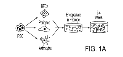
Figs. 1A-10. Reconstruction of Anatomical and Physiological Properties of the
Human Blood-brain- barrier in vitro (iBBB). 1A, Schematic of iBBB formation
from
iPSCs. 1B, iBBB stained for endothelial cell marker CD144 demonstrating the
presence
of multicellular endothelial vessels. Scale bar, 50 p.m. 1 C, Pericytes
localize to
endothelial vessels after two weeks in culture. Pericytes are labeled with
5M22 (also
known as TAGLN) and BEC labeled with tight junction protein ZO-1. Scale bar,
50 p.m.
1D, Pericytes are labeled with NG2 and BECs with CD144. 1E, Astrocytes
surround
.. endothelial vessels after two weeks in culture. Astrocytes are labeled with
GFAP and BECs
are labeled with CD144. Scale bar, 50 p.m. 1F, Aquaporin 4 (AQP4), is
expressed on BEC
vessels labeled with ZO-1, pan-astrocyte marker 510013. Scale bar, 50 p.m. 1G,
qRT-
PCR measuring the expression of genes reported to be predictive markers of BBB
models. All expression is normalized to pan-endothelial marker PECAM to
account for
potential differences in BEC cell number. CLDN, RAGE, JAMA, and LRP1; p <
0.0001. PgP; p = 0.0001, GLUT1; p = 0.0032. 1H, qRT-PCR measuring the
expression of
transporters, adhesion molecules, and efflux-pumps, and tight-junctions found
in the BBB.
All expression levels are normalized to BECs alone. Y-axis is the expression
level in BECs
isolated from the iBBB normalized to BECs cultured alone. X-axis is BECs co-
cultured
with astrocytes normalized to BECs cultured alone. Circles represent means
from three
biological replicates and three PCR replicates. II, Cartoon depicting
transwell setup for
measuring iBBB permeability 1J, Representative image of BECs (ZO-1), pericytes
(5M22) and astrocytes (S10013) co-cultured on transwell membrane. 1K, Trans-
endothelial electrical resistance (TEER) measurements from HuVECs, HuVECs plus
pericytes (P) and astrocytes (A), BECs only and the iBBB. Circles represent
single
measurements from individual transwells. Differences were analyzed by one-way
ANOVA with Bonferroni's post-hoc analysis (p < 0.0001). 1L, Permeability of
fluorescently labeled molecules for BECs alone or iBBB. All values are
reported as a
percent of each molecule's permeability across a blank transwell membrane.
Stars
.. represent significance determined by multiple student's t-test (FDR =
0.01). 1M, BBB
properties of the iBBB require cooperative interaction of pericytes and
astrocytes. The
permeability of 4 kDa dextran was quantified in the iBBB and compared to BECs
with 2x
-5-
CA 03127452 2021-07-21
WO 2020/154374
PCT/US2020/014572
pericytes, 2x astrocytes, or BECs with mouse embryonic fibroblasts (MEFs).
Permeability
is normalized to BECs alone. One-way ANOVA (p < 0.0001) with Bonferroni's
multiple
comparisons. 1N, ABCG2 expression is up-regulated in the iBBB. One-way ANOVA
with Bonferroni's post-hoc analysis (p < 0.0001). 10, Polarization of Pgp was
measured by rhodamine 123 transport for both a BECs monolayer and the iBBB
from the
apical to basolateral surface and vice versa. Inhibitor-treated samples were
normalized to
each respective non-inhibitor-treated sample. Stars represent significance
determined by
multiple student's t-tests (FDR = 0.01).
Figs. 2A-2L. APOE4 increases A13 accumulation in the iBBB. 2A,
Cartoon depicting the experimental paradigm for exposing iBBBs to exogenous
amyloid-I3 2B, A13 selectively accumulates on non-AD iBBBs exposed to media
conditioned by iPSC-derived neuronal cells from a familial AD patient with an
APP-
duplication (APP1.1). iBBB derived from APOE3/3 iPSC line (E3/3 parental) from
a
healthy 75-year-old female. 6e10 antibody recognizes AI31-16 epitope. Scale
bar, 50 p.m.
2C, The APOE3/3 parental iPSC line was genetically edited to an isogenic
APOE4/4
allowing the generation of genetically identical iBBBs. Isogenic APOE4/4 iBBBs
accumulated more A13 compared to the parental APOE3/3 iBBB when simultaneously
exposed to APP1.1 conditioned media for 96 hours. Scale bar, 50 p.m. 2D,
Quantification
of A13 accumulation in two isogenic iBBBs with reciprocal genetic editing
strategies.
Arrows indicate direction of genetic editing where the right-facing arrow
indicates editing
from APOE3/3 to APOE4/4 and the left-facing arrow indicates editing from
APOE4/4 to
APOE3/3. Total area positive for A13 was divided by total nuclei and then
normalized to
the mean amyloid/nuclei from all E3/3 samples such that the mean of E3/E3 is
set to
100%. Automated image analysis was performed with ImageJ. Student t-test (p =
0.0114). 2E, APOE3/4 heterozygous iBBBs accumulate significantly more A13 than
APOE3/3 iBBBs. Quantification performed as described in 2D. 2F, Representative
images
depicting that iBBBs derived from isogenic APOE3/3 and APOE4/4 individuals
exhibit
high levels of amyloid accumulation assay with anti- amyloid antibody D54D2.
2G,
Quantification of amyloid in isogenic iBBBs for Thioflavin T (p = 0.0258), and
two
.. different amyloid antibodies D54D2 (p =0.0020) and 12F4 (p = 0.0054). 2H,
Quantification of soluble versus in soluble A13 1-40 in remaining in the iBBB
culture
media 96 hours after inoculation with 20 nM A13 1-40 (p = 0.0319). 21
Representative
-6-
CA 03127452 2021-07-21
WO 2020/154374
PCT/US2020/014572
three-dimensional IMARIS renderings depicting vascular amyloid accumulation in
APOE3/3 and APOE4/4 iBBBs. iBBBs were allowed to mature for 1 month and then
simultaneously exposed to neuronal conditioned media from the fAD APP1.1 line.
Three-dimensional surfaces of 6e10 and Vecad staining were created using
IMARIS
software. The total area of 6e10 within 20 i.t.M of the Vecad surfaces was
measured. This
was normalized to the total area of the Vecad surfaces Scale bar, 10 p.m. 2J,
Quantification
of vascular (< 20 p.m from BEC vessel) (p = 0.0055) and non-vascular (> 20 p.m
from
BEC vessel) (p= 0.0062) using IMARIS software. Amyloid area was normalized to
total
vascular area for each image. 2K, Representative image depicting amyloid
accumulation
in non-vascular cells positive for astrocyte marker S1000 Scale bar p.m. 2L,
Quantification showing the number of astrocytes positive for amyloid for each
isogenic
genotype. (p= 0.0003).
Figs. 3A-3E. Pericytes are required for increased A13 deposition in the
iBBB. 3A, Representative images depicting combinatorially interchange of E3/3
and E4/4
isogenic cell-types reveals that E4/4 expression in pericytes is required for
increased
A13 iBBB accumulation. 3B, Quantification of A13 accumulation in isogenic
iBBBs for
each permutation of combinatorial matrix. 3C, Segregating each isogenic
permutation
based on relative A13 levels (low or high), reveals that E3/3 and E4/4 BECs
and
astrocytes are equally represented between the two conditions, however,
pericytes are not.
.. For the low A13 condition only E3/3 pericytes are present. In contrast, for
the high A13
condition, only E4/4 pericytes are present. 3D, Quantification of A13
accumulation in
iBBBs derived from AP03/3 (3), H9 is APOE3/4 heterozygous and 210 is APOE3/3
homozygous. 3E, Quantification of A13 accumulation in isogenic iBBBs and
APOE3/3
iBBBs treated with pericyte conditioned media from either E3/3 (parental) or
E4/4
(isogenic) pericytes. Media was conditioned for 48 hours and added iBBBs with
1:1 ratio
of fresh media and 20 nM A13-FITC for 96 hours.
Figs. 4A-4L. APOE and Calcineurin signaling are up-regulated in APOE4
pericytes. 4A, Heat map depicting differentially expressed genes between
isogenic
APOE3/3 and APOE4/4 pericytes. (q = 0.01) 4B, APOE gene expression is
significantly
up-regulated in APOE4/4 pericytes whereas it is down-regulated in E4/4
astrocytes.
Expression values from qRT-PCR from different RNA than used for RNAseq
experiment Astrocyte (p = 0.0009), Pericytes (p < 0.0001). 4C,
Immunofluorescence
-7-
CA 03127452 2021-07-21
WO 2020/154374
PCT/US2020/014572
staining and quantification of APOE in isogenic pericytes. Scale bar, 50 p.m.
Dots are
mean APOE fluorescence intensity from four independent images from a single
well.
Four wells were measured for each genotype. Unpaired Two- tailed t test (p =
0.0005).
4D, Western blot and quantification for APOE protein in APOE isogenic
pericyte. Two
constitutively expressed proteins in pericytes are included smooth muscle
actin (SMA) and
GAPDH. (p = 0.0033) 4E, qRT-PCR showing APOE gene expression is also up-
regulated
in an additional isogenic pair that was edited from E4/4 to E3/3 and three
APOE3/4
heterozygous pericytes from iPSC lines derived from individuals with sporadic
AD and
H9 hESC line. Arrows indicated the direction of genetic editing. All values
are
normalized to the mean expression in all APOE3/3 (n = 4) pericytes.
Significance
determined by One-way ANOVA (p <0.0001) with Bonferroni's multiple comparison
test
to E3/3 pericytes. 4F, Violin plots depicting APOE expression in pericytes
isolated from
post-mortem hippocampus of APOE4 carriers. Differential expression was
measured
using a two-tailed Wilcoxon rank sum test, considering cells with detected
expression of
APOE. 4G, Representative images and quantification depicting the expression of
APOE
protein in hippocampal NG2-positive pericytes in post-mortem brains from APOE4-
carriers (n= 6) and non-carriers (n = 6). For each genotype more than 250 NG2-
positive
pericytes were identified. Unpaired t test, p = 0.0068. 4H, Isogenic iBBBs
that are
deficient for APOE by genetically knocking-out (KO) display similar amyloid
accumulation to E3/3 iBBBs. Significance displayed as One-way ANOVA (p <
0.0001)
with Bonferroni's multiple comparison test 41 Immunodepleting APOE from APOE4
pericyte conditioned media significantly reduces amyloid accumulation in the
APOE3
iBBB. One-way ANOVA (p < 0.0001) with Bonferroni's multiple comparison test.
4J,
Transcription factors differentially expressed between APOE3/3 and E4/4
isogenic pairs
(q < 0.05). The five transcription factors highlighted are reported to bind
APOE gene
regulatory elements. 4K, APOE isogenic pericytes stained for NFATc 1 and 5M22.
NFATc 1 is present in both cytoplasm and nucleus. Dephosphorylation of NFAT by
calcineurin leads to NFAT translocation to the nucleus. Quantification of
NFATc 1
staining per nuclei for each APOE3/3 and APOE4/4. 150 cells were analyzed for
each
genotype. Significance determined by students t-test, (p <0.0001). 4L, Nfatcl
expression in
brain pericytes of APOE3 and APOE4 knock-in mice. Unpaired two-tailed t test
(p =
0.0041). was measured using a two- tailed Wilcoxon rank sum test, considering
cells with
-8-
CA 03127452 2021-07-21
WO 2020/154374
PCT/US2020/014572
detected expression of APOE.
Figs. 5A-5N. Inhibition of Calcineurin reduces APOE expression and
ameliorates A13 deposition 5A and 5B Expression of APOE in isogenic (a) and
heterozygous (b) pericytes after two weeks treatment with DMSO, CsA, FK506 or
INCA6. One-way ANOVA (p < 0.0001) with Bonferroni's multiple comparison. 5C,
Soluble APOE protein is significantly reduced following two-week treatment
with
calcineurin inhibitor CsA. APOE concentration in pericyte conditioned media
was
quantified using ELISA from three separated biological replicates. Multiple
Student t-
tests. Discovery determined using FDR method with Benjamini and Hochberg with
Q =
1%. 5D and 5E, Expression of NFATc 1 (d) and APOE (e) is down- regulated in
pericytes by CsA treatment. Bars are mean value from 3 biological replicates
One-way
ANOVA (NFATcl, p = 0.0013; APOE, p < 0.0001) with Bonferroni's multiple
comparison 5F, Heat map depicting differentially expressed genes between
isogenic
APOE3/3 pericytes treated with DMSO and APOE4/4 pericytes treated with DMSO,
or
2 i.t.M CsA. Genes and organized by hierarchical clustering using Spearmann's
Rank
correlation with average linkage. Boxes outline genes clustering together. The
total genes
for each cluster are presented on the right side of the heatmap depicted
values are mean
normalized counts from 3 independent biological replicates 5G, Representative
images of
E4/4 pericytes treated with DMSO, CsA, or FK506 for two weeks and then exposed
to 20
nM A13-FITC for 96 hours. 5H, Quantification of A13 accumulation in iBBBs
treated
with DSMO, CsA, or FK506. iBBBs were pre-treated with chemicals for two weeks
and
then exposed to 20 nM A13 for 96 hours. Significance determined via One-way
ANOVA
(p < 0.0001) with Bonferroni's multiple comparison. (Scale bar = 10 p.m) 51,
Quantification of A13 accumulation in APOE3/4 heterozygous iBBBs treated with
DSMO, CsA, or FK506. iBBBs were pre-treated with chemicals for two weeks and
then exposed to 20 nM A13 for 96 hours. Significance determined via One-way
ANOVA (p < 0.0001) with Bonferroni's multiple comparison. 5J, Quantification
of
A13 accumulation in iBBBs treated with conditioned media from APOE4/4 pericyte
that
were treated with calcineurin inhibitors for one at least week prior media
harvesting. One-
way ANOVA (p< 0.0001) with Bonferroni's multiple comparisons. 5K, APOE protein
concentration in the hippocampus of mice treated with either cyclosporine A or
vehicle.
APOE was measured by ELISA. Each dot represents mean APOE concentration from
one
-9-
CA 03127452 2021-07-21
WO 2020/154374
PCT/US2020/014572
mouse. Unpaired two-tailed t test (p = 0.0456). 5L, Representative image and
quantification of immunostaining for APOE in cortical pericytes from APOE4 KI
x 5xFAD
mice treated with cyclosporine A or vehicle. Unpaired two-tailed t test (p =
0.0427). 5M,
Representative image of concurrent reduction of vascular APOE protein and
amyloid
following a three-week treatment with CsA. 5N, Representative images and
quantification of vascular amyloid in the hippocampus following treatment of 6-
month-
old APOE4KI x 5XFAD female mice with either vehicle or CsA for three weeks.
Amyloid was detected and quantified with two independent anti-amyloid
antibodies
(6e10 and 12F4). Unpaired two-tailed t test (6e10, p = 0.0055; 12F4, p =
0.0242). (Scale
Bars = 25 p.m).
Figs. 6A-60. 6A and 6B iPSC-derived brain endothelial cells stained with CD144
(VE-Cadherin), CD31 (PECAM), ZO1 and GLUT1. 6C and 6D, iPSC-derived astrocytes
stained with GFAP, S loop and AQP4 6E and 6F Comparative expression analysis
of
genes in iPSC-derived astrocytes from RNA-sequencing that are reported to be
the most
differentially upregulated in 6E, fibroblasts and 6F, oligodendrocytes when
compared
to astrocytes from 6G, 6H, 61 iPSC-derived pericytes stained with CD13, 5M22,
NG2,
and SMA. 6J. Comparative expression analysis of the top differentially
upregulated
genes in pericytes compared to smooth muscle cells (SMCs). Expression is
represented as
FPKM values from bulk RNA-sequencing 6K, Comparative expression analysis of
the
top differentially upregulated genes in SMCs compared to pericytes. Expression
is
represented as FPKM values from bulk RNA-sequencing 6L, Expression of the top
three
differentially upregulated genes in pericytes compared to fibroblasts. 6M,
Expression of
the top three differentially upregulated genes in fibroblasts compared to
pericytes. 6N,
Expression of pericyte and mesenchymal marker genes in iPSC-derived pericytes.
For 6E,
6F, 6J, 6K, 6L, 6M, differential gene lists are based on analysis provided
shown as average
counts compared to FPKM from bulk RNA-sequencing of iPSC- derived astrocytes
and
pericytes. 60, Global hierarchical clustering of transcriptomes (23,588 genes)
demonstrates that iPSC-derived pericytes cluster with primary human brain
pericytes.
Clustering was performed by spearman rank correlation with complete linkage.
Mouse
brain pericyte transcriptional dataset was obtained from G5E117083. Arterial
smooth
muscle cell (SMC) dataset from G5E78271.
Figs 7A-7J. 7A Three-dimensional vascular network of endothelial cells stained
-10-
CA 03127452 2021-07-21
WO 2020/154374
PCT/US2020/014572
with CD144 scale bar = 200 p.m. 7B, one week after formation pericytes labeled
with
SM22 are homogeneously dispersed and rudimentary vessels started forming.
After two
weeks endothelial vessels have formed and pericytes have homed to perivascular
space.
7C, Astrocytes are dispersed throughout iBBB cultures. 7D, mRNA expression of
AQP4 in each cell type alone, pair-wise and combined. 7E, iBBB without
astrocytes do
not stain for AQP4. In iBBBs with astrocytes AQP4 densely stains along
endothelial
vessels. 7F, Immunostaining for LAMA4 showing that Matrigel does not contain
LAMA4 however iBBB cultures remodel basement membrane surrounding endothelial
vessels to contain LAMA4. 7G, PLVAP mRNA expression is upregulated in BECs
from iBBB cultures compared to BECs cultured alone. 7H, PLVAP mRNA expression
is downregulated in BECs from iBBB upon removal of VEGFA from culture media.
71,
iBBB cultured in trans-well format express high levels of BBB marker CLDN5 and
Z01.
7J, Polarization of ABCG2 was measured by Hoechst transport for both a BECs
monolayer and the iBBB from the apical to the basolateral surface and vice
versa.
Samples treated with the ABCG2 specific inhibitor K0143 were normalized to
each
respective non-inhibitor treated sample. Stars represent significance
determined by multiple
student's t-test (FDR = 0.01).
Figs. 8A-8J. 8A iBBBs generated from a familial AD patient iPSC with
duplication
of the APP gene (APP1.1) do not inherently have higher amyloid levels than non-
AD
controls (AG09173). 8B, iBBBs generated from iPSCs with a familial AD-
associated
mutation (M1461) in the PSEN1 gene do not inherently have higher amyloid
levels than its
non-AD isogenic control. 8C, Media conditioned by neuronal cells derived from
familial
AD patient has significantly higher A13 (1-42). Student t-test (p = 0.0022).
8D,
Representative images depicting that iBBBs derived from APOE3/4 individuals
exhibit
high levels of A13 accumulation relative to iBBBs generated from APOE3/3
individuals.
8E and 8F, Representative images depicting that iBBBs derived from isogenic
APOE3/3
and APOE4/4 individuals exhibit high levels of amyloid accumulation assay with
anti-
amyloid antibody Thioflavin T (f) and 12F4 (e). 8G and 8H, Representative
images and
quantification of A13 accumulation in isogenic iBBBs exposed to 20 nM A13-FITC
for 1-
40 and 1-42 isoforms. The total area positive for A13 was divided by total
nuclei and
then normalized to the mean amyloid/nuclei from all E3/3 samples such that the
mean
of E3/E3 is set to 100% for each isoform. Students t-test, 1-40 p = 0.0044; 1-
42 p>
-11-
CA 03127452 2021-07-21
WO 2020/154374
PCT/US2020/014572
0.00001. 81 and 8J, Normalized amyloid accumulation in isogenic pericyte
Figs. 9A-9C. 9A, Quantification of A13 accumulation in deconstructed iBBBs.
BPA3 and BPA4 indicate all E3/3 and E4/4 iBBBs respectively where B = BECs
only,
BA = BECs and astrocytes, and BP = BECs and pericytes. Analysis was performed
by
One-way ANOVA with Bonferroni's post-hoc analysis (p < 0.0001). 9B, Exposing
APOE4/4 astrocytes to APOE4/4 pericyte conditioned media significantly
increases
amyloid accumulation compared APOE3/3 pericyte conditioned media. Student t
test,
p < 0.0001. 9C Quantification and representative image of APOE protein
expression in
pericytes (NG2-positive cells) and non-pericytes (NG2-negative) cells. Student
t test, p <
0.0001.
Figs. 10A-10H. 10A and 10B, GO analysis (from Toppfun) depicting biological
processes associated with up-regulated (a) and down-regulated (b) genes. 10C
and 10D,
Expression of APOE in isogenic pericytes (c) and astrocyte (d) measured by RNA
sequencing each condition represents three biological replicates pericyte, q =
0.0003
astrocyte, q = 0.0006 10E Violin plots depicting APOE expression in pericytes
isolated
from post-mortem prefrontal cortex of APOE4-carriers (n = 7) compared to non-
carriers (n
= 18). Differential expression was measured using a two-tailed Wilcoxon rank
sum test,
considering cells with detected expression of APOE (p = 0.0026). 'OF, Images
and
quantification of APOE protein expression in post-mortem human prefrontal
cortex from
APOE4 carriers and non-carriers. Unpaired two-tailed t test (p = 0.023). 10G,
Differential
plot of representative maker genes showing that pericytes and endothelial
cells isolated
from human hippocampus segregated into distinct cellular clusters 10H, Violin
plots
depicting APOE expression in endothelial cells isolated from post-mortem
hippocampus
APOE4-carriers (n = 16) compared to non-carriers (n = 46). Differential
expression was
measured using a two-tailed Wilcoxon rank sum test, considering cells with
detected
expression of APOE.
Figs. 11A-11L. 11A, Increasing the soluble APOE concentration through the
addition of recombinant APOE protein to iBBB culture increases amyloid
accumulation.
One-way ANOVA with Bonferroni's post-hoc analysis (p = 0.0011)K 11B and 11C,
Representative western blot and quantification depicting nuclear NFATcl
expression in
isogenic APOE3 and 4 pericytes. Unpaired student t test, p = 0.0254. 11D,
Expression
of calcineurin catalytic subunits measured by RNAseq. PPP3CA (q = 0.0003);
PPP3CC
-12-
CA 03127452 2021-07-21
WO 2020/154374
PCT/US2020/014572
(q = 0.0188).11E, Expression of negative Regulators of Calcineurin genes
(RCANs)
measured by RNAseq. RCAN2 (q = 0.0003); RCAN3 (q = 0.0123). 11F, Expression of
DYRKs kinases known to phosphorylate NFAT measured by RNAseq. DYRK4 (q =
0.0003). 11G, Expression of predicted NFAT response gene, VCAM1 and ACTG2, in
pericytes. Expression is quantified by qRT-PCR and normalized to the average
of E3/3
cells. Significance determined by One-way ANOVA (p <0.0001) with Bonferroni's
multiple
comparison. 11H and 111, Violin plots depicting NFATC1 (h) and NFATC2 (i)
expression in pericytes isolated from post-mortem prefrontal cortex of APOE4-
carriers
(n = 16) compared to non-carriers (n = 46). Differential expression was
measured using
a two-tailed Wilcoxon rank sum test, considering cells with detected
expression of APOE.
11J and 11K, Violin plots depicting NFATC1 and NFATC2 expression in
endothelial cells
isolated from post- mortem hippocampus of APOE4-carriers (n = 16) and non-
carriers (n =
46). Differential expression 11L, Violin plots depicting NFATC2 expression in
endothelial
cells isolated from post-mortem prefrontal cortex of APOE4-carriers (n = 7
compared to
non-carriers (n = 18). Differential expression was measured using a two-tailed
Wilcoxon
rank sum test, considering cells with detected expression of APOE (p = 0.035).
Figs. 12A- 12K. 12A, Chemical structures of CsA, FK506, and INCA6 showing
highly dissimilar structures. 12B, Expression of PGK1, HPRT, and GAPDH in
pericytes
after two weeks with DMSO, Cyclosporine A (CsA), FK506 or INCA6. One-way
ANOVA (p < 0.0001) with Bonferroni's multiple comparison. 12C and12D,
Representative immunofluorescence imaging of APOE protein staining in
pericytes after
two weeks of treatment with chemicals. Scale bar, 50 p.m. 12E DEGs and
associated GO
terms for up-regulated and down-regulated genes in E3 and E4 CsA-treated
pericytes. 12F
and 12G. Representative imaging and quantification depicting APOE protein
expression
in the APOE4KI mouse cortical slices following treatment with cyclosporine A
(CsA) for
one week. Unpaired, two tailed t test (p = 0.0009). 12H, Quantification of
amyloid
APOE4KI mouse cortical slices treated with either CsA or FK506 for one week
and then
exposed to 20 nM A13 for 48 hours. One-way ANOVA (p = 0.0105) with
Bonferroni's
multiple comparison. 121, APOE mRNA expression in primary pericytes isolated
from
brain microvasculature of APOE4 knock-in mice treated with DMSO, Cyclosporine
A, or
FK506. One-way ANOVA (p = 0.0139) with Bonferroni's multiple comparison. 12J,
Representative image of immunostaining for APOE in hippocampal pericytes from
-13-
CA 03127452 2021-07-21
WO 2020/154374
PCT/US2020/014572
APOE4 KI x 5xFAD mice treated with cyclosporine A or vehicle for one week.
12K,
Representative images of vascular amyloid in the hippocampus following
treatment of 6-
month-old APOE4KI x 5XFAD female mice with either vehicle or CsA. Amyloid was
detected and quantified with two independent anti-amyloid antibodies (6e10 and
12F4).
Figs. 13A-13C show the genotype distinction between APOE4/4 cells (isogenic)
and APOE3/3 (Parental) in permeability of a BBB membrane. 13A is a schematic
showing
the iBBB with fluorescent molecules positioned on the Apical surface. 13B is a
schematic
showing the iBBB with fluorescent molecules transitioning through the iBBB
from the
Apical surface to the Basolateral surface. 13C shows that the iBBB prepared
with isogenic
APOE4/4 cells allows greater permeability and accumulation of the fluorescent
molecules
than iBBB generated using parental APOE3/3 cells.
Figs. 14A-14B show the genotype distinction between APOE4/4 cells (isogenic)
and APOE3/3 (Parental) in permeability of a BBB membrane. 14A is a schematic
showing
the iBBB with fluorescent molecules positioned on the Apical surface. 14B is a
graph
.. showing that the iBBB prepared with isogenic APOE4/4 cells allows greater
permeability
and accumulation of multiple compounds than iBBB generated using parental
APOE3/3
cells.
Figs. 15A-15F shows that APOE4 increases the permability of iBBB membrane.
15A is a graph showing that the iBBB prepared with isogenic APOE4/4 cells
allows
greater permeability and accumulation of cadaverine molecules on the
Basolateral surface
of the iBBB than iBBB generated using parental APOE3/3 cells. 15B is a graph
showing
that the iBBB prepared with isogenic APOE4/4 cells allows greater permeability
and
accumulation of 4 kDa Dextran molecules on the Basolateral surface of the iBBB
than
iBBB generated using parental APOE3/3 cells. 15C is a graph showing that the
iBBB
prepared with isogenic APOE4/4 cells allows greater permeability and
accumulation of 10
kDa Dextran molecules on the Basolateral surface of the iBBB than iBBB
generated using
parental APOE3/3 cells. 15D is a graph showing that the iBBB prepared with
isogenic
APOE4/4 cells allows greater permeability and accumulation of BSA molecules on
the
Basolateral surface of the iBBB than iBBB generated using parental APOE3/3
cells. 15E
is a graph showing that the iBBB prepared with isogenic APOE4/4 cells allows
greater
permeability and accumulation of 70kDa Dextran molecules on the Basolateral
surface of
the iBBB than iBBB generated using parental APOE3/3 cells. 15F is a graph
showing that
-14-
CA 03127452 2021-07-21
WO 2020/154374
PCT/US2020/014572
the iBBB prepared with isogenic APOE4/4 cells allows greater permeability and
accumulation of transferrin molecules on the Basolateral surface of the iBBB
than iBBB
generated using parental APOE3/3 cells.
Fig. 16 is a graph showing that the iBBB prepared with isogenic APOE4/4 cells
allows greater permeability and accumulation of A1342-FITC on the Basolateral
surface of
the iBBB than iBBB generated using parental APOE3/3 cells.
Figs. 17A-17C show in vivo cyclosporine A reduces APOE in and around cortical
pericytes. 17A is a schematic showing the experimental steps wherein APOE4K1 x
5xFAD mice are injected with vehicle control or 10 mg/kg cyclosporin A
intraperitoneal,
daily for 3 weeks. APOE protein and vascular amyloid are quantified. 17B is a
graph
showing the results generated by ELISA assay and demonstrating that
cyclosporin A
resulted in less production of APOE protein relative to vehicle. 17C is images
and a graph
showing the results of immunohistochemistry of the hippocampus and
demonstrating that
cyclosporin A resulted in less accumulation of APOE protein relative to
vehicle.
Figs. 18A-18B show in vivo cyclosporine A reduces APOE and vascular amyloid
in and around hippocampus vasculature. 18A is an image showing the results
generated
by immunohistochemistry of the hippocampus and demonstrating that cyclosporin
A
resulted in less production of APOE/amyloid protein relative to vehicle. 18B
is images
and a graph showing the results of immunohistochemistry of the hippocampus and
demonstrating that cyclosporin A resulted in less accumulation of vascular
amyloid
protein relative to vehicle.
Figs. 19A-19D show in vivo cyclosporine A and FK506 reduce APOE and vascular
amyloid in and around hippocampus vasculature in vivo. 19A is an image showing
the
results generated by immunohistochemistry of the hippocampus and demonstrating
control levels of vascular amyloid protein. 19B is an image showing the
results generated
by immunohistochemistry of the hippocampus and demonstrating that cyclosporin
A (10
mg/ml) resulted in less production of amyloid protein relative to vehicle
control. 19C is
an image showing the results generated by immunohistochemistry of the
hippocampus
and demonstrating that FK506 (10 mg/ml) resulted in less production of amyloid
protein
relative to vehicle control. 19D is a graph depicting the results of the data
generated in
19A-19C.
-15-
CA 03127452 2021-07-21
WO 2020/154374
PCT/US2020/014572
DETAILED DESCRIPTION
A human 3D in vitro model of the BBB (iBBB) which recapitulates numerous
molecular and physiological features of the in vivo BBB has been developed.
The iBBB is a
unique model of a capillary system which allows for the analysis of capillary
transport and
activity. Prior art artificial BBB s have typically been 2 dimensional systems
and/or of a larger
size that more closely mimics a larger vessel. The iBBB of the invention
provides advantages
not previously found in prior art devices.
As described in further detail in the Examples, the iBBB has been developed
and
extensively studied herein. It's relevance to the physiologic system has been
established
through extensive analysis and characterization. The iBBB was further designed
and
validated as a neurodegenerative model. This was through the elucidation of
the mechanisms
underlying one of the strongest genetic risks factor (APOE4) for
cerebrovascular amyloid
accumulation. The data generated and described herein using the iBBB revealed
that
pericytes, the smooth muscle component of cerebral vasculature, are required
for the
pathogenic effects of APOE4. Subsequent mechanistic dissection pinpointed that
APOE itself
is highly up-regulated in APOE4 pericytes and that up-regulation is required
for increased
amyloid accumulation. Using post-mortem human brain tissue, it was confirmed
that APOE
is also upregulated in human brain pericytes of APOE4 carriers compared to non-
carriers.
Global transcriptional profiling further revealed that CaN/NFAT signaling in
E4 pericytes is
highly active. It was further demonstrated that pharmacological inhibition of
CaN/NFAT
signaling markedly reduced APOE expression in the iBBB and in vivo mouse brain
and
rescues the pathological amyloid phenotype observed in APOE4 iBBBs. These
findings have
profound implications for the treatment, diagnosis and further analysis of
cerebral amyloid
angiopathy (CAA). CAA is a form of angiopathy in which amyloid beta (AP)
peptide is
deposited in the walls of small to medium blood vessels of the central nervous
system and
meninges. The buildup of AP is associated with cognitive decline.
NFAT/CaN signaling is up-regulated during cognitive aging and
neurodegeneration.
In aged rats, up-regulation of CaN leads to poor cognitive performance.
Despite the
correlation of up-regulated NFAT/CaN signaling in neurodegeneration it remains
unknown
whether NFAT/CaN has a causal role in neurodegeneration. Uncertainty
surrounding whether
CaN and NFAT are viable targets for treatment of neurodegenerative disease
such as
Alzheimer's disease (AD) and who would benefit from these treatments has
limited the
-16-
CA 03127452 2021-07-21
WO 2020/154374
PCT/US2020/014572
development of therapeutic strategies in this area. The results described
herein, provide
significant advances in understanding the system and identifying therapeutic
targets for the
treatment of disease associated with AP deposition on small vessels. The data
identify the
cell-type (pericytes), soluble factor (APOE), and regulatory pathway
(calcineurin/NFAT)
.. through which APOE4 acts to predispose CAA pathology. The iBBB was also
demonstrated
to model genotype-related differences in BBB permeability. The relevance of
these
observations to human neurobiology was further validated using post-mortem
human brain
tissue and mouse models to demonstrate that these cellular and molecular
insights can be
translated to an in vivo setting for therapeutic intervention. Through
multiple lines of
evidence, the iBBB has been shown to be a tractable model and provide
biological insight
into how genetic variants can influence cerebral vascular pathology, thereby
opening new
therapeutic opportunities. Importantly, it was shown that treatment of mice in
vivo with
cyclosporine A showed a significant reduction of cerebrovascular amyloid.
Thus, in some aspects, the invention is an in vitro blood brain barrier (iBBB)
that is
composed of a 3 dimensional (3D) matrix having human brain endothelial cell
(BEC), human
pluripotent-derived pericytes and human pluripotent-derived astrocytes
arranged therein. The
human brain endothelial cells (BECs) form a vessel comprised of a large
interconnected
network of human pluripotent-derived positive endothelial cells.
The vessel has a size on the order of a capillary. A capillary is an extremely
small
.. blood vessel located within the tissues of the body that transports blood.
Capillaries measure
in size from about 5 to 10 microns in diameter. Capillary walls are thin and
are composed of
endothelium. The iBBB is on the order of approximately 5 to 50 microns in
length. In some
embodiments, the iBBB is 5 to 30 microns in length. In some embodiments, the
iBBB is 10 to
20 microns in length. In other embodiments, the iBBB is 3-50 microns, 5- 45
microns, 5- 40
microns, 5- 35 microns, 5- 30 microns, 5- 25 microns, 5- 20 microns, 5- 15
microns, 5- 10
microns, 8-50 microns, 8- 45 microns, 8- 40 microns, 8- 35 microns, 8- 30
microns, 8- 25
microns, 8- 20 microns, 8- 15 microns, 8- 10 microns, 10-50 microns, 10- 45
microns, 10- 40
microns, 10- 35 microns, 10- 30 microns, 10- 25 microns, 10- 20 microns, 10-
15 microns, or
10- 12 microns in length.
The endothelial cells, pericytes, and astrocytes are optionally human
pluripotent-
derived cells. For instance, the cells may be iPSC-derived cells, such as iPSC-
derived CD144
positive cells. Autologous induced pluripotent stem cells (iPSCs) can be
differentiated into
-17-
CA 03127452 2021-07-21
WO 2020/154374
PCT/US2020/014572
any cell type of the three germ layers: endoderm (e.g. the stomach linking,
gastrointestinal
tract, lungs, etc), mesoderm (e.g. muscle, bone, blood, urogenital tissue,
etc) or ectoderm (e.g.
epidermal tissues and nervous system tissues). The term "pluripotent cells"
refers to cells that
can self-renew and proliferate while remaining in an undifferentiated state
and that can, under
the proper conditions, be induced to differentiate into specialized cell
types. Pluripotent cells,
encompass embryonic stem cells and other types of stem cells, including fetal,
amniotic, or
somatic stem cells. Exemplary human stem cell lines include the H9 human
embryonic stem
cell line. Additional exemplary stem cell lines include those made available
through the
National Institutes of Health Human Embryonic Stem Cell Registry and the
Howard Hughes
Medical Institute HUES collection.
Pluripotent stem cells also encompasses induced pluripotent stem cells, or
iPSCs, a
type of pluripotent stem cell derived from a non-pluripotent cell. Examples of
parent cells
include somatic cells that have been reprogrammed to induce a pluripotent,
undifferentiated
phenotype by various means. Such "iPS" or "iPSC" cells can be created by
inducing the
expression of certain regulatory genes or by the exogenous application of
certain proteins.
Methods for the induction of iPS cells are known in the art. As used herein,
hiPSCs are
human induced pluripotent stem cells, and miPSCs are murine induced
pluripotent stem cells.
The cells are seeded onto a 3D matrix or scaffold material. The matrix or
scaffold
material, may be, for instance, a hydrogel. The matrix may be formed of
naturally derived
biomaterials such as polysaccharides, gelatinous proteins, or ECM components
comprising
the following or functional variants thereof: agarose; alginate; chitosan;
dextran; gelatin;
laminins; collagens; hyaluronan; fibrin, and mixtures thereof. Alternatively
the matrix may be
a hydrogel formed of Matrigel, Myogel and Cartigel, or a combination of
Matrigel, Myogel
and Cartigel and a naturally derived biomaterial or biomaterials. The hydrogel
may be a
macromolecule of hydrophilic polymers that are linear or branched, preferably
wherein the
polymers are synthetic, more preferably wherein the polymers are poly(ethylene
glycol)
molecules and most preferably wherein the poly(ethylene glycol) molecules are
selected from
the group comprising: poly(ethylene glycol), polyaliphatic polyurethanes,
polyether
polyurethanes, polyester polyurethanes, polyethylene copolymers, polyamides,
polyvinyl
alcohols, poly(ethylene oxide), polypropylene oxide, polyethylene glycol,
polypropylene
glycol, polytetramethylene oxide, polyvinyl pyrrolidone, polyacrylamide,
poly(hydroxy ethyl
acrylate), poly(hydroxyethyl methacrylate) and mixtures thereof.
-18-
CA 03127452 2021-07-21
WO 2020/154374
PCT/US2020/014572
The 3D matrix may be generated using an optimal mixture of endothelial cells,
pericytes, and astrocytes. For instance, in some embodiments the iBBB may be
generated
using about 5 parts endothelial cells to about 1 part astrocytes to about 1
part pericytes. In
other embodiments the iBBB may be generated using about 1 million endothelial
cells per
ml, about 200,000 astrocytes per ml and about 200,000 pericytes per ml.
A unique feature of the 3D matrix is that the cells are seeded onto the matrix
and self-
assemble into a BBB like structure. The cells arrange themselves such that the
BECs form a
large interconnected network of cells, similar to a capillary wall. The
pericytes are arranged
proximal to the BEC vessel on an apical surface. The human pluripotent-derived
astrocytes
are dispersed throughout the 3D matrix. However some of the astrocytes are
positioned
proximal to the BEC vessel and have GFAP- positive projections into the
perivascular space.
The iBBB has structural properties that mimic in vivo BBB tissue. In addition
to the
manner in which the cells assemble in the 3D structure, the iBBB and cells
found therein
have structural properties which are associated with in vivo BBB such as
expression of
specific genes associated with cells in BBB in vivo. For instance the
astrocytes express AQP4
and the BEC may express at least any one of CLDN5, GLUT1, JAMA, PgP, LRP1, and
RAGE. In some embodiments the BEC may express at least any one of PECAM,
ABCG2,
CDH5, CGN, SLC38A5, ABCC2, VWF, and SLC7A5. The cells also produce LAMA4
which has been observed in the matrix. PgP and ABCG2 have been found to be
expressed on
the apical surface of the iBBB. The levels of PgP and ABCG2 expressed on the
apical surface
are 2-3 times greater than levels of PgP and ABCG2 expressed on BEC cultured
alone or co-
cultured with astrocytes. These important markers demonstrate the similarity
with in vivo
BBB.
The iBBB also has functional properties that mimic in vivo BBB tissue.
Functional
properties associated with the iBBB (that mimic in vivo BBB) include, for
instance, a TEER
that exceeds 5,500 Ohm x cm2, reduced molecular permeability and polarization
of efflux
pumps relative to BEC cultured alone or co-cultured with astrocytes.Trans-
endothelial
electrical resistance (TEER) is a measurement of electrical resistance across
an endothelial
monolayer that is used as a sensitive and reliable quantitative indicator of
permeability. All
immortalized endothelial cell lines that form barriers exhibit TEER values
below 150 Ohms/
cm2. Likewise, peripheral endothelial cells such as human umbilical cord
vascular endothelial
cells (HuVECs) have relatively high permeability and thus exhibit low TEER. In
agreement
-19-
CA 03127452 2021-07-21
WO 2020/154374
PCT/US2020/014572
with these reported observations, the data presented herein demonstrate TEER
values of
approximately 100 Ohms/ cm2 when HuVECs were cultured in trans-well
configuration.
HuVEC TEER values did not increase by co-culturing with astrocytes or
pericytes. iPSC-
derived BECs cultured alone had significantly higher TEER values with an
average of 5900
Ohms cm2. However, the TEER values for BECs cultured alone exhibited a high
degree of
variability (SD = +/- 2150 Ohms/cm2). Co-culturing BECs with pericytes and
astrocytes in
the iBBB disclosed herein reduced TEER variability (SD = +/- 513.9 Ohms/cm2)
and led to a
significant increase in the average resistance (8030 Ohms cm2) suggesting the
iBBB is less
permeable than HuVECs, or BECs cultured alone. These functional properties
make the
iBBB unique among capillary sized artificial BBB.
Several AD-risk genes are expressed in cells that constitute the BBB and may
directly
influence the accumulation and clearance of A13. In particular, Apolipoprotein
E (APOE)
protein is highly expressed in cells of the BBB. In humans, there are three
genetic
polymorphisms of APOE, e2, e3, and e4. The E4 isoform of APOE (APOE4) is the
most
.. significant known risk factor for CAA and sporadic AD. The genotype of the
cell plays an
important role in the iBBB and related assays. In some embodiments the AP
producing cell
expressed APOE3 and/or APOE4. The AP producing cell may have an APOE3/3
genotype or
an APOE3/4 genotype or an APOE4/4 genotype. In some embodiments the cells have
an
APOE4/4 genotype.
The data generated herein has revealed that pericytes play an important role
in the
production of amyloid-f3 peptide (AP). In view of these findings, other
aspects of the
invention relate to methods of identifying an inhibitor of amyloid-f3 peptide
(AP) production
and/or accumulation, by contacting an AP producing cell with an APOE4 positive
pericyte
factor and at least one candidate inhibitor and detecting an amount of AP in
the presence and
absence of the candidate inhibitor, wherein a reduced quantity of AP
associated with the cell
in the presence of the candidate inhibitor relative an amount of AP associated
with the cell in
the absence of the candidate inhibitor indicates that the candidate inhibitor
is an inhibitor of
Aft The APOE4 positive pericyte factor may be a soluble factor in APOE4
pericyte
conditioned media, such as APOE protein.
The methods may further involve contacting the BEC vessel described herein
with
the inhibitor of AP, and detecting the effect of the inhibitor of AP on the
production of AP by
the iBBB relative to an iBBB which has not been contacted with the inhibitor
of AP.
-20-
CA 03127452 2021-07-21
WO 2020/154374
PCT/US2020/014572
The invention, in some aspects, relates to methods for inhibiting amyloid
synthesis in
a subject. It has been discovered that subjects having or at risk of
developing amyloid
accumulation can be identified based on genotype, whether they are APOE4
positive and
successfully treated with compounds identified using the assays described
herein. If the
subject is APOE4 positive, those subjects are at risk of developing AP
disorders such as
CAA. However, those subjects are also sensitive to treatment with an inhibitor
of a
calcineurin/NFAT pathway. While APOE4 has previously been associated with
patients that
have some AP disorders such as Alzheimer's, this genotype has not previously
been linked as
a successful predictor of a calcineurin/NFAT inhibitory activity. Prior work
looking at
inhibitors of this pathway in diseased individuals has not shown consistent
positive results in
patients. The findings of the invention have provided a link between genotype
and successful
therapeutic utility of compounds in the calcineurin/NFAT pathway.
NFAT (nuclear factor of activated T cells) is a transcriptional activator. In
its inactive
state NFAT resides in the cytoplasm where it is phosphorylated. Increases in
intracellular
Ca2+ lead to activation of the calmodulin-dependent phosphatase calcineurin
(CaN), which
subsequently dephosphorylates NFAT permitting its translocation to the nucleus
where it
promotes gene activation. In some embodiments the NFAT inhibitor may be a
calcinuerin
inhibitor and/or may be lipid soluble. The NFAT inhibitor may be selected
from: cyclosporin,
cyclosporin derivatives, tacrolimus derivatives, pyrazoles, pyrazole
derivatives, phosphatase
inhibitors, SlP receptor modulators, toxins, paracetamol metabolites, fungal
phenolic
compounds, coronary vasodilators, phenolic adeide, flavanols, thiazole
derivatives,
pyrazolopyrimidine derivatives, benzothiophene derivatives, rocaglamide
derivatives, diaryl
triazoles, barbiturates, antipsychotics (penothiazines), serotonin
antagonists, salicylic acid
derivatives, phenolic compounds derived from propolis or pomegranate,
imidazole
derivatives, pyridinium derivatives, furanocumarins, alkaloids, triterpenoids,
terpenoids,
oligonucleotides, peptides, A 285222, endothall, 4-
(fluoromethyl)phenylphosphate FMPP,
norcantharidin, tyrphostins, okadaic acid, RCP1063, cya/cypa (cyclophilin A),
isa247
(voclosporin)/cypa, [dat-sar]3-cya, fk506/fkbp12, ascomyxin/fkbp12,
pinecrolimus/FKBP12,
1,5-dibenzoyloxymethyl-norcantharidin, am404, btpl, btp2, dibefurin,
dipyridamole,
gossypol, kaempferol, lie 120, NCI3, PD 144795, Roc-1, Roc-2, Roc-3, ST 1959
(DLI111-it),
thiopental, pentobarbital, thiamylal, secobarbital, trifluoperazine,
tropisetron, UR-1505, WIN
53071, caffeic acid phenylethyl ester, KRM-III, YM-53792, punicalagin,
imperatorin,
-21-
CA 03127452 2021-07-21
WO 2020/154374
PCT/US2020/014572
quinolone alkaloids compounds, impres sic acid, oleanane triterpenoid, gomisin
N, CaN457-482-
AID, CaN424-521-AID, mFATc21 06-121-SPREIT, VIVIT peptide, R11-Vivit, ZIZIT
cis-pro,
INCA1, INCA6, INCA2, AKAP79330-357, RCAN1, RCANl-4141-197-exon7, RCAN 1 -4143-
163-
CIC peptide, RCAN1-495-118-SP repeat peptide, LxVPc 1 peptide, MCV1, VacA,
A238L, and
A238200-213.
A calcineurin inhibitor may disrupt the activity of calcineurin directly or
indirectly. In
some embodiments, the calcineurin inhibitor is cyclosporine A, FK506
(tacrolimus),
pimecrolimus, or a cyclosporine analog, such as voclosporin. Cyclosporine A
and FK506 are
both clinically prescribed as immunosuppressants following organ
transplantation. Other
calcineurin inhibitors are known in the art. For instance, others are
disclosed in US
2019/0085040,
A calcineurin/NFAT pathway inhibitor, as used herein, is a compound that
disrupts
the activity of the NFAT pathway. Exemplary calcineurin/NFAT inhibitors
include, but are
not limited to, peptides such as antibodies small molecule compounds, and
other compounds
which may disrupt interactions. Calcineurin/NFAT inhibitors also include small
molecule
inhibitors that directly inhibit one or more components of the
calcineurin/NFAT, or other
agents that inhibit the binding interaction. In some embodiments the small
molecule
inhibitors are Cyclosporin or FK506.
The calcineurin/NFAT inhibitory compounds of the invention may exhibit any one
or
more of the following characteristics: (a) reduces activity of the NFAT
pathway; (b) prevents,
ameliorates, or treats any aspect of a neurodegenerative disease; (c) reduces
synaptic
dysfunction; (d) reduces cognitive dysfunction; and (e) reduces amyloid-f3
peptide (AP)
accumulation. One skilled in the art can prepare such inhibitory compounds
using the
guidance provided herein.
The terms reduce, interfere, inhibit, and suppress refer to a partial or
complete
decrease in activity levels relative to an activity level typical of the
absence of the inhibitor.
For instance, the decrease may be by at least 20%, 50%, 70%, 85%, 90%, 100%,
150%,
200%, 300%,or 500%, or by 10-fold, 20-fold, 50-fold, 100-fold, 1000-fold, or
104-fold.
In other embodiments, the calcineurin/NFAT compounds described herein are
small
molecules, which can have a molecular weight of about any of 100 to 20,000
Daltons, 500 to
15,000 Daltons, or 1000 to 10,000 Daltons. Libraries of small molecules are
commercially
available. The small molecules can be administered using any means known in
the art,
-22-
CA 03127452 2021-07-21
WO 2020/154374
PCT/US2020/014572
including inhalation, intraperitoneally, intravenously, intramuscularly,
subcutaneously,
intrathecally, intraventricularly, orally, enterally, parenterally,
intranasally, or dermally. In
general, when the calcineurin/NFAT inhibitor according to the invention is a
small molecule,
it will be administered at the rate of 0.1 to 300 mg/kg of the weight of the
patient divided into
one to three or more doses. For an adult patient of normal weight, doses
ranging from 1 mg
to 5 g per dose can be administered.
The above-mentioned small molecules can be obtained from compound libraries.
The
libraries can be spatially addressable parallel solid phase or solution phase
libraries. See,
e.g., Zuckermann et al. J. Med .Chem. 37, 2678-2685, 1994; and Lam Anticancer
Drug Des.
12:145, 1997. Methods for the synthesis of compound libraries are well known
in the art,
e.g., DeWitt et al. PNAS USA 90:6909, 1993; Erb et al. PNAS USA 91:11422,
1994;
Zuckermann et al. J. Med. Chem. 37:2678, 1994; Cho et al. Science 261:1303,
1993; Carrell
et al. Angew Chem. Int. Ed. Engl. 33:2059, 1994; Care11 et al. Angew Chem.
Int. Ed. Engl.
33:2061, 1994; and Gallop et al. J. Med. Chem. 37:1233, 1994. Libraries of
compounds may
be presented in solution (e.g., Houghten Biotechniques 13:412-421, 1992), or
on beads (Lam
Nature 354:82-84, 1991), chips (Fodor Nature 364:555-556, 1993), bacteria
(U.S. Patent No.
5,223,409), spores (U.S. Patent No. 5,223,409), plasmids (Cull et al. PNAS USA
89:1865-
1869, 1992), or phages (Scott and Smith Science 249:386-390, 1990; Devlin
Science
249:404-406, 1990; Cwirla et al. PNAS USA 87:6378-6382, 1990; Felici J. Mol.
Biol.
222:301-310, 1991; and U.S. Patent No. 5,223,409).
Alternatively, the inhibitors described herein may inhibit the expression of a
component of the calcineurin/NFAT pathway. Compounds that inhibit the
expression include,
for example, morpholino oligonucleotides, small interfering RNA (siRNA or
RNAi),
antisense nucleic acids, or ribozymes. RNA interference (RNAi) is a process in
which a
dsRNA directs homologous sequence-specific degradation of messenger RNA. In
mammalian cells, RNAi can be triggered by 21-nucleotide duplexes of small
interfering RNA
(siRNA) without activating the host interferon response. The dsRNA used in the
methods
disclosed herein can be a siRNA (containing two separate and complementary RNA
chains)
or a short hairpin RNA (i.e., a RNA chain forming a tight hairpin structure),
both of which
can be designed based on the sequence of the target gene.
Optionally, a nucleic acid molecule to be used in the method described herein
(e.g., an
antisense nucleic acid, a small interfering RNA, or a microRNA) as described
above contains
-23-
CA 03127452 2021-07-21
WO 2020/154374
PCT/US2020/014572
non-naturally-occurring nucleobases, sugars, or covalent internucleoside
linkages
(backbones). Such a modified oligonucleotide confers desirable properties such
as enhanced
cellular uptake, improved affinity to the target nucleic acid, and increased
in vivo stability.
Calcineurin/NFAT inhibitors include antibodies and fragments thereof. An
antibody
(interchangeably used in plural form) is an immunoglobulin molecule capable of
specific
binding to a target, such as a carbohydrate, polynucleotide, lipid,
polypeptide, etc., through at
least one antigen recognition site, located in the variable region of the
immunoglobulin
molecule.
As used herein, the term "antibody" encompasses not only intact (i.e., full-
length)
polyclonal or monoclonal antibodies, but also antigen-binding fragments
thereof (such as
Fab, Fab', F(ab')2, Fv), single chain (scFv), mutants thereof, fusion proteins
comprising an
antibody portion, humanized antibodies, chimeric antibodies, diabodies, linear
antibodies,
single chain antibodies, multispecific antibodies (e.g., bispecific
antibodies) and any other
modified configuration of the immunoglobulin molecule that comprises an
antigen
recognition site of the required specificity, including glycosylation variants
of antibodies,
amino acid sequence variants of antibodies, and covalently modified
antibodies. An antibody
includes an antibody of any class, such as IgD, IgE, IgG, IgA, or IgM (or sub-
class thereof),
and the antibody need not be of any particular class. Depending on the
antibody amino acid
sequence of the constant domain of its heavy chains, immunoglobulins can be
assigned to
different classes. There are five major classes of immunoglobulins: IgA, IgD,
IgE, IgG, and
IgM, and several of these may be further divided into subclasses (isotypes),
e.g., IgGl, IgG2,
IgG3, IgG4, IgAl and IgA2. The heavy-chain constant domains that correspond to
the
different classes of immunoglobulins are called alpha, delta, epsilon, gamma,
and mu,
respectively. The subunit structures and three-dimensional configurations of
different classes
of immunoglobulins are well known.
The inhibitors described herein can be identified or characterized using
methods
known in the art, whereby reduction, amelioration, or neutralization of
compound in the
calcineurin/NFAT pathway is detected and/or measured. Further, a suitable
calcineurin/NFAT inhibitor may be screened from a combinatory compound library
using
any of the assay methods known in the art and/or using the pericyte or iBBB
assays described
herein.
-24-
CA 03127452 2021-07-21
WO 2020/154374
PCT/US2020/014572
One or more of the calcineurin/NFAT inhibitors described herein can be mixed
with a
pharmaceutically acceptable carrier (excipient), including buffer, to form a
pharmaceutical
composition for use in reducing calcineurin/NFAT pathway activity.
"Acceptable" means
that the carrier must be compatible with the active ingredient of the
composition (and
preferably, capable of stabilizing the active ingredient) and not deleterious
to the subject to be
treated. As used herein a pharmaceutically acceptable carrier does not include
water and is
more than a naturally occurring carrier such as water. In some embodiments the
pharmaceutically acceptable carrier is a formulated buffer, a nanocarrier, an
IV solution etc.
Pharmaceutically acceptable excipients (carriers) including buffers, which are
well
known in the art. See, e.g., Remington: The Science and Practice of Pharmacy
20th Ed.
(2000) Lippincott Williams and Wilkins, Ed. K. E. Hoover. The pharmaceutical
compositions
to be used in the present methods can comprise pharmaceutically acceptable
carriers,
excipients, or stabilizers in the form of lyophilized formulations or aqueous
solutions.
(Remington: The Science and Practice of Pharmacy 20th Ed. (2000) Lippincott
Williams and
Wilkins, Ed. K. E. Hoover). Acceptable carriers, excipients, or stabilizers
are nontoxic to
recipients at the dosages and concentrations used, and may comprise buffers
such as
phosphate, citrate, and other organic acids; antioxidants including ascorbic
acid and
methionine; preservatives (such as octadecyldimethylbenzyl ammonium chloride;
hexamethonium chloride; benzalkonium chloride, benzethonium chloride; phenol,
butyl or
benzyl alcohol; alkyl parabens such as methyl or propyl paraben; catechol;
resorcinol;
cyclohexanol; 3-pentanol; and m-cresol); low molecular weight (less than about
10 residues)
polypeptides; proteins, such as serum albumin, gelatin, or immunoglobulins;
hydrophilic
polymers such as polyvinylpyrrolidone; amino acids such as glycine, glutamine,
asparagine,
histidine, arginine, or lysine; monosaccharides, disaccharides, and other
carbohydrates
including glucose, mannose, or dextrans; chelating agents such as EDTA; sugars
such as
sucrose, mannitol, trehalose or sorbitol; salt-forming counter-ions such as
sodium; metal
complexes (e.g., Zn-protein complexes); and/or non-ionic surfactants such as
TWEENTm
(polysorbate), PLURONICS TM (poloxamers) or polyethylene glycol (PEG).
Pharmaceutically
acceptable excipients are further described herein.
In some examples, the pharmaceutical composition described herein comprises
liposomes containing the calcineurin/NFAT inhibitor, which can be prepared by
methods
known in the art, such as described in Epstein, et al., Proc. Natl. Acad. Sci.
USA 82:3688
-25-
CA 03127452 2021-07-21
WO 2020/154374
PCT/US2020/014572
(1985); Hwang, et al., Proc. Natl. Acad. Sci. USA 77:4030 (1980); and U.S.
Pat. Nos.
4,485,045 and 4,544,545. Liposomes with enhanced circulation time are
disclosed in U.S.
Pat. No. 5,013,556. Particularly useful liposomes can be generated by the
reverse phase
evaporation method with a lipid composition comprising phosphatidylcholine,
cholesterol
and PEG-derivatized phosphatidylethanolamine (PEG-PE). Liposomes are extruded
through
filters of defined pore size to yield liposomes with the desired diameter.
The active ingredients (e.g., an calcineurin/NFAT inhibitor) may also be
entrapped in
microcapsules prepared, for example, by coacervation techniques or by
interfacial
polymerization, for example, hydroxymethylcellulose or gelatin-microcapsules
and poly-
(methylmethacylate) microcapsules, respectively, in colloidal drug delivery
systems (for
example, liposomes, albumin microspheres, microemulsions, nano-particles and
nanocapsules) or in macroemulsions. Such techniques are known in the art, see,
e.g.,
Remington, The Science and Practice of Pharmacy 20th Ed. Mack Publishing
(2000).
In other examples, the pharmaceutical composition described herein can be
formulated in sustained-release format. Suitable examples of sustained-release
preparations
include semipermeable matrices of solid hydrophobic polymers containing the
antibody,
which matrices are in the form of shaped articles, e.g., films, or
microcapsules. Examples of
sustained-release matrices include polyesters, hydrogels (for example, poly(2-
hydroxyethyl-
methacrylate), or poly(vinylalcohol)), polylactides (U.S. Pat. No. 3,773,919),
copolymers of
L-glutamic acid and 7 ethyl-L-glutamate, non-degradable ethylene-vinyl
acetate, degradable
lactic acid-glycolic acid copolymers such as the LUPRON DEPOTTm (injectable
microspheres composed of lactic acid-glycolic acid copolymer and leuprolide
acetate),
sucrose acetate isobutyrate, and poly-D-(-)-3-hydroxybutyric acid.
The pharmaceutical compositions to be used for in vivo administration must be
sterile.
This is readily accomplished by, for example, filtration through sterile
filtration membranes.
Therapeutic antibody compositions are generally placed into a container having
a sterile
access port, for example, an intravenous solution bag or vial having a stopper
pierceable by a
hypodermic injection needle.
The pharmaceutical compositions described herein can be in unit dosage forms
such
as tablets, pills, capsules, powders, granules, solutions or suspensions, or
suppositories, for
oral, parenteral or rectal administration, or administration by inhalation or
insufflation.
-26-
CA 03127452 2021-07-21
WO 2020/154374
PCT/US2020/014572
For preparing solid compositions such as tablets, the principal active
ingredient can be
mixed with a pharmaceutical carrier, e.g. conventional tableting ingredients
such as corn
starch, lactose, sucrose, sorbitol, talc, stearic acid, magnesium stearate,
dicalcium phosphate
or gums, and other pharmaceutical diluents, e.g. water, to form a solid
preformulation
composition containing a homogeneous mixture of a compound of the present
invention, or a
non-toxic pharmaceutically acceptable salt thereof. When referring to these
preformulation
compositions as homogeneous, it is meant that the active ingredient is
dispersed evenly
throughout the composition so that the composition may be readily subdivided
into equally
effective unit dosage forms such as tablets, pills and capsules. This solid
preformulation
composition is then subdivided into unit dosage forms of the type described
above containing
from 0.1 to about 500 mg of the active ingredient of the present invention.
The tablets or pills
of the novel composition can be coated or otherwise compounded to provide a
dosage form
affording the advantage of prolonged action. For example, the tablet or pill
can comprise an
inner dosage and an outer dosage component, the latter being in the form of an
envelope over
the former. The two components can be separated by an enteric layer that
serves to resist
disintegration in the stomach and permits the inner component to pass intact
into the
duodenum or to be delayed in release. A variety of materials can be used for
such enteric
layers or coatings, such materials including a number of polymeric acids and
mixtures of
polymeric acids with such materials as shellac, cetyl alcohol and cellulose
acetate.
Suitable surface-active agents include, in particular, non-ionic agents, such
as
polyoxyethylenesorbitans (e.g., TWEENTm 20, 40, 60, 80 or 85) and other
sorbitans (e.g.,
SPANTM 20, 40, 60, 80 or 85). Compositions with a surface-active agent will
conveniently
comprise between 0.05 and 5% surface-active agent, and can be between 0.1 and
2.5%. It
will be appreciated that other ingredients may be added, for example mannitol
or other
pharmaceutically acceptable vehicles, if necessary.
Suitable emulsions may be prepared using commercially available fat emulsions,
such
as INTRALIPIDTm, LIPOSYNTm, INFONUTROLTm, LIPOFUNDINTM and
LIPIPHYSANTm. The active ingredient may be either dissolved in a pre-mixed
emulsion
composition or alternatively it may be dissolved in an oil (e.g., soybean oil,
safflower oil,
cottonseed oil, sesame oil, corn oil or almond oil) and an emulsion formed
upon mixing with
a phospholipid (e.g., egg phospholipids, soybean phospholipids or soybean
lecithin) and
water. It will be appreciated that other ingredients may be added, for example
glycerol or
-27-
CA 03127452 2021-07-21
WO 2020/154374
PCT/US2020/014572
glucose, to adjust the tonicity of the emulsion. Suitable emulsions will
typically contain up to
20% oil, for example, between 5 and 20%. The fat emulsion can comprise fat
droplets
between 0.1 and 1.0 .im, particularly 0.1 and 0.5 .im, and have a pH in the
range of 5.5 to 8Ø
The emulsion compositions can be those prepared by mixing a calcineurin/NFAT
inhibitor with lritralipidTM (a lipid emulsion) or the components thereof
(soybean oil, egg
phospholipids, glycerol and water).
Pharmaceutical compositions for inhalation or insufflation include solutions
and
suspensions in pharmaceutically acceptable, aqueous or organic solvents, or
mixtures thereof,
and powders. The liquid or solid compositions may contain suitable
pharmaceutically
acceptable excipients as set out above. In some embodiments, the compositions
are
administered by the oral or nasal respiratory route for local or systemic
effect.
Compositions in preferably sterile pharmaceutically acceptable solvents may be
nebulised by use of gases. Nebulised solutions may be breathed directly from
the nebulising
device or the nebulising device may be attached to a face mask, tent or
intermittent positive
pressure breathing machine. Solution, suspension or powder compositions may be
administered, preferably orally or nasally, from devices which deliver the
formulation in an
appropriate manner.
To practice the methods disclosed herein, an effective amount of the
pharmaceutical
composition described above can be administered to a subject (e.g., a human)
in need of the
treatment via a suitable route (e.g., intravenous administration).
The subject to be treated by the methods described herein can be a human
patient
having, suspected of having, or at risk for a neurodegenerative disease.
Examples of a
neurodegenerative disease include, but are not limited to, CAA, MCI (mild
cognitive
impairment), post-traumatic stress disorder (PTSD), Alzheimer's Disease,
memory loss,
attention deficit symptoms associated with Alzheimer disease,
neurodegeneration associated
with Alzheimer disease, dementia of mixed vascular origin, dementia of
degenerative origin,
pre-senile dementia, senile dementia, dementia associated with Parkinson's
disease, vascular
dementia, progressive supranuclear palsy or cortical basal degeneration.
The subject to be treated by the methods described herein can be a mammal,
more
preferably a human. Mammals include, but are not limited to, farm animals,
sport animals,
pets, primates, horses, dogs, cats, mice and rats. A human subject who needs
the treatment
may be a human patient having, at risk for, or suspected of having a
neurodegenerative
-28-
CA 03127452 2021-07-21
WO 2020/154374
PCT/US2020/014572
disease (e.g., MCI). A subject having a neurodegenerative disease can be
identified by
routine medical examination, e.g., clinical exam, medical history, laboratory
tests, MRI
scansõ CT scans, or cognitive assessments. A subject suspected of having a
neurodegenerative disease might show one or more symptoms of the disorder,
e.g., memory
loss, confusion, depression, short-term memory changes, and/or impairments in
language,
communication, focus and reasoning. A subject at risk for a neurodegenerative
disease can
be a subject having one or more of the risk factors for that disorder. For
example, risk factors
associated with neurodegenerative disease include (a) age, (b) family history,
(c) genetics, (d)
head injury, and (e) heart disease.
"An effective amount" as used herein refers to the amount of each active agent
required to confer therapeutic effect on the subject, either alone or in
combination with one or
more other active agents. Effective amounts vary, as recognized by those
skilled in the art,
depending on the particular condition being treated, the severity of the
condition, the
individual patient parameters including age, physical condition, size, gender
and weight, the
duration of the treatment, the nature of concurrent therapy (if any), the
specific route of
administration and like factors within the knowledge and expertise of the
health practitioner.
These factors are well known to those of ordinary skill in the art and can be
addressed with
no more than routine experimentation. It is generally preferred that a maximum
dose of the
individual components or combinations thereof be used, that is, the highest
safe dose
according to sound medical judgment. It will be understood by those of
ordinary skill in the
art, however, that a patient may insist upon a lower dose or tolerable dose
for medical
reasons, psychological reasons or for virtually any other reasons.
Empirical considerations, such as the half-life, generally will contribute to
the
determination of the dosage. For example, antibodies that are compatible with
the human
immune system, such as humanized antibodies or fully human antibodies, may be
used to
prolong half-life of the antibody and to prevent the antibody being attacked
by the host's
immune system. Frequency of administration may be determined and adjusted over
the
course of therapy, and is generally, but not necessarily, based on treatment
and/or suppression
and/or amelioration and/or delay of a neurodegenerative disease.
Alternatively, sustained
continuous release formulations of an calcineurin/NFAT inhibitor may be
appropriate.
Various formulations and devices for achieving sustained release are known in
the art.
-29-
CA 03127452 2021-07-21
WO 2020/154374
PCT/US2020/014572
In one example, dosages for a calcineurin/NFAT inhibitor as described herein
may be
determined empirically in individuals who have been given one or more
administration(s) of
calcineurin/NFAT inhibitor. Individuals are given incremental dosages of the
inhibitor. To
assess efficacy of the inhibitor, an indicator of a neurodegenerative disease
(such as cognitive
function) can be followed.
Generally, for administration of any of the peptide inhibitors described
herein, an
initial candidate dosage can be about 2 mg/kg. For the purpose of the present
disclosure, a
typical daily dosage might range from about any of 0.1 [tg/kg to 3 vg/kg to 30
[tg/kg to 300
vg/kg to 3 mg/kg, to 30 mg/kg to 100 mg/kg or more, depending on the factors
mentioned
above. For repeated administrations over several days or longer, depending on
the condition,
the treatment is sustained until a desired suppression of symptoms occurs or
until sufficient
therapeutic levels are achieved to alleviate a neurodegenerative disease, or a
symptom
thereof. An exemplary dosing regimen comprises administering an initial dose
of about 2
mg/kg, followed by a weekly maintenance dose of about 1 mg/kg of the antibody,
or
followed by a maintenance dose of about 1 mg/kg every other week. However,
other dosage
regimens may be useful, depending on the pattern of pharmacokinetic decay that
the
practitioner wishes to achieve. For example, dosing from one-four times a week
is
contemplated. In some embodiments, dosing ranging from about 3 vg/mg to about
2 mg/kg
(such as about 3 vg/mg, about 10 vg/mg, about 30 vg/mg, about 100 vg/mg, about
300
vg/mg, about 1 mg/kg, and about 2 mg/kg) may be used. In some embodiments,
dosing
frequency is once every week, every 2 weeks, every 4 weeks, every 5 weeks,
every 6 weeks,
every 7 weeks, every 8 weeks, every 9 weeks, or every 10 weeks; or once every
month, every
2 months, or every 3 months, or longer. The progress of this therapy is easily
monitored by
conventional techniques and assays. The dosing regimen can vary over time.
For the purpose of the present disclosure, the appropriate dosage of a
calcineurin/NFAT inhibitor will depend on the specific calcineurin/NFAT
inhibitor(s) (or
compositions thereof) employed, the type and severity of neurodegenerative
disease, whether
the inhibitor is administered for preventive or therapeutic purposes, previous
therapy, the
patient's clinical history and response to the inhibitor, and the discretion
of the attending
physician. Typically the clinician will administer a calcineurin/NFAT
inhibitor until a
dosage is reached that achieves the desired result. Administration of a
calcineurin/NFAT
inhibitor can be continuous or intermittent, depending, for example, upon the
recipient's
-30-
CA 03127452 2021-07-21
WO 2020/154374
PCT/US2020/014572
physiological condition, whether the purpose of the administration is
therapeutic or
prophylactic, and other factors known to skilled practitioners. The
administration of a
calcineurin/NFAT inhibitor may be essentially continuous over a preselected
period of time
or may be in a series of spaced dose, e.g., either before, during, or after
developing
neurodegenerative disease.
As used herein, the term "treating" refers to the application or
administration of a
composition including one or more active agents to a subject, who has a
neurodegenerative
disease, a symptom of a neurodegenerative disease, or a predisposition toward
a
neurodegenerative disease, with the purpose to cure, heal, alleviate, relieve,
alter, remedy,
ameliorate, improve, or affect the disorder, the symptom of the disease, or
the predisposition
toward a neurodegenerative disease.
Alleviating a neurodegenerative disease includes delaying the development or
progression of the disease, or reducing disease severity. Alleviating the
disease does not
necessarily require curative results. As used therein, "delaying" the
development of a disease
means to defer, hinder, slow, retard, stabilize, and/or postpone progression
of the disease.
This delay can be of varying lengths of time, depending on the history of the
disease and/or
individuals being treated. A method that "delays" or alleviates the
development of a disease,
or delays the onset of the disease, is a method that reduces probability of
developing one or
more symptoms of the disease in a given time frame and/or reduces extent of
the symptoms
in a given time frame, when compared to not using the method. Such comparisons
are
typically based on clinical studies, using a number of subjects sufficient to
give a statistically
significant result.
"Development" or "progression" of a disease means initial manifestations
and/or
ensuing progression of the disease. Development of the disease can be
detectable and
assessed using standard clinical techniques as well known in the art. However,
development
also refers to progression that may be undetectable. For purpose of this
disclosure,
development or progression refers to the biological course of the symptoms.
"Development"
includes occurrence, recurrence, and onset. As used herein "onset" or
"occurrence" of a
neurodegenerative disease includes initial onset and/or recurrence.
In some embodiments, the calcineurin/NFAT inhibitor is administered to a
subject in
need of the treatment at an amount sufficient to enhance synaptic memory
function by at least
20% (e.g., 30%, 40%, 50%, 60%, 70%, 80%, 90% or greater). Synaptic function
refers to the
-31-
CA 03127452 2021-07-21
WO 2020/154374
PCT/US2020/014572
ability of the synapse of a cell (e.g., a neuron) to pass an electrical or
chemical signal to
another cell (e.g., a neuron). Synaptic function can be determined by a
conventional assay.
Conventional methods, known to those of ordinary skill in the art of medicine,
can be
used to administer the pharmaceutical composition to the subject, depending
upon the type of
disease to be treated or the site of the disease. This composition can also be
administered via
other conventional routes, e.g., administered orally, parenterally, by
inhalation spray,
topically, rectally, nasally, buccally, vaginally or via an implanted
reservoir. The term
"parenteral" as used herein includes subcutaneous, intracutaneous,
intravenous,
intramuscular, intraarticular, intraarterial, intrasynovial, intrasternal,
intrathecal, intralesional,
and intracranial injection or infusion techniques. In addition, it can be
administered to the
subject via injectable depot routes of administration such as using 1-, 3-, or
6-month depot
injectable or biodegradable materials and methods.
Injectable compositions may contain various carriers such as vegetable oils,
dimethylactamide, dimethyformamide, ethyl lactate, ethyl carbonate, isopropyl
myristate,
ethanol, and polyols (glycerol, propylene glycol, liquid polyethylene glycol,
and the like).
For intravenous injection, water soluble antibodies can be administered by the
drip method,
whereby a pharmaceutical formulation containing the antibody and a
physiologically
acceptable excipients is infused. Physiologically acceptable excipients may
include, for
example, 5% dextrose, 0.9% saline, Ringer's solution or other suitable
excipients.
Intramuscular preparations, e.g., a sterile formulation of a suitable soluble
salt form of the
antibody, can be dissolved and administered in a pharmaceutical excipient such
as Water-for-
Injection, 0.9% saline, or 5% glucose solution.
Treatment efficacy can be assessed by methods well-known in the art, e.g.,
monitoring synaptic function or memory loss in a patient subjected to the
treatment.
The practice of the present invention will employ, unless otherwise indicated,
conventional techniques of molecular biology (including recombinant
techniques),
microbiology, cell biology, biochemistry and immunology, which are within the
skill of the
art.
EXAMPLES
In order that the invention described herein may be more fully understood, the
following examples are set forth. The examples described in this application
are offered to
-32-
CA 03127452 2021-07-21
WO 2020/154374
PCT/US2020/014572
illustrate the methods, compositions, and systems provided herein and are not
to be construed
in any way as limiting their scope.
Materials and Methods
Cell lines and differentiation
All hESC and hiPSC were maintained in feeder-free conditions in mTeSR1 medium
(Stem Cell Technologies) on Matrigel coated plates (BD Biosciences). iPSC
lines were
generated by the Picower Institute for Learning and Memory iPSC Facility.
CRISPR/Cas9
genome editing was performed as previously described. All iPSC and hESC lines
used in this
study are listed in Table 2. ESC/iPSC were passaged at 60-80% confluence using
0.5mM
EDTA solution for 5 minutes and reseeding 1:6 onto matrigel-coated plates.
BEG Differentiation from iPSC
BEC differentiation was adapted from Qian et al., 2017(Directed
differentiation of
human pluripotent stem cells to blood-brain barrier endothelial cells. Sci Adv
3, e1701679
(2017)). Human ESC/iPSC's were disassociated to single cell via Accutase and
reseeded at
35*103/cm2 onto matrigel coated plates in mTeSR1 supplemented with 10 i.t.M
Y27632 (Stem
Cell Technologies). For the next two days, media was replaced with mTesR1
medium daily.
On the third day, the medium as changed to DeSR1 medium (DMEM/F12 with
Glutamax
(Life Technologies) Supplemented with 0.1 mM B-mercaptoethanol, 1X MEM-NEAA,
1X
penicillin-streptomycin and 6 i.t.M CHIR99021 (R&D Systems). The following 5
days the
medium was changed to DeSR2 (DMEM/F12 with Glutamax (Life Technologies)
Supplemented with 0.1 mM B-mercaptoethanol, 1X MEM-NEAA, 1X penicillin-
streptomycin and B-27 (Invitrogen)) and changed every day. After 5 days of
DeSR2, the
medium was changed to hECSR1 Human Endothelial SFM (ThermoFisher) supplemented
with B-27, 10 i.t.M retinoic acid and 20 ng/mL bFGF. The BEC's were then split
using
Accutase and reseeded with hECSR1 supplemented with 10 i.t.M Y27632. The BECs
were
then maintained through hECSR2 medium (hECSR1 medium lacking RA+bFGF).
Pericyte Differentiation Protocol
Pericytes differentiation was adapted from Patsch et al., 2015(Patsch, C. et
al.
Generation of vascular endothelial and smooth muscle cells from
humanpluripotent stem
cells. Nat. Cell Biol. 17, 994-1003 (2015)) and Kumar et al., 2017(Kumar, A.
et al.
Specification and Diversification of Pericytes and Smooth Muscle Cells from
-33-
CA 03127452 2021-07-21
WO 2020/154374
PCT/US2020/014572
Mesenchymoangioblasts. Cell Rep 19, 1902-1916 (2017)). iPSC's were
disassociated to
single cell via Accutase and reseeded onto Matrigel-coated plates at 40,000
cells/cm2 in
mTeSR1 media supplemented with 10 i.t.M Y27632. On day one media was changed
to
N2B27 media (1:1 DMEM:F12 with Glutamax and Neurobasal Media (Life
Technologies)
supplemented with B-27, N-2, and penicillin-streptomycin) with 25 ng/ml BMP4
(Thermo
Fisher PHC9531) and 8 i.t.M CHIR99021. On day 4 and 5 medium was changed to
N2B27
Supplemented with 10 ng/mL PDGF-BB (Pepprotech, 100-14B) and 2 ng/mL Activin A
(R&D Systems, 338-AC-010). Pericytes were then maintained in N2B27 media until
co-
cultured.
NPC Differentiation Protocol
NPCs were differentiated using dual SMAD inhibition and FGF2 supplementation
as
described in Chambers et al., Nat. Biotech 2009 (Chambers, S. M. et al.
Combined small-
molecule inhibition accelerates developmental timing and converts human
pluripotent stem
cells into nociceptors. Nat Biotechnol 30, 715-720 (2012)).
Astrocyte Differentiation Protocol
Astrocytes were differentiated as described in TCW, J et al., 2017(TCW, J. et
al. An
Efficient Platform for Astrocyte Differentiation from Human Induced
Pluripotent Stem Cells.
Stem Cell Reports 9, 600-614 (2017)). NPC's were cultured with Neurobasal NPC
Medium
(DMEM/F12+GlutaMAX, Neurobasal Media, N-2 Supplement, B-27 Supplement, 5mL
GlutaMAX, 10mL NEAA, 10mL penicillin-streptomycin) supplemented with bFGF
(20ng/mL). Astrocyte differentiation was induced using astrocyte medium (AM)
(Science11,
1801). AM was changed every other day and cells passaged at a 1:3 split when
90%
confluent.
iBBB permeability studies
BECs were enzymatically dissociated by Accutase for 5 minutes following
differentiation from iPSC's. BECs were resuspended with hECSR1 supplemented
with 10
i.t.M Y27632 onto 24 well Matrigel-coated transwell polyester membrane cell
culture inserts
(0.4 p.m pore size)(Corning, 29442-082) at a density of 500,000-1,000,000
cells/cm2 to
achieve a confluent monolayer. 24 hours after seeding pericytes, astrocytes or
MEFS were
seeded on top of the BECs at a density of 50,000 cells/cm2. Permeability
assays were
completed when TEER values plateaued with minimum values >1000 Ohms/cm2 for
two
consecutive days, typically 6 days post-seeding. 4 kDa, 10 kDa, and 70 kDa
labeled with
-34-
CA 03127452 2021-07-21
WO 2020/154374
PCT/US2020/014572
fluorescein isothiocyanate (Sigma, 46944, FD10S, 46945), Transferrin
(ThermoFisher T-
13342), Alexa Fluor 555 Cadaverine (ThermoFisher a30677), BSA (ThermoFisher
A34786)
were mixed with media and a standard curve was generated. 600i.tL Fresh media
was added
to the bottom of the transwell, 100i.tL dye and media were added to the top.
Permeability
assays were conducted at 37 C for 1 hour. Media from the bottom of the
transwell chamber
was collected and analyzed via plate reader. For Efflux transporter Assays,
cells were pre-
incubated with 10i.tM rhodamine 123 (ThermoFisher, R302) and Hoechst dye,
5i.t.M reversine
121, or 5 i.t.M K0143 (Cayman Chemical 15215) for one hour at 37 C.
3D Cultures
1 x 10⁶ BECs/ml, and 2 x 10⁵ Astrocytes/ml and 2 x 10⁵ pericytes/ml were mixed
together and encapsulated in Matrigel supplemented with 10% FBS, 10 ng/ml PDGF-
BB, 10
ng/ml VEGF, and 10 ng/ml bFGF. Matigel cell solution was then seeded onto
glass bottom
culture dish. Matrigel was allowed to solidify for 40 minutes at 37 C and
then grown in
complete Astrocyte Media (SciCell) supplemented 10 ng/ml VEGFA. After two
weeks
VEGFA was withdrawn and iBBBs were subsequently cultured in astrocyte media
only. 3D
cultures matured for 1 month prior to experimentation and analysis. For
imaging experiments,
3D cultures were fixed with 4% PFA overnight at 4 C, washed and blocked for 24
hours
each, then incubated with primary and secondary antibodies overnight at 4 C
each followed
by a minimum of 48 hours washing.
Amyloid Beta accumulation
Amyloid accumulation was determined using both neuronal cell conditioned media
and 20 nM recombinant labeled Hilyte fluor 488 13¨amyloid (1-40) (Anaspec, AS-
60491-01)
and 13¨amyloid (1-42) (Anaspec, AS-60479-01) resuspended in PBS. A13
accumulation for
each cell line and experimental permutation was determined from 2D cultures
containing all
three cells types containing same ratio of cells as 3D experiments. Total area
positive for A13
was divided by the total number of nuclei and normalized to experimental
controls. At least
four images for each biological replicate were analyzed and for each condition
at least three
biological replicates were employed. 2D quantifications were corroborated by
3D imaging
and analysis.
Immunofluorescence staining and APOE Immuno-depletion
Cells were washed with PBS and fixed for 15 minutes with 4% PFA (Electron
Microscopy Sciences 15714-S). Samples were then washed with PBS three times
for five
-35-
CA 03127452 2021-07-21
WO 2020/154374
PCT/US2020/014572
minutes followed by a permeabilization in PBST for 30 minutes. Cells were
blocked in PBST
(0.1% Triton X-100) containing 5% Normal Donkey Serum (Millipore S30) and
0.05%
sodium azide. Primary antibody staining was done overnight at 4 C. Primary
antibodies are
listed Table 1. Cells were washed three times for 5 minutes with PBST and
incubated an hour
at room temperature with their secondary antibody. For immunodepleting
experiments,
APOE was immunodepleted from pericyte conditioned media by incubating
conditioned
media with 5 i.t.g of anti-APOE or non-specific IgG control antibodies
overnight at 4 C.
Antibodies were then removed with magnetic protein A/G beads.
Western blot and Elisa lysis preparation
Cells were washed with PBS and then dissociated using Accutase. Cells were
then
counted using a hemocytometer with trypan blue and normalized to total cell
number. Cells
were then washed twice with PBS and lysed with RIPA buffer. Samples were
resolved on 4-
20% precast polyacrylamide gels (Bio-Rad 4561095). Protein was transferred
onto PVDF
membranes and blocked with TBST (50 mM Tris, 150 mM NaCl, 0.1% Tween 20) and
5%
Milk for one hour at room temperature. Samples were probed overnight at 4 C on
shaking
incubator with the indicated primary antibodies. Soluble APOE was quantified
from media
condition by pericytes for 48 hours using APOE ELISA kit (ThermoFisher,
EHAPOE).
RNA analysis of iPSC-derived cell lines
Total RNA was isolated using Trizol and zymogen RNA-direct spin column treated
with DNAse on column of 30 minutes prior to washing and elution. For RT-PCRs,
500 ng of
total RNA was reverse transcribed into cDNA with iScript (BioRad). Expression
was
quantified by SsoFast EvaGreen supermix (BioRad). For RNAsequencing, extracted
total
RNA was subject to QC using an Advanced Analytical-fragment Analyzer before
library
preparation using Illumina Neoprep stranded RNA-seq library preparation kit.
Libraries were
pooled for sequencing using Illumina HiSeq2000 or NextSeq500 platforms at the
MIT
Biomicro Center. The raw fastq data were aligned to human hg19 assembly using
STAR
2.4.0 RNA-seq aligner. Mapped RNA-seq reads covering the edited APOE3/4 site
were used
to validate data genotypes. Gene raw counts were generated from the mapped
data using
feature Counts tool. The mapped reads were also processed by Cufflinks2.2 with
hg19
reference gene annotation to estimate transcript abundances. Gene differential
expression test
between APOE3 and APOE4 groups of each cell type was performed using Cuffdiff
module
with adjusted q-value < 0.05 for statistical significance. Geometric method
was chosen as the
-36-
CA 03127452 2021-07-21
WO 2020/154374
PCT/US2020/014572
library normalization method for Cuffdiff. Color-coded scatterplots were used
to visualize
group FPKM values for differentially expressed genes and other genes.
Single-nucleus RNA-sequencing and Human post-mortem tissue staining
Human hippocampal single-nuclei transcriptomic data profiled as part of The
Religious Orders Study and Rush Memory and Aging Project
(haps ://www.synapse.org/#!Synapse: syn3219045) was analyzed for computational
identification and extraction of pericyte and endothelial single-cell
transcriptomes. Putative
pericyte and endothelial cells were identified by annotating groups of
clustering cells
presenting enriched expression of either pericyte or endothelial markers.
Identified cells
formed disjointed cell groups that did not display enrichment of neuronal,
oligodendrocyte,
oligodendrocyte progenitors, microglia or astrocyte markers. Cell type
annotation was
conducted using ACTIONet computational framework
(http://compbio.mit.edu/ACTIONet/).
A total of 614 putative endothelial and 4,523 putative pericyte cells with
detected expression
of either APOE, NFATC1, or NFATC2 were detected and considered for analysis.
Differential expression for APOE and NFAT genes in APOE4 vs. non-carrier cells
was
measured using a two-sided Wilcoxon rank sum test, considering cells with
detected
expression for the genes. snRNA-seq of prefrontal cortex was analyzed further
to identify
putative pericytes and endothelial cells by extracting a cluster of cells
specifically enriched
with expression of pericyte markers. Identified cells (n=495 cells). Human
Post-mortem
tissues were stained with the exception that hippocampal sections which had
been imbedded
in paraffin and, therefore, xylene deparaffination and re-hydration steps
preceded the staining
protocol.
In vivo administration of cyclosporine A.
All experiments were performed according to the Guide for the Care and Use of
Laboratory Animals and were approved by the National Institute of Health and
the
Committee on Animal Care at Massachusetts Institute of Technology. 5XFAD mice
were
obtained from The Jackson Laboratory and APOE4KI were obtained from Taconic.
5XFAD
and APOE4KI mice were crossed for at least eight generations. Cylcosporine A
was prepared
1 mg/ml in olive oil and injected interperitoneally at a concentration of 10
mg/kg into 6-
month-old female mice daily for three weeks. Animals were anaesthetized with
gaseous
isoflurane and transcardially perfused with ice-cold phosphate-buffered saline
(PBS). Brains
-37-
CA 03127452 2021-07-21
WO 2020/154374
PCT/US2020/014572
were dissected out and split sagittally. One hemisphere was frozen, and one
was post-fixed in
4% paraformaldehyde at 4 C overnight. The fixed hemisphere was sliced at a
thickness of 40
i.t.M using a Leica vibratome. Slices were blocked for two hours at room
temperature and then
incubated with primary antibody overnight at 4 C, subsequently washed five
times for ten
minutes in PBS, and incubated with secondary antibody and Hoechst (1:10000)
for two hours
at room temperature. Slices were then washed five times for ten minutes in PBS
then
mounted for imaging. Researchers performing imaging, quantification, and
analysis were
blind to experimental group of each mouse and unblinded only following
analysis.
Isolation of primary mouse brain pericytes
Primary brain pericytes were isolated from 6 to 8 week old APOE4 knock-in
mice.
Primary brain pericytes were subsequently expanded for at least two passages
and then
treated with 2.5 i.t.M cyclosporine A or 5 i.t.M FK506 for two weeks. Gene
expression was
analyzed by RT-qPCR for human APOE and normalized to mouse GAPDH.
Results
Example 1: Reconstruction of Anatomical and Physiological Properties of the
Human Blood-brain barrier in vitro
The human BBB is a multicellular tissue formed through the interactions of
three cells
types: brain endothelial cells (BECs), smooth muscle cells and pericytes, and
astrocytes. To
reconstruct the BBB in vitro, we first optimized protocols for efficiently
differentiating
human iPSCs into BECs and astrocytes with morphology and marker expression
characteristic of each cell type (FIG. 6a-d). Through RNA-sequencing, we
validated that
iPSC-derived astrocytes express no or low levels of genes that are identified
to be
differentially upregulated in fibroblasts (Steap4, Lum, Dpepl , Inmt, and
Lama]) and
oligodendrocytes (Slpr5, Cldnl 1 , Opalin, and Mal) compared to astrocytes
(FIG. 6e and f).
To differentiate iPSCs into pericytes we generated a common mural cell
progenitor by
exposing iPSCs to Wnt inhibition while simultaneously activating BMP. We then
exposed
this progenitor to high levels of PDGF-BB while inhibiting TGF-I3 signaling
via Activin A,
conditions known to bias differentiation to pericytes over smooth muscle cells
(SMC).
Similar to pericytes, these iPSC-derived cells expressed CD13, NG2, SMA, and
5M22 (Fig.
la; FIG. 6g-i). Definitive identification of pericytes is challenging due to
the lack of specific
markers. Therefore, to more extensively characterize the identity of iPSC-
derived pericytes
-38-
CA 03127452 2021-07-21
WO 2020/154374
PCT/US2020/014572
we performed RNA-sequencing of iPSC-derived pericytes and determined the
expression of
genes that are reported to be differentially up-regulated in pericytes
relative to smooth muscle
(SMCs). We found that iPSC-derived pericytes robustly expressed TGFBI, IGF2,
FXYD6,
SFRP2, TMEM56, ALDH1A1, UCHL1, DCHS1, NUAK1, and FAM105A which are among
the most differentially upregulated genes in pericytes when compared to SMCs
(FIG. 6j). In
contrast, iPSC-derived pericytes did not express SGCA, SUSD5, and OLFR78 which
are
among the top significantly upregulated genes in SMCs compared to pericytes
(FIG. 6k).
Likewise, iPSC-derived pericytes did not express genes highly expressed in
vascular
fibroblasts (SFRP4, MOXD1, and GJB6) but instead highly expressed genes
reported to be
differentially up-regulated in pericytes (Impa2, Hspb7, and Cnnl) when
compared to vascular
fibroblasts (FIG. 61 and m). Our RNA-sequencing also did not detect the
expression of
common mesenchymal marker genes (SNAI, CDH1, and AKAP1), in iPSC-derived
pericytes
but instead robustly detected pericyte and SMC marker genes ACTA2, CD248,
DLK1,
PDGFRB and DES (FIG. 6n). Global hierarchical clustering revealed that human
iPSC-
derived pericytes are more similar to primary human brain pericytes than
arterial SMCs,
primary mouse brain pericytes or human iPSC-derived astrocyte, microglia, or
neurons (FIG.
6o). Collectively, this data demonstrates that these cells express pericyte
markers while
lacking markers for genes highly upregulated in SMCs, fibroblasts, and
mesenchymal cells.
BECs, pericytes, and astrocytes were subsequently encapsulated in Matrigel
providing
a 3D extracellular matrix. To promote the establishment and survival of each
cell type in 3D
culture, the Matrigel was initially supplemented with 10% fetal bovine serum
and growth
factors (10 ng/ml PDGF-BB and 10 ng/ml VEGFA) critical for each of the cell-
type. We
reasoned that over time these growth factors and positional cues would
diffuse, and the cells
would become reliant upon paracrine signaling from each other precipitating
self-assembly
into a tissue. Indeed, after two weeks in the hydrogel matrix, BECs assembled
into large (> 5
mm2) networks of interconnected CD144-positive cells resembling blood vessels
(Fig. lb;
FIG. 7a). In vivo endothelial cells secrete PDGF-BB recruiting pericytes to
the perivascular
space surrounding endothelial vessels. Initially, pericytes were evenly
dispersed throughout
the Matrigel (FIG. 7b). However, after two weeks, the pericytes reorganized to
occupy
positions proximal to the BEC vessels. In the iBBB, we observed SM22-positive
and NG2
positive cells lining large and small endothelial vessels potentially
reflective of SMC and
pericyte coverage of venule to capillary like structures seen in vivo (Fig. lc
and d; FIG. 7b).
-39-
CA 03127452 2021-07-21
WO 2020/154374
PCT/US2020/014572
In contrast, astrocytes remained more evenly dispersed throughout the 3D
culture. However,
numerous astrocytes surrounded each endothelial vessel and extend GFAP-
positive
projections into the perivascular space (Fig. le, FIG. 7c). In vivo astrocytes
extend processes
known as "end-feet" onto the brain vasculature where they express transport
molecules such
as aquaporin 4 (AQP4) that regulate the transport of water and other molecules
across the
BBB. In cultures lacking astrocytes (BECs alone, Pericytes alone, or BECs +
pericytes) we
did not detect the expression of AQP4 mRNA or protein by qRT-PCR or
immunocytochemistry (FIG. 7d and e). In contrast, 3D co-cultures that
contained all three-
cell types, robustly expressed AQP4 mRNA and endothelial vessels were lined
with S10013
and GFAP-positive astrocytes expressing AQP4 (Fig. if; FIG. 7d and 7e). In the
brain,
pericytes, astrocytes, and BECs secrete extracellular matrices creating
basement membranes
that surround the BBB. In vivo BECs secrete laminin a4 (LAMA4), which lines
endothelial
cells. Through immunostaining we found that LAMA4 is not naturally present in
Matrigel
(FIG. 7f). However, after 1 month in culture we found LAMA4 immunoreactivity
surrounding endothelial vessels of the iBBB (FIG. 7f). This suggests that iBBB
cultures
remodel the extracellular matrix to acquire basement membrane proteins found
in the in vivo
BBB. Collectively, these observations suggest the 3D co-culture of BECs,
pericytes, and
astrocytes generates vascular structures with anatomical properties consistent
with the BBB.
Transplantation studies have demonstrated that the BBB is not an intrinsic
function of
endothelial cells, but rather is endowed through cooperative interactions with
pericytes and
astrocytes. In vivo BECs up-regulate tight-junction proteins, cellular
adhesion molecules, and
solute transporters that generate a specialized barrier restricting
paracellular diffusion of
fluids, chemicals, and toxins. For example, CLDN5, JAMA, PgP, LRP1, RAGE, and
GLUT]
encode tight-junction proteins, transporters, and receptors expressed on BECs
and are critical
to the function of the BBB that have been used as biomarkers for BBB
formation. To
examine whether the interaction of BECs with astrocytes and pericytes in our
in vitro BBB
model resulted in elevated expression of these and other BBB genes, we
performed
transcriptional profiling by qRT-PCR of BECs cultured alone, with astrocytes
or pericytes,
and the iBBB that included astrocytes and pericytes. We found that the RNA
expression of
BBB predictive biomarkers CLDN5, JAMA, PgP, LRP1, RAGE, and GLUT] were
significantly higher in BECs from the iBBB than BECs cultured alone and BECs
co-cultured
with astrocytes or pericytes except for CLDN5 which was up-regulated to
similar levels as
-40-
CA 03127452 2021-07-21
WO 2020/154374
PCT/US2020/014572
the iBBB when astrocytes were co-cultured with BECs (Fig. 1g). In addition,
numerous other
solute transporters, tight-junction components and, cellular adhesion
molecules including
PECAM, ABCG2, CDH5, CGN, SLC38A5, ABCC2, VWF, and SLC7A5, were selectively up-
regulated in the iBBB model compared to BECs alone or co-cultured with
astrocytes (Fig.
1h). These genes are highly expressed in the BBB and their cooperative action
is thought to
endow the BBB with its unique barrier properties. We did observe high
expression of
PLVAP, a marker of angiogenic endothelium known to be induced by VEGFA. We
found
that the expression of PLVAP was not influenced by the presence of pericytes
or astrocytes
but was significantly decreased upon removal of VEGFA from culture media (FIG.
7g and h).
These observations suggest that BECs in the iBBB are able to respond to
soluble cues such as
VEGFA. To minimize the effects of VEGFA we subsequently cultured the iBBB in
VEGFA
containing media only for the first two weeks of iBBB formation. Collectively,
our results
demonstrate that co-culture of iPSC-derived BECs, pericytes, and astrocytes
generates a
multicellular tissue with fundamental anatomical and molecular properties of
the BBB that
are observed in vivo.
The BBB is a highly selective membrane that restricts the passage of most
molecules
into the central nervous system. To examine whether the iBBB exhibits
increased functional
properties of the BBB we established a trans-well system by first generating a
confluent
monolayer of BECs on a permeable membrane and subsequently layering on top
pericytes
and then astrocytes (Fig. li and j). In the trans-well configuration, BECs
highly expressed
tight junction proteins ZO-1, and CLDN5 that are associated with the BBB (FIG.
7i). Trans-
endothelial electrical resistance (TEER) is a measurement of electrical
resistance across an
endothelial monolayer that is used as a sensitive and reliable quantitative
indicator of
permeability. All immortalized endothelial cell lines that form barriers
exhibit TEER values
below 150 Ohms/cm. Likewise, peripheral endothelial cells such as human
umbilical cord
vascular endothelial cells (HuVECs) have relatively high permeability and thus
exhibit low
TEER. In agreement with these reported observations, we observed TEER values
of
approximately 100 Ohms/cm2when we cultured HuVECs in our trans-well
configuration
(Fig. lk). HuVEC TEER values did not increase by co-culturing with astrocytes
or pericytes.
As previously reported, iPSC-derived BECs cultured alone had significantly
higher TEER
values with an average of 5900 Ohms cm2 (Fig. lk). However, the TEER values
for BECs
cultured alone exhibited a high degree of variability (SD = +/- 2150 Ohms). Co-
culturing
-41-
CA 03127452 2021-07-21
WO 2020/154374
PCT/US2020/014572
BECs with pericytes and astrocytes reduced TEER variability (SD = +/- 513.9
Ohms) and led
to a significant increase in the average resistance (8030 Ohms cm2) suggesting
the iBBB is
less permeable than HuVECs, or BECs cultured alone (Fig, lk).
To more fully assess the barrier properties of the iBBB we compared the
paracellular
permeability of molecules with molecular weights ranging from 0.1 to 80 kDa.
For molecules
that ranged between 0.1 to 10 kDa, we observed an approximately 50% reduction
in
paracellular permeability of the iBBB compared to BECs alone (Fig. 11).
Molecules with
higher molecular weights of 70 and 80 kDa crossed the iBBB far less
efficiently compared to
BECs alone with 70 and 90% reductions (Fig. 11). To rule out the possibility
that the reduced
permeability of the iBBB was simply the result of additional layers of cells,
we layered on
top of BECs double the normal number of pericyte-only, astrocytes-only or a
non-relevant
cell type, mouse embryonic fibroblasts (MEFs). Neither astrocytes, pericytes
nor MEFs
cultured alone with BECs led to a reduced permeability whereas the co-culture
of astrocytes
and pericytes in the iBBB led to a significant reduction in permeability (Fig.
1m). This
demonstrates that the reduced permeability of the iBBB requires the
cooperative presence of
both astrocytes and pericytes and, is not just an effect of physically
layering additional cells.
In conjunction with molecular profiling and TEER values, this establishes that
cooperative
interaction of astrocytes, pericytes, and brain endothelial cells in the iBBB
imparts molecular
and functional properties consistent with a physiological BBB.
Endothelial cells in the BBB express efflux pumps that are selectively present
on the
apical surface. Expression and polarization of efflux pumps is an important
mechanism by
which the BBB prevents small and lipophilic molecules from entering the brain.
Molecular
profiling identified two common efflux pumps p-glycoprotein (Pgp) and ABCG2 to
be up-
regulated more than 2-fold and 3-fold respectively in the iBBB compared to
BECs alone or
BECs co-cultured with astrocytes (Fig. lg and n). To determine whether Pgp is
polarized on
the apical surface of the iBBB we measured the efflux of rhodamine 123 in the
presence and
absence of the Pgp-specific inhibitor reversine 121, from the apical surface
to the basolateral
and vice versa. Inhibition of Pgp dramatically increased the permeability of
rhodamine 123
from the apical to basolateral side, but not from the basolateral to apical
surface (Fig. lo).
This suggests that Pgp is largely localized to the apical membrane of the iBBB
(Fig. lo).
Consistent with a polarized endothelium, inhibition of ABCG2 with the specific
ABCG2
inhibitor K0143 also robustly increased the apical to basolateral transport of
Hoechst 33258
-42-
CA 03127452 2021-07-21
WO 2020/154374
PCT/US2020/014572
an ABCG2 substrate (FIG. 7j). Collectively, these results demonstrate that the
iBBB has high
TEER, reduced molecular permeability, and polarization of efflux pumps, which
are all key
functional properties of the BBB in vivo. Differences between the in vivo
human BBB and
iBBB likely remain; however, as we demonstrate below the iBBB can provide
disease-
relevant insight into human BBB biology which can be leveraged to reduce
disease pathology
in vivo.
Example 2: APOE4 increases A13 accumulation in the iBBB
Most (>90%) Alzheimer's disease patients and 20-40% of non-demented elderly
people exhibit amyloid deposits along their cerebral vasculature, a condition
known as CAA.
CAA impairs BBB function, promoting cerebral ischemia, hemorrhages, and
accelerating
cognitive decline. Thus, we sought to examine amyloid accumulation in our iBBB
model,
first testing whether iBBBs derived from control or familial AD (fAD) patient
lines
intrinsically accumulate amyloid. Consistent with low levels of A13 produced
by iBBB cell
types, we did not detect appreciable accumulation of amyloid in fAD iBBBs
derived from
patients with duplication of the APP gene encoding amyloid precursor protein
and a separate
isogenic pair with a PSEN1m146I mutation and its corrected non-AD control
(FIG. 8a and b).
In contrast, neurons highly express APP and are the most significant source of
amyloid in the
human brain. Therefore, we next utilized A13-rich conditioned media from
control and fAD
neuronal cultures generated from an iPSC line with duplication of the APP
gene. First, we
allowed the iBBB to form and mature over 1 month and subsequently exposed it
to media
conditioned by fAD neuronal cells that we confirmed contained elevated levels
of AI31-42 by
ELISA (Fig. 2a; FIG. 8c). iBBBs exposed to media conditioned by non-AD neural
cells for
96 hours exhibited minimal A13 accumulation (Fig. 2b). In contrast, iBBBs
exposed to fAD
neural media for 96 hours had significantly more amyloid accumulation,
suggesting that the
iBBB can model BBB amyloid deposition observed in vivo.
During aging, A13 levels naturally rise in the human brain. Genetic
polymorphisms
that influence A13 deposition and clearance are thought to sporadically
precipitate pathologies
associated with AD and CAA. In humans, there are three genetic polymorphisms
of APOE, 2,
3, and 4. Both clinical and mouse studies have found that APOE4 is the most
significant
known risk factor for CAA and sporadic AD. However, the underlying mechanism
is largely
unknown. To examine whether A13 accumulation is influenced by APOE genotype in
the
-43-
CA 03127452 2021-07-21
WO 2020/154374
PCT/US2020/014572
iBBB, we generated iBBBs from isogenic APOE3/3 (E3/3) and APOE4/4 (E4/4)
iPSCs,
previously reported. When we exposed the iBBB to conditioned media with
elevated A13
isogenic E4/4 iBBBs consistently exhibited significantly more 6e10-positive
amyloid
accumulation compared to the parental E3/3 iBBBs (Fig. 2c). We next examined
whether
genetically modifying iPSCs from an E4/4 individual to E3/3 could rescue the
amyloid
phenotype. We again observed that E4/4 iBBB exhibit significantly more 6e10-
positive
amyloid accumulation in additional clones of the original isogenic pair and a
second isogenic
pair with the opposite editing strategy (E4/4-risk to E3/3-non-risk),
suggesting that increased
amyloid deposition in the E4/4 iBBBs is unlikely the result of clonal
variation or genetic
editing (Fig. 2d). APOE3/4 (E3/4) heterozygous humans also have an increased
incidence of
CAA and AD. Therefore, we next examined whether iBBBs generated from E3/4
heterozygotes exhibit increased amyloid deposition compared to E3/3 iBBBs.
Consistent with
clinical observations, iBBBs generated from three different E3/4 heterozygous
individuals
exhibited significantly more amyloid accumulation than iBBBs generated from
E3/3
individuals (Fig 2e; FIG. 8d).
We quantified iBBB A13 accumulation with four additional methods. First, using
two
additional antibodies D54D2 (detects several aggregated isoforms of AO, such
as Af31_37, Ar31-
38, Ar31-39, Ar31-40 and Af31_42), and 12F4 (detects A131_42 oligomers), we
further validate that
amyloid accumulation is elevated in the APOE4 iBBB compared to the APOE3 iBBB
(Fig.
2f and g and FIG. 8e). We also found that APOE4 iBBBs exposed to fAD
conditioned media
exhibited significantly higher staining with the chemical dye Thioflavin T
(ThT) that binds
fibril amyloid (FIG. 8f). Furthermore, E4 iBBBs directly exposed to 20 nM
fluorescently
labeled A13 peptides (20 nM 1-40/1-42) for 96 hours exhibited higher levels of
A13
accumulation suggesting that the phenotype is intrinsic to E4 iBBBs rather
than a secondary
response requiring factors in the conditioned media (FIG. 8g). We
independently tested
synthetic A13 1-40 and 1-42 isoforms, and found they both exhibited
significantly more
amyloid in E4 iBBBs when tested alone (FIG. 9h). We also found that increased
amyloid
accumulation in the APOE4 iBBB corresponded with a reduction of soluble
monomeric A13
in the APOE4 iBBB culture media compared to APOE3 iBBBs, further suggesting
that
APOE4 iBBBs accumulate more amyloid than APOE3 iBBBs (Fig. 2h). Collectively,
these
data demonstrate that similar to clinical studies APOE4 iBBBs accumulate more
amyloid
compared to isogenic APOE3 iBBBs.
-44-
CA 03127452 2021-07-21
WO 2020/154374
PCT/US2020/014572
Next, we determined the spatial distribution of the increased amyloid
accumulation in
the APOE4 iBBB. When cultured alone in 2D, both APOE4 pericytes and BECs
accumulated
more fluorescently labeled A13 than their APOE3 counterparts (FIG. 8i and j).
Using high-
resolution IMARIS image analysis, we quantified amyloid via 6e10-positive
immunoreactivity that is less than 20 p.m from the center of VE-Cadherin-
positive vessels,
defined as "vascular amyloid", and 6e10-positive immunoreactivity that is
greater than 20
p.m from the center of a vessel, defined as "non-vascular amyloid" (Fig. 2i).
In agreement
with APOE4 BECs and pericytes accumulating more amyloid in 2D, we found
significantly
more 6e10-positive amyloid signal on and surrounding BEC vessels of the APOE4
iBBBs
compared to APOE3/3 iBBBs (Fig. 2i and j). Interestingly, non-vascular amyloid
was also
increased in the parenchymal space surrounding each vessel in APOE4 iBBB (Fig.
2j). This
non-vascular amyloid appeared cellular surrounding nuclei positive for
astrocytic markers
GFAP and S10013 (Fig. 2k). We found in the APOE4 iBBB approximately 36.8% of
astrocytes contained amyloid whereas significantly less (16.8%) of APOE3
astrocytes
contained amyloid (Fig. 21). Collectively, these results demonstrate that the
iBBB can model
aspects of amyloid accumulation observed in CAA and AD and a common genetic
predisposition to these pathologies.
Example 3: Pericytes are required for increased A13 deposition in the iBBB
The observed increase in A13 deposition may require APOE4 expression in all or
only
some of the cell types present in the BBB. Pinpointing the responsible cells
would permit
subsequent studies to dissect and target the underlying mechanisms. Therefore,
to determine
the cellular origins of increased A13 deposition we performed combinatorial
experiments by
generating iBBBs from the eight possible permutations of E3/3 and E4/4 from
isogenic
iPSCs. We first allowed the iBBBs to mature for 1 month then exposed them to
20 nM
synthetic FITC-labeled A13 for 96 hours and quantified the total A13-FITC
accumulation in
each permutation. As previously observed, all E4/4 iBBBs exhibited
significantly more
amyloid deposition than all E3/3 iBBBs (Fig. 3 a and b). To analyze the
combinatorial
effects, we first segregated each of the iBBB permutation based on whether
they exhibited
low A13 statistically similar (p <0.05) to the all E3/3 iBBB (low A13) or the
all E4/4 iBBB
(high A13), and then looked for cellular commonalities between the two
conditions (Fig. 3c).
Both the low and high A13 conditions equally contained astrocytes and BECs
from both E3/3
-45-
CA 03127452 2021-07-21
WO 2020/154374
PCT/US2020/014572
and E4/4 genotypes (Fig. 3c). However, strikingly, all the low A13 conditions
contained only
E3/3 pericytes whereas all the iBBBs that exhibited high A13 accumulation
contained only
E4/4 pericytes (Fig. 3b-c). This strongly suggests that E4/4 pericytes are
necessary for the
increased amyloid phenotype observed in E4 iBBBs. We further confirmed that
replacing
only E4/4 pericytes with pericytes derived from a different E3/3 individual
(210) resulted in a
significant reduction in iBBB amyloid deposition regardless of the BEC's or
astrocytes'
genotype (Fig. 3d). To examine whether one copy of E4 in pericytes is
sufficient to cause
increased amyloid deposition we performed a combinatorial experiment with E3/4
heterozygous BECs, astrocyte and pericytes derived from the H9 human embryonic
stem cell
line that is APOE3/4 heterozygous (Fig. 3d). Again, substituting E3/3
astrocytes or BECs
with E3/4 astrocytes or BECs did not significantly increase iBBB amyloid
accumulation (Fig.
3d). However, as observed with E4/4 homozygous pericytes, replacing E3/3
pericytes with
heterozygous E3/4 pericytes increased iBBB amyloid accumulation to a similar
level as
observed in the all E3/4 iBBB (Fig. 3d). This demonstrates that both APOE4
heterozygous
.. and homozygous pericytes alone are sufficient to increase amyloid
accumulation in the iBBB
(Fig. 3d). To further confirm that APOE4 pericytes are sufficient to increase
vascular
amyloid accumulation, we deconstructed the iBBB into BECs alone, BECs with
pericytes, or
BECs with astrocytes from each genotype. E4/4 BECs alone or BECs with
astrocytes did not
recapitulate the observed phenotype. However, E4/4 BECs with pericytes led to
a significant
increase in amyloid accumulation (FIG. 9a). Similarly, we exposed E3/3 iBBBs
to media
conditioned by either E3/3 or E4/4 pericytes and then added 20 nM A13-FITC to
all
conditions. This revealed that E4/4 pericyte conditioned media is sufficient
to increase
amyloid accumulation of the E3/3 iBBB (Fig. 3e). We also found that treating
APOE4
astrocytes with APOE4 pericyte conditioned media significantly increased
astrocytic amyloid
accumulation (FIG. 9b). Together, these results demonstrate that expression of
APOE4 in
pericytes promotes increased A13 deposition in the iBBB via an unknown soluble
factor.
Pericytes and smooth muscle cells express high levels of APOE in the mouse
brain (FIG. 9c)
and pericyte degeneration is accelerated in APOE4 individuals. Recently,
pericytes were
found to constrict capillaries and induce hypoxia in response to A13. How
genetic
polymorphisms influence pericytes during disease pathogenesis is poorly
understood.
-46-
CA 03127452 2021-07-21
WO 2020/154374
PCT/US2020/014572
Example 4: APOE and Calcineurin signaling are up-regulated in APOE4
pericytes
To further examine how pericytes and APOE4 jointly promote increased amyloid
deposition we next performed global transcriptional profiling of isogenic iPSC-
derived
APOE3/3 and APOE4/4 pericytes. Previously, we found that hundreds to thousands
of genes
were differentially expressed between isogenic E3/3 and E4/4 cells including
iPSCs (150
genes), neurons (443 genes), astrocytes (1325 genes), and microglia-like cells
(1458 genes)
generated from the same iPSC lines. We found a much larger number of genes
(4286) to be
differentially expressed (DEGs; q <0.05) between isogenic pericytes with 2,303
genes
significantly up-regulated and 1,983 genes down-regulated in E4/4 pericytes
(Fig. 4a). Gene
ontology analysis suggested that the biological processes involved in protein
targeting to the
membrane and endoplasmic reticulum are up-regulated in APOE4 pericytes whereas
mitosis
and cell cycle progression are down-regulated (FIG. 10a and b). Previously, we
observed the
expression of APOE in E4/4 astrocytes to be down-regulated. Similar to
previous reports in
mice, we found human iPSC-derived pericytes highly express APOE based on
relative
comparison of astrocyte and pericyte APOE FPKM values from RNA-sequencing
(FIG. 10c).
However, in striking contrast to astrocytes, pericytes exhibited robust up-
regulation of APOE
in E4/4 pericytes whereas genetically identical E4/4 astrocytes exhibited the
reverse
expression profile with reduced level of APOE compared to E3/3 (FIG. 10d). We
confirmed
differential up-regulation and down-regulation of APOE in pericytes and
astrocytes
respectively via qRT-PCR of RNA harvested from samples independent from the
RNAseq
samples (Fig. 4b). We found that increased APOE gene expression in E4/4
pericytes
translates to an increase in protein via immunofluorescence imaging and
western blotting
(Fig. 4c and d). APOE gene expression was also up-regulated in E4/4 pericytes
from our
reciprocal isogenic pair suggesting the effect is unlikely to be an artifact
of genetic editing or
clonal variation (Fig. 4e). Furthermore, pericytes from multiple APOE3/4
heterozygous
individuals consistently expressed higher APOE mRNA than E3/3 pericytes
including E3/3
pericytes generated from non-edited iPSC (Fig. 4e).
To confirm the relevance of these findings in the human brain we employed
single-
nucleus RNA-sequencing (snRNAseq) to assess the expression of APOE in
pericytes and
endothelial cells from our recently published single cell transcriptomic study
of the BA10
region of human prefrontal cortex using single-nucleus RNA-seq. We found that
the
-47-
CA 03127452 2021-07-21
WO 2020/154374
PCT/US2020/014572
transcriptional cluster of pericytes partially overlapped with that of
endothelial cells. To
simplify our analysis, we treated the two cell populations as a single
pericyte/endothelial
cluster. We found that cortical pericytes/endothelial cells from APOE4-
carriers (n = 7
individuals) exhibited significantly higher APOE mRNA expression compared to
non-
.. carriers (n = 18 individuals) (FIG. 10e). In addition to scRNAseq we
performed
immunohistochemistry to specifically examine the expression of APOE in human
brain
pericytes. In the human prefrontal cortex, we found that APOE protein
expression in the
NG2-positive pericytes from APOE4-carriers (n = 4 individuals) was
significantly elevated
compared to non-carriers (n = 4 individuals) (FIG. 10f). To further assess
whether APOE is
elevated in in vivo APOE4 pericytes from other brain regions we analyzed
snRNAseq data of
the hippocampus of APOE4-carriers (n = 16 individuals) and non-carriers (n =
46
individuals). A larger number of cells from the hippocampus dataset enabled a
clear
separation of endothelial cells and pericytes based on marker gene expression
(FIG. 10g).
Similar to the prefrontal cortex, we found that expression of APOE in
hippocampal pericytes
from APOE4-carriers was significantly higher compared to non-carriers (Fig.
4f) whereas in
endothelial cells there was no significant difference in APOE expression
between APOE4-
carriers and non-carriers (FIG. 10h). To further validate this observation, we
analyzed APOE
expression in human hippocampal pericytes using immunohistochemistry from a
different
cohort of APOE4-carriers and non-carriers. Similar to snRNAseq we observed
that APOE4-
.. carriers (n = 6 individuals) exhibited significantly higher APOE
immunoreactivity that non-
carriers (n = 6 individuals) in NG2-positive pericytes (Fig. 4g).
Collectively, these results are
consistent with the notion that in vivo human brain pericytes from APOE4-
carriers express
higher APOE than pericytes from non-carriers across multiple brain regions.
APOE is a soluble protein that binds A13 promoting its interaction with cells
and the
extracellular environment. Mouse knockout studies have demonstrated that APOE
is required
for CAA pathologies and haploinsufficiency of APOE3 and APOE4 reduces cerebral
amyloid
accumulation in knock-in mice. Therefore, the increased expression of APOE
observed in E4
pericytes could promote the increased seeding and deposition of amyloid
observed in APOE4
iBBBs and human carriers. To explore this scenario, we generated isogenic APOE
deficient
iPSC lines using CRISPR/Cas9 editing. We then produced isogenic iBBBs that
were E3/3,
E4/4, or deficient for APOE (Knockout, KO). Again E4/4 iBBBs displayed higher
levels of
amyloid accumulation compared to E3/3. In contrast, APOE-deficient iBBBs had
reduced
-48-
CA 03127452 2021-07-21
WO 2020/154374
PCT/US2020/014572
levels of florescent A13 accumulation similar to the E3/3 iBBBs (Fig. 4h). To
test whether
APOE is directly required for increased amyloid accumulation, we first
immunodepleted
APOE from pericyte conditioned media and then exposed the APOE3 iBBBs to APOE-
depleted or control media (non-specific IgG or no depletion). These cultures
were
subsequently exposed to fluorescently labeled A13 for 96 hours. Immuno-
depletion of APOE
from the APOE4/4 pericyte conditioned media led to a significant reduction in
the
accumulation of A13 compared to non-depleted or non-specific IgG depleted
controls (Fig.
4i). This suggests that elevated APOE concentrations increase amyloid
deposition. To further
examine this hypothesis, we next used recombinant human APOE protein to
increase the
concentrations of APOE in the APOE3 iBBB culture media to approximately the
levels
observed in APOE4 iBBB culture media (200 ng/ml) and subsequently exposed
these iBBB
to fluorescently labeled A13 for 96 hours. We found that increasing APOE
concentrations,
regardless of E3 or E4, was sufficient to increase A13 accumulation in APOE3/3
iBBB to
similar levels in APOE4 to levels (FIG. 11a). This demonstrates that APOE
protein
abundance directly correlates with amyloid accumulation. Therefore, given that
pericytes are
an abundant source of APOE, we hypothesized that reducing APOE protein in
APOE4
pericytes could lead to reduced amyloid accumulation.
Next, we sought to identify regulatory pathways underlying the differential
expression
of APOE genotypes in pericytes. In particular, we were interested in potential
DNA binding
proteins that may mediate the up-regulation of APOE in E4 pericytes. Thus, we
first
identified all transcription factors differentially expressed between isogenic
E3/3 and E4/4
pericytes. In E4/4 compared to isogenic E3/3 pericytes 127 transcription
factors were
differentially up-regulated and 101 down-regulated (with q < 0.05) (Fig. 4j).
To pinpoint
transcription factors that could regulate APOE expression, we next assessed
whether any of
the differentially expressed transcription factors have been reported to bind
APOE gene
regulatory elements. Up-regulation of NFATs and C/EBPs in E4/4 pericytes
suggests that the
increased expression of either NFAT or C/EBP in E4/4 pericytes could
contribute to the
increased expression of APOE. We found the up-regulation of NFAT signaling
particularly
interesting because a dysregulation of NFAT, its upstream effector
calcineurin, and calcium
signaling have been observed during aging, AD, and cognitive decline. However,
the
mechanistic details underlying these observations are limited.
-49-
CA 03127452 2021-07-21
WO 2020/154374
PCT/US2020/014572
We confirmed that E4 pericytes contain significantly higher cytoplasmic and
nuclear
NFATcl protein by immunostaining and western blotting (Fig. 4k; FIG. 1 lb and
c).
Furthermore, the genes encoding the catalytic subunits of CaN, PPP3CA and
PPP3CC were
significantly up-regulated (49.8% and 26.5%, respectively) in E4/4 pericytes
(FIG. 11d). In
contrast, negative Regulators of Calcineurin, RCAN2, and RCAN3, kinases that
phosphorylate and inhibit CaN phosphatase activity, were down-regulated (-
23.7% and -
27.7%, respectively) in E4/4 pericytes (FIG. 11e). Similarly, in APOE4/4 iPSC-
derived
pericytes, we observed that DYRK4, a kinase that phosphorylates NFAT promoting
its
cytoplasmic retention, was significantly down-regulated (-38.9%) (FIG. 11f).
We did not
observe significant changes in DYRK 1-3 by RNA-sequencing (FIG. 110.
Collectively, these
results indicate that E4/4 pericytes exhibit bidirectional alterations of
intracellular molecules
consistent with elevated CaN/NFAT-signaling yielding an environment that could
actively
promote NFAT-mediated transcription. To test this, we examined by qRT-PCR
whether
genes reported to be NFAT-responsive in pericytes are up-regulated in E4
pericytes.
Consistently, both ACTG2 and VCAM1 were significantly up-regulated across both
E4 homo-
and heterozygous pericytes (FIG. 11g).
To examine whether NFAT is upregulated in APOE4 pericytes in vivo, we first
examined Nfatcl expression in mice in which the murine APOE coding region was
genetically replaced with either the human APOE3 or APOE4 coding regions.
Comparing
APOE expression in Ng2-positive pericytes using immunohistochemistry, we found
that
APOE4 knock-in mice (APOE4KI) exhibited approximately 86% higher Nfatcl
protein
staining in brain vascular Ng2-positive pericytes compared to APOE3 knock-in
(APOE3KI)
mice (Fig. 41). Similarly, snRNA-seq transcriptomics analysis of the human
hippocampus
revealed that both NFATcl and NFATc2 are significantly higher in pericytes
from APOE4-
carriers (n = 16 individuals) relative to non-carriers (n = 46 individuals)
(FIG. 11h and i)
whereas neither NFATcl or NFATc2 are differentially expressed in endothelial
cells (FIG. 11
j and k). In the prefrontal cortex, we also observed significant upregulation
of NFATc2
mRNA in human cortical pericytes/endothelial cells from APOE4-carriers via
snRNAseq
(FIG. 111). Collectively, multiple lines of in vitro and in vivo evidence
suggest that several
components of the NFAT/CaN signaling pathway are altered in E4 pericytes,
which could
promote the expression of APOE and lead to APOE4-mediated amyloid
accumulation.
-50-
CA 03127452 2021-07-21
WO 2020/154374
PCT/US2020/014572
Example 5: Inhibition of Calcineurin (CaN) reduces APOE expression and
ameliorates A13 deposition
To determine if dysregulation of the calcineurin pathway in E4/4 pericytes
contributes
to up-regulated APOE expression, we set out to inhibit calcineurin signaling
using well-
established CaN inhibitors cyclosporine A (CsA) (2 t.M), FK506 (5 t.M) , and
INCA6 ( 5
i.t.M) (FIG. 12a). After two weeks of CaN inhibition independently with each
of the three
inhibitors, APOE expression was significantly reduced in APOE4/4 pericytes as
measured by
qRT-PCR (Fig. 5a). Calcineurin inhibition also tended to reduce APOE gene
expression in
APOE3/3 pericytes, however given the lower expression of APOE the trend was
more modest
(Fig. 5a). Inhibition of CaN did not significantly reduce constitutively
expressed proteins
such as PGK1, HPRT, and GAPDH, suggesting that APOE down-regulation is not a
result of
cellular death or global transcriptional repression (FIG. 12b). To examine
whether inhibition
of CaN also reduced the expression of APOE in E3/4 heterozygous pericytes, we
treated
pericytes derived from three E3/4 individuals and two additional E3/3 control
individuals.
Similar to homozygous E4/4 pericytes, E3/4 heterozygous pericytes exhibited a
significant
reduction in the expression of APOE when treated with each of the three CaN
inhibitors (Fig.
5b). In addition to APOE gene expression, inhibition of CaN also reduced
intracellular APOE
protein measured by immunofluorescence in both E4/4 homozygous and E3/4
heterozygous
lines (FIG. 12c and 12d). Likewise, CsA also significantly reduced the
concentration of
soluble APOE protein present in the media of cultured pericytes when measured
by ELISA
(Fig Sc). Together, these results establish that chemical inhibition of CaN in
E4 pericytes
leads to a reduction in both APOE gene expression and APOE protein.
To capture an unbiased assessment of additional changes that occur when CaN is
inhibited in E4 pericytes we performed global transcriptional profiling of
E3/3 pericytes
treated with DMSO and isogenic E4/4 pericytes treated with either DMSO or 2
i.t.M CsA. In
CsA treated pericytes the expression of NFATcl was significantly down-
regulated to a
comparable level observed in E3/3 DMSO treated pericytes (Fig. 5d). As
predicted, down-
regulation of NFATcl by CsA correlated with reduced expression of APOE in E4
pericytes in
agreement with the qRT-PCR data presented in Fig. 4 b(Fig. 5e). E4/4 pericytes
treated with
DMSO exhibited more than 4,000 differentially expressed genes compared to E3/3
pericytes
treated with DMSO (Fig. 5f). In contrast, E4/4 pericytes treated with CsA
exhibited a
transcriptional profile closer to E3/3 pericytes (Fig. 5f). CsA led to
upregulation of 860 genes
-51-
CA 03127452 2021-07-21
WO 2020/154374
PCT/US2020/014572
that exhibited similar expression levels to E3/3 DMSO-treated pericytes (Fig.
5f). Gene
ontology (GO) analysis suggests that these genes are involved in RNA
processing
(GO:0006396, GO:0016071, and GO:0034660), and processes associated with
peptide
synthesis (GO:0043604 and GO:0043043) (FIG. 11e). 2,783 genes exhibited
moderate up
regulation in response to CsA reaching intermediate expression levels that
were in between
E3/3 and E4/4 pericytes. GO analysis categorized these genes to be involved in
intracellular
protein transport and localization (GO:0006886, GO:0015031, and GO:0034613),
cellular
catabolic processes and macromolecule localization (GO:0044248 and GO:0070727)
(FIG.
12e). Interestingly, the genes down-regulated in E4/4 pericytes by CsA showed
a more
modest similarity to E3/3 pericytes (Fig. 5f). CsA-treatment led to down-
regulation of 1881
genes to expression levels that were in between E3 and E4 pericytes (Fig. 5f).
GO analysis of
these genes suggests involvement in GTPase activity and neural tube closure
(GO:0043087,
GO:0051056, GO:0043547, GO:0007264, GO:0001843). Overall, treatment of E4
pericytes
with CsA led to increased transcriptional similarity to E3/3 pericytes with
Spearman's rank
correlation analysis demonstrating that while DMSO treated E4/4 pericytes
exhibited a global
transcriptional profile similarity of 0.889 with E3 pericytes, CsA treatment
increased that
similarity to 0.937. This suggests that pharmacological inhibition of CaN in
E4 pericytes
broadly imparts transcriptional changes leading to increased similarity with
E3 pericytes. In T
cells CaN/NFAT is associated with inflammatory responses and up-regulation of
inflammatory response genes including interleukins and tumor necrosis factors.
While we
observed elevated CaN/NFAT signaling in E4 pericytes we did not observe
significant up-
regulation of classical inflammatory genes suggesting that the CaN/NFAT
response is likely
cell-type specific.
APOE is required for high levels of amyloid deposition in vivo and in our iBBB
(Fig.
4h and i; FIG. 10g). Therefore, a reduction in APOE protein could also reduce
amyloid
deposition. To test this hypothesis, we treated two isogenic pairs of iBBBs
with CsA or
FK506 for two weeks and subsequently added 20 nM of A131-4o442-FITC for 96
hours. In
agreement with this hypothesis, both CsA and FK506 treatment led to
significant reductions
in amyloid accumulation in two-independent APOE4/4 iBBBs compared to their
isogenic
APOE3/3 controls (Fig. 5g and h). We found the ability of CaN inhibition to
reduce amyloid
build-up also occurred to APOE3/4 heterozygous iBBBs (Fig. Si).
-52-
CA 03127452 2021-07-21
WO 2020/154374
PCT/US2020/014572
Previously, we observed that media conditioned by E4/4 pericytes was
sufficient to
increase amyloid accumulation of E3/3 iBBBs (Fig. 3e). The increased amyloid
deposition
due to E4/4 pericyte conditioned media is likely due to increased soluble
APOE. Therefore,
we hypothesized that treatment of E4/4 pericytes with CaN inhibitors would
reduce soluble
APOE and thereby lead to a reduction in iBBB amyloid accumulation. Indeed, we
observed
that whereas conditioned media from E4/4 pericytes treated with DMSO caused a
significant
increase in amyloid deposition in the E3/3 iBBB, media harvested from E4/4
pericytes treated
with CsA, FK506, or INCA6 resulted in significantly reduced amyloid
accumulation (Fig.
5j). To further extend this observation, we prepared cortical slice cultures
from the ApoE4KI
mice, and subsequently treated them with either DMSO, CsA or FK506 for 1 week.
We then
added 20 nM A131_40442-FITC to the cultures for an additional 48 hours after
which we
quantified the accumulation of A13-FITC for each condition. We found that
compared to the
DMSO control both CsA and FK506 reduced APOE protein abundance and
accumulation of
A13 FITC in APOE4KI cortical slice cultures (FIG. 12f-h).
The genotype distinction between APOE4/4 cells (isogenic) and APOE3/3
(parental)
was assessed in terms of permeability of an iBBB membrane. The results are
shown in
Figs. 13-16. Fig. 13A presents a schematic showing the iBBB with fluorescent
molecules
positioned on the Apical surface, which are then allowed to transition through
the iBBB
from the Apical surface to the Basolateral surface (Fig. 13B). The results are
shown in Fig.
13C, demonstrating that the iBBB prepared with isogenic APOE4/4 cells allows
greater
permeability and accumulation of the fluorescent molecules than iBBB generated
using
parental APOE3/3 cells.
A study showing that the iBBB prepared with isogenic APOE4/4 cells allows
greater permeability and accumulation of multiple compounds than iBBB
generated using
parental APOE3/3 cells (showed schematically as the iBBB with fluorescent
molecules
positioned on the Apical surface in Fig.14A) was also performed. The data is
shown in
Fig. 14B in summary form. Figs. 15A-15F are a series of graphs showing the
full data set
for each tested compound (cadaverine (15A), 4 kDa Dextran (15B), 10 kDa
Dextran
(15C), BSA (15D), 70kDa Dextran (15E), and transferrin (15E). Fig. 16 is a
graph
showing that the iBBB prepared with isogenic APOE4/4 cells allows greater
permeability
and accumulation of A1342-FITC on the Basolateral surface of the iBBB than
iBBB
generated using parental APOE3/3 cells.
-53-
CA 03127452 2021-07-21
WO 2020/154374
PCT/US2020/014572
Taken together, our results demonstrate that dysregulation of CaN/NFAT
signaling in
APOE4 pericytes leads to increased amyloid accumulation through up-regulation
of APOE
expression in human pericytes, and that this phenotype is ameliorated through
pharmacological inhibition of CaN signaling. To further examine this finding
we first isolated
brain microvasculature from APOE4KI mice and subsequently selected for
pericytes by
culturing in pericyte selection media for 3 weeks resulting in nearly
homogenous pericyte
cultures. We then treated these APOE4KI primary brain pericyte cultures for
two weeks with
DMSO, CsA or FK506. Similar to iPSC-derived human pericytes, primary mouse
brain
pericytes isolated from APOE4KI mice down-regulated APOE mRNA expression in
response
.. to CsA and FK506 (FIG. 12i).
Next, to examine whether this biological insight can be applied in vivo to
reduce
disease pathology we employed 6-month-old APOE4 KI mice crossed to the 5XFAD
AD
mouse model (APO4KI x 5XFAD) and treated them with CsA (10 mg/kg) for three
weeks
via intraperitoneal injection. CsA treatment led to a significant reduction of
APOE
.. concentration in the hippocampus measured by ELISA (Fig. 5k).
Immunohistochemistry
revealed that APOE protein expression was also reduced in and around cortical
and
hippocampal pericytes of APOE4KI x 5XFAD mice treated with CsA compared to
control
mice (Fig. 51; FIG. 12j). Co-staining for 6e10 and APOE showed that reduced
APOE
occurred simultaneously with lower levels of 6e10-positive vascular amyloid
(Fig. 5m).
Therefore, we quantified vascular amyloid with two separate antibodies (6e10
and 12F4) that
recognize distinct peptide sequences of A13 oligomers. We found that CsA
treated mice had
significantly reduced vascular amyloid by 70.6% +/- 18.4 (6e10) and 47.8%+/-
4.1 (12F4) in
the hippocampus compared to vehicle treated mice (Fig. 5n and FIG. 12k). These
results
demonstrate that CaN/NFAT inhibition can reduce pericyte APOE levels and
vascular
.. amyloid in vivo.
Cyclosporine A was demonstrated to reduce APOE and amyloid protein
production/accumulation in vivo (Figs 17A-C, Figs 18A-B and Figs 19A-19C).
APOE4K1 x 5xFAD mice were injected with vehicle control or 10 mg/kg
cyclosporin A
intraperitoneal, daily for 3 weeks and APOE protein and vascular amyloid were
.. quantified (schematically presented in Fig. 17A). The data is shown in Figs
17B-C. A
graph showing the results generated by ELISA assay and demonstrating that
cyclosporin
A resulted in less production of APOE protein relative to vehicle is shown in
Fig 17B.
-54-
CA 03127452 2021-07-21
WO 2020/154374
PCT/US2020/014572
Fig. 17C is images and a graph showing the results of immunohistochemistry of
the
hippocampus and demonstrating that cyclosporin A resulted in less accumulation
of
APOE protein relative to vehicle in and around cortical pericytes.
In vivo cyclosporine A reduces APOE and vascular amyloid in and around
hippocampus vasculature. Fig 18A is an image showing the results generated by
immunohistochemistry of the hippocampus and demonstrating that cyclosporin A
resulted
in less production of APOE/amyloid protein relative to vehicle. Fig. 18B is
images and a
graph showing the results of immunohistochemistry of the hippocampus and
demonstrating that cyclosporin A resulted in less accumulation of vascular
amyloid
protein relative to vehicle. In Figs. 19A-19D it is shown that in vivo
cyclosporine A and
FK506 reduce APOE and vascular amyloid in and around hippocampus vasculature
in
vivo. In Fig. 19C an image showing the results generated by
immunohistochemistry of the
hippocampus and demonstrating that FK506 (10 mg/ml) resulted in less
production of
amyloid protein relative to vehicle control.
Table 1. Antibodies used in this study.
Antibody Host species Vendor Catalogue No.
Dilution
S-100B Mouse Sigma-Aldrich S2532
1:500
ZO-1 Mouse Thermo Fisher MA3-39100
1:500
VE-Cadherin/CD144 Goat R&D Systems AF938
1:500
5M22 Rabbit abcam ab14106
1:500
Aquaporin 4 Rabbit Thermo Fisher PA5-53234
1:500
6E10 Mouse BioLegend SIG-39320
1:500
CD31/PECAM-1 Sheep R&D Systems AF806
1:500
GAPDH Mouse Santa Cruz Sc-32233
1:500
APOE Rabbit abcam EP1374Y
1:500
SMA Mouse R&D Systems MAB1420
1:500
GLUT-1 Rabbit abcam ab15309
1:500
-55-
CA 03127452 2021-07-21
WO 2020/154374
PCT/US2020/014572
CLDN5 Mouse Thermo Fisher 352588
1:500
GFAP Rabbit Millipore Sigma AB5804 1:500
NFATcl Mouse Thermo Fisher MA3024
1:50
Hoechst 33342 Thermo Fisher H3570
1:2000
NG2 Mouse BD Bioscience 554275
1:100
D54D2 Rabbit Cell Signaling 8243S
1:500
12F4 Mouse BioLegend 805501 1:500
Thioflavin T Sigma-Aldrich T3516
10 iiM
CD13 Rabbit Abcam EPR4058 1:100
Secondary Antibody
Donkey anti-mouse Alexa 488 Thermo Fisher A-21202
1:1000
Donkey anti-mouse Alexa 555 Thermo Fisher A-31570
1:1000
Donkey anti-goat Alexa Alexa 555 Thermo Fisher A-21432 1:1000
Donkey anti-Rabbit Alexa 488 Thermo Fisher A-21206
1:1000
10
-56-
CA 03127452 2021-07-21
WO 2020/154374
PCT/US2020/014572
Table 2. Pluripotent cell lines used in study.
APOE Age at
Line genotype Sex biopsy
210 N!A APOE3I3 33
z ..A1)231 iiii4ik ::ikpoE31.4i :t,t es
....
i0:60a40ii iitAk .:APOE311i 11:;k1V
68.:
. .. ..
i * iii0D33gil 1,1/:k :APOE31:* iF ei,z
= =====
=::.:.:.:.:.::=:
.:& iii0D360ii tlitk .:APOE3la
........: itti .':7.e
li )$:i .gAiDittOZ iNtiitc: .:Apoew.4i iF gl:t
11 'ir: *I Siii NM .:W*31.4i p ANIA.:
i8:ii 009173 (E31t3)ii OarentAiii Okl:)0E3/3 P w
li "igii p31E3 done 4:: isogonic APOE3/3 F 75
li 10i E4 E4 s'<.)gerlic..: APOE414 iF 75
lil
li Iti 'i'b1-E.4. -Ione Y 1
..... , ,-, ......* kiogenic :.\POEillht V 75
la ,i0:41.0788 (sADE3M iiisogerti6 :.ikPOE3flii iF ..:10::::
.. ...... ...
11 iiti :::ADE4/4:: f.?arentaP :APOEzti.A f:
70:'
..:,::::::.
li.................10i.........................................................
.......modi.........................................................nw.........
...............................................................................
............iiikt.............................................M................
..lil
OTHER EMBODIMENTS
The invention is further captured in one or more of the following paragraph
embodiments.
Paragraph 1. An in vitro blood brain barrier (iBBB) comprising a 3 dimensional
(3D) matrix comprising
a human brain endothelial cell (BEC) vessel comprised of a large
interconnected
network of human pluripotent-derived positive endothelial cells encapsulated
in a 3D matrix,
human pluripotent-derived pericytes proximal to the BEC vessel on an apical
surface,
and
human pluripotent-derived astrocytes dispersed throughout the 3D matrix,
wherein a
plurality of the astrocytes are proximal to the BEC vessel and have GFAP-
positive
projections into the perivascular space.
Paragraph 2. An in vitro blood brain barrier (iBBB) comprising a 3 dimensional
(3D) matrix comprising
a human brain endothelial cell (BEC) vessel comprised of a large
interconnected
network of endothelial cells encapsulated in a 3D matrix,
-57-
CA 03127452 2021-07-21
WO 2020/154374
PCT/US2020/014572
pericytes proximal to the BEC vessel on an apical surface, wherein the
pericytes have
an E4/E4 genotype, and
astrocytes proximal to the BEC vessel, wherein a plurality of the astrocytes
have
positive projections into the perivascular space.
Paragraph 3. The iBBB of any of the above Paragraphs, wherein the astrocytes
express AQP4.
Paragraph 4. The iBBB of any of the above Paragraphs, wherein the 3D matrix
comprises LAMA4.
Paragraph 5. The iBBB of any of the above Paragraphs, wherein the BEC express
at
least any one of JAMA, PgP, LRP1, and RAGE.
Paragraph 6. The iBBB of any of the above Paragraphs, wherein PgP and ABCG2
are expressed on the apical surface.
Paragraph 7. The iBBB of any of the above Paragraphs, wherein levels of PgP
and
ABCG2 expressed on the apical surface are 2-3 times greater than levels of PgP
and ABCG2
expressed on BEC cultured alone or co-cultured with astrocytes.
Paragraph 8. The iBBB of any of the above Paragraphs, wherein the iBBB has a
TEER that exceeds 5,500 Ohm x cm2, exhibits reduced molecular permeability and
polarization of efflux pumps relative to BEC cultured alone or co-cultured
with astrocytes.
Paragraph 9. The iBBB of any of the above Paragraphs, wherein the iBBB is not
cultured with retinoic acid.
Paragraph 10. The iBBB of any of the above Paragraphs, wherein the human
pluripotent are iPSC-derived CD144 cells.
Paragraph 11. The iBBB of any of the above Paragraphs, wherein the iBBB is
generated using 5 parts endothelial cells to 1 part astrocytes to 1 part
pericytes.
Paragraph 12. The iBBB of any of the above Paragraphs, wherein the iBBB is
generated using about 1 million endothelial cells per ml, about 200,000
astrocytes per ml and
about 200,000 pericytes per ml.
Paragraph 13. The iBBB of any of the above Paragraphs, wherein the iBBB is 5
to 50
microns in length.
Paragraph 14. The iBBB of any of the above Paragraphs, wherein the iBBB is 5
to 30
microns in length.
-58-
CA 03127452 2021-07-21
WO 2020/154374
PCT/US2020/014572
Paragraph 15. The iBBB of any of the above Paragraphs, wherein the iBBB is 10
to
20 microns in length.
Paragraph 16. The iBBB of any of the above Paragraphs, wherein the BEC vessel
is a
capillary size.
Paragraph 17. A method for identifying an effect of a compound on a blood
brain
barrier, comprising:
providing an iBBB of any of the above Paragraphs, contacting the BEC vessel of
the
iBBB with a compound, and detecting the effect of the compound on the iBBB
relative to an
iBBB which has not been contacted with the compound.
Paragraph 18. The method of any of the above Paragraphs, wherein the effect of
the
compound on the iBBB is measured as a change in expression of an extracellular
matrix
factor.
Paragraph 19. The method of any of the above Paragraphs, wherein the effect of
the
compound on the iBBB is measured as a change in expression of gene.
Paragraph 20. The method of any of the above Paragraphs, wherein the effect of
the
compound on the iBBB is measured as a change in expression of a soluble
factor.
Paragraph 21. The method of any of the above Paragraphs, wherein the compound
alters one or more functional properties of the iBBB.
Paragraph 22. The method of any of the above Paragraphs, wherein the
functional
properties of the iBBB are cell migration, molecular permeability or
polarization of efflux
pumps.
Paragraph 23. The method of any of the above Paragraphs, wherein the effect of
the
compound on the iBBB is measured as a change in amyloid deposits.
Paragraph 24. A method for identifying an inhibitor of amyloid-f3 peptide (AP)
production and/or accumulation, comprising:
contacting an AP producing cell with an APOE4 positive pericyte factor and at
least
one candidate inhibitor and detecting an amount of AP in the presence and
absence of the
candidate inhibitor, wherein a reduced quantity of AP associated with the cell
in the presence
of the candidate inhibitor relative an amount of AP associated with the cell
in the absence of
the candidate inhibitor indicates that the candidate inhibitor is an inhibitor
of Aft
Paragraph 25. The method of any of the above Paragraphs, wherein the APOE4
positive pericyte factor is a soluble factor in APOE4 pericyte conditioned
media.
-59-
CA 03127452 2021-07-21
WO 2020/154374
PCT/US2020/014572
Paragraph 26. The method of c any of the above Paragraphs, wherein the soluble
factor is APOE protein.
Paragraph 27. The method of any of the above Paragraphs, wherein the APOE4
positive pericyte factor is APOE protein produced by pericytes.
Paragraph 28. The method of any of the above Paragraphs, wherein the AP
producing
cell expressed APOE3.
Paragraph 29. The method of any of the above Paragraphs, wherein the AP
producing
cell has an APOE3/3 genotype or an APOE3/4 genotype.
Paragraph 30. The method of any of the above Paragraphs, wherein the AP
producing
cell is an APOE4 positive pericyte.
Paragraph 31. The method of any of the above Paragraphs, wherein the pericyte
has
an APOE4/4 genotype.
Paragraph 32. The method of any of the above Paragraphs, wherein the pericyte
has
an APOE3/4 genotype.
Paragraph 33. The method of any of the above Paragraphs, wherein the APOE4
positive pericyte factor is a soluble factor produced by an APOE4 pericyte co-
incubated with
the AP producing cell.
Paragraph 34. The method of any of the above Paragraphs, wherein the AP
producing
cell is an astrocyte or a endothelial cell.
Paragraph 35. The method of any one of any of the above Paragraphs, further
comprising providing an iBBB of any one of any of the above Paragraphs,
contacting the
BEC vessel of the iBBB with the inhibitor of AP, and detecting the effect of
the inhibitor of
AP on the production of AP by the iBBB relative to an iBBB which has not been
contacted
with the inhibitor of Aft
Paragraph 36. A method for inhibiting amyloid synthesis in a subject,
comprising
determining whether a subject has or is at risk of developing amyloid
accumulation by
identifying the subject as APOE4 positive,
if the subject is APOE4 positive, administering to the subject an inhibitor of
calcineurin/NFAT pathway in an effective amount to inhibit amyloid synthesis
in the subject,
wherein the inhibitor of calcineurin/NFAT pathway is not cyclosporin.
Paragraph 37. A method for inhibiting amyloid synthesis in a subject,
comprising
-60-
CA 03127452 2021-07-21
WO 2020/154374
PCT/US2020/014572
administering to the subject having or at risk of having CAA an inhibitor of
calcineurin/NFAT pathway in an effective amount to inhibit amyloid synthesis
in the subject,
wherein the inhibitor of calcineurin/NFAT pathway is not cyclosporin.
Paragraph 38. A method for inhibiting amyloid synthesis in a subject,
comprising
administering to the subject an inhibitor of C/EBP pathway in an effective
amount to
inhibit amyloid synthesis in the subject.
Paragraph 39. The method of any of the above Paragraphs, wherein the subject
has
Alzheimer's disease.
Paragraph 40. The method of any of the above Paragraphs, wherein the subject
has
CAA.
Paragraph 41. The method of any of the above Paragraphs, wherein the subject
has
not been diagnosed with Alzheimer's disease.
Paragraph 42. The method of any of the above Paragraphs, wherein the subject
does
not have Alzheimer's disease.
Paragraph 43. The method of any of the above Paragraphs, wherein the inhibitor
of
calcineurin/NFAT pathway is a small molecule inhibitor.
Paragraph 44. The method of any of the above Paragraphs, wherein the inhibitor
of
calcineurin/NFAT pathway is FK506.
All of the features disclosed in this specification may be combined in any
combination. Each feature disclosed in this specification may be replaced by
an alternative
feature serving the same, equivalent, or similar purpose. Thus, unless
expressly stated
otherwise, each feature disclosed is only an example of a generic series of
equivalent or
similar features. From the above description, one skilled in the art can
easily ascertain the
essential characteristics of the present disclosure, and without departing
from the spirit and
scope thereof, can make various changes and modifications of the present
disclosure to adapt
it to various usages and conditions. Thus, other embodiments are also within
the claims.
EQUIVALENTS AND SCOPE
Those skilled in the art will recognize, or be able to ascertain using no more
than
routine experimentation, many equivalents to the specific embodiments of the
present
disclosure described herein. The scope of the present disclosure is not
intended to be limited
to the above description, but rather is as set forth in the appended claims.
In the claims
-61-
CA 03127452 2021-07-21
WO 2020/154374
PCT/US2020/014572
articles such as "a," "an," and "the" may mean one or more than one unless
indicated to the
contrary or otherwise evident from the context. Claims or descriptions that
include "or"
between one or more members of a group are considered satisfied if one, more
than
one, or all of the group members are present in, employed in, or otherwise
relevant to a given
.. product or process unless indicated to the contrary or otherwise evident
from the context.
The present disclosure includes embodiments in which exactly one member of the
group is
present in, employed in, or otherwise relevant to a given product or process.
The present
disclosure includes embodiments in which more than one, or all of the group
members are
present in, employed in, or otherwise relevant to a given product or process.
Furthermore, the present disclosure encompasses all variations, combinations,
and
permutations in which one or more limitations, elements, clauses, and
descriptive terms from
one or more of the listed claims is introduced into another claim. For
example, any claim that
is dependent on another claim can be modified to include one or more
limitations found in
any other claim that is dependent on the same base claim. Where elements are
presented as
lists, e.g., in Markush group format, each subgroup of the elements is also
disclosed, and any
element(s) can be removed from the group. It should it be understood that, in
general, where
the present disclosure, or aspects of the present disclosure, is/are referred
to as comprising
particular elements and/or features, certain embodiments of the present
disclosure or aspects
of the present disclosure consist, or consist essentially of, such elements
and/or features. For
purposes of simplicity, those embodiments have not been specifically set forth
in haec verba
herein. It is also noted that the terms "comprising" and "containing" are
intended to be open
and permits the inclusion of additional elements or steps. Where ranges are
given, endpoints
are included. Furthermore, unless otherwise indicated or otherwise evident
from the context
and understanding of one of ordinary skill in the art, values that are
expressed as ranges can
assume any specific value or sub¨range within the stated ranges in different
embodiments of
the present disclosure, to the tenth of the unit of the lower limit of the
range, unless the
context clearly dictates otherwise.
This application refers to various issued patents, published patent
applications, journal
articles, and other publications, all of which are incorporated herein by
reference. If there is a
__ conflict between any of the incorporated references and the instant
specification, the
specification shall control. In addition, any particular embodiment of the
present disclosure
that falls within the prior art may be explicitly excluded from any one or
more of the claims.
-62-
CA 03127452 2021-07-21
WO 2020/154374
PCT/US2020/014572
Because such embodiments are deemed to be known to one of ordinary skill in
the art, they
may be excluded even if the exclusion is not set forth explicitly herein. Any
particular
embodiment of the present disclosure can be excluded from any claim, for any
reason,
whether or not related to the existence of prior art.
Those skilled in the art will recognize or be able to ascertain using no more
than
routine experimentation many equivalents to the specific embodiments described
herein. The
scope of the present embodiments described herein is not intended to be
limited to the above
Description, but rather is as set forth in the appended claims. Those of
ordinary skill in the
art will appreciate that various changes and modifications to this description
may be made
.. without departing from the spirit or scope of the present disclosure, as
defined in the
following claims.
-63-