Note: Descriptions are shown in the official language in which they were submitted.
CA 03127682 2021-07-22
WO 2020/163286 PCT/US2020/016525
SYSTEMS AND METHODS FOR LYMPH NODES AND VESSELS IMAGING
CROSS-REFERENCE TO RELATED APPLICATIONS
[0001] This application is based on, claims the benefit of, and claims
priority to U.S.
Provisional Application No. 62/848,178, filed May 15, 2019, and to U.S.
Provisional Application
No. 62/800,674, filed February 4, 2019, which are hereby incorporated by
reference herein in their
entirety for all purposes.
STATEMENT REGARDING FEDERALLY SPONSORED RESEARCH
[0002] This invention was made with government support under P30 CA014051
awarded by
the National Institutes of Health. The government has certain rights in the
invention.
BACKGROUND
[0003] Lymph nodes, also known as lymph glands, are oval-shaped organs that
are widely
present throughout the human and animal bodies. Lymph node is an integral part
of the lymphatic
system, which is responsible for the immune responses to protect the body from
diseases and
infections. The condition of lymph nodes can be directly indicative to one's
health conditions.
Swollen lymph nodes can be an indication of bacterial infection, virus
infection, cancer, etc.
Checking the condition of lymph nodes by imaging them is extremely useful to
disease diagnosis,
prevention, and treatment.
[0004] Currently, there are a number of imaging modalities to visualize and
examine the lymph
nodes. Traditionally, the standard method is lymphography. Lymphography
involves injecting
radiocontrast agents into patients and visualize the lymph nodes and lymphatic
vessels with X-ray.
This procedure is invasive, causes significant discomfort and involves using
radioactive agents.
[0005] In recent years, cross sectional imaging modalities, including
Computational
Tomography (CT) and Magnetic Resonance Imaging (MRI), have become increasingly
popular, in
replacement of lymphography in lymph node visualization. Ultrasound and
Positron Emission
Tomography (PET) have also been demonstrated to be useful. Although with these
techniques
mentioned above, doctors are able to identify lymph nodes and make a
reasonably accurate
judgment of their conditions, they are general-purpose imaging modalities, so
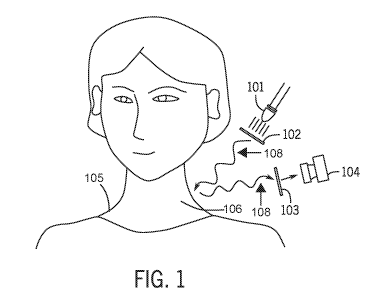
their working
mechanisms are not designed to give the best contrast for lymph nodes
specifically, unless specific
1
CA 03127682 2021-07-22
WO 2020/163286 PCT/US2020/016525
contrasting agents are injected. As a result, other organs and tissues show up
in these images with
the same or sometimes even better contrast compared to lymph nodes, causing
distractions to the
task of finding and examining the lymph nodes. These general-purposed imaging
modalities are not
only not specific to lymph nodes, but also possess their own critical
drawbacks. CT involves X-ray
exposure and PET involves radioactive agents, which need to be carefully
controlled in prevention
of health hazards. MM requires expensive instrumentation and is not compatible
with patients with
metal implants. Ultrasound provides low imaging contrast and resolution mainly
because of its long
imaging wavelength.
[0006] Another common practice for lymph node imaging involves injecting
dyes, either blue
dyes or fluorescent dyes. The most common dye used for lymph node
visualization is methylene
blue, which is actually toxic. The dosage of this dye has to be carefully
managed. Indocyanine
green, a fluorescent dye, has also been used for lymph node imaging. Systems
leveraging
fluorescence dyes such indocyanine green and methylene blue include systems a
FLARE TM
system, a fluobeam system, SPY, FDPM, and a Photodynamic Eye system. Most of
these use either
a single image sensor (typically, a CCD) to capture visible (a reference
image) and fluorescence
images sequentially, or multiple cameras to image different spectra
simultaneously or sequentially.
[0007] Dye based methods have numerous drawbacks. One drawback is that dyes
can stimulate
negative responses to some patients, especially people with kidney
complications. Another
drawback is that the dye injection method can be unreliable because of the
leaky nature of the
lymphatic system. Additionally, certain dye-based methods require invasive
application of the dye.
[0008] For imaging systems that produce multiple images with the use of
multiple cameras
and/or sequential image acquisition, subsequent image registration is
required. To properly
coordinate differences in spatial parameters of the multiple images, such
image processing must
take into account changes in angular coordinate, potential relative motion
between the system and
the subject, or both. Other types of imagers include specialized CMOS sensors
that can collect light
via red-green-blue channel(s) (RGB) as well as a single channel in NIR-1.
[0009] There are some other reports in academic papers about using novel
optical techniques to
image lymph nodes, including optical speckle imaging, optical coherence
tomography, etc.
However, optical speckle imaging is highly susceptible to motion artifact, and
optical coherence
tomography involves sophisticated instrumentation and offers poor imaging
contrast.
[0010] In summary, given the critical importance of lymph nodes to human
health, there are no
convenient and highly effective methods for visualizing lymph nodes. Cross
sectional imaging
2
CA 03127682 2021-07-22
WO 2020/163286
PCT/US2020/016525
methods are not convenient and not specific for visualization of lymph nodes
unless contrasting
agents are injected. Dye-based imaging techniques are generally highly
invasive and incompatible
with clinical settings like routine checks. A new imaging modality that is
able to conveniently
image lymph nodes with high specificity, high contrast without injecting any
imaging contrasting
agents will be a powerful tool for medical practitioners to examine the health
of the patients,
evaluate the effectiveness of a certain treatment, stage one's cancer
condition, and so many other
medical applications.
SUMMARY
[0011] The following is intended to give a brief summary of the disclosure
and is not intended
to limit the scope of the disclosure.
[0012] In
one aspect, the present disclosure provides a system for imaging a lymphatic
component. The system includes an optical source configured to provide
infrared illumination
having a polarization to a region of a subject having at least one lymphatic
component, a sensor
configured to sense a reflected portion of the infrared illumination having an
opposite polarization
to that of the polarization of illumination directly reflected from the
region, and a controller in
communication with the sensor. The controller is configured to receive, from
the sensor,
information corresponding to the reflected portion of the infrared
illumination, generate at least one
image indicative of the at least one lymphatic component in the subject using
the information, and
output the at least one image to at least one of a display and/or a memory.
[0013] In another aspect, the present disclosure provides a method for
imaging lymph nodes or
lymphatic vessels in vivo without a contrast agent. The method includes
providing, using an optical
source, an infrared illumination having a polarization to an in vivo region of
a subject having
lymph nodes or lymphatic vessels that are free of a contrast agent, detecting
a reflected portion of
the infrared illumination directly reflected from the region and having a
opposite polarization to the
polarization using a sensor positioned to receive the illumination directly
reflected from the region,
and generating at least one image indicative of the lymph nodes or lymphatic
vessels that are free
of a contrast agent in the subject using the reflected portion of the infrared
illumination.
[0014] In yet another aspect, the present disclosure provides a method for
imaging lymph nodes
or lymphatic vessels without a mirror. The method includes providing, using an
optical source, an
infrared illumination to a region of a subject having lymph nodes or lymphatic
vessels, detecting a
reflected portion of the infrared illumination directly reflected from the
region using a sensor
3
CA 03127682 2021-07-22
WO 2020/163286 PCT/US2020/016525
positioned to receive the illumination directly reflected from the region, and
generating at least one
image indicative of the lymph nodes or lymphatic vessels in the subject using
the reflected portion
of the infrared illumination.
[0015] In a further aspect, a system for imaging a lymphatic component is
provided. The
system includes an optical source configured to provide infrared illumination
having a polarization
to a region of a subject having at least one lymphatic component, a sensor
configured to sense a
reflected portion of the infrared illumination having an opposite polarization
to that of the
polarization directly reflected from the region, generate at least one image
indicative of the at least
one lymphatic component in the subject based on the reflected portion of the
infrared illumination,
and output the at least one image to at least one of an external display or an
external memory.
BRIEF DESCRIPTION OF THE DRAWINGS
[0016] Figure 1 shows a schematic diagram of an example of an imaging
system in accordance
with certain aspects of the present disclosure.
[0017] Figure 2 illustrates another example of an imaging system in
accordance with certain
aspects of the present disclosure.
[0018] Figure 3 shows a schematic diagram of yet another exemplary
embodiment of an
imaging system in accordance with certain aspects of the present disclosure.
[0019] Figure 4 shows an example of hardware that can be used to implement
a computing
device and an interface platform shown in Figure 3 in accordance with some
embodiments of the
disclosed subject matter.
[0020] Figure 5 shows an exemplary flowchart of a process included in an
image generation
and analysis application.
[0021] Figure 6 shows an exemplary flowchart of another process included in
the image
generation and analysis application.
[0022] Figure 7A shows imaging results of a region imaged without using
polarizers.
[0023] Figure 7B shows imaging results of the same region as Figure 7A
imaged using
polarizers.
[0024] Figure 8A shows an image of a region of a mouse taken with a
standard camera.
[0025] Figure 8B shows an image of the region of the mouse of Figure 8A
taken using an
imaging system before an adjuvant is injected.
4
CA 03127682 2021-07-22
WO 2020/163286 PCT/US2020/016525
[0026] Figure 8C shows an image of the region of the mouse of Figure 8A
taken using the
imaging system forty-eight hours after the adjuvant is injected.
[0027] Figure 9A shows an image of a region of a mouse taken with a
standard camera.
[0028] Figure 9B shows an image of the region of the mouse of Figure 9A
taken using an
imaging system before an adjuvant is injected.
[0029] Figure 9C shows an image of the region of the mouse of Figure 9A
taken using the
imaging system forty-eight hours after the adjuvant is injected.
[0030] Figure 10A shows an exemplary image taken with an InGaAs camera when
an
illumination wavelength of 1000 nm was used.
[0031] Figure 10B shows an exemplary image taken with an InGaAs camera when
an
illumination wavelength of 1175 nm was used.
[0032] Figure 10C shows an exemplary image taken with an InGaAs camera when
an
illumination wavelength of 1250 nm was used.
[0033] Figure 10D shows an exemplary image taken with an InGaAs camera when
an
illumination wavelength of 1375 nm was used.
[0034] Figure 10E shows an exemplary image taken with an InGaAs camera when
an
illumination wavelength of 1550 nm was used.
[0035] Figure 11A shows an image including a lymph node generated using 690
nm
wavelength illumination.
[0036] Figure 11B shows an image including the lymph node of Figure 11A of
Figure 11A
generated using 730 nm wavelength illumination.
[0037] Figure 11C shows an image including the lymph node of Figure 11A
generated using
810 nm wavelength illumination.
[0038] Figure 11D shows an image including the lymph node of Figure 11A
generated using
900-950 nm wavelength illumination.
[0039] Figure 11E shows an image including the lymph node of Figure 11A
generated using
1000 nm wavelength illumination.
[0040] Figure 11F shows an image including the lymph node of Figure 11A
generated using
1125 nm wavelength illumination.
[0041] Figure 11G shows an image including the lymph node of Figure 11A
generated using
1175 nm wavelength illumination.
CA 03127682 2021-07-22
WO 2020/163286 PCT/US2020/016525
[0042] Figure 11H shows an image including the lymph node of Figure 11A
generated using
1250 nm wavelength illumination.
[0043] Figure 111 shows an image including the lymph node of Figure 11A
generated using
1300 nm wavelength illumination.
[0044] Figure 11J shows an image including the lymph node of Figure 11A
generated using
1375 nm wavelength illumination.
[0045] Figure 11K shows an image including the lymph node of Figure 11A
generated using
1550 nm wavelength illumination.
[0046] Figure 11L shows an image including the lymph node of Figure 11A
generated using
1575 nm wavelength illumination.
[0047] Figure 11M shows an image including the lymph node of Figure 11A
generated using 8-
101.tm wavelength illumination.
[0048] Figure 12A shows an image of tissue generated using a regular camera
and ambient
visible light.
[0049] Figure 12B shows an image of the tissue of Figure 12A generated
using an embodiment
of the imaging system of Figure 3.
DETAILED DESCRIPTION
[0050] In one exemplary embodiment, depicted in Figure 1, an imaging system
100 is provided
for imaging lymphatic components. As used herein lymphatic components can
include at least one
of a lymph node or a lymphatic vessel. The imaging system 100 can include a
LED light source
101 emitting between 900 and 1300 nm used as the light source. The LED light
source 101 may
also be referred to as the light source 101. The imaging system 100 can
include a linear polarizer
102 mounted on a rotational mount and placed in front of the LED light source
101 to create
linearly polarized illumination light 107 (i.e., illumination). The linear
polarizer 102 can include
linear polarizing film. The linearly polarized illumination 107 is shone onto
a subject of interest
105, which can be either a human, as depicted in Figure 1, or an animal.
[0051] The light source 101 can be oriented towards an in vivo target
region 106 of the subject
of interest 105. The in vivo target region 106 may also be referred to as an
in vivo region or target
region herein. In some embodiments, the target region 106 may be an ex vivo
region such as a
tissue portion. The ex vivo tissue portion may include fat, lymph nodes,
and/or lymphatic vessels,
and the lymph nodes, and/or lymphatic vessels can be imaged as if the tissue
portion was in vivo.
6
CA 03127682 2021-07-22
WO 2020/163286 PCT/US2020/016525
[0052] Some light sources, such as certain lasers, are inherently linearly
polarized. In the case
of these inherently linearly polarized light sources, creating linearly
polarized illumination does not
require the use of linear polarizers. Thus, the linear polarizer 102 may not
be required when the
light source 101 is inherently linearly polarized. In other words, some
imaging systems may not
include the linear polarizer 102. Polarized illumination helps improve the
imaging contrast of this
technique, but is not necessary. A clear contrast of the lymph nodes can be
formed even without
any polarizers, as shown in Figures 7A-B.
[0053] Still referring to Figure 1, the imaging system 100 can include a
sensor 104, which can
be a camera. The sensor 104 is used to visualize the illuminated area on a
human or an animal. The
light source can be oriented towards the target region 106 of the subject of
interest 105. The
imaging system 100 can include another linear polarizer 103, which may be
referred to as the
sensor linear polarizer 103. The sensor linear polarizer 104 can include
linear polarizing film. The
sensor linear polarizer 103 can be placed in front of the sensor 104 and/or
positioned between the
sensor 104 and the target region 106.
[0054] An ideal imaging contrast can be formed when the polarization of
incoming light 108
before the sensor 104 and the polarizer 103 in front of the sensor 104 is
orthogonal to the polarizer
103 in front of the sensor 104. The incoming light can include a portion of
the linearly polarized
illumination 107 that has interacted with tissues in the in vivo region 106.
In principle, linearly
polarized illumination remains mostly linearly polarized when reflecting off
the surface of human
or animal skin. The polarization of linearly polarized light does not change
when bouncing directly
away from the surface of the skin. Only a small portion of the light became
randomly polarized,
because it traveled relatively deeply into the biological tissues, which
serves as randomly scattering
media. By placing the sensor linear polarizer 103 in front of the sensor 104
orthogonal to the
direction of the incoming light 108, the sensor linear polarizer 103 filters
out the light reflected by
the surface of human or animal skins and lets through only the portion of the
light 107 emitted from
light source 101 that interacted with deeper tissues. When the light reflected
from the surface of the
skin (i.e., surface glare) is reduced to the minimum level, the imaging system
100 achieves the best
contrast and deepest penetration depth. In practice, this ideal contrast can
be formed by rotating one
of the polarizers, either the sensor linear polarizer 103 or the linear
polarizer 102 in front of the
light source 101, until the lowest overall intensity detected by the sensor
104 is reached. The lowest
overall intensity can be associated with a threshold contrast level. The
threshold contract level can
be within a predetermined range of the lowest overall intensity, such as
within ten percent of the
7
CA 03127682 2021-07-22
WO 2020/163286 PCT/US2020/016525
lowest overall intensity, and the polarizer (e.g., the sensor linear polarizer
103 or the linear
polarizer 102 in front of the light source 101) and/or light source 101 can be
adjusted until the
threshold contract level is achieved at the sensor 104.
[0055] After linearly polarized photons interact with tissue in the target
region 106 and go
through scattering, the linearly polarized photons slowly lose their linear
polarization. After around,
for example, ten scattering events, the linearly polarized photons become
completely depolarized
photons. These completely-depolarized photons then reach the sensor linear
polarizer 103 in front
of the sensor 104. Because the sensor linear polarizer 103 in front of the
sensor 104 is
approximately orthogonal to the linear polarizer 102 in front of the light
source 101, only the
photons that are now completely depolarized and have the opposite polarization
are allowed to be
detected by the sensor 104. Therefore, only the photons that interacted at a
deeper level with the
tissue in the target region 106 are "selected" to be analyzed, and surface
glare and unnecessary
surface features are removed.
[0056] When the wavelength of light emitted from the light source 101 is
much longer than
visible light (e.g., 1550 nm), imaging quality can be improved further. Longer
wavelengths are
associated with lower scattering effect. As a result, much thicker tissue is
required to completely
depolarize linearly polarized light with longer wavelengths as compared to
linearly polarized light
with shorter wavelengths. Imaging systems, such as the imaging system 100, can
therefore provide
light having longer wavelengths to the subject (e.g., the subject 105) in
order better image deeper
tissues as compared to shorter wavelengths (e.g., wavelengths in the visible
light spectrum).
[0057] In the case that the light source 101 is a laser that is already
linearly polarized without
using a polarizer, the threshold contrast level be met by rotating either the
sensor linear polarizer
103 or the laser itself. The relative orthogonal relationship is important and
the absolute directions
of polarization are not. The optimal contrast can be achieved through either
rotating polarizers,
light sources, or sensors, as long as the orthogonal polarization relationship
is met. It is noted that
the imaging system 100 of Figure 1 does not require a mirror, and does not
require a mirror or other
reflective surface as is common in certain imaging techniques, which can
reduce the cost to build
the imaging system 100 of Figure 1 as compared to other imaging techniques.
[0058] The present disclosure recognizes that lymph nodes are birefringent,
i.e. responsive to
polarized light. Lymph nodes and/or lymph vessels can contain collagen, which
is birefringent.
Furthermore, the tissues surrounding the lymph nodes such as layers of fat
(i.e. lipids) are generally
not birefringent. Thus, the present disclosure recognizes that cross-
polarization, i.e. the orthogonal
8
CA 03127682 2021-07-22
WO 2020/163286 PCT/US2020/016525
polarization relationship described above, can be used to exploit the
difference in birefringence
between lymph nodes and/or lymph vessels and the surrounding tissue in order
to generate an
image of the lymph nodes and/or lymph vessels. In some embodiments, the light
source 101 may
provide illumination with a wavelength of 1200-1600 nm, which can correspond
to one or more
absorption peaks of the collagen in lymph nodes and/or lymph vessels included
in the target region
106. Using illumination wavelengths of 1200-1600 nm can therefore improve the
imaging contrast
between the lymph nodes and/or lymph vessels and the surrounding tissue. As
described above,
longer wavelengths may improve the imaging resolution of the lymph nodes
and/or lymph vessels
due to reduced scattering effects.
[0059] Additionally, illumination that includes longer wavelength light,
especially 1550 nm
wavelength light, can improve the contrast of lymphatic components in the
target region 106.
Generally, the lymphatic components are surrounded by fat. Lymph nodes and
lymphatic vessels
are high in water, while fat is very low in water. Absorption of photons
occurs at 1550 nm in water,
which is likely why using 1550 nm wavelength light to illuminate the target
region 106 can
improve the contrast (and therefore visibility) of the lymph nodes and/or
lymphatic vessels in
images generated using the imaging system 100. When generating images using
1550 nm
illumination wavelength light, lymph nodes and lymphatic vessels appear dark,
while fat is bright.
[0060] Furthermore, illumination that includes longer wavelength light,
especially 1550 nm
wavelength light, can improve the contrast of lymphatic components against
surrounding blood in
the target region 106. While blood contains a high amount of water, blood also
contains a high
amount of cells. The cells are highly scattering and overwhelm the water
absorption effect. In
testing, the imaging system 100 has been shown to generate images where blood
and/or
hemorrhage in the target region 106 are not visible, even compared to fat.
Suppressing the visibility
of blood and/or hemorrhage is an advantage of the imaging system 100 over
other imaging
modalities that generate images with visible blood and/or hemorrhages.
Hemorrhages can be
mistaken as lymph nodes, and are then harvested to be analyzed. Suppressing
and/or removing
hemorrhages from images may reduce the number of false positives that
pathologists identify when
diagnosing patients.
[0061] While the imaging system 100 has been described as being applied to
an in vivo region
of a subject, it is appreciated the imaging system can also be applied to an
ex vivo tissue specimen
as well. For example, the target region 106 can include a tissue packet that
can include lymph
nodes and fat. The tissue packet may have been removed from the subject 105
during a
9
CA 03127682 2021-07-22
WO 2020/163286 PCT/US2020/016525
lymphadenectomy procedure performed after a tumor and relevant lymph nodes
have been
identified. The lymph nodes may then need to be separated from the fat and any
other surrounding
tissue included in the tissue packet during a grossing step. Typically,
pathologists remove the
lymph nodes via manual palpation and visual inspection, which is prone to
error because lymph
nodes are often translucent and appear similar to fat, lymph nodes may be as
small as lmm across,
and the locations of lymph nodes are often unpredictable. The imaging system
100 can be used to
visualize the lymph nodes and display the lymph nodes to the pathologist, who
can then efficiently
and accurately remove the lymph nodes from the target region 106. Cancer
organizations may
require a certain number of lymph nodes to be examined for specific types of
cancer. The number
of lymph nodes required may range from twelve to thirty-eight. The imaging
system 100 can,
therefore, help the pathologist acquire the required number of lymph nodes by
potentially reducing
the number of lymph nodes missed in the target region 106.
[0062] In Figure 2, an illustration of another exemplary embodiment of an
imaging system 200
is shown. In this exemplary imaging system 200, a halogen lamp with continuous
light illumination
is used as a light source 201. In order to reduce background from direct
reflection at wavelengths
out of the range of 900¨ 1300 nm, longpass filters with cut-off wavelengths at
900 nm or 1000 nm
are used to filter out light with shorter wavelengths. The imaging system 200
can include a primary
longpass filter 203 and a secondary longpass filter 205. Each of the primary
longpass filter 203 and
the secondary longpass filter 205 can have a cut-off wavelength selected from
900 nm to 1000 nm,
inclusive. The imaging system 200 can include a linear polarizer 202 on a
screw mount. The linear
polarizer 202 can include linear polarizing film. The linear polarizer 202 can
be placed in front of
the light source 201 to make the illumination light from the light source 201
(e.g., the halogen
lamp) linearly polarized. The primary longpass filter 203 can be placed in
front of light source 201
in order to filter out as much light emitted from the light source 201 that is
below the cut-off
wavelength as possible.
[0063] A regular commercially available silicon camera is used as a sensor
204 included in the
imaging system 200. In some embodiments, a black silicon camera and/or an
InGaAs camera can
be used as the sensor 204. A sensor linear polarizer 206 is placed in front of
the sensor 204 on a
screw mount. The sensor linear polarizer 206 can include linear polarizing
film. A lens (not
shown), which may be a telecentric lens, is also placed in front of the sensor
204 to form an image.
The telecentric lens can enhance the measurement accuracy of the imaging
system 200 by helping
to normalize the size of a lymph node in an image generated using the sensor
204 regardless of how
CA 03127682 2021-07-22
WO 2020/163286 PCT/US2020/016525
far away the lymph node is from the sensor 204. The primary longpass filter
203 was also placed in
front of the sensor 204 to filter out the unwanted background from either
ambient light or the light
source 201 (e.g., the halogen lamp). In some embodiments, there may not be a
need to calibrate the
sensor 204 for different ambient and/or background light amounts because the
secondary longpass
filter 205 can eliminate background light, which may include visible
frequencies below the cutoff
frequency of the secondary longpass filter 205. Eliminating the need to
calibrate the sensor 204 can
save time in detecting the lymphatic components, as well as make the imaging
system 200 more
robust as compared to an imaging system that requires calibration of one or
more sensors. The light
source 201 and the sensor 204 should both point at the same area of interest
on the subject being
studied, either a human or an animal, such as a person 207 as shown in Figure
2. It is noted that the
imaging system 200 of Figure 2 does not include a mirror, and does not require
a mirror or other
reflective surface as is common in certain imaging techniques. This can reduce
the cost to build the
imaging system 200 of Figure 2 as compared to other imaging systems and/or
techniques.
[0064] In some embodiments, a controller (not shown) may be included in the
imaging system
200. The controller can be coupled to an optical source such as a laser or
LED, as well as a sensor
such as a camera. The controller can be coupled to and in communication with
the optical source
and the sensor. The controller can be configured to cause the optical source
to provide the infrared
illumination to the region by controlling power supplied to the optical
source. The controller can
also receive information from the sensor corresponding to the infrared
illumination reflected from
the subject. The infrared illumination reflected can be referred to as a
reflected portion of the
infrared illumination that was originally supplied by the optical source. The
controller can also
generate at least one image indicative of the lymph nodes in the subject using
the information
received.
[0065] Referring now to Figures 1 and 2 as well as Figure 3, a schematic
diagram of yet
another exemplary embodiment of an imaging system 300 is shown. In some
embodiments, the
imaging system 300 can be approximately the size of a shoebox, and can
therefore be a bench-top
imaging device. The imaging system 300 can include an interface platform 302.
The interface
platform 302 can include at least one memory, at least one processor, and any
number of
connection interfaces capable of communication with sensors and optical
sources (not shown). The
interface platform 302 can also store (e.g., in the at least one memory) and
execute (e.g., using the
at least one processor) at least a portion of an image generation and analysis
application 304. As
will be described below, the interface platform 302 can be coupled to and in
communication with a
11
CA 03127682 2021-07-22
WO 2020/163286 PCT/US2020/016525
computing device 334 included in the imaging system 300 that may also store
and/or execute at
least a portion of the image generation and analysis application 304. The
interface platform 302 can
be a controller, a laptop computer, a desktop computer, or another device
capable of receiving
signals from a sensor and outputting control signals to an optical source. The
controller can be a
microcontroller, such as a Raspberry Pi 4 Model B. In some embodiments, the
controller can be an
Intel NUC computer configured to operate using a Windows operating system.
[0066] The interface platform 302 can be coupled to and in communication
with an
illumination generation system 306 included in the imaging system 300. The
illumination
generation system 306 can include an optical source 308. The interface
platform 302 can be
coupled to and in communication with the optical source 308. The interface
platform 302 can
output control signals to the optical source 308 in order to cause the optical
source 308 to provide
illumination. In some embodiments, the optical source 308 may output suitable
data (e.g., total
lifetime hours of operation) to the interface platform 302. The illumination
generation system 306,
and more specifically, the optical source 308, can be oriented to provide
illumination 314 to a target
region 318 that may be in vivo (e.g., included in a subject 316 such as a
patient) or ex vivo, as will
be described further below. The illumination 314 output by the illumination
generation system 306
can be referred to as the provided illumination 314. The illumination 314 can
be infrared
illumination. The infrared illumination can include light in the near-infrared
range (800-1400 nm
wavelength) and/or light in the short-wave infrared range (1400-3000 nm
wavelength).
[0067] The optical source 308 can include at least one of an LED such as a
single LED, a
plurality of LEDs such as an LED array, a halogen lamp such as a tungsten
halogen lamp, a quartz-
halogen lamp, or a quartz iodine lamp, a laser, or another suitable optical
source capable of
outputting light at one or more predetermined wavelengths. In some
embodiments, the optical
source 308 may output one or more discrete wavelengths of light, such as 1550
nm, 1375 nm, 1300
nm, and/or other wavelengths selected from 800 nm to 1700 nm wavelengths. For
example, the
optical source 308 may only output 1550 nm wavelength light. In some
embodiments, the optical
source can output one or more discrete frequencies from a subrange of
wavelengths within the 800
nm to 2000 nm range, such as a subrange of 1200-1600 nm wavelengths. In some
embodiments,
the optical source 308 may output a continuous range of wavelengths of light,
such as 900-1300
nm, 1500-1600 nm, 1200-1600 nm, 1000-1700nm (i.e., near-infrared), and/or
other ranges of
wavelengths within 800-2000 nm. In some embodiments, the optical source 308
may be the light
source 101 of Figure 1 or the light source 201 of Figure 2. In particular, the
optical source 308 may
12
CA 03127682 2021-07-22
WO 2020/163286 PCT/US2020/016525
output longer wavelength light, especially 1550 nm wavelength light, in order
to better contrast
lymphatic components against surrounding fat, blood, and/or hemorrhages as
described above. For
the imaging system 300 to function properly, the optical source 308 does not
need to emit a range
of wavelengths of light. In testing, excellent imaging has been obtained using
only 1550 nm
wavelength light. However, the imaging system 300 can perform suitable imaging
using multiple
wavelengths of light. It is contemplated that light with wavelengths up to
2600 nm could be used,
as some sensors such as certain InGaAs cameras stop responding beyond 2600 nm.
Thus, light with
wavelengths ranging from 800-2600 nm might be used in the imaging system 300.
In testing, light
with wavelengths below 800 nm has not performed as well as light with higher
wavelengths, such
as 800-1700 nm.
[0068] In some embodiments, the illumination generation system 306 can
include a polarizer
310 such as a linear polarizer. For certain optical sources that are not
inherently polarized, such as
halogen optical sources, the imaging system 300 may include a polarizer 310.
The polarizer 310
can include linear polarizing film. The polarizer 310 can be mounted and
placed in front of the
optical source 310 to create linearly polarized illumination light. The
polarizer 310 can be mounted
on a rotational mount or other suitable mount to allow for adjustment of the
polarizer 310. Thus,
the illumination 314 provided to the target region 318 can be linearly
polarized. Polarized
illumination can improve imaging contrast in images generated by the imaging
system 300, but it is
not necessary. In some embodiments, the polarizer 310 may be the linear
polarizer 102 of Figure 1
or the linear polarizer 202 of Figure 2. If the optical source 308 is an
inherently polarized device,
such as certain lasers, the polarizer 310 may not be included in the imaging
system 300. In some
embodiments, the polarizer 310 can be a circular polarizer.
[0069] In some embodiments, the illumination generation system 306 can
include an optical
filter 312. The optical filter 312 can be a longpass filter such as a cold
mirror, a colored glass filter,
a thermoset allyl diglycol carbonate (ADC) filter, or another suitable filter
capable of attenuating
lower wavelength light (e.g., visible light) and passing higher wavelength
light (e.g., infrared light).
The longpass filter may have a cut-off wavelength of no less than 800 nm. For
example, the cut-off
wavelength may be 800 nm, 900 nm, or 1000 nm. The optical filter 312 can be
placed in front of
light source optical source 308 in order to filter out as much light emitted
from the optical source
308 that is below the cut-off wavelength as possible. In some embodiments, the
optical filter 312
may be the primary longpass filter 203 of Figure 2. In some embodiments, the
optical filter 312 can
be a bandpass filter such as a hard coated filter or a colored glass filter.
The bandpass filter may
13
CA 03127682 2021-07-22
WO 2020/163286 PCT/US2020/016525
only pass a range of light wavelengths within a 800-2000 nm window, or a
subrange of the 800-
2000 nm window. For example, the bandpass filter may only pass 900-1700 nm
wavelength light.
Thus, the illumination 314 provided to the target region 318 can be longpass
filtered or bandpass
filtered.
[0070] The optical source 308, the polarizer 310, and/or the optical filter
312 can be physically
arranged (i.e., positioned) relative to each other as shown in Figure 1 and/or
Figure 2. For example,
the optical source 308 and the polarizer 310 can be arranged in similar
fashion to the light source
101 and the linear polarizer 102, respectively, as shown in Figure 1. As
another example, the
optical source 308, the polarizer 310, and the optical filter 312 can be
arranged in similar fashion to
the light source 201, the linear polarizer 202, and the longpass filter 203,
respectively, as shown in
Figure 2. The optical source 308 can output the illumination 314 that may pass
through and be
polarized by the polarizer 310 and/or pass through and be attenuated by the
optical filter 312. The
illumination 314, which may be polarized and/or attenuated, is then provided
to the target region
318.
[0071] As mentioned above, the optical source 308, and by extension the
illumination
generation system 306, can be oriented to provide the illumination 314 to the
target region 318. In
some embodiments, the target region 318 can be an in vivo region included in
the subject 316. In
these embodiments, the target region 318 may be referred to as the in vivo
region. The subject 316
can be a human patient. In other embodiments, the target region 318 can be an
ex vivo region. In
these embodiments, the target region 318 may be referred to as the ex vivo
region. For example, the
target region 318 can be a tissue portion removed from a subject for grossing
purposes as described
above. The imaging system 300 can be used to aid in the grossing of the tissue
portion by
visualizing lymphatic components for a practitioner.
[0072] At least a portion of the illumination 314 can be provided to the
target region 318. The
target region 318 may include one or more lymphatic components. The provided
illumination 314
can interact with the lymphatic components and the surrounding tissue in the
target region 318. At
least a portion of the provided illumination 314 may become randomly polarized
as described
above. At least a portion of the provided illumination 314 can be reflected as
reflected illumination
320. The reflected illumination 320 can include light that has interacted with
deep tissue in the
target region 318.
[0073] The interface platform 302 can be coupled to and in communication
with a sensing
system 322 included in the imaging system 300. The sensing system 322 can
include a sensor 324.
14
CA 03127682 2021-07-22
WO 2020/163286 PCT/US2020/016525
The interface platform 302 can be coupled to and in communication with the
sensor 324. The
sensor 324 can sense the reflected illumination 320 and output signals
associated with an image
based on the sensed reflected illumination 320. The interface platform 302 can
receive the signals
indicative of the image from the sensor 324. The signals can include
information about the image.
In some embodiments, the information can include the image formatted in a
predetermined image
format such as PNG, JPEG, DICOM (i.e., included in a DICOM file), etc. In some
embodiments,
the information can also include metadata about the image, such as the time
the image was taken or
a patient associated with the image. In some embodiments, the sensor 324 can
include a camera,
such as a silicon camera including a silicon complementary metal oxide
semiconductor (CMOS)
camera or a silicon charge-coupled device (CCD) camera with phosphor coating,
a germanium
camera, a germanium-tin on silicon camera, a black silicon camera, a quantum
dot shortwave
infrared (SWIR) camera, and/or an InGaAs camera. The InGaAs camera may be a
nitrogen cooled
InGaAs camera. The sensor 324 can include a mercury-cadmium-telluride (HgCdTe
or MCT)
camera. The sensor 324 can be responsive to light including at least a portion
of the light ranging
from 800 nm ¨ 2000 nm in wavelength, especially wavelengths at or near 1550
nm. It is noted that
the imaging system 300 may only require a single sensor (e.g., a silicon
camera), in contrast to
other systems that may require multiple sensors and/or cameras.
[0074] The sensing system 322 can include a lens 326 positioned in front of
the sensor 324. In
some embodiments, the lens 326 can be integral with the sensor 324, such as if
the sensor 324 and
the lens 326 are sold as a single off-the-shelf component. The lens 326 can
improve the imaging
capabilities of the sensor 324. For example, the lens 326 can be a telecentric
lens. The telecentric
lens can enhance the measurement accuracy of the imaging system 300 by helping
to normalize the
size of a lymph node in an image generated using the sensor 324 regardless of
how far away the
lymph node is from the sensor 324.
[0075] In some embodiments, the sensing system 300 can include a light
diffuser 328 such as a
piece of frosted glass or a tissue paper. The light diffuser 328 can be
inserted between the lens 326
and a polarizer 332 that can be included in the sensing system 322. The light
diffuser 328 can
create a more evenly distributed light pattern in the reflected illumination
320. The light diffuser
328 may improve the imaging capabilities of the sensor 324 as a result of the
more evenly
distributed light pattern.
[0076] In some embodiments, the sensing system 322 can include an optical
filter 330
positioned in front of the sensor 324. The optical filter 330 can be a
longpass filter such as a cold
CA 03127682 2021-07-22
WO 2020/163286 PCT/US2020/016525
mirror, a colored glass filter, a thermoset ADC filter, or another suitable
filter capable of
attenuating lower wavelength light (e.g., visible light) and passing higher
wavelength light (e.g.,
infrared light). The longpass filter can have a cut-off wavelength of no less
than 800 nm. For
example, the cut-off wavelength may be 800 nm, 900 nm, or 1000 nm. In some
embodiments, there
may not be a need to calibrate the sensor 324 for different ambient and/or
background light
amounts because the optical filter 330 can eliminate background light, which
may include visible
frequencies below the cutoff frequency of the optical filter 330. In some
embodiments, the optical
filter 330 may be the secondary longpass filter 205 as shown in Figure 2. In
some embodiments, the
optical filter 330 can be a bandpass filter such as a hard coated filter or a
colored glass filter. The
bandpass filter may only pass a range of light wavelengths within a 800-2000
nm window, or a
subrange of the 800-2000 nm window. For example, the bandpass filter may only
pass 900-1700
nm wavelength light. Thus, the reflected illumination 320 provided to the
sensor 324 can be
longpass filtered or bandpass filtered.
[0077] As mentioned above, the sensing system can include the polarizer
332. The polarizer
332 can be a linear polarizer. In some embodiments, the polarizer 332 can be a
circular polarizer.
The polarizer 332 can be placed in front of the sensor 324. The polarizer 332
can include linear
polarizing film. Similar to the polarizer 310 included in the illumination
generation system 306, the
polarizer 332 included in the sensing system 322 can be mounted on a
rotational mount or other
suitable mount to allow for adjustment. The linear polarizers 310, 332, can be
rotated or otherwise
adjusted to create an ideal imaging contrast as described above. The polarizer
332 can remove any
light having the same polarization as the provided illumination 314 from the
reflected illumination
320. The sensor 324 can detect light included in the reflected illumination
320 having the opposite
polarization as the provided illumination 314.
[0078] In some embodiments, the sensor 324 can be coupled to and in
communication with the
external display 372. Alternatively or in addition, the sensor 324 can be
coupled to and in
communication with a memory 374 that may be included in the imaging system 300
or external to
the imaging system 300. For example, the memory 374 can be flash memory
included in a memory
card. In embodiments where the sensor 324 is coupled to and in communication
with the external
display 372 and/or the memory 374, the sensor 324 can be configured to sense
the reflected portion
of the provided illumination 314 and generate at least one image indicative of
the any lymphatic
components in the target region 318 based on the reflected portion (i.e., the
reflected illumination
16
CA 03127682 2021-07-22
WO 2020/163286 PCT/US2020/016525
320) of the provided illumination 314. The sensor 324 may also be configured
to output the at least
one image to at least one of the external display 372 or the memory 374.
[0079] In some embodiments, the optical source 308 may not be coupled to a
controller or other
device, and may only need to be coupled to a power source. In these
embodiments, the optical
source 308 can provide the illumination 314 to the target region 318
constantly or semi-constantly.
In some embodiments, the interface platform 304 can supply power to the
optical source 308 (i.e.,
act as the power source). In other embodiments, the optical source 308 can
receive power from wall
power, one or more batteries, or another suitable power source.
[0080] In some embodiments, the sensor 324 can be coupled to the external
display 372 and/or
the memory 374, and the optical source may be coupled to a power supply
without being coupled to
the interface platform 304 and/or other suitable device. Thus, the imaging
system 300 can be
implemented without the use of a controller or computational device.
[0081] In some embodiments, the imaging system 300 can be Class-1, 510(k)-
exempt, and/or
good manufacturing practice (GMP) exempt.
[0082] The imaging system 300 may also include the external display 372
and/or the
computing device 334. As mentioned above, the interface platform 302 can be
coupled to and in
communication with the computing device 334. The imaging system 300 can
include a
communication network 336. The communication network 336 can facilitate
communication
between the interface platform 302 and the computing device 334. The interface
platform 302 can
also be coupled to and in communication with the external display 372.
[0083] In some embodiments, communication network 336 can be any suitable
communication
network or combination of communication networks. For example, communication
network 336
can include a Wi-Fi network (which can include one or more wireless routers,
one or more
switches, etc.), a peer-to-peer network (e.g., a Bluetooth network), a
cellular network (e.g., a 3G
network, a 4G network, etc., complying with any suitable standard, such as
CDMA, GSM, LTE,
LTE Advanced, WiMAX, etc.), a wired network, etc. In some embodiments,
communication
network 336 can be a local area network, a wide area network, a public network
(e.g., the Internet),
a private or semi-private network (e.g., a corporate or university intranet),
any other suitable type of
network, or any suitable combination of networks. Communications links shown
in FIG. 3 can each
be any suitable communications link or combination of communications links,
such as wired links,
fiber optic links, Wi-Fi links, Bluetooth links, cellular links, etc. In some
embodiments, the
17
CA 03127682 2021-07-22
WO 2020/163286 PCT/US2020/016525
computing device 334 can implement portions of the image generation and
analysis application
304.
[0084] Referring now to Figure 3 as well as Figure 4, an example of
hardware that can be used
to implement a computing device 334 and an interface platform 302 shown in
Figure 3 in
accordance with some embodiments of the disclosed subject matter is shown. As
shown in Figure
4, the computing device 334 can include a processor 350, a display 352, an
input 354, a
communication system 356, and memory 358. The processor 350 can implement at
least a portion
of the image generation and analysis application 304, which can, for example
be executed from a
program (e.g., saved and retrieved from memory 358). The processor 350 can be
any suitable
hardware processor or combination of processors, such as a central processing
unit ("CPU"), a
graphics processing unit ("GPU"), etc., which can execute a program, which can
include the
processes described below.
[0085] In some embodiments, the display 352 can present a graphical user
interface. In some
embodiments, the display 352 can be implemented using any suitable display
devices, such as a
computer monitor, a touchscreen, a television, etc. In some embodiments, the
inputs 354 of the
computing device 334 can include indicators, sensors, actuatable buttons, a
keyboard, a mouse, a
graphical user interface, a touch-screen display, etc. In some embodiments,
the inputs 354 can
allow a user (e.g., a medical practitioner, such as a radiologist) to interact
with the computing
device 334, and thereby to interact with the interface platform 302 (e.g., via
the communication
network 336).
[0086] In some embodiments, the communication system 356 can include any
suitable
hardware, firmware, and/or software for communicating with the other systems,
over any suitable
communication networks. For example, the communication system 356 can include
one or more
transceivers, one or more communication chips and/or chip sets, etc. In a more
particular example,
communication system 356 can include hardware, firmware, and/or software that
can be used to
establish a coaxial connection, a fiber optic connection, an Ethernet
connection, a USB connection,
a Wi-Fi connection, a Bluetooth connection, a cellular connection, etc. In
some embodiments, the
communication system 356 allows the computing device 334 to communicate with
the interface
platform 302 (e.g., directly, or indirectly such as via the communication
network 336).
[0087] In some embodiments, the memory 358 can include any suitable storage
device or
devices that can be used to store instructions, values, etc., that can be
used, for example, by
processor 350 to present content using display 352, to communicate with the
interface platform 302
18
CA 03127682 2021-07-22
WO 2020/163286 PCT/US2020/016525
via communications system(s) 356, etc. Memory 358 can include any suitable
volatile memory,
non-volatile memory, storage, or any suitable combination thereof For example,
memory 358 can
include RAM, ROM, EEPROM, one or more flash drives, one or more hard disks,
one or more
solid state drives, one or more optical drives, etc. In some embodiments,
memory 358 can have
encoded thereon a computer program for controlling operation of computing
device 334 (or
interface platform 302). In such embodiments, processor 350 can execute at
least a portion of the
computer program to present content (e.g., user interfaces, images, graphics,
tables, reports, etc.),
receive content from the interface platform 302, transmit information to the
interface platform 302,
etc.
[0088] As shown in Figure 4, the interface platform 302 can include a
processor 360, a display
362, an input 364, a communication system 366, memory 368, and connectors 370.
In some
embodiments, the processor 360 can implement at least a portion of the image
generation and
analysis application 304, which can, for example be executed from a program
(e.g., saved and
retrieved from memory 368). The processor 360 can be any suitable hardware
processor or
combination of processors, such as a central processing unit ("CPU"), a
graphics processing unit
("GPU"), etc., which can execute a program, which can include the processes
described below.
[0089] In some embodiments, the display 362 can present a graphical user
interface. In some
embodiments, the display 362 can include any suitable display devices, such as
a computer
monitor, a touchscreen, a television, etc. In some embodiments, the inputs 364
of the interface
platform 302 can include indicators, sensors, actuatable buttons, a keyboard,
a mouse, a graphical
user interface, a touch-screen display, and the like. In some embodiments, the
inputs 364 allow a
user (e.g., a first responder) to interact with the interface platform 302,
and thereby to interact with
the computing device 334 (e.g., via the communication network 336). The
computing device 334
can also be coupled to and in communication with an external display 372 that
can provide at least
some of the functionality of the display 352.
[0090] As shown in Figure 4, the interface platform 302 can include the
communication system
366. The communication system 366 can include any suitable hardware, firmware,
and/or software
for communicating with the other systems, over any suitable communication
networks. For
example, the communication system 366 can include one or more transceivers,
one or more
communication chips and/or chip sets, etc. In a more particular example,
communication system
366 can include hardware, firmware, and/or software that can be used to
establish a coaxial
connection, a fiber optic connection, an Ethernet connection, a USB
connection, a Wi-Fi
19
CA 03127682 2021-07-22
WO 2020/163286 PCT/US2020/016525
connection, a Bluetooth connection, a cellular connection, etc. In some
embodiments, the
communication system 366 allows the interface platform 302 to communicate with
the computing
device 334 (e.g., directly, or indirectly such as via the communication
network 336). It is
contemplated that the communication system 366 could communicate with the
optical source
and/or the sensor 324, and thus provide at least some of the functionality of
the connectors 370,
which will be described below.
[0091] In some embodiments, the memory 368 can include any suitable storage
device or
devices that can be used to store instructions, values, etc., that can be
used, for example, by
processor 360 to present content using display 362, to communicate with the
computing device 334
via communications system(s) 366, etc. Memory 368 can include any suitable
volatile memory,
non-volatile memory, storage, or any suitable combination thereof For example,
memory 368 can
include RAM, ROM, EEPROM, one or more flash drives, one or more hard disks,
one or more
solid state drives, one or more optical drives, etc. In some embodiments,
memory 368 can have
encoded thereon a computer program for controlling operation of the interface
platform 302 (or
computing device 334). In such embodiments, processor 360 can execute at least
a portion of the
computer program to present content (e.g., user interfaces, graphics, tables,
reports, etc.), receive
content from the computing device 334, transmit information to the computing
device 334, etc.
[0092] In some embodiments, the connectors 370 can be wired connections,
such that the
optical source 308 and the sensor 324 can communicate with the interface
platform 302, and thus
can communicate with the computing device 334 (e.g., via the communication
system 366 and
being directly, or indirectly, such as via the communication network 336).
Additionally or
alternatively, the optical source 308 and/or the sensor 324 can send
information to and/or receive
information from the interface platform 302 (e.g., using the connectors 370,
and/or the
communication systems 366).
[0093] Referring now to Figures 3-4 as well as Figure 5, an exemplary
flowchart of a process
400 included in the image generation and analysis application 304 is shown. In
some embodiments,
the interface platform 302 and the computing device 334 may each execute a
portion of the process
400 in order to generate images of the target region 318, which may contain
lymphatic components,
such as lymph nodes and/or lymphatic vessels. As described above, the target
region 318 can be in
vivo (e.g., included in the subject 316) or ex vivo (e.g., a tissue packet
removed from a subject). In
some embodiments, the interface platform 302 may execute the entire process
400.
CA 03127682 2021-07-22
WO 2020/163286 PCT/US2020/016525
[0094] At 402, the process 400 can cause the optical source 308 to provide
the illumination 314
to the target region 318. The target region 318 may include lymphatic
components. The provided
illumination 314 may pass through the polarizer 310 and/or the optical filter
312. The provided
illumination 314 is then provided to the target region 318. At least a portion
of the provided
illumination 314 can then be reflected as the reflected illumination 320
towards the sensing system
322, as described above. The reflected illumination 320 may pass through the
polarizer 332, the
optical filter 330, the light diffuser 328, and/or the lens 326 before
reaching the sensor 324. In some
embodiments, the process 400 may not need to cause the optical source 308 to
provide illumination
if the optical source 308 is continuously or semi-continuously providing the
illumination 314 to the
target region 318. In other words, in some embodiments, the process 400 may
not implement step
402.
[0095] At 404, the process 400 can detect a reflected portion of the
illumination 314. The
reflected portion can be the reflected illumination 320. The reflected portion
can be directly
reflected from the target region 318. Because the reflected portion is
directly reflected from the
target region 318, the system 300 does not require the use of a mirror or
other reflector to redirect
the reflected portion towards the sensor 324. The process 404 can detect the
reflected portion using
the sensor 324. Detecting the reflected portion of the illumination 314 can
include receiving signals
from the sensor 324 in response to the reflected portion.
[0096] At 406, the process 400 can generate at least one image indicative
of one or more
lymphatic components, such as lymph nodes and lymphatic vessels, if present in
the target region
318 using the reflected portion of the illumination 314. The process 400 may
generate the at least
one image based on the signals received from the sensor 324 at 404. The
process 400 may generate
the image based on the signals from the sensor 324. In some embodiments, the
signals output by
the sensor 324 can include the at least one image indicative of the lymphatic
components. The
process 400 may reformat and/or compress the at least one image received from
the sensor.
Alternatively, the process 400 may store the at least one image (i.e., in the
memory 358 and/or the
memory 368) as received from the sensor 324.
[0097] At 408, the process 400 can output the at least one image to at
least one of a display
and/or a memory. The display can be the display 362 that can be included in
the interface platform
302, the display 352 that can be included in the computational device 334, or
the external display
372. The memory can be the memory 368 included in the interface platform 302
or the memory
21
CA 03127682 2021-07-22
WO 2020/163286 PCT/US2020/016525
358 included in the computing device 334. The memory can be a memory outside
the imaging
system 300, such as a memory included in a remote server.
[0098] Referring now to Figures 3-4 as well as Figure 6, an exemplary
flowchart of a process
450 included in the image generation and analysis application 304 is shown. In
some embodiments,
the interface platform 302 and the computing device 334 may each execute a
portion of the process
450 in to train a segmentation machine learning model and/or a classification
machine learning
model, as well as analyze images produced by an imaging system (e.g., the
imaging system 300 in
Figure 3 and Figure 4) using the segmentation machine learning model and/or
the classification
machine learning model after the model(s) have been trained.
[0099] At 452, the process 450 can receive training data for a segmentation
model. The
segmentation model can be a machine learning model such as a convolutional
neural network. The
convolutional neural network may include U-Net network architecture. The
training data for the
segmentation model can include raw images and associated segments. The raw
images can be
generated using an imaging system such as the imaging system 100 in Figure 1,
the imaging system
200 in Figure 1, or the imaging system 300 in Figure 3. The segments can be
areas of the images
that either correspond to lymph nodes or the absence of lymph nodes. In some
embodiments, the
segments can also include areas that correspond to lymphatic vessels. Thus,
the segmentation
model can be trained to segment lymph nodes and lymphatic vessels in images.
The segments can
be previously identified by a qualified practitioner such as an oncologist. In
some embodiments, the
segmentation model can be a predetermined algorithm configured to identify
lymph nodes that may
not require training.
[00100] At 454, the process 450 can receive training data for a classification
model. The
classification model can be a machine learning model such as a recurrent
neural network. The
classification model can be trained to classify entire images. The training
data for the classification
model can include a number of raw images generated using an imaging system
such as the imaging
system 100 in Figure 1, the imaging system 200 in Figure 1, or the imaging
system 300 in Figure 3.
The training data can also include a number of segmented images corresponding
to the number of
raw images. The segmented images can be produced by providing the raw images
to the trained
segmentation model. In some embodiments, the training data can include a
classification of each
segmented lymph node and/or lymphatic vessels. The classification can be
malignant or healthy,
and can be provided by a suitable medical practitioner. In some embodiments,
each classification
can be associated with an entire raw image included in the training data.
22
CA 03127682 2021-07-22
WO 2020/163286 PCT/US2020/016525
[00101] At 456, the process 450 can train the segmentation model using the
training data for the
segmentation model. After the segmentation model is trained at 456, the
segmentation model can
be referred to as the trained segmentation model.
[00102] At 458, the process 450 can train the classification model using the
training data for the
classification model. Depending on the training data, the classification model
can be trained to
identify individual lymphatic components (i.e., lymph nodes and/or lymphatic
vessels) as malignant
or healthy, or trained to identify entire images as healthy or malignant.
After the classification
model is trained at 456, the classification model can be referred to as the
trained classification
model.
[00103] At 460, the process 450 can provide an image to the trained
segmentation model. In
some embodiments, the process 450 can sequentially provide any number of
images to the trained
segmentation model at 460.
[00104] At 462, the process 450 can receive a number of segments associated
with the image
provided to the trained segmentation model. In some embodiments, the process
450 can receive a
number of segments for each image provided to the trained segmentation model
at 460.
[00105] At 464, the process 450 can provide an image to the trained
classification model. In
some embodiments, the process 450 can sequentially provide any number of
images to the trained
classification model at 464.
[00106] At 466, the process 450 can receive a classification for the image
provided to the trained
classification model. In some embodiments, the process 450 can receive a
number of classification
associated with the number of images provided to the trained model at 464.
[00107] At 468, the process 450 can output any received segment(s) and/or
classification(s) to at
least one of a display and/or memory. The display can be the display 362 that
can be included in the
interface platform 302, the display 352 that can be included in the
computational device 334, or the
external display 372. The memory can be the memory 368 included in the
interface platform 302 or
the memory 358 included in the computing device 334. The memory can be a
memory outside the
imaging system 300, such as a memory included in a remote server. External
processes may
perform further analysis on the received segments. For example, the segments
can be used to
determine features of each segmented lymphatic component, including lymph node
size, lymph
node aspect ratio, lymph node symmetry, lymph node border clarity, lymph node
curvature, and/or
lymphatic vessel patterns. Further analysis can be performed on the features
of each lymphatic
component. In some embodiments, the process 450 can output a heat map for each
image
23
CA 03127682 2021-07-22
WO 2020/163286 PCT/US2020/016525
identifying distinguishing features in each raw image (and, by extension, the
lymphatic
components) that led to the classifications for each lymphatic component
and/or raw image.
[00108] It is understood that the image generation and analysis application
304 may include one
or both of the process 400 of Figure 5 and the process 450 of Figure 6. In
some embodiments,
multiple applications may be implemented in order to execute one or both of
the process 400 of
Figure 5 and the process 450 of Figure 6.
[00109] Figures 7A and 7B show imaging results of an imaging system
constructed in
accordance with the imaging systems described herein. Figure 7A shows imaging
results of a
region imaged without using polarizers. Figure 7B shows imaging results of the
same region
imaged using polarizers. The region shown in Figures 7A and 7B includes a
lymph node 500 The
polarizers improve imaging contrast, but lymph nodes can be visualized with or
without polarizers.
[00110] Figures 8A-C shows exemplary imaging results of mice. The imaging
system used
includes an LED emitting around 1200 nm light as a light source and a liquid
nitrogen cooled
InGaAs camera as a sensor. Figure 8A shows an image of a region of a mouse
taken with a
standard camera. Figure 8B shows an image of the region of the mouse taken
using the imaging
system before an adjuvant is injected. A lymph node 504 and a bladder 508 can
be visualized.
Figure 8C shows an image of the region of the mouse taken using the imaging
system forty-eight
hours after the adjuvant is injected. The lymph node 504 and the bladder 508
can be visualized. The
results show the lymph node 504 has significantly grown in size in the forty-
eight hour period after
the adjuvant is injected.
[00111] Figures 9A-C shows exemplary imaging results of mice. The imaging
system used
includes a halogen lamp as a light source with longpass filters to filter
light from the lamp, and a
standard silicon camera as a sensor, similar to the imaging system 200 in
Figure 2. Figure 9A
shows an image of a region of a mouse taken with a standard camera. Figure 9B
shows an image of
the region of the mouse taken using the imaging system before an adjuvant is
injected. Figure 9C
shows an image of the region of the mouse taken using the imaging system forty-
eight hours after
the adjuvant is injected. The results show the lymph nodes, such as lymph node
512, have
significantly grown in size in the forty-eight hour period after the adjuvant
is injected. The results
are similar in quality to more expensive systems such as the imaging system
100 shown in Figure 1.
Furthermore, the imaging system used to generate Figures 9B-C is more
compatible with ambient
light than other imaging systems.
24
CA 03127682 2021-07-22
WO 2020/163286 PCT/US2020/016525
[00112] Figures 10A-10E show imaging results of a lymph node using various
illumination
wavelengths and an InGaAs camera. Figure 10A was taken when an illumination
wavelength of
1000 nm was used. Figure 10B was taken when an illumination wavelength of 1175
nm was used.
Figure 10C was taken when an illumination wavelength of 1250 nm was used.
Figure 10D was
taken when an illumination wavelength of 1375 nm was used. Figure 10E was
taken when an
illumination wavelength of 1550 nm was used.
[00113] Figures 11A-M show imaging results of a lymph node in an ex-vivo pig
mesenteric
tissue sample. The lymph node was imaged using different illumination
wavelengths and sensors
included in an imaging system in accordance with embodiments of the invention.
A single
wavelength LED optical source was used to generate illumination wavelengths of
690 nm and 730
nm. A continuous wavelength lamp with a bandpass filter was used generate
illumination
wavelengths ranging from 810 nm to 1575 nm. A continuous wavelength lamp
without a bandpass
filter was used to generate the 8-10 1.tm illumination. The 8-10 1.tm
illumination was achieved
because the sensor used was a heat camera only sensitive to 8-10 1.tm
wavelength light. For 690 nm
and 730 nm wavelength illumination, a silicon camera was used as the sensor.
For illumination
wavelengths ranging from 810 nm to 1575 nm, an InGaAs camera was used as the
sensor. For all
illumination wavelengths, the imaging system included orthogonally positioned
polarizers. Each
individual illumination wavelength represents the most dominant wavelength in
a band of
wavelength.
[00114] For each illumination wavelength, the signal-to-noise ratio was
measured in order to
measure the performance of the illumination wavelength. A higher signal-to-
noise ratio is
preferable because the lymph node will stand out more against surrounding
tissue.
[00115] Figure 11A shows an image including the lymph node generated using 690
nm
wavelength illumination. Figure 11B shows an image including the lymph node
generated using
730 nm wavelength illumination. Figure 11C shows an image including the lymph
node generated
using 810 nm wavelength illumination. Figure 11D shows an image including the
lymph node
generated using 900-950 nm wavelength illumination. Figure 11E shows an image
including the
lymph node generated using 1000 nm wavelength illumination. Figure 11F shows
an image
including the lymph node generated using 1125 nm wavelength illumination.
Figure 11G shows an
image including the lymph node generated using 1175 nm wavelength
illumination. Figure 11H
shows an image including the lymph node generated using 1250 nm wavelength
illumination.
Figure 111 shows an image including the lymph node generated using 1300 nm
wavelength
CA 03127682 2021-07-22
WO 2020/163286 PCT/US2020/016525
illumination. Figure 11J shows an image including the lymph node generated
using 1375 nm
wavelength illumination. Figure 11K shows an image including the lymph node
generated using
1550 nm wavelength illumination. Figure 11L shows an image including the lymph
node generated
using 1575 nm wavelength illumination. Figure 11M shows an image including the
lymph node
generated using 8-101.tm wavelength illumination.
[00116] Table 1 below summarizes the signal-to-noise ratio for each
illumination wavelength.
The results in Table 1 show that 1550 illumination wavelength performed the
best, with the highest
signal-to-noise ratio of 24. Illumination wavelengths ranging from 1175-1375
had comparable
performance that provide usable performance. Illumination wavelengths at or
below 810 nm had
much worse performance than illumination wavelengths ranging from 900-1575 nm.
The 8-10 1.tm
wavelength illumination performed significantly worse than the 1550 nm or 1575
nm wavelength
illumination, suggesting that illumination wavelengths significantly above
1575 nm may result in
decreased performance.
Table 1
Illumination Wavelength Signal-To-Noise Ratio
Corresponding Figure
690 nm 4 11A
730 nm 2 11B
810 nm 3 11C
900-950 nm 5 11D
1000 nm 9 11E
1125 nm 8 11F
1175 nm 10 11G
1250 nm 12 11H
1300 nm 11 111
1375 nm 13 11J
1550 nm 20 11K
1575 nm 24 11L
8-101.tm 8 11M
[00117] Referring now to Figure 12A and Figure 12B, a comparison of images of
a lymph node
516 in an ex-vivo human tissue sample generated using different imaging
techniques is shown.
Figure 12A shows an image of the lymph node 516 generated using a regular
camera and ambient
26
CA 03127682 2021-07-22
WO 2020/163286 PCT/US2020/016525
visible light. Figure 12B shows an image of the lymph node 516 generated using
an embodiment of
the imaging system 300 of Figure 3. The lymph node 516 is much more clearly
visualized in Figure
12B.
[00118] This disclosure provides various embodiments of imaging systems that
each provide a
set of advantages over other imaging systems. One advantage is that the
imaging systems are
entirely non-invasive and label-free. This advantage makes the provided
imaging systems stand out
against the commonly used techniques based on methylene blue, Indocyanine
green, and other
injected dyes. The imaging systems do not require injection or operation
(e.g., a cutting operation)
to achieve high imaging contrast of the lymph nodes. It is also noted that the
lymph nodes are in
vivo when imaged by the imaging system, in contrast to other imaging systems
that require lymph
nodes and/or surrounding tissue to be removed from a subject in order to
perform imaging of the
lymph nodes.
[00119] Another advantage of the imaging systems provided herein is the
increased safety
compared to other imaging modalities. The systems only use infrared light at
very low intensity.
Images shown in the figures listed in this document were taken with only 1 mW
optical power
illumination, which is thousands of times lower than the exposure limit
imposed by regulations.
This advantage makes the disclosed imaging systems stand out against CT, PET,
and others that
inherently pose health hazards to patients. This disclosure describes an
optical method for
visualizing lymph nodes conveniently without any injection. The method uses
illumination light
between 800-1700 nm and sensors that are able to detect this wavelength range
or part of this
wavelength range. The imaging systems can utilize the illumination light to
detect lymph nodes
using the inherent absorption spectrum of lymph nodes. Using illumination
light between 800-1700
nm in wavelength, and especially 1550 nm in wavelength, the imaging system
generates images
showing lymph nodes that naturally stand out from their surrounding tissues
including fat, blood,
and/or hemorrhages as described above. Image contrast of lymph nodes can be
improved by the
implementation of polarizers; however, they are not necessary for the method.
This disclosure
provides systems and methods to visualize lymph nodes noninvasively and can
become a powerful
tool for health screening, disease prevention, diagnosis, and treatment.
[00120] Certain embodiments of imaging systems provided by the disclosure can
also be
economically constructed. For example, embodiments similar to the imaging
system 200 of Figure
2 may cost less than 100 dollars to build. Thus, certain lymph node imaging
systems can be
constructed far more affordably than any of the cross-sectional imaging
modalities. CT, MM,
27
CA 03127682 2021-07-22
WO 2020/163286 PCT/US2020/016525
Ultrasound and PET instruments cost from tens of thousands of US dollars to
millions of US
dollars. The affordability of these provided imaging systems will help make a
far larger impact in
clinical settings. These imaging systems can be potentially used by medical
practitioners or even
regular consumers to conduct routine health checks, track disease
reoccurrence, etc. Also, unlike
the cross-sectional modalities, the wavelength range of the disclosed imaging
systems are specific
to natural lymph nodes and lymphatic vessels, (i.e. lymph nodes and lymphatic
vessels without any
external injections). Even imaging systems that include relatively more
expensive components
(e.g., an InGaAs camera used as the sensor) may still be constructed more
economically than at
least some of the cross-sectional modalities mentioned above.
[00121] It should be understood that the above described steps of the
processes of Figure 6 can
be executed or performed in an order or sequence not limited to the order and
sequence shown and
described in the figures. Also, some of the above steps of the processes of
Figures 5 and 6 can be
executed or performed substantially simultaneously where appropriate or in
parallel to reduce
latency and processing times.
[00122] In some embodiments, aspects of the present disclosure, including
computerized
implementations of methods, can be implemented as a system, method, apparatus,
or article of
manufacture using standard programming or engineering techniques to produce
software, which
can be firmware, hardware, or any combination thereof to control a processor
device, a computer
(e.g., a processor device operatively coupled to a memory), or another
electronically operated
controller to implement aspects detailed herein. Accordingly, for example,
embodiments of the
invention can be implemented as a set of instructions, tangibly embodied on a
non-transitory
computer-readable media, such that a processor device can implement the
instructions based upon
reading the instructions from the computer-readable media. Some embodiments of
the invention
can include (or utilize) a device such as an automation device, a special
purpose or general purpose
computer including various computer hardware, software, firmware, and so on,
consistent with the
discussion below.
[00123] The term "article of manufacture" as used herein is intended to
encompass a computer
program accessible from any computer-readable device, carrier (e.g., non-
transitory signals), or
media (e.g., non-transitory media). For example, computer-readable media can
include but can be
not limited to magnetic storage devices (e.g., hard disk, floppy disk,
magnetic strips, and so on),
optical disks (e.g., compact disk (CD), digital versatile disk (DVD), and so
on), smart cards, and
flash memory devices (e.g., card, stick, and so on). Additionally, it should
be appreciated that a
28
CA 03127682 2021-07-22
WO 2020/163286 PCT/US2020/016525
carrier wave can be employed to carry computer-readable electronic data such
as those used in
transmitting and receiving electronic mail or in accessing a network such as
the Internet or a local
area network (LAN). Those skilled in the art will recognize many modifications
may be made to
these configurations without departing from the scope or spirit of the claimed
subject matter.
[00124] Certain operations of methods according to the invention, or of
systems executing those
methods, may be represented schematically in the Figures or otherwise
discussed herein. Unless
otherwise specified or limited, representation in the Figures of particular
operations in particular
spatial order may not necessarily require those operations to be executed in a
particular sequence
corresponding to the particular spatial order. Correspondingly, certain
operations represented in the
Figures, or otherwise disclosed herein, can be executed in different orders
than can be expressly
illustrated or described, as appropriate for particular embodiments of the
invention. Further, in
some embodiments, certain operations can be executed in parallel, including by
dedicated parallel
processing devices, or separate computing devices configured to interoperate
as part of a large
system.
[00125] As used herein in the context of computer implementation, unless
otherwise specified or
limited, the terms "component," "system," "module," etc. can be intended to
encompass part or all
of computer-related systems that include hardware, software, a combination of
hardware and
software, or software in execution. For example, a component may be, but is
not limited to being, a
processor device, a process being executed (or executable) by a processor
device, an object, an
executable, a thread of execution, a computer program, or a computer. By way
of illustration, both
an application running on a computer and the computer can be a component. One
or more
components (or system, module, and so on) may reside within a process or
thread of execution,
may be localized on one computer, may be distributed between two or more
computers or other
processor devices, or may be included within another component (or system,
module, and so on).
[00126] As used herein, the term, "controller" and "processor" include any
device capable of
executing a computer program, or any device that can include logic gates
configured to execute the
described functionality. For example, this may include a processor, a
microcontroller, a field-
programmable gate array, a programmable logic controller, etc.
[00127] The discussion herein is presented for a person skilled in the art to
make and use
embodiments of the invention. Various modifications to the illustrated
embodiments will be readily
apparent to those skilled in the art, and the generic principles herein can be
applied to other
embodiments and applications without departing from embodiments of the
invention. Thus,
29
CA 03127682 2021-07-22
WO 2020/163286 PCT/US2020/016525
embodiments of the invention can be not intended to be limited to embodiments
shown, but can be
to be accorded the widest scope consistent with the principles and features
disclosed herein. The
detailed description is to be read with reference to the figures, in which
like elements in different
figures have like reference numerals. The figures, which can be not
necessarily to scale, depict
selected embodiments and can be not intended to limit the scope of embodiments
of the invention.
Skilled artisans will recognize the examples provided herein have many useful
alternatives and fall
within the scope of embodiments of the invention.
[00128] Thus, as described above, systems and methods are provided to
visualize lymphatic
components with near-infrared (800 ¨ 1400 nm) and/ or short-wave infrared
(1400 ¨ 3000 nm). For
example, illumination between 800-1700 nm may be used. For some applications,
an illumination
wavelength between 1500-1600 nm such as 1550 nm may beneficial for imaging
lymphatic
components. In one embodiment, only 1550 nm wavelength illumination may be
used.
[00129] The systems and methods described herein provide near-infrared and/or
short-infrared
imaging techniques that use one or multiple near-infrared or short-wave
infrared light sources and
sensors. The imaging system can work together with polarizers. A polarizer can
be placed in front
of the light source(s), which may also be referred to as optical source(s),
and another polarizer can
be placed in front of the sensor(s). The rotational angle of between two
polarizers can be adjusted
to minimize direct reflection off the skin of a human or animal and optimize
the visualization of
lymphatic components. In some configurations, the use of a polarizer in front
of the light source(s)
can be unnecessary, and the imaging system can function without the polarizer
positioned in front
of the light source(s). Some light sources emit linearly polarized light due
to its inherent working
mechanism without a polarizer. Thus, polarizers are helpful for improving the
contrast of lymphatic
components; however, they are not necessary. Lymphatic components can still be
visualized
without any polarizers or polarization modifications, particularly when the
illumination wavelength
is between 800 ¨ 1700 nm, and the sensor is ready to detect light in this
wavelength range.
[00130] In one aspect, the present disclosure provides a lymphatic component
imaging system.
The system includes an optical source configured to provide infrared
illumination to a region of a
subject having at least one lymphatic component, a sensor configured to sense
a reflected portion of
the infrared illumination directly reflected from the region, and a controller
in communication with
the optical source and the sensor and configured to cause the optical source
to provide the infrared
illumination to the region, receive, from the sensor, information
corresponding to the reflected
CA 03127682 2021-07-22
WO 2020/163286 PCT/US2020/016525
portion of the infrared illumination, and generate at least one image
indicative of the at least one
lymphatic component in the subject using the information.
[00131] The system may be configured to generate the at least one image
indicative of the at
least one lymphatic component without reference light. The system may be
configured to generate
the at least one image indicative of the at least one lymphatic component
without information from
ambient light surrounding the sensor. In the system, the optical source may
include a laser. In the
system, the optical source may include a light emitting diode. The system may
further include a
longpass or bandpass filter arranged between the region and the optical source
and having with a
cutoff wavelength of no less than 800 nm. In the system, the sensor may
include at least one of a
silicon camera, an InGaAs camera, and a black silicon camera. The system may
further include a
polarizer arranged between the region and the sensor. The system may not
include a contrast agent
and the at least one lymphatic component may include a lymph node or a
lymphatic vessel. In the
system, the infrared illumination may have an illumination wavelength of 800
¨1700 nm.
[00132] In another aspect, the present disclosure provides a method for
imaging lymphatic
components without a contrast agent. The method includes providing, using an
optical source, an
infrared illumination to an in vivo region of a subject having lymphatic
components, detecting a
reflected portion of the infrared illumination directly reflected from the
region using a sensor
positioned thereabout, and generating at least one image indicative of the
lymphatic components in
the subject using the reflected portion of the infrared illumination.
[00133] In the method, the infrared illumination may have an illumination
wavelength of 800-
2000 nm. In the method, the infrared illumination may be provided without use
of a polarizer. The
method may further include rotating a polarizer in front of the sensor until a
lowest overall intensity
is detected by the sensor. In the method, the infrared illumination may have
an optical power of no
more than 1 mW. The method may further include positioning a polarizer between
the region and
the sensor, and arranging the polarizer to be approximately orthogonal to the
infrared illumination
directly reflected from the region. The method may further include adjusting
at least one of the
polarizer and the light source until a threshold contrast level is achieved at
the sensor.
[00134] In yet another aspect, the present disclosure provides a method for
imaging lymphatic
components without a mirror. The method includes providing, using an optical
source, an infrared
illumination to a region of a subject having lymphatic components, detecting a
reflected portion of
the infrared illumination directly reflected from the region using a sensor
positioned thereabout,
and generating at least one image indicative of the lymphatic components in
the subject using the
31
CA 03127682 2021-07-22
WO 2020/163286 PCT/US2020/016525
reflected portion of the infrared illumination. In the method, the infrared
illumination may have an
illumination wavelength of 800-2000 nm. In the method, the infrared
illumination may be provided
without use of a polarizer.
[00135] Although the invention has been described in considerable detail with
reference to
certain embodiments, one skilled in the art will appreciate that the present
invention can be
practiced by other than the described embodiments, which have been presented
for purposes of
illustration and not of limitation. Therefore, the scope of the appended
claims should not be limited
to the description of the embodiments contained herein.
32