Note: Descriptions are shown in the official language in which they were submitted.
CA 03127751 2021-07-23
WO 2020/157629
PCT/IB2020/050611
- 1 -
Method of Treating Signs and Symptoms of Osteoarthritis
Field
The present invention relates to the treatment of signs and symptoms of
osteoarthritis with an anti-nerve growth factor (NGF) antibody.
Background
Osteoarthritis (OA) is a major cause of pain and locomotor disability
(McAlindon
et al. Osteoarthritis Cartilage. 2014;22(3):363-388). Despite a number of
treatment
options and guidelines for management of pain associated with OA, many
patients report
dissatisfaction with or the need to change medications because adequate pain
control is
not achieved (McAlindon et al 2014, Hochberg et al. Arthritis Care Res
(Hoboken).
2012;64(4):465-474; Zhang et al. Osteoarthritis Cartilage. 2008;16(2):137-162.
Non-
steroidal anti-inflammatory drugs (NSAIDs) and opioids are standard
pharmacologic
treatments for OA pain, but these are often associated with increased risk of
adverse
events (AEs), including gastrointestinal and cardiovascular AEs, multi-organ
failure, and
potential for dependence or addiction (McAlindon et al; Hochberg et al; Zhang
et al). The
elderly and/or patients with diabetes, in particular, are more susceptible to
these AEs
than the rest of the population (Kim et al. BMJ open diabetes research & care.
2015;3(1):e000133; Sowers et al. Arch Intern Med. 2005;165(2):161-168; Wehling
et al.
European journal of clinical pharmacology. 2014; 70(10): 1159-1172).
Development of novel pharmacologic therapies targeting the function of key
pain
modulators may provide new treatment options with improved efficacy and/or
safety.
Nerve growth factor (NGF) is a neurotrophin and key mediator of pain, with a
demonstrated role in pain signal transduction and pathophysiology. Tanezumab
is a
humanized anti-NGF monoclonal antibody that has high specificity and affinity
for NGF,
thereby blocking binding of NGF to its receptors, TrkA and p75 (Abdiche et al.
Protein
Sci. 2008;17(8):1326-1335; Hefti et al. Trends Pharmacol Sci. 2006;27(2):85-
91; Mantyh
et al. Anesthesiology. 2011;115(1):189-204). In randomized clinical trials in
patients with
chronic pain conditions (OA and chronic low back pain), tanezumab provided
clinically
meaningful improvements by significantly reducing pain and improving physical
function
CA 03127751 2021-07-23
WO 2020/157629
PCT/IB2020/050611
- 2 -
and Patient's Global Assessment (PGA) of OA (Balnescu et al. Ann Rheum Dis.
2014;73(9):1665-1672; Brown et al. J Neurol Sci. 2014;345(1-2):139-147; Brown
et al. J
Pain. 2012;13(8):790-798; Brown et al. Arthritis Rheum. 2013;65(7):1795-1803;
Ekman
et al. J Rheumatol. 2014;41(11):2249-2259; Evans et al. J Urol.
2011;185(5):1716-1721;
Gimbel et al. Pain. 2014;155(9):1793-1801; Katz et al. Pain. 2011;152(10):2248-
2258;
Kivitz et al. Pain. 2013;154(7):1009-1021; Lane et al. N Engl J Med.
2010;363(16):1521-
1531; Nagashima et al. Osteoarthritis Cartilage. 2011;19(12):1405-1412;
Schnitzer et al.
Ann Rheum Dis. 2015;74(6):1202-1211; Schnitzer et al. Osteoarthritis
Cartilage.
2011; 19(6):639-646; Spierings et al. Pain. 2013;154(9):1603-1612; Spierings
et al. Pain.
2014;155(11):2432-2433). During conduct of late-phase development studies,
unexpected AEs requiring total joint replacement led the US Food and Drug
Administration to impose a partial clinical hold on all NGF-inhibitor
therapies in
development (for all indications except for cancer pain). A blinded
Adjudication
Committee reviewed and adjudicated the joint-related AEs and determined
tanezumab
treatment in higher doses and in combination with NSAIDs was associated with
an
increase in rapidly progressive OA (Hochberg et al. Arthritis Rheumatol.
2016;68(2):382-
391). The partial clinical hold was subsequently lifted and risk-mitigation
strategies have
been incorporated into anti-NGF antibody trial design.
Safety is a concern regarding long-term therapy with opioids or NSAIDs.
Opioids
are associated with a variety of common adverse effects including somnolence,
sedation,
nausea, vomiting, dizziness, dry mouth, pruritus, smooth muscle spasm, urinary
retention, and constipation. This is due to the presence of opioid receptors,
and a role for
opioid receptor signaling, in a variety of structures both within and outside
of the CNS,
such as the GI tract (e.g., constipation), vestibular system (e.g., nausea and
dizziness),
and the medulla (e.g., vomiting). Respiratory depression is a less common AE
with
chronic opioid use, but this potentially serious event is mediated through
activation of p-
opioid receptors located in respiratory centers of the brainstem and/or
structures that
signal CO2 retention to the brainstem. Finally, traditional p-opioid receptor
agonists
activate dopamine signaling in the mesolimbic system by inhibiting release of
GABA from
inhibitory interneurons, which can produce euphoria and lead to a powerful
rewarding
state in some patients. Unmonitored opioid use can result in the development
of addiction
and it is estimated that 11.5 million people abuse opioids in the US, and
deaths due to
CA 03127751 2021-07-23
WO 2020/157629
PCT/IB2020/050611
- 3 -
opioid overdose have risen over the past decade, with approximately 42,000
deaths per
year.
Summary
The invention disclosed herein is directed to treatment of signs and symptoms
of
osteoarthritis in patients who have a history of inadequate pain relief or
intolerance to
analgesic therapy including opioids.
Accordingly, in one aspect, the invention provides a method for treating signs
and
symptoms of osteoarthritis (OA) in a patient, the method comprising
administering to the
patient an anti-nerve growth factor (NGF) antibody at a dose of 2.5 mg every 8
weeks via
subcutaneous injection; wherein the patient has a history of inadequate pain
relief or
intolerance to analgesic therapy and the treatment with the anti-NGF antibody
effectively
improves signs and symptoms of OA by at least 16 weeks after start of
treatment with
the anti-NGF antibody.
In a further aspect, the invention provides a method for treating signs and
symptoms of osteoarthritis (OA) in a patient, the method comprising
administering to the
patient an anti-nerve growth factor (NGF) antibody at a dose of 5 mg every 8
weeks via
subcutaneous injection; wherein the patient has a history of inadequate pain
relief or
intolerance to analgesic therapy and the treatment with the anti-NGF antibody
effectively
improves signs and symptoms of OA by at least 16 weeks after start of
treatment with
the anti-NGF antibody.
In some embodiments, the anti-NGF antibody is tanezumab.
In some embodiments the treatment effectively reduces pain associated with OA.
In some embodiments the pain is moderate to severe chronic pain associated
with OA.
In some embodiments, the treatment improves OA signs and symptoms as
measured by WOMAC Pain subscale, WOMAC Physical Function subscale and/or
Patient Global Assessment of OA (PGA-0A).
In some embodiments, the treatment effectively improves signs and symptoms of
OA by at least 24 weeks or by at least 56 weeks after start of treatment.
In some embodiments the treatment improves WOMAC Pain, WOMAC Physical
Function and/or PGA-0A compared to a baseline value prior to or at start of
treatment.
CA 03127751 2021-07-23
WO 2020/157629
PCT/IB2020/050611
- 4 -
In some embodiments the treatment improves OA signs and symptoms compared
to analgesic therapy. In some embodiments, the analgesic therapy may include
an
NSAID and/or an opioid. In some embodiments the analgesic therapy does not
include
administration of an opioid. In some embodiments the analgesic therapy does
not include
the administration of an NSAID. In some embodiments, the treatment improves OA
signs
and symptoms compared to the OA signs and symptoms before start of treatment
with
the NGF antibody.
In some embodiments, the treatment further improves one or more clinical
measures selected from a) reduction in WOMAC Pain subscale of 50% at week 16
and/or at week 24 of treatment; b) reduction in WOMAC Pain subscale from
baseline to
week 2 of treatment; or c) reduction in average pain score in index joint from
baseline at
week 1 of treatment.
In some embodiments the patient has a history of inadequate pain relief or
intolerance to analgesic therapy, which can include NSAIDs, tramadol or
opioids. In some
embodiments, the patient has a history of inadequate pain relief or
intolerance to at least
two, at least three, or at least four different classes of analgesics. In some
embodiments,
the patient has a history of inadequate pain relief or intolerance to at least
two, at least
three, at least four analgesics. In some embodiments the patient has a history
of
unwillingness to take one or more analgesics, in an embodiment an opioid
analgesic, in
prior treatment. In some embodiments the patient was unable to take an
analgesic due
to contraindication. In some embodiments the patient was unable to take
tramadol or
opioids due to contraindication. In some embodiments the patient is diagnosed
with
opioid addiction. The rationale for choice of this population is to optimize
the potential
benefit-risk relationship for patients to be treated by selecting patients who
have pain that
is more severe or treatment-resistant and who have limited treatment options
remaining.
In some embodiments, the patient was previously treated with the analgesic
therapy prior to administering the anti-NGF antibody.
In some embodiments, the patient has a history of treatment with at least one,
at
least two, at least three, at least four or at least five analgesic therapies.
The analgesic
may be from the same or different class of analgesic.
In some embodiments, the analgesic therapy comprises the administration of an
opioid to the patient. In some embodiments the analgesic therapy comprises the
administration of tramadol to the patient. In some embodiments the analgesic
therapy
CA 03127751 2021-07-23
WO 2020/157629
PCT/IB2020/050611
- 5 -
comprises the administration of an NSAID to the patient. In some embodiments,
the
NSAID is selected from naproxen, celecoxib or diclofenac.
In some embodiments, the treatment averts opioid addiction in the patient. In
some
embodiments, the treatment with the anti-NGF antibody avoids administration of
an
opioid and averts opioid addiction.
In some embodiments the patient has a history of addiction to analgesics. In
some
embodiments, the patient has a history of addiction to opioids. In some
embodiments,
the patient has a history of addiction to tramadol.
In some embodiments, the analgesic may be selected from opioids, NSAIDs,
acetaminophen. In some embodiments the NSAID is selected from ibuprofen,
naproxen,
naprosyn, diclofenac, ketoprofen, tolmetin, slindac, mefenamic acid,
meclofenamic acid,
diflunisal, flufenisal, piroxim, sudoxicam, isoxicam; a COX-2 inhibitor
selected from
celecoxib, rofecoxib, DUP-697, flosulide, meloxicam, 6-methoxy-2
naphthylacetic acid,
MK-966, nabumetone, nimesulide, NS-398, SC-5766, SC-58215, T-614; or
combinations
thereof. In some embodiments, the opioid may be any compound exhibiting
morphine-
like biological activity. In some embodiments, the opioid analgesic is
selected from:
tramadol, morphine, codeine, dihydrocodeine, diacetylmorphine, hydrocodone,
hydromorphone, levorphanol, oxymorphone, alfentanil, buprenorphine,
butorphanol,
fentanyl, sufentanyl, meperidine, methadone, nalbuphine, propoxyphene and
.. pentazocine; or combinations thereof.
In some embodiments, the patient is not administered an NSAID during the
treatment with the anti-NGF antibody. In some embodiments, the patient is not
administered an NSAID for 16 weeks after the last dose of the antibody.
In some embodiments the patient has moderate to severe osteoarthritis pain.
In some embodiments, the patient has been diagnosed with osteoarthritis for at
least two, at least three, at least four, at least five, at least six years
prior to treatment
with the anti-NGF antibody.
In some embodiments, the OA is of the hip, knee, shoulder or hand. In some
embodiments, OA is of the hip or knee.
In some embodiments, the anti-NGF antibody is administered for at least two,
three, four, five, six or more doses at eight weekly intervals.
In some embodiments the 2.5 mg dose is increased to 5 mg after at least one
eight
week dose.
CA 03127751 2021-07-23
WO 2020/157629
PCT/IB2020/050611
- 6 -
In some embodiments, patients have non-response if WOMAC Pain is reduced by
<30%; moderate response if WOMAC Pain is reduced by 30(:)/0 but <50%; and
substantial response if WOMAC Pain is reduced by 50(:)/0 from baseline. In
some
embodiments, the level of reduction is assessed between two time points during
treatment.
In some embodiments, patients having non-response with 2.5 mg dose receive
increased subsequent doses. In some embodiments, the dose is increased to 5
mg.
In some embodiments, patients not having satisfactory clinical response after
receiving two doses do not receive further doses.
In some embodiments, the patient, prior to administering the anti-NGF
antibody,
has a) WOMAC Pain subscale measure of
in the osteoarthritic joint; b) WOMAC
Physical Function subscale measure of
in the osteoarthritic joint; and/or c) a PGA-0A
measure of fair, poor, or very poor.
In some embodiments, the patient, prior to administering the anti-NGF
antibody,
has a Kellgren-Lawrence x-ray grade of In some embodiments, the patient has
a
Kellgren-Lawrence grade of 2, 3 or 4. In some embodiments the patient has
severe
radiographic osteoarthritis with a Kellgren-Lawrence grade of 4 in the index
joint.
In some embodiments, the patient, prior to administering the anti-NGF
antibody,
has been receiving a stable dose regimen of an NSAID. In some embodiments, the
patient, prior to administering the anti-NGF antibody, has not been receiving
an NSAID.
In some embodiments, the patient has had a prior adverse event following
administration
of an NSAID.
In some embodiments, the patient is subjected to radiographic assessment of
the
osteoarthritic joint prior to starting treatment with the anti-NGF antibody.
In some
embodiments, if radiographic assessment identified rapidly progressive
osteoarthritis of
the joint, the patient is excluded from the treatment with the anti-NGF
antibody.
In some embodiments, the method further comprises conducting a radiographic
assessment of the osteoarthritic joint at regular intervals during treatment
with the anti-
NGF antibody.
In some embodiments, a patient may be excluded from treatment, before or
during
treatment, with the anti-NGF antibody if there is radiographic evidence of any
of the
following conditions in any screening radiograph as determined by a central
radiology
reviewer and as defined in an imaging atlas: excessive malalignment of the
knee, severe
CA 03127751 2021-07-23
WO 2020/157629
PCT/IB2020/050611
- 7 -
chondrocalcinosis; other arthropathies (e.g., rheumatoid arthritis), systemic
metabolic
bone disease (e.g., pseudogout, Paget's disease; metastatic calcifications),
large cystic
lesions, primary or metastatic tumor lesions, stress or traumatic fracture.
In some embodiments, a patient may be excluded from treatment, before or
during
treatment, with the anti-NGF antibody if there is radiographic evidence of any
of the
following conditions as determined by the central radiology reviewer and as
defined in an
imaging atlas at screening: 1) rapidly progressive osteoarthritis, 2) atrophic
or hypotrophic
osteoarthritis, 3) subchondral insufficiency fractures, 4) spontaneous
osteonecrosis of the
knee (SPONK), 5) osteonecrosis, or 6) pathologic fracture.
In some embodiments the patent is monitored for the development of signs and
symptoms of rapidly progressive osteoarthritis prior to each dose. In some
embodiments
monitoring include radiographic assessment (such as X-ray).
In some embodiments the radiographic assement is performed annually during
treatment. The radiographic assessment may be a bilateral assessment of the
hip and/or
knee. In some embodiments, symptoms of rapidly progressive osteoarthritis may
include
new onset, severe persistent pain or swelling in a joint. In some embodiments,
treatment
is discontinued if a patient develops rapidly progressive osteoarthritis.
In some embodiments, the anti-NGF antibody comprises three CDRs from the
variable heavy chain region having the sequence shown in SEQ ID NO: 1 and
three
CDRs from the variable light chain region having the sequence shown in SEQ ID
NO: 2.
In some embodiments, the anti-NGF antibody comprises a HCDR1 having the
sequence
shown in SEQ ID NO:3, a HCDR2 having the sequence shown in SEQ ID NO:4, a
HCDR3
having the sequence shown in SEQ ID NO:5, a LCDR1 having the sequence shown in
SEQ ID NO:6, a LCDR2 having the sequence shown in SEQ ID NO:7, and a LCDR3
having the sequence shown in SEQ ID NO:8. In some embodiments, the anti-NGF
antibody comprises a variable heavy chain region having the sequence shown in
SEQ ID
NO: 1 and a variable light chain region having the sequence shown in SEQ ID
NO: 2. In
some embodiments, the anti-NGF antibody comprises a heavy chain having the
sequence shown in SEQ ID NO: 9 and a light chain having the sequence shown in
SEQ
ID NO: 10. In some embodiments, the C-terminal lysine (K) of the heavy chain
amino
acid sequence of SEQ ID NO: 9 is optional. Thus, in some embodiments the heavy
chain
amino acid sequence lacks the C-terminal lysine (K) and has the sequence shown
in
SEQ ID NO: 11.
CA 03127751 2021-07-23
WO 2020/157629
PCT/IB2020/050611
- 8 -
In some embodiments, the method can further comprise administering an
effective
amount of a second therapeutic agent.
Also provided is an anti-NGF antibody for use in a method for treating signs
and
symptoms of osteoarthritis (OA) in a patient, the method comprising
administering to the
patient an anti-nerve growth factor (NGF) antibody at a dose of 2.5 mg or 5 mg
every 8
weeks via subcutaneous injection; wherein the patient has a history of
inadequate pain
relief or intolerance to analgesic therapy, and the treatment with the anti-
NGF antibody
effectively improves signs and symptoms of OA by at least 16 weeks after start
of
treatment with the anti-NGF antibody.
Also provided is the use of an anti-NGF antibody in the manufacture of a
medicament for the treatment of signs and symptoms of osteoarthritis (OA) in a
patient,
the treatment comprising administering to the patient an anti-nerve growth
factor (NGF)
antibody at a dose of 2.5 mg every 8 weeks via subcutaneous injection; wherein
the
patient has a history of inadequate pain relief or intolerance to analgesic
therapy, and the
treatment with the anti-NGF antibody effectively improves signs and symptoms
of OA by
at least 16 weeks after start of treatment with the anti-NGF antibody.
Also provided is an anti-NGF antibody for use in a method for treating signs
and
symptoms of osteoarthritis (OA) in a patient, the method comprising
administering to the
patient an anti-nerve growth factor (NGF) antibody at a dose of 2.5 mg or 5 mg
every 8
weeks via subcutaneous injection; wherein the patient has a history of
inadequate pain
relief or intolerance to analgesic therapy, and the use of the anti-NGF
antibody in the
treatment is to effectively improve signs and symptoms of OA by at least 16
weeks after
start of treatment with the anti-NGF antibody.
Also provided is the use of an anti-NGF antibody in the manufacture of a
medicament for the treatment of signs and symptoms of osteoarthritis (OA) in a
patient,
the treatment comprising administering to the patient an anti-nerve growth
factor (NGF)
antibody at a dose of 2.5 mg every 8 weeks via subcutaneous injection; wherein
the
patient has a history of inadequate pain relief or intolerance to analgesic
therapy, and the
use of the anti-NGF antibody in the treatment is to effectively improve signs
and
symptoms of OA by at least 16 weeks after start of treatment with the anti-NGF
antibody.
Brief Description of the Figures/Drawings
CA 03127751 2021-07-23
WO 2020/157629
PCT/IB2020/050611
- 9 -
Figure 1 is a study outline for the study described in Example 1.
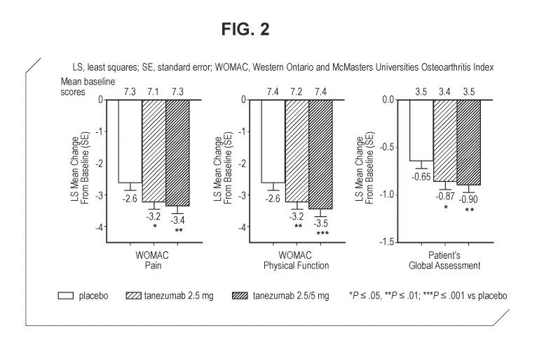
Figure 2 shows change from baseline to Week 16 in the WOMAC Pain, WOMAC
Physical Function, and PGA-0A for the study described in Example 1.
Figure 3 shows changes in WOMAC Pain, WOMAC Physical Function and
Average Daily Pain in the Index Joint Scores during the treatment period for
the study
described in Example 1.
Figure 4 shows WOMAC Pain Responder rates at week 16 for the study described
in Example 1.
Figure 5 shows WOMAC Pain responder rates at week 16 in non-responders at
week 8 for the study described in Example 1.
Figure 6 shows change, from baseline, in WOMAC Pain, WOMAC Physical
Function, and PGA-0A scores at week 24 for the study described in Example 2.
Figure 7 shows the change from baseline for the WOMAC Pain Subscale up to
Week 24.
Figure 8 shows the change from baseline for the WOMAC Physical Function
Subscale up to Week 24.
Figure 9 shows the change from baseline for the Patient Global Assessment of
OA up to Week 24.
Figure 10 shows the change from baseline for the WOMAC Pain Subscale Up to
Week 56 for the study described in Example 3.
Figure 11 shows the change from baseline for the WOMAC Physical Function
Subscale up to Week 56 for the study described in Example 3.
Figure 12 shows the change from baseline for the Patient Global Assessment of
OA up to week 56 for the study described in Example 3.
Detailed Description
The invention disclosed herein is directed to treatment of signs and symptoms
of
osteoarthritis in patients who have a history of inadequate pain relief or
intolerance to
analgesic therapy.
Accordingly, in one aspect, the invention provides a method for treating signs
and
symptoms of osteoarthritis (OA) in a patient, the method comprising
administering to the
patient an anti-nerve growth factor (NGF) antibody at a dose of 2.5 mg every 8
weeks via
subcutaneous injection; wherein the patient has a history of inadequate pain
relief or
CA 03127751 2021-07-23
WO 2020/157629
PCT/IB2020/050611
- 10 -
intolerance to analgesic therapy and the treatment with the anti-NGF antibody
effectively
improves signs and symptoms of OA by at least 16 weeks after start of
treatment with
the anti-NGF antibody.
In a further aspect, the invention provides a method for treating signs and
symptoms of osteoarthritis (OA) in a patient, the method comprising
administering to the
patient an anti-nerve growth factor (NGF) antibody at a dose of 5 mg every 8
weeks via
subcutaneous injection; wherein the patient has a history of inadequate pain
relief or
intolerance to analgesic therapy and the treatment with the anti-NGF antibody
effectively
improves signs and symptoms of OA by at least 16 weeks after start of
treatment with
the anti-NGF antibody.
General Techniques
The practice of the present invention will employ, unless otherwise indicated,
conventional techniques of molecular biology (including recombinant
techniques),
microbiology, cell biology, biochemistry and immunology, which are within the
skill of the
art. Such techniques are explained fully in the literature, such as, Molecular
Cloning: A
Laboratory Manual, second edition (Sambrook et al., 1989) Cold Spring Harbor
Press;
Oligonucleotide Synthesis (M.J. Gait, ed., 1984); Methods in Molecular
Biology, Humana
Press; Cell Biology: A Laboratory Notebook (J.E. Cellis, ed., 1998) Academic
Press;
Animal Cell Culture (R.I. Freshney, ed., 1987); Introduction to Cell and
Tissue Culture
(J.P. Mather and P.E. Roberts, 1998) Plenum Press; Cell and Tissue Culture:
Laboratory
Procedures (A. Doyle, J.B. Griffiths, and D.G. Newell, eds., 1993-1998) J.
Wiley and
Sons; Methods in Enzymology (Academic Press, Inc.); Handbook of Experimental
Immunology (D.M. Weir and C.C. Blackwell, eds.); Gene Transfer Vectors for
Mammalian
Cells (J.M. Miller and M.P. Cabs, eds., 1987); Current Protocols in Molecular
Biology
(F.M. Ausubel et al., eds., 1987); PCR: The Polymerase Chain Reaction, (Mullis
et al.,
eds., 1994); Current Protocols in Immunology (J.E. Coligan et al., eds.,
1991); Short
Protocols in Molecular Biology (Wiley and Sons, 1999); Immunobiology (C.A.
Janeway
and P. Travers, 1997); Antibodies (P. Finch, 1997); Antibodies: a practical
approach (D.
Catty., ed., IRL Press, 1988-1989); Monoclonal antibodies: a practical
approach (P.
Shepherd and C. Dean, eds., Oxford University Press, 2000); Using antibodies:
a
CA 03127751 2021-07-23
WO 2020/157629
PCT/IB2020/050611
- 1 1 -
laboratory manual (E. Harlow and D. Lane (Cold Spring Harbor Laboratory Press,
1999);
The Antibodies (M. Zanetti and J.D. Capra, eds., Harwood Academic Publishers,
1995).
Definitions
The following terms, unless otherwise indicated, shall be understood to have
the
following meanings:
An "antibody" is an immunoglobulin molecule capable of specific binding to a
target, such as a carbohydrate, polynucleotide, lipid, polypeptide, etc.,
through at least
one antigen recognition site, located in the variable region of the
immunoglobulin
molecule. As used herein, the term encompasses not only intact polyclonal or
monoclonal
antibodies, but also, unless otherwise specified, any antigen binding portion
thereof that
competes with the intact antibody for specific binding, fusion proteins
comprising an
antigen binding portion, and any other modified configuration of the
immunoglobulin
molecule that comprises an antigen recognition site. Antigen binding portions
include, for
example, Fab, Fab', F(ab')2, Fd, Fv, domain antibodies (dAbs, e.g., shark and
camelid
antibodies), fragments including complementarity determining regions (CDRs),
single
chain variable fragment antibodies (scFv), maxibodies, minibodies,
intrabodies,
diabodies, triabodies, tetrabodies, v-NAR and bis-scFv, and polypeptides that
contain at
least a portion of an immunoglobulin that is sufficient to confer specific
antigen binding to
the polypeptide. An antibody includes an antibody of any class, such as IgG,
IgA, or IgM
(or sub-class thereof), and the antibody need not be of any particular class.
Depending
on the antibody amino acid sequence of the constant region of its heavy
chains,
immunoglobulins can be assigned to different classes. There are five major
classes of
immunoglobulins: IgA, IgD, IgE, IgG, and IgM, and several of these may be
further
divided into subclasses (isotypes), e.g., IgGi, IgG2, IgG3, IgG4, IgAi and
IgA2. The heavy-
chain constant regions that correspond to the different classes of
immunoglobulins are
called alpha, delta, epsilon, gamma, and mu, respectively. The subunit
structures and
three-dimensional configurations of different classes of immunoglobulins are
well known.
A "variable region" of an antibody refers to the variable region of the
antibody light
chain or the variable region of the antibody heavy chain, either alone or in
combination.
As known in the art, the variable regions of the heavy and light chains each
consist of
four framework regions (FRs) connected by three complementarity determining
regions
CA 03127751 2021-07-23
WO 2020/157629
PCT/IB2020/050611
- 12 -
(CDRs) also known as hypervariable regions, and contribute to the formation of
the
antigen binding site of antibodies. If variants of a subject variable region
are desired,
particularly with substitution in amino acid residues outside of a CDR region
(i.e., in the
framework region), appropriate amino acid substitution, preferably,
conservative amino
acid substitution, can be identified by comparing the subject variable region
to the
variable regions of other antibodies which contain CDR1 and CDR2 sequences in
the
same canonical class as the subject variable region (Chothia and Lesk, J Mol
Biol 196(4):
901-917, 1987).
In certain embodiments, definitive delineation of a CDR and identification of
residues comprising the binding site of an antibody is accomplished by solving
the
structure of the antibody and/or solving the structure of the antibody-ligand
complex. In
certain embodiments, that can be accomplished by any of a variety of
techniques known
to those skilled in the art, such as X-ray crystallography. In certain
embodiments, various
methods of analysis can be employed to identify or approximate the CDR
regions. In
certain embodiments, various methods of analysis can be employed to identify
or
approximate the CDR regions. Examples of such methods include, but are not
limited to,
the Kabat definition, the Chothia definition, the AbM definition, the contact
definition, and
the conformational definition.
The Kabat definition is a standard for numbering the residues in an antibody
and
is typically used to identify CDR regions. See, e.g., Johnson & Wu, 2000,
Nucleic Acids
Res., 28: 214-8. The Chothia definition is similar to the Kabat definition,
but the Chothia
definition takes into account positions of certain structural loop regions.
See, e.g., Chothia
et al., 1986, J. Mol. Biol., 196: 901-17; Chothia et al., 1989, Nature, 342:
877-83. The
AbM definition uses an integrated suite of computer programs produced by
Oxford
Molecular Group that model antibody structure. See, e.g., Martin et al., 1989,
Proc Natl
Acad Sci (USA), 86:9268-9272; "AbMTm, A Computer Program for Modeling Variable
Regions of Antibodies," Oxford, UK; Oxford Molecular, Ltd. The AbM definition
models
the tertiary structure of an antibody from primary sequence using a
combination of
knowledge databases and ab initio methods, such as those described by
Samudrala et
al., 1999, "Ab Initio Protein Structure Prediction Using a Combined
Hierarchical
Approach," in PROTEINS, Structure, Function and Genetics Suppl., 3:194-198.
The
contact definition is based on an analysis of the available complex crystal
structures. See,
e.g., MacCallum et al., 1996, J. Mol. Biol., 5:732-45. In another approach,
referred to
CA 03127751 2021-07-23
WO 2020/157629
PCT/IB2020/050611
- 13 -
herein as the "conformational definition" of CDRs, the positions of the CDRs
may be
identified as the residues that make enthalpic contributions to antigen
binding. See, e.g.,
Makabe et al., 2008, Journal of Biological Chemistry, 283:1156-1166. Still
other CDR
boundary definitions may not strictly follow one of the above approaches, but
will
nonetheless overlap with at least a portion of the Kabat CDRs, although they
may be
shortened or lengthened in light of prediction or experimental findings that
particular
residues or groups of residues do not significantly impact antigen binding. As
used
herein, a CDR may refer to CDRs defined by any approach known in the art,
including
combinations of approaches. The methods used herein may utilize CDRs defined
according to any of these approaches. For any given embodiment containing more
than
one CDR, the CDRs may be defined in accordance with any of Kabat, Chothia,
extended,
AbM, contact, and/or conformational definitions.
As known in the art, a "constant region" of an antibody refers to the constant
region
of the antibody light chain or the constant region of the antibody heavy
chain, either alone
or in combination.
As used herein, "monoclonal antibody" refers to an antibody obtained from a
population of substantially homogeneous antibodies, i.e., the individual
antibodies
comprising the population are identical except for possible naturally-
occurring mutations
that may be present in minor amounts. Monoclonal antibodies are highly
specific, being
directed against a single antigenic site. Furthermore, in contrast to
polyclonal antibody
preparations, which typically include different antibodies directed against
different
determinants (epitopes), each monoclonal antibody is directed against a single
determinant on the antigen. The modifier "monoclonal" indicates the character
of the
antibody as being obtained from a substantially homogeneous population of
antibodies,
and is not to be construed as requiring production of the antibody by any
particular
method. For example, the monoclonal antibodies to be used in accordance with
the
present invention may be made by the hybridoma method first described by
Kohler and
Milstein, 1975, Nature 256:495, or may be made by recombinant DNA methods such
as
described in U.S. Pat. No. 4,816,567. The monoclonal antibodies may also be
isolated
from phage libraries generated using the techniques described in McCafferty et
al., 1990,
Nature 348:552-554, for example.
As used herein, "humanized" antibody refers to forms of non-human (e.g.
murine)
antibodies that are chimeric immunoglobulins, immunoglobulin chains, or
fragments
CA 03127751 2021-07-23
WO 2020/157629
PCT/IB2020/050611
- 14 -
thereof (such as Fv, Fab, Fab', F(ab')2 or other antigen-binding subsequences
of
antibodies) that contain minimal sequence derived from non-human
immunoglobulin.
Preferably, humanized antibodies are human immunoglobulins (recipient
antibody) in
which residues from a CDR of the recipient are replaced by residues from a CDR
of a
non-human species (donor antibody) such as mouse, rat, or rabbit having the
desired
specificity, affinity, and capacity. The humanized antibody may comprise
residues that
are found neither in the recipient antibody nor in the imported CDR or
framework
sequences, but are included to further refine and optimize antibody
performance.
In some instances, Fv framework region (FR) residues of the human
immunoglobulin are replaced by corresponding non-human residues. Furthermore,
the
humanized antibody may include residues that are found neither in the
recipient antibody
nor in the imported CDR or framework sequences, but are included to further
refine and
optimize antibody performance.
In general, the humanized antibody will include
substantially all of at least one, and typically two, variable domains, in
which all or
substantially all of the CDR regions correspond to those of a non-human
immunoglobulin
and all or substantially all of the FR regions are those of a human
immunoglobulin
consensus sequence. The humanized antibody optimally also will include at
least a
portion of an immunoglobulin constant region or domain (Fc), typically that of
a human
immunoglobulin. In some aspects of the invention the antibodies have Fc
regions
modified as described in PCT International Publication No. WO 99/58572. Other
forms
of humanized antibodies have one or more CDRs (CDR L1, CDR L2, CDR L3, CDR H1,
CDR H2, or CDR H3) which may be altered with respect to the original antibody,
which
are also termed one or more CDRs "derived from" one or more CDRs from the
original
antibody.
Humanization can be essentially performed following the method of Winter and
co-workers (Jones et al. Nature 321:522-525 (1986); Riechmann et al. Nature
332:323-
327 (1988); Verhoeyen et al. Science 239:1534-1536 (1988)), by substituting
rodent or
mutant rodent CDRs or CDR sequences for the corresponding sequences of a human
antibody. See also U.S. Patent Nos. 5,225,539; 5,585,089; 5,693,761;
5,693,762;
5,859,205; which are incorporated herein by reference in its entirety. In some
instances,
residues within the framework regions of one or more variable regions of the
human
immunoglobulin are replaced by corresponding non-human residues (see, for
example,
U.S. Patent. Nos. 5,585,089; 5,693,761; 5,693,762; and 6,180,370).
Furthermore,
CA 03127751 2021-07-23
WO 2020/157629
PCT/IB2020/050611
- 15 -
humanized antibodies may include residues that are not found in the recipient
antibody
or in the donor antibody. These modifications are made to further refine
antibody
performance (e.g., to obtain desired affinity). In general, the humanized
antibody will
include substantially all of at least one, and typically two, variable
domains, in which all
or substantially all of the hypervariable regions correspond to those of a non-
human
immunoglobulin and all or substantially all of the framework regions are those
of a human
immunoglobulin sequence. The humanized antibody optionally also will include
at least
a portion of an immunoglobulin constant region (Fc), typically that of a human
immunoglobulin. For further details see Jones et al. Nature 321:522-525
(1986);
Riechmann et al. Nature 332:323-327(1988); and Presta Curr. Op. Struct. Biol.
2:593-
596 (1992); which are incorporated herein by reference in its entirety.
Accordingly, such
"humanized" antibodies may include antibodies wherein substantially less than
an intact
human variable domain has been substituted by the corresponding sequence from
a non-
human species. In practice, humanized antibodies are typically human
antibodies in
which some CDR residues and possibly some framework residues are substituted
by
residues from analogous sites in rodent antibodies. See, for example, U.S.
Patent Nos.
5,225,539; 5,585,089; 5,693,761; 5,693,762; 5,859,205. See also U.S. Patent
No.
6,180,370, and PCT International Publication No. WO 01/27160, where humanized
antibodies and techniques for producing humanized antibodies having improved
affinity
for a predetermined antigen are disclosed.
A "human antibody" is one which possesses an amino acid sequence which
corresponds to that of an antibody produced by a human and/or has been made
using
any of the techniques for making human antibodies as disclosed herein. This
definition
of a human antibody specifically excludes a humanized antibody comprising non-
human
antigen binding residues.
The term "chimeric antibody" is intended to refer to antibodies in which the
variable
region sequences are derived from one species and the constant region
sequences are
derived from another species, such as an antibody in which the variable region
sequences are derived from a mouse antibody and the constant region sequences
are
derived from a human antibody.
The antibody "tanezumab" is a humanized immunoglobulin G Type 2 (IgG2)
monoclonal antibody directed against human nerve growth factor (NGF).
Tanezumab
binds to human NGF with high affinity and specificity and blocks the activity
of NGF
CA 03127751 2021-07-23
WO 2020/157629
PCT/IB2020/050611
- 16 -
effectively in cell culture models. Tanezumab and/or its murine precursor have
been
shown to be an effective analgesic in animal models of pathological pain
including
arthritis, cancer pain, and post-surgical pain. Tanezumab has the sequences
for the
variable heavy chain region and variable light chain region of SEQ ID Nos: 1
and 2,
.. respectively. The heavy chain and light chain sequences are provided in SEQ
ID NOs:
9 and 10, or SEQ ID NOs: 11 and 10. The C-terminal lysine (K) of the heavy
chain
amino acid sequence of SEQ ID NO: 9 is optional and may be processed,
resulting in a
heavy chain amino acid sequence lacking the C-terminal lysine (K) and having
the
sequence shown in SEQ ID NO: 11. Sequences of tanezumab are provided in Table
1
below. Tanezumab is described, as antibody E3, in W02004/058184, herein
incorporated by reference.
As known in the art, "polynucleotide," or "nucleic acid," as used
interchangeably
herein, refer to chains of nucleotides of any length, and include DNA and RNA.
The
nucleotides can be deoxyribonucleotides, ribonucleotides, modified nucleotides
or bases,
and/or their analogs, or any substrate that can be incorporated into a chain
by DNA or
RNA polymerase. A polynucleotide may comprise modified nucleotides, such as
methylated nucleotides and their analogs. If present, modification to the
nucleotide
structure may be imparted before or after assembly of the chain. The sequence
of
nucleotides may be interrupted by non-nucleotide components. A polynucleotide
may be
further modified after polymerization, such as by conjugation with a labeling
component.
Other types of modifications include, for example, "caps", substitution of one
or more of
the naturally occurring nucleotides with an analog, internucleotide
modifications such as,
for example, those with uncharged linkages (e.g., methyl phosphonates,
phosphotriesters, phosphoamidates, carbamates, etc.) and with charged linkages
(e.g.,
phosphorothioates, phosphorodithioates, etc.), those containing pendant
moieties, such
as, for example, proteins (e.g., nucleases, toxins, antibodies, signal
peptides, poly-L-
lysine, etc.), those with intercalators (e.g., acridine, psoralen, etc.),
those containing
chelators (e.g., metals, radioactive metals, boron, oxidative metals, etc.),
those
containing alkylators, those with modified linkages (e.g., alpha anomeric
nucleic acids,
etc.), as well as unmodified forms of the polynucleotide(s). Further, any of
the hydroxyl
groups ordinarily present in the sugars may be replaced, for example, by
phosphonate
groups, phosphate groups, protected by standard protecting groups, or
activated to
prepare additional linkages to additional nucleotides, or may be conjugated to
solid
CA 03127751 2021-07-23
WO 2020/157629
PCT/IB2020/050611
- 17 -
supports. The 5' and 3' terminal OH can be phosphorylated or substituted with
amines or
organic capping group moieties of from 1 to 20 carbon atoms. Other hydroxyls
may also
be derivatized to standard protecting groups. Polynucleotides can also contain
analogous
forms of ribose or deoxyribose sugars that are generally known in the art,
including, for
example, 2'-0-methyl-, 2'-0-allyl, 2'-fluoro- or 2'-azido-ribose, carbocyclic
sugar analogs,
alpha- or beta-anomeric sugars, epimeric sugars such as arabinose, xyloses or
lyxoses,
pyranose sugars, furanose sugars, sedoheptuloses, acyclic analogs and abasic
nucleoside analogs such as methyl riboside. One or more phosphodiester
linkages may
be replaced by alternative linking groups. These alternative linking groups
include, but
are not limited to, embodiments wherein phosphate is replaced by
P(0)S("thioate"),
P(S)S ("dithioate"), (0)NR2 ("am idate"), P(0)R, P(0)OR', CO or CH2
("formacetal"), in
which each R or R' is independently H or substituted or unsubstituted alkyl (1-
20 C)
optionally containing an ether (-0-) linkage, aryl, alkenyl, cycloalkyl,
cycloalkenyl or
araldyl. Not all linkages in a polynucleotide need be identical. The preceding
description
applies to all polynucleotides referred to herein, including RNA and DNA.
An antibody that "preferentially binds" or "specifically binds" (used
interchangeably
herein) to an epitope is a term well understood in the art, and methods to
determine such
specific or preferential binding are also well known in the art. A molecule is
said to exhibit
"specific binding" or "preferential binding" if it reacts or associates more
frequently, more
rapidly, with greater duration and/or with greater affinity with a particular
cell or substance
than it does with alternative cells or substances. An antibody "specifically
binds" or
"preferentially binds" to a target if it binds with greater affinity, avidity,
more readily, and/or
with greater duration than it binds to other substances. For example, an
antibody that
specifically or preferentially binds to a target (e.g., PD-1) epitope is an
antibody that binds
this epitope with greater affinity, avidity, more readily, and/or with greater
duration than it
binds to other target epitopes or non-target epitopes. It is also understood
by reading this
definition that, for example, an antibody (or moiety or epitope) that
specifically or
preferentially binds to a first target may or may not specifically or
preferentially bind to a
second target. As such, "specific binding" or "preferential binding" does not
necessarily
require (although it can include) exclusive binding. Generally, but not
necessarily,
reference to binding means preferential binding.
As used herein, "substantially pure" refers to material which is at least 50%
pure
(i.e., free from contaminants), more preferably, at least 90% pure, more
preferably, at
CA 03127751 2021-07-23
WO 2020/157629
PCT/IB2020/050611
- 18 -
least 95% pure, yet more preferably, at least 98% pure, and most preferably,
at least 99%
pure.
A "host cell" includes an individual cell or cell culture that can be or has
been a
recipient for vector(s) for incorporation of polynucleotide inserts. Host
cells include
progeny of a single host cell, and the progeny may not necessarily be
completely identical
(in morphology or in genomic DNA complement) to the original parent cell due
to natural,
accidental, or deliberate mutation. A host cell includes cells transfected in
vivo with a
polynucleotide(s) of this invention.
As known in the art, the term "Fc region" is used to define a C-terminal
region of
an immunoglobulin heavy chain. The "Fc region" may be a native sequence Fc
region or
a variant Fc region. Although the boundaries of the Fc region of an
immunoglobulin heavy
chain might vary, the human IgG heavy chain Fc region is usually defined to
stretch from
an amino acid residue at position Cys226, or from Pro230, to the carboxyl-
terminus
thereof. The numbering of the residues in the Fc region is that of the EU
index as in
Kabat. Kabat et al., Sequences of Proteins of Immunological Interest, 5th Ed.
Public
Health Service, National Institutes of Health, Bethesda, Md., 1991. The Fc
region of an
immunoglobulin generally comprises two constant domains, CH2 and CH3. As is
known
in the art, an Fc region can be present in dimer or monomeric form.
As used in the art, "Fc receptor" and "FcR" describe a receptor that binds to
the
Fc region of an antibody. The preferred FcR is a native sequence human FcR.
Moreover,
a preferred FcR is one which binds an IgG antibody (a gamma receptor) and
includes
receptors of the FcyRI, FcyRII, and FcyRIII subclasses, including allelic
variants and
alternatively spliced forms of these receptors. FcyRII receptors include
FcyRIIA (an
"activating receptor") and FcyRIIB (an "inhibiting receptor"), which have
similar amino
acid sequences that differ primarily in the cytoplasmic domains thereof. FcRs
are
reviewed in Ravetch and Kinet, 1991, Ann. Rev. Immunol., 9:457-92; Capel et
al., 1994,
Immunomethods, 4:25-34; and de Haas et al., 1995, J. Lab. Clin. Med., 126:330-
41.
"FcR" also includes the neonatal receptor, FcRn, which is responsible for the
transfer of
maternal IgGs to the fetus (Guyer et al., 1976, J. Immunol., 117:587; and Kim
et al., 1994,
J. Immunol., 24:249).
The term "compete", as used herein with regard to an antibody, means that a
first
antibody, or an antigen-binding portion thereof, binds to an epitope in a
manner
sufficiently similar to the binding of a second antibody, or an antigen-
binding portion
CA 03127751 2021-07-23
WO 2020/157629
PCT/IB2020/050611
- 19 -
thereof, such that the result of binding of the first antibody with its
cognate epitope is
detectably decreased in the presence of the second antibody compared to the
binding of
the first antibody in the absence of the second antibody. The alternative,
where the
binding of the second antibody to its epitope is also detectably decreased in
the presence
of the first antibody, can, but need not be the case. That is, a first
antibody can inhibit the
binding of a second antibody to its epitope without that second antibody
inhibiting the
binding of the first antibody to its respective epitope. However, where each
antibody
detectably inhibits the binding of the other antibody with its cognate epitope
or ligand,
whether to the same, greater, or lesser extent, the antibodies are said to
"cross-compete"
with each other for binding of their respective epitope(s). Both competing and
cross-
competing antibodies are encompassed by the present invention. Regardless of
the
mechanism by which such competition or cross-competition occurs (e.g., steric
hindrance, conformational change, or binding to a common epitope, or portion
thereof),
the skilled artisan would appreciate, based upon the teachings provided
herein, that such
competing and/or cross-competing antibodies are encompassed and can be useful
for
the methods disclosed herein.
A "functional Fc region" possesses at least one effector function of a native
sequence Fc region. Exemplary "effector functions" include C1q binding;
complement
dependent cytotoxicity; Fc receptor binding; antibody-dependent cell-mediated
cytotoxicity; phagocytosis; down-regulation of cell surface receptors (e.g. B
cell receptor),
etc. Such effector functions generally require the Fc region to be combined
with a binding
domain (e.g. an antibody variable domain) and can be assessed using various
assays
known in the art for evaluating such antibody effector functions.
A "native sequence Fc region" comprises an amino acid sequence identical to
the
amino acid sequence of an Fc region found in nature. A "variant Fc region"
comprises an
amino acid sequence which differs from that of a native sequence Fc region by
virtue of
at least one amino acid modification, yet retains at least one effector
function of the native
sequence Fc region. Preferably, the variant Fc region has at least one amino
acid
substitution compared to a native sequence Fc region or to the Fc region of a
parent
polypeptide, e.g. from about one to about ten amino acid substitutions, and
preferably,
from about one to about five amino acid substitutions in a native sequence Fc
region or
in the Fc region of the parent polypeptide. The variant Fc region herein will
preferably
possess at least about 80% sequence identity with a native sequence Fc region
and/or
CA 03127751 2021-07-23
WO 2020/157629
PCT/IB2020/050611
- 20 -
with an Fc region of a parent polypeptide, and most preferably, at least about
90%
sequence identity therewith, more preferably, at least about 95%, at least
about 96%, at
least about 97%, at least about 98%, at least about 99% sequence identity
therewith.
As used herein, "treatment" is an approach for obtaining beneficial or desired
clinical results. For purposes of this invention, beneficial or desired
clinical results include
reduction or improvement in signs and symptoms of osteoarthritis, for example
as
compared to before administration of the anti-NGF antibody.
"Ameliorating" means a lessening or improvement of one and more signs or
symptoms of osteoarthritis, for example as compared to not administering an
anti-NGF
antibody as described herein. "Ameliorating" also includes shortening or
reduction in
duration of a symptom.
As used herein, an "effective dosage" or "effective amount" of drug, compound,
or
pharmaceutical composition is an amount sufficient to effect any one or more
beneficial
or desired results. In more specific aspects, an effective amount prevents,
alleviates or
ameliorates signs or symptoms of osteoarthritis, and/or prolongs the survival
of the
subject being treated. For prophylactic use, beneficial or desired results
include
eliminating or reducing the risk, lessening the severity, or delaying the
outset of the
disease, including biochemical, histological and/or behavioral symptoms of the
disease,
its complications and intermediate pathological phenotypes presenting during
development of the disease. For therapeutic use, beneficial or desired results
include
clinical results such as reducing one or more signs or symptoms of
osteoarthritis such
as, for example, osteoarthritis of the hip, knee, shoulder or hand, decreasing
the dose of
other medications required to treat the disease, enhancing the effect of
another
medication, and/or delaying the progression of the disease in patients. An
effective
dosage can be administered in one or more administrations. For purposes of
this
invention, an effective dosage of drug, compound, or pharmaceutical
composition is an
amount sufficient to accomplish prophylactic or therapeutic treatment either
directly or
indirectly. As is understood in the clinical context, an effective dosage of a
drug,
compound, or pharmaceutical composition may or may not be achieved in
conjunction
with another drug, compound, or pharmaceutical composition. Thus, an
"effective
dosage" may be considered in the context of administering one or more
therapeutic
agents, and a single agent may be considered to be given in an effective
amount if, in
conjunction with one or more other agents, a desirable result may be or is
achieved.
CA 03127751 2021-07-23
WO 2020/157629
PCT/IB2020/050611
- 21 -
The term "inadequate pain relief or intolerance to analgesic therapy" refers
to a
patient who has experienced an adverse event after treatment with the
analgesic; who is
refractory to treatment with the analgesic; who shows no clinically meaningful
improvement in one or more measures of signs and symptoms of osteoarthritis
including
pain; who is addicted to the analgesic therapy (including opioids); or who is
unwilling to
take the analgesic therapy. Thus, the term includes reference to a patient for
whom use
of other analgesics is ineffective or not appropriate.
Treatment "effectively improves" or "effectively reduces" when assessment of
the
sign or symptom of osteoarthritis is quantified via a clinical measure
relative to baseline
and during and/or after the treatment period. The difference between the
clinical measure
at baseline and during/after treatment is compared and used to determine
whether the
sign or symptom has improved and the treatment is effective. This comparison
can
include comparison to placebo or to one or more of the prior therapies. In
some
embodiments, the comparison can be to placebo or to treatment with an
analgesic
therapy, such as an opioid or an NSAID. In some embodiments, the comparison
can be
to a sign or symptom before start of treatment with the NGF antibody. For
example, the
WOMAC Pain subscale measure can be determined for the patient at baseline and
then
determined throughout the treatment period, such as at weeks 2, 4, 8, 16, 24,
32, 40, 48,
56, or longer. Similarly, the WOMAC Physical Function subscale measure can
also be
determined in this manner. Yet further, the PGA-0A measure can also be
determined in
this manner.
In some embodiments the treatment effectively reduces WOMAC Pain by at least
about 2.5, at least about 2.6, at least about 2.7, at least about 2.8, at
least about 2.9, at
least about 3.0, at least about 3.1, at least about 3.2, at least about 3.3,
or at least about
3.4, at least about 3.5, at least about 3.6, at least about 3.7 compared to
baseline
WOMAC Pain prior to or at start of treatment. In some embodiments, the
treatment
reduces WOMAC Pain by greater than 20%7 25%7 30%7 35%7 40%7 45%7 50%7 55%7
60%, 65%, 70%, 75%, 80%, 85%, 90% or 95 A compared to baseline prior to or at
start
of treatment. In some embodiments the treatment effectively reduces WOMAC Pain
score compared to placebo for tanezumab. In some embodiments the treatment
effectively reduces WOMAC Pain score by at least about 0.2, 0.25, 0.3, 0.35,
0.4, 0.45,
0.5, 0.55, 0.6, 0.65, 0.7, 0.75, 0.8, 0.85, 0.9, 0.95, or 1 compared to
placebo for
tanezumab. In some embodiments the treatment effectively reduces WOMAC Pain
score
CA 03127751 2021-07-23
WO 2020/157629
PCT/IB2020/050611
- 22 -
compared to baseline and/or placebo for tanezumab to a greater extent than an
opioid
analgesic or an NSAID analgesic. In some embodiments the treatment reduces
WOMAC
Pain score by at least about 0.1, 0.15, 0.2, 0.25, 0.3, 0.35 more than an
NSAID. In some
embodiments the reduction in WOMAC Pain score is observed at week 16, 24, 32,
40,
48 or 56 of treatment. In some embodiments the change from baseline is based
on the
Least Squares Mean.
In some embodiments the treatment effectively reduces WOMAC Physical
Function score by at least about 2.5, at least about 2.6, at least about 2.7,
at least about
2.8, at least about 2.9, at least about 3.0, at least about 3.1, at least
about 3.2, at least
about 3.3, or at least about 3.4, at least about 3.5, at least about 3.6, at
least about 3.7
compared to baseline prior to or at start of treatment. In some embodiments,
the
treatment reduces WOMAC Physical Function by greater than 20%, 25%, 30%, 35%,
40%, 45%, 50%, 55%, 60%, 65%, 70%, 75%, 80%, 85%, 90% or 95% compared to
baseline prior to or at start of treatment. In some embodiments the treatment
effectively
reduces WOMAC Physical Function score compared to placebo for tanezumab. In
some
embodiments the treatment effectively reduces WOMAC Physical Function score by
at
least about 0.2, 0.25, 0.3, 0.35, 0.4, 0.45, 0.5, 0.55, 0.6, 0.65, 0.7, 0.75,
0.8, 0.85, 0.9,
0.95, 1 or 1.1 compared to placebo for tanezumab. In some embodiments the
treatment
effectively reduces WOMAC Physical Function score compared to baseline and/or
placebo to a greater extent than an opioid analgesic or an NSAID analgesic. In
some
embodiments the treatment reduces WOMAC Physical Function score by at least
about
0.1, 0.15, 0.2, 0.25, 0.3, 0.35, 0.4 more than an NSAID. In some embodiments
the
reduction in WOMAC Physical Function score is observed at week 16, 24, 32, 40,
48 or
56 of treatment. In some embodiments the change from baseline is based on the
Least
Squares Mean.
In some embodiments the treatment effectively reduces PGA-0A score by at least
about 0.7, 0.75, 0.8, 0.85, 0.9, 0.95, 1, or 1.1 compared to baseline prior to
or at start of
treatment. In some embodiments, the treatment reduces PGA-0A by greater than
15%,
20%, 25%, 30% or 40% compared to baseline prior to or at start of treatment.
In some
embodiments the treatment effectively reduces PGA-0A score compared to placebo
for
tanezumab. In some embodiments the treatment effectively reduces PGA-0A score
by
at least about 0.1, 0.15, 0.2, 0.25, 0.3, 0.35, 0.4, 0.45, or 0.5 compared to
placebo for
tanezumab. In some embodiments the treatment effectively reduces PGA-0A score
CA 03127751 2021-07-23
WO 2020/157629
PCT/IB2020/050611
- 23 -
compared to baseline and/or placebo for tanezumab to a greater extent than an
opioid
analgesic or an NSAID analgesic. In some embodiments the reduction in PGA-0A
score
is observed at week 16, 24, 32, 40, 48 or 56 of treatment. In some embodiments
the
change from baseline is based on the Least Squares Mean.
The term "baseline" refers to a value of a sign or symptom associated measure
for
a patient prior to administration of the anti-NGF antibody as part of the
treatment method.
In some embodiments, the term "baseline" refers to a value of a sign or
symptom
associated measure for control healthy subjects that do not have
osteoarthritis.
In some embodiments, treatment with the anti-NGF antibody effectively improves
signs and symptoms of OA by at least 8 weeks after start of treatment with the
antibody.
In some embodiments, treatment with the anti-NGF antibody effectively improves
signs
and symptoms of OA by at least 10 weeks after start of treatment with the
antibody. In
some embodiments, treatment with the anti-NGF antibody effectively improves
signs and
symptoms of OA by at least 12 weeks after start of treatment with the
antibody. In some
embodiments, treatment with the anti-NGF antibody effectively improves signs
and
symptoms of OA by at least 14 weeks after start of treatment with the
antibody. In some
embodiments, treatment with the anti-NGF antibody effectively improves signs
and
symptoms of OA by at least 16 weeks after start of treatment with the
antibody. In some
embodiments, treatment with the anti-NGF antibody effectively improves signs
and
symptoms of OA by at least 24 weeks after start of treatment with the
antibody. In some
embodiments, treatment with the anti-NGF antibody effectively improves signs
and
symptoms of OA by at least 32 weeks after start of treatment with the
antibody. In some
embodiments, treatment with the anti-NGF antibody effectively improves signs
and
symptoms of OA by at least 40 weeks after start of treatment with the
antibody.
In some embodiments, treatment with the anti-NGF antibody effectively improves
signs and symptoms of OA within 1 week after start of treatment with the
antibody. In
some embodiments, treatment with the anti-NGF antibody effectively improves
signs and
symptoms of OA within 2 weeks after start of treatment with the antibody.
In some embodiments, the treatment improves pain associated with OA. In some
embodiments the pain is moderate to severe pain associated with OA; and is
optionally
chronic pain.
The WOMAC Pain subscale is comprised of 5 questions regarding the amount of
pain experienced due to OA in the index joint (selected study knee or hip) in
the past 48
CA 03127751 2021-07-23
WO 2020/157629
PCT/IB2020/050611
-24 -
hours. The WOMAC Pain subscale is calculated as the mean of the scores from
the five
individual questions, which may not be a whole (integer) number. The WOMAC
Pain
subscale NRS scores for each question, and the WOMAC Pain subscale score,
range
from 0 to 10, with higher scores indicating higher pain.
The WOMAC Physical function subscale is comprised of 17 questions regarding
the degree of difficulty experienced due to arthritis in the index joint
(selected study knee
or hip) in the past 48 hours. The WOMAC Physical Function subscale is
calculated as
the mean of the scores from the seventeen individual questions, which may not
be a
whole (integer) number. The WOMAC Physical Function subscale NRS scores for
each
question, and the WOMAC Physical Function subscale score, range from 0 to 10
with
higher scores indicating worse function. This refers to the subject's ability
to move around
and perform usual activities of daily living.
The PGA-0A measure is based on a question to patients: "Considering all the
ways your osteoarthritis in your [joint] affects you, how are you doing
today?". Patients
rate their condition using the following scale:
Grade Description
1 ¨ Very Good - Asymptomatic and no limitation of normal activities
2 ¨ Good - Mild symptoms and no limitation of normal activities
3 ¨ Fair - Moderate symptoms and limitation of some normal activities
4 ¨ Poor - Severe symptoms and inability to carry out most normal activities
5 ¨ Very Poor - Very severe symptoms which are intolerable and inability to
carry
out all normal activities
Kellgren-Lawrence x-ray grade is a method of classifying the severity of
osteoarthritis (Kellgren and Lawrence., Ann Rheum Dis 2000: 16(4): 494-502).
Rapidly progressive osteoarthritis (RPOA) of the hip was first described by
Forestier in 1957 and subsequently described in a number of studies as
atrophic
osteoarthritis, rapidly destructive osteoarthritis, rapidly destructive
arthropathy, rapidly
progressive hip disease, or rapidly destructive coxarthrosis. Rapidly
progressive hip
osteoarthritis is characterized by subjects who typically present with hip
pain, often
severe, with radiographs that show rapid joint space narrowing as a result of
chrondrolysis from a prior radiograph and, subsequently, an osteolytic phase
with severe
progressive atrophic bone destruction involving the femoral head and the
acetabulum.
There can be marked flattening of the femoral head and loss of subchondral
bone in the
CA 03127751 2021-07-23
WO 2020/157629
PCT/IB2020/050611
- 25 -
weight bearing area and in some cases the femoral head appears sheared off.
Osteophytes are typically conspicuously small or absent. Bone sclerosis is
often present
at sites of impaction of the femoral head and the acetabulum, subchondral
detritus is
invariably present and bone fragmentation and debris are commonly observed
that can
lead to synovitis. Lequesne proposed that subjects with 2 mm/year or greater
of joint
space narrowing or loss of more than 50% of the joint space within 1 year
should be
considered to have rapidly progressive osteoarthritis. Due to a lack of
longitudinal studies,
it is not clear what proportion of subjects with rapid loss of joint space
(chondrolysis) will
progress to have bone destruction. Rapid progression of osteoarthritis has
also been
described in the shoulder and the knee.
The incidence of rapidly progressive osteoarthritis in the overall
osteoarthritis
population is not well defined. For rapid progression of hip osteoarthritis,
the prevalence
ranges from approximately 2% to 18% based on clinical case series analyses.
The
pathophysiology of rapidly progressive osteoarthritis is not understood.
Various
mechanisms have been proposed including; ischemia, venous stasis, local
nutritional
deficiencies, synovitis, mechanical overloading, NSAID or corticosteroid use,
intra
articular deposition of hydroxyapatite or pyrophosphate crystals and
subchondral
insufficiency fractures.
There is a lack of data in the literature on the rate of rapidly progressive
OA in a
progressed OA population and the causes of this disease progression. As
described by
Hochberg et al (Arthritis Rheumatol., vol. 68, no. 2. pp. 382-391). "Rapidly
progressive
osteoarthritis is characterized by pain, with radiographs showing rapid joint
space
narrowing as a result of chondrolysis (type-1)." Possibly subsequently, these
patients
progress to an osteolytic phase with severe progressive atrophic bone
destruction (type-
2). However, this continuity is not clear due to a lack of longitudinal
studies (Hochberg et
al., Arthritis Rheumatol., vol. 68, no. 2. pp. 382-391).
The term "Index joint" refers to the most painful joint at screening before
start of
treatment and is the joint that is assessed during treatment. For example, the
index joint
is the most painful joint of the left and right hips and knees at screening
before start of
the treatment.
Radiographic assessments (x-rays) of both knees, both hips and both shoulders
can be performed or obtained prior to treatment, at screening, and also during
treatment.
Other major joints exhibiting signs or symptoms suggestive of osteoarthritis
may also be
CA 03127751 2021-07-23
WO 2020/157629
PCT/IB2020/050611
-26 -
imaged. A major joint is defined as a mobile synovial joint in the limbs such
as shoulders,
elbows, wrists, hips, knees, ankles and excluding the joints of the toes and
hands. Any
joint imaged at screening or other at risk joints identified during the study
period should
also be imaged.
A central radiology reader (Central Reader) may review the radiology images
for
assessment of eligibility including determination and identification of
exclusionary joint
conditions. Radiographs required at screening may be obtained at least two
weeks prior
to the beginning of the Initial Pain Assessment Period (IPAP) to permit
central radiology
review of the images and to establish subject eligibility for initial dosing
with an NGF
antibody. In some embodiments, subjects may not be permitted to start dosing
with an
NGF antibody until the screening radiographs are reviewed and eligibility is
established.
The X-ray technologists, in addition to their professional training and
certifications,
are trained in performing the radiographic protocols for the knees, hips, and
shoulders.
To facilitate reproducibility and accuracy of joint space width measurement in
the knees
and hips, a semi-automated software and positioning frame standardized subject
and
joint positioning protocol can be utilized. The Core Imaging Laboratory may be
responsible for working with the sites to ensure quality, standardization and
reproducibility of the radiographic images/assessments made at the Screening
and
follow-up time-points. Additional details regarding the required X-rays may be
provided
.. in a site imaging manual.
Central radiology readers (Central Readers) may be board certified
radiologists or
have the international equivalent as musculoskeletal radiologists. The Central
Readers
may be governed by an imaging atlas and an imaging Charter which includes a
specific
description of the scope of their responsibilities. Central Readers may review
the
radiology images at Screening for assessment of eligibility (including
determination of
Kellgren-Lawrence Grade) and identification of exclusionary joint conditions
such as
rapidly progressive osteoarthritis, atrophic or hypotrophic osteoarthritis,
subchondral
insufficiency fractures (spontaneous osteonecrosis of the knee [SPONN),
primary
osteonecrosis and pathological fractures. After start of treatment, the
Central Reader may
review radiology images for diagnosis of joint conditions that would warrant
further
evaluation by the Adjudication Committee such as possible or probable rapidly
progressive osteoarthritis, subchondral insufficiency fractures (spontaneous
osteonecrosis of the knee [SPONN), primary osteonecrosis or pathological
fracture.
CA 03127751 2021-07-23
WO 2020/157629
PCT/IB2020/050611
- 27 -
For subjects who are identified with a possible or probable joint event (i.e.,
rapidly
progressive osteoarthritis, subchondral insufficiency fractures, spontaneous
osteonecrosis of the knee (SPONK), primary osteonecrosis or pathological
fracture) and
subjects undergoing total joint replacement for any reason, all images and
other source
documentation may be provided to the blinded Adjudication Committee for review
and
adjudication of the event. The Adjudication Committee's assessment of the
event may
represent the final classification of the event.
Patients may be excluded from treatment with the anti-NGF antibody, during or
before treatment with the anti-NGF antibody, if there is radiographic evidence
of any of
the following conditions in any screening radiograph as determined by a
central radiology
reviewer and as defined in an imaging atlas: excessive malalignment of the
knee, severe
chondrocalcinosis; other arthropathies (e.g., rheumatoid arthritis), systemic
metabolic
bone disease (e.g., pseudogout, Paget's disease; metastatic calcifications),
large cystic
lesions, primary or metastatic tumor lesions, stress or traumatic fracture. In
some
embodiments a patient may be excluded from treatment with the anti-NGF
antibody,
before or during the treatment with the anti-NGF antibody, if there is
radiographic
evidence of any of the following conditions as determined by the central
radiology
reviewer and as defined in an imaging atlas at screening: 1) rapidly
progressive
osteoarthritis, 2) atrophic or hypotrophic osteoarthritis, 3) subchondral
insufficiency
fractures, 4) spontaneous osteonecrosis of the knee (SPONK), 5) osteonecrosis,
or 6)
pathologic fracture.
In some embodiments the patents are monitored for the development of signs and
symptoms of rapidly progressive osteoarthritis prior to each dose. In some
embodiments
monitoring include radiographic assessment (such as X-ray). In some
embodiments the
radiographic assement is performed annually during treatment. The radiographic
assessment may be a bilateral assessment of the hip and/or knee. In some
embodiments,
symptoms of rapidly progressive osteoarthritis may include new onset, severe
persistent
pain or swelling in a joint. In some embodiments, treatment is discontinued if
a patient
develops rapidly progressive osteoarthritis.
A "patient", an "individual" or a "subject", used interchangeably herein, is a
mammal, more preferably, a human. Mammals also include, but are not limited
to, farm
animals (e.g., cows, pigs, horses, chickens, etc.), sport animals, pets,
primates, horses,
dogs, cats, mice and rats.
CA 03127751 2021-07-23
WO 2020/157629
PCT/IB2020/050611
- 28 -
As used herein, "vector" means a construct, which is capable of delivering,
and,
preferably, expressing, one or more gene(s) or sequence(s) of interest in a
host cell.
Examples of vectors include, but are not limited to, viral vectors, naked DNA
or RNA
expression vectors, plasmid, cosmid or phage vectors, DNA or RNA expression
vectors
associated with cationic condensing agents, DNA or RNA expression vectors
encapsulated in liposomes, and certain eukaryotic cells, such as producer
cells.
As used herein, "expression control sequence" means a nucleic acid sequence
that directs transcription of a nucleic acid. An expression control sequence
can be a
promoter, such as a constitutive or an inducible promoter, or an enhancer. The
expression control sequence is operably linked to the nucleic acid sequence to
be
transcribed.
As used herein, "pharmaceutically acceptable carrier" or "pharmaceutical
acceptable excipient" includes any material which, when combined with an
active
ingredient, allows the ingredient to retain biological activity and is non-
reactive with the
subject's immune system. Examples include, but are not limited to, any of the
standard
pharmaceutical carriers such as a phosphate buffered saline solution, water,
emulsions
such as oil/water emulsion, and various types of wetting agents. Preferred
diluents for
aerosol or parenteral administration are phosphate buffered saline (PBS) or
normal
(0.9%) saline. Compositions comprising such carriers are formulated by well-
known
conventional methods (see, for example, Remington's Pharmaceutical Sciences,
18th
edition, A. Gennaro, ed., Mack Publishing Co., Easton, PA, 1990; and
Remington, The
Science and Practice of Pharmacy 20th Ed. Mack Publishing, 2000).
The term "effector function" refers to the biological activities attributable
to the Fc
region of an antibody. Examples of antibody effector functions include, but
are not limited
to, antibody-dependent cell-mediated cytotoxicity (ADCC), Fc receptor binding,
complement dependent cytotoxicity (CDC), phagocytosis, C1q binding, and down
regulation of cell surface receptors (e.g., B cell receptor; BCR). See, e.g.,
U.S. Pat No.
6,737,056. Such effector functions generally require the Fc region to be
combined with
a binding domain (e.g., an antibody variable domain) and can be assessed using
various
assays known in the art for evaluating such antibody effector functions. An
exemplary
measurement of effector function is through Fcy3 and/or C1q binding.
As used herein "antibody-dependent cell-mediated cytotoxicity" or "ADCC"
refers
to a cell-mediated reaction in which nonspecific cytotoxic cells that express
Fc receptors
CA 03127751 2021-07-23
WO 2020/157629
PCT/IB2020/050611
- 29 -
(FcRs) (e.g. natural killer (NK) cells, neutrophils, and macrophages)
recognize bound
antibody on a target cell and subsequently cause lysis of the target cell.
ADCC activity
of a molecule of interest can be assessed using an in vitro ADCC assay, such
as that
described in U.S. Patent No. 5,500,362 or 5,821,337. Useful effector cells for
such
assays include peripheral blood mononuclear cells (PBMC) and NK cells.
Alternatively,
or additionally, ADCC activity of the molecule of interest may be assessed in
vivo, e.g.,
in an animal model such as that disclosed in Clynes et al., 1998, PNAS (USA),
95:652-
656.
"Complement dependent cytotoxicity" or "CDC" refers to the lysing of a target
in
the presence of complement. The complement activation pathway is initiated by
the
binding of the first component of the complement system (C1q) to a molecule
(e.g. an
antibody) complexed with a cognate antigen. To assess complement activation, a
CDC
assay, e.g. as described in Gazzano-Santoro et al., J. Immunol. Methods, 202:
163
(1996), may be performed.
The term "kon" or "ka", as used herein, refers to the rate constant for
association of
an antibody to an antigen. Specifically, the rate constants (kon or ka and
koff or kd) and
equilibrium dissociation constants are measured using whole antibody (i.e.
bivalent) and
monomeric proteins.
The term "koff " or "kd", as used herein, refers to the rate constant for
dissociation
of an antibody from the antibody/antigen complex.
The term "KD", as used herein, refers to the equilibrium dissociation constant
of an
antibody-antigen interaction.
Reference to "about" a value or parameter herein includes (and describes)
embodiments that are directed to that value or parameter per se. For example,
description referring to "about X" includes description of "X." Numeric ranges
are
inclusive of the numbers defining the range. Generally speaking, the term
"about" refers
to the indicated value of the variable and to all values of the variable that
are within the
experimental error of the indicated value (e.g. within the 95% confidence
interval for the
mean) or within 10 percent of the indicated value, whichever is greater. Where
the term
"about" is used within the context of a time period (years, months, weeks,
days etc.), the
term "about" means that period of time plus or minus one amount of the next
subordinate
time period (e.g. about 1 year means 11-13 months; about 6 months means 6
months
CA 03127751 2021-07-23
WO 2020/157629
PCT/IB2020/050611
- 30 -
plus or minus 1 week; about 1 week means 6-8 days; etc.), or within 10 per
cent of the
indicated value, whichever is greater.
The term "subcutaneous administration" refers to the administration of a
substance
into the subcutaneous layer.
The term "preventing" or "prevent" refers to (a) keeping a disorder from
occurring
or (b) delaying the onset of a disorder or onset of symptoms of a disorder.
It is understood that wherever embodiments are described herein with the
language "comprising," otherwise analogous embodiments described in terms of
"consisting of" and/or "consisting essentially of" are also provided.
Where aspects or embodiments of the invention are described in terms of a
Markush group or other grouping of alternatives, the present invention
encompasses not
only the entire group listed as a whole, but each member of the group
individually and all
possible subgroups of the main group, but also the main group absent one or
more of the
group members. The present invention also envisages the explicit exclusion of
one or
more of any of the group members in the claimed invention.
Unless otherwise defined, all technical and scientific terms used herein have
the
same meaning as commonly understood by one of ordinary skill in the art to
which this
invention belongs. In case of conflict, the present specification, including
definitions, will
control. Throughout this specification and claims, the word "comprise," or
variations such
as "comprises" or "comprising" will be understood to imply the inclusion of a
stated integer
or group of integers but not the exclusion of any other integer or group of
integers. Unless
otherwise required by context, singular terms shall include pluralities and
plural terms
shall include the singular. Any example(s) following the term "e.g." or for
example" is not
meant to be exhaustive or limiting.
Exemplary methods and materials are described herein, although methods and
materials similar or equivalent to those described herein can also be used in
the practice
or testing of the present invention. The materials, methods, and examples are
illustrative
only and not intended to be limiting.
Anti-NGF antibodies
Provided herein are anti-NGF antibodies for use in the method of treatment.
In one aspect, the anti-NGF antibody binds to NGF and inhibits binding of NGF
to
trkA and/or p75.
CA 03127751 2021-07-23
WO 2020/157629
PCT/IB2020/050611
-31 -
In an embodiment, the antibody comprises three CDRs from the heavy chain
variable region of SEQ ID NO: 1. In some embodiments, the antibody comprises
three
CDRs from the light chain variable region of SEQ ID NO: 2. In some embodiments
the
antibody comprises three CDRs from the heavy chain variable region of SEQ ID
NO: 1
and three CDRs from the light chain variable region of SEQ ID NO: 2.
In some embodiments, the CDRs may be defined in accordance with any of Kabat,
Chothia, extended, AbM, contact, and/or conformational definitions. In some
embodiments, the CDRS shown in SEQ ID NO:3, SEQ ID NO:4, SEQ ID NO:5, SEQ ID
NO:6, SEQ ID NO:7, and SEQ ID NO:8 are determined by a combination of the
Kabat
.. and Chothia methods.
Exemplary antibody sequences used for the present invention include, but are
not
limited to, the sequences listed below.
Table 1
SEQ ID NO: Sequence
1 Variable heavy chain region:
QVQLQESGPGLVKPSETLSLTCTVSGFSLIGYDLNWIRQPPGKGLEW
IGIIWGDGTTDYNSAVKSRVTISKDTSKNQFSLKLSSVTAADTAVYYC
ARGGYVVYATSYYFDYWGQGTLVTVS
2 Variable light chain region:
DIQMTQSPSSLSASVGDRVTITCRASQSISNNLNVVYQQKPGKAPKLL
IYYTSRFHSGVPSRFSGSGSGTDFTFTISSLQPEDIATYYCQQEHTLP
YTFGQGTKLEIKRT
3 Extended FICDR1:
GFSLIGYDLN
4 Extended FICDR2:
IIWGDGTTDYNSAVKS
5 Extended FICDRa
GGYVVYATSYYFDY
6 Extended LCDRI:
RASQSISNNLN
CA 03127751 2021-07-23
WO 2020/157629 PCT/IB2020/050611
- 32 -
SEQ ID NO: Sequence
7 Extended LCDR2:
YTSRFHS
8 Extended LCDR3:
QQEHTLPYT
9 Heavy chain*:
QVQLQESGPGLVKPSETLSLTCTVSGFSLIGYDLNWIRQPPGKGLEW
I G I IWG DGTTDYN SAVKS RVTIS KDTS KN Q FS LKLSSVTAADTAVYYC
ARGGYVVYATSYYFDYWGQGTLVTVSSASTKGPSVFPLAPCSRSTS
ESTAALGCLVKDYFPEPVTVSWNSGALTSGVHTFPAVLQSSGLYSLS
SVVTVPSSNFGTQTYTCNVDHKPSNTKVDKTVERKCCVECPPCPAP
PVAGPSVFLFPPKPKDTLMISRTPEVTCVVVDVSHEDPEVQFNVVYV
DGVEVHNAKTKPREEQFNSTFRVVSVLTVVHQDWLNGKEYKCKVS
NKGLPSSIEKTISKTKGQPREPQVYTLPPSREEMTKNQVSLTCLVKG
FYPSDIAVEWESNGQPENNYKTTPPMLDSDGSFFLYSKLTVDKSRW
QQGNVFSCSVMHEALHNHYTQKSLSLSPGK
Light chain:
DIQMTQSPSSLSASVGDRVTITCRASQSISNNLNVVYQQKPGKAPKLL
IYYTSRFHSGVPSRFSGSGSGTDFTFTISSLQPEDIATYYCQQEHTLP
YTFGQGTKLEIKRTVAAPSVFIFPPSDEQLKSGTASVVCLLNNFYPRE
AKVQWKVDNALQSGNSQESVTEQDSKDSTYSLSSTLTLSKADYEKH
KVYACEVTHQGLSSPVTKSFNRGEC
11 Heavy chain (C-terminal lysine (K) processed)
QVQLQESGPGLVKPSETLSLTCTVSGFSLIGYDLNWIRQPPGKGLEW
I G I IWG DGTTDYN SAVKS RVTIS KDTS KN Q FS LKLSSVTAADTAVYYC
ARGGYVVYATSYYFDYWGQGTLVTVSSASTKGPSVFPLAPCSRSTS
ESTAALGCLVKDYFPEPVTVSWNSGALTSGVHTFPAVLQSSGLYSLS
SVVTVPSSNFGTQTYTCNVDHKPSNTKVDKTVERKCCVECPPCPAP
PVAGPSVFLFPPKPKDTLMISRTPEVTCVVVDVSHEDPEVQFNVVYV
DGVEVHNAKTKPREEQFNSTFRVVSVLTVVHQDWLNGKEYKCKVS
NKGLPSSIEKTISKTKGQPREPQVYTLPPSREEMTKNQVSLTCLVKG
FYPSDIAVEWESNGQPENNYKTTPPMLDSDGSFFLYSKLTVDKSRW
QQGNVFSCSVMHEALHNHYTQKSLSLSPG
[* C-terminal lysine (K) of the heavy chain amino acid sequence of SEQ ID NO:
9 is
optional]
CA 03127751 2021-07-23
WO 2020/157629
PCT/IB2020/050611
- 33 -
In one embodiment, the antibody is tanezumab.
In some embodiments, the antibody comprises a HCDR1 having the sequence
shown in SEQ ID NO:3, a HCDR2 having the sequence shown in SEQ ID NO:4, a
HCDR3
having the sequence shown in SEQ ID NO:5, a LCDR1 having the sequence shown in
SEQ ID NO:6, a LCDR2 having the sequence shown in SEQ ID NO:7, and a LCDR3
having the sequence shown in SEQ ID NO:8.
In some embodiments, the antibody comprises a heavy chain variable region (VH)
having the sequence shown in SEQ ID NO: 1. In some embodiments, the antibody
comprises a light chain variable region (VL) having the amino acid sequence of
SEQ ID
NO: 2. In some embodiments, the antibody comprises a heavy chain variable
region (VH)
having the sequence shown in SEQ ID NO: 1 and a light chain variable region
(VL) having
the amino acid sequence of SEQ ID NO: 2.
In some embodiments, the antibody comprises a heavy chain having the amino
acid sequence shown in SEQ ID NO: 9 and a light chain having the amino acid
sequence
shown in SEQ ID NO: 10. In some embodiments, the C-terminal lysine (K) of the
heavy
chain amino acid sequence of SEQ ID NO: 9 is optional. Thus, in some
embodiments the
heavy chain amino acid sequence lacks the C-terminal lysine (K) and has the
sequence
shown in SEQ ID NO: 11. Thus, in some embodiments, the antibody comprises a
heavy
chain having the amino acid sequence shown in SEQ ID NO: 11 and a light chain
having
the amino acid sequence shown in SEQ ID NO: 10.
In some embodiments, the antibody is fasinumab or REGN475 (see, for example,
US 2009/0041717, herein incorporated by reference) or has the same or
substantially the
same amino acid sequence as fasinumab or REGN475. In some embodiments, the
antibody is fulranumab.
The antibodies as described herein can be made by any method known in the art.
An antibody may be made recombinantly using a suitable host cell. A nucleic
acid
encoding an anti-NGF antibody of the present disclosure can be cloned into an
expression vector, which can then be introduced into a host cell, where the
cell does
not otherwise produce an immunoglobulin protein, to obtain the synthesis of an
antibody in the recombinant host cell. Any host cell susceptible to cell
culture, and to
expression of protein or polypeptides, may be utilized in accordance with the
present
invention. In certain embodiments, the host cell is mammalian. Mammalian cell
lines
available as hosts for expression are well known in the art and include many
CA 03127751 2021-07-23
WO 2020/157629
PCT/IB2020/050611
- 34 -
immortalized cell lines available from the American Type Culture Collection
(ATCC).
Nonlimiting exemplary mammalian cells include, but are not limited to, NSO
cells, HEK
293 and Chinese hamster ovary (CHO) cells, and their derivatives, such as 293-
6E and
CHO DG44 cells, CHO DX611, and Potelligent CHOK1SV cells (BioWa/Lonza,
Allendale, NJ). Mammalian host cells also include, but are not limited to,
human
cervical carcinoma cells (HeLa, ATCC CCL 2), baby hamster kidney (BHK, ATCC
CCL
10) cells, monkey kidney cells (COS), and human hepatocellular carcinoma cells
(e.g.,
Hep G2). Other non-limiting examples of mammalian cells that may be used in
accordance with the present invention include human retinoblasts (PER.C6C);
CruCell,
Leiden, The Netherlands); monkey kidney CV1 line transformed by SV40 (COS-7,
ATCC CRL 1651); human embryonic kidney line 293 (HEK 293) or 293 cells
subcloned
for growth in suspension culture (Graham et al., J. Gen Virol. 1997; 36:59);
mouse
sertoli cells (TM4, Mather, Biol. Reprod. 1980; 23:243-251); monkey kidney
cells (CV1
ATCC CCL 70); African green monkey kidney cells (VERO-76, ATCC CRL-1 587);
canine kidney cells (MDCK, ATCC CCL 34); buffalo rat liver cells (BRL 3A, ATCC
CRL
1442); human lung cells (W138, ATCC CCL 75); human liver cells (Hep G2, HB
8065);
mouse mammary tumor (MMT 060562, ATCC CCL51); TR1 cells (Mather et al., Annals
N.Y. Acad. Sci. 1982; 383:44-68); MRC 5 cells; FS4 cells; a human hepatoma
line (Hep
G2); and numerous myeloma cell lines, including, but not limited to, BALB/c
mouse
myeloma line (NSO/1, ECACC No: 85110503), NSO cells and Sp2/0 cells.
Additionally, any number of commercially and non-commercially available cell
lines that express polypeptides or proteins may be utilized. One skilled in
the art will
appreciate that different cell lines might have different nutrition
requirements and/or
might require different culture conditions for optimal growth and polypeptide
or protein
expression and will be able to modify conditions as needed.
For the production of hybridoma cell lines, the route and schedule of
immunization
of the host animal are generally in keeping with established and conventional
techniques
for antibody stimulation and production, as further described herein. General
techniques
for production of human and mouse antibodies are known in the art and/or are
described
herein.
It is contemplated that any mammalian subject including humans or antibody
producing cells therefrom can be manipulated to serve as the basis for
production of
mammalian, including human and hybridoma cell lines. Typically, the host
animal is
CA 03127751 2021-07-23
WO 2020/157629
PCT/IB2020/050611
- 35 -
inoculated intraperitoneally, intramuscularly, orally, subcutaneously,
intraplantar, and/or
intradermally with an amount of immunogen, including as described herein.
Hybridomas can be prepared from the lymphocytes and immortalized myeloma
cells using the general somatic cell hybridization technique of Kohler, B. and
Milstein, C.,
Nature 256:495-497, 1975 or as modified by Buck, D. W., et al., In Vitro,
18:377-381,
1982. Available myeloma lines, including but not limited to X63-Ag8.653 and
those from
the Salk Institute, Cell Distribution Center, San Diego, Calif., USA, may be
used in the
hybridization. Generally, the technique involves fusing myeloma cells and
lymphoid cells
using a fusogen such as polyethylene glycol, or by electrical means well known
to those
skilled in the art. After the fusion, the cells are separated from the fusion
medium and
grown in a selective growth medium, such as hypoxanthine-aminopterin-thymidine
(HAT)
medium, to eliminate unhybridized parent cells. Any of the media described
herein,
supplemented with or without serum, can be used for culturing hybridomas that
secrete
monoclonal antibodies. As another alternative to the cell fusion technique,
EBV
immortalized B cells may be used to produce the monoclonal antibodies of the
subject
invention. The hybridomas are expanded and subcloned, if desired, and
supernatants
are assayed for anti-immunogen activity by conventional immunoassay procedures
(e.g.,
radioimmunoassay, enzyme immunoassay, or fluorescence immunoassay).
Hybridomas that may be used as source of antibodies encompass all derivatives,
progeny cells of the parent hybridomas that produce monoclonal antibodies.
Hybridomas that produce antibodies used for the present invention may be grown
in vitro or in vivo using known procedures. The monoclonal antibodies may be
isolated
from the culture media or body fluids, by conventional immunoglobulin
purification
procedures such as ammonium sulfate precipitation, gel electrophoresis,
dialysis,
chromatography, and ultrafiltration, if desired. Undesired activity, if
present, can be
removed, for example, by running the preparation over adsorbents made of the
immunogen attached to a solid phase and eluting or releasing the desired
antibodies off
the immunogen. Immunization of a host animal with cells expressing the
antibody target
(e.g., PD-1), a human target protein (e.g., PD-1), or a fragment containing
the target
amino acid sequence conjugated to a protein that is immunogenic in the species
to be
immunized, e.g., keyhole limpet hemocyanin, serum albumin, bovine
thyroglobulin, or
soybean trypsin inhibitor using a bifunctional or derivatizing agent, for
example,
maleimidobenzoyl sulfosuccinimide ester (conjugation through cysteine
residues), N-
CA 03127751 2021-07-23
WO 2020/157629
PCT/IB2020/050611
- 36 -
hydroxysuccinimide (through lysine residues), glutaraldehyde, succinic
anhydride,
S0Cl2, or R1N=C=NR, where R and R1 are different alkyl groups, can yield a
population
of antibodies (e.g., monoclonal antibodies).
If desired, the antibody (monoclonal or polyclonal) of interest may be
sequenced
and the polynucleotide sequence may then be cloned into a vector for
expression or
propagation. The sequence encoding the antibody of interest may be maintained
in
vector in a host cell and the host cell can then be expanded and frozen for
future use.
Production of recombinant monoclonal antibodies in cell culture can be carried
out
through cloning of antibody genes from B cells by means known in the art. See,
e.g.
Tiller et al., J. Immunol. Methods 329, 112, 2008; U.S. Pat. No. 7,314,622.
In some embodiments, antibodies may be made using hybridoma technology. It is
contemplated that any mammalian subject including humans or antibody producing
cells
therefrom can be manipulated to serve as the basis for production of
mammalian,
including human, hybridoma cell lines. The route and schedule of immunization
of the
host animal are generally in keeping with established and conventional
techniques for
antibody stimulation and production, as further described herein. Typically,
the host
animal is inoculated intraperitoneally, intramuscularly, orally,
subcutaneously,
intraplantar, and/or intradermally with an amount of immunogen, including as
described
herein.
In some embodiments, antibodies as described herein are glycosylated at
conserved positions in their constant regions (Jefferis and Lund, 1997, Chem.
Immunol.
65:111-128; Wright and Morrison, 1997, TibTECH 15:26-32). The oligosaccharide
side
chains of the immunoglobulins affect the protein's function (Boyd et al.,
1996, Mol.
Immunol. 32:1311-1318; Wittwe and Howard, 1990, Biochem. 29:4175-4180) and the
intramolecular interaction between portions of the glycoprotein, which can
affect the
conformation and presented three-dimensional surface of the glycoprotein
(Jefferis and
Lund, supra; Wyss and Wagner, 1996, Current Opin. Biotech. 7:409-416).
Oligosaccharides may also serve to target a given glycoprotein to certain
molecules
based upon specific recognition structures. Glycosylation of antibodies has
also been
reported to affect antibody-dependent cellular cytotoxicity (ADCC). In
particular,
antibodies produced by CHO cells with tetracycline-regulated expression of
[3(1,4)-N-
acetylglucosaminyltransferase III (GnTIII), a glycosyltransferase catalyzing
formation of
CA 03127751 2021-07-23
WO 2020/157629
PCT/IB2020/050611
- 37 -
bisecting GIcNAc, was reported to have improved ADCC activity (Umana et al.,
1999,
Nature Biotech. 17:176-180).
Glycosylation of antibodies is typically either N-linked or 0-linked. N-linked
refers
to the attachment of the carbohydrate moiety to the side chain of an
asparagine residue.
The tripeptide sequences asparagine-X-serine, asparagine-X-threonine, and
asparagine-X-cysteine, where X is any amino acid except proline, are the
recognition
sequences for enzymatic attachment of the carbohydrate moiety to the
asparagine side
chain. Thus, the presence of either of these tripeptide sequences in a
polypeptide creates
a potential glycosylation site. 0-linked glycosylation refers to the
attachment of one of the
sugars N-acetylgalactosamine, galactose, or xylose to a hydroxyamino acid,
most
commonly serine or threonine, although 5-hydroxyproline or 5-hydroxylysine may
also be
used.
Addition of glycosylation sites to the antibody is conveniently accomplished
by
altering the amino acid sequence such that it contains one or more of the
above-
described tripeptide sequences (for N-linked glycosylation sites). The
alteration may also
be made by the addition of, or substitution by, one or more serine or
threonine residues
to the sequence of the original antibody (for 0-linked glycosylation sites).
The glycosylation pattern of antibodies may also be altered without altering
the
underlying nucleotide sequence. Glycosylation largely depends on the host cell
used to
express the antibody. Since the cell type used for expression of recombinant
glycoproteins, e.g. antibodies, as potential therapeutics is rarely the native
cell, variations
in the glycosylation pattern of the antibodies can be expected (see, e.g. Hse
et al., 1997,
J. Biol. Chem. 272:9062-9070).
In addition to the choice of host cells, factors that affect glycosylation
during
recombinant production of antibodies include growth mode, media formulation,
culture
density, oxygenation, pH, purification schemes and the like. Various methods
have been
proposed to alter the glycosylation pattern achieved in a particular host
organism
including introducing or overexpressing certain enzymes involved in
oligosaccharide
production (U.S. Patent Nos. 5,047,335; 5,510,261 and 5,278,299).
Glycosylation, or
certain types of glycosylation, can be enzymatically removed from the
glycoprotein, for
example, using endoglycosidase H (Endo H), N-glycosidase F, endoglycosidase
F1,
endoglycosidase F2, endoglycosidase F3. In addition, the recombinant host cell
can be
CA 03127751 2021-07-23
WO 2020/157629
PCT/IB2020/050611
- 38 -
genetically engineered to be defective in processing certain types of
polysaccharides.
These and similar techniques are well known in the art.
Other methods of modification include using coupling techniques known in the
art,
including, but not limited to, enzymatic means, oxidative substitution and
chelation.
Modifications can be used, for example, for attachment of labels for
immunoassay.
Modified polypeptides are made using established procedures in the art and can
be
screened using standard assays known in the art, some of which are described
below
and in the Examples.
Polynucleotides, vectors, and host cells
The invention also provides polynucleotides encoding any of the anti-NGF
antibodies as described herein. In one aspect, the invention provides a method
of making
any of the polynucleotides described herein. Polynucleotides can be made and
expressed by procedures known in the art.
In another aspect, the invention provides compositions (such as a
pharmaceutical
compositions) comprising any of the polynucleotides of the invention.
In some
embodiments, the composition comprises an expression vector comprising a
polynucleotide encoding any of the anti-NGF antibodies described herein.
In another aspect, provided is an isolated cell line that produces the anti-
NGF
antibodies as described herein.
Polynucleotides complementary to any such sequences are also encompassed by
the present invention. Polynucleotides may be single-stranded (coding or
antisense) or
double-stranded, and may be DNA (genomic, cDNA or synthetic) or RNA molecules.
RNA
molecules include HnRNA molecules, which contain introns and correspond to a
DNA
molecule in a one-to-one manner, and mRNA molecules, which do not contain
introns.
Additional coding or non-coding sequences may, but need not, be present within
a
polynucleotide of the present invention, and a polynucleotide may, but need
not, be linked
to other molecules and/or support materials.
Polynucleotides may comprise a native sequence (i.e., an endogenous sequence
that encodes an antibody or a fragment thereof) or may comprise a variant of
such a
sequence.
Polynucleotide variants contain one or more substitutions, additions,
deletions and/or insertions such that the immunoreactivity of the encoded
polypeptide is
not diminished, relative to a native immunoreactive molecule. The effect on
the
CA 03127751 2021-07-23
WO 2020/157629
PCT/IB2020/050611
- 39 -
immunoreactivity of the encoded polypeptide may generally be assessed as
described
herein. Variants preferably exhibit at least about 70% identity, more
preferably, at least
about 80% identity, yet more preferably, at least about 90% identity, and most
preferably,
at least about 95% identity to a polynucleotide sequence that encodes a native
antibody
or a fragment thereof.
Two polynucleotide or polypeptide sequences are said to be "identical" if the
sequence of nucleotides or amino acids in the two sequences is the same when
aligned
for maximum correspondence as described below. Comparisons between two
sequences are typically performed by comparing the sequences over a comparison
window to identify and compare local regions of sequence similarity. A
"comparison
window" as used herein, refers to a segment of at least about 20 contiguous
positions,
usually 30 to about 75, or 40 to about 50, in which a sequence may be compared
to a
reference sequence of the same number of contiguous positions after the two
sequences
are optimally aligned.
Optimal alignment of sequences for comparison may be conducted using the
MegAlign program in the Lasergene suite of bioinformatics software (DNASTAR
, Inc.,
Madison, WI), using default parameters. This program embodies several
alignment
schemes described in the following references: Dayhoff, M.O., 1978, A model of
evolutionary change in proteins - Matrices for detecting distant
relationships. In Dayhoff,
M.O. (ed.) Atlas of Protein Sequence and Structure, National Biomedical
Research
Foundation, Washington DC Vol. 5, Suppl. 3, pp. 345-358; Hein J., 1990,
Unified
Approach to Alignment and Phylogenes pp. 626-645 Methods in Enzymology vol.
183,
Academic Press, Inc., San Diego, CA; Higgins, D.G. and Sharp, P.M., 1989,
CABIOS
5:151-153; Myers, E.W. and Muller W., 1988, CABIOS 4:11-17; Robinson, E.D.,
1971,
Comb. Theor. 11:105; Santou, N., Nes, M., 1987, Mol. Biol. Evol. 4:406-425;
Sneath,
P.H.A. and Sokal, R.R., 1973, Numerical Taxonomy the Principles and Practice
of
Numerical Taxonomy, Freeman Press, San Francisco, CA; Wilbur, W.J. and Lipman,
D.J., 1983, Proc. Natl. Acad. Sci. USA 80:726-730.
Preferably, the "percentage of sequence identity" is determined by comparing
two
optimally aligned sequences over a window of comparison of at least 20
positions,
wherein the portion of the polynucleotide or polypeptide sequence in the
comparison
window may comprise additions or deletions (i.e., gaps) of 20 percent or less,
usually 5
to 15 percent, or 10 to 12 percent, as compared to the reference sequences
(which does
CA 03127751 2021-07-23
WO 2020/157629 PCT/IB2020/050611
-40 -
not comprise additions or deletions) for optimal alignment of the two
sequences. The
percentage is calculated by determining the number of positions at which the
identical
nucleic acid bases or amino acid residue occurs in both sequences to yield the
number
of matched positions, dividing the number of matched positions by the total
number of
.. positions in the reference sequence (i.e. the window size) and multiplying
the results by
100 to yield the percentage of sequence identity.
Variants may also, or alternatively, be substantially homologous to a native
gene,
or a portion or complement thereof. Such polynucleotide variants are capable
of
hybridizing under moderately stringent conditions to a naturally occurring DNA
sequence
encoding a native antibody (or a complementary sequence).
Suitable "moderately stringent conditions" include prewashing in a solution of
5 X
SSC, 0.5% SDS, 1.0 mM EDTA (pH 8.0); hybridizing at 50 C-65 C, 5 X SSC,
overnight;
followed by washing twice at 65 C for 20 minutes with each of 2X, 0.5X and
0.2X SSC
containing 0.1 % SDS.
As used herein, "highly stringent conditions" or "high stringency conditions"
are
those that: (1) employ low ionic strength and high temperature for washing,
for example
0.015 M sodium chloride/0.0015 M sodium citrate/0.1% sodium dodecyl sulfate at
50 C;
(2) employ during hybridization a denaturing agent, such as formamide, for
example, 50%
(v/v) formamide with 0.1% bovine serum
album in/0.1% Fico11/0.1%
polyvinylpyrrolidone/50 mM sodium phosphate buffer at pH 6.5 with 750 mM
sodium
chloride, 75 mM sodium citrate at 42 C; or (3) employ 50% formamide, 5 x SSC
(0.75 M
NaCI, 0.075 M sodium citrate), 50 mM sodium phosphate (pH 6.8), 0.1% sodium
pyrophosphate, 5 x Denhardt's solution, sonicated salmon sperm DNA (50 pg/ml),
0.1%
SDS, and 10% dextran sulfate at 42 C, with washes at 42 C in 0.2 x SSC (sodium
chloride/sodium citrate) and 50% formamide at 55 C, followed by a high-
stringency wash
consisting of 0.1 x SSC containing EDTA at 55 C. The skilled artisan will
recognize how
to adjust the temperature, ionic strength, etc. as necessary to accommodate
factors such
as probe length and the like.
It will be appreciated by those of ordinary skill in the art that, as a result
of the
degeneracy of the genetic code, there are many nucleotide sequences that
encode a
polypeptide as described herein. Some of these polynucleotides bear minimal
homology
to the nucleotide sequence of any native gene. Nonetheless, polynucleotides
that vary
due to differences in codon usage are specifically contemplated by the present
invention.
CA 03127751 2021-07-23
WO 2020/157629
PCT/IB2020/050611
- 41 -
Further, alleles of the genes comprising the polynucleotide sequences provided
herein
are within the scope of the present invention. Alleles are endogenous genes
that are
altered as a result of one or more mutations, such as deletions, additions
and/or
substitutions of nucleotides. The resulting mRNA and protein may, but need
not, have
an altered structure or function. Alleles may be identified using standard
techniques (such
as hybridization, amplification and/or database sequence comparison).
The polynucleotides of this invention can be obtained using chemical
synthesis,
recombinant methods, or PCR. Methods of chemical polynucleotide synthesis are
well
known in the art and need not be described in detail herein. One of skill in
the art can use
the sequences provided herein and a commercial DNA synthesizer to produce a
desired
DNA sequence.
For preparing polynucleotides using recombinant methods, a polynucleotide
comprising a desired sequence can be inserted into a suitable vector, and the
vector in
turn can be introduced into a suitable host cell for replication and
amplification, as further
discussed herein. Polynucleotides may be inserted into host cells by any means
known
in the art. Cells are transformed by introducing an exogenous polynucleotide
by direct
uptake, endocytosis, transfection, F-mating or electroporation. Once
introduced, the
exogenous polynucleotide can be maintained within the cell as a non-integrated
vector
(such as a plasmid) or integrated into the host cell genome. The
polynucleotide so
amplified can be isolated from the host cell by methods well known within the
art. See,
e.g., Sambrook et al., 1989.
Alternatively, PCR allows reproduction of DNA sequences. PCR technology is
well
known in the art and is described in U.S. Patent Nos. 4,683,195, 4,800,159,
4,754,065
and 4,683,202, as well as PCR: The Polymerase Chain Reaction, Mullis et al.
eds.,
Birkauswer Press, Boston, 1994.
RNA can be obtained by using the isolated DNA in an appropriate vector and
inserting it into a suitable host cell. When the cell replicates and the DNA
is transcribed
into RNA, the RNA can then be isolated using methods well known to those of
skill in the
art, as set forth in Sambrook et al., 1989, supra, for example.
Suitable cloning vectors may be constructed according to standard techniques,
or
may be selected from a large number of cloning vectors available in the art.
While the
cloning vector selected may vary according to the host cell intended to be
used, useful
cloning vectors will generally have the ability to self-replicate, may possess
a single target
CA 03127751 2021-07-23
WO 2020/157629
PCT/IB2020/050611
-42 -
for a particular restriction endonuclease, and/or may carry genes for a marker
that can
be used in selecting clones containing the vector. Suitable examples include
plasmids
and bacterial viruses, e.g., pUC18, pUC19, Bluescript (e.g., pBS SK+) and its
derivatives,
mp18, mp19, pBR322, pMB9, ColE1, pCR1, RP4, phage DNAs, and shuttle vectors
such
as pSA3 and pAT28. These and many other cloning vectors are available from
commercial vendors such as BioRad, Strategene, and Invitrogen.
Expression vectors are further provided. Expression vectors generally are
replicable polynucleotide constructs that contain a polynucleotide according
to the
invention. It is implied that an expression vector must be replicable in the
host cells either
as episomes or as an integral part of the chromosomal DNA. Suitable expression
vectors
include but are not limited to plasmids, viral vectors, including
adenoviruses, adeno-
associated viruses, retroviruses, cosmids, and expression vector(s) disclosed
in PCT
Publication No. WO 87/04462. Vector components may generally include, but are
not
limited to, one or more of the following: a signal sequence; an origin of
replication; one or
more marker genes; suitable transcriptional controlling elements (such as
promoters,
enhancers and terminator). For expression (i.e., translation), one or more
translational
controlling elements are also usually required, such as ribosome binding
sites, translation
initiation sites, and stop codons.
The vectors containing the polynucleotides of interest can be introduced into
the
host cell by any of a number of appropriate means, including electroporation,
transfection
employing calcium chloride, rubidium chloride, calcium phosphate, DEAE-
dextran, or
other substances; microprojectile bombardment; lipofection; and infection
(e.g., where
the vector is an infectious agent such as vaccinia virus). The choice of
introducing vectors
or polynucleotides will often depend on features of the host cell.
The invention also provides host cells comprising any of the polynucleotides
described herein. Any host cells capable of over-expressing heterologous DNAs
can be
used for the purpose of isolating the genes encoding the antibody, polypeptide
or protein
of interest. Non-limiting examples of mammalian host cells include but not
limited to COS,
HeLa, and CHO cells. See also PCT Publication No. WO 87/04462. Suitable non-
mammalian host cells include prokaryotes (such as E. coli or B. subtiffis) and
yeast (such
as S. cerevisae, S. pombe; or K. lactis). Preferably, the host cells express
the cDNAs at
a level of about 5 fold higher, more preferably, 10 fold higher, even more
preferably, 20
fold higher than that of the corresponding endogenous antibody or protein of
interest, if
CA 03127751 2021-07-23
WO 2020/157629
PCT/IB2020/050611
-43 -
present, in the host cells. Screening the host cells for a specific binding to
NGF is effected
by an immunoassay or FACS. A cell overexpressing the antibody or protein of
interest
can be identified.
Compositions
The invention also provides pharmaceutical compositions comprising an
effective
amount of an anti-NGF antibody as described herein. Examples of such
compositions,
as well as how to formulate, are also described herein.
It is understood that the compositions can comprise more than one anti-NGF
antibody.
The composition used in the present invention can further comprise
pharmaceutically acceptable carriers, excipients, or stabilizers (Remington:
The Science
and practice of Pharmacy 20th Ed., 2000, Lippincott Williams and Wilkins, Ed.
K. E.
Hoover), in the form of lyophilized formulations or aqueous solutions.
Acceptable carriers,
excipients, or stabilizers are nontoxic to recipients at the dosages and
concentrations,
and may comprise buffers such as phosphate, citrate, and other organic acids;
antioxidants including ascorbic acid and methionine; preservatives (such as
octadecyldimethylbenzyl ammonium chloride; hexamethonium chloride;
benzalkonium
chloride, benzethonium chloride; phenol, butyl or benzyl alcohol; alkyl
parabens such as
methyl or propyl paraben; catechol; resorcinol; cyclohexanol; 3-pentanol; and
m-cresol);
low molecular weight (less than about 10 residues) polypeptides; proteins,
such as serum
albumin, gelatin, or immunoglobulins; hydrophilic polymers such as
polyvinylpyrrolidone;
amino acids such as glycine, glutamine, asparagine, histidine, arginine, or
lysine;
monosaccharides, disaccharides, and other carbohydrates including glucose,
mannose,
or dextrans; chelating agents such as EDTA; sugars such as sucrose, mannitol,
trehalose
or sorbitol; salt-forming counter-ions such as sodium; metal complexes (e.g.
Zn-protein
complexes); and/or non-ionic surfactants such as TWEENTm, PLURONICSTM or
polyethylene glycol (PEG). Pharmaceutically acceptable excipients are further
described
herein.
The anti-NGF antibody, and compositions thereof, can also be used in
conjunction
with, or administered separately, simultaneously, or sequentially with other
agents that
serve to enhance and/or complement the effectiveness of the agents.
CA 03127751 2021-07-23
WO 2020/157629
PCT/IB2020/050611
-44 -
Methods for improving signs and symptoms of osteoarthritis
In one aspect, the invention provides a method for treating signs and symptoms
of osteoarthritis (OA) in a patient.
In some embodiments, the methods described herein further comprise a step of
treating a subject with an additional form of therapy. In some embodiments,
the additional
form of therapy is an additional therapeutic agent which may be selected from
an NGF
antagonist, a trkA antagonist, an IL-1 antagonist, an IL-6 antagonist, an IL-
6R antagonist,
an opioid, acetaminophen, a local anesthetic, an NMDA modulator, a cannabinoid
receptor agonist, a P2X family modulator, a VR1 antagonist, a substance P
antagonist,
a Nav1.7 antagonist, a cytokine or cytokine receptor antagonist, a steroid,
other
inflammatory inhibitors and a corticosteroid.
In some embodiments, the method described herein does not comprise
administration of an NSAID to the patient. In some embodiments, the method
described
herein does not comprise administration of an opioid to the patient.
With respect to all methods described herein, reference to anti-NGF antibodies
also includes compositions comprising one or more additional agents. These
compositions may further comprise suitable excipients, such as
pharmaceutically
acceptable excipients including buffers, which are well known in the art. The
present
invention can be used alone or in combination with other methods of treatment.
The anti-NGF antibodies as described herein are administered to a subject via
systemic administration (e.g., intravenous or subcutaneous administration).
Preferably
the antibodies are administered via subcutaneous injection.
Various formulations of an anti-NGF antibody may be used for administration.
Pharmaceutically acceptable excipients are known in the art, and are
relatively inert
substances that facilitate administration of a pharmacologically effective
substance. For
example, an excipient can give form or consistency, or act as a diluent.
Suitable
excipients include but are not limited to stabilizing agents, wetting and
emulsifying agents,
salts for varying osmolarity, encapsulating agents, buffers, and skin
penetration
enhancers. Excipients as well as formulations for parenteral and nonparenteral
drug
delivery are set forth in Remington, The Science and Practice of Pharmacy 20th
Ed. Mack
Publishing, 2000.
In some embodiments, these agents are formulated for administration by
injection
(e.g., intraperitoneally, intravenously, subcutaneously, intramuscularly,
intraarticularly
CA 03127751 2021-07-23
WO 2020/157629
PCT/IB2020/050611
-45 -
etc.). Accordingly, these agents can be combined with pharmaceutically
acceptable
vehicles such as saline, Ringer's solution, dextrose solution, and the like.
The particular
dosage regimen, i.e., dose, timing and repetition, will depend on the
particular individual
and that individual's medical history.
In some embodiments the anti-NGF antibody, such as tanezumab, is administered
in a formulation described in W02010/032220, herein incorporated by reference.
In some embodiments, the formulation is a liquid formulation and comprises an
anti-NGF antibody at a concentration of about 2.5 mg/ml, 5 mg/ml, 10 mg/ml or
20 mg/ml;
and a histidine buffer.
In some embodiments, the formulation further comprises a surfactant which may
be polysorbate 20. In some embodiments, the formulation further comprises
trehalose
dehydrate or sucrose. In some embodiments, the formulation further comprises a
chelating agent, which may be EDTA; in some embodiments disodium EDTA. In some
embodiments, the formulation is of pH 6.0 0.3.
In some embodiments, the formulation comprises about 2.5 mg/ml, 5 mg/ml, 10
mg/ml or 20 mg/ml tanezumab; about 10 mM histidine buffer; about 84 mg/ml
trehalose
dehydrate; about 0.1 mg/ml Polysorbate 20; about 0.05 mg/ml disodium EDTA;
wherein
the formulation is of a pH 6.0 0.3.
In some embodiments the formulation comprises about 2.5 mg/ml or 5 mg/ml. In
some embodiments, the formulation has a total volume of about 1 ml.
In some embodiments the formulation is contained in a glass or plastic vial or
syringe. In some embodiments the formulation is contained in a pre-filled
glass or plastic
vial or syringe.
The anti-NGF antibody can be administered every eight weeks. For repeated
administrations over several doses, the treatment is sustained until a desired
suppression
of signs and symptoms of osteoarthritis occurs. The progress of this therapy
can be
monitored by conventional techniques and assays.
The dosing regimen (including the specific anti-NGF antibodies used) can vary
over time. For example in some embodiments, the dosage is 2.5 mg administered
every
eight weeks. In some embodiments the dosage is 5 mg administered every eight
weeks.
In some embodiments the dosage of 2.5 mg can be increased to 5 mg for
subsequent
administrations. For example, the dosage of 2.5 mg can be administered at
start of
therapy and then a dosage of 5 mg can be administered at eight weeks, with a
dosage
CA 03127751 2021-07-23
WO 2020/157629
PCT/IB2020/050611
-46 -
of 5 mg being administered at sixteen weeks and each subsequent eight weekly
dosage.
In addition as another example, the dosage of 2.5 mg can be administered at
start of
therapy and at eight weeks, with a dosage of 5 mg being administered at
sixteen weeks
and each subsequent eight weekly dosage. In addition as another example, the
2.5 mg
dosage can be administered at start of therapy and then for one, two, or more
eight
weekly dosages before subsequent dosages of 5 mg every eight weeks are
administered.
In some aspects in which the antibody is fasinumab (see, for example, US
2009/0041717, herein incorporated by reference), the antibody is administered
at a dose
of between 0.5 mg to 50 mg. In some embodiments the antibody is administered
at dose
between 0.5 mg and 12 mg. In some embodiments the antibody is administered at
a dose
of 1 mg, 2 mg, 3 mg, 4 mg, 5 mg, 6 mg, 7 mg, 8 mg, 9 mg or 10 mg. In some
embodiments
the antibody is administered subcutaneously or intravenously. In some
embodiments the
antibody is administered every four weeks or every eight weeks.
In some aspects in which the antibody is comprises the same or substantially
the
same amino acid sequence as fasinumab (see, for example, US 2009/0041717,
herein
incorporated by reference), the antibody is administered at a dose of between
0.5 mg to
50 mg. In some embodiments the antibody is administered at dose between 0.5 mg
and
12 mg. In some embodiments the antibody is administered at a dose of 1 mg, 2
mg, 3
mg, 4 mg, 5 mg, 6 mg, 7 mg, 8 mg, 9 mg or 10 mg. In some embodiments the
antibody
is administered subcutaneously or intravenously. In some embodiments the
antibody is
administered every four weeks or every eight weeks.
In some embodiments a loading dose (or induction dose) is administered
followed
by the administration of maintenance doses at a lower amount or at lower
frequency.
For the purpose of the present invention, the appropriate dosage of an anti-
NGF
antibody will depend on the antibody employed, the type and severity of
symptoms to be
treated, whether the agent is administered for preventive or therapeutic
purposes,
previous therapy, the patient's clinical history and response to the agent,
the patient's
clearance rate for the administered agent, and the discretion of the attending
physician.
Typically the clinician will administer an anti-NGF antibody until a dosage is
reached that
achieves the desired result. Dose and/or frequency can vary over course of
treatment.
Empirical considerations, such as the half-life, generally will contribute to
the
determination of the dosage. Frequency of administration may be determined and
CA 03127751 2021-07-23
WO 2020/157629
PCT/IB2020/050611
-47 -
adjusted over the course of therapy, and is generally, but not necessarily,
based on
treatment and/or suppression and/or amelioration and/or delay of symptoms.
In one embodiment, dosages for an anti-NGF antibody may be determined
empirically in individuals who have been given one or more administration(s)
of an anti-
NGF antibody. For example, individuals are given incremental dosages of an
anti-NGF
antibody. To assess efficacy, an indicator of the signs and symptoms of
osteoarthritis
can be followed.
Administration of an anti-NGF antibody as described herein in accordance with
the
method in the present invention can be continuous or intermittent, depending,
for
example, upon the recipient's physiological condition, whether the purpose of
the
administration is therapeutic or prophylactic, and other factors known to
skilled
practitioners. The administration of an anti-NGF antibody may be essentially
continuous
over a preselected period of time or may be in a series of spaced doses.
In some embodiments, more than anti-NGF antibody may be present. At least
one, at least two, at least three, at least four, at least five different, or
more anti-NGF
antibodies can be present. Generally, those anti-NGF antibodies may have
complementary activities that do not adversely affect each other.
In some embodiments, the anti-NGF antibody may be administered in combination
with the administration of one or more additional therapeutic agents.
In some embodiments, an anti-NGF antibody administration is combined with a
treatment regimen further comprising a traditional therapy including surgery.
Formulations
Therapeutic formulations of the anti-NGF antibody used in accordance with the
present invention are prepared for storage by mixing the protein having the
desired
degree of purity with optional pharmaceutically acceptable carriers,
excipients or
stabilizers (Remington, The Science and Practice of Pharmacy 20th Ed. Mack
Publishing,
2000), in the form of lyophilized formulations or aqueous solutions.
Acceptable carriers,
excipients, or stabilizers are nontoxic to recipients at the dosages and
concentrations
employed, and may comprise buffers such as phosphate, citrate, and other
organic acids;
salts such as sodium chloride; antioxidants including ascorbic acid and
methionine;
preservatives (such as octadecyldimethylbenzyl ammonium chloride;
hexamethonium
chloride; benzalkonium chloride, benzethonium chloride; phenol, butyl or
benzyl alcohol;
CA 03127751 2021-07-23
WO 2020/157629
PCT/IB2020/050611
-48 -
alkyl parabens, such as methyl or propyl paraben; catechol; resorcinol;
cyclohexanol; 3-
pentanol; and m-cresol); low molecular weight (less than about 10 residues)
polypeptides; proteins, such as serum albumin, gelatin, or immunoglobulins;
hydrophilic
polymers such as polyvinylpyrrolidone; amino acids such as glycine, glutamine,
asparagine, histidine, arginine, or lysine; monosaccharides, disaccharides,
and other
carbohydrates including glucose, mannose, or dextrins; chelating agents such
as EDTA;
sugars such as sucrose, mannitol, trehalose or sorbitol; salt-forming counter-
ions such
as sodium; metal complexes (e.g. Zn-protein complexes); and/or non-ionic
surfactants
such as TWEENTm, PLURONICSTM or polyethylene glycol (PEG).
Liposomes containing the anti-NGF antibody are prepared by methods known in
the art, such as described in Epstein, et al., Proc. Natl. Acad. Sci. USA
82:3688 (1985);
Hwang, et al., Proc. Natl Acad. Sci. USA 77:4030 (1980); and U.S. Pat. Nos.
4,485,045
and 4,544,545. Liposomes with enhanced circulation time are disclosed in U.S.
Patent
No. 5,013,556. Particularly useful liposomes can be generated by the reverse
phase
evaporation method with a lipid composition comprising phosphatidylcholine,
cholesterol
and PEG-derivatized phosphatidylethanolamine (PEG-PE). Liposomes are extruded
through filters of defined pore size to yield liposomes with the desired
diameter.
The active ingredients may also be entrapped in microcapsules prepared, for
example, by coacervation techniques or by interfacial polymerization, for
example,
hydroxymethylcellulose or gelatin-microcapsules and poly-(methylmethacrylate)
microcapsules, respectively, in colloidal drug delivery systems (for example,
liposomes,
albumin microspheres, microemulsions, nano-particles and nanocapsules) or in
macroemulsions. Such techniques are disclosed in Remington, The Science and
Practice
of Pharmacy 20th Ed. Mack Publishing (2000).
Sustained-release preparations may be prepared. Suitable examples of
sustained-release preparations include semipermeable matrices of solid
hydrophobic
polymers containing the antibody, which matrices are in the form of shaped
articles, e.g.
films, or microcapsules. Examples of sustained-release matrices include
polyesters,
hydrogels (for example, poly(2-hydroxyethyl-methacrylate), or
poly(vinylalcohol)),
polylactides (U.S. Pat. No. 3,773,919), copolymers of L-glutamic acid and 7
ethyl-L-
glutamate, non-degradable ethylene-vinyl acetate, degradable lactic acid-
glycolic acid
copolymers such as the LUPRON DEPOT TM (injectable microspheres composed of
lactic
CA 03127751 2021-07-23
WO 2020/157629
PCT/IB2020/050611
-49 -
acid-glycolic acid copolymer and leuprolide acetate), sucrose acetate
isobutyrate, and
poly-D-(-)-3-hydroxybutyric acid.
The formulations to be used for in vivo administration must be sterile. This
is
readily accomplished by, for example, filtration through sterile filtration
membranes.
Therapeutic anti-NGF antibody compositions are generally placed into a
container having
a sterile access port, for example, an intravenous solution bag or vial having
a stopper
pierceable by a hypodermic injection needle.
The compositions according to the present invention may be in unit dosage
forms
such as tablets, pills, capsules, powders, granules, solutions or suspensions,
or
suppositories, for oral, parenteral or rectal administration, or
administration by inhalation
or insufflation.
For preparing solid compositions such as tablets, the principal active
ingredient is
mixed with a pharmaceutical carrier, e.g. conventional tableting ingredients
such as corn
starch, lactose, sucrose, sorbitol, talc, stearic acid, magnesium stearate,
dicalcium
phosphate or gums, and other pharmaceutical diluents, e.g. water, to form a
solid
preformulation composition containing a homogeneous mixture of a compound of
the
present invention, or a non-toxic pharmaceutically acceptable salt thereof.
When referring
to these preformulation compositions as homogeneous, it is meant that the
active
ingredient is dispersed evenly throughout the composition so that the
composition may
be readily subdivided into equally effective unit dosage forms such as
tablets, pills and
capsules. This solid preformulation composition is then subdivided into unit
dosage forms
of the type described above containing from about 0.1 to about 500 mg of the
active
ingredient of the present invention. The tablets or pills of the novel
composition can be
coated or otherwise compounded to provide a dosage form affording the
advantage of
prolonged action. For example, the tablet or pill can comprise an inner dosage
and an
outer dosage component, the latter being in the form of an envelope over the
former. The
two components can be separated by an enteric layer that serves to resist
disintegration
in the stomach and permits the inner component to pass intact into the
duodenum or to
be delayed in release. A variety of materials can be used for such enteric
layers or
coatings, such materials including a number of polymeric acids and mixtures of
polymeric
acids with such materials as shellac, cetyl alcohol and cellulose acetate.
Suitable surface-active agents include, in particular, non-ionic agents, such
as
polyoxyethylenesorbitans (e.g. TweenTm 20, 40, 60, 80 or 85) and other
sorbitans (e.g.
CA 03127751 2021-07-23
WO 2020/157629
PCT/IB2020/050611
- 50 -
SpanTM 20, 40, 60, 80 or 85). Compositions with a surface-active agent will
conveniently
comprise between 0.05 and 5% surface-active agent, and can be between 0.1 and
2.5%.
It will be appreciated that other ingredients may be added, for example
mannitol or other
pharmaceutically acceptable vehicles, if necessary.
Suitable emulsions may be prepared using commercially available fat emulsions,
such as IntralipidTM, LiposynTM, InfonutrolTM, LipofundinTM and LipiphysanTM.
The active
ingredient may be either dissolved in a pre-mixed emulsion composition or
alternatively
it may be dissolved in an oil (e.g. soybean oil, safflower oil, cottonseed
oil, sesame oil,
corn oil or almond oil) and an emulsion formed upon mixing with a phospholipid
(e.g. egg
phospholipids, soybean phospholipids or soybean lecithin) and water. It will
be
appreciated that other ingredients may be added, for example glycerol or
glucose, to
adjust the tonicity of the emulsion. Suitable emulsions will typically contain
up to 20% oil,
for example, between 5 and 20%. The fat emulsion can comprise fat droplets
between
0.1 and 1.0 pm, particularly 0.1 and 0.5 pm, and have a pH in the range of 5.5
to 8Ø
The emulsion compositions can be those prepared by mixing an anti-NGF
antibody with IntralipidTM or the components thereof (soybean oil, egg
phospholipids,
glycerol and water).
Compositions for inhalation or insufflation include solutions and suspensions
in pharmaceutically acceptable, aqueous or organic solvents, or mixtures
thereof, and
powders. The liquid or solid compositions may contain suitable
pharmaceutically
acceptable excipients as set out above. In some embodiments, the compositions
are
administered by the oral or nasal respiratory route for local or systemic
effect.
Compositions in preferably sterile pharmaceutically acceptable solvents may be
nebulised by use of gases. Nebulised solutions may be breathed directly from
the
nebulising device or the nebulising device may be attached to a face mask,
tent or
intermittent positive pressure breathing machine. Solution, suspension or
powder
compositions may be administered, preferably orally or nasally, from devices
which
deliver the formulation in an appropriate manner.
Kits
The invention also provides kits comprising any or all of the anti-NGF
antibodies
described herein. Kits of the invention include one or more containers
comprising an
anti-NGF antibody described herein and instructions for use in accordance with
any of
CA 03127751 2021-07-23
WO 2020/157629
PCT/IB2020/050611
-51 -
the methods of the invention described herein. Generally, these instructions
comprise a
description of administration of the anti-NGF antibody for the above described
therapeutic
treatments. In some embodiments, kits are provided for producing a single-dose
administration unit. In certain embodiments, the kit can contain both a first
container
having a dried protein and a second container having an aqueous formulation.
In certain
embodiments, kits containing single and multi-chambered pre-filled syringes
(e.g., liquid
syringes and lyosyringes) are included.
The instructions relating to the use of an anti-NGF antibody generally include
information as to dosage, dosing schedule, and route of administration for the
intended
treatment. The containers may be unit doses, bulk packages (e.g., multi-dose
packages)
or sub-unit doses. Instructions supplied in the kits of the invention are
typically written
instructions on a label or package insert (e.g., a paper sheet included in the
kit), but
machine-readable instructions (e.g., instructions carried on a magnetic or
optical storage
disk) are also acceptable.
The kits of this invention are in suitable packaging. Suitable packaging
includes,
but is not limited to, vials, bottles, jars, flexible packaging (e.g., sealed
Mylar or plastic
bags), and the like. Also contemplated are packages for use in combination
with a
specific device, such as an inhaler, nasal administration device (e.g., an
atomizer) or an
infusion device such as a minipump. A kit may have a sterile access port (for
example
the container may be an intravenous solution bag or a vial having a stopper
pierceable
by a hypodermic injection needle). The container may also have a sterile
access port
(for example the container may be an intravenous solution bag or a vial having
a stopper
pierceable by a hypodermic injection needle). At least one active agent in the
composition
is an anti-NGF antibody. The container may further comprise a second
pharmaceutically
active agent.
Kits may optionally provide additional components such as buffers and
interpretive
information. Normally, the kit comprises a container and a label or package
insert(s) on
or associated with the container.
Examples
Example 1:
CA 03127751 2021-07-23
WO 2020/157629
PCT/IB2020/050611
- 52 -
Study Design
This was a randomized, double-blind, placebo-controlled, multicenter, parallel-
group, dose-titration study (16-week treatment period, 24-week safety follow-
up period)
assessing the efficacy and safety of subcutaneous (SC) tanezumab in patients
with
moderate to severe OA of the hip or knee. All patients provided written
informed consent
before participating. The study was conducted in compliance with the
Declaration of
Helsinki and all International Conference on Harmonisation Good Clinical
Practice
guidelines.
The primary objective was to demonstrate superior efficacy of 2 SC tanezumab
treatment regimens at week 16¨fixed dosing (2.5 mg administered at baseline
and week
8) and forced dose titration (2.5 mg administered at baseline and 5 mg at week
8)¨
com pared with placebo treatment. The secondary objectives evaluated: (1) the
efficacy
of tanezumab titrated from 2.5 mg to 5 mg at week 8 compared with two
administrations
of tanezumab 2.5 mg 8 weeks apart; and (2) the safety of both tanezumab dosing
regimens.
The study was divided into 3 periods: screening (37 days), treatment (16
weeks),
and safety follow-up (24 weeks). Screening procedures included a washout
period of
prohibited medications and an Initial Pain Assessment Period (IPAP) (3-7 days
prior to
random ization/baseline).
Study Population
Patients were aged
years and diagnosed with hip or knee OA according to
American College of Rheumatology criteria with x-ray confirmation at screening
(Kellgren-Lawrence x-ray grade
Entry criteria included an index joint WOMAC Pain
subscale at both screening and baseline, a baseline WOMAC Physical Function
subscale
and a baseline Patient's Global Assessment of Osteoarthritis (PGA-0A) of
"fair," "poor," or "very poor." Index joint was defined as the most painful
hip or knee at
screening. Patients had a documented history of: (1) insufficient pain relief
from
acetaminophen; (2) insufficient pain relief, intolerance to, or
contraindication to NSAIDs;
and (3) insufficient pain relief, intolerance to, or contraindication to
either tramadol or
opioids (or were unwilling to take opioids).
Main exclusion criteria included: evidence of prespecified joint safety
conditions
(eg, rapidly progressive OA [RPOA], subchondral insufficiency fracture,
osteonecrosis,
CA 03127751 2021-07-23
WO 2020/157629
PCT/IB2020/050611
- 53 -
pathologic fracture) in any major joint on screening x-rays as determined by
the central
reader; a history of diseases that could confound index joint efficacy
assessments (eg,
rheumatoid arthritis, seronegative spondyloarthropathies,
gout,
chondrocalcinosis/pseudogout); significant trauma or surgery in a hip, knee,
or shoulder
in the previous year; any planned surgery during the study; any recent intra-
articular
corticosteroid or hyaluronic acid injection in the index joint; a history of
neurological
conditions (eg, peripheral or autonomic neuropathy, Alzheimer's disease,
multiple
sclerosis); a Survey of Autonomic Symptoms (Zilliox et al., 2011;76(12):1099-
1105) score
>7 at screening; and pregnancy or breastfeeding.
Treatment
Using a computer-generated randomization code, patients were randomized in
equal allocation to 1 of 3 SC treatment regimens: placebo at baseline and week
8 (i.e.,
placebo); tanezumab 2.5 mg at baseline and week 8 (i.e., tanezumab 2.5 mg);
and
tanezumab 2.5 mg at baseline and 5 mg at week 8 (i.e., tanezumab 2.5/5 mg;
Figure 1).
Permitted concomitant treatments included aspirin 325 mg/d for cardiovascular
prophylaxis and stable doses of medications for other non-OA, non-pain
conditions.
Analgesics were prohibited except as follows. NSAIDs for non-OA conditions
were
permitted for up to 10 days/8-week period between baseline and week 24, but
not within
48 hours of a study visit. Rescue medication (acetaminophen) was allowed 3000
mg/d
and days/week during the treatment period, but not within 24 hours of
an in-clinic visit
where efficacy assessments were collected. Standard of care treatment for OA
pain was
permitted 16 weeks after the last study drug dose.
Efficacy Assessments
The co-primary efficacy endpoints were change from baseline to week 16 in
WOMAC Pain subscale, WOMAC Physical Function subscale, and PGA-0A (Theiler et
al., Osteoarthritis Cartilage. 2002;10(6):479-481; Stengaard-Pederson et al.,
Rheumatology (Oxford). 2004;43(5):592-595). WOMAC Pain and WOMAC Physical
Function subscales measured symptoms within the last 48 hours; both used an 11-
point
numeric rating scale where higher scores indicated higher levels of pain or
worse
function. For PGA-0A, patients answered the question "Considering all the ways
your
CA 03127751 2021-07-23
WO 2020/157629
PCT/IB2020/050611
- 54 -
osteoarthritis in your hip/knee affects you, how are you doing today?" on a
scale from
l="very good" to 5="very poor."
A key secondary efficacy endpoint was the WOMAC Pain 50(:)/0 responder rate at
week 16. Other secondary efficacy endpoints were WOMAC Pain responder rates
(30(:)/0,
70(:)/0, and 90(:)/0) at week 16 and patient-reported rescue medication use
during weeks
2, 4, 8, 12, and 16.
Individual patient transitions in WOMAC Pain response categories (non-
response defined as <30% reduction in WOMAC Pain; moderate response as 30(:)/0
but
<50% reduction in WOMAC Pain; and substantial response as 50(:)/0 reduction
from
baseline in WOMAC Pain) between adjacent time points through the study (from
week
4 to week 8, from week 8 to week 12, and from week 12 to week 16), were
investigated
using contingency tables. These analyses were conducted post hoc. Consistent
non-
responders were those patients with less than a 30% reduction from baseline in
WOMAC Pain at two consecutive time points. Consistent moderate responders were
those patients with at least a 30% but less than 50% reduction from baseline
in
WOMAC Pain at two consecutive time points. Consistent substantial responders
were
those patients who maintained at least a 50% reduction from baseline in WOMAC
Pain
at two consecutive time points.
Safety Assessments
Safety assessments included: adverse event (AE) reporting; laboratory testing;
12-lead electrocardiogram; sitting vital signs; orthostatic blood pressure
assessments;
physical examinations; musculoskeletal examinations; neurological examinations
reported using the Neuropathy Impairment Score (Dyck et al., Neurology.
1995;45(6):1115-1121); Survey of Autonomic Symptoms scores (Zilliox et al.,
Neurology.
2011;76(12):1099-1105); adjudication of joint safety events including total
joint
replacements (TJRs); and anti-drug antibody assessments. AEs were coded using
MedDRA v21Ø AE severity and relationship to study treatment were assessed by
the
investigator.
Neurological Assessments
Patients meeting protocol-specified criteria were further evaluated for
peripheral
neuropathy and/or sympathetic autonomic neuropathy. Neurological examinations
were
CA 03127751 2021-07-23
WO 2020/157629
PCT/IB2020/050611
- 55 -
conducted at all clinic visits by investigators or designated physicians who
received
protocol-required training, and findings were recorded using the Neuropathy
Impairment
Score (Dyck et al). Patients were referred to a consulting neurologist if an
AE of peripheral
neuropathy or abnormal peripheral sensation was reported as: (1) serious; (2)
of severe
intensity; (3) resulted in study withdrawal; or (4) was ongoing at the end of
study
participation. Patients with reported AEs suggestive of sympathetic autonomic
neuropathy (i.e., bradycardia, orthostatic hypotension, syncope, anhidrosis,
or
hypohydrosis) of any seriousness or severity were further evaluated by a
cardiologist or
neurologist.
Joint Safety Events
Investigators performed musculoskeletal examinations at each study visit.
Investigators evaluated patients reporting increased severe or persistent pain
via eDiary
lasting
weeks to determine if additional follow-up was needed. Post-baseline x-rays
(scheduled or for cause) were assessed by the central reader for possible or
probable
RPOA, subchondral insufficiency fracture, primary osteonecrosis, or pathologic
fracture.
If warranted, magnetic resonance imaging scans were performed and/or patients
were
referred to an orthopedic surgeon. All cases of possible or probable joint
safety events or
cases of TJR for any reason were adjudicated by a blinded Adjudication
Committee
consisting of orthopedic surgery, rheum atology, orthopedic pathology, and
.. musculoskeletal radiology experts.
Statistics
Using a combined analysis of 2 previous studies (Brown et al., J Pain.
2012;13(8):790-798; Brown et al., Arthritis Rheum. 2013;65(7):1795-1803), a
sample
size of approximately 230 subjects per treatment group was determined to
provide 90%
power to achieve statistical significance at the 5% 2-sided level for
comparisons of
tanezumab 2.5 mg and tanezumab 2.5/5 mg versus placebo across all 3 co-primary
endpoints. Co-primary endpoints were analyzed in the intent-to-treat
population (all
patients who received
study medication dose) using an analysis of covariance model,
with terms for baseline score, baseline patient diary average pain, index
joint, Kellgren-
.. Lawrence grade, and treatment group, and study site as a random effect.
Missing data
were handled with a multiple imputation strategy dependent on the reason for
discontinuation.
CA 03127751 2021-07-23
WO 2020/157629
PCT/IB2020/050611
- 56 -
The co-primary endpoints used a step-down strategy, defined as first testing
tanezumab 2.5/5 mg versus placebo and, if statistically significant, then
testing
tanezumab 2.5 mg versus placebo. Tanezumab treatment groups were declared
superior
to placebo if all 3 co-primary endpoints were significant. The key secondary
efficacy
endpoint was tested using the Hochberg procedure for both tanezumab regimens
versus
placebo, contingent on successful primary comparisons. Comparisons for other
secondary endpoints were unadjusted. Comparisons between the tanezumab dose
regimens were descriptive only. Safety assessments were summarized by
treatment
group as percentages of the treatment group population.
Results
Patients
Randomization included 698 patients, and 696 received
study treatment dose
(2 randomized patients who did not meet eligibility criteria were discontinued
prior to
dosing). All patients who received
study treatment dose were analyzed for efficacy
and safety. Patient baseline characteristics were similar across treatment
groups (Table
2).
Efficacy
Both tanezumab 2.5 mg and 2.5/5 mg demonstrated statistically significant
improvement in WOMAC Pain, WOMAC Physical Function, and PGA-0A compared with
placebo at week 16 (P.05; Figure 2); thus, both tanezumab dosing regimens met
the
co-primary endpoints.
Tanezumab 2.5 mg (which both active treatment groups received at baseline)
demonstrated efficacy within the first week. Compared with placebo, both
tanezumab
groups had statistically significant improvements in average daily index joint
pain at week
1 (least squares [LS] mean difference standard error [SE] versus placebo:
¨0.33 0.15
for tanezumab 2.5 mg group and ¨0.38 0.15 for tanezumab 2.5/5 mg group, both
P <
0.05), with onset evident on day 3 (tanezumab 2.5 mg group) and day 5
(tanezumab 2.5/5
mg group). Both tanezumab dosing regimens demonstrated statistically
significant
efficacy in WOMAC Pain and WOMAC Physical Function at first assessment (week
2)
versus placebo (WOMAC Pain mean change from baseline [SE] ¨2.20 [0.21], ¨2.87
[0.21], and ¨2.89 [0.21] in the placebo, tanezumab 2.5 mg, and tanezumab 2.5/5
mg
groups, respectively, F'.01 for each tanezumab arm versus placebo; WOMAC
Physical
CA 03127751 2021-07-23
WO 2020/157629
PCT/IB2020/050611
- 57 -
Function mean change [SE] ¨2.14 [0.21], ¨2.89 [0.21], and ¨3.05 [0.21] in the
placebo,
tanezumab 2.5 mg, and tanezumab 2.5/5 mg groups, respectively, P.001 for each
tanezumab arm versus placebo). At week 16, a greater proportion of patients in
each
tanezumab regimen (54.5% and 57.1% in the tanezumab 2.5 mg and tanezumab 2.5/5
mg groups, respectively) reported 50(:)/0 reduction from baseline in WOMAC
Pain
subscale compared with placebo (37.9%, P.001 for all). A greater proportion of
patients
in each tanezumab regimen also reported 30(:)/0 and 70(:)/0 reduction from
baseline in
WOMAC Pain compared with placebo at week 16 (P.05 for all); there was no
significant
difference across treatment groups at the 90(:)/0 response level at week 16
(data not
shown). In general, the majority of patients who did not achieve 15(:)/0,
30(:)/0, or 50(:)/0
reduction from baseline at week 8 also did not respond at week 16. However, of
patients
who did not achieve a 50(:)/0 reduction from baseline for WOMAC Pain at week
8, a higher
proportion (33%) experienced 50(:)/0 improvement relative to baseline at week
16 after
receiving tanezumab 5 mg at week 8 compared with those receiving another 2.5
mg dose
(22%) or those treated with placebo (19%).
There was a modest benefit of dose titration when the tanezumab 2.5/5 mg
group received tanezumab 5 mg after the week 8 efficacy assessments. Of those
tanezumab-treated patients with no treatment response (<30% reduction in WOMAC
Pain) at the week 8 assessment, up-titration to the 5 mg dose resulted in
22.2% (20/90)
transitioning to a moderate response and 18.9% (17/90) to a substantial
response at
week 12 (the tanezumab 2.5/5 mg group), compared with 11.4% (10/88) and 15.9%
(14/88), respectively, in the tanezumab 2.5 mg group who received a second
dose of
tanezumab 2.5 mg. However, in tanezumab-treated patients already achieving a
moderate or substantial response (30(:)/0 reduction in WOMAC Pain) at the week
8
assessment, up-titration to the 5 mg dose did not increase the probability of
maintaining
a moderate or substantial response at week 12: 59.7% (138/231) of patients in
the 2.5
mg group and 59.2% (138/233) of patients in the 2.5/5 mg group maintained a
moderate or substantial response from week 8 to week 12. Therefore, non-
responders
benefited from dose titration while responders did not. At the week 12 and
week 16
assessments, efficacy improvements from baseline were modestly greater for the
tanezumab 2.5/5 mg group than the tanezumab 2.5 mg group (Figure 3).
The proportion of patients who took rescue medication was not significantly
different between the two tanezumab treatment groups and placebo, except at
week 2,
CA 03127751 2021-07-23
WO 2020/157629
PCT/IB2020/050611
- 58 -
in which more placebo-treated patients reported taking rescue medication
compared with
those treated with tanezumab 2.5/5 mg (P.05), and at week 4, in which more
placebo-
treated patients reported taking rescue medication than patients in either
tanezumab
treatment arm (P.05 for both; data not shown).
Safety
The frequency of AEs was similar across treatment groups. Most AEs were mild
to moderate in severity (data not shown). Nasopharyngitis, pain in extremity,
and
paresthesia occurred in 3(:)/c, of patients in any treatment group and more
frequently in
tanezumab-treated patients compared with placebo during the treatment period.
Seven
patients were discontinued due to AEs. One death due to non-small cell lung
cancer
stage IV and one due to suicide were reported during the safety follow-up
period in the
tanezumab 2.5/5 mg group; neither was considered treatment-related.
Neurological Assessments
The incidence of AEs of abnormal peripheral sensation was low across treatment
groups, and all were mild or moderate in severity. Most new or worsened
abnormalities
at the final neurological examination were deemed not clinically significant.
There was no
substantial difference in Neuropathy Impairment Score change from baseline
between
tanezumab-treated patients and placebo at any time point (data not shown). No
diagnoses of sympathetic neuropathy were reported by the principal
investigator in
patients evaluated by cardiology or neurology specialists.
Joint Safety Events
Thirty-seven patients had adjudicated joint safety events. Most patients
(30/37,
81%) had joint safety events adjudicated as normal OA progression. Adjudicated
RPOA
cases were classified by the predefined terms: type 1 (accelerated joint space
narrowing
mm]; n=4) or type 2 (damage or deterioration of the joint; n=2), and were seen
only
in tanezumab-treated patients (6/464; 1.3%). The majority of adjudicated joint
safety
events were TJRs (28/37 patients; 76%). Most patients underwent TJRs that were
of the
index joint (26/28 patients; 93%), elective (i.e., there was no associated AE
and the TJR
was adjudicated as normal OA progression; 21/28 patients; 75%), adjudicated as
normal
CA 03127751 2021-07-23
WO 2020/157629
PCT/IB2020/050611
- 59 -
OA progression (26/28 patients; 93%), and occurred after the treatment period
(19/28
patients; 68%).
Discussion
Tanezumab demonstrated superior efficacy in both dose arms compared with
placebo at week 16 across all 3 co-primary endpoints in this study in which
approximately
85% of patients had knee index joints and approximately 15% had hip index
joints.
Improvements in pain and physical function were significant at the first time-
point
measured at week 2. Forced titration of tanezumab from 2.5 mg to 5 mg resulted
in
modest efficacy improvements compared with patients who continued on tanezumab
2.5
mg.
Overall, tanezumab was generally safe and well-tolerated. Across treatment
groups, more AEs occurred during the treatment period versus the safety follow-
up
period. Nasopharyngitis, pain in extremity, and paresthesia were each observed
in 3(:)/0
of patients in any treatment group and were more common in tanezumab-treated
patients
than in placebo-treated patients. The observed pattern for paresthesia and
pain in
extremity is consistent with data from previous controlled phase 3 tanezumab
studies of
OA. In the present study, several of the most common AEs overall (eg,
arthralgia,
paresthesia) were also among the most common AEs in previous tanezumab studies
of
OA. However, in the present study, arthralgia was less common in tanezumab-
treated
patients than in placebo-treated patients, a pattern that differed from
previous tanezumab
studies.
In this study, few patients overall (37/696; 5%) experienced joint safety
events that
warranted adjudication, and most events (30/37; 81 A) were adjudicated as
normal OA
progression. Overall, RPOA (type 1, accelerated joint space narrowing; type 2,
damage
or deterioration of the joint) occurred in 6 (1.3%) tanezumab-treated patients
(Pivec et
al., Orthopedics. 2013;36(2):118-125). No joint safety events were adjudicated
as
osteonecrosis, subchondral insufficiency fracture (one case was considered to
have been
present before the study), or pathologic fracture. Events adjudicated as RPOA
(types 1
and 2) occurred more frequently in the tanezumab 2.5 mg group (2.2%) compared
with
tanezumab 2.5/5 mg (0.4%), suggesting no tanezumab dose-response effect for
RPOA
in this study. There was no consistent pattern of pain relief or of severe
increase in pain
associated with RPOA cases. Both RPOA type 2 events occurred in index joints
that were
CA 03127751 2021-07-23
WO 2020/157629
PCT/IB2020/050611
-60 -
Kellgren-Lawrence grade 4 at screening, and neither patient reported NSAID use
during
the study. In one RPOA type 2 case, the screening x-ray adjudicated after
study
completion suggested that this patient had atrophic OA and possible
osteonecrosis
before tanezumab treatment.
All TJRs occurred in joints that were Kellgren-Lawrence grade 3-4 at
screening.
Most TJRs occurred after the treatment period (68% of patients) and were
elective (75%
of patients), i.e., the TJR was not associated with an AE and the events were
adjudicated
as normal OA progression. More TJRs occurred in tanezumab-treated patients
than in
placebo-treated patients; however, most TJRs were in joints adjudicated as
normal OA
progression. The higher TJR incidence in tanezumab-treated patients may be
explained
at least partially by the higher degree of pain relief experienced by these
patients; for
example, patients who experienced satisfaction with treatment may have been
less
tolerant of severe pain after washout and less willing to remain in pain. A
total of 2
tanezumab-treated patients had TJRs and adjudicated RPOA. The longer
observation
period in this study may contribute to the higher incidence of TJRs compared
with prior
tanezumab studies.
The adjudication process together with the long post-treatment observation
period
allowed for a more comprehensive assessment of OA development in patients
treated
with tanezumab. However, the short duration and limited study population
represent
limitations. The 16-week treatment period is too short to assess the ability
to maintain
efficacy in treating symptomatic OA pain with repeated tanezumab dosing over
longer
periods. While the study population is appropriate for demonstrating efficacy,
larger
patient populations studied over longer durations are required for more
precise estimates
of safety events.
In conclusion, the present study findings suggest that both SC tanezumab 2.5
mg
and 2.5/5 mg dosing regimens are generally safe and well-tolerated.
Neurological AEs
and RPOA incidence were low among tanezumab-treated patients. Although more
tanezumab-treated patients underwent TJRs, most TJRs occurred in joints
adjudicated
as normal OA progression. Moreover, the study efficacy findings suggest that
both
tanezumab 2.5 mg and 2.5/5 mg may provide significant pain relief and improved
function
in patients with moderate to severe hip or knee OA who have demonstrated
intolerance
or incomplete response to standard OA pain treatments.
CA 03127751 2021-07-23
WO 2020/157629
PCT/IB2020/050611
- 61 -
Table 2: Patient Demographic Characteristics and Baseline Measurements
Placebo Tanezumab Tanezumab
2.5 mg SC 2.5/5 mg SC
(n=232) (n=231) (n=233)
Female, no. (%) 157 (67.7) 145 (62.8) 151 (64.8)
Age, mean (range), y 60.4 (31-85) 60.9 (27-84) 61.2 (32-83)
Race, no. (%)
White 156 (67.2) 178 (77.1) 170 (73.0)
Black or African American 60 (25.9) 43 (18.6) 50 (21.5)
Asian 13 (5.6) 5 (2.2) 8 (3.4)
Other 3(1.3) 5(2.2) 5(2.1)
History of inadequate pain relief from
or intolerance to classes of pain
medication,
n(%)
Acetaminophen / Paracetamol 232 (100) 230 (99.6) 232 (99.6)
Inadequate pain relief 232 (100) 230 (99.6) 231 (99.1)
Intolerability 0 0 1 (0.4)
NSAIDs-oral 232 (100) 230 (99.6) 233 (100)
Contraindication 5 (2.2) 12 (5.2) 7 (3.0)
Inadequate pain relief 211 (90.9) 209 (90.5) 211 (90.6)
Intolerability 23 (9.9) 16 (6.9) 22 (9.4)
Opioids 179 (77.2) 172 (74.5) 180 (77.3)
Contraindication 1(0.4) 5(2.2) 3(1.3)
Inadequate pain relief 58 (25.0) 69 (29.9) 58 (24.9)
Intolerability 33 (14.2) 28 (12.1) 33 (14.2)
Unwilling to take 90 (38.8) 78 (33.8) 89 (38.2)
Tramadol 71 (30.6) 73 (31.6) 79 (33.9)
Contraindication 0 1 (0.4) 0
Inadequate pain relief 62 (26.7) 53 (22.9) 65 (27.9)
Intolerability 9 (3.9) 19 (8.2) 14 (6.0)
Time since OA diagnosis, mean 9.4 (0.1- 9.5 (0.0- 9.1 (0.0-
(range), y 49.9) 48.4) 52.5)
Index joint, no. (%)
Hip 33 (14.2) 34 (14.7) 35 (15.0)
Knee 199 (85.8) 197 (85.3) 198 (85.0)
WOMAC Pain subscale, mean 7.3 (4.2- 7.1 (4.8- 7.3 (5.0-
(range) 10.0) 10.0) 10.0)
WOMAC Physical Function subscale, 7.4 (4.4- 7.2 (5.1-9.9) 7.4 (3.2-9.9)
mean (range) 10.0)
Patient's Global Assessment of
Osteoarthritis, no. (%)
Good (2) 0 1 (0.4) 0
Fair (3) 134 (57.8) 144 (62.3) 125 (53.6)
Poor (4) 89 (38.4) 74 (32.0) 92 (39.5)
Very poor (5) 9 (3.9) 12 (5.2) 16 (6.9)
Patient's Global Assessment of 3.46 (3-5) 3.42 (2-5) 3.53 (3-5)
Osteoarthritis, mean (range)
CA 03127751 2021-07-23
WO 2020/157629
PCT/IB2020/050611
-62 -
Kellgren-Lawrence grade of index
joint, no. (%) 0 1 (0.4) 0
1 65 (28.0) 60 (26.0) 59
(25.4)
2 98 (42.2) 101 (43.7) 105
(45.3)
3 69 (29.7) 69 (29.9) 68
(29.3)
4
NSAID, nonsteroidal anti-inflammatory drug; OA, osteoarthritis; SC,
subcutaneous;
WOMAC, Western Ontario and McMasters Universities Osteoarthritis Index.
Table 3: Change from Baseline to Week 16 in WOMAC Pain subscale, WOMAC
Physical Function subscale, and Patient's Global Assessment of OA.
placebo tanezumab tanezumab
2.5 mg
2.5/5 mg
N = 232 N = 231
N = 233
WOMAC Paina
Mean (Range) Baseline Pain Score 7.30 (4.2, 10.0) 7.08 (4.8,
7.33 (5.0,
10.0) 10.0)
LS Mean (SE) Change from Baseline -2.64 (0.23)
-3.23 (0.23) -3.37 (0.22)
Diff of LS Means (SE) -0.60 (0.24)
-0.73 (0.24)
p-value 0.0129
0.0023
WOMAC Physical Functionb
Mean (Range) Baseline Physical 7.18 (5.1,
7.39 (3.2,
7.38 (4.4, 10.0)
Function Score 9.9)
9.9)
LS Mean (SE) Change from Baseline -2.56 (0.22)
-3.22 (0.22) -3.45 (0.22)
Diff of LS Means (SE) -0.66 (0.24)
-0.89 (0.24)
p-value 0.0065
0.0002
PGA-0Ac
Mean (Range) Baseline Score 3.46 (3, 5)
3.42 (2, 5) 3.53 (3, 5)
LS Mean (SE) Change from Baseline -0.65 (0.08)
-0.87 (0.08) -0.90 (0.08)
Diff of LS Means (SE) -0.22 (0.09)
-0.25 (0.09)
p-value 0.0109
0.0038
aWOMAC Pain subscale on an 11-point numerical rating scale; higher score
indicates
higher pain levels
bWOMAC Physical Function subscale on an 11-point numerical rating scale;
higher
score indicates worse function
cPGA-OA scale ranges from 1 = "very good" to 5 = "very poor"
Example 2:
Objectives:
To assess efficacy and safety of tanezumab in patients with moderate to severe
OA pain,
who have not responded to or cannot tolerate standard of care analgesics.
CA 03127751 2021-07-23
WO 2020/157629
PCT/IB2020/050611
-63 -
Methods:
A randomized, double-blind, placebo-controlled study (24-week treatment; 24-
week
safety follow-up) was conducted in patients from Europe and Japan with OA of
the knee
or hip based on American College of Rheumatology criteria. Key inclusion
criteria were
Western Ontario and McMaster Universities Osteoarthritis Index (WOMAC) Pain
and
Physical Function scores in the index joint; a Patient Global Assessment
of OA (PGA-
OA) score of fair, poor, or very poor; a history of insufficient pain relief
or intolerance to
acetaminophen, oral NSAID, and either tramadol or opioids (or unwilling to
take opioids).
Patients received subcutaneous tanezumab (2.5 or 5 mg) or placebo at baseline,
week
8, and week 16. Co-primary endpoints were change from baseline in WOMAC Pain,
WOMAC Physical Function, and PGA-0A scores at week 24 compared with placebo.
Three key secondary endpoints were: reduction in the WOMAC Pain subscale of
50(:)/0
at Week 24, WOMAC Pain subscale change from Baseline to Week 2, and weekly
average pain score (based on daily diary) in the index joint change from
Baseline to Week
1. Safety, including adjudication of joint safety events, was assessed.
Results:
.. The demographic and baseline characteristics (Table 3) were similar across
the study
arms.
Table 4: Key Demographic and Baseline Characteristics ¨ Safety Population
tanezumab tanezumab 5
placebo 2.5 mg mg (N=284)
(N=282) (N=283)
Age (years)
Mean (Range) 64.2 (26, 87) 65.2 (41, 88)
65.2 (32, 89)
Sex [n(%)]
Male 86 (30.5) 85 (30.0) 91
(32.0)
Female 196 (69.5) 198 (70.0) 193
(68.0)
Race [n(%)]
White 247 (87.6) 245 (86.6) 248
(87.3)
Black or African American 0 0 0
Asian 34 (12.1) 38 (13.4) 34
(12.0)
Other 1(0.4) 0 2(0.7)
Years since Index Joint
Osteoarthritis Diagnosis
Mean (Range) 7.4 (0.0, 37.9) 6.0 (0.0, 30.6) 6.7
(0.0, 37.7)
CA 03127751 2021-07-23
WO 2020/157629
PCT/IB2020/050611
-64 -
tanezumab tanezumab
5
placebo 2.5 mg mg (N=284)
(N=282) (N=283)
Index Joint [n(%)]
Hip 47 (16.7) 49 (17.3) 48
(16.9)
Knee 235 (83.3) 234 (82.7) 236
(83.1)
Kellgren-Lawrence Gradea of Index
Hip or Knee [n(%)]
0 0 2(0.7) 0
1 0 0 0
2 59 (20.9) 49 (17.3) 58
(20.4)
3 123 (43.6) 131 (46.3) 121
(42.6)
4 100 (35.5) 101 (35.7) 105
(37.0)
WOMAC Painb at Baseline
Mean (Range) 6.59 (4.4, 9.4) 6.70 (2.8, 10.0)
6.60 (4.6, 9.6)
WOMAC Physical Function' at
Baseline
Mean (Range) 6.67 (5.1, 9.4) 6.77 (4.9, 9.8)
6.76 (4.6, 9.4)
PGA-0A at Baseline [n(%)]
Very Good (1) 0 0 1(0.4)
Good (2) 0 0 1(0.4)
Fair (3) 145 (51.6) 132 (46.8) 136
(47.9)
Poor (4) 117 (41.6) 129 (45.7) 129
(45.4)
Very Poor (5) 19(6.8) 21(7.4) 17(6.0)
Mean (Range) 3.55 (3.0, 5.0) 3.61 (3.0, 5.0)
3.56 (1.0, 5.0)
aKellgren-Lawrence grade is a method of classifying OA severity, ranging from
0 (no OA) to 4
(severe OA)
bWOMAC Pain scores range from 0 (no pain) to 10 (extreme pain); mean of 5 pain
questions
cWOMAC Physical Function scores range from 0 (no difficulty) to 10 (extreme
difficulty); mean of
17 physical function questions
The three co-primary endpoints were change from Baseline to Week 24 in WOMAC
Pain, Physical Function, and PGA-0A. Using the step-down testing procedure
(equivalent to a single graphical node in gate keeping approach), tanezumab 5
mg
treatment provided significantly larger improvements from Baseline than
placebo
treatment for all 3 co-primary endpoints (Table 5 and Figure 6). For the
tanezumab 2.5
mg treatment, two co-primary endpoints achieved significant improvement, WOMAC
Pain (p=0.0088) and WOMAC Physical Function (p=0.0008), but PGA-0A did not
achieve significant improvement (p=0.1092) at Week 24. Therefore, no further
testing
was performed (and drawing of conclusions), particularly with respect to key
secondary
endpoints.
As illustrated in Figure 7, Figure 8, and Figure 9, the change from Baseline
for WOMAC
Pain and Physical Function were similar over time between the tanezumab 2.5mg
and 5
CA 03127751 2021-07-23
WO 2020/157629
PCT/IB2020/050611
-65 -
mg treatment groups, whereas for PGA-0A the tanezumab 5 mg dose treatment
group
was slightly more improved than for the tanezumab 2.5 mg dose treatment group
over
time.
Table 5 Change from Baseline for Co-Primary Endpoints at Week 24 (ITT,
Multiple
Imputation)
tanezumab
tanezumab
placebo
(N=282) 2.5 mg 5 mg
(N=283) (N=284)
WOMAC Pain
LS Mean (SE) -2.24(0.17) -
2.70(0.17) -2.85 (0.17)
LS Mean Difference vs. placebo (SE) -0.46 (0.18) -
0.62(0.18)
p-value 0.0088 0.0006
WOMAC Physical Function
LS Mean (SE) -2.11(0.17) -2.70
(0.17) -2.82 (0.17)
LS Mean Difference vs. placebo (SE) -0.59 (0.18) -0.71
(0.17)
p-value 0.0008 <.0001
PGA-0A
LS Mean (SE) -0.72(0.06) -0.82
(0.06) -0.90 (0.06)
LS Mean Difference vs. placebo (SE) -0.11(0.07) -
0.19(0.07)
p-value 0.1092 0.0051
ITT=Intent-to-Treat, PGA-0A=Patient's Global Assessment of Osteoarthritis,
SE=standard error.
A change from Baseline <0 is an improvement.
The three key secondary endpoints were 50(:)/0 reduction in the WOMAC Pain
subscale
at Week 24, WOMAC Pain subscale change from Baseline to Week 2, and weekly
average pain score change from Baseline to Week 1. The testing procedure
followed
the graphical approach provided in the Appendix. Due to the non-significant
results of
tanezumab 2.5 mg vs. placebo treatment for PGA-0A (p=0.1092), no key secondary
endpoints could be tested. Therefore, all key secondary endpoints were
concluded to
be not significantly better than placebo treatment. However, all key secondary
endpoints were numerically better than placebo treatment for both tanezumab
treatment
groups (Table 6).
Table 6: Results of Key Secondary Efficacy Endpoints (ITT)
CA 03127751 2021-07-23
WO 2020/157629
PCT/IB2020/050611
-66 -
tanezumab tanezumab 5
placebo
(N=282) 2.5 mg mg
(N=283) (N=284)
Treatment Response: Reduction in the
WOMAC Pain subscale of >50% at
Week 241
Number (%) of subjects with >50% 95 128 136
reduction (33.8%) (45.4%) (47.9%)
Odds Ratio vs. placebo 1.72 1.87
p-value* 0.0022 0.0004
WOMAC Pain sub scale Change from
Baseline to Week 22
LS Mean (SE) -1.35 (0.14) -2.02 (0.14) -
1.69 (0.14)
LS Mean Difference vs. placebo (SE) -0.67(0.14) -0.34(0.14)
p-value* <.0001 0.0149
Average pain score in the index joint
change from Baseline to Week 12
LS Mean (SE) -0.57 (0.11) -1.06(0.11) -
0.93 (0.11)
LS Mean Difference vs. placebo (SE) -0.49(0.11) -0.36(0.11)
p-value* <.0001 0.0009
1: Mixed BOCF and LOCF. 2: Multiple Imputation
ITT=Intent-to-Treat, BOCF=baseline observation carried forward, LOCF=last
observation carried
forward
*These are nominal (unadjusted) p-values. Due lack of significance of
tanezumab 2.5 mg for
PGA-0A (p=0.1092), the testing procedure was stopped. No key secondary
endpoints can be
declared as significantly better than placebo treatment.
In addition, at Week 24, the efficacy of tanezumab (both doses) was better
than
placebo in patients with an index joint of Kellgren-Lawrence (KL) grade 4 for
WOMAC
Pain (LS mean difference SE versus placebo for tanezumab 2.5 mg, ¨0.19 0.23
[nominal P=0.419] for KL2/3 and ¨0.84 0.28 [nominal P=0.002] for KL4; and for
tanezumab 5 mg, ¨0.32 0.23 [nominal P=0.173] for KL2/3 and ¨0.98 0.28 [nominal
P=0.001] for KL4).
Treatment-emergent AEs occurred in 55%, 53%, and 57% of patients in the
placebo,
tanezumab 2.5 mg, and tanezumab 5 mg groups, respectively (Table 1). The
incidence
of serious AEs was higher in both tanezumab groups (2.5 mg = 2.8%; 5 mg =
3.2%)
relative to placebo (1.1%). Discontinuations due to AEs were similar across
groups.
AEs occurring in 3(:)/c, of patients in any group, and more frequently in both
tanezumab
groups relative to placebo, were back pain and OA. TJRs occurred in 6.7%,
7.8%, and
CA 03127751 2021-07-23
WO 2020/157629
PCT/IB2020/050611
-67 -
7.0% of patients in the placebo, tanezumab 2.5 mg, and tanezumab 5 mg groups,
respectively. Most joint safety events were adjudicated as normal progression
of OA
(58/79; 73.4%). Pre-specified composite joint safety endpoint events occurred
in 0%,
1.8%, and 3.2% of patients in the placebo, tanezumab 2.5 mg, and tanezumab 5
mg
groups, respectively. This included 12 patients with rapidly progressive OA
(Type 1
n=8; Type 2 n=4), 1 patient with subchondral insufficiency fracture, and 1
patient with
primary osteonecrosis.
Conclusion:
Tanezumab 5 mg significantly improved all 3 co-primary endpoints of pain,
physical
function, and PGA-0A. Tanezumab 2.5 mg provided significant improvement in
pain and
physical function but failed to reach significance on PGA-0A. The key
secondary efficacy
endpoints for both treatment groups were numerically better than the placebo
treatment
group.
Tanezumab was effective in patients with severe radiographic osteoarthritis,
notably in
patients with an index joint Kellgren-Lawrence grade 4.
A similar number of total joint replacements (TJRs) were reported in the three
treatment
groups so there was no difference across treatment groups for the incidence.
Joint safety
events were more prevalent with tanezumab than placebo.
Tanezumab has potential as a non-opioid option to improve the treatment of
signs and
symptoms including pain of osteoarthritis, a debilitating, progressive
condition.
Example 3:
Study Design
This study was a randomized, double-blind, active-controlled, multicenter,
parallel-
.. group, Phase 3 trial of the safety and efficacy of tanezumab when
administered by SC
injection for 56 weeks compared to NSAIDs in patients with osteoarthritis (OA)
of the
knee or hip, based on American College of Rheumatology criteria with x-ray
confirmation. Patients had Baseline WOMAC Pain and Physical Function scores
and
CA 03127751 2021-07-23
WO 2020/157629
PCT/IB2020/050611
-68 -
a Baseline PGA-0A of lair, "poor, or 'very poor.' Patients were receiving a
stable dose
of oral NSAID therapy and had documented history indicating that previous
treatment
for their OA with acetaminophen and either tramadol or opioids (1) had not
provided
adequate pain relief, or (2) could not be taken by the patient due to
contraindication or
inability to tolerate (tramadol, opioids), or (3) the patient was unwilling to
take (opioids).
Approximately 3000 patients (approximately 1000 per treatment group) were
planned
for randomization to one of 3 treatment groups in a 1:1:1 ratio, stratified by
the factors
of index joint (hip, knee), highest Kellgren-Lawrence grade of any knee or hip
joint (2, 3,
4), and NSAID treatment during Screening (naproxen, celecoxib, diclofenac).
Patients received a total of seven SC injections, each separated by 8 weeks
and daily
oral (PO) study medication BID through Week 56. The 3 treatment groups were:
= tanezumab 2.5 mg SC and placebo for NSAID BID PO;
= tanezumab 5 mg SC and placebo for NSAID BID PO;
= placebo for tanezumab SC and NSAID BID PO.
The NSAID was naproxen 500 mg BID, celecoxib 100 mg BID, or diclofenac ER 75
mg
BID.
This study was designed with a total (post-randomization) duration of 80 weeks
and
consisted of three periods: Screening (up to a maximum of 37 days), Double-
blind
Treatment (56 weeks), and Safety Follow-up (24 weeks) (Figure 1). The
Screening
Period included a Washout Period (lasting 2-30 days) if required, and an
Initial Pain
Assessment Period (7 days prior to Randomization/Baseline; minimum 3 days).
At the Week 16 visit, patients must have had a 30% or greater reduction in
WOMAC
Pain subscale relative to Baseline in the index joint and a 15% or greater
reduction in
WOMAC Pain subscale from Baseline at either Week 2, 4 or 8 in order to
continue
investigational product. Patients who did not meet these response criteria
were
discontinued from the Treatment Period and entered the Safety Follow-up
period.
Patient Population
CA 03127751 2021-07-23
WO 2020/157629
PCT/IB2020/050611
-69 -
The Intent-to-Treat (ITT) Population included all patients who were randomized
and
received at least one dose of SC study drug. This analysis set was primary for
all
efficacy endpoints, which were analyzed according to randomization assignment.
The Safety Population included all patients who received at least one dose of
SC study
treatment. This analysis set was primary for all safety endpoints, which were
analyzed
according to treatment received.
In this study, the ITT and Safety Populations were identical.
Between 20 Aug 2015 and 08 Aug 2017, a total of 3021 patients were randomized
at
307 centers in the US, Europe, South America, and Asia/Pacific. Altogether,
1008
patients were randomized to tanezumab 2.5 mg, 1005 to tanezumab 5 mg, and 1008
to
NSAID. Six, seven, and twelve patients in each of the treatment groups,
respectively,
were randomized and not treated. Further patient disposition is shown in Table
7 and
Table 8.
Table 7 Patient Disposition
tanezumab tanezumab
2.5 mg 5 mg NSAID
Randomized 1008 1005 1008
Not treated 6 7 12
Safety Population, n (%) 1002 (99.4) 998 (99.3) 996
(98.8)
ITT Population, n (%) 1002 (99.4) 998 (99.3) 996
(98.8)
Completed Treatment Phasea, n (%) 447 (44.6) 419 (42.0) 446
(44.8)
Discontinued Treatment Phasea, n(%) 555 (55.4) 579 (58.0) 550
(55.2)
Adverse Event 74 (7.4) 104 (10.4) 58
(5.8)
Death 2 (0.2) 3 (0.3) 0
Lost to Follow-Up 14(1.4) 11(1.1)
11(1.1)
Withdrawal By Subject 63(6.3) 62(6.2)
55(5.5)
Insufficient Clinical Response 60 (6.0) 63 (6.3)
91(9.1)
Protocol Violation 18 (1.8) 31(3.1) 27
(2.7)
Other 100 (10.0) 98 (9.8) 88
(8.8)
Patient Meets Protocol Specified Pain Criteria 224 (22.4) 207
(20.7) 220 (22.1)
for Discontinuation
Completed Study, n (%) 741 (74.0) 729 (73.0) 757
(76.0)
Discontinued Studya, n (%) 261 (26.0) 269 (27.0) 239
(24.0)
Adverse Event 23 (2.3) 22 (2.2) 8
(0.8)
Death 4 (0.4) 4 (0.4) 0
Lost to Follow-Up 25(2.5) 21(2.1)
31(3.1)
Withdrawal By Subject 97 (9.7) 104 (10.4) 100
(10.0)
Insufficient Clinical Response 19 (1.9) 21(2.1) 22
(2.2)
Protocol Violation 4 (0.4) 6 (0.6) 4
(0.4)
CA 03127751 2021-07-23
WO 2020/157629
PCT/IB2020/050611
- 70 -
Other 89 (8.9) 91(9.1) 74
(7.4)
aDenominator is number of subjects in the Safety Population; bPatients
completed the study if they completed the
safety follow-up period, regardless of whether they completed the treatment
phase.
Table 8 Patient Disposition for Safety Follow-Up
tanezumab tanezumab
2.5 mg 5 mg NSAID
Safety Population 1002 998 996
Completed Treatment Phase 447 (44.6) 419 (42.0)
446 (44.8)
Completed Safety Follow-Up 422 (42.1) 386 (38.7)
414 (41.6)
Discontinued Safety Follow-Up 24 (2.4) 28 (2.8) 28 (2.8)
Did not enter Safety Follow-Up 1(0.1) 5 (0.5) 4 (0.4)
Discontinued Treatment Phase 555 (55.4) 579 (58.0)
550 (55.2)
Completed Safety Follow-Up 319 (31.8) 343 (34.4)
343 (34.4)
Discontinued Safety Follow-Up 115 (11.5) 128 (12.8)
102 (10.2)
Did not enter Safety Follow-Up 121 (12.1) 108 (10.8)
105 (10.5)
The demographic and baseline characteristics (Table 9) were similar across the
three
treatment groups.
Table 9 Key Demographic and Baseline Characteristics - Safety Population
tanezumab tanezumab
2.5 mg 5 mg NSAID
(N=1002) (N=998) (N=996)
Age (years)
Mean (Range) 60.3 (28, 90) 61.2 (31, 87)
60.3 (28, 88)
Sex [n(%)]
Male 365 (36.4) 344 (34.5) 334
(33.5)
Female 637 (63.6) 654 (65.5) 662
(66.5)
Race [n(%)]
White 705 (70.4) 712 (71.3) 680
(68.3)
Black or African American 166 (16.6) 162 (16.2) 186
(18.7)
Asian 110 (11.0) 95 (9.5) 99(9.9)
Other 21(2.1) 29 (2.9) 31(3.1)
NSAID Cohort rn (%)1
Celecoxib 324 (32.3) 325 (32.6) 321
(32.2)
Diclofenac 193 (19.3) 191 (19.1) 193
(19.4)
Naproxen 485 (48.4) 482 (48.3) 482
(48.4)
Years since Index Joint Osteoarthritis
Diagnosis
Mean (Range) 8.0 (0.0, 52.3) 7.9 (0.0, 50.4)
8.1 (0.0, 44.4)
Index Joint rn(%)1
Hip 151 (15.1) 148 (14.8) 144
(14.5)
Knee 851 (84.9) 850 (85.2) 852
(85.5)
Kellgren-Lawrence Gmdea of Index
Hip or Knee rn(%)1
0 0 4(0.4) 1(0.1)
CA 03127751 2021-07-23
WO 2020/157629
PCT/IB2020/050611
-71 -
1 2 (0.2) 2 (0.2) 3
(0.3)
2 298 (29.7) 303 (30.4) 291
(29.2)
3 475 (47.4) 474 (47.5) 476
(47.8)
4 227 (22.7) 215 (21.5) 225
(22.6)
WOMAC Painb at Baseline
Mean (Range) 7.01 (3.6, 10.0) 7.02 (1.6,
10.0) 6.96 (2.6, 10.0)
WOMAC Physical Function c at
Baseline
Mean (Range) 7.09 (1.5, 10.0) 7.08 (1.1,
10.0) 6.99 (2.4, 10.0)
PGA-0A at Baseline 1-n(%)1
Very Good (1) 1(0.1) 0 1(0.1)
Good (2) 5 (0.5) 7 (0.7) 3
(0.3)
Fair (3) 557 (55.7) 569 (57.2) 592
(59.6)
Poor (4) 381 (38.1) 369 (37.1) 355
(35.7)
Very Poor (5) 56 (5.6) 50 (5.0) 43
(4.3)
Mean (Range) 3.49 (1, 5) 3.46 (2, 5) 3.44
(1, 5)
aKellgren-Lawrence grade is a method of classifying OA severity, ranging from
0 (no OA) to 4 (severe OA);
bWOMAC Pain scores range from 0 (no pain) to 10 (extreme pain); mean of 5 pain
questions
cWOMAC Physical Function scores range from 0 (no difficulty) to 10 (extreme
difficulty); mean of 17 physical
function questions
Efficacy: Key Results & Supportive Findings
The three co-primary endpoints were change from Baseline to Week 16 in WOMAC
Pain, WOMAC Physical Function, and PGA-0A. For the tanezumab 5 mg treatment,
two co-primary endpoints achieved significant improvement, WOMAC Pain
(p=0.0148)
and WOMAC Physical Function (p=0.0030), but PGA-0A did not achieve significant
improvement (p=0.3431) at Week 16 (Table 10). Therefore, under the specified
testing
procedure, no further hypothesis testing (and drawing of conclusions) was
performed,
particularly with respect to key secondary endpoints. See also Figure 10,
Figure 11, and
Figure 12.
Table 10 Change from Baseline for Co-Primary Endpoints at Week 16 (ITT,
Multiple
Imputation)
tanezumab 2.5 tanezumab 5
mg mg NSAID
(N=1002) (N=998)
(N=996)
WOMAC Pain
LS Mean (SE) -3.22 (0.11) -3.33 (0.11) -3.07
(0.11)
LS Mean Difference vs. NSAID (SE) -0.15 (0.11) -0.26 (0.11)
CA 03127751 2021-07-23
WO 2020/157629
PCT/IB2020/050611
- 72 -
p-value 0.1597 0.0148
WOMAC Physical Function
LS Mean (SE) -3.27(0.11) -3.39(0.11)
-3.08(0.11)
LS Mean Difference vs. NSAID (SE) -0.19 (0.11) -0.31 (0.10)
p-value 0.0691 0.0030
PGA-0A
LS Mean (SE) -0.96 (0.04) -0.97 (0.04)
-0.94 (0.04)
LS Mean Difference vs. NSAID (SE) -0.02 (0.04) -0.04 (0.04)
p-value 0.6332 0.3431
ITT=Intent-to-Treat, 0A=osteoarthritis, SE=standard error
A change from baseline <0 is an improvement.
The key secondary endpoint was 50(:)/0 improvement in the WOMAC Pain subscale
at
Week 16. Due to the non-significant results of tanezumab 5 mg vs. NSAID
treatment for
PGA-0A, the key secondary endpoint could not be tested. Therefore, the key
secondary endpoint was concluded to be not significantly better than NSAID
treatment.
However, the key secondary endpoint was numerically better than NSAID
treatment for
both tanezumab treatment groups (Table 11).
Table 11 WOMAC Pain Subscale Response: ?50% Reduction from Baseline at
Week 16 (ITT, Mixed BOCF/LOCF)
tanezumab tanezumab
2.5 mg 5 mg NSAID
(N=1002) (N=998) (N=996)
Number (%) of patients with 549 (54.9%) 562 (56.5%) 512 (51.5%)
>50% reduction
Odds Ratio vs. NSAID 1.15 1.22
(95% CI) (0.96, 1.37) (1.02, 1.46)
p-value' 0.1322 0.0262
ITT=Intent-to-Treat, BOCF=baseline observation carried forward, LOCF=last
observation carried forward,
Cl=confidence interval
1These are nominal (unadjusted) p-values. The testing strategy followed the
graphical approach. Due to lack of
significance of tanezumab 5 mg for PGA-0A, the testing procedure was stopped.
No key secondary endpoints can
be declared as significantly better than NSAID treatment.
Other levels of improvement (30%, 70%, and 90%) in the WOMAC Pain subscale at
Week
16 are shown in Table 12. The tanezumab 5 mg treatment group exhibited
significant
CA 03127751 2021-07-23
WO 2020/157629
PCT/IB2020/050611
- 73 -
improvements at the 70% and 90% level compared to the NSAID treatment group
based on
unadjusted p-values.
Table 12 WOMAC Pain Subscale Response: ?30%, ?70%, and ?90% Reduction from
Baseline at Week 16 (ITT, Mixed BOCF/LOCF)
tanezumab tanezumab
2.5 mg 5 mg NSAID
(N=1002) (N=998) (N=996)
Number (%) of patients with 718 (71.8%) 725 (72.9%) 685 (68.9%)
>30% reduction
Odds Ratio vs. NSAID 1.15 1.21
(95% CI) (0.95, 1.39) (1.00, 1.47)
p-value 0.1635 0.0529
Number (%) of patients with 289 (28.9%) 348 (35.0%) 286 (28.8%)
>70% reduction
Odds Ratio vs. NSAID 1.00 1.33
(95% CI) (0.83, 1.22) (1.10, 1.61)
p-value 0.9805 0.0033
Number (%) of patients with 103 (10.3%) 126 (12.7%) 84 (8.5%)
>90% reduction
Odds Ratio vs. NSAID 1.24 1.57
(95% CI) (0.92, 1.69) (1.17, 2.11)
p-value 0.1590 0.0024
ITT=Intent-to-Treat, BOCF=baseline observation carried forward, LOCF=last
observation carried forward,
Cl=confidence interval
p-values are nominal (unadjusted).
Table 13 below summarizes the analysis results of change from Baseline at Week
56
for WOMAC Pain, WOMAC Physical Function, and PGA-0A. Also see Figure 10,
Figure 11, and Figure 12 for the time course of the treatment response. There
were no
notable treatment differences between tanezumab and NSAID treatment groups for
change from Baseline at Week 56.
Table 13 Change from Baseline for WOMAC Pain, WOMAC Physical Function, and
PGA-0A at Week 56 (ITT, Multiple Imputation)
tanezumab 2.5 tanezumab 5
mg mg NSAID
(N=1002) (N=998) (N=996)
CA 03127751 2021-07-23
WO 2020/157629
PCT/IB2020/050611
- 74 -
WOMAC Pain
LS Mean (SE) -2.44 (0.13) -2.37 (0.13)
-2.42 (0.14)
LS Mean Difference vs. NSAID (SE) -0.02 (0.14) 0.05 (0.14)
p-value 0.8782 0.7076
WOMAC Physical Function
LS Mean (SE) -2.45 (0.14) -2.36 (0.13)
-2.41 (0.14)
LS Mean Difference vs. NSAID (SE) -0.05 (0.14) 0.05 (0.14)
p-value 0.7305 0.7330
PGA-0A
LS Mean (SE) -0.65 (0.05) -0.60 (0.05)
-0.66 (0.05)
LS Mean Difference vs. NSAID (SE) 0.01 (0.05) 0.06 (0.05)
p-value 0.8856 0.2814
ITT=Intent-to-Treat, 0A=osteoarthritis, SE=standard error
A change from baseline <0 is an improvement, p-values
are nominal (unadjusted).
Safety
The safety population consisted of 1002 patients who were treated with
tanezumab 2.5
mg, 998 treated with tanezumab 5 mg, and 996 treated with NSAID. The largest
proportions of patients received 2 doses of SC study medication (31.8%, 30.4%,
and
33.5% of patients in the tanezumab 2.5 mg, tanezumab 5 mg, and NSAID treatment
groups, respectively) or 7 doses (46.3%, 43.7%, and 44.9%, respectively).
Table 14 summarizes treatment-emergent adverse events during the treatment
period.
Adverse events were reported by a greater proportion of patients in the
tanezumab 5
mg treatment group (67.1%) than in the tanezumab 2.5 mg treatment group
(62.8%),
while the proportion of patients with adverse events was lowest in the NSAID
treatment
group (60.3%).
Table 14 Incidence of Treatment-Emergent Adverse Events During the Treatment
Period (All Causalities) ¨ Safety Population
tanezumab tanezumab
2.5 mg 5 mg NSAID
(N=1002) (N=998) (N=996)
Number (%) of patients
CA 03127751 2021-07-23
WO 2020/157629
PCT/IB2020/050611
- 75 -
Adverse Event 629 (62.8) 670 (67.1) 601
(60.3)
Serious Adverse Event 51(5.1) 80 (8.0) 46(4.6)
Severe Adverse Event 45 (4.5) 68 (6.8) 45 (4.5)
Discontinued from study 23 (2.3) 20 (2.0) 7 (0.7)
Discontinued study drug and 53 (5.3) 88 (8.8) 52 (5.2)
continued study
The most frequent adverse events (3`)/0 in any treatment group) are shown in
Table 15.
Arthralgia, nasopharyngitis, osteoarthritis, joint swelling, rapidly
progressive
osteoarthritis, and headache were reported more frequently in each tanezumab
treatment group than in the NSAID treatment group (>1% difference between
treatment
groups). No events were reported more frequently (>1% difference) in the NSAID
treatment group than in both tanezumab treatment groups.
Table 15 Incidence of Most Frequent (?3%) Treatment-Emergent Adverse Events
During the Treatment Period (All Causalities) - Safety Population
tanezumab tanezumab
2.5 mg 5 mg NSAID
(N=1002) (N=998) (N=996)
Number (%) of Patients n (%) n (%) n (%)
Arthralgia 133 (13.3) 165 (16.5) 117
(11.7)
Nasopharyngitis 57 (5.7) 67 (6.7) 40 (4.0)
Back pain 34(3.4) 55(5.5) 35(3.5)
Osteoarthritis 39 (3.9) 54 (5.4) 23 (2.3)
Joint swelling 43(4.3) 48(4.8) 10(1.0)
Peripheral edema 19 (1.9) 43 (4.3) 17 (1.7)
Rapidly progressive osteoarthritis 18 (1.8) 41 (4.1) 4 (0.4)
Pain in extremity 31(3.1) 37(3.7) 28(2.8)
Paraesthesia 18 (1.8) 30 (3.0) 13 (1.3)
Fall 65 (6.5) 53 (5.3) 46 (4.6)
Headache 56 (5.6) 45 (4.5) 25 (2.5)
Musculoskeletal pain 43(4.3) 41(4.1) 37(3.7)
Upper respiratory tract infection 57 (5.7) 45 (4.5) 59 (5.9)
Bolded values represent the highest value across treatment groups.
A total of 10 deaths were reported in this study. Five deaths were reported
during the
treatment period (2 patients in tanezumab 2.5 mg treatment group and 3
patients in
tanezumab 5 mg treatment group), 3 during the safety follow-up period of the
study (2
patients in the tanezumab 2.5 mg treatment group and 1 patient in the
tanezumab 5 mg
treatment group) and 2 occurred after patient discontinuation from the study
(1 patient
in the tanezumab 5 mg treatment group and 1 patient in the NSAID treatment
group).
CA 03127751 2021-07-23
WO 2020/157629
PCT/IB2020/050611
- 76 -
Four of the 5 deaths that occurred during the treatment period were due to
cardiovascular causes (myocardial infarction or cardiac arrest) and the fifth
death was
caused by a pulmonary embolism. All five patients had relevant medical history
of
hypertension and/or coronary artery disease. Two of the 3 deaths that occurred
during
the safety follow-up period were related to respiratory failure in patients
with chronic
lung disease and extensive histories of tobacco use. The third death that
occurred
during the safety follow-up period was due to mixed morphine / codeine
toxicity. For the
patients who died after study discontinuation, the patient in the tanezumab
group had a
history of hypertension and died from sudden death (no autopsy information was
available); the patient in the NSAID treatment group had a history of
hypertension and
obesity and died from a cardio-respiratory arrest. None of the deaths were
considered
to be treatment-related by the study investigator.
The most frequent serious adverse events (2 patients in any treatment group)
are
provided in Table 16. The tanezumab 5 mg treatment group had the highest
overall
incidence of serious adverse events compared to the tanezumab 2.5 mg and NSAID
treatment groups. Osteoarthritis, rapidly progressive OA, and arthralgia were
reported
more frequently in the tanezumab 5 mg treatment group than in the tanezumab
2.5 mg
and NSAID treatment groups (0.5`)/0 treatment difference).
Table 16 Incidence of Most Frequent Treatment-Emergent Serious Adverse Events
During the Treatment Period (All Causalities; ?2 patients in any treatment
group) ¨
Safety Population
tanezumab tanezumab
2.5 mg 5 mg NSAID
(N=1002) (N=998) (N=996)
n(%) n(%) n(%)
Any event 51(5.1) 80 (8.0) 46 (4.6)
Osteoarthritis 9 (0.9) 17 (1.7) 4 (0.4)
RPOA 3(0.3) 11(1.1) 0
Arthralgia 4 (0.4) 9 (0.9) 0
Subchondral 1(0.1) 4 (0.4) 2 (0.2)
insufficiency fracture
Meniscus injury 1(0.1) 3(0.3) 1(0.1)
CA 03127751 2021-07-23
WO 2020/157629
PCT/IB2020/050611
-77 -
Acute kidney injury 0 2(0.2) 0
Cellulitis 0 2 (0.2) 0
Intervetebral disc 0 2 (0.2) 1(0.1)
protrusion
Osteonecrosis 0 2 (0.2) 0
Pneumonia 1(0.1) 2(0.2) 1(0.1)
Cerebrovascular 2 (0.2) 0 0
accident
Acute myocardial 1(0.1) 1(0.1) 3 (0.3)
infarction
Gastrointestinal 0 0 2 (0.2)
hemorrhage
A total of 335 patients had joint safety events that met criteria for
adjudication (Table 17
and Table 18). The highest number of patients with events requiring
adjudication were
in the tanezumab 5 mg treatment group (171 [17.1`)/0]), followed by the
tanezumab 2.5
mg treatment group (115 [11.5%]), and the NSAID treatment group (49 [4.9%]).
The
incidence and observation time-adjusted rates of the primary composite joint
safety
endpoint (rapidly progressive OA, primary osteonecrosis, subchondral
insufficiency
fracture, pathologic fracture) were highest in the tanezumab 5 mg treatment
group
(7.1% and 71.5 events/1000 patient-years) compared to the tanezumab 2.5 mg
treatment group (3.8% and 37.4 events/1000 patient-years) and the NSAID
treatment
group (1.5% and 14.8 events/1000 patient-years). The differences in
observation time-
adjusted rates between each tanezumab treatment group and the NSAID treatment
group were statistically significant (tanezumab 2.5 mg vs. NSAID, p=0.0017;
tanezumab 5 mg vs. NSAID, p<0.0001; Table 15).
Of the 124 patients with a primary composite joint safety endpoint across the
three
treatment groups, the vast majority of the events were rapidly progressive OA
type 1
(88 events [71`)/0]) followed by rapidly progressive OA type 2 (18 [15%]) and
subchondral insufficiency fracture (17 [14%]). There was 1 event of primary
osteonecrosis and 0 events of pathologic fracture observed across the
treatment
groups. The affected joint for the primary composite endpoint was a knee in 96
patients,
a hip in 25 patients, and a shoulder in 3 patients. In 28 of the 124 patients,
the primary
composite endpoint was associated with a total joint replacement in the
affected joint
CA 03127751 2021-07-23
WO 2020/157629
PCT/IB2020/050611
- 78 -
(12 rapidly progressive OA type 1, 11 rapidly progressive OA type 2, 1 primary
osteonecrosis, and 4 subchondral insufficiency fracture).
The treatment differences in the primary composite endpoint are primarily
driven by
increased rates of rapidly progressive OA. For rapidly progressive OA (types 1
and 2
combined), the rates were higher for both tanezumab treatment groups
(tanezumab 2.5
mg, 3.2% and 31.4 events/1000 patient-years [p=0.0027]; tanezumab 5 mg, 6.3%
and
63.3 events/1000 patient-years [p=<0.0001]) compared to the NSAID treatment
group
(1.2% and 11.9 events/1000 patient-years). In addition, the rate of rapidly
progressive
OA type 2 was higher in the tanezumab 5 mg treatment group (1.4% and 13.9
.. events/1000 patient-years; p=0.0008) compared with the NSAID treatment
group (0.1%
and 1.0 event/1000 patient-years), whereas the rate difference between the
tanezumab
2.5 mg (0.3% and 2.9 events/1000 patient-years) and NSAID treatment groups was
not
significantly different. The rate differences for subchondral insufficiency
fracture
between either tanezumab treatment group and the NSAID treatment were not
statistically different although they were numerically higher.
Table 17 Summary of Patients with Adjudicated Joint Safety Outcomes, Primary
Outcome - Safety Population
tanezumab tanezumab
2.5 mg 5 mg NSAID
(N=1002) (N=998) (N=996)
Patients analyzed by the Adjudication 115(11.5%) 171(17.1%) 49
(4.9%)
Committee, n (%)
Primary Composite Joint Safety Endpoint 38 (3.8%) 71(7.1%) 15 (1.5%)
(1), n (%) [95% CI] [2.7%, 5.2%] [5.6%,
8.9%] [0.8%, 2.5%]
Secondary Composite Joint Safety Endpoint 9 (0.9%) 22 (2.2%)
5 (0.5%)
(2), n (%) [95% CI] [0.4%, 1.7%] [1.4%,
3.3%] [0.2%, 1.2%]
Rapidly Progressive OA, n (%) [95% CI] 32 (3.2%) 63 (6.3%) 11(1.1%)
[2.2%, 4.5%] [4.9%, 8.0%] [0.6%,
2.0%]
Rapidly Progressive OA type 1, n(%) 29(2.9%) 49(4.9%) 10(1.0%)
[95% CI] [1.9%, 4.1%] [3.7%, 6.4%]
[0.5%, 1.8%]
Rapidly Progressive OA type 2, n (%) 3(0.3%) 14 (1.4%)
1(0.1%)
[95% CI] [0.1%, 0.9%] [0.8%, 2.3%]
[0.0%, 0.6%]
Primary Osteonecrosis, n (%) [95% CI] 0 1(0.1%) 0
[0.0%, 0.4%] [0.0%, 0.6%] [0.0%,
0.4%]
Pathological Fracture, n (%) [95% CI] 0 0 0
[0.0%, 0.4%] [0.0%, 0.4%] [0.0%,
0.4%]
Subchondral Insufficiency Fracture, n (%) 6 (0.6%) 7 (0.7%)
4 (0.4%)
[95% CI] [0.2%, 1.3%] [0.3%, 1.4%]
[0.1%, 1.00/0]
CA 03127751 2021-07-23
WO 2020/157629
PCT/IB2020/050611
- 79 -
Not enough information to determine Rapid 2 (0.2%) 0 0
vs. Normal Progression of OA. n OA)
Normal Progression of OA, n (%) 66 (6.6%) 79 (7.9%)
27 (2.7%)
Other Joint Outcome, n(%) 9 (0.9%) 21(2.1%)
7 (0.7%)
0A=osteoarthritis, Cl=confidence interval
(1) The primary composite joint safety endpoint includes any subject with an
adjudicated outcome of primary
osteonecrosis, rapidly progressive OA type 1 or type 2, subchondral
insufficiency fracture, or pathological
fracture.
(2) The secondary composite joint safety endpoint includes any subject with an
adjudicated outcome of
primary osteonecrosis, rapidly progressive OA type 2, subchondral
insufficiency fracture, or pathological
fracture.
Primary outcome for each subject is shown, according to the following
hierarchy: primary osteonecrosis, rapidly
progressive OA type 2, subchondral insufficiency fracture, pathological
fracture, rapidly progressive OA type 1,
not enough information to determine rapid vs. normal progression of OA, other,
normal progression of OA.
Includes adjudicated event up to the end of the safety follow-up period or 26
weeks after the end of the treatment
period, whichever is later.
Table 18 Summary and Analysis of Observation Time-Adjusted Rates of
Adjudicated
Joint Safety Outcomes - Safety Population
tanezumab tanezumab
2.5 mg 5 mg NSAID
(N=1002) (N=998) (N=996)
Patients analyzed by the Adjudication 115 (11.5%) 171(17.1%)
49 (4.9%)
Committee, n (%)
Primary Composite Joint Safety Endpoint 1017 993 1011
(1), observation time (patient-years)
n (x/1000 patient-years) 38 (37.4) 71 (71.5)
15 (14.8)
[95% CI] [27.2, 51.31 [56.7,
90.21 [8.9, 24.61
rate difference vs. NSAID [95% CI] 22.5 [8.5, 36.6] 56.7
[38.4, 74.9]
p-value 0.0017 <0.0001
Secondary Composite Joint Safety Endpoint 1026 1008 1014
(2), observation time (patient-years)
n (x/1000 patient-years) 9 (8.8) 22 (21.8) 5 (4.9)
[95% CI] [4.6, 16.91 [14.4,
33.1] [2.1, 11.81
rate difference vs. NSAID [95% CI] 3.8 [-3.3, 11.01 16.9
[6.8,27.0J
p-value 0.2939 0.0010
Rapidly Progressive OA, observation time 1018 995 1012
(patient-years)
n (x/1000 patient-years) 32 (31.4) 63 (63.3)
12 (11.9)
[95% CI] [22.2, 44.41 [49.5,
81.11 [6.7, 20.91
rate difference vs. NSAID [95% CI] 19.6 [6.8, 32.4] 51.5
[34.5, 68.5]
p-value 0.0027 <0.0001
Rapidly Progressive OA 1020 998 1012
type 1, observation time (patient-years)
n (x/1000 patient-years) 29 (28.4) 49 (49.1)
11 (10.9)
[95% CI] [19.8, 40.91 [37.1,
65.01 [6.0, 19.61
rate difference vs. NSAID [95% CI] 17.6 [5.4, 29.81 38.2
[23.1, 53.41
CA 03127751 2021-07-23
WO 2020/157629
PCT/IB2020/050611
- 80 -
p-value 0.0047 <0.0001
Rapidly Progressive OA 1027 1010 1016
type 2, observation time (patient-years)
n (x/1000 patient-years) 3 (2.9) 14 (13.9) 1(1.0)
[95% CI] [0.9, 9.1] [8.2, 23.4] [0.1,
7.0]
rate difference vs. NSAID [95% CI] 1.9 [-1.9, 5.8] 12.9 [5.4, 20.41
p-value 0.3214 0.0008
Primary Osteonecrosis, observation time 1028 1013 1016
(patient-years)
n (x/1000 patient-years) 0 (0) 1 (1.0) 0 (0)
[95% CI] [NE, NE] [0.1, 7.0] [NE,
NE]
Pathological Fracture, observation time 1028 1013 1016
(patient-years)
n (x/1000 patient-years) 0 (0) 0 (0) 0 (0)
[95% CI] [NE, NE] [NE, NE] [NE,
NE]
Subchondral Insufficiency Fracture, 1027 1012 1014
observation time (patient-years)
n (x/1000 patient-years) 6 (5.8) 7 (6.9) 4
(3.9)
[95% CI] [2.6, 13.01 [3.3, 14.5] [1.5,
10.5]
rate difference vs. NSAID [95% CI] 1.9 [-4.2, 8.0] 3.0 [-3.4, 9.4]
p-value 0.5394 0.3636
NE=not estimable, 0A=osteoarthritis, Cl=confidence interval
(1) The primary composite joint safety endpoint includes any subject with an
adjudicated outcome of primary
osteonecrosis, rapidly progressive OA type 1 or type 2, subchondral
insufficiency fracture, or pathological
fracture.
(2) The secondary composite joint safety endpoint includes any subject with an
adjudicated outcome of primary
osteonecrosis, rapidly progressive OA type 2, subchondral insufficiency
fracture, or pathological fracture.
Includes adjudicated event up to the end of the safety follow-up period or 26
weeks after the end of the treatment
period, whichever is later.
In total 89 patients had a joint safety event adjudicated to rapidly
progressive OA type 1
(49 in the tanezumab 5 mg treatment group, 29 in the tanezumab 2.5 mg
treatment
group, and 11 in the NSAID treatment group). In these 89 patients, there were
a total of
94 joints affected by rapidly progressive OA type 1 with 84% (79) of events
occurring in
a knee (Table 19). A hip was the affected joint for 15% (14) of events and for
one event
(1%) the affected joint was the shoulder. Ninety (96%) of the 94 affected
joints
adjudicated to rapidly progressive OA type 1 had radiographic evidence of OA
on the
Screening x-ray (Kellgren Lawrence [KL] grade 1, n=18; KL grade 2, n=39; KL
grade 3,
n=33; KL grade 4, n=0). A total joint replacement was associated with rapidly
progressive OA type 1 in 14% (13) of the 94 events.
Rapidly progressive OA type 2 occurred in 18 patients (14 in the tanezumab 5
mg
treatment group, 3 patients in the tanezumab 2.5 mg treatment group, and 1
patient in
CA 03127751 2021-07-23
WO 2020/157629
PCT/IB2020/050611
-81 -
the NSAID treatment group) with 20 joints affected in total. Across treatment
groups,
there were 10 affected knees, 8 affected hips, and 2 affected shoulders. Of
the 20
affected joints adjudicated to rapidly progressive OA type 2, 16 (80%) had
radiographic
evidence of OA at Screening (KL grade 1, n=1; KL grade 2, n=1; KL grade 3,
n=6; KL
grade 4, n=8). The event of rapidly progressive OA type 2 was associated with
a total
joint replacement for 11 out of 20 (55%) affected joints.
Seventeen patients had joint safety events adjudicated to subchondral
insufficiency
fracture (7 in the tanezumab 5 mg treatment group, 6 patients in the tanezumab
2.5 mg
.. treatment group, and 4 patients in the NSAID treatment group). Most joints
affected by
subchondral insufficiency fracture were knees (14 events [82%]); 15 (88%)
affected
joints had evidence of radiographic OA on the Screening radiograph.
In total, 172 patients only had joint safety events adjudicated to normal
progression of
OA (79 in the tanezumab 5 mg treatment group, 66 in the tanezumab 2.5 mg
treatment
group, and 27 in the NSAID treatment group). There were a total of 213 joints
adjudicated to normal progression of OA (152 knees, 57 hips, 2 shoulders and 2
other
joints). Altogether 206 of the 213 affected joints adjudicated to normal
progression of
OA had radiographic evidence of OA on the Screening radiograph (Kellgren
Lawrence
[KL] grade 1, n=5; KL grade 2, n=29; KL grade 3, n=113; KL grade 4, n=59).
Table 19 Summary of Details of Adjudicated Joint Safety Outcomes ¨ Joint-Level
¨ Safety Population
tanezumab tanezumab
2.5 mg 5 mg NSAID
(N=1002) (N=998) (N=996)
Rapidly Progressive OA type 1, n 30 53 11
Associated with TJR, n (%)
Yes 3 (10.0%) 8 (15.1%) 2 (18.2%)
No 27 (90.0%) 45 (84.9%) 9
(81.8%)
Joint affected, n (%)
Knee 26 (86.7%) 44 (83.0%) 9
(81.8%)
Hip 4 (13.3%) 8 (15.1%) 2 (18.2%)
Shoulder 0 1(1.9%) 0
Baseline Kellgren-Lawrence Grade of
Affected Joint, n (%)
Not Available 0 1(1.9%) 0
0 1(3.3%) 2 (3.8%) 0
1 6(20.0%) 11(20.8%) 1(9.1%)
CA 03127751 2021-07-23
WO 2020/157629
PCT/IB2020/050611
- 82 -
2 11(36.7%) 23 (43.4%) 5
(45.5%)
3 12 (40.0%) 16 (30.2%) 5
(45.5%)
4 0 0 0
Rapidly Progressive OA type 2, n 3 16 1
Associated with TJR, n (%)
Yes 1(33.3%) 9 (56.3%)
1(100.0%)
No 2 (66.7%) 7 (43.8%) 0
Joint affected, n (%)
Knee 2 (66.7%) 8 (50.0%) 0
Hip 1(33.3%) 7 (43.8%) 0
Shoulder 0 1(6.3%) 1(100.0%)
Baseline Kellgren-Lawrence Grade of
Affected Joint, n (%)
Not Available 0 1 (6.3%) 1 (100.0%)
0 0 2(12.5%) 0
1 0 1(6.3%) 0
2 0 1(6.3%) 0
3 1(33.3%) 5 (31.3%) 0
4 2 (66.7%) 6 (37.5%) 0
Subchondral Insufficiency Fracture, n 6 7 4
Associated with TJR, n (%)
Yes 0 3 (42.9%)
1(25.0%)
No 6 (100.0%) 4 (57.1%) 3
(75.0%)
Joint affected, n (%)
Knee 4 (66.7%) 6 (85.7%) 4
(100.0%)
Hip 2 (33.3%) 1(14.3%) 0
Shoulder 0 0 0
Baseline Kellgren-Lawrence Grade of
Affected Joint, n (%)
Not Available 0 0 0
0 0 1(14.3%)
1(25.0%)
1 0 1(14.3%) 0
2 2 (33.3%) 4 (57.1%)
1(25.0%)
3 4 (66.7%) 1(14.3%)
1(25.0%)
4 0 0 1(25.0%)
0A=osteoarthritis
Includes adjudicated event up to the end of the safety follow-up period or 26
weeks after the end of the treatment
period, whichever is later.
Among the 335 total patients who had a joint safety event meeting the criteria
for
adjudication, a total of 157 patients had a total joint replacement (TJR)
during the study
observation period (Table 20). There were 53 (5.3%) in the tanezumab 2.5 mg
treatment
group, 79 (7.9%) in the tanezumab 5 mg treatment group, and 25 (2.5%) in the
NSAID
treatment group. The rate differences vs. NSAID for TJR were statistically
greater for both
tanezumab treatment groups. Eighty-five of the patients (54%) with TJRs had at
least
one TJR associated with an adverse event and/or adjudicated to a composite
joint safety
event (i.e., the surgery was not considered elective). As described above, of
the patients
CA 03127751 2021-07-23
WO 2020/157629
PCT/IB2020/050611
- 83 -
who had a total joint replacement, 12 patients had an adjudication outcome of
rapidly
progressive OA type 1, 11 patients had an adjudication outcome of rapidly
progressive
OA type 2, 1 patient had an adjudication outcome of primary osteonecrosis, and
4
patients had an adjudication outcome of subchondral insufficiency fracture.
For the
remaining 129 patients who had a TJR, their adjudication outcome was not
enough
information to determine rapid vs. normal progression of OA (n=2), normal
progression
of OA (n=122), or Other (n=5).
Nineteen (12%) of the 157 patients had two or more TJRs during the observation
period
for a total of 176 TJRs reported during the observation period. Approximately
82% of
the TJRs occurred in affected joints that were KL Grade 3 or 4 at Screening
and
approximately 70% of the TJRs occurred in an index joint. The joints replaced
were the
knee (n=102), hip (n=69) and shoulder (n=5).
Table 20 Summary of Total Joint Replacements ¨ Safety Population
tanezumab tanezumab
2.5 mg 5 mg NSAID
(N=1002) (N=998) (N=996)
Number (%) of patients with >1 53 (5.3%) 79 (7.9%) 25
(2.5%)
reported TJR
[95% confidence interval] [4.0%, 6.9%] [6.3%, 9.8%]
[1.6%, 3.7%]
Observation time-adjusted
incidence of patients with >1
reported TJR
Observation time (patient-years) 1022 1004 1013
n (x/1000 patient-years) 53 (51.8) 79 (78.7) 25 (24.7)
[95% CI] [39.6 , 67.9] [63.1 , 98.11
[16.7 , 36.5]
rate difference vs. NSAID [95% 27.2 [10.2, 44.1] 54.0 [34.1, 73.8]
CI]
p-value 0.0017 <0.0001
TJR=total joint replacement, Cl=confidence interval
Interpretation of Primary Results
The primary safety objective of the study was to characterize the long-term
risk of joint
safety events in patients with OA of the knee or hip who received tanezumab
2.5 mg or
tanezumab 5 mg SC versus NSAID treatment over the course of 56-weeks of
treatment
using a composite adjudicated endpoint for joint safety. The observation time-
adjusted
CA 03127751 2021-07-23
WO 2020/157629
PCT/IB2020/050611
- 84 -
rate of the primary composite endpoint was 37.4 events/1000 patient-years for
the
tanezumab 2.5 mg treatment group, 71.5 events/1000 patient-years for the
tanezumab
mg treatment group, and 14.8 events/1000 patient-years for the NSAID treatment
group. The rates for the tanezumab treatment groups were statistically
significantly
5 higher than in the NSAID treatment group.
Rates for rapidly progressive OA (types 1 and 2 combined and type 1) were
significantly
higher for each tanezumab treatment group compared to the NSAID treatment
group.
The rate of rapidly progressive OA type 2 was significantly higher in the
tanezumab 5
mg treatment group compared with the NSAID treatment group.
The primary efficacy objective of the study was not achieved with tanezumab
2.5 or 5
mg. There was statistically significant improvement in the co-primary efficacy
endpoints
of change from Baseline to Week 16 in WOMAC Pain and WOMAC Physical Function,
but not PGA-0A, for the tanezumab 5 mg treatment versus NSAID treatment. There
was no statistically significant improvement in any of the co-primary efficacy
endpoints
for the tanezumab 2.5 mg treatment group versus NSAID treatment at Week 16.
There was some evidence that treatment with tanezumab 5 mg provided superior
responder rates (50`)/0 improvement in the WOMAC Pain at Week 16) compared to
NSAID treatment, although this could not be declared statistically significant
due to the
non-significant result for the co-primary endpoint of PGA-0A.
There were no notable treatment differences for change from Baseline at Week
56 for
WOMAC Pain, WOMAC Physical Function, and PGA-0A.
The adverse event data are consistent with previous tanezumab OA studies and
no new
safety signals were identified.
In embodiments that refer to a method of treatment as described herein, such
embodiments are also further embodiments for use in that treatment, or
alternatively for
the manufacture of a medicament for use in that treatment.
Although the disclosed teachings have been described with reference to various
CA 03127751 2021-07-23
WO 2020/157629
PCT/IB2020/050611
- 85 -
applications, methods, kits, and compositions, it will be appreciated that
various changes
and modifications can be made without departing from the teachings herein and
the
claimed invention below. The foregoing examples are provided to better
illustrate the
disclosed teachings and are not intended to limit the scope of the teachings
presented
herein. While the present teachings have been described in terms of these
exemplary
embodiments, the skilled artisan will readily understand that numerous
variations and
modifications of these exemplary embodiments are possible without undue
experimentation. All such variations and modifications are within the scope of
the current
teachings.
All references cited herein, including patents, patent applications, papers,
text
books, and the like, and the references cited therein, to the extent that they
are not
already, are hereby incorporated by reference in their entirety. In the event
that one or
more of the incorporated literature and similar materials differs from or
contradicts this
application, including but not limited to defined terms, term usage, described
techniques,
or the like, this application controls.
The foregoing description and Examples detail certain specific embodiments of
the invention and describes the best mode contemplated by the inventors. It
will be
appreciated, however, that no matter how detailed the foregoing may appear in
text, the
invention may be practiced in many ways and the invention should be construed
in
accordance with the appended claims and any equivalents thereof