Note: Descriptions are shown in the official language in which they were submitted.
CA 03127868 2021-07-26
WO 2020/160457
PCT/US2020/016191
Treatment of Atopic Dermatitis Using Mesenchymal Stem Cells and
Immune Modulation
TECHNICAL FIELD
[0001] This
disclosure relates to methods and compositions for diagnosis and
treatment of inflammatory skin diseases using Mesenchymal Stem Cells.
BACKGROUND
[0002] Canine
atopic dermatitis (AD) is a genetically-predisposed inflammatory
and pruritic allergic skin disorder that affects approximately 10% of dogs
worldwide.
Although pathogenesis of canine AD remains elusive, epidermal barrier
dysfunction
and immune dysregulation following allergen exposure are believed to be
implicated
in development of AD. It is also known that allergic skin inflammation is in
part
attributed to diminished skin barrier function and increased Type 2 Helper T
Cell
(Th2) activity. In the acute phase, defects in the skin barrier facilitate
contact of the
environmental allergens to epidermal antigen presenting cells (APCs). The APCs
then capture the allergens and present them to IgE-coated mast cells which can
release histamine, cytokines, and chemokines. A plethora of immune cells
migrate
into the vicinity, including eosinophils and Th2 cells. Th2 cells in turn
secrete pro-and
anti-inflammatory cytokines including IL-4, IL-13, IL-5, IL-31 and IL-10.
After the
acute Th2 response, it is thought that a subsequent Type 1 Helper T Cell (Th1)
response occurs, mediated by factors including interferon-y (I FN-y)
[0003] To date,
diagnosis of canine AD remains clinical examination and
exclusion of other possible causes, and no reliable biomarkers are available
to
distinguish canine AD from other similarly presenting diseases. To address
this
issue, efforts have been made by examining specific immune cells, cytokines
and
genes in peripheral blood of both AD dogs and healthy controls. However, only
limited studies with some contradictory results have been reported in this
area.
SUMMARY
[0004]
Disclosed herein are methods for diagnosing atopic dermatitis (AD)
comprising determining the expression levels of at least one marker, for
example
miR-203 or miR-483, and comparing said expression levels with those in a
patient
1
CA 03127868 2021-07-26
WO 2020/160457
PCT/US2020/016191
without AD, wherein increased miR-203 and/or miR-483 expression levels
indicate a
patient suffering from AD.
[0005] Further
disclosed are methods for diagnosing AD comprising determining
the expression levels of, for example, PIAS1, RORA, SH2B1 and comparing said
expression levels with those in a patient without AD, wherein decreased PIAS1,
RORA, or SH2B1 expression levels indicate a patient suffering from AD.
[0006] Further
disclosed are methods for diagnosing AD comprising determining
the expression level of, for example, phosphodiesterase 4D (PDE4D) gene in
peripheral blood mononuclear cells (PBMCs), and comparing said expression
levels
with those in a patient without AD, wherein increased expression levels
indicate a
patient suffering from AD.
[0007] Further
disclosed are methods for pre-selecting AD patients to be
appropriate for adipose-derived mesenchymal stem cell (MSC) treatment, wherein
said pre-selecting comprises determining the expression levels of at least one
marker, for example miR-203 and miR-483, and comparing said expression levels
with those in a patient without AD, wherein increased miR-203 and miR-483
expression levels indicate a patient suffering from AD, or determining the
expression
levels of, for example, PIAS1, RORA, SH2B1 and comparing said expression
levels
with those in a patient without AD, wherein decreased PIAS1, RORA, SH2B1
expression levels indicate a patient suffering from AD, or determining the
expression
level of, for example, phosphodiesterase 4D (PDE4D) gene in peripheral blood
mononuclear cells (PBMCs) and comparing said expression levels with those in a
patient without AD, wherein increased expression levels indicate a patient
suffering
from AD.
[0008] Further
disclosed are methods to correlate MSC potency by testing
methods, for example methods for diagnosing atopic dermatitis (AD) comprising
determining the expression levels of, for example, miR-203 and miR-483, and
comparing said expression levels with those in a patient without AD, wherein
increased miR-203 and miR-483 expression levels indicate a patient suffering
from
AD, methods for diagnosing AD comprising determining the expression levels of
PIAS1, RORA, SH2B1 and comparing said expression levels with those in a
patient
without AD, wherein decreased PIAS1, RORA, SH2B1 expression levels indicate a
2
CA 03127868 2021-07-26
WO 2020/160457
PCT/US2020/016191
patient suffering from AD, and methods for diagnosing AD comprising
determining
the expression level of phosphodiesterase 4D (PDE4D) gene in peripheral blood
mononuclear cells (PBMCs) and comparing said expression levels with those in a
patient without AD, wherein increased expression levels indicate a patient
suffering
from AD, for AD patient screening and improvement post-treatment.
[0009] Further
disclosed are methods for treating AD comprising administration
of MSC, for example modified or stimulated MSC, to a patient in need thereof.
Further disclosed are methods wherein said patient is a mammal, particularly
canine
and human.
[0010] Further
disclosed are methods wherein said MSC is obtained from
adipose tissue, bone marrow, umbilical cord or placenta. Further disclosed are
methods wherein said administration comprises at least one of subcutaneous,
intra-
articular, intra-lesional, intravenous, intra-peritoneal or intramuscular
administration.
Further disclosed are methods wherein MSCs are administered 1-10 times with 1-
6
months intervals.
[0011] Further
disclosed are methods wherein said MSC are autologous. Further
disclosed are methods wherein said MSC are allogenic. Further disclosed are
methods wherein said MSC are administered in a dose between 1x10³ cells and
1x10¹² cells.
[0012] Further
disclosed are methods for modifying MSC to produce a cytokine,
comprising altering the genetic makeup of the MSC, wherein altering the
genetic
makeup of the MSC can comprise introduction of non-native DNA or stimulation
of
expression of native DNA, or both.
[0013] Further
disclosed are methods for stimulating MSC to produce a cytokine,
for comprising applying a signaling molecule the MSC, wherein the signaling
molecule can comprise, for example, a cytokine, mRNA, miRNA, or the like.
[0014] In
embodiments, the immune system of atopic dermatitis patient is
imbalanced and has an abnormal 0D4:0D8 ratio.
[0015] In
embodiments, mesenchymal stem cells are stimulated by one, two or
more cytokines prior administration. In embodiments, MSCs will be incubated
with
other factors selected from at least one atopic dermatitis biomarker. In
embodiments,
3
CA 03127868 2021-07-26
WO 2020/160457
PCT/US2020/016191
The stimulants can be added all at the same time or in different orders, for
example,
sequentially, to achieve maximum effect.
[0016] In
embodiments, cytokines and biomarkers are chosen by comparing the
blood of the normal control patients and the blood of the patients with atopic
dermatitis.
[0017] In
embodiments, MSCs must be incubated with stimulatory cytokines or
biomarkers for a minimum of 12 hand a maximum of 24 h.
[0018] In
embodiments, after co incubation of MSCs with cytokines or other
factors, the cells are washed to remove excess stimulants.
[0019] In
embodiments, cytokines used for MSC stimulation will result in
production of other cytokines by the MSCs that modulate the immune system of
the
patient systemically and locally at the skin site.
[0020] In
embodiments, MSCs can migrate to the site of inflammation at the skin
and directly interact with the immune cells resident at the site of skin
inflammation.
[0021] In
embodiments, stimulated MSCs have an accelerated effect on immune
balance of the host result in quicker 0D4:0D8 balance.
[0022] In
embodiments, MSCs can be modified by genetic manipulation to
become more anti allergic.
[0023] In
embodiments, modification of MSCs can be achieved by insertion of
cDNA for upregulation of a factor that is anti-allergic or by downregulation
of factors
that are allergy inducers through miRNA or knock-out technique.
[0024] Further
disclosed is a composition comprising (a) isolated mesenchymal
stem cells; (b) isolated interferon gamma; and (c) isolated interleukin-1
alpha,
interleukin-1 beta or tumor necrosis factor alpha, in admixture with a
pharmaceutically acceptable carrier. A kit for attenuating an immune response
is
also provided.
[0025] Further
disclosed is a method for attenuating an immune response by
administering an effective amount of a disclosed composition to a subject in
need of
treatment.
4
CA 03127868 2021-07-26
WO 2020/160457
PCT/US2020/016191
[0026] Further
disclosed are methods for enhancing a local immune response is
also provided. This method involves administering to a subject in need of
treatment
an effective amount of iNOS-deficient or IDO-deficient mesenchymal stem cells
thereby enhancing a local immune response. In certain embodiments, the local
immune response is to a vaccine or tumor.
FIGURES
[0027] Figure
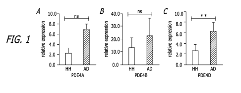
1. RT-PCR results show elevated expression of PDE4 gene
AD dogs compared to the healthy controls. Expression levels of PDE4A (A),
PDE4B (B) and PDE4D (C) were elevated in the PBMCs of canine AD dogs in
comparison with those of the healthy controls. Canine GAPDH gene was used as
an
internal control. Each bar is representative of a triplicate experiment for
each patient
(healthy: n = 8, Atopic: n = 9). T-test, ns ¨ Not Significant, * P < 0.05, **P
< 0.01.
RT-PCR results represent relative expression of AD dogs normalized to that of
the
health controls.
[0028] Figure
2. RT-PCR results show elevated expression of miR-203 and
miR-483 and decreased expression of the specific genes (PIAS1, RORA and
SH2B1) in canine AD dogs compared to the healthy controls. (A) Expression
levels of miR-203 and miR-483 were elevated in the plasma of canine AD dogs by
approximately 2.5-fold and 1.6-fold respectively in comparison with those of
the
healthy controls. The canine miR-39 was used as the internal control. (B)
Expression
levels of the three specific genes (PIAS1, RORA and SH2B1) were significantly
downregulated in PBMCs of the canine AD dogs compared to the healthy dogs.
Canine GAPDH gene was used as an internal control. Each bar is representative
of
a triplicate experiment for each patient (Healthy: n = 8, Atopic: n = 9). T-
test, ns ¨
Not Significant, * P < 0.05, **P < 0.01. RT-PCR results represent relative
expression
of AD dogs normalized to that of the health controls.
[0029] Figure
3. Analysis of CD4+ T Cell compared to CD8+ T Cells in
healthy vs Atopic Canines. (A) CD4 vs CD8 flow plot from PBMCs of 1 healthy
canine and 1 atopic canine. All plots were gated on lymphocytes and CD3+
Cells.
Dead cells were excluded by 7-AAD. (B) Comparison of CD4+/CD8+ T Cell ratios
between healthy canines (n = 8) and atopic canines (n = 9). The mean CD4+/CD8+
CA 03127868 2021-07-26
WO 2020/160457
PCT/US2020/016191
ratio of healthy canines was 2.031 0.3105 compared to 2.21 0.3626 for
atopic
canines (results expressed as mean ratio SEM). P=0.3585 and not significant.
[0030] Figure
4. Cytokine Profiles of Healthy vs Atopic Canines. Serum was
isolated from whole blood extracted from either healthy or AD dogs. Expression
of a
multitude of cytokines including A) IL-13, B) IL-31, C) TNF-a, D) FN-y, E) IL-
10, F)
IL-4, G) TGF- 131 were analyzed via ELISA. Each bar is representative of a
duplicate
experiment for each patient (Healthy: n = 8, Atopic: n = 9). T-test, NS ¨ Not
Significant, *P < 0.05, **P <o0, ***P < 0.001, ****P <0.001.
DETAILED DESCRIPTION
[0031] As used
herein, the term "about" will mean up to plus or minus 5% of the
particular term.
[0032] As used
herein, the phrase "consisting essentially of" refers to excluding
other active ingredients or any other ingredient that can materially affect
the basic
characteristic of a composition, formulation or structure, but generally
including
excipients.
[0033] As used
herein, an "effective amount" refers to that amount of stem cells,
cytokines, or a therapeutic compostion containing both, that is sufficient to
modulate,
attenuate, or induce an immune response (i.e., suppression of T cell responses
or
promotion of an immune response) in the subject thereby reducing at least one
sign
or symptom of the disease or disorder under treatment.
[0034] As used
herein, the terms "treat," "treating," or "treatment" and the like
refers to alleviating signs or symptoms of the disease accomplished by a
administering a composition to a patient in need of such treatment. Such
alleviation
can occur prior to signs or symptoms of the disease appearing, as well as
after their
appearance, therefore it encompasses prophylactic and active treatment. In
addition,
"treat," "treating" or "treatment" does not require complete alleviation of
signs or
symptoms, or a cure. At a cellular level it may include reduction of diseased
or target
cellular population by at least 10%, 25%, 50%, 75%, 80%, 85%, 90%, 95%, or 99%
as compared to untreated cells or cells treated with control or a comparative
agent.
[0035] As used
herein, the terms "administration" or "administering" or "treatment
regimen" within the scope of the present invention includes a single
therapeutic
6
CA 03127868 2021-07-26
WO 2020/160457
PCT/US2020/016191
delivery, or multiple or repeated deliveries, or a control delivery
therapeutic of any of
the individual components of the present invention or in combination. Such
terms are
further meant to include modes of deliveries such as locally, systemically,
intravascularly, intramuscularly, intra-peritoneally, inside the blood-brain
barrier,
organ-specific interventional injection or via other various routes.
[0036] The
articles "a" and "an" are used herein to refer to one or to more than
one (i.e., to at least one) of the grammatical object of the article. By way
of example,
"an element" means one element or more than one element.
[0037] The
terms "comprise," "comprising," "include," "including," "have," and
"having" are used in the inclusive, open sense, meaning that additional
elements
may be included. The terms "such as", "e.g.", as used herein are non-limiting
and are
for illustrative purposes only. "Including" and "including but not limited to"
are used
interchangeably.
[0038] The term
"or" as used herein should be understood to mean "and/or",
unless the context clearly indicates otherwise.
[0039] The term
"treatment" or "treating" refers to any therapeutic intervention in
a mammal, for example a companion animal, including: (i) prevention, that is,
causing the clinical symptoms not to develop, e.g., preventing infection or
inflammation from occurring and/or developing to a harmful state; (ii)
inhibition, that
is, arresting the development of clinical symptoms, e.g., stopping an ongoing
infection so that the infection is eliminated completely or to the degree that
it is no
longer harmful; and/or (iii) relief, that is, causing the regression of
clinical symptoms,
e.g., causing a relief of fever and/or inflammation caused by or associated
with a
microbial infection.
[0040] The
terms "reducing", "suppressing" and "inhibiting" have their commonly
understood meaning of lessening or decreasing.
[0041] The
terms "effective," "effective amount," and "therapeutically effective
amount" refer to that amount of MSC and/or a pharmaceutical composition
thereof
that produces a beneficial result.
[0042] The
phrases "parenteral administration" and "administered parenterally"
are art-recognized terms, and include modes of administration other than
enteral and
topical administration, such as injections, and include, without limitation,
retro-orbital,
7
CA 03127868 2021-07-26
WO 2020/160457
PCT/US2020/016191
intraocular, intravenous, intramuscular, intrapleural, intravascular,
intrapericardial,
intraarterial, intrathecal, intracapsular, intraorbital, intracardiac,
intradermal,
intraperitoneal, transtracheal, subcutaneous,
subcuticular, intra-articular,
subcapsular, subarachnoid, intraspinal and intrastemal injection and infusion.
[0043] The term
"pharmaceutical composition" refers to a formulation containing
the therapeutically active agents described herein in a form suitable for
administration to a subject. In a preferred embodiment, the pharmaceutical
composition is in bulk or in unit dosage form. The unit dosage form is any of
a variety
of forms, including, for example, a capsule, an IV bag, a tablet, a single
pump on an
aerosol inhaler, or a vial. The quantity of active ingredient (e.g., MSC) in a
unit dose
of composition is an effective amount and is varied according to the
particular
treatment involved. One skilled in the art will appreciate that it is
sometimes
necessary to make routine variations to the dosage depending on the age and
condition of the patient. The dosage will also depend on the route of
administration.
In a preferred embodiment, the active ingredients are mixed under sterile
conditions
with a pharmaceutically acceptable carrier, and with any preservatives,
buffers, or
propellants that are required.
[0044] The
terms "pharmaceutically acceptable" or "therapeutically acceptable"
refers to a substance which does not interfere with the effectiveness or the
biological
activity of the active ingredients and which is not toxic to the host.
[0045] The
phrase "pharmaceutically acceptable carrier" is art-recognized, and
includes, for example, pharmaceutically acceptable materials, compositions or
vehicles, such as a liquid or solid filler, diluent, excipient, solvent, or
encapsulating
material, involved in carrying or transporting any subject composition from
one
organ, or portion of the body, to another organ, or portion of the body. Each
carrier
must be "acceptable" in the sense of being compatible with the other
ingredients of a
subject composition and not injurious to the patient. In certain embodiments,
a
pharmaceutically acceptable carrier is non-pyrogenic. Some examples of
materials
which may serve as pharmaceutically acceptable carriers include: (1) sugars,
such
as lactose, glucose and sucrose; (2) starches, such as corn starch and potato
starch;
(3) cellulose, and its derivatives, such as sodium carboxymethyl cellulose,
ethyl
cellulose and cellulose acetate; (4) powdered tragacanth, (5) malt; (6)
gelatin; (7)
talc; (8) excipients, such as cocoa butter and suppository waxes; (9) oils,
such as
8
CA 03127868 2021-07-26
WO 2020/160457
PCT/US2020/016191
peanut oil, cottonseed oil, sunflower oil, sesame oil, olive oil, corn oil and
soybean
oil; (10) glycols, such as propylene glycol; (11) polyols, such as glycerin,
sorbitol,
mannitol and polyethylene glycol; (12) esters, such as ethyl oleate and ethyl
laurate,
(13) agar; (14) buffering agents, such as magnesium hydroxide and aluminum
hydroxide; (15) alginic acid; (16) pyrogen-free water; (17) isotonic saline;
(18)
Ringer's solution; (19) ethyl alcohol; (20) phosphate buffer solutions; and
(21) other
non-toxic compatible substances employed in pharmaceutical formulations.
[0046] A
"patient," "subject," or "host" to be treated by the subject method may
mean either a human or non-human animal, such as a mammal. Thus, the subject
of
the herein disclosed methods can be a human, non-human primate, horse, pig,
rabbit, dog, sheep, goat, cow, cat, guinea pig, or rodent. The term does not
denote a
particular age or sex. Thus, adult and newborn subjects, as well as fetuses,
whether
male or female, are intended to be covered. In one aspect, the subject is a
mammal.
A patient refers to a subject afflicted with a disease or disorder.
[0047] The term
"in vitro" refers to an artificial environment and to processes or
reactions that occur within an artificial environment. In vitro environments
include,
but are not limited to, test tubes and cell culture. The term "in vivo" refers
to the
natural environment (e.g., an animal or a cell) and to processes or reaction
that
occur within a natural environment.
DIAGNOSIS OF ATOPIC DERMATITIS
[0048]
Disclosed herein are methods for diagnosing AD. For example, in
embodiments, disclosed are methods for diagnosing atopic dermatitis (AD)
comprising determining the expression levels of at least one marker, for
example
miR-203 or miR-483, and comparing said expression levels with those in a
patient
without AD, wherein increased miR-203 and/or miR-483 expression levels
indicate a
patient suffering from AD.
[0049] Further
disclosed are methods for diagnosing AD comprising determining
the expression levels of, for example, PIAS1, RORA, SH2B1 and comparing said
expression levels with those in a patient without AD, wherein decreased PIAS1,
RORA, or SH2B1 expression levels indicate a patient suffering from AD.
[0050] Further
disclosed are methods for diagnosing AD comprising determining
the expression level of, for example, phosphodiesterase 4D (PDE4D) gene in
9
CA 03127868 2021-07-26
WO 2020/160457
PCT/US2020/016191
peripheral blood mononuclear cells (PBMCs), and comparing said expression
levels
with those in a patient without AD, wherein increased expression levels
indicate a
patient suffering from AD.
[0051] Further
disclosed are methods for pre-selecting AD patients to be
appropriate for adipose-derived mesenchymal stem cell (MSC) treatment, wherein
said pre-selecting comprises determining the expression levels of at least one
marker, for example miR-203 and miR-483, and comparing said expression levels
with those in a patient without AD, wherein increased miR-203 and miR-483
expression levels indicate a patient suffering from AD, or determining the
expression
levels of, for example, PIAS1, RORA, SH2B1 and comparing said expression
levels
with those in a patient without AD, wherein decreased PIAS1, RORA, SH2B1
expression levels indicate a patient suffering from AD, or determining the
expression
level of, for example, phosphodiesterase 4D (PDE4D) gene in peripheral blood
mononuclear cells (PBMCs) and comparing said expression levels with those in a
patient without AD, wherein increased expression levels indicate a patient
suffering
from AD.
[0052] Further
disclosed are methods to correlate MSC potency by testing
methods, for example methods for diagnosing atopic dermatitis (AD) comprising
determining the expression levels of, for example, miR-203 and miR-483, and
comparing said expression levels with those in a patient without AD, wherein
increased miR-203 and miR-483 expression levels indicate a patient suffering
from
AD, methods for diagnosing AD comprising determining the expression levels of
PIAS1, RORA, SH2B1 and comparing said expression levels with those in a
patient
without AD, wherein decreased PIAS1, RORA, SH2B1 expression levels indicate a
patient suffering from AD, and methods for diagnosing AD comprising
determining
the expression level of phosphodiesterase 4D (PDE4D) gene in peripheral blood
mononuclear cells (PBMCs) and comparing said expression levels with those in a
patient without AD, wherein increased expression levels indicate a patient
suffering
from AD, for AD patient screening and improvement post-treatment.
CA 03127868 2021-07-26
WO 2020/160457
PCT/US2020/016191
TREATMENTS FOR ATOPIC DERMATITIS
[0053]
Disclosed herein are treatments for AD, in particular canine treatments,
comprising administration of stem cells. In embodiments, the MSC are
stilumated or
modified to produce a cell signaling molecule, for example a cytokine.
[0054] Stem
cells are specialized cells, capable of renewing themselves through
cell division as well as differentiating into multi-lineage cells. These cells
are
categorized as embryonic stem cells (ESC), induced pluripotent stem cells
(iPSC),
and adult stem cells. Mesenchymal stem cells (MSC) are adult stem cells which
can
be isolated from human and animal sources. Human MSC (hMSC) are non-
haematopoietic, multipotent stem cells with the capacity to differentiate into
mesodermal lineage such as osteocytes, adipocytes and chondrocytes as well
ectodermal (neurocytes) and endodermal lineages (hepatocytes). MSC express
cell
surface markers including cluster of differentiation (CD)29, 0D44, 0D73, 0D90,
0D105, and lack the expression of 0D14, 0D34, 0D45, and HLA (human leucocyte
antigen)-DR. hMSC have been isolated from various tissues, including adipose
tissue, amniotic fluid, endometrium, dental tissues, umbilical cord, and
Wharton's
jelly. hMSC have been cultured long-term in specific media without any severe
abnormalities. Furthermore, MSC have immunomodulatory features, and can
secrete
cytokines and immune-receptors which regulate the microenvironment in the host
tissue. Multilineage potential, immunomodulation and secretion of anti-
inflammatory
molecules makes MSC an effective tool in the treatment of chronic diseases.
MSC
are not to be confused with haematopoietic (blood) stem cells that are also
found in
bone marrow. Morphologically, mesenchymal stem cells have long thin cell
bodies
with a large nucleus. As with other stem cell types, MSC have a high capacity
for self
renewal while maintaining multipotency.
[0055] MSC are
typically identified based upon the expression or lack of
expression of particular markers. For example, MSCs are 0D34-, CD1 1 b, CD11c-
,
0D45-, MHO class II, 0D44+, Sca-1+, and MHO class I low. In addition, MSCs can
be identified by their ability to differentiate into various mesenchymal cell
types. In
vitro experiments have demonstrated that culture conditions, additives, growth
factors and cytokines can precisely induce MSC to develop into a selected
mesenchymal cells. For example, dexamethasone in combination with
isobutilmethylxanthine or insulin or a mixture of isobutilmethylxanthine,
insulin and
11
CA 03127868 2021-07-26
WO 2020/160457
PCT/US2020/016191
indomethacin has been shown to push the MSCs toward differentiating into
adipocytes. Similarly, MSCs can differentiate into skeletal muscle cells when
stimulated with 5-azacytidine. 13-VGF has been shown to cause mesenchymal stem
cells to differentiate into cardiac muscle cells.
[0056]
Disclosed embodiments comprise compositions for treating a patient, for
example an animal such as a canine, suffering from an inflammatory disease
such
as atopic dermatitis, said composition comprising MSC derived from progenitor
cells
harvested from, for example, placental tissue, bone marrow, dental tissue,
testicle
tissue, uterine tissue, umbilical cord tissue, or skin tissue that are
allogeneic or
autologous to a target patient; and a saline solution, and wherein the
composition is
operable to reduce or eliminate the symptoms of the dermatitis. In
embodiments, the
MSC can be stimulated or modified, for example by introducing non-native DNA
or
applying a cell signaling molecule.
[0057]
Embodiments comprise combination treatments comprising administration
of stem cells with another active agent, for example a PDE4 (phosphodiesterase-
4)
inhibitor.
[0058] Further
disclosed are methods for modifying MSC to produce a cytokine,
comprising altering the genetic makeup of the MSC, wherein altering the
genetic
makeup of the MSC can comprise introduction of non-native DNA or stimulation
of
expression of native DNA, or both.
[0059] Further
disclosed are methods for stimulating MSC to produce a cytokine,
for comprising applying a signaling molecule the MSC, wherein the signaling
molecule can comprise, for example, a cytokine, mRNA, miRNA, or the like.
[0060] In
embodiments, isolated MSC can be formulated into a pharmaceutically-
acceptable composition, for example by using at least one pharmaceutically-
acceptable carrier. In embodiments, a pharmaceutically-acceptable carrier
means a
carrier that is useful in preparing a pharmaceutical composition or
formulation that is
generally safe, non-toxic, and neither biologically nor otherwise undesirable,
and
includes a carrier that is acceptable for veterinary use as well as human
pharmaceutical use. The pharmaceutically acceptable carrier can comprise, for
example, saline solution, phosphate buffered saline (PBS), Ringer's serum,
Ringer's
lactate serum, lactose, dextrose, sucrose, sorbitol, mannitol, starch, rubber
arable,
12
CA 03127868 2021-07-26
WO 2020/160457
PCT/US2020/016191
potassium phosphate, arginate, gelatin, potassium silicate, microcrystalline
cellulose,
polyvinylpyrrolidone, cellulose, water, syrups, methylcellulose, methylhydroxy
benzoate, propylhydroxy benzoate, talc, magnesium stearate, and mineral oils.
[0061]
Disclosed embodiments can comprise administration of MSC to treat
atopic dermatitis. For example, adipose-derived MSC can be used. In
embodiments, the stem cells may be autologous to the subject. If available,
autologous stem cells can be beneficial to the subject because they will
reduce or
eliminate the potential for adverse immune responses, e.g., rejection of the
stem
cells or graft-versus-host disease. Autologous stem cells may be, e.g., stem
cells
isolated directly from the subject (e.g., MSC), or iPS cells produced from non-
stem
cells from the subject.
[0062] In some
embodiments, in cases where autologous stem cells are not
available or not indicated for a particular subject, allogeneic stem cells may
be used.
Allogeneic stem cells should be matched as closely as possible to the subject
(e.g.,
via HLA genotype) in order to reduce the likelihood of rejection or graft-
versus-host
disease. In other embodiments, the stem cell donor is a first-degree-relative
(e.g.,
parent, sibling, or child) of the subject, which increases the likelihood of
finding a
closely-matched donor. In yet other embodiments, the stem cell donor can be an
extended relative of the subject. In some embodiments, the stem cell donor can
be
from the same race or ethnic group as the subject. However, certain stem cells
can
be immune-privileged and can be used allogeneically without matching between
the
donor and subject.
[0063] In yet
another embodiment, methods for stimulating an immune response
in a patient in need is described. In such embodiment, patients are
administered
effective amounts of a composition comprising, for example in the case of
cancer, an
inhibitor to inducible nitric oxide synthase, an inhibitor to indoleamine 2, 3-
dioxygenase, a population of inducible nitric oxide synthase (iNOS)-deficient
mesenchymal stem cells, a population of indoleamine 2,3-dioxygenase (ID0)-
deficient mesenchymal stem cells or any combinations thereof. In a preferred
embodiment, the method cause inhibition of the production of one or more of
nitrogen oxide (NO), indoleamine 2, 3 dioxygenase (ID0), or prostaglandin E 2
(PGE2), 1-MT, 1400W, L-NMMA or other suitable agents. In this embodiment, the
above mentioned inhibitors of iNOS or IDO are administered individually or as
a
13
CA 03127868 2021-07-26
WO 2020/160457
PCT/US2020/016191
mixture. In this aspect of the invention, the patient's status is post
receiving a
regimen of immune therapy including a regimen including the stimulated or
modified
MSCs described herein, or another immune therapy regimen which can include
treatment with indicated interferons, antibody, cell therapy or other
therapies that
modulate immune response.
[0064] In embodiments, adipose-derived MSC are used for treatment of
patients.
[0065] Appropriate MSC dosage can be, for example, 1x10³ cells, 2.5x10³
cells,
5x10³ cells,1x104 cells, 2.5x10⁴ cells, 5x10⁴ cells, 1x10⁵ cells, 2.5x10⁵
cells, 5x10⁵
cells, 1x10⁸ cells, 2.5x10⁸ cells, 5x10⁸ cells, 1x10⁷ cells, 2.5x10⁷ cells,
5x10⁷ cells,
1x10⁸ cells, 2.5x10⁸ cells, 5x10⁸ cells, 1x10⁹ cells, 2.5x10⁹ cells, 5x10⁹
cells, 1x101
cells, 2.5x101 cells, 5x101 cells, 1x10¹¹ cells, 2.5x10¹¹ cells, 5x10¹¹
cells, 1x10¹²
cells, 2.5x10¹² cells, 5x10¹² cells, 1x10¹³ cells, 2.5x10¹³ cells, 5x10¹³
cells, 1x10¹⁴
cells, 2.5x10¹⁴ cells, 5x10¹⁴ cells, 1x10¹⁵ cells, 2.5x10¹⁵ cells, 5x10¹⁵
cells, or more,
or the like.
[0066] In embodiments, appropriate MSC dosage can be, for example, between
1x10³ cells and 2.5x10³ cells, between 5x10³ cells and 1x10⁴ cells, between
2.5x10⁴
cells and 5x10⁴ cells, between 1x10⁵ cells and 2.5x10⁵ cells, between 5x10⁵
cells
and 1x10⁸ cells, between 2.5x10⁸ cells, between 5x10⁸ cells and 1x10⁷ cells,
between 2.5x10⁷ cells and 5x10⁷ cells, between 1x10⁸ cells and 2.5x10⁸ cells,
between 5x10⁸ cells and 1x10⁹ cells, between 2.5x10⁹ cells and 5x10⁹ cells,
between 1x101 cells and 2.5x101 cells, between 5x101 cells and 1x10¹¹
cells,
between 2.5x10¹¹ cells and 5x10¹¹ cells, between 1x10¹² cells and 2.5x10¹²
cells,
between 5x10¹² cells and 1x10¹³ cells, between 2.5x10¹³ cells and 5x10¹³
cells,
between 1x10¹⁴ cells and 2.5x10¹⁴ cells, between 5x10¹⁴ cells and 1x10¹⁵
cells,
between 2.5x10¹⁵ cells and 5x10¹⁵ cells, or more, or the like.
[0067] In embodiments, appropriate MSC dosage can be, for example, not less
than 1x10³ cells, not less than 2.5x10³ cells, not less than 5x10³ cells, not
less than
1x10⁴ cells, not less than 2.5x10⁴ cells, not less than 5x10⁴ cells, not less
than 1x10⁵
cells, not less than 2.5x10⁵ cells, not less than 5x10⁵ cells, not less than
1x10⁸ cells,
not less than 2.5x10⁸ cells, not less than 5x10⁸ cells, not less than 1x10⁷
cells, not
less than 2.5x10⁷ cells, not less than 5x10⁷ cells, not less than 1x10⁸ cells,
not less
than 2.5x10⁸ cells, not less than 5x10⁸ cells, not less than 1x10⁹ cells, not
less than
14
CA 03127868 2021-07-26
WO 2020/160457
PCT/US2020/016191
2.5x10⁹ cells, not less than 5x10⁹ cells, not less than 1x101 cells, not less
than
2.5x10¹⁹ cells, not less than 5x10¹⁹ cells, not less than 1x101' cells, not
less than
2.5x10¹¹ cells, not less than 5x10¹¹ cells, not less than 1x10¹² cells, not
less than
2.5x10¹² cells, not less than 5x10¹² cells, not less than 1x10¹³ cells, not
less than
2.5x10¹³ cells, not less than 5x10¹³ cells, not less than 1x10¹⁴ cells, not
less than
2.5x10¹⁴ cells, not less than 5x10¹⁴ cells, not less than 1x10¹⁵ cells, not
less than
2.5x10¹⁵ cells, not less than 5x10¹⁵ cells, or more, or the like.
[0068] In
embodiments, appropriate MSC dosage can be, for example, not more
than 1x10³ cells, not more than 2.5x10³ cells, not more than 5x10³ cells, not
more
than 1x10⁴ cells, not more than 2.5x10⁴ cells, not more than 5x10⁴ cells, not
more
than 1x10⁵ cells, not more than 2.5x10⁵ cells, not more than 5x10⁵ cells, not
more
than 1x10⁶ cells, not more than 2.5x10⁶ cells, not more than 5x10⁶ cells, not
more
than 1x10⁷ cells, not more than 2.5x10⁷ cells, not more than 5x10⁷ cells, not
more
than 1x10⁸ cells, not more than 2.5x10⁸ cells, not more than 5x10⁸ cells, not
more
than 1x10⁹ cells, not more than 2.5x10⁹ cells, not more than 5x10⁹ cells, not
more
than 1x10¹⁹ cells, not more than 2.5x10¹⁹ cells, not more than 5x10¹⁹ cells,
not more
than 1x101' cells, not more than 2.5x10¹¹ cells, not more than 5x10¹¹ cells,
not more
than 1x10¹² cells, not more than 2.5x10¹² cells, not more than 5x10¹² cells,
not more
than 1x10¹³ cells, not more than 2.5x10¹³ cells, not more than 5x10¹³ cells,
not more
than 1x10¹⁴ cells, not more than 2.5x10¹⁴ cells, not more than 5x10¹⁴ cells,
not more
than 1x10¹⁵ cells, not more than 2.5x10¹⁵ cells, not more than 5x10¹⁵ cells,
or more,
or the like.
[0069] The
disclosed methods can also involve the co-administration of bioactive
agents with the stem cells. By "co-administration" is meant administration
before,
concurrently with, e.g., in combination with bioactive agents in the same
formulation
or in separate formulations, or after administration of a therapeutic
composition as
described above.
[0070]
Disclosed pharmaceutical compositions can also be provided as a kit. A
kit of the invention can contain a pharmaceutically acceptable carrier; an
isolated
population of stimulated or modified MSC, and further instructions for using
the kit in
a method for attenuating an immune response. In this aspect of the invention,
the
cells stimulated with, for example, cytokine components of the kit can be
administered. The kit also optionally may include a means of administering the
cells,
CA 03127868 2021-07-26
WO 2020/160457
PCT/US2020/016191
for example by injection. In an optional embodiment, the compositions of this
invention suitable for parenteral administration can further contain
antioxidant(s) in
combination with one or more pharmaceutically-acceptable sterile isotonic
aqueous
or nonaqueous solutions, suspensions or in the form of sterile lyophilized
powders
which may be reconstituted into sterile injectable solutions or dispersions
just prior to
use, which may contain the combination of the antioxidants, minerals and
vitamins,
buffers, solutes which render the final formulation isotonic.
[0071] As used
herein, the phrase, "bioactive agents" refers to any organic,
inorganic, or living agent that is biologically active or relevant. For
example, a
bioactive agent can be a protein, a polypeptide, a nucleic acid, a
polysaccharide
(e.g., heparin), an oligosaccharide, a mono- or disaccharide, an organic
compound,
an organometallic compound, or an inorganic compound. It can include a living
or
senescent cell, bacterium, virus, or part thereof. It may include a
biologically active
molecule such as a hormone, a growth factor, a growth factor-producing virus,
a
growth factor inhibitor, a growth factor receptor, an anti-inflammatory agent,
an
antimetabolite, an integrin blocker, or a complete or partial functional sense
or
antisense gene, including siRNA. It can also include a man-made particle or
material, which carries a biologically relevant or active material. An example
is a
nanoparticle comprising a core with a drug and a coating on the core.
[0072]
Bioactive agents may also include drugs such as chemical or biological
compounds that can have a therapeutic effect on a biological organism. Non-
limiting
examples include, but are not limited to, growth factors, anti-rejection
agents, anti-
inflammatory agents, anti-infective agents (e.g., antibiotics and antiviral
agents), and
analgesics and analgesic combinations. Anti-inflammatory agents may be useful
as
additional agents to counteract the inflammatory aspects of the fibrotic
process.
[0073]
Combinations, blends, or other preparations of any of the foregoing
examples can be made and still be considered bioactive agents within the
intended
meaning herein. Aspects of the present disclosure directed toward bioactive
agents
may include any or all of the foregoing examples. In other embodiments, the
bioactive agent may be a growth factor. A growth factor is any agent which
promotes the proliferation, differentiation, and functionality of the
implanted stem cell.
Non-limiting examples of suitable growth factors may include, but are not
limited to,
leukemia inhibitory factor (LIF), epidermal growth factor (EGF), fibroblast
growth
16
CA 03127868 2021-07-26
WO 2020/160457
PCT/US2020/016191
factor (FGF), insulin-like growth factor (IGF), vascular endothelial growth
factor
(VEGF), human growth hormone (hGH), platelet-derived growth factor (PDGF),
interleukins, cytokines, and/or combinations thereof.
[0074] In some
embodiments, the bioactive agent can be an immunosuppressive
agent. An immunosuppressive agent is any agent which prevents, delays the
occurrence of, or decreases the intensity of the undesired immune response,
e.g.,
rejection of a transplanted cell, tissue, or organ, or graft-versus-host
disease.
Preferred are immunosuppressive agents which suppress cell-mediated immune
responses against cells identified by the immune system as non-self. Examples
of
immunosuppressive agents may include, but are not limited to, cyclosporin,
cyclophosphamide, prednisone, dexamethasone, methotrexate, azathioprine,
mycophenolate, thalidomide, FK-506, systemic steroids, as well as a broad
range of
antibodies, receptor agonists, receptor antagonists, and other such agents as
known
to one skilled in the art. In other embodiments, bioactive agents that may be
administered include anti-fibrotic agents including, but not limited to,
nintedanib, INT-
767, emricasan, VBY-376, PF-04634817, EXC 001, GM-CT-01, GCS-100,
Refanalin, SAR156597, tralokinumab, pomalidomide, STX-100, 00-930,
simtuzumab, anti-miR-21, PRM-151, BOT191, palomid 529, IMD1041, serelaxin,
PEG-relaxin, ANG-4011, FT011, pirfenidone, F351 (perfenidone derivative), THR-
184, CCX-140, FG-3019, avosentan, GKT137831, PF-00489791, pentoxifylline,
fresolimumab, and LY2382770.
[0075] Further
disclosure related to isolation, stimulation, modification of stem
cells, and their therapeutic uses, can be found, for example, in US 8,685,728,
US
9,301,979, US20190046577A1, Genetic Engineering of Mesenchymal Stem Cells
and Its Application in Human Disease Therapy. Hum Gene Therapy; 2010 Nov;
21(11): 1513-1526, and Therapeutic Potential of Genetically Modified
Mesenchymal
Stem Cells; Gene Therapy volume 15, pages 711-715 (2008), each of which is
incorporated by reference in its entirety.
Example 1 - Treatment of Atopic Dermatitis
[0076]
MicroRNAs (miRNAs), which interfere with mRNA translation, are
becoming recognized as powerful biomarkers for various diseases. Here, we
examine miR-203 and miR-483 expression, along with the 0D4+/0D8+ cell ratio,
total
17
CA 03127868 2021-07-26
WO 2020/160457
PCT/US2020/016191
IgE, expression levels of the three AD associated genes (PIAS1, RORA, SH2B1)
and a panel of cytokines (IL-4, IL-10, IL-13, IL-31, IFN- y, TGF- 81, TNF-a)
in AD
dogs compared to their healthy controls.
Animals
[0077] A total
of nine client-owned AD dogs (six males and three females) with
naturally occurring non-seasonal AD were enrolled in this study from August
2017 to
March 2018. The AD dog breeds reported by owners include Miniature Pinscher
mix,
Golden Retriever, Brittney Spaniel, German Shepard, Shih Tzu, Papillon, Great
Dane, Cocker Spaniel, Boxer, Poodle and Terrier mix. In addition, another
eight
client-owned healthy dogs without AD (5 males and 4 females) were enrolled in
this
study as controls. The healthy dog breeds reported by owners include Rat
Terrier,
Chihuahua Mix, Chihuahua, Terrier Mix, Pitbull Mix, Plot Hound, and Cattle Dog
Cross.
Inclusion Criteria for AD Dogs
[0078] Clinical
diagnosis of AD was based on detailed interpretation of patient
history and clinical signs and exclusion of other possible skin dermatosis
that can
present as AD. Flea combing, skin scrapings, skin cytologies and elimination
diet
trials were performed. These are in accordance with the guidelines developed
by the
International Committee for Allergic Diseases in Animals (ICADA) diagnosis of
canine AD. Patients in AD group were over one year of age, with a body
condition
score of at least 4 on a 9-point scale. Underlying systemic diseases were
ruled out
through thorough physical examination and serum chemistry and hematology
analysis. Participants should be on effective flea control.
Exclusion Criteria for AD Dogs
[0079] Clinical
evidence of ectoparasite infestations (flea allergy dermatitis,
scabies etc.), bacterial or fungal cutaneous infections, food allergies, and
seasonality
of the cutaneous condition resulted in exclusion from the study. Ongoing
treatment
with anti-inflammatory or immunosuppressive medications (antihistamines,
glucocorticoids, and NSAIDS), also resulted in exclusion, unless appropriate
weaning times were followed.
18
CA 03127868 2021-07-26
WO 2020/160457
PCT/US2020/016191
Inclusion Criteria for Healthy Dogs
[0080]
Participants that were more than 1 year of age, had a body condition
score of at least 4 on a 9-point scale, with no history or clinical signs of
pruritus or
immune modulating disease conditions were enrolled in the study.
Flow Cytometry
[0081]
Peripheral Blood Mononuclear Cells (PBMCs) were isolated from whole
blood collected in EDTA vacutainers. 2mL of whole blood was then diluted with
6 mL
of PBS. Diluted whole blood was layered on top of 2mL of Ficoll-Paque PLUS (GE
Healthcare Catalog #17-1440-02) and centrifuged at 2500 rpm for 25 minutes (no
brake). The PBMC interphase was collected. Next, red blood cells (RBCs) were
lysed with 1X RBC Lysis Buffer (BioLegend Catalog #420301) followed by
spinning
and resuspension in cell staining buffer (BioLegend Catalog #420201). Antibody
staining was conducted using Bio-Rad Anti-dog CD3 Clone CA17.2Al2:FITC, CD4
Clone YKIX302.9:RPE, CD8 YCATE55.9:Alexa Fluor647 (Bio-Rad) and staining with
10uL of isotype control Bio-Rad MSE IgG1:FITC/RAT IgG2a:RPE/RAT IgG1:Alexa
Fluor647 (Bio-Rad Catalog #TCO23). Cells were resuspended in 400u1 of cell
staining buffer, stained with 5uL of 7-AAD Viability Dye (BioLegend Catalog
#420404) and analyzed on the BD Accuri C6 Flow Cytometer.
Serum ELISA Analysis
[0082] Serum
ELISA analysis was carried out according to the manufacturer's
protocols, and the following ELISA kits were used for this study.
Canine Cytokine Company ELISA Kit Catalog
Number
IL-4 NeoBioLab Canine 1L4 ELISA Kit CI0014
IL-10 R&D Systems Canine 11_10 Quantikine CA1000
ELISA Kit
IL-13 NeoBioLab Canine 1L13 ELISA Kit CI0043
IL-31 NeoBioLab Canine 1L31 ELISA Kit CI0041
I FN- y R&D Systems Canine IFN- y Quantikine CAIFOO
ELISA Kit
TGF- 31 R&D Systems Mouse/Rat/Porcine/Canine MB100B
TGF- [31 Quantikine ELISA
Kit
IgE Abcam Canine IgE ELISA Kit Ab157700
TNF-a R&D Systems Canine TNF-a Quantikine CATA00
19
CA 03127868 2021-07-26
WO 2020/160457
PCT/US2020/016191
ELISA Kit
RNA Extraction and Real-Time FOR
[0083] RNA was
extracted from PBMCs by using the RNeasy Mini Kit (Qiagen,
Catalog #74104) according to manufacturer's protocol with an additional DNAse
I
digestion step. Extracted RNA was then converted into cDNA using the High
Capacity cDNA Reverse Transcription Kit following the manufacturer's
instructions
(Applied Biosystems, Catalog #4368814). The reactions were performed in
triplicate
on Bio-Rad CFX connected Real-Time FOR system. Canine glyceraldehyde-3-
phosphate dehydrogenase (GAPDH) gene was used as an internal control. The
following primer sets were used:
Gene Forward Reverse
Canine PIAS1 TGGAGTTGATGGATGCTTGAG GGACACTGGAGATGCTTGAT
Canine RORA AAGGCTGCAAGGGCTTTTTC CTGCGTACAAGCTGTCTCTT
Canine SH2B1 CGTCCTCACTTTCAACTTCCA GACACGACATAGCTGACAAGA
Canine GAPDH GGAGAAAGCTGCCAAATATG ACCAGGAAATGAGCTTGACA
MicroRNA Expression by real-time FOR
[0084] Whole
blood collected with EDTA-coated tubes was spun down and the
plasma supernatant containing miRNA was collected. Extraction of miRNA was
performed by following the protocol outlined in miRNeasy Serum/Plasma Kit
(Qiagen, Catalog #217184) and the miRNeasy Serum/Plasma Spike-In Control was
used (Qiagen, Catalog #219610). The reverse transcription was conducted by
following the protocol of "Taqman Small RNA Assays" (Applied Biosystems,
Catalog
# 4366596). TaqMan real-time FOR assays was performed Bio-Rad CFX connected
Real-Time FOR system according to the manufacturers instructions. Data were
normalized to the internal control miR-39. The following TaqMan probe and
primer
sets (ThermoFisher) were used: miR-39 (RT 000200), miR-203 (RT 000507) and
miR-483 (RT 002560).
RESULTS
PDE4D gene expression is significantly upregulated in AD dog PBMCs
CA 03127868 2021-07-26
WO 2020/160457
PCT/US2020/016191
[0085]
Phosphodiesterase 4 (PDE4) is a cyclic AMP-degrading enzyme in
leukocytes. Several decades ago, increased PDE activity was demonstrated in
patients with atopic dermatitis (AD). Currently, several PDE4 inhibitors in
both
topical and oral formulation have been developed to target the inflammatory
cascade of AD. This review shows the pathogenic rationale behind these
inhibitors,
and discusses multiple PDE4 inhibitors that are under evaluation or in the
market.
PDE4 inhibitors may be considered as favorable agents in the repertoire of
current
interventions for AD. Multiple studies have shown that inhibition of PDE4 is
beneficial to canine AD. However gene expression of PDE4s in PBMCs of canine
AD
has not been reported. We therefore examined all four PDE4 isoforms to verify
whether any of these isoforms may be a potential marker(s) for canine AD.
Blood
samples were collected from eight healthy dogs and nine AD dogs, and RNA was
then extracted from PBMCs of these blood samples. The RT-PCR results indicated
that three out of four PED4A isoforms (PDE4A, PDE4B and PDE4D) are upregulated
in the AD samples whereas PDE4C gene is not detectable in both AD and heathy
dog samples (Figure 1). Particularly, PDE4D gene expression in AD samples
shows
statistically significant upregulation by approximately 2.4-fold in comparison
to that of
the health control samples (p<0.01) (Figure 1C). Though the gene expression
levels
of PDE4A and PDE4B were also elevated in AD samples verse the health control
samples, both of the increases are not statistically significant (P=0.053 for
PDE4A
and P=0.097 for PDE4B). In summary, PDE4D may be a potential marker for AD
dogs.
MiR-203 and miR-483 are upregulated in AD dog plasma.
[0086]
Circulating MiR-203 and miR-483 were previously shown to be
upregulated in children AD sera. However, to date, no study of miRNAs in AD
dogs
has been reported. As dog AD has many similar characteristics with its human
counterpart, we examined miR-203 and miR-483 expression changes in the plasma
of both AD dogs and healthy controls. Blood samples were collected from eight
healthy dogs and nine AD dogs, and their plasma was prepared, followed by
miRNA
extraction. RT-PCR reactions were conducted to quantify expression of miR-203
and
miR-483, and miR-39 was used as the internal control. Our results showed
elevated
expression levels of miR-203 and miR-483 by approximately 2.5-fold (P=0.047)
and
1.6-fold (statistically not significant P=0.074) respectively in the plasma of
AD dogs in
21
CA 03127868 2021-07-26
WO 2020/160457
PCT/US2020/016191
comparison with those of the healthy controls (Figure 2A). This result
suggests that
miR-203 may be a possible biomarker for AD of both humans and dogs.
Verification of PIAS1, RORA and SH2B1 gene downregulation in the PBMCs of AD
dogs
[0087] We
performed RT-PCR reactions to examine these gene expressions
(PIAS1, RORA and SH2B1) in PBMCs of both the AD dogs and healthy controls. In
agreement with the previous report, our results confirmed that expression
levels of
all three gene (PIAS1, RORA and SH2B1) were significantly downregulated in the
PBMCs of the AD dogs by approximately 1.5-fold for RORA gene (P=0.007) and 2-
fold for both genes of PIAS1 (P=0.027) and SH2B1 (P=0.004) in comparison with
the
healthy dogs (Figure 2B).
The CD4+/CD8+ ratio of T lymphocytes, cytokines and total IgE in AD dogs
[0088] T
lymphocytes are critical for the development and regulation of cell-
mediated immune responses. We compared the CD4+/CD8+ ratio of T lymphocytes
in the PBMCs from the AD dogs and healthy controls by flow cytometry analysis.
Our
data indicates a modest increase in CD4+ T Cells in the AD dogs' samples in
comparison with the healthy controls (Figure 3A), resulting in a slight
elevation of
the CD4+/CD8+ ratio in AD dogs (2.388 0.3747) vs. healthy controls (2.101
0.2826) (Figure
3B). Nevertheless, this increase of CD4+/CD8+ ratio of T
lymphocytes associated with canine AD was not statistically significant
(P=0.3585).
[0089] As
canine AD is an inflammatory related-skin disease, we next examined
a panel of cytokines of including TH2 cytokines (IL-4, IL-13 and IL-31), TH1
cytokine
IFN-y, anti-inflammatory cytokines (IL-10 and TGF- 31), and pro-inflammatory
cytokine TNF-a by ELISA. Consistent with the previous reports, inflammatory
cytokines IL-13, IL-31 and TNF-a were significantly elevated (Figure 4A, 4B
and 4C
statistically non-significant for IL-13) whereas the pro-inflammatory cytokine
IFN-y
and anti-inflammatory cytokine IL-10 were dramatically decreased in AD patient
sera
(Figure 4D and 4E). In addition, expression of IL-4, which induces Th2 cell
differentiation and B-cell class switching to IgE, was slightly increased in
AD patient
sera as compared to the healthy controls (Figure 4F). This result is similar
with the
former report of IL-4 expression in canine AD patient plasma. In addition,
previous
reports about TGF-81 expression in canine AD are controversial. For instance,
22
CA 03127868 2021-07-26
WO 2020/160457
PCT/US2020/016191
Fedenko et al. showed a significant elevation of TGF-81 in AD patient blood
samples
vs their healthy controls, whereas others reported opposite results. Our study
indicated that TGF-81 expression in the canine AD sera is elevated by
approximately
2.8-fold in comparison with the healthy controls (Figure 4G). Finally, the
total serum
IgE level displayed no significant difference in the sera of AD dogs and the
healthy
controls which is in agreement with previous reports (Figure 4H).
DISCUSSION
[0090]
Approximately 10% of dogs suffer from AD and the pathogenesis of
canine AD has not been fully understood. To date, diagnosis of canine AD
relies on
a combination of patient history, clinical examination, allergy testing and
response to
diet trials/therapies, and reliable AD specific biomarkers are lacking. Here,
we
assessed all four PDE4 isoforms, specific miRNA expression, expression levels
of
genes associated with canine AD, the 0D4+/0D8+ ratio of T lymphocytes, and a
panel of cytokines in peripheral blood of both canine AD dogs and healthy
controls.
We, for the first time, report statistically significant expression increase
of PDE4D
gene in PBMCs and miR-203 in sera from AD dogs. Particularly, the increase of
PDE4D gene expression is well aligned with previous studies of that inhibition
of
PDE4 is beneficial to both humans and dogs with AD. Moreover, the increase of
miR-203 in plasma of dogs is consistent with the previous study in serum of
children
with AD, highlighting similarities of AD in both dogs and humans. In addition,
controversial results of the CD4+/CD8+ T cell ratio were reported in
association with
AD dogs before, and our result suggests a slight but not statistically
significant
increase of the CD4+/CD8+ ratio in AD dogs, which is in line with Beccati et
al who
showed no significant differences in the ratio of healthy dogs, atopic dogs
and atopic
dogs treated with cyclosporin.
[0091]
Furthermore, our study demonstrates the down-regulation of three genes
(PIAS1, RORA, SH2B1) that are associated with canine AD in comparison to
healthy
dogs. Interestingly, RORA was recently reported to involve in transactivation
of IL-10
promoter, and downregulation of RORA may contribute to the decrease of serum
IL-
in our study.
[0092] Finally,
our cytokine profiling showed significantly elevated expression
levels of Th2 inflammatory cytokines IL-13 and IL-31 and pro-inflammatory
cytokine
23
CA 03127868 2021-07-26
WO 2020/160457
PCT/US2020/016191
TNF-a, and dramatically decreased expression levels of TH1 cytokine IFN-y and
anti-inflammatory cytokine IL-10 in AD dogs. These results suggest that the
Th2
response is increased and the Th1 response is decreased in the isolated PBMCs
from AD dogs. Particularly, IL-31 (a TH2 cytokine) has attracted a lot of
attention in
recent years for its role in pruritus and atopic inflammation, and recent
studies
demonstrated serum IL-31 levels positively correlate with disease severity in
both
children and dogs with AD. Although total serum IgE levels are elevated in the
humans with AD, they show no clinical relevance in the AD dogs in this study,
which
is in agreement with previous reports. In addition, controversial results
regarding
immunosuppressive cytokine TGF-61 expression have been shown in canine AD.
Here, our study indicated that TGF-61 expression was dramatically elevated in
AD
dog sera in comparison with healthy controls, and the reported variations of
TGF-61
expression could be attributed to the varying degree of severity of
inflammation and
pruritus in AD patients. Moreover, TGF-61 is usually immunosuppressive and can
block inflammatory reactions, and elevated levels might indicate a feedback
loop in
reaction to the inflammatory response. In summary, our study of biomarkers in
peripheral blood may provide important insight and groundwork for developing a
less-invasive method for rapid diagnosis of AD disease and assessment of
treatment
efficacy.
SUPPLEMENTAL DATA
Table 1. Healthy Age, Sex, Breed, Spay/Neuter
Patient Age (Year & Sex Breed Spay/Neuter
Month)
H1 9 Years Female Rat Terrier Spay
H3 9 Years Male Chihuahua Neuter
H4 2 Years & 5 Female Mixed Spay
Months
H6 1 Year & 2 Months Male Pitbull Mix Neuter
H7 1 Year & 8 Months Female Plott Hound N/A
24
CA 03127868 2021-07-26
WO 2020/160457
PCT/US2020/016191
H8 1 Year & 8 Months Male Plott Hound Neuter
H9 2 Years Female Cattle Dog Spay
Cross
H10 2 Years Male Cattle Dog Neuter
Cross
Table 2. Atopic Age, Sex, Breed, Spay/Neuter
Patient Age (Year & Sex Breed
Spay/Neuter
Month)
AD400 10 Years & 7 Female Golden Retriever Spay
months
AD500 5 Years & 2 Male Miniature Pinscher Neuter
Months Mix
AD900 3 Years & 7 Female German Shepard Spay
Months
AD1000 6 Years & 8 Male Shih Tzu Neuter
Months
AD1200 5 Years and 10 Female Great Dane Spay
Months
AD1300 10 Years & 1 Male Cocker Spaniel Neuter
Month
AD1400 7 Years & 1 Month Male Boxer Neuter
AD1500 8 Years & 1 Month Male Poodle Neuter
AD1600 5 Years & 7 Male Terrier Mix Neuter
Months
CA 03127868 2021-07-26
WO 2020/160457
PCT/US2020/016191
Example 2 - Treatment of Atopic Dermatitis
[0093] An 8 year old canine suffers from atopic dermatitis. To treat the
animal,
autologous MSC at a dose of 1x10⁷cells are administered subcutaneously via
injection. Within a week, the patient's symptoms decrease.
Example 3 - Treatment of Atopic Dermatitis
[0094] A 4 year old dog suffers from atopic dermatitis. Autologous MSCs are
administered at a dose of 2.5x10⁵ cells via injection. Within a week, the
patient's
symptoms decrease.
Example 4 - Treatment of Atopic Dermatitis
[0095] A 13 year old dog suffers from atopic dermatitis. Allogeneic Adipose-
derived MSCs are administered at a dose of 1x10⁷ cells via injection. Within
two
weeks, the patient's symptoms decrease.
Example 5 - Stimulation of MSC
[0096] MSC are treated with a cell signaling molecule to stimulate anti-
inflammatory and immune modulatory cytokine production.
Example 6 - Modification of MSC
[0097] MSC are transformed with non-native DNA to produce a specific
cytokine.
Example 7 - Chemoattractive Property of MSCs is Induced by Proinflammatory
Cytokines
[0098] In several studies, effective immunosuppression by MSCs in vivo has
been achieved with as few as one to five MSCs per million somatic cells and
often
endures for months, with complete cure of immune disorders in some instances.
Considering that MSCs are immobile after settling in tissues, and that
immunosuppression is mediated by NO, which acts only very locally near its
source,
this immunosuppressive effect is astonishing. It was contemplated that
cytokine-
induced MSCs might have a mechanism to attract immune cells to their vicinity,
where the locally high concentrations of NO could act effectively on the
target T
cells. To explore this, co-cultures of MSCs and splenocytes were monitored
over
time under the microscope. Upon anti-CD3-stimulation, the splenocytes were
observed to actively migrate toward the spindle-shaped MSCs. In contrast, no
26
CA 03127868 2021-07-26
WO 2020/160457
PCT/US2020/016191
migration occurred in the absence of anti-0D3 stimulation. Since splenocytes
have
limited viability, the lack of locomotion toward MSCs in the absence of
stimulation
might be due to the poor health of these cells in vitro. To exclude this,
activated-
splenocyte-supernatant-primed MSCs were examined for their ability to attract
A1.1
T hybridoma cells, which survive well even in the absence of IL-2. Under these
conditions, time-lapse microvideography revealed brisk migration of T cells
toward
MSCs within 1.5 hours of co-culture initiation. Without priming of MSCs,
however,
there was no net movement of T cells toward the MSCs. Therefore, MSCs promote
the migration of T cells only after MSCs having been exposed to
proinflammatory
cytokines.
[0099] To
examine the role of various cytokines in enabling MSCs to attract T
cells, MSCs were pretreated with various combinations of recombinant cytokines
and
the resultant migration of pre-activated T cells in co-cultures was observed.
This
analysis indicated that the same T cell cytokine pairs (i.e., IFN gamma and
TNF
alpha, IFN gamma and IL-1 alpha, or IFN gamma and IL-1 beta) that had induced
the immunosuppressive function of MSCs also caused them to attract T cells.
Likewise, using antibody neutralization of specific cytokines, it was found
that
migration toward MSCs was prevented by anti-IFN.gamma. alone, or by blocking
TNF alpha, IL-1 alpha and IL-1 beta as a threesome, identical to their effects
on
activated-splenocyte-supernatant-induced MSC suppression of T cell
proliferation.
Therefore, the cytokine-induced immunosuppressive function of MSCs is likely
to
depend on the migration of lymphocytes into proximity with MSCs, where NO
levels
are highest.
[00100] Unless otherwise indicated, all numbers expressing quantities of
ingredients, properties such as molecular weight, reaction conditions, and so
forth
used in the specification and claims are to be understood as being modified in
all
instances by the term "about." Accordingly, unless indicated to the contrary,
the
numerical parameters set forth in the specification and attached claims are
approximations that may vary depending upon the desired properties sought to
be
obtained by the present invention. At the very least, and not as an attempt to
limit
the application of the doctrine of equivalents to the scope of the claims,
each
numerical parameter should at least be construed in light of the number of
reported
significant digits and by applying ordinary rounding techniques.
Notwithstanding that
27
CA 03127868 2021-07-26
WO 2020/160457
PCT/US2020/016191
the numerical ranges and parameters setting forth the broad scope of the
invention
are approximations, the numerical values set forth in the specific examples
are
reported as precisely as possible. Any numerical value, however, inherently
contains certain errors necessarily resulting from the standard deviation
found in
their respective testing measurements.
[00101] The
terms "a," "an," "the" and similar referents used in the context of
describing the invention (especially in the context of the following claims)
are to be
construed to cover both the singular and the plural, unless otherwise
indicated herein
or clearly contradicted by context. Recitation of ranges of values herein is
merely
intended to serve as a shorthand method of referring individually to each
separate
value falling within the range. Unless otherwise indicated herein, each
individual
value is incorporated into the specification as if it were individually
recited herein. All
methods described herein can be performed in any suitable order unless
otherwise
indicated herein or otherwise clearly contradicted by context. The use of any
and all
examples, or exemplary language (e.g., "such as") provided herein is intended
merely to better illuminate the invention and does not pose a limitation on
the scope
of the invention otherwise claimed. No language in the specification should be
construed as indicating any non-claimed element essential to the practice of
the
invention.
[00102]
Groupings of alternative elements or embodiments of the invention
disclosed herein are not to be construed as limitations. Each group member may
be
referred to and claimed individually or in any combination with other members
of the
group or other elements found herein. It is anticipated that one or more
members of
a group may be included in, or deleted from, a group for reasons of
convenience
and/or patentability. When any such inclusion or deletion occurs, the
specification is
deemed to contain the group as modified thus fulfilling the written
description of all
Markush groups used in the appended claims.
[00103] Certain
embodiments of this invention are described herein, including the
best mode known to the inventors for carrying out the invention. Of course,
variations on these described embodiments will become apparent to those of
ordinary skill in the art upon reading the foregoing description. The inventor
expects
skilled artisans to employ such variations as appropriate, and the inventors
intend for
the invention to be practiced otherwise than specifically described herein.
28
CA 03127868 2021-07-26
WO 2020/160457
PCT/US2020/016191
Accordingly, this invention includes all modifications and equivalents of the
subject
matter recited in the claims appended hereto as permitted by applicable law.
Moreover, any combination of the above-described elements in all possible
variations thereof is encompassed by the invention unless otherwise indicated
herein
or otherwise clearly contradicted by context.
[00104] Specific
embodiments disclosed herein may be further limited in the
claims using consisting of or consisting essentially of language. When used in
the
claims, whether as filed or added per amendment, the transition term
"consisting of"
excludes any element, step, or ingredient not specified in the claims. The
transition
term "consisting essentially of" limits the scope of a claim to the specified
materials
or steps and those that do not materially affect the basic and novel
characteristic(s).
Embodiments of the invention so claimed are inherently or expressly described
and
enabled herein.
[00105]
Furthermore, numerous references have been made to patents and
printed publications throughout this specification. Each of
the above-cited
references and printed publications are individually incorporated herein by
reference
in their entirety.
[00106] In
closing, it is to be understood that the embodiments of the invention
disclosed herein are illustrative of the principles of the present invention.
Other
modifications that may be employed are within the scope of the invention.
Thus, by
way of example, but not of limitation, alternative configurations of the
present
invention may be utilized in accordance with the teachings herein.
Accordingly, the
present invention is not limited to that precisely as shown and described.
29