Note: Descriptions are shown in the official language in which they were submitted.
CA 03127963 2021-07-26
WO 2020/163753 PCT/US2020/017276
Lighted Cannula System
Field of the Inventions
[0001] The inventions described below relate to the field
of minimally invasive brain or spine surgery.
Background
[0002] U.S. Patent 10,172,525 discloses a cannula and
proximally mounted camera system for improved visualization of
the brain during minimally invasive surgery. The system
includes a cannula with a camera mounted on the proximal end
of the cannula with a view into the cannula lumen and the
tissue within and below the lumen, along with a prism,
reflector or other suitable optical element oriented between
the camera and the lumen of the cannula to afford the camera a
view into the cannula while minimizing obstruction of the
lumen. Lighting disclosed in this patent included lights in
the cannula to illuminate the distal end of the cannula or
tissue near the distal end of the cannula, or light sources
provided outside the assembly, or from lights mounted on the
proximal end of the cannula.
Summary
[0003] The devices and methods describe below provide for
improved lighting and/or reduced lighting requirements for
cannulas used for minimally invasive surgery. A cannula
suitable for use in minimally invasive surgery is improved
with a highly polished and very smooth luminal wall and/or
LED's or other light sources focused at particular angles
relative to the axis of the cannula.
1
CA 03127963 2021-07-26
WO 2020/163753 PCT/US2020/017276
Brief Description of the Drawings
[0004] Figures 1 through 4 illustrate a lighted cannula
system.
[0005] Figures 5 through 7 illustrate a lighted cannula
system with an cannula tube of non-uniform diameter.
[0006] Figure 8 illustrates a lighted cannula system with a
cannula tube of non-uniform diameter, with a proximal light
source consisting of two LED's.
Detailed Description of the Inventions
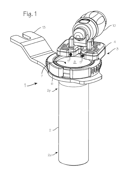
[0007] Figure 1 illustrates a cannula system 1 for
accessing a target site in the body of a patient. The cannula
system comprises a cannula tube 2 and a lighting assembly 3
disposed proximate the proximal end 2p of the cannula tube.
The lighting assembly 3 comprises a housing 4 with a number of
lights 5 (LED's, incandescent bulbs, etc.). The lighting
assembly may be mounted on a ring, or partial ring 6 as
illustrated, and may be permanently fixed or releasably
attachable to the proximal end 2p of the cannula tube, through
releasable attachment means such as a C-ring expandable to
engage a groove in the proximal end outer surface, or with an
annular snap ring, or with screw threads or other easily
attachable and detachable mechanisms. The lighting assembly
may instead be directly fixed to the proximal end of the
cannula tube or fixed on the ring 18 which in turn is fixed to
the cannula tube (as shown in Figure 5 through 8). The
cannula tube is characterized by a distal end 2d and a
proximal end 2p, and a lumen 7 extending from the proximal end
to the distal end, a central longitudinal axis 2L defined by
the lumen, and a luminal surface 8 on the inner wall of the
cannula tube. The cannula tube most conveniently has a
circular radial cross section, but the radial cross section
may be varied to provide for access to particular surgical
2
CA 03127963 2021-07-26
WO 2020/163753 PCT/US2020/017276
sites. The cannula tube may consist of an opaque material,
non-transmissive to visible light, such as metal, or it may
comprise an opaque construction including a luminal surface
comprising an opaque material which is non-transmissive to
visible light in a cannula tube of transmissive or non-
transmissive material (for example, an acrylic tube with a
metallic coating).
[0008] The lighting assembly 3 is disposed proximate the
proximal end of the cannula tube, and is configured to hold
light 5 proximal to the proximal opening of the cannula tube
(this is preferable, but the lights may extend slightly
distally into the lumen) to project light into the lumen of
the cannula tube. The cannula tube may consist of an opaque
material, non-transmissive to visible light, and is preferably
made of metal such as stainless steel or aluminum.
[0009] The effectiveness of the lighting is preferably
enhanced by providing a very smooth surface on the inner wall
of the cannula tube. Preferably, the luminal surface is
highly polished/smooth with an Average Roughness of 8 micro-
inches or smoother (8-6 inches, equivalent to Ra (um) 0.2 (0.2
microns), USA #8 finish, Japan Buff #300, or ISO N4 or
smoother), to enhance the transmission of light from the
proximal end of the cannula to the distal end of the cannula
and a target site beyond the distal end of the cannula. The
lights of Figure 1 may have a total output of 200 to 700
lumens, which, in combination with the smooth luminal surface,
will provide in ample light at a surgical workspace at the
distal end of the cannula tube. Combinations of slightly
rougher surfaces with higher power lights may be used. The
luminal surface may be provided in a Average Roughness in the
range of 9 to 32 micro-inches (between 0.22 to 0.81
micrometers, ISO N5 or N6 finish, #6 or #7 finish (roughly),
Japan Buff #100 or smoother) and the lights may be chosen to
provide additional lumens, in the higher end of the range.
3
CA 03127963 2021-07-26
WO 2020/163753 PCT/US2020/017276
Alternatively, the luminal surface may be provided in a
Average Roughness in the range of 33 to 63 micro-inches (0.82
to 1.6 micrometers, ISO N7 finish, USA #3 or #4 finish) and
the lights may be chosen to provide additional lumens, in the
higher end of the range.
[0010] As illustrated in Figure 2, the light 5 are
characterized by a main beam axis 9, which may be directed at
an angle al of 700 to 85 , though preferably about 80
downward (distally) from the radial axis 2R, or, comparably,
directed at an angle Bi or 5 to 15 , and preferably about 10 ,
inward relative to the long axis 2L of the cannula tube,
directed distally, in this embodiment where the cannula has a
distal portion with a straight inner bore (of consistent
diameter throughout the length of the distal portion) and a
proximal conical section with a conical bore which is larger
than the diameter of the straight inner bore at the proximal
end of the proximal conical section and necks down to match
the diameter of the straight inner bore of the straight distal
portion.
[0011] As illustrated in Figure 2, the lights are
characterized by a main beam axis 9, which may be directed at
an angle al of 80 from the radial axis 2R, directed distally,
or at an angle Bi of 10 relative to the luminal surface of the
cannula tube (toward the center of the lumen).
[0012] Though Figures 1 and 2 illustrate the system with a
cannula tube having a conical lumen in a proximal portion of
the cannula tube, the cannula tube may be isodiametric
throughout its length, having a consistent or uniform inner
diameter and straight luminal walls from the proximal end to
the distal end, without a conical portion or a neckdown
portion.
4
CA 03127963 2021-07-26
WO 2020/163753 PCT/US2020/017276
[0013] Figure 3 is a view of the cannula system from the
bottom, or distal end of the cannula tube. As shown in this
Figure, the beam axis 9 may be aimed to intersect the central
axis 2C of the cannula tube, or the beam axis may be aimed at
angle yfrom the radian 2R (the line between the LED and the
central axis 2C, or, along a chord of the circle defined by
the cannula tube). This angle is preferably in the range of
about 10 to 300. As shown in Figures 3 and 4, the light
source may consist of only two LED's disposed over (proximal
to) the proximal end of the cannula tube, either directly or
on the ring 6 and separated by a first arc a2 of 500 to 700,
and preferably about 600 as shown in Figure 3 (or, conversely,
the second arc B2 of 2900 to 3100, and preferably about 3000 as
shown in Figure 3). The light source may consist of two pairs
of closely spaced lights, with the pairs similarly separated.
Preferably, the lights and any associated lenses are disposed
proximal to the proximal opening of the cannula tube without
extending distally into the lumen. The proximal end of the
cannula tube has an inner bore/lumen that is conical, with a
proximal opening slightly larger than the diameter of the
distal portion of the cannular tube.
[0014] As shown in Figure 1, the cannula system may include
a camera assembly 10 secured to the proximal end of the
cannula, with a portion of the camera assembly overhanging the
lumen and extending into a cylindrical space defined by the
lumen of the cannula tube. The camera assembly has a distal-
most optical surface, which may be a distal surface of an
objective lens or a prism (the prism 11 is shown in Figure 2,
and the distal-most optical surface 12 is visible in the
distal view of Figure 3), and the distal-most optical surface
is disposed proximate the proximal end of the cannula tube.
The objective lens or prism may be the portion of the camera
assembly overhanging the lumen. The distal-most optical
surface of the camera system is spaced proximally from the
CA 03127963 2021-07-26
WO 2020/163753
PCT/US2020/017276
proximal end of the cannula tube in the illustration, but may
be placed a short distance distal to the very proximal edge of
the cannula tube (without extending to the distal end of the
cannula tube). Also as shown in Figure 1, the cannula system
can include a tab 13 for securing the cannula to a table-fixed
flex arm. As illustrated in Figures 3 and 4, the distal most
optical surface of the camera assembly is disposed between the
lights, in the smaller arc a2 separating the two lights. A
gap in the housing, between the two lights (or two pairs of
lights), provides an unobstructed sight-line between the
distal-most optical surface and the workspace at the distal
end of the cannula tube, and the distal most optical surface
of the camera assembly is disposed within this gap or proximal
to the gap.
[0015] Figure 5 illustrates a second version of the cannula
system for accessing a target site in the body of a patient.
The cannula system 14 of Figure 5 comprises a cannula tube 15
and a lighting assembly 16 disposed proximate the proximal end
of the cannula tube. The lighting assembly 16 comprises a
number of lights 17 (LED's, incandescent bulbs, etc.) mounted
on a ring 18 as illustrated (though a partial ring may be
used, or the ring may be omitted), and may be permanently
fixed or releasably attachable to the proximal end of the
cannula tube, through releasable attachment means such as an
annular snap ring, a threaded fitting (or a C-ring expandable
to engage a groove in the proximal end outer surface). The
cannula tube is characterized by a distal end 15d and a
proximal end 15p, and a lumen 19 extending from the proximal
end to the distal end, a central longitudinal axis 15L defined
by the lumen, and a luminal surface 20 on an inner wall of the
cannula tube. The inner diameter of the cannula tube proximal
end 15p is longitudinally isodiametric (straight-walled, and
not conical as in Figure 2), and the inner diameter of the
cannula tube distal end 15d is longitudinally isodiametric,
6
CA 03127963 2021-07-26
WO 2020/163753 PCT/US2020/017276
and the inner diameter of the cannula tube distal end is
smaller than the inner diameter of that cannula tube proximal
end, and the cannula tube proximal end 15p and cannula tube
distal end 15d are joined by a neck-down portion 15N of the
cannula tube.
[0016] Similar to the construction described in relation to
Figures 1 through 3, the lighting assembly 16 of Figure 5 is
disposed proximate the proximal end of the cannula tube, and
is configured to project light into the lumen of the cannula
tube. The cannula tube may consist of an opaque material,
non-transmissive to visible light, again preferably metal such
as stainless steel or aluminum. The luminal surface is highly
polished/smooth with a Average Roughness less that 8 micro-
inches, to enhance the transmission of light from the proximal
end of the cannula to the distal end of the cannula and a
target site beyond the distal end of the cannula. The lights
of Figure 5 may have a total output of 1500 to 2500 lumens,
which, in combination with the smooth luminal surface, will
provide in ample light at a surgical workspace at the distal
end of the cannula tube. As with the cannula tube of Figure
1, the lights may be chosen to provide additional lumens, in
the higher end of the range, with luminal walls of Average
Roughness within the range of 9 to 32 micro-inches or in the
range of 33 to 63 micro-inches.
[0017] As shown in Figure 6, the lighting assembly 16 may
comprise a plurality of LED's 17 disposed on the proximal end
of the cannula tube, either directly fixed to the proximal end
of the cannula tube or fixed on the ring 18 which in turn is
fixed to the cannula tube. The ring 18 may be permanently
fixed or releasably attachable to the proximal end 15p of the
cannula tube, through releasable attachment means such as a C-
ring expandable to engage a groove in the proximal end outer
surface, or with an annular snap ring, or with screw threads
or other easily attachable and detachable mechanisms.
7
CA 03127963 2021-07-26
WO 2020/163753
PCT/US2020/017276
[0018] As shown in the cross section of Figure 7, the
lights 17 are characterized by a main beam axis 21, which may
be directed parallel to the straight side wall or the portion
of the luminal surface on the inner wall of the proximal end
of the cannula tube (that is, the beam axes of each LED may be
parallel to a portion of the luminal surface on an inner wall
of the cannula). Alternatively, as in the systems of Figures
1 and 2, the main beam axis 21 may also be directed at an
angle al of 700 to 85 , though preferably about 80 downward
(distally) from the radial axis 2R, or, comparably, directed
at an angle Bi of 5 to 15 , and preferably about 10 relative
to the luminal surface of the cannula tube (toward the center
of the lumen).
[0019] The cannula system of Figure 5 may include a camera
assembly 10 secured to the proximal end of the cannula, with a
portion of the camera assembly overhanging the lumen and
extending into a cylindrical space defined by the lumen of the
cannula tube. The camera assembly has a distal-most optical
surface, which may be a distal surface of an objective lens or
a prism, and the distal-most optical surface is disposed
proximate the proximal end of the cannula tube, the objective
lens or prism may be the portion of the camera assembly
overhanging the lumen. The distal-most optical surface of the
camera system is spaced proximally from the proximal end of
the cannula tube in the illustration, but may be placed a
short distance distal to the very proximal edge of the cannula
tube.
[0020] Figures 8 illustrates a lighted cannula system with
a cannula tube of non-uniform diameter, with a proximal light
source consisting of two LED's 5. Figure 8 illustrates that
the cannula tube of Figure 5 can be combined with the two-LED
light source of Figures 1 through 4, to obtain the benefits of
the larger proximal lumen in a system using a light source
consisting of two LED's. In this embodiment, the two LED's
8
CA 03127963 2021-07-26
WO 2020/163753 PCT/US2020/017276
(or two pairs) can be aimed directly distally, with the beam
axes parallel to the side wall of the cannula tube, as with
Figure 7, or the beam axes may be angled toward the center of
the lumen, as with Figure 2.
[0021] The extreme smoothness of the luminal surface
provides for abundant reflection of light from the proximal
light sources into the cannula distal end and minimization of
shadows cast by tools disposed within the cannula lumen,
without the need to resort to more complex tube constructions
such as optical fibers embedded in the cannula wall, or
optical transmission of light from a light ring into a
transparent wall, or construction of the cannula wall to serve
as a light guide with rough surface features needed to extract
and deliver light at that proximal end of the cannula tube.
Though the cannula tube can comprise a transparent material,
it is more conveniently made of metal, such as stainless steel
or aluminum, which can be made with thinner walls vis-a-vis
plastics, and can be sterilized and re-used, and is not
subject to abrasion or skiving from abrading tools (more of a
concern for spinal surgery). Thus, the cannula tube can
consist of an opaque material, preferably metal, without
embedded optical fibers or wave guide features. The cannula
tube can also consist of a transparent polymer, without
embedded optical fibers or wave guide features, though the
transparency of the tube is not necessary to obtain the
advantages of the inventive features of the cannula system.
[0022] Alternatively, the cannula tube can be made of other
materials, with a highly reflective material adhered to the
luminal walls, which will also provide for good light
transmission from the proximal lighting assembly, without
embedded optical fibers or wave guide features.
[0023] The luminal surface of the cannula tube may be
coated to enhance performance in various aspects. The luminal
9
CA 03127963 2021-07-26
WO 2020/163753 PCT/US2020/017276
surface may be coated with parylene or other dielectric
compound for use in surgeries that require delivery of
ablation energy through tools to be inserted into a surgical
workspace through the cannula tube. The luminal surface may
be coated with a hydrophobic coating, or a lipophobic or
oleophobic coating, to minimize build-up of body fluids or
irrigation fluids during use.
[0024] While the preferred embodiments of the devices and
methods have been described in reference to the environment in
which they were developed, they are merely illustrative of the
principles of the inventions. The elements of the various
embodiments may be incorporated into each of the other species
to obtain the benefits of those elements in combination with
such other species, and the various beneficial features may be
employed in embodiments alone or in combination with each
other. Other embodiments and configurations may be devised
without departing from the spirit of the inventions and the
scope of the appended claims.