Note: Descriptions are shown in the official language in which they were submitted.
CA 03128219 2021-07-28
WO 2020/160491
PCT/US2020/016244
MICROFLUIDIC EXTRUSION
CROSS-REFERENCE TO RELATED APPLICATION(S)
[0001] This application claims the benefit of co-pending
application
serial number 62/800,317, filed February 1, 2019, the disclosure of which is
hereby incorporated by reference in its entirety.
STATEMENT REGARDING GOVERNMENTAL SUPPORT
[0002] The data presented in this application was supported at
least in
part by DARPA Contract HR0011-15-9-0006. The US government has certain
rights in the invention.
BACKGROUND OF THE INVENTION
1. Field of the Disclosure
[0003] The present disclosure relates to a method for
manufacturing
collagen fibers and their incorporation into scaffolds and implantable
biocompatible devices prepared with such fibers. In particular, the disclosure
relates to a method for extruding collagen fibers having superior mechanical
strength, biocompatibility and immunological properties.
2. Description of Related Art
[0004] Collagen is a fibrous insoluble protein consisting of
bundles of
tiny reticular fibrils. Collagen protein molecules combine to form white,
glistening, inelastic fibers of the tendons, the ligaments, and the fascia.
Collagen
is found in connective tissue, including skin, bone, ligaments, and cartilage.
[0005] In particular, collagen fibrils combine to form tough
connective
tissue such as ligaments and tendons. Many efforts have been made to
manufacture collagen-containing tissue for use in the body to replace damaged
1
CA 03128219 2021-07-28
WO 2020/160491
PCT/US2020/016244
collagen body parts, including in particular ligaments and tendons. Such
implantable devices may replace the damaged part directly or may serve to
provide a scaffold to facilitate repair of, and eventually replace, damaged
soft
tissues such as tendons and ligaments.
[0006] Such products must function in a variety of challenging
biomechanical environments in which multiple functional parameters must be
addressed. These parameters include, for example, compatibility with bodily
tissue and fluids, strength, flexibility, and biodegradability.
[0007] There is a need in the art for a system and method that
addresses the shortcomings of the prior art discussed above.
SUMMARY OF THE INVENTION
[0008] In one aspect, the disclosure is directed to a
biopolymer fiber
comprising a collagen, wherein the biopolymer fiber has one or more of the
following characteristics:
[0009] an ultimate tensile strength of between about 1 MPa to
about
1,700 MPa;
[0010] a modulus of elasticity of between about 10 MPa to about
20,000 MPa;
[0011] a strain at break of between about 2 percent and about
45
percent elongation;
[0012] an average fiber diameter between about 10 pm and about
90 pm;
[0013] maintains its strength after soaking in DPBS at room
temperature for at least about 1 hour; and
[0014] wherein the filament exhibits an ordered, longitudinally
oriented structure.
[0015] In another aspect, the disclosure is directed to a
bundle of the
biopolymer fibers comprising between 2 and about 10,000 fibers.
2
CA 03128219 2021-07-28
WO 2020/160491
PCT/US2020/016244
[0016] In still another aspect, the disclosure is directed to
an
implantable biopolymer scaffold for supporting repair of a soft tissue injury
comprising the biopolymer fibers or the bundle.
[0017] The disclosure also is directed to a woven sheet-like
support, a
patch, or a brace comprising biopolymer fibers.
[0018] In yet another aspect, the disclosure is directed to a
method for
producing a biopolymer fiber. The method comprises the steps of:
[0019] dissolving collagen in an acid solution to form a
collagen
solution;
[0020] passing the collagen solution at a first speed through a
first
needle having a first diameter simultaneously with passing a formation buffer
solution at a second speed through a second needle coaxially surrounding the
first
needle and having a second diameter greater than the first diameter to form a
sheath around the collagen solution to form a coaxial flow,
[0021] wherein the second flow rate of the foundation buffer
solution
through the second needle is at least twice the first flow rate of the
collagen
solution through the first needle,
[0022] passing the coaxially-flowing collagen and formation
buffer
solution through a reaction zone comprising a fibril-forming bath for a time
and at
speeds sufficient to form a fiber,
[0023] dehydrating the collagen fiber at an extrusion speed,
and
[0024] withdrawing the fiber onto a spool at a third speed
greater than
the extrusion speed sufficient to increase molecular alignment and reduce the
diameter of the fiber.
[0025] In another aspect, the disclosure is directed to a
method for
producing a biopolymer fiber. The method comprises the steps of:
[0026] dissolving collagen in an acid solution to form a
collagen
solution;
[0027] passing the collagen solution at a first speed through a
first
needle having a first diameter into a formation buffer solution,
3
CA 03128219 2021-07-28
WO 2020/160491
PCT/US2020/016244
[0028] passing the collagen and formation buffer solution
through a
reaction zone comprising a fibril-forming bath for a time and at speeds
sufficient
to form a fiber,
[0029] dehydrating the collagen fiber at an extrusion speed,
and
[0030] withdrawing the fiber onto a spool at a speed of between
about
2 times the extrusion speed and about 10 times the extrusion speed sufficient
to
increase molecular alignment and reduce the diameter of the fiber.
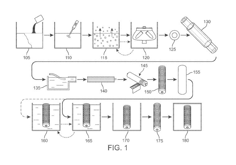
[0031] In yet another aspect, the disclosure is directed to a
method for
producing a biopolymer fiber comprising the steps of:
[0032] dissolving clinical-grade collagen in an acid solution
to form a
collagen solution;
[0033] passing the collagen solution at a first volumetric flow
rate
through a first needle to yield a first speed simultaneously with passing a
formation buffer solution at a second speed in a tube coaxially surrounding
the
first needle and forming a sheath around the collagen solution to form a
coaxial
flow,
[0034] wherein the speed of the foundation buffer solution is
between
about 2 times and about 20 times the first speed of the collagen solution
through
the first needle,
[0035] passing the coaxially-flowing collagen and formation
buffer
solution through a reaction zone comprising a fibril-forming bath for a time
and at
speeds sufficient to form a fiber,
[0036] dehydrating the collagen fiber at an extrusion speed,
and
[0037] withdrawing the fiber at a third speed greater than the
extrusion
speed sufficient to increase molecular alignment and reduce the diameter of
the
fiber.
[0038] In a further aspect, the disclosure is directed to a
method for
producing a biopolymer fiber comprising the steps of:
[0039] dissolving clinical-grade collagen in an acid solution
to form a
collagen solution;
4
CA 03128219 2021-07-28
WO 2020/160491
PCT/US2020/016244
[0040] extruding the solution through a nozzle into a guide
that passes
the extruded solution into a bath of formation buffer;
[0041] dehydrating fiber formed in the formation buffer bath;
and
[0042] collecting the fiber.
[0043] In a still further aspect, the disclosure is directed to
a method
for producing a biopolymer fiber comprising the steps of:
[0044] dissolving clinical-grade collagen in an acid solution
to form a
collagen solution;
[0045] passing the collagen solution at a first speed through a
first
needle having a first diameter into a formation buffer solution,
[0046] passing the collagen and formation buffer solution
through a
reaction zone comprising a fiber-forming bath for a time and at speeds
sufficient
to form a fiber,
[0047] dehydrating the collagen fiber at an extrusion speed,
and
[0048] withdrawing the fiber onto a spool at a speed of between
about
2 times the extrusion speed and about 12 times the extrusion speed, in one or
more
stages, sufficient to increase molecular alignment and reduce the diameter of
the
fiber.
[0049] The disclosure also includes an aspect of providing an
implantable biopolymer scaffold for supporting repair of a soft tissue injury
comprising the biopolymer fibers, a method for supporting the repair of a soft
tissue injury comprising the implantation of the biopolymer scaffold.
[0050] Other systems, methods, features, and advantages of the
invention will be, or will become, apparent to one of ordinary skill in the
art upon
examination of the following figures and detailed description. It is intended
that
all such additional systems, methods, features and advantages be included
within
this description and this summary, be within the scope of the invention, and
be
protected by the following claims.
CA 03128219 2021-07-28
WO 2020/160491
PCT/US2020/016244
BRIEF DESCRIPTION OF THE DRAWINGS
[0051] The invention can be better understood with reference to
the
following drawings and description. The components in the figures are not
necessarily to scale, emphasis instead being placed upon illustrating the
principles
of the invention. Moreover, in the figures, like reference numerals designate
corresponding parts throughout the different views.
[0052] FIG. 1 is a schematic diagram of an embodiment of a
method
disclosed in the specification;
[0053] FIG. 2 is a schematic diagram illustrating a first step
in the
formation of a collagen solution in an embodiment of the disclosure;
[0054] FIG. 3 is a schematic illustration of use of a degasser
in
collagen preparation in an embodiment of a method disclosed in the
specification;
[0055] FIG. 4 is a schematic illustration of a centrifuge
suitable for use
in an embodiment of a method disclosed herein;
[0056] FIG. 5 is a schematic illustration of centrifuge use in
an
embodiment of the disclosure;
[0057] FIG. 6 is a schematic illustration of an embodiment of
coaxial
needles used to form collagen fiber in an embodiment;
[0058] FIG. 7 is a schematic illustration of a collagen fiber
reaction
zone comprising a fibril-forming bath in an embodiment of the disclosure;
[0059] FIG. 8 is a schematic embodiment of a dehydration bath
in an
embodiment of the disclosure;
[0060] FIG. 9 is a schematic diagram of an embodiment of a
device for
separating collagen fiber from a dehydrating bath;
[0061] FIG. 10 is a schematic diagram of a fiber collection
system in
an embodiment of the disclosure;
[0062] FIG. 11 is a schematic illustration of fiber collecting
spools in
an embodiment of the disclosure;
[0063] FIG. 12 is a schematic illustration of end treating in
accordance
with an embodiment of the disclosure;
6
CA 03128219 2021-07-28
WO 2020/160491
PCT/US2020/016244
[0064] FIG. 13 is a schematic illustration of a method of an
embodiment of the disclosure;
[0065] FIG. 14 is a schematic illustration of a method of an
embodiment of the disclosure;
[0066] FIG. 15 is a schematic illustration of an embodiment of
an
apparatus suitable for use to make product of the disclosure;
[0067] FIG. 16 illustrates additional details of the apparatus
of
FIG. 15;
[0068] FIG. 17 is a table summarizing compositions used in the
disclosure;
[0069] FIG. 18 is a table summarizing conditions used for
embodiments;
[0070] FIG. 19 is a graph summarizing a mechanical property
relevant
to the disclosure;
[0071] FIG. 20 is a graph summarizing another mechanical
property
relevant to the disclosure;
[0072] FIG. 21 is a graph summarizing still another mechanical
property relevant to the disclosure;
[0073] FIG. 22 is magnified images of compositions manufactured
in
accordance with the disclosure;
[0074] FIG. 23 is a graph summarizing the width of embodiments
of
the disclosure;
[0075] FIG. 24 is a graph summarizing the thickness of
embodiments
of the disclosure;
[0076] FIG. 25 is a graph summarizing a mechanical property of
embodiments of the disclosure;
[0077] FIG. 26 is a graph summarizing another mechanical
property of
embodiments of the disclosure;
[0078] FIG. 27 is a graph summarizing a relationship of a
mechanical
property of embodiments of the disclosure;
7
CA 03128219 2021-07-28
WO 2020/160491
PCT/US2020/016244
[0079] FIG. 28 is a graph summarizing a relationship of another
mechanical property of embodiments of the disclosure;
[0080] FIG. 29 is graphs summarizing properties of embodiments
of
the disclosure;
[0081] FIG. 30 is color images of embodiments of the
disclosure;
[0082] FIG. 31 is a graph summarizing a property of embodiments
of
the disclosure;
[0083] FIG. 32 is a graph summarizing another property of
embodiments of the disclosure;
[0084] FIG. 33 is a graph summarizing still another property of
embodiments of the disclosure;
[0085] FIG. 34 is magnified color images of an embodiment of
the
disclosure;
[0086] FIG. 35 is magnified color images of a microfiber
product;
[0087] FIG. 36 is magnified color images of a control
microfiber
product;
[0088] FIG. 37 is an image of features of an embodiment of the
disclosure;
[0089] FIG. 38 is an image of features of an embodiment of the
disclosure;
[0090] FIG. 39 is a graph summarizing features of embodiments
of the
disclosure;
[0091] FIG. 40 is a graph summarizing how size of an embodiment
of
the disclosure changes with time;
[0092] FIG. 41 is a graph summarizing how a mechanical property
of
an embodiment of the disclosure changes with time;
[0093] FIG. 42 is a graph summarizing how another mechanical
property of an embodiment of the disclosure changes with time;
[0094] FIG. 43 is a graph summarizing how yet another property
of an
embodiment of the disclosure changes with time;
8
CA 03128219 2021-07-28
WO 2020/160491
PCT/US2020/016244
[0095] FIG. 44 is a graph summarizing how still another
property of an
embodiment of the disclosure changes with time;
[0096] FIG. 45 is a table summarizing comparative information;
[0097] FIG. 46 illustrates an embodiment of a method of the
disclosure;
[0098] FIG. 47 summarizes the properties and characteristics of
an
embodiment of the disclosure;
[0099] FIG. 48 summarizes the properties and characteristics of
an
embodiment of the disclosure;
[00100] FIG. 49 summarizes the properties and characteristics of an
embodiment of the disclosure;
[00101] FIG. 50 summarizes the properties and characteristics of an
embodiment of the disclosure;
[00102] FIG. 51 summarizes the properties and characteristics of an
embodiment of the disclosure;
[00103] FIG. 52 summarizes the properties and characteristics of an
embodiment of the disclosure;
[00104] FIG. 53 summarizes the properties and characteristics of an
embodiment of the disclosure;
[00105] FIG. 54 summarizes the properties and characteristics of an
embodiment of the disclosure;
[00106] FIG. 55 summarizes the properties and characteristics of an
embodiment of the disclosure;
[00107] FIG. 56 summarizes the properties and characteristics of an
embodiment of the disclosure;
[00108] FIG. 57 is an SEM image of an embodiments of the disclosure;
and
[00109] FIG. 58 is an SEM image of an embodiments of the disclosure.
9
CA 03128219 2021-07-28
WO 2020/160491
PCT/US2020/016244
DETAILED DESCRIPTION
[00110] In one aspect, the disclosure is directed to a biopolymer fiber
comprising collagen, wherein the biopolymer fiber has one or more of the
following characteristics:
[00111] an ultimate tensile strength of between about 1 MPa to about
1,700 MPa;
[00112] a modulus of elasticity of between about 10 MPa to about
20,000 MPa;
[00113] a strain at break of between about 2 percent and about 45
percent elongation;
[00114] an average fiber diameter between about 10 um and about
90 um;
[00115] maintains its strength after soaking in DPBS at room
temperature for at least about 1 hour; and
[00116] wherein the filament exhibits an ordered, longitudinally
oriented structure.
[00117] In another aspect, the disclosure is directed to an implantable
biopolymer scaffold for supporting repair of a soft tissue injury comprising
at least
one biopolymer sheet comprising biopolymer fibers, wherein the biopolymer
comprises collagen and the biopolymer fibers have one or more of the following
characteristics:
[00118] an ultimate tensile strength of between about 1 MPa to about
1,700 MPa;
[00119] a modulus of elasticity of between about 10 MPa to about
20,000 MPa;
[00120] a strain at break of between about 2 percent and about 45
percent elongation;
[00121] an average fiber diameter between about 10 um and about
90 um;
[00122] maintains its strength after soaking in DPBS at room
temperature for at least about 1 hour; and
CA 03128219 2021-07-28
WO 2020/160491
PCT/US2020/016244
[00123] wherein the filament exhibits an ordered, longitudinally
oriented structure.
[00124] The fibers exhibit an ordered, longitudinally-oriented structure,
and allow cellular infiltration following implantation of the fibers, and
devices
made with the inventive fibers, into a subject.
[00125] In another aspect, the disclosure includes an implantable
biopolymer scaffold for repair or replacement of a human body part.
[00126] Biopolymer fiber typically is formed of collagen. In particular,
telocollagen typically is obtained from any source (human, bovine,
recombinants,
jelly fish, etc.). Bio-acceptable polymer, such as silk fibroin; other types
of
collagen such as type II collagen; fibrin/fibrinogen; basement membrane
proteins;
hyaluronic acid, poly ethylene oxide, poly ethylene glycol, poly caprolactone,
polyethylnene, polyhydroxybutyrate, PDLA; PDLLA and high molecular weight
PDLLA; PLGA; and blends thereof, may be blended with collagen to form
biopolymer fiber.
[00127] In still another aspect, the disclosure is directed to a method for
producing a biopolymer fiber comprising the steps of dissolving collagen in an
acid solution to form a collagen solution. In one embodiment of this method,
the
collagen then is passed at a first speed through a first needle having a first
diameter to have a first speed simultaneously with passing a formation buffer
at a
second volumetric flow rate through a second needle coaxially surrounding the
first needle and having a second diameter greater than the first diameter to
form a
sheath around the collagen solution to form a coaxial flow. The second
volumetric flow rate of the formation buffer through the second needle is at
least
about twice the first volumetric flow rate of the collagen solution through
the first
needle.
[00128] The coaxially-flowing collagen and formation buffer are passed
through a reaction zone comprising a fibril-forming bath for a time and at
speeds
sufficient to form a fiber, which is withdrawn onto a spool at a third speed
greater
than the first speed, and typically twice the speed at which the fiber is
extruded
11
CA 03128219 2021-07-28
WO 2020/160491
PCT/US2020/016244
through the dehydration bath, sufficient to increase molecular alignment and
reduce the diameter of the fiber. The fibers then may be cross-linked and
dried.
[00129] In yet another aspect, the disclosure is directed to an alternative
method for producing a biopolymer fiber. In this embodiment, a collagen
solution
is prepared and injected into a reaction zone in a comprising a fiber-forming
bath,
such as a bath of formation buffer, for a time and at speeds sufficient to
form a
fiber. The fiber is withdrawn on a spool at a speed between about 2 to about
10
times faster than the injection speed to increase molecular alignment and
reduce
the diameter of the fiber. The fibers then may be cross-linked and dried.
[00130] In various embodiments of the disclosure, collagen or collagen
and other suitable biopolymers are made into biopolymer or collagen fiber. For
ease of understanding, the features of the disclosure will be described as
they
relate to collagen. However, collagen may be blended or combined with suitable
biopolymers in various combinations and proportions to obtain fibers of the
type
disclosed herein.
[00131] Throughout the specification, steps that might typically be
taken together during a typical manufacturing process, such as washing and
drying
or soaking and drying, may be taken or repeated as appropriate to achieve a
desired result. For example, in an embodiment, a composition may be washed and
dried before advancing to the next step. In some embodiments, the material may
be washed a second time and dried a second time before advancing to the next
processing step, or may be washed a second time, then advanced.
[00132] In other embodiments, a first washing or drying step may be
made optional. Thus, a material typically washed, then dried, may go directly
to
the drying step, and then moved on to the next processing step. The skilled
practitioner can recognize circumstances under which steps may be repeated or
eliminated.
[00133] The constructs, such as scaffolds, made from the fibers, allow
cellular ingrowth, that is, various types of cells from the animal into which
the
fiber (and devices made from the fiber) is implanted will grow into the pores
of
the scaffold, preferably aligned with the fibers in the scaffold. Constructs
and
12
CA 03128219 2021-07-28
WO 2020/160491
PCT/US2020/016244
scaffolds comprise single layer and multi-layer articles that may be used as a
substitute for a known repair feature, such as sutures used to re-attach body
parts,
for example opposing ends of a ruptured Achilles tendon. In addition to
providing
supporting structures for use in repairing torn or damaged tendons,
embodiments
of the disclosure are suitable in ligament repair as well. Thus, other
exemplary
ligaments for which the scaffolds or the present invention may be used to
provide
support include the ACL, MCL, PCL, UCL, and other human and animal
ligaments. Other surgeries for which products of the disclosure are useful
include
superior capsular reconstruction as a treatment option for superior rotator
cuff
tears, and in particular for otherwise irreparable or difficult to repair
partial or full
tears. Similarly, a multi-layered sheet may be used to overlap a repair to
strengthen it.
[00134] In particular, embodiments of the disclosure may be suitable for
repair of ligaments, tendons, and other soft tissues of animals of all types.
Collagen fibers of the disclosure may be used, for example, to reattach torn
ligaments and tendons, even those with only a partial tear. Plural fibers also
may
be twisted, bundled, braided, interwoven, or otherwise arranged to improve a
form
factor that is easier to work with than a single fiber is to manipulate, for
example
during surgery. Improving the form factor may make it easier to locate a fiber
or
platform accurately. Other form factors may be constructed to serve as a
reinforcement or internal brace for a torn natural body part. A brace connects
from one bone to another bone to support a joint. Typically, a brace forms an
isometric joint with restored biomechanics and the isometry of the native
joint.
[00135] The efficacy of collagen fiber produced in accord with
embodiments of the disclosure may be illustrated by studying repairs made in,
for
example, rabbits. In particular, reinforcements and internal brace and over-
sewn
structures of rabbit knees are suitable for evaluating the properties and
characteristics of collagen fiber of the disclosure and of structures made
from this
fiber.
[00136] FIG. 1 illustrates an embodiment of a system and method for
manufacturing collagen fiber. The system and method may be described as
13
CA 03128219 2021-07-28
WO 2020/160491
PCT/US2020/016244
comprising four sections or manufacturing areas. A collagen solution is
prepared
in the first section, and collagen fiber is formed in the second section. The
collagen fiber then is collected in the third section and then may be post-
processed
to yield wet or dry collagen fiber in the fourth section, post-treatments or
end of
treatment.
[00137] The steps in the system and method illustrated in FIG. 1 may be
grouped into four categories, as follows:
Category Name Steps Included
1 Preparing Collagen Solution 105-120
2 Forming Collagen Fiber 125-130
3 Collecting Collagen Fiber 135-150
4 Post-Treatment or End Treatment 155-180
[00138] As seen at step 105 of FIG. 1, collagen is combined with an
acidic solution and stirred thoroughly at step 110. In some embodiments, the
acid
is between about 0.01 M and about 0.50 M acetic acid. In other embodiments,
the
acid is between about 0.01 M and about 0.50 M hydrochloric acid. The solution
may be degassed at step 115, and then centrifuged at step 120 to remove
residual
bubbles. Resultant collagen solution is extruded from a needle, and there may
be
a second needle co-axial therewith that supplies a formation buffer solution
in step
125. The resultant forming fiber may continue in through a formation tube in
step
130. The resultant product is a formed collagen fiber.
[00139] The fiber then continues to a collection system, wherein the
fiber is separated from the formation buffer solution at step 135 and
dehydrated at
step 140. The collagen fiber is recovered at step 145 and air-dried at step
150.
Then, post-processing may be carried out, as illustrated at step 155, step
160, step
165, and step 170. Air-dried collagen fiber on a spool is submerged in cross-
linking solution at step 155, optionally washed at step 160, air-dried at step
165,
and desiccated to form dried fiber at step 170. As illustrated in FIG. 1 by
the dot-
dash line, material may be optionally washed at step 160, dried at step 165,
and
returned to wash step 160.
14
CA 03128219 2021-07-28
WO 2020/160491
PCT/US2020/016244
[00140] Alternatively, collagen is injected into a bath of formation
solution to form a fiber. In this system, a second needle for coaxial
injection of
formation buffer is not necessary. Collagen thus injected is introduced to a
collection system through dehydration at step 140. The fiber then is processed
in
accordance with the remainder of the processing steps.
[00141] FIG. 1 provides a generalized view of a system and method for
carrying out an embodiment of the disclosure. Additional details and
disclosure
are included in the following particular aspects and embodiments of the
description.
[00142] In an embodiment, the disclosure is directed to a method for
producing a biopolymer fiber comprising the steps of dissolving collagen in an
acidic solution to form a collagen solution. The collagen then is passed at a
first
volumetric flow rate through a first needle having a first diameter to have a
first
speed simultaneously with passing a formation buffer at a second volumetric
flow
rate through a second needle coaxially surrounding the first needle and having
a
second diameter greater than the first diameter to form a sheath around the
collagen solution to form a coaxial flow. The second volumetric flow rate of
the
formation buffer through the second needle is at least twice the first
volumetric
flow rate of the collagen solution through the first needle.
[00143] The coaxially-flowing collagen and formation buffer is passed
through a reaction zone comprising a fibril-forming bath for a time and at
volumetric flow rates sufficient to form a fiber, which is withdrawn onto a
spool
at a third speed greater than the first speed. The third speed, typically
about twice
the speed at which the fiber is extruded through the dehydration bath, is
sufficient
to increase molecular alignment and reduce the diameter of the fiber. The
fibers
then are cross-linked and dried.
[00144] In another embodiment, the disclosure is directed to an
alternative method for producing a biopolymer fiber. Collagen solution is
prepared and injected into a reaction zone in a comprising a fibril-forming
bath,
such as a bath of formation buffer, for a time and at speeds sufficient to
form a
fiber. The second needle to form coaxial flow of formation buffer is not
needed.
CA 03128219 2021-07-28
WO 2020/160491
PCT/US2020/016244
Rather, collagen fiber is injected directly into the fibril-forming bath, and
then
carried through the dehydration bath. The fiber is carried through by being
withdrawn on a spool at a speed between about 2 to about 4 times faster than
the
injection speed to increase molecular alignment and reduce the diameter of the
fiber. The fibers then may be cross-linked and dried.
[00145] An embodiment of the method 1300 is summarized in FIG. 13.
A collagen solution is prepared, as illustrated. A collagen solution is formed
at
step 1305. A biopolymer may be mixed with the collagen. Collagen is dissolved
in an acidic solution to form a viscous solution. The solution is stirred at
step
1310 to ensure thorough mixing. The mixed solution may have entrapped gas,
and so may be degassed one or more times in degasser step 1315. The collagen
solution then may be centrifuged, as illustrated at step 1320. Optionally, the
degas/centrifuge steps may be repeated, as shown by the dot-dash lines on FIG.
1
and as feature 1316 on FIG. 13, to reduce the volume of gas entrapped in the
solution.
[00146] Thus-prepared collagen solution is formed into a collagen fiber
by coaxial extrusion with a formation buffer solution that serves as a sheath
for
the fiber core, as shown at step 1325. The formation buffer solution
volumetric
flow rate typically is at least twice the volumetric flow rate of the forming
collagen. This arrangement suppresses formation of individual fibrils;
stretches
and orients the fiber; and may smooth the surface of the fiber by imparting
flow-
induced crystallization to the fiber.
[00147] The collagen fiber then is collected. As formation of the
collagen fiber is completed at step 1330, the collagen then is separated from
the
formation buffer solution at step 1335 and dehydrated in a dehydrating
solution at
step 1340. The dehydrated collagen then is collected on a rotating spool in
step
1345, which further stretches the fiber by rotating at a rate greater than,
and
typically about twice, the rate at which the fiber is supplied from
dehydrating
solution step 1340. Thus-collected fiber then is air-dried on the spool in
step 1350.
[00148] In an alternative embodiment, collagen solution is formed into
a collagen fiber by direct injection into formation buffer solution. Thus,
step 1325
16
CA 03128219 2021-07-28
WO 2020/160491
PCT/US2020/016244
is skipped. The fiber is collected, separated from formation buffer solution,
and
dehydrated in a dehydrating solution at step 1340. The fiber is collected on a
rotating spool in step 1345, which collects fiber at a speed of between about
2
times the formation speed and about 4 times the formation speed.
[00149] Fiber that has been air-dried on the spool then may be post-
processed. Fiber may be cross-linked in a cross-linking solution at step 1355,
and
then may be rinsed at step 1360. The fiber then is air dried at step 1365 and
desiccated at step 1370 to yield dry cross-linked collagen fiber.
[00150] The equipment used in making collagen fiber is made of
conventional materials of construction suitable for resisting attack by any of
the
raw materials used to make collagen fiber in accordance with embodiments of
the
disclosure. Metals, plastics, and other materials have properties and
characteristics suitable to resist attack by raw materials, intermediates,
solvents,
and products during manufacture of collagen fiber.
[00151] Another aspect of the disclosure is directed to a collagen fiber
having one or more of the following characteristics:
[00152] an ultimate tensile strength of between about 1 MPa to about
1,700 MPa;
[00153] a modulus of elasticity of between about 10 MPa to about
20,000 MPa;
[00154] a strain at break of between about 4 percent and about 12
percent elongation;
[00155] an average fiber diameter between about 16 pm and about
70 pm; and
[00156] at least maintains its strength after soaking in
biological fluid
for about 1 hour.
[00157] The fiber exhibits an ordered, longitudinally-oriented structure,
and the fiber allows infiltration of cellular growth.
[00158] A fiber of this embodiment is manufactured in accordance with
the method of an embodiment of the disclosure. Collagen may be obtained from
many sources and in various forms. The quality of the collagen fiber may be
17
CA 03128219 2021-07-28
WO 2020/160491
PCT/US2020/016244
related to the quality of the raw material used. In some embodiments, bovine
collagen typically is used. Bovine collagen may be obtained in natural form or
as
lyophilized powder.
[00159] Bovine collagen 202 may be made into a viscous solution 203
by dissolution in an acidic solution. Both mineral acids, such as hydrochloric
acid,
and organic acids, such as acetic acid, may be used to prepare a collagen
solution.
For example, in an embodiment, Type I bovine collagen with telopeptide ends
intact may be dissolved in about 0.01 M acetic acid to about 0.5 M acetic acid
201
in vessel 210 to form a viscous solution 203 comprising about 16 mg
collagen/mL
of solution. Solution concentrations may range from about 10 mg collagen/mL of
solution to about 19 mg collagen/mL of solution. In another embodiment,
lyophilized Type I bovine corium with telopeptide ends attached is mixed into
a
mineral acid, such as HC1 having a concentration of from about 0.01 M to about
0.5 M, to form a solution having a concentration between about 10 mg
collagen/mL of solution to about 19 mg collagen/mL of solution, typically
about
16 mg collagen/mL of solution.
[00160] In embodiments, collagen is allowed to dissolve for at least
about 14 hours, typically at least about 15 hours, and more typically at least
about
16 hours. In some embodiments, collagen solution 301 is degassed in degasser
300 to remove bubbles from collagen solution 301. Screen 304 ensures that
collagen is not drawn out of the degasser through the degasser gas flow exit
303.
Degasser 303 typically is operated at a pressure of between about 0 psia and
about
3 psia. Collagen solution may be exposed to up to about 2 degassing cycles,
typically between about 1 and about 2 cycles. Degassing removes gas bubbles
that likely would interfere with and disrupt extrusion of fibrous collagen.
[00161] Degassed collagen then may be further degassed in a centrifuge.
Centrifuge 400 is illustrated with top 408 open, making bowl 403 visible in
FIG. 4.
Tubes of material to be centrifuged and tubes used to ensure balance of the
centrifuge are placed into the wells in the rotating bowl 409. Case 405 is
sufficiently robust to contain any debris should any of the interior parts
fail during
use.
18
CA 03128219 2021-07-28
WO 2020/160491
PCT/US2020/016244
[00162] FIG. 5 illustrates centrifuge 500 having containment bowl 501
and lid 508. The centrifuge rotates rapidly counterclockwise, as illustrated
by
movement arrow 505. Tube 502 illustrates a tube before centrifugation
containing
collagen solution 503. As can be seen, the collagen solution is homogeneous
and
has trapped bubbles in the otherwise homogenous collagen solution 512.
[00163] Centrifugation at relative centrifugal force, or g values,
between about 400 rcf and about 4,000 rcf, typically between about 600 rcf and
about 1,000 rcf, and more typically between about 700 rcf and about 800 rcf,
is
suitable to reduce the entrapped bubble volume to essentially zero within
between
about 3 minutes and about 15 minutes, typically between about 4 minutes and
about 10 minutes, and more typically between about 5 minutes and about 7
minutes.
[00164] In some embodiments, a pair of related steps may be repeated
by alternating between the steps. For example, collagen may be processed in
degasser 303 for one cycle, then in centrifuge 500 for 5 minutes, and then
returned
to degasser 303 for a cycle, then centrifuged again for 5 minutes. Operating
in
this alternative way may provide improved efficiency. This improved efficiency
may be realized by taking advantage of a shorter treatment time to achieve a
given
quantity of bubbles or to achieve a better result than linear processing may
achieve.
[00165] Collagen then is coextruded with a solution to form collagen
fiber. Extrusion of collagen solution at the core of a coaxial fluid may, in
some
embodiments, aid formation of a collagen fiber.
[00166] In some embodiments of the disclosure, collagen then is
introduced to the center of a coaxial flow needle, with a formation buffer
solution
introduced to the outer needle. Thus, the formation buffer solution forms a
sheath
around the collagen. As illustrated in FIG. 6, collagen solution 650 is pumped
through pump 655 and introduced into inner needle 603 as collagen flow 601.
Simultaneously, formation buffer solution 660 is introduced to outer needle
604 as
formation buffer solution flow 606. Outer needle 604 is coaxial with inner
needle
603 so that formation buffer solution forms a sheath around the central core
of
19
CA 03128219 2021-07-28
WO 2020/160491
PCT/US2020/016244
collagen. As the materials exit the needle to flow into a reaction zone
comprising
a fibril-forming bath 701 (shown in FIG. 7), formation buffer solution 607 has
formed a sheath around collagen fiber 602, which begins to form as a solid
fiber.
[00167] The diameter of a resultant product collagen fiber is made
smaller than the inner diameter of the central needle by downstream
processing.
The diameter of the central needle may be larger than the target diameter of
the
finished fiber. In some embodiments, the inner diameter of the central needle
is
between about 0.05 mm and about 100 mm; in some embodiments, the inner
diameter of the central needle is between about 0.1 mm and about 50 mm; in
still
other embodiments, the inner diameter of the central needle is between about
0.2
mm and about 20 mm; in yet other embodiments, the inner diameter of the
central
needle is between about 0.3 mm and about 10 mm, and more typically between
about 0.35 mm and about 5 mm. In some embodiments, an even narrower range
of inner diameter of the central needle, such as between about 0.03 mm and
about
mm, typically between about 0.10 mm and about 3 mm, still more typically
between about 0.30 mm and about 1 mm, and even more typically between about
0.35 mm and about 0.50 mm.
[00168] In some embodiments, the inner diameter of the central needle
is between about 0.38 mm and about 0.44 mm, typically between about 0.39 mm
and about 0.43 mm, and more typically between about 0.40 mm and about
0.42 mm.
[00169] In some embodiments, the inner diameter of the surrounding
outer coaxial needle that supplies formation buffer solution typically is
between
about 1.95 times the inner diameter of the central needle and about 2.15 times
the
inner diameter of the central needle, typically between about 2.00 times the
inner
diameter of the central needle and about 2.10 times the inner diameter of the
central needle, and more typically about 2.05 times the inner diameter of the
central needle.
[00170] In embodiments, formation buffer solution may be any solution
that aids formation of a collagen fiber. Formation buffer solution typically
is a
solution comprising TES, also known as 24(2-Hydroxy-1,1-
CA 03128219 2021-07-28
WO 2020/160491
PCT/US2020/016244
bis(hydroxymethyl)ethyllaminolethanesulfonic acid or N-
lTris(hydroxymethyl)methyll-2-aminoethanesulfonic acid, together with salts
and
buffering agents.
[00171] In some embodiments of the disclosure, formation buffer
solution is WSB, a solution comprising 30 mM TES, 4.14 mg/mL sodium
phosphate monobasic dihydrate, 12.1 mg/mL sodium phosphate dibasic
heptahydrate, 135 mM NaCl, and 10 percent w/v PEG (polyethylene glycol).
Similar solutions also may be suitable.
[00172] The flow rates of the collagen solution and of the formation
buffer solution are adjusted so that the formation buffer solution sheath
remains
intact within the extrusion needles and reaction zone comprising a fibril-
forming
bath. The speed of the formation buffer solution also is established to be
greater
than the speed of the collagen solution so as to provide a stretch to the
collagen
fiber to improve the quality of the fiber. Indeed, in this way, the collagen
will be
urged to form a relatively straight, continuous fiber without kinks and other
physical shape aberrations. In some embodiments, fibers may be substantially
circular, ovoid, square, rectangular, ribbon-like, triangular, or irregularly
shaped.
[00173] In embodiments of the disclosure, the speed of formation buffer
solution in the needle reaction zone comprising a fiber-forming bath is higher
than
the speed of the collagen solution. Formation buffer solution is used to
neutralize
the collagen solution and to assist with fibrillogenesis. Further, the higher
speed
of the formation buffer solution is used to pull or stretch the collagen
stream,
which creates an extensional field that helps align the collagen monomers in a
process called flow-induced crystallization. This alignment helps collagen
polymerize and increases the strength of the resultant product.
[00174] In some embodiments, the volumetric flow rate of the
formation buffer solution in the needle is between about 5 times the
volumetric
flow rate of the collagen solution in the needle and about 10 times the
volumetric
flow rate of the collagen solution in the needle, typically between about 7
times
the volumetric flow rate of the collagen solution in the needle and about 9
times
the volumetric flow rate of the collagen solution in the needle, and more
typically
21
CA 03128219 2021-07-28
WO 2020/160491
PCT/US2020/016244
between about 7.5 times the volumetric flow rate of the collagen solution in
the
needle and about 8.5 times the volumetric flow rate of the collagen solution
in the
needle. In particular, 8 times the volumetric flow rate of the collagen
solution in
the needle is effective.
[00175] In embodiments, collagen stream 702 and formation buffer
solution sheath 707 enter reaction zone comprising a fibril-forming bath 701
of
reaction system 700, as illustrated in FIG. 7. The reaction zone may have a
structure, such as a formation tube, that forms reaction zone 701. However,
typically, no structure need be present. The collagen fiber continues to
polymerize to form collagen fiber product as the streams flow through reaction
zone comprising a fibril-forming bath 701. Collagen fiber 752 and formation
buffer solution 702 flow out of reaction zone comprising a fibril-forming bath
701.
[00176] In embodiments, the speed of the collagen is adjusted to afford
the collagen a reaction or polymerization time of between about 15 seconds and
about 60 seconds, typically between about 20 seconds and about 50 seconds, and
more typically between about 25 seconds and about 40 seconds.
[00177] As shown in FIG. 8, in embodiments, at the end of the
polymerization period, collagen fiber 852 has formed and separates from
formation buffer solution 808 as the streams flow out of reaction zone
comprising
a fibril-forming bath 701. The excess formation buffer solution flows into
basin
801. Dehydration system 800 is so designed as to catch formation buffer
solution
808 in basin 801 and to introduce collagen fiber 852 to dehydration bath 802.
[00178] Dehydration solution affords the opportunity to remove water
from the collagen fiber, reduce fiber diameter, and aid in fibrillogenesis. In
embodiments, dehydration solution comprises a solution of between about 10
percent ethanol in MilliQ water and about 35 percent ethanol in MilliQ water,
typically between about 15 percent ethanol in MilliQ water and about 30
percent
ethanol in MilliQ water, and more typically between about 15 percent ethanol
in
MilliQ water and about 25 percent ethanol in MilliQ water. Skilled
practitioners
recognize that MilliQ water, also written as Milli-Q water, is highly purified
water
produced in equipment available from Millipore Sigma, Burlington, MA USA.
22
CA 03128219 2021-07-28
WO 2020/160491
PCT/US2020/016244
[00179] Collagen fiber 852 is passed through dehydration bath 802 for
between about 10 seconds and about 50 seconds, typically between about 15
seconds and about 45 seconds, and more typically between about 20 seconds and
about 40 seconds. Throughout the period, collagen fiber 852 remains submerged
in dehydration bath 802. The volume of dehydration bath 802 is between about
400 times the volume of formation buffer solution pumped per minute and about
800 times the volume of formation buffer solution pumped per minute, typically
between about 450 times the volume of formation buffer solution pumped per
minute and about 750 times the volume of formation buffer solution pumped per
minute, and more typically between about 500 times the volume of formation
buffer solution pumped per minute and about 700 times the volume of formation
buffer solution pumped per minute 601.
[00180] FIG. 9 illustrates the end part of the dehydration bath, from
which dehydrated collagen fiber is removed from the dehydration bath. As seen
in embodiments illustrated in FIG. 9, dehydrated collagen fiber 930 is removed
from dehydration bath 802 at hook 910 on ring 920. As can be seen, hook 910 is
retained in dehydration bath 802 by ring 920. Hook 910 tends to aid in removal
of
dehydration bath from the dehydrated collagen fiber. Dehydrated collagen fiber
930 is pulled upwardly in the direction of arrow 940 by rotation of spool
1001, as
shown on FIG. 10. Because the collection speed of spool 1001 (FIG. 10) is
greater than the extrusion flow rate of collagen fiber 930, hook 920, or a
similar
device, is appropriate to ensure that the collagen fiber remains submerged in
the
dehydration bath 802. As dehydrated collagen fiber 930 is lifted above the
level
of the dehydration bath, fluid droplets 905 can be seen to be falling off
dehydrated
collagen fiber 930.
[00181] FIG. 10 illustrates collection of dehydrated fiber onto spool
1001. In embodiments, spool 1001 is rotated by motor 1011 in a clockwise
direction, as shown by arrow 1016. Spool 1001 is rotated at a speed that
provides
a draw ratio of between about 1.5 and about 3, typically between about 1.75
and
about 2.5, and more typically between about 1.90 and 2.20. Similarly, spool
1002
is rotated at the same speed. The draw ratio is the ratio between the spooling
23
CA 03128219 2021-07-28
WO 2020/160491
PCT/US2020/016244
speed and the extrusion speed. Thus, there is a tension on first collagen
fiber 1050
that pulls the fiber upward at hook 910. The fiber then is pulled above the
wall of
dehydrating bath 802 and onto spool 1001.
[00182] Alternatively, in some embodiments of the disclosure, collagen
is introduced directly into a fibril-forming bath, without the coaxial needles
of
FIG. 6 to form a coaxial flow illustrated in FIG. 7. Rather, collagen fiber is
formed as collagen solution 852 is injected directly from a needle into fiber-
forming bath 870, after which processing proceeds as with the coaxial
formation
method which is an alternative embodiment of the disclosure. The needle size
for
the collagen injection is selected in the same way the needle size is selected
for
the coaxial injection method, and the fiber is drawn through the fibril-
formation
bath and then into the dehydration bath in the same manner as other
embodiments.
However, spool 1001 in FIG. 10 is rotated at a speed that yields a draw speed
of
between about 2 times the fiber formation rate and about 4 times the fiber
formation rate, typically between about 2.5 times the fiber formation rate and
about 3.5 times the fiber formation rate, and more typically between about
2.75
and 3.25 the fiber formation rate. Then, post-processing is carried out in the
same
manner as for other embodiments.
[00183] Arrow 1020 indicates passage of time for some embodiments,
during which spool 1001 has been translated relative to the position at the
end of
dehydration bath 802 so as to form a single layer of fiber on the spool. Thus,
spool rotation continues at the same speed and fiber 1052 is kept in tension
as the
spool is translated until spool 1002 is essentially full. Time arrow 1030
illustrates
a passage of time until the fiber supply is exhausted. Spool 1055 may then be
recovered. The translational speed may be adjusted to adjust separation
between
fibers on a spool.
[00184] To ensure that tension is maintained on a fiber as the spool is
rotated, typically the fiber is in contact with the entirety on the surface of
a spool,
such a spool 1110 used in some embodiments, as shown in FIG. 11. However, a
spool need not have a continuous surface, as does spool 1110. In other
embodiments, a number of rods could extend along the length of the spool. One
24
CA 03128219 2021-07-28
WO 2020/160491
PCT/US2020/016244
such spool formed from rods is spool 1120 in FIG. 11, which comprises first
rod
1125, second rod 1126, and third rod 1127. The rods provide sufficient surface
to
wind collagen thereon.
[00185] Fibers of the disclosure also may be chemically post-processed.
FIG. 12 illustrates potential post-processing steps. In embodiments, spool
1210
containing collagen fiber is air-dried at 1220 for at least about 15 minutes,
typically at least about 20 minutes, and more typically at least about 30
minutes.
Air-dried fiber-containing tube 1210 then is placed in a container for cross-
linking.
Typically, a container in embodiments of the disclosure minimizes the volume
of
the cross-linking container to reduce the amount of cross-linker required.
Thus, as
illustrated in FIG. 12, cylinder 1230 contains the amount of cross-linking
fluid
necessary to cover cylindrical spool 1210, as shown at 1231. In embodiments,
the
volume of cross-linking solution per meter of fiber is at least about 3 pL,
typically
at least about 4.5 pL, and more typically at least about 6 pL.
[00186] Fibers of the disclosure may be functionalized to provide amino
groups, or, like collagen, may contain amino groups that can be crosslinked
with
aldehydes. Typically, small chain aldehydes, and more typically glyoxal (GLY)
or with other conventional crosslinking reagents. For example, crosslinkers
such
as 1-ethyl-3-(3-dimethylaminopropyl)carbodiimide hydrochloride (EDC),
N-hydroxysuccinimide (NHS), genipin, glyceraldehyde, glutaraldehyde, o-
dextran,
and low M procyanidin and high M procyanidin may be used. Alternatively, if
the
fiber is functionalized with carboxyl groups, then EDC and other carbodiimides
may be used for crosslinking. Isocyanates react with both OH groups and
amines.
Therefore, isocyanate-based crosslinkers may be used to crosslink the OH
groups
to each other within, for example, the functionalized PDLLA (linking an OH
group to another OH group) to improve media stability and strength.
Isocyanates
also may be used to link collagen to OH groups in functionalized PDLLA via the
NH2 group (that is, amine group) from the collagen. Additionally,
photocrosslinkers can be used.
CA 03128219 2021-07-28
WO 2020/160491
PCT/US2020/016244
[00187] The following reaction sequences are exemplary of cross-
linking reactions available in embodiments of the disclosure. In each of the
exemplary reactions, P = polymer, which is the fiber in these reactions:
0
A) PNH C ------------ C + HaN ¨P ------------- P N C ¨C=N P
H H
Glyoxal
9 0
B)11 + ¨p ---
H 4
aralIciehyde
a
Lys
C) Lyu1--NH2 H H2N ¨Lysine .. job,- 9H +
OH 9-42 NH2
GiyceFaidellyde oH 0
--wLysine-
11
H0---;L ----NH
H2C
[00188] In particular, glyoxal provides suitable cross-linking in
embodiments of the disclosure. In embodiments of the disclosure, a solution of
10
mM glyoxal in a solution of 70 percent ethanol and 30 percent MilliQ water is
used for cross-linking. The concentrations or proportions of these components
may be varied to provide the desired cross-linking degree and functionality.
[00189] In embodiments of the disclosure, 0.25 mM EDC solution in
the sheath may be used as cross-linking solution.
26
CA 03128219 2021-07-28
WO 2020/160491
PCT/US2020/016244
[00190] As shown in FIG. 12, in some embodiments, tube and spool
1231 are rolled as illustrated schematically by arrow 1235. For example, the
roller may roll the tube and spool at about 1 RPM. Rolling continues for a
time
sufficient to obtain the desired degree of cross-linking. In some embodiments,
at
least about 24 hours is sufficient to obtain the desired degree of cross-
linking.
Increasing cross-link time increases strength of bonds within the fibers and
improves stability of resultant product. Thus, in some embodiments, materials
are
allowed to cross-link for at least about 48 hours, typically for at least
about 72
hours. Cross-linking time of as much as about 1 month has been found to
increase
cross-link strength even more. The container may be moved in any manner that
ensures that the entire coil is submersed in cross-linking fluid.
[00191] In some embodiments, the spool containing cross-linked
collagen fiber 1211 then is removed from the tube and optionally is placed in
a
MilliQ water rinse for about 10 mm, as shown at rinse tank 1240 and arrow
1221.
Rinsed spool and fiber 1212 then are placed in a bath comprising 100 mM
glycine
1250 for a time sufficient to deactivate excess glyoxal. Typically, 10 minutes
is
sufficient. Removing glyoxal helps reduce cytotoxicity of the fibers. Other
cross-
linking agents may be removed in a similar way, if necessary or appropriate.
[00192] In embodiments in which the rinsing step is skipped, spool and
fiber 1213 are placed in glycine at glycine bath 1250. Processing to dry fiber
is as
in embodiments with the rinse step.
[00193] Spool containing collagen fiber 1214 from glycine bath then is
again rinsed in MilliQ water at tank 1260. In embodiments, 10 minutes is
sufficient to remove the glycine. Spool and fiber 1215 then is air-dried at
1270 for
about an hour before being placed into a desiccating chamber 1280 for about 24
hours. Dry, flexible fibers 1217 of FIG. 12 then are recovered.
[00194] Embodiments of the disclosure are directed to a method 1300 in
FIG. 13 for producing a biopolymer fiber. In the embodiments, collagen is
dissolved in an acid solution 1305 to form a collagen solution 1310. In some
embodiments, a compatible biopolymer is included with collagen. The collagen
27
CA 03128219 2021-07-28
WO 2020/160491
PCT/US2020/016244
solution then may be degassed 1315, then centrifuged 1320 to obtain a collagen
solution.
[00195] The collagen solution then is coextruded with formation buffer
solution as a sheath 1325. The collagen solution is passed at a first speed
through
a first needle having a first diameter simultaneously with passing the
formation
buffer at a second speed through a second needle coaxially surrounding the
first
needle and having a second diameter greater than the first diameter to form a
sheath around the collagen solution to form a coaxial flow. The second speed
of
the foundation buffer through the second needle is at least twice the first
speed of
the collagen solution through the first needle.
[00196] In some embodiments, the inner diameter of the central needle
is between about 0.05 mm and about 100 mm; in some embodiments, the inner
diameter of the central needle is between about 0.1 mm and about 50 mm; in
still
other embodiments, the inner diameter of the central needle is between about
0.2
mm and about 20 mm; in yet other embodiments, the inner diameter of the
central
needle is between about 0.3 mm and about 10 mm, and more typically between
about 0.35 mm and about 5 mm. in some embodiments, an even narrower range
of inner diameter of the central needle, such as between about 0.03 mm and
about
mm, typically between about 0.10 mm and about 3 mm, still more typically
between about 0.30 mm and about 1 mm, and even more typically between about
0.35 mm and about 0.50 mm.
[00197] In some embodiments, the inner diameter of the central needle
is between about 0.38 mm and about 0.44 mm, typically between about 0.39 mm
and about 0.43 mm, and more typically between about 0.40 mm and about
0.42 mm.
[00198] In some embodiments, the inner diameter of the surrounding
outer coaxial needle that supplies formation buffer solution typically is
between
about 1.95 times the inner diameter of the central needle and about 2.15 times
the
inner diameter of the central needle, typically between about 2.00 times the
inner
diameter of the central needle and about 2.10 times the inner diameter of the
28
CA 03128219 2021-07-28
WO 2020/160491
PCT/US2020/016244
central needle, and more typically about 2.05 times the inner diameter of the
central needle.
[00199] In embodiments, formation buffer solution may be any solution
that aids formation of a collagen fiber. Formation buffer solution typically
is a
solution comprising TES, also known as 24(2-Hydroxy-1,1-
bis(hydroxymethyl)ethyl)aminolethanesulfonic acid or N-
lTris(hydroxymethyl)methyll-2-aminoethanesulfonic acid, together with salts
and
buffering agents.
[00200] In some embodiments of the disclosure, formation buffer
solution is WSB, a solution comprising 30 mM TES, 4.14 mg/mL sodium
phosphate monobasic dihydrate, 12.1 mg/mL sodium phosphate dibasic
heptahydrate, 135 mM NaCl, and 10 percent w/v PEG (polyethylene glycol).
Similar solutions also may be suitable.
[00201] In some embodiments, the volumetric flow rate of the
formation buffer solution in the needle is between about 5 times the
volumetric
flow rate of the collagen solution in the needle and about 10 times the
volumetric
flow rate of the collagen solution in the needle, typically between about 7
times
the volumetric flow rate of the collagen solution in the needle and about 9
times
the volumetric flow rate of the collagen solution in the needle, and more
typically
between about 7.5 times the volumetric flow rate of the collagen solution in
the
needle and about 8.5 times the volumetric flow rate of the collagen solution
in the
needle. In particular, 8 times the volumetric flow rate of the collagen
solution in
the needle is effective.
[00202] In embodiments, the coaxially-flowing collagen and formation
buffer flow through a reaction zone comprising a fibril-forming bath for a
time
and at speeds sufficient to form a fiber 1330. Formed collagen fiber then is
separated from the formation buffer solution 1335 and put into a dehydrating
solution 1340. Dehydration solution affords the opportunity to remove water
from
the collagen fiber, reduce fiber diameter, and aid in fibrillogenesis. In
embodiments, dehydration solution comprises a solution of between about 10
percent ethanol in MilliQ water and about 35 percent ethanol in MilliQ water,
29
CA 03128219 2021-07-28
WO 2020/160491
PCT/US2020/016244
typically between about 15 percent ethanol in MilliQ water and about 30
percent
ethanol in MilliQ water, and more typically between about 15 percent ethanol
in
MilliQ water and about 25 percent ethanol in MilliQ water.
[00203] In some embodiments, the fiber is withdrawn 1345 onto a spool
at a third speed greater than the first speed sufficient to increase molecular
alignment and reduce the diameter of the fiber. This speed typically is at
least
about twice the speed at which the fiber flows through the dehydrating bath.
[00204] In other embodiments, step 1325 is skipped, and the coaxial
sheath formation is not utilized. Rather, collagen solution is injected into
formation buffer solution, and motivated through the formation buffer solution
and the dehydrating fluid by rotating the collection spools to provide a rate
that
pulls the fiber at a speed between about 2 times the injection speed and about
4
times the injection speed. The remainder of the steps, including potential
post-
processing, then are carried out.
[00205] In embodiments, the fibers are cross-linked in step 1355 after a
short air-drying period in step 1350. Typically, cross-linking is carried out
in a
glyoxal solution with agitation for a period sufficient to achieve cross-
linking. In
embodiments, the fiber is left on the spool. It is typical to minimize the
volume of
the cross-linking container to reduce the amount of cross-linker required.
[00206] Cross-linking material may be any suitable cross-linker. In
particular, glyoxal provides suitable cross-linking in embodiments of the
disclosure. In embodiments of the disclosure, a solution of 10 mM glyoxal in a
solution of 70 percent ethanol and 30 percent MilliQ water is used for cross-
linking. The concentrations or proportions of these components may be varied
to
provide the desired cross-linking degree and functionality. In embodiments,
the
volume of cross-linking solution per meter of fiber is at least about 3 pL,
typically
at least about 4.5 pL, and more typically at least about 6 pL. A 24 hour cross-
linking period often is suitable to achieve the amount of cross-linking.
However,
typically, a cross-linking period of at least about 48 hours provides
increased
cross-linking, and a period of at least about 72 hours provides even more
cross-
linking.
CA 03128219 2021-07-28
WO 2020/160491
PCT/US2020/016244
[00207] The spool containing cross-linked collagen fiber then is
removed from the cross-linking container and, in some embodiments of the
disclosure, is placed in a MilliQ water rinse for about 10 minutes. In other
embodiments, the spool need not be rinsed. Spool and fiber then are placed in
a
bath comprising 100 mM glycine bath step 1360 for a time sufficient to
deactivate
excess glyoxal. Typically, 10 minutes is sufficient. Removing glyoxal may help
reduce cytotoxicity of the fibers.
[00208] Other processing steps may be taken if glyoxal is not used as
the cross-linking agent. The skilled practitioner will recognize appropriate
post-
processing steps appropriate for these other cross-linking systems.
[00209] In embodiments, the spool containing collagen fiber from the
glycine bath then is again rinsed in MilliQ water at step 1365. In
embodiments,
minutes is sufficient to remove the glycine. Spool and fiber 1214 then is air-
dried at step 1270 for about an hour before being placed into a desiccating
chamber 1370 for about 24 hours. Dry, flexible fibers are recovered.
[00210] In embodiments of the disclosure, the fiber produced is a
biopolymer fiber comprising collagen. The biopolymer fiber has one or more of
the following characteristics:
[00211] an ultimate tensile strength of between about 20 MPa to about
170 MPa;
[00212] a modulus of elasticity of between about 200 MPa to about
3,500 MPa;
[00213] a strain at break of between about 4 percent and about 12
percent elongation;
[00214] an average fiber diameter between about 16 pm and about
70 pm after drying; and
[00215] at least maintains its strength after soaking in
biological fluid
for about 1 hour.
[00216] The fiber exhibits an ordered, longitudinally-oriented structure,
and the fiber allows infiltration of cellular growth.
31
CA 03128219 2021-07-28
WO 2020/160491
PCT/US2020/016244
[00217] In another aspect, the disclosure is directed to an implantable
biopolymer scaffold for supporting repair of a soft tissue injury, or for
repair or
replacement for a human body part. The scaffold comprises at least one
biopolymer sheet comprising biopolymer fibers, wherein the biopolymer
comprises collagen and the biopolymer fibers have one or more of the following
characteristics:
[00218] an ultimate tensile strength of between about 20 MPa to about
170 MPa;
[00219] a modulus of elasticity of between about 200 MPa to about
3,500 MPa;
[00220] a strain at break of between about 4 percent and about 12
percent elongation;
[00221] an average fiber diameter between about 16 pm and about
70 pm after soaking for about 1 hour in phosphate-buffered saline solution;
and
[00222] at least maintains its strength after soaking in
biological fluid
for about 24 hours.
[00223] The fiber exhibits an ordered, longitudinally-oriented structure,
and allows infiltration of cellular growth. The sheet comprises fibers
arranged in
a typical way for convenience of handling during use. For example, a single
fiber
would be exceedingly difficult to use because of the small diameter. Thus, it
is
necessary or appropriate to form scaffolds, or structures larger than a single
fiber,
to provide fiber-containing products suitable for repair or replacement of a
body
part. Thus, for example, it is possible to braid several fibers together to
form a
strand comprising collagen fibers. Such a strand may be useful, for example,
to
oversew a rupture in a ligament or tendon. These and other uses will become
apparent to the user.
[00224] Throughout the disclosure, testing of properties and
characteristics is carried out on 10 randomly-gathered fibers. Strength tests
are
carried out with 10 fibers and a load of between about 0.3 N and about 2 N.
[00225] As noted herein, the stability of the collagen fiber is at least
maintained, even after 1 hour in biologic solution. Further, additional cross-
32
CA 03128219 2021-07-28
WO 2020/160491
PCT/US2020/016244
linking achieved by continuing the cross-linking period to at least about 48
hours,
and even further to 72 hours, significantly reduces swelling of the fiber and
maintains or increases load capacity.
[00226] The following example is an example of an embodiment of the
disclosure and is not meant to be limiting in any way.
[00227] EXAMPLE 1
[00228] Collagen, type I bovine, with telopeptide ends intact, was
removed from packaging, and was combined with 0.05 M acetic acid to create a
viscous solution having a collagen concentration of 16 mg/mL. The solution was
allowed to dissolve collagen for 16 hours before being degassed for several
cycles.
Excess bubbles were removed by centrifuging at about 750 rcf before and after
degassing for 5 minutes. Collagen was aspirated into a 5 mL syringe and then
attached to the center luer fitting of a coaxial needle (0.41 mm ID for
collagen
inlet and 0.84 mm ID for formation buffer inlet). The collagen syringe and
coaxial needle were then placed onto a syringe pump to be pumped at 60 pL/min.
[00229] The pH of formation buffer solution was adjusted to 8.0 0.1
and placed into a covered beaker. The formation buffer solution was WSB, also
known as wet spinning buffer, a solution comprising 30 mM TES, 4.14 mg/mL
sodium phosphate monobasic dihydrate, 12.1 mg/mL sodium phosphate dibasic
heptahydrate, 135 mM NaCl, and 10 percent w/v PEG (polyethylene glycol).
[00230] Tubing was placed at the bottom of the beaker and through a
peristaltic pump and then attached to the outer coaxial needle via a luer
fitting,
thus creating the outer sheath flow for the collagen. Formation buffer was
used to
neutralize the collagen solution and to assist with fibrillogenesis. Formation
buffer solution was flowed at 500 'IL/min. The faster formation buffer was
also
used to pull or stretch the collagen stream, and created an extensional field
that
helps align the collagen monomers in a process called flow-induced
crystallization.
This alignment helped collagen polymerize more readily and increased the
strength of the end product.
[00231] The collagen and formation buffer streams entered the reaction
zone comprising a fibril-forming bath, in which the fiber had time to
polymerize
33
CA 03128219 2021-07-28
WO 2020/160491
PCT/US2020/016244
and form a long chain. The reaction zone comprising a fibril-forming bath ends
at
the inlet of a dehydration bath that caught the used formation buffer in a
reservoir
and allowed the fiber to travel roughly 45 cm through 20% ethanol and 80%
MilliQ water. This bath helped remove water from the collagen fiber, reduced
its
diameter, and aided in fibrillogenesis. The bath was 2.5 cm wide and held
roughly 300 mL of solution.
[00232] After the fiber traveled through the bath, it was then spooled
onto a 50 mm diameter spool that was 300 mm long and rotated at roughly 5 RPM,
thus creating a draw ratio of approximately 2 (ratio between spooling speed
and
extrusion speed). This draw ratio helped further increase the molecular
alignment
and reduced fiber diameter which ultimately increased strength. Translational
speed of the spool was adjusted to alter the spacing between fibers.
[00233] The spools were allowed to air dry for at least 15 minutes
before being placed into a cylindrical tube for crosslinking. The inner
diameter of
this tube was close to the outer diameter of the spool to reduce the amount of
crosslinker required for full submersion. One-hundred twenty mL of 10 mM
glyoxal in 70% ethanol and 30% MilliQ water was prepared and poured into the
tube. The spool then was put into the tube. The tube and spool then were
placed
on a roller at approximately 1 RPM for 24 hours.
[00234] After 24 hours, the spool was removed from the tube and
placed in a MilliQ bath for 10 minutes. The spool was then placed in a bath of
100 mM glycine for 10 minutes to deactivate any excess glyoxal to help reduce
cytotoxicity, followed by a final bath of MilliQ water for 10 minutes to
remove
any remaining glycine. The spool and fibers then were air dried for
approximately
an hour before being placed into a desiccating chamber for 24 hours.
[00235] After desiccation, the fibers were dry and flexible which makes
them easily manipulated into useful shapes for building scaffolds. Resulting
fibers had an average diameter of 25 um and a tensile strength of
approximately
100 MPa after a half hour soak in PBS. PBS, also known as phosphate-buffered
saline, is a buffer solution commonly used in biological research. It is a
water-
based salt solution containing disodium hydrogen phosphate, sodium chloride,
and,
34
CA 03128219 2021-07-28
WO 2020/160491
PCT/US2020/016244
in some formulations, potassium chloride and potassium dihydrogen phosphate.
The buffer helps to maintain a constant pH. The osmolarity and ion
concentrations of the solutions match those of the human body (i.e., are
isotonic).
[00236] Stability testing for 7 days in DMEM, a synthetic cell culture
medium comprising amino acids, calcium chloride, potassium chloride,
magnesium sulfate, sodium chloride, and monosodium phosphate, glucose, and
vitamins folic acid, nicotinamide, riboflavin, and B12, at 37 C shows a loss
of
approximately 25% original strength. DMEM also contains iron and phenol red
for pH indication.
[00237] This example illustrates production of fiber within the scope of
the claims in accordance with a method within the scope of the claims. The
fiber
will produce scaffolds, in accordance with the claims, for repair or
replacement of
human body parts.
[00238] EXAMPLE 2
[00239] Collagen, type I bovine, with telopeptide ends intact, was
removed from packaging, and was combined with 10 mM hydrochloric acid to
create a viscous solution having a collagen concentration of 16 mg/mL. The
solution was allowed to dissolve collagen for 16 hours before being
centrifuged at
733 rcf for 5 minutes. Excess bubbles are removed by degassing for 2 minutes,
and then centrifuging again at 733 rcf for 10 minutes. Collagen was aspirated
into
a 20 mL syringe and then attached to the center luer fitting of a coaxial
needle
(0.41 mm ID for collagen inlet). The collagen needle was then placed onto a
syringe pump to be pumped at 50 pL/min.
[00240] The pH of formation buffer solution was adjusted to 8.0 0.1
and placed into a long bath. The formation buffer solution was WSB, also known
as wet spinning buffer, a solution comprising 30 mM TES, 4.14 mg/mL sodium
phosphate monobasic dihydrate, 12.1 mg/mL sodium phosphate dibasic
heptahydrate, 135 mM NaCl, and 10 percent w/v PEG (polyethylene glycol).
[00241] Formation buffer was used to neutralize the collagen solution
and to assist with fibrillogenesis. Collagen was pumped into the formation
buffer
solution and is guided through the bath. The collagen formation buffer
solution
CA 03128219 2021-07-28
WO 2020/160491
PCT/US2020/016244
comprises the reaction zone, in which the fiber had time to polymerize and
form a
long chain. The reaction zone comprising a fibril-forming bath ends at the
inlet of
a dehydration bath of 20% ethanol and 80% MilliQ water through which the fiber
is guided. This bath helped remove water from the collagen fiber, reduced its
diameter, and aided in fibrillogenesis. Both baths were 2.5 cm wide and held
roughly 300 mL of solution.
[00242] After the fiber traveled through the baths, it was then spooled
onto a 50 mm diameter spool that was 300 mm long and rotated at roughly 10
RPM, thus creating a draw ratio of at least approximately 2 (ratio between
spooling speed and extrusion speed). This draw ratio helped further increase
the
molecular alignment and reduced fiber diameter which ultimately increased
strength. Translational speed of the spool was adjusted to alter the spacing
between fibers.
[00243] The spools were allowed to air dry for at least 15 minutes but
no more than 1 hour before being placed into a cylindrical tube for
crosslinking.
The inner diameter of this tube was close to the outer diameter of the spool
to
reduce the amount of crosslinker required for full submersion. One-hundred
twenty mL of 10 mM Glyoxal in 70% ethanol and 30% MilliQ water was
prepared and poured into the tube. The spool then was put into the tube. The
tube
and spool then were placed on a roller at approximately 1 RPM for at least 24
hours and up to 72 hours.
[00244] After 24 or up to 72 hours, the spool was removed from the
tube and air dried for approximately an hour before being placed into a
desiccating chamber for 24 hours.
[00245] After desiccation, the fibers were dry and flexible which makes
them easily manipulated into useful shapes for building scaffolds. Resulting
fibers had an average wet diameter of 30 um and a tensile strength of
approximately 120 MPa after a half hour soak in PBS. PBS, also known as
phosphate-buffered saline, is a buffer solution commonly used in biological
research. It is a water-based salt solution containing disodium hydrogen
phosphate, sodium chloride, and, in some formulations, potassium chloride and
36
CA 03128219 2021-07-28
WO 2020/160491
PCT/US2020/016244
potassium dihydrogen phosphate. The buffer helps to maintain a constant pH.
The osmolarity and ion concentrations of the solutions match those of the
human
body (i.e., are isotonic).
[00246] Stability testing for 7 days in DMEM, a synthetic cell culture
medium comprising amino acids, calcium chloride, potassium chloride,
magnesium sulfate, sodium chloride, and monosodium phosphate, glucose, and
vitamins folic acid, nicotinamide, riboflavin, and B12, at 37 C shows a loss
of
approximately 25% original strength. DMEM also contains iron and phenol red
for pH indication.
[00247] This example illustrates production of fiber within the scope of
the claims in accordance with a method within the scope of the claims. The
fiber
will produce scaffolds, in accordance with the claims, for repair or
replacement of
human body parts.
[00248] Additional Disclosure and Comparative Information
[00249] In embodiments of the disclosure, clinical-grade atelocollagen
and telocollagen may be used to form microfluidics extruded collagen
microfibers
which then can be crosslinked with biological and benign crosslinkers such as
glyoxal or DL-Glyceraldehyde (DLG). These cross-linked fibers demonstrated
hydrated ultimate tensile strength near 300 MPa and modulus over 3 GPa,
significantly stronger than 50 other crosslinking strategies tested and
exceeding
native human Achilles tendon and anterior cruciate ligament strength. Glyoxal
cross-linked fibers further retained 50% of the initial load-bearing capacity
through 3-6 months in culture. Collagen fibers implanted in rats demonstrated
biocompatibility, promoted the production of new, host-generated aligned
collagen growing along the fibers, and in the case of glyoxal crosslinking,
promoted an elevated pro-regenerative M2 macrophage response. Embodiments
of the disclosure demonstrate marked improvements in healing compared with
other crosslinked fibers comprising conventional and synthetic materials,
making
embodiments of the disclosure superior fibers for generating strong collagen
sutures or use as a device for ligament, tendon, or other soft tissue repairs.
37
CA 03128219 2021-07-28
WO 2020/160491
PCT/US2020/016244
[00250] Attempts to create materials suitable for tendon and ligament
repairs have yet to produce a suitable product. To date, autografts,
allografts, and
synthetic materials as sutures, braces, or grafts for soft tissue closure or
joining,
for example, have been found to have significant clinical limitations.
Allografts
such as dead, decellularized, and chemically treated implants, can be slow to
integrate, inflammatory, and may possibly delay healing (Seon, Song and Park,
2006). Synthetic grafts can break down into acidic byproducts damaging
surrounding tissue (Taylor et al., 1994; van Sliedregt et al., 1994; Matsusue
et al.,
1995). Synthetic grafts often do not match the mechanical or material
properties
of tendons or ligaments (Hogan et al., 2015), which may lead to generation of
stress risers and creation of a debilitating non-isometry if used in a joint
space.
Autografting extends surgery time and associated trauma (e.g. blood loss, risk
of
infection) due to the need for a second procedure to recover the autologous
tissue,
causing additional trauma in the process (Chen et al., 2009; Perrone et al.,
2017).
Joint reconstruction with autografting or allografting further results in a
higher
incidence and severity of premature osteoarthritis, thus affecting the quality
of life
(Leiter et al., 2014; Smith et al., 2014; Perrone et al., 2017). Rising rates
of post-
traumatic osteoarthritis has become a significant problem for military
veterans
(Showery et al., 2016).
[00251] There remains an unmet need in manufacturing an ideal
biological, strong, material for tendon and ligament repair sutures and for
resorbable sutures. Synthetic non-resorbable suture with collagen-coating
(e.g.
Collagen-Coated FiberWire ) has been made available in an attempt to improve
biocompatibility, reduce inflammation and reduce abrasiveness from the strong
synthetic materials, particularly for orthopedic indications.
[00252] Crosslinked fibers extruded from type I collagen may produce
strong product. However, these products are unsatisfactory and present
biological,
strength, and other objections. For example, most crosslinkers are cytotoxic,
use
harsh chemicals foreign to the body, and are also not used in currently marked
U.S.
Food and Drug Administration (FDA) approved or cleared products, making their
use more challenging for clinical translation. In addition to their potential
use in
38
CA 03128219 2021-07-28
WO 2020/160491
PCT/US2020/016244
augmenting ACL or AT repair, braided collagen fibers have the potential to be
used as sutures for general, ocular, and plastic and cosmetic surgery if shown
to
have high uniform tensile properties, consistent uniform diameters,
biocompatibility and controllable resorption with regenerative capacity.
[00253] Embodiments of the disclosure are directed to a novel
microfluidic-extrusion system to produce microfibers of clinical type I
collagen as
filaments and as thin ribbon-like structures. Embodiments of the disclosure
satisfy rigorous mechanical, biochemical, cytocompatibility, and
biocompatibility
criteria, making fiber embodiments of the disclosure having properties
specifically
for biomedical use. Embodiments of the disclosure exhibiting order from the
molecular-scale through mesoscale and up to macroscales required to produce
useful products, these collagen fibers disclosed herein have potential
applications
in tendon and ligament repair, wound closure, and other indications where an
advanced collagen-suture-based biomaterial may be beneficial across the fields
of
surgery in medicine.
[00254] FIG. 14 illustrates schematically manufacture of collagen
microfiber in accordance with embodiments of the disclosure, and potential
biomedical applications of suitable products. Freeze-dried collagen is
dissolved in
acid in step 1401, wherein collagen molecules 1402 are obtained. Extruded
microfibers 1403 are twisted in spinneret 1404 to form twisted microfibers
1405.
The microfibers comprise assembled molecular collagen 1406. The collagen may
be spooled at step 1407.
[00255] Collagen may be more suitably used in three-dimensional
structures formed by twisting or braiding individual fibers. Braided fibers
1411 or
twisted fibers 1405 then may be used to suture tear 1415 in an anterior
cruciate
ligament (ACL) in knee 1412 of a patient. Collagen ACL sutures 1414 are used
to
repair the tear, and collagen skin sutures 1415 may be used to close the
wound.
[00256] Any number of fibers may be associated, whether twisted or
not, to form a bundle, and bundles may be assembled into larger bundles. For
example, bundles may comprise between 2 fibers and about 10,000 fibers, or
between about 4 fibers and about 6,000 fibers, typically between about 8
fibers
39
CA 03128219 2021-07-28
WO 2020/160491
PCT/US2020/016244
and about 4,000 fibers, and more typically between about 12 fibers and about
2,000 fibers. Then, bundles may be combined, by twisting or otherwise, to form
larger bundles. Bundles that are combined need not have equal numbers of
fibers.
[00257] Bundles may be described by the number of fibers in the
bundle. For example, a 5-fiber bundle may be called a penta-fiber; 8 fibers
would
produce an octa-fiber, and so on. Systems and equipment with other numbers of
nozzles or extruders may be used to produce such bundles.
[00258] FIG. 15 and FIG. 16 illustrate a method for obtaining collagen
fiber and a system in which the reactions can be carried out. In embodiments
of
the disclosure, up to 2% (w/v) clinical grade lyophilized telocollagen (Telo)
or
atelocollagen (Atelo) (Collagen Solutions, CA) or methacrylated collagen
(Advanced BioMatrix, CA) was dissolved in up to 0.05 M acid (most typically
acetic or hydrochloric) overnight by agitation. As shown in system 1500,
acidified collagen 1501 was then pumped through the center of a nozzle system.
The system may include coaxially arranged conduits or needles 1503.
Neutralizing alkaline formation phosphate buffer containing salts (Sodium
chloride, Sodium Phosphate Dibasic, Sodium Phosphate Monobasic, and N-
Tris(hydroxymethyl)methy1-2-aminoethanesulfonic acid) and PEG (polyethylene
glycol) was pumped 1501 through the system in the outer portion of the coaxial
conduit 1503. The formation buffer ran at a volumetric flow rate that is
between
about 5 times and about 20 times, typically between about 8 times and 15
times,
and most typically about ten times the rate at which collagen was introduced,
which causes the protein to be extended and partially aligned, imparting
mechanical strength to the resulting fiber 1504. The fiber became more solid
as it
passed through the formation tube before entering a bath 1505 of 20% aqueous
ethanol. In addition to dehydrating the fiber, this bath helped remove
residual
formation buffer thus contributing to improved strength and stability of the
resultant collagen microfiber. After dehydration 1506, the microfiber 1507 may
collected on two-bar device 1508. Other suitable collections devices also can
be
used.
CA 03128219 2021-07-28
WO 2020/160491
PCT/US2020/016244
[00259] In some embodiments, acidified collagen fibers may be formed
by extrusion before entering the formation bath. For example, acidified
collagen
fibers may be formed by use of spinneret 1404. In some embodiments, a
plurality
of syringes of acidified collagen may be formed simultaneously.
[00260] FIG. 46 illustrates system 4600, which uses an array of syringes
to form acidified collagen fibers. System 4600 may be considered to be a high-
throughput system. In some embodiments of the disclosure, clinical grade
collagen is dissolved in acid (acetic acid or a mineral acid such as HC1) in a
closed
container made of materials inert to the acid and to the collagen.
Polypropylene is
one such material. The volume of the collagen and acid typically is less than
about 50 % of the volume of the closed container to encourage thorough mixing.
The solution is stirred overnight, or for between about 16 hours and 30 hours,
typically between about 15 hours and about 20 hours. The solution then is
centrifuged to degas the solution.
[00261] The degassed solution then is placed into syringes. The
number of syringes used equals the number of fibers to be formed
simultaneously.
System 4600 in FIG. 46 shows use of outlets from 8 syringes mounted in
rotatable
plate 4601. The plunger of each syringe is pressed into the barrel of the
syringe
by a plate (not shown) to ensure that the fibers are extruded in essentially
equal
quantities. Acidified collagen is pressed through first nozzle 4602 to form
first
fiber 4612; through second nozzle 4603 to form second fiber 4613; through
third
nozzle 4604 to form third fiber 4614; through fourth nozzle 4605 to form
fourth
fiber 4615; through fifth nozzle 4606 to form fifth fiber 4616; and through
the
remaining nozzles. In some embodiments, not all nozzles are used. In
embodiments of the disclosure, rotatable plate 4601 may have more or fewer
nozzles mounted through it.
[00262] The fibers are gathered at guide 4630 and fed into formation
buffer bath 4640. The fibers are kept taught after extrusion. In some
embodiments, rotatable plate 4601 may be turned in either direction to produce
twisted fiber. In system 4600, rotatable plate 4601 may be rotated by rotation
of
drive plate 4620, which meshes with rosette notches 4621. Any suitable drive
41
CA 03128219 2021-07-28
WO 2020/160491
PCT/US2020/016244
system may be used. In some embodiments, rotatable plate 4601 is not rotated,
so
the resultant bundle of fibers is not twisted. However, the bundle is
maintained
under tension by a tensioner on the fiber bundle as the fibers are dehydrated,
and
until it is wound on a collector. Typically, a grooved cylinder is a suitable
collector, particularly for wet fibers.
[00263] FIG. 16 illustrates details of the nozzle system 1600. Pump
1502 pumps liquid acidified collagen 1620 into the center needle of biaxial
needle
1503. Buffer solution, also called sheathing solution, from 1610 is introduced
in
the direction of flow arrow 1611 into the outer needle of biaxial needle 1503,
thus
forming collagen microfibers 1504 as the polymerization process proceeds. The
collagen fluid is focused by the sheathing fluid 1611 in extensional flow. The
detailed view of the needle illustrates how acidified liquid collagen 1620
moves in
the direction of the low speed arrows, and then increases in speed, as
indicated by
high speed arrows 1630 and higher speed arrows 1640. Similarly, sheathing
fluid
moved in the direction of speed arrows 1611, arrows 1635, and arrows 1645.
Flow of the reactants continues in the direction of arrow 1650, where the
shading
indicates how buffer (sheathing) fluid 1670 , which contains phosphate,
interacts
with the collagen solution and removes water 1680 from the collagen stream.
[00264] Once the microfibers 1507 were collected onto device 1508,
they were air-dried for half hour and subsequently crosslinked under different
experimental conditions. Chemical reagents used during extrusion and
crosslinking are included in Table 1 in FIG. 17.
[00265] In situ crosslinking (chemical or enzymatic) for the groups
shown in Table 2 in FIG. 18 was performed by dissolving the amount shown in
FIG. 18 for each crosslinker in acidified collagen mixture for time stated in
FIG.
18. Concentrations and times of crosslinking for some materials were obtained
from specific references identified in FIG. 18. FIG. 18 summarizes strength
comparisons. Conditions in italics were selected for characterization post
optimization of collection methods. Microfibers from in situ crosslinked
collagen
were then extruded on to a two-bar device and kept taut as shown in Figure 15.
FIG. 18 also shows that the cross-linker can be present in an amount between
42
CA 03128219 2021-07-28
WO 2020/160491
PCT/US2020/016244
about 5 mM and about 500 mM, typically between about 10 mM and about 500
mM, and more typically between about 25 mM and about 250 mM.
[00266] More typically, however, un-crosslinked microfibers may be
collected on solid spool 1110 (see FIG. 11) with closely spaced grooves.
Microfibers were collected directly onto these grooves while maintaining
tautness.
Collection onto spools typically is more efficient than the two-bar device.
Spools
of un-cross-linked microfibers were cross-linked chemically in 70% aqueous
ethanol as used for the 2-bar device. Tube containing the microfiber spool in
crosslinker solution was placed on rollers, as shown in FIG. 12, and rotated
at 1
rpm to ensure uniform crosslinking of microfibers.
[00267] For post-extrusion chemical crosslinking, un-crosslinked or in
situ crosslinked and taut collagen microfibers that were extruded on two-bar
device 1508 or grooved roller 1110 were air dried for half hour and then
submerged into a solution of crosslinker in 70% ethanol solution and placed on
a
rocker at low speed. The aqueous ethanol medium ensured that microfibers
remained dehydrated throughout the crosslinking period. After crosslinking,
microfibers were stored in a desiccator until further tests were performed.
[00268] In some embodiments, collagen fiber is wet or damp when
collected. In such cases, the fibers may tend to stick to each other if they
are
allowed to touch, especially during collection. Thus, in some embodiments, a
two- or multi-bar collector device may be advantageously used because it may
allow the fibers to dry before being contacted by another fiber. In some
embodiments, a grooved roller is particularly useful for collection of wet
fibers
because fiber-to-fiber contact is precluded, as only 1 fiber is collected in a
groove.
[00269] In some embodiments, the fiber may be dried by blowing a gas,
typically air, over the fiber after it leaves the dehydration bath and before
the fiber
is collected. The fiber is suspended between the dehydration bath and a
collector,
which may be a flat cylinder, a bobbin, or any other suitable collector. The
fibers
need not be kept apart from each other as they are dry.
[00270] In some embodiments, the fiber may be dried by passing air at
room temperature over the fiber at room temperature and at a speedbag 0.25
m/sec
43
CA 03128219 2021-07-28
WO 2020/160491
PCT/US2020/016244
and about 10 m/sec, typically between about 1 m/sec and about 4 m/sec, and
more
typically about 2 m/sec. The speed of the drying air should not be so high as
to
rupture, tear, or break, the fiber. The air is passed over for about the time
it takes
to dry the fiber, typically equal to the time it takes the fiber to travel
about 1 meter.
The drying air may be moved by fans in an open system or in a recycling
system.
In some embodiments, the collection device, such as a bobbin or a flat
cylinder.
The cylinder rotates at a draw speed between about 1 time and about 9 times
the
formation speed. In such a circumstance, essentially infinitely long fibers
may be
made.
[00271] The dehydrothermal treatment (DHT) for crosslinking
microfibers involved dehydrating relaxed extruded microfibers at 110 C and
under vacuum for 1, 3, and 5 days with or without additional crosslinking in
glyoxal, as described above.
[00272] For UltraViolet Radiation (UVR) mediated crosslinking,
methacrylated collagen was used for extrusion. The extruded microfibers were
then exposed to a 365 nm emitting UV light source for 20 minutes. These
microfibers were then placed in a desiccator or further crosslinked with 10 mM
glyoxal in 70% aqueous ethanol.
[00273] Mechanical properties of single microfibers were generated
using a "discrete fiber" test method wherein the cross-sectional area of
individual
microfibers and a known quantity of microfibers on a cartridge are averaged to
determine the ultimate tensile strength (UTS), modulus, and strain at failure
(%),
because a single microfiber was too delicate to consistently handle. While two-
bar collection setup 1508 led to microfibers being cylindrical, the
microfibers
collected on the solid, grooved spool were thin and ribbon-like. The ribbon-
like
collagen fibers have a width between about 10 pm and about 70 pm, typically
between about 15 pm and about 60 pm, and more typically between about 20 pm
and about 50 pm. The ribbon-like collagen fibers have a thickness between
about
4 pm and 20 pm, typically between about 5 pm and about 18 pm, and most
typically between about 6 pm and about 17 pm.
44
CA 03128219 2021-07-28
WO 2020/160491
PCT/US2020/016244
[00274] Widths were measured from analyzing images obtained at 10
different points on 3 separate, 1.5-inch long, microfibers using an inverted
light
microscope, such as Axio Vert.A1 Model, Zeiss, Germany, and ImageJ software
(NIH Shareware, Bethesda, MD). Cross-sectional images of microfiber bundles
using a Scanning Electron Microscope (SEM) were used to determine the
thickness of the microfiber using Image J software. In order to meet the
demands
of rigorous mechanical testing that would be relevant with regard to the
performance of embodiments of collagen microfibers of the disclosure in vivo,
a
high-throughput method of wet-tensile-testing our microfiber samples, such as
that disclosed in Gentleman et al., 2003, may be used.
[00275] A bath and sample holding system was used to provide wet-
tensile strength mechanical testing of bundles in a cartridge. This system
made it
possible to provide a 30-minute soak while processing a sample every 5 minutes
during soaking of extruded microfiber embodiments of the disclosure. The
soaking fluid may be Gibco's Dulbecco's phosphate-buffered saline (DPBS),
available from Thermo Fisher Scientific. Typically, a minimum of 4 cartridges
were mechanically wet tested at room temperature under uniaxial tensile
testing
on an MTS Criterion Model 42 (Eden Prairie, MN) at a pulling rate of 1 mm/s to
obtain stress vs strain curves. Discrete-fiber testing was done to generate
results
quickly while optimizing processing parameters.
[00276] The bath and sample holding system includes a bath filled
sufficiently to cover the materials being tested in fluid. The fluid may be
Gibco's
Dulbecco's phosphate-buffered saline (DPBS). During testing, the sample holder
was held in fluid by jaws at opposite ends of the tensile tester. Testing was
carried out by moving the jaws away from each other.
[00277] The UTS of wet embodiments of the disclosure typically is
between about 1 MPa and about 800 MPa, typically between about 75 MPa and
about 400 MPa, and more typically between about 90 MPa and about 350 MPa,
and even more typically between about 100 MPa and about 325 MPa. The
modulus of wet embodiments of the disclosure is between about 10 MPa and
CA 03128219 2021-07-28
WO 2020/160491
PCT/US2020/016244
about 7,500 MPa, typically between about 100 MPa and about 6,000 MPa, and
more typically between about 1,000 and 4,000 MPa.
[00278] The UTS of dry embodiments of the disclosure typically is
between about 25 MPa and about 1,900 MPa, typically between about 100 MPa
and about 1,800 MPa, and more typically between about 5000 MPa and about
1,700 MPa, and even more typically between about 1,200 MPa and about 1,700
MPa. The modulus of dry embodiments of the disclosure is between about 14,000
MPa and about 20,000 MPa, typically between about 15,000 MPa and about
19,000 MPa, and more typically between about 15,500 and 18,500 MPa.
[00279] Testing to compare certain embodiments of wet and dry fibers
showed comparative ranges of Ultimate Tensile Strength of about 1 to about 755
MPa for wet fibers vs. about 25 to about 1650 MPa for dry fibers; a Modulus of
Elasticity of about 10 to 7,200 MPa for wet fibers vs. about 15,950 to about
18,600 MPa for dry fibers; a Strain at Break of about 2 to about 41 % for dry
fibers vs. about 9 to about 14 % for dry fibers; and an Average Fiber Diameter
of
about 14 to about 82 pm for wet fibers vs. about 10 to about 70 pm for dry
fibers.
[00280] SEM imaging was used to obtain cross-sectional as well as
longitudinal microstructural signatures of un-cross-linked and cross-linked
extruded microfibers. SEM imaging was performed using a Zeiss Evo 10
microscope (Zeiss) with a 10 kV beam intensity. For cross-sections, microfiber
bundles were soaked in DPBS for 30 minutes, dried for an hour on SEM stubs,
sputter coated, and imaged.
[00281] For TEM, dry microfibers from Telo GLY group (telocollagen
cross-linked with glyoxal) were re-hydrated using distilled water. These were
then fixed in 2% glutaraldehyde (Electron Microscopy Sciences, PA) and 4%
paraformaldehyde (Alfa Aesar, MA) at room temperature for 30 minutes.
Subsequently, 2 washes (10 minutes for each wash) using cacodylate buffer
(Electron Microscopy Sciences,) was done. This was followed by a 30- minute
incubation in 1% Osmium Tetroxide (Electron Microscopy Sciences), one wash in
cacodylate buffer and 2 washes in distilled water (10 minutes each).
Dehydration
through a series of ascending ethanol concentrations (once in 30%, 50%, 70%
and
46
CA 03128219 2021-07-28
WO 2020/160491
PCT/US2020/016244
95% for 10 minutes and twice in 100% for 10 minutes each wash) was performed;
microfibers were then immersed twice in 1:1 mixture of ethanol and propylene
oxide (Electron Microscopy Sciences) mixture for 10 minutes, followed by 100%
propylene oxide treatment for 10 minutes. These samples were left overnight in
1:1 EPON 812:propylene oxide (Electron Microscopy Sciences). EPON 812 is a
glycerol-based aliphatic epoxy resin. Next day, the samples were immersed in
4:1
EPON 812:propylene oxide for 4 hours and transferred to 100% EPON 812 for
overnight incubation. Next day, samples were transferred to fresh EPON 812
resin, embedded into bullet capsules (Electron Microscopy Sciences) and
polymerized at 60 C for 12 hours. The molds were thinly sectioned and imaged
using TEM (Model No. Jeol 1230, Jeol USA, MA). Alternative methods for
determining these values may be used.
[00282] Ninhydrin assay may be used to evaluate the amount of free
amino groups in cross-linked microfibers. For this, un-cross-linked as well as
crosslinked microfibers were cut into lengths ranging from 14-16 cm each. At
the
same time, various known concentrations of a standard amino acid, glycine
(Sigma-Aldrich), were prepared in 0.05% acetic acid according to
manufacturer's
protocol. The microfiber samples and glycine solutions were heated in
ninhydrin
solution (Sigma-Aldrich) for 20 minutes followed by cooling to room
temperature
for at least 1.5 hours. Then, 95% ethanol was added to each of the samples and
glycine standards. Optical absorbance of these samples was recorded with an
ultraviolet-visible spectrophotometer (SpectraMax i3, Molecular Devices, ODU,
Norfolk, VA) at 570 nm. Other methods of testing may be used.
[00283] Absorbance of various known glycine concentrations was used
to obtain a standard curve. The amount of free amino groups in un-crosslinked
samples (Mux) and crosslinked (Mx) microfibers is proportional to the optical
absorbance of the solution and was obtained from the standard curve of glycine
that was generated. In order to calculate Degree of crosslinking, Equation 1
was
used, as follows:
[00284] Cr o ss? filen g Degree, 0431 = mx) X 100 ..
mux
47
CA 03128219 2021-07-28
WO 2020/160491
PCT/US2020/016244
.................................................................... Equation
1
[00285] Differential Scanning Calorimetry (DSC) and Fourier-
Transform Infrared (FTIR) Spectroscopy were used to determine whether amide
bonds characteristic of type I collagen were present. Testing of microfibers
was
performed using a Differential Scanning Calorimeter (D5C2500, TA Instruments,
DE) and FTIR spectroscopy was performed on Platinum ATR (Brucker, Billercia,
MA). FTIR spectra was used to confirm the presence of three major peaks of
amide bonds characteristic of type I collagen at 1235 cm-1, 1560 cm-1, and
1650
cm-1 wavelengths. Un-cross-linked and cross-linked microfibers were compared
to the starting material by assessing shifts in peaks with the Essential FTIR
bioinformatics software (Operant, Madison, WI).
[00286] Extruded microfluidic fibers as single fibers, bundles of 150
microfibers (held together by coated Vicryl 4-0 (Ethicon, NJ) suture and cut
to a
final size of 10 mm), or on cartridges used for mechanical testing as
described
above were sealed inside Tyvek pouches with a STERRAD chemical indicator
(4MD Medical Solutions, Lakewood, NJ) and sent for E-beam sterilization (Steri-
Tek, Fremont, CA) using a 20 KGy +/-2 KGy target dose.
[00287] Sterilized glyoxal and DL-Glyceraldehyde crosslinked
microfibers were hydrated in tenocyte growth media for 30 minutes and placed
in
24 well plates that were pre-coated with Poly(2-hydroxyethyl methacrylate)
(pHEMA) (Sigma Aldrich). Twenty-five thousand human tenocytes (ZenBio,
NC) (in 100 ul tenocyte growth media) were seeded on sterilized microfibers in
triplicates. After seeding, cells were allowed to attach for 1 hour before an
additional 500 ul of tenocyte growth media was added. After 12 days, tenocyte
attached microfibers were stained with live cellular stain, CellTrackerTm
Green
CMFDA (5-chloromethylfluorescein diacetate) (Thermo Fisher Scientific)
following manufacturer's protocol. Samples were then fixed using 4%
paraformaldehyde and subsequently stained with a nuclear stain, DAPI (Thermo
Fisher Scientific) to visualize attached tenocytes on microfibers using a
confocal
microscope (Zeiss Axio Observer Z1, Zeiss).
48
CA 03128219 2021-07-28
WO 2020/160491
PCT/US2020/016244
[00288] Cytotoxicity (or cell viability) of embodiments of extruded
microfibers on human tenocytes was assessed using the CyQuant Lactate
Dehydrogenase (LDH) cytotoxicity assay kit (Invitrogen) and MTT assay kit
(Sigma Aldrich) following manufacturer's protocol. Briefly, after determining
the
optimum seeding density for the assay, 7x10³ tenocytes were plated on each
well
of 48 well plates and allowed to grow for 24 hours in tenocyte growth media in
a
humidified incubator maintained at 37 C and 5% CO2. Sterilized microfiber
bundles were rinsed for 10 minutes in cell culture media and placed on
tenocytes
in each well. Tenocytes grown on plastic (cells only) were used as positive
(for
cell survival or viability). Zinc dibutyldithiocarbamate (ZDBC) film and 10 mM
glyoxal chemical were used as negative (for cell survival or viability)
controls.
The effects of Ethicon Vicryl suture were also assessed in this experiment as
it
was used to hold extruded microfiber bundles together. Wells seeded with
tenocytes but containing no samples were set up to evaluate the maximum and
spontaneous LDH release as described in the manufacturer's protocol. Samples
were incubated for 7 days before evaluating the release of LDH in the media.
The % cytotoxicity using LDH assay was calculated following manufacturer's
protocol. The % Cell Survival was then calculated as 100 - % Cytotoxicity. In
embodiments of the disclosure, % cell survivability is at least about 94 %,
typically at least about 95 %, more typically at least about 96 %, and most
typically at least about 97 %. It is also possible to achieve 98% or 99 % cell
survivability. The % Cell viability using MTT assay was calculated following
manufacturer's protocol. In embodiments of the disclosure, the % Cell
viability is
at least about 70 %, typically at least about 80 %, more typically at least
about
85 %, and most typically at least about 90 %. Other suitable test methods are
available
[00289] The health and viability of live tenocytes growing with
extruded microfiber embodiments of the disclosure was also assessed using the
AlamarBlueTM assay (Bio-Rad, Hercules, CA) as per manufacturer's protocol.
[00290] Embodiments of cross-linked microfiber bundles were
subcutaneously implanted into rats. All surgical procedures were conducted
49
CA 03128219 2021-07-28
WO 2020/160491
PCT/US2020/016244
according to a protocol approved by Institutional Animal Care and Use
Committee (IACUC), Old Dominion University, Norfolk, VA. Per ISO 10993-6,
n=3 crosslinked collagen microfiber bundles (prepared and sterilized as
described
above) or collagen coated FiberWire (suture control) were implanted
subcutaneously in female Sprague Dawley rats. Rats were anesthetized with
isoflurane inhalation. Flanks were shaved, and Nair depilatory cream was
applied
to remove hair from surgical site. Incisions were made dorsally in the flank
area
and a hemostat was used to create a pocket for implants. Once scaffolds were
placed in the pocket, the incision was closed using suture. After 4 weeks, the
rats
were humanely euthanized for tissue collection.
[00291] Harvested microfiber explants at 4 weeks were fixed in 4%
paraformaldehyde (Alfa Aesar) for 24 hours then transferred to DPBS (Thermo
Fisher Scientific). The samples were sectioned to obtain 5 um thickness and
serial sections were stained with hematoxylin & eosin (H&E) as well as
Masson's
Trichrome at IDEXX (West Sacramento). Polarized light microscopy was used to
image collagen organization in the tissues surrounding the implants.
[00292] Immunolabeling was also performed on serial sections to detect
the presence of CCR7 (M1) and CD163 (M2) macrophage phenotypes in native
tissues surrounding our implants using standard protocols provided by antibody
manufacturers. Briefly, after deparaffinization, antigen retrieval (20 minutes
boiling in 10 mM Citrate Buffer pH 6), permeabilization and blocking with 2.5%
horse serum, slides were stained for either CD163 (M2 macrophage phenotype),
or CCR7 (M1 macrophage phenotype). The M2 macrophage marker, mouse anti-
rat CD163 (#MCA342GA, BioRad, CA), was diluted to 1:30 for an overnight
incubation in humidified chamber. Post incubation, slides were washed in PBS
and incubated with a goat anti-mouse secondary antibody (#A-11005, Thermo
Fisher Scientific) at a 1:50 dilution for 1-hour in the dark at room
temperature.
CCR7, a M1 macrophage marker, was diluted at 1:50 in PBS for an overnight
incubation (#MA5-31992, Thermo Fisher Scientific). Next day, after PBS wash
steps (3 times), a goat anti-rabbit fluorescent antibody (#A32740, Thermo
Fisher
Scientific) was applied to the slides at a concentration of 1:200 for 1-hour
in the
CA 03128219 2021-07-28
WO 2020/160491
PCT/US2020/016244
dark at room temperature. For primary controls, serum-blocked slides were
either
stained with IgG Mouse (1:30) (Thermo Fisher Scientific) and goat anti-mouse
secondary antibody (1:50) or IgG Rabbit (1:200) (Thermo Fisher Scientific) and
goat anti-rabbit secondary antibody (1:200). For secondary controls, serum-
blocked slides were stained with only the secondary fluorescent antibodies.
All
antibodies were diluted in blocking serum. All slides were stained for the
nucleus
with DAPI for 5 minutes, washed in PBS and mounted using VectaMount (Vector
Labs, CA) for visualization and analysis.
[00293] The immunolabeled slides were examined and imaged using an
inverted light microscope (Axio Vert.A1 Model, Zeiss). Fluorescence images
were acquired for the test and control slides (data not shown) under same
exposure conditions. The images for the test samples were evaluated.
Quantitative analysis was performed to obtain the number of cells expressing
M1
only, M2 only, M1 and M2, and/or no M1/M2 phenotype. Here 4-5 areas per
image (3 images were analyzed per test sample) of approximately 20-30 um at
the
interface of the implants and native tissue (2-3 cell layers) were analyzed
using a
high-power microscope field (40x magnification). The total number of cells was
determined by counting DAPI stained nuclei. The number of cells labeled
positively for each marker(s) was also counted. The proportion of cells that
were
labeled with the specific marker(s) was determined as a percentage of total
number of cells in that region.
[00294] Embodiments of the disclosure also were subjected to long
term stability testing. Telo GLY microfibers were de-spooled under tension
onto
cartridges. Six sterilized cartridges were hydrated and mechanically tested as
described above to obtain mechanical properties of the microfibers prior to
incubating the remainder of the sterilized cartridges in a petri dish
containing
Eagle's Minimum Essential Medium (EMEM) (ATCC, VA) supplemented with
1% Gibco Antibiotic-Antimycotic (ABAM) (Thermo Fisher Scientific) to
suppress bacterial and fungal contamination in an incubator maintained at 37 C
and 5% CO2. Throughout the duration of the experiment, it was ensured that the
cartridges were always submerged in sterile contamination-free media and hence
51
CA 03128219 2021-07-28
WO 2020/160491
PCT/US2020/016244
remain hydrated. Six soaked cartridges were removed at 1 week, 1 month, 3
months, and 6 months to perform MTS testing. Simultaneously, microfiber
diameters were measured (as described above) to determine the extent of
swelling
of the microfibers over time.
[00295] An unpaired two tail t-test was used to assess any significant
differences in a property or characteristic between any two groups. A two-way
ANOVA followed by the post-hoc Tukey's Multiple Comparison Test also were
used to assess differences in UTS for different crosslinker groups in Table 1
in
FIG. 17. Ordinary one-way ANOVA followed by Dunnett's multiple comparisons
test also was performed to assess differences in health and viability, as
described
in additional detail below. A priori, p values <0.05 were defined as
significant.
All tests were performed using GraphPad Prism 7, and all parameters are
expressed as Mean Standard Error of the Mean (S.E.M.).
[00296] Additional Examples
[00297] Examples were obtained by carrying out embodiments of the
disclosed products and methods. A robust microfluidic extrusion setup of FIG.
15
and FIG. 16 was designed and used to consistently generate collagen
microfibers
for subsequent testing. This approach yielded continuous microfiber production
without defects for crosslinking.
[00298] To strengthen and stabilize the collagen microfibers, a wide
range of conventional, novel, and combination crosslinking conditions were
screened. Table 2 in FIG. 18 shows a summary of crosslinkers and mean UTS of
50 types of crosslinked microfibers as compared to the un-crosslinked
microfibers
using the testing method described above. This data showed different
crosslinkers/crosslinking protocols (crosslinking in situ or post extrusion,
range of
crosslinker concentrations and time of crosslinking) affected the UTS of the
microfibers to varying degrees. The crosslinking condition that had
significantly
high mean UTS amongst all the conditions tested with that crosslinker has been
starred (p<0.01) in Table 2 in FIG. 18.
[00299] As shown in FIG. 18, crosslinking procedures with chemicals
post extrusion, such as glyoxal (10 mM and 72 hours post extrusion, 121.2 7
52
CA 03128219 2021-07-28
WO 2020/160491
PCT/US2020/016244
MPa) and DL-Glyceraldehyde (25 mM and 72 hours post extrusion, 128 12
MPa), resulted in microfibers with UTS nearly 20-fold higher than the un-
crosslinked microfiber (6.1 1 MPa). Notably, crosslinking using EDC and
EDC/NHS on microfluidic microfibers using this extrusion setup yielded UTS
values (16.6 2 MPa and 30.2 1 MPa respectively), which is significantly
lower
than the glyoxal and DL-Glyceraldehyde groups described above. In situ
crosslinking using chemical crosslinkers such as choline bitartrate (1 mM or
100
mM), EGCG (200 pM and 1 mM) and D-sorbitol (200 mM) resulted in a
significant decrease (p<0.01) in UTS compared to un-crosslinked microfiber.
Physical crosslinking techniques such as DHT (3 days, 16.2 1 MPa) post
extrusion also yielded microfibers stronger than the un-crosslinked
microfiber, but
was weaker than the chemical crosslinking groups using glyoxal and DL-
Glyceraldehyde described above. UVR treatment (1.9 0.2 MPa) of
methacrylated collagen microfibers post extrusion also yielded fibers
significantly
weaker than un-crosslinked collagen microfibers (p<0.01).
[00300] Since crosslinking of extruded microfibers using glyoxal were
amongst the highest in UTS, further crosslinking of some of the in situ (L-
Lysine
or D-Sorbitol) or otherwise crosslinked fibers (DHT and UVR) was carried out
with 10 mM glyoxal for various time points. Additional crosslinking with
glyoxal
increased the UTS of all these groups with most significant increase (p<0.01)
observed for L-Lysine (10 mM, 2 hours)/Glyoxal (10 mM, 24 hours) (96.9 5
MPa) and UVR (0.3 hours)/Glyoxal (10 mM, 24 hours) (86.6 10 MPa) groups.
[00301] Turning now to FIG. 19, FIG. 20, and FIG. 21, the mechanical
properties of representative microfibers from crosslinker groups tested in
Table 2
in FIG. 18 were compared to values reported for human ACL (Noyes and Grood,
1976; Peters et al., 2018), Achilles tendon (Wren et al., 2001) and dermis
(Gallagher et al., 2012). FIG. 19 sets forth the UTS, MPA, in graph 1900.
Graph
2000 in FIG. 20 summarizes Modulus I MPa, and graph 2100 in FIG. 21 is
directed to Strain at Failure, %. The ACL values are shown as line 1930, line
2030, and line 2130. The AT values are shown as line 1910, line 2010, and line
2110, and the dermis values are shown as line 1920, 2020, and 2120. Each
result
53
CA 03128219 2021-07-28
WO 2020/160491
PCT/US2020/016244
relates to a single microfiber. Results illustrated in FIG. 19 revealed that
mean
UTS of collagen microfibers for some of the crosslinking groups, notably, 10
mM
glyoxal with or without 10 mM L-Lysine in situ and 25 mM DL-Glyceraldehyde
are equal to or greater than reported UTS of human ACL, AT, and dermis.
[00302] These charts reveal that the mechanical properties of the
microfibers extruded as described above can be tuned to match and/or exceed
those for human Anterior Cruciate Ligament (ACL), human Achilles Tendon
(AT), and human dermis by changing crosslinking scenarios. The data is
obtained
from least 4 identical replicates and error bars indicate S.E.M.
54
CA 03128219 2021-07-28
WO 2020/160491
PCT/US2020/016244
[00303] Examples 3 through 6;
[00304] Comparative Examples 1, 2, and 3
[00305] Four crosslinking conditions shown in italics from the initial
screen shown in Table 2 in FIG. 18 and FIG. 19, FIG. 20, and FIG. 21,
factoring
salient considerations such as mechanical performance, processing time, and/or
cost, for further evaluation. The following fibers are exemplified herein:
[00306] Example 3 is telocollagen cross-linked with 10 mM glyoxal for
72 hours (Telo GLY). Example 4 telocollagen cross-linked with 25 mM DL-
Glyceraldehyde for 24 hours (Telo DLG). Example 5 is atelocollagen cross-
linked with 10 mM glyoxal for 24 hours (Atelo GLY). Example 6 is atelocollagen
cross-linked with 25 mM DL-Glyceraldehyde for 72 hours (Atelo DLG).
Comparative Example 1 is telocollagen cross-linked with 0.25 mM EDC for 24
hours (Telo EDC). These groups were compared to un-cross-linked microfibers
(Comparative Example 2) and dry Telo GLY fibers (Comparative Example 3).
Telo EDC group (Comparative Example l)was used for comparison as it is a
commonly used benign crosslinker in the field (Cornwell et al., 2007; Enea et
al.,
2011; Ahmad et al., 2015). Additionally, a high draw collection (high
collection
speed compared with raw material feed) onto a grooved solid spool 1110, as
compared with two-bar device 1508, was used to generate thin, ribbon-like
microfibers shown in FIG. 22 to further optimize material properties.
[00307] FIG. 22 shows images of Example 3 Telo GLY microfiber(s)
depicting ultrastructural features. Frame A is a light microscopy image of a
single
dry extruded crosslinked microfiber. Frame B and frame C are SEM images of a
dry single microfiber at different magnifications. Frame D shows a cross-
section
of bundled microfibers soaked for 30 minutes in PBS. Frame D reveals
structural
details and evidence that extrusion using an embodiment of a novel
microfluidic
setup disclosed herein and shown in FIG. 15 and FIG. 16, followed by the
crosslinking strategy, manufactured consistent, uniform and thin ribbon-like
microfibers, as shown by arrows 2206. Arrow 2201 and arrow 2202 in frame B
indicate crevice and ridges along the longitudinal axis of dry microfiber.
Arrow
2204 and arrow 2205 in frame C show the fibrous sub-fiber structure of wet
CA 03128219 2021-07-28
WO 2020/160491
PCT/US2020/016244
microfiber. Frame E, frame F, and frame G represent longitudinal TEM images of
extruded microfibers.
[00308] Optimization of crosslinking chemistry and changes in
collection methods led to significant differences in mechanical properties, as
summarized in FIG. 23, FIG. 24, FIG. 25, FIG. 26, FIG. 27, and FIG. 28.
Tensile
testing was carried out in the bath and sample holding system described above.
Width and thickness of DPBS soaked ribbon-like collagen microfibers FIG. 23
and FIG. 24 measured from representative images such as those shown for
Example 3 in FIG. 22, frame A, frame B, and frame C, were used calculate the
improved UTS, graph 2500 in FIG. 25, and modulus on graph 2600 in FIG. 26.
Example 3, Example 4, Example 5, and Example 6 were similarly tested, as were
Comparative Example 1, Comparative Example 2, and Comparative Example 3.
[00309] When compared to the wet un-crosslinked (34.1 2 um)
ribbon-like collagen microfibers, wet Atelo GLY (39.2 1 um) and Telo EDC
(46.4 2 um) ribbon-like collagen microfibers showed a significantly higher
width (p<0.05), as indicated by indicator 2402. Wet Atelo GLY ribbon-like
collagen microfibers were also significantly thicker (11.9 0.5 um) than the
un-
crosslinked ribbon-like collagen microfibers (9.2 0.5 um) (p<0.01), as
indicated
by indicator 2301 in FIG. 23 and indicator 2303 in FIG. 23. Thickness of Telo
GLY (11.1 0.5 um), Telo DLG (8.6 0.2 um) and Atelo DLG (10.9 0.4 um),
and widths of Telo GLY (36.1 0.7 um), Telo DLG (35.4 0.8 um) and Atelo
DLG (31.1 1 um) ribbon-like collagen microfibers upon soaking in DPBS,
were similar to that for the un-cross-linked ribbon-like collagen fiber. The
most
significant change in UTS shown in graph 2500 of FIG. 25 was observed for un-
crosslinked ribbon-like collagen fibers; mean UTS, identified as indicator
2504,
and modulus, identified in graph 2600 of FIG. 26 as indicator 2604, increased
from 6.1 1 MPa and 119.8 23 MPa to 35.8 3 MPa and 701 53 MPa.
Ribbon-like collagen microfibers from groups such as Telo GLY (121 7 MPa
UTS and 1103 63 MPa modulus to 299 15 MPa and 3431 86 MPa
respectively) and Atelo DLG (128 MPa UTS and 1734 79 MPa modulus to 231
18 MPa and 3408 185 MPa respectively) demonstrated at least a 2-fold
56
CA 03128219 2021-07-28
WO 2020/160491
PCT/US2020/016244
increase in mean UTS, as shown in graph 2700 in FIG. 27, and modulus, as shown
in graph 2800 in FIG. 28. There was no change in strain at failure (%) for all
groups tested.
[00310] There was a significant increase in tensile properties of all the
extruded ribbon-like collagen microfibers from a grooved solid spool. Un-cross-
linked ribbon-like collagen microfiber group demonstrated the highest fold
change
in mean UTS, and modulus compared to other crosslinker groups. Graph 2700 in
FIG. 27 demonstrates the significant fold change of UTS in comparison to the
data
reported in FIG. 19, FIG. 20, and FIG. 21, and graph 2800 in FIG. 28
demonstrates the significant fold change in modulus in comparison to the data
reported in Figure FIG. 19, FIG. 20, and FIG. 21 for each of Example 3 through
Example 6 and Comparative Example 1 through Comparative Example 3.
[00311] An unpaired two tail t-test was used to assess any significant
differences between any two groups in Figure 23, FIG. 24, FIG. 25, FIG. 26,
FIG.
27, FIG. 28, FIG. 29, FIG. 30, FIG. 31, FIG. 32, FIG. 33, FIG. 37, FIG. 38,
and
FIG. 39. Two-way ANOVA followed by the post-hoc Tukey's Multiple
Comparison Test and unpaired two tail t-test were used to assess differences
in
UTS for different cross-linker groups in Table 1 in FIG. 17. Ordinary one-way
ANOVA followed by Dunnett's multiple comparisons test was performed to
assess differences in Figure 31, FIG. 32, and FIG. 33, described in additional
detail below. A priori, p values <0.05 were defined as significant. All tests
were
performed using GraphPad Prism 7, and all parameters are expressed as Mean
Standard Error of the Mean (S.E.M.).
[00312] Results shown as Mean S.E.M. and is representative of 3
replicates from 2 or more separate experiments. For indicator 2301, p<0.05.
For
indicator 2402 and indicator 2502, p<0.01. For indicator 2303, p<0.005. For
indicator 2504 and indicator 2604, p<0.0001.
[00313] For embodiments of the disclosure, microfiber ultra-structure
was determined using light microscope, SEM, and TEM imaging. Other types of
imaging may be used. In Example 3, glyoxal cross-linked telocollagen
microfibers were characterized. Light microscopy imaging, shown in frame A of
57
CA 03128219 2021-07-28
WO 2020/160491
PCT/US2020/016244
FIG. 22, and SEM imaging in frame B of FIG. 22, confirmed homogenous width
of dry microfiber along the longitudinal axis. Figure 22, frame B, and high
magnification SEM (frame C of Figure 22) imaging of longitudinal section
revealed parallel alignment of ridges and crevices within the dry microfiber,
as
shown by FIG. 22, frame D. Frame D of FIG. 22 highlights cross-sectional
features of DPBS soaked extruded crosslinked microfiber bundle using SEM.
These images reveal ultrastructural features of an external smooth surface
with
apparent fibrous sub-fiber structure, as shown at arrows 2206. This
demonstrates
that extruded crosslinked microfibers are consistent, thin, and ribbon-like.
Further
evidence that collagen alignment from the molecular- through nano-scale in
native
connective tissue is recapitulated in our crosslinked microfibers is revealed
from
TEM imaging, frame E, frame F, and frame G of FIG. 22.
[00314] To biochemically assess the degree of crosslinking,
biochemical, and biophysical characterization of embodiments of crosslinked
microfibers, a ninhydrin assay was used. The results are set forth in graph
2901 in
FIG. 29. Example 3, Telo GLY (86 1 %) and Example 6, Atelo DLG (82
3 %) microfibers demonstrated significantly higher degree of crosslinking
compared to Example 5, Atelo GLY (68 4 %) and Example 4, Telo DLG (59
6 %), highlighting that higher time of crosslinking improved crosslinking
efficiency.
[00315] Primary and secondary protein structure of the extruded
collagen microfibers also was assessed. SDS-PAGE analysis of the acidified
starting material confirmed the presence of primary alpha, beta and gamma
chains
of collagen. However, due to the inability of the microfibers to be dissolved
in
acid, it was not possible to detect any collagen in the microfiber acid
extracts.
[00316] Biophysical characterization using differential scanning
calorimetry (DSC) measurements on extruded microfibers revealed an
insignificant increase in melting temperatures between the un-crosslinked and
the
crosslinked microfiber groups, as depicted in graph 2902 in FIG. 29 for the
same
samples tested for graph 2901. However, the average melting temperature of all
the extruded microfibers (74 3 C) (line 2930 on graph 2902) was
significantly
58
CA 03128219 2021-07-28
WO 2020/160491
PCT/US2020/016244
higher than that for the human AT (line 2920 on graph 2902) (60 C) (Wiegnad,
Patczai and Lorinczy, 2017), indicative of greater overall structural
stability
(Sanchez-Ruiz, 1995). FTIR spectra in graph 2903 of FIG. 29 showed no
significant peak shifts in the amide I (-1650 cm-1), amide II (-1560 cm-1),
amide
III (-1235 cm-1), amide A (¨ 3285 cm-') and amide B (-2917 cm-')regions,
indicating that the secondary structure of microfibers was unchanged after the
extrusion as well as the crosslinking process used in this disclosure. Data in
graph
2901 is shown as Mean S.E.M. and is representative of 3 replicates from 2
separate experiments. In graph 2901, indicator 2910 depicts p<0.05.
[00317] Cellular attachment, metabolic activity, and cytotoxicity of the
extruded microfibers also were determined for embodiments of the disclosure,
as
shown in illustrations 3001 and 3002 of depiction 3000 in FIG. 30, and in FIG.
31,
FIG. 32, and FIG. 33. These figures include Example 3 through Example 6,
Comparative Example 2, and other samples. Human tenocytes were used to
assess collagen fiber cytocompatibility, as described above. The attachment of
tenocytes on Telo GLY microfibers with elongated morphology (Example 5) is
shown in illustration 3001 in FIG. 30. About 70% of tenocytes seeded on the
Telo
GLY microfibers remained attached after 12 days. No significant change in
tenocytes metabolic activity over 7 days was noted by AlamarBlue fluorescence,
as summarized in FIG. 31 for Example 3 through Example 6, Comparative
Example 1, and other samples, when compared to the positive control (Cells
only
group). However, metabolic activity for cells growing with microfibers from
selected fiber groups was significantly higher (p<0.05) than that for negative
controls (10 mM glyoxal chemical and ZDBC film). As illustrated in FIG. 32,
viability of tenocytes incubated with microfibers was between 75% to 85%
compared to tenocytes growing on plastic (100%) when assayed using the MTT
reagent. The negative controls (10 mM GLY chemical and ZDBC film)
demonstrated significantly lower (p<0.005) tenocyte survival than the "Cells
Only", Telo DLG (Example 4), Atelo GLY (Example 5), and Telo GLY (Example
3) groups. Similar results were observed using LDH assay (Figure 33), wherein
the all the extruded microfiber groups except for Atelo DLG and Telo DLG
59
CA 03128219 2021-07-28
WO 2020/160491
PCT/US2020/016244
elicited tenocyte viability similar to the "Cells Only" group. As indicated at
indicator 3303 of FIG. 33, at the end of 7 days, the 10 mM GLY chemical group
did not have enough tenocytes (ND) to be assayed by LDH release into the
media.
A commercially available coated Vicryl suture from Ethicon, typically
recommended in wound closure, provided a comparison. Results indicated that
microfiber embodiments of the disclosure exhibited significantly lower
cytotoxicity (p<0.005) than the suture using both LDH and MTT assays (FIG. 32
and FIG. 32). Overall, multiple assays were used to establish
cytocompatibility of
the extruded microfibers.
[00318] In particular, image 3001 and image 3002 show representative
confocal images of human tenocytes attached to Telo GLY microfibers (Example
3), with DAPI (arrows 3005) and live cell stain (CMFDA, as shown at arrows
3003), respectively, showing cytoplasmic extensions and elongated nuclei. FIG.
31 shows no significant change in metabolic activity of human tenocytes
incubated with the crosslinked microfibers assayed using AlamarBlue after 7
days
of incubation compared to the cells only group. Metabolic activity was
significantly lower in tenocytes incubated with negative controls (ZDBC film
and
mM GLY chemical) and Vicryl suture in comparison to the microfiber groups.
MTT assay results summarized in FIG. 32 revealed a decrease in viability for
tenocytes incubated with the microfiber groups compared to the cells only
group
but a significant increase compared to negative controls. On the other hand,
LDH
assay results shown in FIG. 33 show a significant decrease in cell survival
for the
Atelo DLG (Example 6) and Telo DLG (Example 4) microfiber groups as well as
the negative controls. Both MTT and LDH assays were performed at 7 days post
incubation with tenocytes. All data in FIG. 32 and FIG. 33 was normalized to
the
cells only group. (ND) at indicator 3303 indicates that the 10 mM Glyoxal
chemical treatment group had a significant arrest in proliferation resulting
in
insufficient number of cells to detect LDH at the end of the assay timepoint.
In
these figures, indicator 3101 and indicator 3301 show p<0.05, indicator 3202
shows p<0.01, indicator 3103 and indicator 3203 identifies p<0.005, and
indicator
3204 and indicator 3304 identify p<0.0001).
CA 03128219 2021-07-28
WO 2020/160491
PCT/US2020/016244
[00319] To assess biocompatibility of extruded microfiber embodiments
of the disclosure, sterilized microfiber bundles of the selected 4 cross
linker
groups of Example 3 through Example 6 (Atelo DLG, Telo DLG, Telo GLY and
Atelo GLY) were subcutaneously implanted in rats per ISO 10993-6. Microfiber
bundles implanted from each of the 4 crosslinker groups in FIG. 23, FIG. 24,
FIG.
25, FIG. 26, FIG. 27, and FIG. 28, and the suture control (collagen coated
FiberWire ) group elicited a distinct host tissue response characterized by
different extents of cellular infiltration, vascularization, collagen
deposition and
tissue remodeling shown in FIG. 34, FIG. 35, and FIG. 36. Amongst these,
glyoxal cross-linked microfiber groups (Telo (GLY), Example 3, or Atelo (GLY)
Example 5) showed lower pro-inflammatory response in comparison to DL-
Glyceraldehyde (Telo DLG, Example 4, or Atelo DLG, Example 6) cross-linked
groups. Representative H & E staining images of the Telo GLY (Example 3)
group shown in transverse image 3401 and longitudinal image 3402 of FIG. 34
demonstrated significantly high cellular infiltration compared to the suture
control
shown in Figure 36, including transverse image 3601 and longitudinal image
3602.
The suture control in FIG. 36 elicited an intense inflammatory response at 4
weeks
compared to the microfiber implants.
[00320] Deposition of newly formed collagen was visualized by
Masson's Trichrome staining in native tissue surrounding the Telo (GLY)
(Example 3) microfiber implants in image 3501 of FIG. 35. Longitudinal section
stained with Masson's Trichrome is as shown in image 3502 of FIG. 35, as well
as
polarized light imaging of image 3602 of FIG. 36, show deposition of newly
formed collagen around the microfibers in an organized fashion.
[00321] Blood vessels and capillaries were identified within the
microfiber implants and in surrounding tissues as observed in higher
magnification cross-sectional images of H & E stained sections (yellow arrows
in
image 3401 of FIG. 34).
[00322] FIG. 34, FIG. 35, and FIG. 36 are representative images of rat
subcutaneous implants at 4 weeks for the Telo GLY (Example 3) group. Image
3401 and image 3501 show microfibers m, identified by arrows 3410, on stained
61
CA 03128219 2021-07-28
WO 2020/160491
PCT/US2020/016244
slides illustrating transverse sections of H&E in FIG. 34 and Masson's
Trichrome
in FIG. 35. Inset 3490 shows the entire section of the H&E implant, and inset
3495 shows the portion depicted in image 3401. Similarly, inset 3590 shows the
entire section of the Masson's Trichrome implant, and inset 3595 shows the
portion depicted in image 3501. Both image 3401 and image 3501 illustrate
significant cellular infiltration. Arrows 3420 in FIG. 34 point at blood
vessels
within the implants. Image 3601 shows negligible cellular infiltration in
control
samples, collagen coated FiberWire using H&E staining. Image 3402 shows
H&E and image 35032 shows Masson's Trichrome staining for longitudinal
sections of the microfibers' implant. Polarized light image 3602 in FIG. 36
shows
aligned microfibers as well as newly formed collagen surrounding the implants.
Image 3402, image 3502, and image 3602 demonstrate the formation of new
aligned collagen in native tissue surrounding the implants. The legend "Suture
Control" on image 3601 identifies the collagen coated FiberWire .
[00323] Immunostaining was used to determine extents of macrophage
polarization in native tissue around microfiber implants from 4 crosslinker
groups,
Figure 37 and FIG. 38 are representative immunofluorescent images showing
expression patterns of CCR7 (M1) (image 3700 on FIG. 37) and CD163 (M2)
(image 3800 on FIG. 38) macrophage phenotype in the native tissue of rats
surrounding Telo GLY (Example 3) microfiber implants at 4 weeks. Figure 38
shows quantitation of the percentage of macrophages that expressed both M1 and
M2, M1 only, M2 only, or no M1/M2 phenotype. Glyoxal cross-linked groups
(Telo (GLY), Example 3, and Atelo (GLY), Example 5) demonstrated
significantly higher proportion of macrophages expressing M1 and M2 phenotype
(about 40%) compared to the DL-Glyceraldehyde cross-linked groups (Telo DLG,
Example 4, and Atelo DLG, Example 6), as shown in graph 3900 of FIG. 39.
Furthermore, between the Telo (GLY), Example 3, and Atelo (GLY), Example 5,
groups, Telo GLY implants elicited a small subset of cells expressing M2 only
phenotype (6%), while the rest of the groups had negligible M2 only phenotype;
Atelo GLY (0.2%), Telo DLG (0%) and Atelo DLG (0%) (Figure 39). There was
a significantly higher proportion of cells with M1 phenotype in the DL-
62
CA 03128219 2021-07-28
WO 2020/160491
PCT/US2020/016244
Glyceraldehyde crosslinked groups; Telo DLG (64%) and Atelo DLG (58%)
compared to the glyoxal crosslinked groups; Telo GLY (24%) and Atelo GLY
(19%). Staining with appropriate controls, as described above, revealed
negligible
non-specific background staining (not shown). Sectioning artifacts of the
suture
control samples, and significant background staining, made it difficult to
perform
this analysis on these samples.
[00324] Representative immunofluorescent image 3700 and image 3800
show examples of the host macrophage response to the Telo (GLY), Example 3,
microfibers, identified as m at arrows 3840, at 4 weeks. Yellow arrows 3710
and
yellow arrows 3810 indicate examples of cells expressing both M1 and M2.
Orange arrow 3720 and orange arrow 3820 indicate examples of cells expressing
M1 only. White arrows 3730 and white arrows 3830 indicate cells expressing M2
only phenotype. Arrows 3840 point to microfiber bundles, identified by m.
Graph 3900 of FIG. 39 shows the % of cells expressing M1 and M2, M1 only, M2
only, or no M1/M2 phenotype for the 4 groups of cross-linked microfibers.
Results from this analysis show initiation of pro-regenerative M2 macrophage
phenotype in all microfiber groups tested. Glyoxal cross-linked fiber groups
showed higher proportion of cells with M1 and M2 phenotype compared to the
DL-Glyceraldehyde cross-linked fiber groups. Furthermore, Telo GLY group had
a small but significant subset of M2 only macrophages. In graph 3900,
indicator
3901 identifies p<0.05, indicator 3902 indicates p<0.01, and indicator 3903
indicates p<0.005).
[00325] The effect of long-term hydration was determined for
embodiments of the disclosure of microfibers in culture media on mechanical
properties and degree of swelling. Because Telo (GLY), Example 3, microfibers
showed optimal mechanical properties, cytocompatibility, and biocompatibility,
this group was further tested for long-term stability mimicking in vitro
physiological conditions. Incubation in EMEM (Eagle's Minimum Essential
Medium) led to increase in microfiber width by 53% (36.4 1.1 um, Day 0 to
56.0 1.6 um, 6 months) in 6 months as shown by graph 4000 in FIG. 40. This
graph shows the degree of swelling of wet microfibers over time. The swelling
63
CA 03128219 2021-07-28
WO 2020/160491
PCT/US2020/016244
was accompanied by a significant loss in mechanical properties. Graph 4100 in
FIG. 41 shows that mean force at failure decreased by 54% from its initial
value in
6 months. Mean UTS (graph 4200 in FIG. 42) and modulus (graph 4300 in FIG.
43) also reduced by 82% from the starting timepoint in 6 months. There was no
significant change in strain at break (%) between Day 0 and 6 months of
incubation (graph 4400 in FIG. 44).
[00326] Therefore, Figure 40, FIG. 41, FIG. 42, FIG. 43, and
FIG. 44
show that Telo GLY microfibers were stable and did not dissolve when incubated
in conditions mimicking in vitro biological environment (sterile cell culture
media
in a humidified incubator maintained at 37 C and 5% CO2) for up to 6 months.
[00327] As can be seen from the figures, mechanical stability of Telo
GLY microfibers incubated in sterile EMEM and under tension assessed after 1
week, 1 month, 3 months and 6 months in a humidified incubator at 37 C and 5%
CO2 shows that Telo GLY microfibers at the end of 6 months swell by 50% (FIG.
40), lost 60% of the Force at Failure (FIG. 41), lost 80% of UTS (FIG. 42),
and
lost 80% of Modulus (FIG. 43) compared to Day 0. However, there was no
significant change in % Strain at failure at the end of 6 months (FIG. 44).
All the
values depicted in these figures have been normalized to a value of 1 for Day
0.
The continuous lines in these figures have been drawn by inspection only to
serve
as a guide to the reader. Data shown as mean S.E.M. and is representative of
at
least 5 replicates.
[00328] SDS -PAGE was used to compare the collagen starting
materials (lyophilized telo- or atelo-collagen), un-cross-linked, and cross-
linked
microfibers. Collagen starting materials readily dissolved in 50 mM HC1 after
overnight agitation. However, the extruded microfibers did not go into
solution at
a concentration of 0.5 mg/ml and so provided no bands. To confirm the presence
or absence of collagen in acid extracts of microfibers, these extracts were
run with
the solution of starting material and a pre-stained molecular weight marker
(HiMark, Invitrogen, CA) on a gradient gel (3% ¨ 8%) (Invitrogen).
SimplyBlueTM (Invitrogen, CA) was used to stain gels followed by rinses with
deionized water to de-stain them. The gels were then imaged under white light
to
64
CA 03128219 2021-07-28
WO 2020/160491
PCT/US2020/016244
view all visible protein bands. Thus, SDS-PAGE shows that the extruded fiber
from groups with maximum UTS and un-cross-linked fibers were resistant to acid
hydrolysis when compared to the acidified starting materials that show type I
collagen fingerprint with bands characteristic of monomeric regions at about
115 kDa, dimeric regions at about 230 kDa, and trimeric regions at about
460 kDa regions.
[00329] FIG. 45 summarizes mechanical tensile properties of some of
the best performing (hydrated) cross-linked collagen fibers published in
literature,
compared to an embodiment of the disclosure.
[00330] In summary, this disclosure is directed to a novel microfluidic
extrusion process to manufacture type I collagen microfibers with precision,
consistency, and scalability as biocompatible fibers to be used in indications
ranging from natural sutures to engineered connective tissue. This disclosure
reveals the biomanufactured glyoxal crosslinked telocollagen microfiber
embodiments of the disclosure demonstrate dry and wet-tensile properties
superior
to prior crosslinked collagen extruded microfibers (Paul and Bailey, 2003;
Caruso
and Dunn, 2004; Zeugolis, Paul and Attenburrow, 2009; Enea et al., 2011).
[00331] While many prior studies do not report whether tensile testing
was performed on hydrated fibers or provide arguably misleading results for
dry
fibers, or does not disclose how the fibers were wetted if fully hydrated,
results of
embodiments of the disclosure herein provide dry and hydrated properties of
optimized crosslinked fibers with a detailed methodology for testing, which is
critical for comparisons and growth within the field.
[00332] Collection of fibers on a grooved drum led to significant
alterations in structural and hydrated mechanical properties of all the
crosslinked
microfiber (see FIG. 22, FIG. 23, FIG. 24, FIG. 25, FIG. 26, FIG. 27, and FIG.
28). Mechanically, this improved strength may be related to tempering,
thinning,
and improved molecular alignment to the ribbons which were once fibers,
resulting in fiber tensile properties stronger than the ACL, Achilles tendon,
dermis,
or any other soft connective tissue.
CA 03128219 2021-07-28
WO 2020/160491
PCT/US2020/016244
[00333] In embodiments of the disclosure, determining the degree of
crosslinking mechanism efficiency is emphasized. Insufficient crosslinking can
lead to lower tensile strengths while overuse of chemical crosslinker can lead
to
residues of crosslinker on the surface of the microfibers resulting in
cytotoxicity.
The ninhydrin assay (FIG. 36) revealed that groups with maximum crosslinking
degree were those that were crosslinked for 72 hours (Telo GLY and Atelo DLG),
which also correlated with a significant increase in tensile strength. The
chemistry of crosslinking using aldehydes involves the formation of Schiff's
base
type compounds with functional amino groups in collagen, leading to strong
molecular bonds (Fathima et al., 2004).
[00334] Chemical analysis of embodiments of extruded microfibers
revealed that these were further resistant to acid hydrolysis. The
microfluidics
apparatus, or setup, disclosed herein generated microfibers with chemical
stability
higher than the lyophilized starting material suggesting tight packing of the
collagen molecules in the microfibers resulting in a stable higher order
structure
and suggests low internal moisture content. Such higher order structure has
been
reported in native connective tissues (Benjamin, Kaiser and Milz, 2008; Wang,
Guo and Li, 2012). Integrity of the secondary structure in the extruded
microfibers was confirmed from FTIR analysis shown in FIG. 29 which also
suggested that neither the extrusion process nor the crosslinking technique
denatured the collagen.
[00335] While crosslinking of collagen in a biomimetic might help
improving tensile properties, degradation of chemicals (e.g. glutaraldehyde)
used
for crosslinking can be toxic (Gough, Scotchford and Downes, 2002; Umashankar,
Kumari and Mohanan, 2012). Amongst other chemicals that exhibit somewhat
less cytotoxicity, EDC or EDC/NHS as crosslinker has been a popular basic
research choice for collagen microfibers (Enea et al., 2011; Ahmad et al.,
2015;
Shepherd et al., 2015). However, there is only a negligible improvement in
tensile
strength and noted toxicity with these classic crosslinkers, making them
poorly
suited for use in connective tissue repair. We show in this study the
development
of mechanically superior extruded collagen microfibers chemically crosslinked
66
CA 03128219 2021-07-28
WO 2020/160491
PCT/US2020/016244
with either glyoxal or glyceraldehyde, which are highly cytocompatible (see
FIG.
30, FIG. 31, FIG. 32, and FIG. 33) and biocompatible in vivo as shown in FIG.
34,
FIG. 35, and FIG. 36), per standardized ISO 10993 testing as is commonly
required for USFDA approval. Furthermore, collagen microfibers cross-linked
with glyoxal are resistant to acid hydrolysis, demonstrate ultrastructural
features
down to the near molecular level, and remain stable in cell culture media for
at
least 6 months with the ability of a single microfiber to retain around
between
about 30 % and about 50 %, typically about 40 %, of their initial load
carrying
capacity and a UTS value higher than native ACL (see FIG. 40, FIG. 41, FIG.
42,
FIG. 43, and FIG. 44).
[00336] Augmenting suture repair of ACL or Achilles tendon with
collagen based microfibers or using a collagen-based braided suture in wound
healing as described herein requires the collagen-based material not only to
support the tissue mechanically but also to promote tissue remodeling at a
reasonable rate (Dunn, Avasarala and Zawadsky, 1993). Therefore, for
biomedical
application, in vitro and/or in vivo biocompatibility tests are critical to
establish
the effects of these chemically crosslinked microfibers on cytotoxicity,
inflammatory response, and regenerative response. Embodiments of extruded
microfiber bundles of the disclosure were cytocompatible and demonstrate
minimal toxicity to human tenocytes. Microfluidic extruded microfibers of the
disclosure further supported the attachment of human tenocytes and assumed the
elongated shape as observed on connective tissue (Benjamin, 2010).
Biocompatibility has been defined as the ability of an implant to "locally
trigger
and guide non-fibrotic wound-healing, reconstruction and tissue integration"
(Ratner, 2011). Cross-linked microfiber bundle implants following subcutaneous
implantation in rats for 4 weeks manifested low (glyoxal groups) to moderate
(glyceraldehyde groups) inflammatory response, with the glyoxal-telocollagen
group demonstrating initiation of a pro-regenerative response. Additionally,
long-
term stability data and rat histology images indicated stability of the
microfibers
for up to at least 6 months in vitro and 4 weeks in vivo. Thus, embodiments of
the
67
CA 03128219 2021-07-28
WO 2020/160491
PCT/US2020/016244
disclosure are able to maintain strength in vivo for at least about 1 month
and in
vitro for at least about 3 months, and up to about 6 months
[00337] Macrophages are a heterogenous mix of mononuclear cells that
are activated in the host as a response to tissue damage (Mosser, 2003; Gordon
and Taylor, 2005) such as, during implantation of materials. Macrophage
phenotype polarization at the interface of the implant and the host tissue
(Kasner
et al., 2009; Brown et al., 2012) is important in determining the potential of
the
host to overcome pro-inflammatory signals and transition towards tissue repair
and remodeling in response to the surgical implant. Macrophage phenotype has
been broadly characterized as Ml, or "classically" activated, possessing pro-
inflammatory signals and M2, or "alternatively" activated, possessing
immunoregulatory or tissue remodeling characteristics (Mills et al., 2000).
However, it is important to note that activated macrophages possess plasticity
in a
way that they are able to switch from M1 to M2 and from M2 to M1 phenotypes
easily. This plasticity is triggered by changes in the local microenvironment
(Porcheray et al., 2005; Stout et al., 2005). Due to this, macrophages may
also
adopt transitional characteristics of both M1 and M2 phenotype (Brown and
Badylak, 2013). In embodiments of the disclosure, the proportion of cells
exhibiting Ml, M1 and M2, or M2 phenotypes were determined. These
determinations suggest the following: (1) at 4 weeks of implantation the
glyoxal
crosslinking groups had cells with more M1 and M2 or M2 only phenotype
indicating that a tissue remodeling response had been initiated by the host at
4
weeks. This suggests that the microfibers from the glyoxal groups were most
biocompatible. To the best of our knowledge, such in depth analysis of
immunologic response has not been performed using crosslinked collagen
microfibers.
[00338] Incorporation of collagen into sutures for wound healing has
been a challenge. However, this disclosure provides a method and apparatus for
manufacture of effective products. A collagen coated FiberWire non-resorbable
suture (the only collagen based synthetic suture available in the market) was
used
as control for investigation herein. This FiberWire showed limited cellular
68
CA 03128219 2021-07-28
WO 2020/160491
PCT/US2020/016244
infiltration leading to very little ingrowth or regeneration of native tissue
around
the implants. In contrast, glyoxal cross-linked collagen microfiber
embodiments
of the disclosure in the form of suture-like bundles showed significant
cellular
infiltration with newly-formed collagen in the surrounding tissue, indicative
of
regenerative healing.
[00339] Embodiments of the disclosure illustrate that microfluidic
extrusion of type I clinical quality collagen fibers cross-linked with glyoxal
exhibit exemplary tensile strength, structural stability, cytocompatibility,
and
biocompatibility, exceeding prior reported pure collagen made by other
biomanufacturing processes. Using glyoxal to stabilize collagen fibers
presents a
clinically relevant, safe, and effective method for additive biomanufacturing
of
collagen microfibers. These optimized collagen microfibers can readily be
manufactured into diverse biomedical applications ranging surgical suture,
ligament internal braces, tissue engineered ligaments, tendons and other
strong,
fibrous tissues, designed for significantly improving human health.
[00340] EXAMPLE 7
[00341] Collagen solution and formation buffer were prepared. Clinical
grade lyophilized atelocollagen (Symatese, France) in an amount sufficient to
form a solution having a concentration of 1.6% (w/v) was dissolved in 0.05 M
acetic acid in a closed polypropylene container. The solution was stirred
overnight at room temperature at 180 rpm. The total volume of the solution was
less than half of the volume of the container to ensure uniform mixing. On the
next day, the acidified collagen mixture was spun down in a centrifuge at 730
g
for 5 minutes. The solution was degassed for 2 minutes and spun down for 10
minutes at 730 g to remove bubbles. The resultant acidified atelocollagen was
pulled up into eight 20 mL syringes (Hsw Norm-Ject Sterile Luer-Lock
Syringes, VWR) to be used directly with high-output collagen microfiber
extrusion equipment illustrated in FIG. 46.
[00342] To prepare the formation buffer, to 100 ml of Milli-Q water, 10
gm PEG (polyethylene glycol) (35 KDa, ChemCruz), 0.686 gm TES (N-
Tris(hydroxymethyl)nethy1-2-aminoethanesulfonic acid) (Sigma Aldrich),
69
CA 03128219 2021-07-28
WO 2020/160491
PCT/US2020/016244
0.790 gm Sodium Chloride (Sigma Aldrich), 0.414 gm Sodium Phosphate
Monobasic (Baker Analyzed), and 1.21 gm Sodium Phosphate Dibasic (Sigma
Aldrich) were added. This mixture was stirred overnight at room temperature in
a
glass beaker on a stir-plate at 400 rpm. On the next day, the pH of this
solution
was adjusted to 8 by adding 10 M Sodium Hydroxide (Sigma Aldrich) and the
solution then was filtered using a 0.45 pm filter.
[00343] On the day of the extrusion, to 800 ml of Milli-Q water, 200 ml
of ethanol (Fisher Scientific) was mixed to obtain a 20% ethanol solution for
the
dehydration bath.
[00344] FIG. 46 shows a portion of system 4600, in which the acidified
atelocollagen was processed. A syringe array pump was mounted to cooperate
with rotatable plate 4601 and all 8 syringes. Example fiber bundles were made
with and without twisting. The fiber bundle travelled through the formation
bath
and became stronger as the buffering solution neutralized the acid and formed
fibrils. The twisted and non-twisted bundles then entered the 20% aqueous
ethanol dehydration bath, which removes water and further strengthens the
fiber.
A tensioning rig keeps constant tension on the fiber bundle until the bundle
is
deposited onto a grooved spool (not shown) at the end of the bath. The tension
in
the fibers aided in drawing and organizing the collagen for strength and
stability.
The spooled collagen then was dried, crosslinked in glyoxal, and used for
forming
3D grafts.
[00345] For post-extrusion chemical crosslinking, un-crosslinked taut
collagen fiber bundles were collected on big grooved spools were air dried for
half
hour and then submerged into a solution of crosslinker in 70% ethanol solution
in
a large acrylic tube and then placed on a rocker at 1 rpm. The aqueous ethanol
medium ensured that microfibers remained dehydrated throughout the
crosslinking period. After crosslinking, microfibers were stored in a
desiccator
until further tests were performed.
[00346] The chemical crosslinker used was glyoxal, a dialdehyde, at a
concentration of 10 mM. The chemistry of crosslinking using aldehydes involves
CA 03128219 2021-07-28
WO 2020/160491
PCT/US2020/016244
the formation of Schiff's base type compounds with functional amino groups in
collagen, leading to strong molecular bonds.
[00347] Mechanical properties of the thus-produced single fiber bundles
were generated using a "discrete fiber" test method wherein the cross-
sectional
area of individual fiber bundles and a known quantity of fiber bundles on a
cartridge were averaged to determine the ultimate tensile strength (UTS),
modulus,
and strain at failure (%). Diameters of fibers were measured from analyzing
images obtained at 10 different points on 3 separate, 1.5-inch long, fiber
bundles
using an inverted light microscope (Axio Vert.A1 Model, Zeiss, Germany) and
ImageJ software (NIH Shareware, Bethesda, MD).
[00348] FIG. 47 through FIG. 51 show results from mechanical testing
of the fiber bundles under different test conditions, i.e., for non-twisted
fibers and
for twisted fibers. There were no significant differences between fiber types.
FIG. 52 through FIG. 56 show results from mechanical testing of non-twisted
fiber bundles after cross-linking in glyoxal at various times. There were
differences at levels p<0.01 (**) in peak load (FIG. 53) and UTS (FIG. 55) and
at
p<0.001 (****) in modulus (FIG. 54) and UTS (FIG. 55).
[00349] FIG. 57 is a cross-sectional image of microfiber bundles
obtained using a Scanning Electron Microscope (SEM). FIG. 57 illustrates the
structure of two multi-fiber bundles. First bundle 5701 and second fiber
bundle
5702 comprise 8 fibers. First bundle 5701 clearly illustrates first fiber
5710,
second fiber 5720, and third fiber 5730. FIG. 57 also distinctly shows the
second
end 5721 of second fiber 5720, third end 5721 of third fiber 5730, and fourth
end
5739 of a fiber that is otherwise not distinctly identifiable. Surface 5705 is
the
ends of all of the fibers in the first fiber bundle.
[00350] Second fiber bundle 5702 shows fifth fiber 5740 and fifth end
5741; sixth fiber 5750 and sixth end 5751; seventh fiber 5760 and seventh end
5761; and eighth end 5749 of a fiber that is otherwise not distinctly
identifiable.
Surface 5706 is the end of all of the fibers in second fiber bundle 5702.
[00351] Fig. 58 is an SEM image of an octa-fiber bundle.
71
CA 03128219 2021-07-28
WO 2020/160491
PCT/US2020/016244
[00352] While various embodiments of the invention have been
described, the description is intended to be exemplary, rather than limiting
and it
will be apparent to those of ordinary skill in the art that many more
embodiments
and implementations are possible that are within the scope of the invention.
Accordingly, the invention is not to be restricted except in light of the
attached
claims and their equivalents. Also, various modifications and changes may be
made within the scope of the attached claims.
72
CA 03128219 2021-07-28
WO 2020/160491
PCT/US2020/016244
REFERENCES:
All documents identified in this specification, including the following
articles and
patent properties, are incorporated by reference in their entireties.
PCT Application No. 1PCT/US2018/000119, attached hereto as Appendix A; and
PCT Application No. PCT/US2018/57412, attached hereto as Appendix B;
Ahmad, Z. et al. (2015) 'Effect of 1-ethyl-3-(3-dimethylaminopropyl)
carbodiimide and N-hydroxysuccinimide concentrations on the mechanical and
biological characteristics of cross-linked collagen fibres for tendon repair',
Regenerative Biomaterials, 2(2), pp. 77-85. doi: 10.1093/rb/rbv005.
Benjamin, M. (2010) The structure of tendons and ligaments, Regenerative
Medicine and Biomaterials for the Repair of Connective Tissues. Woodhead
Publishing. doi: 10.1533/9781845697792.2.351.
Benjamin, M., Kaiser, E. and Milz, S. (2008) 'Structure-function relationships
in
tendons: A review', Journal of Anatomy, 212, pp. 211-228. doi: 10.1111/j.1469-
7580.2008.00864.x.
Bokor, D. J. et al. (2015) 'Preliminary investigation of a biological
augmentation
of rotator cuff repairs using a collagen implant: A 2-year MRI follow-up',
Muscles, Ligaments and Tendons Journal, 5(3), pp. 144-150. doi:
10.11138/mltj/2015.5.3.144.
Brown, B. N. et al. (2012) 'Macrophage phenotype as a predictor of
constructive
remodeling following the implantation of biologically derived surgical mesh
materials', Acta Biomaterialia, 8(3), pp. 978-987. doi:
10.1016/j.actbio.2011.11.031.
Brown, B. N. and Badylak, S. F. (2013) 'Expanded applications, shifting
paradigms and an improved understanding of host-biomaterial interactions',
Acta
Biomaterialia. Acta Materialia Inc., 9(2), pp. 4948-4955. doi:
10.1016/j.actbio.2012.10.025.
Caruso, A. B. and Dunn, M. G. (2004) 'Functional evaluation of collagen fiber
scaffolds for ACL reconstruction: Cyclic loading in proteolytic enzyme
solutions',
Journal of Biomedical Materials Research - Part A, 69(1), pp. 164-171. doi:
73
CA 03128219 2021-07-28
WO 2020/160491
PCT/US2020/016244
10.1002/jbm.a.20136.
Chattopadhyay, S. and Raines, R. T. (2014) 'Review collagen-based biomaterials
for wound healing', Biopolymers. doi: 10.1002/bip.22486.
Chen, J. et al. (2009) 'Scaffolds for tendon and ligament repair: Review of
the
efficacy of commercial products', Expert Review of Medical Devices, 6(1), pp.
61-73. doi: 10.1586/17434440.6.1.61.
Chu, C. C. (2013) 'Materials for absorbable and nonabsorbable surgical
sutures',
in Biotextiles As Medical Implants, pp. 275-334. doi:
10.1533/9780857095602.2.275.
Cornwell, K. G. et al. (2007) `Crosslinking of discrete self-assembled
collagen
threads: Effects on mechanical strength and cell-matrix interactions', Journal
of
Biomedical Materials Research Part A, 80, pp. 362-371.
Cornwell, K. G., Downing, B. R. and Pins, G. D. (2004) 'Characterizing
fibroblast
migration on discrete collagen threads for applications in tissue
regeneration',
Journal of Biomedical Materials Research - Part A, 71, pp. 55-62. doi:
10.1002/jbm.a.30132.
Dai, J. et al. (2014) 'Acceleration of wound healing in acute full-thickness
skin
wounds using a collagen-binding peptide with an affinity for MSCs', Burns &
Trauma, 2(4), p. 181. doi: 10.4103/2321-3868.143623.
Delgado, L. M. et al. (2015) 'To Cross-Link or Not to Cross-Link? Cross-
Linking
Associated Foreign Body Response of Collagen-Based Devices', Tissue
Engineering Part B: Reviews, 21(3), pp. 298-313. doi:
10.1089/ten.teb.2014.0290.
Dunn, M. G., Avasarala, P. N. and Zawadsky, J. P. (1993) 'Optimization of
extruded collagen fibers for ACL reconstruction', Journal of Biomedical
Materials Research, 27(12), pp. 1545-52. doi: 10.1002/jbm.820271211.
Elder, S. et al. (2017) 'Suitability of EGCG as a Means of Stabilizing a
Porcine
Osteochondral Xenograft.', Journal of functional biomaterials, 8(43). doi:
10.3390/jfb8040043.
Enea, D. et al. (2011) 'Extruded collagen fibres for tissue engineering
applications: Effect of crosslinking method on mechanical and biological
properties', Journal of Materials Science: Materials in Medicine, 22(6), pp.
1569-
74
CA 03128219 2021-07-28
WO 2020/160491
PCT/US2020/016244
1578. doi: 10.1007/s10856-011-4336-1.
Enea, D. et al. (2013) 'Collagen fibre implant for tendon and ligament
biological
augmentation. In vivo study in an ovine model', Knee Surgery, Sports
Traumatology, Arthroscopy, 21(8), pp. 1783-1793. doi: 10.1007/s00167-012-
2102-7.
Fathima, N. N. et al. (2004) 'Interaction of aldehydes with collagen: Effect
on
thermal, enzymatic and conformational stability', International Journal of
Biological Macromolecules, 34(4), pp. 241-247. doi:
10.1016/j.ijbiomac.2004.05.004.
Gallagher, A. J. et al. (2012) 'Dynamic tensile properties of human skin', in
2012
IRCOBI Conference Proceedings - International Research Council on the
Biomechanics of Injury, pp. 494-502.
Gentleman, E. et al. (2003) 'Mechanical characterization of collagen fibers
and
scaffolds for tissue engineering', Biomaterials, 24(21), pp. 3805-3813. doi:
10.1016/SO142-9612(03)00206-0.
Gigante, A. et al. (2009) 'Collagen I membranes for tendon repair: Effect of
collagen fiber orientation on cell behavior', Journal of Orthopaedic Research,
27,
pp. 826-832. doi: 10.1002/jor.20812.
Gordon, S. and Taylor, P. R. (2005) `Monocyte and macrophage heterogeneity',
Nature Reviews Immunology, 5(12), pp. 953-964. doi: 10.1038/nri1733.
Gough, J. E., Scotchford, C. A. and Downes, S. (2002) `Cytotoxicity of
glutaraldehyde crosslinked collagen/poly(vinyl alcohol) films is by the
mechanism
of apoptosis', Journal of Biomedical Materials Research, 61(1), pp. 121-130.
doi:
10.1002/jbm.10145.
Haugh, M. G., Jaasma, M. J. and O'Brien, F. J. (2009) 'The effect of
dehydrothermal treatment on the mechanical and structural properties of
collagen-
GAG scaffolds', Journal of Biomedical Materials Research - Part A, 89(2), pp.
363-369. doi: 10.1002/jbm.a.31955.
Haynl, Christian, Hofmann, Eddie, Pawar, Kiran, Forster, Stephan, and Thomas
Scheibel 'Microfluidics-produced collagen fibers show extraordinary mechanical
properties, NanoLetters
CA 03128219 2021-07-28
WO 2020/160491
PCT/US2020/016244
Hogan, M. V. et al. (2015) 'Tissue engineering of ligaments for reconstructive
surgery', Arthroscopy - Journal of Arthroscopic and Related Surgery.
Arthroscopy Association of North America, 31(5), pp. 971-979. doi:
10.1016/j.arthro.2014.11.026.
Van Kampen, C. et al. (2013) 'Tissue-engineered augmentation of a rotator cuff
tendon using a reconstituted collagen scaffold: A histological evaluation in
sheep',
Muscles, Ligaments and Tendons Journal, 3(229-235). doi:
10.11138/mltj/2013.3.3.229.
Kasner, E. et al. (2009) 'Macrophage phenotype and remodeling outcomes in
response to biologic scaffolds with and without a cellular component',
Biomaterials, 30(8), pp. 1482-1491. doi:
10.1016/j.biomaterials.2008.11.040.Macrophage.
Kato, Y. P. et al. (1989) 'Mechanical properties of collagen fibres: a
comparison
of reconstituted and rat tail tendon fibres', Biomaterials, 10, pp. 38-42.
doi:
10.1016/0142-9612(89)90007-0.
Kiapour, A. M. et al. (2015) 'Validation of Porcine Knee as a Sex-specific
Model
to Study Human Anterior Cruciate Ligament Disorders', Clinical Orthopaedics
and Related Research, 473(2), pp. 639-650. doi: 10.1007/s11999-014-3974-2.
Koob, T. J. et al. (2001) Tiocompatibility of NGDA-polymerized collagen
fibers.
II. Attachment, proliferation, and migration of tendon fibroblasts in vitro',
Journal
of Biomedical Materials Research, 56, pp. 40-48. doi: 10.1002/1097-
4636(200107)56:1<40::AID-JBM1066>3Ø00;24.
Kudur, M. H. et al. (2009) 'Sutures and suturing techniques in skin closure',
Indian Journal of Dermatology, Venereology and Leprology, 75(4), pp. 425-434.
doi: 10.4103/0378-6323.53155.
Lee, C. H., Singla, A. and Lee, Y. (2001) 'Biomedical applications of
collagen',
International Journal of Pharmaceutics, 221, pp. 1-21. doi: 10.1016/SO378-
5173(01)00691-3.
Leiter, J. R. S. et al. (2014) 'Long-term follow-up of ACL reconstruction with
hamstring autograft', Knee Surgery, Sports Traumatology, Arthroscopy, 22(5),
pp.
1061-1069. doi: 10.1007/s00167-013-2466-3.
76
CA 03128219 2021-07-28
WO 2020/160491
PCT/US2020/016244
Liu, S. H. et al. (1995) 'Collagen in tendon, ligament, and bone healing: A
current
review', Clinical Orthopaedics and Related Research, (318), pp. 265-278.
Maghdouri-White, Y. et al. (2019) Tiomanufacturing and Translational Research
of an Aligned Collagen-Based Electrospun Tissue ENgineered Device (TEND)
for Tendon Regeneration', manuscript submitted.
Matsusue, Y. et al. (1995) 'Tissue reaction of bioabsorbable ultra high
strength
poly (L-lactide) rod: A long-term study in rabbits', Clinical Orthopaedics and
Related Research, 317, pp. 246-253.
Meena, C., Mengi, S. A. and Deshpande, S. G. (1999) 'Biomedical and industrial
applications of collagen', Proceedings of the Indian Academy of Sciences:
Chemical Sciences, 111(2), pp. 319-329. doi: 10.1007/BF02871912.
Meyer, M. (2019) 'Processing of collagen based biomaterials and the resulting
materials properties', BioMedical Engineering Online. BioMed Central, 18(1),
pp.
1-74. doi: 10.1186/s12938-019-0647-0.
Mills, C. D. et al. (2000) `M-1/M-2 Macrophages and the Th1/Th2 Paradigm',
The Journal of Immunology, 164(12), pp. 6166-6173. doi:
10.4049/jimmuno1.164.12.6166.
Ming-Che, W., Pins, G. D., Silver, F. H., 'Collagen fibres with improved
strength
for the repair of soft tissue injuries. Biomaterials 15, 507-512 (1994).
Mosser, D. M. (2003) 'The many faces of macrophage activation', Journal of
Leukocyte Biology, 73(2), pp. 209-212. doi: 10.1189/j1b.0602325.
Noyes, F. and Grood, E. (1976) 'The strength of the anterior cruciate ligament
in
humans and Rhesus monkeys', The Journal of Bone & Joint Surgery, 58(8), pp.
1074-1082. doi: 10.2106/00004623-197658080-00006.
Paul, R. G. and Bailey, A. J. (2003) 'Chemical stabilisation of collagen as a
biomimetic.', TheScientificWorldJoumal, 3, pp. 138-155. doi:
10.1100/tsw.2003.13.
Perrone, G. S. et al. (2017) 'Bench-to-bedside: Bridge-enhanced anterior
cruciate
ligament repair', Journal of Orthopaedic Research. doi: 10.1002/jor.23632.
Peters, A. E. et al. (2018) 'Tissue material properties and computational
modelling of the human tibiofemoral joint: a critical review', PeerJ, 6, p.
e4298.
77
CA 03128219 2021-07-28
WO 2020/160491
PCT/US2020/016244
doi: 10.7717/peerj.4298.
Porcheray, F. et al. (2005) 'Macrophage activation switching: An asset for the
resolution of inflammation', Clinical and Experimental Immunology, 142, pp.
481-489. doi: 10.1111/j.1365-2249.2005.02934.x.
Rangaraj, A., Harding, K. and Leaper, D. (2011) 'Role of collagen in wound
management', Wounds UK, 7(2), pp. 54-63.
Ratcliffe, A. et al. (2015) 'Scaffolds for Tendon and Ligament Repair and
Regeneration', Annals of Biomedical Engineering, 43(3), pp. 819-831. doi:
10.1007/s10439-015-1263-1.
Ratner, B. D. (2011) 'The biocompatibility manifesto: Biocompatibility for the
twenty-first century', Journal of Cardiovascular Translational Research, 4,
pp.
523-527. doi: 10.1007/s12265-011-9287-x.
Reddy, N., Reddy, R. and Jiang, Q. (2015) `Crosslinking biopolymers for
biomedical applications', Trends in Biotechnology. Elsevier Ltd, 33(6), pp.
362-
369. doi: 10.1016/j.tibtech.2015.03.008.
Sanchez-Ruiz, J. M. (1995) 'Differential scanning calorimetry of proteins.',
in
Proteins: Structure, Function, and Engineering. Subcellular Biochemistry, pp.
133-176. doi: 10.1007/978-1-4899-1727-0_6.
Schlegel, T. F. et al. (2018) `Radiologic and clinical evaluation of a
bioabsorbable
collagen implant to treat partial-thickness tears: a prospective multicenter
study',
Journal of Shoulder and Elbow Surgery, 27(2), pp. 242-251. doi:
10.1016/j.jse.2017.08.023.
Seon, J. K., Song, E. K. and Park, S. J. (2006) `Osteoarthritis after anterior
cruciate ligament reconstruction using a patellar tendon autograft',
International
Orthopaedics, 30(2), pp. 94-98. doi: 10.1007/s00264-005-0036-0.
Shaerf, D. A. (2014) 'Anterior cruciate ligament reconstruction best practice:
A
review of graft choice', World Journal of Orthopedics, 5(1), p. 23. doi:
10.5312/wjo.v511.23.
Shepherd, D. V. et al. (2015) 'The process of EDC-NHS cross-linking of
reconstituted collagen fibres increases collagen fibrillar order and
alignment', APL
Materials, 3(1), pp. 1-13. doi: 10.1063/1.4900887.
78
CA 03128219 2021-07-28
WO 2020/160491
PCT/US2020/016244
Sherman, 0. H. and Banffy, M. B. (2004) 'Anterior cruciate ligament
reconstruction: Which graft is best?', Arthroscopy - Journal of Arthroscopic
and
Related Surgery, 20(9), pp. 974-980. doi: 10.1016/S0749-8063(04)00842-4.
Showery, J. E. et al. (2016) 'The Rising Incidence of Degenerative and
Posttraumatic Osteoarthritis of the Knee in the United States Military',
Journal of
Arthroplasty, 31(10), pp. 2108-2114. doi: 10.1016/j.arth.2016.03.026.
van Sliedregt, A. et al. (1994) 'Evaluation of polylactide monomers in an in
vitro
biocompatibility assay', Biomaterials, 15(4), pp. 251-256. doi: 10.1016/0142-
9612(94)90047-7.
Smith, T. 0. et al. (2014) 'Is reconstruction the best management strategy for
anterior cruciate ligament rupture? A systematic review and meta-analysis
comparing anterior cruciate ligament reconstruction versus non-operative
treatment', Knee, 21(2), pp. 462-470. doi: 10.1016/j.knee.2013.10.009.
Stout, R. D. et al. (2005) 'Macrophages Sequentially Change Their Functional
Phenotype in Response to Changes in Microenvironmental Influences', The
Journal of Immunology, 175, pp. 342-349. doi: 10.4049/jimmuno1.175.1.342.
Taylor, M. S. et al. (1994) 'Six bioabsorbable polymers: In vitro acute
toxicity of
accumulated degradation products', Journal of applied biomaterials, 5(2), pp.
151-157. doi: 10.1002/jab.770050208.
Tsugawa, A. J. and Verstraete, F. J. M. (2012) 'Suture materials and
biomaterials',
in Oral and Maxillofacial Surgery in Dogs and Cats, pp. 69-78. doi:
10.1016/B978-0-7020-4618-6.00007-5.
Umashankar, P., Kumari, T. and Mohanan, P. (2012) `Glutaraldehyde treatment
elicits toxic response compared to decellularization in bovine pericardium',
Toxicology International, 19, pp. 51-58. doi: 10.4103/0971-6580.94513.
Vavken, P. et al. (2012) `Biomechanical outcomes after bioenhanced anterior
cruciate ligament repair and anterior cruciate ligament reconstruction are
equal in
a porcine model', Arthroscopy - Journal of Arthroscopic and Related Surgery,
28(5), pp. 672-680. doi: 10.1016/j.arthro.2011.10.008.
Vijayaraghavan, R. et al. (2010) `Biocompatibility of choline salts as
crosslinking
agents for collagen based biomaterials', Chemical Communications, 46(2), pp.
79
CA 03128219 2021-07-28
WO 2020/160491
PCT/US2020/016244
294-296. doi: 10.1039/b910601d.
Vunjak-Novakovic, G. et al. (2004) 'Tissue Engineering of Ligaments', Annual
Review of Biomedical Engineering. Annual Reviews, 6(1), pp. 131-156. doi:
10.1146/annurev.bioeng.6.040803.140037.
Wang, J. H.-C., Guo, Q. and Li, B. (2012) 'Tendon Biomechanics and
Mechanobiology - a mini-review of basic concepts', Journal of Hand Therapy,
25(2), pp. 133-141. doi: 10.1016/j.jht.2011.07.004.Tendon.
Wang, L. and Stegemann, J. P. (2011) `Glyoxal crosslinking of cell-seeded
chitosan/collagen hydrogels for bone regeneration', Acta Biomaterialia, 7(6),
pp.
2410-2417. doi: 10.1016/j.actbio.2011.02.029.
Watts, G. (1975) 'SUTURES FOR SKIN CLOSURE', The Lancet, 305(7906), p.
581.
Wiegnad, N., Patczai, B. and Lorinczy, D. (2017) 'The Role of Differential
Scanning Calorimetry in the Diagnostics of Musculoskeletal Diseases', EC
Orthopaedics, 4, pp. 164-177.
Woo, S. L. Y. et al. (1999) 'Tissue engineering of ligament and tendon
healing',
in Clinical Orthopaedics and Related Research. doi: 10.1097/00003086-
199910001-00030.
Wren, T. A. L. et al. (2001) 'Mechanical properties of the human achilles
tendon',
Clinical Biomechanics, 16, pp. 245-251. doi: 10.1016/S0268-0033(00)00089-9.
Yaari, Amit, Schilt, Yaelle, Tamburu, Carmen, Raviv, Uri, and Shoseyov, Oded,
'Wet Spinning and Drawing of Human Recombinant Collagen', ACS Biomaterials
Yang, G., Rothrauff, B. B. and Tuan, R. S. (2013) 'Tendon and ligament
regeneration and repair: Clinical relevance and developmental paradigm', Birth
Defects Research Part C - Embryo Today: Reviews, pp. 203-222. doi:
10.1002/bdrc.21041.
Yannas, I. V. and Tobolsky, A. V (1967) 'Cross-linking of Gelatine by
Dehydration', Nature, 215(5100), pp. 509-510. doi: 10.1038/215509b0.
CA 03128219 2021-07-28
WO 2020/160491
PCT/US2020/016244
Zeugolis, D. I., Paul, G. R. and Attenburrow, G. (2009) 'Cross-linking of
extruded
collagen fibers-A biomimetic three-dimensional scaffold for tissue engineering
applications', Journal of Biomedical Materials Research - Part A, 89(4), pp.
895-
908. doi: 10.1002/jbm.a.32031.
81