Note: Descriptions are shown in the official language in which they were submitted.
CA 03128793 2021-08-03
WO 2020/169620 PCT/EP2020/054263
Method of producing a binder-toxin fusion protein in a plant cell or a whole
plant
Field of the invention
The present application relates to the field of a binder-toxin fusion proteins
Background
Conjugates combining a target binder and a toxin have been developed forty
years ago and
now represent a major hope to fight cancer. These conjugates are mainly
represented by the
class of Antibody-Drug-Conjugates (ADC), consisting of a monoclonal antibody
chemically
conjugated to a chemical cytotoxic agent via a linker. These drugs combine the
specificity of
monoclonal antibodies to target cancer cells with the high toxic potency of
the payload, to
kill targeted cells, while sparing healthy tissues.
However, the concept has proved difficult to translate into clinical success.
Despite their
specificity to tumor cells, adverse effects frequently occur without that
products have reached
their effective therapeutic dose, resulting in a relatively narrow therapeutic
window which
may limit their clinical response. Dose-limiting toxicity was typically
observed in non-
targeted-expressing tissues (off-target effects), mainly due to the unwanted
release of toxin
due to the relative instability of the linkers used, rather than due to
specificity problems of the
antibody (Drake and Rabuka (2015).
1
CA 03128793 2021-08-03
WO 2020/169620 PCT/EP2020/054263
In addition to off-target toxicity, it has been shown that only a very few
percentage (1-2%) of
intact conjugates reaches the targeted tumor (Peters and Brown (2015). Due to
this poor
targeting efficiency, the cytotoxic payload must have high potency, to still
evoke targeted cell
death at low doses.
Yet, because of the above described relative instability of the linkers, the
use of high potency
toxins requires linkers which must be stable within the bloodstream, to avoid
premature toxin
release, so as to widen the therapeutic window (Parslow et al. (2016)).
Chemically conjugated toxic payloads bear the risk of undesired dissociation
from the
chemical linker leading to off target toxicity (Alewine et al. (2014)). This
instability limits
the efficacy of immunoconjugates manufactured that way.
One approach to widen the therapeutic window of antibody drug conjugates is
the
development of antibodies fused to highly cytotoxic proteins or peptides,
mainly from plants
and bacteria. This approach was initially developed in the early 1980s. A
first generation of
such conjugates consisted of cytotoxic peptides chemically coupled to an
antibody.
However, the already discussed relative instability of the chemical
conjugation, combined
with the high immunogenicity of the native cytotoxic proteins, was considered
a major
obstacle that withstood the therapeutic usability of these conjugates.
However, these conjugates have, in theory, enormous potential for their use in
therapy. The
mechanism of action of most protein toxins is based on protein synthesis
inhibition, which
differs from the organic cytotoxins commonly used (mainly, tubulin inhibitors
or RNA
polymerase inhibitors), meaning non-overlapping toxicity profile, and
facilitated combination
with standard therapies.
Further, cytotoxic proteins appear to be efficient also in chemo-refractory
patients, suggesting
that they are not affected by tumor resistance mechanisms observed for organic
cytotoxins.
Further, unlike most organic cytotoxins, cytotoxic proteins are also effective
against
quiescent cells, i.e., cells which are non-dividing.
2
CA 03128793 2021-08-03
WO 2020/169620 PCT/EP2020/054263
Further, cytotoxic proteins can be coexpressed with an antibody, namely in the
foun of a
fusion protein. Such recombinant production of a fusion proteins reduces
undesirable payload
dissociation observed for chemical linker technologies.
Genetically fusing a protein binder, e.g., an antibody, to a cytotoxic protein
via a peptide
linker provides a couple of advantages. Besides the mere linking role, linkers
may affect
folding, stability, pharmacokinetic profile and biological activity of the
fusion protein, as well
as its production yield in the host cells.
Two categories of linker exist, namely stable and cleavable linkers. Stable
linkers consist of
stable peptide sequence which may have a prolonged plasma half-life and avoid
unintended
release of the cytotoxic protein. The entire conjugate is internalized into
the cell, and then the
toxin is released by intracellular degradation of the protein binder.
Many mammalian protease sensitive sequences can be used as linkers in the
design of
cleavable bio-conjugates. In particular, sequences sensitive to enzymes
overexpressed by
cancer cells can be used to activate cytotoxic fusion protein into targeted
cells or in the
tumoral environment. Yet, these complex fusion proteins using a mammalian
enzyme
sensitive sequence are difficult to produce into standard systems (mammalian
cells (e.g. CHO
or HEK), insect cells and yeasts) mainly because of the presence of the
specific enzyme even
with a low background expression. In this case, the propeptide is activated by
the release of
its active domain, inducing inhibition of host cell proliferation.
Consequently, binder-toxin
fusion proteins with mammalian proteolytic cleavable linkers must be produced
in bacteria
expression systems. However, bacteria are unable to properly fold complex
proteins with
multiple domains and lack the ability to form disulphide bonds (Yin et al.
(2007)). These
limitations restrain the bacteria system to the production of single chain
antibody fragment
(scFv) and aglycosylated cytotoxic protein based binder-toxin fusion proteins.
However, the
small size of bacterial scFy based bio-conjugates induces rapid renal
clearance, limiting the
therapeutic window of these molecules (Guo et al. (2016).
W02009064815 discloses that algae chloroplast contains the necessary machinery
to fold
complex binder-toxin fusion proteins avoiding host cell killing as observed
into standard
system. Indeed, the alga Chlatnydomonas reinhardtii have a single chloroplast
like those
found in prokaryotes but containing proteins allowing the folding of complex
proteins
3
CA 03128793 2021-08-03
WO 2020/169620 PCT/EP2020/054263
(protein disulphide isomerase, chaperones). W02009064815 demonstrated the
suitability of
the green alga chloroplast to produce soluble and functional single chain
antibody (CD22)
with hinge, CH2, CH3 domains of a human IgG1 fused to a protein toxin by a non-
cleavable
linker consisting of two repeated G4S sequence (four glycine's followed by a
serine).
However, one major concern is the post-translational modification of micro-
algae
chloroplasts, particularly the lack of enzymatic machinery for N-
glycosylation, a critical
attribute for biopharmaceuticals (Mathieu-Rivet et al. (2014)).
Another approach based on human embryonic kidney cells (HEK-293T) was
developed using
an uncleavable binder-toxin fusion protein composed of a Vascular endothelial
growth factor
121 (VEG121) linked via G4S stable linker to a protected Granzyme B
(Mohamedali et al
(2013)). The functional domain of protease Granzyme B is protected by an extra
histidine tag
linked by an enterokinase sensitive site most probably to avoid host cell
killing. After
production, the protected site must be removed by an additional step of
enterokinase
treatment. Even though an anti-tumor efficacy was observed, the production of
this kind of
binder-toxin fusion protein is complex.
Said limitation can be overcome by using a fully recombinant bioconjugation of
a targeting
moiety linked via a peptide linker to a peptide payload. Such recombinant
immunoconjugate
has for example been designed using a CD20-specific single chain variable
fragment (scFv)
conjugated to a modified shiga-like toxin, and expressed in a prokaryotic
expression system
(W02014164680A1). The resulting compound displayed promising results in Phase
Ulb in
NHL, demonstrating that a full recombinant immunoconjugate can reduce the off-
target
toxicity observed for antibodies chemically conjugated to a peptide toxin.
However, scFv antibodies based immunoconjugates have been shown to have a very
limited
blood half-life due to renal clearance which limits their efficacy, while
bacterial models often
used to produce scFv based immunotoxins are not suitable to produce full
length antibodies
or scFv-Fc structures due to their inability to allow the formation of
disulfide bridges.
Further, such bacterial systems do not glycosylate the antibodies or antibody
fragments
produced therewith, which may likewise lead to reduced half-life or reduces
effector
functions of the antibodies produced. For these reasons, typically, mammalian
expression
systems like CHO (Chinese hamster ovary cells) are being used to produce such
complex
antibodies or antibody fragments or derivatives.
4
CA 03128793 2021-08-03
WO 2020/169620 PCT/EP2020/054263
However, production of an active protein toxin payload, or a conjugate
comprising the latter,
is hardly achievable in mammalian cells, due to toxicity reasons, in
particular if a cleavage
site is used that is recognized by a mammalian protease. However, it is of
great interest to
have, in an immunoconjugate, such cleavage site that is recognized by a
mammalian
protease.
This would allow to activate the toxin after the binder-toxin fusion protein
has bound to its
target, e.g., at the site of disease characterized by said target. Due to its
susceptibility to
mammalian proteases, the cleavage site will be cleaved and the thus activated
toxin will be
released.
It is hence one object of the present invention to provide an efficient
production system to
harness the therapeutic potential of new binder-toxin fusion proteins.
It is one further object of the present invention to provide a method for the
production of a
binder-toxin fusion protein that
a) allows the formation of disulfide bridges in the binder protein and/or the
glycosylation of the binder protein
b) allows the use of a protein toxin that is toxic to mammalian cells, and/or
c) allows the incorporation of a cleavage site that is recognized by a
mammalian
protease.
It is yet another object of the present invention to enable the production of
glycosylated
binder-toxin fusion proteins combining a targeting moiety recombinantly fused
to a cytotoxic
protein which is activated after linker cleavage, with a prolonged serum half-
life.
Any of these objects would be desirable to be solved even if the actual binder
protein (a) is
not glycosylated, (b) uses a toxin that is not toxic to mammalian cells, or
(c) does not have
such cleavage site, because the mere fact that such method allows any of the
three provides
high flexibility, and allows a large array of binder-toxin fusion proteins to
be produced.
These and further objects are met with methods and means according to the
independent
claims of the present invention. The dependent claims are related to specific
embodiments.
CA 03128793 2021-08-03
WO 2020/169620 PCT/EP2020/054263
Summary of the Invention
The present invention provides a method to produce a binder-toxin fusion
protein. The
invention and general advantages of its features will be discussed in detail
below.
Brief Description of the Figures
Figure 1. 30 us of Nicotiana benthamiana leaf extracts after 4 and 6 days post
agroinfiltration (4 or 6 dpa) were analyzed by westemblotting using anti human
IgG Fc part
antibodies. The binary transformation vector containing the p19 silencing
suppressor gene (+)
or not (-) were used. Agrobacterium tumefaciens (A. t.) LBA4404 (lane 1 to 4)
or GV3101
(lane 5 to 8) were used to express the fusion protein, strains are indicated
on the top of the
figure. A negative control using an empty pPZP-ATB binary plasmid into A. t.
LBA4404 (C-
). 200 ng of human serum IgGs (Sigma, 15154) were used as a positive control
(C+). The 4-
20% acrylamide SDS-PAGE were performed under non-reducing conditions. Integral
protein
size is indicated by a star.
Figure 2. 25 g of Nicotiana benthamiana leaf extracts after 4 days post
agroinfiltration were
analyzed by western blotting using anti human IgG Fc part and anti-human (left
panel)
Granzyme B (right panel) antibodies. Agrobacterium tumefaciens (A. t.) LBA4404
without
p19 silencing suppressor gene were used to express the fusion protein. 50 ng
of human serum
IgGs (Sigma, 15154) were used as a positive control (C+). The 4-20% acrylamide
SDS-
PAGE was performed under reducing (+ DTT) or non-reducing conditions (- DTT).
L
indicate the protein ladder (molecular-weight size marker). Integral protein
size is indicated
by a star. Monomeric integral size is indicated by A.
Figure 3. 40 g of Nicotiana benthamiana leaf extracts after 4 days post
agroinfiltration were
analyzed by western blotting using anti human IgG Fc part antibodies. 50 ng of
human serum
IgGs (Sigma, 15154) were used as a positive control (Human serum IgG). The 4-
20%
acrylamide SDS-PAGE was performed under non-reducing conditions. Expressed
constructs
are indicated on top of the corresponding lane. L indicates the protein ladder
(molecular-
weight size marker) Integral protein size is indicated by a star.
6
CA 03128793 2021-08-03
WO 2020/169620 PCT/EP2020/054263
Figure 4. In vitro cytotoxicity and binding
A: In vitro cytotoxicity by WST-1 assay. 10 1 of purified scFv-Fc-LINK1-TOX2,
wherein
LINK1 is cleavable by the subtilisin-like proprotein convertase family, the
TOX2 inhibit
protein synthesis and the scFv-Fc bind a cell surface protein expressed on B
cells, have been
diluted into 40 pi of growth medium before to be added on Raji cells.
Viability have been
observed by optical density reading 72 hours after incubation. DMSO (5%) or
triton (2%),
serve as positive controls. Buffer control only have been used (10 l into 40
1 of growth
medium) to standardize the effect of scFv-Fc-LINK1-TOX2 and positive controls.
Untreated Buffer Positive scFv-Fc- Positive control 2
control control 1 LINK1-TOX2
% viability 114,814815 100 15,20467836
12,80701754 12,98245614
(standardisation
100% PBS)
B: Binding capacity analysis by ELISA. Antigen have been coated on 96 well
microplate. 50
1 of purified constructions have been incubated for 1 hour. Goat anti- human
Fc antibody
HRPO have been added for the detection. 50 I of specific naked mAb have been
used as
positive control (linear red curve). 501.LI of nonspecific naked mAb have been
used as
negative control (linear grey curve). FCS = Furin cleavage site, GB = Granzyme
B
Positive Negative mAb scFv-Fc scFv-Fc- HC-LINK2- scFv-Fc-FCS-
control control FCS-GB TOX2 + LC TOX2
2,5 0,73715 0,0198 0,7619 0,8166 0,6431 0,7819 0,6595
1,25 0,65905 0,0112 0,6754 0,7639 0,5946 0,7625 0,6329
0,625 0,54315 0,0081 0,5611 0,6674 0,513 0,7179 0,5933
0,313 0,4548 0,0053 0,4084 0,5429 0,4001 0,6266 0,5226
0,156 0,2911 0,0045 0,2812 0,3896 0,2725 0,5039 0,409
0,0781 0,18805
0 0,0017
Figure 4. 25 ig of Nicotiana tabacum plant cells extracts after 3 days post
cocultivation were
analyzed by westernblotting using anti human IgG Fc part and anti-human (left
panel)
Granzyme B (right panel) antibodies. Agrobacterium tumefaci ens (A. t.)
LBA4404 harboring
7
CA 03128793 2021-08-03
WO 2020/169620 PCT/EP2020/054263
binary plasmids with p19 silencing suppressor gene were used to express the
fusion protein.
The 4-20% acrylamide SDS-PAGE was performed under non-reducing conditions. L
indicate
the protein ladder (molecular-weight size marker). Integral protein size is
indicated by a star.
Figure 5. 33 uL of Nicotiana tabacum 5 days old stable plant cells extracts
were analyzed by
westernblotting using anti human IgG Fc part antibody. A selected plant cell
line stably
expressing scFv-Fc-FCS-Granzyme B has been used for cell extract analysis. The
4-20%
acrylamide SDS-PAGE was perfoimed under non-reducing conditions. L indicate
the protein
ladder (molecular-weight size marker). Integral protein size is indicated by a
star.
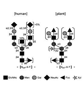
Figure 7. N-glycosylation patterns produced by human cells (left) and tobacco
plants or cells
(right).
Figure 8. (Left panel) 33 uL of Nicotiana tabacum plant cells extracts after 3
days post
cocultivation were analyzed by westernblotting using anti human IgG Fc
antibody.
Agrobacterium nunefaciens (A. t.) LBA4404 harboring binary plasmids with p19
silencing
suppressor gene were used to express the fusion protein. (Right panel) 40 ug
of Nicotiana
benthamiana leaf extracts after 4 days post agroinfiltration were analyzed by
western blotting
using anti human IgG Fc part antibodies. The 4-20% acrylamide SDS-PAGE were
performed
under non-reducing conditions. Expressed constructs are indicated on top of
the
corresponding lane. TOX2 indicates a toxin from toxin class 2 as disclosed
herein (protein
synthesis inhibitor). L indicates the protein ladder (molecular-weight size
marker). Integral
protein size is indicated by a star.
Figure 9: Structures of binder-toxin formats according to the invention.
9A-C: Toxin is fused to a C-terminus of an antibody chain. A: (scFv-FC)-
(cleavage site)-
toxin/protoxin; B: HC plus LC-(cleavage site)-toxin/protoxin, and C: LC plus
HC-(cleavage
site)-toxin/protoxin.
9D-G Toxin is fused to an N-terminus of an antibody chain.
8
CA 03128793 2021-08-03
WO 2020/169620 PCT/EP2020/054263
scFv-FC is a specific antibody format as discussed herein. LC is the light
chain of an IgG
antibody. HC is the heavy chain of an IgG antibody. CS means cleavage site,
and Tox means
toxin/protoxin. The bars between the different chains symbolize disulfide
bonds.
Figure 10: Results of peptide glycoform analysis. To demonstrate that peptides
taken from
the antibodies disclosed herein and comprising an N-glycosylation site have
characteristic
glycoforms which separate them from proteins produced in mammals, different
strains of
Nicotiana benthamiana were used, some of which being glycoengineered by RNA
interference (RNAi) technology (Material provided by NOMAD Bioscience GmbH,
Munich)
, to obtain a targeted down-regulation of the endogenous 13-1,2-
xylosyltransferase (XylT) and
a 1,3-fucosyltransferase (FucT), Fig, 10 shows the MS spectra of two such
peptides as
produced in the different strains.
Figure 11: Results of cleavage assay. scFv-Fc-FCS-GB (FCS = Furin cleavage
site, GB =
Granzyme B) have been expressed from Nicotiana benthamiana. Protein have been
purified
by protein A chromatography from leaf extracts after 4 days post
agroinfiltration. Purified
material have been exposed to recombinant furin. The 4-20% acrylamide SDS-PAGE
was
performed under reducing conditions. L indicate the protein ladder (molecular-
weight size
marker). Resulting free GB protein after cleavage is indicated by a star.
Recombinant protein
April was used as cleavage control. Cleavage related band of April are
indicated by a A.
Figure 12: Purified protein analysis by SDS PAGE Coomassie blue. Several
constructions
have been expressed from Nicotiana benthamiana. Protein have been purified by
protein A
chromatography from leaf extracts after 4 days post agroinfiltration.7 1 of
purified protein
have been added to 7 ill of loading buffer (2x). Then 12 p.1 have been loaded
into respective
well. The 4-20% acrylamide SDS-PAGE was performed under non-reducing
conditions L
indicates the protein ladder (molecular-weight size marker). Integral protein
size is indicated
by a star.
Figure 13: Purified protein analysis by SDS PAGE Coomassie blue. Several
constructions
have been expressed from Nicotiana benthamiana. Protein have been purified by
protein A
chromatography from leaf extracts after 4 days post agroinfiltration. 7 1 of
purified protein
have been added to 7 al of loading buffer (2x). Then 12 I have been loaded
into respective
well. The 4-20% acrylamide SDS-PAGE was performed under reducing conditions. L
9
indicates the protein ladder (molecular-weight size marker). Integral protein
size is indicated
by a star.
Detailed Description of the Invention
Before the invention is described in detail, it is to be understood that this
invention is not
limited to the particular component parts of the devices described or process
steps of the
methods described as such devices and methods may vary. It is also to be
understood that the
terminology used herein is for purposes of describing particular embodiments
only, and is not
intended to be limiting. It must be noted that, as used in the specification
and the appended
claims, the singular forms "a", "an", and "the" include singular and/or plural
referents unless
the context clearly dictates otherwise. It is moreover to be understood that,
in case parameter
ranges are given which are delimited by numeric values, the ranges are deemed
to include
these limitation values.
It is further to be understood that embodiments disclosed herein are not meant
to be
understood as individual embodiments which would not relate to one another.
Features
discussed with one embodiment are meant to be disclosed also in connection
with other
embodiments shown herein. If, in one case, a specific feature is not disclosed
with one
embodiment, but with another, the skilled person would understand that does
not necessarily
mean that said feature is not meant to be disclosed with said other
embodiment. The skilled
person would understand that it is the gist of this application to disclose
said feature also for
the other embodiment, but that just for purposes of clarity and to keep the
specification in a
manageable volume this has not been done.
According to a first aspect of the invention, a method of producing a binder-
toxin fusion
protein is provided,
a) one protein binder selected from the group consisting of
6851807
Date Recue/Date Received 2021-08-24
CA 03128793 2021-08-03
WO 2020/169620 PCT/EP2020/054263
= an antibody
= an antibody fragment or derivative retaining target binding capacity, or
= an antibody mimetic,
b) optionally, a peptide linker, and
c) at least one protein toxin or protein protoxin
said method comprising the steps of:
(i) contacting a plant cell or a whole plant with a nucleic acid construct
comprising in
operational linkage at least the following
A) at least one polynucleotide encoding for the protein binder, or a target
binding
chain or domain thereof, and either
WI) a polynucleotide encoding for a cleavable peptide linker and a
polynucleotide encoding for a protein toxin, or
B2) a polynucleotide encoding for a protein protoxin, which protoxin
comprises a cleavable domain for activation thereof,
(ii) allowing the construct to integrate into the nucleus of the plant cell,
or of one or more
cells of the whole plant, and
(iii) expressing the fusion protein encoded by the nucleic acid construct.
The inventors found, surprisingly, that with such method, recombinant binder-
toxin fusion
proteins can be produced at large scale and with high productivity.
As used herein, the term "plant" (including the cells derived therefrom)
relates to algae
(including Chlorophyta and Charophyta/Streptophyta, as well as
Mesostigmatophyceae,
Chlorokybophyceae and Spirotaenia), and also to land plants (Embryophytes),
including
Gymnospertms and Angiosperms, including Mono- and Dicotyledonae.
In one embodiment, the plant or plant cell with which the nucleic acid
construct is contacted
is not a chloroplast, or not a chloroplast of an algae, in particular not the
chloroplast of
Chlamydomonas reinhardtii. In another embodiment, structure in the plant or
plant cell with
11
CA 03128793 2021-08-03
WO 2020/169620 PCT/EP2020/054263
which the nucleic acid construct is contacted is not a chloroplast, or not a
chloroplast of an
algae, in particular not the chloroplast of Chlamydomonas reinhardtii.
As used herein, the term "protein toxin" or "protein protoxin" is meant to
encompass
cytotoxic and/or cytostatic proteins, or the pro-variants thereof
As used herein the term "cytostatic protein" refers to a protein that can
inhibit cell
proliferation or cell division without necessarily killing the cell. Suitably,
the cytostatic agent
inhibits the proliferation of tumor cells.
As used herein, the term "cytotoxic protein" refers to a protein that is
harmful to cells and
ultimately causes cell death. In some embodiments, the cytotoxic protein harms
rapidly
dividing cells such as tumor cells and causes tumor cell death, especially
tumor cell death
while not causing damage to or causing less damage to non-tumor cells.
The terms "protein toxin" or "protein protoxin", refer without limitation to
toxins that are, by
their chemical nature, proteins (i.e., peptides having a length of > 50 amino
acid residues) or
polypeptides (i.e., peptides having a length of > 10 - < 50 amino acid
residues). A protoxin,
in the meaning of the present invention, is a precursor of a toxin, also
called a latent toxin,
which needs to be activated, e.g., by cleaving off an inhibitory amino acid
sequence, or by
undergoing a conformational change. The terms "protoxin" and "protein
protoxin" are used
interchangeably here and mean the same subject matter.
Plant-based recombinant protein expression has been developed for 3 decades.
Today, several
therapeutic proteins, such as monoclonal antibodies (mAbs), produced in plants
or plant cells
(such as tobacco or tobacco cells) have been tested in clinical trials and are
commercialized
or are close to be (Yao et al. (2015)). Plant-based recombinant protein
expression, including
antibodies. can also be carried out in algae. (Hempel et al., 2011)
Plants have a couple of advantages over prokaryotic and eukaryotic cells
systems regarding
their low cost of production, inherent product safety, easy upscaling, their
ability to fold and
assemble complex proteins and to carry out complex post-translational
modifications.
Besides, plant suspension cells cultures offer increased reproducibility and
safety during
production (no known microbes, insects or mammalian pathogens), and meets
current good
12
CA 03128793 2021-08-03
WO 2020/169620 PCT/EP2020/054263
manufacturing practice production requirements. Plant suspension cells
cultures only require
simple defined nutrients to grow, offering much less expensive operational
costs than
mammalian or microbial systems.
The term "fusion protein" as used herein refers to a protein that has a
peptide component
operably linked to at least one additional component and that differs from a
natural protein in
the composition and/or organization of its domains.
The term "operably linked" as used herein, when referring to two or more
polynucleotides,
means a situation when the different polynucleotides are placed in a
functional relationship
with one another. For instance, a promoter is operably linked to a coding
sequence if the
promoter effects the transcription of the coding sequence. Likewise, the
coding sequence of a
signal peptide is operably linked to the coding sequence of a polypeptide if
the signal peptide
effects the extracellular secretion of that polypeptide. According to one
embodiment of the
present invention, when the respective polynucleotides encode different
peptides, "operably
linked" means that the respective polynucleotides are contiguous and, where
necessary to join
two protein coding regions, the open reading frames are aligned.
The twit "cleavable peptide linker" as used herein refers to an internal amino
acid sequence
within the fusion protein which contains residues linking the binder moiety
and toxin protein
so as to render the toxin protein incapable of exerting its toxic effect
outside the target cell or
limiting its ability of toxin protein to inhibit cell growth (cytostasis) or
to cause cell death
(cytotoxicity). In such way, the protein toxin is maintained inactive as long
as it is in the
plasma, until it reaches the target cell, where the cytotoxic payload will be
selectively
released and/or activated (Grawunder & Stein, 2017). Inside the target cell,
the cleavable
linker sequence is cleaved and the toxin protein becomes active or toxic. The
fusion protein
of the invention is composed of a cell-specific binder moiety and an protein
toxin moiety
linked by a a specific amino acid residue or amino acid sequence that has
cleavage
recognition site for specific proteases, particularly but not limited to
cancer specific protease,
and/or are cleavable under specifics conditions such as, without limitation,
acid and/or
reducing conditions. Sequences encoding cleavage recognition sites for
specific protease may
be identified among known ubiquitous human protease and/or by testing the
expression of
cancer associate protease. Also the linker sequence should not interfere with
the role of the
binder moiety in cell binding and internalization into lysosomes.
13
CA 03128793 2021-08-03
WO 2020/169620 PCT/EP2020/054263
The term "cleavable domain" of a protoxin relates to a sequence that, once
cleaved by
hydrolysis or enzymatic cleavage, activates the toxin part of the protoxin.
Many protoxins
have an amino acid domain that is specifically cleaved by an enzyme, or by pH
dependent
hydrolysis (e.g. after endocytosis in the endosomes), so as to release the
active toxin part into
the cytosol. Such cleavable domains double act as "naturally occurring"
cleavable peptide
linkers (or "intrinsic cleavage sites"), contrary to the cleavable peptide
linkers which have to
be used in case the toxin does not comprise a cleavable domain for activation,
e.g., because it
does not come as a protoxin.
The term "selective activation/release" refers to the activation of ability of
protoxin or protein
toxin to inhibit cell growth (cytostasis) or to cause cell death
(cytotoxicity) under particular
conditions This selective activation refers to an unnatural or not naturally
found modifiable
activation moiety of a protein toxin that, upon modification, converts
inactive toxin (protein
protoxin) into an active toxin or a native cleavable linker of a protoxin.
When the selectively
modifiable activation moiety is a component of the toxin fusion protein,
modification of the
modifiable activation moiety can result directly in the protoxin becoming
toxic to the target
cell, or can result in the protoxin assuming a form that is natively
activatable to become toxic
to the target cell. Natively activatable protoxins comprise, for example,
modification of the
modifiable activation moiety such that it is sensitive to endogenous
components of the target
cell, or the environment surrounding the target cells. (e.g., a target cell
specific protease or a
ubiquitous protease), and/or specific conditions such as, without limitation,
acid and/or
reducing conditions. Natively active toxin can be modified to be inactive
(protoxin) into the
toxin fusion using natural or unnatural and not naturally found modifiable
activation moiety
such that it is sensitive to endogenous components of the target cell, or the
environment
surrounding the target cells and/or specific conditions such as, without
limitation, acid and/or
reducing conditions. Modifiable activation moiety is defined as cleavable
linker in the present
invention.
Hence, while a cleavable linker provides clear advantages over a stable linker
as regards the
activity profile, the use thereof complicates the production of respective
binding protein-toxin
conjugates in mammalian, insect and yeast cells, because cleavage of the
linker leads to self-
intoxication of the production system. This, however, does not apply to plant-
based
production systems, because
14
CA 03128793 2021-08-03
WO 2020/169620 PCT/EP2020/054263
(i) they don't cleave the linker (due to lack of respective proteases or
reducing/hydrolyzing conditions) and/or
(ii) the respective protein toxin which is toxic to mammals or mammalian cells
is not
toxic to plants or plant cells.
As used herein, the term "monoclonal antibody", shall refer to an antibody
composition
having a homogenous antibody population, i.e., a homogeneous population
consisting of a
whole immunoglobulin, or an antigen binding fragment or derivative thereof
Particularly
preferred, such antibody is selected from the group consisting of IgG, IgD,
IgE, IgA and/or
IgM, or a fragment or derivative thereof
As used herein, the term "fragment" shall refer to fragments of such antibody
retaining target
binding capacities, e.g.
= a CDR (complementarity determining region),
= a hypervariable region,
= a variable domain (Fv),
= an IgG heavy chain (consisting of VH, CH1, hinge, CH2 and CH3 regions),
= an IgG light chain (consisting of VL and CL regions), and/or
= a Fab and/or F(ab)2.
As used herein, the term "derivative" shall refer to protein constructs being
structurally
different from, but still having some structural relationship to the common
antibody concept,
e.g., scFv, scFv-FC, Fab and/or F(ab)2, as well as bi-, tri- or higher
specific antibody
constructs or monovalent antibodies, and further retaining target binding
capacities. All these
items are explained below.
Other antibody derivatives known to the skilled person are Diabodies, Camelid
Antibodies,
Nanobodies, Domain Antibodies, bivalent homodimers with two chains consisting
of scFvs,
IgAs (two IgG structures joined by a J chain and a secretory component), shark
antibodies,
antibodies consisting of new world primate framework plus non-new world
primate CDR,
dimerised constructs comprising CH3+VL+VH, and antibody conjugates (e.g.
antibody or
fragments or derivatives linked to a toxin, a cytokine, a radioisotope or a
label). These types
are well described in literature and can be used by the skilled person on the
basis of the
present disclosure, with adding further inventive activity.
Methods for the production of a hybridoma cell have been previously described
(see Kohler
and Milstein 1975, incorporated herein by reference). Essentially, e.g., a
mouse is immunized
with a human soluble Guanylyl Cyclase (sGC) protein, followed by B-cell
isolation from said
mouse and fusion of the isolated B-cell with a myeloma cell.
Methods for the production and/or selection of chimeric or humanized mAbs are
known in
the art. Essentially, e.g., the protein sequences from the murine anti sGC
antibody which are
not involved in target binding are replaced by corresponding human sequences.
For example,
US6331415 by Genentech describes the production of chimeric antibodies, while
US6548640
by Medical Research Council describes CDR grafting techniques and US5859205 by
Celltech describes the production of humanised antibodies.
Methods for the production and/or selection of fully human mAbs are known in
the art. These
can involve the use of a transgenic animal which is immunized with human sGC,
or the use
of a suitable display technique, like yeast display, phage display, B-cell
display or ribosome
display, where antibodies from a library are screened against human sGC in a
stationary
phase.
In vitro antibody libraries are, among others, disclosed in US6300064 by
MorphoSys and
US6248516 by MRC/Scripps/Stratagene. Phage Display techniques are for example
disclosed in US5223409 by Dyax. Transgenic mammal platforms are for example
described
in EP1480515A2 by TaconicArtemis.
IgG, scFv, scFv-FC, Fab and/or F(ab)2 are antibody formats well known to the
skilled person.
Related enabling techniques are available from the respective textbooks.
As used herein, the term 'Tab" relates to an IgG fragment comprising the
antigen binding
region, said fragment being composed of one constant and one variable domain
from each
heavy and light chain of the antibody.
16
6851807
Date Recue/Date Received 2021-08-24
As used herein, the term -F(ab)2" relates to an IgG fragment consisting of two
Fab fragments
connected to one another by one or more disulfide bonds.
As used herein, the term -scFv" relates to a single-chain variable fragment
being a fusion of
the variable regions of the heavy and light chains of immunoglobulins, linked
together with a
short linker, usually serine (S) or glycine (G). This chimeric molecule
retains the specificity
of the original immunoglobulin, despite removal of the constant regions and
the introduction
of a linker peptide.
As used herein, the term -scFv-FC" relates to a specific antibody format. This
format is
particularly stable and can be expressed with high yield in plant cells and
plants. scFv-FC
constructs are for example disclosed in Bujak et al (2014). scFv-Fc constructs
are dimeric
constructs comprising two chains associated to one another for example by one
or more
disulfide bonds, wherein each of which consist of a structure as follows (in N-
>C direction):
VL-linker-VI-Linker-FC, or
VH-linker-VL-Linker-FC
with VL being the variable domain of the light chain of an antibody, VI-I
being the variable
domain of the heavy chain of an antibody, and FC being the constant domain of
an antibody.
The use of a full-length IgG-shaped antibody or a scFv-Fc binding domain
confers a longer
half-life to the conjugate. Moreover, the Fc part of the antibody might be of
utmost
importance when CDC (Complement dependent cytotoxicity) or ADCC (Antibody
dependent
cellular cytotoxicity) activation is required.
Modified antibody formats are for example bi- or trispecific antibody
constructs, antibody-
based fusion proteins, immunoconjugates and the like. These types are well
described in
literature and can be used by the skilled person on the basis of the present
disclosure, with
adding further inventive activity. Furthermore, also monovalent antibodies
have been
previously described in US 2004/0033561 Al (referred to therein as monobodies)
or
W02007048037.
17
6851807
Date Recue/Date Received 2021-08-24
Antibody mimetics are organic compounds ¨ in most cases recombinant proteins
or peptides -
that, like antibodies, can specifically bind antigens, but that are not
structurally related to
antibodies. Common advantages over antibodies are better solubility, tissue
penetration,
stability towards heat and enzymes, and comparatively low production costs.
Antibody
mimetics are being developed as therapeutic and diagnostic agents, and
encompass, inter
alia, Affibody molecules, Affilins, Ubiquitins, Affimers, Affitins,
Alphabodies, Anticalins,
Avimers, DARPins, Fynomers, Kunitz domain peptides, Monobodies and nanoCLAMPs.
Antibody mimetics are discussed in great detail, inter alia, in Gebauer and
Skerra (2009).
Generally, the protein binder may consist of a single chain. This is the case,
e.g., where the
protein binder is a scFv antibody, or a scFv-FC. In this case, the entire
protein binder may be
encoded on a single polynucleotide.
In another embodiment the protein binder may comprise two or more chains, like
e.g. in a full
size IgG or in a F(ab)2 fragment. In such case it may be provided that the
nucleic acid
construct may comprise two or more polynucleotides encoding for the different
chains or
domains for the protein binder.
In another embodiment where the protein binder comprises two or more chains it
may be
provided that two nucleic acid constructs are provided, the first comprising
the three
polynucleotides encoding for the first chain of the protein binder, the linker
and the toxin,
while the second comprises the polynucleotide encoding for the second chain of
the protein
binder.
According to one embodiment of the invention, the method further comprises the
step of (iv)
recovering and/or purifying the fusion protein expressed in step (iii)
According to one other embodiment of the invention, the plant or plant cell is
from the genus
Nicotiana.
18
6851807
Date Recue/Date Received 2021-08-24
In one embodiment, when using a plant, the expression of the fusion protein is
a transient
expression. In another embodiment, when using a plant cell, the expression of
the fusion
protein is either a transient or stable expression.
As used herein, the term transient expression" relates to the temporary
expression of genes
that are expressed for a short time after a nucleic acid, most frequently
plasmid DNA
encoding an expression cassette, has been introduced into the host cells or
plants.
As used herein, the term -stable expression" relates to expression of genes
that are expressed
continuously in time after a nucleic acid, most frequently plasmid DNA
encoding an
expression cassette, has been introduced into the host cells' genome (nuclear
or plastid
integration). In stably transfected cells, the foreign gene becomes part of
the genome and is
therefore replicated.
Both transient and stable expression could be induced by an -inducible
promoter". These
promoters selectively express an operably linked DNA sequence following to the
presence of
an endogenous or exogenous stimulus or in response to chemical, environmental,
hormonal,
and/or developmental signals. These regulatory element are, without
limitation, sensitive to
ethanol, heat, light, stress, jasmone, salicylic acid, phytohormones, salt,
flooding or drought,
as reviewed by Abdel-Ghany et al (2015) and discussed in US 10344290 B2.
Inducible
promotors including, but not limited to, synthetic components discuss in Ali
et al (2019).
The genus Nicotiana encompasses tobacco plants. Tobacco plants or plant cells
have already
been tested to produce recombinant immunotherapeutic binder-toxin fusion
proteins
composed of a small sFy fragment linked to a protein toxin with a stable
linker (Francisco et
al. (1997), and US6140075A.
Another example of protein fusion is the transient production in Nicotiana
benthamiana of
the human immunocytokine IL2 recombinantly fused to a scFv-Fc via a non-
cleavable linker
(Marusic et al. (2016). However, the production of an antibody linked to a
highly potent
protein toxin via a cleavable linker has never been disclosed in a plant
system.
19
6851807
Date Recue/Date Received 2021-08-24
CA 03128793 2021-08-03
WO 2020/169620 PCT/EP2020/054263
According to one further embodiment of the invention, the plant cell is at
least one selected
from the group consisting of:
= Nicotiana tabacum cv. BY2,
= Nicotiana tabacum NT-1,
= Arabidopsis thahana,
= Daucus carota, and'or
= Oyrza sativa.
Nicotiana tabacum cv. BY2 aka Tobacco BY-2 cells and cv. Nicotiana tabacum 1
(NT-1, a
sibling of BY-2) are nongreen, fast growing plant cells which can multiply
their numbers up
to 100-fold within one week in adequate culture medium and good culture
conditions. This
cultivar of tobacco is kept as a cell culture and more specifically as cell
suspension culture (a
specialized population of cells growing in liquid medium, they are raised by
scientists in
order to study a specific biological property of a plant cell). In cell
suspension cultures, each
of the cells is floating independently or at most only in short chains in a
culture medium.
Each of the cells has similar properties to the others.
The model plant system is comparable to HeLa cells for human research. Because
the
organism is relatively simple and predictable it makes the study of biological
processes
easier, and can be an intermediate step towards understanding more complex
organisms.
They are used by plant physiologists and molecular biologists as a model
organism, and also
used as model systems for higher plants because of their relatively high
homogeneity and
high growth rate, featuring still general behaviour of plant cell. The
diversity of cell types
within any part of a naturally grown plant (in vivo) makes it very difficult
to investigate and
understand some general biochemical phenomena of living plant cells. The
transport of a
solute in or out of the cell, for example, is difficult to study because the
specialized cells in a
multicellular organism behave differently. Cell suspension cultures such as
tobacco BY-2
provide good model systems for these studies at the level of a single cell and
its
compartments because tobacco BY-2 cells behave very similarly to one another.
The
influence of neighboring cells behavior is in the suspension is not as
important as it would be
in an intact plant. As a result any changes observed after a stimulus is
applied can be
statistically correlated and it could be decided if these changes are
reactions to the stimulus or
just merely coincidental. BY-2 and NT-1 cells are relatively well understood
and often used
in research, including the expression of heterologous proteins, in particular
antibodies
(Hellwig et al (2004). Such methods are disclosed in Halckinen et al. (2018),
the content of
which is incorporated herein by reference.
Torres (1989) discusses methods to establish Carrot Cell Suspension Cultures
(Daucus
carota). Shaaltiel et al (2007) discuss the production of enzymes using a
carrot cell based
expression system. Daucus carota and Oryza saliva are also discussed as
suitable plant-cell
based expressions systems in Santos et al (2016). The Production of
recombinant proteins in
Nicotiana tabacum, Arabidopsis thaliana, Oryza saliva is disclosed in Plasson
et al (2009).
Generally, the present invention can be practiced with any plant variety for
which cells of
the plant can be transformed with an DNA construct suitable for expression of
a foreign
polypeptide and cultured under standard plant cell culture conditions. Plant
cells suspension
or plant tissues culture is preferred, although callus culture or other
conventional plant
cell culture methods may be used.
According to one other embodiment of the invention, the plant is Nicotiana
benthamiana.
The production of antibodies in Nicotiana plants is for example disclosed in
Daniell et al.
(2001).
Other plants or plant cells that can be used in the context of the present
invention include, but
are not limited to, lettuce (Lactuca spp.), spinach (Spinacia oleracea), and
Arabidopsis
(Arabidopsis spp).
According to one further embodiment of the invention, step (i) of contacting a
plant cell or a
whole plant with a nucleic acid construct involves the use of a an -expression
vector" alone
or mediated by Agrobacterium tumefaciens, a vector derived therefrom, or
particle
bombardement/biolistic.
As used herein, the term "expression vector" relates to a vectors such as
plasmids, viruses,
bacteriophage, integrable DNA fragments, and other vehicles, which enable to
introduce
21
6851807
Date Recue/Date Received 2021-08-24
nucleic acid construct into a plant or plant cell. Expression vectors useful
in the present
methods are well known in the art. The term -vector" means a nucleic acid
molecule, such as
a plasmid, comprising regulatory elements and a site for introducing nucleic
acid construct.
In some embodiment, vectors can be based on, but not limited to, full viral
system,
deconstructed virus, or non-viral element. The Geneware (platform described in
U.S. Pat. No.
7,939,318) and MagnIcon, respectively, are the best known full viral system
and
deconstructed virus system, described in Altman et al (2011).
Usually the used viral vectors could be classified both into RNA or DNA based
vectors. As
example, RNA vectors could include, without limitation, Tobamovirus (e.g.
Tobacco Mosaic
Virus (TMV)), cowpea mosaic virus (CMV), potexvirus (e.g. potato virus X
(PVX)) or
Tobravirus (e.g.tobacco rattle virus (TRV)). DNA vectors have often associated
to
Caulimovirus and gemini-virus who include mastrevirus, curtovirus,
topocuvirus, and
begomovirus. As example Bean Yellow dwarf (BeYDV) and tobacco yellow dwarf
virus
both from mastrevirus, have been widely used.
As an alternative, vectors can be based on non-viral elements and include for
example 5' and
3' untranslated region that are synthetic (Peyret et al., 2019) or coming from
other proteins
(Diamos, et al., 2016)..
It is important to note here that vectors could be replicative or not
replicative. Also the
replicability of the vector could be induced once activation of regulatory
element as for In
Plant Activation (Impact) System describe by Dugdale et al. (2013).
Agrobacterium transfection is for example described in Mayo et al (2006). TMV
transfection
is for example described in Hussain Shah et al (2013). Particle
bombardment/biolistic are for
example disclosed in Kikkert et al (2005).
22
6851807
Date Recue/Date Received 2021-08-24
CA 03128793 2021-08-03
WO 2020/169620 PCT/EP2020/054263
It is important to stress that the advantages of the method according to the
invention are not
restricted to embodiments where tobacco plants or cell lines are used. For
example, the fact
that the conjugates can be expressed in plants because the cleavable liker or
cleavable domain
is left intact by the plant or plant cells is a universal principle for plants
that allows the
expression of the conjugates without harming the plants or plant cells. Hence,
the concept of
the present invention is applicable, and enabled in all plants or plant cells
that can be used for
recombinant expression.
Nucleic acids can be expressed in plants or plant cells under the control of a
suitable operably
linked promoter that is capable of expression in a given plant or plant cell.
Any conventional
method can be employed for plant cell transformation, culture, and
regeneration, including,
but not limited to, those described in the Examples below.
For the transformation of the nuclear genome, conventional techniques may be
used. All
known means for transiently or stably introducing foreign DNA into plant cells
may be used,
for example Ti plasmids, Ri plasmids, plant virus vectors, electroporation,
protoplast fusion,
particle gun bombardment, or penetration of DNA into cells such as pollen,
microspore, seed
and immature embryo. Viral vectors such as the Gemini viruses or the satellite
viruses may
also be used as introducing means. Agrobacterium tumefaciens and rhizogenes
constitute the
preferred means to introduce expression vectors. In this case, the sequence of
the invention is
introduced into an appropriate vector with all the necessary regulatory
sequences such as
promoters, terminators and the like, as well as, in the case of stable plant
generation, any
sequence necessary for selecting the transformants which have integrated the
heterologous
sequences. In particular case of induced expression, the sequence of the
invention is
introduced into vector containing inducible promotor and all necessary
regulatory sequences.
The introduction of a nucleic acid molecule(s) into the plant cell can be
carried out in a
transient or stable manner either by transformation of the nuclear genome, or
by
transformation of the chloroplast genome of the plant cell, or by
transformation of the
mitochondrial genome.
The transformation of the nuclear genome of the plant cell is often carried
out using the
targeting signals mentioned above and which determine the cellular compartment
where the
expression and/or accumulation of the protein will occur.
23
The targeting sequences can, besides the peptide, also comprise an endoplasmic
retention
signal, consisting of the KDEL, SEKDEL or HEKDEL peptides. These signals
normally exist
at the C-terminal end of the protein and remain on the mature protein.
According to one embodiment of the invention, the peptide linker or the
cleavable domain in
the protoxin is specifically or non-specifically cleavable by an enzyme
expressed by a
mammalian cell, or an enzyme that is produced by a mammalian host.
Examples of such enzymes and their cleavage sites are shown in the following
table (see also
Choi et al (2012) Reference is made, in this table, to the -Merops" database
for more
enabling information as regards the respective enzymes.
https ://www.ebi.ac.uk/merops/index.shtml.
24
6851807
Date Recue/Date Received 2021-08-24
CA 03128793 2021-08-03
WO 2020/169620
PCT/EP2020/054263
Class Enzyme class example cleavage sequence (one letter reference
code)
general motif (examples only)
X can be any naturally
proteinogenic amino acid
"Linker Proprotein Furin RXR/KR,5/A/G/Nxxx
merops S08.071
class 1" convertase
Endosomal subtilisin/kexi
and/or n family
Lysosomal Cathepsins Cathepsin B xxF/xV/R/G/L/S/A,
A/F/Lxxx merops C01.060
Cleavage
site Cathepsin E xxxL/E0/xxx merops A01.010
Cathepsin D xxxL/Epcxxx merops A01.009
Cathepsin L xxL/V/F/IR/Ic S/A/Gxxx merops C01.032
Cathepsin K xK/R/GF/L/I/V/Pxvocxx merops C01.036
Cathepsin C xSxE/SoocxG/R merops C01.070
Granzyme Granzyme B V/IxxD,ocxxx merops S01.010
"Linker Caspases Caspase 3 DxxD,A/G/S/Txxx Or
merops C14.003
class 2" xxxx,GGFV
Cytosolic Caspase 8 D/Lxx%G/S/Axxx merops C14.009
cleavage
site Kallikereins hK 1 xxF/IR/Y,R/SxGx
merops S01.160
(hK) hK 2 G/K/Axx1:14,xxxG/S/T merops S01.161
hK3 S/IS/QxY/Q/RSSxx
hK10 No determined
"Linker Matrix MMP2 x13/Axx,L/Ixxx merops M10.003
class 3" metallo
M M P1 xP/Axx,1,111xx merops M10.001
Cell proteases
surface M M P3 xxxR/N/G4/Kxx merops S01.072
cleavage
site MMP7 xPA/G/Lx,Axxx merops M10.005
MMP8 GP/A/Sxx,I,Lxxx merops M10.002
M M P9 GP/Axx,Lxxx merops M10.004
P2 is preferably a L
P1 is preferably a G
M M P12 GP/A/GL/AJGxj,Lxxx merops M10.009
M M P14 xPxx,Lxxx merops M10.014
Matriptase Matriptase 2 xxxRJ/G/Rxxx merops S01.308
Matriptase 1 xxxR,K/V/A/RVxx merops S01.302
tissue-type Urokinase type xSG/SR/K,{,xR/Vxx merops S01.231
plasminogen plasminogen
activator activator (uPA)
The cleavage site is described from the cleavage site point (represented by d.
The letter x
refer to all amino acids. When there are several preferential amino acid,
there are separated
by a slash (/).
Such enzyme is preferably a protease. In one embodiment, said peptide linker
is not cleavable
by a plant enzyme.
Furin is an enzyme which belongs to the subtilisin-like proprotein convertase
family, and
cleaves proteins C-terminally of the canonic basic amino acid sequence motif
Arg-X-
Arg/Lys-Arg (RX(R/K)R), wherein X can be any naturally proteinogenic amino
acid. Said
motif is called a furin cleavage site herein. Preferably, the sequence thereof
is
HRRRKRSLDTS.
Cathepsins are proteases found in all animals as well as other organisms. Most
of the
members become activated at the low pH found in lysosomes. Cathepsin B is
capable of
cleaving a peptide sequence which comprises the dipeptide motif Val-Ala (VA).
Said motif is
called a Cathepsin B cleavage site herein. The skilled artisan finds
sufficient enabling
information on cathepsins and their cleavage sites in Turk el al (2012).
Granzyme B is a serine protease which cleaves at unique tetrapeptide
sequences. More than
580 of such tetrapeptide cleavage sites exist (Wee et al. (2011)). The
tetrapeptide Ile-Glu-
Pro-Asp (IEPD) was identified as the optimal tetrapeptide cleavage sequence in
vitro.
However, emerging data on granzyme B substrates suggest that the in vivo
cleavage
specificities are far more diverse, with numerous substrates possessing
cleavage specificities
extending beyond the tetrapeptide sequence (VanDamme et al. (2009)).
The native Granzyme B proenzyme is activated by cleavage with Cathepsin C
(dipeptidyl
peptidase I), at the cleavage site havening the following sequence: DAGE'IIGG.
In a such
way, the activated Granzyme B enzyme has an N-terminus starting with IIGGHE,
and shown
herein as SEQ ID NO: 1. This is hence the N-truncated sequence of the
activated enzyme that
is used according to one embodiment of the invention.
26
6851807
Date Recue/Date Received 2021-08-24
Caspases (cysteine-aspartic proteases, cysteine aspartases or cysteine-
dependent aspartate-
directed proteases) are a family of protease enzymes playing essential roles
in programmed
cell death. Over 1500 caspase substrates have been discovered in the human
proteome. The
general cleavage motif is DXXD-A/G/S/T, wherein X can be any naturally
proteinogenic
amino acid. The skilled artisan finds sufficient enabling information on
caspases and their
cleavage sites in Kumar el al (2014).
Matrix metalloproteinases (MMPs), also known as matrixins, are calcium-
dependent zinc-
containing endopeptidases; other family members are adamalysins, serralysins,
and astacins.
Collectively, these enzymes are capable of degrading all kinds of
extracellular matrix
proteins, but also can process a number of bioactive molecules. The skilled
artisan finds
sufficient enabling information on Matrix Metallo Proteases and their cleavage
sites in
Eckard el al (2016).
Generally, the skilled artisan is capable, by routine considerations and
literature referral, to
select specific cleavage sites that match with the respective mammalian
enzyme, to control
target specific release of the protein toxin or protoxin. General guidelines
to find these
cleavage sites are e.g. disclosed in Rawlings (2016).
According to one embodiment of the invention, the peptide linker or the
cleavable domain in
the protoxin is not cleavable by an enzyme expressed by a plant cell, or an
enzyme that is
produced by a plant host. The skilled person has a bunch of routine methods at
hand to check
whether this condition is met. See e.g., Wilbers et al (2016).
According to one embodiment of the invention, at least one protein toxin or
protoxin is an
enzyme. Because protein toxins most often act as enzymes, one toxin molecule
can work on
many substrate molecules, thus having a multiplexing toxicity effect on the
cell - contrary to
nonenzymatic toxins, which usually only work stoichiometrically with regard to
the target
structure.
According to one embodiment of the invention, the protein toxin is at least
one of the group
selected from
(1) cell death inducing proteins (-toxin class 1")
27
6851807
Date Recue/Date Received 2021-08-24
(2) protein synthesis inhibitors (-toxin class 2")
(3) membrane perturbating proteins (-toxin class 3")
(4) cell division inhibiting proteins (-toxin class 4")
Cell death inducing proteins consist, without limitation, to protein directly
or indirectly acting
on apoptosis pathway and/or affecting nucleic components. Cell death inducing
protein target
apoptosis mediated-protein, RNA and DNA, in all its formats, according to
proteolytic and/or
nucleolytic activities.
The class of protein synthesis inhibitors comprises proteins preventing proper
operation of
the ribosome. This class is mainly represented without limitation, by the
group of ADP-
ribosylating protein. These proteins catalyzed the ADP-ribosylation of the
mammalian
elongation factor 2, leading to its inactivation and blockage of the ribosome.
Membrane perturbating proteins include, but are not limited to, the group of
pore-forming
protein. This group of protein inserts pore into the plasma membrane allowing
the free
passage of electrolytes and other small molecules to disrupt the membrane
integrity leading
to cell lysis.
Cell division inhibiting protein is a group of protein interrupting the cell
cycle by acting on
element of the cytoskeleton (microtubule, tubulin, actin) and/or on protein
regulating the cell
cycle progression. The inhibition of microtubule dynamic and alteration of
cyclin dependent
protein inducing mitotic arrest and lead to cell removal.
According to one embodiment of the invention, the protein toxin or protoxin is
a de-
immunized variant of a native protein toxin. Recombinant methods to de-
immunize protein
toxins by sequence modification are disclosed, e.g., in Schmohl et al. (2015),
or Grinberg and
Benhar (2017).
According to one embodiment, at least one protein toxin is a mammalian toxin,
preferably
selected from the group of Granzymes, more preferably Granzyme B, or a
fragment thereof
that retains the toxic activity of said protein toxin.
28
6851807
Date Recue/Date Received 2021-08-24
CA 03128793 2021-08-03
WO 2020/169620 PCT/EP2020/054263
Granzymes are serine proteases released by cytoplasmic granules within
cytotoxic T cells and
natural killer (NK) cells. They induce programmed cell death (apoptosis) in
the target cell,
thus eliminating cells that have become cancerous or are infected with viruses
or bacteria.
Granzymes also kill bacteria and inhibit viral replication. In NK cells and T
cells, granzymes
are packaged in cytotoxic granules with perforin. Granzymes can also be
detected in the
rough endoplasmic reticulum, golgi complex, and the trans-golgi reticulum. The
contents of
the cytotoxic granules function to permit entry of the granzymes into the
target cell cytosol.
The granules are released into an immune synapse formed with a target cell,
where perforin
mediates the delivery of the granzymes into endosomes in the target cell, and
finally into the
target cell cytosol. Granzymes are part of the serine esterase family.
Granzyme B has both a cell death inducing effect and also inhibits cell
division (Weidle et al
(2014). Granzyme B cytolytic proteins induce apoptosis after release from the
endosome. In
addition, Granzyme B can cleave further death-substrates such as poly(ADP
ribose)polymerase, DNA-dependent protein kinase, components of the
cytoskeleton and the
nuclear mitotic apparatus as well as proteins involved in stress response and
cellular
homeostasi s.
Granzyme B activates apoptosis by activating caspases (especially caspase-3),
which cleaves
many substrates, including caspase-activated DNase to execute cell death.
Granzyme B also
cleaves the protein Bid, which recruits the proteins Bax and Bak to change the
membrane
permeability of the mitochondria, causing the release of cytochrome c (which
is one of the
parts needed to activate caspase-9 via the apoptosome), Smac/Diablo and
Omi/HtrA2 (which
suppress the inhibitor of apoptosis proteins (IAPs)), among other proteins.
Granzyme B also
cleaves many of the proteins responsible for apoptosis in the absence of
caspase activity. The
other granzymes activate cell death by caspase-dependent and caspase-
independent
mechanisms.
In addition to killing their target cells, granzymes can target and kill
intracellular pathogens.
Granzyme B cleaves viral proteins to inhibit viral activation and replication.
The granzymes
bind directly to the nucleic acids DNA and RNA; this enhances their cleavage
of nucleic acid
binding proteins.
29
In one embodiment, said protein toxin or protoxin is not toxic to plants or
plant cells. The
skilled person has a bunch of routine methods at hand to check whether this
condition is met.
See e.g., Klaine and Lewis (1995) for an overview.
According to another aspect of the invention, a binder-toxin fusion protein
produced with a
method according to the above description is provided. In further embodiments,
such binder-
toxin fusion protein may have all the structural or functional limitations as
set forth in the
above description. This includes, in particular, the format of the protein
binder, the linker or
cleavage sites, and the specific toxin.
According to another aspect of the invention, a binder-toxin fusion protein
comprising at
least:
a) one protein binder selected
b) optionally, a peptide linker, and
c) at least one protein toxin or protein protoxin
is provided, wherein the binder-toxin fusion protein is encoded by a nucleic
acid construct
comprising in operational linkage at least the following
A) at least one polynucleotide encoding for the protein binder, or a target
binding
chain or domain thereof, and either
B1) a polynucleotide encoding for a cleavable peptide linker and a
polynucleotide encoding for a protein toxin, or
B2) a polynucleotide encoding for a protein protoxin, which protoxin
comprises a cleavable domain for activation thereof.
Again, in further embodiments, such binder-toxin fusion protein may have all
the structural
or functional limitations as set forth in the above description. This
includes, in particular, the
format of the protein binder, the linker or cleavage sites, and the specific
toxin.
6851807
Date Recue/Date Received 2021-08-24
According to one embodiment of the invention, said protein comprises at least
one plant-
specific N-glycan. N-glycans are glycans that are linked to the amide group of
asparagine
(Asn) residues in a protein, mostly in an Asn-X-Thr or Asn-X-Ser (NXT or NXS)
motif,
where X is any amino acid except proline. Typical plant-specific N-glycans are
disclosed in
Gomord et al. (2010), and differ significantly from mammalian N-glycan
patterns. See also
Figure 7.
According to one embodiment of the invention, the protein binder binds to
human CD20. In
one embodiment, the protein binder is an antibody, an antibody fragment or
derivative
retaining target binding capacity, or an antibody mimetic.
In one embodiment, the binder comprises at least one of
a) a set comprising the 3 heavy chain CDRs and the 3 light chain CDRs as
comprised in
Rituximab (C2B8)
b) a heavy chain CDR/light chain CDR set of a), with the proviso that at
least one of the
CDRs has up to 3 amino acid substitutions relative to the respective CDR as
specified
in a),while maintaining its capability to bind to human CD20,
c) a heavy chain CDR/light chain CDR combination of a), with the proviso
that at least
one of the CDRs has a sequence identity of >66 % relative to the respective
CDR as
specified in a), while maintaining its capability to bind to human CD20.
wherein the CDRs are embedded in a suitable protein framework so as to be
capable to bind
to human CD20.
Rituximab (also known and C2B8) and the different patents disclosing its
sequence, are
enclosed in Storz U (2014).
The full length sequences of Rituximab are shown herein as SEQ ID NOs 4 (heavy
chain)
and 5 (light chain). The variable domains of Rituximab are for example,
disclosed in claim 1
and Figures 4 and 5 of EP2000149B1.
31
6851807
Date Recue/Date Received 2021-08-24
Sequences that correspond to the CDRs of Rituximab are for example disclosed
in SEQ ID
NOs 9 - 14 shown herein below.
In some embodiments, at least one of the CDRs has a sequence identity of >67,
preferably
>68, more preferably any one of >69, >70, >71, >72, >73, >74, >75, >76, >77,
>78,
>79, >80, >81, >82, >83, >84, >85, >86, >87, >88, >89, >90, >91, >92, >93,
>94,
>95, >96, >97, >98 or most preferably >99 % sequence identity relative to the
respective
CDRs.
In another embodiment, at least one of the CDRs has been modified by affinity
maturation or
other modifications, resulting in a sequence modification compared to the
sequences
disclosed above.
In some embodiments, at least one of the CDRs has up to 2, and preferably 1
amino acid
substitutions relative to the respective CDR as specified in a) or b).
In one embodiment, the binder comprises at least one of
a) the heavy chain/light chain variable domain pair as comprised in
Rituximab (C2B8)
b) the heavy chain/light chain variable domain pair of a), with the proviso
that at least
one of the domains has a sequence identity of >80 % relative to a)
c) the heavy chain/light chain variable domain sequence pair of a), with
the proviso that
at least one of the domains has up to 10 amino acid substitutions relative to
a),
respectively,
while maintaining its capability to bind to human CD20.
In some embodiments, at least one of the domains has a sequence identity of
>81, preferably
>82, more preferably >83, >84, >85, >86, >87, >88, >89, >90, >91, >92, >93,
>94,
>95, >96, >97, >98 or most preferably >99 % relative to the heavy chain/light
chain
variable domain pair of Rituximab (C2B8).
32
6851807
Date Recue/Date Received 2021-08-24
CA 03128793 2021-08-03
WO 2020/169620 PCT/EP2020/054263
In some embodiments, at least one of the domains has up to 9, preferably up to
8, more
preferably up to 7, 6, 5, 4, 3 or 2 and most preferably up to 1 amino acid
substitutions relative
to the heavy chain/light chain variable domain pair of Rituximab (C2B8)
According to some embodiments of the invention, at least one amino acid
substitution in the
single chain diabody is a conservative amino acid substitution.
In one embodiment, the protein binder
= has a target binding affinity of >50 % to human CD20, compared to one of
the
protein binders defined above, and/or
= competes for binding to human CD20 with one of the protein binders
defined
above.
As used herein, the teini "target binding affinity" refers to the affinity of
a binding molecule
according to the invention, to its target, and is expressed numerically using
"KD" values. In
general, a higher KD value corresponds to a weaker binding. In some
embodiments, the
"KD" is measured by a radiolabeled antigen binding assay (MA) or surface
plasmon
resonance (SPR) assays, using, e.g., a BIAcoreTm-2000 or a BIAcoreTm-3000. In
certain
embodiments, an "on-rate" or "rate of association" or "association rate" or
"kon" and an "off-
rate" or "rate of dissociation" or "dissociation rate" or "koff' are also
determined with the
surface plasmon resonance (SPR) technique. In additional embodiments, the
"KD", "kon",
and "koff' are measured using the Octet Systems.
As used herein, the term "competes for binding" is used in reference to one of
the antibodies
defined by the sequences as above, meaning that the actual antibody as an
activity which
binds to the same target, or target epitope or domain or subdomain, as does
said sequence
defined antibody, and is a variant of the latter. The efficiency (e.g.,
kinetics or
thermodynamics) of binding may be the same as or greater than or less than the
efficiency of
the latter. For example, the equilibrium binding constant for binding to the
substrate may be
different for the two antibodies.
33
CA 03128793 2021-08-03
WO 2020/169620 PCT/EP2020/054263
As used herein, the term "maintaining the capability to bind to a given
target" means, for
example, that the respective variant has a target binding affinity of >50 %
compared to that of
the non-modified peptide.
In this context, a "conservative amino acid substitution", as used herein, has
a smaller effect
on antibody function than a non-conservative substitution. Although there are
many ways to
classify amino acids, they are often sorted into six main groups on the basis
of their structure
and the general chemical characteristics of their R groups.
In some embodiments, a "conservative amino acid substitution" is one in which
the amino acid
residue is replaced with an amino acid residue having a similar side chain.
For example,
families of amino acid residues having similar side chains have been defined
in the art. These
families include amino acids with
= basic side chains (e.g., lysine, arginine, histidine),
= acidic side chains (e.g., aspartic acid, glutamic acid),
= uncharged polar side chains (e.g., glycine, asparagine, glutamine,
serine, threonine,
tyrosine, cysteine),
= nonpolar side chains (e.g., alanine, valine, leucine, isoleucine,
proline, phenylalanine,
methionine, tryptophan),
= beta-branched side chains (e.g., threonine, valine, isoleucine) and
= aromatic side chains (e.g., tyrosine, phenylalanine, tryptophan, hi sti
dine).
Other conserved amino acid substitutions can also occur across amino acid side
chain
families, such as when substituting an asparagine for aspartic acid in order
to modify the
charge of a peptide. Conservative changes can further include substitution of
chemically
homologous non-natural amino acids (i.e. a synthetic non-natural hydrophobic
amino acid in
place of leucine, a synthetic non-natural aromatic amino acid in place of
tryptophan).
"Percentage of sequence identity" is determined by comparing two optimally
aligned
sequences over a comparison window, wherein the portion of the polynucleotide
sequence in
the comparison window may comprise additions or deletions (i.e., gaps) as
compared to the
reference sequence (e.g., a polypeptide), which does not comprise additions or
deletions, for
34
CA 03128793 2021-08-03
WO 2020/169620 PCT/EP2020/054263
optimal alignment of the two sequences. The percentage is calculated by
determining the
number of positions at which the identical nucleic acid base or amino acid
residue occurs in
both sequences to yield the number of matched positions, dividing the number
of matched
positions by the total number of positions in the window of comparison and
multiplying the
result by 100 to yield the percentage of sequence identity.
In further embodiments, the binder-toxin fusion protein has one of the
following structures:
= (scFv-FC)-(cleavage site)-toxin/protoxin (dimer)
= tetramer of two HC and two LC-(cleavage site)-toxin/protoxin
= tetramer of two LC and two HC-(cleavage site)-toxin/protoxin
Such structures are for example shown in Figure 9, wherein CS means cleavage
site, and Tox
means toxin/protoxin. HC stands for Heavy chain of an IgG antibody, LC stands
for Light
chain of an IgG antibody. scFv-FC stands for a scFv-FC construct.
In further embodiments, the binder-toxin fusion protein has one of the
following stnictures
(N->C-orientation):
= (scFv-FC)-CS-toxin (dimer) (->toxin linked to C-terminus of FC region)
(see Fig. 9A)
= tetramer of two HC and two LC-CS- toxin (->toxin linked to C-terminus of
LC) (see
Fig. 9B)
= tetramer of two LC and two HC-CS- toxin (->toxin linked to C-terminus of
HC) (see
Fig. 9C)
= toxin-CS-(scFv-FC) (dimer) (->toxin linked to N-terminus of scFv) (see
Fig. 9D)
= tetramer of two HC and two toxin-CS-LC (->toxin linked to N-terminus of
LC) (see
Fig. 9E)
= tetramer of two LC and two toxin-CS-HC (->toxin linked to N-terminus of
HC) (see
Fig. 9F)
= toxin-CS-(-FC-scFv) (dimer) (->toxin linked to N-terminus of FC) (see
Fig. 9G)
Therein, CS stands for "cleavage site", which is in one embodiment a furin
cleavage site.
In one embodiment, the toxin is GranzymeB.
CA 03128793 2021-08-03
WO 2020/169620 PCT/EP2020/054263
In further embodiments, the binder-toxin fusion protein comprises at least one
of one of
According to another embodiment, the binder-toxin fusion protein comprises at
least one of
one of
= the amino acid sequence as set forth in SEQ ID NO: 2 or SEQ ID NO: 17
= two amino acid sequences as set forth in SEQ ID NO. 4 (HC of C2B8) and
two amino
acid sequences as set forth in SEQ ID NO: 6 (LC of C2B8-FCS-Granzyme B)
= two amino acid sequences as set forth in SEQ ID NO: 7 (HC of C2B8-FCS-
Granzyme
B) and two amino acid sequences as set forth in SEQ ID NO: 5 (LC of C2B8)
As regards the first option, the binder-toxin fusion protein may comprise two
of the said
sequences, associated to one another by means of at least one disulfide bond.
As regards the second and third option, the binder-toxin fusion protein may
comprise two
such pairs, conjugated to one on the by a set of disulfide bonds.
According to another aspect of the invention, a pharmaceutical composition is
provided
which comprises at least the fusion protein according to any the above
description, and
optionally one or more pharmaceutically acceptable excipients.
According to another aspect of the invention, a combination is provided which
comprises (i)
the fusion protein according to the above description or the pharmaceutical
composition
according the above description and (ii) one or more therapeutically active
compounds.
According to further embodiments of the invention, the binder-toxin fusion
protein according
to the above description, or the composition according to the above
description, or the
combination according to the above description, is provided for (the
manufacture of a
medicament for) use in the treatment of a human or animal subject
= suffering from,
= being at risk of developing, and/or
= being diagnosed for,
36
CA 03128793 2021-08-03
WO 2020/169620 PCT/EP2020/054263
developing a neoplastic disease, or for the prevention of such condition.
According to another aspect of the invention, a method for treating a human or
animal subject
= suffering from,
= being at risk of developing, and/or
= being diagnosed for
developing a neoplastic disease, or for the prevention of such condition is
provided, said
method comprising the administration of a therapeutically effective amount of
the binder-
toxin fusion protein according to the above description, or the composition
according to the
above description, or the combination according to the above description.
It is in this respect important to stress that N-Glycans produced by plants
are markedly
different from those produced, e.g., in mammals. In particular, N-Glycans
produced by
tobacco plants have
= a Fucose residue conjugated to the proximal N-Acetyl-Glucosamine residue
via a a3
glycosidic link (instead of a6 as in mammals)
= a Xylose residue conjugated to the proximal Mannose residue via a 132
glycosidic link
= two distal N-Acetyl-Glucosamine residues, each of which carry a Fucose
residue via a
a3 glycosidic link, and a Galactose residue via a 03 glycosidic link (instead
of a
neuraminic acid in mammals).
On the other hand, proteins recombinantly expressed in e.g. algae often lack
any kind of
glycosylation. Algae are however capable of expressing IgG shaped antibodies,
or antibody
fragments having a one or more disulfide bridges.
Hence, a binder-toxin conjugate produced by the novel method as discussed
above has also
novel structural features over a binder-toxin conjugate produced in a
different expression
system See for example Figure 7 for an illustration of an N-Glycan produced by
tobacco
plants.
37
CA 03128793 2021-08-03
WO 2020/169620 PCT/EP2020/054263
Examples
While the invention has been illustrated and described in detail in the
drawings and foregoing
description, such illustration and description are to be considered
illustrative or exemplary
and not restrictive; the invention is not limited to the disclosed
embodiments. Other
variations to the disclosed embodiments can be understood and effected by
those skilled in
the art in practicing the claimed invention, from a study of the drawings, the
disclosure, and
the appended claims. In the claims, the word "comprising" does not exclude
other elements
or steps, and the indefinite article "a" or "an" does not exclude a plurality.
The mere fact that
certain measures are recited in mutually different dependent claims does not
indicate that a
combination of these measures cannot be used to advantage. Any reference signs
in the
claims should not be construed as limiting the scope.
All amino acid sequences disclosed herein are shown from N-terminus to C-
terminus; all
nucleic acid sequences disclosed herein are shown 5'->3'.
Materials and Methods
Genetic construct
Full length Rituximab HC and LC sequences have been used to develop mAb based
binder-
toxin fusion proteins. Variable parts sequences of the heavy and light chains
of rituximab
sequences have been assembled in a single chain scFy and fused to a human IgG1
Fc part
sequence. A human furin cleavage sequence was then used to fuse the human
Granzyme B
sequence at the C-terminal part of the LC or the HC of the full-length
rituximab or to the C-
terminal part of the scFv-Fc to obtain HC + LC-FCS-Granzyme B, HC-FCS-Granzyme
B +
LC and scFv-c-FCS-Granzyme B fusion proteins sequences. Another binder-toxin
fusion
protein was realized with scFv-Fc part linked to Granzyme B without cleavage
site to obtain
scFv-Fc- Granzyme B. These sequences were produced by gene synthesis flanked
with XbaI
and IsceI.
The backbone of the pPZP200 binary plasmid was used to construct a new binary
plasmid
pPZP-ATB containing consecutively a nptll kanamycin resistance cassette, one
(scFv-Fc
38
format) or two (mAb format) gene(s) of interest expression cassette(s) and a
GFP expression
cassette. The ORFs coding for rituximab HC and LC, LC-FCS-GB, HC-FCS-GB, scFv-
Fc-
FCS-GB, scFv-Fc-GB were introduced into the appropriate binary plasmids
between the
Cauliflower Mosaic Virus p35S promotor (p35S) and the Agrobacterium Nopaline
Synthase
terminator (tNOS) using XbaI and IsceI. A version of the pPZP-ATB binary
plasmid
including a tomato bushy stunt virus p19 gene expression cassette between the
gene of
interests and the GFP cassettes was also generated.
Transient Expression in Nicotiana benthamiana plant leaves
Nicotiana benthaminana grown under 16h light/8h darkness photocycle, 22 +/- 3
C. 7-8
weeks old plants leaves were transiently transformed by syringe infiltration.
Agrobacterium
tumefaciens LBA4404 (pBBR1MCS-5.virGN54D) or GV3101 (pMP90RK) harboring the
pPZP-ATB-scFv-Fc-F-GB or the pPZP-ATB-scFv-Fc-F-GB-p19 binary plasmids
reaching an
600 nm optical density (0D600) around 0.8-1.0 were collected by centrifugation
at 3500g for
min. Eventually, bacteria were adjusted to an 0D600 of 0.5 in infiltration
buffer (10 mM
MgCl2, 10 mM MES, 100 jiM acetosyringone, pH 5,6) and the mixture was
infiltrated using
a needless syringe. Infiltrated regions were harvested 4 and 6 days post
agroinfiltration.
Entire leaves harvested 4 days post agroinfiltration were used for protein A
purification.
Expression in N. tabacum cells
Nicotiana tabacum plant suspension cells were grown 5 days at 130 rpm, 25 C in
plant
culture media as described by Nagata et al. (1992). Agrobacterium tumefaciens
LBA4404
(pBBR1MCS-5.virGN54D) harboring the pPZP-ATB binary plasmids reaching an 600
nm
optical density (0D600) around 0.8-1.0 were collected by centrifugation at
2000g for 5 min.
Plant cells and bacterial cells were then cocultivated in cocultivation media
for 30 min before
a 2000g 5 min centrifugation. After supernatant removal, cells were plated on
solid
cocultivation media for two days. In the case of transient transformation,
cells were then
collected and washed three times and cultivated in plant cultivation media
containing
Cefotaxim and Carbeniclin before being harvested for further analysis. In the
case of stable
transformation, after the 2 days of solid cocultivation, cells were washed and
plated on plant
media containing selective kanamycin and Cefotaxim
39
6851807
Date Recue/Date Received 2021-08-24
and Carbeniclin antibiotics. Callus were selected 4 weeks later and
subcultured on solid
media or in liquid suspension cultures for subsequent analysis.
Protein analysis: ELISA, SDS-PAGE and Westernblot
Collected leaves tissues (120 mg) were ground in 400 L extraction buffer (250
mM
Sorbitol, 60 mM Tris, Na2EDTA, 0.6% Polyclar AT, 1 mM PMSF, pH8.0 supplemented
with 2 g/mL each of protease inhibitors: Leupeptin, aprotinin, antipain,
pepstatin A,
Chymostatin). Homogenized tissue was centrifugated at 4 C for 40 min at
18200g.
Supernatant was then recovered, froze in liquid nitrogen and stored at -20 C.
Extracted tissue were analyzed by westernblotting. Proteins were boiled for 5
min in reducing
or non-reducing SDS loading buffer (80 mM Tris¨HC1, pH 6.8, 2% SDS, 10%
glycerol,
0.005% bromophenol blue supplemented with 1 mM PMSF and protease inhibitor
cocktail: 2
g/mL each of leupeptin, aprotinin, antipam, chymostatin and pepstatin),
centrifuged for 5
min at 13 000 rpm and separated by SDS-PAGE (4-20% polyacrylamide). For
Western
blotting, proteins were electrotransferred onto a PVDF membrane (Biorad) using
a semi-dry
electrophoretic device (Biorad Trans-Blot Turbo); then, the membrane was
blocked for 1 h
at room temperature with 3% (w/v) non-fat milk powder in TBST buffer (50 mM
Tris¨HC1,
150 mM NaCl, 0.5% Tween0 20, pH 7.5) and then incubated (TBS-Tween 0.1% + 0.5%
non-fat dry milk) for 1 h at room temperature with HRP-conjugated antibodies
against the
anti-human IgG Fc specific region (A0170; Sigma-Aldrich), at a dilution of 1 :
10.000 or
against human Granzyme B primary antibody (EPR20129-217; Abeam) at a dilution
of 1 :
10.000. The anti-Granzyme B antibody was followed by HRP-conjugated anti-
rabbit
antibodies (0545; Sigma), at a dilution of 1 : 10 000. Proteins were detected
by enhanced
chemiluminescence (Amersham Imager 600/GE; GE Healthcare).
Anti CD20 ELISA
For anti-CD20 conjugate specificity analysis, plant extracts were analyzed by
96 well
microplate (Greiner) were coated with 100 L 5 g/mL CD20 (AcroBiosystems) for
2h at
37 C then washed 5 times in washing buffer (TBS Tween0 0,1%). Blocking was
then
performed with 200 L BSA 1% in TBS pH8.0 for 30 min at RT then washed 5 times.
100 L
Anti-CD20 control antibody was loaded to realize a calibration curve between 5
and 0 g/mL
7276389
Date Recue/Date Received 2022-02-11
and 100 L samples were loaded on the same 96 well plates for comparison for
2h at RT then
washed 5 times. 100 L of 1/150.000 diluted detection antibody (goat anti-
human HRPO,
Bethyl) was loaded and incubated lh at RT. Revelation was then performed with
100 L
TMB reaction buffer (Zentech) for 30 min and finally stopper with H3PO4 1M.
Enzymatic
activity was then analyzed by spectrometry at 450 nm.
Further ELISA
For specificity analysis of a conjugate specific for a non-disclosed antigen
(structure
expressed on the surface of human cells, and overexpressed in some cancers,
called antigen X
herein), a purified binder-toxin fusion protein comprising a binder against X
was analysed by
96 well microplate (Greiner). The wells were coated with 50 I of antigen X
(2,5 g/mL) for
lh at 37 C then washed 5 times with 250 L washing buffer (PBS Tween0 0,1%).
Blocking
was then performed with 150 L hydrocasein (3.6%) in PBST for 30 min at RT then
washed
times. 50 L anti antigen control antibody was loaded to realize a calibration
curve between
5 and 0 g/niL and 50 L samples were loaded on the same 96 well plates for
comparison for
lh at RT then washed 5 times. 50 L of 1/200.000 diluted detection antibody
(goat anti-
human HRPO, Bethyl) was loaded and incubated lh at RT. Revelation was then
performed
with 50 1., TMB reaction buffer (Zentech) for 15 min and finally stop with
H3PO4 1M.
Enzymatic activity was then analyzed by spectrometry at 450 nm. Results are
shown in Fig.
4B.
Protein A purification
Four days post agroinfiltration, leaves were collected, weighted and grinded
in a blender
using 2 mL of extraction buffer (250 mM Sorbitol, 60 mM Tris, Na2EDTA, 0.6%
Polyclar
AT, 1 mM PMSF, pH8.0 supplemented with 2 g/mL each of protease inhibitors
:Leupeptin,
aprotinin, antipain, pepstatin A, Chymostatin) per gram of fresh
agroinfiltrated leaves. The
mixture was then filtered through a double Miracloth (Millipore) layer. The
filtrate was then
centrifugated at 4 C for 30 min at 20.000g. Supernatant was then loaded onto
protein A resin
preequilibrated with extraction buffer. Resin was then washed with 10 column
volume of 60
mM Tris pH8.0 and elution was performed using 100 mM glycine pH3.0 directly
buffered
with 10% Tris 1M pH8Ø Enriched protein fractions were then collected and
freeze in liquid
nitrogen.
41
7276389
Date Recue/Date Received 2022-02-11
CA 03128793 2021-08-03
WO 2020/169620 PCT/EP2020/054263
In vitro cytotoxicity assay
The anti-CD20 antibody Rituximab and Rituximab based conjugates effects on
target cells
Raji (CD20+) and non-target cells Loucy (CD20) is evaluated by an in vitro
cytotoxicity
assay (MTT) Briefly, Raji and Loucy cell lines are cultivated and resuspended
at a density of
1.105 cells/50nL in FBS free culture medium and 50 n1 of the cell suspension
was dispatched
in 96-well flat bottom plate. Then, Rituximab and conjugates comprising the
latter are
incubated with the cells at various concentrations for 6, 24, and 48h at 37 C,
5% CO2. For
each time point, the cytotoxicity is evaluated using a MTT kit assay (Roche).
Cell viability is
calculated by measuring the absorbance at 550 nm. Vi ability mean were
calculated for
triplicate assays.
Further in vitro cytotoxicity assay
The effect of a purified binder-toxin fusion protein comprising a binder
against the non-
disclosed antigen X was analyzed on the viability of cell lines was assessed
using the Cell
Proliferation Reagent W S T-1 (Merck, 5015944001). W S T-1 (443-(4-Iodopheny1)-
2-(4-
nitrophenyl)-2H-5-tetrazoliorl,3-benzene disulfonate) is the substrate for
mitochondrial
dehydrogenases and is cleaved to formazan. The amount of formazan dye formed
is directly
correlated to the number of metabolically active cells in the culture.
Cells were seeded in 96 well microtiter plates at a density of 30.000
cells/well in 50 ml of
growth medium. Dilutions of the binder-toxin fusion protein or buffer were
prepared by
adding 10 ml of binder-toxin fusion protein or buffer to 40 ml of growth
medium and the
mixture was incubated to the cells. Binder-toxin fusion proteins were tested
in duplicate.
Buffer and positive control (2% Triton and 5% DMSO) were tested in triplicate.
After 72h incubation at 37 C with 5% CO2, 10 ml of Cell Proliferation Reagent
WST-1
(Merck, 5015944001) was added to each well and the plates were incubated for
an additional
4h at 37 C and 5% CO2. The formazan dye was quantified by reading the OD of
the plates at
450 nm and 690 nm for background subtraction.
42
To determine the % of viability of non-treated cells and cells treated with
binder-toxin fusion
protein and positive controls, the average OD value of the wells treated with
buffer was
calculated (Ontsonm-OD69o..) and set to 100% viability. Results are shown in
Fig. 4A.
Peptide glycoform analysis
To demonstrate that binder-toxin fusion proteins made with the method
according to the
present invention are structurally different from binder-toxin fusion proteins
made in other
expression systems, like e.g. mammalian, glycoforms of the peptides EEQYNSTYR
and
(NFSNDIMLLQLER, both comprising a bold-marked N-glycosylation site, disclosed
as SEQ
ID NOs 15 and 16 herein, and being comprised in some of the binder-toxin
fusion proteins
disclosed herein) made with different Nicotiana benthamiana strains were
analyzed. ATB 3
and ATB 4 are wild types, ATB 22 and ATB 24 were knocked-down by RNAi for the
endogenous 131,2-xylosyltransferase (XylT or XT) and a1,3-fucosyltransferase
(FucT or FT)
genes (for methods see Strasser et al (2008).
Strain name ATB_3 ATB_4 ATB_22 ATB_24
comments Wild type Wild type AXT/FT AXT/FT
Briefly, the samples were digested in solution. The proteins were S-alkylated
with
iodoacetamide and digested with Trypsin (Promega).
The digested samples were loaded on a BioBasic C18 column (BioBasic-18, 150 x
0.32 mm,
gm, Thermo Scientific) using 80 mM ammonium formiate buffer as the aqueous
solvent. A
gradient from 5% B (B: 80% ACCN) to 40% B in 45 min was applied, followed by a
15min
gradient from 40% B to 90% B that facilitates elution of large peptides, at a
flow rate of 6
gL/min. Detection was performed with QTOF MS (Bruker maXis 4G) equipped with
the
standard ESI source in positive ion, DDA mode (= switching to MSMS mode for
eluting
peaks). MS-scans were recorded (range: 150-2200 Da) and the 3 highest peaks
were selected
for fragmentation. Instrument calibration was performed using ESI calibration
mixture
(Agilent). The three possible glycopeptides were identified as sets of peaks
consisting of the
peptide moiety and the attached N-glycan varying in the number of HexNAc
units, hexose,
43
6851807
Date Recue/Date Received 2021-08-24
CA 03128793 2021-08-03
WO 2020/169620 PCT/EP2020/054263
deoxyhexose and pentose residues. The theoretical masses of these
glycopeptides were
determined with a spread sheet using the monoisotopic masses for amino acids
and
monosaccharides.
Manual glycopeptide searches were made using DataAnalysis 4.0 (Bruker). For
the
quantification of the different glycoforms the peak areas of EICs (Extracted
Ion
Chromatograms) of the first four isotopic peaks
Cleavage assays
Cleavability allowing the release of the toxin have been proved in vitro after
addition of
recombinant furin on purified scFv-Fc-FCS-GB (binder-toxin fusion protein).
The reaction
was performed overnight at 25 degree following addition of 25 units/ml furin
(NEB P8077S)
to 6 microgram of binder-toxin fusion protein into 35 1 of cleavage buffer
(20 mM Hepes,
Triton X-100 0,1 %, 1 mM CaCl2, pH7.5). Cleavage have been visualized by SDS
Page
Coomassie blue gel (4-20% polyacrylamide). A proliferation-inducing ligand
(APRIL) from
mouse was used as control of furin cleavage. APRIL (SRP3189 - Sigma-Aldrich,
20 lig
lyophilizate protein) was resuspended into 20 [IL of water containing 0.1%
BSA.
Results
In this study, we designed several recombinant binder-toxin fusion proteins
composed of a
binding moiety fused to a toxin by a cleavable linker and successfully
expressed them in
plant cells or entire plants. Rituximab (C2B8) based full-length mAb, scFv-Fc
format and
scFv format were used as binding moieties. Rituximab and its sequence is
described in Storz
(2014). The furin cleavage sequence (FCS) having the amino acid sequence
HRRRKRSLDTS was used as cleavable linker. Another binder-toxin fusion protein
constructed with scFv-Fc recombinantly linked to native granzyme B without
cleavage
sequence was also used as example of recombinant non cleavable linkers. Human
Granzyme
B was used as payload example. Payload position may vary on the binding
moiety, in this
study we exemplified fusion of the Granzyme B at the C-terminus of a full
length mAb heavy
or light chains.
44
CA 03128793 2021-08-03
WO 2020/169620 PCT/EP2020/054263
Several recombinant binder-toxin fusion proteins based on the scFv-Fc format
have been
constructed: scFv-Fc-FCS-Granzyme B, scFv-Fc- Granzyme B, scFv-Fc alone was
also
constructed as control. Two binder-toxin fusion proteins based on full length
mAb have been
constructed: HC+LC-FCS-Granzyme B, HC-FCS-Granzyme B+LC. Unconjugated mAb
alone was also constructed as control.
The following table summarizes the conjugates produced herein:
Conjugate protein Linker Cleavage sequence Protein toxin Sequence
type binder (AA one letter code) Ref
scFv-Fc scFv-Fc NA NA 1
(control)
scFv-Fc-FCS- scFv-Fc furin cleavage HRRRKRSLDTS
Granzyme B 2
GranzymeB site (FCS)
scFv-Fc- scFv-Fc No cleavage site NA Granzyme B 3
GranzymeB
mAb Heavy NA NA NA 4 (HC) and
(control) chain + 5 (LC)
light chain
HC+LC-FCS- heavy furin cleavage HRRRKRSLDTS Granzyme B 4 and 6
Granzyme B chain site (on light
+ fused chain C-terminal
light chain part)
HC-FCS- fused furin cleavage HRRRKRSLDTS Granzyme B 7 and 5
Granzyme heavy site (on heavy
B+LC chain chain C-terminal
+ light part)
chain
scFv-Fc- scFv-Fc Linker of class 2 LINK2 TOX2
LIN K2-TOX2
scFv-Fc-FCS- scFv-Fc furin cleavage RHRR TOX2
TOX2 site (FCS)
scFv-Fc- scFv-Fc Linker of class 3 LINK3 TOX3
LIN K3-TOX3
The DNA sequence of each DNA construct was plant codon optimized and
introduced in a
pPZP-ATB agrobacterium binary transformation vector between the Cauliflower
Mosaic
Virus p35S promotor (p35S) and the Agrobacterium Nopaline Synthase terminator
(tNOS).
Approaches for codon optimizing in tobacco plants are disclosed in Rouwendal
et al. (1997),
the content of which is incorporated herein by reference.
The same construct was also introduced in a pPZP-ATB-p19 carrying a
supplementary
expression cassette for the silencing suppressor gene p19. In this example, an
additional
neomycin phosphotransferase II (nptII) kanamycin resistance cassette was
inserted for stable
clone selection. All constructs were electroporated into Agrobacterium
tumefaciens
LBA4404 or GV3101.
Entire plant expression system
Agrobacterium strains were first used to transiently transform Nicotiana
benthamiana by
syringe agroinfiltration with a construct encoding a scFv-Fc-F-Granzyme B
conjugate.
Agroinfiltrated plant tissue extracts were then analyzed by western blotting
using anti-human
IgG Fc part antibodies (Figure 1) after 4 and 6 days post agroinfiltration
(dpa). The impact of
the use of Agrobacterium tumefaciens LBA4404 or Agrobacterium GV3101 strains,
as well
of the harvesting day and of p19 co-expression on the fusion protein
expression was
evaluated. The fusion protein is expressed and seems mainly intact after 4
days post
agroinfiltation when expressed using Agrobacterium tumefaciens LBA4404 strain
(Fig 1, lane
1 &2). More degradation product seems to appear after 6 days post
agroinfiltration or using
GV3101 strain. Addition of the p19 suppressor of gene silencing seems to have
no effect on
expression when carried out by the LBA4404 (Figure 1, lane 2 & 4) compared to
the GV3101
strain showing expression only in presence of the p19 (Figure 1, lane 6 & 8).
Regarding the
harvesting time, more degradation fragments appears after 6 days post
agroinfiltration
(Figure 1, lane 3, 4 & 8).
The scFv-Fc-FCS-Granzyme B fusion protein is composed of two monomeric form of
80
kDa resulting in complete dimeric form of around 160 kDa. Each monomer
components have
been identified by western blotting on proteins extracts after Agrobacterium
tumefaciens
LBA4404 infection as used in Figure 1 (left panel), using specific detection
mAb directed
against human IgG Fc part (Figure 2, left) or the human Granzyme B (Figure 2,
right) in
reducing (+DTT) and non-reducing (-DTT) conditions. Detection of the full size
scFv-Fc-
FCS-Granzyme B (Figure 2) is confirmed by the detection of a signal at about
250 kDa using
anti IgG Fc part (left panel, -DTT) and by the detection of a signal at the
same size (right
panel, -DTT) using an anti-Granzyme B antibody. Note that the human serum IgGs
of around
150 kDa also result in a signal around 250 kDa. In reducing conditions, the
monomeric form
is also detected by both anti IgG Fc antibody and anti-Granzyme B antibody
with a signal
46
6851807
Date Recue/Date Received 2021-08-24
CA 03128793 2021-08-03
WO 2020/169620 PCT/EP2020/054263
between 70 and 100 kDa (Indicated by A on the left and right panel) near to
the monomer
expected size of 80 kDa.
Other formats of binder-toxin fusion proteins have been transiently expressed
in Nicotiana
benthamiana using Agrobacterium tumelaciens LBA4404, plant leaves extracts are
presented
Figure 3. scFv-Fc-FCS-Granzyme B, scFv-Fc-Granzyme B is detected at the
expected size.
Moreover, the full mAb based binder-toxin fusion protein HC-FCS-Granzyme B +
LC is also
detected at the expected size.
Plant cells expression system
Agrobacterium strains were also used to transiently transform Nicotiana
tabacum cv. Bright
Yellow 2 cells (BY-2 cells). Different binder-toxin fusion proteins have been
transiently
expressed in Nicotiana tabacum cv. BY-2. The intracellular extracts were then
analyzed by
western blotting using a anti Fc- human IgG.
The expression of the scFv-Fc-FCS-Granzyme B identity has been confirmed in
Figure 5, the
dimeric full size fusion protein is detected at a size around 250 kDa by the
anti-human IgG Fc
part and the anti-Granzyme B antibody.
These binder-toxin fusion proteins can be stably expressed in plant suspension
cells as well.
As an example, we performed a Nicotiana tabactun cv. BY-2 plant cells stable
transformation
by cocultivation with agrobacterium strain. In this case, the kanamycin
resistance cassette
was used to select stable plant cells clones. Figure 6 shows the scFv-FCS-
Granzyme B
binder-toxin fusion protein presence in the stable plant suspension cells at
the integral
expected size indicating that this kind of molecule can be stably expressed in
plant cell
system
A new binder-toxin fusion protein composed of a scFv-Fc, a furin cleavage site
and a toxin
among the toxin family 2 (scFv-Fc-FCS-TOX2) was also successfully expressed in
plant
cells (Figure 8, left panel) and in entire plant (Figure 8, right panel). TOX2
is a toxin from
class 2 as disclosed herein (protein synthesis inhibition). The full size
binder-toxin fusion
protein is detected at its expected size which is similar to scFv-Fc-FCS-
Granzyme B already
presented above.
47
Together, these data confirm that binder-toxin fusion proteins composed of a
protein binder
linked via a human cleavable sequence to a cytotoxic protein payload are
producible in entire
plants or plant cells systems.
The binding function of the scFv-Fc-GB conjugate produced in plant cells has
been assessed
by anti CD20 Elisa, having an affinity comparable with that of Rituximab (data
not shown).
In vitro cytotoxicity assays were also performed on CD20+ cells (Raji cells).
Similar or
improved cytotoxicity compared to rituximab is awaited. These results suggests
that the Fc
part of the scFv-Fc-GB is functional and exhibits biological cytotoxicity on
targeted cells.
Peptide glycoform analysis
In Fig 10, the MS spectra of the glycosites (EEQYNSTYR and NFSNDIMLLQLER) are
shown (SEQ ID NO 15 and 16, comprised in some of the binder toxin fusion
peptides
disclosed herein.
The major glycoforms identified were complex type glycans (GnGn/GnGnXF). Other
glycoforms (Man5-Man9, GnGnF, GnGnX, MMXF, Man5Gn and GnM(X)(F)) were
detected as well. Table 1 lists structures and their relative proportions. All
samples contained
non-glycosylated peptide.
The peak heights in the MS spectra roughly reflect the molar ratios of the
glycoforms (Note
that more than one charge state is present per glycoform). In Error! Reference
source not
found. and Table 2 the quantitation of the different glycoforms is shown
(quantified by
integration of ETC of the first 4 isotopic peaks). For an explanation of the
abbreviations of the
different glycoforms see Strasser et al (2008).
EEQYNSTYR ATB_3 ATB_4 ATB_22 ATB_24
glycan % of total
48
6851807
Date Recue/Date Received 2021-08-24
CA 03128793 2021-08-03
WO 2020/169620
PCT/EP2020/054263
not glyc 10.23 14.70 30.84 2.15
Man5 1.37 1.10 4.38 0.68
Man6 0.66 0.69 0.90
Man7 1.37 1.32 1.94
Man8 0.94 1.22 1.95 0.94
Man9 0.94 1.26 1.01
MM 1.21
MMF 0.58
MMX
MMXF 1.29 7.03
GnM 0.64 0.44 4.81 8.49
GnMF 0.72 0.60
GnMX 0.79 0.65
GnMXF 3.40 6.35 0.71
GnGn 7.43 5.56 45.33 82.49
GnGnF 5.89 2.90 0.63 0.83
GnGnX 8.71 7.14 0.41
GnGnXF 48.52 44.45
AGnF 1.48 1.23
Gn(FA)XF 5.21 2.29
Man4Gn 2.08 1.86
Man5Gn 3.50 0.77
AGn 0.43 0.49 1.43 0.67
Proportion of glycoforms in % for EEQYNSTYR
NFSN DI MLLQLE R ATB_3 ATB_4 ATB_22 ATB_24
glycan % of total
not glyc 20.53 23.92 45.24 15.28
Man5 7.25 2.48 0.42
Man6 0.80 0.82
Man7 0.50
Man8 0.49 1.39 1.94
Man9 0.85
MGn 0.85 1.87 5.27
MGnF 0.47 2.10 0.93 1.90
MGnX 1.01 0.91 0.43
MGnXF 18.77 10.22 0.79
GnGn 34.53 48.23
GnGnF 0.63 2.17 14.57
GnGnX 0.56 0.85 1.09
GnGnXF 35.89 30.80 0.56
Gn(FA) 0.90 10.84 10.98
Gn(FA)XF 21.01 18.01 0.48
49
CA 03128793 2021-08-03
WO 2020/169620 PCT/EP2020/054263
Proportion of glycoforms in % for NFSNDIMLLQLER
Please note that one possible isomer is given and the used method only gives
the composition
of the glycans. For glycan names, the proglycan nomenclature was used
(http://www.proglycan.com/protein-glycosylation-analysis/nomenclature). see
also the
reference document "What's your name, sugar? - A simple abbreviation system
for complex
N-glycan structures"
According to this nomenclature, MGnX means for example
Mana-6
Man0-4G1cNAc13-4GIcNAc
/ I
GIcNAc[3-2Mana-3 Xy113-2
References
= Daniell H et al., Trends Plant Sci. 2001 May; 6(5): 219-226.
= Hakkinen S et a!,, Front Plant Sci. 2018; 9: 45.
= Gomord V et al., (2010),. Plant Biotechnology Journal, 8: 564-587
= Storz U, MAbs. 2014 Jul-Aug;6(4):820-37
= Nagata, T et al., (1992). International Review of Cytology (Vol. 132, pp.
1-30).
= Schmohl J et al, Toxins (Basel). 2015 Oct; 7(10): 4067-4082.
= Yehudit Grinberg Y and Benhar I, Biomedicines. 2017 Jun; 5(2): 28.
= Grawunder, Ulf, and Stefan Barth, eds. Next Generation Antibody Drug
Conjugates
(ADCs) and Immunotoxins. Springer, 2017
= Yao, Jian, et al,, International journal of molecular sciences 16.12
(2015): 28549-
28565
= Yin, Jiechao, et al. Journal of biotechnology 127.3 (2007): 335-347.
= Alewine, Christine, Raffit Hassan, and Ira Pastan. The oncologist (2015):
theoncologist-2014.
= Guo, Rui, et al. International journal of pharmaceutics 511.1(2016): 538-
549.
= Mathieu-Rivet, Elodie, et al. Frontiers in plant science 5 (2014): 359.
CA 03128793 2021-08-03
WO 2020/169620
PCT/EP2020/054263
= Mohamedali, Khalid A., et al. Molecular cancer therapeutics (2013).
= Drake, Penelope M., and David Rabuka. Current opinion in chemical biology
28
(2015): 174-180.
= Peyret, H., Brown, J. K., & Lomonossoff, G. P. (2019). Plant methods,
15(1), 108.
= Diamos, A. G., Rosenthal, S. H., & Mason, H. S. (2016). Frontiers in
plant science, 7,
200.
= Peters, Christina, and Stuart Brown. Bioscience reports (2015):
BSR20150089.
= Parslow, Adam C., et al. Biomedicines 4.3 (2016): 14
= Rouwendal, G.J., Mendes, 0., Wolbert, E.J. et al. Plant Mol Biol (1997)
33: 989.
= Kikkert JR et al. (2005), Methods in Molecular Biology 286:61-78
= Mayo KJ et al. Nat Protoc. 2006;1(3):1105-11.
= Hussain Shah K et al, Plant Mol Biol Report. 2013; 31(6): 1529-1538.
= Gebauer and Skerra, Curr Opin Chem Biol. 2009 Jun;13(3):245-55.
= Kohler and Milstein, Nature. Bd. 256, S. 495-497
= Francisco JA., et al,, Binder-toxin fusion protein chemistry 8.5 (1997):
708-713.
= Marusic C et al., Plant biotechnology journal 14.1 (2016): 240-251.
= Wee UK et al, BMC Genomics. 2011; 12(Suppl 3): S11.
= Van Damme P et al, Mol Cell Proteomics. 2009 Feb; 8(2):258-72.
= Santos R et al., Front. Plant Sci., Front. Plant Sci., 11 March 2016
= Klaine, S. J. and M A. Lewis. Algal And Plant Toxicity Testing. 1995
Chapter 8, in
Hoffman et al (eds), Handbook of Ecotoxicology. Lewis Publishers, Boca Raton,
FL,
163-184, (1995).
= Turk V et al, Biochimica et Biophysica Acta (BBA) - Proteins and
Proteomics,
Volume 1824, Issue 1, January 2012, Pages 68-88
= Kumar S et al, PLoS One. 2014; 9(10): el 10539
= Eckard U et al, Matrix Biology, Volume 49, January 2016, Pages 37-60
= Rawlings ND, Biochimie, Volume 122, March 2016, Pages 5-30
= Torres, K. C. (1989),In Tissue Culture Techniques for Horticultural
Crops(pp. 161-
163). Springer, Boston, MA.)
= Shaaltiel, Y et al., (2007) Plant biotechnology journal, 5(5), 579-590
= Plasson, C et al., (2009), Recombinant Proteins From Plants (pp. 145-
161). Humana
Press.
51
CA 03128793 2021-08-03
WO 2020/169620
PCT/EP2020/054263
= Bujak E et al., Methods Mol Biol. 2014;1131:315-34
= Choi, KY, et al., Theranostics 2.2 (2012): 156.
= Wilbers, RH et al, Plant biotechnology journal, 14(8), 1695-1704
= Weidle, U1-1 et al, Cancer Genomics-Proteomics, 11(2), 67-79
= Hellwig, S et al., Nature biotechnology, 22(11), 1415
= Strasser R et al., Plant Biotechnology Journal(2008) 6, pp. 392-402
= What's your name, sugar? - A simple abbreviation system for complex N-
glycan
structures ¨ Anon, 2007 Hussain Shah et al (2013)
= Dugdale, Benjamin, et al., The Plant Cell 25.7 (2013): 2429-2443
= Hempel F et al., PLoS One. 2011; 6(12)
= Abdel-Ghany S. E. (2015). Engineering of plants for the production of
commercially
important products: approaches and accomplishments. In Plant biology and
biotechnology (pp. 551-577). Springer.
= Ali, S., & Kim, W. C. (2019). A fruitful decade using synthetic promoters
in the
improvement of transgenic plants. Frontiers in plant science, 10.
= Altman, A., & Hasegawa, P. M. (Eds.). (2011). Plant biotechnology and
agriculture:
prospects for the 21st century. Academic press.
Abbreviations as used herein
HC = antibody heavy chain
LC = antibody Light chain
TOX = Toxin
CS = cleavage site
FCS = Furing cleavage site
LINK = Linker/Cleavage site
LINK2: Linker of class 2 (=cytosolic cleavage site),
LINK3: Linker of class 3 (¨cell surface cleavage site)
GB = Granzyme B
TOX2: Toxin of class 2 (=protein synthesis inhibition),
TOX3: Toxin of class 3 (=membrane perturbating protein),
scFv: single chain format
scFv-FC: scFv-FC-fot mat
52
CA 03128793 2021-08-03
WO 2020/169620 PCT/EP2020/054263
Sequences
The following sequences form part of the disclosure of the present
application. A WIPO ST
25 compatible electronic sequence listing is provided with this application,
too. For the
avoidance of doubt, if discrepancies exist between the sequences in the
following table and
the electronic sequence listing, the sequences in this table shall be deemed
to be the correct
ones.
No Qualifier Fig Sequence
1 anti CD20
QIVLSQSPAILSASPGEKVTMTCRAS SSVSYI HWFQQKP GS SPKPWIYATSNLASGVPVR
scFv-Fc
FSGSGS GT S YS LT I S RVEAEDAATYYCQQWT SNP PT FGGGT KLEI KGGGGS GGGGS GGGG
SQVQLQQP GAELVKPGASVKMS CKAS GYT FT S YNMHWVKQT PGRGLEWI GAI YP GNGDT S
YNQKFKGKATLTADKSS STAYMQLS S LT SEDSAVYYCARSTYYGGDWYFNVWGAGTTVTV
SAEP KS CDKTHT CP P CPAP ELLGGP SVFLFPPKPKDTLMI SRT PEVT CVVVDVSHEDP EV
KFNWYVDGVEVHNAKTKPREEQYNSTYRVVSVLTVLHQDWLNGKEYKCKVSNKALPAP I E
KT I S KAKGQP REPQVYT LP PSRDELTKNQVSLTCLVKGFYP SDIAVEWESNGQPENNYKT
TPPVLDSDGS FFLYS KLTVDKSRWQQGNVFSCSVMHEALHNHYTQKS LS LS PGK
2 anti CD20 9A
QIVLSQSPAILSASPGEKVTMTCRAS SSVSYI HWFQQKP GS SPKPWIYATSNLASGVPVR
scFv-Fc-FCS-
FSGSGS GT S YS LT I S RVEAEDAATYYCQQWT SNP PT FGGGT KLEI KGGGGS GGGGS GGGG
G B
SQVQLQQP GAELVKP GASVKMS CKAS GYT FT S YNMHWVKQT PGRGLEWI GAI YP GNGDT S
ranzyme
YNQKFKGKATLTADKSS STAYMQLS S LT SEDSAVYYCARSTYYGGDWYFNVWGAGTTVTV
SAEP KS CDKTHT CP P CPAP ELLGGP SVFLFP P KP KDT LMI SRT PEVT CVVVDVSHEDP EV
KFNWYVDGVEVHNAKTKPREEQYNSTYRVVSVLTVLHQDWLNGKEYKCKVSNKALPAP I E
KT I S KAKGQP REPQVYT LP PSRDELTKNQVSLTCLVKGFYP SDIAVEWESNGQPENNYKT
T P PVLDSDGS FFLYS KLTVDKSRWQQGNVFSCSVMHEALHNHYTQKS LS LS PGKHRRRKR
SLDT SI I GGHEAKPHSRPYMAYLMIWDQKS LKRCGGFL RDDFVLTAAHCWGSS INVTLG
AHNIKEQEPTQQFI PVKRP I PHPAYNPKNFSNDIMLLQLERKAKRTRAVQP LRLPSNKAQ
VKPGQT CSVAGWGQTAP LGKHSHT LQEVKMTVQEDRKCESDLRHYYDS T I ELCVGDPEIK
KT S FKGDS GGP LVCNKVAQGIVS YGRNNGMP P RACT KVS SFVHWIKKTMKRY
3 anti CD20
QIVLSQSPAILSASPGEKVTMTCRAS S SVS YI HWFQQKP GS SPKPWIYATSNLASGVPVR
scFv-Fc-
FSGSGS GT S YS LT I S RVEAEDAATYYCQQWT SNP PT FGGGTKLEI KGGGGSGGGGSGGGG
SQVQLQQP GAELVKP GASVKMS CKAS GYT FT S YNMHWVKQT PGRGLEWI GAI YP GNGDT S
Granzyme B
YNQKFKGKATLTADKSS STAYMQLS S LT SEDSAVYYCARSTYYGGDWYFNVWGAGTTVTV
SAEP KS CDKTHT CP P CPAP ELLGGP SVFLFPPKPKDTLMI SRT PEVT CVVVDVSHEDP EV
KFNWYVDGVEVHNAKTKPREEQYNSTYRVVSVLTVLHQDWLNGKEYKCKVSNKALPAP I E
KT I S KAKGQP REPQVYT LP PSRDELTKNQVSLTCLVKGFYP SDIAVEWESNGQPENNYKT
TPPVLDSDGS FFLYS KLTVDKSRWQQGNVFSCSVMHEALHNHYTQKS LS LS P GKGEI I GG
HEAKPHSRPYMAYLMIWDQKSLKRCGGFLI RDDFVLTAAHCWGSS INVTLGAHNIKEQEP
TQQFI PVKRP I PHPAYNPKNFSNDIMLLQLERKAKRTRAVQPLRLPSNKAQVKPGQTCSV
PGWGQTAP LGKHSHT LQEVKMTVQEDRKCESDLRHYYDS T ELCVGDPEIKKTS FKGDSG
GPLVCNKVAQGIVSYGRNNGMPPRACTKVS SFVHWIKKTMKRY
4 HC of anti 913
QVQLQQ PGAELVKPGASVKMS CKAS GYT FT SYNMHWVKQT P GRGLEWI GAI YPGNGDT SY
CD20
NQKFKGKATLTADKS S S TAYMQL S S LT S ED SAVYYCARS TYYGGDWYFNVWGAGTTVTVS
P.AST KGP SVFP LAPS SKST SGGTAALGCLVKDYFPEPVTVSWNSGALTSGVHTFPAVLQS
antibody
SGLYSLSSVVTVP SS SLGTQTYICNVNHKP SNTKVDKKAEP KS CDKTHT CP PCPAPELLG
C2B8 G P SVFL FP PKPKDTLMI S RT P EVT CVVVDVSHED
PEVKFNWYVDGVEVHNAKTKPREEQY
NSTYRVVSVLTVLHQDWLNGKEYKCKVSNKAL PAP I EKT I S KAKGQP REPQVYT LP P S RD
ELTKNQVSLTCLVKGFYPSDIAVEWESNGQPENNYKTT P PVLDSDGS FFLYSKLTVDKSR
WQQGNVFSCSVMHEALHNHYTQKSLSLSPGK
LC of anti gc Q
IVLSQSPAILSAS P GEKVTMTCRAS SSVSYI HWFQQKP GS SPKPWIYATSNLASGVPVR
CD20
FSGS GS GT S YS LT I S RVEAEDAATYYCQQWT SNP PT FGGGT KLEI KRTVAAP SVFI FP PS
DEQLKS GTASVVCLLNNFYPREAKVQWKVDNALQ SGNSQESVT EQDS KDS T YSL S S TLTL
53
CA 03128793 2021-08-03
WO 2020/169620 PCT/EP2020/054263
antibody SKADYEKHKVYACEVTHQGLS S PVT KS FNRGEC
C2 B8
6 (anti CD20 9B QIVLSQSPAILSASPGEKVTMTCRAS S SVS YI HWFQQKP GS
SPKPWIYATSNLASGVPVR
antibody FSGS GS GT S YS LT I S RVEAEDAATYYCQQWT SNP PT FGGGT KLEI
KRTVAAP SVFI FP PS
DEQL KS GTASVVG LLNN FYP REAKVQWKVDNALQ S GNSQESVT EQDS KDS T YSL S S T LTL
C2B8)-LC-FCS-
SKADYEKHKVYACEVTHQGLS S PVT KS FNRGECHRRRKRSLDT SI I GGHEAKPHSRPYMA
GranzymeB YLMIWDQKSLKRCGGFL I RDDFVLTAAHCWGS S INVT LGAHNI KEQEPTQQ FI
PVKRP I P
HPAYNPKNFSNDIMLLQLERKAKRTRAVQPLRLP SNKAQVKPGQTCSVAGWGQTAPLGKH
SHTLQEVKMTVQEDRKCES DLRHYYDST I ELCVGDP EI KKT S FKGDSGGPLVC:NKVAQGI
VSYGRNNGMP PRACTKVSS FVHWIKKTMKRY
7 (anti CD20 gc QVQLQQ PGAELVKPGASVKMS CKAS GYT FT
SYNMHWVKQTPGRGLEWIGAIYPGNGDT SY
antibod NQKFKGKATLTADKS S S TAYMQL S S LT S ED SAVYYCARS
TYYGGDWYFNVWGAGTTVTVS
y
PAST KGP SVFP LAP S SKST SGGTAALGCLVKDYEPEPVTVSWNSGALTSGVHTFPAVLQS
C2B8)-HC-
SGLYSLSSVVTVP SS SLGTQTYICNVNHKP SNTKVDKKAEP KS CDKTHT CP PCPAPELLG
FCS- G P SVFL FP PKPKDTLMI
SRTPEVTCVVVDVSHEDPEVKFNWYVDGVEVHNAKTKPREEQY
GranzymeB NSTYRVVSVLTVLHQDWLNGKEYKCKVSNKAL PAP I EKT I S KAKGQP
REPQVYT LP P S RD
ELTKNQVS LT CLVKGFYP S DIAVEWESNGQ PENNYKTT P PVLDSDGS FELYSKLTVDKSR
WQQGNVFS CSVMHEALHNHYTQKS L S LS PGKHRRRKRSLDT SI I GGHEAKP HSRPYMAYL
MIWDQKSLKRCGGFL I RDDFVLTAAHCWGS SINVTLGAHNIKEQEPTQQFI PVKRP I PHP
AYNPKNFSNDIMLLQLERKAKRTRAVQPLRLP SNKAQVKPGQTCSVAGWGQTAPLGKHSH
TLQEVKMTVQEDRKCES DLRHYYDST I ELCVGDP EI KKT SFKGDSGGPLVCNKVAQGIVS
YGRNNGMP PRACTKVSS FVHWIKKTMKRY
8 GranzymeB I GGHEAKPHS RPYMAYLMIWDQKSLKRCGGFLI RDDFVLTAAHCWGSS
INVTLGAHNIK
EQEPTQQFI PVKRP I PHPAYNPKNFSNDIMLLQLERKAKRTRAVQPLRLPSNKAQVKPGQ
TCSVAGWGQTAP LGKHSHT LQEVKMTVQEDRKCESDLRHYYDS T I ELCVGDPEIKKTS FK
GDSGGPLVCNKVAQGIVSYGRNNGMP PRACTKVS SFVHWIKKTMKRY
9 Rituximab YT FT SYNMH
HCDR1
Rituximab W I GAIYPGNGDT SY
HCDR2
11 Rituximab RSTYYGGDWYFNV
HCDR3
12 Rituximab SSVSYIH
LCDR1
13 Rituximab FWIYAT SNLAS
LCDR2
14 Rituximab QQWT SNP P
LCDR3
Glycosylation EEQYNSTYR
site
16 Glycosylation NFSNDTMT,T,QT,F,R
site
17 Granzyme B ¨ 9G II GGHEAKPHS RPYMAYLMIWDQKSLKRCGGFLI RDDEVLTAAHCWGSS
INVALGAHNIK
FCS-FC-VH-VL EQEPTQQFI PVKRP I
PHPAYNPKNFANDIMLLQLERKAKRTRAVQPLRLPSNKAQVKPGQ
TCSVAGWGQTAP LGKHSHT LQEVKMTVQEDRKCESDLRHYYDS T I ELCVGDPEIKKTS FK
GDSGGPLVCNKVAQGIVSYGRNNGMP PRACTKVS S FVHWI KKTMKRYHRRRKRS LDT S DK
THTCPPCPAPELLGGPSVFLFPPKPKDTLMI SRT PEVTCVVVDVSHEDPEVKFNWYVDGV
EVHNAKTKPREEQYN STYRVVSVLTVLHQDWLNGKEYKCKVSNKALPAP E KT I SKAKGQ
PREPQVYT LP PSRDELTKNQVSLTCLVKGFYP SDIAVEWESNGQPENNYKTT PPVLDSDG
SFFLYSKLTVDKSRWQQGNVESCSVMHEALHNHYTQKSLSLSPGGGGSQIVLSQSPAI LS
AS PGEKVTMT CRASS SVSYIHWFQQKPGS S PKPWIYAT SNLAS GVPVRFSGSGS GT SYSL
T I SRVEAEDAATYYCQQWT SNP PT FGGGTKLEI KGGGGS GGGGSGGGGSQVQLQQP GAEL
VKPGASVKMS CKASGYT FT SYNMHWVKQTPGRGLEWI GAIYPGNGDT SYNQKFKGKAT LT
ADKS SSTAYMQLS S LT S ED SAVYYCARS TYYGGDWYFNVWGAGTTVTVSA
18 Furin HRRRKRSLDT S
Cleavage site
54