Note: Descriptions are shown in the official language in which they were submitted.
WO 2020/186243
PCT/US2020/022816
METHODS, DEVICES, AND SYSTEMS FOR ANALYTE DETECTION AND
ANALYSIS
CROSS-REFERENCE
[0001] The present application claims priority to and benefit of U.S.
Provisional Application
No. 62/818,549, filled March 14, 2019, U.S. Provisional Application No.
62/837,684, filed April
23, 2019, U.S. Provisional Application No. 62/914,293 filed October 11,2019,
U.S. Application
No. 16/445,798, filled June 19, 2019, U.S. Application No. 16/677,067 filed
November 7, 2019,
and U.S. Application No. 16/677,115 filed November 7, 2019, the entire
contents of each of
which are herein incorporated by reference.
BACKGROUND
[0002] Biological sample processing has various applications in the
fields of molecular
biology and medicine (e.g., diagnosis). For example, nucleic acid sequencing
may provide
information that may be used to diagnose a certain condition in a subject and
in some cases tailor
a treatment plan. Sequencing is widely used for molecular biology
applications, including vector
designs, gene therapy, vaccine design, industrial strain design and
verification. Biological sample
processing may involve a fluidics system and/or a detection system.
SUMMARY
[0003] Despite the prevalence of biological sample processing systems
and methods, such
systems and methods may have low efficiency that can be time-intensive and
wasteful of
valuable resources, such as reagents. Recognized herein is a need for methods
and systems for
sample processing and/or analysis with high efficiency.
[0004] The present disclosure provides methods, devices, and systems for
sample processing
and/or analysis. The methods, devices, and systems described herein may
comprise an open
substrate, or use thereof The open substrate may comprise one or more analytes
thereon. For
example, the one or more analytes may be coupled, attached, immobilized, or
otherwise
associated, directly or indirectly (e.g., via an intermediary object, such as
a binder or linker) with
the open substrate. The open substrate may comprise an array. In some
instances, an
environment of the open substrate, such as the local environment surrounding
the open substrate,
may be controlled, such as to facilitate one or more reactions, or one or more
detections. The
methods, devices, and systems described herein may comprise immersion optics
systems, or use
thereof An immersion optics system may be configured to detect analytes, or
activities thereof,
- 1 -
WO 2020/186243
PCT/US2020/022816
on the open substrate. The methods, devices, and systems described herein may
comprise spatial
indexing of the open substrate, or array thereof, or use thereof.
[0005] In various aspects, the present disclosure provides a method for
scanning a surface,
the method comprising: (a) scanning a scanning field comprising a portion of a
surface using a
scanning system, wherein the scanning field has an orientation with respect to
a rotational axis of
the surface; and (b) rotating (i) the surface about the rotational axis of the
surface and (ii) the
scanning field about a rotational axis of the scanning field such that the
scanning field
substantially maintains the orientation with respect to the rotational axis of
the surface prior to,
during, or subsequent to translation of the surface relative to the scanning
field.
[0006] In some embodiments, the scanning field has a substantially
rectilinear shape. In
some embodiments, the scanning field has a long axis, and wherein the
orientation comprises a
line coinciding with the long axis of the scanning field passing through the
rotational axis of the
surface. In some embodiments, the scanning field traces an arc on the surface.
In some
embodiments, scanning the surface comprises imaging the surface. In some
embodiments, the
scanning field comprises an imaging field. In some embodiments, the scanning
field traces a
scanning path on the surface, and the scanning path comprises an imaging path.
In some
embodiments, the scanning system comprises an imaging system.
[0007] In some embodiments, the orientation comprises a long axis of the
scanning field,
wherein the long axis is parallel to a radial line passing through (i) the
rotational axis of the
surface and (ii) the rotational axis of the scanning field. In some
embodiments, translation of the
surface relative to the scanning field comprises translating in a direction
that is not directly
toward or away from the rotational axis of the surface. In some embodiments,
translation of
surface relative to the scanning field comprises translating along a
translation path, wherein a
line comprising a net displacement along the translation path does not
intersect both the scanning
field and the rotational axis of the surface.
[0008] In some embodiments, the scanning field rotates with respect to
the surface around
the rotational axis of the scanning field. In some embodiments, the rotational
axis of the scanning
field is substantially perpendicular to the surface. In some embodiments, the
rotational axis of
the scanning field is substantially parallel to the rotational axis of the
surface. In some
embodiments, the rotational axis of the scanning field passes through an axis
of symmetry of the
scanning field. In some embodiments, the scanning field is rotated by rotating
an objective. In
some embodiments, the scanning field is rotated by rotating a lens. In some
embodiments, the
scanning field is rotated by rotating a prism. In some embodiments, the
scanning field is rotated
by rotating a mirror. In some embodiments, the scanning field is rotated by
rotating a camera. In
- 2 -
WO 2020/186243
PCT/US2020/022816
some embodiments, scanning field is rotated by rotating a diffractive optical
element (DOE). In
some embodiments, the scanning field is rotated using a motor.
[0009] In some embodiments, the surface is substantially circular and
wherein the scanning
field is translated along a chord of the surface. In some embodiments, the
surface is substantially
circular and wherein the rotational axis of the scanning field is translated
along a chord of the
surface. In some embodiments, the chord does not pass through the rotational
axis of the surface.
In some embodiments, the scanning field is translated by moving the surface.
In some
embodiments, the scanning field is translated by moving the scanning system.
In some
embodiments, the scanning field traces a circle on the surface. In some
embodiments, the
scanning field traces a spiral on the surface. In some embodiments, rotating
the surface and
translation of the surface are performed simultaneously. In some embodiments,
the translation of
the surface is linear with respect to the rotational axis of the surface. In
some embodiments, the
translation of the surface is not substantially circular with respect to the
surface. In some
embodiments, the translation of the surface increases or decreases a distance
between the
rotational axis of the scanning field and the rotational axis of the surface.
[0010] In some embodiments, the scanning system comprises an objective
in optical
communication with the surface. In some embodiments, the scanning system
comprises a
camera. In some embodiments, the scanning field is in optical communication
with a camera. In
some embodiments, the camera is a time delay integration (TDD camera having a
line rate. In
some embodiments, the camera is a multi-line TDI camera. In some embodiments,
the camera
comprises an array of sensors and the rotational axis of the scanning field
passes through a center
of the sensor array. In some embodiments, the line rate is set such that the
camera takes an image
when the scanning field has advanced along the surface from a first location
to a second location,
which second location is adjacent to the first location. In some embodiments,
the line rate is
variable. In some embodiments, the line rate is higher when the objective is
located farther from
the rotational axis of the surface.
[0011] In some embodiments, the scanning system further comprises a tube
lens. In some
embodiments, the scanning system comprises two objectives, the objective and a
second
objective, in optical communication with the surface. In some embodiments, the
two objectives
are on a same side of the surface with respect to a plane normal to the
surface and intersecting
the rotational axis of the surface. In some embodiments, the two objectives
are on opposite sides
of the surface with respect to a plane normal to the surface and intersecting
the rotational axis of
the surface. In some embodiments, the two objectives trace circular paths on
the surface. In some
embodiments, the circular paths are concentric. In some embodiments, the
objective and the
- 3 -
WO 2020/186243
PCT/US2020/022816
second objective trace alternating circular paths. In some embodiments, the
objective traces the
circular paths closer to the axis of rotation, and the second objective traces
the circular paths
farther from the rotational axis of the surface. In some embodiments, the two
objectives trace
individual spiral paths on the surface. In some embodiments, the spiral paths
are interleafed. In
some embodiments, the spiral paths are concentric and the objective traces the
spiral path closer
to the rotational axis of the surface, and the second objective traces the
spiral path farther from
the rotational axis of the surface. In some embodiments, the objective traces
a first path, the first
path having a first width corresponding to a first width of the scanning
field, and wherein the
second objective traces a second path, the second path having a second path
width corresponding
to a second width of a second scanning field. In some embodiments, the first
path width and the
second path width overlap by no more than 30%, no more than 20%, no more than
10%, no more
than 5%, no more than 1%.
[0012] In some embodiments, the scanning system comprises four
objectives in optical
communication with the surface. In some embodiments, the four objectives are
positioned on a
same side of the surface with respect to a plane normal to the surface and
intersecting the
rotational axis of the surface. In some embodiments, a first two of the four
objectives are
positioned on a first side of the surface and a second two of the four
objectives are positioned on
a second side of the surface opposite the first side with respect to a plane
normal to the surface
and intersecting the rotational axis of the surface. In some embodiments, the
scanning system
comprises 5, 6, 7, 8, 9, 10, 11, 12, 13, or more objectives in optical
communication with the
surface.
[0013] In some embodiments, the surface is rotated at a constant angular
velocity. In some
embodiments, the camera is configured to take images at a given frequency and
the surface is
rotated relative to the objective at a variable angular velocity. In some
embodiments, the angular
velocity is varied such that, at the given frequency, the camera takes an
image when the scanning
field is at a first location and when the scanning field is at a second
location, which second
location is adjacent to the first location.
[0014] In some embodiments, the method further comprises illuminating a
portion of the
surface defined by an illumination field. In some embodiments, the
illumination field is
illuminated using a laser. In some embodiments, the illumination field is
illuminated using a
light emitting diode (LED) or a lamp. In some embodiments, a power of the
laser is adjusted to
maintain a constant brightness on the surface and/or not saturate the camera.
In some
embodiments, the illumination field at least partially overlaps with the
scanning field. In some
embodiments, the scanning field encompasses the illumination field. In some
embodiments, the
- 4 -
WO 2020/186243
PCT/US2020/022816
illumination field has a shape that is substantially similar to the scanning
field. In some
embodiments, the illumination field is a substantially rectilinear shape. In
some embodiments,
the illumination field has a long axis.
[0015] In some embodiments, the scanning system further comprises a
plurality of
illumination fields. In some embodiments, one or more of the plurality of
illumination fields
have a shape that is substantially linear. In some embodiments, the method
further comprises
rotating the illumination field such that the illumination field maintains a
defined orientation
with respect to the rotational axis of the surface. In some embodiments, the
illumination field
maintains a fixed orientation with respect to the scanning field. In some
embodiments, the
defined orientation comprises a line coinciding with the long axis of the
illumination field
passing through the rotational axis of the surface. In some embodiments, the
defined orientation
comprises the long axis of the illumination field being parallel to a radial
line, wherein the radial
line passes through the rotational axis of the surface and the rotational axis
of the illumination
field. In some embodiments, the scanning field and the illumination field are
rotated together. In
some embodiments, the long axis of the illumination field is parallel to the
long axis of the
scanning field. In some embodiments, the illumination field rotates around a
rotational axis of
the illumination field. In some embodiments, the rotational axis of the
illumination field is
substantially perpendicular to the surface. In some embodiments, the
rotational axis of the
illumination field is substantially parallel to the rotational axis of the
surface. In some
embodiments, the rotational axis of the illumination field passes through an
axis of symmetry of
the illumination field. In some embodiments, the rotational axis of the
illumination field is the
same as the rotational axis of the scanning field. In some embodiments, the
illumination field is
rotated by rotating a lens. In some embodiments, the illumination field is
rotated by rotating a
diffractive optical element (DOE). In some embodiments, the illumination field
is rotated by
rotating a prism. In some embodiments, the illumination field is rotated by
rotating a mirror. In
some embodiments, the illumination field is rotated by rotating a laser. In
some embodiments,
the illumination field is rotated using a motor.
[0016] In some embodiments, the method further comprises scanning a
second portion of the
surface defined by a second scanning field. In some embodiments, the second
scanning field is
scanned using a second scanning system. In some embodiments, the second
scanning system
comprises a second objective in optical communication with the surface. In
some embodiments,
the second objective is focused independently of a first objective. In some
embodiments, the
second objective has a fixed position relative to the first objective. In some
embodiments, the
second scanning field has an orientation with respect to the rotational axis
of the surface. In some
- 5 -
WO 2020/186243
PCT/US2020/022816
embodiments, the second scanning field is radially adjacent to the scanning
field. In some
embodiments, the scanning field and the second scanning field have the same
orientation with
respect to the rotational axis of the surface. In some embodiments, the second
scanning field is
rotated independently of the scanning field such that the second scanning
field maintains the
orientation with respect to the rotational axis of the surface. In some
embodiments, the second
scanning field is rotated in coordination with the scanning field.
[0017] In some embodiments, the first objective and the second objective
are part of a
scanning module, and the scanning module is translated relative to the surface
along a line
extending radially from the rotational axis of the surface. In some
embodiments, the surface is
substantially circular and wherein at least one of either the first objective
or the second objective
is not translated along a chord that passes through the rotational axis of the
surface. In some
embodiments, the first objective and second objective are on a same side of
the surface with
respect to a plane normal to the surface and intersecting the rotational axis
of the surface, and
both the first objective and the second objective are translated together
toward or away from the
rotational axis of the surface. In some embodiments, the first objective and
second objective are
on an opposite side of the surface with respect to a plane normal to the
surface and intersecting
the rotational axis of the surface. In some embodiments, (i) the first
objective is translated toward
the rotational axis of the surface when the second objective is translated
away from the rotational
axis of the surface or (ii) the first objective is translated away from the
rotational axis of the
surface when the second objective is translated toward the rotational axis of
the surface.
100181 In some embodiments, the surface is substantially circular and
wherein the first
objective and second objective are translated along parallel chords on either
side of a plane
normal to the surface and intersecting the rotational axis of the surface and
equidistant from the
rotational axis of the surface. In some embodiments, the surface is mounted on
a rotational
module. In some embodiments, the rotational module is translated relative to
the scanning
system. In some embodiments, the rotational module is stationary and the
scanning module is
translatable. In some embodiments, the scanning module is stationary and the
rotational module
is translatable. In some embodiments, the rotational module is mounted on a
track. In some
embodiments, the scanning module is mounted on a scanning module track. In
some
embodiments, the scanning module track is linear. In some embodiments, a
plurality of surfaces
are mounted on a plurality of rotational modules and wherein the plurality of
rotational modules
are mounted on a stage and the stage is rotated to bring each of the
rotational modules in optical
communication with the scanning module.
- 6 -
WO 2020/186243
PCT/US2020/022816
[0019] In some embodiments, subsequent to scanning the surface, the
rotational module is
moved to a chemistry module. In some embodiments, the method further comprises
translating a
second rotational module such that a second surface is in optical
communication with the
scanning module. In some embodiments, the surface comprises an array of
nucleic acid colonies.
In some embodiments, the nucleic acid colonies are labeled with a fluorophore.
In some
embodiments, an intensity of the fluorophore is indicative of a sequence of
the nucleic acid
colony. In some embodiments, a laser excites the fluorophore at a first
wavelength and a camera
detects an emission from the fluorophore at a second wavelength. In some
embodiments, the
laser illuminates an illumination field and the camera scans a scanning field.
[0020] In some embodiments, two or more of scanning, rotating the
surface, rotating the
scanning field, and translation occur simultaneously. In some embodiments,
three or more of
scanning, rotating the surface, rotating the scanning field, and translation
occur simultaneously.
In some embodiments, scanning, rotating the surface, rotating the scanning
field, and translation
occur independently. In some embodiments, the method further comprises
repeating steps (a)
and (b). In some embodiments, steps (a) and (b) are repeated for each base in
a nucleic acid
polymerization reaction, thereby sequencing the nucleic acid.
[0021] In various aspects, the present disclosure provides a scanning
system comprising: a
surface configured to rotate about a rotational axis of the surface; a
detector in optical
communication with the surface, wherein the detector has an scanning field
comprising a first
portion of the surface; and an illumination source configured to illuminate an
illumination region
comprising a second portion of the surface, wherein the illumination region
and the scanning
field at least partially overlap, wherein the detector is configured to
maintain an orientation of
the scanning field with respect to the rotational axis of the surface during
(i) rotation of the
surface about the rotational axis and (ii) translation of the surface relative
to the scanning field.
[0022] In some embodiments, the scanning field traces an arc on the
surface. In some
embodiments, scanning the surface comprises imaging the surface. In some
embodiments, the
scanning field comprises an imaging field. In some embodiments, the scanning
field traces a
scanning path along the surface, and wherein the scanning path comprises an
imaging path. In
some embodiments, the scanning system comprises an imaging system. In some
embodiments,
the detector comprises a line scan camera In some embodiments, the line scan
camera comprises
a TDI-line scan camera. In some embodiments, the TDI-line scan camera images a
first scanning
field on a first camera region. In some embodiments, the TDI-line scan camera
images a second
scanning field on a second camera region. In some embodiments, the TDI-line
scan camera
images a first scanning field on a first camera region and images the first
scanning field on a
- 7 -
WO 2020/186243
PCT/US2020/022816
second camera region. In some embodiments, the first camera region and the
second camera
region detect different wavelengths. In some embodiments, the first camera
region and the
second camera region detect different dynamic ranges.
[0023] In some embodiments, the surface is configured to translate along
an axis of
translation with respect to the scanning field. In some embodiments, the axis
of translation
intersects the rotational axis of the surface and a center point of the
scanning field. In some
embodiments, the axis of translation does not intersect the rotational axis of
the surface and a
center point of the scanning field. hi some embodiments, an orientation of the
scanning field
changes from a first orientation to a second orientation with respect to the
rotational axis of the
surface upon translation of the surface. In some embodiments, the scanning
field is configured to
rotate about a rotational axis of the scanning field with respect to the
rotational axis of the
surface to correct the orientation of the scanning field from the second
orientation to the first
orientation with respect to the rotational axis of the surface. hi some
embodiments, the scanning
field is configured to rotate by rotating an objective. In some embodiments,
the scanning field is
configured to rotate by rotating a lens. In some embodiments, the scanning
field is configured to
rotate by rotating a prism. In some embodiments, the scanning field is
configured to rotate by
rotating a mirror. In some embodiments, the scanning field is configured to
rotate by rotating the
detector. In some embodiments, the scanning field is configured to rotate by
rotating a diffractive
optical element (DOE).
[0024] In some embodiments, the illumination source comprises a laser or
a light emitting
diode (LED). In some embodiments, the illumination source comprises a
substantially circular
illumination profile. In some embodiments, the substantially circular
illumination profile is
expanded along a single axis. In some embodiments, the substantially circular
illumination
profile is expanded along a single axis using a cylindrical lens. In some
embodiments, the
scanning system further comprises a plurality of illumination sources having
substantially
circular illumination profiles, wherein the substantially circular
illumination profiles are
expanded along a single axis. In some embodiments, the illumination source
passes through a
grating.
[0025] In some embodiments, the first portion of the surface is
configured to move with
respect to the scanning field. In some embodiments, a first region of the
first portion of the
surface is configured to move at a first velocity with respect to the scanning
field, and a second
region of the first portion of the surface is configured to move at a second
velocity with respect
to the scanning field. In some embodiments, the first region is closer to the
rotational axis of the
surface than the second region and the first velocity slower than the second
velocity. In some
- 8 -
WO 2020/186243
PCT/US2020/022816
embodiments, an image of the first region is magnified on the detector by a
first magnification
factor and an image of the second region is magnified on the detector by a
second magnification
factor. In some embodiments, the first magnification factor and the second
magnification factor
are different.
[0026] In some embodiments, the scanning system further comprises a lens
having a lens
axis positioned in an optical path between the scanning field and the
detector, wherein the lens
axis is not perpendicular to the surface. In some embodiments, the scanning
system further
comprises an objective positioned in an optical path between the scanning
field and the detector.
In some embodiments, the objective is in fluidic contact with the surface. In
some embodiments,
the objective and the surface are different temperatures.
[0027] In some embodiments, the scanning system further comprises a
temperature gradient
across a fluid contacting the surface and the objective. In some embodiments,
the objective
comprises an insulating spacer in contact with the fluid. In some embodiments,
the insulating
spacer comprises an air gap. In some embodiments, the objective is heated to
reduce the
temperature gradient. In some embodiments, the objective is cooled to increase
the temperature
gradient. In some embodiments, the fluid is configured to exchange during
rotation
[0028] In some embodiments, the method further comprises (i) scanning a
focal region of the
surface using an autofocus system to generate a focal map of the focal region
and (ii) adjusting a
focus of the surface relative to the scanning system based on the focal map
while scanning the
scanning field. In some embodiments, the surface rotates about the rotational
axis of the surface
with respect to the scanning field while scanning the focal region of the
surface using the
autofocus system. In some embodiments, the focal region comprises the scanning
field. In some
embodiments, the focal region comprises a field in close proximity to the
scanning field. In some
embodiments, the focal region does not comprise the scanning field. In some
embodiments, the
focal region is scanned prior to scanning. In some embodiments, the focal
region is scanned
while scanning.
[0029] In some embodiments, the objective is configured to maintain
fluidic contact with the
surface while the surface is rotated about the rotational axis of the surface
with respect to the
objective. In some embodiments, the objective is configured to move in a
direction
approximately normal to the surface to leave and re-enter fluidic contact with
the surface. In
some embodiments, the objective is configured to retain a droplet of fluid
adherent to the
objective when the objective leaves fluidic contact with the surface. In some
embodiments, the
objective is configured to displace bubbles between the surface and the
objective when the
objective re-enters fluidic contact with the surface. In some embodiments, the
scanning system
- 9 -
WO 2020/186243
PCT/US2020/022816
further comprises an adaptor attached to the objective and configured to
facilitate bubble
displacement.
[0030] In some embodiments, the scanning system further comprises a
chamber surrounding
the surface and the objective configured to maintain a higher humidity in the
chamber as
compared to outside the chamber. In some embodiments, the chamber comprises a
reservoir
beneath the surface configured to collect fluid. In some embodiments, the
reservoir comprises a
fluid level, and wherein the reservoir is configured to maintain an
approximately constant fluid
level. In some embodiments, the reservoir is configured to dispense a volume
of fluid
approximately equal to a volume of fluid collected by the reservoir. In some
embodiments, atop
portion of the chamber is held at a first temperature, the objective is held
at a second
temperature, the surface is held at a third temperature, and the reservoir is
held at a fourth
temperature. In some embodiments, the first temperature is higher than the
second temperature.
In some embodiments, the third temperature is lower than the fourth
temperature. In some
embodiments, the second temperature is higher than the third temperature and
lower than the
first temperature.
[0031] In various aspects, the present disclosure provides a method for
sequencing a nucleic
acid molecule, the method comprising: (i) providing an array of nucleic acid
molecules on an
uncovered surface; (ii) dispersing a layer of a solution over the uncovered
surface at a rate of at
least 1 nanoliter per second when measured at a temperature of 25 degrees
Celsius, wherein the
solution comprises reagents including at least one nucleotide that
incorporates into a growing
nucleic acid strand that is complementary to a nucleic acid molecule of the
array of nucleic acid
molecules; and (iii) detecting one or more signals that are indicative of the
nucleotide
incorporated into the growing nucleic acid strand.
[0032] In various aspects, the present disclosure provides a method of
processing a plurality
of nucleic acid samples, comprising. (i) providing said plurality of nucleic
acid samples, wherein
said plurality of nucleic acid samples comprises a first nucleic acid sample
comprising a first set
of nucleic acid molecules and a second nucleic acid sample comprising a second
set of nucleic
acid molecules, wherein each sample of said plurality of nucleic acid samples
has an identifiable
sample origin; (ii) loading said first nucleic acid sample onto a first region
of a substrate as a first
array of said first set of nucleic acid molecules and loading said second
nucleic acid sample onto
a second region of said substrate as a second array of said second set of
nucleic acid molecules,
wherein said first region is different from said second region; (iii)
dispersing a solution across
said substrate, wherein said solution comprises reagents sufficient to react
with nucleic acid
molecules of said first array or said second array; (iv) detecting one or more
signals that are
- 10 -
WO 2020/186243
PCT/US2020/022816
indicative of a reaction between said reagents and said nucleic acid molecules
of said first array
or said second array; and (v) based at least in part on (a) said one or more
signals and (b)
locations, from said first region and said second region, from which said one
or more signals are
detected, analyzing said first nucleic acid sample and said second nucleic
acid sample, and
determine (1) a first subset of said nucleic acid molecules of said first
array or said second array
as originating from said first nucleic acid sample and (2) a second subset of
said nucleic acid
molecules of said first array or said second array as originating from said
second nucleic acid
sample
[0033] In various aspects, the present disclosure provides a method for
processing a plurality
of nucleic acid samples, comprising: (i) providing said plurality of nucleic
acid samples, wherein
said plurality of nucleic acid samples comprises a first nucleic acid sample
comprising a first set
of nucleic acid molecules and a second nucleic acid sample comprising a second
set of nucleic
acid molecules; (ii) loading said first nucleic acid sample onto a substrate
to associate said first
set of nucleic acid molecules to a first array of individually addressable
locations; (iii) imaging
said substrate to identify said first array of individually addressable
locations; (iv) loading said
second nucleic acid sample onto a substrate to associate said second set of
nucleic acid
molecules to a second array of individually addressable locations; (v) imaging
said substrate to
identify said second array of individually addressable locations; (vi)
dispersing a solution across
said substrate, wherein said solution comprises reagents sufficient to react
with nucleic acid
molecules of said first array or said second array; (vii) detecting one or
more signals that are
indicative of a reaction between said reagents and said nucleic acid molecules
of said first array
or said second array; and (viii) based at least in part on (a) said one or
more signals and (b)
locations, from said first array of individually addressable locations and
said second array of
individually addressable locations, from which said one or more signals are
detected, analyzing
said first nucleic acid sample and said second nucleic acid sample, and
determining (1) a first
subset of said nucleic acid molecules of said first array or said second array
as originating form
said first nucleic acid sample and (20 a second subset of said nucleic acid
molecules of said first
array or said second array as originating from said second nucleic acid
sample.
[0034] In various aspects, the present disclosure provides a method for
processing a plurality
of nucleic acid samples, wherein each of said plurality of nucleic acid
samples comprises a
fluorescent dye; (i) providing said plurality of nucleic acid samples, wherein
each of said
plurality of nucleic acid samples comprises a fluorescent dye; (ii) separating
said plurality of
nucleic acid samples into a first set of one or more samples and a second set
of one or more
samples; (iii) loading said first set of one or more samples onto a first set
of regions on a
-11-
WO 2020/186243
PCT/US2020/022816
substrate, with one sample per region in said first set of regions; (iv)
imaging said substrate to
identify (a) locations within said first set of regions and (b) locations
within a second set of
regions on said substrate, wherein said second set of regions are different
from said first set of
regions, where said first set of one or more samples are associated; (v)
loading said second set of
one or more samples onto said second set of regions on a substrate, with one
sample per region
in said second set of regions; (vi) imaging said substrate to identify (a)
locations within said first
set of regions and (b) locations within said second set of regions where said
second set of one or
more samples are associated; (vii) dispersing a solution across said
substrate, wherein said
solution comprises reagents sufficient to react with nucleic acid molecules of
said first set of one
or more samples or said second set of one or more samples; (viii) detecting
one or more signals
that are indicative of a reaction between said reagents and said nucleic acid
molecules; and (ix)
based at least in part on (a) said one or more signals and (b) locations, from
said first set of
regions and said second set of regions, from which said one or more signals
are detected,
analyzing said each of said plurality of nucleic acid samples.
[0035] In various aspects, the present disclosure provides a method for
processing a
biological analyte comprising: (i) moving a substrate through or along a reel,
wherein a surface
of said substrate comprises an array having immobilized thereto said
biological analyte; (ii)
bringing said surface of said substrate in contact with a reservoir comprising
a solution, wherein
said solution comprises a plurality of probes; (iii) subjecting said
biological analyte to conditions
sufficient to conduct a reaction between a probe of said plurality of probes
and said biological
analyte, to couple said probe to said biological analyte; and (iv) detecting
one or more signals
from said probe coupled to said biological analyte, thereby analyzing said
biological analyte,
wherein said substrate is moved through or along said reel in the same
direction for at least two
consecutive cycles of (ii)-(iv).
[0036] In various aspects, the present disclosure provides a system for
analyzing a biological
analyte, comprising: a substrate comprising a biological analyte, wherein said
substrate is
maintained at or above a first temperature that is higher than an ambient
temperature of an
environment exposed to said substrate; and an optical imaging objective in
optical
communication with said substrate and exposed to said environment, wherein
said optical
imaging objective is subject to a temperature gradient between said first
temperature of said
substrate and said ambient temperature of said environment, wherein said
optical imaging
objective comprises a first optical element and a second optical element
adjacent to said first
optical element, wherein said second optical element is disposed farther from
said substrate than
said first optical element, wherein said first optical element is configure to
be at least partially
- 12 -
WO 2020/186243
PCT/US2020/022816
immersed in an immersion fluid in contact with said substrate, wherein said
second optical
element is in optical communication with said substrate through said first
optical element, and
wherein said first optical element is configured such that a second
temperature of said second
optical element is maintained at or below a predetermined threshold.
[0037] In various aspects, the present disclosure provides a method for
analyzing a biological
analyte, comprising: (i) providing a substrate comprising a biological
analyte, wherein said
substrate is at a first temperature that is higher than an ambient temperature
of an environment
exposed to said substrate; (ii) providing an optical imaging objective in
optical communication
with said substrate and exposed to an environment, wherein said optical
imaging objective is
subject to a temperature gradient between said first temperature of said
substrate and said
ambient temperature of said environment, wherein said optical imaging
objective comprises a
first optical element and a second optical element adjacent to said first
optical element, wherein
said second optical element is disposed farther from said substrate than said
first optical element,
and wherein said first optical element is at least partially immersed in an
immersion fluid in
contact with said substrate; (iii) controlling or maintaining a second
temperature of said fist
optical element to regulate a magnitude or location of said temperature
gradient through said
optical imaging objective such that a third temperature gradient through said
optical element is
maintained below a predetermined threshold; and (iv) using said optical
imaging objective to
detect one or more signals from said biological analyte, during movement of
said substrate
relative to said optical imaging objective.
[0038] In various aspects, the present disclosure provides a method for
storing a substrate
comprising a nucleic acid molecule-coated surface, comprising: (i) providing
said substrate
having a surface comprising a first set of nucleic acid molecules immobilized
thereto, wherein
nucleic acid molecules of said first set of nucleic acid molecules are
configured to capture
sample nucleic acid molecules derived from one or more nucleic acid samples;
(ii) bringing said
substrate comprising said surface comprising said first set of nucleic acid
molecules into contact
with a second set of nucleic acid molecules under conditions sufficient to
yield a treated surface
in which at least 90% of nucleic acid molecules of said first set of nucleic
acid molecules are
hybridized to nucleic acid molecules of said second set of nucleic acid
molecules, wherein said
second set of nucleic acid molecules are not said sample nucleic acid
molecules; and (iii) storing
said substrate having said treated surface for a time period of at least 1
hour.
[0039] In various aspects, the present disclosure provides a method for
nucleic acid
processing, comprising: (i) providing a substrate having a treated surface
comprising a first set of
nucleic acid molecules immobilized thereto, wherein at least 90% of nucleic
acid molecules of
- 13 -
WO 2020/186243
PCT/US2020/022816
said first set of nucleic acid molecules are hybridized to nucleic acid
molecules of a second set of
nucleic acid molecules, wherein nucleic acid molecules of said first set of
nucleic acid molecules
are configured to capture sample nucleic acid molecules derived from one or
more nucleic acid
samples, wherein said second set of nucleic acid molecules are not said sample
nucleic acid
molecules, and wherein said substrate having said treated substrate has been
stored for a time
period of at least 1 hour; and (ii) removing said nucleic acid molecules of
said second set of
nucleic acid molecules from said treated surface.
[0040] In various aspects, the present disclosure provides a kit,
comprising- a substrate
comprising a treated surface, wherein said treated surface comprises a
plurality of pairs of bound
nucleic acid molecules, wherein each pair of said plurality of pairs comprises
a first nucleic acid
molecule of a first set of nucleic acid molecules at least partially
hybridized to a second nucleic
acid molecule of a second set of nucleic acid molecules, wherein said first
set of nucleic acid
molecules is immobilized to said surface, wherein at least 90% of nucleic acid
molecules of said
first set of nucleic acid molecules are paired with a nucleic acid molecule of
said second set of
nucleic acid molecules, wherein nucleic acid molecules of said first set of
nucleic acid molecules
are configured to capture sample nucleic acid molecules derived from one or
more nucleic acid
samples when said nucleic acid molecules of said first set of nucleic acid
molecules are not
paired with nucleic acid molecules of said second set of nucleic acid
molecules.
[0041] In various aspects, the present disclosure provides a kit,
comprising. a substrate
comprising a surface comprising a first set of nucleic acid molecules
immobilized thereto,
wherein said first set of nucleic acid molecules comprises one or more first
nucleic acid
molecules, which one or more first nucleic acid molecules are configured to
capture sample
nucleic acid molecules derived from one or more nucleic acid samples; and a
solution
comprising a second set of nucleic acid molecules, wherein said second set of
nucleic acid
molecules comprises one or more second nucleic acid molecules, which one or
more second
nucleic acid molecules are not said sample nucleic acid molecules; wherein
said second set of
nucleic acid molecules is selected such that, upon bringing said solution in
contact with said
surface, at least 70% of said one or more first nucleic acid molecules bind to
a second nucleic
acid molecule of said second set of nucleic acid molecules to generate one or
more pairs of
bound nucleic acid molecules, wherein each pair of said one or more pairs
comprises (i) a first
nucleic acid molecule of said first set of nucleic acid molecules and a second
nucleic acid
molecule of said second set of nucleic acid molecules, and (ii) a section of
substantially
complementary sequences.
-14-
WO 2020/186243
PCT/US2020/022816
[0042] In various aspects, the present disclosure provides a method for
storing a substrate
comprising a nucleic acid molecule-coated surface, comprising: (i) providing a
substrate having
a surface comprising a first set of nucleic acid molecules immobilized
thereto, wherein nucleic
acid molecules of said first set of nucleic acid molecules are configured to
capture sample
nucleic acid molecules derived from one or more nucleic acid samples, and
wherein each nucleic
acid molecule of said first set of nucleic acid molecules comprises a first
nucleic acid sequence
and a second nucleic acid sequence, which second nucleic acid sequence is
substantially
complementary to said first nucleic acid sequence; (ii) generating a treated
surface by subjecting
said surface to conditions sufficient to bind said first nucleic acid sequence
of a nucleic acid
molecule of said first set of nucleic acid molecules to said second nucleic
acid sequence of said
nucleic acid molecule to provide an immobilized hairpin molecule; and (iii)
storing said substrate
having said treated surface for a time period of at least 1 hour.
[0043] In various aspects, the present disclosure provides a method for
storing a substrate
comprising a nucleic acid molecule-coated surface, comprising: (i) providing a
substrate having
a surface comprising a first set of nucleic acid molecules immobilized
thereto, wherein nucleic
acid molecules of said first set of nucleic acid molecules are configured to
capture sample
nucleic acid molecules derived from one or more nucleic acid samples, and
wherein each nucleic
acid molecule of said nucleic acid molecules of said first set of nucleic acid
molecules comprises
a first nucleic acid sequence; (ii) providing a second set of nucleic acid
molecules, wherein each
nucleic acid molecule of said second set of nucleic acid molecules comprises a
second nucleic
acid sequence that is substantially complementary to said first nucleic acid
sequence, and
wherein said second set of nucleic acid molecules are not said sample nucleic
acid molecules;
(iii) bringing said surface comprising said first set of nucleic acid
molecules into contact with
said second set of nucleic acid molecules to generate a treated surface in
which at least 70% of
nucleic acid molecules of said first set of nucleic acid molecules are
hybridized to nucleic acid
molecules of said second set of nucleic acid molecules; and (iv) storing said
treated surface for at
least one hour, wherein, for each nucleic acid molecule of said first set of
nucleic acid molecules
hybridized to a nucleic acid molecule of said second set of nucleic acid
molecules, said first
nucleic acid sequence is hybridized to said second nucleic acid sequence, and
wherein said first
nucleic acid sequence hybridized to said second nucleic acid sequence at least
partially denatures
between about 40 degrees C and 60 degrees C.
[0044] In various aspects, the present disclosure provides a method for
detecting or
analyzing an analyte, comprising: (i) providing an open substrate comprising a
central axis, said
open substrate comprising an array of analytes immobilized adjacent to said
open substrate,
-15-
WO 2020/186243
PCT/US2020/022816
wherein at least one analyte of said array of analytes is bound to a probe;
and (ii) using a detector
system to perform a non-linear scan of said open substrate to detect at least
one signal or signal
change from said bound probe, wherein said detector system comprises a line-
scan camera and
an illumination source, wherein said illumination source is configured to
generate an illuminated
region on said open substrate, wherein said open substrate comprises a first
area and a second
area, wherein said first area and said second area: (a) comprise different
subsets of said array of
analytes, (b) are at different radial positions of said open substrate with
respect to said central
axis, and (c) are spatially resolved by said detector system; and wherein said
bound probe is
disposed in said first area of said open substrate, and wherein said non-
linear scan is performed
during relative non-linear motion between said open substrate and one or both
of (1) said line-
scan camera and (2) said illuminated region.
[0045] In various aspects, the present disclosure provides an apparatus
for analyte detection
or analysis, comprising: a housing configured to receive an open substrate
having an array of
analytes immobilized adjacent thereto, wherein at least one analyte of said
array of analytes is
bound to a probe; and a detector system, wherein said detector system
comprises a line-scan
camera and an illumination source, wherein said illumination source is
configured to generate an
illuminated region on said open substrate, wherein said open substrate
comprises a first area and
a second area, wherein said first area and said second area: (a) comprise
subsets of said array of
immobilized analytes, (b) are at different radial positions of said open
substrate with respect to
said central axis, and (c) are spatially resolved by said detector system;
wherein said bound
probe is disposed in said first area of said open substrate, and wherein said
detector system is
programmed to perform a non-linear scan of said open substrate and detect at
least one signal or
signal change from said bound probe at said first area of said open substrate,
wherein said non-
linear scan is performed during relative non-linear motion between said open
substrate and one
or both of (1) said line-scan camera and (2) said illuminated region,
[0046] In various aspects, the present disclosure provides a computer-
readable medium
comprising non-transitory instructions stored thereon, which when executed
cause one or more
computer processors to implement a method for detecting or analyzing an
analyte, the method
comprising: providing an open substrate about a central axis, said open
substrate comprising an
array of analytes immobilized adjacent to said open substrate, wherein at
least one analyte of
said array of analytes is bound to a probe; and using a detector system to
perform a non-linear
scan of said open substrate to detect at least one signal or signal change
from said bound probe,
wherein said detector system comprises a line-scan camera and an illumination
source, wherein
said illumination source is configured to generate an illuminated region on
said open substrate,
- 16 -
WO 2020/186243
PCT/US2020/022816
wherein said open substrate comprises a first area and a second area, wherein
said first area and
said second area: (a) comprise different subsets of said array of analytes,
(b) are at different
radial positions of said open substrate with respect to said central axis, and
(c) are spatially
resolved by said detector system; wherein said bound probe is disposed in said
first area of said
open substrate; and wherein said non-linear scan is performed during relative
non-linear motion
between said open substrate and one or both of (1) said line-scan camera and
(2) said illuminated
region.
[0047] In various aspects, the present disclosure provides a method for
nucleic acid sample
processing, comprising: (i) providing a first source comprising a first set of
nucleic acid
molecules and a second source comprising a second set of nucleic acid
molecules, wherein said
first source is different than said second source; (ii) directing said first
set of nucleic acid
molecules from said first source to a substrate to yield said first set of
nucleic acid molecules
immobilized in a first array adjacent to said substrate; (iii) imaging said
substrate to identify a
first set of locations on said substrate with said first array adjacent to
said substrate; (iv) directing
said second set of nucleic acid molecules from said second source to said
substrate to yield said
second set of nucleic acid molecules immobilized in a second array adjacent to
said substrate,
wherein said second array is different than said fist array; (v) imaging said
substrate to identify a
second set of locations on said substrate with said second array adjacent to
said substrate; and
(vi) using (a) signals detected from said first array and said second array
and (b) locations from
which said signals are detected to identify (1) said first set of nucleic acid
molecules or
sequences thereof with said first source and (2) said second set of nucleic
acid molecules or
sequences thereof with said second source, wherein said first set of locations
and said second set
of locations each comprise at least 1,000,000 locations.
[0048] In various aspects, the present disclosure provides a method for
scanning a surface,
comprising: (i) using a scanner to scan a scanning field comprising a portion
of a surface,
wherein the scanning field has an orientation with respect to a rotational
axis of the surface; and
(ii) rotating (a) the surface about the rotational axis of the surface and (b)
the scanning field
about a rotational axis of the scanning field to substantially maintain the
orientation of the
scanning field with respect to the rotational axis of the surface prior to,
during, or subsequent to
translation of the surface and the scanning field relative to one another.
[0049] In various aspects, the present disclosure provides a system,
comprising- a scanner
configured to scan a scanning field comprising a portion of a surface, wherein
the scanning field
has an orientation with respect to a rotational axis of the surface, and a
controller configured to
direct rotation of (i) the surface about the rotational axis of the surface
and (ii) the scanning field
- 17 -
WO 2020/186243
PCT/US2020/022816
about a rotational axis of the scanning field to substantially maintain the
orientation of the
scanning field with respect to the rotational axis of the surface prior to,
during, or subsequent to
translation of the surface and the scanning field relative to one another.
[0050] In various aspects, the present disclosure provides a method for
analyzing a biological
material, comprising: (i) activating a device comprising (a) a substrate
comprising a surface
having said biological material, wherein said surface is at a first
temperature that is greater than
an ambient temperature, (b) an optical imaging objective in optical
communication with said
surface, wherein said optical imaging objective comprises a temperature
gradient between said
first temperature and said ambient temperature, wherein said optical imaging
objective
comprises (1) a first optical element that is at least partially immersed in
an immersion fluid in
contact with said surface, and (2) a second optical element in optical
communication with said
surface through at least said first optical element, and wherein said second
optical element is
maintained at or below a second temperature different from said first
temperature; and (ii) using
said optical imaging objective to collect a signal from said surface having
said biological
material.
[0051] In various aspects, the present disclosure provides a system for
analyzing a biological
material, comprising: a platform configured to support a substrate comprising
a surface having
said biological material, wherein said surface is configured to be at a first
temperature that is
greater than an ambient temperature when said substrate is supported by said
platform; an optical
imaging objective configured to be in optical communication with said surface
when said
substrate is supported by said platform, wherein said optical imaging
objective is configured to
comprise a temperature gradient between said first temperature and said
ambient temperature,
wherein said optical imaging objective comprises (1) a first optical element
that is configured to
be at least partially immersed in an immersion fluid in contact with said
surface, and (2) a second
optical element in optical communication with said surface through at least
said first optical
element, and wherein said second optical element is configured to be
maintained at or below a
second temperature different from said first temperature; and one or more
computer processors
that are individually or collectively programmed to direct collection of a
signal from said surface
having said biological material using at least said optical imaging objective.
[0052] Another aspect of the present disclosure provides a non-
transitory computer readable
medium comprising machine executable code that, upon execution by one or more
computer
processors, implements any of the methods above or elsewhere herein.
[0053] Another aspect of the present disclosure provides a system
comprising one or more
computer processors and computer memory coupled thereto. The computer memory
comprises
- 18 -
WO 2020/186243
PCT/US2020/022816
machine executable code that, upon execution by the one or more computer
processors,
implements any of the methods above or elsewhere herein.
[0054] Additional aspects and advantages of the present disclosure will
become readily
apparent to those skilled in this art from the following detailed description,
wherein only
illustrative embodiments of the present disclosure are shown and described. As
will be realized,
the present disclosure is capable of other and different embodiments, and its
several details are
capable of modifications in various obvious respects, all without departing
from the disclosure.
Accordingly, the drawings and description are to be regarded as illustrative
in nature, and not as
restrictive.
INCORPORATION BY REFERENCE
[0055] All publications, patents, and patent applications mentioned in
this specification are
herein incorporated by reference to the same extent as if each individual
publication, patent, or
patent application was specifically and individually indicated to be
incorporated by reference. To
the extent publications and patents or patent applications incorporated by
reference contradict the
disclosure contained in the specification, the specification is intended to
supersede and/or take
precedence over any such contradictory material.
BRIEF DESCRIPTION OF THE DRAWINGS
[0056] The novel features of the invention are set forth with
particularity in the appended
claims. A better understanding of the features and advantages of the present
invention will be
obtained by reference to the following detailed description that sets forth
illustrative
embodiments, in which the principles of the invention are utilized, and the
accompanying
drawings (also "Figure" and "FIG." herein), of which:
[0057] HG. 1 shows a computer control system that is programmed or
otherwise configured
to implement methods provided herein;
[0058] FIG. 2 shows a flowchart for an example of a method for
sequencing a nucleic acid
molecule;
[0059] FIG. 3 shows a system for sequencing a nucleic acid molecule;
[0060] HG. 4A shows a system for sequencing a nucleic acid molecule in a
first vertical
level;
[0061] HG. 4B shows a system for sequencing a nucleic acid molecule in a
second vertical
level;
[0062] FIG. 5A shows a first example of a system for sequencing a
nucleic acid molecule
using an array of fluid flow channels;
- 19 -
WO 2020/186243
PCT/US2020/022816
[0063] FIG. 5B shows a second example of a system for sequencing a
nucleic acid molecule
using an array of fluid flow channels;
[0064] FIG. 6 shows a computerized system for sequencing a nucleic acid
molecule;
[0065] FIG. 7 illustrates a system with different environmental
conditions in an open
substrate system.
[0066] FIG. 8A ¨ FIG. SD illustrate schemes for line-scan cameras. FIG.
SA illustrates
rows of pixels for a time delay and integration (TDI) line-scan camera. FIG.
SB illustrates a
trilinear pixel scheme for a color line-scan camera including red (R), green
(G), and blue (B)
pixels. FIG. 8C and FIG. 8D illustrate bilinear pixel schemes for a color line-
scan camera
including red, green, and blue pixels.
[0067] FIG. 9 shows an optical system for continuous area scanning of a
substrate during
rotational motion of the substrate;
10068] FIG. 10A shows an optical system for imaging a substrate during
rotational motion
of the substrate using tailored optical distortions;
[0069] FIG. 10B shows an optical system for imaging a substrate during
rotational motion of
the substrate using tailored optical distortions;
[0070] FIG. 10C shows an example of induced tailored optical distortions
using a cylindrical
lens;
[0071] FIG. 11A illustrates schematically a scheme for expanding a laser
beam to provide a
laser line.
[0072] FIG. 11B illustrates schematically a scheme for expanding a laser
beam to provide a
laser line.
[0073] FIG. 11C shows an optical system for shaping a laser beam;
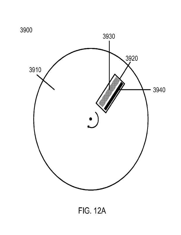
[0074] FIG. 12A ¨ FIG. 12C illustrate schemes for detection of signals
emitted by a
material coupled to an open substrate. FIG. 12A illustrates a scheme in which
an open substrate
rotates and a detector system remains stationary during detection. FIG. 12B
illustrates a scheme
in which an open substrate remains stationary and a detector system rotates
during detection.
FIG. 12C illustrates a scheme in which an open substrate rotates during
delivery and dispersal of
a solution to the open substrate (left panel) and remains stationary during
detection with a
rotating detector system (right panel)
[0075] FIG. 13A shows a first example of an interleaved spiral imaging
scan;
[0076] FIG. 13B shows a second example of an interleaved imaging scan;
[0077] FIG. 13C shows an example of a nested imaging scan,
10078] FIG. 14 shows a configuration for a nested circular imaging scan;
- 20 -
WO 2020/186243
PCT/US2020/022816
[0079] FIG. 15 shows a cross-sectional view of an immersion optical
system;
[0080] FIG. 16 illustrates schematically an exemplary temperature
gradient during optical
imaging.
[0081] FIG. 17A ¨ FIG. 17B illustrate schematically exemplary methods to
regulate
temperature of the substrate.
[0082] FIG. 17C ¨ FIG. 17D illustrate schematically exemplary methods to
regulate
temperature of the substrate.
100831 FIG. 17E illustrates schematically exemplary methods to regulate
temperature of the
substrate.
[0084] FIG. 18 illustrates schematically bubble formation in a fluid.
[0085] HG. 19 illustrates schematically an adapter for an optical
imaging system.
[0086] FIG. 20A illustrates schematically an exemplary method to
displace bubbles,
showing a substrate with a fluid dispensed thereto.
[0087] FIG. 20B illustrates schematically an exemplary method to
displace bubbles,
showing an optical imaging objective in contact with the fluid.
[0088] FIG. 21 illustrates schematically a method for dispensing and
removing immersion
fluid onto a substrate.
[0089] FIG. 22A ¨ FIG. 22B illustrate schematically a method for
trapping bubbles and
exemplary adapters for optical imaging objectives.
[0090] HG. 23A shows an architecture for a system comprising a
stationary axis substrate
and moving fluidics and optics;
[0091] FIG. 23B shows an architecture for a system comprising a
translating axis substrate
and stationary fluidics and optics;
[0092] FIG. 23C shows an architecture for a system comprising a
plurality of stationary
substrates and moving fluidics and optics,
[0093] HG. 23D shows an architecture for a system comprising a plurality
of moving
substrates on a rotary stage and stationary fluidics and optics;
[0094] HG. 23E shows an architecture for a system comprising a plurality
of stationary
substrates and moving optics;
[0095] FIG. 23F shows an architecture for a system comprising a
plurality of moving
substrates and stationary fluidics and optics;
[0096] HG. 23G shows an architecture for a system comprising a plurality
of moving
substrates and stationary fluidics and optics;
- 21 -
WO 2020/186243
PCT/US2020/022816
[0097] FIG. 2311 shows an architecture for a system comprising a
plurality of substrates
moved between a plurality of processing bays;
[0098] FIG. 231 shows an architecture for a system comprising a
plurality of imaging heads
scanning with shared translation and rotational axes and independently
rotating fields;
[0099] FIG. 23J shows an architecture for a system comprising a
plurality of imaging heads
scanning with shared translation and rotational axes and independently
rotating fields;
[0100] FIG. 23K shows an architecture for a system comprising multiple
spindles scanning
with a shared optical detection system;
[0101] FIG. 24 shows an architecture for a system comprising a plurality
of rotating
spindles;
[0102] FIG. 25 shows a flowchart for an example of a method for
processing an analyte;
[0103] FIG. 26 shows a first example of a system for isolating an
analyte; and
[0104] FIG. 27 shows a second example of a system for isolating an
analyte.
[0105] FIG. 28 shows examples of control systems to compensate for
velocity gradients
during scanning.
[0106] FIG. 29A shows motion of a substrate relative to two imaging
heads located on the
same side of an axis of rotation of the substrate.
[0107] FIG. 29B shows motion of a substrate relative to two imaging
heads located on
opposite sides of an axis of rotation of the substrate.
[0108] FIG. 29C shows motion of a substrate relative to three imaging
heads.
101091 FIG. 29D shows motion of a substrate relative to four imaging
heads.
[0110] FIG. 29E shows motion of a substrate relative to four imaging
heads.
[0111] FIG. 29F shows motion of a substrate relative to four imaging
heads.
[0112] FIG. 29G shows motion of a substrate relative to four imaging
heads.
101131 FIG. 30A shows successive ring paths of two imaging heads located
on the same side
of an axis of rotation of a substrate.
[0114] FIG. 30B shows successive ring paths of two imaging heads located
on opposite
sides of an axis of rotation of a substrate.
[0115] FIG. 30C shows staggered ring paths of two imaging heads located
on the same side
of an axis of rotation of a substrate.
101161 FIG. 3013 shows staggered ring paths of two imaging heads located
on opposite sides
of an axis of rotation of a substrate.
[0117] FIG. 31A shows rotating scan directions of imaging heads due to
non-radial motion
of a substrate.
- 22 -
WO 2020/186243
PCT/US2020/022816
[0118] FIG. 31B shows rotating scan directions of imaging heads due to
non-radial motion
of a substrate.
[0119] FIG. 32 shows a flowchart for an example of a method for analyte
detection or
analysis.
[0120] FIG. 33 illustrates schematically an axis of rotation and
relative translation of a
surface and an axis of rotation of an imaging field;
[0121] FIG. 34A illustrates schematically an optical system for rotating
an imaging field;
[0122] FIG. 34B illustrates schematically an optical system for rotating
an imaging field;
[0123] FIG. 34C illustrates schematically an optical system for rotating
an imaging field;
[0124] FIG. 35A shows an example of imaging head positioning for optimal
scanning
efficiency;
[0125] FIG. 35B shows an example of imaging head positioning for optimal
scanning
efficiency;
[0126] FIG. 35C shows an example of imaging head positioning for optimal
scanning
efficiency;
[0127] FIG. 36A ¨ FIG. 3611 illustrate schematically methods for
processing a biological
analyte.
[0128] FIG. 37A ¨ FIG. 37G illustrate different examples of cross-
sectional surface profiles
of a substrate.
[0129] FIG. 38A ¨ FIG. 38D illustrate a method of making an
oligonucleotide-coated
surface resistant to nucleic acid contaminants.
[0130] FIG. 39A ¨ FIG. 3911 illustrate two examples of spatial loading
schemes.
[0131] FIG. 40 illustrates multiplex sample processing schemes.
[0132] FIG. 41 illustrates schematically an exemplary optical layout;
[0133] FIG. 42 shows an example of an image generated by imaging a
substrate with an
analyte immobilized thereto.
[0134] FIG. 43 shows an example of data obtained from a diagnostic
procedure.
[0135] FIG. 44 shows example data of a diagnostic procedure. Panels A-F
show spatial
plots of diagnostic metrics computed on scanned images at different
individually addressable
locations.
[0136] FIG. 45A shows example data of flow-based sequencing.
[0137] FIG. 4511¨ FIG. 45C illustrate exemplary data from processed
images.
[0138] FIG. 46A shows a plot of aligned genomic reads. FIG. 4613 shows
aligned coverage
distribution over a reference genome.
-23-
WO 2020/186243
PCT/US2020/022816
[0139] FIG. 47A illustrates a partial cross-sectional view of a barrier
system maintaining a
fluid barrier.
[0140] FIG. 47B illustrates a perspective view of a chamber of the
barrier system of FIG.
47A.
[0141] FIG. 48shows a system and method for spiral loading a sample or a
reagent onto a
substrate.
[0142] FIG. 49 shows an exemplary coating of a substrate with a
hexagonal lattice of beads.
101431 FIG. 50A and FIG. 50B illustrate methods for loading beads onto a
substrate. FIG.
50A illustrates a method for loading beads onto specific regions of a
substrate. FIG. 50B
illustrates a method for loading a subset of beads onto specific regions of a
substrate.
[0144] FIG. 51 illustrates a method for spiral loading a sample or a
reagent onto an open
substrate.
DETAILED DESCRIPTION
[0145] While various embodiments of the invention have been shown and
described herein,
it will be obvious to those skilled in the art that such embodiments are
provided by way of
example only. Numerous variations, changes, and substitutions may occur to
those skilled in the
art without departing from the invention. It should be understood that various
alternatives to the
embodiments of the invention described herein may be employed.
[0146] The term "processing an analyte," as used herein, generally
refers to one or more
stages of interaction with one more sample substances. Processing an analyte
may comprise
conducting a chemical reaction, biochemical reaction, enzymatic reaction,
hybridization reaction,
polymerization reaction, physical reaction, any other reaction, or a
combination thereof with, in
the presence of, or on, the analyte. Processing an analyte may comprise
physical and/or chemical
manipulation of the analyte. For example, processing an analyte may comprise
detection of a
chemical change or physical change, addition of or subtraction of material,
atoms, or molecules,
molecular confirmation, detection of the presence of a fluorescent label,
detection of a Forster
resonance energy transfer (FRET) interaction, or inference of absence of
fluorescence. The term
"analyte" may refer to molecules, cells, biological particles, or organisms.
In some instances, a
molecule may be a nucleic acid molecule, antibody, antigen, peptide, protein,
or other biological
molecule obtained from or derived from a biological sample. An analyte may
originate from,
and/or be derived from, a biological sample, such as from a cell or organism.
An analyte may be
synthetic.
- 24 -
WO 2020/186243
PCT/US2020/022816
[0147] The term "sequencing," as used herein, generally refers to a
process for generating or
identifying a sequence of a biological molecule, such as a nucleic molecule.
Such sequence may
be a nucleic acid sequence, which may include a sequence of nucleic acid
bases. Sequencing
may be single molecule sequencing or sequencing by synthesis, for example.
Sequencing may be
performed using template nucleic acid molecules immobilized on a support, such
as a flow cell
or one or more beads.
[00148] The term "biological sample," as used herein, generally refers to any
sample from a
subject or specimen. The biological sample can be a fluid or tissue from the
subject or specimen.
The fluid can be blood (e.g., whole blood), saliva, urine, or sweat. The
tissue can be from an
organ (e.g., liver, lung, or thyroid), or a mass of cellular material, such
as, for example, a tumor.
The biological sample can be a feces sample, collection of cells (e.g., cheek
swab), or hair
sample. The biological sample can be a cell-free or cellular sample. Examples
of biological
samples include nucleic acid molecules, amino acids, polypeptides, proteins,
carbohydrates, fats,
or viruses. In an example, a biological sample is a nucleic acid sample
including one or more
nucleic acid molecules, such as deoxyribonucleic acid (DNA) and/or ribonucleic
acid (RNA).
The nucleic acid molecules may be cell-free or cell-free nucleic acid
molecules, such as cell free
DNA or cell free RNA. The nucleic acid molecules may be derived from a variety
of sources
including human, mammal, non-human mammal, ape, monkey, chimpanzee, reptilian,
amphibian, avian, or plant sources. Further, samples may be extracted from
variety of animal
fluids containing cell free sequences, including but not limited to blood,
serum, plasma, vitreous,
sputum, urine, tears, perspiration, saliva, semen, mucosal excretions, mucus,
spinal fluid,
amniotic fluid, lymph fluid and the like. Cell free polynucleotides may be
fetal in origin (via
fluid taken from a pregnant subject) or may be derived from tissue of the
subject itself
[00149] The term "subject," as used herein, generally refers to an individual
from whom a
biological sample is obtained. The subject may be a mammal or non-mammal. The
subject may
be an animal, such as a monkey, dog, cat, bird, or rodent. The subject may be
a human. The
subject may be a patient. The subject may be displaying a symptom of a
disease. The subject
may be asymptomatic. The subject may be undergoing treatment. The subject may
not be
undergoing treatment. The subject can have or be suspected of having a
disease, such as cancer
(e.g., breast cancer, colorectal cancer, brain cancer, leukemia, lung cancer,
skin cancer, liver
cancer, pancreatic cancer, lymphoma, esophageal cancer or cervical cancer) or
an infectious
disease. The subject can have or be suspected of having a genetic disorder
such as
achondroplasia, alpha-I antitrypsin deficiency, antiphospholipid syndrome,
autism, autosomal
dominant polycystic kidney disease, Charcot-Marie-tooth, cri du chat, Crohn's
disease, cystic
- 25 -
WO 2020/186243
PCT/US2020/022816
fibrosis, Dercum disease, down syndrome, Duane syndrome, Duchenne muscular
dystrophy,
factor V Leiden thrombophilia, familial hypercholesterolemia, familial
Mediterranean fever,
fragile x syndrome, Gaucher disease, hemochromatosis, hemophilia,
holoprosencephaly,
Huntington's disease, Klinefelter syndrome, Marfan syndrome, myotonic
dystrophy,
neurofibromatosis, Noonan syndrome, osteogenesis imperfecta, Parkinson's
disease,
phenylketonuria, Poland anomaly, porphyria, progeria, retinitis pigmentosa,
severe combined
immunodeficiency, sickle cell disease, spinal muscular atrophy, Tay-Sachs,
thalassemia,
trimethylaminuria, Turner syndrome, velocardiofacial syndrome, WAGR syndrome,
or Wilson
disease.
[0150] The terms "nucleic acid," "nucleic acid molecule," "nucleic acid
sequence," "nucleic acid
fragment," "oligonucleotide" and "polynucleotide," as used herein, generally
refer to a
polynucleotide that may have various lengths, such as either
deoxyribonucleotides or
deoxyribonucleic acids (DNA) or ribonucleotides or ribonucleic acids (RNA), or
analogs thereof.
Non-limiting examples of nucleic acids include DNA, RNA, genomic DNA or
synthetic
DNA/RNA or coding or non-coding regions of a gene or gene fragment, loci
(locus) defined
from linkage analysis, exons, introns, messenger RNA (mRNA), transfer RNA,
ribosomal RNA,
short interfering RNA (siRNA), short-hairpin RNA (shRNA), micro-RNA (miRNA),
ribozymes,
cDNA, recombinant nucleic acids, branched nucleic acids, plasmids, vectors,
isolated DNA of
any sequence, and isolated RNA of any sequence. A nucleic acid molecule can
have a length of
at least about 10 nucleic acid bases ("bases"), 20 bases, 30 bases, 40 bases,
50 bases, 100 bases,
200 bases, 300 bases, 400 bases, 500 bases, 1 kilobase (kb), 2 kb, 3, kb, 4
kb, 5 kb, 10 kb, 20 kb,
30 kb, 40 kb, 50 kb, 100 kb, 200 kb, 300 kb, 400 kb, 500 kb, 1 megabase (Mb),
or more. A
nucleic acid molecule (e.g., polynucleotide) can comprise a sequence of four
natural nucleotide
bases: adenine (A); cytosine (C); guanine (G); and thymine (T) (uracil (U) for
thymine (T) when
the polynucleotide is RNA). A nucleic acid molecule may include one or more
nonstandard
nucleotide(s), nucleotide analog(s) and/or modified nucleotide(s).
[0151] Nonstandard nucleotides, nucleotide analogs, and/or modified analogs
may include, but
are not limited to, diaminopurine, 5-fluorouracil, 5-bromouracil, 5-
chlorouracil, 5-iodouracil,
hypoxanthine, xanthine, 4- acetylcytosine, 5-(carboxyhydroxylmethyOuracil, 5-
carboxymethylaminomethy1-2-thiouridine, 5-carboxymethylaminomethyluracil,
dihydrouracil,
beta-D-galactosylqueosine, inosine, N6-isopentenyladenine, 1-methylguanine, 1-
methylinosine,
2,2-dimethylguanine, 2-methyladenine, 2-methylguanine, 3-methylcytosine, 5-
methylcytosine,
N6-adenine, 7-methylguanine, 5-methylaminomethyluracil, 5-methoxyaminomethy1-2-
thiouracil,
beta-D-mannosylqueosine, 5'-methoxycarboxymethyluracil, 5-methoxyuracil, 2-
methylthio-046-
- 26 -
WO 2020/186243
PCT/US2020/022816
isopentenyladenine, uracil-5-oxyacetic acid (v), wybutoxosine, pseudouracil,
queosine, 2-
thiocytosine, 5-methyl-2-thiouracil, 2-thiouracil, 4-thiouracil, 5-
methyluracil, uracil-5- oxyacetic
acid methylester, uracil-5-oxyacetic acid(v), 5-methyl-2-thiouracil, 3-(3-
amino-3-N-2-
carboxypropyl) uracil, (acp3)w, 2,6- diaminopurine, ethynyl nucleotide bases,
1-propynyl
nucleotide bases, azido nucleotide bases, phosphoroselenoate nucleic acids and
the like. In some
cases, nucleotides may include modifications in their phosphate moieties,
including
modifications to a triphosphate moiety. Additional, non-limiting examples of
modifications
include phosphate chains of greater length (e.g., a phosphate chain having, 4,
5, 6, 7, 8, 9, 10 or
more phosphate moieties), modifications with thiol moieties (e.g., alpha-thio
triphosphate and
beta-thiotriphosphates) or modifications with selenium moieties (e.g.,
phosphoroselenoate
nucleic acids). Nucleic acid molecules may also be modified at the base moiety
(e.g., at one or
more atoms that typically are available to form a hydrogen bond with a
complementary
nucleotide and/or at one or more atoms that are not typically capable of
forming a hydrogen
bond with a complementary nucleotide), sugar moiety or phosphate backbone.
Nucleic acid
molecules may also contain amine -modified groups, such as aminoallyl-dUTP (aa-
dUTP) and
aminohexhylacrylamide-dCTP (aha-dCTP) to allow covalent attachment of amine
reactive
moieties, such as N-hydroxysuccinimide esters (NI-1S). Alternatives to
standard DNA base pairs
or RNA base pairs in the oligonucleotides of the present disclosure can
provide higher density in
bits per cubic mm, higher safety (resistant to accidental or purposeful
synthesis of natural
toxins), easier discrimination in photo- programmed polymerases, or lower
secondary structure.
Nucleotide analogs may be capable of reacting or bonding with detectable
moieties for
nucleotide detection.
[0152] The term "nucleotide," as used herein, generally refers to any
nucleotide or nucleotide
analog. The nucleotide may be naturally occurring or non-naturally occurring.
The nucleotide
analog may be a modified, synthesized or engineered nucleotide. The nucleotide
analog may not
be naturally occurring or may include a non-canonical base. The naturally
occurring nucleotide
may include a canonical base. The nucleotide analog may include a modified
polyphosphate
chain (e.g., triphosphate coupled to a fluorophore). The nucleotide analog may
comprise a label.
The nucleotide analog may be terminated (e.g., reversibly terminated). The
nucleotide analog
may comprise an alternative base.
[00153] The terms "amplifying," "amplification," and "nucleic acid
amplification" are used
interchangeably and generally refer to generating one or more copies of a
nucleic acid or a
template. For example, "amplification" of DNA generally refers to generating
one or more
copies of a DNA molecule. Moreover, amplification of a nucleic acid may be
linear, exponential,
- 27 -
WO 2020/186243
PCT/US2020/022816
or a combination thereof Amplification may be emulsion based or may be non-
emulsion based.
Non-limiting examples of nucleic acid amplification methods include reverse
transcription,
primer extension, polymerase chain reaction (PCR), ligase chain reaction
(LCR), helicase-
dependent amplification, asymmetric amplification, rolling circle
amplification, recombinase
polymerase reaction (RPA), and multiple displacement amplification (MDA).
Where PCR is
used, any form of PCR may be used, with non-limiting examples that include
real-time PCR,
allele-specific PCR, assembly PCR, asymmetric PCR, digital PCR, emulsion PCR,
dial-out PCR,
helicase-dependent PCR, nested PCR, hot start PCR, inverse PCR, methylation-
specific PCR,
miniprimer PCR, multiplex PCR, nested PCR, overlap-extension PCR, thermal
asymmetric
interlaced PCR and touchdown PCR. Moreover, amplification can be conducted in
a reaction
mixture comprising various components (e.g., a primer(s), template,
nucleotides, a polymerase,
buffer components, co-factors, etc.) that participate or facilitate
amplification. In some cases, the
reaction mixture comprises a buffer that permits context independent
incorporation of
nucleotides. Non-limiting examples include magnesium-ion, manganese-ion and
isocitrate
buffers. Additional examples of such buffers are described in Tabor, S. et al.
C.C. PNAS, 1989,
86, 4076-4080 and U.S. Patent Nos. 5,409,811 and 5,674,716, each of which is
herein
incorporated by reference in its entirety.
100154] The terms "dispense" and "disperse" may be used interchangeably
herein_ In some
cases, dispensing may comprise dispersing and/or dispersing may comprise
dispensing.
Dispensing generally refers to distributing, depositing, providing, or
supplying a reagent,
solution, or other object, etc. Dispensing may comprise dispersing, which may
generally refer to
spreading.
100155] Useful methods for clonal amplification from single molecules include
rolling circle
amplification (RCA) (Lizardi et al., Nat. Genet. 19:225-232 (1998), which is
incorporated herein
by reference), bridge PCR (Adams and Kron, Method for Performing Amplification
of Nucleic
Acid with Two Primers Bound to a Single Solid Support, Mosaic Technologies,
Inc. (Winter
Hill, Mass.); Whitehead Institute for Biomedical Research, Cambridge, Mass.,
(1997); Adessi et
al., Nucl. Acids Res. 28:E87 (2000); Pemov et al., Nucl. Acids Res. 33:el
1(2005); or U.S. Pat.
No. 5,641,658, each of which is incorporated herein by reference), polony
generation (Mitra et
al., Proc. Natl. Acad. Sci. USA 100:5926-5931 (2003); Mitra et al., Anal.
Biochem. 320:55-
65(2003), each of which is incorporated herein by reference), and clonal
amplification on beads
using emulsions (Dressman et at., Proc. Natl. Acad. Sci. USA 100:8817-8822
(2003), which is
incorporated herein by reference) or ligation to bead-based adapter libraries
(Brenner et at., Nat.
Biotechnol. 18:630-634 (2000); Brenner et al., Proc. Natl. Acad. Sci. USA
97:1665-1670
- 28 -
WO 2020/186243
PCT/US2020/022816
(2000)); Reinartz, et al., Brief Fund. Genomic Proteomic 1:95-104 (2002), each
of which is
incorporated herein by reference).
[0156] The term "detector," as used herein, generally refers to a device that
is capable of
detecting a signal, including a signal indicative of the presence or absence
of one or more
incorporated nucleotides or fluorescent labels. The detector may detect
multiple signals. The
signal or multiple signals may be detected in real-time during, substantially
during a biological
reaction, such as a sequencing reaction (e.g., sequencing during a primer
extension reaction), or
subsequent to a biological reaction. In some cases, a detector can include
optical and/or
electronic components that can detect signals. The term "detector" may be used
in detection
methods. Non-limiting examples of detection methods include optical detection,
spectroscopic
detection, electrostatic detection, electrochemical detection, acoustic
detection, magnetic
detection, and the like. Optical detection methods include, but are not
limited to, light absorption,
ultraviolet-visible (UV-vis) light absorption, infrared light absorption,
light scattering, Rayleigh
scattering, Raman scattering, surface-enhanced Raman scattering, Mie
scattering, fluorescence,
luminescence, and phosphorescence. Spectroscopic detection methods include,
but are not
limited to, mass spectrometry, nuclear magnetic resonance (NMR) spectroscopy,
and infrared
spectroscopy. Electrostatic detection methods include, but are not limited to,
gel-based
techniques, such as, for example, gel electrophoresis. Electrochemical
detection methods
include, but are not limited to, electrochemical detection of amplified
product after high-
performance liquid chromatography separation of the amplified products.
101571 The term "continuous area scanning," as used herein, generally refers
to area scanning in
rings, spirals, or arcs on a rotating substrate using an optical imaging
system and a detector.
Continuous area scanning may scan a substrate or array along a nonlinear path.
Alternatively or
in addition, continuous area scanning may scan a substrate or array along a
linear or substantially
linear path. The detector may be a continuous area scanning detector. The
scanning direction
may be substantially 0 in an (R, 0) coordinate system in which the object
rotation motion is in a
0 direction. Across any field of view on the object (substrate) imaged by a
scanning system, the
dB
apparent velocity may vary with the radial position (R) of the field point on
the object as R
Continuous area scanning detectors may scan at the same rate for all image
positions and
therefore may not be able to operate at the correct scan rate for all imaged
points in a curved (or
arcuate or non-linear) scan. Therefore, the scan may be corrupted by velocity
blur for imaged
field points moving at a velocity different than the scan velocity. Continuous
rotational area
scanning may comprise an optical detection system or method that makes
algorithmic, optical,
and/or electronic corrections to substantially compensate for this tangential
velocity blur, thereby
- 29 -
WO 2020/186243
PCT/US2020/022816
reducing this scanning aberration. For example, the compensation is
accomplished
algorithmically by using an image processing algorithm that deconyolves
differential velocity
blur at various image positions corresponding to different radii on the
rotating substrate to
compensate for differential velocity blur. In some cases, the camera or
scanner may apply or use
a blur to compensate for differential velocity blur.
[0158] In another example, the compensation is accomplished by using an
anamorphic
magnification gradient This may serve to magnify the substrate in one axis
(anamorphic
magnification) by different amounts at two or more substrate positions
transverse to the scan
direction. The anamorphic magnification gradient may modify the imaged
velocities of the two
or more positions to be substantially equal thereby compensating for
tangential velocity
differences of the two positions on the substrate. This compensation may be
adjustable to
account for different velocity gradients across the field of view at different
radii on the substrate.
[0159] The imaging field of view may be segmented into two or more regions,
each of which
can be electronically controlled to scan at a different rate. These rates may
be adjusted to the
mean projected object velocity within each region. The regions may be
optically defined using
one or more beam splitters or one or more mirrors. The two or more regions may
be directed to
two or more detectors. The regions may be defined as segments of a single
detector.
[0160] The term "continuous area scanning detector," as used herein, generally
refers to an
imaging array sensor capable of continuous integration over a scanning area
wherein the
scanning is electronically synchronized to the image of an object in relative
motion. A
continuous area scanning detector may comprise a time delay and integration
(TDI) charge
coupled device (CCD), Hybrid TDI, or complementary metal oxide semiconductor
(CMOS)
pseudo TDI device. For example, a continuous area scanning detector may
comprise a TDI line-
scan camera.
101611 The term "open substrate", as used herein, generally refers to a
substantially planar
substrate in which a single active surface is physically accessible at any
point from a direction
normal to the substrate. Substantially planar may refer to planarity at a
micrometer level or
nanometer level. Alternatively, substantially planar may refer to planarity at
less than a
nanometer level or greater than a micrometer level (e.g., millimeter level).
[0162] The term "anamorphic magnification", as used herein, generally refers
to differential
magnification between two axes of an image. An anamorphic magnification
gradient may
comprise differential anamorphic magnification in a first axis across a
displacement in the
second axis. The magnification in the second axis may be unity or any other
value that is
substantially constant over the field.
-30-
WO 2020/186243
PCT/US2020/022816
[0163] The term "field of view", as used herein, generally refers to the area
on the sample or
substrate that is optically mapped to the active area of the detector.
Processing an analyte using an open substrate
[0164] Prior microfluidic systems have utilized substrates containing
numerous long, narrow
channels. The typical flow cell geometry for such substrates introduces a need
to compromise
between two competing requirements: 1) minimizing volume to minimize reagent
usage; and 2)
maximizing effective hydraulic diameter to minimize flow time. This trade-off
may be especially
important for washing operations, which may require large wash volumes and
thus long amounts
of time to complete.. The tradeoff is illustrated by the Poiseuille equation
that dictates flow in the
laminar regime and is thus inherent to microfluidic systems that utilize such
flow cell
geometries. Such flow cell geometries may also be susceptible to
contamination. Because such
flow cell geometries allow for a finite, limited number of channels in the
microfluidic systems,
such finite number of channels may be shared between a plurality of different
mixtures
comprising different analytes, reagents, agents, and/or buffers. Contents of
fluids flowing
through the same channels may be contaminated.
[0165] Described herein are devices, systems, and methods for processing
analytes using
open substrates or flow cell geometries that can address at least the
abovementioned problems.
The devices, systems and methods may be used to facilitate any application or
process involving
a reaction or interaction between an analyte and a fluid (e.g., a fluid
comprising reagents, agents,
buffers, other analytes, etc.). Such reaction or interaction may be chemical
(e.g., polymerase
reaction) or physical (e.g., displacement). The systems and methods described
herein may
benefit from higher efficiency, such as from faster reagent delivery and lower
volumes of
reagents required per surface area. The systems and methods described herein
may avoid
contamination problems common to microfluidic channel flow cells that are fed
from multiport
valves which can be a source of carryover from one reagent to the next. The
devices, systems,
and methods may benefit from shorter completion time, use of fewer resources
(e.g., various
reagents), and/or reduced system costs. The open substrates or flow cell
geometries may be used
to process any analyte, such as but not limited to, nucleic acid molecules,
protein molecules,
antibodies, antigens, cells, and/or organisms, as described herein. The open
substrates or flow
cell geometries may be used for any application or process, such as, but not
limited to,
sequencing by synthesis, sequencing by ligation, amplification, proteomics,
single cell
processing, barcoding, and sample preparation, as described herein.
-31-
WO 2020/186243
PCT/US2020/022816
[0166] The systems and methods may utilize a substrate comprising an
array (such as a
planar array) of individually addressable locations. Each location, or a
subset of such locations,
may have immobilized thereto an analyte (e.g., a nucleic acid molecule, a
protein molecule, a
carbohydrate molecule, etc.). For example, an analyte may be immobilized to an
individually
addressable location via a support, such as a bead. A plurality of analytes
immobilized to the
substrate may be copies of a template analyte. For example, the plurality of
analytes may have
sequence homology. In other instances, the plurality of analytes immobilized
to the substrate
may be different. The plurality of analytes may be of the same type of analyte
(e.g., a nucleic
acid molecule) or may be a combination of different types of analytes (e.g.,
nucleic acid
molecules, protein molecules, etc.). One or more surfaces of the substrate may
be exposed to a
surrounding open environment, and accessible from such surrounding open
environment. For
example, the array may be exposed and accessible from such surrounding open
environment In
some cases, as described elsewhere herein, the surrounding open environment
may be controlled
and/or confined in a larger controlled environment.
[0167] Reagents may be dispensed to the substrate to multiple locations,
and/or multiple
reagents may be dispensed to the substrate to a single location, via different
mechanisms. In
some cases, dispensing (to multiple locations and/or of multiple reagents to a
single location)
may be achieved via relative motion of the substrate and the dispenser (e.g.,
nozzle). For
example, a reagent may be dispensed to the substrate at a first location, and
thereafter travel to a
second location different from the first location due to forces (e.g.,
centrifugal forces, centripetal
forces, inertial forces, etc.) caused by motion of the substrate. In another
example, a reagent may
be dispensed to a reference location, and the substrate may be moved relative
to the reference
location such that the reagent is dispensed to multiple locations of the
substrate. In some cases,
dispensing (to multiple locations and/or of multiple reagents to a single
location) may be
achieved without relative motion between the substrate and the dispenser. For
example, multiple
dispensers may be used to dispense reagents to different locations, and/or
multiple reagents to a
single location, or a combination thereof (e.g., multiple reagents to multiple
locations). In
another example, an external force (e.g., involving a pressure differential),
such as wind, may be
applied to one or more surfaces of the substrate to direct reagents to
different locations across the
substrate. In another example, the method for dispensing reagents (e.g., to
multiple locations
and/or of multiple reagents to a single location) may comprise vibration. In
such an example,
reagents may be distributed or dispensed onto a single region or multiple
regions of the substrate
(or a surface of the substrate). The substrate (or a surface thereof) may then
be subjected to
vibration, which may spread the reagent to different locations across the
substrate (or the
-32-
WO 2020/186243
PCT/US2020/022816
surface). Alternatively or in conjunction, the method may comprise using
mechanical, electric,
physical, or other means to dispense reagents to the substrate. For example,
the solution may be
dispensed onto a substrate and a physical scraper (e.g., a squeegee) may be
used to spread the
dispensed material or spread the reagents to different locations and/or to
obtain a desired
thickness or uniformity across the substrate. Beneficially, such flexible
dispensing may be
achieved without contamination of the reagents. In some instances, where a
volume of reagent is
dispensed to the substrate at a first location, and thereafter travels to a
second location different
from the first location, the volume of reagent may travel in a path or paths,
such that the travel
path or paths are coated with the reagent. In some cases, such travel path or
paths may
encompass a desired surface area (e.g., entire surface area, partial surface
area(s), etc.) of the
substrate.
10168] Reagents may be dispensed over the uncovered surface or substrate
at a desired flow
rate. The flow rate of fluid dispensing may be about (e.g., at ambient
temperature, or about 25
degrees Celsius) 1 picoliter/min, 10 picoliters/min, 100 picoliters/min, 1
nanoliter/min, 10
nanoliters/min, 100 nanoliters/min, 1 microliter/min, 10 microliters/min, 100
microliters/min, 1
milliliter/min, 10 milliliters/min, 100 milliliters/min, up to 1 liter/min.
The flow rate of fluid
dispensing may be between any of these flow rates. The flow rate of fluid
dispensing may be at
least any of these flow rates. Alternatively, the flow rate of fluid
dispensing may be at most any
of these flow rates. The flow rate may be tuned according to desired
properties of the reagent or
solution layer (e.g., thickness).
[0169] Solutions may comprise reagents, samples, or any useful
substance. The solution may
comprise a fluid that has desirable flow properties. For example, the fluid
may have a
temperature-variable viscosity. The solution may comprise a non-Newtonian
fluid. The solution
may comprise a power law fluid, such as a shear-thinning (thixotropic) or
shear-thickening fluid.
The solution may comprise a Newtonian fluid.
[0170] In some cases, the substrate may be rotatable about an axis. The
analytes may be
immobilized to the substrate during rotation. Reagents (e.g., nucleotides,
antibodies, washing
reagents, enzymes, etc.) may be dispensed onto the substrate prior to or
during rotation (for
instance, spun at a high rotational velocity) of the substrate to coat the
array with the reagents
and allow the analytes to interact with the reagents. For example, when the
analytes are nucleic
acid molecules and when the reagents comprise nucleotides, the nucleic acid
molecules may
incorporate or otherwise react with (e.g., transiently bind) one or more
nucleotides. In another
example, when the analytes are protein molecules and when the reagents
comprise antibodies,
the protein molecules may bind to or otherwise react with one or more
antibodies. In another
- 33 -
WO 2020/186243
PCT/US2020/022816
example, when the reagents comprise washing reagents, the substrate (and/or
analytes on the
substrate) may be washed of any unreacted (and/or unbound) reagents, agents,
buffers, and/or
other particles.
[0171] In some cases, the substrate may be movable in any vector or
direction, as described
elsewhere herein. For example, such motion may be non-linear (e.g., in
rotation about an axis).
In another example, such motion may be linear. In other examples, the motion
may be a hybrid
of linear and non-linear motion. The analytes may be immobilized to the
substrate during any
such motion. Reagents (e.g., nucleotides, antibodies, washing reagents,
enzymes, etc.) may be
dispensed onto the substrate prior to or during motion of the substrate to
facilitate coating of the
array with the reagents and allow the analytes to interact with the reagents.
[0172] In some cases, where the substrate is rotatable, high speed
coating across the substrate
may be achieved via tangential inertia directing unconstrained spinning
reagents in a partially
radial direction (that is, away from the axis of rotation) during rotation, a
phenomenon
commonly referred to as centrifugal force. High speed rotation may involve a
rotational speed of
at least 1 revolution per minute (rpm), at least 2 rpm, at least 5 rpm, at
least 10 rpm, at least 20
rpm, at least 50 rpm, at least 100 rpm, at least 200 rpm, at least 500 rpm, at
least 1,000 rpm, at
least 2,000 rpm, at least 5,000 rpm, at least 10,000 rpm, or greater. This
mode of directing
reagents across a substrate may be herein referred to as centrifugal or
inertial pumping. Inertial
forces may direct unconstrained reagents across the substrate in any direction
during any type of
motion (e.g., rotational motion, non-rotational motion, linear motion, non-
linear motion,
accelerated motion, etc.) of the substrate.
[0173] One or more signals (such as optical signals) may be detected
from a detection area
on the substrate prior to, during, or subsequent to, the dispensing of
reagents to generate an
output. For example, the output may be an intermediate or final result
obtained from processing
of the analyte. Signals may be detected in multiple instances. The dispensing,
rotating (or other
motion), and/or detecting operations, in any order (independently or
simultaneously), may be
repeated any number of times to process an analyte. In some instances, the
substrate may be
washed (e.g., via dispensing washing reagents) between consecutive dispensing
of the reagent&
One or more detection operations can be performed within a desired time frame.
For example,
the detection operation can be performed within about 1 minute, 50 seconds, 40
seconds, 30
seconds, 20 seconds, 10 seconds or less than 10 seconds. In some instances, at
least two
detection operations can be performed within 1 minute, 50 seconds, 40 seconds,
30 seconds, 20
seconds, 10 seconds or less than 10 seconds etc. In some instances, at least
three detection
-34-
WO 2020/186243
PCT/US2020/022816
operations can be performed within 1 minute, 50 seconds, 40 seconds, 30
seconds, 20 seconds,
seconds or less than 10 seconds.
[0174] Provided herein is a method for processing a biological analyte,
comprising providing
a substrate comprising an array having immobilized thereto the biological
analyte, wherein the
substrate is rotatable with respect to a central axis. In some instances, the
array can be a planar
array. In some instances, the array can be an array of wells. In some
instances, the substrate can
be textured and/or patterned. The method can comprise directing a solution
across the substrate
and bringing the solution in contact with the biological analyte during
rotation of the substrate.
The solution may be directed in a radial direction (e.g., outwards) with
respect to the substrate to
coat the substrate and contact the biological analytes immobilized to the
array. In some
instances, the solution may comprise a plurality of probes. In some instances,
the solution may
be a washing solution. The method can comprise subjecting the biological
analyte to conditions
sufficient to conduct a reaction between at least one probe of the plurality
of probes and the
biological analyte. The reaction may generate one or more signals from the at
least one probe
coupled to the biological analyte. The method can comprise detecting one or
more signals,
thereby analyzing the biological analyte.
[0175] In other cases, provided herein is a method for processing a
biological analyte,
comprising providing a substrate comprising an array having immobilized
thereto the biological
analyte, wherein the substrate is movable with respect to a reference axis.
The method can
comprise directing a solution across the substrate and bringing the solution
in contact with the
biological analyte during motion of the substrate. In some instances, the
motion can be linear. In
some instances, the motion can be non-linear. In some instances, the motion
can be a hybrid
between linear and non-linear motion.
[0176] In other cases, provided herein is a method for processing a
biological analyte,
comprising providing a substrate comprising an array having immobilized
thereto the biological
analyte. In some instances, the method can comprise dispensing a solution to
two different
locations on the substrate and/or array. In some instances, the method can
comprise dispensing
multiple solutions to a single location on the substrate and/or array, such as
using multiple
dispensers. In some instances, the method can comprise dispensing multiple
solutions to multiple
locations on the substrate and/or array. In some instances, the method can
comprise dispensing a
single solution to a single location. The substrate may be in relative motion
with respect to one
or more dispensers. The substrate may be stationary with respect to one or
more dispensers. One
or more dispensing operations can be performed within a desired time frame.
For example, the
dispensing operation can be performed within 1 minute, 50 seconds, 40 seconds,
30 seconds, 20
-35-
WO 2020/186243
PCT/US2020/022816
seconds, 10 seconds or less than 10 seconds. In some instances, at least two
dispensing
operations can be performed within 1 minute, 50 seconds, 40 seconds, 30
seconds, 20 seconds,
seconds or less than 10 seconds etc. In some instances, at least three
dispensing operations
can be performed within 1 minute, 50 seconds, 40 seconds, 30 seconds, 20
seconds, 10 seconds
or less than 10 seconds.
[0177] Any operation or process of one or more methods disclosed herein
may be performed
within a desired time frame. In some instances, a combination of two or more
operations or
processes disclosed herein may be performed within a desired time frame. For
example, the
dispensing operation and the detection method may both be performed within 1
minute, 50
seconds, 40 seconds, 30 seconds, 20 seconds, 10 seconds or less than 10
seconds. In some
instances, at least two dispensing and detection operations can be performed
within 1 minute, 50
seconds, 40 seconds, 30 seconds, 20 seconds, 10 seconds or less than 10
seconds etc. In some
instances, at least three dispensing and detection operations can be performed
within 1 minute,
50 seconds, 40 seconds, 30 seconds, 20 seconds, 110 seconds or less than 10
seconds.
[0178] One or more methods disclosed herein may obviate the need for
barcoding of analytes
(e.g., nucleic acid molecules), which may be time-consuming and expensive. For
example,
alternative or in addition to barcoding, the substrate and/or array may be
spatially indexed to
identify the analytes, as described elsewhere herein. One or more methods
disclosed herein may
obviate the need for unique barcoding of individual analytes (e.g., individual
nucleic acid
molecules).
[0179] The biological analyte may be any analyte that comes from a
sample. For instance,
the biological analyte may be a macromolecule, e.g., a nucleic acid molecule,
a carbohydrate, a
protein, a lipid, etc. The biological analyte may comprise multiple
macromolecular groups, e.g.,
glycoproteins, proteoglycans, ribozymes, liposomes, etc. The biological
analyte may be an
antibody, antibody fragment, or engineered variant thereof, an antigen, a
cell, a peptide, a
polypeptide, etc. In some cases, the biological analyte comprises a nucleic
acid molecule. The
nucleic acid molecule may comprise at least about 10, 100, 1000, 10,000,
100,000, 1,000,000,
10,000,000, 100,000,000, 1,000,000,000 or more nucleotides. Alternatively or
in addition, the
nucleic acid molecule may comprise at most about 1,000,000,000, 100,000,000,
10,000,000,
1,000,000, 100,000, 10,000, 1000, 1100, 10 or fewer nucleotides. The nucleic
acid molecule may
have a number of nucleotides that is within a range defined by any two of the
preceding values
In some cases, the nucleic acid molecule may also comprise a common sequence,
to which an N-
ma may bind. An N-mer may comprise 1, 2, 3, 4, 5, or 6 nucleotides and may
bind the common
sequence. In some cases, the nucleic acid molecules may be amplified to
produce a colony of
-36-
WO 2020/186243
PCT/US2020/022816
nucleic acid molecules attached to the substrate or attached to beads that may
associate with or
be immobilized to the substrate. In some instances, the nucleic acid molecules
may be attached
to beads and subjected to a nucleic acid reaction, e.g., amplification, to
produce a clonal
population of nucleic acid molecules attached to the beads.
101801 Nucleic acid molecules in any given nucleic acid sample may each
comprise a key
sequence. The key sequence may be a synthetic sequence. In some instances, the
key sequence
may be at most about 6 bases in length, 5 bases in length, 4 bases in length,
3 bases in length, 2
bases in length, or I base in length Alternatively, the key sequence may be
greater than 6 bases
in length. The key sequence may be indicative of the originating sample. For
example, the key
sequence may be unique to a sample such that each sample of a plurality of
samples has a unique
key sequence. Individual analytes in a single sample may share the same key
sequence.
Alternatively, each sample may have a unique key sequence between its
immediate neighboring
samples when loaded onto the substrate. Beneficially, where two samples
comprising different
key sequences are loaded into adjacent or otherwise proximate regions on the
substrate, nucleic
acid molecules originating from different samples may be readily
differentiated based on the
different key sequences even where there is cross-contamination between
regions (e.g., outlying
nucleic acid molecules that are inadvertently loaded onto a neighboring region
due to spillover,
etc.) with relatively short reads (e.g., which are much shorter than reads of
unique barcode
sequences that are configured to differentiate individual molecules).
[0181] The substrate may be a solid substrate. The substrate may
entirely or partially
comprise one or more of rubber, glass, silicon, a metal such as aluminum,
copper, titanium,
chromium, or steel, a ceramic such as titanium oxide or silicon nitride, a
plastic such as
polyethylene (PE), low-density polyethylene (LDPE), high-density polyethylene
(HDPE),
polypropylene (PP), polystyrene (PS), high impact polystyrene (HIPS),
polyvinyl chloride
(PVC), polyvinylidene chloride (PVDC), acrylonitrile butadiene styrene (ABS),
polyacetylene,
polyamides, polycarbonates, polyesters, polyurethanes, polyepoxide, polymethyl
methacrylate
(PMMA), polytetrafluoroethylene (FIFE), phenol formaldehyde (PF), melamine
formaldehyde
(ME), urea-formaldehyde (UF), polyetheretherketone (PEEK), polyetherimide
(PEI), polyimides,
polylactic acid (PLA), furans, silicones, polysulfones, any mixture of any of
the preceding
materials, or any other appropriate material. The substrate may be entirely or
partially coated
with one or more layers of a metal such as aluminum, copper, silver, or gold,
an oxide such as a
silicon oxide (Six0y, where x, y may take on any possible values), a
photoresist such as SUS, a
surface coating such as an aminosilane or hydrogel, polyacrylic acid,
polyacrylamide dextran,
polyethylene glycol (PEG), or any combination of any of the preceding
materials, or any other
-37-
WO 2020/186243
PCT/US2020/022816
appropriate coating. A substrate may be fully or partially opaque to visible
light. In some cases, a
substrate may be at least 5%, at least 10%, at least 15%, at least 20%, at
least 25%, at least 30%,
at least 35%, at least 40%, at least 45%, at least 50%, at least 55%, at least
60%, at least 65%, at
least 70%, at least 75%, at least 80%, at least 85%, at least 90%, at least
95%, at least 96%, at
least 97%, at least 98%, at least 99%, at least 99.5%, or 100% opaque to
visible light. The
substrate may have an opacity that is within a range defined by any two of the
preceding values.
A substrate may be fully or partially transparent to visible light. In some
cases, a substrate may
be at least 5%, at least 10%, at least 15%, at least 20%, at least 25%, at
least 30%, at least 35%,
at least 40%, at least 45%, at least 50%, at least 55%, at least 60%, at least
65%, at least 70%, at
least 75%, at least 80%, at least 85%, at least 90%, at least 95%, at least
96%, at least 97%, at
least 98%, at least 99%, at least 99.5%, or 100% transparent to visible light.
The substrate may
have a transparency that is within a range defined by any two of the preceding
values. In some
cases, an illumination power (e.g., a laser power) may be adjusted based on
the opacity or
transparency of the substrate. The one or more layers may have a thickness of
at least 1
nanometer (nm), at least 2 nm, at least 5 nm, at least 10 nm, at least 20 nm,
at least 50 nm, at
least 100 nm, at least 200 nm, at least 500 nm, at least lmicrometer (gm), at
least 2 pm, at least 5
pm, at least 10 pm, at least 20 pm, at least 50 pm, at least 100 pm, at least
200 gm, at least 500
pm, or at least 1 millimeter (mm). The one or more layers may have a thickness
that is within a
range defined by any two of the preceding values. A surface of the substrate
may be modified to
comprise any of the binders or linkers described herein. A surface of the
substrate may be
modified to comprise active chemical groups, such as amines, esters,
hydroxyls, epoxides, and
the like, or a combination thereof. In some instances, such binders, linkers,
active chemical
groups, and the like may be added as an additional layer or coating to the
substrate.
[0182] The substrate may have the general form of a cylinder, a
cylindrical shell or disk, a
rectangular prism, or any other geometric form. The substrate may have a
thickness (e.g., a
minimum dimension) of at least 100 pm, at least 200 pm, at least 500 pm, at
least 1 mm, at least
2 mm, at least 5 mm, or at least 10 mm. The substrate may have a thickness
that is within a range
defined by any two of the preceding values. The substrate may have a first
lateral dimension
(such as a width for a substrate having the general form of a rectangular
prism or a radius for a
substrate having the general form of a cylinder) of at least 1 mm, at least 2
mm, at least 5 mm, at
least 10 mm, at least 20 mm, at least 50 mm, at least 100 mm, at least 200 mm,
at least 500 mm,
or at least 1,000 mm. The substrate may have a first lateral dimension that is
within a range
defined by any two of the preceding values. The substrate may have a second
lateral dimension
(such as a length for a substrate having the general form of a rectangular
prism) or at least 1 mm,
-38-
WO 2020/186243
PCT/US2020/022816
at least 2 mm, at least 5 mm, at least 10 mm, at least 20 mm, at least 50 mm,
at least 100 mm, at
least 200 mm, at least 500 mm, or at least 1,000 mm. The substrate may have a
second lateral
dimension that is within a range defined by any two of the preceding values.
[0183] A surface of the substrate may be planar. A surface of the
substrate may be uncovered
and may be exposed to an atmosphere. Alternatively or in addition, a surface
of the substrate
may be textured or patterned_ For example, the substrate may comprise grooves,
troughs, hills,
and/or pillars. The substrate may define one or more cavities (e.g., micro-
scale cavities or nano-
scale cavities). The substrate may define one or more channels. The substrate
may have a regular
textures and/or patterns across the surface of the substrate. For example, the
substrate may have
regular geometric structures (e.g., wedges, cuboids, cylinders, spheroids,
hemispheres, etc_)
above or below a reference level of the surface. Alternatively, the substrate
may have irregular
textures and/or patterns across the surface of the substrate. For example, the
substrate may have
any arbitrary structure above or below a reference level of the substrate. In
some instances, a
texture of the substrate may comprise structures having a maximum dimension of
at most about
100%, 90%, 80%, 70%, 60%, 50%, 40%, 30%, 20%, 10%, 9%, 8%, 7%, 6%, 5%, 4%, 3%,
2%,
1%, 0.1%, 0.01%, 0.001%, 0.0001%, 0.00001% of the total thickness of the
substrate or a layer
of the substrate. In some instances, the textures and/or patterns of the
substrate may define at
least part of an individually addressable location on the substrate. A
textured and/or patterned
substrate may be substantially planar.
[0184] For example, FIG. 37A - FIG. 37G illustrate different examples of
cross-sectional
surface profiles of a substrate. FIG. 37A illustrates a cross-sectional
surface profile of a substrate
having a completely planar surface. FIG. 37B illustrates a cross-sectional
surface profile of a
substrate having semi-spherical troughs or grooves. FIG. 37C illustrates a
cross-sectional
surface profile of a substrate having pillars, or alternatively or in
conjunction, wells. FIG. 37D
illustrates a cross-sectional surface profile of a substrate having a coating.
FIG. 37E illustrates a
cross-sectional surface profile of a substrate having spherical particles.
FIG. 37F illustrates a
cross-sectional surface profile of FIG. 37B, with a first type of binders
seeded or associated with
the respective grooves. FIG. 37G illustrates a cross-sectional surface profile
of FIG. 37B, with a
second type of binders seeded or associated with the respective grooves.
10185] The substrate may comprise an array. For instance, the array may
be located on a
lateral surface of the substrate. The array may be a planar array. The array
may have the general
shape of a circle, annulus, rectangle, or any other shape. The array may
comprise linear and/or
non-linear rows. The array may be evenly spaced or distributed. The array may
be arbitrarily
spaced or distributed. The array may have regular spacing. The array may have
irregular spacing.
-39-
WO 2020/186243
PCT/US2020/022816
The array may be a textured array. The array may be a patterned array. The
array may comprise a
plurality of individually addressable locations. The individually addressable
locations may be
arranged in any convenient pattern. For example, the individually addressable
locations may be
randomly oriented on the array. The plurality of individually addressable
locations may form
separate radial regions around a disk-shaped substrate. The plurality of
individually addressable
locations may form a square, rectangle, disc, circular, annulus, pentagonal,
hexagonal,
heptagonal, octagonal, array, or any other pattern. One or more types of
individually addressable
locations may be generated. The one or more types of individually addressable
locations may
form alternating regions of the different types of individually addressable
locations. The one or
more types of individually addressable locations may form blocked regions of
the different types
of individually addressable locations. For example, in cases when two types (A
and B) of
individually addressable locations are desired, the individually addressable
locations may be
arrayed as alternating ABABAB, blocked AAABBB, or random, e.g. ABBAAB, AABBBA,
BABBAA, etc. The types of individually addressable locations may be arrayed in
any useful
pattern, such as a square, rectangle, disc, annulus, pentagon, hexagon, radial
pattern, etc. In some
cases, the two types of individually addressable locations may have different
chemical, physical,
and/or biological properties (e.g., hydrophobicity, charge, color, topography,
size, dimensions,
geometry, etc.). For example, a first type of individually addressable
location may bind a first
type of biological analyte but not a second type of biological analyte, and a
second type of
individually addressable location may bind the second type of biological
analyte but not the first
type of biological analyte.
101861 The analyte to be processed may be immobilized to the array. The
array may
comprise one or more binders described herein, such as one or more physical or
chemical linkers
or adaptors, that are coupled to a biological analyte. For instance, the array
may comprise a
linker or adaptor that is coupled to a nucleic acid molecule. Alternatively or
in addition, the
biological analyte may be coupled to a bead, which bead may be immobilized to
the array. In
some cases, a subset of the array may not be coupled to a sample or analyte.
For example, in
substrates that are configured to rotate about a central axis, the samples may
not be coupled to a
plurality of individually addressable locations of the array located near the
central axis_ In some
cases, the array may be coupled to a sample or an analyte, but not all of the
array may be
processed. For example, the substrate may be coupled to a sample or analyte
(e.g., comprising
nucleic acid molecules), but the region of the array that is in proximity to
the border of the array
may not be subjected to further processing (e.g., detection).
- 40 -
WO 2020/186243
PCT/US2020/022816
[0187] The individually addressable locations may comprise locations of
analytes or groups
of analytes that are accessible for manipulation. The manipulation may
comprise placement,
extraction, reagent dispensing, seeding, heating, cooling, or agitation. The
extraction may
comprise extracting individual analytes or groups of analytes. For instance,
the extraction may
comprise extracting at least 2, at least 5, at least 10, at least 20, at least
50, at least 100, at least
200, at least 500, or at least 1,000 analytes or groups of analytes.
Alternatively or in addition, the
extraction may comprise extracting at most 1,000, at most 500, at most 200, at
most 100, at most
50, at most 20, at most 10, at most 5, or at most 2 analytes or groups of
analytes. The
manipulation may be accomplished through, for example, localized microfluidic,
pipet, optical,
laser, acoustic, magnetic, and/or electromagnetic interactions with the
analyte or its
surroundings.
[0188] In some cases, the individually addressable locations may be
indexed, e.g., spatially,
such that the analyte immobilized or coupled to each individually addressable
location may be
identified. In some embodiments, the individually addressable locations are
indexed by
demarcating part of the substrate. In some embodiments, the surface of the
substrate is
demarcated using etching. In some embodiments, the surface of the substrate is
demarcated using
a notch in the surface. In some embodiments, the surface of the substrate is
demarcated using a
dye or ink. In some embodiments, the surface of the substrate is demarcated by
depositing a
topographical mark on the surface. In some embodiments, a sample, such as a
control nucleic
acid sample, may be used to demarcate the surface of the substrate. As will be
appreciated, a
combination of positive demarcations and negative demarcations (lack thereof)
may be used to
index the individually addressable locations. In some instances, a single
reference point or axis
(e.g., single demarcation) may be used to index all individually addressable
locations. In some
embodiments, each of the individually addressable locations is indexed. In
some embodiments, a
subset of the individually addressable locations is indexed. In some
embodiments, the
individually addressable locations are not indexed, and a different region of
the substrate is
indexed.
[0189] Individually addressable locations, or individual regions
comprising the individually
addressable locations, may be indexed, or otherwise distinguished. In some
instances, the
individually addressable locations, or individual regions may be distinguished
solely by sample
loading (e.g., without physical demarcations). In some instances, a single
region may be
distinguished from other regions. In some instances, a single type of region
may be distinguished
from other types of regions. For example, different types of regions may
comprise different types
of analytes or different sets of samples. For example, a first type of region
("A") may comprise a
-41-
WO 2020/186243
PCT/US2020/022816
first set of samples (or first type of sample), and a second type of region
("B") may comprise a
second set of samples (or second type of sample). The substrate may comprise a
set of multiple
region A's and a set of multiple region B's, wherein the multiple region A's
are distinguishable
from the multiple region B's. Different samples may be loaded onto the
different types of
regions in a predetermined spatial configuration to allow such distinction.
[0190] In some cases, a key or barcode sequence on the sample may be
used to distinguish
and/or index the spatial locations, originating sample, or a combination
thereof For example,
nucleic acid molecules in any given nucleic acid sample may each comprise a
key sequence. The
key sequence may be a synthetic sequence. The key sequence may be at most
about 6 bases in
length, 5 bases in length, 4 bases in length, 3 bases in length, 2 bases in
length, or 1 base in
length. Alternatively, the key sequence may be greater than 6 bases in length.
The key sequence
may be indicative of the originating sample. For example, the key sequence may
be unique to a
sample such that each sample of a plurality of samples has a unique key
sequence. Individual
analytes of a single sample may share a common key sequence. Alternatively,
each sample may
have a unique key sequence between its immediate neighboring samples when
loaded onto the
substrate. Beneficially, where two samples comprising different key sequences
are loaded into
adjacent or otherwise proximate regions on the substrate, nucleic acid
molecules originating
from different samples may be readily differentiated based on the different
key sequences even
where there is cross-contamination between regions (e.g., outlying nucleic
acid molecules that
are inadvertently loaded onto a neighboring region due to spillover, etc.)
with relatively short
reads (e.g., which are much shorter than reads of barcode sequences that are
configured to
differentiate individual molecules).
[0191] In some cases, spatial separation of analytes may be used to
augment or replace the
use of key or barcode sequences. For example, FIG, 40 illustrates schemes for
analysis of
analytes in a single region or in multiple regions, including 7, 15, and 96
regions. For example,
as shown in FIG. 40, analytes may be distributed across the entire surface
(upper left, "1-plex")
distributed in discrete regions, including 7 regions (upper right, "7-plex"),
15 regions (lower left,
"15-plex"), or 96 regions (lower right, "96-plex"). Analytes may be
distributed in about 5, about
10, about 15, about 20, about 25, about 30, about 35, about 40, about 45,
about 50, about 60,
about 70, about 80, about 90, about 1100, about 200, about 300, about 400, or
about 500 regions.
Analytes may be distributed in from 5 to 10, from 10 to 15, from 15 to 20,
from 20 to 25, from
25 to 30, from 30 to 35, from 35 to 40, from 40 to 45, from 45 to 50, from 50
to 60, from 60 to
70, from 70 to 80, from 80 to 90, from 90 to 100, from 100 to 200, from 200 to
300, from 300 to
400, or from 400 to 500 regions. In some cases, each region may contain a
different analyte.
- 42 -
WO 2020/186243
PCT/US2020/022816
[0192] In some cases, different types of regions may be used for sample
processing. A first
type of region ("A") may comprise a first set of samples (or first type of
sample), and a second
type of region ("B") may comprise a second set of samples (or second type of
sample). The first
type of region and the second type of region may be disposed apart from one
another in an
ordered fashion, as described elsewhere herein. In some cases, the first type
of region and the
second type of region may be disposed at a distance from a reference axis of
the substrate. For
example, the first type of region may be disposed at least 1 micrometer, 10
micrometers, 100
micrometers, 1 millimeter, 10 millimeters, 100 millimeters, 1 centimeter, 10
centimeters, 100
centimeters or more from the reference axis of the substrate. Similarly, the
second type of region
may be disposed at a distance from a reference axis of the substrate. For
example, the first type
of region may be disposed at least 1 micrometer, 10 micrometers, 100
micrometers, 1 millimeter,
millimeters, 100 millimeters, 1 centimeter, 10 centimeters, 100 centimeters or
more from the
reference axis of the substrate. Both types of regions may be disposed at
least 1 micrometer, 10
micrometers, 100 micrometers, 1 millimeter, 10 millimeters, 100 millimeters, 1
centimeter, 10
centimeters, 100 centimeters or more from the reference axis of the substrate.
[0193] For example, FIG. 39A ¨ FIG. 39B illustrate two examples of
spatial loading
schemes. In FIG. 39A, a substrate comprises two types of regions "A"s and "B"s
which are
disposed in radially alternating fashion with respect to a central axis of the
substrate. In HG.
39B, a substrate comprises two types of regions "A"s and "B"s which are
disposed in
triangularly alternating fashion across the substrate. Sample locations may be
determined by
loading a first set of samples to the A regions, wherein the first set of
samples comprises a
plurality of beads coupled to analytes of the first set of samples, and
detecting the plurality of
beads and/or analytes and their locations on the substrate, and then loading
the second set of
samples to the B regions, wherein the second set of samples comprises a
plurality of beads
coupled to analytes of the second set of samples, and detecting the plurality
of beads and/or
analytes and their locations on the substrate. Each sample in the first set of
samples and the
second set of samples may be associated with a label (e.g., fluorescent dye).
Even though the
first set of samples is primarily loaded onto the A regions, there may be some
crossovers in
which stray beads from the first set of samples are immobilized to the B
regions. Even though
the second set of samples is primarily loaded onto the B regions, there may be
some crossovers
in which stray beads from the second set of samples are immobilized to the A
regions. The
locations of the analytes of the first set of samples, including the cross-
over beads, can be
determined from the first image. The locations of the analytes of the second
set of samples,
including the cross-over beads, can be determined from the second image.
Beneficially, where
-43-
WO 2020/186243
PCT/US2020/022816
the same type of fluorescent dye identifies analytes of two different samples
("P" and "Q"), and
"P" is deposited to an A region, and "Q" is deposited to a B region, based on
the type of region
where the fluorescent signal is detected, one may identify if the analyte is
of the "P" sample or
the "Q" sample. The different regions may be alternating. The plurality of
regions may form any
pattern, such as a triangular, square, rectangle, disc, circular, annulus,
pentagonal, hexagonal,
heptagonal, octagonal, array, or any other pattern. The plurality of regions
may form irregular
patterns. The plurality of regions may be discrete regions that are not
patterned. The plurality of
regions may be interleaved, interspersed, non-contiguous, and/or different in
size.
[0194] While examples herein describe two types of regions, there may be
any number of
regions (e.g., alternating regions) to achieve the alternating spatial
distinction described herein.
For example, there may be atl at least 1, at least 2, at least 3, at least 4,
at least 5, at least 6, at
least 7, at least 8, at least 9, or at least 10 regions.
[0195] In some cases, an individually addressable location may comprise
a distinct surface
chemistry. The distinct surface chemistry may distinguish between different
addressable
locations. The distinct surface chemistry may distinguish between different
regions. For
example, a first location has a first affinity towards an object (e.g., a bead
comprising nucleic
acid molecules, e.g., amplicons, immobilized thereto) and a second location
has a second,
different affinity towards the object due to the distinct surface chemistries.
The first location and
the second location may or may not be located in the same region. The first
location and the
second location may or may not be disposed on the surface in alternating
fashion. In another
example, a first region (e.g., comprising a plurality of individually
addressable locations) has a
first affinity towards an object and a second region has a second, different
affinity towards the
object due to the distinct surface chemistries. A first location type or
region type may comprise
a first surface chemistry, and a second location type or region type may
comprise a second
surface chemistry. In some cases, a third location type or region type may
comprise a third
surface chemistry. For example, a first location type or region type may
comprise a positively
charged surface chemistry and/or a hydrophobic surface chemistry, and a second
location type or
region type may comprise a negatively charged surface chemistry and/or a
hydrophilic surface
chemistry, as shown in FIG. 50A. The same object (e.g., a bead comprising
nucleic acid
molecules, e.g., amplicons, immobilized thereto) may have higher affinity
towards a first
location type or region type compared to a second location type or region
type. The same object
may be attracted towards a first location type or region type and repelled
from a second location
type or region type. In other examples, a first location type or region type
comprising a first
surface chemistry (e.g., a positively charged surface chemistry or a
negatively charged surface
-44-
WO 2020/186243
PCT/US2020/022816
chemistry) may interact with (e.g., have an affinity towards) a first sample
type (e.g., a bead
comprising nucleic acid molecules, e.g., amplicons, immobilized thereto) and
exclude a second
sample type (e.g., a bead lacking nucleic acid molecules, e.g., amplicons,
immobilized thereto,
e.g., entirely or in substantial volume), for example as illustrated in FIG.
50B. In some cases, a
surface chemistry may comprise an amine. In some cases, a surface chemistry
may comprise a
silane (e.g., tetramethylsilane). In some cases, the surface chemistry may
comprise
hexamethyldisilazane (111MDS). In some cases, the surface chemistry may
comprise (3-
aminopropyl)triethoxysilane (APTMS). In some cases, the surface chemistry may
comprise a
surface primer molecule or any oligonucleotide molecule that has any degree of
affinity towards
another molecule.
[0196] An individually addressable location of a plurality of locations
(e.g., alternating
locations) may have an area. In some cases, a location may have an area of
about 0_1 square
micron (RIO, about 0.2 pm2, about 0.25 pm', about 0.3 pine, about 0.4 pm2,
about 0_5 pm2,
about 0.6 gm2, about 0.7 pm2, about 0.8 pm2, about 0.9 pm2, about 1 pm2, about
Li pm2, about
1.2 pm2, about 1.25 pm2, about 1.3 gm2, about 1.4 pm2, about 1.5 pm2, about
1.6 pm2, about 1.7
gm2, about 1.75 pm2, about 1.8 litTI2, about 1.9 m2, about 2 pm2, about 2.25
pm2, about 2.5 pm2,
about 2.75 pm2, about 3 pm2, about 3.25 pm2, about 3.5 pm2, about 3.75 pm2,
about 4 pm', about
4.25 pm2, about 4.5 pm2, about 4.75 pm2, about 5 pm2, about 5.5 pm2, or about
6 pm2. A
location may have an area that is within a range defined by any two of the
preceding values. A
location may have an area that is less than about 0.1 pm2 or greater than
about 6 pm2. In some
cases, a location may have a width of about 0.1 micron (pm), about 0.2 pm,
about 0.25 pm,
about 0.3 pm, about 0.4 pm, about 0.5 pm, about 0.6 pm, about 0.7 pm, about
0.8 pm, about 0.9
gm, about 1 pm, about 1.1 um, about 1.2 pm, about 1.25 gm, about 1.3 gm, about
1.4 gm, about
1.5 pm, about 1.6 pm, about 1.7 gm, about 1.75 pm, about 1.8 pm, about 1.9 pm,
about 2 gm,
about 2.25 pm, about 2.5 pm, about 2.75 pm, about 3 pm, about 3.25 pm, about
3.5 pm, about
3.75 pm, about 4 pm, about 4.25 pm, about 4.5 pm, about 4.75 pm, about 5 pm,
about 5.5 pm, or
about 6 gm. In some cases, a location may have a width that is within a range
defined by any two
of the preceding values. A location may have a width that is less than about
0.1 pm or greater
than about 6 pm. Locations (e.g., of a same type) may be distributed on a
substrate with a pitch
determined by the distance between the center of a first location and the
center of the closest or
neighboring location (e.g., of the same type). Locations may be spaced with a
pitch of about 0.1
micron (pm), about 0.2 gm, about 0.25 pm, about 0.3 gm, about 0.4 pm, about
0.5 pm, about 0.6
pm, about 0.7 m, about 0.8 gm, about 0.9 pm, about 1 pm, about 1.1 pm, about
1.2 p.m, about
1.25 pm, about 1.3 pm, about L4 p.m, about 1.5 p.m, about 1.6 pm, about 1.7
pm, about 1.75 gm,
-45-
WO 2020/186243
PCT/US2020/022816
about 1.8 pm, about 1.9 gm, about 2 gm, about 2.25 pm, about 2.5 gm, about
2.75 gm, about 3
pin, about 3.25 gm, about 3.5 pm, about 3.75 pm, about 4 pm, about 4.25 gm,
about 4.5 pm,
about 4.75 pm, about 5 pm, about 5.5 gm, about 6 pm, about 6.5 gm, about 7 gm,
about 7.5 pm,
about 8 gm, about 8.5 pm, about 9 pm, about 9.5 gm, or about 10 gm. In some
case the locations
may be positioned with a pitch that is within a range defined by any two of
the preceding values.
The locations may be positioned with a pitch of less than about 0.1 gm or
greater than about 10
pm. In some cases, the pitch between any two locations of the same type may be
determined as
a function of a size of a loading object (e.g., bead). For example, where the
loading object is a
bead having a maximum diameter, the pitch may be at least about the maximum
diameter of the
loading object.
101971 While examples herein generally describe the loading of two
samples or two sets of
samples, any number of samples, or sets of samples, may be immobilized to the
substrate. For
example, the substrate may have immobilized thereto at least 1, at least 2, at
least 3, at least 4, at
least 5, at least 6, at least 7, at least 8, at least 9, at least 10 samples,
or sets of samples. In some
cases, at least about 10, 100, 1000, 10,000, 100,000, 1,000,000, 10,000,000,
100,000,000,
1,000,000,000 or more samples, or sets of samples, may be immobilized.
Alternatively or in
addition, the substrate may comprise at most about 1,000,000,000, 100,000,000,
10,000,000,
1,000,000, 100,000, 10,000, 1000, 100, 10 or fewer samples, or sets of
samples. When the
sample is a nucleic acid sample, at least 1, at least 2, at least 3, at least
4, at least 5, at least 6, at
least 7, at least 8, at least 9, at least 10 nucleic acid samples may be
immobilized to the substrate.
In some cases, at least about 10, 100, 1000, 10,000, 100,000, 1,000,000,
10,000,000,
100,000,000, 1,000,000,000 or more nucleic acid samples may be immobilized.
Alternatively or
in addition, the substrate may comprise at most about 1,000,000,000,
100,000,000, 10,000,000,
1,000,000, 100,000, 10,000, 1000, 100, 10 or fewer nucleic acid samples.
Beneficially, multiple
samples may be simultaneously processed on the same substrate, without needing
to otherwise
barcode the multiple samples (e.g., with a common barcode sequence per sample)
to distinguish
them.
101981 Indexing may be performed using a detection method and may be
performed at any
convenient or useful step. A substrate that is indexed, e.g., demarcated, may
be subjected to
detection, such as optical imaging, to locate the indexed locations,
individually addressable
locations, and/or the biological analyte. Imaging may be performed using a
detection unit.
Imaging may be performed using one or more sensors. Imaging may not be
performed using the
naked eye. The substrate that is indexed may be imaged prior to loading of the
biological
analyte. Following loading of the biological analyte onto the individually
addressable locations,
- 46 -
WO 2020/186243
PCT/US2020/022816
the substrate may be imaged again, e.g. to determine occupancy or to determine
the positioning
of the biological analyte relative to the substrate. In some cases, the
substrate may be imaged
after iterative cycles of nucleotide addition (or other probe or other
reagent), as described
elsewhere herein. The indexing of the substrate and known initial position
(individually
addressable location) of the biological analyte may allow for analysis and
identification of the
sequence information for each individually addressable location and/or
position. Additionally,
spatial indexing may allow for identification of errors that may occur, e.g.,
sample
contamination, sample loss, etc.
[0199] In some cases, indexing may be performed to identify, process,
and/or analyze more
than one type of biological analyte, as described above. For example, a first
type of biological
analyte, which may be labeled, may be loaded onto a first set of locations
within a substrate. The
substrate may be imaged for a first indexing step of the first type of
biological analyte. A second
type of biological analyte may be loaded onto a second set of locations within
the substrate and
imaged for a second indexing step of the second type of biological analyte. In
some cases, the
second type of biological analyte may be labeled in a way such that the second
type of biological
analyte is distinguishable from the first type of biological analyte.
Alternatively, the first type of
biological analyte and the second type of biological analyte may be labeled in
substantially the
same detectable manner (e.g., same dye), and the first and second images may
be processed to
generate a differential image, wherein overlapping signals are attributed to
the locations of the
first type of biological analyte and different signals are attributed to the
locations of the second
type of biological analyte. Alternatively, the first type of biological
analyte and the second type
of biological analyte may be labeled by cleavable (or otherwise removable)
labels or tags (e.g.,
fluorescent tags), and the label cleaved after each imaging operation, such
that only the relevant
analyte locations are imaged at each imaging operation. Henceforth, the
substrate may be
analyzed and all of the locations comprising the first biological analyte may
be attributed to the
first biological analyte, and all of the locations comprising the second
biological analyte may be
attributed to the second analyte. In some cases, labeling of the first and
second analyte may not
be necessary, and the attribution of the location to either the first or
second analyte may be
performed based on spatial location alone. This process may be repeated for
any number or types
of biological analytes.
102001 The array may be coated with binders. For instance, the array may
be randomly
coated with binders. Alternatively, the array may be coated with binders
arranged in a regular
pattern (e.g., in linear arrays, radial arrays, hexagonal arrays etc.). The
array may be coated with
binders on at least 10%, at least 20%, at least 30%, at least 40%, at least
50%, at least 60%, at
- 47 -
WO 2020/186243
PCT/US2020/022816
least 70%, at least 80%, at least 90%, at least 91%, at least 92%, at least
93%, at least 94%, at
least 95%, at least 96%, at least 97%, at least 98%, or at least 99% of the
number of individually
addressable locations, or of the surface area of the substrate. The array may
be coated with
binders on a fraction of individually addressable locations, or of the surface
areas of the
substrate, that is within a range defined by any two of the preceding values.
The binders may be
integral to the array. The binders may be added to the array. For instance,
the binders may be
added to the array as one or more coating layers on the array.
102011 The binders may immobilize biological analytes through non-
specific interactions,
such as one or more of hydrophilic interactions, hydrophobic interactions,
electrostatic
interactions, physical interactions (for instance, adhesion to pillars or
settling within wells), and
the like. The binders may immobilize biological analytes through specific
interactions. For
instance, where the biological analyte is a nucleic acid molecule, the binders
may comprise
oligonucleotide adaptors configured to bind to the nucleic acid molecule.
Alternatively or in
addition, such as to bind other types of analytes, the binders may comprise
one or more of
antibodies, oligonucleotides, nucleic acid molecules, aptamers, affinity
binding proteins, lipids,
carbohydrates, and the like. The binders may immobilize biological analytes
through any
possible combination of interactions. For instance, the binders may immobilize
nucleic acid
molecules through a combination of physical and chemical interactions, through
a combination
of protein and nucleic acid interactions, etc. The array may comprise at least
about 10, 100,
1000, 10,000, 100,000, 1,000,000, 10,000,000, 100,000,000 or more binders.
Alternatively or in
addition, the array may comprise at most about 100,000,000, 10,000,000,
1,000,000, 100,000,
10,000, 1000, 100, 10 or fewer binders. The array may have a number of binders
that is within a
range defined by any two of the preceding values. In some instances, a single
binder may bind a
single biological analyte (e.g., nucleic acid molecule). In some instances, a
single binder may
bind a plurality of biological analytes (e.g., plurality of nucleic acid
molecules). In some
instances, a plurality of binders may bind a single biological analyte. Though
examples herein
describe interactions of binders with nucleic acid molecules, the binders may
immobilize other
molecules (such as proteins), other particles, cells, viruses, other
organisms, or the like.
102021 In some instances, each location, or a subset of such locations,
may have immobilized
thereto an analyte (e.g., a nucleic acid molecule, a protein molecule, a
carbohydrate molecule,
etc.). In other instances, a fraction of the plurality of individually
addressable location may have
immobilized thereto an analyte. A plurality of analytes immobilized to the
substrate may be
copies of a template analyte. For example, the plurality of analytes (e.g.,
nucleic acid molecules)
may have sequence homology. In other instances, the plurality of analytes
immobilized to the
- 48 -
WO 2020/186243
PCT/US2020/022816
substrate may not be copies. The plurality of analytes may be of the same type
of analyte (e.g., a
nucleic acid molecule) or may be a combination of different types of analytes
(e.g., nucleic acid
molecules, protein molecules, etc.).
[0203] In some instances, the array may comprise a plurality of types of
binders. For
example, the array may comprise different types of binders to bind different
types of analytes.
For example, the array may comprise a first type of binders (e.g.,
oligonucleotides) configured to
bind a first type of analyte (e.g., nucleic acid molecules), and a second type
of binders (e.g.,
antibodies) configured to bind a second type of analyte (e.g., proteins), and
the like In another
example, the array may comprise a first type of binders (e.g., first type of
oligonucleotide
molecules) to bind a first type of nucleic acid molecules and a second type of
binders (e.g.,
second type of oligonucleotide molecules) to bind a second type of nucleic
acid molecules, and
the like. For example, the substrate may be configured to bind different types
of analytes in
certain fractions or specific locations on the substrate by having the
different types of binders in
the certain fractions or specific locations on the substrate.
[0204] A biological analyte may be immobilized to the array at a given
individually
addressable location of the plurality of individually addressable locations.
An array may have
any number of individually addressable locations. For instance, the array may
have at least 1, at
least 2, at least 5, at least 10, at least 20, at least 50, at least 100, at
least 200, at least 500, at least
1,000, at least 2,000, at least 5,000, at least 10,000, at least 20,000, at
least 50,000, at least
100,000, at least 200,000, at least 500,000, at least 1,000,000, at least
2,000,000, at least
5,000,000, at least 10,000,000, at least 20,000,000, at least 50,000,000, at
least 100,000,000, at
least 200,000,000, at least 500,000,000, at least 1,000,000,000, at least
2,000,000,000, at least
5,000,000,000, at least 10,000,000,000, at least 20,000,000,000, at least
50,000,000,000, or at
least 100,000,000,000 individually addressable locations. The array may have a
number of
individually addressable locations that is within a range defined by any two
of the preceding
values. Each individually addressable location may be digitally and/or
physically accessible
individually (from the plurality of individually addressable locations). For
example, each
individually addressable location may be located, identified, and/or accessed
electronically or
digitally for mapping, sensing, associating with a device (e.g., detector,
processor, dispenser,
etc.), or otherwise processing. As described elsewhere herein, each
individually addressable
location may be indexed. Alternatively, the substrate may be indexed such that
each individually
addressable location may be identified during at least one step of the
process. Alternatively or in
addition, each individually addressable location may be located, identified,
and/or accessed
- 49 -
WO 2020/186243
PCT/US2020/022816
physically, such as for physical manipulation or extraction of an analyte,
reagent, particle, or
other component located at an individually addressable location.
[0205] Multiple biological analytes may be immobilized to the array at
spatially discrete
locations. Spatial separation of biological analytes may be obtained using
masks or bathers, as
described elsewhere herein. Alternatively or in conjunction, biological
analytes may be separated
using different fluid compositions_ In some cases, the fluid compositions may
be immiscible. For
example, a first solution (e.g., an oil, organic solution, or other
hydrophobic or oleophilic
solution) may comprise a first biological analyte, and a second solution
(e.g., a hydrophilic,
aqueous, polar or ionic solution) may comprise a second biological analyte.
The first and second
solutions may be immiscible. The substrate may be exposed to the first
solution in defined
regions, e.g., using a mask (e.g., covering or shielding the other regions of
the substrate). In
some cases, the first biological analyte associates with defined regions
(e.g., individually
addressable locations), and the first solution may be removed from the
substrate. The substrate
may then be exposed to the second solution_ The second biological analyte may
then associate
with the unoccupied sites of the substrate. Alternatively, the substrate may
be pre-treated such
that biological analytes may be loaded in discrete locations. In one non-
limiting example, the
substrate may be patterned with discrete hydrophobic and hydrophilic regions
(e.g., using
photolithography, soft lithography, etching, etc.) that can attract or repel a
subset of the
biological analytes. In another non-limiting example, an inert polymer such as
polyethylene
glycol (PEG) may be patterned in discrete regions to prevent attachment or the
biological analyte
to the substrate in the discrete regions.
[0206] Each individually addressable location may have the general shape
or form of a
circle, pit, bump, rectangle, or any other shape or form. Each individually
addressable location
may have a first lateral dimension (such as a radius for individually
addressable locations having
the general shape of a circle or a width for individually addressable
locations having the general
shape of a rectangle). The first lateral dimension may be at least 1 nanometer
(nm), at least 2 nm,
at least 5 nm, at least 10 nm, at least 20 nm, at least 50 nm, at least 100
nm, at least 200 nm, at
least 500 nm, at least 1,000 nm, at least 2,000 nm, at least 5,000 nm, or at
least 10,000 nm. The
first lateral dimension may be within a range defined by any two of the
preceding values. Each
individually addressable location may have a second lateral dimension (such as
a length for
individually addressable locations having the general shape of a rectangle).
The second lateral
dimension may be at least 1 nanometer (nm), at least 2 nm, at least 5 nm, at
least 10 nm, at least
20 nm, at least 50 nm, at least 100 nm, at least 200 nm, at least 500 nm, at
least 1,000 nm, at
least 2,000 nm, at least 5,000 nm, or at least 10,000 nm. The second lateral
dimension may be
- 50 -
WO 2020/186243
PCT/US2020/022816
within a range defined by any two of the preceding values. In some instances,
each individually
addressable locations may have or be coupled to a binder, as described herein,
to immobilize an
analyte thereto. In some instances, only a fraction of the individually
addressable locations may
have or be coupled to a binder. In some instances, an individually addressable
location may have
or be coupled to a plurality of binders to immobilize an analyte thereto.
[0207] The individually addressable locations may be generated using a
variety of methods.
In one embodiment, the method may comprise generation of individually
addressable locations
using one or more barriers. In some embodiments, the barrier may be removed
during any
convenient operation. For example, the barrier may be removed prior to or
after coupling the
analyte to the individually addressable locations. The barrier may be removed
prior to or after
loading of the solution comprising a plurality of probes. The barrier may be
removed prior to or
after subjecting the analyte to conditions sufficient to conduct a reaction
between the probe and
the analyte. The bather may be removed prior to or after detection of one or
more signals from
the coupled probe and analyte. The bather may be removed prior to or after
detection of the
coupled probe and analyte. The bather may be removed prior to or after
repeating any of the
abovementioned processes. In some cases, the barriers may not be removed.
[0208] The barrier may comprise a physical, chemical, biological, or any
other type of
obstruction. In some embodiments, the barrier comprises a physical
obstruction. In one such
example, a mold may be used, wherein a portion of the mold may obstruct the
movement of fluid
to a specified region. The mold may be generated using a variety of means,
such as injection
molding, machining, heat treatment, fiber spinning, joining and bonding,
casting, rolling,
forging, 3D printing, etc. In some embodiments, the bather may be configured
to dissolve at any
convenient step. The barrier may be configured to dissolve, evaporate, or
sublime. In some cases,
the barrier may be melted and removed. In some cases, removal of the bather or
part of the
barrier may be achieved using an air knife. In some cases, the barrier
comprises a chemical
obstruction. In some cases, the bather comprises a polymer. The barrier may
comprise
polyethylene glycol (PEG). In some cases, the bather may comprise a solution.
The solution may
be viscous. The solution may have a temperature-variable viscosity. The
solution may be a non-
Newtonian fluid. The solution may be a power law fluid, such as a shear-
thinning (e.g.,
thixotropic) or shear-thickening fluid. The solution may be a Newtonian fluid.
In some
embodiments, the bather comprises a fluid that is immiscible with a loading
solution. In some
cases, the bather is a hydrophobic region on the substrate.
[0209] A mask may be additionally or alternatively used to prevent
coupling of the sample
and/or biological analyte with a region of the substrate. Alternatively or in
conjunction, a subset
-51-
WO 2020/186243
PCT/US2020/022816
of the individually addressable locations comprising the biological analyte
may be masked, e.g.,
to prevent coupling of the probe to the biological analyte. A mask may
comprise a barrier, such
as a physical, chemical or biological barrier. A mask may comprise a film with
removed
sections. In some cases, the mask may be interfaced with the substrate prior
to introduction of
the biological analyte. In such cases, introduction of the biological analyte
may allow for
coupling of the biological analyte to exposed regions of the mask-substrate
interface, whereas
the non-exposed regions may remain free of the biological analyte. At any
convenient process,
the substrate may be un-masked. Any combinations of masks may be used. For
example, a first
mask may be used to load a first biological analyte to a desired region.
Subsequently, the first
mask may be removed, and a second mask may be used to load a second biological
analyte to a
desired region. The first and second region may have overlapping regions or
may remain
spatially distinct. A barrier and mask may be used in conjunction or
separately.
[0210] The analytes bound to the individually addressable locations may
include, but are not
limited to, molecules, cells, organisms, nucleic acid molecules, nucleic acid
colonies, beads,
clusters, polonies, DNA nanoballs, or any combination thereof (e.g., bead
having attached
thereto one or more nucleic acid molecules, e.g., one or more clonal
populations of nucleic acid
molecules). The bound analytes may be immobilized to the array in a regular,
patterned,
periodic, random, or pseudo-random configuration, or any other spatial
arrangement. In some
embodiments, the analytes are bound to bead(s) which may then associate with
or be
immobilized to the substrate or regions of the substrate (e.g., individually
addressable locations).
In some embodiments, the analytes comprise a bead or a plurality of beads. In
some cases, the
bead or plurality of beads may comprise another analyte (e.g., nucleic acid
molecule) or a clonal
population of other analytes (e.g., a nucleic acid molecule that has been
amplified on the bead).
Such other analytes may be attached or otherwise coupled to the bead. For
example, an analyte
may comprise a plurality of beads, each bead having a clonal population of
nucleic acid
molecules attached thereto. In some cases, the bead is magnetic, and
application of a magnetic
field or using a magnet may be used to direct the analytes or beads comprising
the analytes to the
individually addressable locations. In some cases, a fluid may be used to
direct the analyte to the
individually addressable locations. The fluid may be a ferrofluid, and a
magnet may be used to
direct the fluid to the individually addressable locations. The individually
addressable locations
may alternatively or in conjunction comprise a material that is sensitive to a
stimulus, e.g.,
thermal, chemical, or electrical or magnetic stimulus. For example, the
individually addressable
location may comprise a photo-sensitive polymer or reagent that is activated
when exposed to
electromagnetic radiation. In some cases, a caged molecule may be used to
reveal binding (e.g.,
- 52 -
WO 2020/186243
PCT/US2020/022816
biotin) moieties on the substrate. Subsequent exposure to a particular
wavelength of light may
result in un-caging of the binding moieties. A bead, e.g., with streptavidin,
comprising the
analyte may then associate with the uncaged binding moieties. In some cases, a
subset of the
individually addressable locations may not contain beads. In such cases, blank
beads may be
added to the substrate. The blank beads may then occupy the regions that are
unoccupied by an
analyte. In some cases, the blank beads have a higher binding affinity or
avidity for the
individually addressable locations than the beads comprising the analyte. In
some cases,
unoccupied locations may be destroyed. In some cases, unoccupied locations may
be subjected
to a process to remove any unbound analyte, e.g., aspiration, washing, air
blasting etc. In some
cases, the sample comprising the biological analyte may be loaded onto the
substrate using a
device, e.g., a microfluidic device, closed flow cell, etc. The loaded
biological analyte may then
associate with or be immobilized to the substrate or the individually
addressable locations of the
substrate. In such cases, the device may be removed following loading of the
sample.
[0211] A biological analyte may be bound to any number of beads.
Different biological
analytes may be bound to any number of beads. The beads may be unique (i.e.,
distinct from
each other). Any number of unique beads may be used. For instance, at least
about 10, 100,
1000, 10,000, 100,000, 1,000,000, 10,000,000, 100,000,000 or more different
beads may be
used. Alternatively or in addition, at most about 100,000,000, 10,000,000,
1,000,000, 100,000,
10,000, 1000, 100, 10 or fewer different beads may be used. A number of
different beads can be
within a range defined by any two of the preceding values. The beads may be
distinguishable
from one another using a property of the beads, such as color, reflectance,
anisotropy, brightness,
fluorescence, etc.
[0212] A sample may be diluted such that the approximate occupancy of
the individually
addressable locations is controlled. A sample may be diluted at least to a
dilution of 1:1, 1:2, 1:3,
1:4, 1:5,1:6, 1:7, 1:8, 1:9, 1:10, 1:20, 1:30, 1:40, 1:50, 1:60, 1:70,1:80,
1:90, 1:100, 1:200,
1:300, 1:400, 1:500, 1:600, 1:700, 1:800, 1:900, 1:1000, 1:10000, 1:100000,
1:1000000,
1:10000000, 1:100000000. Alternatively, a sample may be diluted at most to a
dilution of A
sample may be diluted at least to a dilution of 1:1, 1:2, 1:3, 1:4, 1:5, 1:6,
1:7, 1:8, 1:9, 1:10, 1:20,
1:30, 1:40, 1:50, 1:60, 1:70, 1:80, 1:90, 1:100, 1:200, 1:300, 1:400, 1:500,
1:600, 1:700, 1:800,
1:900, 1:1000, 1:10000, 1:100000, 1:1000000, 1:10000000, 1:100000000. A
dilution between
any of these dilution values may also be used.
[0213] In some instances, a sample may comprise beads. Beads may be
dispersed on a
surface in any pattern, or randomly. Beads may be dispersed on one or more
regions (e.g., a
region having a particular surface chemistry) of a surface. In some cases,
beads may be dispersed
-53-
WO 2020/186243
PCT/US2020/022816
on a surface or a region of a surface in a hexagonal lattice, as shown in FIG.
49, which illustrates
in the right panel a zoomed out image of a portion of a surface, and in the
left panel a zoomed in
image of a section of the portion of the surface. In some instances, a sample
comprising beads
may be dispersed on a surface comprising distinct locations/regions
differentiated by surface
chemistry (e.g., as illustrated in FIG. 50A and FIG. 50B). For example, a
sample comprising
beads may be dispensed on a surface comprising positively charged
locations/regions and/or
hydrophobic locations/regions. The beads may have a high affinity for a first
location type or
region type (e.g., positively charged). The beads may have a low affinity for
a second location
type or region type (e.g., hydrophobic). A location may comprise no more than
1, no more than
2, no more than 3, no more than 4, no more than 5, no more than 6, no more
than 7, no more than
8, no more than 9, or no more than 10 beads per location. In some embodiments,
a bead may be
substantially centered within an individually addressable location. A location
may have a width
that is up to about 0.5 times, up to about 0.6 times, up to about 0.7 times,
up to about 0.8 times,
up to about 0.9 times, up to about 1 times, up to about 1.1 times, up to about
1.2 times, up to
about 1.3 times, up to about 1.4 times, up to about 1.5 times, up to about 1.6
times, up to about
1.7 times, up to about 1.8 times, up to about 1.9 times, up to about 2 times,
up to about 2.1 times,
up to about 2.2 times, up to about 2.3 times, up to about 2.4 times, up to
about 2.5 times, up to
about 2.6 times, up to about 2.7 times, up to about 2.8 times, up to about 2.9
times, or up to about
3 times the diameter (e.g., maximum diameter) of the bead. In some
embodiments, a region may
be spaced with a pitch determined by the distance between the center of a
first location and the
center of the closest or neighboring location of the same type. A location may
be spaced with a
pitch that is at least about 1 times, at least about 1.2 times, at least about
1.4 times, at least about
1.6 times, at least about 1.8 times, at least about 2 times, at least about
2.2 times, at least about
2.4 times, at least about 2.6 times, at least about 2.8 times, at least about
3 times, at least about
3.2 times, at least about 3.4 times, at least about 3.6 times, at least about
3.8 times, at least about
4 times, at least about 4.2 times, at least about 4.4 times, at least about
4.6 times, at least about
4.8 times, or at least about 5 times the diameter (e.g.,. maximum diameter) of
the bead. In some
cases, one or more of a location size, a location spacing, a bead affinity, a
location surface
chemistry may be adjusted to reduce a deviation of a bead contact point from
the center of a
region.
[0214] A surface comprising a plurality of individually addressable
locations may be loaded
with beads. The beads may be loaded onto the surface at an occupancy
determined by the
number of locations of a given location type comprising at least one bead out
of the total number
of locations of the same location type. A surface comprising a plurality of
locations may have
-54-
WO 2020/186243
PCT/US2020/022816
occupancy of at least about 50%, at least about 60%, at least about 70%, at
least about 80%, at
least about 85%, at least about 86%, at least about 87%, at least about 88%,
at least about 89%,
at least about 90%, at least about 91%, at least about 92%, at least about
93%, at least about
94%, at least about 95%, at least about 96%, at least about 97%, at least
about 98%, at least
about 99.5%, or up to about 100%. For example, a surface may have at least
about 90% of the
locations of a given location type loaded with at least one bead. Beads may
land on the surface
with a landing efficiency determined by the number of beads that bind to the
surface out of the
total number of beads dispensed on the surface. Beads may be dispensed onto a
surface with a
landing efficiency of at least about 10%, at least about 20%, at least about
30%, at least about
40%, at least about 50%, at least about 55%, at least about 60%, at least
about 65%, at least
about 70%, at least about 75%, at least about 80%, at least about 90%, up to
about 100%. In
some embodiments, one or more of a temperature, an incubation time, a
surfactant, or a salt
concentration of a solution comprising beads may be adjusted to increase bead
occupancy. In
some embodiments, one or more of a temperature, an incubation time, a
surfactant, or a salt
concentration of a solution comprising beads may be adjusted to increase bead
loading
efficiency.
[0215] In some cases, at least about 5%, 10%, 15%, 20%, 25%, 30%, 35%,
40%, 45%, 50%,
55%, 60%, 65%, 70%, 75%, 80%, 85%, 90%, or 95% of the available surface area
of a substrate
may be configured to accept a bead. Where less than 100% of the available
surface area loads
thereon a bead (e.g., have a bead immobilized thereto), the negative space
(e.g., locations in
which there is no bead) may be used as a reference to identify and/or index
different individually
addressable locations of the positive space (e.g., locations in which there is
a bead). In an
example, a single individually addressable location acting in negative space
is sufficient to index
the entire substrate. In such an example, the single individually addressable
location will always
remain 'dark' during imaging, such as during sequencing, as opposed to other
individually
addressable location in the positive space which will light up (e.g.,
fluoresce) at different points
in time, such that the single individually addressable location which is
always 'dark' may act as a
reference against all other individually addressable locations. In other
examples, multiple
individually addressable locations acting in negative space may facilitate
indexing of the
substrate. Alternatively or in addition, a reference bead which is always
'bright' (e.g., always
fluorescing regardless of time point) may be used as a reference to identify
and/or index different
individually addressable locations of the positive space. In such cases, even
with 100% or
substantially 100% of the available surface area loaded with beads, including
the reference bead,
the different individually addressable locations may be identified and/or
indexed.
-55-
WO 2020/186243
PCT/US2020/022816
[0216] A sample comprising beads may be dispensed on the surface. In
some cases, the
beads may be dispensed on the surface in a substantially spiral pattern. For
example, the beads
may be dispensed in a spiral pattern using the system shown in FIG. 48 and/or
the method
shown in FIG. 51. In some instances, the beads may be dispersed in a spiral
pattern moving
radially inward toward an axis of rotation of the surface. In some instances,
the beads may be
dispersed in a spiral pattern moving radially outward from an axis of rotation
of the surface. As
shown in FIG. 48, a sample (e.g., a sample comprising beads) may be dispensed
from a dispense
probe 4801 (e.g., a nozzle) to a substrate 4803 (e.g., a wafer) to form a
layer 4805. The dispense
probe may be positioned at a fixed height ("Z") above the substrate. In the
illustrated example,
the beads are retained in the layer 4805 by electrostatic retention, and may
immobilize to the
substrate. A set of beads may each comprise a population of amplified products
(e.g., nucleic
acid molecules) immobilized thereto, which amplified products accumulate to a
negative charge
on the bead with affinity to a positive charge. The substrate comprises
alternating surface
chemistry between distinguishable locations, in which a first location type
comprises APTMS
carrying a positive charge with affinity towards the negative charge of the
amplified bead (e.g., a
bead comprising amplified products immobilized thereto, and as distinguished
from a negative
bead which does not the comprise the same), and a second location type
comprises HMDS which
has lower affinity and/or is repellant of the amplified bead. Within the layer
4805 comprising
the dispensed sample, an amplified bead may successfully land on a first
location of the first
location type (as in 4807). In the illustrated example, the location size is 1
micron, the pitch
between the different locations of the same location type (e.g., first
location type) is 2 microns,
and the layer has a depth of 15 micron. In order to obtain a substantially
spiral pattern, the
substrate and/or the dispense probe may have angular and/or linear velocity
with respect to each
other.
102171 The sample may be dispensed as shown onto an open surface as
illustrated in FIG.
51. In some cases, the substrate may be rotating relative to the dispensing
probe. In some cases,
the dispensing probe may be moving radially relative to the substrate with
respect to the axis of
rotation of the substrate. In some cases, the substrate may be moving linearly
relative to the
dispense probe. In some cases, the substrate may be rotating relative to the
dispense probe while
moving linearly relative to the dispense probe, thereby dispensing the sample
in a spiral pattern.
In some cases, the substrate may be rotating relative to the dispense probe
while the dispense
probe is moving radially relative to the substrate with respect to the axis of
rotation of the
substrate, thereby dispensing the sample in a spiral pattern. The substrate
and the dispense probe
may move in any configuration with respect to each other to achieve the
substantially spiral
- 56 -
WO 2020/186243
PCT/US2020/022816
pattern. The substrate may be rotating with a rotational frequency of no more
than 60 rpm, no
more than 50 rpm, no more than 40 rpm, no more than 30 rpm, no more than 25
rpm, no more
than 20 rpm, no more than 15 rpm, no more than 14 rpm, no more than 13 rpm, no
more than 12
rpm, no more than 11 rpm, no more than 10 rpm, no more than 9 rpm, no more
than 8 rpm, no
more than 7 rpm, no more than 6 rpm, no more than 5 rpm, no more than 4 rpm,
no more than 3
rpm, no more than 2 rpm, or no more than 1 rpm. In some cases the rotational
frequency may be
within a range defined by any two of the preceding values. In some cases the
substrate may be
rotating with a rotational frequency of about 5 rpm.
[0218] A spiral dispensing pattern may have a path width determined by
the width of a
region coated by fluid dispensed during a single rotation of the surface. A
spiral dispensing
pattern may have a path pitch determined by the distance between the center of
a fluid
dispensing path at a first position and the center of a fluid dispensing path
at a second position
after one rotation of the substrate. In some instances, the path width may be
greater than the path
pitch. For example, the fluid dispensed along the path during a substrate
rotation may overlap the
fluid dispensed along the path during the preceding substrate rotation. In
some instances, the
path pitch may be greater than the path width. For example, the fluid
dispensed along the path
during a substrate rotation may be separated from the fluid dispensed along
the path during the
preceding substrate rotation. In some instances, the path width may be similar
to the path pitch.
For example, the fluid dispensed along the path during a substrate rotation
not be substantially
separated from the fluid dispensed along the path during the preceding
substrate rotation, and the
fluid dispensed along the path during a substrate rotation may not
substantially overlap the fluid
dispensed along the path during the preceding substrate rotation.
[0219] The substrate may be configured to rotate with respect to an
axis. In some instances,
the systems, devices, and apparatus described herein may further comprise a
rotational unit
configured to rotate the substrate. The rotational unit may comprise a motor
and/or a rotor to
rotate the substrate. Such motor and/or rotor may be mechanically connected to
the substrate
directly or indirectly via intermediary components (e.g., gears, stages,
actuators, discs, pulleys,
etc.). The rotational unit may be automated. Alternatively or in addition, the
rotational unit may
receive manual input. The axis of rotation may be an axis through the center
of the substrate
(e.g., as shown in FIG. 33). The axis may be an off-center axis. For instance,
the substrate may
be affixed to a chuck (such as a vacuum chuck) of a spin coating apparatus.
The substrate may be
configured to rotate with a rotational velocity of at least 1 revolution per
minute (rpm), at least 2
rpm, at least 5 rpm, at least 10 rpm, at least 20 rpm, at least 50 rpm, at
least 100 rpm, at least 200
rpm, at least 500 rpm, at least 1,000 rpm, at least 2,000 rpm, at least 5,000
rpm, or at least 10,000
- 57 -
WO 2020/186243
PCT/US2020/022816
rpm. The substrate may be configured to rotate with a rotational velocity that
is within a range
defined by any two of the preceding values. The substrate may be configured to
rotate with
different rotational velocities during different operations described herein.
The substrate may be
configured to rotate with a rotational velocity that varies according to a
time-dependent function,
such as a ramp, sinusoid, pulse, or other function or combination of
functions. The time-varying
function may be periodic or aperiodic.
[0220] The substrate may be configured to move in any vector with
respect to a reference
point. In some instances, the systems, devices, and apparatus described herein
may further
comprise a motion unit configured to move the substrate. The motion unit may
comprise any
mechanical component, such as a motor, rotor, actuator, linear stage, drum,
roller, pulleys, etc.,
to move the substrate. Such components may be mechanically connected to the
substrate directly
or indirectly via intermediary components (e.g., gears, stages, actuators,
discs, pulleys, etc.). The
motion unit may be automated. Alternatively or in addition, the motion unit
may receive manual
input. The substrate may be configured to move with any velocity. In some
instances, the
substrate may be configured to move with different velocities during different
operations
described herein. The substrate may be configured to move with a velocity that
varies according
to a time-dependent function, such as a ramp, sinusoid, pulse, or other
function or combination
of functions. The time-varying function may be periodic or aperiodic.
[0221] A solution may be provided to the substrate prior to or during
rotation (or other
motion) of the substrate to centrifugally (or otherwise inertially) direct the
solution across the
array. In some instances, the solution may be provided to the planar array
during rotation of the
substrate in pulses, thereby creating an annular wave of the solution moving
radially outward. In
some instances, the solution may be provided to the planar array during other
motion of the
substrate in pulses, thereby creating a wave of the solution moving in a
certain direction. The
pulses may have periodic or non-periodic (e.g., arbitrary) intervals. A series
of pulses may
comprise a series of waves producing a surface-reagent exchange. The surface-
reagent exchange
may comprise washing in which each subsequent pulse comprises a reduced
concentration of the
surface reagent. The solution may have a temperature different than that of
the substrate, thereby
providing a source or sink of thermal energy to the substrate or to an analyte
located on the
substrate. The thermal energy may provide a temperature change to the
substrate or to the
analyte. The temperature change may be transient. The temperature change may
enable, disable,
enhance, or inhibit a chemical reaction, such as a chemical reaction to be
carried out upon the
analyte. For example, the chemical reaction may comprise denaturation,
hybridization, or
annealing of nucleic acid molecules. The chemical reaction may comprise a step
in a polymerase
- 58 -
WO 2020/186243
PCT/US2020/022816
chain reaction (PCR), bridge amplification, or other nucleic acid
amplification reaction. The
temperature change may modulate, increase, or decrease a signal detected from
the analyte.
[0222] The array may be in fluid communication with at least one sample
inlet (of a fluid
channel). The array may be in fluid communication with the sample inlet via a
non-solid gap,
e.g., an air gap. In some cases, the array may additionally be in fluid
communication with at least
one sample outlet. The array may be in fluid communication with the sample
outlet via an
airgap. The sample inlet may be configured to direct a solution to the array.
The sample outlet
may be configured to receive a solution from the array. The solution may be
directed to the array
using one or more dispensing nozzles. For example, the solution may be
directed to the array
using at least 1, at least 2, at least 3, at least 4, at least 5, at least 6,
at least 7, at least 8, at least 9,
at least 10, at least 11, at least 12, at least 13, at least 14, at least 15,
at least 16, at least 17, at
least 18, at least 19, or at least 20 dispensing nozzles. The solution may be
directed to the array
using a number of nozzles that is within a range defined by any two of the
preceding values. In
some cases, different reagents (e.g., nucleotide solutions of different types,
different probes,
washing solutions, etc.) may be dispensed via different nozzles, such as to
prevent
contamination. Each nozzle may be connected to a dedicated fluidic line or
fluidic valve, which
may further prevent contamination_ A type of reagent may be dispensed via one
or more nozzles.
The one or more nozzles may be directed at or in proximity to a center of the
substrate.
Alternatively, the one or more nozzles may be directed at or in proximity to a
location on the
substrate other than the center of the substrate. Alternatively or in
combination, one or more
nozzles may be directed closer to the center of the substrate than one or more
of the other
nozzles. For instance, one or more nozzles used for dispensing washing
reagents may be directed
closer to the center of the substrate than one or more nozzles used for
dispensing active reagents.
The one or more nozzles may be arranged at different radii from the center of
the substrate. Two
or more nozzles may be operated in combination to deliver fluids to the
substrate more
efficiently. One or more nozzles may be configured to deliver fluids to the
substrate as a jet,
spray (or other dispersed fluid), and/or droplets. One or more nozzles may be
operated to
nebulize fluids prior to delivery to the substrate. For example, the fluids
may be delivered as
aerosol particles.
[0223] The solution may be dispensed on the substrate while the
substrate is stationary; the
substrate may then be subjected to rotation (or other motion) following the
dispensing of the
solution. Alternatively, the substrate may be subjected to rotation (or other
motion) prior to the
dispensing of the solution; the solution may then be dispensed on the
substrate while the
substrate is rotating (or otherwise moving).
- 59 -
WO 2020/186243
PCT/US2020/022816
[0224] Rotation of the substrate may yield a centrifugal force (or
inertial force directed away
from the axis) on the solution, causing the solution to flow radially outward
over the array. In
this manner, rotation of the substrate may direct the solution across the
array. Continued rotation
of the substrate over a period of time may dispense a fluid film of a nearly
constant thickness
across the array. The rotational velocity of the substrate may be selected to
attain a desired
thickness of a film of the solution on the substrate. The film thickness may
be related to the
rotational velocity by equation (1):
k(t) ¨ _________________________________________________________________
(1) 42rp
co- ¨3p.0
Here, h(t) is the thickness of the fluid film at time t, p is the viscosity of
the fluid, co is the
rotational velocity, and C is a constant.
[0225] Alternatively or in combination, the viscosity of the solution
may be chosen to attain
a desired thickness of a film of the solution on the substrate. For instance,
the rotational velocity
of the substrate or the viscosity of the solution may be chosen to attain a
film thickness of at least
nanometers (nm), at least 20 nm, at least 50 nm, at least 100 nm, at least 200
nm, at least 500
nm, at least 1 micrometer (gm), at least 2 pm, at least 5 gm, at least 10 pm,
at least 20 m, at
least 50 pm, at least 100 pm. at least 200 gm, at least 500 pm, or at least 1
mm. The rotational
velocity of the substrate and/or the viscosity of the solution may be chosen
to attain a film
thickness that is within a range defined by any two of the preceding values.
The viscosity of the
solution may be controlled by controlling a temperature of the solution. The
thickness of the film
may be measured or monitored. Measurements or monitoring of the thickness of
the film may be
incorporated into a feedback system to better control the film thickness. The
thickness of the film
may be measured or monitored by a variety of techniques. For instances, the
thickness of the
film may be measured or monitored by thin film spectroscopy with a thin film
spectrometer,
such as a fiber spectrometer.
[0226] In some instances, one or more factors such as the rotational
velocity of the substrate,
the acceleration of the substrate (e.g., the rate of change of velocity),
viscosity of the solution,
angle of dispensing (e.g., contact angle of a stream of reagents) of the
solution, radial coordinates
of dispensing of the solution (e.g., on center, off center, etc.), temperature
of the substrate,
temperature of the solution, and other factors may be adjusted and/or
otherwise optimized to
attain a desired wetting on the substrate and/or a film thickness on the
substrate, such as to
facilitate uniform coating of the substrate. In some cases, a surfactant may
be added to the
solution, or a surfactant may be added to the surface to facilitate uniform
coating or to facilitate
sample loading efficiency. Such optimization may prevent the solution from
exiting the substrate
- 60 -
WO 2020/186243
PCT/US2020/022816
along a relatively focused stream or travel path such that the fluid only
contacts the substrate at
partial surface areas (as opposed to the entire surface area)¨in such cases, a
significantly larger
volume of reagents may have to be dispensed to achieve uniform and full
coating of the
substrate. Such optimization may also prevent the solution from scattering or
otherwise
reflecting or bouncing off the substrate upon contact and disturbing the
surface fluid.
Alternatively or in conjunction, the thickness of the solution may be adjusted
using mechanical,
electric, physical, or other mechanisms. For example, the solution may be
dispensed onto a
substrate and subsequently leveled using, e.g., a physical scraper such as a
squeegee, to obtain a
desired thickness of uniformity across the substrate.
[0227] In some cases, rotation of the substrate may be slow enough so as
not to yield
substantial centrifugal force (or inertial force directed away from the axis)
on the solution.
Beneficially, one or more reagents dispensed onto the substrate may remain
substantially local to
a landing location, e.g., without significantly and/or pre-emptively
travelling outwards with
respect to a rotational axis such as to invade another reaction space on the
open substrate. The
substrate may be rotating with a rotational frequency of no more than 60 rpm,
no more than 50
rpm, no more than 40 rpm, no more than 30 rpm, no more than 25 rpm, no more
than 20 rpm, no
more than 15 rpm, no more than 14 rpm, no more than 13 rpm, no more than 12
rpm, no more
than 11 rpm, no more than 10 rpm, no more than 9 rpm, no more than 8 rpm, no
more than 7
rpm, no more than 6 rpm, no more than 5 rpm, no more than 4 rpm, no more than
3 rpm, no more
than 2 rpm, or no more than 1 rpm. In some cases the rotational frequency may
be within a range
defined by any two of the preceding values. In some cases the substrate may be
rotating with a
rotational frequency of about 5 rpm. In some cases, the solution may be
dispensed on the surface
in a spiral pattern. For example, the solution may be dispensed in a spiral
pattern using the
system shown in FIG. 48 or FIG. 51. As shown in FIG. 48, a solution (e.g., a
sample or a wash
solution) may be dispensed from a dispense probe (e.g., a nozzle). hl some
embodiments, a
solution may be dispensed from a plurality of dispense probes. For example, a
first reagent in a
solution may be dispensed from a first dispense probe, a second reagent in a
solution may be
dispensed from a second dispense probe, and a third reagent in a solution may
be dispensed from
a third dispense probe. The reagents dispensed from different dispense probes
may combine on
the substrate to form a homogenous solution. The dispense probe may be
positioned at a fixed
height above a substrate (e.g., a wafer). The reagent may be dispensed onto an
open surface, as
shown in FIG. 51. In some cases, the substrate may be rotating relative to the
dispensing probe.
In some cases, the dispensing probe may be moving radially relative to the
substrate with respect
to the axis of rotation of the substrate. In some cases, the substrate may be
moving linearly
- 61 -
WO 2020/186243
PCT/US2020/022816
relative to the dispense probe. In some cases, the substrate may be rotating
relative to the
dispense probe while moving linearly relative to the dispense probe, thereby
dispensing the
sample in a spiral pattern. In some cases, the substrate may be rotating
relative to the dispense
probe while the dispense probe is moving radially relative to the substrate
with respect to the
axis of rotation of the substrate, thereby dispensing the sample in a spiral
pattern. The rotational
velocity of the substrate, the rate of flow of the solution, or the viscosity
of the solution may be
chosen to attain a film thickness of at least 10 nanometers (nm), at least 20
nm, at least 50 nm, at
least 100 nm, at least 200 nm, at least 500 nm, at least 1 micrometer (pm), at
least 2 pm, at least
pm, at least 10 pm, at least 20 pm, at least 50 pm, at least 100 pm. at least
200 pm, at least 500
pm, or at least 1 mm.
102281 In some cases, the solution may be heated prior to being
dispensed on the substrate.
The solution may be at a higher temperature than the ambient temperature. The
solution may be
heated to about 25 C, about 26 C, about 27 C, about 28 C, about 29 C,
about 30 C, about
31 C, about 32 C, about 33 C, about 34 C, about 35 C, about 36 C, about
370 C, about 38
C, about 39 C, about 40 C, about 45 C, about 50 C, about 55 C, about 600
C, about 65 C,
about 70 C, about 75 C, about 800 C, about 85 C, about 90 C, about 95 C
prior to
dispensing. In some cases, a solution may be heated to a temperature that is
within a range
defined by any two of the preceding values.
102291 In some instances, the substrate may be rotated at a variable
angular velocity. The
angular velocity of the substrate may be varied such that the linear velocity
of the substrate
relative to a dispensing probe is substantially maintained as the radial
distance of the dispensing
probe from the axis of rotation of the substrate changes. For example, the
angular velocity of the
substrate may decrease as the dispensing probe dispenses a fluid in a spiral
path progressing
outward with respect to the axis of rotation of the substrate. In another
example, the angular
velocity of the substrate may increase as the dispensing probe dispenses a
fluid in a spiral path
progressing inward with respect to the axis of rotation of the substrate. In
some instances, a
dispensing probe may dispense a fluid at a variable flow rate. The flow rate
of the dispensing
probe may be varied such that the amount of fluid dispensed per unit area of
the substrate is
substantially maintained. For example, the flow rate of the dispensing probe
may increase as the
dispensing probe dispenses a fluid in a spiral path progressing outward with
respect to the axis of
rotation of the substrate. In another example, the flow rate of the dispensing
probe may decrease
as the dispensing probe dispenses a fluid in a spiral path progressing inward
with respect to the
axis of rotation of the substrate.
- 62 -
WO 2020/186243
PCT/US2020/022816
[0230] One or more solutions dispensed on a surface may undergo a
reaction on the surface.
For example, a first solution (e.g., comprising a reactant) dispensed on the
surface may react
with a second solution (e.g., comprising an enzyme) dispensed on the surface
on top of the first
solution. One or more solutions dispensed on a surface may deactivate or
quench a chemical
reaction. For example, a quenching solution (e.g., comprising EDTA or an acid)
may be added to
the substrate on top of a reaction to quench the reaction. A solution (e.g., a
solution comprising a
reactant, a solution comprising an enzyme, or a quenching solution) may be
dispensed on the
surface in a pattern (e.g., a spiral pattern). In some embodiments, a
quenching solution is
dispensed on the surface in the same pattern as a solution comprising a
reactant, thereby
maintaining a substantially constant reaction time at each point on the
surface to which a solution
is dispensed. In some embodiments, a quenching solution is dispensed on the
surface in the same
pattern as a solution comprising an enzyme, thereby maintaining a
substantially constant reaction
time at each point on the surface to which a solution is dispensed. For
example, a solution
comprising a reactant may be dispensed onto a surface in a spiral path
directed inward toward an
axis of rotation of the substrate. The solution comprising the reactant may be
dispensed at a
constant rate, and the substrate may rotated at a variable rate such that the
volume dispensed per
unit area is substantially constant. A solution comprising an enzyme may be
dispensed along the
same spiral path as the solution comprising the reagent. The solution
comprising the enzyme
may be dispensed a constant rate, and the substrate may rotated at a variable
rate such that time
between dispensing the solution comprising the reactant and the solution
comprising the enzyme
is substantially the same at any point along the spiral path. A quenching
solution may be
dispensed along the same spiral path as the solution comprising the enzyme.
The quenching
solution may be dispensed a constant rate, and the substrate may rotated at a
variable rate such
that time between dispensing the solution comprising the enzyme and the
quenching solution is
substantially the same at any point along the spiral path. Alternatively or in
addition, similarly,
one or more solutions dispensed on a surface may activate or catalyze a
chemical reaction. For
example, an activating solution (e.g., comprising catalysts) may be added to
the substrate on top
of a reaction (e.g., in the same dispense pattern) to activate or catalyze a
reaction.
[0231] A variety of methods may be employed to dispense one or more
solutions onto a
substrate to ensure a substantially similar reaction time across an area of
the substrate in contact
with the one or more solutions. In some embodiments, a solution may be spin-
coated onto a
surface by dispensing the solution at or near the axis of rotation of a
rotating substrate such that
the centrifugal force of the rotating substrate facilitates the outward spread
of the solution away
from the axis of rotation. Spin-coating may be well-suited for dispensing one
or more solutions
-63-
WO 2020/186243
PCT/US2020/022816
that initiate or quench a reaction that occurs on a time scale that is slow
relative to the dispensing
time. In some embodiments, one or more solutions may be delivered directly to
the reaction site
without substantial displacement of the one or more solution from the point of
delivery. Methods
of direct delivery of a solution to the reaction site may include aerosol
delivery of the solution,
applying the solution using an applicator, curtain-coating the solution, slot-
die coating,
dispensing the solution from a translating dispense probe, dispensing the
solution from an array
of dispense probes, dipping the substrate into the solution, or contacting the
substrate to a sheet
comprising the solution.
[0232] Aerosol delivery may comprise delivering a solution to the
substrate in aerosol form
by directing the solution to the substrate using a pressure nozzle or an
ultrasonic nozzle.
Applying the solution using an applicator may comprise contacting the
substrate with an
applicator comprising the solution and translating the applicator relative to
the substrate. For
example, applying the solution using an applicator may comprise painting the
substrata The
solution may be applied in a pattern by translating the applicator, rotating
the substrate,
translating the substrate, or a combination thereof. The pattern may be a
spiral pattern. The
pattern may be a circular pattern. Curtain-coating may comprise dispensing the
solution from a
dispense probe to the substrate in a continuous stream (e.g., a curtain or a
flat sheet) and
translating the dispense probe relative to the substrate. A solution may be
curtain-coated in a
pattern by translating the dispense probe, rotating the substrate, translating
the substrate, or a
combination thereof. The pattern may be a spiral pattern. The pattern may be a
circular pattern.
Slot-die coating may comprise dispensing the solution from a dispense probe
positioned near the
substrate such that the solution forms a meniscus between the substrate and
the dispense probe
and translating the dispense probe relative to the substrate. A solution may
be slot-die coated in a
pattern by translating the dispense probe, rotating the substrate, translating
the substrate, or a
combination thereof. The pattern may be a spiral pattern. The pattern may be a
circular pattern.
Dispensing the solution from a translating dispense probe may comprise
translating the dispense
probe relative to the substrate in a pattern (e.g., a spiral pattern, a
circular pattern, a linear
pattern, a striped pattern, a cross-hatched pattern, or a diagonal pattern).
Dispensing the solution
from an array of dispense probes may comprise dispensing the solution from an
array of nozzles
(e.g., a shower head) positioned above the substrate such that the solution is
dispensed across an
area of the substrate substantially simultaneously. Dipping the substrate into
the solution may
comprise dipping the substrate into a reservoir comprising the solution. In
some embodiments,
the reservoir may be a shallow reservoir to reduce the volume of the solution
required to coat the
substrate. Contacting the substrate to a sheet comprising the solution may
comprise bringing the
- 64 -
WO 2020/186243
PCT/US2020/022816
substrate in contact with a sheet of material (e.g., a porous sheet or a
fibrous sheet) permeated
with the solution. The solution may be transferred to the substrate. In some
embodiments, the
sheet of material may be a single-use sheet. In some embodiments, the sheet of
material may be a
reusable sheet. In some embodiments, a solution may be dispensed onto a
substrate using the
method illustrated in FIG. 51. As shown in FIG. 51, a jet of a solution may be
dispensed from a
nozzle to a rotating substrata The nozzle may translate radially relative to
the rotating substrate,
thereby dispensing the solution in a spiral pattern onto the substrate.
[0233] One or more solutions or reagents may be delivered to a substrate
by any of the
delivery methods disclosed herein. In some embodiments, two or more solutions
or reagents are
delivered to the substrate using the same delivery method. In some
embodiments, two or more
solutions are delivered to the substrate such that the time between contacting
a solution or
reagent and a subsequent solution or reagent is substantially similar for each
region of the
substrate contacted to the one or more solutions or reagents. In some
embodiments, a solution or
reagent may be delivered as a single mixture. In some embodiments, the
solution or reagent may
be dispensed in two or more component solutions. For example, each component
of the two or
more component solutions may be dispensed from a distinct nozzle. The distinct
nozzles may
dispense the two or more component solutions substantially simultaneously to
substantially the
same region of the substrate such that a homogenous solution forms on the
substrate. In some
embodiments, dispensing of each component of the two or more components may be
temporally
separated. Dispensing of each component may be performed using the same
method. For
example, a first component and a second component are dispensed onto the
substrate using the
same method at substantially the same rate and in substantially the same
pattern such that the
time between contacting the first component and the second component is
substantially similar
for each region of the substrate contacted to the first component and the
second component. In
some embodiments, a first solution may start a reaction on the substrate
(e.g., a solution
comprising magnesium). In some embodiments, a second solution may stop a
reaction on the
substrate (e.g., a solution comprising ethylenediaminetetraac,etic acid
(EDTA)). In some
embodiments, the time between starting the reaction and stopping the reaction
may be
substantially the same at each region of the substrate contacted to the first
solution and the
second solution. A first solution may form a substantially uniform film upon
delivery to the
substrate. A second solution may comprise a rapidly diffusing component that
may diffuse
rapidly upon contact with the first solution. In some embodiments, the rapidly
diffusing
component may start a reaction, or the rapidly diffusing component may stop a
reaction.
- 65 -
WO 2020/186243
PCT/US2020/022816
[0234] In some embodiments, direct delivery of a solution or reagent may
be combined with
spin-coating. For example, a first solution may be delivered directly in a
spiral pattern using any
of the direct delivery methods disclosed herein. The spiral pattern may be
directed inward toward
an axis of rotation of the substrate. The first solution may start a reaction.
A second solution may
be delivered in the same pattern as the first solution. The second solution
may stop the reaction.
The second solution may wash away the first solution. The first solution and
the second solution
may be dispensed such that the reaction proceeds for a substantially fixed
time at each spatial
region of the substrate.
[0235] A solution may be incubated on the substrate. In some
embodiments, the solution
may be incubated on the substrate under conditions that maintain a layer of
fluid on the surface.
The solution may be incubated for at least about 5 minutes, up to about 10
minutes, up to about
15 minutes, up to about 20 minutes, up to about 25 minutes, up to about 30
minutes, up to about
35 minutes, up to about 40 minutes, up to about 45 minutes, up to about 50
minutes, up to about
55 minutes, up to about 60 minutes, up to about 65 minutes, up to about 70
minutes, up to about
75 minutes, up to about 80 minutes, up to about 85 minutes, or up to about 90
minutes. In some
cases the incubation time may be within a range defined by any two of the
preceding values. In
some cases, the incubation may be for more than 90 minutes. In some instances,
the layer of
fluid may maintain a film thickness of at least 10 nanometers (nm), at least
20 nm, at least 50
nm, at least 100 nm, at least 200 nm, at least 500 nm, at least 1 micrometer
(pm), at least 2 pm,
at least 5 pm, at least 10 pm, at least 20 pm, at least 50 pm, at least 100
pm. at least 200 gm, at
least 500 gm, or at least 1 mm during incubation. One or more of the
temperature of the
chamber, the humidity of the chamber, the rotation of the substrate, or the
composition of the
fluid may be adjusted such that the layer of fluid is maintained during
incubation. In some
instances, the substrate may be rotated at an rotational frequency of no more
than 60 rpm, no
more than 50 rpm, no more than 40 rpm, no more than 30 rpm, no more than 25
rpm, no more
than 20 rpm, no more than 15 rpm, no more than 14 rpm, no more than 13 rpm, no
more than 12
rpm, no more than 11 rpm, no more than 10 rpm, no more than 9 rpm, no more
than 8 rpm, no
more than 7 rpm, no more than 6 rpm, no more than 5 rpm, no more than 4 rpm,
no more than 3
rpm, no more than 2 rpm, or no more than 1 rpm_ In some cases the rotational
frequency may be
within a range defined by any two of the preceding values. In some cases the
substrate may be
rotating with a rotational frequency of about 5 rpm.
[0236] The substrate or a surface thereof may comprise other features
that aid in solution or
reagent retention on the substrate or thickness uniformity of the solution or
reagent on the
substrate. In some cases, the surface may comprise a raised edge (e.g., a rim)
which may be used
- 66 -
WO 2020/186243
PCT/US2020/022816
to retain solution on the surface. The surface may comprise a rim near the
outer edge of the
surface, thereby reducing the amount of the solution that flows over the outer
edge.
[0237] The solution may be a reaction mixture comprising a variety of
components. For
example, the solution may comprise a plurality of probes configured to
interact with the analyte.
For example, the probes may have binding specificity to the analyte. In
another example, the
probes may not have binding specificity to the analyte. A probe may be
configured to
permanently couple to the analyte_ A probe may be configured to transiently
couple to the
analyte. For example, a nucleotide probe may be permanently incorporated into
a growing strand
hybridized to a nucleic acid molecule analyte. Alternatively, a nucleotide
probe may transiently
bind to the nucleic acid molecule analyte. Transiently coupled probes may be
subsequently
removed from the analyte. Subsequent removal of the transiently coupled probes
from an analyte
may or may not leave a residue (e.g., chemical residue) on the analyte. A type
of probe in the
solution may depend on the type of analyte. A probe may comprise a functional
group or moiety
configured to perform specific functions. For example, a probe may comprise a
label (e.g., dye).
A probe may be configured to generate a detectable signal (e.g., optical
signal), such as via the
label, upon coupling or otherwise interacting with the analyte. In some
instances, a probe may be
configured to generate a detectable signal upon activation (e.g., application
of a stimulus). In
another example, a nucleotide probe may comprise reversible terminators (e.g.,
blocking groups)
configured to terminate polymerase reactions (until unblocked). The solution
may comprise
other components to aid, accelerate, or decelerate a reaction between the
probe and the analyte
(e.g., enzymes, catalysts, buffers, saline solutions, chelating agents,
reducing agents, other
agents, etc.). In some instances, the solution may be a washing solution. In
some instances, a
washing solution may be directed to the substrate to bring the washing
solution in contact with
the array after a reaction or interaction between reagents (e.g., a probe) in
a reaction mixture
solution with an analyte immobilized on the array. The washing solution may
wash away any
free reagents from the previous reaction mixture solution.
102381 A detectable signal, such as an optical signal (e.g., fluorescent
signal), may be
generated upon reaction between a probe in the solution and the analyte. For
example, the signal
may originate from the probe and/or the analyte. The detectable signal may be
indicative of a
reaction or interaction between the probe and the analyte. The detectable
signal may be a non-
optical signal. For example, the detectable signal may be an electronic
signal. The detectable
signal may be detected by one or more sensors. For example, an optical signal
may be detected
via one or more optical detectors in an optical detection scheme described
elsewhere herein. The
signal may be detected during rotation of the substrate. The signal may be
detected following
- 67 -
WO 2020/186243
PCT/US2020/022816
termination of the rotation. The signal may be detected while the analyte is
in fluid contact with
the solution. The signal may be detected following washing of the solution. In
some instances,
after the detection, the signal may be muted, such as by cleaving a label from
the probe and/or
the analyte, and/or modifying the probe and/or the analyte. Such cleaving
and/or modification
may be affected by one or more stimuli, such as exposure to a chemical, an
enzyme, light (e.g.,
ultraviolet light), or temperature change (e.g., heat). In some instances, the
signal may otherwise
become undetectable by deactivating or changing the mode (e.g., detection
wavelength) of the
one or more sensors, or terminating or reversing an excitation of the signal.
In some instances,
detection of a signal may comprise capturing an image or generating a digital
output (e.g.,
between different images).
102391 The operations of directing a solution to the substrate and
detection of one or more
signals indicative of a reaction between a probe in the solution and an
analyte in the array may
be repeated one or more times. Such operations may be repeated in an iterative
manner. For
example, the same analyte immobilized to a given location in the array may
interact with
multiple solutions in the multiple repetition cycles. For each iteration, the
additional signals
detected may provide incremental, or final, data about the analyte during the
processing. For
example, where the analyte is a nucleic acid molecule and the processing is
sequencing,
additional signals detected for each iteration may be indicative of a base in
the nucleic acid
sequence of the nucleic acid molecule. The operations may be repeated at least
1, at least 2, at
least 5, at least 10, at least 20, at least 50, at least 100, at least 200, at
least 500, at least 1,000, at
least 2,000, at least 5,000, at least 10,000, at least 20,000, at least
50,000, at least 100,000, at
least 200,000, at least 500,000, at least 1,000,000, at least 2,000,000, at
least 5,000,000, at least
10,000,000, at least 20,000,000, at least 50,000,000, at least 100,000,000, at
least 200,000,000, at
least 500,000,000, or at least 1,000,000,000 cycles to process the analyte. In
some instances, a
different solution may be directed to the substrate for each cycle. For
example, at least 1, at least
2, at least 5, at least 10, at least 20, at least 50, at least 100, at least
200, at least 500, at least
1,000, at least 2,000, at least 5,000, at least 10,000, at least 20,000, at
least 50,000, at least
100,000, at least 200,000, at least 500,000, at least 1,000,000, at least
2,000,000, at least
5,000,000, at 1east10,000,000, at least 20,000,000, at least 50,000,000, at
least 100,000,000, at
least 200,000,000, at least 500,000,000, or at least 1,000,000,000 solutions
may be directed to
the substrate.
[0240] In some instances, a washing solution may be directed to the
substrate between each
cycle (or at least once during each cycle). For instance, a washing solution
may be directed to the
substrate after each type of reaction mixture solution is directed to the
substrate. The washing
- 68 -
WO 2020/186243
PCT/US2020/022816
solutions may be distinct. The washing solutions may be identical. The washing
solution may be
dispensed in pulses during rotation, creating annular waves as described
herein. The washing
solution may be dispensed in a continuous stream during rotation while the
stream moves
radially with respect to the axis of rotation of the substrate, thereby
dispensing the washing
solution in a spiral pattern. In some instances, the washing solution may be
dispensed in a spiral
pattern progressing outward with respect to an axis of rotation of the
substrate. In some
instances, the washing solution may be dispensed in a spiral pattern
progressing inward with
respect to an axis of rotation of the substrate. For example, at least 1, at
least 2, at least 5, at least
10, at least 20, at least 50, at least 100, at least 200, at least 500, at
least 1,000, at least 2,000, at
least 5,000, at least 10,000, at least 20,000, at least 50,000, at least
100,000, at least 200,000, at
least 500,000, at least 1,000,000, at least 2,000,000, at least 5,000,000, at
least10,000,000, at
least 20,000,000, at least 50,000,000, at least 100,000,000, at least
200,000,000, at least
500,000,000, or at least 1,000,000,000 washing solutions may be directed to
the substrate.
[0241] In some instances, a subset or an entirety of the solution(s) may
be recycled after the
solution(s) have contacted the substrate. Recycling may comprise collecting,
filtering, and
reusing the subset or entirety of the solution. The filtering may be molecule
filtering.
Nucleic acid sequencing using a rotating array
[0242] In some instances, a method for sequencing may employ sequencing
by synthesis
schemes wherein a nucleic acid molecule is sequenced base-by-base with primer
extension
reactions. For example, a method for sequencing a nucleic acid molecule may
comprise
providing a substrate comprising an array having immobilized thereto the
nucleic acid molecule.
The array may be a planar array. The substrate may be configured to rotate
with respect to an
axis. The method may comprise directing a solution comprising a plurality of
nucleotides across
the array prior to or during rotation of the substrate. Rotation of the
substrate may facilitate
coating of the substrate surface with the solution. The nucleic acid molecule
may be subjected to
a primer extension reaction under conditions sufficient to incorporate or
specifically bind at least
one nucleotide from the plurality of nucleotides into a growing strand that is
complementary to
the nucleic acid molecule. A signal indicative of incorporation or binding of
at least one
nucleotide may be detected, thereby sequencing the nucleic acid molecule.
[0243] In some instances, the method may comprise, prior to providing
the substrate having
immobilized thereto the nucleic acid molecule, immobilizing the nucleic acid
molecule to the
substrate. For example, a solution comprising a plurality of nucleic acid
molecules comprising
the nucleic acid molecule may be directed to the substrate prior to, during,
or subsequent to
- 69 -
WO 2020/186243
PCT/US2020/022816
rotation of the substrate, and the substrate may be subject to conditions
sufficient to immobilize
at least a subset of the plurality of nucleic acid molecules as an array on
the substrate.
[0244] HG. 2 shows a flowchart for an example of a method 200 for
sequencing a nucleic
acid molecule. In a first operation 210, the method may comprise providing a
substrate, as
described elsewhere herein. The substrate may comprise an array of a plurality
of individually
addressable locations. The array may be a planar array. The array may be a
textured array. The
array may be a patterned array. For example, the array may define individually
addressable
locations with wells and/or pillars. A plurality of nucleic acid molecules,
which may or may not
be copies of the same nucleic acid molecule, may be immobilized to the array.
Each nucleic acid
molecule from the plurality of nucleic acid molecules may be immobilized to
the array at a given
individually addressable location of the plurality of individually addressable
locations.
[0245] The substrate may be configured to rotate with respect to an
axis. The axis may be an
axis through the center or substantially center of the substrate. The axis may
be an off-center
axis. For instance, the substrate may be affixed to a chuck (such as a vacuum
chuck) of a spin
coating apparatus. The substrate may be configured to rotate with a rotational
velocity of at least
1 revolution per minute (rpm), at least 2 rpm, at least 5 rpm, at least 10
rpm, at least 20 rpm, at
least 50 rpm, at least 100 rpm, at least 200 rpm, at least 500 rpm, at least
1,000 rpm, at least
2,000 rpm, at least 5,000 rpm, or at least 10,000 rpm. The substrate may be
configured to rotate
with a rotational velocity that is within a range defined by any two of the
preceding values. The
substrate may be configured to rotate with different rotational velocities
during different
operations described herein. The substrate may be configured to rotate with a
rotational velocity
that varies according to a time-dependent fimction, such as a ramp, sinusoid,
pulse, or other
function or combination of functions. The time-varying function may be
periodic or aperiodic
[0246] In a second operation 220, the method may comprise directing a
solution across the
array prior to or during rotation of the substrate. The solution may be
centrifugally directed
across the array. In some instances, the solution may be directed to the array
during rotation of
the substrate in pulses, thereby creating an annular wave of the solution
moving radially
outward. In some instances, the solution may be directed to the array during
rotation of the
substrate in a continuous stream while the stream moves radially with respect
to an axis of
rotation of the substrate, thereby directing the solution to the array in a
spiral pattern. In some
cases, the substrate may be configured to rotate with a velocity of no more
than 60 rpm, no more
than 50 rpm, no more than 40 rpm, no more than 30 rpm, no more than 25 rpm, no
more than 20
rpm, no more than 15 rpm, no more than 14 rpm, no more than 13 rpm, no more
than 12 rpm, no
more than 11 rpm, no more than 10 rpm, no more than 9 rpm, no more than 8 rpm,
no more than
- 70 -
WO 2020/186243
PCT/US2020/022816
7 rpm, no more than 6 rpm, no more than 5 rpm, no more than 4 rpm, no more
than 3 rpm, no
more than 2 rpm, or no more than 1 rpm. In some cases the rotational frequency
may be within a
range defined by any two of the preceding values. The solution may have a
temperature different
than that of the substrate, thereby providing a source or sink of thermal
energy to the substrate or
to a nucleic acid molecule located on the substrate. The thermal energy may
provide a
temperature change to the substrate or to the nucleic acid molecule. The
temperature change may
be transient. The temperature change may enable, disable, enhance, or inhibit
a chemical
reaction, such as a chemical reaction to be carried out upon the nucleic acid
molecule. The
chemical reaction may comprise denaturation, hybridization, or annealing of
the plurality of
nucleic acid molecules. The chemical reaction may comprise a step in a
polymerase chain
reaction (PCR), bridge amplification, or other nucleic acid amplification
reaction. The
temperature change may modulate, increase, or decrease a signal detected from
the nucleic acid
molecules (or from probes in the solution).
[0247] In some cases, a solution may comprise beads. The beads may be
coated with a
nucleic acid molecule to be sequenced. The solution comprising beads may be
dispensed onto
the substrate using the methods described herein. For example, the solution
comprising beads
may be dispensed onto the substrate in a spiral pattern, as illustrated in
FIG. 48 or HG. 51. In
some cases, the beads may preferentially interact with a first region type of
the substrate (e.g., a
positively charged region), as illustrated in FIG. 50A. In some cases, a bead
may not interact
with a second region type of the substrate (e.g., a hydrophobic region). In
some cases, a bead
coated with a nucleic acid molecule may interact with a first region of the
substrate (e.g., a
positively charged region), and a bead that is not coated with a nucleic acid
molecule may not
interact with the first region type of the substrate, as shown in FIG. 50B.
[0248] In some instances, the solution may comprise probes configured to
interact with
nucleic acid molecules. For example, in some instances, such as for performing
sequencing by
synthesis, the solution may comprise a plurality of nucleotides (in single
bases). The plurality of
nucleotides may include nucleotide analogs, naturally occurring nucleotides,
and/or non-
naturally occurring nucleotides, collectively referred to herein as
"nucleotides." The plurality of
nucleotides may or may not be bases of the same type (e.g., A, T, G, C, etc.).
For example, the
solution may or may not comprise bases of only one type. The solution may
comprise at least 1
type of base or bases of at least 2, at least 3, at least 4, at least 5, at
least 6, at least 7, at least 8, at
least 9, or at least 10 types. For instance, the solution may comprise any
possible mixture of A,
T, C, and G. In some instances, the solution may comprise a plurality of
natural nucleotides and
non-natural nucleotides. The plurality of natural nucleotides and non-natural
nucleotides may or
- 71 -
WO 2020/186243
PCT/US2020/022816
may not be bases of the same type (e.g., A, T, G, C). In some cases, the
solution may comprise
probes that are oligomeric (e.g., oligonucleotide primers). For example, in
some instances, such
as for performing sequencing by synthesis, the solution may comprise a
plurality of nucleic acid
molecules, e.g., primers, that comprise 1, 2, 3, 4, 5, 6, 7, 8, 9, 10, 20, 30,
40, 50, 60, 70, 80, 90,
100 or more nucleotide bases. The plurality of nucleic acid molecules may
comprise nucleotide
analogs, naturally occurring nucleotides, and/or non-naturally occurring
nucleotides, collectively
referred to herein as "nucleotides." The plurality of nucleotides may or may
not be bases of the
same type (e.g., A, T, G, C, etc.). For example, the solution may or may not
comprise bases of
only one type. The solution may comprise at least 1 type of base or bases of
at least 2, at least 3,
at least 4, at least 5, at least 6, at least 7, at least 8, at least 9, or at
least 10 types. For instance, the
solution may comprise any possible mixture of A, T, C, and G. In some
instances, the solution
may comprise a plurality of natural nucleotides and non-natural nucleotides.
The plurality of
natural nucleotides and non-natural nucleotides may or may not be bases of the
same type (e.g.,
A, T, G, C).
1002491 One or more nucleotides of the plurality of nucleotides may be
terminated (e.g.,
reversibly terminated) For example, a nucleotide may comprise a reversible
terminator, or a
moiety that is capable of terminating primer extension reversibly. Nucleotides
comprising
reversible terminators may be accepted by polymerases and incorporated into
growing nucleic
acid sequences analogously to non-reversibly terminated nucleotides. A
polymerase may be any
naturally occurring (i.e., native or wild-type) or engineered variant of a
polymerase (e.g., DNA
polymerase, Taq polymerase, etc.). Following incorporation of a nucleotide
analog comprising a
reversible terminator into a nucleic acid strand, the reversible terminator
may be removed to
permit further extension of the nucleic acid strand. A reversible terminator
may comprise a
blocking or capping group that is attached to the 3'-oxygen atom of a sugar
moiety (e.g., a
pentose) of a nucleotide or nucleotide analog. Such moieties are referred to
as 31-0-blocked
reversible terminators. Examples of 3'-0-blocked reversible terminators
include, for example, 3'-
0NH2 reversible terminators, 3'-0-ally1 reversible terminators, and 3'-0-
aziomethyl reversible
terminators. Alternatively, a reversible terminator may comprise a blocking
group in a linker
(e.g., a cleavable linker) and/or dye moiety of a nucleotide analog. 31-
unblocked reversible
terminators may be attached to both the base of the nucleotide analog as well
as a fluorescing
group (e.g., label, as described herein). Examples of 31-unblocked reversible
terminators include,
for example, the "virtual terminator" developed by Helicos BioSciences Corp.
and the "lightning
terminator" developed by Michael L. Metzker et al. Cleavage of a reversible
terminator may be
- 72 -
WO 2020/186243
PCT/US2020/022816
achieved by, for example, irradiating a nucleic acid molecule including the
reversible terminator.
In some instances the plurality of nucleotides may not comprise a terminated
nucleotide.
[00250] One or more nucleotides of the plurality of nucleotides may be labeled
with a dye,
fluorophore, or quantum dot. For example, the solution may comprise labeled
nucleotides. In
another example, the solution may comprise unlabeled nucleotides. In another
example, the
solution may comprise a mixture of labeled and unlabeled nucleotides. Non-
limiting examples of
dyes include SYBR green, SYBR blue, DAPI, propidium iodine, Hoechst, SYBR
gold, ethidium
bromide, acridine, proflavine, acridine orange, acriflavine, fluorcoumanin,
ellipticine,
daunomycin, chloroquine, distamycin D, chromomycin, homidium, mithramycin,
ruthenium
polypyridyls, anthramycin, phenanthridines and acridines, ethidium bromide,
propidium iodide,
hexidium iodide, dihydroethidium, ethidium homodimer-1 and -2, ethidium
monoazide, and
ACMA, Hoechst 33258, Hoechst 33342, Hoechst 34580, DAPI, acridine orange, 7-
AAD,
actinomycin D, LDS751, hydroxystilbamidine, SYTOX Blue, SYTOX Green, SYTOX
Orange,
POPO-I, POPO-3, YOYO-1, YOYO-3, TOTO-1, TOTO-3, J0J0-1, LOLO-1, BOBO-1,
BOBO-3, P0-PRO-1, PO-PRO-3, 80-PRO-1, BO-PRO-3, TO-PRO-1, TO-PRO-3, TO-PRO-5,
JO-PRO-I, LO-PRO-1, YO-PRO-1, YO-PRO-3, PicoGreen, OliGreen, RiboGreen, SYBR
Gold,
SYBR Green I, SYBR Green II, SYBR DX, SYTO-40, -41, -42, -43, -44, -45 (blue),
SYTO-13, -
16, -24, -21, -23, -12, -11, -20, -22, -15, -14, -25 (green), SYTO-81, -80, -
82, -83, -84, -85
(orange), SYTO-64, -17, -59, -61, -62, -60, -63 (red), fluorescein,
fluorescein isothiocyanate
(FITC), tetramethyl rhodamine isothiocyanate (TRITC), rhodamine, tetramethyl
rhodamine, R-
phycoerythrin, Cy-2, Cy-3, Cy-3.5, Cy-5, Cy5.5õ Cy-7, Texas Red, Phar-Red,
allophycocyanin
(APC), Sybr Green I, Sybr Green II, Sybr Gold, CellTracker Green, 7-AAD,
ethidium
homodimer I, ethidium homodimer II, ethidium homodimer III, ethidium bromide,
umbelliferone, eosin, green fluorescent protein, erythrosin, coumarin, methyl
coumarin, pyrene,
malachite green, stilbene, lucifer yellow, cascade blue,
dichlorotriazinylamine fluorescein,
dansyl chloride, fluorescent lanthanide complexes such as those including
europium and terbium,
cearboxy tetrachloro fluorescein, 5 and/or 6-carboxy fluorescein (FAM), VIC, 5-
(or 6-)
iodoacetamidofluorescein, 54[2(and 3)-5-(Acetylmercapto)-succinyl]amino]
fluorescein
(SAMSA-fluorescein), lissamine rhodamine B sulfonyl chloride, 5 and/or 6
carboxy rhodamine
(ROX), 7-amino-methyl-coumarin, 7-Amino-4-methylcoumarin-3-acetic acid (AMCA),
BODIPY fluorophores, 8-methoxypyrene-1,3,6-trisulfonic acid trisodium salt,
3,6-Disulfonate-4-
amino-naphthalimide, phycobiliproteins, Alto 390, 425, 465, 488, 495, 532,
565, 594, 633, 647,
647N, 665, 680 and 700 dyes, AlexaFluor 350, 405, 430, 488, 532, 546, 555,
568, 594, 610, 633,
635, 647, 660, 680, 700, 750, and 790 dyes, DyLight 350, 405, 488, 550, 594,
633, 650, 680,
-73-
WO 2020/186243
PCT/US2020/022816
755, and 800 dyes, or other fluorophores, Black Hole Quencher Dyes (Biosearch
Technologies)
such as BH1-0, BHQ-1, BHQ-3, BHQ-10); QSY Dye fluorescent quenchers (from
Molecular
Probes/Invitrogen) such QSY7, QSY9, QSY21, QSY35, and other quenchers such as
Dabcyl and
Dabsyl; Cy5Q and Cy7Q and Dark Cyanine dyes (GE Healthcare); Dy-Quenchers
(Dyomies),
such as DYQ-660 and DYQ-661; and ATTO fluorescent quenchers (ATTO-TEC GmbH),
such
as ATTO 540Q, 580Q, 612Q. In some cases, the label may be one with linkers.
For instance, a
label may have a disulfide linker attached to the label. Non-limiting examples
of such labels
include Cy5-azide, Cy-2-azide, Cy-3-azide, Cy-3.5-azide, Cy5.5-azide and Cy-7-
azide. In some
cases, a linker may be a cleavable linker. In some cases, the label may be a
type that does not
self-quench or exhibit proximity quenching. Non-limiting examples of a label
type that does not
self-quench or exhibit proximity quenching include Bimane derivatives such as
Monobromobimane. Alternatively, the label may be a type that self-quenches or
exhibits
proximity quenching. Non-limiting examples of such labels include Cy5-azide,
Cy-2-azide, Cy-
3-azide, Cy-3.5-azide, Cy5.5-azide and Cy-7-azide. In some instances, a
blocking group of a
reversible terminator may comprise the dye.
[0251] The solution may be directed to the array using one or more
nozzles. In some cases,
different reagents (e.g., nucleotide solutions of different types, washing
solutions, etc.) may be
dispensed via different nozzles, such as to prevent contamination. Each nozzle
may be connected
to a dedicated fluidic line or fluidic valve, which may further prevent
contamination. A type of
reagent may be dispensed via one or more nozzles. The one or more nozzles may
be directed at
or in proximity to a center of the substrate. Alternatively, the one or more
nozzles may be
directed at or in proximity to a location on the substrate other than the
center of the substrate.
Two or more nozzles may be operated in combination to deliver fluids to the
substrate more
efficiently.
[0252] The solution may be dispensed on the substrate while the
substrate is stationary; the
substrate may then be subjected to rotation following the dispensing of the
solution.
Alternatively, the substrate may be subjected to rotation prior to the
dispensing of the solution;
the solution may then be dispensed on the substrate while the substrate is
rotating. Rotation of
the substrate may yield a centrifugal force (or inertial force directed away
from the axis) on the
solution, causing the solution to flow radially outward over the array.
[0253] In a third operation 230, the method may comprise subjecting the
nucleic acid
molecule to a primer extension reaction. The primer extension reaction may be
conducted under
conditions sufficient to incorporate at least one nucleotide from the
plurality of nucleotides into a
- 74 -
WO 2020/186243
PCT/US2020/022816
growing strand that is complementary to the nucleic acid molecule. The
nucleotide incorporated
may or may not be labeled.
[0254] In some cases, the operation 230 may further comprise modifying
at least one
nucleotide. Modifying the nucleotide may comprise labeling the nucleotide. For
instance, the
nucleotide may be labeled, such as with a dye, fluorophore, or quantum dot.
The nucleotide may
be cleavably labeled. In some instances, modifying the nucleotide may comprise
activating (e.g.,
stimulating) a label of the nucleotide.
102551 In a fourth operation 240, the method may comprise detecting a
signal indicative of
incorporation of the at least one nucleotide. The signal may be an optical
signal. The signal may
be a fluorescence signal. The signal may be detected during rotation of the
substrate. The signal
may be detected following termination of the rotation. The signal may be
detected while the
nucleic acid molecule to be sequenced is in fluid contact with the solution.
The signal may be
detected following fluid contact of the nucleic acid molecule with the
solution. The operation
240 may further comprise modifying a label of the at least one nucleotide. For
instance, the
operation 240 may further comprise cleaving the label of the nucleotide (e.g.,
after detection).
The nucleotide may be cleaved by one or more stimuli, such as exposure to a
chemical, an
enzyme, light (e.g., ultraviolet light), or heat. Once the label is cleaved, a
signal indicative of the
incorporated nucleotide may not be detectable with one or more detectors.
[0256] The method 200 may further comprise repeating operations 220,
230, and/or 240 one
or more times to identify one or more additional signals indicative of
incorporation of one or
more additional nucleotides, thereby sequencing the nucleic acid molecule. The
method 200 may
comprise repeating operations 220, 230, and/or 240 in an iterative manner. For
each iteration, an
additional signal may indicate incorporation of an additional nucleotide. The
additional
nucleotide may be the same nucleotide as detected in the previous iteration.
The additional
nucleotide may be a different nucleotide from the nucleotide detected in the
previous iteration. In
some instances, at least one nucleotide may be modified (e.g., labeled and/or
cleaved) between
each iteration of the operations 220, 230, or 240. For instance, the method
may comprise
repeating the operations 220, 230, and/or 240 at least 1, at least 2, at least
5, at least 10, at least
20, at least 50, at least 100, at least 200, at least 500, at least 1,000, at
least 2,000, at least 5,000,
at least 10,000, at least 20,000, at least 50,000, at least 100,000, at least
200,000, at least
500,000, at least 1,000,000, at least 2,000,000, at least 5,000,000, at least
10,000,000, at least
20,000,000, at least 50,000,000, at least 100,000,000, at least 200,000,000,
at least 500,000,000,
or at least 1,000,000,000 times. The method may comprise repeating the
operations 220, 230,
- 75 -
WO 2020/186243
PCT/US2020/022816
and/or 240 a number of times that is within a range defined by any two of the
preceding values.
The method 200 may thus result in the sequencing of a nucleic acid molecule of
any size.
[0257] The method may comprise directing different solutions to the
array during rotation of
the substrate in a cyclical manner. For instance, the method may comprise
directing a first
solution containing a first type of nucleotide (e.g., in a plurality of
nucleotides of the first type)
to the array, followed by a second solution containing a second type of
nucleotide, followed by a
third type of nucleotide, followed by a fourth type of nucleotide, etc. In
another example,
different solutions may comprise different combinations of types of
nucleotides. For example, a
first solution may comprise a first canonical type of nucleotide (e.g., A) and
a second canonical
type of nucleotide (e.g., C), and a second solution may comprise the first
canonical type of
nucleotide (e.g., A) and a third canonical type of nucleotide (e.g., T), and a
third solution may
comprise the first canonical type, second canonical type, third canonical
type, and a fourth
canonical type (e.g., G) of nucleotide. In another example, a first solution
may comprise labeled
nucleotides, and a second solution may comprise unlabeled nucleotides, and a
third solution may
comprise a mixture of labeled and unlabeled nucleotides. The method may
comprise directing at
least 1, at least 2, at least 5, at least 10, at least 20, at least 50, at
least 100, at least 200, at least
500, at least 1,000, at least 2,000, at least 5,000, at least 10,000, at least
20,000, at least 50,000,
at least 100,000, at least 200,000, at least 500,000, at least 1,000,000, at
least 2,000,000, at least
5,000,000, at 1east10,000,000, at least 20,000,000, at least 50,000,000, at
least 100,000,000, at
least 200,000,000, at least 500,000,000, or at least 1,000,000,000 solutions
to the array. The
method may comprise directing a number of solutions that is within a range
defined by any two
of the preceding values to the array. The solutions may be distinct. The
solutions may be
identical.
[0258] The method may comprise directing at least 1, at least 2, at
least 5, at least 10, at least
20, at least 50, at least 100, at least 200, at least 500, at least 1,000, at
least 2,000, at least 5,000,
at least 10,000, at least 20,000, at least 50,000, at least 100,000, at least
200,000, at least
500,000, at least 1,000,000, at least 2,000,000, at least 5,000,000, at
least10,000,000, at least
20,000,000, at least 50,000,000, at least 100,000,000, at least 200,000,000,
at least 500,000,000,
or at least 1,000,000,000 washing solutions to the substrate. For instance, a
washing solution
may be directed to the substrate after each type of nucleotide is directed to
the substrate. The
washing solutions may be distinct. The washing solutions may be identical. The
washing
solution may be dispensed in pulses during rotation, creating annular waves as
described herein.
The washing solution may be dispensed in a continuous stream during rotation
while the stream
- 76 -
WO 2020/186243
PCT/US2020/022816
moves radially with respect to the axis of rotation of the substrate, thereby
dispensing the
washing solution in a spiral pattern.
[0259] The method may further comprise recycling a subset or an entirety
of the solution(s)
after the solution(s) have contacted the substrate. Recycling may comprise
collecting, filtering,
and reusing the subset or entirety of the solution. The filtering may be
molecule filtering.
[0260] The operations 220 and 230 may occur at a first location and the
operation 240 may
occur at a second location. The first and second locations may comprise first
and second
processing bays, respectively, as described herein (for instance, with respect
to FIG. 23H). The
first and second locations may comprise first and second rotating spindles,
respectively, as
described herein (for instance, with respect to FIG. 24). The first rotating
spindle may be
exterior or interior to the second rotating spindle. The first and second
rotating spindles may be
configured to rotate with different angular velocities. Alternatively, the
operation 220 may occur
at a first location and the operations 230 and 240 may occur at the second
location.
[0261] The method may further comprise transferring the substrate
between the first and
second locations. Operations 220 and 230 may occur while the substrate is
rotated at a first
angular velocity and operation 240 may occur while the substrate is rotated at
a second angular
velocity. The first angular velocity may be less than the second angular
velocity. The first
angular velocity may be between about 0 rpm and about 100 rpm. The second
angular velocity
may be between about 100 rpm and about 1,000 rpm. Alternatively, the operation
220 may occur
while the substrate is rotated at the first angular velocity and the
operations 230 and 240 may
occur while the substrate is rotated at the second angular velocity.
[0262] Many variations, alterations, and adaptations based on the method
200 provided
herein are possible. For example, the order of the operations of the method
200 may be changed,
some of the operations removed, some of the operations duplicated, and
additional operations
added as appropriate. Some of the operations may be performed in succession.
Some of the
operations may be performed in parallel. Some of the operations may be
performed once. Some
of the operations may be performed more than once. Some of the operations may
comprise sub-
operations. Some of the operations may be automated. Some of the operations
may be manual.
Some of the operations may be performed separately, e.g., in different
locations or during
different steps and/or processes. For example, directing a solution comprising
a plurality of
probes to the substrate may occur separately from the reaction and detection
processes.
[0263] For example, in some cases, in the third operation 230, instead
of facilitating a primer
extension reaction, the nucleic acid molecule may be subject to conditions to
allow transient
binding of a nucleotide from the plurality of nucleotides to the nucleic acid
molecule. The
- 77 -
WO 2020/186243
PCT/US2020/022816
transiently bound nucleotide may be labeled. The transiently bound nucleotide
may be removed,
such as after detection (e.g., see operation 240). Then, a second solution may
be directed to the
substrate, this time under conditions to facilitate the primer extension
reaction, such that a
nucleotide of the second solution is incorporated (e.g., into a growing strand
hybridized to the
nucleic acid molecule). The incorporated nucleotide may be unlabeled. After
washing, and
without detecting, another solution of labeled nucleotides may be directed to
the substrate, such
as for another cycle of transient binding.
[0264] In some instances, such as for performing sequencing by ligation,
the solution may
comprise different probes. For example, the solution may comprise a plurality
of oligonucleotide
molecules. For example, the oligonucleotide molecules may have a length of
about 2 bases, 3
bases, 4 bases, 5 bases, 6 bases, 7 bases, 8 bases, 9 bases, 10 bases or more.
The oligonucleotide
molecules may be labeled with a dye (e.g., fluorescent dye), as described
elsewhere herein. In
some instances, such as for detecting repeated sequences in nucleic acid
molecules, such as
homopolymer repeated sequences, dinucleotide repeated sequences, and
trinucleotide repeated
sequences, the solution may comprise targeted probes (e.g., homopolymer probe)
configured to
bind to the repeated sequences. The solution may comprise one type of probe
(e.g., nucleotides).
The solution may comprise different types of probes (e.g., nucleotides,
oligonucleotide
molecules, etc.). The solution may comprise different types of probes (e.g.,
oligonucleotide
molecules, antibodies, etc.) for interacting with different types of analytes
(e.g., nucleic acid
molecules, proteins, etc.). Different solutions comprising different types of
probes may be
directed to the substrate any number of times, with or without detection
between consecutive
cycles (e.g., detection may be performed between some consecutive cycles, but
not between
some others), to sequence or otherwise process the nucleic acid molecule,
depending on the type
of processing.
[0265] FIG. 3 shows a system 300 for sequencing a nucleic acid molecule
or processing an
analyte. The system may be configured to implement the method 200 or 1400.
Although the
systems (e.g., 300, 400, 500a, 500b, etc.) are described with respect to
processing nucleic acid
molecules, the systems may be used to process any other type of biological
analyte, as described
herein.
[0266] The system may comprise a substrate 310. The substrate may
comprise any substrate
described herein, such as any substrate described herein with respect to FIG.
2. The substrate
may comprise an array. The substrate may be open. The array may comprise one
or more
locations 320 configured to immobilize one or more nucleic acid molecules or
analytes. The
array may comprise any array described herein, such as any array described
herein with respect
- 78 -
WO 2020/186243
PCT/US2020/022816
to method 200. For instance, the array may comprise a plurality of
individually addressable
locations. The array may comprise a linker (e.g., any binder described herein)
that is coupled to
the nucleic acid molecule to be sequenced. Alternatively or in combination,
the nucleic acid
molecule to be sequenced may be coupled to a bead; the bead may be immobilized
to the array.
The array may be textured. The array may be a patterned array. The array may
be planar.
[0267] The substrate may be configured to rotate with respect to an axis
305. The axis may
be an axis through the center of the substrate. The axis may be an off-center
axis. The substrate
may be configured to rotate at any rotational velocity described herein, such
as any rotational
velocity described herein with respect to method 200 or 1400.
[0268] The substrate may be configured to undergo a change in relative
position with respect
to first or second longitudinal axes 315 and 325. For instance, the substrate
may be translatable
along the first and/or second longitudinal axes (as shown in FIG. 3).
Alternatively, the substrate
may be stationary along the first and/or second longitudinal axes.
Alternatively or in
combination, the substrate may be translatable along the axis (as shown in
FIG. 4). Alternatively
or in combination, the substrate may be stationary along the axis. The
relative position of the
substrate may be configured to alternate between positions. The relative
position of the substrate
may be configured to alternate between positions with respect to one or more
of the longitudinal
axes or the axis. The relative position of the substrate may be configured to
alternate between
positions with respect to any of the fluid channels described herein. For
instance, the relative
position of the substrate may be configured to alternate between a first
position and a second
position. The relative position of the substrate may be configured to
alternate between at least 1,
at least 2, at least 3, at least 4, at least 5, at least 6, at least 7, at
least 8, at least 9, at least 10, at
least 11, at least 12, at least 13, at least 14, at least 15, at least 16, at
least 17, at least 18, at least
19, or at least 20 positions. The relative position of the substrate may be
configured to alternate
between a number of positions that is within a range defined by any two of the
preceding values.
The first or second longitudinal axes may be substantially perpendicular with
the axis. The first
or second longitudinal axes may be substantially parallel with the axis. The
first or second
longitudinal axes may be coincident with the axis.
[0269] The system may comprise a first fluid channel 330. The first
fluid channel may
comprise a first fluid outlet port 335 The first fluid outlet port may be
configured to dispense a
first fluid to the array. The first fluid outlet port may be configured to
dispense any fluid
described herein, such as any solution described herein. The first fluid
outlet port may be
external to the substrate. The first fluid outlet port may not contact the
substrate. The first fluid
outlet port may be a nozzle. The first fluid outlet port may have an axis that
is substantially
- 79 -
WO 2020/186243
PCT/US2020/022816
coincident with the axis. The first fluid outlet port may have an axis that is
substantially parallel
to the axis.
[0270] The system may comprise a second fluid channel 340. The second
fluid channel may
comprise a second fluid outlet port 345. The second fluid outlet port may be
configured to
dispense a second fluid to the array. The second fluid outlet port may be
configured to dispense
any fluid described herein, such as any solution described herein. The second
fluid outlet port
may be external to the substrate. The second fluid outlet port may not contact
the substrate. The
second fluid outlet port may be a nozzle. The second fluid outlet port may
have an axis that is
substantially coincident with the axis. The second fluid outlet port may have
an axis that is
substantially parallel to the axis.
[0271] The first and second fluids may comprise different types of
reagents. For instance, the
first fluid may comprise a first type of nucleotide, such as any nucleotide
described herein, or a
nucleotide mixture. The second fluid may comprise a second type of nucleotide,
such as any
nucleotide described herein, or a nucleotide mixture. Alternatively, the first
and second fluids
may comprise the same type of reagents (e.g., same type of fluid is dispensed
through multiple
fluid outlet ports (e.g., nozzles) to increase coating speed). Alternatively
or in combination, the
first or second fluid may comprise a washing reagent. The first fluid channel
330 and the second
fluid channel 340 may be fluidically isolated. Beneficially, where the first
and second fluids
comprise different types of reagents, each of the different reagents may
remain free of
contamination from the other reagents during dispensing.
[0272] The first fluid outlet port may be configured to dispense the
first fluid during rotation
of the substrate. The second fluid outlet port may be configured to dispense
the second fluid
during rotation of the substrate. The first and second fluid outlet ports may
be configured to
dispense at non-overlapping times. Alternatively, the first and second fluid
outlet ports may be
configured to dispense at overlapping times, such as when the first fluid and
the second fluid
comprise the same type of reagents. The substrate may be configured to rotate
with a different
speed or a different number of rotations when the first and second outlet
ports dispense.
Alternatively, the substrate may be configured to rotate with the same speed
and number of
rotations when the first and second outlet ports dispense. During rotation,
the array may be
configured to direct the first fluid in a substantially radial direction away
from the axis. The first
fluid outlet port may be configured to direct the first fluid to the array
during at least 1, at least 2,
at least 5, at least 10, at least 20, at least 50, at least 100, at least 200,
at least 500, at least 1,000,
at least 2,000, at least 5,000, at least 10,000, at least 20,000, at least
50,000, at least 100,000, at
least 200,000, at least 500,000, or at least 1,000,000 full rotations of the
substrate. The first fluid
- 80 -
WO 2020/186243
PCT/US2020/022816
outlet port may be configured to direct the first fluid to the array during a
number of full
rotations that is within a range defined by any two of the preceding values.
[0273] The system may comprise a third fluid channel 350 comprising a
third fluid outlet
port 355 configured to dispense a third fluid. The system may comprise a
fourth fluid channel
360 comprising a fourth fluid outlet port 365 configured to dispense a fourth
fluid. The third and
fourth fluid channels may be similar to the first and second fluid channels
described herein. The
third and fourth fluids may be the same or different fluids as the first
and/or second fluids. In
some cases, at least 1, 2, 3, 4, 5, 6, 7, 8, 9, 10, 15, 20 or more fluids (or
reagents) may be
employed. For example, 5-10 fluids (or reagents) may be employed.
[0274] Although FIG. 3 shows a change in position of the substrate, as
an alternative or in
addition to, one or more of the first, second, third, and fourth fluid
channels may be configured
to undergo a change in position. For instance, any of the first, second,
third, or fourth fluid
channel may be translatable along the first and/or second longitudinal axes.
Alternatively, any of
the first, second, third, or fourth fluid channel may be stationary along the
first and/or second
longitudinal axes. Alternatively or in addition, any of the first, second,
third, or fourth fluid
channel may be translatable along the axis. Alternatively or in addition, any
of the first, second,
third, or fourth fluid channel may be stationary along the axis.
[0275] The relative position of one or more of the first, second, third,
and fourth fluid
channels may be configured to alternate between positions with respect to one
or more of the
longitudinal axes or the axis. For instance, the relative position of any of
the first, second, third,
or fourth fluid channel may be configured to alternate between a first
position and a second
position (e.g., by moving such channel, by moving the substrate, or by moving
the channel and
the substrate). The relative position of any of the first, second, third, or
fourth fluid channel may
be configured to alternate between at least 1, at least 2, at least 3, at
least 4, at least 5, at least 6,
at least 7, at least 8, at least 9, at least 10, at least 11, at least 12, at
least 13, at least 14, at least
15, at least 16, at least 17, at least 18, at least 19, at least 20 or more
positions. The relative
position of any of the first, second, third, or fourth fluid channel may be
configured to alternate
between a number of positions that is within a range defined by any two of the
preceding values.
The first or second longitudinal axes may be substantially perpendicular to
the axis. The first or
second longitudinal axes may be substantially parallel to the axis. The first
or second
longitudinal axes may be coincident with the axis.
102761 In some instances, the system may comprise one or more fluid
channels for receiving
fluid from the substrate (not shown in FIG. 3). Refenring to FIG. 4A - FIG.
4B, a fifth fluid
channel 430 may comprise a first fluid inlet port 435. The first fluid inlet
port may be located at
- 81 -
WO 2020/186243
PCT/US2020/022816
a first level of the axis (as shown in FIG. 4). In some instances, the first
fluid inlet port may
surround the periphery of the substrate 310 (e.g., circularly). The first
fluid inlet port may be
downstream of and in fluid communication with the substrate 310 when the
substrate is in a first
position, such as with respect to the axis. The fifth fluid channel may be in
fluid communication
with the first fluid channel 330 (as shown in FIG. 3). For example, the first
fluid inlet port may
be configured to receive a solution passing from the first fluid outlet port
to the substrate and
thereafter off the substrate (e.g., due to inertial forces during rotation of
the substrate). For
instance, the first fluid inlet port may be configured to receive the solution
in a recycling process
such as the recycling process described herein with respect to method 200 or
1400. In some
instances, the solution received by the fifth fluid channel via the first
fluid inlet port may be fed
back (e.g., after filtering) to the first fluid channel to be dispensed via
the first fluid outlet port to
the substrate. The fifth fluid channel and the first fluid channel may define
at least part of a first
cyclic fluid flow path. The first cyclic fluid flow path may comprise a
filter, such as a filter
described herein with respect to method 200 or 1400. The filter may be a
molecular filter. In
other instances, the solution received by the fifth fluid channel may be fed
back (e.g., after
filtering) to different fluid channels (other than the first fluid channel) to
be dispensed via
different fluid outlet ports.
[0277]
The system may comprise a sixth fluid channel
440. The sixth fluid channel may
comprise a second fluid inlet port 445. The second fluid inlet port may be
located at a second
level of the axis (as shown in FIG. 4). In some instances, the second fluid
inlet port may
surround the periphery of the substrate 310. The second fluid inlet port may
be downstream of
and in fluid communication with the substrate 310 when the substrate is in a
second position,
such as with respect to the axis. The sixth fluid channel may be in fluid
communication with the
second fluid channel 340. For example, the second fluid inlet port may be
configured to receive a
solution passing from the second fluid outlet port to the substrate and
thereafter off the substrate.
For instance, the second fluid inlet port may be configured to receive the
solution in a recycling
process such as the recycling process described herein with respect to method
200 or 1400. In
some instances, the solution received by the sixth fluid channel via the
second fluid inlet port
may be fed back (e.g., after filtering) to the second fluid channel to be
dispensed via the second
fluid outlet port to the substrate. The sixth fluid channel and the second
fluid channel may define
at least part of a second cyclic fluid flow path. The second cyclic fluid flow
path may comprise a
filter, such as a filter described herein with respect to method 200 or 1400.
The filter may be a
molecular filter.
- 82 -
WO 2020/186243
PCT/US2020/022816
[0278] The system may comprise a shield (not shown) that prevents fluid
communication
between the substrate and the second fluid inlet port when the substrate is in
the first position and
between the substrate and the first fluid inlet port when the substrate is in
the second position.
[0279] The system may further comprise one or more detectors 370. The
detectors may be
optical detectors, such as one or more photodetectors, one or more
photodiodes, one or more
avalanche photodiodes, one or more photomultipliers, one or more photodiode
arrays, one or
more avalanche photodiode arrays, one or more cameras, one or more charged
coupled device
(CCD) cameras, or one or more complementary metal oxide semiconductor (CMOS)
cameras.
The cameras may be TDI or other continuous area scanning detectors described
herein,
including, for example, TDI line-scan cameras. The detectors may be
fluorescence detectors. The
detectors may be in sensing communication with the array. For instance, the
detectors may be
configured to detect a signal from the array. The signal may be an optical
signal. The signal may
be a fluorescence signal. The detectors may be configured to detect the signal
from the substrate
during rotation of the substrate. The detectors may be configured to detect
the signal from the
substrate when the substrate is not rotating. The detectors may be configured
to detect the signal
from the substrate following termination of the rotation of the substrate.
FIG. 3 shows an
example region 375 on the substrate that is optically mapped to the detector.
[0280] The system may comprise one or more sources (not shown in FIG. 3)
configured to
deliver electromagnetic radiation to the substrate. The sources may comprise
one or more optical
sources (e.g., illumination sources). The sources may comprise one or more
incoherent or
coherent optical sources. The sources may comprise one or more narrow
bandwidth or
broadband optical sources. The sources may be configured to emit optical
radiation having a
bandwidth of at most 1 hertz (Hz), at most 2 Hz, at most 5 Hz, at most 10 Hz,
at most 20 Hz, at
most 50 Hz, at most 100 Hz, at most 200 Hz, at most 500 Hz, at most 1
kilohertz (kHz), at most
2 kHz, at most 5 kHz, at most 10 kHz, at most 20 kHz, at most 50 kHz, at most
100 kHz, at most
200 kHz, at most 500 kHz, at most 1 megahertz (MHz), at most 2 MHz, at most 5
MHz, at most
MHz, at most 20 MHz, at most 50 MHz, at most 100 MHz, at most 200 MHz, at most
500
MHz, at most 1 gigahertz (GHz), at most 2 GHz, at most 5 GHz, at most 10 GHz,
at most 20
GHz, at most 50 GHz, at most 100 GHz, or a bandwidth that is within a range
defined by any
two of the preceding values. The source may comprise one or more light
emitting diodes (LEDs).
The sources may comprise one or more lasers. The sources may comprise one or
more single-
mode laser sources. The sources may comprise one or more multi-mode laser
sources. The
sources may comprise one or more laser diodes. A laser may be a continuous
wave laser or a
pulsed laser. A beam of light emitted by a laser may be a Gaussian or
approximately Gaussian
-83-
WO 2020/186243
PCT/US2020/022816
beam, which beam may be manipulated using one or more optical elements (e.g.,
mirrors, lenses,
prisms, waveplates, etc.). For example, a beam may be collimated. In some
cases, a beam may be
manipulated to provide a laser line (e.g., using one or more Powell lenses or
cylindrical lenses).
FIG. 11A shows an example of beam shaping using a cylindrical lens to provide
a laser line. A
collimated beam having a radius ro is incident upon a cylindrical piano-
concave lens having a
focal length The beam will expand with a half-angle equivalent to rof The
laser line will
have a thickness of approximately 2ro and a length L of approximately
2(ro/j)(z-Fj) at a distance z
from the lens. In some embodiments, a beam thickness may be expanded along a
single axis, for
example the y-axis, while the beam thickness remains substantially unchanged
along a second
axis, for example the x-axis, as shown in FIG. 11B. Expansion along a single
axis may be
achieved using a cylindrical lens, for example a plano-concave cylindrical
lens having a focal
length of ¨f along the axis of expansion. The beam shaping lens may be part of
a line shaper
element, as shown in FIG. 11C. The line shaper element may comprise one or
more optical
elements configured to expand a beam along a single axis. The line shaper
element may further
comprise one or more optical elements to collimate the expanded beam, for
example a second
cylindrical lens. In some embodiments, the second cylindrical lens is a plano-
convex cylindrical
lens. The expanded beam may result in a laser line, as shown in FIG. 11B. A
laser line may
impinge directly on a substrate or may be projected onto the substrate such
that is approximately
perpendicular to a central axis about which the open substrate may rotate.
102811 The sources (e.g., optical or illumination sources) of a system
may be configured to
emit light comprising one or more wavelengths in the ultraviolet (about 100 nm
to about 400
nm), visible (about 400 nm to about 700 nm), or infrared (about 700 nm to
about 10,000 nm)
regions of the electromagnetic spectrum, or any combination therefore. For
instances, the sources
may emit radiation comprising one or more wavelengths in the range from 600 nm
to 700 nm.
The sources may emit radiation, either individually or in combination, having
an optical power
of at least 0.05 watts (W), at least 0.1 W, at least 0.2 W, at least 0.5 W, at
least 1 W, at least 2 W,
at least 5 W, at least 10 W, or an optical power that is within a range
defined by any two of the
preceding values. The sources may be configured to interact with molecules on
the substrate to
generate detectable optical signals that may be detected by the optical
detectors. For instance, the
sources may be configured to generate optical absorption, optical reflectance,
scattering,
phosphorescence, fluorescence, or any other optical signal described herein.
[0282] The system may comprise a seventh, eighth, ninth, tenth,
eleventh, twelfth, thirteenth,
fourteenth, fifteenth, sixteenth, seventeenth, eighteenth, nineteenth, or
twentieth fluid channel.
Each fluid channel may comprise a fluid outlet port or a fluid inlet port in
fluid communication
- 84 -
WO 2020/186243
PCT/US2020/022816
with the substrate. For instance, the ninth, tenth, thirteenth, fourteenth,
seventeenth, or eighteenth
fluid channel may comprise a fluid outlet port. The seventh, eighth, eleventh,
twelfth, fifteenth,
sixteenth, nineteenth, or twentieth fluid channel may comprise a fluid inlet
port. Alternatively,
the system may comprise more than twenty fluid channels comprising a fluid
outlet port or a
fluid inlet port.
[0283] Thus, the system may comprise fifth, sixth, seventh, eighth,
ninth, or tenth fluid outlet
ports. The fifth, sixth, seventh, eighth, ninth, or tenth fluid outlet ports
may be configured to
dispense fifth, sixth, seventh, eighth, ninth, or tenth fluids to the array.
The fifth, sixth, seventh,
eighth, ninth, or tenth fluid outlet ports may be configured to dispense any
fluid described herein,
such as any solution described herein. The fifth, sixth, seventh, eighth,
ninth, or tenth fluid outlet
ports may be similar to the first, second, third, or fourth fluid outlet ports
described herein.
Alternatively, the system may comprise more than ten fluid outlet ports.
[0284] The fluid channels may be fluidically isolated from one another.
For instance, the
fluid channels may be fluidically isolated upstream of the first, second,
third, fourth, fifth, sixth,
seventh, eighth, ninth, or tenth fluid outlet ports. The fifth, sixth,
seventh, eighth, ninth, or tenth
fluid outlet ports may be external to the substrate. The fifth, sixth,
seventh, eighth, ninth, or tenth
fluid outlet ports may not contact the substrate. The fifth, sixth, seventh,
eighth, ninth, or tenth
fluid outlet ports may be a nozzle.
[0285] The system may comprise third, fourth, fifth, sixth, seventh,
eighth, ninth, or tenth
fluid inlet ports. The third, fourth, fifth, sixth, seventh, eighth, ninth, or
tenth fluid inlet ports
may be in fluid communication with the substrate when the substrate is in a
third, fourth, fifth,
sixth, seventh, eighth, ninth, or tenth position (e.g., with respect to the
axis), respectively.
Alternatively, the system may comprise more than ten fluid inlet ports.
[0286] The ninth, tenth, thirteenth, fourteenth, seventeenth, or
eighteenth fluid channel may
be in fluid communication with the seventh, eighth, eleventh, twelfth,
fifteenth, or sixteenth,
fluid channel, respectively; each pair of fluid channels may define at least
part of a third, fourth,
fifth, sixth, seventh, eighth, ninth, or tenth cyclic fluid flow path,
respectively. Each cyclic fluid
flow path may be configured similarly to the first or second cyclic fluid flow
paths described
herein, with the fluid inlet port of the cyclic fluid flow path configured to
receive a solution
passing from the fluid outlet port of the cyclic fluid flow path to the
substrate. Each cyclic fluid
flow path may be configured to receive the solution in a recycling process as
described herein
Each cyclic fluid flow path may comprise a filter as described herein.
[0287] The fifth, sixth, seventh, eighth, ninth, or tenth fluids may
comprise different types of
reagents. For instance, the fifth, sixth, seventh, eighth, ninth, or tenth
fluid may comprise a fifth,
- 85 -
WO 2020/186243
PCT/US2020/022816
sixth, seventh, eighth, ninth, or tenth type of nucleotide, respectively, such
as any nucleotide
described herein. Alternatively or in combination, the fifth, sixth, seventh,
eighth, ninth, or tenth
fluid may comprise a washing reagent.
[0288] The fifth, sixth, seventh, eighth, ninth, or tenth fluid outlet
port may be configured to
dispense the fifth, sixth, seventh, eighth, ninth, or tenth fluid,
respectively, during rotation of the
substrate. The fifth, sixth, seventh, eighth, ninth, or tenth fluid outlet
ports may be configured to
dispense at overlapping or non-overlapping times.
102891 FIG. 4A shows a system 400 for sequencing a nucleic acid molecule
in a first vertical
level. The system may be substantially similar to system 300 described herein
or may differ from
system 300 in the arrangement of one or more of its elements. The system 400
may comprise
substrate 310 described herein. The system 400 may utilize vertical motion
parallel to the axis
305 to expose (e.g., make available fluid communication) the substrate 310 to
different fluid
channels. The system may comprise first fluid channel 330 and first fluid
outlet port 335
described herein. The system may comprise second fluid channel 340 and second
fluid outlet
port 345 described herein. The system may comprise third fluid channel 350 and
third fluid
outlet port 355 described herein. The system may comprise fourth fluid channel
360 and fourth
fluid outlet port 365 described herein. The system may comprise detector 370
described herein.
The detector may be in optical communication with the region shown. The system
may comprise
any optical source described herein (not shown in FIG. 4A).
[0290] The fifth fluid channel 430 and first fluid inlet port 435 may be
arranged at a first
level along the vertical axis, as shown in FIG. 4A and FIG. 4B. The sixth
fluid channel 440 arid
second fluid inlet port 445 may be arranged at a second level along the
vertical axis. In this
manner, the system may be viewed as comprising first and second fluid flow
paths, with each
fluid flow path located at a different vertical level. The substrate 310 may
be vertically movable
between the first level and the second level, from the first level to the
second level, and from the
second level to the first level. As an alternative, the substrate may be
vertically fixed, but the
levels may be vertically movable with respect to the substrate 310. As another
alternative, the
substrate and the levels may be vertically movable.
[0291] The system 400 may comprise multiple levels. The levels may be
vertically orientated
relative to one another. The system may include at least 2, 3, 4, 5, 6, 7, 8,
9, 10, 20, 30, 40, 50,
100 or more levels. Each level may include one or more sub-levels (e.g., an
incremental level
between any two levels). Each level may be for dispensing and/or recovering a
different fluid (or
reagent). Some levels may be for dispensing the same fluid (or reagent).
- 86 -
WO 2020/186243
PCT/US2020/022816
[0292] While in the first vertical level, the substrate may be in fluid
communication with the
fifth fluid channel and the first fluid inlet port, but not the sixth fluid
channel and the second
fluid inlet port. The substrate may be isolated from the sixth fluid channel
and the second fluid
inlet port by a shield (not shown), as described herein. A first fluid or
first solution described
herein may be dispensed to the substrate while the substrate is in this first
vertical level. For
example, any excess of the first solution spinning off the substrate may be
received by the first
fluid inlet port while the substrate is at the first vertical level. In
another example, a washing
solution (e.g., dispensed from a different fluid outlet port than the first
fluid) spinning off the
substrate with some of the first fluid may be received by the first fluid
inlet port while the
substrate is at the first vertical level. The substrate may then be moved to a
second vertical level
by vertically moving the substrate. Alternatively, the fifth or sixth fluid
channels may be moved
vertically. Alternatively or in addition, the substrate and one or more of the
fluid channels may
be moved relative to the other (e.g., along the axis).
[0293] FIG. 4B shows the system 400 for sequencing a nucleic acid
molecule in a second
vertical level. While in the second vertical level, the substrate may be in
fluid communication
with the sixth fluid channel and the second fluid inlet port, but not the
fifth fluid channel and the
first fluid inlet port. The substrate may be isolated from the fifth fluid
channel and the first fluid
inlet port by a shield (not shown), as described herein. A second fluid or
second solution
described herein may be dispensed to the substrate while the substrate is in
this second vertical
position. Alternatively, the first solution may be removed while the substrate
is in the second
vertical position. In some cases, the first solution may be recycled while the
substrate is in the
second vertical position. The substrate may then be moved back to the first
vertical level, or to
another vertical level described herein, by vertically moving the substrate.
Alternatively, the fifth
or sixth fluid channels may be moved vertically. Alternatively or in addition,
the substrate and
one or more of the fluid channels may be moved relative to the other (e.g.,
along the axis).
[0294] The third, fourth, fifth, sixth, seventh, eighth, ninth, or tenth
fluid inlet ports may be
located at third, fourth, fifth, sixth, seventh, eighth, ninth, or tenth
vertical levels, respectively.
The substrate may be moved to the third, fourth, fifth, sixth, seventh,
eighth, ninth, or tenth
vertical levels by vertically moving the substrate or by vertically moving the
first, second, third,
fourth, fifth, sixth, seventh, eighth, ninth, tenth, eleventh, twelfth,
thirteenth, fourteenth,
fifteenth, sixteenth, seventeenth, eighteenth, nineteenth, or twentieth fluid
flow channels. At any
of the first, second, third, fourth, fifth, sixth, seventh, eighth, ninth,
tenth or more vertical levels,
any fluid solution described herein may be dispensed to the substrate. At any
of the first, second,
third, fourth, fifth, sixth, seventh, eighth, ninth, tenth or more vertical
levels, any fluid solution
- 87 -
WO 2020/186243
PCT/US2020/022816
described herein may be removed from the substrate. At any of the first,
second, third, fourth,
fifth, sixth, seventh, eighth, ninth, tenth or more vertical levels, any fluid
solution described
herein may be recycled from the substrate.
[0295] FIG. 5A shows a first example of a system 500a for sequencing a
nucleic acid
molecule using an way of fluid flow channels. The system may be substantially
similar to
system 300 or 400 described herein and may differ from system 300 or 400 in
the arrangement of
one or more of its elements. The system 500a may utilize a geometrical
arrangement of a
plurality of fluid flow channels to expose the substrate to different fluids.
The system 500a may
comprise substrate 310 described herein. The system may comprise first fluid
channel 330 and
first fluid outlet port 335 described herein_ The system may comprise second
fluid channel 340
and second fluid outlet port 345 described herein. The system may comprise
fifth fluid channel
430 and first fluid inlet port 435 described herein (not shown in FIG. 5A).
The system may
comprise sixth fluid channel 440 and second fluid inlet port 445 described
herein (not shown in
FIG. 5A). The system may comprise detector 370 described herein (not shown in
FIG. 5A). The
system may comprise any illumination source described herein (not shown in
FIG. 5A).
[0296] The first fluid channel and first fluid outlet port may be
arranged at a first position, as
shown in FIG. 5A. The second fluid channel and second fluid outlet port may be
arranged at a
second position. The system may be configured to dispense a first fluid from
the first fluid outlet
port and a second fluid from the second fluid outlet port.
[0297] The system may comprise any of third, fourth, seventh, eighth,
ninth, tenth, eleventh,
twelfth, thirteenth, fourteenth, fifteenth, sixteenth, seventeenth,
eighteenth, nineteenth, or
twentieth fluid channels described herein. The system may comprise any of
third, fourth, fifth,
sixth, seventh, eighth, ninth, or tenth fluid outlet ports described herein.
The system may
comprise any of third, fourth, fifth, sixth, seventh, eighth, ninth, or tenth
fluid inlet ports
described herein.
[0298] The third, fourth, fifth, sixth, seventh, eighth, ninth, or tenth
fluid outlet ports may be
located at third, fourth, fifth, sixth, seventh, eighth, ninth, or tenth
positions, respectively. The
system may be configured to dispense a third, fourth, fifth, sixth, seventh,
eighth, ninth, or tenth
fluid from the third, fourth, fifth, sixth, seventh, eighth, ninth, or tenth
fluid outlet port,
respectively.
[0299] Any two or more of the first, second, third, fourth, seventh,
eighth, ninth, tenth,
eleventh, twelfth, thirteenth, fourteenth, fifteenth, sixteenth, seventeenth,
eighteenth, nineteenth,
twentieth, or more fluid channels may form an array of fluid flow channels.
The array of fluid
flow channels may be moveable. Alternatively, the array of fluid flow channels
may be at a fixed
- 88 -
WO 2020/186243
PCT/US2020/022816
location with respect to the substrate. Each fluid flow channel of the array
of fluid flow channels
may be positioned such that a longitudinal axis of the fluid flow channel
forms an angle with the
rotational axis of the substrate. The angle may have a value of at least 0
degrees, at least 5
degrees, at least 10 degrees, at least 15 degrees, at least 20 degrees, at
least 25 degrees, at least
30 degrees, at least 35 degrees, at least 40 degrees, at least 45 degrees, at
least 50 degrees, at
least 55 degrees, at least 60 degrees, at least 65 degrees, at least 70
degrees, at least 75 degrees,
at least 80 degrees, at least 85 degrees, or at least 90 degrees. The angle
may have a value that is
within a range defined by any two of the preceding values. Each fluid channel
of the array of
fluid channels may make a similar angle with the substrate. Alternatively, one
or more fluid
channels may make different angles with the substrate.
[0300] FIG. 5B shows a second example of a system 500b for sequencing a
nucleic acid
molecule using an array of fluid flow channels.
[0301] The system may be substantially similar to system 300 or 400
described herein and
may differ from system 300 or 400 in the arrangement of one or more of its
elements. The
system 500b may utilize a plurality of fluid flow channels configured to move
relative to the
substrate to expose the substrate to different fluids. The system 500b may
comprise substrate 310
described herein. The system may comprise first fluid channel 330 and first
fluid outlet port 335
described herein. The system may comprise second fluid channel 340 and second
fluid outlet
port 345 described herein. The system may comprise fifth fluid channel 430 and
first fluid inlet
port 435 described herein (not shown in FIG. 5B). The system may comprise
sixth fluid channel
440 and second fluid inlet port 445 described herein (not shown in FIG. 5B).
The system may
comprise detector 370 described herein (not shown in FIG. 5B). The system may
comprise any
optical source described herein (not shown in FIG. 5B).
[0302] The first fluid channel and first fluid outlet port may be
attached to a fluid dispenser
510. The fluid dispenser may be a moveable fluid dispenser, such as comprising
a moveable
gantry arm, as shown in FIG. 5B. As an alternative, the fluid dispenser may be
fixed or
stationary. The fluid dispenser may be configured to move to a first position
to dispense a first
fluid from the first fluid outlet port. The second fluid channel and second
fluid outlet port may
also be attached to the fluid dispenser. The fluid dispenser may be configured
to move to a
second position to dispense a second fluid from the second fluid outlet port.
[0303] The system may comprise any of third, fourth, seventh, eighth,
ninth, tenth, eleventh,
twelfth, thirteenth, fourteenth, fifteenth, sixteenth, seventeenth,
eighteenth, nineteenth, or
twentieth fluid channels described herein. The system may comprise any of
third, fourth, fifth,
sixth, seventh, eighth, ninth, or tenth fluid outlet ports described herein.
The system may
- 89 -
WO 2020/186243
PCT/US2020/022816
comprise any of third, fourth, fifth, sixth, seventh, eighth, ninth, or tenth
fluid inlet ports
described herein.
[0304] The third, fourth, fifth, sixth, seventh, eighth, ninth, or tenth
fluid outlet ports may be
attached to the fluid dispenser. The fluid dispenser may be configured to move
to a third, fourth,
fifth, sixth, seventh, eighth, ninth, or tenth position to dispense a third,
fourth, fifth, sixth,
seventh, eighth, ninth, or tenth fluid from the third, fourth, fifth, sixth,
seventh, eighth, ninth, or
tenth fluid outlet port, respectively. Alternatively, the fluid dispenser may
be kept stationary and
the substrate 310 may be moved to different positions to receive different
fluids.
[0305] HG. 6 shows a computerized system 600 for sequencing a nucleic
acid molecule.
The system may comprise a substrate 310, such as a substrate described herein
with respect to
method 200 or 1400, or system 300. The system may further comprise a fluid
flow unit 610. The
fluid flow unit may comprise any element associated with fluid flow described
herein, such as
any or all of elements 330, 335, 340, 345, 350, 355, 360, 365, 430, 435, 440,
445, and 370
described herein with respect to system 300, 400, 500a, or 500b. The fluid
flow unit may be
configured to direct a solution comprising a plurality of nucleotides
described herein to an array
of the substrate prior to or during rotation of the substrate. The fluid flow
unit may be configured
to direct a washing solution described herein to an array of the substrate
prior to or during
rotation of the substrate. In some instances, the fluid flow unit may comprise
pumps,
compressors, and/or actuators to direct fluid flow from a first location to a
second location. With
respect to method 1400, the fluid flow system may be configured to direct any
solution to the
substrate 310. With respect to method 1400, the fluid flow system may be
configured to collect
any solution from the substrate 310. The system may further comprise a
detector 370, such as
any detector described herein with respect to system 300 or 400. The detector
may be in sensing
communication with the array of the substrate.
[0306] The system may further comprise one or more computer processors
620. The one or
more processors may be individually or collectively programmed to implement
any of the
methods described herein. For instance, the one or more processors may be
individually or
collectively programmed to implement any or all operations of the methods of
the present
disclosure, such as method 200 or 1400. In particular, the one or more
processors may be
individually or collectively programmed to: (i) direct the fluid flow unit to
direct the solution
comprising the plurality of nucleotides across the array during or prior to
rotation of the
substrate; (ii) subject the nucleic acid molecule to a primer extension
reaction under conditions
sufficient to incorporate at least one nucleotide from the plurality of
nucleotides into a growing
strand that is complementary to the nucleic acid molecule; and (iii) use the
detector to detect a
- 90 -
WO 2020/186243
PCT/US2020/022816
signal indicative of incorporation of the at least one nucleotide, thereby
sequencing the nucleic
acid molecule.
[0307] While the rotational system has been described with respect to
sequencing
applications, such rotational schemes may be used for other applications
(e.g., pre-sequencing
applications, sample preparation, etc.), such as template seeding and surface
amplification
processes. For example, the reagents dispensed during or prior to rotation of
the substrate may be
tailored to the other applications. While the reagents dispensed to the
substrate in the rotational
system have been described with respect to nucleotides, any reagent that may
react with a
nucleic acid molecule (or any other molecule or cell) immobilized to the
substrate, such as
probes, adaptors, enzymes, and labelling reagents, may be dispensed to the
substrate prior to,
during, or subsequent to rotation to achieve high speed coating of the
substrate with the
dispensed reagents.
[0308] The systems described herein (such as any of systems 300, 400,
500a, or 500b, or any
other system described herein), or any element thereof, may be environmentally
controlled. For
instance, the systems may be maintained at a specified temperature or
humidity. The systems (or
any element thereof) may be maintained at a temperature of at least 20 degrees
Celsius ( C), at
least 25 C, at least 30 C, at least 35 C, at least 40 C, at least 45 C,
at least 50 C, at least 55
C, at least 60 C, at least 65 C, at least 70 C, at least 75 C, at least 80
C, at least 85 C, at least
90 C, at least 95 C, at least 100 C, at most 100 C, at most 95 C, at most
90 C, at most 85 C,
at most 80 C, at most 75 C, at most 70 C, at most 65 C, at most 60 C, at
most 55 C, at most
50 C, at most 45 C, at most 40 C, at most 35 C, at most 30 C, at most 25
C, at most 20 C, or
at a temperature that is within a range defined by any two of the preceding
values.
[0309] Different elements of the system may be maintained at different
temperatures or
within different temperature ranges, such as the temperatures or temperature
ranges described
herein. Elements of the system may be set at temperatures above the dew point
to prevent
condensation. Elements of the system may be set at temperatures below the dew
point to collect
condensation.
[0310] FIG. 7 illustrates a system with different environmental
conditions in an open
substrate system. An open substrate system may comprise a substrate 3502 and a
container 3504
enclosing the substrate. The substrate 3502 may be any substrate described
herein. The container
3504 may define a surrounding environment of the substrate 3502. In some
instances, the
surrounding environment may be confined and/or closed. In some instances, the
surrounding
environment may be sealed (e.g., hermetically sealed, frictionally sealed,
pneumatically, etc.). In
some instances, the surrounding environment may be sealed using a pressure
differential (e.g.,
- 91 -
WO 2020/186243
PCT/US2020/022816
pneumatic pressure, mechanical pressure, etc.). The open substrate system may
comprise at least
two non-overlapping regions, a first region 3522 and a second region 3524,
having different
environmental conditions. In some instances, the first region 3522, contacting
or in proximity to
a surface of the substrate 3502, such as the surface that comprises one or
more analytes as
described herein, may be maintained at a first set of temperatures and first
set of humidities. In
some instances, the second region 3524, contacting or in proximity to a top
portion of the
container 3504 (or otherwise referred to herein as a lid or cover), may be
maintained at a second
set of temperatures and second set of humidities. The first set of
temperatures and first set of
humidities may be controlled such as to prevent or minimize evaporation of one
or more reagents
on the surface of the substrate. For example, the first set of temperatures
and first set of
humidities may be configured to prevent less than 80%, less than 70%, less
than 60%, less than
50%, less than 40%, less than 30%, less than 20%, less than 10%, less than 5%,
less than 4%,
less than 3%, less than 2%, or less than 1% evaporation of the volume of the
solution layer
dispensed on the uncovered surface. The second set of temperatures and second
set of humidities
may also be controlled such as to enhance or restrict condensation. For
example, the first set of
temperatures may be the lowest temperatures within the surrounding environment
of the open
substrate system. For example, the second set of temperatures may be the
highest temperatures
within the surrounding environment of the open substrate system. In some
instances, the
environmental conditions of the different regions may be achieved by
controlling the
temperature of the enclosure. In some instances, the environmental conditions
of the different
regions may be achieved by controlling the temperature of selected parts or
whole of the
container. In some instances, the environmental conditions of the different
regions may be
achieved by controlling the temperature of selected parts or whole of the
substrate. In some
instances, the environmental conditions of the different regions may be
achieved by controlling
the temperature of reagents dispensed to the substrate. Any combination
thereof may be used to
control the environmental conditions of the different regions. Heat transfer
may be achieved by
any method, including for example, conductive, convective, and radiative
methods. For example,
the first region 3522 may be maintained at cooler temperatures by controlling
the temperature of
the substrate 3502, and the second region 3524 may be maintained at warmer
temperatures by
controlling the temperature of a top portion of the container 3504, via
conduction.
[0311] The system may further comprise a reservoir beneath the substrate
3522 (not shown
in FIG. 7). The reservoir may be configured to hold fluid. The reservoir may
be configured to
collect fluid, precipitation, or condensation from other surfaces, for example
from the substrate
3522 or the top portion of the container 3504. Fluid may be removed from the
reservoir. In some
- 92 -
WO 2020/186243
PCT/US2020/022816
cases, fluid may be removed from the reservoir volumetrically. For example,
fluid may be
removed from the reservoir volumetrically to balance an amount of fluid added
to the system. In
some cases, fluid is continuously added to the system and fluid is
continuously removed from the
reservoir. The amount of fluid added may be equal to the amount of fluid
removed. In some
cases, a volume of fluid in the reservoir is held constant. The volume of
fluid in the reservoir
may be determined based on a relative humidity of the system. The relative
humidity of the
system may depend on the volume of fluid in the reservoir, the amount of fluid
in the system, the
temperature of the system, or any combination thereof
[0312] An open substrate system of the present disclosure may comprise a
barrier system
configured to maintain a fluid barrier. FIG. 47A illustrates a partial cross-
sectional view of a
barrier system 4700 maintaining a fluid barrier 4713. FIG. 47B illustrates a
perspective view of
a chamber 4715 of the barrier system 4700. The barrier system 4700, and/or
respective
components thereof, may correspond to the system with different environmental
conditions
illustrated in HG. 7, and/or respective components thereof A substrate 310,
shown for example
in FIG. 15¨ FIG. 24, may be positioned within the barrier system 4700.
[0313] The barrier system 4700 comprises a sample environment 4705
defined by a plate
4703, the chamber 4715, and the fluid bather 4713. The chamber 4715 and the
plate 4703 may
be separated by a physical gap. The sample environment 4705 may be isolated
(and/or insulated)
from an external environment 4707.
[0314] The fluid bather 4713 may act as a transition region between the
sample
environment 4705 and the external environment 4707. A substrate (e.g.,
substrate 310 as shown
in FIG. 15¨ FIG. 24) may be positioned within the sample environment 4705. The
fluid barrier
4713 may comprise fluids (e.g., air) from the sample environment 4705, the
external
environment 4707, or both. The fluid barrier 4713 may be a low pressure
region. The fluid
barrier 4713 may have lower pressure than the sample environment, the external
environment, or
both. The fluid bather 4713 may be maintained via a fluid flow unit, such as a
pressure-altering
apparatus 4711. The fluid bather 4713 may comprise fluid in coherent motion or
bulk motion.
[0315] The pressure-altering apparatus 4711 may be integral to the
chamber 4715. For
example, as illustrated in FIG. 47A and FIG. 47B, the pressure-altering
apparatus may be
integrated as a fluid channel 4720 in a wall of the chamber 4715. For example,
suction may be
applied through the fluid channel 4720 to draw in fluids from the external
environment 4707, or
sample environment 4705, or both, to generate a partial vacuum curtain (e.g.,
in coherent motion,
in bulk motion, etc.), thereby creating the fluid barrier 4713. Otherwise, the
fluid may be
subjected to negative pressure. The fluid exhaust may be expelled at another
end of the fluid
-93-
WO 2020/186243
PCT/US2020/022816
channel. Alternatively or in addition to, the apparatus may not be integral to
the chamber 4715.
The fluid flow unit and/or the pressure-altering apparatus 4711 may be
operated via one or more
compressors (e.g., to generate negative pressure), pumps (e.g., to generate
positive pressure),
suction apparatus, and/or other devices to provide the lower pressure in the
transition region. The
chamber 4715 may comprise one or more fluid channels 4720 for implementing
fluid bathers of
the present disclosure.
[0316] While two pressure-altering apparatus 4711 is illustrated in FIG.
47A and FIG. 47B,
it will be appreciated that there may be any number of such apparatus. For
example, there may
be at least about 2, 3, 4, 5, 6, 7, 8, 9, 10, 20, 30, 40, 50 or more such
apparatus. Alternatively or
in addition to, there may be at most about 50, 40, 30, 20, 10, 9, 8, 7, 6, 5,
4, 3, or 2 such
apparatus. In some instances, one or more pressure-altering apparatus 4711 may
be implemented
as an annular fluid channel surrounding the sample environment region, or
other fluid channel
along a perimeter or boundary of the sample environment region. In some
instances, one or more
additional fluid flow channels (e.g., 4733) may be provided near a bottom of
the chamber to
draw in excess fluid (e.g., liquids, gases) from the sample environment
region.
[0317] Beneficially, the fluid barrier 4713 may provide a low friction
or zero friction seal
between the sample environment 4705 and the external environment 4707. In some
instances, a
fluid flow rate through the fluid barrier 4713 may be at least about 5 liters
per minute (L/min),
5.5 L/min, 6 L/min, 6.5 L/min, 7 L/min, 7.5 L/min, 8 L/min, 8.5 L/min, 9
L/min, 9.5 L/min, 10
L/min, 10.5 L/min, 11 L, 11.5 L/min, 12 L/min, 12.5 L/min, 13 L/min, 13.5
L/min, 14 L/min,
14.5 L/min, 15 L/min, or more. Alternatively or in addition to, the fluid flow
rate may be at most
about 15 L/min, 14.5 L/min, 14 L/min, 13.5 L/min, 13 L/min, 12.5 L/min, 12
L/min, 11.5 L/min,
11 L/min, 10.5 L/min, 10 L/min, 9.5 L/min, 9 L/min, 8.5 L/min, 8 L/min, 7.5
L/min, 7 L/min,
6.5 L/min, 6 L/min, 5.5 L/min, 5 L/min, or less. As will be appreciated the
fluid flow rate may
vary with different parameters (e.g., minimal distance between the plate and
chamber, pressure,
temperature, etc.). In some examples, for a gap of about 500 microns between
the plate 4703 and
the chamber 4715, the fluid flow rate can be about 10 L/min or about 13
milliliters per minute
(rnL/min) per millimeter (mm) along the circumference for a velocity of about
0.42 meters per
second (m/s). The barrier systems, methods, and apparatus that can be used in
the open substrate
systems of the present disclosure are described in U.S. Pat. No. 10,512,911
and International
Patent Application No. PCT/US19/64916, filed December 6, 2019, each of which
is entirely
incorporated herein by reference.
[0318] The system may be temperature controlled. In some cases, the
elements of the system
may be held at different temperatures. The differential temperatures of
individual elements in the
- 94 -
WO 2020/186243
PCT/US2020/022816
system may control the accumulation of condensation or precipitation on the
individual elements
of the system. The top portion of the container 3504 may be held at a
different temperature than
the substrate 3502, an objective (for example, as shown in HG. 15), or the
reservoir.
Alternatively or in addition, the substrate may be held at a different
temperature than the top
portion of the container, the objective, or the reservoir. Alternatively or in
addition, the reservoir
may be held at a different temperature than the top portion of the container,
the objective, or the
substrate. Alternatively or in addition, the objective may be held at a
different temperature than
the top portion of the container, the reservoir, or the substrate. In some
cases, the top portion of
the container is held at a higher temperature than at least one other element
in the system to
prevent the accumulation of condensation on the top surface of the container.
In an exemplary
configuration, the top portion of the container is held at the highest
temperature, the substrate is
held at the lowest temperature, and the reservoir and the objective are held
at intermediate
temperatures, thereby preventing condensation from forming on the top portion
of the container
or from forming or dripping onto the objective. In another example, the
objective is held at the
highest temperature, the top portion of the container is held at an
intermediate temperature, and
the substrate and the reservoir are held at lower temperatures than the top
portion of the
container, thereby preventing condensation from forming on the top portion of
the container or
from forming or dripping onto the objective. In some cases, the objective may
be fully or
partially surrounded by a seal. The seal may be configured to prevent moisture
from the
container surrounding the substrate (for example, as shown in FIG. 7) from
reaching other
optical components in the system (for example, as described with respect to
FIG. 41). The seal
may comprise a flexible material. The flexible seal may be configured to allow
relative motion
of individual elements of the system while maintaining the seal. In some
embodiments, the
flexible seal may stretch, expand, or contract. For example, the flexible seal
may be configured
to allow independent motion of two or more imaging heads, as described with
respect to FIG.
29F ¨ FIG. 29G. Alternatively or in addition, the seal may comprise a
waterproof material. For
example, the seal may be rubber, silicone, latex, plastic, Teflon, nitrite,
elastin, an elastomer, or a
polymer. The seal may surround the objective and contact the top portion of
the container. In
some cases, a portion of the objective comprising a front lens is not covered
by the seal. The
front lens of the objective may be exposed to the container surrounding the
substrate. In some
cases, the front lens of the objective may be in fluidic contact with the
substrate.
103191 The systems (or any element thereof) may be maintained at a
relative humidity of at
least 5%, at least 10%, at least 15%, at least 20%, at least 25%, at least
30%, at least 35%, at
least 40%, at least 45%, at least 50%, at least 55%, at least 60%, at least
65%, at least 70%, at
- 95 -
WO 2020/186243
PCT/US2020/022816
least 75%, at least 80%, at least 85%, at least 90%, at least 95%, at least
100%, at most 100%, at
most 95%, at most 90%, at most 85%, at most 80%, at most 75%, at most 70%, at
most 65%, at
most 60 Xo, at most 55%, at most 50%, at most 45%, at most 40%, at most 35%,
at most 30%, at
most 25%, at most 20%, at most 15%, at most 10%, at most 5%, or a relative
humidity that is
within a range defined by any two of the preceding values. The systems (or any
element thereof)
may be configured such that less than 80%, less than 70%, less than 60%, less
than 50%, less
than 40%, less than 30%, less than 20%, less than 10%, less than 5%, less than
4%, less than 3%,
less than 2%, or less than 1% of the volume of the solution layer dispensed on
the uncovered
surface evaporates.
[0320] The systems (or any element thereof) may be contained within a
sealed container,
housing, or chamber that insulates the system (or any element thereof) from
the external
environment or atmosphere, allowing for the control of the temperature or
humidity. An
environmental unit (e.g., humidifiers, heaters, heat exchangers, compressors,
etc.) may be
configured to regulate one or more operating conditions in each environment.
In some instances,
each environment may be regulated by independent environmental units. In some
instances, a
single environmental unit may regulate a plurality of environments. In some
instances, a plurality
of environmental units may, individually or collectively, regulate the
different environments. An
environmental unit may use active methods or passive methods to regulate the
operating
conditions. For example, the temperature may be controlled using heating or
cooling elements.
The humidity may be controlled using humidifiers or dehumidifiers. In some
instances, a part of
the internal environment within the container or chamber may be further
controlled from other
parts of the internal environment. Different parts may have different local
temperatures,
pressures, and/or humidity. For example, the internal environment may comprise
a first internal
environment and a second internal environment separated by a seal.
103211 Alternatively or in conjunction, the systems or methods described
herein may
comprise a solution comprising an agent that may reduce evaporation. For
example, the solution
may comprise glycerol, which can prevent evaporation of the solution.
[0322] In some instances, the seal may comprise an immersion objective
lens, which is
described in further detail elsewhere herein. For example, an immersion
objective lens may be
part of a seal that separates the internal environment in the container into a
first internal
environment having 100% (or substantially 100%) humidity and a second
environment having
one or more of an ambient temperature, pressure or humidity. The immersion
objective lens may
be in contact with one or more of a detector and imaging lens.
- 96 -
WO 2020/186243
PCT/US2020/022816
Substrate preparation and contaminant-resistant substrates
[0323] As described above, a substrate may comprise a surface comprising
a plurality of
binders coupled thereto. In some cases, the plurality of binders may comprise
a plurality of
nucleic acid molecules (e.g., a plurality of nucleic acid molecules that are
directly coupled to the
surface or that are indirectly coupled to the surface via a plurality of
linkers, as described herein).
Oligonucleotide (e.g., nucleic acid molecule)-coated surfaces (e.g.,
substantially planar
substrates and/or particles, including substrates having a plurality of
particles immobilized
thereto) may be employed for various applications, including for capturing
specific sequences of
nucleic acid molecules for, e.g., gene expression analysis by hybridization
capture (gene arrays),
single nucleotide polymorphism (SNP) genotyping,, capturing a subset of
sequencing libraries
(e.g., targeted capture or exome sequencing), synthesis of cDNA from mRNA via
oligo-dT
capture, and on-surface amplification of nucleic acid molecules for downstream
analysis such as
next generation sequencing. An oligonucleotide-coated surface may be prepared
in advance of its
use in any such application and may be stored between its generation and its
eventual use (e.g.,
during transport from a manufacturing site to an operating site, sample
processing and
preparation, etc.). An oligonucleotide-coated surface may be stored for at
least 1 hour, and in
some cases may be stored for months or even years. During storage, an
oligonucleotide-coated
surface may come into contact with one or more solutions or other materials
that may contain
nucleic acid molecules, which may be considered contaminants. Contaminant
nucleic acid
molecules may hybridize to oligonucleotides coupled to a surface, leading to
decreased
efficiency in downstream analysis (e.g., during use in an application such as
those described
herein) and/or erroneous results in downstream analysis. For example, an
oligonucleotide-coated
surface prepared for use in a sequencing analysis may become contaminated with
non-relevant
sequencing libraries during handling of the surface prior to its use in the
sequencing analysis
(e.g., prior to placement of the substrate comprising the surface in a
sequencing instrument or to
commencement of an amplification process, such as a clonal amplification
process).
[0324] Non-relevant interactions of oligonucleotides (e.g., binders)
coupled to a surface of a
substrate may be reduced by blocking the oligonucleotides that are attached to
the surface (e.g.,
bound oligonucleotides) with oligonucleotides comprising sequences that are
fully or partially
complementary to the sequences of the oligonucleotides that are attached to
the surface.
Blocking oligonucleotides may be provided in solution and may be considered
"free"
oligonucleotides. For example, blocking oligonucleotides may fully or
partially hybridize to all
or a subset of the oligonucleotides coupled to a surface of a substrate,
thereby providing a
partially double-stranded nucleic acid molecule comprising a bound
oligonucleotide and a
- 97 -
WO 2020/186243
PCT/US2020/022816
blocking oligonucleotide. Such a partially double-stranded nucleic acid
molecule may be
resistant to hybridization to nucleic acid molecules with which the surface
may come into
contact, including potential contaminant nucleic acid molecules that may not
be relevant to any
eventual analysis such as eventual nucleic acid sequencing. Blocking
oligonucleotides may be
removed from the oligonucleotide-coated surface (e.g., via application of an
appropriate
stimulus, such as a chemical or thermal stimulus, or via enzymatic
degradation) to provide an
oligonucleotide-coated surface that may be ready to use in an analysis process
(e.g., as described
herein). The surface may undergo one or more washing processes (e.g., one or
more wash flows)
to remove blocking oligonucleotides. Removing the blocking oligonucleotides
may provide the
oligonucleotides coupled to the surface as free oligonucleotides that may
participate in various
reactions, including capture of complementary or partially complementary
nucleic acid
molecules of interest.
[0325] An oligonucleotide-coated surface may be stored for any useful
amount of time. For
example, an oligonucleotide-coated surface may be stored for at least 1 hour,
such as at least 2
hours, 6 hours, 12 hours, 24 hours, 36 hours, 2 days, 3 days, 4 days, 5 days,
6 days, 1 week, 1
month, 2 months, 3 months, 4 months, 5 months, 6 months, 1 year, or longer. An
oligonucleotide-coated surface may be stored under any useful conditions. For
example, an
oligonucleotide-coated surface may be stored under standard temperature and
pressure
conditions (e.g., room temperature), such as between about 18 'V to about 30
C, such as
between about 20 C to about 25 C, and about 1 atmosphere. An oligonucleotide-
coated surface
may be stored in a dry environment (e.g., in air or a nitrogen- or argon-
enriched environment) or
in a solution (e.g., a buffered solution such as saline sodium citrate).
[0326] An oligonucleotide-coated surface may be stored in a package or
container, which
package or container may contain one or more such oligonucleotide-coated
surfaces. For
example, multiple oligonucleotide-coated surfaces may be provided in a given
package or
container. A package or container comprising one or more oligonucleotide-
coated surfaces may
be a rigid package or container or a flexible package or container. For
example, one or more
oligonucleotide-coated surfaces, such as one or more substantially planar
substrates comprising
the one or more oligonucleotide-coated surfaces, may be provided in a flexible
package. A
package or container may comprise or be formed of, for example, a glass,
plastic polymer, metal
(e.g., metal foil), or any other material. A package or container comprising
one or more
oligonucleotide-coated surfaces may be sealed (e.g., hermetically sealed). A
package or
container comprising one or more oligonucleotide-coated surfaces may be
resealable upon
opening. For example, a first oligonucleotide-coated surface may be removed
from the package
- 98 -
WO 2020/186243
PCT/US2020/022816
or container and a second oligonucleotide-coated surface may be retained
within the package or
container. An oligonucleotide-coated surface may also be configured for
storage outside of a
package or container for a period of time, such as for at least about 1 hour,
2 hours, 6 hours, or
longer (e.g., as described herein).
[0327] An oligonucleotide-coated surface may be prepared at a
manufacturing and/or
shipping site. Alternatively, an oligonucleotide-coated surface may be
prepared by a user, such
as a user of a sequencing instrument. In some cases, an oligonucleotide-coated
surface
comprising a plurality of blocking oligonucleotides coupled (e.g., hybridized)
to a plurality of
oligonucleotides coupled to the oligonucleotide-coated surface may be prepared
at a
manufacturing and/or shipping site. Alternatively, an oligonucleotide-coated
surface comprising
a plurality of blocking oligonucleotides coupled (e.g., hybridized) to a
plurality of
oligonucleotides coupled to the oligonucleotide-coated surface may be prepared
by a user, such
as a user of a sequencing instrument. A plurality of blocking oligonucleotides
coupled to a
plurality of oligonucleotides coupled to the oligonucleotide-coated surface
may be removed by a
user, such as a user of a sequencing instrument. For example, a plurality of
blocking
oligonucleotides coupled to a plurality of oligonucleotides coupled to the
oligonucleotide-coated
surface may be removed by a user shortly before a user makes use of the
oligonucleotide-coated
surface (e.g., as described herein, such as for a sequencing application).
[0328] An oligonucleotide-coated surface may be used one or more times
for one or more
applications. For example, an oligonucleotide-coated surface may be configured
for one-time
use. Alternatively, an oligonucleotide-coated surface may be configured to be
used multiple
times, for the same and/or different applications. For example,
oligonucleotides coupled to a
surface may be "recharged" for use in a subsequent application, or a surface
may be washed
clean and new oligonucleotides may be coupled to the surface for use in a
subsequent
application. In another example, an oligonucleotide-coated surface may
comprise one or more
different regions comprising one or more different oligonucleotides (e.g.,
binders) coupled
thereto (e.g., as described herein). The one or more different
oligonucleotides may be configured
for use in one or more different applications. In an example, an
oligonucleotide-coated surface
comprises a first plurality of oligonucleotides coupled to a first region and
a second plurality of
oligonucleotides coupled to a second region, where the first plurality of
oligonucleotides and the
second plurality of oligonucleotides have different nucleic acid sequences.
The first plurality of
oligonucleotides may be configured to at least partially hybridize to a first
plurality of blocking
oligonucleotides, while the second plurality of oligonucleotides may be
configured to at least
partially hybridize to a second plurality of blocking oligonucleotides, where
the first plurality of
- 99 -
WO 2020/186243
PCT/US2020/022816
blocking oligonucleotides and the second plurality of blocking
oligonucleotides have different
nucleic acid sequences. The first plurality of blocking oligonucleotides
hybridized to the first
plurality of oligonucleotides coupled to the surface may be removable upon
application of a first
stimulus (e.g., as described herein) and the second plurality of blocking
oligonucleotides
hybridized to the second plurality of oligonucleotides coupled to the surface
may be removable
upon application of a second stimulus, which second stimulus differs from the
first stimulus.
Accordingly, the first and second pluralities of blocking oligonucleotides may
be provided to the
oligonucleotide-coated surface (e.g., at the same or different times) to
provide a doubly-treated
surface. The first plurality of blocking oligonucleotides hybridized to
oligonucleotides of the first
plurality of oligonucleotides coupled to the surface may be removed (e.g.,
after a first period of
storage) by application of the first stimulus to provide the first plurality
of oligonucleotides
coupled to the first region free to participate in a first application such as
a first sequencing
assay. Application of the first stimulus may not affect the second plurality
of blocking
oligonucleotides coupled to the second plurality of oligonucleotides coupled
to the second
region. Accordingly, the second plurality of blocking oligonucleotides
hybridized to
oligonucleotides of the second plurality of oligonucleotides coupled to the
surface may be
retained during the duration of the first application. The second plurality of
blocking
oligonucleotides hybridized to oligonucleotides of the second plurality of
oligonucleotides
coupled to the surface may be removed (e.g., after a second period of storage)
by application of
the second stimulus to provide the second plurality of oligonucleotides
coupled to the second
region free to participate in a second application such as a second sequencing
assay.
[0329] Oligonucleotides may be coupled to an oligonucleotide-coated
surface via any useful
mechanism, including, for example, non-specific interactions (e.g., one or
more of hydrophilic
interactions, hydrophobic interactions, electrostatic interactions, physical
interactions (for
instance, adhesion to pillars or settling within wells), and the like) or
specific interactions (es ,
as described herein).
[0330] Oligonucleotides may be coupled to an oligonucleotide-coated
surface randomly or
semi-randomly. Alternatively, oligonucleotides may be coupled to an
oligonucleotide-coated
surface in a predetermined pattern (e.g., as described herein). In some cases,
a substrate
comprising an oligonucleotide-coated surface may comprise one or more
different binders (e.g.,
dispersed with a plurality of oligonucleotides or disposed on a different
region of the substrate).
For example, a substrate comprising an oligonucleotide-coated surface may
comprise a first set
of oligonucleotides coupled to the surface and a second set of
oligonucleotides coupled to the
surface, where the oligonucleotides of the first set of oligonucleotides have
a nucleic acid
- 100 -
WO 2020/186243
PCT/US2020/022816
sequence that differs from a nucleic acid sequence of oligonucleotides of the
second set of
oligonucleotides. In an example, oligonucleotides of the first set of
oligonucleotides may
comprise a first nucleic acid sequence and oligonucleotides of the second set
of oligonucleotides
may comprise a second nucleic acid sequence that differs from the first
nucleic acid sequence. In
some cases, oligonucleotides of the first set of oligonucleotides and
oligonucleotides of the
second set of oligonucleotides may comprise a common third nucleic acid
sequence, such as a
poly(T) sequence.
103311 Oligonucleotides may be coupled to one or more particles
immobilized to a surface of
a substrate. For example, a surface of a substrate may comprise a plurality of
particles (e.g.,
beads) immobilized thereto (e.g., as described herein), which plurality of
particles comprise a
plurality of oligonucleotides coupled thereto. In some cases, each particle
comprises a different
plurality of oligonucleotides coupled thereto (e.g., a plurality of
oligonucleotides comprising a
nucleic acid sequence that differs from a nucleic acid sequence of another
plurality of
oligonucleotides coupled to a different particle). For example, each particle
of a plurality of
panicles to a surface of a substrate may comprise a plurality of
oligonucleotides coupled thereto,
where all of the oligonucleotides coupled to a given particle comprise a
common barcode
sequence and where each plurality of oligonucleotides coupled to each
different particle of the
plurality of particles comprises a different barcode sequence (e.g., as
described herein).
10332] An oligonucleotide-coated surface may comprise any useful number
of
oligonucleotides coupled thereto (e.g., as described herein). For example, an
oligonucleotide-
coated surface may comprise at least 10, 100, 1,000, 10,000, 100,000,
1,000,000, 10,000,000,
100,000,000 or more oligonucleotides. In some cases, an oligonucleotide-coated
surface
comprises multiple regions comprising multiple different pluralities of
oligonucleotides, which
different pluralities of oligonucleotides may have the same or different
nucleic acid sequences
and may comprise the same or different numbers of oligonucleotides. For
example, an
oligonucleotide-coated surface may comprise a first region comprising a first
plurality of
oligonucleotides and a second region comprising a second plurality of
oligonucleotides, where
the first plurality of oligonucleotides and/or the second plurality of
oligonucleotides comprises at
least 10, 100, 1000, 10,000, 100,000, 1,000,000, 10,000,000, 100,000,000 or
more
oligonucleotides. The density of oligonucleotides coupled to a region of a
surface may be, for
example, at least about 1,000 molecules per mm2, such as at least about 10,000
molecules per
mm2, 100,000 molecules per mm2, 1,000,000 molecules per mm2, 10,000,000
molecules per
mm2, or more. The density of oligonucleotides coupled to a surface may vary by
region. For
example, a surface may comprise a first region comprising a first density of
oligonucleotides
- 101 -
WO 2020/186243
PCT/US2020/022816
coupled thereto and a second region comprising a second density of
oligonucleotides coupled
thereto, where the first density is higher than the second density.
[0333] Oligonucleotides coupled to a surface of a substrate may comprise
one or more
different nucleic acid sequences. For example, an oligonucleotide coupled to a
surface of a
substrate may comprise a barcode sequence, an adapter sequence, a primer
sequence (e.g., a
universal primer sequence), a poly(T) sequence, a random N-mer sequence, a
flow cell adapter
sequence, a sequencing primer, a unique molecular identifier, a key sequence,
an index
sequence, or any other useful sequence. One or more sequences of an
oligonucleotide coupled to
a surface may be configured to capture a particular sample molecule or
population thereof. In
some cases, an oligonucleotide-coated surface may comprise a plurality of
oligonucleotides
coupled thereto, wherein each oligonucleotide of the plurality of
oligonucleotides comprises at
least one common or shared sequence. For example, each oligonucleotide of a
plurality of
oligonucleotides coupled to an oligonucleotide-coated surface or a given
region thereof may
comprise a common barcode sequence. Alternatively or in addition, each
oligonucleotide of the
plurality of oligonucleotides coupled to an oligonucleotide-coated surface or
a given region
thereof may comprise a poly(T) sequence (e.g., for capture of sample nucleic
acid molecules
comprising a poly(A) sequence, such as mRNA molecules) or another specific
capture sequence.
In some cases, each oligonucleotide of a plurality of oligonucleotides coupled
to an
oligonucleotide-coated surface or a region thereof may comprise one or more
common
sequences (e.g., as described herein) and a different unique molecular
identifier or key sequence.
103341 Oligonucleotides coupled to a surface of a substrate may have any
useful length. For
example, an oligonucleotide coupled to a surface of a substrate may comprise
at least 6 bases,
such as 7, 8, 9, 10, 12, 14, 16, 18, 20, 25, 30, 35, or more bases. In some
cases, only a portion of
the bases of an oligonucleotide coupled to a surface of a substrate may be
accessible to a
blocking or other oligonucleotide. For example, one or more nucleotides of an
oligonucleotide
coupled to a surface of a substrate may comprise a blocking moiety and/or may
be coupled to
other moieties, such as a moiety immobilizing the oligonucleotide to the
surface. In some cases,
an oligonucleotide coupled to a surface of a substrate may comprise one or
more reversible
terminators.
[0335] Similarly, a blocking oligonucleotide may have any useful length.
For example, a
blocking oligonucleotide may comprise at least 6 bases, such as 7, 8, 9, 10,
12, 14, 16, 18, 20,
25, 30, 35, or more bases. In some cases, only a portion of the bases of a
blocking
oligonucleotide may be available to hybridize to an oligonucleotide coupled to
an
oligonucleotide-coated surface. For example, one of more nucleotides of a
blocking
- 102 -
WO 2020/186243
PCT/US2020/022816
oligonucleotide may comprise a blocking moiety and/or may be coupled to other
moieties. In
some cases, a blocking oligonucleotide may comprise one or more groups that
may be
substantially inert or unreactive (e.g., in a buffered solution). In some
cases, a blocking
oligonucleotide may comprise one or more reversible terminators.
103361 Oligonucleotides coupled to a surface of a substrate may have any
useful
composition. Oligonucleotides coupled to a surface may comprise nucleotides,
nucleotide
analogs, nonstandard nucleotides, and/or modified analogs (e.g., as described
herein). For
example, oligonucleotides coupled to a surface may comprise DNA nucleotides,
RNA
nucleotides, and/or a mixture thereof Similarly, blocking oligonucleotides
coupled to a surface
may have any useful composition, provided that the blocking oligonucleotides
comprise a
nucleic acid sequence that is fully or partially complementary to
oligonucleotides coupled to a
surface of a substrate. Blocking oligonucleotides may comprise DNA
nucleotides, RNA
nucleotides, and/or a mixture thereof. In an example, an oligonucleotide
coupled to a surface
comprises DNA nucleotides and a blocking oligonucleotide configured to
hybridize partially or
completely to the oligonucleotide coupled to the surface comprises DNA
nucleotides. In another
example, an oligonucleotide coupled to a surface comprises RNA nucleotides and
a blocking
oligonucleotide configured to hybridize partially or completely to the
oligonucleotide coupled to
the surface comprises RNA nucleotides.
103371 An oligonucleotide coupled to a surface may comprise an adapter
or complement
thereof. For example, an oligonucleotide may comprise a sequence complementary
to a sequence
of an adapter coupled to a sample nucleic acid molecule (e.g., a single-
stranded sample nucleic
acid molecule, such as a single-stranded sample RNA molecule). An adapter may
be a single-
stranded adapter and may have any useful composition. For example, an adapter
may comprise
DNA nucleotides, RNA nucleotides, or a combination thereof An adapter may have
any useful
length and other properties. An adapter may be disposed at an end of an
oligonucleotide that is
distal from the surface to which the oligonucleotide is coupled. The adapter
may comprise a
barcode sequence (e.g., as described herein).
103381 An oligonucleotide coupled to a surface and/or a blocking
oligonucleotide may
comprise a functional feature such as a terminator (e.g., reversible
terminator), blocking moiety,
or a label or reporter moiety. For example, a blocking oligonucleotide may
comprise a label
moiety such as a fluorescent label (e.g., a dye, as described herein). A label
moiety or other
functional feature may be linked to a nucleotide of an oligonucleotide via a
linker moiety. For
example, a nucleotide of a blocking oligonucleotide may comprise a label
moiety (e.g., dye)
linked to the base of the nucleotide via a linker moiety. The nucleotide may
be disposed at an
- 103 -
WO 2020/186243
PCT/US2020/022816
end of the blocking oligonucleotide. Alternatively or in addition, a
nucleotide of a blocking
oligonucleotide may comprise a terminator (e.g., reversible terminator). The
terminator may be
linked to the sugar of the nucleotide via a linker moiety. The nucleotide may
be disposed at the
end of the blocking oligonucleotide. Such functional features may facilitate
control of the
interaction between blocking oligonucleotides and oligonucleotides coupled to
a surface of a
substrate and/or provide a mechanism for identifying where blocking
oligonucleotides have
hybridized to oligonucleotides coupled to a surface. Alternatively or in
addition, an
oligonucleotide coupled to a surface may comprise a label or reporter moiety,
which label or
reporter moiety may emit a first signal when the oligonucleotide is uncoupled
and a second
signal when the oligonucleotide is coupled to a blocking oligonucleotide. For
example, the
second signal may be attenuated, decreased, quenched, or amplified relative to
the first signal. In
some cases, no detectable signal may be emitted by the label or reporter
moiety when the
oligonucleotide coupled to the surface is hybridized to a blocking
oligonucleotide. In this
manner, coupling between oligonucleotides coupled to a surface and blocking
oligonucleotides
may be monitored (e.g., to gauge the blocking efficiency of the blocking
oligonucleotides). For
example, oligonucleotides coupled a surface may each comprise a dye that emits
a signal when
the oligonucleotides are "free," which signal is severely attenuated when the
oligonucleotides are
"blocked" (e.g., hybridized to blocking oligonucleotides). By optically
interrogating the surface
before and after provision of the blocking oligonucleotides, the blocking
efficiency of the
blocking oligonucleotides (and thus the contamination resistance of the
treated surface) can be
gauged. In some cases, different fluorescent dyes may be used for different
areas of a surface
(e.g., for oligonucleotides having different nucleic acid sequences that may
be coupled to
different areas of the surface).
[0339] A treated surface comprising a plurality of oligonucleotides
immobilized thereto and
a plurality of blocking oligonucleotides coupled to oligonucleotides of the
plurality of
oligonucleotides may have any degree of "contamination resistance." The
percentage of
oligonucleotides of the plurality of oligonucleotides that are coupled to
blocking
oligonucleotides of the plurality of blocking oligonucleotides may be
indicative of the resistance
of the treated surface to contamination. In some cases, at least 50% of the
oligonucleotides of the
plurality of oligonucleotides may be coupled to blocking oligonucleotides. For
example, at least
55%, 6004, 65%, 70%, 75%, 80%, 85%, 90%, 95%, 96%, 97%, 98%, 99%, or more of
the
oligonucleotides of the plurality of oligonucleotides may be coupled to
blocking
oligonucleotides. Coupling between oligonucleotides coupled to the surface and
blocking
- 104 -
WO 2020/186243
PCT/US2020/022816
oligonucleotides may be monitored via optical detection (e.g., as described
herein) or any other
useful method.
[0340] FIG. 38A ¨ FIG. 38D illustrate a blocking oligonucleotide scheme.
In FIG. 38A, a
substrate comprising bound oligonucleotides is provided. In FIG. 38B, the
bound
oligonucleotides are blocked using blocking oligonucleotides (e.g., as
described herein). As
shown in FIG. 38C, contaminant nucleic acid molecules cannot bind to the bound
oligonucleotides while the blocking oligonucleotides are coupled to the bound
oligonucleotides.
FIG. 38D shows removal of the blocking oligonucleotides using various
mechanisms, including
heat denaturation, chemical denaturation, chemical degradation, and enzymatic
degradation.
After the blocking oligonucleotides have been removed, relevant target nucleic
acid molecules
(e.g., from a sample for various applications such as sequencing) may be able
to bind to
substrate-bound oligonucleotides (e.g., substrate-bound oligonucleotides
comprising sequences
that are at least partially complementary to the target nucleic acid
molecules, as described
herein).
[0341] In an aspect, the present disclosure provides a method for
storing a substrate
comprising a nucleic acid molecule-coated surface. A substrate having a
surface comprising a
first set of nucleic acid molecules immobilized thereto may be provided.
Nucleic acid molecules
of the first set of nucleic acid molecules may be configured to capture sample
nucleic acid
molecules derived from one or more nucleic acid samples (e.g., nucleic acid
samples for
sequencing). The substrate comprising the surface comprising the first set of
nucleic acid
molecules may be brought into contact with a second set of nucleic acid
molecules under
conditions sufficient to yield a treated surface in which at least 70% (e.g.,
at least 75%, 80%,
85%, 90%, or more) of nucleic acid molecules of the first set of nucleic acid
molecules may be
hybridized to nucleic acid molecules of the second set of nucleic acid
molecules, wherein the
second set of nucleic acid molecules are not the sample nucleic acid
molecules. Excess nucleic
acid molecules of the second set of nucleic acid molecules may be washed away.
The substrate
having the treated surface may be stored for a period of time, such as at
least 1 hour, 6 hours, 12
hours, 24 hours, 2 days, or longer. The treated surface may be stored under
any useful conditions
(e.g., as described herein). During storage of the treated surface, each
nucleic acid molecule of
the first set of nucleic acid molecules that is hybridized to a nucleic acid
molecule of the second
set of nucleic acid molecules may not hybridize to another nucleic acid
molecule.
[0342] The second set of nucleic acid molecules may be provided to the
surface of the
substrate in a solution. Each nucleic acid molecule of the second set of
nucleic acid molecules
may comprise a sequence that is substantially complementary to a sequence of
the first set of
- 105 -
WO 2020/186243
PCT/US2020/022816
nucleic acid molecules. The sequence of the first set of nucleic acid
molecules may comprise at
least 6 bases, such as at least 10 bases, 20 bases, or more. Each nucleic acid
molecule of the first
set of nucleic acid molecules may comprise at least 6 bases, such as at least
10 bases, 20 bases,
or more. The first set of nucleic acid molecules and/or the second set of
nucleic acid molecules
may comprise DNA nucleotides, RNA nucleotides, or a combination thereof. Each
nucleic acid
molecule of the first set of nucleic acid molecules may comprise the same
nucleic acid sequence.
In some cases, the first set of nucleic acid molecules may comprise one or
more different nucleic
acid sequences. The first set of nucleic acid molecules may comprise a first
subset of nucleic
acid molecules comprising a first nucleic acid sequence and a second subset of
nucleic acid
molecules comprising a second nucleic acid sequence, which first and second
nucleic acid
sequences are different. The first subset of nucleic acid molecules and the
second subset of
nucleic acid molecules may both comprise a third nucleic acid sequence. The
third nucleic acid
sequence may comprise a poly(T) sequence.
[0343] The nucleic acid molecules of the first set of nucleic acid
molecules may be
immobilized to the surface at independently addressable locations. The
independently
addressable locations may be substantially planar and may comprise one or more
wells. Nucleic
acid molecules of the first set of nucleic acid molecules may be immobilized
to the surface of the
substrate according to a predetermined pattern. A density of the first set of
nucleic acid
molecules on the surface may be at least 10,000 molecules per mm2, such as at
least 100,000,
1,000,000, 10,000,000, or more molecules per mm2. The surface of the substrate
may be
substantially planar. The substrate may comprise one or more particles
immobilized thereto.
[0344] The method may further comprise, subsequent to a period of
storage of the treated
surface, removing nucleic acid molecules of the second set of nucleic acid
molecules from the
treated surface. The nucleic acid molecules may be removed via, for example,
enzymatic
degradation or via denaturing via chemical or thermal stimulation (e.g.,
application of a chemical
stimulus such as sodium hydroxide). After removing these nucleic acid
molecules, the first set of
nucleic acid molecules immobilized to the surface may be used for, e.g.,
hybridization capture,
single nucleotide polymorphism (SNP) genotypingõ sequencing library capture,
synthesis of
nucleic acid molecules, on-surface amplification, downstream processing or
analysis of nucleic
acid molecules or derivatives thereof, or combinations thereof
103451 In another aspect, the present disclosure provides a method for
preparing a substrate
having a treated surface for use in nucleic acid processing. A substrate
having a treated surface
may be provided, which substrate comprises a first set of nucleic acid
molecules immobilized
thereto. At least 70% (e.g., at least 80%, 85%, 90%, 95%, or more) of nucleic
acid molecules of
- 106 -
WO 2020/186243
PCT/US2020/022816
the first set of nucleic acid molecules may be hybridized to nucleic acid
molecules of a second
set of nucleic acid molecules. Nucleic acid molecules of the first set of
nucleic acid molecules
may be configured to capture sample nucleic acid molecules derived from one or
more nucleic
acid samples. The second set of nucleic acid molecules is distinct from the
sample nucleic acid
molecules. The substrate having the treated substrate may have been stored for
a time period of
at least 1 hour, such as at least 6 hours, 12 hours, 24 hours, 2 days, or
longer. The treated surface
may have been stored under any useful conditions (e.g., as described herein).
During storage of
the treated surface, each nucleic acid molecule of the first set of nucleic
acid molecules that is
hybridized to a nucleic acid molecule of the second set of nucleic acid
molecules may not
hybridize to another nucleic acid molecule_
[0346] Nucleic acid molecules of the second set of nucleic acid
molecules from the treated
surface may be removed (e.g., as described herein). For example, the nucleic
acid molecules may
be removed from the treated surface via enzymatic degradation or via
denaturing via chemical or
thermal stimulation (e.g., application of a chemical stimulus such as sodium
hydroxide). After
removing these nucleic acid molecules, the first set of nucleic acid molecules
immobilized to the
surface may be used for, e.g., hybridization capture, single nucleotide
polymorphism (SNP)
genotyping, sequencing library capture, synthesis of nucleic acid molecules,
on-surface
amplification, downstream processing or analysis of nucleic acid molecules or
derivatives
thereof, or combinations thereof.
[0347] Each nucleic acid molecule of the second set of nucleic acid
molecules may comprise
a sequence that is substantially complementary to a sequence of the first set
of nucleic acid
molecules. The sequence of the first set of nucleic acid molecules may
comprise at least 6 bases,
such as at least 10 bases, 20 bases, or more. Each nucleic acid molecule of
the first set of nucleic
acid molecules may comprise at least 6 bases, such as at least 10 bases, 20
bases, or more. The
first set of nucleic acid molecules and/or the second set of nucleic acid
molecules may comprise
DNA nucleotides, RNA nucleotides, or a combination thereof Each nucleic acid
molecule of the
first set of nucleic acid molecules may comprise the same nucleic acid
sequence. In some eases,
the first set of nucleic acid molecules may comprise one or more different
nucleic acid
sequences. The first set of nucleic acid molecules may comprise a first subset
of nucleic acid
molecules comprising a first nucleic acid sequence and a second subset of
nucleic acid molecules
comprising a second nucleic acid sequence, which first and second nucleic acid
sequences are
different. The first subset of nucleic acid molecules and the second subset of
nucleic acid
molecules may both comprise a third nucleic acid sequence. The third nucleic
acid sequence may
comprise a poly(T) sequence.
- 107 -
WO 2020/186243
PCT/US2020/022816
[0348] The nucleic acid molecules of the first set of nucleic acid
molecules may be
immobilized to the surface at independently addressable locations. The
independently
addressable locations may be substantially planar and may comprise one or more
wells. Nucleic
acid molecules of the first set of nucleic acid molecules may be immobilized
to the surface of the
substrate according to a predetermined pattern. A density of the first set of
nucleic acid
molecules on the surface may be at least 10,000 molecules per min2, such as at
least 100,000,
1,000,000, 10,000,000, or more molecules per mm2. The surface of the substrate
may be
substantially planar. The substrate may comprise one or more particles
immobilized thereto.
[0349] In another aspect, the present disclosure provides a method for
storing a substrate
comprising a nucleic acid molecule-coated surface, comprising providing a
substrate having a
surface comprising a first set of nucleic acid molecules immobilized thereto.
Nucleic acid
molecules of the first set of nucleic acid molecules may be configured to
capture sample nucleic
acid molecules derived from one or more nucleic acid samples. Each nucleic
acid molecule of
the nucleic acid molecules of the first set of nucleic acid molecules may
comprise a first nucleic
acid sequence. A second set of nucleic acid molecules may also be provided,
wherein each
nucleic acid molecule of the second set of nucleic acid molecules comprises a
second nucleic
acid sequence that may be substantially complementary to the first nucleic
acid sequence. The
second set of nucleic acid molecules may be distinct from the sample nucleic
acid molecules_
The surface comprising the first set of nucleic acid molecules may be brought
into contact with
the second set of nucleic acid molecules to generate a treated surface in
which at least 70% (e.g.,
at least 75%, 80%, 85%, 90%, 95%, or more) of nucleic acid molecules of the
first set of nucleic
acid molecules may be hybridized to nucleic acid molecules of the second set
of nucleic acid
molecules. For each nucleic acid molecule of the first set of nucleic acid
molecules hybridized to
a nucleic acid molecule of the second set of nucleic acid molecules, the first
nucleic acid
sequence may be hybridized to the second nucleic acid sequence. The first
nucleic acid sequence
hybridized to the second nucleic acid sequence may at least partially denature
between about 40
C and 60 C, such as between about 50 C and 60 C. The treated surface may
then be stored
for a period of time, such as for at least one hour, 2 hours, 6 hours, 12
hours, 24 hours, 2 days, or
longer The treated surface may be stored under any useful conditions (e.g., as
described herein).
During storage of the treated surface, each nucleic acid molecule of the first
set of nucleic acid
molecules that is hybridized to a nucleic acid molecule of the second set of
nucleic acid
molecules may not hybridize to another nucleic acid molecule.
[0350] The second set of nucleic acid molecules may be provided to the
surface of the
substrate in a solution_ The first nucleic acid sequence and the second
nucleic acid sequence may
- 108 -
WO 2020/186243
PCT/US2020/022816
each comprise at least 6 bases, such as at least 10 bases, 20 bases, or more.
Each nucleic acid
molecule of the second set of nucleic acid molecules may comprise at least 6
bases, such as at
least 10 bases, 20 bases, or more. Similarly, each nucleic acid molecule of
the first set of nucleic
acid molecules may comprise at least 6 bases, such as at least 10 bases, 20
bases, or more. A
given nucleic acid molecule of the first set of nucleic acid molecules and a
given nucleic acid
molecule of the second set of nucleic acid molecules may comprise the same
number of
nucleotides. Alternatively, a given nucleic acid molecule of the first set of
nucleic acid molecules
and a given nucleic acid molecule of the second set of nucleic acid molecules
may comprise a
different number of nucleotides. The first set of nucleic acid molecules
and/or the second set of
nucleic acid molecules may comprise DNA nucleotides, RNA nucleotides, or a
combination
thereof In some cases, the first set of nucleic acid molecules may comprise
one or more different
nucleic acid sequences. The first set of nucleic acid molecules may comprise a
first subset of
nucleic acid molecules comprising the first nucleic acid sequence and a second
subset of nucleic
acid molecules comprising a third nucleic acid sequence, which first and third
nucleic acid
sequences are different. The first subset of nucleic acid molecules and the
second subset of
nucleic acid molecules may both comprise a fourth nucleic acid sequence. The
fourth nucleic
acid sequence may comprise a poly(T) sequence.
[0351] The nucleic acid molecules of the first set of nucleic acid
molecules may be
immobilized to the surface at independently addressable locations. The
independently
addressable locations may be substantially planar and may comprise one or more
wells. Nucleic
acid molecules of the first set of nucleic acid molecules may be immobilized
to the surface of the
substrate according to a predetermined pattern. A density of the first set of
nucleic acid
molecules on the surface may be at least 10,000 molecules per min2, such as at
least 100,000,
1,000,000, 10,000,000, or more molecules per mm2. The surface of the substrate
may be
substantially planar and may comprise a plurality of wells. The substrate may
comprise one or
more particles immobilized thereto.
[0352] The method may further comprise, subsequent to a period of
storage of the treated
surface, removing nucleic acid molecules of the second set of nucleic acid
molecules from the
treated surface. The nucleic acid molecules may be removed via, for example,
enzymatic
degradation or via denaturing via chemical or thermal stimulation (e.g.,
application of a chemical
stimulus such as sodium hydroxide). The nucleic acid molecules of the second
set of nucleic acid
molecules may be removed from said treated surface by denaturing said first
nucleic acid
sequence hybridized to the second nucleic acid sequence, e.g., by heating the
treated surface or a
solution in contact with the treated surface to between about 40 C and 60 C.
After removing
- 109 -
WO 2020/186243
PCT/US2020/022816
these nucleic acid molecules, the first set of nucleic acid molecules
immobilized to the surface
may be used for, e.g., hybridization capture, single nucleotide polymorphism
(SNP) genotyping,
sequencing library capture, synthesis of nucleic acid molecules, on-surface
amplification,
downstream processing or analysis of nucleic acid molecules or derivatives
thereof, or
combinations thereof
[0353] In some cases, a single nucleic acid molecule may play the role
of both a nucleic acid
molecule coupled to a surface and a blocking nucleic acid molecule. For
example, a nucleic acid
molecule coupled to a surface may comprise a first sequence and a second
sequence, which
second sequence may be complementary to the first sequence. The second
sequence may
hybridize to the first sequence to provide a hairpin molecule that is
immobilized to the surface.
Such a scheme may provide a higher blocking efficiency and thus a higher
contamination
resistance. The portion of the nucleic acid molecule including the second
sequence may be
separated from the immobilized portion of the nucleic acid molecule including
the first sequence
(e.g., by cleaving the molecule at a cleavage site disposed between the first
and second
sequences) and the portion of the nucleic acid molecule including the second
sequence may be
removed (e.g., via denaturation or enzymatic degradation) and washed away.
[0354] Accordingly, in another aspect, the present disclosure may
provide a method for
storing a substrate comprising a nucleic acid molecule-coated surface,
comprising providing a
substrate having a surface comprising a first set of nucleic acid molecules
immobilized thereto,
wherein nucleic acid molecules of the first set of nucleic acid molecules may
be configured to
capture sample nucleic acid molecules derived from one or more nucleic acid
samples. Each
nucleic acid molecule of the first set of nucleic acid molecules may comprise
a first nucleic acid
sequence and a second nucleic acid sequence, which second nucleic acid
sequence is
substantially complementary to the first nucleic acid sequence. The first
sequence and the second
sequence may each comprise at least 6 bases, such as at least 10 bases, 12
bases, 15 bases, 20
bases, or more. A treated surface may be generated by subjecting the surface
to conditions
sufficient to bind the first nucleic acid sequence of a nucleic acid molecule
of the first set of
nucleic acid molecules to the second nucleic acid sequence of the nucleic acid
molecule to
provide an immobilized hairpin molecule. The substrate having the treated
surface may then be
stored for a time period of at least 1 hour, such as at least 2 hours, 6
hours, 12 hours, 24 hours, 2
days, or longer. The treated surface may be stored under any useful conditions
(e.g., as described
herein). During storage of the treated surface, each nucleic acid molecule of
the first set of
nucleic acid molecules may not hybridize to another nucleic acid molecule. At
least 70% (e.g., at
least 75%, 80%, 85%, 90%, 95%, or more) of nucleic acid molecules of the first
set of nucleic
- 110 -
WO 2020/186243
PCT/US2020/022816
acid molecules may be present as immobilized hairpin molecules during storage
of the treated
surface.
[0355] The nucleic acid molecules of the first set of nucleic acid
molecules may be
immobilized to the surface at independently addressable locations. The
independently
addressable locations may be substantially planar and may comprise one or more
wells. Nucleic
acid molecules of the first set of nucleic acid molecules may be immobilized
to the surface of the
substrate according to a predetermined pattern. A density of the first set of
nucleic acid
molecules on the surface may be at least 10,000 molecules per mm2, such as at
least 100,000,
1,000,000, 10,000,000, or more molecules per mm2. The surface of the substrate
may be
substantially planar, and/or may comprise a plurality of wells. The substrate
may comprise one
or more particles immobilized thereto.
[0356] The first set of nucleic acid molecules may comprise one or more
different nucleic
acid sequences. For example, the first set of nucleic acid molecules may
comprise a first subset
of nucleic acid molecules comprising the first nucleic acid sequence and the
second nucleic acid
sequence, and a second subset of nucleic acid molecules comprising a third
nucleic acid
sequence and a fourth nucleic acid sequence. The third nucleic acid sequence
may be
substantially complementary to the fourth nucleic acid sequences. The first
nucleic acid sequence
may be different from the third and fourth nucleic acid sequences. The first
subset of nucleic acid
molecules and the second subset of nucleic acid molecules may both comprise a
fifth nucleic
acid sequence, which fifth nucleic acid sequence may comprise a poly(T)
sequence.
[0357] The method may further comprise, subsequent to storage of the
treated surface for a
period of time, separating the second sequence from the first sequence of the
immobilized
hairpin molecule. Separating the first and second sequences may be achieved
via enzymatic
degradation or denaturation using a chemical or thermal stimulus (e.g., a
chemical stimulus such
as sodium hydroxide). After separating these sequences, the first set of
nucleic acid molecules
immobilized to the surface may be used for, e.g., hybridization capture,
single nucleotide
polymorphism (SNP) genotyping, sequencing library capture, synthesis of
nucleic acid
molecules, on-surface amplification, downstream processing or analysis of
nucleic acid
molecules or derivatives thereof, or combinations thereof. Each nucleic acid
molecule of the first
set of nucleic acid molecules may comprise a cleavable base. The cleavable
base may be
disposed between the first and second sequences of the nucleic acid molecule.
Subsequent to
separating the first and second sequences of the immobilized hairpin molecule,
the nucleic acid
molecule may be cleaved at the cleavable base, thereby removing the second
sequence of the
nucleic acid molecule from the surface.
- 111 -
WO 2020/186243
PCT/US2020/022816
[0358] The present disclosure also provides kits including treated
surfaces and kits for
preparing treated surfaces. A kit may include a substrate comprising a treated
surface and one or
more reagents for processing the treated surface (e.g., for removing blocking
oligonucleotides
from the treated surface and preparing the surface for use in a subsequent
application). A kit may
include a substrate comprising a surface and a plurality of oligonucleotides
for coupling to the
substrate. The kit may also include a plurality of blocking oligonucleotides
configured to
hybridize to the plurality of oligonucleotides, as well as reagents for
removing the blocking
oligonucleotides and/or preparing the surface for use in a subsequent
application A kit provided
herein may also comprise reagents for use in a subsequent application, and/or
instructions for
storing, preparing, unblocking, or otherwise utilizing a surface of a
substrate.
[0359] In an aspect, the present disclosure provides a kit comprising a
substrate comprising a
treated surface, wherein the treated surface comprises a plurality of pairs of
bound nucleic acid
molecules, wherein each pair of the plurality of pairs comprises a first
nucleic acid molecule of a
first set of nucleic acid molecules at least partially hybridized to a second
nucleic acid molecule
of a second set of nucleic acid molecules. The first set of nucleic acid
molecules may be
immobilized to the surface. At least 70% (e.g., 75%, 80%, 85%, 90%, 95%, or
more) of nucleic
acid molecules of the first set of nucleic acid molecules may be paired with a
nucleic acid
molecule of the second set of nucleic acid molecules. Nucleic acid molecules
of the first set of
nucleic acid molecules may be configured to capture sample nucleic acid
molecules derived from
one or more nucleic acid samples when the nucleic acid molecules of the first
set of nucleic acid
molecules are not paired with nucleic acid molecules of the second set of
nucleic acid molecules.
[0360] The treated surface may be stored for a period of time, such as
for at least 6 hours, 12
hours, 24 hours, 2 days, or longer. The treated surface may be stored under
any useful conditions
(e.g., as described herein). During storage of the treated surface, each
nucleic acid molecule of
the first set of nucleic acid molecules in each pair of the plurality of pairs
may not hybridize to
another nucleic acid molecule (e.g., a sample nucleic acid molecule).
[0361] The kit may comprise one or more reagents for processing nucleic
acid molecules.
For example, the kit may comprise a kit further comprising a chemical stimulus
(e.g., sodium
hydroxide) configured to remove second nucleic acid molecules from the treated
surface.
[0362] The surface of the substrate may be substantially planar, and/or
may comprise a
plurality of wells. In some cases, the substrate may comprise one or more
particles (e.g., beads)
immobilized thereto. Nucleic acid molecules of the first set of nucleic acid
molecules may be
immobilized to the surface at independently addressable locations. The
independently
addressable locations may be substantially planar, and/or may comprise one or
more wells. In
- 112 -
WO 2020/186243
PCT/US2020/022816
some cases, a density of the first set of nucleic acid molecules on the
surface may be at least
10,000 molecules per mm2, such as at least 100,000, 1,000,000, 10,000,000, or
more molecules
per mm2. Nucleic acid molecules of the first set of nucleic acid molecules may
be immobilized to
the surface according to a predetermined pattern or may be randomly
distributed on the surface.
[0363] The second nucleic acid molecule may comprise a sequence that is
substantially
complementary to a sequence of the first nucleic acid molecule. The sequence
of the first nucleic
acid molecule and/or the second nucleic acid molecule may comprise at least 6
bases, such as at
least 10, 12, 16, 20, or more bases. In some cases, the first nucleic acid
molecule and the second
nucleic acid molecule may comprise the same number of nucleotides.
Alternatively, the first
nucleic acid molecule and the second nucleic acid molecule may comprise
different numbers of
nucleotides. Each nucleic acid molecule of the second set of nucleic acid
molecules may
comprise at least 6 bases. The first and/or second set of nucleic acid
molecules may comprise
DNA nucleotides, RNA nucleotides, or a mixture thereof
[0364] Each nucleic acid molecule of the first set of nucleic acid
molecules may comprise
the same nucleic acid sequence. Alternatively, the first set of nucleic acid
molecules may
comprise one or more different nucleic acid sequences. For example, the first
set of nucleic acid
molecules may comprise a first subset of nucleic acid molecules comprising a
first nucleic acid
sequence and a second subset of nucleic acid molecules comprising a second
nucleic acid
sequence. The first and second nucleic acid sequences may be different. The
first and second
subsets of nucleic acid molecules may both comprise a third nucleic acid
sequence, which third
nucleic acid sequence may comprise a poly(T) sequence.
[0365] In another aspect, the present disclosure provides a kit
comprising a substrate
comprising a surface comprising a first set of nucleic acid molecules
immobilized thereto,
wherein the first set of nucleic acid molecules comprises one or more first
nucleic acid
molecules. One or more first nucleic acid molecules may be configured to
capture sample
nucleic acid molecules derived from one or more nucleic acid samples. The kit
may also
comprise a solution comprising a second set of nucleic acid molecules, wherein
the second set of
nucleic acid molecules comprises one or more second nucleic acid molecules,
which one or more
second nucleic acid molecules are not said sample nucleic acid molecules. The
second set of
nucleic acid molecules may be selected such that, upon bringing the solution
in contact with the
surface, at least 70% of the one or more first nucleic acid molecules (e.g.,
at least 75%, 80%,
85%, 90%, 90%, or more) bind to a second nucleic acid molecule of the second
set of nucleic
acid molecules to generate one or more pairs of bound nucleic acid molecules.
Each pair of the
one or more pairs may comprise (i) a first nucleic acid molecule of the first
set of nucleic acid
- 113 -
WO 2020/186243
PCT/US2020/022816
molecules and a second nucleic acid molecule of the second set of nucleic acid
molecules, and
(ii) a section of substantially complementary sequences. Each nucleic acid
molecule of the first
set of nucleic acid molecules in each pair of the one or more pairs may not
hybridize to another
nucleic acid molecule (e.g., during storage of the treated surface). For
example, paired nucleic
acid molecules may not hybridize to a sample nucleic acid molecule.
[0366] The treated surface may be stored for a period of time, such as
for at least 6 hours, 12
hours, 24 hours, 2 days, or longer. The treated surface may be stored under
any useful conditions
(e.g., as described herein).
[0367] The kit may comprise one or more reagents for processing nucleic
acid molecules.
For example, the kit may comprise a kit further comprising a chemical stimulus
(e.g., sodium
hydroxide) configured to remove second nucleic acid molecules from the treated
surface.
[0368] The surface of the substrate may be substantially planar, and/or
may comprise a
plurality of wells. In some cases, the substrate may comprise one or more
particles (e.g., beads)
immobilized thereto. Nucleic acid molecules of the first set of nucleic acid
molecules may be
immobilized to the surface at independently addressable locations. The
independently
addressable locations may be substantially planar, and/or may comprise one or
more wells. In
some cases, a density of the first set of nucleic acid molecules on the
surface may be at least
10,000 molecules per mm2, such as at least 100,000, 1,000,000, 10,000,000, or
more molecules
per mm2. Nucleic acid molecules of the first set of nucleic acid molecules may
be immobilized to
the surface according to a predetermined pattern or may be randomly
distributed on the surface.
[0369] The section of substantially complementary sequences of each pair
of the one or more
pairs may comprise a first sequence of a first nucleic acid molecule of the
one or more first
nucleic acid molecules and a second sequence of a second nucleic acid molecule
of the one or
more second nucleic acid molecules. The first sequence may be substantially
complementary to
the second sequence. The first and second sequences may each comprise the same
number of
bases. In some cases, the first and second sequences may each comprise between
about 6-20
bases. A first nucleic acid molecule of the one or more first nucleic acid
molecules and a second
nucleic acid molecule of the one or more second nucleic acid molecules may
comprise the same
number of nucleotides. Alternatively, a first nucleic acid molecule of the one
or more first
nucleic acid molecules and a second nucleic acid molecule of the one or more
second nucleic
acid molecules may comprise different numbers of nucleotides. Each nucleic
acid molecule of
the second set of nucleic acid molecules may comprise at least 6 bases. The
first and/or second
set of nucleic acid molecules may comprise DNA nucleotides, RNA nucleotides,
or a mixture
thereof The sequence of a nucleic acid molecule of the first nucleic acid
molecules and/or a
- 114 -
WO 2020/186243
PCT/US2020/022816
nucleic acid molecule of the second nucleic acid molecules may comprise at
least 6 bases, such
as at least 10, 12, 16, 20, or more bases.
[0370] Each nucleic acid molecule of the first set of nucleic acid
molecules may comprise
the same nucleic acid sequence. Alternatively, the first set of nucleic acid
molecules may
comprise one or more different nucleic acid sequences. For example, the first
set of nucleic acid
molecules may comprise a first subset of nucleic acid molecules comprising a
first nucleic acid
sequence and a second subset of nucleic acid molecules comprising a second
nucleic acid
sequence. The first and second nucleic acid sequences may be different. The
first and second
subsets of nucleic acid molecules may both comprise a third nucleic acid
sequence, which third
nucleic acid sequence may comprise a poly(T) sequence.
Optical systems for imaging a rotating substrate
[0371] For a substrate exhibiting a smooth, stable rotational motion, it
may be simpler or
more cost-effective to image the substrate using a rotational motion system
instead of a
rectilinear motion system. Rotational motion, as used herein, may generally
refer to motion in a
polar coordinate system, comprising an angular component 9, and a radial
component r, that is
predominantly in an angular direction, co. Prior optical imaging systems have
utilized time delay
and integration (TDI) cameras to achieve high duty cycles and maximum
integration times per
field point. A TDI camera (e.g., a TDI line-scan camera) may use a detection
principle similar to
a charge coupled device (CCD) camera. Compared to a CCD camera, the TDI camera
may shift
electric charge, row by row, across a sensor at the same rate as an image
traverses the focal plane
of the camera. In this manner, the TDI camera may allow longer image
integration times while
reducing artifacts such as blurring that may be otherwise associated with long
image exposure
times. A TDI camera may perform integration while simultaneously reading out
and may
therefore have a higher duty cycle than a camera that performs these functions
in a serial manner.
Use of a TDI camera to extend integration times may be important for high
throughput
fluorescent samples, which may be limited in signal production by fluorescent
lifetimes. For
instance, alternative imaging techniques, such as point scanning, may be
precluded from use in
high throughput systems as it may not be possible to acquire an adequate
number of photons
from a point in the limited amount of integration time required for high
speeds due to limits
imposed by fluorescence lifetimes of dye molecules.
[0372] FIG. SA ¨ FIG. SD illustrate example schemes for a line-scan
camera. As shown in
FIG. SA, a TDI line-scan camera may comprise two or more vertically arranged
rows of pixels
(such as 3, 4, 5, 6, 7, 8, 9, 10, 24, 36, 48, 50, 60, 72, 84, 96, 100, 108,
120, 128, 132, 150, 200,
- 115 -
WO 2020/186243
PCT/US2020/022816
256, 512, 1024, 2000, 2048, 4000, 4096, 8000, 8192, 12000, 16000, 16384, or
more pixels).
During operation of the camera (e.g., movement of the camera relative to an
open substrate),
photoelectrons from each pixel in a given row may be summed into the row below
the given row
(e.g., in the direction of relative object motion) by shifting accumulated
charges between pixel
rows. FIG. 8B and FIG. 8C show pixel schemes for use in color line-scan
cameras. Such
cameras may include rows of pixels having different color filters to detect
and/or block light of
different wavelengths. For example, FIG. 8B shows a trilinear pixel scheme
including rows of
red, green, and blue filters. This trilinear pixel scheme may be replicated
one or more times to
facilitate TDI applications. FIG. 8C shows a bilinear pixel scheme including a
row of alternating
red and blue filters and a row of green filters. FIG. 8D shows an alternative
bilinear pixel
scheme including multiple Bayer patterns (e.g., 2x2 pixel arrays including a
first row alternating
blue and green pixels and a second row alternating green and red pixels). Like
the trilinear
scheme, the bilinear patterns may be replicated one or more times to
facilitate TDI applications.
The color line-scan schemes depicted in FIG. 8B ¨ FIG. 8D may be substituted
by alternative
color combinations, including cyan, yellow, green, and magenta; red, green,
blue, and emerald;
cyan, magenta, yellow, and white; or any other color combination, in any
arrangements (e.g.,
alternating, non-alternating).
[0373] Prior TDI detection schemes may be limited in their applicability
to the imaging of
rotating systems, such as the rotating nucleic acid sequencing systems
described herein. When
scanning a curved path, such as the curved path generated by the rotating
systems described
herein, a TDI sensor may only be able to shift charge (commonly referred to as
clocking or line
triggering) at the correct rate for a single velocity. For instance, the TDI
sensor may only be able
to clock at the correct rate along an arc located at a particular distance
from the center of
rotation. Locations at smaller distances from the center of rotation may clock
too quickly, while
locations at smaller distances from the center of rotation may clock too
slowly. In either case, the
mismatch between the rotational speed of the rotating system and the clock
rate of the TDI
sensor may cause blurring that varies with the distance of a location from the
center of the
rotating system. This effect may be referred to as tangential velocity blur.
The tangential velocity
blur may produce an image distortion of a magnitude a defined by equation (2):
hw A.
2R 2R
(2)
[0374] Here, it, w, and A are the effective height, width, and area,
respectively, of the TDI
sensor projected to the object plan. These values may be adjusted using one of
more optical
elements (e.g., lenses, prisms, mirrors, etc.). R is the distance of the
center of the field from the
- 116 -
WO 2020/186243
PCT/US2020/022816
center of the rotating system. The effective height, width, and area of the
sensor are the height,
width, and area, respectively, that produce signal. In the case of
fluorescence imaging, the
effective height, width, and area of the sensor may be the height, width, and
area, respectively,
that correspond to illuminated areas on the sample. In addition to the
tangential velocity blur
effect, Equation (2) implies that increasing sensor area, which may be a goal
of many imaging
systems, may introduce imaging complications for TDI imaging of rotating
systems.
Consequently, prior TDI systems may require small image sensors to image
rotating systems and
may thus be unfit for simultaneous high-sensitivity and high-throughput
imaging of such
systems.
[0375] Described herein are systems and methods for imaging rotating
systems that can
address at least the abovementioned problems. The systems and methods
described herein may
benefit from higher efficiency, such as from faster imaging time.
[0376] FIG. 9 shows an optical system 700 for continuous area scanning
of a substrate
during rotational motion of the substrate. The term "continuous area scanning
(CAS)," as used
herein, generally refers to a method in which an object in relative motion is
imaged by
repeatedly, electronically or computationally, advancing (docking or
triggering) an array sensor
at a velocity that compensates for object motion in the detection plane (focal
plane). CAS can
produce images having a scan dimension larger than the field of the optical
system. TDI
scanning may be an example of CAS in which the clocking entails shifting
photoelectric charge
on an area sensor during signal integration. For a TDI sensor, at each
clocking step, charge may
be shifted by one row, with the last row being read out and digitized. Other
modalities may
accomplish similar function by high speed area imaging and co-addition of
digital data to
synthesize a continuous or stepwise continuous scan.
[0377] The optical system may comprise one or more sensors 710. As
shown, in FIG. 9, the
sensors may detect an image optically projected from the sample. The optical
system may
comprise one or more optical elements, such as the optical element 810
described in the context
of FIG. 8. An optical element may be, for example, a lens, prism, mirror, wave
plate, filter,
attenuator, grating, diaphragm, beam splitter, diffuser, polarizer,
depolarizer, retroreflector,
spatial light modulator, or any other optical element. The system may comprise
a plurality of
sensors, such as at least 2, at least 5, at least 10, at least 20, at least
50, at least 100, at least 200,
at least 500, or at least 1,000 sensors. The system may comprise a at least 2,
at least 4, at least 8,
at least 16, at least 32, at least 64, at least 128, at least 256, at least
512, or at least 1,024 sensors.
The plurality of sensors may be the same type of sensor or different types of
sensors.
Alternatively, the system may comprise at most about 1000, 500, 200, 100, 50,
20, 10, 5, 2, or
- 117 -
WO 2020/186243
PCT/US2020/022816
fewer sensors. Alternatively, the system may comprise at most about 1024, 512,
256, 128, 64,
32, 16, 8, 4, 2, or fewer sensors. The system may comprise a number of sensors
that is within a
range defined by any two of the preceding values. The sensors may comprise
image sensors. The
sensors may comprise CCD cameras. The sensors may comprise CMOS cameras. The
sensors
may comprise TDI cameras (e.g., TDI line-scan cameras). The sensors may
comprise pseudo-
TDI rapid frame rate sensors. The sensors may comprise CMOS TDI or hybrid
cameras. The
sensors may be integrated together in a single package. The sensors may be
integrated together
in a single semiconductor substrate. The system may further comprise any
optical source
described herein (not show in FIG. 9).
[0378] The sensors may be configured to detect an image from a
substrate, such as the
substrate 310 described herein, during rotational motion of the substrate. The
rotational motion
may be with respect to an axis of the substrate. The axis may be an axis
through the center of the
substrate. The axis may be an off-center axis. The substrate may be configured
to rotate at any
rotational speed described herein. The rotational motion may comprise compound
motion. The
compound motion may comprise rotation and an additional component of radial
motion. The
compound motion may be a spiral (or substantially spiral). The compound motion
may be a ring
(or substantially ring-like).
[0379] Each sensor may be located at a conjugate focal plane with
respect to the substrate.
Each sensor may be in optical communication with the substrate. The conjugate
focal plane may
be the approximate plane in an imaging system (e.g., CAS sensor) at which an
image of a region
of the substrate forms. A sensor may be located at a plane conjugate to a
plane comprising the
substrate (e.g., an image plane). The conjugate focal plane may be segmented
into a plurality of
regions, such as at least 2, at least 5, at least 10, at least 20, at least
50, at least 100, at least 200,
at least 500, or at least 1000 regions. The conjugate focal plane may be
segmented into at least 2,
at least 4, at least 8, at least 16, at least 32, at least 64, at least 128,
at least 256, at least 512, or at
least 1,024 regions. The conjugate focal plane may be segmented into a number
of regions that is
within a range defined by any two of the preceding values. The conjugate focal
plane may be
segmented into a plurality of regions along an axis substantially normal to a
projected direction
of the rotational motion. An angle between the axis and the projected
direction of the rotational
motion may be no more than 1 degree, no more than 2 degrees, no more than 3
degrees, no more
than 4 degrees, no more than 5 degrees, no more than 6 degrees, no more than 7
degrees, no
more than 8 degrees, no more than 9 degrees, no more than 10 degrees, no more
than 11 degrees,
no more than 12 degrees, no more than 13 degrees, no more than 14 degrees, or
no more than 15
degrees from normal, or an angle that is within a range defined by any two of
the preceding
- 118 -
WO 2020/186243
PCT/US2020/022816
values. The conjugate focal plane may be segmented into a plurality of regions
along an axis
parallel to a projected direction of the rotational motion. The conjugate
focal plane may be
spatially segmented. For instance, the conjugate focal plane may be segmented
by abutting or
otherwise arranging a plurality of sensors in a single focal plane and
clocking each of the sensors
independently.
[0380] Alternatively or in combination, the conjugate focal plane may be
segmented by
optically splitting the conjugate focal plane into a plurality of separate
focal paths, each of which
may form a sub-image on an independent sensor of the plurality of sensors and
which may be
clocked independently. The focal path may be optically split using one or more
optical elements,
such as a lens array, mirror, or prism. Each sensor of the plurality of
sensors may be in optical
communication with a different region of the rotating substrate. For instance,
each sensor may
image a different region of the rotating substrate. Each sensor of the
plurality of sensors may be
clocked at a rate appropriate for the region of the rotating substrate imaged
by the sensor, which
may be based on the distance of the region from the center of the rotating
substrate or the
tangential velocity of the region. For example, a first sensor (e.g., a line-
scan camera) imaging a
first region through a first objective positioned farther from the axis of
rotation of the rotating
substrate may be clocked at a faster rate than a second sensor imaging a
second region through a
second objective positioned closer to the axis of rotation of the rotating
substrate.
[0381] One or more of the sensors may be configured to be in optical
communication with at
least 2 of the plurality of regions in the conjugate focal plane. One or more
of the sensors may
comprise a plurality of segments. Each segment of the plurality of segments
may be in optical
communication with a region of the plurality of regions. Each segment of the
plurality of
segments may be independently clocked. The independent clocking of a segment
may be linked
to a velocity of an image in an associated region of the focal plane. The
independent clocking
may comprise TDI line rate or pseudo-TDI frame rate.
[0382] The system may further comprise a controller (not shown). The
controller may be
operatively coupled to the one or more sensors. The controller may be
programmed to process
optical signals from each region of the rotating substrate. For instance, the
controller may be
programmed to process optical signals from each region with independent
clocking during the
rotational motion. The independent clocking may be based at least in part on a
distance of each
region from a projection of the axis and/or a tangential velocity of the
rotational motion. The
independent clocking may be based at least in part on the angular velocity of
the rotational
motion. While a single controller has been described, a plurality of
controllers may be
configured to, individually or collectively, perform the operations described
herein.
- 119 -
WO 2020/186243
PCT/US2020/022816
[0383] FIG. 10A shows an optical system 800 for imaging a substrate
during rotational
motion of the substrate using tailored optical distortions. The optical system
may comprise one
or more sensors 710. The one or more sensors may comprise any sensors
described herein. The
optical system may comprise any optical sources described herein (not shown in
FIG. 10A).
FIG. 10B shows optical system 801 for imaging a substrate during rotational
motion of the
substrate using tailored optical distortions. The optical system may comprise
one or more sensors
710. The one or more sensors may comprise any sensors described herein. The
optical system
may comprise any optical sources described herein (not shown in FIG. 10B). The
optical system
may comprise a lens 810, for example a piano-convex lens. In some embodiments,
the substrate
310 is tilted with respect to the lens 810 and the detector 710. In some
embodiments, the lens
810 is tilted with respect to the detector 710, thereby producing anamorphic
magnification of
light (e.g., fluorescence or scattered light) from the substrate. Anamorphic
magnification may
result in differential magnification of light from a first region of the
substrate 820 and a second
region of the substrate 830. The light from the first region of the substrate
may be magnified by a
first amount at a first position on the detector 825, and the light from the
second region of the
substrate may be magnified by a second amount at a second position on the
detector 835. In
some embodiments, the anamorphic magnification may occur along a single axis.
In some
embodiments, a cylindrical lens may be used to produce anamorphic
magnification along a
single axis.
[0384] The sensors may be configured to detect an image from a
substrate, such as the
substrate 310 described herein, during rotational motion of the substrate. The
rotational motion
may be with respect to an axis of the substrate. The axis may be an axis
through the center of the
substrate. The axis may be an off-center axis. The substrate may be configured
to rotate at any
rotational speed described herein.
[0385] The system 800 may further comprise an optical element 810. The
optical element
may be in optical communication with the sensor. The optical element may be
configured to
direct optical signals from the substrate to the sensor. The optical element
may produce an
optical magnification gradient across the sensor. At least one of the optical
elements and the
sensor may be adjustable. For instance, at least one of the optical elements
and the sensor may be
adjustable to generate an optical magnification gradient across the sensor.
The optical
magnification gradient may be along a direction substantially perpendicular to
a projected
direction of the rotational motion of the substrate. The optical element may
be configured to
rotate, tilt, or otherwise be positioned to engineer the optical magnification
gradient. The optical
element may produce a magnification that scales approximately as the inverse
of the distance to
- 120 -
WO 2020/186243
PCT/US2020/022816
the axis of the substrata The magnification gradient may be produced by
selecting a relative
orientation of the substrate, optical element, and sensor. For instance, the
magnification gradient
may be produced by tilting the object and image planes as shown in FIG. 10A
and FIG. 10B.
The magnification gradient may display geometric properties. For instance, a
ratio of a first
optical magnification of a first region 820 at a maximum distance from the
center of the substrate
to a second optical magnification of a second region 830 at a minimum distance
from the center
of the substrate may be substantially equal to a ratio of the maximum distance
to the minimum
distance. In this manner, the first and second optical magnifications may be
in the same ratio as
the radii of their respective sample regions. Although the system 800 and
system 801 as shown
include a single optical element 810, the system 800 or system 801 may include
a plurality of
optical elements, such as at least 2, 3, 4, 5, 6, 7, 8, 9, 10, 20, 30, 40, 50,
100, or more optical
elements. Various arrangements or configurations of optical elements may be
employed. For
example, the system 800 may include a lens and a mirror for directing light.
[0386] The optical element may be a lens. The lens may be a field lens.
The lens may be a
cylindrical lens (for instance, as shown in FIG. 10C). The cylindrical lens
may be piano-
cylindrical. The lens may be plane-concave or piano-convex. The cylindrical
lens may have a
positive or negative curvature. The curvature of the cylindrical lens may
vary. The curvature of
the cylindrical lens may vary in a direction perpendicular to a projected
direction of rotational
motion. The shape of a surface of the lens may be conical. The lens may be
tilted with respect to
the sensor, thereby producing an anamorphic magnification gradient. The tilt
of the lens may be
adjustable, thereby producing an adjustable anamorphic magnification gradient.
[0387] FIG. 10C shows an example of induced tailored optical distortions
using a cylindrical
lens. As shown in FIG. 10C, a cylindrical lens may have a first side A and a
second side B. The
first side A may be located closer to an image sensor (such as a TDI camera
sensor described
herein) than the second side B. Such a configuration may be achieved by
tilting the cylindrical
lens in relation to the image sensor. In this manner, the cylindrical lens may
direct light to
different locations on the image sensor, with light passing through side 13
being directed more
divergently than light passing through side A. In this manner, the cylindrical
lens may provide an
anamorphic magnification gradient across the image sensor, as depicted in FIG.
10C.
[0388] Tilting of the lens may provide an anamorphic magnification
gradient across the
sensor. The tilt and hence anamorphic gradient may be in a direction
substantially perpendicular
to the image motion on the sensor. The tilt of the lens may be adjustable. The
adjustment may be
automatic by using a controller. The adjustment may be coupled to the radius
of the scanned
substrate region relative to the substrate axis of rotation. The ratio of the
minimum to maximum
- 121 -
WO 2020/186243
PCT/US2020/022816
anamorphic magnification may be exactly or approximately in the ratio of the
minimum to
maximum projected radii relative to the substrate axis of rotation.
[0389] Alternatively or in combination, a gradient in the radius of
curvature of the lens may
provide an anamorphic magnification gradient across the sensor. The curvature
gradient may be
in a direction substantially direction perpendicular to the image motion on
the sensor.
[0390] The system may further comprise a controller (not shown). The
controller may be
operatively coupled to the sensor and the optical element. The controller may
be programmed to
direct the adjustment of at least one of the sensors and the optical element
to generate an optical
magnification gradient across the sensor. The magnification gradient may be
generated along a
direction substantially perpendicular to a projected direction of the
rotational motion. The
controller may be programmed to direct adjustment of the sensor and/or the
optical element to
produce an anamorphic optical magnification gradient. The optical
magnification gradient may
be across the sensor in a direction substantially perpendicular to a projected
direction of the
rotational motion. The controller may be programmed to direct rotation or tilt
of the optical
element. The controller may be programmed to direct adjustment of the
magnification gradient.
For instance, the controller may be programmed to direct adjustment of the
magnification
gradient at least in part on a radial range of a field dimension relative to a
projection about the
axis of the substrate. The controller may be programmed to subject the
rotational motion to the
substrate. While a single controller has been described, a plurality of
controllers may be
configured to, individually or collectively, perform the operations described
herein.
103911 The optical systems described herein may utilize multiple scan
heads. The multiple
scan heads may be operated in parallel along different imaging paths. For
instance, the scan
heads may be operated to produce interleaved spiral scans, nested spiral
scans, interleaved ring
scans, nested ring scans, or a combination thereof. A scan head may comprise
one or more of a
detector element such as a camera (e.g., a TDI line-scan camera), an
illumination source (e.g., as
described herein), and one or more optical elements (e.g., as described
herein).
[0392] FIG. 13A shows a first example of an interleaved spiral imaging
scan. A first region
of a scan head may be operated along a first spiral path 910a. A second region
of a scan head
may be operated along a second spiral path 920a. A third region of a scan head
may be operated
along a third spiral path 930a. Each of the first, second, and third regions
may be independently
clocked. The scan head may comprise any optical systems described herein. The
use of multiple
imaging scan paths may increase imaging throughput by increasing imaging rate.
[0393] FIG. 13B shows a second example of an interleaved spiral imaging
scan. A first scan
head may be operated along a first spiral path 910b. A second scan head may be
operated along a
- 122 -
WO 2020/186243
PCT/US2020/022816
second spiral path 920b. A third scan head may be operated along a third
spiral path 930b. Each
of the first, second, and third scan heads may be independently clocked or
clocked in unison.
Each of the first, second, and third scan heads may comprise any optical
systems described
herein. The use of multiple imaging scan paths may increase imaging throughput
by increasing
net imaging rate. Throughput of the optical system can be multiplied by
operating many scan
heads of a field width in parallel. For example, each scan head may be fixed
at a different angle
relative to the center of substrate rotation.
103941 FIG. 13C shows an example of a nested spiral imaging scan. A
first scan head may
be operated along a first spiral path 910c. A second scan head may be operated
along a second
spiral path 920e. A third scan head may be operated along a third spiral path
930c. Each of the
first, second, and third scan heads may be independently clocked. Each of the
first, second, and
third scan heads may comprise any optical systems described herein. The use of
multiple
imaging scan paths may increase imaging throughput by increasing imaging rate.
The scan heads
may move together in the radial direction. Throughput of the optical system
can be multiplied by
operating many scan heads of a field width in parallel. For example, each scan
head may be fixed
at a different angle. The scans may be in discrete rings rather than spirals.
[0395] While FIG. 13A ¨ FIG. 13C illustrate three imaging paths, there
may be any number
of imaging paths and any number of scan heads. For example, there may be at
least about 2, 3, 4,
5, 6, 7, 8, 9, 10, or more imaging paths or scan heads. Alternatively, there
may be at most about
10, 9, 8, 7, 6, 5, 4, 3, 2, or fewer imaging paths or scan heads. Each scan
head may be configured
to receive light having a wavelength within a given wavelength range. For
instance, the first scan
head may be configured to receive first light having a wavelength within a
first wavelength
range. The second scan head may be configured to receive second light having a
wavelength
within a second wavelength range. The third scan head may be configured to
receive third light
having a wavelength within a third wavelength range. Similarly, fourth, fifth,
sixth, seventh,
eighth, ninth, or tenth scan heads may be configured to receive fourth, fifth,
sixth, seventh,
eighth, ninth, or tenth light, respectively, each of the fourth, fifth, sixth,
seventh, eighth, ninth, or
tenth light having a wavelength within a fourth, fifth, sixth, seventh,
eighth, ninth, or tenth
wavelength range, respectively. The first, second, third, fourth, fifth,
sixth, seventh, eighth, ninth,
or tenth wavelength ranges may be identical. The first, second, third, fourth,
fifth, sixth, seventh,
eighth, ninth, or tenth wavelength ranges may partially overlap. Any 2, 3, 4,
5, 6, 7, 8, 9, or 10 of
the first, second, third, fourth, fifth, sixth, seventh, eighth, ninth, or
tenth wavelength ranges may
be distinct. The first, second, third, fourth, fifth, sixth, seventh, eighth,
ninth, or tenth wavelength
ranges may be in the ultraviolet, visible, or near infrared regions of the
electromagnetic
- 123 -
WO 2020/186243
PCT/US2020/022816
spectrum. Each of the first, second, third, fourth, fifth, sixth, seventh,
eighth, ninth, or tenth
wavelength ranges may comprise a wavelength emitted by a fluorophore, dye, or
quantum dot
described herein. In this manner, the system may be configured to detect
optical signals from a
plurality of fluorophores, dyes, or quantum dots.
103961 Scanning a surface may comprise detecting a focus of the surface
relative to the
detector. In some embodiments, scanning the surface comprises adjusting the
focus of the
surface relative to the detector. The optical systems of this disclosure may
further comprise one
or more autofocus systems to detect the position of the surface relative to an
objective, as
described elsewhere herein. An autofocus system may comprise an autofocus
illumination
source. The autofocus system may detect when the surface moves out of focus
relative to the
detector. The autofocus system may be configured to send a signal to a
focusing system to adjust
a position of the surface relative to the objective, thereby returning the
surface to a focused
position relative to the detector. In some embodiments the autofocus system
may map part or all
of a surface prior to scanning the surface to generate an autofocus map of the
surface. The
autofocus map of the surface may comprise surface textures, irregularities, or
tilts that may
impact the focus. The autofocus map of the surface may be used to anticipate a
focal position of
the surface and adjust the position of the surface relative to the objective
to correct for the
surface textures, irregularities, or tilts. In some embodiments, the autofocus
system may map a
first part of the surface (e.g., a first ring) before scanning the first part
of the surface. The map of
the first part of the surface may be used to anticipate and adjust the focus
of the surface while
scanning the first part of the surface. The map of the first part of the
surface may be used to
predict the focus of the surface while scanning a second part of the surface
(e.g., a second ring).
The second portion of the surface may be close to the first part of the
surface so that the map of
the first part of the surface may approximate a map of the second part of the
surface. The
autofocus system may map the second portion of the surface while scanning the
second portion
of the surface. The map of the second part of the surface may be used to
anticipate and adjust the
focus of the surface while scanning the third part of the surface (e.g., a
third ring). In some
embodiments, a map generated while scanning a third, fourth, fifth, sixth,
seventh, eighth, ninth,
tenth, eleventh, twelfth, or more portion of a surface may be used to
anticipate and adjust the
focus of the surface while scanning a fourth, fifth, sixth, seventh, eighth,
ninth, tenth, eleventh,
twelfth, thirtieth, or more part of the surface, respectively. Sequential
surface portions may be
positioned close together such that the map of a preceding part of the surface
may approximate a
map of the following part of the surface. In some embodiments, the autofocus
system may map
- 124 -
WO 2020/186243
PCT/US2020/022816
the entire surface before scanning. In some embodiments, the autofocus system
adjust the focus
while scanning without generating a map.
[0397] HG. 14 shows a nested circular imaging scan. A first scan head
1005 may be
operated along a first approximately circular path 1010. A second scan head
1015 may be
operated along a second approximately circular path 1020. A third scan head
1025 may be
operated along a third approximately circular path 1030. A fourth scan head
1035 may be
operated along a fourth approximately circular path 1040. A fifth scan head
1045 may be
operated along a fifth approximately circular path 1050. A sixth scan head
1055 may be operated
along a sixth approximately circular path 1060. Each of the first, second,
third, fourth, fifth, and
sixth scan heads may be independently clocked. Each of the first, second,
third, fourth, fifth, and
sixth scan heads may comprise any optical systems described herein. Each of
the first, second,
third, fourth, fifth, and sixth scan heads may be configured to remain in a
fixed location during
scanning of a substrate. Alternatively, one or more of the first, second,
third, fourth, fifth, and
sixth scan heads may be configured to move during scanning of a substrate. The
use of a
plurality of scan heads imaging along approximately circular imaging paths may
greatly increase
imaging throughput. For instance, the configuration of scan heads depicted in
FIG. 14 may allow
all addressable locations on a substrate to be imaged during a single rotation
of the substrate.
Such a configuration may have the additional advantage of simplifying the
mechanical
complexity of an imaging system by requiring only one scanning motion (e.g.,
the rotation of the
substrate).
[0398] While FIG. 14 illustrates six imaging paths and six scan heads,
there may be any
number of imaging paths and any number of scan heads. For example, there may
be at least
about 2, 3, 4, 5, 6, 7, 8, 9, 10, or more imaging paths or scan heads.
Alternatively, there may be
at most about 10, 9, 8, 7, 6, 5, 4, 3, 2, or less imaging paths or scan heads.
Each scan head may
be configured to receive light having a wavelength within a given wavelength
range. For
instance, the first scan head may be configured to receive first light having
a wavelength within a
first wavelength range. The second scan head may be configured to receive
second light having a
wavelength within a second wavelength range. The third scan head may be
configured to receive
third light having a wavelength within a third wavelength range. The fourth
scan head may be
configured to receive fourth light having a wavelength within a fourth
wavelength range. The
fifth scan head may be configured to receive fifth light having a wavelength
within a fifth
wavelength range. The sixth scan head may be configured to receive sixth light
having a
wavelength within a sixth wavelength range. Similarly, seventh, eighth, ninth,
or tenth scan
heads may be configured to receive seventh, eighth, ninth, or tenth light,
respectively, each of the
- 125 -
WO 2020/186243
PCT/US2020/022816
seventh, eighth, ninth, or tenth light having a wavelength within a seventh,
eighth, ninth, or tenth
wavelength range, respectively. The first, second, third, fourth, fifth,
sixth, seventh, eighth, ninth,
or tenth wavelength ranges may be identical. The first, second, third, fourth,
fifth, sixth, seventh,
eighth, ninth, or tenth wavelength ranges may partially overlap. Any 2, 3, 4,
5, 6, 7, 8, 9, or 10 of
the first, second, third, fourth, fifth, sixth, seventh, eighth, ninth, or
tenth wavelength ranges may
be distinct. The first, second, third, fourth, fifth, sixth, seventh, eighth,
ninth, or tenth wavelength
ranges may be in the ultraviolet, visible, or near infrared regions of the
electromagnetic
spectrum. Each of the first, second, third, fourth, fifth, sixth, seventh,
eighth, ninth, or tenth
wavelength ranges may comprise a wavelength emitted by a fluorophore, dye, or
quantum dot
described herein. In this manner, the system may be configured to detect
optical signals from a
plurality of fluorophores, dyes, or quantum dots.
[0399]
FIG. 29A ¨ FIG. 29D, FIG. 30A ¨ FIG. 30D, and
FIG. 31A ¨ FIG. 31B show
additional examples of imaging schemes involving multiple imaging heads. For
example, FIG.
31B shows rotating scan directions of multiple imaging heads due to non-radial
motion of a
substrate.
[0400]
FIG. 15 shows a cross-sectional view of an
immersion optical system 1100. The
system 1100 may be used to optically image the substrates described herein.
The system 1100
may be integrated with any other optical system or system for nucleic acid
sequencing described
herein (such as any of systems 300, 400, 500a, 500b, 700, or 800), or any
element thereof. The
system may comprise an optical imaging objective 1110. The optical imaging
objective may be
an immersion optical imaging objective. The optical imaging objective may be
configured to be
in optical communication with a substrate, such as substrate 310 described
herein. The optical
imaging objective may be configured to be in optical communication with any
other optical
elements described herein. The optical imaging objective may be partially or
completely
surrounded by an enclosure 1120. The enclosure may partially or completely
surround a sample-
facing end of the optical imaging objective. The enclosure and fluid may
comprise an interface
between the atmosphere in contact with the substrate and the ambient
atmosphere. The
atmosphere in contact with the substrate and the ambient atmosphere may differ
in relative
humidity, temperature, and/or pressure. The enclosure may have a generally cup-
like shape or
form. The enclosure may be any container. The enclosure may be configured to
contain a fluid or
immersion fluid 1140 (such as water or an aqueous or organic solution) in
which the optical
imaging objective is to be immersed. The enclosure may be configured to
maintain a minimal
distance 1150 between the substrate and the enclosure in order to avoid
contact between the
enclosure and the substrate during rotation of the substrate. In some
instances, air or a pressure
- 126 -
WO 2020/186243
PCT/US2020/022816
differential may be used to maintain the minimal distance. The minimal
distance may be at least
100 nm, at least 200 nm, at least 500 nm, at least 1 pm, at least 2 pm, at
least 5 pm, at least 10
pm, at least 20 pm, at least 50 pm, at least 100 pm, at least 200 pm, at least
500 pm, at least 1
mm, or a distance that is within a range defined by any two of the preceding
values. Even with a
minimal distance, the enclosure may contain the fluid due to surface tension
effects. The system
may comprise a fluid flow tube 1130 configured to deliver fluid to the inside
of the enclosure.
The fluid flow tube may be connected to the enclosure through an adaptor 1135.
The adaptor
may comprise a threaded adaptor, a compression adaptor, or any other adaptor.
An electrical
field application unit (not shown) can be configured to regulate a
hydrophobicity of one or more
surfaces of a container to retain at least a portion of the fluid contacting
the immersion objective
lens and the open substrate, such as by applying an electrical field.
[0401] As used herein, the fluid contacting the immersion objective lens
may be referred to
as "immersion fluid" or "fluid". The immersion fluid may comprise any suitable
immersion
medium for imaging. For example, the immersion medium may comprise an aqueous
solution.
Non-limiting examples of aqueous immersion fluids include water. In some
cases, the aqueous
solution may comprise salts, surfactants, oils and/or any other chemicals or
reagents useful in
imaging. In some cases, the immersion medium comprises an organic solution.
Non-limiting
examples of organic immersion fluids include oils, perfluorinated polyethers,
perfluorocarbons,
and hydrofluorocarbons. In some cases, the immersion fluid may be
substantially the same as the
wash buffer, as described elsewhere herein, or any buffer used in the
processes described herein.
The immersion fluid may be tuned based on the optical requirements of the
systems and methods
described herein. For example, where a high numerical aperture (NA) is
required, the appropriate
immersion fluid (e.g., oil) may be used for imaging. In some cases, the
immersion fluid may be
selected to match an index of refraction of a solution on the substrate (e.g.,
a buffer), a surface
(e.g., a coverslip or the substrate), or an optical component (e.g., an
objective lens).
[0402] The optical imaging objective may be in fluidic contact with an
open substrate. The
open substrate may comprise a layer of fluid covering the surface of the
substrate. The optical
imaging objective may be configured to scan the surface comprising the layer
of fluid. The layer
of fluid on the surface may comprise the same fluid as the immersion fluid.
The layer of fluid on
the surface may comprise a different fluid than the immersion fluid. The layer
of fluid on the
surface may be miscible with the immersion fluid, or the layer of fluid on the
surface may be
immiscible with the immersion fluid. In some cases, the layer of fluid is
deeper where it contacts
the optical imaging objective than at other points on the surface. A portion
of the layer of fluid
may adhere to the optical imaging objective. In some cases, the portion of the
layer of fluid may
- 127 -
WO 2020/186243
PCT/US2020/022816
move with the optical imaging objective relative to the substrate during
scanning. The optical
imaging objective may remain in fluidic contact with the substrate during
scanning. The optical
imaging objective may be configured to have a long travel distance in a
vertical direction relative
to the substrate. In some cases, the optical imaging objective may be
configured to lift away
from the substrate such that the optical imaging objective is no longer in
fluidic contact with the
substrate. For example, the optical imaging objective may be lifted away from
the substrate
while fluid is being dispensed on the substrate. A portion of the layer of
fluid, the immersion
fluid, or both may adhere to the optical imaging objective when it leaves
fluidic contact with the
substrate. The portion of the layer of fluid adhering to the optical imaging
objective may prevent
bubbles from forming or accumulating between the substrate and the optical
imaging objective
when the optical imaging objective re-enters fluidic contact with the
substrate.
[0403] The optical imaging objective may be configured to scan a side of
the substrate that
does not comprise a layer of fluid. For example, the optical imaging objective
may be configured
to scan a bottom surface of the substrate. In some cases, the optical imaging
objective may not be
in fluidic contact with the substrate. For example, the optical imaging
objective may be an air
objective.
[0404] The fluid may be in contact with the substrate. The optical
imaging objective and
enclosure may be configured to provide a physical barrier between a first
location in which
chemical processing operations are performed and a second location in which
detection
operations are performed. In this manner, the chemical processing operations
and the detection
operations may be performed with independent operation conditions and
contamination of the
detector may be avoided. The first and second locations may have different
humidities,
temperatures, pressures, or atmospheric admixtures.
[0405] A system of the present disclosure may be contained in a
container or other closed
environment. For example, a container may isolate an internal environment 1160
from an
external environment 1170. The internal environment 1160 may be controlled
such as to localize
temperature, pressure, ancUor humidity, as described elsewhere herein. In some
instances, the
external environment 1170 may be controlled. In some instances, the internal
environment 1160
may be further partitioned, such as via, or with aid of, the enclosure 1120 to
separately control
parts of the internal environment (e.g., first internal environment for
chemical processing
operations, second internal environment for detection operations, etc.). The
different parts of the
internal environment may be isolated via a seal. For example, the seal may
comprise the
immersion objective described herein.
- 128 -
WO 2020/186243
PCT/US2020/022816
[0406] A system of the present disclosure may be configured to analyze a
dynamic (e.g.,
rotating or otherwise moving) open substrate (e.g., as described herein) using
a stationary
detector system. Alternatively or additionally, one or more components of a
detector system may
be in motion. For example, a detector system may comprise a sensor (e.g.,
camera) and an
illumination source. The sensor may be in motion while an optical element
(e.g., prism) remains
stationary. The illumination source may move in tandem with the sensor. For
example, the
sensor may be a line-scan camera (e.g., a TDI line-scan camera) and the
illumination source may
be an LED line light or a laser (e.g., a laser having a beam expanded to a
line), and the
illumination source may illuminate the area being detected by the sensor. The
sensor (and,
optionally, the illumination source) may rotate at a same or different rate as
the open substrate.
In some cases, the sensor (and, optionally, the illumination source) may
translate across the open
substrate in a predefined pattern, such as a spiral pattern. Alternatively,
the sensor (and,
optionally, the illumination source) may translate radially across the open
substrate. In some
cases, the sensor (and, optionally, the illumination source) may remain in a
same physical
location but may rotate about a central axis of the detector system or
component(s) thereof In
other cases, the illumination source may illuminate an area of the open
substrate that may be
larger than an area that is detectable by the sensor in a given scan or
collection of scans.
However, illumination over a broad swath of the open substrate may promote
bleaching of beads
and/or fluorophores that may be disposed on the open substrate. Accordingly,
the illumination
source may be configured to illuminate only a limited area of the open
substrate at a given time
(e.g., an area that may be, at least partially overlaps with, or is within an
area detectable by the
sensor).
[0407] In another example, a detector system may comprise a sensor
(e.g., camera), an
illumination source, and one or more optical elements (e.g., lenses, mirrors,
prisms, etc.), and the
sensor and illumination source may remain stationary while an optical element
(e.g., prism) is in
motion. For instance, the optical element may rotate at a same rate as the
open substrate, or the
optical element may translate across the open substrate (e.g., radially or in
a predefined pattern,
such as a spiral pattern). In some cases, the optical element may remain in a
same physical
location but may rotate about a central axis (e.g., of the optical element or
the detector system).
Motion of an optical element of a detector system relative to an open
substrate in motion may
have the effect of enabling detection at one or more different areas of the
open substrate. For
example, the movement of one or more optical elements of the detector system
may result in
illumination of different areas of the open substrate to permit detection of
signal associated with
the different areas of the open substrate. Distortions of the illumination
(e.g., laser light) and
- 129 -
WO 2020/186243
PCT/US2020/022816
variation in detection sensitivities over different areas of the open
substrate may be compensated
for via subsequent processing (e.g., using a processor, as described herein).
[0408] Alternatively, a system of the present disclosure may be
configured to analyze a
stationary open substrate using a detector system comprising one or more
dynamic components.
For example, a detector system may comprise a sensor (e.g., camera) and an
illumination source.
The sensor may be in motion while an optical element (e.g., prism) remains
stationary. The
illumination source may move in tandem with the sensor. For example, the
sensor may be a line-
scan camera (e.g., a TDI line-scan camera) and the illumination source may be
an LED line light
or a laser (e.g., a laser having a beam expanded to a line), and the
illumination source may
illuminate the area being detected by the sensor. The sensor (and, optionally,
the illumination
source) may rotate (e.g., about a central axis of the open substrate). In some
cases, the sensor
(and, optionally, the illumination source) may translate across the open
substrate in a predefined
pattern, such as a spiral pattern. Alternatively, the sensor (and, optionally,
the illumination
source) may translate radially across the open substrate. In some cases, the
sensor (and,
optionally, the illumination source) may remain in a same physical location
but may rotate about
a central axis of the detector system or component(s) thereof.
[0409] In another example, a detector system may comprise a sensor
(e.g., camera), an
illumination source, and one or more optical elements (e.g., lenses, mirrors,
prisms, etc.), and the
sensor and illumination source may remain stationary while an optical element
(e.g., prism) is in
motion. For instance, the optical element may rotate (e.g., about a central
axis of the open
substrate or about a central axis of the optical element or the detector
system) or translate across
the open substrate (e.g., radially or in a predefined pattern, such as a
spiral pattern). Motion of an
optical element of a detector system relative to a stationary open substrate
may have the effect of
enabling detection at one or more different areas of the open substrate. For
example, the
movement of one or more optical elements of the detector system may result in
illumination of
different areas of the open substrate to permit detection of signal associated
with the different
areas of the open substrate. Distortions of the illumination (e.g., laser
light) and variation in
detection sensitivities over different areas of the open substrate may be
compensated for via
subsequent processing (e.g., using a processor, as described herein).
[0410] A system may be calibrated (e.g., using an open substrate that
does not comprise an
analyte, or comprises a known analyte or collection thereof) to facilitate any
detection schemes
provided herein.
[0411] In any of the preceding examples, multiple sensors and/or
illumination sources may
be used (e.g., to detect different areas of the open substrate, as described
herein). The multiple
- 130 -
WO 2020/186243
PCT/US2020/022816
sensors and/or illumination sources may all remain stationary or may all be in
motion during a
detection process. In other cases, certain sensors and/or illumination sources
may be in motion
and other sensors and/or illumination sources may be stationary during a
detection process.
Some or all sensors may analyze the substrate. For example, only sensors in
motion, or only
sensors that are stationary, may detect signals from the open substrate.
[0412] The scan direction of one or more detector systems (e.g., imaging
head) may rotate
due to non-radial motion of the detector system relative to a substrate. For
example, a detector
system may have different tangential velocity vectors relative to the
substrate while tracing
different imaging paths at different radial positions along the substrate,
which tangential velocity
vectors may point in substantially different directions. Such an effect may be
manifested as a
rotation of the imaging field as a first detector system traces a first set of
imaging paths or as a
second detector system traces a second set of imaging paths (see, e.g., FIG.
31A and FIG. 31B).
[0413] The present disclosure provides an apparatus in which processing
of an analyte on an
open substrate and detection of a signal associated with the analyte are
performed in the same
environment. For example, the open substrate may be retained in the same or
approximately the
same physical location during processing of an analyte and subsequent
detection of a signal
associated with a processed analyte. For a system in which the detector system
or a component
thereof is in motion during detection, the apparatus may comprise a mechanical
component
configured to affect motion of the detector system of component thereof
[0414] The present disclosure also provides an apparatus in which
processing of an analyte
on an open substrate and detection of a signal associated with the analyte are
performed in
different environments. For example, the open substrate may be retained in a
first physical
location during processing of an analyte and the in a second physical location
during detection of
a signal associated with a processed analyte. The open substrate may be
transferred between
various physical locations via, for example, a mechanical component. In some
cases, the open
substrate may be transferred between various physical locations using a
robotic arm, elevator
mechanism, or another mechanism. The first physical location may be disposed,
for example,
above, below, adjacent to, or across from the second physical location. For
example, the first
physical location may be disposed above the second physical location, and the
open substrate
may be transferred between these locations between analyte processing and
detection. In another
example, the first physical location may be disposed adjacent to the second
physical location,
and the open substrate may be transferred between these locations between
analyte processing
and detection. The first and second physical locations may be separated by a
bather, such as a
retractable barrier.
- 131 -
WO 2020/186243
PCT/US2020/022816
104151 FIG. 12A ¨ FIG. 12C illustrate various detection schemes. FIG.
12A illustrates a
scheme involving a system 3900 in which open substrate 3910 rotates and
detector system 3920
remains stationary during detection. Detector system 3920 may comprise line-
scan camera (e.g.,
TDI line-scan camera) 3930 and illumination source 3940. FIG. 12B illustrates
an alternative
scheme involving a system 3900 in which open substrate 3910 remains stationary
and detector
system 3920 rotates during detection. FIG. 12C illustrates a scheme involving
an apparatus
comprising a first system 3950 in which open substrate 3910 is subjected to
analyte processing.
As shown in FIG. 3, first system 3950 may comprise a plurality of fluid
channels 3960,3970,
3980, and 3990, which plurality of fluid channels may comprise a plurality of
fluid outlet ports
3965,3975, 3985, and 3995. The apparatus may be configured to transfer open
substrate 3910 to
second system 3900, in which open substrate 3910 is configured to remain
stationary and
detector system 3920 is configured to rotate during detection. While examples
described herein
provide relative rotational motion of the substrates and/or detector systems,
the substrates and/or
detector systems may alternatively or additionally undergo relative non-
rotational motion, such
as relative linear motion, relative non-linear motion (e.g., curved, arcuate,
angled, etc.), and any
other types of relative motion.
[0416] In an aspect, the present disclosure provides a method for
analyte detection or
analysis comprising providing an open substrate comprising a central axis
(e.g., as described
herein). The open substrate may be, for example, a wafer or disc, such as a
wafer or disc having
one or more substances patterning its surface. The open substrate may be
substantially planar.
The open substrate may have an array of immobilized analytes thereon (e.g., as
described
herein). The immobilized analytes may be immobilized to the array via one or
more binders. The
array may comprise at least 100,000 such binders. In some cases, an
immobilized analyte of the
immobilized analytes may be coupled to a bead, and the bead may be immobilized
to the array.
An immobilized analyte may comprise a nucleic acid molecule.
[0417] A solution having a plurality of probes may be delivered (e.g.,
as described herein) to
a region proximal to the central axis to introduce the solution to the open
substrate. The solution
may be dispersed across the open substrate such that at least one probe of the
plurality of probes
may bind to at least one immobilized analyte of the immobilized analytes to
form a bound probe.
The plurality of probes may comprise a plurality of oligonucleotide molecules.
Alternatively, the
plurality of probes may comprise a plurality of nucleotides or nucleotide
analogs. All or a subset
of the plurality of nucleotides or nucleotide analogs may be fluorescently
labeled. In an example,
the immobilized analytes may comprise nucleic acid molecules and the plurality
of probes may
comprise fluoresc,ently labeled nucleotides, such that at least one
fluorescently labeled nucleotide
- 132 -
WO 2020/186243
PCT/US2020/022816
of the fluorescently labeled nucleotides binds to at least one nucleic acid
molecule of the nucleic
acid molecules via nucleotide complementarily binding. All or a subset of the
plurality of
nucleotides or nucleotide analogs may comprise the same base (e.g., the same
canonical
nudeobase, such as A, T, C, or G). Similarly, all or a subset of the plurality
of nucleotides or
nucleotide analogs may be reversibly terminated. Reversible terminators and,
in some cases,
fluorescent moieties such as dyes, may be cleaved from nucleotides (e.g.,
subsequent to their
incorporation into a growing nucleic acid strand) using a cleaving agent,
which cleaving agent
may be included in another solution provided to the open substrate (e.g., as
described herein).
The open substrate may also be provided with a wash solution to remove excess
probes and other
reagents, which wash solution may be dispersed across the open substrate
(e.g., during rotation
of the open substrate using at least centrifugal force, as described herein).
[0418] After generation of the bound probe, a detector system may be
used to detect at least
one signal from the bound probe. The detector system may comprise a line-scan
camera (e.g., a
TDI line-scan camera) and an illumination source (e.g., an LED line light or a
laser, such as a
continuous wave laser). In some cases, the illumination source may comprise a
laser and the
detector system may comprise an optical element (e.g., a cylindrical lens)
configured to change a
shape of a beam (e.g., Gaussian beam) emitted by the laser (e.g., as described
herein). The open
substrate may comprise a first area and a second area, where the first area
and the second area
comprise subsets of the array of immobilized analytes, are at different radial
positions of the
open substrate with respect to the central axis and are spatially resolvable
by the detector system.
The bound probe may be disposed in the first area of the open substrate. The
detector system
may perform a non-linear scan of the open substrate. The illumination source
and the detector
system are described in greater detail with respect to FIG. 41.
[0419] During the dispersal and delivery processes, the open substrate
may be rotating (e.g.,
in a first physical location). The detector system (e.g., sensor and
illumination source) may be
stationary during these processes.
[0420] During the detection process, the open substrate may be
stationary. The sensor and/or
the illumination source of the detector system may be in motion during
detection. For example,
the sensor and the illumination source may be rotating during detection,
optionally at the same
rate. The sensor and/or the illumination source may rotate about a central
axis of the open
substrate. Alternatively, the sensor and/or the illumination source may rotate
about a central axis
of the detector system or a component thereof and remain in a same physical
location. The
sensor and/or illumination source may translate relative to the open substrate
in a predetermined
pattern, such as a spiral pattern. Alternatively, the line-scan camera and/or
illumination source
- 133 -
WO 2020/186243
PCT/US2020/022816
may translate (e.g., radially translate) across the open substrate. The
detector system may further
comprise a prism (e.g., a Dove prism), which prism may rotate during the
detection process (e.g.,
about a central axis of the open substrate or about a central axis of the
detector system or a
component thereof while remaining in a same physical location). In an example,
the prism may
rotate or otherwise translate relative to the open substrate while the sensor
and illumination
source remain stationary. Such a prism may be used to disperse light to and
from the open
substrate, e.g., to disperse light from the illumination source to the open
substrate and to detect
optical signal from the open substrate, such as fluorescence.
[0421] The detector system may be configured to illuminate an area of
the open substrate
using the illumination source and subsequently detect a signal from the area
using a sensor (e.g.,
line-scan camera). For example, the illumination source may illuminate an area
of the open
substrate (e.g., a stationary open substrate) prior to its detection by the
sensor. In such a situation,
the sensor and illumination source may move in tandem relative to the open
substrate. One or
more optical elements, such as one or more lenses, mirrors, filters, or other
optical elements, may
move in tandem with these other components of the detector system (e.g., to
manipulate light
provided to or detected from the open substrate).
[0422] During the dispersal and/or delivery processes, an additional
probe may be formed,
which additional bound probe may be disposed in the second area of the open
substrate. During
detection, at least one signal may be detected from the additional bound probe
at the same time
as the at least one signal from the bound probe. These signals may be detected
with different
sensitivities.
[0423] The detector system may compensate for velocity differences at
different radial
positions of the array with respect to the central axis within a scanned area.
The detector system
may comprise an optical imaging system having an anamorphic magnification
gradient
substantially transverse to a scanning direction along the open substrate,
where the anamorphic
magnification gradient may at least partially compensate for tangential
velocity differences that
are substantially perpendicular to the scanning direction. Detection may
comprise reading two or
more regions on the open substrate at two or more different scan rates,
respectively, to at least
partially compensate for tangential velocity differences in the two or more
regions. Detection
may further comprise using an immersion objective lens in optical
communication with the
detector system and the open substrate to detect the at least one signal
(e.g., as described herein).
The immersion objective lens may be in contact with a fluid that is in contact
with the open
substrate. The fluid may be in a container, and an electric field may be used
to regulate a
- 134 -
WO 2020/186243
PCT/US2020/022816
hydrophobicity of one or more surfaces of the container to retain at least a
portion of the fluid
contacting the immersion objective lens and the open substrate.
[0424] The delivery and/or dispersal processes may be performed in a
first environment
having a first operating condition, the detection process may be performed in
a second
environment having a second operating condition different from the first
operating condition.
The first and second environments may be disposed in the same physical
location. For example,
the delivery and/or dispersal processes may be performed under a first set of
conditions while the
open substrate is retained in a first physical location, and the detection
process may be performed
under a second set of conditions while the open substrate is retained in the
same physical
location. Alternatively, the first environment may comprise a first physical
location in which the
open substrate is accessible to a rotational unit configured to rotate the
open substrate during the
delivery and/or dispersal processes. The second environment may comprise a
second physical
location in which the open substrate is accessible to the detector system. As
noted above, one or
more components of the detector system and/or the open substrate may be in
motion during the
detection process. The second physical location may comprise a mechanism for
supporting the
open substrate while retaining it in a stationary state as well as a mechanism
(e.g., a rotational
unit) for moving the detector system or a component thereof relative to the
open substrate (e.g.,
as described herein). The first and second environments may in physical
proximity to one
another. In an example, the first environment may be disposed in a first
physical location of an
apparatus that is located above a second physical location of the apparatus
that is part of the
second environment. In another example, the first environment may be disposed
in a first
physical location of an apparatus that is located adjacent or somewhat
adjacent to a second
physical location of the apparatus that is part of the second environment. The
first and second
environments may be separable by one or more barriers. In an example, a
retractable barrier such
as a sliding door separates the first and second environments. The retractable
barrier may remain
in a closed state during delivery and/or dispersal processes and may then be
retracted to permit
translation of the open substrate from the first environment to the second
environment for
subsequent detection. The retractable bather may be retained in a closed state
during the
detection process. The open substrate may be retained in a container, which
container is
transferred with the open substrate between the first and second environments.
[0425] The first and second environments may comprise one or more
different operating
conditions. For example, the first environment may comprise a first
temperature, humidity, and
pressure and the second environment may comprise a second temperature,
humidity, and
pressure, where at least one of temperature, humidity, and pressure differ
between the first and
- 135 -
WO 2020/186243
PCT/US2020/022816
second environments. A given environment may comprise multiple temperature,
humidity,
and/or pressure zones, and one or more such zones may differ in the first and
second
environments.
[0426]
The present disclosure also provides apparatus
and computer readable media for
implementing the methods provided herein. For example, the present disclosure
provides a
computer-readable medium comprising non-transitory instructions stored
thereon, which when
executed cause one or more computer processors to implement the methods
provided herein. The
present disclosure also provides an apparatus for analyte detection or
analysis comprising a
housing configured to receive an open substrate having an array of immobilized
analytes thereon
(e.g., as described herein). The apparatus may comprise one or more dispensers
configured to
deliver a solution having a plurality of probes to a region proximal to a
central axis of the open
substrate. The apparatus may also comprise a rotational unit configured to
rotate the open
substrate about the central axis to disperse the solution across the open
substrate at least by
centrifugal force, such that at least one probe of the plurality of probes
binds to at least one
immobilized analyte of the immobilized analytes to form a bound probe. The
rotational unit may
be disposed in a first area of the apparatus, which first area is distinct
from a second area of the
apparatus. The apparatus may also comprise a detector system, which detector
system may
comprise a sensor (e.g., line-scan camera) and an illumination source (e.g.,
as described herein).
The detector system may be disposed in the second area of the apparatus.
Alternatively, the
detector system may be disposed in the first area of the apparatus. The open
substrate may
comprise a first area and a second area, where the first area and the second
area comprise subsets
of the array of immobilized analytes, are at different radial positions of the
open substrate with
respect to the central axis and are spatially resolved by the detector system.
The bound probe
may be disposed in the first area of the open substrate, and the detector
system may be
programmed to perform a non-linear scan of the open substrate and detect at
least one signal
from the bound probe at the first area of the open substrate. The apparatus
may comprise one or
more processors configured to, for example, direct dispersal and delivery of
one or more
solutions to the open substrate or direct the detector system to detect one or
more signals from
the open substrate. The processor may be programmed to direct the detector
system to
compensate for velocity differences at different radial positions of the array
with respect to the
central axis of the open substrate within a scanned area. For example, the
processor may be
programmed to direct the detector system to scan two or more regions of the
open substrate at
two or more different scan rates, respectively, to at least partially
compensate for tangential
velocity differences in the two or more regions. The apparatus may further
comprise one or more
- 136 -
WO 2020/186243
PCT/US2020/022816
optics, such as one or more optics that are configured to generate an
anamorphic magnification
gradient that is, e.g., substantially transverse to a scanning direction along
the open substrate
(e.g., as described herein). A processor may be programmed to adjust the
gradient to compensate
for different imaged radial positions with respect to the central axis of the
open substrate.
System Architectures for High-Throughput Processing
[0427] The nucleic acid sequencing systems and optical systems described
herein (or any
elements thereof) may be combined in a variety of architectures.
[0428] FIG. 23A shows an architecture for a system 1200a comprising a
stationary substrate
and moving fluidics and optics. The system 1200a may comprise substrate 310
described herein.
The substrate may be configured to rotate, as described herein. The substrate
may be adhered or
otherwise affixed to a chuck (not shown in FIG. 23A), as described herein. The
system may
further comprise fluid channel 330 and fluid outlet port 335 described herein,
and/or any other
fluid channel and fluid outlet port described herein. The fluid channel and
fluid outlet port may
be configured to dispense any solution described herein. The fluid channel and
fluid outlet port
may be configured to move 1215a relative to the substrate. For instance, the
fluid channel and
fluid outlet port may be configured to move to a position above (such as near
the center of) the
substrate during periods of time in which the fluid channel and fluid outlet
port are dispensing a
solution. The fluid channel and fluid outlet port may be configured to move to
a position away
from the substrate during the period in which the fluid channel and fluid
outlet port are not
dispensing a solution. Alternatively, the reverse may apply. The system may
further comprise
optical imaging objective 1110 described herein. The optical imaging objective
may be
configured to move 1210a relative for the substrate. For instance, the optical
imaging objective
may be configured to move to a position above (such as near the center of) the
substrate during
periods of time in which the substrate is being imaged. The optical imaging
objective may be
configured to move to a position away from the substrate during the period in
which the
substrate is not being imaged. The system may alternate between dispensing of
solutions and
imaging, allowing rapid sequencing of the nucleic acids attached to the
substrate using the
systems and methods described herein.
[0429] FIG. 23B shows an architecture for a system 1200b comprising a
moving substrate
and stationary fluidics and optics. The system 1200b may comprise substrate
310 described
herein. The substrate may be configured to rotate, as described herein. The
substrate may be
adhered or otherwise affixed to a chuck (not shown in FIG. 23B), as described
herein. The
system may further comprise fluid channel 330 and fluid outlet port 335
described herein, or any
- 137 -
WO 2020/186243
PCT/US2020/022816
other fluid channel and fluid outlet port described herein. The fluid channel
and fluid outlet port
may be configured to dispense any solution described herein. The system may
further comprise
optical imaging objective 1110 described herein. The fluid channel, fluid
outlet port, and optical
imaging objective may be stationary. The substrate may be configured to move
1210b relative to
the fluid channel, fluid outlet port, and optical imaging objective. For
instance, the substrate may
be configured to move to a position such that the fluid channel and fluid
outlet port are above
(such as near the center of) the substrate during periods of time in which the
fluid channel and
fluid outlet port are dispensing a solution. The substrate may be configured
to move to a position
away from the fluid channel and fluid outlet port during the period in which
the fluid channel
and fluid outlet port are not dispensing a solution. The substrate may be
configured to radially
scan the objective over the substrate during periods of time in which the
substrate is being
imaged. The substrate may be configured to move to a position away from the
optical imaging
objective during the period in which the substrate is not being imaged. The
system may alternate
between dispensing of solutions and imaging, allowing rapid sequencing of the
nucleic acids
attached to the substrate using the systems and methods described herein.
[0430] FIG. 23C shows an architecture for a system 1200c comprising a
plurality of
stationary substrates and moving fluidics and optics. The system 1200c may
comprise first and
second substrates 310a and 310b. The first and second substrates may be
similar to substrate 310
described herein. The first and second substrates may be configured to rotate,
as described
herein. The first and second substrates may be adhered or otherwise affixed to
first and second
chucks (not shown in FIG. 23C), as described herein. The system may further
comprise first
fluid channel 330a and first fluid outlet port 335a. First fluid channel 330a
may be similar to
fluid channel 330 described herein or any other fluid channel described
herein. First fluid outlet
port 335a may be similar to fluid outlet port 335 described herein or any
other fluid outlet port
described herein. The system may further comprise second fluid channel 330b
and second fluid
outlet port 33513. Second fluid channel 330b may be similar to fluid channel
330 described
herein or any other fluid channel described herein. Second fluid outlet port
335a may be similar
to fluid outlet port 335 described herein or any other fluid outlet port
described herein. The first
fluid channel and first fluid outlet port may be configured to dispense any
solution described
herein. The second fluid channel and second fluid outlet port may be
configured to dispense any
solution described herein.
104311 The system may further comprise optical imaging objective 1110.
Optical imaging
objective 1110 may be configured to move 1210c relative to the first and
second substrates. For
instance, the optical imaging objective may be configured to move to a
position above (such as
- 138 -
WO 2020/186243
PCT/US2020/022816
near the center of, or radially scanning) the first substrate during periods
of time in which the
first fluid channel and first fluid outlet port are not dispensing a solution
to the second substrate
(and during which the first substrate is to be imaged). The optical imaging
objective may be
configured to move to a position away from the first substrate during the
period in which the first
fluid channel and first fluid outlet port are dispensing a solution. The
optical imaging objective
may be configured to move to a position above (such as near the center of, or
radially scanning)
the second substrate during periods of time in which the second fluid channel
and second fluid
outlet port are not dispensing a solution to the second substrate (and during
which the second
substrate is to be imaged). The optical imaging objective may be configured to
move to a
position away from the second substrate during the period in which the second
fluid channel and
second fluid outlet port are dispensing a solution.
[0432] The timing of dispensing of a solution and imaging of a substrate
may be
synchronized. For instance, a solution may be dispensed to the first substrate
during a period of
time in which the second substrate is being imaged. Once the solution has been
dispensed to the
first substrate and the second substrate has been imaged, the optical imaging
objective may be
moved from the second substrate to the first substrate. A solution may then be
dispensed to the
second substrate during a period of time in which the first substrate is being
imaged. This
alternating pattern of dispensing and imaging may be repeated, allowing rapid
sequencing of the
nucleic acids attached to the first and second substrates using the systems
and methods described
herein. The alternating pattern of dispensing and imaging may speed up the
sequencing by
increasing the duty cycle of the imaging process or the solution dispensing
process.
[0433] Though depicted as comprising two substrates, two fluid channels,
two fluid outlet
ports, and one optical imaging objective in FIG,. 23C, system 1200e may
comprise any number
of each of the substrates, fluid channels, fluid outlet ports, and optical
imaging objectives. For
instance, the system may comprise at least 1, at least 2, at least 3, at least
4, at least 5, at least 6,
at least 7, at least 8, at least 9, or at least 10 substrates. Each substrate
may be adhered or
otherwise affixed to a chuck as described herein. The system may comprise at
least 1, at least 2,
at least 3, at least 4, at least 5, at least 6, at least 7, at least 8, at
least 9, or at least 10 fluid
channels and/or at least 1, at least 2, at least 3, at least 4, at least 5, at
least 6, at least 7, at least 8,
at least 9, or at least 10 fluid outlet ports. Each fluid channel and fluid
outlet port may be
configured to dispense a solution as described herein. The system may comprise
at least 1, at
least 2, at least 3, at least 4, at least 5, at least 6, at least 7, at least
8, at least 9, or at least 10
optical imaging objectives. Each optical imaging objective may be moved
between substrates as
described herein.
- 139 -
WO 2020/186243
PCT/US2020/022816
[0434] FIG. 23D shows an architecture for a system 1200d comprising a
plurality of moving
substrates on a rotary stage and stationary fluidics and optics. The system
1200d may comprise
first and second substrates 310a and 310b. The first and second substrates may
be similar to
substrate 310 described herein. The first and second substrates may be
configured to rotate, as
described herein. The first and second substrates may be adhered or otherwise
affixed to first and
second chucks (not shown in FIG. 23D), as described herein. The first and
second substrates
may be affixed to a rotating stage 1220d (such as approximately at opposing
ends of the rotating
stage). The rotating stage may be configured to rotate about an axis. The axis
may be an axis
through the center of the substrate. The axis may be an off-center axis. The
rotating stage may
approximately scan the radius of the substrate 310b. The system may further
comprise fluid
channel 330 and fluid outlet port 335. The fluid channel and fluid outlet port
may be configured
to dispense any solution described herein. The system may further comprise
optical imaging
objective 1110. A longitudinal axis of the imaging objective 1110 may not be
coincident with a
central axis of the second substrate 310b (although this is difficult to
distinguish in FIG. 23D).
The imaging objective 1110 may be positioned at some distance from a center of
the second
substrate 310b.
[0435] The rotating stage may be configured to alter the relative
positions of the first and
second substrates to carry out different sequencing operations. For instance,
the rotating stage
may be configured to rotate such that the optical imaging objective is in a
position above or in
optical communication with the first substrate during periods of time in which
the fluid channel
and fluid outlet port are not dispensing a solution to the first substrate
(and during which the first
substrate is to be imaged). The rotating stage may be configured to rotate
such that the optical
imaging objective is away from the first substrate during the period in which
the fluid channel
and fluid outlet port are dispensing a solution to the first substrate. The
rotating stage may be
configured to rotate such that the optical imaging objective is in a position
above or in optical
communication with the second substrate during periods of time in which the
fluid channel and
fluid outlet port are not dispensing a solution to the second substrate (and
during which the
second substrate is to be imaged). The rotating stage may be configured to
rotate such that the
optical imaging objective is away from the second substrate during the period
in which the fluid
channel and fluid outlet port are dispensing a solution to the second
substrate.
[0436] The timing of dispensing of a solution and imaging of a substrate
may be
synchronized. For instance, a solution may be dispensed to the first substrate
during a period of
time in which the second substrate is being imaged. Once the solution has been
dispensed to the
first substrate and the second substrate has been imaged, the rotating stage
may be rotated such
- 140 -
WO 2020/186243
PCT/US2020/022816
that a solution may be dispensed to the second substrate during a period of
lime in which the first
substrate is being imaged. This alternating pattern of dispensing and imaging
may be repeated,
allowing rapid sequencing of the nucleic acids attached to the first and
second substrates using
the systems and methods described herein. The alternating pattern of
dispensing and imaging
may speed up the sequencing by increasing the duty cycle of the imaging
process or the solution
dispensing process.
[0437] Though depicted as comprising two substrates, one fluid channel,
one fluid outlet
port, and one optical imaging objective in FIG. 231), system 1200d may
comprise any number
of each of the substrates, fluid channels, fluid outlet ports, and optical
imaging objectives. For
instance, the system may comprise at least 1, at least 2, at least 3, at least
4, at least 5, at least 6,
at least 7, at least 8, at least 9, or at least 10 substrates. Each substrate
may be adhered or
otherwise affixed to a chuck as described herein. The system may comprise at
least 1, at least 2,
at least 3, at least 4, at least 5, at least 6, at least 7, at least 8, at
least 9, or at least 10 fluid
channels and at least 1, at least 2, at least 3, at least 4, at least 5, at
least 6, at least 7, at least 8, at
least 9, or at least 10 fluid outlet ports. Each fluid channel and fluid
outlet port may be
configured to dispense a solution as described herein. The system may comprise
at least 1, at
least 2, at least 3, at least 4, at least 5, at least 6, at least 7, at least
8, at least 9, or at least 10
optical imaging objectives. The rotating stage may be rotated to place any
substrate under any
fluid channel, fluid outlet port, or optical imaging objective at any time.
104381 FIG. 23E shows an architecture for a system 1200e comprising a
plurality of
stationary substrates and moving optics. The system 1200d may comprise first
and second
substrates 310a and 310b. The first and second substrates may be similar to
substrate 310
described herein. The first and second substrates may be configured to rotate,
as described
herein. The first and second substrates may be adhered or otherwise affixed to
first and second
chucks (not shown in FIG. 23E), as described herein. The system may anther
comprise first
fluid channel 330a and first fluid outlet port 335a. First fluid channel 330a
may be similar to
fluid channel 330 described herein or any other fluid channel described
herein. First fluid outlet
port 335a may be similar to fluid outlet port 335 described herein or any
other fluid outlet port
described herein. The first fluid channel and first fluid outlet port may be
configured to dispense
any solution described herein. The system may further comprise second fluid
channel 330b and
second fluid outlet port 335b. Second fluid channel 330b may be similar to
fluid channel 330
described herein or any other fluid channel described herein. Second fluid
outlet port 335b may
be similar to fluid outlet port 335 described herein or any other fluid outlet
port described herein.
- 141 -
WO 2020/186243
PCT/US2020/022816
The second fluid channel and second fluid outlet port may be configured to
dispense any solution
described herein.
[0439] The system may further comprise optical imaging objective 1110.
The optical
imaging objective may be attached to an imaging arm 1230e. The optical imaging
objective may
be configured to move 1220e along the optical imaging arm to image an entire
area of the first or
second substrate. The optical imaging arm may be configured to rotate 1210e.
The optical
imaging arm may be configured to rotate such that the optical imaging
objective is in a position
above or in optical communication with the first substrate during periods of
time in which the
first fluid channel and first fluid outlet port are not dispensing a solution
to the first substrate
(and during which the first substrate is to be imaged). The optical imaging
arm may be
configured to rotate such that the optical imaging objective is away from the
first substrate
during the period in which the first fluid channel and first fluid outlet port
are dispensing a
solution to the first substrate. The optical imaging arm may be configured to
rotate such that the
optical imaging objective is in a position above or in optical communication
with the second
substrate during periods of time in which the second fluid channel and second
fluid outlet port
are not dispensing a solution to the second substrate (and during which the
second substrate is to
be imaged). The optical imaging arm may be configured to rotate such that the
optical imaging
objective is away from the second substrate during the period in which the
second fluid channel
and second fluid outlet port are dispensing a solution to the second
substrate.
[0440] The timing of dispensing of a solution and imaging of a substrate
may be
synchronized. For instance, a solution may be dispensed to the first substrate
during a period of
time in which the second substrate is being imaged. Once the solution has been
dispensed to the
first substrate and the second substrate has been imaged, the optical imaging
arm may be rotated
such that a solution may be dispensed to the second substrate during a period
of time in which
the first substrate is being imaged. This alternating pattern of dispensing
and imaging may be
repeated, allowing rapid sequencing of the nucleic acids attached to the first
and second
substrates using the systems and methods described herein. The alternating
pattern of dispensing
and imaging may speed up the sequencing by increasing the duty cycle of the
imaging process or
the solution dispensing process.
[0441] Though depicted as comprising two substrates, two fluid channels,
two fluid outlet
ports, and one optical imaging objective in FIG. 23E, system 1200e may
comprise any number
of each of the substrates, fluid channels, fluid outlet ports, and optical
imaging objectives. For
instance, the system may comprise at least 1, at least 2, at least 3, at least
4, at least 5, at least 6,
at least 7, at least 8, at least 9, or at least 10 substrates. Each substrate
may be adhered or
- 142 -
WO 2020/186243
PCT/US2020/022816
otherwise affixed to a chuck as described herein. The system may comprise at
least 1, at least 2,
at least 3, at least 4, at least 5, at least 6, at least 7, at least 8, at
least 9, or at least 10 fluid
channels and at least 1, at least 2, at least 3, at least 4, at least 5, at
least 6, at least 7, at least 8, at
least 9, or at least 10 fluid outlet ports. Each fluid channel and fluid
outlet port may be
configured to dispense a solution as described herein. The system may comprise
at least 1, at
least 2, at least 3, at least 4, at least 5, at least 6, at least 7, at least
8, at least 9, or at least 10
optical imaging objectives. The optical imaging arm may be rotated to place
any substrate under
any fluid channel, fluid outlet port, or optical imaging objective at any
time.
[0442] FIG. 23F shows an architecture for a system 1200f comprising a
plurality of moving
substrates and stationary fluidics and optics. The system 1200f may comprise
first and second
substrates 310a and 310b. The first and second substrates may be similar to
substrate 310
described herein. The first and second substrates may be configured to rotate,
as described
herein. The first and second substrates may be adhered or otherwise affixed to
first and second
chucks (not shown in FIG. 23F), as described herein. The first and second
substrates may be
affixed to opposing ends of a moving stage 12201 The moving stage may be
configured to move
12101 The system may further comprise first fluid channel 330a and first fluid
outlet port 335a
First fluid channel 330a may be similar to fluid channel 330 described herein
or any other fluid
channel described herein. First fluid outlet port 335a may be similar to fluid
outlet port 335
described herein or any other fluid outlet port described herein. The first
fluid channel and first
fluid outlet port may be configured to dispense any solution described herein.
The system may
further comprise second fluid channel 330b and second fluid outlet port 3351].
Second fluid
channel 330b may be similar to fluid channel 330 described herein or any other
fluid channel
described herein. Second fluid outlet port 335b may be similar to fluid outlet
port 335 described
herein or any other fluid outlet port described herein. The second fluid
channel and second fluid
outlet port may be configured to dispense any solution described herein. The
system may further
comprise optical imaging objective 1110.
[0443] The moving stage may be configured to move such that the optical
imaging objective
is in a position above or in optical communication with the first substrate
during periods of lime
in which the first fluid channel and first fluid outlet port are not
dispensing a solution to the first
substrate (and during which the first substrate is to be imaged). The moving
stage may be
configured to move such that the optical imaging objective is away from the
first substrate
during the period in which the first fluid channel and first fluid outlet port
are dispensing a
solution to the first substrate. The moving stage may be configured to move
such that the optical
imaging objective is in a position above or in optical communication with the
second substrate
- 143 -
WO 2020/186243
PCT/US2020/022816
during periods of time in which the second fluid channel and second fluid
outlet port are not
dispensing a solution to the second substrate (and during which the second
substrate is to be
imaged). The moving stage may be configured to move such that the optical
imaging objective is
away from the second substrate during the period in which the second fluid
channel and second
fluid outlet port are dispensing a solution to the second substrate.
[0444] The timing of dispensing of a solution and imaging of a substrate
may be
synchronized. For instance, a solution may be dispensed to the first substrate
during a period of
time in which the second substrate is being imaged. Once the solution has been
dispensed to the
first substrate and the second substrate has been imaged, the moving stage may
move such that a
solution may be dispensed to the second substrate during a period of time in
which the first
substrate is being imaged. This alternating pattern of dispensing and imaging
may be repeated,
allowing rapid sequencing of the nucleic acids attached to the first and
second substrates using
the systems and methods described herein. The alternating pattern of
dispensing and imaging
may speed up the sequencing by increasing the duty cycle of the imaging
process or the solution
dispensing process.
[0445] Though depicted as comprising two substrates, two fluid channels,
two fluid outlet
ports, and one optical imaging objective in FIG. 23F, system 1200f may
comprise any number
of each of the substrates, fluid channels, fluid outlet ports, and optical
imaging objectives. For
instance, the system may comprise at least 1, at least 2, at least 3, at least
4, at least 5, at least 6,
at least 7, at least 8, at least 9, or at least 10 substrates. Each substrate
may be adhered or
otherwise affixed to a chuck as described herein. The system may comprise at
least 1, at least 2,
at least 3, at least 4, at least 5, at least 6, at least 7, at least 8, at
least 9, or at least 10 fluid
channels and at least 1, at least 2, at least 3, at least 4, at least 5, at
least 6, at least 7, at least 8, at
least 9, or at least 10 fluid outlet ports. Each fluid channel and fluid
outlet port may be
configured to dispense a solution as described herein. The system may comprise
at least 1, at
least 2, at least 3, at least 4, at least 5, at least 6, at least 7, at least
8, at least 9, or at least 10
optical imaging objectives. The moving stage may move so as to place any
substrate under any
fluid channel, fluid outlet port, or optical imaging objective at any time.
[0446] FIG. 23G shows an architecture for a system 2300g comprising a
plurality of moving
substrates and stationary fluidics and optics. The system 2300g may comprise
first and second
substrates 310a and 310b. The first and second substrates may be similar to
substrate 310
described herein. The first and second substrates may be configured to rotate,
as described
herein. The first and second substrates may be adhered or otherwise affixed to
first and second
chucks (not shown in FIG. 23G), as described herein. The first and second
substrates may be
- 144 -
WO 2020/186243
PCT/US2020/022816
configured to translate along a stationary stage 2320g. The first and second
substrates may be
configured to move between a first fluid station comprising a first fluid
channel 330a and first
fluid outlet port 335a, a second fluid station comprising a second fluid
channel 330b and second
fluid outlet port 335b, and an imaging station comprising an optical imaging
objective 1110.
FIG. 23G illustrates a configuration where the first substrate is positioned
at the first fluid
station and the second substrate is positioned at the imaging station. In
another configuration, the
first and second substrates may undergo relative translation with respect to
the optical head so
that the first substrate is positioned at the imaging station and the second
substrate is positioned
at the second fluid station. The first and second translating substrates may
be configured to move
such that the optical imaging objective is in a position above or in optical
communication with
the first substrate during periods of time in which the first fluid channel
and first fluid outlet port
are not dispensing a solution to the first substrate (and during which the
first substrate is to be
imaged). The first and second translating substrates may be configured to move
such that the
optical imaging objective is away from the first substrate during the period
in which the first
fluid channel and first fluid outlet port are dispensing a solution to the
first substrate. The first
and second translating substrates may be configured to move such that the
optical imaging
objective is in a position above or in optical communication with the second
substrate during
periods of time in which the second fluid channel and second fluid outlet port
are not dispensing
a solution to the second substrate (and during which the second substrate is
to be imaged). The
first and second translating substrates may be configured to move such that
the optical imaging
objective is away from the second substrate during the period in which the
second fluid channel
and second fluid outlet port are dispensing a solution to the second
substrate.
[0447] The timing of dispensing of a solution and imaging of a substrate
may be
synchronized. For instance, a solution may be dispensed to the first substrate
during a period of
time in which the second substrate is being imaged. Once the solution has been
dispensed to the
first substrate and the second substrate has been imaged, the moving stage may
move such that a
solution may be dispensed to the second substrate during a period of time in
which the first
substrate is being imaged. This alternating pattern of dispensing and imaging
may be repeated,
allowing rapid sequencing of the nucleic acids attached to the first and
second substrates using
the systems and methods described herein. The alternating pattern of
dispensing and imaging
may speed up the sequencing by increasing the duty cycle of the imaging
process or the solution
dispensing process.
[0448] Though depicted as comprising two substrates, two fluid channels,
two fluid outlet
ports, and one optical imaging objective in FIG. 23G, system 2300g may
comprise any number
- 145 -
WO 2020/186243
PCT/US2020/022816
of each of the substrates, fluid channels, fluid outlet ports, and optical
imaging objectives. For
instance, the system may comprise at least 1, at least 2, at least 3, at least
4, at least 5, at least 6,
at least 7, at least 8, at least 9, or at least 10 substrates. Each substrate
may be adhered or
otherwise affixed to a chuck as described herein. The system may comprise at
least 1, at least 2,
at least 3, at least 4, at least 5, at least 6, at least 7, at least 8, at
least 9, or at least 10 fluid
channels and at least 1, at least 2, at least 3, at least 4, at least 5, at
least 6, at least 7, at least 8, at
least 9, or at least 10 fluid outlet ports. Each fluid channel and fluid
outlet port may be
configured to dispense a solution as described herein. The system may comprise
at least 1, at
least 2, at least 3, at least 4, at least 5, at least 6, at least 7, at least
8, at least 9, or at least 10
optical imaging objectives. The translating substrates may move so as to place
any substrate
under any fluid channel, fluid outlet port, or optical imaging objective at
any time.
[0449] FIG. 2311 shows an architecture for a system 1200g comprising a
plurality of
substrates moved between a plurality of processing bays. The system 1200g may
comprise first,
second, third, and fourth substrates 310a, 310b, 310c, 310d, and 310e,
respectively. The first,
second, third, fourth, and fifth substrates may be similar to substrate 310
described herein. The
first, second, third, fourth, and fifth substrates may be configured to
rotate, as described herein.
The first, second, third, fourth, and fifth substrates may be adhered or
otherwise affixed to first,
second, third, fourth, and fifth chucks (not shown in FIG. 23H), respectively,
as described
herein.
[0450] The system may further comprise first fluid channel 330a and
first fluid outlet port
335a. First fluid channel 330a may be similar to fluid channel 330 described
herein or any other
fluid channel described herein. First fluid outlet port 335a may be similar to
fluid outlet port 335
described herein or any other fluid outlet port described herein. The first
fluid channel and first
fluid outlet port may be configured to dispense any solution described herein.
The first fluid
channel and first fluid outlet port may be regarded as a first processing bay.
The first processing
bay may be configured to perform a first processing operation, such as
dispensing of a first
solution to any of the first, second, third, fourth, or fifth substrates.
[0451] The system may further comprise second fluid channel 330b and
second fluid outlet
port 335b. Second fluid channel 330b may be similar to fluid channel 330
described herein or
any other fluid channel described herein. Second fluid outlet port 335b may be
similar to fluid
outlet port 335 described herein or any other fluid outlet port described
herein. The second fluid
channel and second fluid outlet port may be configured to dispense any
solution described
herein. The second fluid channel and second fluid outlet port may be regarded
as a second
processing bay or processing station. The second processing bay may be
configured to perform a
- 146 -
WO 2020/186243
PCT/US2020/022816
second processing operation, such as dispensing of a second solution to any of
the first, second,
third, fourth, or fifth substrates.
[0452] The system may further comprise third fluid channel 330c and
third fluid outlet port
335c. Third fluid channel 330c may be similar to fluid channel 330 described
herein or any other
fluid channel described herein. Third fluid outlet port 335c may be similar to
fluid outlet port
335 described herein or any other fluid outlet port described herein. The
third fluid channel and
third fluid outlet port may be configured to dispense any solution described
herein. The third
fluid channel and third fluid outlet port may be regarded as a third
processing bay or processing
station. The third processing bay may be configured to perform a third
processing operation,
such as dispensing of a third solution to any of the first, second, third,
fourth, or fifth substrates.
[0453] The system may further comprise fourth fluid channel 330d and
fourth fluid outlet
port 335d. Fourth fluid channel 330d may be similar to fluid channel 330
described herein or any
other fluid channel described herein. Fourth fluid outlet port 335d may be
similar to fluid outlet
port 335 described herein or any other fluid outlet port described herein. The
fourth fluid channel
and fourth fluid outlet port may be configured to dispense any solution
described herein. The
fourth fluid channel and fourth fluid outlet port may be regarded as a fourth
processing bay or
processing station. The fourth processing bay may be configured to perform a
fourth processing
operation, such as dispensing of a fourth solution to any of the first,
second, third, fourth, or fifth
substrates.
[0454] The system may further comprise a scanning optical imaging
objective 1110. The
optical imaging objective may be regarded as a fifth processing bay or
processing station.
[0455] The system may further comprise a moving arm 1220g. The moving
arm may be
configured to move laterally 1210g or rotate 1215g. The moving arm may be
configured to move
any of the first, second, third, fourth, or fifth substrates between different
processing stations
(such as by picking up substrates and moving them to new locations). For
instance, at a first
point in time, the first substrate may undergo a first operation (such as
dispensing of a first
solution) at the first processing bay, the second substrate may undergo a
second operation (such
as dispensing of a second solution) at the second processing bay, the third
substrate may undergo
a third operation (such as dispensing of a third solution) at the first
processing bay, the fourth
substrate may undergo a fourth operation (such as dispensing of a fourth
solution) at the fourth
processing bay, and the fifth substrate may be imaged at the fifth processing
bay. Upon
completion of one or more of the first, second, third, or fourth operations,
or of imaging, the
moving arm may move one or more of the first, second, third, fourth, or fifth
substrates to one or
more of the first, second, third, fourth, or fifth processing bays, where
another operation may be
- 147 -
WO 2020/186243
PCT/US2020/022816
completed. The pattern of completing one or more operations and moving one or
more substrates
to another processing bay to complete another operation may be repeated,
allowing rapid
sequencing of the nucleic acids attached to the first, second, third, fourth,
and fifth substrates
using the systems and methods described herein. The alternating pattern of
dispensing and
imaging may speed up the sequencing by increasing the duty cycle of the
imaging process or the
solution dispensing process.
[0456] Though depicted as comprising five substrates, four fluid
channels, four fluid outlet
ports, and one optical imaging objective in FIG. 23H, system 1200g may
comprise any number
of each of the substrates, fluid channels, fluid outlet ports, and optical
imaging objectives. For
instance, the system may comprise at least 1, at least 2, at least 3, at least
4, at least 5, at least 6,
at least 7, at least 8, at least 9, or at least 10 substrates. Each substrate
may be adhered or
otherwise affixed to a chuck as described herein. The system may comprise at
least 1, at least 2,
at least 3, at least 4, at least 5, at least 6, at least 7, at least 8, at
least 9, or at least 10 fluid
channels and at least 1, at least 2, at least 3, at least 4, at least 5, at
least 6, at least 7, at least 8, at
least 9, or at least 10 fluid outlet ports. Each fluid channel and fluid
outlet port may be
configured to dispense a solution as described herein. The system may comprise
at least 1, at
least 2, at least 3, at least 4, at least 5, at least 6, at least 7, at least
8, at least 9, or at least 10
optical imaging objectives. The moving arm may move so as to place any
substrate under any
fluid channel, fluid outlet port, or optical imaging objective at any time.
[0457] FIG. 231 shows an architecture for a system 1200h comprising a
plurality of imaging
heads scanning with shared translation and rotational axes and independently
rotating fields. The
system may comprise first and second read heads 1005 and 1015, respectively,
configured to
image substrate 310. The first and second read heads may be similar to any
read head described
herein (such as with respect to FIG. 14). At a particular point in time, the
first and second read
heads may be configured to image first and second paths 1010 and 1020,
respectively. The first
and second paths may be similar to any paths described herein (such as with
respect to FIG. 14).
The first and second read heads may be configured to move 1210h in a
substantially radial
direction over the spinning substrate, thereby scanning the substrate. In the
event that either the
first or second read head does not move precisely radially, an image field or
sensor of the read
head may rotate to maintain a substantially tangential scan direction, as
described with respect to
FIG. 34. A field rotation may be accomplished using rotating prisms.
Alternatively or in
addition, mirrors or other optical elements may be used.
[0458] Though depicted as comprising two read heads and two imaging
paths in FIG. 231,
system 1200h may comprise any number of read heads or imaging paths. For
instance, the
- 148 -
WO 2020/186243
PCT/US2020/022816
system may comprise at least 1, at least 2, at least 3, at least 4, at least
5, at least 6, at least 7, at
least 8, at least 9, or at least 10 read heads. The system may comprise at
least 1, at least 2, at least
3, at least 4, at least 5, at least 6, at least 7, at least 8, at least 9, or
at least 10 imaging paths.
[0459] FIG. 23J shows an architecture for a system 2300j comprising a
plurality of imaging
heads scanning with shared translation and rotational axes and independently
rotating fields. The
system may comprise first, second, third, and fourth read heads 1005, 1015,
1025, and 1035,
respectively, configured to image substrate 310. The first, second, third, and
fourth read heads
may be similar to any read head described herein (such as with respect to FIG.
14). At a
particular point in time, the first, second, third, and fourth read heads may
be configured to
image first, second, third, and fourth paths 1010, 1020, 1030, and 1040,
respectively. The first,
second, third, and fourth paths may be similar to any paths described herein
(such as with respect
to FIG. 14). The first, second, third, and fourth read heads may be configured
to move 1210h in
a substantially radial direction over the spinning substrate, thereby scanning
the substrate. In the
event that the first, second, third, and fourth read head does not move
precisely radially, an
image field or sensor of the read head may rotate to maintain a substantially
tangential scan
direction. A field rotation may be accomplished using rotating prisms.
Alternatively or in
addition, mirrors or other optical elements may be used.
[0460] Though depicted as comprising four read heads and four imaging
paths in FIG. 23J,
system 2300j may comprise any number of read heads or imaging paths. For
instance, the system
may comprise at least 1, at least 2, at least 3, at least 4, at least 5, at
least 6, at least 7, at least 8,
at least 9, or at least 10 read heads. The system may comprise at least 1, at
least 2, at least 3, at
least 4, at least 5, at least 6, at least 7, at least 8, at least 9, or at
least 10 imaging paths.
[0461] FIG. 23K shows an architecture for a system 1200i comprising
multiple spindles
scanning with a shared optical detection system. The system may comprise first
and second
substrates 310a and 3101, respectively. The first and second substrates may be
similar to
substrate 310 described herein. The first and second substrates may be affixed
to first and second
spindles, respectively. The first and second spindles may impart rotational
motion to the first and
second substrates, respectively. The system may comprise first and second
optical imaging
objectives 1110a and 1110b, respectively. The first and second optical imaging
objectives may
be similar to optical imaging objective 1110 described herein. The first and
second optical
imaging objectives may be configured to collect light from the first and
second substrates,
respectively. The first and second optical imaging objectives may pass light
collected from the
first and second substrates, respectively, to first and second minors 1280a
and 1280b,
- 149 -
WO 2020/186243
PCT/US2020/022816
respectively. In some cases, only one of the first and second optical imaging
objective will
collect light at a particular instance in time.
[0462] The first and second min-ors may pass the light to a shared
movable mirror. When in
a first configuration 1285a, the shared movable mirror may direct light from
the first substrate to
a beamsplitter 1295. The beamsplitter may comprise a dichroic mirror. For
example, as
illustrated in HG. 23K, the beamsplitter may be configured to reflect an
excitation light from an
excitation light source 1290 toward a substrate and transmit the light from a
substrate toward the
detector 370. In an alternative configuration (not shown in FIG. 23K), the
beamsplitter may be
configured to transmit an excitation light from an excitation light source
1290 toward the
substrate and reflect the light from a substrate toward the detector 370. The
beamsplitter may
pass or reflect light to a detector 370, allowing the first substrate to be
imaged. The first substrate
may be configured to be translated 12101, allowing different locations on the
first substrate to be
imaged.
[0463] When in a second configuration 1285b, the shared movable mirror
may direct light
from the second substrate to the beamsplitter 1295. The beamsplitter may pass
or reflect light to
a detector 370, allowing the second substrate to be imaged. The second
substrate may be
configured to be translated 12101, allowing different locations on the second
substrate to be
imaged. Thus, by moving the movable mirror, the first and second substrates
may be imaged by
a shared optical system.
[0464] The system may further comprise an excitation light source 1290.
The light source
may be configured to provide excitation light (such as for fluorescence
imaging) to the first or
second substrate. The excitation light may be selectively delivered to the
first or second substrate
using the movable mirror in a similar manner as for detection described
herein.
[0465] Though depicted as comprising two substrates, two imaging optical
objectives, and
two mirrors in FIG. 23K, system 12001 may comprise any number of substrates,
imaging optical
objectives, or mirrors. For instance, the system may comprise at least 1, at
least 2, at least 3, at
least 4, at least 5, at least 6, at least 7, at least 8, at least 9, or at
least 10 substrates. The system
may comprise at least 1, at least 2, at least 3, at least 4, at least 5, at
least 6, at least 7, at least 8,
at least 9, or at least 10 imaging optical objectives. The system may comprise
at least 1, at least
2, at least 3, at least 4, at least 5, at least 6, at least 7, at least 8, at
least 9, or at least 10 mirrors.
[0466] FIG. 231 shows an architecture for a system comprising a
plurality of imaging heads
scanning with shared translation and rotational axes and independently
rotating fields.
[0467] FIG. 23K shows an architecture for a system comprising multiple
spindles scanning
with a shared optical detection system.
- 150 -
WO 2020/186243
PCT/US2020/022816
[0468] FIG. 24 shows an architecture for a system 1300 comprising a
plurality of rotating
spindles. The system 1300 may comprise substrate 310 described herein. The
substrate may be
configured to rotate, as described herein. The system may further comprise
fluid channel 330 and
fluid outlet port 335 described herein, or any other fluid channel and fluid
outlet port described
herein. The fluid channel and fluid outlet port may be configured to dispense
any solution
described herein. The fluid channel and fluid outlet port may be configured to
move 1315a
relative to the substrate. For instance, the fluid channel and fluid outlet
port may be configured to
move to a position above (such as near the center of) the substrate during
periods of time in
which the fluid channel and fluid outlet port are dispensing a solution. The
fluid channel and
fluid outlet port may be configured to move to a position away from the
substrate during the
period in which the fluid channel and fluid outlet port are not dispensing a
solution. The system
may further comprise optical imaging objective 1110 described herein. The
optical imaging
objective may be configured to move 1310a relative for the substrate. For
instance, the optical
imaging objective may be configured to move to a position above (such as near
the center of, or
radially scanning) the substrate during periods of time in which the substrate
is being imaged.
The optical imaging objective may be configured to move to a position away
from the substrate
during the period in which the substrate is not being imaged.
[0469] The system may further comprise a first spindle 1305a and a
second spindle 1305b.
The first spindle may be interior to the second spindle. The first spindle may
be exterior to the
second spindle. The second spindle may be interior to the first spindle. The
second spindle may
be exterior to the first spindle. The first and second spindles may each be
configured to rotate
independently of each other. The first and second spindles may be configured
to rotate with
different angular velocities. For instance, the first spindle may be
configured to rotate with a first
angular velocity and the second spindle may be configured to rotate with a
second angular
velocity. The first angular velocity may be less than the second angular
velocity. The first
spindle may be configured to rotate at a relatively low angular velocity (such
as an angular
velocity between about 0 rpm and about 100 rpm) during periods in which a
solution is being
dispensed to the substrate. The second spindle may be configured to rotate at
a relatively high
angular velocity (such as an angular velocity between about 100 rpm and about
1,000 rpm)
during periods in which the substrate is being imaged. Alternatively, the
reverse may apply. The
substrate may be transferred between the first and second spindles to complete
each of the
dispensing and imaging operations.
[0470] The system may comprise any number of spindles. For example, the
system may
comprise at least about 2, 3, 4, 5, 6, 7, 8, 9, 10, 20, or more spindles.
Alternatively or in addition,
- 151 -
WO 2020/186243
PCT/US2020/022816
the system may comprise at most about 20, 10, 9, 8, 7, 6, 5, 4, 3, 2, or 1
spindle. A given spindle
may be interior or exterior relative to one or more other spindles in the
system. In some
instances, each of the spindles may rotate independently of each other. In
some instances, at least
a subset of the spindles may rotate independently of each other. In some
instances, at least a
subset of the spindles may rotate dependently of each other (e.g.,
simultaneously at the same
angular velocity). The spindles may rotate with respect to the same axis or
different axes. In
some instances, each spindle may rotate with different angular velocities. In
some instances, at
least a subset of the spindles may rotate with different angular velocities.
[0471] Though depicted as utilizing a moving fluid channel and optical
imaging objective in
FIG. 24, the system 1300 may be configured in other manners as described
herein. For instance,
the system may be configured such that the fluid channel and optical imaging
objective are
stationary, and the substrate is configured to move. The system may be
configured in any other
manner described herein.
Nucleic Acid Amplification and Sequencing Applications
[0472] The methods and systems described herein may be applied to a
variety of sequencing
and application techniques and methods. The open substrate systems, the
solution dispensing
methods, the rotating array systems, the substrate systems, the substrate
preparation methods, the
optical systems, the scanning systems, or the scanning methods disclosed
herein, or any
combination thereof, may be applied to a variety of sequencing methods, for
example including
non-terminated sequencing, reversible terminator sequencing, rolling circle
amplification
sequencing, DNA nanoball sequencing, massively parallel sequencing. A
substrate disclosed
herein may comprise one or more sequencing components (e.g., adapters,
primers, beads,
antibodies, DNA nanoballs, nucleic acid templates, polymerases, nucleotides,
fluorescent
nucleotides, terminating nucleotides, or reversibly terminating nucleotides)
suitable for binding
or amplifying a nucleic acid. A sequencing component may be affixed to the
substrate. In some
instances, a sequencing component may be patterned onto the substrate. In some
instances, a
sequencing component may be affixed to the substrate without patterning. The
substrate may
comprise a pattern comprising discrete regions distinguished by surface
chemistry. For example,
the substrate may comprise a pattern comprising one or more regions that
recruit one or more
sequencing components and one or more regions that exclude one or more
sequencing
components. In some instances, a first sequencing component may be recruited
to the substrate,
and a second sequencing component may be recruited to the first sequencing
component.
Additional sequencing components may be recruited, thereby sequencing a
nucleic acid.
- 152 -
WO 2020/186243
PCT/US2020/022816
[0473] One or more sequencing components may be dispensed onto the
substrate using the
solution dispensing methods disclosed herein. For example, the sequencing
components may
include samples, processed samples, supports or particles (e.g., beads, etc.),
amplification
reagents and/or sequencing reagents (e.g., washing solution, buffers, primers,
enzymes, catalysts,
quenchers, nucleotides or analogs thereof, dyes, probes, tags, labels, etc.),
fluidic components
(e.g., surfactant, buffer, etc.), and/or optical components (e.g., reference
beads, dyes, etc.). For
example, a solution comprising a sequencing component may be dispensed onto
the substrate in
a spiral pattern. hi some instances, a solution comprising a sequencing
component may be
dispensed in a circular path, an elliptical path, a linear path, or a non-
linear path. A sequencing
component may be dispensed onto a rotating substrate. A sequencing component
may be
dispensed on a substrate in a pattern to ensure a consistent reaction time at
each region of the
substrate contacted by the sequencing component, as disclosed herein.
Alternatively, a
sequencing component may be dispensed in any manner, including random or semi-
random
dispensing. A substrate comprising a sequencing component may be scanned using
the scanning
systems disclosed herein. A substrate comprising a sequencing component, for
example a
fluorescent component (e.g., a fluorescent nucleotide or a fluorescent
antibody), may be imaged
using the optical systems disclosed herein. The substrate comprising a
sequencing component
may be scanned while the substrate is rotating. In some instances, the
substrate may be scanned
using an optical system comprising one or more objectives. The one or more
objectives may be
configured to enable efficient scanning of the substrate, as disclosed herein.
Reversible Terminator Sequencing
[0474] The systems and methods disclosed herein may be compatible with
reversible
terminator sequencing methods. In some instances, a reversible terminator
sequencing method
may comprise adhering a plurality of adaptors to a substrate. An adaptor may
bind to a DNA
template or a DNA template fragment. The adaptors may be affixed to a
patterned substrate. The
adaptors may be affixed to a substrate without patterning. A patterned
substrate may comprise
one or more regions that recruit the adaptors and one or more regions that
exclude the adaptors.
An adaptor with or without a DNA template or DNA template fragment may be
delivered to a
substrate. For example, an adaptor comprising a DNA template or fragment
thereof may be
delivered to a patterned substrate or a substrate lacking a pattern. In
another example, an adaptor
lacking a DNA template or fragment thereof may be delivered to a patterned
substrate or a
substrate lacking a pattern. A solution comprising a DNA template or DNA
template fragment
may be dispensed to the substrate comprising the adaptors, and they DNA
template or DNA
template fragment may adhere to the adaptors. A solution comprising a DNA
template or a DNA
- 153 -
WO 2020/186243
PCT/US2020/022816
template fragment may be dispensed onto the substrate using any of the
dispensing methods or
patterns disclosed herein. For example, a solution comprising a DNA template
or DNA fragment
may be dispensed locally to targeted regions of the substrate. In another
example, the solution
comprising a DNA template or DNA fragment may be dispensed locally and broadly
dispersed
across the substrate (e.g., spin-coated). In another example, the solution
comprising a DNA
template or DNA fragment may be dispensed onto the substrate in a pattern
(e.g., a spiral pattern,
a circular pattern, an elliptical patter, a linear pattern, or a non-linear
pattern). The solution
comprising a DNA template or DNA fragment may be dispensed while the substrate
is rotating.
[0475] In some instances, a reversible terminator sequencing method may
comprise adhering
a plurality of primers to a substrate. In some instance, a primer may adhere
to an adaptor. A
primer may bind to a DNA template or a DNA template fragment. The primers may
be affixed to
a patterned substrate. The primers may be affixed to a substrate without
patterning. A patterned
substrate may comprise one or more regions that recruit the primers and one or
more regions that
exclude the primers. A solution comprising a DNA template or a DNA template
fragment may
be dispensed onto the substrate comprising primers using any of the dispensing
methods or
patterns disclosed herein. The DNA template or DNA template fragment may be
recruited to the
substrate (e.g., by binding to a primer or an adapter). The DNA template or
DNA template
fragment may be amplified on the substrate. In some instances, the DNA
template or DNA
template fragment may be dispensed onto the substrate such that the rate of
DNA binding to a
region is slower than the amplification doubling rate in the region. For
example, the
amplification doubling rate of a DNA template or DNA template fragment may be
about 2, about
3, about 4, about 5, about 6, about 7, about 8, about 9, or about 10 times the
arrival rate of the
DNA template or DNA template fragment to the region of the substrate. This may
ensure that a
DNA template or DNA template fragment is well amplified before arrival of
another DNA
template or DNA template fragment to the same region.
[0476] A solution comprising DNA molecules may be dispensed onto a
substrate (e.g., any
of the substrates or patterned substrates disclosed herein) with a seeding
efficiency determined
by the fraction of DNA molecules dispensed onto the substrate that adhere to
the substrate. In
some instances, the solution comprising the DNA molecules may be dispensed
with a seeding
efficiency of about 5%, about 6%, about 7%, about 8%, about 9%, about 10%,
about 11%, about
12%, about 13%, about 14%, about 15%, about 20%, or about 25%. In some
instances, the DNA
molecules may adhere to the substrate at a density of about 10,000 DNA
molecules per mm2,
about 20,000 DNA molecules per mm2, about 30,000 DNA molecules per mm2, about
40,000
DNA molecules per mm2, about 50,000 DNA molecules per mm2, about 100,000 DNA
- 154 -
WO 2020/186243
PCT/US2020/022816
molecules per mm2, about 200,000 DNA molecules per mm2, about 300,000 DNA
molecules per
mm2, about 400,000 DNA molecules per mm2, about 500,000 DNA molecules per mm2,
about
1,000,000 DNA molecules per mm2, about 2,000,000 DNA molecules per mm2, about
3,000,000
DNA molecules per mm2, about 4,000,000 DNA molecules per mm2, about 5,000,000
DNA
molecules per mm2, about 6,000,000 DNA molecules per mm2, about 7,000,000 DNA
molecules
per mm2, about 8,000,000 DNA molecules per mm2, about 9,000,000 DNA molecules
per mm2,
or about 10,000,000 DNA molecules per mm2.
104771 The DNA molecules adhered to the substrate may be monoclonal
amplified. In some
cases, at least about 70%, at least about 75%, at least about 80%, at least
about 85%, at least
about 90%, at least about 95%, at least about 98%, or at least about 99% of
the DNA molecules
adhered to the substrate may be amplified.
[0478] One or more DNA molecules (e.g., one or more DNA templates or one
or more DNA
template fragments) may be amplified. Amplification may occur while the DNA
molecule is
adhered to the substrate. Amplification may occur while the DNA molecule is
being dispensed
on the substrate. The DNA molecules may be amplified using a variety of
amplification means,
including but not limited to polymerase chain reaction (PCR), recombinase
polymerase
amplification (RPA), bridge amplification, nucleic acid sequence-based
amplification (NASBA),
loop-mediated isothermal amplification (LAMP), helicase-dependent
amplification (HDA),
rolling circle amplification (RCA), or multiple displacement amplification
(MDA). A DNA
molecule may be amplified using nucleic acids comprising a reversible
terminator. In some
instances, a nucleic acid may comprise a base, a cleavable linker covalently
linked to the base,
and a fluorescent molecule covalently linked to the base via the cleavable
linker. In some
instances, a nucleic acid may comprise a reversibly terminating group
covalently linked to the
nucleic acid (e.g., at the 3' hydroxyl group). A reversible terminator may
comprise 3'-0-
azidomethy reversible terminator, a 3'-ONH2 reversible terminator, a 3'-ONH2
reversible
terminator, or 3'-OH unblocked reversible terminator (e.g., a virtual
terminator or a lightening
terminator).
[0479] An amplified DNA molecule (e.g., comprising a fluorescent
molecule) may be
imaged using the optical systems or scanning methods disclosed herein. In some
instances,
imaging may comprise imaging a plurality of optically resolvable spots. A spot
may comprise a
DNA molecule (e.g., a DNA molecule comprising a fluorescent molecule). In some
cases, at
least about 70%, at least about 75%, at least about 80%, at least about 85%,
at least about 90%,
at least about 95%, at least about 98%, or at least about 99% of the spots
comprising a DNA
molecule comprise a single species of DNA (e.g., are monoclonal).
- 155 -
WO 2020/186243
PCT/US2020/022816
Massively Parallel Sequencing
[0480] The systems and methods disclosed herein may be compatible with
massively parallel
sequencing methods. One or more DNA molecules (e.g., one or more DNA templates
or one or
more DNA template fragments) may be amplified. Amplification may occur while
the DNA
molecule is adhered to the substrate. Amplification may occur while the DNA
molecule is being
dispensed on the substrata The DNA molecules may be amplified using a variety
of
amplification means, including but not limited to polymerase chain reaction
(PCR), recombinase
polymerase amplification (RCA), bridge amplification, nucleic acid sequence-
based
amplification (NASBA), loop-mediated isothermal amplification (LAMP), helicase-
dependent
amplification (HDA), rolling circle amplification (RPA), or multiple
displacement amplification
(MDA). In some instances, massively parallel sequencing may comprise labeling
an amplified
DNA molecule (e.g., a DNA template or a DNA template fragment) with an
antibody (e.g., a
fluorescently labeled antibody). An antibody may bind to a terminated
nucleotide. For example,
an antibody may bind to any of the reversibly terminated nucleotides disclosed
herein. An
antibody may selectively bind to a terminated nucleotide sequence (e.g., a
terminated A, a
terminated T, a terminated C, or a terminated G). In some instances, an
antibody may comprise a
plurality of fluorescent molecules.
[0481] The DNA molecules may dispensed onto a substrate using any of the
methods or
systems disclosed herein. The DNA molecules may be dispensed onto any of the
substrates
disclosed herein (e.g., a pattered substrate or a substrate lacking a
pattern). The antibodies may
be dispensed onto the substrate in any of the patterns disclosed herein (e.g.,
a spiral pattern, a
circular pattern, an elliptical patter, a linear pattern, or a non-linear
pattern). The antibodies may
dispensed onto a substrate using any of the methods or systems disclosed
herein. The antibodies
may be dispensed onto any of the substrates disclosed herein (e.g., a pattered
substrate or a
substrate lacking a pattern). The antibodies may be dispensed onto the
substrate in any of the
patterns disclosed herein (e.g., a spiral pattern, a circular pattern, an
elliptical patter, a linear
pattern, or a non-linear pattern).
[0482] A substrate comprising the DNA molecules and the antibodies may
be imaged using
the optical systems or scanning methods disclosed herein. In some instances,
imaging may
comprise imaging a plurality of optically resolvable spots. A spot may
comprise a DNA
molecule. A spot may comprise an antibody (e.g., an antibody comprising a
fluorescent
molecule). In some cases, at least about 70%, at least about 75%, at least
about 80%, at least
about 85%, at least about 90%, at least about 95%, at least about 98%, or at
least about 99% of
the spots comprising a DNA molecule comprise a single fluorescent antibody.
- 156 -
WO 2020/186243
PCT/US2020/022816
DNA Nanoball Sequencing
[0483] The systems and methods disclosed herein may be compatible with
DNA nanoball
sequencing methods. DNA nanoball sequencing methods may comprise amplifying a
DNA
template or a DNA template fragment using rolling circle replication. DNA
template fragments
(e.g., fragments comprising from about 100 base pairs to about 350 base pairs)
may be ligated to
adapter sequences. The adapter sequences may be ligated to the fragments,
thereby circularizing
the fragments. The circular fragments may be amplified using rolling circle
amplification. In
some aspects, rolling circle amplification of the ligated fragments may
generate single-stranded
copies of the fragments. An amplified nucleic acid molecule comprising
concatenated amplified
fragments may be compacted into a DNA nanoball.
[0484] The DNA fragments may dispensed onto a substrate using any of the
methods or
systems disclosed herein. The DNA fragments may be dispensed onto any of the
substrates
disclosed herein (e.g., a pattered substrate or a substrate lacking a
pattern). The DNA nanoballs
may dispensed onto a substrate using any of the methods or systems disclosed
herein. The DNA
nanoballs may be dispensed onto any of the substrates disclosed herein (e.g.,
a pattered substrate
or a substrate lacking a pattern). In some instances, the DNA fragments may be
amplified while
adhered to the substrate.
[0485] A substrate comprising the DNA fragments or DNA nanoballs may be
imaged using
the optical systems or scanning methods disclosed herein. In some instances,
imaging may
comprise imaging a plurality of optically resolvable spots. A spot may
comprise a DNA
nanoball. A spot may comprise an DNA fragment. In some cases, at least about
70%, at least
about 75%, at least about 80%, at least about 85%, at least about 90%, at
least about 95%, at
least about 98%, or at least about 99% of the spots comprising a DNA nanoball
or DNA
fragment may comprise a single fluorescent species of DNA nanoball or DNA
fragment.
[0486] The systems and methods provided herein may be applicable for any
sequencing or
amplification scheme, such as those described herein. For any sequencing
scheme or
amplification, one or more operations may be performed off the substrate
and/or one or more
operations may be performed on the substrate. For example, in a sequencing
scheme,
amplification is performed off the substrate and amplified products (e.g.,
attached to a support)
are subsequently deposited onto the substrate for sequencing. For example, in
an amplification
scheme, library preparation is performed off the substrate and a library of
template nucleic acid
molecules is deposited onto the substrate for amplification. In another
example, both
amplification and subsequent sequencing is performed on the substrate. Any
sequencing
- 157 -
WO 2020/186243
PCT/US2020/022816
component may be loaded to the substrate, affixed to the substrate and/or
dispensed to an object
affixed to the substrate, as described elsewhere herein.
Application to Other Analytes
[0487] Though described herein as useful for sequencing nucleic acids,
the systems and
method described herein may be applied to other analytes and/or other
applications processing
such analytes. FIG. 25 shows a flowchart for an example of a method 1400 for
processing an
analyte.
[0488] In a first operation 1410, the method may comprise providing a
substrate comprising
a planar array having immobilized thereto an analyte, wherein the substrate is
configured to
rotate with respect to an axis. The axis may be an axis through the center of
the substrate. The
axis may be an off-center axis. The substrate may be any substrate described
herein. In some
instances, the planar array may comprise a single type of analyte. In other
instances, the planar
array may comprise two or more types of analytes. The two or more types of
analytes may be
arranged randomly. The two or more types of analytes may be arranged in a
regular pattern. For
example, two types of analytes may be arranged in a radially alternating
pattern. The analyte
may be any biological sample described herein or derivative thereof For
example, the analyte
may be a single cell analyte. The analyte may be a nucleic acid molecule. The
analyte may be a
protein molecule. The analyte may be a single cell. The analyte may be a
particle. The analyte
may be an organism. The analyte may be part of a colony. In some cases, the
analyte may be or
be derived from a non-biological sample. The analyte may be immobilized in an
individually
addressable location on the planar array. The analyte may be immobilized to
the substrate via a
linker configured to bind to the analyte For example, the linker may comprise
a carbohydrate
molecule. The link may comprise an affinity binding protein. The linker may be
hydrophilic. The
linker may be hydrophobic. The linker may be electrostatic. The linker may be
labeled. The
linker may be integral to the substrate. The linker may be an independent
layer on the substrate.
[0489] In a second operation 1420, the method may comprise directing a
solution comprising
a plurality of reactants across the planar array during rotation of the
substrate. The solution may
comprise any solution or reagent described herein. The plurality of reactants
may be configured
to interact with the analyte immobilized to the planar array. For example,
where the analyte is a
nucleic acid molecule, the plurality of reactants may comprise a plurality of
probes. A given
probe of the plurality of probes may comprise a random sequence or a targeted
sequence, such as
a homopolymer sequence or a dibase or tribase repeating sequence. In some
instances, the probe
may be a dibase probe. In some instances, the probe may be about 1 to 10 bases
in length. In
- 158 -
WO 2020/186243
PCT/US2020/022816
some instances, the probe may be about 10 to 20 bases in length. In some
instances, the probe
may be at least about 1, 2, 3, 4, 5, 6, 7, 8, 9, 10, 11, 12, 13, 14, 15, 16,
17, 18, 19, 20, 30, 40, 50,
or more bases. Alternatively or in combination the probe may be at most about
50, 40, 30, 20,
19, 18, 17, 16, 15, 14, 13, 12, 11, 10, 9, 8, 7, 6, 5, 4, 3, 2, 1 base. In
another example, where the
analyte is a protein molecule, the plurality of reactants may comprise a
plurality of antibodies. A
given antibody of the plurality of antibodies may have binding specificity to
one or more types
of proteins. In other instances, the plurality of reactants may comprise any
combination of a
plurality of oligonucleotide molecules, carbohydrate molecules, lipid
molecules, affinity binding
proteins, aptamers, antibodies, enzymes, or other reagents. The plurality of
reactants may be
hydrophilic. The plurality of reactants may be hydrophobic. The plurality of
reactants may be
electrostatic. The plurality of reactants may be labeled. The plurality of
reactants may comprise a
mixture of labeled and unlabeled components. In some instances, the plurality
of reactants may
not be labeled.
[0490] In an operation 1430, the method may comprise subjecting the
analyte to conditions
sufficient to cause a reaction or interaction between the analyte and the
plurality of reactants. In
an operation 1440, the method may comprise detecting a signal indicative of
the reaction
between the analyte and the plurality of reactants, thereby analyzing the
analyte. In some cases, a
reactant may undergo a reaction with the analyte. Alternatively or in
addition, the reactant may
bind to or interact with the analyte. One or more of the analyte or the
reactant may undergo a
conformational change, chemical change, state change, or any combination
thereof upon
interaction with the analyte.
[0491] The method may further comprise, prior to operation 1410,
directing the analyte
across the substrate comprising the linker. For example, prior to or during
dispensing of the
analyte, the substrate may be rotated to coat the substrate surface and/or the
planar array with the
analyte. In some instances, the analyte may be coupled to a bead, which bead
is immobilized to
the planar array.
[0492] The method may further comprise recycling, as described elsewhere
herein, a subset
of the solution that has contacted the substrata The recycling may comprise
collecting, filtering,
and reusing the subset of the solution. The filtering may comprise molecular
filtering. The
molecular filtering may comprise specific nucleic acid filtering (i.e.
filtering for a specific
nucleic acid). The nucleic acid filtering may comprise exposure of the
solution to an array of
oligonucleotide extension compounds which may specifically bind to contaminant
nucleotides or
nucleic acids.
- 159 -
WO 2020/186243
PCT/US2020/022816
[0493] The signal may be an optical signal. The signal may be a
fluorescence signal. The
signal may be a light absorption signal. The signal may be a light scattering
signal. The signal
may be a luminescent signal. The signal may be a phosphorescence signal. The
signal may be an
electrical signal. The signal may be an acoustic signal. The signal may be a
magnetic signal. The
signal may be any detectable signal. Alternatively or in addition to the
optical sensors described
herein, the system may comprise one or more other detectors (e.g., acoustic
detector, etc.)
configured to detect the detectable signal.
104941 In some instances, the method may further comprise, prior to
operation 1420,
subjecting the substrate to rotation with respect to the central axis.
[0495] In some instances, the method may further comprise terminating
rotation of the
substrate prior to detecting the signal in operation 1440. In other instances,
the signal may be
detected in operation 1440 while the substrate is rotating.
[0496] The signal may be generated by binding of a label to the analyte.
The label may be
bound to a molecule, particle, cell, or organism. The label may be bound to
the molecule,
panicle, cell, or organism prior to operation 1410. The label may be bound to
the molecule,
panicle, cell, or organism subsequent to operation 1410. The signal may be
generated by
formation of a detectable product by a chemical reaction. The reaction may
comprise an
enzymatic reaction. The signal may be generated by formation of a detectable
product by
physical association. The signal may be generated by formation of a detectable
product by
proximity association. The signal generated by proximity association may
comprise Forster
resonance energy transfer (FRET). The proximity association may comprise
association with a
complementation enzyme. The signal may be generated by a single reaction. The
signal may be
generated by a plurality of reactions. The plurality of reactions may occur in
series. The plurality
of reactions may occur in parallel. The plurality of reactions may comprise
one or more
repetitions of a reaction. For example, the reaction may comprise a
hybridization reaction or
ligation reaction. The reaction may comprise a hybridization reaction and a
ligation reaction.
[0497] The method may further comprise repeating operations 1420, 1430,
and 1440 one or
more times. Different solutions may be directed to the planar array during
rotation of the
substrate for consecutive cycles.
[0498] Many variations, alterations, and adaptations based on the method
1400 provided
herein are possible. For example, the order of the operations of the method
1400 may be
changed, some of the operations removed, some of the operations duplicated,
and additional
operations added as appropriate. Some of the operations may be performed in
succession. Some
of the operations may be performed in parallel. Some of the operations may be
performed once_
- 160 -
WO 2020/186243
PCT/US2020/022816
Some of the operations may be performed more than once. Some of the operations
may comprise
sub-operations. Some of the operations may be automated. Some of the
operations may be
manual.
[0499] FIG. 26 shows a first example of a system 1500 for isolating an
analyte. The system
may comprise a plurality of linkers 1510a, 1510b, 1510c, and 1510d. The
plurality of linkers
may be adhered or otherwise affixed to substrate 310 described herein. For
instance, each linker
may be bound to a particular individually addressable location of the
plurality of individually
addressable locations described herein. Linkers 1510a, 1510b, 1510c, and 1510d
may comprise
any linker described herein. Some or all of linkers 1510a, 1510b, 1510c, and
1510d may be the
same. Some or all of linkers 1510a, 1510b, 1510c, and 1510d may be different.
The linkers may
be configured to interact with analytes 1520a and 1520b. For instance, the
linkers may be
configured to bind to analytes 1520a and 1520b through any interaction
described herein.
Analytes 1520a and 1520b may comprise any analyte described herein. Analytes
1520a and
1520b may be the same. Analytes 1520a and 1520b may be different. The linkers
may be
configured to interact specifically with particular analytes and/or types
thereof. For instance,
linker 1510b may be configured to interact specifically with analyte 1520a.
Linker 1510d may
be configured to interact specifically with analyte 1520b. Any linker may be
configured to
interact with any analyte. In this manner, specific analytes may be bound to
specific locations on
the substrate. Though shown as comprising four linkers and two analytes in
FIG. 26, system
1500 may comprise any number of linkers and analytes. For instance, system
1500 may
comprise at least 1, at least 2, at least 5, at least 10, at least 20, at
least 50, at least 100, at least
200, at least 500, at least 1,000, at least 2,000, at least 5,000, at least
10,000, at least 20,000, at
least 50,000, at least 100,000, at least 200,000, at least 500,000, at least
1,000,000, at least
2,000,000, at least 5,000,000, at least 10,000,000, at least 20,000,000, at
least 50,000,000, at
least 100,000,000, at least 200,000,000, at least 500,000,000, at least
1,000,000,000 linkers, or a
number of linkers that is within a range defined by any two of the preceding
values. System 1500
may comprise at least 1, at least 2, at least 5, at least 10, at least 20, at
least 50, at least 100, at
least 200, at least 500, at least 1,000, at least 2,000, at least 5,000, at
least 10,000, at least 20,000,
at least 50,000, at least 100,000, at least 200,000, at least 500,000, at
least 1,000,000, at least
2,000,000, at least 5,000,000, at least 10,000,000, at least 20,000,000, at
least 50,000,000, at
least 100,000,000, at least 200,000,000, at least 500,000,000, at least
1,000,000,000 analytes, or
a number of analytes that is within a range defined by any two of the
preceding values.
[0500] FIG. 27 shows a second example of a system 1600 for isolating an
analyte. The
system may comprise a well configured to physically trap a particle. The well
may comprise an
- 161 -
WO 2020/186243
PCT/US2020/022816
individually addressable location of the plurality of individually addressable
locations described
herein. The well may be configured to trap an analyte. For instance, the well
may be configured
to trap a droplet of blood 1630. For example, the droplet of blood may
comprise white blood
cells 1640, red blood cells 1650, and circulating tumor cells 1660. The well
may be configured to
trap any other analyte described herein. The well may be constructed in layers
using
microfabrication materials and techniques. For instance, the well may comprise
a base layer
1605. The base layer may comprise silicon. The well may comprise an oxide
layer 1610. The
oxide layer may comprise silicon oxide. The well may comprise a metal layer
1615. The metal
may comprise nickel or aluminum. The well may comprise a nanotube layer 1620.
The nanotube
layer may comprise one or more carbon nanotubes. The well may comprise a
confinement layer
1625. The confinement layer may comprise a photoresist. The photoresist may
comprise SU-8.
The nanotube layer and confinement layer may be configured to together trap
the cell.
[0501] FIG. 28 shows examples of control systems to compensate for
velocity gradients
during scanning. Such control system may algorithmically compensate for
velocity gradients.
The control system may predictive or adaptively compensate for tangential
velocity gradients. In
a first control system, illustrated on the left of FIG. 28, the control system
may, based on
scanning of a rotating substrate, measure residual (uncorrected) velocity
errors during scanning,
compute a compensation correction factor, and use the compensation correction
factor to set (or
adjust) a compensation factor to reduce the velocity errors for subsequent
scanning results. The
first control system may be a closed loop control system that removes (or
otherwise reduces)
velocity errors. In a second control system, illustrated on the right of FIG.
28, the control system
may, based on knowledge of the geometry and relative position of the scanning
relative to the
substrate, directly compute (or predict) the expected velocity gradient, and
set (or adjust) the
system to remove the expected gradient.
Multi-head imaging using a common linear motion
105021 Systems and methods described herein may utilize multiple imaging
heads (e.g.,
detector systems, such detector systems comprising a sensor and an
illumination source), with
each imaging head responsible for imaging different locations on a substrate
described herein.
For instance, as described herein, a first imaging head may image the
substrate along a first
imaging path. The first imaging path may comprise a first series of (one or
more) rings, a first
series of (one or more) spirals, or a different first imaging path. Second,
third, fourth, fifth, sixth,
seventh, eighth, ninth, or tenth imaging heads may image the substrate along
second, third,
fourth, fifth, sixth, seventh, eighth, ninth, or tenth imaging paths. The
second, third, fourth, fifth,
- 162 -
WO 2020/186243
PCT/US2020/022816
sixth, seventh, eighth, ninth, or tenth imaging paths may comprise second,
third, fourth, fifth,
sixth, seventh, eighth, ninth, or tenth series of rings, second, third,
fourth, fifth, sixth, seventh,
eighth, ninth, or tenth spirals, or different second, third, fourth, fifth,
sixth, seventh, eighth, ninth,
or tenth imaging paths. An imaging path or scan path may be an imaging path or
scan path on the
substrate or on the sample.
[0503] Such multi-head imaging systems and methods may increase a rate
of imaging of the
substrate and/or decrease an amount of time that may be required to image the
substrate. In some
cases, multiple imaging heads may move independently relative to the
substrate, such as by
independently controlling motions of each of the imaging heads. In some cases
a first sensor may
image a first region of the substrate at a first rate, and a second sensor may
image a second
region of the substrate at a second rate. The imaging rate of the sensor may
be determined based
on the linear velocity of the region being imaged relative to the imaging head
comprising the
sensor. For example, a first sensor may image a first region farther from the
axis of rotation of
the substrate at a faster rate than a second sensor imaging a second region
closer to the axis of
rotation of the substrate.
[0504] As described herein, during detection (e.g., imaging) of a
substrate or region thereof,
the substrate may be stationary and one or more detector systems or components
thereof may be
in motion (e.g., rotating). For example, the substrate may be stationary and
both a sensor (e.g.,
line-scan camera) and an illumination source of a detector system may be in
motion (e.g.,
rotating) during detection. Alternatively, the substrate may be in motion
(e.g., rotating) and one
or more detector systems or components thereof may be stationary. In some
cases, the substrate
and a detector system or component thereof may be in motion. For example, the
substrate may
be rotating, and a sensor and an illumination source of a detector system may
be in motion. For
instance, the sensor and illumination source may translate (e.g., radially
translate) across the
substrate or the sensor and illumination source may remain disposed in a same
physical location
but may rotate about a central axis of the detector system.
[0505] The required motions of the imaging heads may be reduced by
moving the substrate
relative to each of the imaging heads such that each of the imaging heads
shares a single linear
motion with respect to the substrate. Such an improvement may be achieved by
positioning each
scan head at a different initial distance (e.g., radial distance) from a
center of the substrate and
operating each scan head at a different scan rate which depend on the scan
head's initial distance
from the center of the substrate. The single shared linear motion may be along
a linear vector.
For example, the single shared linear motion may result in radial motion
(e.g., directed through
an axis of rotation) or non-radial motion (e.g., not directed through an axis
of rotation) of one or
- 163 -
WO 2020/186243
PCT/US2020/022816
more scan heads. In some cases, the imaging heads may be configured to move
relative to the
substrate in a radial direction, r, in a polar coordinate system comprising
radial component r and
angular component r. In some cases, the imaging heads may be configured to
move in a linear
direction relative to the substrate that is not entirely radial, for example
in a direction comprising
both r and v components. The imaging heads may operate on the same side of the
axis of
rotation of the substrate or on opposite sides of the axis of rotation of the
substrate. In the case of
non-radial linear motion of the one or more heads, the scan direction of each
imaging head may
rotate due to a change in angle relative to the axis of rotation. Such
rotations may be
compensated by counter-rotating (for instance, using a prism) to allow for a
fixed scan direction
for each imaging head.
105061 FIG. 29A shows motion of a substrate relative to two imaging
heads located on the
same side of an axis of rotation of the substrate. The substrate 310 may be
any substrate
described herein. A first imaging head 1005 may be similar to any first
imaging head described
herein. A second imaging head 1015 may be similar to any second imaging head
described
herein. At a first moment in time, the first imaging head 1005 and second
imaging head 1015
may be located on the same side of an axis of rotation 305 of the substrate,
such that the first
imaging head 1005 traces a first imaging path 1010 during rotation of the
substrate and the
second imaging head 1015 traces a second imaging path 1020 during rotation of
the substrate.
The substrate may be configured to move in a linear, radial direction 1810
relative to the first
and second imaging heads. For example, the substrate may be configured to move
in a radial
direction, r, in a polar coordinate system comprising radial component r and
angular component
v. In some cases, the substrate may be configured to move in a linear
direction that is not entirely
radial, for example in a direction comprising both r and 9 components. Thus,
the first and second
imaging paths may vary in location with respect to the substrate over the
course of time Each
imaging head may be in optical communication with an imaging field. For
example, the first and
second imaging heads may be in optical communication with a first and second
imaging fields,
respectively. Each of the first and second imaging fields may be configured to
rotate with respect
to the substrate, as described elsewhere herein. Rotation of the first and
second imaging fields
may be independent, or rotation of the first, second, third, or fourth imaging
fields may be
coordinated.
105071 FIG. 29B shows motion of a substrate relative to two imaging
heads located on
opposite sides of an axis of rotation of the substrate. In comparison with
FIG. 29A, at a first
moment in time, the first imaging head 1005 and second imaging head 1015 may
be located on
opposite sides of an axis of rotation 305 of the substrate, such that the
first imaging head 1005
- 164 -
WO 2020/186243
PCT/US2020/022816
traces a first imaging path 1010 during rotation of the substrate and the
second imaging head
1015 traces a second imaging path 1020 during rotation of the substrate. The
substrate may be
configured to move in a linear, radial direction 1810 relative to the first
and second imaging
heads. Thus, the first and second imaging paths may vary in location with
respect to the substrate
over the course of time. Each imaging head may be in optical communication
with an imaging
field. For example, the first and second imaging heads may be in optical
communication with a
first and second imaging fields, respectively. Each of the first and second
imaging fields may be
configured to rotate with respect to the substrate, as described elsewhere
herein. Rotation of the
first and second imaging fields may be independent, or rotation of the first,
second, third, or
fourth imaging fields may be coordinated.
[0508] FIG. 29C shows motion of a substrate relative to three imaging
heads. A third
imaging head 1025 may be similar to any third imaging head described herein.
At a first moment
in time, the first imaging head 1005 may be located on one side of an axis of
rotation 305 of the
substrate, with respect to a plane containing the axis of rotation, and the
second imaging head
1015 and third imaging head 1025 may be located on the opposite side of the
axis of rotation of
the substrate, such that the first imaging head 1005 traces a first imaging
path 1010 during
rotation of the substrate, the second imaging head 1015 traces a second
imaging path 1020
during rotation of the substrate, and the third imaging head 1025 traces a
third imaging path 1030
during rotation of the substrate. The substrate may be configured to move in a
linear, radial
direction 1810 relative to the first, second, and third imaging heads. Thus,
the first, second, and
third imaging paths may vary in location with respect to the substrate over
the course of time.
Each imaging head may be in optical communication with an imaging field. For
example, the
first, second, and third imaging heads may be in optical communication with a
first, second, and
third imaging fields, respectively. Each of the first, second, and third
imaging fields may be
configured to rotate with respect to the substrate, as described elsewhere
herein. Rotation of the
first, second, and third imaging fields may be independent, or rotation of the
first, second, and
third imaging fields may be coordinated_
[0509] FIG. 29D shows motion of a substrate relative to four imaging
heads. A fourth
imaging head 1035 may be similar to any fourth imaging head described herein.
At a first
moment in time, the first imaging head 1005 and the fourth imaging head 1035
may be located
on one side of an axis of rotation 305 of the substrate, with respect to a
plane containing the axis
of rotation, and the second imaging head 1015 and third imaging head 1025 may
be located on
the opposite side of the axis of rotation of the substrate, such that the
first imaging head 1005
traces a first imaging path 1010 during rotation of the substrate, the second
imaging head 1015
- 165 -
WO 2020/186243
PCT/US2020/022816
traces a second imaging path 1020 during rotation of the substrate, the third
imaging head 1025
traces a third imaging path 1030, and the fourth imaging head 1025 traces a
fourth imaging path
1030 during rotation of the substrate. The substrate may be configured to move
in a linear, radial
direction 1810 relative to the first, second, third, and fourth imaging heads.
Thus, the first,
second, third, and fourth imaging paths may vary in location with respect to
the substrate over
the course of time. Each imaging head may be in optical communication with an
imaging field.
For example, the first, second, third, and fourth imaging heads may be in
optical communication
with a first, second, third, and fourth imaging fields, respectively. Each of
the first, second, third,
and fourth imaging fields may be configured to rotate with respect to the
substrate, as described
elsewhere herein. Rotation of the first, second, third, or fourth imaging
fields may be
independent, or rotation of the first, second, third, or fourth imaging fields
may be coordinated.
105101 FIG. 29E shows a further embodiment of motion of a substrate
relative to four
imaging heads. The first, second, third, and fourth imaging heads, 1005, 1015,
1025, and 1035,
respectively, may be similar to any imaging head described herein. At a first
moment in time, the
first imaging head 1005 and the second imaging head 1015 may be located on one
side of an axis
of rotation 305 of the substrate, with respect to a plane containing the axis
of rotation, and the
third imaging head 1025 and the fourth imaging head 1035 may be located on the
opposite side
of the axis of rotation of the substrate, such that the first imaging head
1015 and the fourth
imaging head 1035 trace a first half and a second half, respectively, of a
first imaging path 1010,
and the second imaging head 1015 and the third imaging head 1025 trace a first
half and a
second half, respectively, of a second imaging path 1020 during rotation of
the substrate 310.
The substrate may be configured to move in a linear, radial direction 1810
relative to the first,
second, third, and fourth imaging heads. Thus, the first, second, third, and
fourth imaging heads
may subsequently trace first and second halves of a third imaging path 1030
and a fourth
imaging path 1040. The first, second, third, and fourth imaging paths may vary
in location with
respect to the substrate over the course of time. Each imaging head may be in
optical
communication with an imaging field. For example, the first, second, third,
and fourth imaging
heads may be in optical communication with a first, second, third, and fourth
imaging fields,
respectively. Each of the first, second, third, and fourth imaging fields may
be configured to
rotate with respect to the substrate, as described elsewhere herein. Rotation
of the first, second,
third, or fourth imaging fields may be independent, or rotation of the first,
second, third, or
fourth imaging fields may be coordinated.
105111 FIG. 29F shows a further embodiment of motion of a substrate
relative to four
imaging heads. The first, second, third, and fourth imaging heads, 1005, 1015,
1025, and 1035,
- 166 -
WO 2020/186243
PCT/US2020/022816
respectively, may be similar to any imaging head described herein. At a first
moment in time, the
first imaging head 1005 and the second imaging head 1015 may be located on one
side of an axis
of rotation 305 of the substrate, with respect to a plane containing the axis
of rotation, and the
third imaging head 1025 and the fourth imaging head 1035 may be located on the
opposite side
of the axis of rotation of the substrate, such that the first imaging head
1005 traces a first imaging
path 1010, the second imaging head 1015 traces a second imaging path 1020, the
third imaging
head 1025 traces a third imaging path 1030, and the fourth imaging head 1035
traces a fourth
imaging path 1040 during rotation of the substrate. The heads may be
configured to translate in a
linear direction. The translation may be radial, or the translation may not be
radial. Translation of
one or more of the first, second, third, or fourth imaging heads may be
coupled. Alternatively or
in addition, translation of the first, second, third, or fourth imaging heads
may be independent. In
some embodiments, translation of the first and second imaging heads may be
coupled, and
translation of the third and fourth imaging heads may be coupled. Thus, the
first, second, third,
and fourth imaging paths may vary in location with respect to the substrate
over the course of
time. Each imaging head may be in optical communication with an imaging field.
For example,
the first, second, third, and fourth imaging heads may be in optical
communication with a first,
second, third, and fourth imaging fields, respectively. Each of the first,
second, third, and fourth
imaging fields may be configured to rotate with respect to the substrate, as
described elsewhere
herein. Rotation of the first, second, third, or fourth imaging fields may be
independent, or
rotation of the first, second, third, or fourth imaging fields may be
coordinated.
105121 FIG. 29G shows a further embodiment of motion of a substrate
relative to four
imaging heads. The first, second, third, and fourth imaging heads, 1005, 1015,
1025, and 1035,
respectively, may be similar to any imaging head described herein. At a first
moment in time, the
first imaging head 1005, the second imaging head 1015, the third imaging head
1025, and the
fourth imaging head 1035 may be located on the same side of an axis of
rotation 305 of the
substrate, with respect to a plane containing the axis of rotation. The first
imaging head 1005
may trace a first imaging path 1010, the second imaging head 1015 may trace a
second imaging
path 1020, the third imaging head 1025 may trace a third imaging path 1030,
and the fourth
imaging head 1035 may trace a fourth imaging path 1040 during rotation of the
substrate. The
heads may be configured to translate in a linear direction. The translation
may be radial, or the
translation may not be radial. Translation of the first, second, third, or
fourth imaging heads may
be independent. Thus, the first, second, third, and fourth imaging paths may
vary in location with
respect to the substrate over the course of time. Each imaging head may be in
optical
communication with an imaging field. For example, the first, second, third,
and fourth imaging
- 167 -
WO 2020/186243
PCT/US2020/022816
heads may be in optical communication with a first, second, third, and fourth
imaging fields,
respectively. Each of the first, second, third, and fourth imaging fields may
be configured to
rotate with respect to the substrate, as described elsewhere herein. Rotation
of the first, second,
third, or fourth imaging fields may be independent, or rotation of the first,
second, third, or
fourth imaging fields may be coordinated.
[0513] FIG. 30A shows successive ring paths of two imaging heads located
on the same side
of an axis of rotation of a substrata At a first moment in time, the first
imaging head (not
depicted in FIG. 30A) and second imaging head (not depicted in FIG. 30A) may
be located on
the same side of an axis of rotation 305 of the substrate 310, such that the
first imaging head
traces a first imaging path 1010a at a first time point during rotation of the
substrate and the
second imaging head traces a second imaging path 1020a at the first time point
during rotation of
the substrate. For example, the two imaging heads may be located and
configured as in FIG.
29A. As the substrate moves in a linear, radial direction 1810 relative to the
first and second
imaging heads, the first and second imaging heads may trace a series of
imaging paths during
rotation of the substrate. For instance, if the first and second imaging heads
are located on the
same side of the axis of rotation of the substrate, the first imaging head may
trace imaging path
1010b at a second time point, imaging path 1010c at a third time point, and
imaging path 1010d
at a fourth time point while the second imaging path may trace imaging path
1020b at the second
time point, imaging path 1020c at the third time point, and imaging path 1020d
at the fourth time
point. When the first and second imaging heads are located on the same side of
the axis of
rotation, the succession of imaging paths {1010a, 10101], 1010c, 1010d) and
{1020a, 1020b,
1020c, 1020d1 may proceed in the same direction with respect to the substrate.
For instance, as
depicted in FIG. 30A, the succession of imaging paths 11010a, 1010b, 1010c,
1010d1 and
{1020a, 1020b, 1020c, 1020d} may both proceed in a direction toward the center
of the
substrate.
105141 FIG. 30B shows successive ring paths of two imaging heads located
on opposite
sides of an axis of rotation of a substrate. In comparison with FIG. 30A, at a
first moment in
time, the first imaging head (not depicted in FIG. 30B) and second imaging
head (not depicted
in FIG. 3013) may be located on opposite sides of an axis of rotation 305 of
the substrate, such
that the first imaging head traces a first imaging path 1010a at a first time
point during rotation
of the substrate and the second imaging head traces a second imaging path
1020a at the first time
point during rotation of the substrate. For example, the two imaging heads may
be located and
configured as in FIG. 2913. As the substrate moves in a linear, radial
direction 1810 relative to
the first and second imaging heads, one of the heads moves towards the central
axis and the other
- 168 -
WO 2020/186243
PCT/US2020/022816
head moves away from the central axis, the first and second imaging heads each
tracing a series
of imaging paths during rotation of the substrate. For instance, if the first
and second imaging
heads are located on opposite sides of the axis of rotation of the substrate,
the first imaging head
may trace imaging path 1010b at a second time point, imaging path 1010c at a
third time point,
and imaging path 1010d at a fourth time point while the second imaging path
may trace imaging
path 1020b at the second time point, imaging path 1020c at the third time
point, and imaging
path 1020d at the fourth time point. When the first and second imaging heads
are located on the
opposite sides of the axis of rotation, the succession of imaging paths
(1010a, 1010b, 1010c,
1010d) and (1020a, 1020b, 1020c, 1020d) may proceed in opposite directions
with respect to
the substrate. For instance, as depicted in FIG. 30B, the succession of
imaging paths (1010a,
1010b, 1010c, 1010d} may proceed in a direction toward the center of the
substrate while the
succession of imaging paths (1020a, 1020b, 1020c, 1020d) may proceed in a
direction away
from the center of the substrate.
105151 FIG. 30C shows staggered ring paths of two imaging heads located
on the same side
of an axis of rotation of a substrate. At a first moment in time, the first
imaging head (not
depicted in FIG. 30C) and second imaging head (not depicted in FIG. 30C) may
be located on
the same side of an axis of rotation 305 of the substrate 310, such that the
first imaging head
traces a first imaging path 1010a at a first time point during rotation of the
substrate and the
second imaging head traces a second imaging path 1020a at the first time point
during rotation of
the substrate. As the substrate moves in a linear, radial direction 1810
relative to the first and
second imaging heads, the first and second imaging heads may trace a series of
imaging paths
during rotation of the substrate. For instance, if the first and second
imaging heads are located on
the same side of the axis of rotation of the substrate, the first imaging head
may trace imaging
path 1010b at a second time point, imaging path 1010c at a third time point,
and imaging path
1010d at a fourth time point while the second imaging path may trace imaging
path 1020b at the
second time point, imaging path 1020c at the third time point, and imaging
path 1020d at the
fourth time point. The succession of imaging paths {1010a, 1010b, 1010c,
1010d} and (1020a,
1020b, 1020c, 1020d1 may be staggered, such that successive imaging paths
toward or away
from the center of the substrate are traced by alternating imaging heads. When
the first and
second imaging heads are located on the same side of the axis of rotation, the
succession of
imaging paths (1010a, 1010b, 1010c, 1010d) and (1020a, 1020b, 1020c, 1020d)
may proceed
in the same direction with respect to the substrate. For instance, as depicted
in FIG. 30C, the
succession of imaging paths (1010a, 1010b, 1010c, 1010d) and (1020a, 1020b,
1020c, 1020d)
may both proceed in a direction toward the center of the substrate.
- 169 -
WO 2020/186243
PCT/US2020/022816
105161 FIG. 30D shows staggered ring paths of two imaging heads located
on opposite sides
of an axis of rotation of a substrate. At a first moment in time, the first
imaging head (not
depicted in FIG. 30D) and second imaging head (not depicted in FIG. 30D) may
be located on
opposite sides of an axis of rotation 305 of the substrate 310, such that the
first imaging head
traces a first imaging path 1010a at a first time point during rotation of the
substrate and the
second imaging head traces a second imaging path 1020a at the first time point
during rotation of
the substrate. As the substrate moves in a linear, radial direction 1810
relative to the first and
second imaging heads, one of the heads moves towards the central axis and the
other head moves
away from the central axis, the first and second imaging heads each tracing a
series of imaging
paths during rotation of the substrate. For instance, if the first and second
imaging heads are
located on opposite sides of the axis of rotation of the substrate, the first
imaging head may trace
imaging path 1010b at a second time point, imaging path 1010c at a third time
point, and
imaging path 1010d at a fourth time point while the second imaging path may
trace imaging path
1020b at the second time point, imaging path 1020c at the third time point,
and imaging path
1020d at the fourth time point The succession of imaging paths {1010a, 1010b,
1010c, 1010d}
and {1020a, 1020b, 1020c, 1020d} may be staggered, such that successive
imaging paths toward
or away from the center of the substrate are traced by alternating imaging
heads. When the first
and second imaging heads are located on the opposite sides of the axis of
rotation, the succession
of imaging paths (1010a, 1010b, 1010c, 1010d) and (1020a, 1020b, 1020c, 1020d)
may
proceed in opposite directions with respect to the substrate. For instance, as
depicted in FIG.
300, the succession of imaging paths (1010a, 1010b, 1010c, 1010d) may proceed
in a direction
toward the center of the substrate while the succession of imaging paths
{1020a, 1020b, 1020c,
1020d) may proceed in a direction away from the center of the substrate.
105171 FIG. 31A ¨ FIG. 31B show rotating scan directions of an imaging
head due to non-
radial motion of the head relative to a substrate (e.g., motion comprising
both rand
components in a polar coordinate system). For example, as shown in FIG. 314,
the head may be
moving along direction 316 relative to the substrate, which is not through the
central axis. At a
first point in time, the first imaging head (not depicted in FIG. 31A) or
second imaging head (not
depicted in FIG. 31A) may be located off-axis from a longitudinal axis 315 of
the substrate 310.
In such a case, the first or second imaging head may have a tangential
velocity relative to the
substrate that changes in direction as the substrate moves with respect to the
first or second
imaging head. For instance, as depicted in FIG. 31A, the second imaging head
may have a
tangential velocity vector 2020a relative to the substrate while tracing the
imaging path 1020a
and a tangential velocity vector 2020b relative to the substrate while tracing
the imaging path
- 170 -
WO 2020/186243
PCT/US2020/022816
1020c. As shown in HG. 31, the tangential velocity vectors 2020a and 2020b may
point in
substantially different directions. Such an effect may be manifested as a
rotation of the imaging
field as the first imaging head traces the succession of imaging paths (1010a,
1010b, 1010c,
1010d) or as the second imaging head traces the succession of imaging paths
{1020a, 1020b,
1020c, 1020d}.
[0518] FIG. 31B shows rotating scan directions of imaging fields of view
due to non-radial
motion of the imaging head relative to the substrate. For example, a first
imaging head (not
shown in FIG. 31B) imaging a first field of view 3101, and a third imaging
head (not shown in
FIG. 31B) imaging a third field of view 3103 may translate relative to the
substrate 310 in
directions 3111 and 3113, respectively, that are not through the central axis
At a first point in
time, the first imaging field 3101 or the third imaging field 3103 may be
located off-axis from a
longitudinal axis 315 of the substrate 310. In such a case, the first or third
imaging field may
have a tangential velocity relative to the substrate that changes in direction
as the substrate
moves with respect to the first or second imaging head. Following non-radial
translation, the first
and third imaging fields may no longer be positioned perpendicular to the
tangential motion of
the substrate (indicated by gray rectangles). In some embodiments the first
and third imaging
fields may undergo a counter-rotation with respect to the substrate following
non-radial
translation such that the first and third imaging fields may be positioned
perpendicular to the
tangential motion of the substrate (indicated by dashed rectangles). Counter-
rotation may be
achieved using any of the methods disclosed herein, such as those described
with respect to FIG.
34A ¨ FIG. 34C.
[0519] FIG. 34A ¨ FIG. 34C shown exemplary optical systems for rotating
an imaging
field. Such a rotation of the imaging field may be compensated by counter-
rotating the imaging
field. For instance, the imaging field may be counter-rotated using a prism
system, such as a
delta rotator prism, a Schmidt rotator, or a Dove prism. An exemplary optical
system for
counter-rotating an imaging field using a Dove prism is shown in FIG. 34B.
Alternatively or in
addition, the compensation may be achieved by using one or more min-ors or
other optical
elements (e.g., beamsplitter (e.g., dichroic mirror)), as described herein.
Alternatively or in
addition, the compensation may be achieved by rotating one or more sensors in
the optical
head(s). For example, the compensation may be achieved by rotating a detector
(e.g., a line-scan
camera) and a line shaping element (e.g., a cylindrical lens). Exemplary
optical systems for
rotating a detector and a line shaping element are shown in FIG. 34A and FIG.
34C. The
imaging field may be rotated about an axis of rotation, which may be counter
to the axis of
- 171 -
WO 2020/186243
PCT/US2020/022816
rotation of the surface, to compensate for a relative translational motion
that may not intersect
the axis of rotation of the surface and the imaging field, as shown in FIG.
33.
105201 FIG. 35A ¨ FIG. 35C illustrate exemplary optical path
trajectories of an optical
system 3500 comprising imaging heads. Two imaging heads 3501 and 3502, each
comprising an
objective, may be positioned to image corresponding regions of a substrate
3503, as shown in
FIG. 35A. The imaging heads may be positioned on opposite sides of a radial
line 3504. In some
embodiments, the two imaging heads may be positioned at different distances
from the radial
line. The distances from the radial line may be determined by the diameter of
the objectives and
optical path trajectories of the imaging heads. The substrate may be
configured to rotate about an
axis of rotation 3505 and translate along an axis of translation 3506 with
respect to the imaging
heads. The substrate may be rotated about the axis of rotation such that the
two imaging heads
trace circular optical path trajectories. Ideal optical path trajectories in
which an entire outer
region of the surface is scanned without overlap are outlined with solid lines
in FIG. 35A ¨ FIG.
35C. An optical path trajectory resulting from coordinated motion of two
imaging heads in
which the optical path trajectories partially overlap is outlined with dashes
in FIG. 35A ¨ FIG.
35C. For clarity, only the initial position of the imaging heads 3501 and 3502
are shown in FIG.
35A ¨ FIG. 35C.
105211 The first optical path 3511 of the first imaging head 3501 may be
concentric to the
first optical path 3521 of the second imaging head 3502, as shown in FIG. 35C.
Upon translation
of the substrate along the axis of translation, the first and second imaging
heads may move to
second optical paths 3512 and 3522, third optical paths 3513 and 3523, third
optical paths, fourth
optical paths 3514 and 3524, fifth optical paths 3515 and 3525, sixth optical
paths 3516 and
3526, seventh optical paths 3517 and 3527, or more optical paths. In some
embodiments, the
optical path trajectories of the two imaging heads partially overlap, with the
amount of overlap
increasing for optical paths closer to the axis of rotation of the substrate.
The optical path
trajectories and the distances of the imaging heads from the radial line may
be optimized for
minimal overlap of the optical path trajectories of the two imaging heads, as
shown in FIG. 35B.
The optical path trajectories may overlap by no more than 0.10%, no more than
0.20%, no more
than 0.50%, no more than 1%, no more than 2%, no more than 3%, no more than
4%, no more
than 5%, no more than 6%, no more than 7%, no more than 8%, no more than 9%,
no more than
10%, no more than 15%, no more than 20%, no more than 30%, no more than 40%,
or no more
than 50%.
105221 In some embodiments, the optical path trajectories of the two
imaging heads do not
substantially overlap. In some embodiments, the optical path trajectories of
the two imaging
- 172 -
WO 2020/186243
PCT/US2020/022816
heads are partially separated, with the amount of separation decreasing for
optical paths closer to
the axis of rotation of the substrate. The optical path trajectories and the
distances of the imaging
heads from the radial line may be optimized to reduce the amount of substrate
that is not scanned
without substantial overlap of the optical path trajectories of the two
imaging heads (not shown
in FIG. 32). In some instances, the unscanned portion of the substrate may
comprise no more
than 0.10%, no more than 0.20%, no more than 0.50%, no more than 1%, no more
than 2%, no
more than 3%, no more than 4%, no more than 5%, no more than 6%, no more than
7%, no more
than 8%, no more than 9%, no more than 10%, no more than 15%, no more than
20%, no more
than 30%, no more than 40%, or no more than 50% of the total substrate
surface. In some cases,
the optical path trajectories of the tow imaging heads may be configured to
reduce the amount of
overlap in order to reduce the amount of photodamage to the substrate or a
reagent on the
substrate.
[0523] The substrate motions described herein, for example those
described with respect to
FIG. 29¨ FIG. 31, may be used to scan a surface comprising an analyte. In some
cases,
scanning the surface may comprise detecting the analyte on the surface. FIG.
32 shows a
flowchart for an example of a method 2100 for analyte detection or analysis.
In a first operation
2110, the method 2100 may comprise rotating an open substrate about a central
axis, the open
substrate having an array of immobilized analytes thereon.
[0524] In a second operation 2120, the method 2100 may comprise
delivering a solution
having a plurality of probes to a region proximal to the central axis to
introduce the solution to
the open substrate.
[0525] In a third operation 2130, the method 2100 may comprise
dispersing the solution
across the open substrate (for instance, at least by centrifugal force) such
that at least one of the
plurality of probes binds to at least one of the immobilized analytes to form
a bound probe.
105261 In a fourth operation 2140, the method 2100 may comprise, during
rotation of the
open substrate, simultaneously using a first detector to perform a first scan
of the open substrate
along a first set of one or more scan paths and a second detector to perform a
second scan of the
open substrate along a second set of one or more scan paths. The first set of
one or more scan
paths and the second set of one or more scan paths may be different. The first
detector or the
second detector may detect at least one signal from the bound probe. The first
detector may be
disposed at a first radial position relative to the central axis. The second
detector is disposed at a
second radial position relative to the central axis. The first detector and
the second detector may
undergo relative motion with respect to the central axis along a same linear
vector, to generate
the first set of one or more scan paths and the second set of one or more scan
paths, respectively.
- 173 -
WO 2020/186243
PCT/US2020/022816
[0527] The first detector and the second detector may operate at
different scan rates. For
instance, the different scan rates of the first detector and the second
detector may be a function of
the first radial position and the second radial position, respectively.
Alternatively, the detectors
may operate at a fixed line rate. For example, algorithmic processing may
resolve oversampling
of the optical head located in the inner radial positions.
[0528] The first set of one or more scan paths may comprise one or more
circular scan paths
having different radii. For instance, the first set of one or more scan paths
may comprise at least
about 1, at least about 2, at least about 3, at least about 4, at least about
5, at least about 6, at least
about 7, at least about 8, at least about 9, at least about 10, at least about
20, at least about 30, at
least about 40, at least about 50, at least about 60, at least about 70, at
least about 80, at least
about 90, at least about 100, or more circular scan paths, at most about 100,
at most about 90, at
most about 80, at most about 70, at most about 60, at most about 50, at most
about 40, at most
about 30, at most about 20, at most about 10, at most about 9, at most about
8, at most about 7, at
most about 6, at most about 5, at most about 4, at most about 3, at most about
2, or at most about
1 circular scan paths, or a number of circular scan paths that is within a
range defined by any two
of the preceding values.
[0529] The second set of one or more scan paths may comprise one or more
circular scan
paths having different radii. For instance, the second set of one or more scan
paths may comprise
at least about 1, at least about 2, at least about 3, at least about 4, at
least about 5, at least about 6,
at least about 7, at least about 8, at least about 9, at least about 10, at
least about 20, at least about
30, at least about 40, at least about 50, at least about 60, at least about
70, at least about 80, at
least about 90, at least about 100, or more circular scan paths, at most about
100, at most about
90, at most about 80, at most about 70, at most about 60, at most about 50, at
most about 40, at
most about 30, at most about 20, at most about 10, at most about 9, at most
about 8, at most
about 7, at most about 6, at most about 5, at most about 4, at most about 3,
at most about 2, or at
most about 1 circular scan paths, or a number of circular scan paths that is
within a range defined
by any two of the preceding values.
[0530] The first set of one or more scan paths may comprise one or more
spiral scan paths.
For instance, the first set of one or more scan paths may comprise at least
about 1, at least about
2, at least about 3, at least about 4, at least about 5, at least about 6, at
least about 7, at least about
8, at least about 9, at least about 10, at least about 20, at least about 30,
at least about 40, at least
about 50, at least about 60, at least about 70, at least about 80, at least
about 90, at least about
100, or more spiral scan paths, at most about 100, at most about 90, at most
about 80, at most
about 70, at most about 60, at most about 50, at most about 40, at most about
30, at most about
- 174 -
WO 2020/186243
PCT/US2020/022816
20, at most about 10, at most about 9, at most about 8, at most about 7, at
most about 6, at most
about 5, at most about 4, at most about 3, at most about 2, or at most about 1
spiral scan paths, or
a number of spiral scan paths that is within a range defined by any two of the
preceding values.
[0531] The second set of one or more scan paths may comprise one or more
spiral scan
paths. For instance, the second set of one or more scan paths may comprise at
least about 1, at
least about 2, at least about 3, at least about 4, at least about 5, at least
about 6, at least about 7, at
least about 8, at least about 9, at least about 10, at least about 20, at
least about 30, at least about
40, at least about 50, at least about 60, at least about 70, at least about
80, at least about 90, at
least about 100, or more spiral scan paths, at most about 100, at most about
90, at most about 80,
at most about 70, at most about 60, at most about 50, at most about 40, at
most about 30, at most
about 20, at most about 10, at most about 9, at most about 8, at most about 7,
at most about 6, at
most about 5, at most about 4, at most about 3, at most about 2, or at most
about 1 spiral scan
paths, or a number of spiral scan paths that is within a range defined by any
two of the preceding
values.
105321 The same linear vector may be in a radial direction through the
central axis. The same
linear vector may not be in a radial direction (e.g., not through the central
axis). The method may
further comprise compensating for velocity differences (such as tangential
velocity differences,
as described herein with respect to FIG. 31) of different areas at different
radial positions with
respect to the central axis. A given scan path of the first set of one or more
scan paths may
comprise the different areas. A given scan path of the second set of one or
more scan paths may
comprise the different areas. The compensating may comprise using one or more
prisms, such as
one or more delta rotator prisms, Schmidt rotators, or Dove prisms.
[0533] The first detector and the second detector may be substantially
stationary during the
relative motion. The open substrate may undergo both rotational and
translation motion during
the relative motion. The first detector and the second detector may undergo
motion during the
relative motion. The open substrate may undergo rotational motion relative to
the first detector
and the second detector and the first detector and second detector may undergo
linear motion
relative to the central axis. The first detector may undergo the relative
motion during rotation of
the open substrate. The second detector may undergo the relative motion during
rotation of the
open substrate. The first detector may undergo the relative motion when the
open substrate is
substantially stationary. The second detector may undergo the relative motion
when the open
substrate is substantially stationary.
[0534] A given scan path of the first set of one or more scan paths may
include an area
scanned during the relative motion. A given scan path of the second set of one
or more scan
- 175 -
WO 2020/186243
PCT/US2020/022816
paths may include an area scanned during the relative motion. A given scan
path of the first set
of one or more scan paths may not include an area scanned during the relative
motion. A given
scan path of the second set of one or more scan paths may not include an area
scanned during the
relative motion.
105351 The first detector and the second detector may have the same
angular position relative
to the central axis. The first detector and the second detector may have
different angular
positions relative to the central axis. The first detector and second detector
may have opposite
angular positions (e.g., having 180 degrees separation) relative to the
central axis.
105361 The first detector may have an angular position of at least about
1 degree, at least
about 2 degrees, at least about 3 degrees, at least about 4 degrees, at least
about 5 degrees, at
least about 6 degrees, at least about 7 degrees, at least about 8 degrees, at
least about 9 degrees,
at least about 10 degrees, at least about 15 degrees, at least about 20
degrees, at least about 25
degrees, at least about 30 degrees, at least about 35 degrees, at least about
40 degrees, at least
about 45 degrees, at least about 50 degrees, at least about 55 degrees, at
least about 60 degrees,
at least about 65 degrees, at least about 70 degrees, at least about 75
degrees, at least about 80
degrees, at least about 81 degrees, at least about 82 degrees, at least about
83 degrees, at least
about 84 degrees, at least about 85 degrees, at least about 86 degrees, at
least about 87 degrees,
at least about 88 degrees, at least about 89 degrees, or more relative to the
central axis, at most
about 89 degrees, at most about 88 degrees, at most about 87 degrees, at most
about 86 degrees,
at most about 85 degrees, at most about 84 degrees, at most about 83 degrees,
at most about 82
degrees, at most about 81 degrees, at most about 80 degrees, at most about 75
degrees, at most
about 70 degrees, at most about 65 degrees, at most about 60 degrees, at most
about 55 degrees,
at most about 50 degrees, at most about 45 degrees, at most about 40 degrees,
at most about 35
degrees, at most about 30 degrees, at most about 25 degrees, at most about 20
degrees, at most
about 15 degrees, at most about 10 degrees, at most about 9 degrees, at most
about 8 degrees, at
most about 7 degrees, at most about 6 degrees, at most about 5 degrees, at
most about 4 degrees,
at most about 3 degrees, at most about 2 degrees, at most about 1 degree, or
less relative to the
central axis, or an angular position relative to the central axis that is
within a range defined by
any two of the preceding values.
105371 The second detector may have an angular position of at least
about 1 degree, at least
about 2 degrees, at least about 3 degrees, at least about 4 degrees, at least
about 5 degrees, at
least about 6 degrees, at least about 7 degrees, at least about 8 degrees, at
least about 9 degrees,
at least about 10 degrees, at least about 15 degrees, at least about 20
degrees, at least about 25
degrees, at least about 30 degrees, at least about 35 degrees, at least about
40 degrees, at least
- 176 -
WO 2020/186243
PCT/US2020/022816
about 45 degrees, at least about 50 degrees, at least about 55 degrees, at
least about 60 degrees,
at least about 65 degrees, at least about 70 degrees, at least about 75
degrees, at least about 80
degrees, at least about 81 degrees, at least about 82 degrees, at least about
83 degrees, at least
about 84 degrees, at least about 85 degrees, at least about 86 degrees, at
least about 87 degrees,
at least about 88 degrees, at least about 89 degrees, or more relative to the
central axis, at most
about 89 degrees, at most about 88 degrees, at most about 87 degrees, at most
about 86 degrees,
at most about 85 degrees, at most about 84 degrees, at most about 83 degrees,
at most about 82
degrees, at most about 81 degrees, at most about 80 degrees, at most about 75
degrees, at most
about 70 degrees, at most about 65 degrees, at most about 60 degrees, at most
about 55 degrees,
at most about 50 degrees, at most about 45 degrees, at most about 40 degrees,
at most about 35
degrees, at most about 30 degrees, at most about 25 degrees, at most about 20
degrees, at most
about 15 degrees, at most about 10 degrees, at most about 9 degrees, at most
about 8 degrees, at
most about 7 degrees, at most about 6 degrees, at most about 5 degrees, at
most about 4 degrees,
at most about 3 degrees, at most about 2 degrees, at most about 1 degree, or
less relative to the
central axis, or an angular position relative to the central axis that is
within a range defined by
any two of the preceding values.
[0538] A given scan path of the first set of one or more scan paths may
include a first area
and a second area. The first area and second area may be at different radial
positions of the open
substrate with respect to the central axis. The first area and second area may
be spatially resolved
by the first detector. A given scan path of the second set of one or more scan
paths may include a
first area and a second area. The first area and second area may be at
different radial positions of
the open substrate with respect to the central axis. The first area and second
area may be spatially
resolved by the second detector.
Reel-to-reel processing of biological analytes
[0539] In some instances, an open substrate system of the present
disclosure may comprise a
substantially flexible substrate. For example, the substantially flexible
substrate may comprise a
film. The substantially flexible substrate may have any degree of
deformability. In some
instances, an open substrate system of the present disclosure may achieve
dispensing via contact
with a reagent reservoir or bath. In some instances, a substantially flexible
substrate may be used
with a reagent reservoir or bath. In some instances, a substantially rigid
substrate may be used
with a reagent reservoir or bath. In some instances, a substantially flexible
substrate may be used
with other dispensing mechanisms (e.g., nozzles) described herein. In some
instances, a
- 177 -
WO 2020/186243
PCT/US2020/022816
substantially rigid substrate may be used with other dispensing mechanisms
(e.g., nozzles)
described herein.
[0540] In an aspect, provided herein are methods for processing a
biological analyte,
comprising (a) providing a flexible substrate comprising an array having
immobilized thereto the
biological analyte, wherein the flexible substrate is able to be moved through
a reel; (b) bringing
the flexible substrate in contact with a reservoir comprising a solution that
comprises a plurality
of probes; (c) subjecting the biological analyte to conditions sufficient to
conduct a reaction
between at least one probe of the plurality of probes and the biological
analyte, to couple the at
least one probe to the biological analyte; and (d) detecting one or more
signals from the at least
one probe coupled to the biological analyte, thereby analyzing the biological
analyte.
[0541] In some embodiments, the method further comprises using a
recirculation tank.
[0542] In some cases, a dimension of the flexible substrate is the width
of a field of view of
the imaging method.
[0543] In some embodiments, the process of bringing the flexible
substrate in contact with a
reservoir and/or the process of subjecting the biological analyte to
conditions sufficient to
conduct a reaction is performed while the flexible substrate is moved through
the reel.
[0544] In some embodiments, the flexible substrate is moved through a
reel to contact the
solution with the biological analyte. In some embodiments, the flexible
substrate is further
moved through a second reel to bring the flexible substrate in contact with a
second reservoir
comprising a second solution. In some cases, the second solution comprises a
wash buffer. In
some cases, the second solution comprises a plurality of probes, wherein the
solution and the
second solution are different.
[0545] In some embodiments, the processes of bringing the flexible
substrate in contact with
the reservoir, subjecting the biological analyte to conditions sufficient to
conduct the reaction,
and detecting may be repeated any number of times, for example, a number of
times sufficient to
complete an assay (e.g., determining a sequence of a nucleic acid molecule).
[0546] In some embodiments, the method further comprises repeating the
processes of
bringing the flexible substrate in contact with the reservoir, subjecting the
biological analyte to
conditions sufficient to conduct the reaction, and detecting with an
additional plurality of probes
that is different than the plurality of probes. In some cases, the plurality
of probes can comprise
any probe described elsewhere herein. For example, the probe may comprise an
oligonucleotide
molecule having any length. For example, the probe may comprise
oligonucleotides 1 to 10
bases in length. A given probe may be a dibase probe. A given probe may be
between 10 to 20
bases in length. In some instances, the plurality of probes may be labeled.
- 178 -
WO 2020/186243
PCT/US2020/022816
[0547] In some embodiments, the biological analyte is a nucleic acid
molecule, and
analyzing the biological analyte comprises identifying a sequence of the
nucleic acid molecule.
In some embodiments, the plurality of probes is a plurality of nucleotides. In
some embodiments,
the plurality of probes is a plurality of oligonucleotide molecules. In some
cases, subjecting the
biological analyte to the conditions sufficient to conduct the reaction
comprises subjecting the
nucleic acid molecule to a primer extension reaction under conditions
sufficient to incorporate at
least one nucleotide from the plurality of nucleotides into a growing strand
that is
complementary to the nucleic acid molecule. In some embodiments, the one or
more signals are
indicative of incorporation of at least one nucleotide. In some embodiments,
the plurality of
nucleotides comprises nucleotide analogs. In some embodiments, the method
further comprises
repeating the processes of bringing the flexible substrate in contact with a
reservoir and
subjecting the biological analyte to conditions sufficient to conduct a
reaction with an additional
plurality of nucleotides that are of a second canonical base type, wherein the
second canonical
base type is different than the first canonical base type. In some
embodiments, the plurality of
probes is a plurality of oligonucleotide molecules. In some embodiments, the
biological analyte
is a nucleic acid molecule, and the subjecting comprises conducting a
complementarity binding
reaction between the at least one probe and the nucleic acid molecule to
identify a presence of
homology between the at least one probe and the biological analyte in the
detection.
[0548] In some embodiments, the detecting is conducted using a sensor
that continuously
scans the array. In some embodiments, the detecting is conducted using a
sensor that scans the
array linearly. In some cases, the detecting is conducted using any sensor or
sensing mechanism
described herein.
[0549] In some embodiments, the method further comprises using a pulling
mechanism to
move the flexible substrate through the reel and into contact with the
reservoir, thereby
dispensing the solution on the flexible substrate. Any other motion units or
mechanisms may be
used to actuate the flexible substrate.
[0550] In some embodiments, the fluid viscosity of the solution or a
velocity of the flexible
substrate is selected to yield a predetermined thickness of a layer of the
solution adjacent to the
array. In some embodiments a squeegee near the substrate may be used to yield
a predetermined
thickness of a layer. In some embodiments, the flexible substrate is textured
or patterned In
some embodiments the flexible substrate is substantially planar.
105511 In some embodiments, the flexible substrate comprises an array
which comprises a
plurality of individually addressable locations, and wherein the biological
analyte is disposed at a
given individually addressable location of the plurality of individually
addressable locations. In
- 179 -
WO 2020/186243
PCT/US2020/022816
some embodiments, the array has immobilized thereto one or more additional
biological
analytes.
[0552] In some embodiments, bringing the flexible substrate in contact
with the reservoir
comprises achieving contact at an area of contact between the flexible
substrate and the
reservoir. In some embodiments, bringing the flexible substrate in contact
with the reservoir
comprises achieving contact along a line of contact between the substrate and
the reservoir.
[0553] In some cases, the biological analyte can comprise any analyte
described elsewhere
herein. The analyte may be a single cell analyte. The analyte may be a nucleic
acid molecule or
clonal population of nucleic acids. The analyte may be a protein molecule. The
analyte may be a
single cell. The analyte may be a particle. The analyte may be an organism.
The analyte may be
part of a colony. The analyte may be immobilized in an individually
addressable location on the
planar array. The array on the flexible substrate may comprise two or more
types of analytes.
The two or more types of analytes may be arranged randomly. The two or more
types of analytes
may be arranged in a regular pattern.
105541 In some instances, the analyte can be immobilized to the flexible
substrate via a
linker. The flexible substrate may comprise the linker that is coupled to the
analyte. The linker
can be any linker described herein. The linker may comprise a carbohydrate
molecule. The linker
may comprise an affinity binding protein. The linker may be hydrophilic. The
linker may be
hydrophobic. The linker may be electrostatic. The linker may be labeled. The
linker may be
integral to the substrate. The linker may be an independent layer on the
substrate. In some
embodiments, the biological analyte is coupled to a bead, which bead is
immobilized to the
flexible substrate. The method may further comprise, prior to providing the
flexible substrate,
directing the biological analyte across the flexible substrate comprising the
linker. The biological
analyte may be coupled to a bead, which bead is immobilized to the substrate.
In some instances,
for example, the flexible substrate comprising the linker may be brought into
contact with a
reservoir comprising a solution comprising the biological analyte.
Alternatively or in addition,
the biological analyte may be dispensed onto the flexible substrate in
accordance with any other
dispensing mechanism described herein.
[0555] The method may further comprise recycling a subset of the
solution that has
contacted the substrate. The recycling may comprise collecting, filtering, and
reusing the subset
of the solution. The filtering may be molecular filtering. For example, the
solution in the
reservoir (after the substrate has passed through) may be recycled.
[0556] The signal may be an optical signal. The signal may be a
fluorescence signal. The
signal may be a light absorption signal. The signal may be a light scattering
signal. The signal
- 180 -
WO 2020/186243
PCT/US2020/022816
may be a luminescent signal. The signal may be a phosphorescence signal. The
signal may be an
electrical signal. The signal may be an acoustic signal. The signal may be a
magnetic signal. The
signal may be generated by binding of a label to the analyte. The label may be
bound to a
molecule, particle, cell, or organism. The label may be bound to the analyte
(e.g., molecule,
panicle, cell, or organism) prior to deposition on the substrate. The label
may be bound to the
analyte subsequent to deposition on the substrate. The signal may be generated
by formation of a
detectable product by a chemical reaction. The reaction may comprise an
enzymatic reaction.
The signal may be generated by formation of a detectable product by physical
association. The
signal may be generated by formation of a detectable product by proximity
association. The
signal generated by proximity association may comprise Forster resonance
energy transfer
(FRET). The proximity association may comprise association with a
complementation enzyme.
The signal may be generated by a single reaction. The signal may be generated
by a plurality of
reactions. The plurality of reactions may occur in series. The plurality of
reactions may occur in
parallel. The plurality of reactions may comprise one or more repetitions of a
reaction. The
reaction may comprise a hybridization reaction or ligation reaction. The
reaction may comprise a
hybridization reaction and a ligation reaction.
[0557] One or more processes of the methods described herein may be
repeated in a
continuous fashion. One or more methods described herein may offer higher
efficiency in
reagent usage. One or more methods described herein may allow for detection of
one or more
signals at multiple locations along the array contemporaneously. In some
cases, throughput may
be altered by changing the dimensions of the flexible substrate. For example,
the flexible
substrate may be a rectangular film, wherein a wider film allows for increased
throughput. In
another example, the length of the reel may be changed to match the detection
method.
[0558] FIG. 36A ¨ FIG. MB schematically illustrate methods for
processing a biological
analyte, as shown in FIG. 36A and FIG. 36B. A flexible substrate such as a
film 2710 has
immobilized thereto the biological analyte. In some cases, the biological
analyte is immobilized
to the film in an arrayed pattern in individually addressable locations. In
other embodiments, the
biological analyte is immobilized to the film in a random orientation. The
film 2710 comprising
the biological analyte immobilized thereto is capable of being moved through a
reel or a series of
reels. In process 2712, the film 2710 comprising the biological analyte
immobilized thereto is
moved through a reel and brought into contact with a reservoir 2730 comprising
a plurality of
probes, such as a plurality of labeled probes. In some cases, the labeled
probe is a fluorescently
labeled nucleotide. The labeled probes may couple to a subset of the
individually addressable
locations comprising the biological analyte, e.g., based on sequence
complementarity. In process
- 181 -
WO 2020/186243
PCT/US2020/022816
2714, the film is then moved through a second reel and brought into contact
with a reservoir
2740 comprising a wash buffer. The wash buffer may allow for removal of
uncoupled probes,
such as probes that are unbound or unhybridized to the film. Detection of one
or more signals
from the at least one probe coupled to the biological analyte may be
performed. In process 2715,
detection can occur using a sensor, such as an imager 2750, in which an image
of the film is
taken. In some cases, the field of view of the image is one of the dimensions
(e.g., the width) of
the film. In some cases, detection may occur a plurality of times during the
processing. For
example, as shown in FIG. 36A, detection may occur after one or more wash step
following
treatment with a probe (e.g., dATP, dCTP, dTTP, dGTP, or dUTP). In some cases,
a surface may
me imaged prior to treatment with a probe, as shown in FIG. 36W In process
2716, the film
2710 is moved through a third reel and brought into contact with a reservoir
2760 comprising a
plurality of probes, such as a plurality of labeled probes. The labeled probes
in reservoir 2760
may be different than the labeled probes in reservoir 2730. As in process
2712, the labeled
probes in reservoir 2760 may couple to a subset of the individually
addressable locations
comprising the biological analyte e.g., based on sequence complementarity.
Processes 2714,
2715 may then be repeated. In some cases, one or more processes may be
performed iteratively.
[0559] In some cases, the biological analyte is a nucleic acid molecule
or clonal population
of nucleic acid molecules, and the film 2710 is moved through a first reel to
contact the film with
a first reservoir comprising a plurality of adenine (e.g., fluorescently
labeled adenine) molecules.
The adenine molecules may then hybridize with a thymine molecule within the
biological
analyte. The film may then be moved through the reel to contact the film with
a wash reservoir to
remove unhybridized probes. Detection of the hybridized molecules may occur.
Since the
sequence of the probe molecule is known, detection of one or more signals may
yield knowledge
of the sequence of the biological analyte. Subsequently, the film may then be
brought into
contact with a reservoir comprising a labeled cytosine, a labeled guanine, or
a labeled thymine,
etc. Again, as each sequence of the probe is known, detection of one or more
signals may yield
knowledge of the sequence of the biological analyte. As will be appreciated,
the specific
nucleotide added to each reservoir can vary; e.g., the first reservoir may
comprise an adenine,
cytosine, guanine, thymine, etc, and the next reservoir may comprise an
adenine, cytosine,
guanine, thymine, etc.
105601 As will be appreciated, any of the processes within the method
described herein may
occur at any convenient step. For example, the flexible substrate may first be
brought into
contact with a first reservoir, followed by a wash reservoir, followed by a
second reservoir, prior
to detection. In other examples, the flexible substrate may be brought into
contact with a
- 182 -
WO 2020/186243
PCT/US2020/022816
plurality of reservoirs comprising probes prior to detection. In other
examples, the flexible
substrate may be brought into contact with a detector prior to or following
contacting the flexible
substrate with any number of reservoirs. Additionally, any number of reels may
be used. For
example, it may be desirable to use a single reel for an operation. In some
cases, more than one
reel may be used. For example, 2, 3, 4, 5, 6, 7, 8, 9, 10,20, 30, 40, 50, 60,
70, 80, 90, 100, 200,
300, 400, 500, 600, 700, 800, 900, 1000 or more reels may be used.
[0561] In some cases, the detection method may comprise multi-channel
imaging.
Immersion optics
[0562] Disclosed herein, in certain embodiments, are systems for using
optical sensors, such
as optical imaging objectives. The present disclosure provides systems for
modulation and
management of temperature for one or more systems or methods of the
disclosure. In some
embodiments of one or more systems and methods described herein, an optical
imaging
objective is used during the detection method. In some cases, the optical
imaging objective is
immersed in a fluid in contact with the substrate, and the optical imaging
objective is in optical
communication with the detector. In some embodiments, the substrate performs
optimally at a
non-ambient temperature (e.g., ¨50 degrees Celsius). In some cases, the
optical imaging
objective may be close to ambient temperature. In such cases, a substrate that
is operating at a
higher temperature (e.g., ¨50 degree Celsius) may be in contact with the
objective that operates
at ambient temperature (-20 degrees Celsius), thereby generating a temperature
gradient between
the substrate and the optical imaging objective. In some cases, it may be
desirable to control the
temperature gradient location and the magnitude of the temperature gradient.
Thus, provided
herein are methods and systems for temperature modulation.
[0563] FIG. 16 illustrates schematically an exemplary temperature
gradient that may arise
between an optical imaging objective and a substrate. The optical imaging
objective 1110 (e.g.,
as described with respect to FIG. 15) may comprise a first element 2810, a
second element 2830,
a third element 2840, and, in some cases, one or more spacers 2820. For
example, the first
element 2810 may comprise a front lens or a meniscus lens, the second element
2830 may
comprise a first lens group such as a triplet lens group, and the third
element 2840 may comprise
a second lens group such as a doublet lens group. Alternatively or in
addition, the first element
2810 may comprise a planoconvex lens, the second element 2830 may comprise a
meniscus lens,
and the third element 2840 may comprise an achromatic lens. The optical
imaging objective
1110 may be at ambient temperature. The substrate 310 may be a substrate
described herein and
may comprise a biological analyte. In some cases, the substrate 310 is heated
to a temperature
- 183 -
WO 2020/186243
PCT/US2020/022816
that is greater than ambient temperature. In some cases, the difference in
temperature between
the substrate 310 and the optical imaging objective 1110 may generate a
temperature gradient
2850. The temperature gradient 2850 may result in heat transfer between the
substrate and the
optical imaging objective 1110 as well as the surrounding environment. In some
cases, it may be
desirable to modulate or regulate the temperature of the system or the
substrate so that the
substrate maintains a constant temperature.
[0564] FIG. 17A ¨ FIG. 17E illustrate schematically example methods to
regulate
temperature of the substrate. FIG. 17A illustrates an embodiment of such a
temperature
regulation method of a system. The system may comprise a substrate 310, which
may be any
substrate described herein, an optical imaging objective 1110 as described
herein, and an
immersion fluid 1140. In some embodiments, it is desirable to maintain the
substrate 310 at an
elevated temperature (e.g., 50 degrees Celsius) while keeping other components
of the system
(e.g., 2830, 2840, 2820) at ambient temperature. In some cases, heat 2920 may
be applied to the
substrate 310. The heat may transfer to other components of the system, such
as the immersion
fluid 1140, and part of the optical imaging objective 1110. In some cases, the
first element 2810
of the optical imaging objective 1110 may be robust to a large temperature
gradient and may not
be critical to the optical path or detection method. In one non-limiting
example, the first element
2810 may be a substantially flat (e.g., planar) surface. In such cases, the
first element 2810 may
be robust to a large temperature gradient and may not influence the optical
path, detection, or
magnification of the substrate or contents disposed thereof. In some cases,
the heat 2920 applied
to the substrate 310 may be transferred conductively away from the optical
imaging objective
1110. For example, the heat 2920 applied to the substrate 310 may transfer to
the immersion
fluid 1140, to the first element 2810, to the one or more spacers 2820, then
toward the outer layer
2930 of the optical imaging objective. The transferred heat may then travel
convectively away
from the optical imaging objective 1110. In some cases, the heat may be
transferred away from
the optical imaging objective and may travel from the substrate 310 to the
immersion fluid 1140,
to the first element 2810_ The heat may travel convectively to the second
element 2830 and to
the one or more spacers 2820 and may travel convectively away from the optical
imaging
objective 1110. In some embodiments, the thermal resistance of one or more
components of the
optical imaging objective 1110 may be modulated. For example, the outer layer
2930 of the
imaging optical imaging objective 1110 may be configured to optimally disperse
heat (e.g., using
brass or a low resistivity material, designing thin layers, etc.).
[0565] In some embodiments, the method may comprise heating the
immersion fluid. In
some cases, the immersion fluid 1140 may be pre-heated and applied to the
substrate 310, so that
- 184 -
WO 2020/186243
PCT/US2020/022816
the substrate maintains an elevated temperature (e.g., 50 degrees Celsius).
The immersion fluid
may be continuously replenished. For example, the system may comprise a fluid
flow tube (e.g.,
1130 in FIG. 15) that is configured to deliver immersion fluid in an enclosed
system. In such
cases, the heat may be transferred away from the optical imaging objective via
convection and
conduction. In some cases, additional heat may be transferred away from the
optical imaging
objective using a cooling element 2910a, such as a fan, which may direct heat
(e.g.,
convectively) away from the optical imaging objective 1110 and reduce the
temperature of the
components of the optical imaging objective 1110.
105661 FIG. 17B illustrates schematically another embodiment of a
temperature regulation
method of a system. The system may comprise a substrate 310, as described
herein, an optical
imaging objective 1110, as described herein, and an immersion fluid 1140. In
some
embodiments, the immersion fluid 1140 may be heated. In some embodiments, heat
2920 is
added to the substrate 310. In some embodiments, the system comprises an
insulating spacer
2935, which may be configured to generate an insulated region 2940, comprising
the second
element 2830 and the third element 2840, which is insulated from the elevated
temperature
region (e.g., the first element 2810, the immersion fluid 1140, and the
substrate 310). In such
cases, the greatest temperature gradient may occur in the space between the
first element 2810
and the second element 2830. In some cases, the insulating spacer 2935 may
have a higher
thermal resistance than glass. In some embodiments, a cooling element 2910a
may be used to
further cool the optical imaging objective 1110. In some embodiments, the
first element 2810
may be configured to rapidly disperse heat (e.g., may be thin). In some
embodiments, the
insulating spacer 2935 may have a higher resistance than the first element
2810, which may
reduce heat transfer to the second 2830 and third 2840 elements. Alternatively
or in addition to
the insulating spacer, there may be a gap (e.g., air gap) disposed between the
first element 2810
and the rest of the objective 1110. In some embodiments, the first element
2810 may have
optical properties that are insensitive to temperature. In some embodiments,
the first element
2810 may have zero or very low optical power, e.g., may be a window or
substantially flat (e.g.,
planar) element, thereby reducing the sensitivity of the first element 2810 to
temperature or
thermally induced dimension fluctuations.
105671 FIG. 17C illustrates schematically another embodiment of a
temperature regulation
method of a system. The system may comprise a substrate 310, as described
herein, an optical
imaging objective 1110, as described herein, an immersion fluid 1140, and a
heating element
2910b. In some embodiments, the optical imaging objective 1110 may be heated
to a desired
temperature (e.g., 50 degrees Celsius) or to a temperature to match the
desired temperature of the
- 185 -
WO 2020/186243
PCT/US2020/022816
substrate 310. In some cases, resistive heaters may be used for the optical
imaging objective.
Heating of the optical imaging objective may result in heat transfer to the
substrate 310. In some
cases, heat 2920 may also be applied to the substrate 310. In some
embodiments, the heating
element 2910b may be used to apply heat to the optical imaging objective,
e.g., via convection.
[0568] FIG. 17D illustrates schematically another embodiment of a
temperature regulation
method of a system. The system may comprise a substrate 310, as described
herein, an optical
imaging objective 1110, as described herein, and an immersion fluid 1140. In
some
embodiments, the optical imaging objective 1110 may be cooled For example,
cooled
immersion fluid 1140 may be continuously circulated between the optical
imaging objective
1110 and the substrate 310. In some cases, the immersion fluid 1140 may be
recycled to
minimize reagent use, as described elsewhere herein. In some embodiments, heat
2920 may be
applied to the substrate 310.
[0569] FIG. 17E illustrates schematically another embodiment of a
temperature regulation
method of a system. The system may comprise a substrate 310, as described
herein, an optical
imaging objective 1110, as described herein, and an immersion fluid 1140. In
some
embodiments, the optical imaging objective 1110 may be cooled while the
substrate 310 is
heated. For example, cooled immersion fluid 1140 may be continuously
circulated between the
optical imaging objective 1110 and the substrate 310. In some cases, the flow
rate of the
immersion fluid 1140 may be controlled such that the temperature gradient 2850
exists primarily
in the immersion fluid 1140, and the immersion fluid 1140 close to the
substrate is at an elevated
temperature, but the immersion fluid 1140 close to the optical imaging
objective 1110 is cooled.
In some cases, the immersion fluid 1140 may be recycled to minimize reagent
use, as described
elsewhere herein.
[0570] As will be appreciated, any combination of mechanisms for
temperature regulation
and/or modulation may be used. For example, the optical imaging objective may
comprise (i) an
outer layer that may conduct heat away from the optical imaging objective and
(ii) a flat or
planar first element with zero or low optical power that is robust to
temperature. In some cases,
the immersion fluid may be heated in addition or alternatively to using an
optical imaging
objective with a conductive outer layer and/or flat first element. Similarly,
a cooling element
may be implemented with any of the described methods and systems. Any suitable
combination
of temperature modulation methods may be used in conjunction with the systems
and methods
described herein.
[0571] Also disclosed herein, in certain embodiments, are methods for
fluid and bubble
control in optical detection systems. In some embodiments, an optical imaging
objective is used
- 186 -
WO 2020/186243
PCT/US2020/022816
during the detection method. In some cases, the optical imaging objective is
immersed in a fluid
in contact with the substrate, and the optical imaging objective is in optical
communication with
the detector. In some cases, the optical imaging objective may comprise a
camera or may be
connected to a camera. In some cases, the camera or the optical imaging
objective comprising
the camera may be in fluidic communication with the substrate. In some
embodiments, the
optical imaging objective or camera is located at a suitable working distance
from the substrate.
In some cases, the optical imaging objective may be immersed in a fluid. In
some embodiments,
the optical imaging objective or camera comprises an adapter that is
configured to maintain a
fluid-filled cavity around the outlet of the optical imaging objective or
camera. In some cases,
the adapter may allow for imaging of the substrate (or an uncovered surface
thereof) at greater
working distances. The adapter may be attached to or encase the optical
imaging objective or
camera. In some cases, the adapter comprises a hydrophobic region, such as the
area that
interfaces with the immersion fluid. The hydrophobic region may allow for
fluid to be directed
towards or stay near the imaging region of the optical imaging objective. For
example, the
hydrophobic region may be configured to retain a volume of fluid between the
optical imaging
objective or camera and the imaged region of the substrate (or uncovered
surface thereof). In
some cases, the adapter comprises a hydrophilic region, such as the area that
interfaces with the
immersion fluid. The hydrophilic region may allow for fluid to be directed
towards or stay near
the imaging region of the optical imaging objective. For example, the
hydrophilic region may be
configured to retain a volume of fluid between the optical imaging objective
or camera and the
imaged region of the substrate (or uncovered surface thereof). In some cases,
the adapter
comprises both a hydrophilic and a hydrophobic region, which may allow for
fluid to be directed
towards or stay near the imaging region of the optical imaging objective or
camera.
105721 FIG. 19 illustrates schematically an exemplary adapter that may
be attached to or
encase the optical imaging objective. The adapter 3100 may allow for imaging
of the substrate at
greater working distances (e.g., greater than 500 microns). In some cases, the
adapter simulates a
shorter working distance by forming a fluid-filled cavity around the optical
imaging objective
1110. In some embodiments, the adapter 3100 comprises one or more inlet ports
3110, which
may dispense the immersion fluid. In some embodiments, the adapter 3100 also
comprises one
or more other ports 3120 (e.g., outlet ports, additional inlet ports, etc.).
Fluid may be directed to
a cavity 3130 surrounding the optical imaging objective 1110. In some cases,
the fluid may be
immersion fluid and may be dispensed on the substrate 310. In some cases, the
adapter 3100
retains a volume of immersion fluid between the adapter and the substrate 310,
e.g., via surface
tension. Use of an adapter may allow for greater working distances while
maintaining immersion
- 187 -
WO 2020/186243
PCT/US2020/022816
of the optical imaging objective 1110 in the immersion fluid. In some cases,
the adapter may
comprise a hydrophobic region that allows for the immersion fluid to remain or
be directed
toward the imaging path of the optical imaging objective 1110.
105731 Suitable working distances between the optical imaging objective
and the substrate
may be any suitable distance for imaging the substrate. In some cases, a
working distance
between 100 and 500 microns (gm) is suitable. For example, a suitable working
distance may be
100, 150, 200, 250, 300, 350, 400, 450, 500 microns. In some cases, a working
distance may be
less than 100 microns. For example, a working distance may be 1, 2, 3, 4, 5,
10, 15, 20, 25, 30,
35, 40, 45, 50, 55, 60, 65, 70, 75, 80, 85, 90, or 95 microns. In some cases,
a working distance
may be greater than 500 microns. For example, a suitable working distance may
be 550, 600,
650, 700, 750, 800, 850, 900, 950, or 1000 or more microns. In some cases, a
suitable working
distance may be more than 1000, 1500, 2000, 2500, 3000, 3500, 4000, 4500 or
more microns. In
some cases, the optical imaging objective may be a long working distance
objective. For
example, the optical imaging objective may have a working distance of greater
than 5, 6, 7, 8, 9,
10, 15, 20, 25 or more millimeters (mm).
105741 In some cases, a working distance may be sufficiently small such
that an immersion
fluid may be retained (e.g., via surface tension) between the optical imaging
objective and the
substrate. In some cases, a working distance may be greater, such that the
immersion fluid does
not touch the optical imaging objective or the substrate. In some cases, an
adapter may be added
to the objective that can form a fluid-filled cavity around the objective,
such that an immersion
fluid may be retained (e.g., via surface tension) between the optical imaging
objective and/or
adapter and the substrate.
105751 In some embodiments, bubbles may form in the immersion fluid,
which may affect
the optical and/or detection performance of the system. For example, bubbles
may form in the
optical path of the optical imaging objective, which may reduce the
performance of imaging,
focusing, and the path of light (e.g., laser, LED, transmitted light, etc.).
In some cases, it is
desirable to prevent bubble formation and/or remove bubbles from the optical
path. Thus,
provided herein are methods and systems for preventing formation of bubbles
and for removal of
bubbles from the optical path.
105761 FIG. 18 demonstrates schematically the formation of bubbles in an
immersion fluid.
An optical imaging objective 1110, as described herein, may be positioned over
a substrate 310,
such as a rotatable substrate, a planar substrate, and/or any substrate
described herein. Disposed
between the optical imaging objective 1110 and the substrate 310 is an
immersion fluid 1140, as
described herein. In some cases, the immersion fluid may comprise bubbles
3010. The bubbles
- 188 -
WO 2020/186243
PCT/US2020/022816
3010 may occur along the optical path of the optical imaging objective 1110,
which may reduce
the imaging performance of the detection method.
105771 In some embodiments, the method may comprise substrate
modification to prevent
bubble formation. In some cases, the method comprises degassing the immersion
fluid before use
in imaging. In some cases, the substrate modification may comprise immersion
lithography. In
some cases, a hydrophobic material, such as a resist, may be deposited onto
the surface of the
substrate. Increasing the hydrophobicity of the substrate may increase the
contact angle of a fluid
on the surface of the substrate and reduce bubble formation.
105781 In some cases, e.g., in immersion lithography, it may be
desirable to minimize the
exposure of the immersion fluid to the substrate_ Thus, the method may
comprise methods to
minimize the area and duration of immersion fluid contact with the substrate.
In some
embodiments, the method comprises dispensing and recovery ports that dispense
immersion
fluid onto the substrate and remove the immersion fluid from the substrate,
respectively.
Recovery of the fluid may be obtained by a variety of means such as
application of pressure or
aspiration, gravity forces, centrifugal forces, capillary forces, electric
forces, magnetic forces,
etc. In some cases, the dispensing and recovery parts may be used to minimize
usage of reagents
(e.g., immersion fluid). In such cases, the immersion fluid may be recycled,
as described
elsewhere herein.
105791 FIG. 21 illustrates schematically a method for dispensing and
removing immersion
fluid onto a substrate. The substrate 310 may be any substrate described
herein. The immersion
fluid 1140 may comprise an imaging buffer. In some cases, minimization of
amount of
immersion fluid may be desired, or minimization of exposure of the substrate
310 to the
immersion fluid 1140 is desired. In some embodiments, the method comprises
dispensing the
immersion fluid 1140 through a dispensing port 3210 and recovering the
immersion fluid 1140
through a recovery port 3220. In some cases, the dispensing port is located
close to the optical
imaging objective 1110. In some cases, the recovery port is located outside,
i.e., radially
outward, of the optical imaging objective 1110 and the dispensing port 3210.
In some cases, a
plurality of dispensing and recovery parts may be used. As will be
appreciated, any number of
dispensing and removal ports may be used. For example, 1, 2, 3,4, 5, 6, 7, 8,
9, 10, 20, 30, 40,
50, 60, 70, 80, 90, 100 or more dispensing ports or removal ports may be used.
In some
embodiments, the number of dispensing ports used may not be equal to the
number of removal
ports used. In some cases, more dispensing ports may be used than removal
ports. In other cases,
more removal ports are used than dispensing ports. In some embodiments, the
dispensing and
removal ports may be part of an adapter 3100 (see FIG. 19).
- 189 -
WO 2020/186243
PCT/US2020/022816
[0580] In some embodiments, the generation of bubbles may be minimized
by controlling
the flow rate of the immersion fluid. In some cases, e.g., in immersion
lithography, the flow rate
of fluid dispensing may be optimized. For example, the flow rate of fluid
dispensing may be 1
picoliter/min, 10 picoliters/min, 100 picoliters/min, 1 nanoliter/min, 10
nanoliters/min, 100
nanoliters/min, 1 microliter/min, 10 microliters/min, 100 microliters/min, 1
milliliter/min, 10
milliliters/min, 100 milliliters/min, or up to 1 liter/min. The flow rate of
fluid dispensing may be
between any of these flow rates. Alternatively, the flow rate of fluid
dispensing may be at most
any of these flow rates. The flow rate may be sufficiently low such that
bubble generation is
minimized. In some embodiments, the flow rate may allow air or bubbles to rise
above the
objective and away from the optical path.
[0581] In some embodiments, the method may comprise dispensing a fluid
on the substrate
and then using the optical imaging objective to displace bubbles. FIG. 20A ¨
FIG. 20B illustrate
schematically a method to displace bubbles. In FIG. 20A, a substrate 310 may
have dispensed
thereto an immersion fluid 1140, as described herein. The immersion fluid 1140
may comprise
bubbles 3010. In FIG. 20B, the optical imaging objective 1110 may be brought
into contact with
the immersion fluid 1140, thus displacing the bubbles 3010. In some
embodiments, the optical
imaging objective 1110 may have attached thereto an adapter 3100 (not shown).
In some cases,
the adapter 3100 may comprise a plurality of dispensing and recovery ports. In
such cases, the
dispensing port or the recovery port may be used to pull the fluid (e.g., via
pressure differences,
capillary forces, etc.) into the adapter and thus away from the optical
imaging objective.
[0582] In some embodiments, the method may comprise using an adapter to
prevent bubble
formation, or to trap or capture bubbles. As described herein, the adapter may
be attached to the
optical imaging objective. In some cases, the adapter may interface with the
immersion fluid. In
some cases, the adapter comprises dispensing ports that may dispense the
immersion fluid onto
the substrate. In some embodiments, the surface of the adapter that interfaces
with the immersion
fluid may be flat. In some cases, a thin layer of glass may be placed between
the optical imaging
objective and the substrate to form a closed cavity to minimize bubble
formation. In such an
embodiment, the thin layer of glass may be placed between the objective and
the wafer to form a
closed cavity. The closed cavity may be filled with an immersion liquid
without bubbles. On the
other end of the thin layer of glass, the fluid may be introduced between the
thin layer of glass
and the substrate.
[0583] In some embodiments, the adapter may be used to remove bubbles
from the
immersion fluid. In some cases, the adapter comprises one or more dispensing
and/or recovery
ports. In some embodiments, the dispensing ports may be used to rapidly flush
immersion fluid
- 190 -
WO 2020/186243
PCT/US2020/022816
onto the substrate, thereby breaking or disrupting larger bubbles into smaller
bubbles, which may
be cleared by a separate mechanism, or which may break. A high rapid flush may
also push
bubbles out of the adapter or away from the optical imaging objective.
[0584] In some embodiments, the adapter may comprise ports that may be
used to remove
bubbles. For example, a suction (i.e., negative pressure) port may be placed
in the adapter that
may attach to the optical imaging objective. In some embodiments, the suction
port may be used
to remove bubbles in the vicinity. In other cases, the adapter may comprise a
dispensing port that
rapidly dispenses fluid onto the substrate to move bubbles toward another area
of the substrate.
The adapter in some cases may also comprise a suction port to aspirate the
bubbles. As will be
appreciated, any combination of the features of the adapter (e.g., dispensing
port, recovery port,
suction ports) may be used.
[0585] In some embodiments, the adapter may be flat relative to the
plane in which the
adapter interfaces with the immersion fluid, for example as shown in FIG. 22A.
In some
embodiments, the adapter may be convex along the plane or area that interfaces
with the
immersion fluid, for example as shown in FIG. 22B. In some cases, the bottom
surface of the
adapter may interface with the immersion fluid and may be partly angled, e.g.,
in a cone shape.
The angled shape may reduce the area of contact between the immersion fluid
and the adapter. In
some cases, the angled shape may guide or direct fluid to the optical path. In
some cases, the
optical imaging objective may be the closest part to the substrate and/or
immersion fluid. In
some embodiments, the adapter may be asymmetrical in shape to reduce the area
of the adapter
in contact with the immersion fluid.
[0586] In some embodiments wherein the adapter is angled, the angle
between the adapter
and the immersion fluid may be any suitable angle. The angle may be, for
example 1, 2, 3, 4, 5,
6,7, 8, 9, 10, 11, 12, 13, 14, 15, 16, 17, 18, 19, 20, 21,22, 23, 24, 25, 26,
27, 28, 29, 30, 31, 32,
33, 34, 35, 36, 37, 38, 39, 40, 41, 42, 43, 44, 45, 46, 47, 48, 49, 50,51, 52,
53, 54, 55, 56, 57, 58,
59, 60 degrees. In some cases, the angle may be at most 1, 2, 3, 4, 5, 6,7, 8,
9, 10, 11, 12, 13, 14,
15, 16, 17, 18, 19, 20, 21,22, 23, 24, 25, 26, 27, 28, 29, 30, 31, 32, 33, 34,
35, 36, 37, 38, 39, 40,
41, 42, 43, 44, 45, 46, 47, 48, 49, 50,51, 52, 53, 54, 55, 56, 57, 58, 59, 60
degrees. In some cases,
the angle may be at least 1, 2, 3,4, 5, 6,7, 8, 9, 10, 11, 12, 13, 14, 15, 16,
17, 18, 19, 20, 21,22,
23, 24, 25, 26, 27, 28, 29, 30, 31, 32, 33, 34, 35, 36, 37, 38, 39, 40, 41,
42, 43, 44, 45, 46, 47, 48,
49, 50,51, 52, 53, 54, 55, 56, 57, 58, 59, 60 degrees. In some cases, the
angle may be a non-
integer angle.
[0587] In some embodiments, the adapter may comprise a trap that may
capture and remove
bubbles from the optical path. For example, the adapter may comprise a cavity
that may direct
- 191 -
WO 2020/186243
PCT/US2020/022816
bubbles into an internal region of the adapter. Alternatively, the cavity may
be connected to an
outlet port that allows for disruption of the bubble or removal of the bubble.
[0588] FIG. 22A ¨ FIG. 2213 illustrate schematically a method for
trapping bubbles. FIG.
22A illustrates an exemplary adapter 3100, as described herein, which encases
an optical
imaging objective 1110, as described herein. The adapter 3100 may be flat or
may be angled
(see, FIG. 22B). The adapter 3100 may interface with an immersion fluid 1140,
which may
comprise bubbles 3010. The adapter 3100 may comprise a cavity that can capture
entrained
bubbles 3010. In some cases, the bubbles may disrupt, break, or pop in the
cavity. In other cases,
the cavity may be connected to a port (not shown). In FIG. 2213, the adapter
may have an angled
bottom, which may reduce the area of contact between the immersion fluid 1140
and the adapter
3100. The angle, 0, may be any suitable or useful angle.
[0589] In some cases, one or more components of the system may be moved
(e.g., translated)
to remove bubbles. In one non-limiting example, the optical imaging objective
may be moved
vertically away from the substrate and then repositioned to an imaging
position, thereby allowing
entrained bubbles to displace and/or break. In some cases, the substrate may
be moved relative to
the objective, thereby allowing entrained bubbles to displace and/or break. In
another non-
limiting example, the substrate may be moved in the plane, e.g., in a circular
motion or linear
motion (e.g., as shown in FIG. 23A ¨ FIG. 23J). In some cases, motion of the
substrate may
generate a shear force and velocity field that causes bubbles to displace
and/or break. In some
cases, a combination of motion planes may be employed. For example, either the
optical imaging
objective or the substrate, or both, may be moved both in a vertical and
planar direction. At any
step in the motion, an immersion fluid may be dispensed onto the substrate.
[0590] In some embodiments, the immersion fluid may be recollected and
recycled (or
recirculated). In some cases, the immersion fluid may be treated prior to
recycling or
recirculation. Treatment may comprise removing debris, removing analytes
(e.g., nucleotides,
proteins, lipids, carbohydrates, etc.), removing beads, or any other
contaminants. Treatment may
comprise degassing, de-bubbling, or removing entrained air. As will be
appreciated any
treatment may comprise any combination of these processes in any convenient
order.
Optical layouts
105911 The present disclosure provides optical systems that are designed
to implement the
methods of the disclosure. HG. 41 shows an exemplary optical system that may
be used to scan
a substrate as disclosed herein, for example a rotating substrate. The optical
system may
comprise one or more distinct optical paths. The one or more optical paths may
comprise
- 192 -
WO 2020/186243
PCT/US2020/022816
mirrored optical layouts. In some embodiments, the optical system may comprise
1, 2, 3, 4, 5, 6,
7, 8, 9, 10, 11, 12, 13, 14, or 15 distinct optical paths. For example, the
optical system may
comprise two distinct optical paths, as shown in FIG. 41.
[0592] An optical path may comprise an excitation path and an emission
path. The excitation
path and the emission path may each comprise a plurality of optical elements
in optical
communication with the substrata In some embodiments, the excitation path
comprises one or
more of an excitation light source, a beam expander element, a line shaper
element, a dichroic,
and an objective. In some embodiment, the emission path may comprise one or
more of an
objective, a dichroic, a tube lens, and a detector. The objective in the
excitation path may be the
same as the objective in the emission path. The objective may be an immersion
objective, or the
objective may be an air objective. In some embodiments, the objective is
immersed in water,
buffer, aqueous solution, oil, organic solvent, index matching fluid, or other
immersion fluid.
The objective may be a 10x, 20x, 5th, or 10th objective.
[0593] The dichroic in the excitation path may be the same as the
dichroic in the emission
path. The dichroic may be a short pass dichroic, or the dichroic may be a long
pass dichroic. In
some embodiments, the dichroic passes the excitation light and reflects the
emission light. In
other embodiments, the dichroic reflects the excitation light and passes the
emission light. The
dichroic may have a cutoff wavelength of about 250 nm, about 300 nm, about 350
nm, about 400
nm, about 450 nm, about 500 nm, about 550 nm, about 600 nm, about 650 nm,
about 700 nm,
about 750 nm, about 800 nm, about 850 nm, about 900 nm, about 950 nm, about
1000 nm, about
1050 nm, or about 1100 run. The dichroic may have a cutoff wavelength of from
250 nm to 300
nm, from 300 nm to 350 nm, from 350 nm to 400 nm, from 400 nm to 450 nm, from
450 nm to
500 nm, from 500 nm to 550 nm, from 550 nm to 600 nm, from 600 nm to 650 nm,
from 650 nm
to 700 nm, from 700 nm to 750 nm, from 750 nm to 800 nm, from 800 nm to 850
nm, from 850
nm to 900 nm, from 900 nm to 950 nm, from 950 nm to 1000 nm, from 1000 nm to
1050 nm,
from 1050 nm to 1100 nm, from 250 nm to 400 nm, from 350 nm to 500 nm, from
450 nm to
600 nm, from 550 nm to 700 nm, from 650 nm to 800 nm, from 750 nm to 900 nm,
from 850 nm
to 1000 nm, or from 950 nm to 1100 nm.
[0594] The excitation light source may be configured to emit light, for
example coherent
light. The excitation light source may comprise one or more light emitting
diodes (LEDs). The
excitation light sources may comprise one or more lasers. The excitation light
sources may
comprise one or more single-mode laser sources. The excitation light sources
may comprise one
or more multi-mode laser sources. The excitation light sources may comprise
one or more laser
diodes. A laser may be a continuous wave laser or a pulsed laser. A beam of
light emitted by a
- 193 -
WO 2020/186243
PCT/US2020/022816
laser may be a Gaussian or approximately Gaussian beam, which beam may be
manipulated
using one or more optical elements (e.g., mirrors, lenses, prisms, waveplates,
etc.). For example,
a beam may be collimated. In some cases, a beam may be manipulated to provide
a laser line
(e.g., using one or more Powell lenses or cylindrical lenses). The excitation
light source may be
coupled to an optical fiber.
105951 The line shaper may be configured to expand the excitation light
source along one
axis, for example as shown in FIG. 11A and FIG. 11B. The line shaper may
comprise one or
more lenses. In some embodiments, the line shaper comprises one or more
cylindrical lenses.
The one or more cylindrical lenses may be convex cylindrical lenses, concave
cylindrical lenses,
or any combination thereof In some embodiments, the line shaper is positioned
in a rotating
mount, for example a motorized rotating mount. The rotational mount may be
configured to
rotate the expanded excitation light source about a central axis without
substantial deviation of
the central point of the excitation light source. In some embodiments, the
line shaper element
may be configured to rotate about the central axis in response to, concurrent
with, or in
anticipation of a translation of the substrate with respect to the optical
system. For example, the
line shaper element may rotate about the central axis such that the axis of
the expanded
excitation light maintains a defined orientation with respect to the
rotational axis of the substrate
upon translation of the substrate with respect to the optical axis in a
direction that is not directly
toward or away from the rotational axis.
105961 The beam expander may comprise one or more lenses. For example,
the beam
expander may comprise two lenses. The lenses may have different focal lengths.
In some
embodiments, the lens closer to the excitation light source may have a shorter
focal length that
the lens farther from the excitation light source. The beam expander may be
configured to
expand the excitation light source about 2x, about 3x, about 4x, about 5x,
about 10x, about 15x,
or about 20x. The beam expander may be configured to collimate the excitation
light source. The
beam expander may be configured to focus the excitation light source.
[0597] The tube lens may comprise one or more lenses. For example, the
tube lens may
comprise two lenses. The lenses may have different focal lengths, or the two
lenses may have
different focal lengths. The tube lens may be configured to expand the
excitation light source
about 2x, about 3x, about 4x, about 5x, about 10x, about 15x, or about 20x.
The tube lens may be
configured to collimate the emission light. The tube lens may be configured to
focus the
emission light.
105981 The detectors may comprise any combination of cameras (e.g., CCD,
CMOS, or line-
scan), photodiodes (e.g., avalanche photo diodes), photoresistors,
phototransistors, or any other
- 194 -
WO 2020/186243
PCT/US2020/022816
optical detector known in the art In some embodiments, the detectors may
comprise one or more
cameras. For example, the cameras may comprise line-scan cameras, such as TIM
line-scan
cameras. In some embodiments, the TDI line-scan camera may comprise two or
more vertically
arranged rows of pixels, as shown with respect to FIG. SA ¨ FIG. SD. The
detector may be
configured to rotated with respect to the substrate to correct for tangential
velocity blur, as
described herein. In some embodiments, the detector may be configured to
rotate in response to,
concurrent with, or in anticipation of a translation of the substrate with
respect to the optical
system. For example, the detector may rotate such that the axis of the imaging
field maintains a
defined orientation with respect to the rotational axis of the substrate upon
translation of the
substrate with respect to the optical axis in a direction that is not directly
toward or away from
the rotational axis. The detector may be configured to rotate concurrently
with a rotation of the
line shaper element, such that the imaging field maintains a defined
orientation with respect to
the axis of the expanded excitation light The detector may be configured to
rotate independently
of the line shaper element.
[0599] The optical path may comprise additional optical components not
shown in FIG. 41.
For example, an optical path may comprise additional splitting, reflecting,
focusing, magnifying,
filtering, shaping, rotating, polarizing, or other optical elements.
[0600] One or more optical elements in the optical path may be
positioned in a mount. A
mount may be a rotational mount. A mount may be a kinematic mount. A mount may
be a
translational mount. A mount may be a stationary mount. In some embodiments, a
mount may
have one or more degrees of freedom. For example, a mount may have one or more
of one-
dimensional translation, two-dimensional translation, three-dimensional
translation, one
dimensional rotation, two-dimensional rotation, or three-dimensional rotation.
[0601] The optical systems of this disclosure may further comprise one
or more autofocus
systems (not shown in FIG. 41). In some embodiments, each optical path in the
optical system
comprises an autofocus system. The autofocus system may comprise an autofocus
illumination
source configured to direct autofocus light through the objective toward the
surface. In some
embodiments, the autofocus illumination source may comprise an infrared (lit)
laser, for
example, a speckle-free IR laser. The autofocus light may pass through one or
more of the
optical elements in the optical path. In some embodiments, the optical path
comprises one or
more optical elements to differentially reflect or combine one or more of the
excitation light, the
emission light, or the autofocus light. The one or more optical elements may
comprise one or
more dichroics. The autofocus light may reflect, refract, or scatter off the
surface toward an
autofocus detector. The autofocus detector may be a position-sensitive
detector. The autofocus
- 195 -
WO 2020/186243
PCT/US2020/022816
light may coincide with the autofocus detector at a discrete position when the
surface is in focus
on an emission detector (e.g., the camera illustrated in FIG. 41). The
autofocus illumination
source and the autofocus detector may be configured such that a change in a
position of the
surface relative to the objective results in a change in position of the
autofocus illumination on
the autofocus detector. For example, a change in a distance between the
surface and the objective
or a tilt of the surface relative to the objective may cause a displacement of
the autofocus
illumination position on the autofocus detector. The autofocus system may send
a signal to a
focusing system in response to the change in position of the autofocus
illumination on the
autofocus detector. The focusing system may adjust the position of the surface
relative to the
objective such that the position of the autofocus illumination on the
autofocus detector returns to
the discrete position when the surface is in focus on the emission detector.
[0602] The optical systems of this disclosure may be aligned such that
the excitation light
and the emission light pass substantially through the center of the optical
elements. In some
embodiments, the excitation light may be aligned with respect to the line
shaper element such
that the position of the excitation light after passing through the line
shaper does not change
substantially upon rotation of the line shaper. The line shaper may be rotated
during alignment
and the position of the excitation light source, the line shaper, or both may
be adjusted to
minimize motion of the position of the excitation light after passing through
the line shaper upon
rotation of the line shaper. In some embodiments, a position of the detector
is aligned with
respect to a rotating mount. For example, the detector is centered within the
rotational mount by
illuminating the center of the detector, rotating the rotational mount, and
adjusting the position of
the detector within the mount so that the position of the illumination does
not move upon
rotation of the rotational mount. In some embodiments, the position of the
excitation light is
aligned at two or more points thereby defining both a position and an angle.
In some
embodiments, the position of the emission light is aligned at two or more
points thereby defining
both a position and an angle.
[0603] The one or more imaging heads of this disclosure may be aligned
with respect to the
substrate. In some embodiments, the positions of the one or more imaging heads
are adjusted in
zero, one, two, or three translational dimensions (e.g., x, y, and z) and
zero, one, two, or three
rotational dimensions (e.g., a, 13, and y). In some embodiments, the position
of one or more
optical elements may be adjusted in any combination of translational or
rotational dimensions
The optical systems of this disclosure may be coarsely aligned at low
excitation power. The
alignment of the optical systems of this disclosure may be precisely aligned
at higher excitation
powers. In some embodiments, the alignment of the optical systems may change
upon increase
- 196 -
WO 2020/186243
PCT/US2020/022816
of the excitation power. In some embodiments, the optical system may be
aligned during one or
more of rotation of the substrate, translation of the substrate, or
translation of one or more
imaging heads. The optical systems of this disclosure may be aligned using any
alignment
method known in the art_
Computer control systems
[0604] The present disclosure provides computer control systems that are
programmed to
implement methods of the disclosure. FIG. 1 shows a computer system 101 that
is programmed
or otherwise configured to sequence a nucleic acid sample. The computer system
101 can
regulate various aspects of methods and systems of the present disclosure.
[0605] The computer system 101 includes a central processing unit (CPU,
also "processor"
and "computer processor" herein) 105, which can be a single core or multi core
processor, or a
plurality of processors for parallel processing. The computer system 101 also
includes memory
or memory location 110 (e.g., random-access memory, read-only memory, flash
memory),
electronic storage unit 115 (e.g., hard disk), communication interface 120
(e.g., network adapter)
for communicating with one or more other systems, and peripheral devices 125,
such as cache,
other memory, data storage and/or electronic display adapters. The memory 110,
storage unit
115, interface 120 and peripheral devices 125 are in communication with the
CPU 105 through a
communication bus (solid lines), such as a motherboard. The storage unit 115
can be a data
storage unit (or data repository) for storing data. The computer system 101
can be operatively
coupled to a computer network ("network") 130 with the aid of the
communication interface
120. The network 130 can be the Internet, an intemet and/or extranet, or an
intranet and/or
extranet that is in communication with the Internet. The network 130 in some
cases is a
telecommunication and/or data network The network 130 can include one or more
computer
servers, which can enable distributed computing, such as cloud computing. The
network 130, in
some cases with the aid of the computer system 101, can implement a peer-to-
peer network,
which may enable devices coupled to the computer system 101 to behave as a
client or a server.
[0606] The CPU 105 can execute a sequence of machine-readable
instructions, which can be
embodied in a program or software. The instructions may be stored in a memory
location, such
as the memory 110. The instructions can be directed to the CPU 105, which can
subsequently
program or otherwise configure the CPU 105 to implement methods of the present
disclosure.
Examples of operations performed by the CPU 105 can include fetch, decode,
execute, and
vvriteback.
- 197 -
WO 2020/186243
PCT/US2020/022816
[0607] The CPU 105 can be part of a circuit, such as an integrated
circuit. One or more other
components of the system 101 can be included in the circuit. In some cases,
the circuit is an
application specific integrated circuit (ASIC).
[0608] The storage unit 115 can store files, such as drivers, libraries
and saved programs.
The storage unit 115 can store user data, e.g., user preferences and user
programs. The computer
system 101 in some cases can include one or more additional data storage units
that are external
to the computer system 101, such as located on a remote server that is in
communication with the
computer system 101 through an intranet or the Internet.
[0609] The computer system 101 can communicate with one or more remote
computer
systems through the network 130. For instance, the computer system 101 can
communicate with
a remote computer system of a user. Examples of remote computer systems
include personal
computers (e.g., portable PC), slate or tablet PC's (e.g., Apple iPad,
Samsung Galaxy Tab),
telephones, Smart phones (e.g., Apple 'Phone, Android-enabled device,
Blackberry ), or
personal digital assistants. The user can access the computer system 101 via
the network 130.
106101 Methods as described herein can be implemented by way of machine
(e.g., computer
processor) executable code stored on an electronic storage location of the
computer system 101,
such as, for example, on the memory 110 or electronic storage unit 115. The
machine executable
or machine-readable code can be provided in the form of software. During use,
the code can be
executed by the processor 105. In some cases, the code can be retrieved from
the storage unit
115 and stored on the memory 110 for ready access by the processor 105. In
some situations, the
electronic storage unit 115 can be precluded, and machine-executable
instructions are stored on
memory 110.
[0611] The code can be pre-compiled and configured for use with a
machine having a
processer adapted to execute the code or can be compiled during runtime. The
code can be
supplied in a programming language that can be selected to enable the code to
execute in a pre-
compiled or as-compiled fashion.
[0612] Aspects of the systems and methods provided herein, such as the
computer system
101, can be embodied in programming. Various aspects of the technology may be
thought of as
"products" or "articles of manufacture" typically in the form of machine (or
processor)
executable code and/or associated data that is carried on or embodied in a
type of machine
readable medium. Machine-executable code can be stored on an electronic
storage unit, such as
memory (e.g., read-only memory, random-access memory, flash memory) or a hard
disk.
"Storage" type media can include any or all of the tangible memory of the
computers, processors
or the like, or associated modules thereof, such as various semiconductor
memories, tape drives,
- 198 -
WO 2020/186243
PCT/US2020/022816
disk drives and the like, which may provide non-transitory storage at any time
for the software
programming. All or portions of the software may at times be communicated
through the Internet
or various other telecommunication networks. Such communications, for example,
may enable
loading of the software from one computer or processor into another, for
example, from a
management server or host computer into the computer platform of an
application server. Thus,
another type of media that may bear the software elements includes optical,
electrical and
electromagnetic waves, such as used across physical interfaces between local
devices, through
wired and optical landline networks and over various air-links. The physical
elements that carry
such waves, such as wired or wireless links, optical links or the like, also
may be considered as
media bearing the software_ As used herein, unless restricted to non-
transitory, tangible "storage"
media, terms such as computer or machine "readable medium" refer to any medium
that
participates in providing instructions to a processor for execution.
[0613] Hence, a machine readable medium, such as computer-executable
code, may take
many forms, including but not limited to, a tangible storage medium, a carrier
wave medium or
physical transmission medium. Non-volatile storage media include, for example,
optical or
magnetic disks, such as any of the storage devices in any computer(s) or the
like, such as may be
used to implement the databases, etc. shown in the drawings. Volatile storage
media include
dynamic memory, such as main memory of such a computer platform. Tangible
transmission
media include coaxial cables; copper wire and fiber optics, including the
wires that comprise a
bus within a computer system. Carrier-wave transmission media may take the
form of electric or
electromagnetic signals, or acoustic or light waves such as those generated
during radio
frequency (RF) and infrared (IR) data communications. Common forms of computer-
readable
media therefore include for example: a floppy disk, a flexible disk, hard
disk, magnetic tape, any
other magnetic medium, a CD-ROM, DVD or DVD-ROM, any other optical medium,
punch
cards paper tape, any other physical storage medium with patterns of holes, a
RAM, a ROM, a
PROM and EPROM, a FLASH-EPROM, any other memory chip or cartridge, a carrier
wave
transporting data or instructions, cables or links transporting such a carrier
wave, or any other
medium from which a computer may read programming code and/or data. Many of
these forms
of computer readable media may be involved in carrying one or more sequences
of one or more
instructions to a processor for execution.
[0614] The computer system 101 can include or be in communication with
an electronic
display 135 that comprises a user interface (UI) 140 for providing, for
example, nucleic acid
sequencing information to a user. Examples of ill's include, without
limitation, a graphical user
interface (GUI) and web-based user interface.
- 199 -
WO 2020/186243
PCT/US2020/022816
[0615] Methods and systems of the present disclosure can be implemented
by way of one or
more algorithms. An algorithm can be implemented by way of software upon
execution by the
central processing unit 105.
Numbered Embodiments
[0616] The following embodiments recite non-limiting permutations of
combinations of
features disclosed herein. Other permutations of combinations of features are
also contemplated.
In particular, each of these numbered embodiments is contemplated as depending
from or
relating to every previous or subsequent numbered embodiment, independent of
their order as
listed. 1. A method for scanning a surface, the method comprising: (a)
scanning a scanning field
comprising a portion of a surface using a scanning system, wherein the
scanning field has an
orientation with respect to a rotational axis of the surface; and (b) rotating
(i) the surface about
the rotational axis of the surface and (ii) the scanning field about a
rotational axis of the scanning
field such that the scanning field substantially maintains the orientation
with respect to the
rotational axis of the surface prior to, during, or subsequent to translation
of the surface relative
to the scanning field. 2. The method of embodiment 1, wherein the scanning
field has a
substantially rectilinear shape. 3. The method of embodiment 1, wherein the
scanning field has a
long axis, and wherein the orientation comprises a line coinciding with the
long axis of the
scanning field passing through the rotational axis of the surface. 4. The
method of embodiment
1, wherein the scanning field traces an arc on the surface. 5. The method of
embodiment 1,
wherein scanning the surface comprises imaging the surface. 6. The method of
embodiment 1,
wherein the scanning field comprises an imaging field. 7. The method of
embodiment 1, wherein
the scanning field traces a scanning path on the surface, and the scanning
path comprises an
imaging path. 8. The method of embodiment 1, wherein the scanning system
comprises an
imaging system. 9. The method of embodiment 1, wherein the orientation
comprises a long axis
of the scanning field, wherein the long axis is parallel to a radial line
passing through (i) the
rotational axis of the surface and (ii) the rotational axis of the scanning
field. 10. The method of
embodiment 1, wherein translation of the surface relative to the scanning
field comprises
translating in a direction that is not directly toward or away from the
rotational axis of the
surface. 11. The method of embodiment 1, wherein translation of surface
relative to the scanning
field comprises translating along a translation path, wherein a line
comprising a net displacement
along the translation path does not intersect both the scanning field and the
rotational axis of the
surface. 12. The method of embodiment 1, wherein the scanning field rotates
with respect to the
surface around the rotational axis of the scanning field. 13. The method of
embodiment 12,
- 200 -
WO 2020/186243
PCT/US2020/022816
wherein the rotational axis of the scanning field is substantially
perpendicular to the surface. 14.
The method of embodiment 12, wherein the rotational axis of the scanning field
is substantially
parallel to the rotational axis of the surface. 15. The method of embodiment
12, wherein the
rotational axis of the scanning field passes through an axis of symmetry of
the scanning field. 16.
The method of embodiment 1, wherein the scanning field is rotated by rotating
an objective. 17.
The method of embodiment 1, wherein the scanning field is rotated by rotating
a lens. 18. The
method of embodiment 1, wherein the scanning field is rotated by rotating a
prism. 19. The
method of embodiment 1, wherein the scanning field is rotated by rotating a
mirror. 20 The
method of embodiment 1, wherein the scanning field is rotated by rotating a
camera. 21. The
method of embodiment 1, wherein the scanning field is rotated by rotating a
diffractive optical
element (DOE). 22. The method of embodiment 1, wherein the scanning field is
rotated using a
motor. 23. The method of embodiment 1, wherein the surface is substantially
circular and
wherein the scanning field is translated along a chord of the surface. 24. The
method of
embodiment 12, wherein the surface is substantially circular and wherein the
rotational axis of
the scanning field is translated along a chord of the surface. 25. The method
of embodiment 24,
wherein the chord does not pass through the rotational axis of the surface.
26. The method of
embodiment 1, wherein the scanning field is translated by moving the surface.
27. The method of
embodiment 1, wherein the scanning field is translated by moving the scanning
system. 28. The
method of embodiment 1, wherein the scanning field traces a circle on the
surface. 29. The
method of embodiment 1, wherein the scanning field traces a spiral on the
surface. 30. The
method of embodiment 1, wherein rotating the surface and translation of the
surface are
performed simultaneously. 31. The method of embodiment 1, wherein the
translation of the
surface is linear with respect to the rotational axis of the surface. 32. The
method of embodiment
1, wherein the translation of the surface is not substantially circular with
respect to the surface.
33. The method of embodiment 1, wherein the translation of the surface
increases or decreases a
distance between the rotational axis of the scanning field and the rotational
axis of the surface.
34. The method of embodiment 1, wherein the scanning system comprises an
objective in optical
communication with the surface. 35. The method of embodiment 1, wherein the
scanning system
comprises a camera. 36. The method of embodiment 1, wherein the scanning field
is in optical
communication with a camera. 37. The method of embodiment 35, wherein the
camera is a time
delay integration (TM) camera having a line rate. 38. The method of embodiment
35, wherein
the camera is a multi-line TDI camera. 39. The method of embodiment 35,
wherein the camera
comprises an array of sensors and the rotational axis of the scanning field
passes through a center
of the sensor array. 40. The method of embodiment 37, wherein the line rate is
set such that the
- 201 -
WO 2020/186243
PCT/US2020/022816
camera takes an image when the scanning field has advanced along the surface
from a first
location to a second location, which second location is adjacent to the first
location. 41. The
method of embodiment 37, wherein the line rate is variable. 42. The method of
embodiment 37,
wherein the line rate is higher when the objective is located farther from the
rotational axis of the
surface. 43. The method of embodiment 1, wherein the scanning system further
comprises a tube
lens. 44. The method of embodiment 34, wherein the scanning system comprises
two objectives,
the objective and a second objective, in optical communication with the
surface. 45. The method
of embodiment 44, wherein the two objectives are on a same side of the surface
with respect to a
plane normal to the surface and intersecting the rotational axis of the
surface. 46. The method of
embodiment 44, wherein the two objectives are on opposite sides of the surface
with respect to a
plane normal to the surface and intersecting the rotational axis of the
surface. 47. The method of
embodiment 44, wherein the two objectives trace circular paths on the surface.
48. The method
of embodiment 47, wherein the circular paths are concentric. 49. The method of
embodiment 48,
wherein the objective and the second objective trace alternating circular
paths. 50. The method
of embodiment 48, wherein the objective traces the circular paths closer to
the axis of rotation,
and the second objective traces the circular paths farther from the rotational
axis of the surface.
51. The method of embodiment 44, wherein the two objectives trace individual
spiral paths on
the surface. 52. The method of embodiment 51, wherein the spiral paths are
interleafed. 53. The
method of embodiment 51, wherein the spiral paths are concentric and the
objective traces the
spiral path closer to the rotational axis of the surface, and the second
objective traces the spiral
path farther from the rotational axis of the surface. 54. The method of
embodiment 44, wherein
the objective traces a first path, the first path having a first width
corresponding to a first width
of the scanning field, and wherein the second objective traces a second path,
the second path
having a second path width corresponding to a second width of a second
scanning field. 55. The
method of embodiment 54, wherein the first path width and the second path
width overlap by no
more than 30%, no more than 20%, no more than 10%, no more than 5%, no more
than 1%. 56.
The method of embodiment 34, wherein the scanning system comprises four
objectives in optical
communication with the surface. 57. The method of embodiment 56, wherein the
four objectives
are positioned on a same side of the surface with respect to a plane normal to
the surface and
intersecting the rotational axis of the surface. 58. The method of embodiment
56, wherein a first
two of the four objectives are positioned on a first side of the surface and a
second two of the
four objectives are positioned on a second side of the surface opposite the
first side with respect
to a plane normal to the surface and intersecting the rotational axis of the
surface. 59. The
method of embodiment 34, wherein the scanning system comprises 5, 6, 7, 8, 9,
10, 11, 12, 13,
- 202 -
WO 2020/186243
PCT/US2020/022816
or more objectives in optical communication with the surface. 60. The method
of embodiment 1,
wherein the surface is rotated at a constant angular velocity. 61. The method
of embodiment 35,
wherein the camera is configured to take images at a given frequency and the
surface is rotated
relative to the objective at a variable angular velocity. 62. The method of
embodiment 35,
wherein the angular velocity is varied such that, at the given frequency, the
camera takes an
image when the scanning field is at a first location and when the scanning
field is at a second
location, which second location is adjacent to the first location. 63. The
method of embodiment
1, further comprising illuminating a portion of the surface defined by an
illumination field. 64.
The method of embodiment 63, wherein the illumination field is illuminated
using a laser. 65.
The method of embodiment 63, wherein the illumination field is illuminated
using a light
emitting diode (LED) or a lamp. 66. The method of embodiment 64, wherein a
power of the laser
is adjusted to maintain a constant brightness on the surface and/or not
saturate the camera. 67.
The method of embodiment 63, wherein the illumination field at least partially
overlaps with the
scanning field. 68. The method of embodiment 63, wherein the scanning field
encompasses the
illumination field. 69. The method of embodiment 63, wherein the illumination
field has a shape
that is substantially similar to the scanning field. 70. The method of
embodiment 63, wherein the
illumination field is a substantially rectilinear shape. 71. The method of
embodiment 63, wherein
the illumination field has a long axis. 72. The method of embodiment 63,
wherein the scanning
system further comprises a plurality of illumination fields. 73. The method of
embodiment 72,
wherein one or more of the plurality of illumination fields have a shape that
is substantially
linear. 74. The method of embodiment 63, further comprising rotating the
illumination field such
that the illumination field maintains a defined orientation with respect to
the rotational axis of the
surface. 75. The method of embodiment 63, wherein the illumination field
maintains a fixed
orientation with respect to the scanning field. 76. The method of embodiment
74, wherein the
defined orientation comprises a line coinciding with the long axis of the
illumination field
passing through the rotational axis of the surface. 77. The method of
embodiment 74, wherein
the defined orientation comprises the long axis of the illumination field
being parallel to a radial
line, wherein the radial line passes through the rotational axis of the
surface and the rotational
axis of the illumination field. 78_ The method of embodiment 63, wherein the
scanning field and
the illumination field are rotated together. 79. The method of embodiment 71,
wherein the long
axis of the illumination field is parallel to the long axis of the scanning
field. 80. The method of
embodiment 63, wherein the illumination field rotates around a rotational axis
of the illumination
field. 81. The method of embodiment 80, wherein the rotational axis of the
illumination field is
substantially perpendicular to the surface. 82. The method of embodiment 80,
wherein the
- 203 -
WO 2020/186243
PCT/US2020/022816
rotational axis of the illumination field is substantially parallel to the
rotational axis of the
surface. 83. The method of embodiment 80, wherein the rotational axis of the
illumination field
passes through an axis of symmetry of the illumination field. 84. The method
of embodiment 80,
wherein the rotational axis of the illumination field is the same as the
rotational axis of the
scanning field. 85. The method of embodiment 63, wherein the illumination
field is rotated by
rotating a lens. 86. The method of embodiment 63, wherein the illumination
field is rotated by
rotating a diffractive optical element (DOE). 87. The method of embodiment 63,
wherein the
illumination field is rotated by rotating a prism. 88. The method of
embodiment 63, wherein the
illumination field is rotated by rotating a mirror. 89. The method of
embodiment 63, wherein the
illumination field is rotated by rotating a laser. 90. The method of
embodiment 63, wherein the
illumination field is rotated using a motor. 91. The method of embodiment 1,
further comprising
scanning a second portion of the surface defined by a second scanning field.
92. The method of
embodiment 91, wherein the second scanning field is scanned using a second
scanning system.
93. The method of embodiment 91, wherein the second scanning system comprises
a second
objective in optical communication with the surface. 94. The method of
embodiment 93, wherein
the second objective is focused independently of a first objective. 95. The
method of
embodiment 93, wherein the second objective has a fixed position relative to
the first objective.
96. The method of embodiment 91, wherein the second scanning field has an
orientation with
respect to the rotational axis of the surface. 97. The method of embodiment
84, wherein the
second scanning field is radially adjacent to the scanning field. 98. The
method of embodiment
91, wherein the scanning field and the second scanning field have the same
orientation with
respect to the rotational axis of the surface. 99. The method of embodiment
91, wherein the
second scanning field is rotated independently of the scanning field such that
the second
scanning field maintains the orientation with respect to the rotational axis
of the surface. 100.
The method of embodiment 91, wherein the second scanning field is rotated in
coordination with
the scanning field. 101. The method of embodiment 93, wherein the first
objective and the
second objective are part of a scanning module, and the scanning module is
translated relative to
the surface along a line extending radially from the rotational axis of the
surface. 102. The
method of embodiment 93, wherein the surface is substantially circular and
wherein at least one
of either the first objective or the second objective is not translated along
a chord that passes
through the rotational axis of the surface. 103. The method of embodiment 93,
wherein the first
objective and second objective are on a same side of the surface with respect
to a plane normal to
the surface and intersecting the rotational axis of the surface, and both the
first objective and the
second objective are translated together toward or away from the rotational
axis of the surface.
- 204 -
WO 2020/186243
PCT/US2020/022816
104. The method of embodiment 93, wherein the first objective and second
objective are on an
opposite side of the surface with respect to a plane normal to the surface and
intersecting the
rotational axis of the surface. 105. The method of embodiment 104, wherein (i)
the first objective
is translated toward the rotational axis of the surface when the second
objective is translated
away from the rotational axis of the surface or (ii) the first objective is
translated away from the
rotational axis of the surface when the second objective is translated toward
the rotational axis of
the surface. 106. The method of embodiment 93, wherein the surface is
substantially circular and
wherein the first objective and second objective are translated along parallel
chords on either
side of a plane normal to the surface and intersecting the rotational axis of
the surface and
equidistant from the rotational axis of the surface. 107. The method of
embodiment 1, wherein
the surface is mounted on a rotational module. 108. The method of embodiment
107, wherein the
rotational module is translated relative to the scanning system. 109. The
method of embodiment
107, wherein the rotational module is stationary and the scanning module is
translatable. 110.
The method of embodiment 107, wherein the scanning module is stationary and
the rotational
module is translatable. 111. The method of embodiment 107, wherein the
rotational module is
mounted on a track. 112. The method of Embodiment 1, wherein the scanning
module is
mounted on a scanning module track. 113. The method of embodiment 112, wherein
the
scanning module track is linear. 114. The method of embodiment 107, wherein a
plurality of
surfaces are mounted on a plurality of rotational modules and wherein the
plurality of rotational
modules are mounted on a stage and the stage is rotated to bring each of the
rotational modules
in optical communication with the scanning module. 115. The method of
embodiment 107,
wherein subsequent to scanning the surface, the rotational module is moved to
a chemistry
module. 116. The method of embodiment 107, further comprising translating a
second rotational
module such that a second surface is in optical communication with the
scanning module. 117.
The method of embodiment 1, wherein the surface comprises an array of nucleic
acid colonies.
118. The method of embodiment 117, wherein the nucleic acid colonies are
labeled with a
fluorophore. 119. The method of embodiment 117, wherein an intensity of the
fluorophore is
indicative of a sequence of the nucleic acid colony. 120. The method of
embodiment 1, wherein
a laser excites the fluorophore at a first wavelength and a camera detects an
emission from the
fluorophore at a second wavelength. 121. The method of embodiment 1, wherein
the laser
illuminates an illumination field and the camera scans a scanning field. 122.
The method of
embodiment 1, wherein two or more of scanning, rotating the surface, rotating
the scanning field,
and translation occur simultaneously. 123. The method of embodiment 1, wherein
three or more
of scanning, rotating the surface, rotating the scanning field, and
translation occur
- 205 -
WO 2020/186243
PCT/US2020/022816
simultaneously. 124. The method of embodiment 1, wherein scanning, rotating
the surface,
rotating the scanning field, and translation occur independently. 125. The
method of embodiment
1 further comprising repeating steps (a) and (b). 126. The method of
embodiment 125, wherein
steps (a) and (b) are repeated for each base in a nucleic acid polymerization
reaction, thereby
sequencing the nucleic acid.
[0617] 127. A scanning system comprising: a surface configured to rotate
about a rotational
axis of the surface; a detector in optical communication with the surface,
wherein the detector
has a scanning field comprising a first portion of the surface; and an
illumination source
configured to illuminate an illumination region comprising a second portion of
the surface,
wherein the illumination region and the scanning field at least partially
overlap, wherein the
detector is configured to maintain an orientation of the scanning field with
respect to the
rotational axis of the surface during (i) rotation of the surface about the
rotational axis and (ii)
translation of the surface relative to the scanning field. 128. The scanning
system of embodiment
127, wherein the scanning field traces an arc on the surface. 129. The
scanning system of
embodiment 127, wherein scanning the surface comprises imaging the surface.
130. The
scanning system of embodiment 127, wherein the scanning field comprises an
imaging field.
131. The scanning system of embodiment 127, wherein the scanning field traces
a scanning path
along the surface, and wherein the scanning path comprises an imaging path.
132. The scanning
system of embodiment 127, wherein the scanning system comprises an imaging
system. 133.
The scanning system of embodiment 127, wherein the detector comprises a line
scan camera.
134. The scanning system of embodiment 133, wherein the line scan camera
comprises a TDI-
line scan camera. 135. The scanning system of embodiment 134, wherein the TDI-
line scan
camera images a first scanning field on a first camera region. 136. The
scanning system of
embodiment 135, wherein the TDI-line scan camera images a second scanning
field on a second
camera region. 137. The scanning system of embodiment 134, wherein the TDI-
line scan camera
images a first scanning field on a first camera region and images the first
scanning field on a
second camera region. 138. The scanning system of embodiment 137, wherein the
first camera
region and the second camera region detect different wavelengths. 139. The
scanning system of
embodiment 137, wherein the first camera region and the second camera region
detect different
dynamic ranges. 140. The scanning system of embodiment 127, wherein the
surface is
configured to translate along an axis of translation with respect to the
scanning field. 141. The
scanning system of embodiment 140, wherein the axis of translation intersects
the rotational axis
of the surface and a center point of the scanning field. 142. The scanning
system of embodiment
140, wherein the axis of translation does not intersect the rotational axis of
the surface and a
- 206 -
WO 2020/186243
PCT/US2020/022816
center point of the scanning field. 143. The scanning system of embodiment
142, wherein an
orientation of the scanning field changes from a first orientation to a second
orientation with
respect to the rotational axis of the surface upon translation of the surface.
144. The scanning
system of embodiment 143, wherein the scanning field is configured to rotate
about a rotational
axis of the scanning field with respect to the rotational axis of the surface
to correct the
orientation of the scanning field from the second orientation to the first
orientation with respect
to the rotational axis of the surface. 145. The scanning system of embodiment
144, wherein the
scanning field is configured to rotate by rotating an objective. 146. The
scanning system of
embodiment 144, wherein the scanning field is configured to rotate by rotating
a lens. 147. The
scanning system of embodiment 144, wherein the scanning field is configured to
rotate by
rotating a prism. 148. The scanning system of embodiment 144, wherein the
scanning field is
configured to rotate by rotating a mirror. 149. The scanning system of
embodiment 144, wherein
the scanning field is configured to rotate by rotating the detector. 150. The
scanning system of
embodiment 144, wherein the scanning field is configured to rotate by rotating
a diffractive
optical element (DOE). 151. The scanning system of embodiment 127, wherein the
illumination
source comprises a laser or a light emitting diode (LED). 152. The scanning
system of
embodiment 127, wherein the illumination source comprises a substantially
circular illumination
profile. 153. The scanning system of embodiment 127, wherein the substantially
circular
illumination profile is expanded along a single axis. 154. The scanning system
of embodiment
153, wherein the substantially circular illumination profile is expanded along
a single axis using
a cylindrical lens. 155. The scanning system of embodiment 153 further
comprising a plurality of
illumination sources having substantially circular illumination profiles,
wherein the substantially
circular illumination profiles are expanded along a single axis. 156. The
scanning system of
embodiment 127, wherein the illumination source passes through a grating. 157.
The scanning
system of embodiment 127, wherein the first portion of the surface is
configured to move with
respect to the scanning field. 158. The scanning system of embodiment 157,
wherein a first
region of the first portion of the surface is configured to move at a first
velocity with respect to
the scanning field, and a second region of the first portion of the surface is
configured to move at
a second velocity with respect to the scanning field. 159. The scanning system
of embodiment
158, wherein the first region is closer to the rotational axis of the surface
than the second region
and the first velocity slower than the second velocity. 160. The scanning
system of embodiment
158, wherein an image of the first region is magnified on the detector by a
first magnification
factor and an image of the second region is magnified on the detector by a
second magnification
factor. 161. The scanning system of embodiment 160, wherein the first
magnification factor and
- 207 -
WO 2020/186243
PCT/US2020/022816
the second magnification factor are different. 162. The scanning system of
embodiment 161,
further comprising a lens having a lens axis positioned in an optical path
between the scanning
field and the detector, wherein the lens axis is not perpendicular to the
surface. 163. The
scanning system of embodiment 127, further comprising an objective positioned
in an optical
path between the scanning field and the detector. 164. The scanning system of
embodiment 163,
wherein the objective is in fluidic contact with the surface. 165. The
scanning system of
embodiment 163, wherein the objective and the surface are different
temperatures. 166. The
scanning system of embodiment 163, further comprising a temperature gradient
across a fluid
contacting the surface and the objective. 167. The scanning system of
embodiment 166, wherein
the objective comprises an insulating spacer in contact with the fluid. 168.
The scanning system
of embodiment 167, wherein the insulating spacer comprises an air gap. 169.
The scanning
system of embodiment 163, wherein the objective is heated to reduce the
temperature gradient.
170. The scanning system of embodiment 163, wherein the objective is cooled to
increase the
temperature gradient. 171. The scanning system of embodiment 163, wherein the
fluid is
configured to exchange during rotation. 172. The method of embodiment 1
further comprising (i)
scanning a focal region of the surface using an autofocus system to generate a
focal map of the
focal region and (ii) adjusting a focus of the surface relative to the
scanning system based on the
focal map while scanning the scanning field. 173. The method of embodiment
172, wherein the
surface rotates about the rotational axis of the surface with respect to the
scanning field while
scanning the focal region of the surface using the autofocus system. 174. The
method of
embodiment 172, wherein the focal region comprises the scanning field. 175.
The method of
embodiment 172, wherein the focal region comprises a field in close proximity
to the scanning
field. 176. The method of embodiment 175, wherein the focal region does not
comprise the
scanning field. 177. The method of embodiment 172, wherein the focal region is
scanned prior to
scanning. 178. The method of embodiment 172, wherein the focal region is
scanned while
scanning. 179. The scanning system of embodiment 164, wherein the objective is
configured to
maintain fluidic contact with the surface while the surface is rotated about
the rotational axis of
the surface with respect to the objective. 180. The scanning system of
embodiment 164, wherein
the objective is configured to move in a direction approximately normal to the
surface to leave
and re-enter fluidic contact with the surface. 181. The scanning system of
embodiment 180,
wherein the objective is configured to retain a droplet of fluid adherent to
the objective when the
objective leaves fluidic contact with the surface. 182. The scanning system of
embodiment 181,
wherein the objective is configured to displace bubbles between the surface
and the objective
when the objective re-enters fluidic contact with the surface. 183. The
scanning system of
- 208 -
WO 2020/186243
PCT/US2020/022816
embodiment 182, further comprising an adaptor attached to the objective and
configured to
facilitate bubble displacement. 184. The scanning system of embodiment 163,
further comprising
a chamber surrounding the surface and the objective configured to maintain a
higher humidity in
the chamber as compared to outside the chamber. 185. The scanning system of
embodiment 184,
wherein the chamber comprises a reservoir beneath the surface configured to
collect fluid. 186.
The scanning system of embodiment 185, wherein the reservoir comprises a fluid
level, and
wherein the reservoir is configured to maintain an approximately constant
fluid level. 187. The
scanning system of embodiment 186, wherein the reservoir is configured to
dispense a volume of
fluid approximately equal to a volume of fluid collected by the reservoir.
188. The scanning
system of embodiment 185, wherein atop portion of the chamber is held at a
first temperature,
the objective is held at a second temperature, the surface is held at a third
temperature, and the
reservoir is held at a fourth temperature. 189. The scanning system of
embodiment 188, wherein
the first temperature is higher than the second temperature. 190. The scanning
system of
embodiment 188, wherein the third temperature is lower than the fourth
temperature. The
scanning system of embodiment 188, wherein the second temperature is higher
than the third
temperature and lower than the first temperature.
Additional Numbered Embodiments
106181 The following embodiments recite non-limiting permutations of
combinations of
features disclosed herein. Other permutations of combinations of features are
also contemplated.
In particular, each of these numbered embodiments is contemplated as depending
from or
relating to every previous or subsequent numbered embodiment, independent of
their order as
listed. 1. A method for sequencing a nucleic acid molecule, the method
comprising: a. providing
an array of nucleic acid molecules on an uncovered surface; b. dispersing a
layer of a solution
over the uncovered surface at a rate of at least 1 nanoliter per second when
measured at a
temperature of 25 degrees Celsius, wherein the solution comprises reagents
including at least one
nucleotide that incorporates into a growing nucleic acid strand that is
complementary to a nucleic
acid molecule of the array of nucleic acid molecules; and c. detecting one or
more signals that
are indicative of the nucleotide incorporated into the growing nucleic acid
strand. 2. The method
of embodiment 1, wherein the uncovered surface is exposed to an atmosphere. 3.
The method of
embodiment 1 or 2, wherein the layer comprises a first surface and a second
surface, wherein the
first surface contacts the uncovered surface and the second surface contacts a
gas. 4. The method
of any one of embodiments 1-3, wherein the uncovered surface is not a flow
cell. 5. The method
of any one of embodiments 1-4, wherein the uncovered surface does not have a
surface facing
- 209 -
WO 2020/186243
PCT/US2020/022816
the uncovered surface. 6. The method of any one of embodiments 1-5, wherein
the uncovered
surface is substantially planar. 7. The method of any one of embodiments 1-6,
wherein the layer
has a thickness of less than about 100 micrometers (ism) on the uncovered
surface. 8. The
method of any one of embodiments 1-7, wherein (b) comprises dispersing the
solution to the
uncovered surface across a non-solid gap. 9. The method of any one of
embodiments 1-8,
further comprising repeating (b) with a plurality of different solutions,
wherein each solution of
the plurality of different solutions is dispersed over the uncovered solution
using its own
dedicated fluidics. 10. The method of any one of embodiments 1-9, wherein the
layer of solution
is dispersed over the uncovered surface by rotating the uncovered surface. 11.
The method of any
one of embodiments 1-10, wherein the uncovered surface is rotated at a first
angular velocity
that directs the solution along a direction away from a central axis of
rotation. 12. The method of
any one of embodiments 1-11, wherein the solution comprises a fluid that is
thixotropic. 13.
The method of any one of embodiments 1-12, wherein the uncovered surface
comprises a rim
near an outer edge of the uncovered surface such that an amount of the
solution that flows over
the outer edge in (b) is reduced. 14. The method of any one of embodiments 1-
13, wherein a
viscosity of the solution is selected such that less than about 50% of the
solution dispensed in (b)
flows over the outer edge in (b). 15. The method of any one of embodiments 1-
14, wherein (c) is
performed by rotating the uncovered surface at a second angular velocity while
the uncovered
surface is in proximity to a camera. 16. The method of any one of embodiments
1-15, wherein
the uncovered surface is capable of folding or bending. 17. The method of any
one of
embodiments 1-16, wherein the uncovered surface is textured or patterned. 18.
The method of
any one of embodiments 1-17, wherein the layer of solution is dispersed over
the uncovered
surface by passing the uncovered surface through and in contact with a
reservoir of the solution.
19. The method of any one of embodiments 1-18, wherein (c) is performed by
passing the
uncovered surface under a camera. 20. The method of any one of embodiments 1-
19, wherein
the uncovered surface moves through a series of solutions, including the
solution, by moving
against a plurality of rotating reels. 21. The method of embodiment 20,
wherein the series of
solutions comprise a series of nucleotide solutions having reagents sufficient
to incorporate one
of the nucleotides (A, T/U, C or G) into the growing nucleic acid strand. 22.
The method of
embodiment 21, wherein the uncovered surface is passed through and in contact
with a washing
solution after each of the nucleotide solutions. 23. The method of embodiment
22, wherein the
uncovered surface is imaged subsequent to passing through each of the washing
solutions. 24.
The method of any one of embodiments 1-23, wherein the layer of solution is
dispersed over the
uncovered surface by spraying the solution over the surface. 25. The method of
any one of
- 210 -
WO 2020/186243
PCT/US2020/022816
embodiments 1-24, wherein the layer of solution is dispersed over the
uncovered surface by
subjecting the uncovered surface to vibration. 26. The method of any one of
embodiments 1-25,
wherein the layer of solution is dispersed over the uncovered surface by
blowing a gas to
displace a volume of the solution over the uncovered surface. 27. The method
of any one of
embodiments 1-26, wherein the layer of solution is dispersed over the
uncovered surface by
contacting the solution with a solid surface and moving the solid surface
across the uncovered
surface. 28. The method of any one of embodiments 1-27, wherein the uncovered
surface is
contained in a housing that encloses an atmosphere, wherein the atmosphere has
a higher
humidity than ambient atmosphere. 29. The method of any one of embodiments 1-
28, wherein
less than about 50% in volume of the layer of solution dispersed on the
uncovered surface
evaporates prior to (c). 30. The method of any one of embodiments 1-29,
wherein the solution
comprises reagents configured to reduce an evaporation rate of the solution.
31. The method of
embodiment 30, wherein the solution comprises glycerol. 32. The method of any
one of
embodiments 1-31, wherein the uncovered surface is maintained at a temperature
near the dew
point. 33. The method of any one of embodiments 28-32, wherein the housing
contains a second
surface that is separate from the uncovered surface, wherein the second
surface has a temperature
that (i) encourages condensation on the second surface and/or (ii) inhibits
condensation on or
above the uncovered surface. 34. The method of embodiment 33, wherein the
housing comprises
walls that are shaped to direct condensation away from the uncovered surface.
35. The method of
any one of embodiments 33-34, wherein a fluid flows in the housing to direct
condensation
away from the uncovered surface. 36. The method of any one of embodiments 1-
35, wherein (c)
is performed by a camera in fluidic communication with the uncovered surface.
37. The method
of embodiment 36, wherein the camera includes an adapter configured to retain
and/or replenish
an immersion fluid between the camera and the uncovered surface. 38. The
method of
embodiment 37, wherein the hydrophobicity or hydrophilicity of the adapter is
selected to retain
a volume of fluid between the camera and the uncovered surface. 39. The method
of any one of
embodiments 36-38, further comprising removing one or more gas bubbles trapped
between the
camera and the uncovered surface. 40. The method of any one of embodiments 36-
39, wherein
the camera has numerical aperture of at least about 0.10. 41. The method of
any one of
embodiments 36-40, wherein the camera detects a single wavelength. 42. The
method of any
one of embodiments 36-41, wherein the camera has an intentional blur. 43. The
method of any
one of embodiments 1-42, further comprising repeating (b) and (c). 44. The
method of any one
of embodiments 1-43, wherein (b) and (c) are repeated for each of four
nucleotide solutions
dispersed during (b). 45. The method of any one of embodiments 1-44, wherein
(b) is repeated
- 211 -
WO 2020/186243
PCT/US2020/022816
at least twice within a period of time of less than about 30 seconds (s). 46.
The method of any
one of embodiments 1-45, wherein (b) is performed within a period of time of
less than about 30
seconds (s). 47. The method of any one of embodiments 1-46, wherein the
solution comprises a
plurality of nucleotides that are not reversibly terminating nucleotides. 48.
The method of any
one of embodiments 1-47, wherein the solution comprises a plurality of
nucleotides that are
labeled. 49. The method of embodiment 48, further comprising cleaving off a
label from a
nucleotide of the plurality of nucleotides that are labeled subsequent to (c).
50. The method of
any one of embodiments 1-49, wherein the solution comprises a plurality of
nucleotides that are
not labeled. 51. The method of any one of embodiments 1-50, further comprising
washing non-
incorporated nucleotides from the solution off of the uncovered surface
between (b) and (c). 52.
The method of any one of embodiments 1-51, further comprising collecting at
least a portion of
the solution subsequent to (b). 53. The method of any one of embodiments 1-52,
further
comprising recovering a reagent from the solution subsequent to (b). 54. The
method of any one
of embodiments 1-53, wherein the solution comprises a plurality of
nucleotides, and wherein at
least 50% of the nucleotides are natural nucleotides. 55. The method of any
one of embodiments
1-54, wherein the one or more signals are fluorescent signals. 56. The method
of any one of
embodiments 1-55, wherein the solution comprises a polymerase, and wherein the
polymerase is
native. 57. The method of any one of embodiments 1-56, wherein the solution
comprises a
polymerase, and wherein the polymerase is not replenished after each
repetition of (b) and (c).
58. The method of any one of embodiments 1-57, wherein the solution comprises
a polymerase,
and wherein the polymerase remains affixed to the nucleic acid molecule
following (c). 59. The
method of any one of embodiments 1-58, wherein the array of nucleic acid
molecules is affixed
to the uncovered surface. 60. The method of any one of embodiments 1-59,
wherein nucleic
acids of the array of nucleic acid molecules are affixed to beads which are
arranged over the
uncovered surface. 61. A method for processing a plurality of nucleic acid
samples, comprising:
(a) providing said plurality of nucleic acid samples, wherein said plurality
of nucleic acid
samples comprises a first nucleic acid sample comprising a first set of
nucleic acid molecules
and a second nucleic acid sample comprising a second set of nucleic acid
molecules, wherein
each sample of said plurality of nucleic acid samples has an identifiable
sample origin; (b)
loading said first nucleic acid sample onto a first region of a substrate as a
first array of said first
set of nucleic acid molecules and loading said second nucleic acid sample onto
a second region
of said substrate as a second array of said second set of nucleic acid
molecules, wherein said first
region is different from said second region, (c) dispersing a solution across
said substrate,
wherein said solution comprises reagents sufficient to react with nucleic acid
molecules of said
- 212 -
WO 2020/186243
PCT/US2020/022816
first array or said second array; (d) detecting one or more signals that are
indicative of a reaction
between said reagents and said nucleic acid molecules of said first array or
said second array;
and (e) based at least in part on (i) said one or more signals and (ii)
locations, from said first
region and said second region, from which said one or more signals are
detected, analyzing said
first nucleic acid sample and said second nucleic acid sample, and determining
(1) a first subset
of said nucleic acid molecules of said first array or said second array as
originating from said
first nucleic acid sample and (2) a second subset of said nucleic acid
molecules of said first array
or said second array as originating from said second nucleic acid sample. 62.
The method of
embodiment 61, wherein said nucleic acid samples comprise nucleic acid
molecules affixed to
beads. 63. The method of embodiment 61, wherein said determining in (e) is
performed without
determining a barcode sequence of said nucleic acid molecules of said first
array or said second
array. 64. The method of embodiment 63, wherein said first set of nucleic acid
molecules and
said second set of nucleic acid molecules do not have a barcode sequence
indicative of an
originating nucleic acid sample. 65. The method of embodiment 61, wherein said
first region
and said second region are on a same surface of said substrate. 66. The method
of embodiment
61, wherein said analyzing in (e) comprises sequencing said nucleic acid
molecules of said first
array or said second array. 67. The method of embodiment 66, wherein said
solution comprises
reagents sufficient to incorporate at least one nucleotide into a growing
nucleic acid strand that is
complementary to a nucleic acid molecule of said nucleic acid molecules of
said first array or
said second array. 68. The method of embodiment 67, further comprising
repeating (c)-(e) with
various nucleotides in said solution to provide sequence information for said
nucleic acid
molecules. 69. The method of embodiment 61, wherein said plurality of nucleic
acid samples
comprises n number of nucleic acid samples, and (b) comprises loading said n
number of nucleic
acid samples ton number of separate regions of said substrate. 70. The method
of embodiment
69, wherein n is at least 3. 71. The method of embodiment 69, wherein n is at
least 5. 72. The
method of embodiment 69, wherein n is at least 10. 73. The method of
embodiment 61, wherein
said first nucleic acid sample or said second nucleic acid sample comprises
1000 nucleic acid
molecules. 74. The method of embodiment 73, wherein said first nucleic acid
sample or said
second nucleic acid sample comprises 10,000 nucleic acid molecules. 75. The
method of
embodiment 74, wherein said first nucleic acid sample or said second nucleic
acid sample
comprises 100,000 nucleic acid molecules. 76. The method of embodiment 61,
wherein (b)
comprises depositing said first nucleic acid sample to said substrate from a
dispenser through an
air gap. 77. The method of embodiment 61, wherein (b) comprises depositing
said first nucleic
acid sample to said substrate through a closed flow cell. 78. The method of
embodiment 61,
- 213 -
WO 2020/186243
PCT/US2020/022816
wherein said first region and said second region have different sizes. 79. The
method of
embodiment 61, wherein said first region and said second region have the same
size. 80. The
method of embodiment 61, wherein said first region and said second region
comprise different
numbers of individually addressable locations on said substrate. 81. The
method of embodiment
61, wherein said first region and said second region comprise the same number
of individually
addressable locations on said substrate. 82. The method of embodiment 61,
wherein, subsequent
to (b), said first set of nucleic acid molecules is attached to a plurality of
beads, which plurality
of beads is immobilized to said substrate 83. The method of embodiment 82,
wherein a bead of
said plurality of beads comprises a plurality of nucleic acid molecules
attached thereto, wherein
said plurality of nucleic acid molecules comprises a colony of nucleic acid
molecules. 84. The
method of embodiment 83, wherein said colony of nucleic acid molecules are
amplification
products derived from a nucleic acid molecule of said first set of nucleic
acid molecules. 85. The
method of embodiment 83, wherein said plurality of nucleic acid molecules are
attached to said
bead prior to (b), and (b) comprises dispensing said plurality of beads to
said substrate. 86. The
method of embodiment 82, wherein, subsequent to (b), said second set of
nucleic acid molecules
is attached to a second plurality of beads, which second plurality of beads is
immobilized to said
substrate 87. The method of embodiment 61, wherein said substrate comprises a
plurality of
individually addressable locations. 88. The method of embodiment 87, wherein
an individually
addressable location of said plurality of individually addressable locations
is configured to
associate with a nucleic acid molecule of said nucleic acid molecules of said
first array or said
second array. 89. The method of embodiment 88, wherein said individually
addressable location
is configured to associate with a bead, wherein said bead comprises said
nucleic acid molecule
attached thereto. 90. The method of embodiment 89, wherein said bead comprises
a plurality of
nucleic acid molecules, including said nucleic acid molecule, attached
thereto. 91. The method of
embodiment 90, wherein said plurality of nucleic acid molecules comprises a
colony of nucleic
acid molecules that are amplification products derived from said nucleic acid
molecule. 92. The
method of embodiment 89, wherein said first set of nucleic acid molecules are
attached to a first
plurality of beads and wherein said second set of nucleic acid molecules are
attached to a second
plurality of beads, wherein said first plurality of beads and said second
plurality of beads are
associated to said plurality of individually addressable locations. 93. The
method of embodiment
92, wherein said first plurality of beads and said second plurality of beads
are distinguishable.
94. The method of embodiment 93, wherein said first plurality of beads and
said second
plurality of beads emit a different wavelength of signals. 95. The method of
embodiment 93,
wherein said first plurality of beads and said second plurality of beads emit
a different intensity
- 214 -
WO 2020/186243
PCT/US2020/022816
of signals. 96. The method of embodiment 92, further comprising, subsequent to
(b), subjecting
individually addressable locations unassociated with said first plurality of
beads and said second
plurality of beads to conditions sufficient to disallow association of
subsequent sample beads to
said individually addressable locations unassociated with said first plurality
of beads and said
second plurality of beads. 97. The method of embodiment 96, further
comprising, subsequent to
(b), contacting said substrate with a plurality of blank beads such that
individually addressable
locations unassociated with said first plurality of beads and said second
plurality of beads are
associated with blank beads. 98. The method of embodiment 97, wherein said
plurality of blank
beads has a higher affinity for said plurality of individually addressable
locations than said first
plurality of beads or said second plurality of beads. 99. The method of
embodiment 61, wherein
said first nucleic acid sample and said second nucleic acid sample are
distinguishable by a
fluorescent dye. 100. The method of embodiment 61, wherein said nucleic acid
molecules each
comprise a synthetic sequence of no more than 6 bases in length. 101. The
method of
embodiment 100, wherein said synthetic sequence is no more than 4 bases in
length. 102. The
method of embodiment 101, wherein said synthetic sequence is no more than 2
bases in length.
103. The method of embodiment 102, wherein said synthetic sequence is no more
than 1 base in
length. 104. The method of embodiment 100, wherein a total number of said
nucleic acid
molecules is greater than a total number of unique synthetic sequences. 105.
The method of
embodiment 100, wherein a subset of nucleic acid molecules originating from
the same nucleic
acid sample of said plurality of nucleic acid samples each comprise a common
synthetic
sequence, which common synthetic sequence is different from synthetic
sequences of another
subset of nucleic acid molecules originating from a different nucleic acid
sample. 106. The
method of embodiment 61, further comprising rotating said substrate with
respect to a reference
axis of said substrate. 107. The method of embodiment 106, wherein said
rotating is performed
subsequent to said dispersing in (c). 108. The method of embodiment 106,
wherein said rotating
is performed during said dispersing in (c). 109. The method of embodiment 106,
wherein said
rotating is performed prior to said dispersing in (c). 110. The method of
embodiment 106,
wherein said dispersing in (c) comprises movement of said solution from a
first location on said
substrate to a second location on said substrate due to centrifugal forces
from said rotating,
wherein said first location and said second location have different radial
distances from said
reference axis. 111. The method of embodiment 106, wherein said first region
and said second
region are disposed at least 1 millimeter (mm) distance from said reference
axis on said
substrate. 112. The method of embodiment 111, wherein said first region and
said second
region are disposed at least 1 centimeter (cm) distance from said reference
axis on said substrate.
- 215 -
WO 2020/186243
PCT/US2020/022816
113. The method of embodiment 61, wherein said first region and said second
region are
arranged radially around said substrate with respect to a central axis of said
substrate. 114. The
method of embodiment 113, wherein said substrate comprises a plurality of
radially alternating
regions, including said first region and said second region, wherein said
plurality of radially
alternating regions comprises a first set of regions of a first type and a
second set of regions of a
second type. 115. The method of embodiment 113, wherein said first set of
regions are
chemically distinct form said second set of regions. 116. The method of
embodiment 113,
wherein said first set of regions and said second set of regions are separated
by barriers. 117. The
method of embodiment 113, wherein said first set of regions and said second
type of regions are
distinguishable only by nucleic acid samples loaded on said first set of
regions and said second
set of regions. 118. The method of embodiment 61, wherein said first region
and said second
region are directly adjacent. 119. The method of embodiment 61, wherein said
first region and
said second region are separated by another region on said substrate. 120. The
method of
embodiment 61, wherein said first region and said second region overlap. 121.
The method of
embodiment 61, wherein in (e) said first subset and said second subset does
not include a third
subset of said nucleic acid molecules of said first array or said second array
that is located
proximate to within 0.5 millimeter (min) of a border of said first region and
said second region.
122. The method of embodiment 61, wherein (b) is performed in a first station
different from a
second station in which (c) or (d) is performed. 123. The method of embodiment
61, wherein
said substrate comprises a physical demarcation, wherein said physical
demarcation is used as a
reference to spatially index said substrate. 124. The method of embodiment
123,wherein said
demarcation comprises one or more of an indentation, notch, physical feature,
dye, and ink on
said substrate. 125. The method of embodiment 123, wherein said demarcation
comprises a
control nucleic acid sample. 126. The method of embodiment 61, wherein said
first region and
said second region are separated by a barrier on said substrate 127. The
method of embodiment
126, wherein said barrier remains fixed to said substrate during (c) or (d).
128. The method of
embodiment 127, wherein said bather remains fixed to said substrate during (c)
and (d). 129.
The method of embodiment 126, wherein said bather is removable. 130. The
method of
embodiment 129, further comprising removing said bather subsequent to (b).
131. The method
of embodiment 130, wherein said barrier dissolves. 132. The method of
embodiment 130,
wherein said barrier evaporates. 133 The method of embodiment 130, wherein
said bather
sublimes. 134. The method of embodiment 130, wherein said bather melts. 135.
The method of
embodiment 126, wherein said bather comprises an injection molded guide. 136.
The method of
embodiment 126, wherein said bather comprises polyethylene glycol (PEG). 137.
The method of
- 216 -
WO 2020/186243
PCT/US2020/022816
embodiment 126, wherein said bather comprises a viscous solution. 138. The
method of
embodiment 137, wherein said viscosity varies in proportion to temperature.
139. The method of
embodiment 126, wherein said bather comprises a fluid that is immiscible with
a loading
solution comprising said first nucleic acid sample and said second nucleic
acid sample. 140. The
method of embodiment 126, wherein said bather comprises a hydrophobic region,
and wherein
said first region and said second region comprise hydrophilic regions. 141.
The method of
embodiment 126, wherein said bather comprises an air knife. 142. The method of
embodiment
61, wherein prior to (b), said substrate is masked with one or more masks such
that said substrate
comprises a subset of one or more masked regions and a subset of one or more
unmasked
regions, wherein said subset of one or more unmasked regions comprises said
first region and
said second region. 143. The method of embodiment 142, further comprising,
prior to (b)
masking said substrate with said one or more masks. 144. The method of
embodiment 142,
further comprising, subsequent to (b), unmasking said substrate from said one
or more masks,
and loading a third nucleic acid sample onto a third region of said one or
more masked regions.
145. The method of embodiment 61, wherein (b) comprises (i) masking said
substrate with said
one or more masks such that said substrate comprises a subset of one or more
masked regions
and a subset of one or more unmasked regions, wherein said subset of one or
more unmasked
regions comprises said first region and said subset of one or more masked
regions comprises said
second region; (ii) loading said first nucleic acid sample; (iii) unmasking
said substrate from
said one or more masks; and (iv) loading said second nucleic acid sample 146.
The method of
embodiment 61, wherein (b) comprises contacting said substrate with a first
loading fluid
comprising said first nucleic acid sample and a second loading fluid
comprising said second
nucleic acid sample, wherein said first loading fluid and said second loading
fluid are
immiscible. 147. The method of embodiment 61, wherein (b) comprises loading
said first
nucleic acid sample and said second nucleic acid sample simultaneously 148.
The method of
embodiment 61, wherein (b) comprises loading said first nucleic acid sample
and said second
nucleic acid sample at discrete times 149. The method of embodiment 148,
wherein said first
nucleic acid sample is loaded prior to loading of said second nucleic acid
sample. 150. The
method of embodiment 149, wherein said substrate is dried between loading of
said first nucleic
acid sample and said second nucleic acid sample. 151. The method of embodiment
61, wherein
(b) comprises applying a magnetic field to direct said first nucleic acid
sample to said substrate.
152. The method of embodiment 151, wherein said magnetic field is applied by
one or more
magnets. 153. The method of embodiment 151, wherein said first set of nucleic
acid molecules
are attached to a plurality of magnetic beads. 154. The method of embodiment
151, wherein a
- 217 -
WO 2020/186243
PCT/US2020/022816
loading fluid comprising said first nucleic acid sample comprises a
ferrofluid. 155. The method
of embodiment 61, further comprising, prior to (b), activating said first
region or said second
region for loading using temperature. 156. The method of embodiment 61,
further comprising,
prior to (b), activating said first region or said second region for loading
using electromagnetic
radiation. 157. The method of embodiment 61, wherein said first region
attracts said first nucleic
acid sample. 158. The method of embodiment 61, wherein said second region
repels said first
nucleic acid sample. 159. The method of embodiment 61, wherein said substrate
comprises a
third region that repels said first nucleic acid sample. 160. The method of
embodiment 61,
further comprising, subsequent to (b), washing nucleic acid molecules
unassociated with said
first region or said second region from said substrate 161. The method of
embodiment 160,
wherein said washing comprises aspirating 162. A method for processing a
plurality of nucleic
acid samples, comprising: (a) providing said plurality of nucleic acid
samples, wherein said
plurality of nucleic acid samples comprises a first nucleic acid sample
comprising a first set of
nucleic acid molecules and a second nucleic acid sample comprising a second
set of nucleic acid
molecules; (b) loading said first nucleic acid sample onto a substrate to
associate said first set of
nucleic acid molecules to a first array of individually addressable locations;
(c) imaging said
substrate to identify said first array of individually addressable locations;
(d) loading said second
nucleic acid sample onto a substrate to associate said second set of nucleic
acid molecules to a
second array of individually addressable locations; (e) imaging said substrate
to identify said
second array of individually addressable locations; (f) dispersing a solution
across said substrate,
wherein said solution comprises reagents sufficient to react with nucleic acid
molecules of said
first array or said second array; (g) detecting one or more signals that are
indicative of a reaction
between said reagents and said nucleic acid molecules of said first array or
said second array;
and (h) based at least in part on (i) said one or more signals and (ii)
locations, from said first
array of individually addressable locations and said second array of
individually addressable
locations, from which said one or more signals are detected, analyzing said
first nucleic acid
sample and said second nucleic acid sample, and determining (1) a first subset
of said nucleic
acid molecules of said first array or said second array as originating from
said first nucleic acid
sample and (2) a second subset of said nucleic acid molecules of said first
array or said second
array as originating from said second nucleic acid sample. 163. The method of
embodiment
162, wherein said analyzing in (h) comprises sequencing said nucleic acid
molecules of said first
array or said second array 164. The method of embodiment 163, wherein said
solution
comprises reagents sufficient to incorporate at least one nucleotide into a
growing nucleic acid
strand that is complementary to a nucleic acid molecule of said nucleic acid
molecules of said
- 218 -
WO 2020/186243
PCT/US2020/022816
first array or said second array. 165. The method of embodiment 164, further
comprising
repeating (f)-(h) with various nucleotides in said solution to provide
sequence information for
said nucleic acid molecules. 166. The method of embodiment 162, wherein said
plurality of
nucleic acid samples comprises n number of nucleic acid samples, and (b)
comprises loading
said n number of nucleic acid samples to n number of separate regions of said
substrate. 167. The
method of embodiment 166, wherein n is at least 3. 168. The method of
embodiment 166,
wherein n is at least 5. 169. The method of embodiment 166, wherein n is at
least 10. 170. The
method of embodiment 162, wherein said first nucleic acid sample or said
second nucleic acid
sample comprises 1000 nucleic acid molecules. 171. The method of embodiment
170, wherein
said first nucleic acid sample or said second nucleic acid sample comprises
10,000 nucleic acid
molecules. 172. The method of embodiment 171, wherein said first nucleic acid
sample or said
second nucleic acid sample comprises 100,000 nucleic acid molecules. 173. The
method of
embodiment 162, wherein (b) comprises depositing said first nucleic acid
sample to said
substrate from a dispenser through an air gap. 174. The method of embodiment
162, wherein (b)
comprises depositing said first nucleic acid sample to said substrate through
a closed flow cell.
175. The method of embodiment 162, wherein said first array of individually
addressable
locations and said second array of individually addressable locations have
different sizes. 176.
The method of embodiment 162, wherein said first array of individually
addressable locations
and said second array of individually addressable locations have the same
size. 177. The method
of embodiment 162, wherein said first array of individually addressable
locations and said
second array of individually addressable locations comprise different numbers
of individually
addressable locations on said substrate. 178. The method of embodiment 162,
wherein said first
array of individually addressable locations and said second array of
individually addressable
locations comprise the same number of individually addressable locations on
said substrate. 179.
The method of embodiment 162, wherein, subsequent to (b), said first set of
nucleic acid
molecules is attached to a plurality of beads, which plurality of beads is
immobilized to said
substrate 180. The method of embodiment 179, wherein a bead of said plurality
of beads
comprises a plurality of nucleic acid molecules attached thereto, wherein said
plurality of nucleic
acid molecules comprises a colony of nucleic acid molecules. 181. The method
of embodiment
180, wherein said colony of nucleic acid molecules are amplification products
derived from a
nucleic acid molecule of said first set of nucleic acid molecules. 182. The
method of
embodiment 180, wherein said plurality of nucleic acid molecules are attached
to said bead prior
to (b), and (b) comprises dispensing said plurality of beads to said
substrate. 183. The method of
embodiment 179, wherein, subsequent to (b), said second set of nucleic acid
molecules is
- 219 -
WO 2020/186243
PCT/US2020/022816
attached to a second plurality of beads, which second plurality of beads is
immobilized to said
substrate 184. The method of embodiment 162, wherein said substrate comprises
a plurality of
individually addressable locations. 185. The method of embodiment 184, wherein
an
individually addressable location of said plurality of individually
addressable locations is
configured to associate with a nucleic acid molecule of said nucleic acid
molecules of said first
array or said second array. 186. The method of embodiment 185, wherein said
individually
addressable location is configured to associate with a bead, wherein said bead
comprises said
nucleic acid molecule attached thereto. 187. The method of embodiment 186,
wherein said bead
comprises a plurality of nucleic acid molecules, including said nucleic acid
molecule, attached
thereto. 188. The method of embodiment 187, wherein said plurality of nucleic
acid molecules
comprises a colony of nucleic acid molecules that are amplification products
derived from said
nucleic acid molecule. 189. The method of embodiment 186, wherein said first
set of nucleic
acid molecules are attached to a first plurality of beads and wherein said
second set of nucleic
acid molecules are attached to a second plurality of beads, wherein said first
plurality of beads
and said second plurality of beads are associated to said plurality of
individually addressable
locations. 190. The method of embodiment 189, wherein said first plurality of
beads and said
second plurality of beads are distinguishable. 191. The method of embodiment
190, wherein
said first plurality of beads and said second plurality of beads emit a
different wavelength of
signals. 192. The method of embodiment 190, wherein said first plurality of
beads and said
second plurality of beads emit a different intensity of signals. 193. The
method of embodiment
189, further comprising, subsequent to (b), subjecting individually
addressable locations
unassociated with said first plurality of beads and said second plurality of
beads to conditions
sufficient to disallow association of subsequent sample beads to said
individually addressable
locations unassociated with said first plurality of beads and said second
plurality of beads. 194.
The method of embodiment 189, further comprising, subsequent to (b),
contacting said substrate
with a plurality of blank beads such that individually addressable locations
unassociated with
said first plurality of beads and said second plurality of beads are
associated with blank beads.
195. The method of embodiment 190, wherein said plurality of blank beads has a
higher affinity
for said plurality of individually addressable locations than said first
plurality of beads or said
second plurality of beads. 196 The method of embodiment 162, wherein said
first nucleic acid
sample and said second nucleic acid sample are distinguishable by a
fluorescent dye. 197. The
method of embodiment 162, wherein said nucleic acid molecules each comprise a
synthetic
sequence of no more than 6 bases in length. 198. The method of embodiment 197,
wherein said
synthetic sequence is no more than 4 bases in length. 199. The method of
embodiment 198,
- 220 -
WO 2020/186243
PCT/US2020/022816
wherein said synthetic sequence is no more than 2 bases in length. 200. The
method of
embodiment 199, wherein said synthetic sequence is no more than 1 base in
length. 201. The
method of embodiment 197, wherein a total number of said nucleic acid
molecules is greater
than a total number of unique synthetic sequences. 202. The method of
embodiment 197,
wherein a subset of nucleic acid molecules originating from the same nucleic
acid sample of said
plurality of nucleic acid samples each comprise a common synthetic sequence,
which common
synthetic sequence is different from synthetic sequences of another subset of
nucleic acid
molecules originating from a different nucleic acid sample 203. The method of
embodiment
162, further comprising rotating said substrate with respect to a reference
axis of said substrate.
204. The method of embodiment 203, wherein said rotating is performed
subsequent to said
dispersing in (0. 205. The method of embodiment 203, wherein said rotating is
performed
during said dispersing in (0. 206. The method of embodiment 203, wherein said
rotating is
performed prior to said dispersing in (f). 207. The method of embodiment 203,
wherein said
dispersing in (0 comprises movement of said solution from a first location on
said substrate to a
second location on said substrate due to centrifugal forces from said
rotating, wherein said first
location and said second location have different radial distances from said
reference axis. 208.
The method of embodiment 203, wherein said first array of individually
addressable locations
and said second array of individually addressable locations are disposed at
least 1 millimeter
(mm) distance from said reference axis on said substrate. 209. The method of
embodiment 208,
wherein said first array of individually addressable locations and said second
array of
individually addressable locations are disposed at least 1 centimeter (cm)
distance from said
reference axis on said substrate. 210. The method of embodiment 162, wherein
said first array of
individually addressable locations and said second array of individually
addressable locations are
arranged radially around said substrate with respect to a central axis of said
substrate. 211. The
method of embodiment 210, wherein said substrate comprises a plurality of
radially alternating
arrays of individually addressable locations, including said first array of
individually addressable
locations and said second array of individually addressable locations, wherein
said plurality of
radially alternating arrays of individually addressable locations comprises a
first set of regions of
a first type and a second set of regions of a second type. 212. The method of
embodiment 210,
wherein said first set of regions are chemically distinct form said second set
of regions. 213. The
method of embodiment 210, wherein said first set of regions and said second
set of regions are
separated by barriers. 214. The method of embodiment 210, wherein said first
set of regions and
said second type of regions are distinguishable only by nucleic acid samples
loaded on said first
set of regions and said second set of regions. 215. The method of embodiment
162, wherein said
- 221 -
WO 2020/186243
PCT/US2020/022816
first array of individually addressable locations and said second array of
individually addressable
locations are directly adjacent. 216. The method of embodiment 162, wherein
said first array of
individually addressable locations and said second array of individually
addressable locations are
separated by another array of individually addressable locations on said
substrate. 217. The
method of embodiment 162, wherein said first array of individually addressable
locations and
said second array of individually addressable locations overlap. 218. The
method of
embodiment 162, wherein in (h) said first subset and said second subset does
not include a third
subset of said nucleic acid molecules of said first array or said second array
that is located
proximate to within 0.5 millimeter (mm) of a border of said first array of
individually
addressable locations and said second array of individually addressable
locations. 219. The
method of embodiment 162, wherein (b) is performed in a first station
different from a second
station in which (0 or (g) is performed. 220. The method of embodiment 162,
wherein said
substrate comprises a physical demarcation, wherein said physical demarcation
is used as a
reference to spatially index said substrate. 221. The method of embodiment
220,wherein said
demarcation comprises one or more of an indentation, notch, physical feature,
dye, and ink on
said substrate. 222. The method of embodiment 220, wherein said demarcation
comprises a
control nucleic acid sample. 223. The method of embodiment 162, wherein said
first array of
individually addressable locations and said second array of individually
addressable locations are
separated by a barrier on said substrate 224. The method of embodiment 223,
wherein said
barrier remains fixed to said substrate during (f) or (g). 225. The method of
embodiment 224,
wherein said barrier remains fixed to said substrate during (f) and (g). 226.
The method of
embodiment 223, wherein said bather is removable. 227. The method of
embodiment 226,
further comprising removing said bather subsequent to (b). 228. The method of
embodiment
227, wherein said barrier dissolves. 229. The method of embodiment 227,
wherein said bather
evaporates. 230. The method of embodiment 227, wherein said bather sublimes,
231. The
method of embodiment 227, wherein said bather melts. 232. The method of
embodiment 223,
wherein said barrier comprises an injection molded guide. 233. The method of
embodiment 223,
wherein said barrier comprises polyethylene glycol (PEG). 234. The method of
embodiment
223, wherein said bather comprises a viscous solution. 235. The method of
embodiment 234,
wherein said viscosity varies in proportion to temperature. 236. The method of
embodiment
223, wherein said bather comprises a fluid that is immiscible with a loading
solution comprising
said first nucleic acid sample and said second nucleic acid sample. 237. The
method of
embodiment 223, wherein said bather comprises a hydrophobic region, and
wherein said first
region and said second region comprise hydrophilic regions. 238. The method of
embodiment
- 222 -
WO 2020/186243
PCT/US2020/022816
223, wherein said barrier comprises an air knife. 239. The method of
embodiment 162, wherein
prior to (b), said substrate is masked with one or more masks such that said
substrate comprises a
subset of one or more masked regions and a subset of one or more unmasked
regions, wherein
said subset of one or more unmasked regions comprises said first array of
individually
addressable locations and said second array of individually addressable
locations. 240. The
method of embodiment 239, further comprising, prior to (b) masking said
substrate with said
one or more masks. 241. The method of embodiment 239, further comprising,
subsequent to (b),
unmasking said substrate from said one or more masks, and loading a third
nucleic acid sample
onto a third array of individually addressable locations of said one or more
masked regions. 242.
The method of embodiment 162, wherein (b) comprises (i) masking said substrate
with said one
or more masks such that said substrate comprises a subset of one or more
masked regions and a
subset of one or more unmasked regions, wherein said subset of one or more
unmasked regions
comprises said first array of individually addressable locations and said
subset of one or more
masked regions comprises said second array of individually addressable
locations; (ii) loading
said first nucleic acid sample; (iii) unmasking said substrate from said one
or more masks; and
(iv) loading said second nucleic acid sample 243. The method of embodiment
162, wherein (b)
comprises contacting said substrate with a first loading fluid comprising said
first nucleic acid
sample and a second loading fluid comprising said second nucleic acid sample,
wherein said first
loading fluid and said second loading fluid are immiscible. 244. The method of
embodiment
162, wherein (b) comprises loading said first nucleic acid sample and said
second nucleic acid
sample simultaneously 245. The method of embodiment 162, wherein (b) comprises
loading
said first nucleic acid sample and said second nucleic acid sample at discrete
times 246. The
method of embodiment 245, wherein said first nucleic acid sample is loaded
prior to loading of
said second nucleic acid sample. 247. The method of embodiment 246, wherein
said substrate is
dried between loading of said first nucleic acid sample and said second
nucleic acid sample. 248.
The method of embodiment 162, wherein (b) comprises applying a magnetic field
to direct said
first nucleic acid sample to said substrate. 249. The method of embodiment
248, wherein said
magnetic field is applied by one or more magnets. 250. The method of
embodiment 248,
wherein said first set of nucleic acid molecules are attached to a plurality
of magnetic beads. 251.
The method of embodiment 248, wherein a loading fluid comprising said first
nucleic acid
sample comprises a ferrofluid. 252. The method of embodiment 162, further
comprising, prior
to (b), activating said first array of individually addressable locations or
said second array of
individually addressable locations for loading using temperature. 253. The
method of
embodiment 162, further comprising, prior to (b), activating said first array
of individually
- 223 -
WO 2020/186243
PCT/US2020/022816
addressable locations or said second array of individually addressable
locations for loading using
electromagnetic radiation. 254. The method of embodiment 162, wherein said
first array of
individually addressable locations attracts said first nucleic acid sample.
255. The method of
embodiment 162, wherein said array of individually addressable locations
repels said first
nucleic acid sample. 256. The method of embodiment 162, wherein said substrate
comprises a
third array of individually addressable locations that repels said first
nucleic acid sample. 257.
The method of embodiment 162, further comprising, subsequent to (b), washing
nucleic acid
molecules unassociated with said first array of individually addressable
locations or said second
array of individually addressable locations from said substrate. 258. The
method of embodiment
257, wherein said washing comprises aspirating. 259. A method for processing a
plurality of
nucleic acid samples, comprising: (a) providing said plurality of nucleic acid
samples, wherein
each of said plurality of nucleic acid samples comprises a fluorescent dye;
(b) separating said
plurality of nucleic acid samples into a first set of one or more samples and
a second set of one
or more samples; (c) loading said first set of one or more samples onto a
first set of regions on a
substrate, with one sample per region in said first set of regions; (d)
imaging said substrate to
identify (i) locations within said first set of regions and (ii) locations
within a second set of
regions on said substrate, wherein said second set of regions are different
from said first set of
regions, where said first set of one or more samples are associated; (e)
loading said second set of
one or more samples onto said second set of regions on a substrate, with one
sample per region
in said second set of regions; (f) imaging said substrate to identify (i)
locations within said first
set of regions and (ii) locations within said second set of regions where said
second set of one or
more samples are associated; (g) dispersing a solution across said substrate,
wherein said
solution comprises reagents sufficient to react with nucleic acid molecules of
said first set of one
or more samples or said second set of one or more samples; (h) detecting one
or more signals
that are indicative of a reaction between said reagents and said nucleic acid
molecules, and (i)
based at least in part on (i) said one or more signals and (ii) locations,
from said first set of
regions and said second set of regions, from which said one or more signals
are detected,
analyzing said each of said plurality of nucleic acid samples. 260. The method
of embodiment
259, wherein said fluorescent dye is attached to a sequencing primer of a
nucleic acid molecule
of said each of said plurality of nucleic acid samples. 261. The method of
embodiment 259,
further comprising (j) loading a primer comprising a label to said substrate,
(ii) subjecting a
nucleic acid molecule of said plurality of nucleic acid samples to conditions
sufficient to interact
with said primer, and (iii) detecting a presence of said nucleic acid molecule
using said label.
262. The method of embodiment 259, wherein said analyzing in (i) comprises
sequencing said
- 224 -
WO 2020/186243
PCT/US2020/022816
nucleic acid molecules of said first set of regions or said second set of
regions. 263. The method
of embodiment 262, wherein said solution comprises reagents sufficient to
incorporate at least
one nucleotide into a growing nucleic acid strand that is complementary to a
nucleic acid
molecule of said nucleic acid molecules of said first set of regions or said
second set of regions.
264. The method of embodiment 263, further comprising repeating (g)-(i) with
various
nucleotides in said solution to provide sequence information for said nucleic
acid molecules.
265. The method of embodiment 259, wherein said plurality of nucleic acid
samples comprises
n number of nucleic acid samples, and (c) comprises loading said n number of
nucleic acid
samples to n number of separate regions of said substrate. 266. The method of
embodiment 265,
wherein n is at least 3. 267. The method of embodiment 265, wherein n is at
least 5. 268. The
method of embodiment 265, wherein n is at least 10 269. The method of
embodiment 259,
wherein said nucleic acid sample comprises 1000 nucleic acid molecules. 270.
The method of
embodiment 269, wherein said nucleic acid sample comprises 10,000 nucleic acid
molecules.
271. The method of embodiment 270, wherein said nucleic acid comprises 100,000
nucleic acid
molecules. 272. The method of embodiment 259, wherein (c) comprises depositing
said first set
of one or more samples to said substrate from a dispenser through an air gap.
273. The method of
embodiment 259, wherein (e) comprises depositing said first set of one or more
samples to said
substrate through a closed flow cell. 274. The method of embodiment 259,
wherein said first set
of regions and said second set of regions comprise different numbers of
individually addressable
locations on said substrate. 275. The method of embodiment 259, wherein said
first set of
regions and said second set of regions comprise the same number of
individually addressable
locations on said substrate. 276. The method of embodiment 259, wherein,
subsequent to (c),
said first set of one or more samples is attached to a plurality of beads,
which plurality of beads
is immobilized to said substrate 277. The method of embodiment 276, wherein a
bead of said
plurality of beads comprises a plurality of nucleic acid molecules attached
thereto, wherein said
plurality of nucleic acid molecules comprises a colony of nucleic acid
molecules. 278. The
method of embodiment 277, wherein said colony of nucleic acid molecules are
amplification
products derived from a nucleic acid molecule of said first set of nucleic
acid molecules. 279.
The method of embodiment 277, wherein said plurality of nucleic acid molecules
are attached to
said bead prior to (c), and (c) comprises dispensing said plurality of beads
to said substrate. 280.
The method of embodiment 276, wherein, subsequent to (c), said second set of
nucleic acid
molecules is attached to a second plurality of beads, which second plurality
of beads is
immobilized to said substrate 281. The method of embodiment 259, wherein said
substrate
comprises a plurality of individually addressable locations. 282. The method
of embodiment
- 225 -
WO 2020/186243
PCT/US2020/022816
281, wherein an individually addressable location of said plurality of
individually addressable
locations is configured to associate with a nucleic acid molecule of said
nucleic acid molecules
of said first array or said second array. 283. The method of embodiment 282,
wherein said
individually addressable location is configured to associate with a bead,
wherein said bead
comprises said nucleic acid molecule attached thereto. 284. The method of
embodiment 283,
wherein said bead comprises a plurality of nucleic acid molecules, including
said nucleic acid
molecule, attached thereto. 285. The method of embodiment 284, wherein said
plurality of
nucleic acid molecules comprises a colony of nucleic acid molecules that are
amplification
products derived from said nucleic acid molecule. 286. The method of
embodiment 283,
wherein said first set of nucleic acid molecules are attached to a first
plurality of beads and
wherein said second set of nucleic acid molecules are attached to a second
plurality of beads,
wherein said first plurality of beads and said second plurality of beads are
associated to said
plurality of individually addressable locations. 287. The method of embodiment
286, wherein
said first plurality of beads and said second plurality of beads are
distinguishable. 288. The
method of embodiment 287, wherein said first plurality of beads and said
second plurality of
beads emit a different wavelength of signals. 289. The method of embodiment
287, wherein said
first plurality of beads and said second plurality of beads emit a different
intensity of signals.
290. The method of embodiment 286, further comprising, subsequent to (c),
subjecting
individually addressable locations unassociated with said first plurality of
beads and said second
plurality of beads to conditions sufficient to disallow association of
subsequent sample beads to
said individually addressable locations unassociated with said first plurality
of beads and said
second plurality of beads. 291. The method of embodiment 286, further
comprising, subsequent
to (c), contacting said substrate with a plurality of blank beads such that
individually addressable
locations unassociated with said first plurality of beads and said second
plurality of beads are
associated with blank beads. 292. The method of embodiment 287, wherein said
plurality of
blank beads has a higher affinity for said plurality of individually
addressable locations than said
first plurality of beads or said second plurality of beads. 293. The method of
embodiment 259,
wherein said first nucleic acid sample and said second nucleic acid sample are
distinguishable by
a fluorescent dye. 294. The method of embodiment 259, wherein said nucleic
acid molecules
each comprise a synthetic sequence of no more than 6 bases in length. 295. The
method of
embodiment 294, wherein said synthetic sequence is no more than 4 bases in
length. 296. The
method of embodiment 295, wherein said synthetic sequence is no more than 2
bases in length.
297. The method of embodiment 296, wherein said synthetic sequence is no more
than 1 base in
length. 298. The method of embodiment 294, wherein a total number of said
nucleic acid
- 226 -
WO 2020/186243
PCT/US2020/022816
molecules is greater than a total number of unique synthetic sequences. 299.
The method of
embodiment 294, wherein a subset of nucleic acid molecules originating from
the same nucleic
acid sample of said plurality of nucleic acid samples each comprise a common
synthetic
sequence, which common synthetic sequence is different from synthetic
sequences of another
subset of nucleic acid molecules originating from a different nucleic acid
sample. 300. The
method of embodiment 259, further comprising rotating said substrate with
respect to a
reference axis of said substrate. 301. The method of embodiment 300, wherein
said rotating is
performed subsequent to said dispersing in (g). 302. The method of embodiment
300, wherein
said rotating is performed during said dispersing in (g). 303. The method of
embodiment 300,
wherein said rotating is performed prior to said dispersing in (g). 304. The
method of
embodiment 300, wherein said dispersing in (g) comprises movement of said
solution from a
first location on said substrate to a second location on said substrate due to
centrifugal forces
from said rotating, wherein said first location and said second location have
different radial
distances from said reference axis. 305. The method of embodiment 300, wherein
said first set
of regions and said second set of regions are disposed at least 1 millimeter
(min) distance from
said reference axis on said substrate. 306. The method of embodiment 305,
wherein said first
set of regions and said second set of regions are disposed at least 1
centimeter (cm) distance from
said reference axis on said substrate. 307. The method of embodiment 259,
wherein said first set
of regions and said second set of regions are arranged radially around said
substrate with respect
to a central axis of said substrate. 308. The method of embodiment 307,
wherein said substrate
comprises a plurality of radially alternating arrays of individually
addressable locations,
including said first set of regions and said second set of regions, wherein
said plurality of radially
alternating arrays of individually addressable locations comprises a first set
of regions of a first
type and a second set of regions of a second type. 309. The method of
embodiment 307, wherein
said first set of regions are chemically distinct form said second set of
regions. 310. The method
of embodiment 307, wherein said first set of regions and said second set of
regions are separated
by barriers. 311. The method of embodiment 307, wherein said first set of
regions and said
second type of regions are distinguishable only by nucleic acid samples loaded
on said first set of
regions and said second set of regions. 312. The method of embodiment 259,
wherein said first
region and said second region are directly adjacent. 313. The method of
embodiment 259,
wherein said first region and said second region are separated by another
region on said
substrate. 314. The method of embodiment 259, wherein said first region and
said second region
overlap. 315. The method of embodiment 259, wherein (e) is performed in a
first station
different from a second station in which (g) or (h) is performed. 316. The
method of
- 227 -
WO 2020/186243
PCT/US2020/022816
embodiment 259, wherein said substrate comprises a physical demarcation,
wherein said
physical demarcation is used as a reference to spatially index said substrate.
317. The method of
embodiment 316,wherein said demarcation comprises one or more of an
indentation, notch,
physical feature, dye, and ink on said substrate. 318. The method of
embodiment 316, wherein
said demarcation comprises a control nucleic acid sample. 319. The method of
embodiment 259,
wherein said first region and said second region are separated by a bather on
said substrate 320.
The method of embodiment 319, wherein said barrier remains fixed to said
substrate during (g)
or (h). 321. The method of embodiment 320, wherein said barrier remains fixed
to said substrate
during (g) and (h). 322. The method of embodiment 319, wherein said barrier is
removable. 323.
The method of embodiment 320, further comprising removing said barrier
subsequent to (g).
324. The method of embodiment 321, wherein said barrier dissolves. 325. The
method of
embodiment 321, wherein said barrier evaporates. 326. The method of embodiment
321,
wherein said barrier sublime& 327. The method of embodiment 321, wherein said
barrier melts.
328. The method of embodiment 319, wherein said barrier comprises an injection
molded guide.
329. The method of embodiment 319, wherein said barrier comprises polyethylene
glycol
(PEG). 330. The method of embodiment 319, wherein said bather comprises a
viscous solution.
331. The method of embodiment 330, wherein said viscosity varies in proportion
to temperature.
332. The method of embodiment 319, wherein said barrier comprises a fluid that
is immiscible
with a loading solution comprising said first set of one or more samples and
said second nucleic
acid sample. 333. The method of embodiment 319, wherein said barrier comprises
a
hydrophobic region, and wherein said first region and said second region
comprise hydrophilic
regions. 334. The method of embodiment 319, wherein said barrier comprises an
air knife. 335.
The method of embodiment 259, wherein prior to (g), said substrate is masked
with one or more
masks such that said substrate comprises a subset of one or more masked
regions and a subset of
one or more unmasked regions, wherein said subset of one or more unmasked
regions comprises
said first region and said second region. 336. The method of embodiment 335,
further
comprising, prior to (g) masking said substrate with said one or more masks.
337. The method of
embodiment 334, further comprising, subsequent to (g), unmasking said
substrate from said one
or more masks, and loading a third set of one or more samples onto a third
region of said one or
more masked regions. 338. The method of embodiment 259, wherein (g) comprises
(i) masking
said substrate with said one or more masks such that said substrate comprises
a subset of one or
more masked regions and a subset of one or more unmasked regions, wherein said
subset of one
or more unmasked regions comprises said first region and said subset of one or
more masked
regions comprises said second region; (ii) loading said first set of one or
more samples; (iii)
- 228 -
WO 2020/186243
PCT/US2020/022816
unmasking said substrate from said one or more masks; and (iv) loading said
second set of one
or more samples. 339. The method of embodiment 259, wherein (g) comprises
contacting said
substrate with a first loading fluid comprising said first set of one or more
samples and a second
loading fluid comprising said second set of one or more samples, wherein said
first loading fluid
and said second loading fluid are immiscible. 340. The method of embodiment
259, wherein (g)
comprises loading said first set of one or more samples and said second set of
one or more
samples simultaneously 341. The method of embodiment 259, wherein (g)
comprises loading
said first set of one or more samples and said second set of one or more
samples at discrete times
342. The method of embodiment 341, wherein said first set of one or more
samples is loaded
prior to loading of said second set of one or more samples. 343. The method of
embodiment
342, wherein said substrate is dried between loading of said first set of one
or more samples and
said second set of one or more samples. 344. The method of embodiment 259,
wherein (g)
comprises applying a magnetic field to direct said first set of one or more
samples to said
substrate. 345. The method of embodiment 344, wherein said magnetic field is
applied by one or
more magnets. 346. The method of embodiment 344, wherein said first set of
nucleic acid
molecules are attached to a plurality of magnetic beads. 347. The method of
embodiment 344,
wherein a loading fluid comprising said first set of one or more samples
comprises a ferrofluid.
348. The method of embodiment 259, further comprising, prior to (g),
activating said first set of
regions or said second set of regions for loading using temperature. 349. The
method of
embodiment 259, further comprising, prior to (g), activating said first set of
regions or said
second set of regions for loading using electromagnetic radiation. 350. The
method of
embodiment 259, wherein said first set of regions attracts said first set of
one or more samples.
351. The method of embodiment 259, wherein said set of regions region repels
said first set of
one or more samples. 352. The method of embodiment 259, wherein said substrate
comprises a
third set of regions that repels said first set of one or more samples. 353.
The method of
embodiment 259, further comprising, subsequent to (g), washing nucleic acid
molecules
unassociated with said first set of regions or said second set of regions from
said substrate. 354.
The method of embodiment 353, wherein said washing comprises aspirating. 355.
A method for
processing a biological analyte, comprising: (a) moving a substrate through or
along a reel,
wherein a surface of said substrate comprises an array having immobilized
thereto said
biological analyte, wherein said; (b) bringing said surface of said substrate
in contact with a
reservoir comprising a solution, wherein said solution comprises a plurality
of probes; (c)
subjecting said biological analyte to conditions sufficient to conduct a
reaction between a probe
of said plurality of probes and said biological analyte, to couple said probe
to said biological
- 229 -
WO 2020/186243
PCT/US2020/022816
analyte; and (d) detecting one or more signals from said probe coupled to said
biological analyte,
thereby analyzing said biological analyte, wherein said substrate is substrate
is moved through or
along said reel through in the same direction for at least two consecutive
cycles of (b)-(d). 356.
The method of embodiment 355, further comprising using a recirculation tank.
357. The method
of embodiment 355, wherein a dimension of said substrate corresponds to a size
of a field of
view of an imaging system used in (d). 358. The method of embodiment 355,
wherein (a) is
performed to bring said surface of said substrate in contact with said
reservoir. 359_ The method
of embodiment 355, further comprising moving said substrate through or along a
second reel.
360. The method of embodiment 355, further comprising bringing said surface of
said substrate
in contact with a second reservoir comprising a second solution. 361. The
method of
embodiment 360, wherein said second solution comprises a wash buffer. 362. The
method of
embodiment 360, wherein said second solution comprises a second probe, and the
method
further comprises subjecting said biological analyte to conditions sufficient
to conduct a reaction
between said second probe and said biological analyte, to couple said second
probe to said
biological analyte. 363. The method of embodiment 360, further comprising
bringing said
surface of said substrate in contact with n numbers of different reservoirs
comprising n number
of solutions. 364. The method of embodiment 355, further comprising repeating
(b)-(d) during
said moving in (a) with additional reservoirs comprising different solutions a
number of times
sufficient to complete an assay of said biological analyte. 365. The method of
embodiment 364,
wherein said biological analyte is a nucleic acid molecule, and said assay
comprises determining
a sequence of said nucleic acid molecule. 366. The method of embodiment 355,
wherein said
probe comprises an oligonucleotide molecule. 367. The method of embodiment
366, wherein
said oligonucleotide molecule comprises 1 to 10 bases in length. 368. The
method of
embodiment 366, wherein said oligonucleotide molecule comprises 10 to 20 bases
in length.
369. The method of embodiment 366, wherein said probe comprises a dibase
probe. 370. The
method of embodiment 355, wherein said probe is labeled. 371. The method of
embodiment
355, wherein said biological analyte comprises a nucleic acid molecule. 372.
The method of
embodiment 371, wherein said analyzing comprises identifying a sequence of
said nucleic acid
molecule. 373. The method of embodiment 371, wherein said plurality of probes
comprises a
plurality of oligonucleotide molecules. 374. The method of embodiment 373,
wherein (c)
comprises conducting a complementarity binding reaction between said probe and
said nucleic
acid molecule to identify a presence of homology between said probe and said
biological analyte.
375. The method of embodiment 371, wherein said plurality of probes comprises
a plurality of
nucleotides. 376. The method of embodiment 375, wherein (c) comprises
subjecting said nucleic
- 230 -
WO 2020/186243
PCT/US2020/022816
acid molecule to a primer extension reaction under conditions sufficient to
incorporate at least
one nucleotide from said plurality of nucleotides into a growing strand that
is complementary to
the nucleic acid molecule. 377. The method of embodiment 375, wherein said
plurality of
nucleotides comprises nucleotide analogs. 378. The method of embodiment 375,
wherein said
one or more signals are indicative of incorporation of at least one
nucleotide. 379. The method of
embodiment 355, wherein said detecting is conducted using a sensor that
continuously scans said
array. 380. The method of embodiment 379, wherein said sensor scans said array
linearly. 381.
The method of embodiment 355, further comprising using a pulling mechanism to
move said
substrate through or along said reel. 382. The method of embodiment 355,
wherein said
substrate is textured or patterned. 383. The method of embodiment 355, wherein
said substrate
is substantially planar. 384. The method of embodiment 355, wherein said array
comprises a
plurality of individually addressable locations, and wherein said biological
analyte is disposed at
an individually addressable location of said plurality of individually
addressable locations. 385.
The method of embodiment 384, wherein said biological analyte is attached to a
bead, wherein
said bead is immobilized to said individually addressable location. 386. A
system for analyzing a
biological analyte, comprising: a substrate comprising a biological analyte,
wherein said
substrate is maintained at or above a first temperature that is higher than an
ambient temperature
of an environment exposed to said substrate; and an optical imaging objective
in optical
communication with said substrate and exposed to said environment, wherein
said optical
imaging objective is subject to a temperature gradient between said first
temperature of said
substrate and said ambient temperature of said environment, wherein said
optical imaging
objective comprises a first optical element and a second optical element
adjacent to said first
optical element, wherein said second optical element is disposed farther from
said substrate than
said first optical element, wherein said first optical element is configured
to be at least partially
immersed in an immersion fluid in contact with said substrate, wherein said
second optical
element is in optical communication with said substrate through said first
optical element, and
wherein said first optical element is configured such that a second
temperature of said second
optical element is maintained at or below a predetermined threshold. 387. The
system of
embodiment 386, wherein said first optical element is a window configured to
allow optical
communication between said substrate and said second optical element. 388. The
system of
embodiment 387, wherein said window is substantially flat. 389. The system of
embodiment
388, wherein said window is flat. 390. The system of embodiment 386, wherein
said optical
imaging objective comprises one or more spacers between optical elements, and
an outer layer
enclosing said optical elements of said optical imaging objective, and wherein
a primary heat
- 231 -
WO 2020/186243
PCT/US2020/022816
flux path from said substrate to said environment through said optical imaging
objective
comprises conductive heat transfer from said substrate to said immersion fluid
to said first
optical element to said one or more spacers to said outer layer, and
convective heat transfer from
said outer layer to said environment _ 391. The system of embodiment 386,
wherein said first
temperature is at least 40 degrees Celsius. 392. The system of embodiment 386,
wherein said
first temperature is at least 50 degrees Celsius. 393. The system of
embodiment 386, wherein
said first temperature is about 50 degrees Celsius. 394. The system of
embodiment 386, wherein
said predetermined threshold is an ambient temperature. 395. The system of
embodiment 386,
wherein said predetermined threshold is at most 30 degrees Celsius. 396. The
system of
embodiment 386, wherein said predetermined threshold is at most 25 degrees
Celsius. 397. The
system of embodiment 386, wherein said predetermined threshold is about 20
degrees Celsius.
398. The system of embodiment 386, wherein at least 50% of said temperature
gradient occurs
within said first optical element. wherein at least 70% of said temperature
gradient occurs within
said first optical element. 399_ The system of embodiment 398, wherein at
least 90% of said
temperature gradient occurs within said first optical element. 400. The system
of embodiment
386, wherein at least a portion of said first optical element is at a
temperature of at least 40
degrees Celsius. 401. The system of embodiment 386, wherein at least a portion
of said first
optical element is at a temperature of at least 50 degrees Celsius. 402. The
system of
embodiment 386, wherein at least a portion of said first optical element is at
a temperature of
about 50 degrees Celsius. 403. The system of embodiment 386, wherein at least
a portion of said
first optical element is at an ambient temperature. 404. The system of
embodiment 386, wherein
said first optical element is at a temperature of at most 30 degrees Celsius.
405. The system of
embodiment 386, wherein said first optical element is at a temperature of at
most 25 degrees
Celsius. 406. The system of embodiment 386, wherein said first optical element
is at a
temperature of about 20 degrees Celsius. 407. The system of embodiment 386,
wherein said
immersion fluid is maintained at a third temperature such that said substrate
is maintained at or
above said first temperature and said second temperature of said second
optical element is
maintained at or below said predetermined threshold. 408. The system of
embodiment 407,
further comprising a fluid flow unit configured to replenish said immersion
fluid in contact with
said substrate and said first optical element to maintain said third
temperature of a volume of
said immersion fluid in contact with said substrate. 409. The system of
embodiment 407,
wherein said third temperature is at least 40 degrees Celsius. 410. The system
of embodiment
407, wherein said third temperature is at least 50 degrees Celsius. 411. The
system of
embodiment 407, wherein said third temperature is about 50 degrees Celsius.
412. The system of
- 232 -
WO 2020/186243
PCT/US2020/022816
embodiment 407, wherein said third temperature is within 5 degrees Celsius of
said first
temperature. 413. The system of embodiment 407, wherein said third temperature
is an ambient
temperature. 414. The system of embodiment 407, wherein said third temperature
is at most 30
degrees Celsius. 415. The system of embodiment 407, wherein said third
temperature is at most
25 degrees Celsius. 416. The system of embodiment 407, wherein said third
temperature is at
most 20 degrees Celsius. 417. The system of embodiment 386, wherein said
optical imaging
objective comprises an insulating spacer disposed between said first optical
element and said
second optical element, wherein said insulating spacer is configured to
insulate heat transfer
from said first optical element and said second optical element. 418. The
system of embodiment
417, wherein said insulating spacer has a thermal resistance higher than a
thermal resistance of
said first optical element. 419. The system of embodiment 386, wherein said
optical imaging
objective comprises a cooling element configured to decrease temperature of an
outer layer of
said optical imaging objective. 420. The system of embodiment 386, further
comprising a fluid
flow unit configured to dispense said immersion fluid to said substrate. 421.
The system of
embodiment 420, wherein said fluid flow unit is configured to dispense said
immersion fluid at a
rate of less than about 1 milliliter per second. 422. The system of embodiment
421, further
comprising a container configured to at least partially enclose said optical
imaging objective with
a cavity disposed between said optical imaging objective and a wall of said
container, and a
pressure unit configured to draw in a volume of said immersion fluid disposed
outside said
container into said container after said optical imaging objective is in
contact with said
immersion fluid. 423. The system of embodiment 422, wherein said dispensing
unit is
configured to replenish said immersion fluid in contact with said first
optical element at a rate of
at least 1 nanoliter per second. 424. The system of embodiment 420, wherein
said dispensing
unit is configured to dispense said immersion fluid to said substrate prior to
bringing said optical
imaging objective in contact with said immersion fluid. 425. The system of
embodiment 424,
further comprising a container configured to at least partially enclose said
optical imaging
objective with a cavity disposed between said optical imaging objective and a
wall of said
container, and a pressure unit configured to draw in a volume of said
immersion fluid disposed
outside said container into said container after said optical imaging
objective is in contact with
said immersion fluid. 426. The system of embodiment 386, further comprising a
container
configured to at least partially enclose said optical imaging objective,
wherein a surface of said
container interfaces said immersion fluid, wherein said surface is angled with
respect to a surface
of said first optical element that interfaces said immersion fluid 427. The
system of embodiment
386, further comprising a casing that at least partially encloses said first
optical element, wherein
- 233 -
WO 2020/186243
PCT/US2020/022816
said casing comprises a cavity adjacent to said first optical element, wherein
said cavity
interfaces said immersion fluid and is configured to direct one or more
bubbles in said
immersion fluid away from said first optical element. 428. The system of
embodiment 427,
wherein said cavity is annular or surrounds said first optical element. 429.
The system of
embodiment 427, wherein said first optical element is substantially flat. 430.
The system of
embodiment 386, further comprising a movement unit operatively coupled to said
substrate or
said optical imaging objective, wherein said movement unit is configured to
subject said
substrate to movement relative to said optical imaging objective. 431 The
system of
embodiment 430, wherein said movement is in a vector that includes a vertical
component that is
substantially perpendicular to a plane of said substrate. 432. The system of
embodiment 430,
wherein said movement is in a vector that includes a horizontal component that
is substantially
parallel to a plane of said substrate. 433. The system of embodiment 430,
wherein said
movement is linear. 434. The system of embodiment 430, wherein said movement
is non-linear.
435. The system of embodiment 430, wherein said movement unit is configured to
subject said
substrate to movement during dispensing of said immersion fluid to said
substrate. 436. The
system of embodiment 430, further comprising one or more computer processors
operatively
coupled to said optical imaging objective and said movement unit, wherein said
one or more
computer processors are individually or collectively programmed to (i) direct
said movement
unit to subject said substrate to movement relative to said optical imaging
objective during
detection of said substrate by said optical imaging objective, and (ii) use
said optical imaging
objective to detect one or more signals from said biological analyte. 437. A
method for analyzing
a biological analyte, comprising: (a) providing a substrate comprising a
biological analyte,
wherein said substrate is at a first temperature that is higher than an
ambient temperature of an
environment exposed to said substrate; (b) providing an optical imaging
objective in optical
communication with said substrate and exposed to an environment, wherein said
optical imaging
objective is subject to a temperature gradient between said first temperature
of said substrate and
said ambient temperature of said environment, wherein said optical imaging
objective comprises
a first optical element and a second optical element adjacent to said first
optical element, wherein
said second optical element is disposed farther from said substrate than said
first optical element,
and wherein said first optical element is at least partially immersed in an
immersion fluid in
contact with said substrate; (c) controlling or maintaining a second
temperature of said first
optical element to regulate a magnitude or location of said temperature
gradient through said
optical imaging objective such that a third temperature of said second optical
element is
maintained below a predetermined threshold; and (d) using said optical imaging
objective to
- 234 -
WO 2020/186243
PCT/US2020/022816
detect one or more signals from said biological analyte, during movement of
said substrate
relative to said optical imaging objective. 438. The method of embodiment 437,
wherein said
first optical element is a window configured to allow optical communication
between said
substrate and said second optical element. 439. The method of embodiment 438,
wherein said
window is substantially flat. 440. The method of embodiment 439, wherein said
window is flat.
441. The method of embodiment 437, wherein said optical imaging objective
comprises one or
more spacers between optical elements, and an outer layer enclosing said
optical elements of said
optical imaging objective, and wherein a primary heat flux path from said
substrate to said
environment through said optical imaging objective comprises conductive heat
transfer from said
substrate to said immersion fluid to said first optical element to said one or
more spacers to said
outer layer, and convective heat transfer from said outer layer to said
environment . 442. The
method of embodiment 437, wherein said first temperature is at least 40
degrees Celsius. 443.
The method of embodiment 437, wherein said first temperature is at least 50
degrees Celsius.
444. The method of embodiment 437, wherein said first temperature is about 50
degrees
Celsius. 445. The method of embodiment 437, wherein said predetermined
threshold is an
ambient temperature. 446 The method of embodiment 437, wherein said
predetermined
threshold is at most 30 degrees Celsius. 447. The method of embodiment 437,
wherein said
predetermined threshold is at most 25 degrees Celsius. 448. The method of
embodiment 437,
wherein said predetermined threshold is about 20 degrees Celsius. 449. The
method of
embodiment 437, wherein at least 50% of said temperature gradient occurs
within said first
optical element, wherein at least 70% of said temperature gradient occurs
within said first optical
element. 450. The method of embodiment 449, wherein at least 90% of said
temperature
gradient occurs within said first optical element. 451. The method of
embodiment 437, wherein
at least a portion of said first optical element is at a temperature of at
least 40 degrees Celsius.
452. The method of embodiment 437, wherein at least a portion of said first
optical element is at
a temperature of at least 50 degrees Celsius. 453. The method of embodiment
437, wherein at
least a portion of said first optical element is at a temperature of about 50
degrees Celsius. 454.
The method of embodiment 437, wherein at least a portion of said first optical
element is at an
ambient temperature. 455. The method of embodiment 437, wherein said first
optical element is
at a temperature of at most 30 degrees Celsius. 456. The method of embodiment
437, wherein
said first optical element is at a temperature of at most 25 degrees Celsius.
457. The method of
embodiment 437, wherein said first optical element is at a temperature of
about 20 degrees
Celsius. 458. The method of embodiment 437, wherein said immersion fluid is
maintained at a
third temperature such that said substrate is maintained at or above said
first temperature and
- 235 -
WO 2020/186243
PCT/US2020/022816
said second temperature of said second optical element is maintained at or
below said
predetermined threshold. 459. The method of embodiment 458, further
comprising, maintaining
said third temperature of a volume of said immersion fluid in contact with
said substrate using a
fluid flow unit configured to replenish said immersion fluid in contact with
said substrate and
said first optical element. 460. The method of embodiment 458, wherein said
third temperature
is at least 40 degrees Celsius. 461. The method of embodiment 458, wherein
said third
temperature is at least 50 degrees Celsius. 462. The method of embodiment 458,
wherein said
third temperature is about 50 degrees Celsius. 463. The method of embodiment
458, wherein
said third temperature is within 5 degrees Celsius of said first temperature.
464. The method of
embodiment 458, wherein said third temperature is an ambient temperature. 465.
The method of
embodiment 458, wherein said third temperature is at most 30 degrees Celsius.
466. The method
of embodiment 458, wherein said third temperature is at most 25 degrees
Celsius. 467. The
method of embodiment 458, wherein said third temperature is at most 20 degrees
Celsius. 468.
The method of embodiment 437, wherein said optical imaging objective comprises
an insulating
spacer disposed between said first optical element and said second optical
element, wherein said
insulating spacer is configured to insulate heat transfer from said first
optical element and said
second optical element. 469. The method of embodiment 468, wherein said
insulating spacer has
a thermal resistance higher than a thermal resistance of said first optical
element. 470. The
method of embodiment 437, wherein said optical imaging objective comprises a
cooling
element configured to decrease temperature of an outer layer of said optical
imaging objective.
471. The method of embodiment 437, further comprising dispensing said
immersion fluid to
said substrate using a fluid flow unit. 472. The method of embodiment 471,
wherein said fluid
flow unit is configured to dispense said immersion fluid at a rate of less
than about 1 milliliter
per second. 473. The method of embodiment 472, further comprising at least
partially enclosing
said optical imaging objective with a container comprising a cavity disposed
between said
optical imaging objective and a wall of said container, and drawing in a
volume of said
immersion fluid disposed outside said container into said container using a
pressure unit after
said optical imaging objective is in contact with said immersion fluid. 474.
The method of
embodiment 473, wherein said dispensing unit is configured to replenish said
immersion fluid in
contact with said first optical element at a rate of at least 1 nanoliter per
second. 475. The
method of embodiment 471, wherein said dispensing unit is configured to
dispense said
immersion fluid to said substrate prior to bringing said optical imaging
objective in contact with
said immersion fluid. 476. The method of embodiment 475, further comprising at
least partially
enclosing said optical imaging objective with a container comprising a cavity
disposed between
- 236 -
WO 2020/186243
PCT/US2020/022816
said optical imaging objective and a wall of said container, and drawing in a
volume of said
immersion fluid disposed outside said container into said container using a
pressure unit after
said optical imaging objective is in contact with said immersion fluid. 477.
The method of
embodiment 437, further comprising at least partially enclosing said optical
imaging objective
with a container, wherein a surface of said container interfaces said
immersion fluid, wherein
said surface is angled with respect to a surface of said first optical element
that interfaces said
immersion fluid 478. The method of embodiment 437, further comprising least
partially
encloses said first optical element with a casing, wherein said casing
comprises a cavity adjacent
to said first optical element, wherein said cavity interfaces said immersion
fluid and is
configured to direct one or more bubbles in said immersion fluid away from
said first optical
element 479. The method of embodiment 478, wherein said cavity is annular or
surrounds said
first optical element. 480. The method of embodiment 478, wherein said first
optical element is
substantially flat. 481. The method of embodiment 437, operatively coupling to
said substrate or
said optical imaging objective a movement unit, wherein said movement unit is
configured to
subject said substrate to movement relative to said optical imaging objective.
482. The method of
embodiment 481, wherein said movement is in a vector that includes a vertical
component that is
substantially perpendicular to a plane of said substrate. 483. The method of
embodiment 481,
wherein said movement is in a vector that includes a horizontal component that
is substantially
parallel to a plane of said substrate. 484. The method of embodiment 481,
wherein said
movement is linear. 485. The method of embodiment 481, wherein said movement
is non-linear.
486. The method of embodiment 481, wherein said movement unit is configured to
subject said
substrate to movement during dispensing of said immersion fluid to said
substrate. 487. The
method of embodiment 481, further comprising using one or more computer
processors
operatively coupled to said optical imaging objective and said movement unit,
wherein said one
or more computer processors are individually or collectively programmed to (i)
direct said
movement unit to subject said substrate to movement relative to said optical
imaging objective
during detection of said substrate by said optical imaging objective, and (ii)
use said optical
imaging objective to detect one or more signals from said biological analyte.
488. A method for
storing a substrate comprising a nucleic acid molecule-coated surface,
comprising: (a) providing
said substrate having a surface comprising a first set of nucleic acid
molecules immobilized
thereto, wherein nucleic acid molecules of said first set of nucleic acid
molecules are configured
to capture sample nucleic acid molecules derived from one or more nucleic acid
samples; (b)
bringing said substrate comprising said surface comprising said first set of
nucleic acid
molecules into contact with a second set of nucleic acid molecules under
conditions sufficient to
- 237 -
WO 2020/186243
PCT/US2020/022816
yield a treated surface in which at least 90% of nucleic acid molecules of
said first set of nucleic
acid molecules are hybridized to nucleic acid molecules of said second set of
nucleic acid
molecules, wherein said second set of nucleic acid molecules are not said
sample nucleic acid
molecules; and (c) storing said substrate having said treated surface for a
time period of at least 1
hour. 489. The method of embodiment 488, further comprising, subsequent to
(c), removing said
nucleic acid molecules of said second set of nucleic acid molecules from said
treated surface.
490. The method of embodiment 489, further comprising, subsequent to said
removing, using
said first set of nucleic acid molecules immobilized to said surface for
hybridization capture,
single nucleotide polymorphism (SNP) genotyping, sequencing library capture,
synthesis of
nucleic acid molecules, on-surface amplification, downstream processing or
analysis of nucleic
acid molecules or derivatives thereof, or combinations thereof. 491. The
method of
embodiment 489 or 490, wherein said nucleic acid molecules of said second set
of nucleic acid
molecules are removed from said treated surface via enzymatic degradation.
492. The method of
embodiment 489 or 490, wherein said nucleic acid molecules of said second set
of nucleic acid
molecules are removed from said treated surface via denaturing via chemical or
thermal
stimulation. 493. The method of embodiment 492, wherein a chemical stimulus is
used to
remove said nucleic acid molecules of said second set of nucleic acid
molecules from said
treated surface. 494. The method of embodiment 493, wherein said chemical
stimulus comprises
sodium hydroxide. 495. The method of any one of embodiments 488-494, wherein,
during
storage of said treated surface, each nucleic acid molecule of said first set
of nucleic acid
molecules that is hybridized to a nucleic acid molecule of said second set of
nucleic acid
molecules does not hybridize to another nucleic acid molecule. 496. The method
of any one of
embodiments 488-495, wherein at least 95% of nucleic acid molecules of said
first set of nucleic
acid molecules are hybridized to nucleic acid molecules of said second set of
nucleic acid
molecules. 497. The method of any one of embodiments 488-496, wherein said
treated surface
is stored at temperatures between about 18 C to about 30 C. 498. The method
of any one of
embodiments 488-497, wherein said treated surface is stored for at least 6
hours. 499. The
method of embodiment 498, wherein said treated surface is stored for at least
24 hours. 500. The
method of embodiment 499, wherein said treated surface is stored for at least
2 days. 501. The
method of any one of embodiments 488-500, wherein said second set of nucleic
acid molecules
is provided to said surface of said substrate in a solution. 502. The method
of any one of
embodiments 488-501, wherein each nucleic acid molecule of said second set of
nucleic acid
molecules comprises a sequence that is substantially complementary to a
sequence of said first
set of nucleic acid molecule& 503. The method of embodiment 502, wherein said
sequence of
- 238 -
WO 2020/186243
PCT/US2020/022816
said first set of nucleic acid molecules comprises at least 6 bases. 504. The
method of any one of
embodiments 488-503, wherein said nucleic acid molecules of said first set of
nucleic acid
molecules are immobilized to said surface at independently addressable
locations. 505. The
method of embodiment 504, wherein said independently addressable locations are
substantially
planar. 506. The method of embodiment 504 or 505, wherein said independently
addressable
locations comprise one or more wells. 507. The method of any one of
embodiments 488-506,
wherein said nucleic acid molecules of said first set of nucleic acid
molecules are immobilized to
said surface of said substrate according to a predetermined pattern. 508. The
method of any one
of embodiments 488-507, wherein a density of said first set of nucleic acid
molecules on said
surface is at least 1,000,000 molecules per mm2. 509. The method of any one of
embodiments
488-508, wherein each nucleic acid molecule of said first set of nucleic acid
molecules
comprises the same nucleic acid sequence. 510. The method of any one of
embodiments 488-
509, wherein said first set of nucleic acid molecules comprises one or more
different nucleic acid
sequences. 511. The method of embodiment 510, wherein said first set of
nucleic acid molecules
comprises a first subset of nucleic acid molecules comprising a first nucleic
acid sequence and a
second subset of nucleic acid molecules comprising a second nucleic acid
sequence, which first
and second nucleic acid sequences are different 512. The method of embodiment
511, wherein
said first subset of nucleic acid molecules and said second subset of nucleic
acid molecules both
comprise a third nucleic acid sequence. 513. The method of embodiment 512,
wherein said third
nucleic acid sequence comprises a poly(T) sequence. 514. The method of any one
of
embodiments 488-513, wherein said second set of nucleic acid molecules
comprises DNA
nucleotides. 515. The method of any one of embodiments 488-513, wherein said
second set of
nucleic acid molecules comprises RNA nucleotides. 516. The method of any one
of
embodiments 488-513, wherein said second set of nucleic acid molecules
comprises a mixture of
RNA and DNA nucleotides. 517. The method of any one of embodiments 488-516,
wherein
each nucleic acid molecule of said second set of nucleic acid molecules
comprises at least 6
bases. 518. The method of any one of embodiments 488-517, wherein said surface
of said
substrate is substantially planar. 519. The method of any one of embodiments
488-518, wherein
said substrate comprises one or more particles immobilized thereto. 520. A
method for nucleic
acid processing, comprising: (a) providing a substrate having a treated
surface comprising a first
set of nucleic acid molecules immobilized thereto, wherein at least 90% of
nucleic acid
molecules of said first set of nucleic acid molecules are hybridized to
nucleic acid molecules of a
second set of nucleic acid molecules, wherein nucleic acid molecules of said
first set of nucleic
acid molecules are configured to capture sample nucleic acid molecules derived
from one or
- 239 -
WO 2020/186243
PCT/US2020/022816
more nucleic acid samples, wherein said second set of nucleic acid molecules
are not said
sample nucleic acid molecules, and wherein said substrate having said treated
substrate has been
stored for a time period of at least 1 hour; and (b) removing said nucleic
acid molecules of said
second set of nucleic acid molecules from said treated surface. 521. The
method of embodiment
520, further comprising, subsequent to (b), using said first set of nucleic
acid molecules
immobilized to said surface for hybridization capture, single nucleotide
polymorphism (SNP)
genotyping, sequencing library capture, synthesis of nucleic acid molecules,
on-surface
amplification, downstream processing or analysis of nucleic acid molecules or
derivatives
thereof, or combinations thereof. 522. The method of embodiment 520 or 521,
wherein said
nucleic acid molecules of said second set of nucleic add molecules are removed
from said
treated surface via enzymatic degradation. 523. The method of any one of
embodiments 520-
522, wherein said nucleic acid molecules of said second set of nucleic acid
molecules are
removed from said treated surface via denaturing via chemical or thermal
stimulation. 524. The
method of embodiment 523, wherein a chemical stimulus is used to remove said
nucleic acid
molecules of said second set of nucleic acid molecules from said treated
surface. 525. The
method of embodiment 524, wherein said chemical stimulus comprises sodium
hydroxide. 526.
The method of any one of embodiments 520-525, wherein, during storage of said
treated
surface, each nucleic acid molecule of said first set of nucleic acid
molecules that is hybridized
to a nucleic acid molecule of said second set of nucleic acid molecules does
not hybridize to
another nucleic acid molecule. 527. The method of any one of embodiments 520-
526, wherein
at least 95% of nucleic acid molecules of said first set of nucleic acid
molecules are hybridized to
nucleic acid molecules of said second set of nucleic acid molecules. 528. The
method of any one
of embodiments 520-527, wherein said treated surface has been stored at
temperatures between
about 18 "V to about 30 'C. 529. The method of any one of embodiments 520-528,
wherein said
treated surface has been stored for a time period of at least 6 hours. 530.
The method of
embodiment 529, wherein said treated surface has been stored for a time period
of at least 24
hours. 531. The method of embodiment 530, wherein said treated surface has
been stored for a
time period of at least 2 days. 532. The method of any one of embodiments 520-
531, wherein
each nucleic acid molecule of said second set of nucleic acid molecules
comprises a sequence
that is substantially complementary to a sequence of said first set of nucleic
acid molecules. 533.
The method of embodiment 532, wherein said sequence of said first set of
nucleic acid
molecules comprises at least 6 bases. 534. The method of any one of
embodiments 520-533,
wherein said nucleic acid molecules of said first set of nucleic acid
molecules are immobilized to
said surface at independently addressable locations. 535. The method of
embodiment 534,
- 240 -
WO 2020/186243
PCT/US2020/022816
wherein said independently addressable locations are substantially planar.
536. The method of
embodiment 534 or 535, wherein said independently addressable locations
comprise one or more
wells. 537. The method of any one of embodiments 520-536, wherein said nucleic
acid
molecules of said first set of nucleic acid molecules are immobilized to said
surface of said
substrate according to a predetermined pattern. 538. The method of any one of
embodiments
520-537, wherein a density of said first set of nucleic acid molecules on said
surface is at least
1,000,000 molecules per mm2. 539. The method of any one of embodiments 520-
538, wherein
each nucleic acid molecule of said first set of nucleic acid molecules
comprises the same nucleic
acid sequence. 540. The method of any one of embodiments 520-539, wherein said
first set of
nucleic acid molecules comprises one or more different nucleic acid sequences.
541. The method
of embodiment 540, wherein said first set of nucleic acid molecules comprise a
first subset of
nucleic acid molecules comprising a first nucleic acid sequence and a second
subset of nucleic
acid molecules comprising a second nucleic acid sequence, which first and
second nucleic acid
sequences are different. 542. The method of embodiment 541, wherein said first
subset of
nucleic acid molecules and said second subset of nucleic acid molecules both
comprise a third
nucleic acid sequence. 543. The method of embodiment 542, wherein said third
nucleic acid
sequence comprises a poly(T) sequence. 544. The method of any one of
embodiments 520-543,
wherein said second set of nucleic acid molecules comprises DNA nucleotides.
545. The method
of any one of embodiments 520-543, wherein said second set of nucleic acid
molecules
comprises RNA nucleotides. 546. The method of any one of embodiments 520-543,
wherein
said second set of nucleic acid molecules comprises a mixture of RNA and DNA
nucleotides.
547. The method of any one of embodiments 520-546, wherein each nucleic acid
molecule of
said second set of nucleic acid molecules comprises at least 6 bases. 548. The
method of any one
of embodiments 520-547, wherein said surface of said substrate is
substantially planar. 549. The
method of any one of embodiments 520-548, wherein said substrate comprises one
or more
particles immobilized thereto. 550. A kit, comprising: a substrate comprising
a treated surface,
wherein said treated surface comprises a plurality of pairs of bound nucleic
acid molecules,
wherein each pair of said plurality of pairs comprises a first nucleic acid
molecule of a first set of
nucleic acid molecules at least partially hybridized to a second nucleic acid
molecule of a second
set of nucleic acid molecules, wherein said first set of nucleic acid
molecules is immobilized to
said surface, wherein at least 90% of nucleic acid molecules of said first set
of nucleic acid
molecules are paired with a nucleic acid molecule of said second set of
nucleic acid molecules,
wherein nucleic acid molecules of said first set of nucleic acid molecules are
configured to
capture sample nucleic acid molecules derived from one or more nucleic acid
samples when said
- 241 -
WO 2020/186243
PCT/US2020/022816
nucleic acid molecules of said first set of nucleic acid molecules are not
paired with nucleic acid
molecules of said second set of nucleic acid molecules. 551. The kit of
embodiment 550,
wherein said treated surface is stored for at least 24 hours. 552. The kit of
embodiment 551,
wherein said treated surface is stored for at least 2 days. 553. The kit of
any one of embodiments
550-552, wherein, during storage of said treated surface, each nucleic acid
molecule of said first
set of nucleic acid molecules in said each pair of said plurality of pairs
does not hybridize to
another nucleic acid molecule. 554. The kit of any one of embodiments 550-553,
further
comprising a chemical stimulus configured to remove second nucleic acid
molecules from said
treated surface. 555. The kit of embodiment 554, wherein said chemical
stimulus comprises
sodium hydroxide. 556. The kit of any one of embodiments 550-555, wherein at
least 95% of
nucleic acid molecules of said first set of nucleic acid molecules are at
least partially hybridized
to nucleic acid molecules of said second set of nucleic acid molecules. 557.
The kit of any one of
embodiments 550-556, wherein said treated surface is stored at temperatures
between about 18
"V to about 30 C. 558. The kit of any one of embodiments 550-557, wherein
said second
nucleic acid molecule comprises a sequence that is substantially complementary
to a sequence of
said first nucleic acid molecule. 559. The kit of embodiment 558, wherein said
sequence of said
first nucleic acid molecule comprises at least 6 bases. 560. The kit of
embodiment 558 or 559,
wherein said sequence of said second nucleic acid molecule comprises at least
6 bases. 561. The
kit of any one of embodiments 550-560, wherein said first nucleic acid
molecule and said
second nucleic acid molecule comprise the same number of nucleotides. 562. The
kit of any one
of embodiments 550-561, wherein said first nucleic acid molecule and said
second nucleic acid
molecule comprise different numbers of nucleotides. 563. The kit of any one of
embodiments
550-562, wherein nucleic acid molecules of said first set of nucleic acid
molecules are
immobilized to said surface at independently addressable locations. 564. The
kit of embodiment
563, wherein said independently addressable locations are substantially
planar. 565. The kit of
embodiment 563 or 564, wherein said independently addressable locations
comprise one or more
wells. 566. The kit of any one of embodiments 550-565, wherein a density said
first set of
nucleic acid molecules on said surface is at least 1,000,000 molecules per
mm2. 567. The kit of
any one of embodiments 550-566, wherein each nucleic acid molecule of said
first set of nucleic
acid molecules comprises the same nucleic acid sequence. 568. The kit of any
one of
embodiments 550-567, wherein said first set of nucleic acid molecules
comprises one or more
different nucleic acid sequences. 569. The kit of embodiment 568, wherein said
first set of
nucleic acid molecules comprises a first subset of nucleic acid molecules
comprising a first
nucleic acid sequence and a second subset of nucleic acid molecules comprising
a second nucleic
- 242 -
WO 2020/186243
PCT/US2020/022816
acid sequence, which first and second nucleic acid sequences are different.
570. The kit of
embodiment 569, wherein said first subset of nucleic acid molecules and said
second subset of
nucleic acid molecules both comprise a third nucleic acid sequence. 571. The
kit of embodiment
570, wherein said third nucleic acid sequence comprises a poly(T) sequence.
572. The kit of any
one of embodiments 550-571, wherein said second set of nucleic acid molecules
comprises
DNA nucleotides. 573. The kit of any one of embodiments 550-571, wherein said
second set of
nucleic acid molecules comprises RNA nucleotides. 574. The kit of any one of
embodiments
550-571, wherein said second set of nucleic acid molecules comprises a mixture
of RNA and
DNA nucleotides. 575. The kit of any one of embodiments 550-574, wherein each
nucleic acid
molecule of said second set of nucleic acid molecules comprises at least 6
bases. 576. The kit of
any one of embodiments 550-575, wherein said surface of said substrate is
substantially planar.
577. The kit of any one of embodiments 550-576, wherein said surface of said
substrate
comprises a plurality of wells. 578. The kit of any one of embodiments 550-
577, wherein said
substrate comprises one or more particles immobilized thereto. 579. A kit,
comprising: a
substrate comprising a surface comprising a first set of nucleic acid
molecules immobilized
thereto, wherein said first set of nucleic acid molecules comprises one or
more first nucleic acid
molecules, which one or more first nucleic acid molecules are configured to
capture sample
nucleic acid molecules derived from one or more nucleic acid samples; and a
solution
comprising a second set of nucleic acid molecules, wherein said second set of
nucleic acid
molecules comprises one or more second nucleic acid molecules, which one or
more second
nucleic acid molecules are not said sample nucleic acid molecules; wherein
said second set of
nucleic acid molecules is selected such that, upon bringing said solution in
contact with said
surface, at least 70% of said one or more first nucleic acid molecules bind to
a second nucleic
acid molecule of said second set of nucleic acid molecules to generate one or
more pairs of
bound nucleic acid molecules, wherein each pair of said one or more pairs
comprises (i) a first
nucleic acid molecule of said first set of nucleic acid molecules and a second
nucleic acid
molecule of said second set of nucleic acid molecules, and (ii) a section of
substantially
complementary sequences_ 580. The kit of embodiment 579, further comprising a
chemical
stimulus configured to remove second nucleic acid molecules from said surface.
581. The kit of
embodiment 580, wherein said chemical stimulus comprises sodium hydroxide.
582. The kit of
any one of embodiments 579-581, wherein, upon bringing said solution in
contact with said
surface, at least 90% of said one or more first nucleic acid molecules of said
first set of nucleic
acid molecules bind to a second nucleic acid molecule of said second set of
nucleic acid
molecules. 583. The kit of any one of embodiments 579-582, wherein each
nucleic acid
- 243 -
WO 2020/186243
PCT/US2020/022816
molecule of said first set of nucleic acid molecules in each pair of said one
or more pairs does
not hybridize to another nucleic acid molecule. 584. The kit of any one of
embodiments 579-
583, wherein said section of substantially complementary sequences of each
pair of said one or
more pairs comprises a first sequence of a first nucleic acid molecule of said
one or more first
nucleic acid molecules and a second sequence of a second nucleic acid molecule
of said one or
more second nucleic acid molecules, which first sequence is substantially
complementary to said
second sequence. 585. The kit of embodiment 584, wherein said first sequence
and said second
sequence each comprise between about 6-20 bases. 586. The kit of any one of
embodiments
579-585, wherein a first nucleic acid molecule of said one or more first
nucleic acid molecules
and a second nucleic acid molecule of said one or more second nucleic acid
molecules have the
same number of nucleotides. 587. The kit of any one of embodiments 579-586,
wherein a first
nucleic acid molecule of said one or more first nucleic acid molecules and a
second nucleic acid
molecule of said one or more second nucleic acid molecules have different
numbers of
nucleotides. 588. The kit of any one of embodiments 579-587, wherein said
first set of nucleic
acid molecules is immobilized to said surface at independently addressable
locations. 589. The
kit of embodiment 588, wherein said independently addressable locations are
substantially
planar. 590. The kit of embodiment 588 or 589, wherein said independently
addressable
locations comprise one or more wells. 591. The kit of any one of embodiments
579-590,
wherein said first set of nucleic acid molecules is immobilized to said
surface according to a
predetermined pattern. 592. The kit of any one of embodiments 579-591, wherein
a density said
first set of nucleic acid molecules on said surface is at least 1,000,000
molecules per mm2. 593.
The kit of any one of embodiments 579-592, wherein said first set of nucleic
acid molecules
comprises one or more different nucleic acid sequences. 594. The kit of
embodiment 593,
wherein said first set of nucleic acid molecules comprises a first subset of
nucleic acid molecules
comprising a first nucleic acid sequence and a second subset of nucleic acid
molecules
comprising a second nucleic acid sequence, which first and second nucleic acid
sequences are
different. 595. The kit of embodiment 594, wherein said first subset of
nucleic acid molecules
and said second subset of nucleic acid molecules both comprise a third nucleic
acid sequence.
596. The kit of embodiment 595, wherein said third nucleic acid sequence
comprises a poly(T)
sequence. 597. The kit of any one of embodiments 579-596, wherein said second
set of nucleic
acid molecules comprises DNA nucleotides. 598. The kit of any one of
embodiments 579-596,
wherein said second set of nucleic acid molecules comprises RNA nucleotides.
599. The kit of
any one of embodiments 579-596, wherein said second set of nucleic acid
molecules comprises
a mixture of RNA and DNA nucleotides. 600. The kit of any one of embodiments
579-599,
- 244 -
WO 2020/186243
PCT/US2020/022816
wherein each nucleic acid molecule of said second set of nucleic acid
molecules comprises at
least 6 bases. 601. The kit of any one of embodiments 579-600, wherein said
surface of said
substrate is substantially planar. 602. The kit of any one of embodiments 579-
601, wherein said
surface of said substrate comprises a plurality of wells. 603. The kit of any
one of embodiments
579-602, wherein said substrate comprises one or more panicles immobilized
thereto. 604. A
method for storing a substrate comprising a nucleic acid molecule-coated
surface, comprising:
(a) providing a substrate having a surface comprising a first set of nucleic
acid molecules
immobilized thereto, wherein nucleic acid molecules of said first set of
nucleic acid molecules
are configured to capture sample nucleic acid molecules derived from one or
more nucleic acid
samples, and wherein each nucleic acid molecule of said first set of nucleic
acid molecules
comprises a first nucleic acid sequence and a second nucleic acid sequence,
which second
nucleic acid sequence is substantially complementary to said first nucleic
acid sequence; (b)
generating a treated surface by subjecting said surface to conditions
sufficient to bind said first
nucleic acid sequence of a nucleic acid molecule of said first set of nucleic
acid molecules to said
second nucleic acid sequence of said nucleic acid molecule to provide an
immobilized hairpin
molecule; and (c) storing said substrate having said treated surface for a
time period of at least 1
hour. 605. The method of embodiment 604, further comprising, subsequent to
(c), separating
said second sequence from said first sequence of said immobilized hairpin
molecule. 606_ The
method of embodiment 605, wherein said separating comprises an enzymatic
degradation or
denaturation using a chemical or thermal stimulus. 607. The method of
embodiment 606,
wherein said chemical stimulus comprises sodium hydroxide. 608. The method of
any one of
embodiments 605-607, further comprising, subsequent to said separating, using
said first set of
nucleic acid molecules immobilized to said surface for hybridization capture,
single nucleotide
polymorphism (SNP) genotyping, sequencing library capture, synthesis of
nucleic acid
molecules, on-surface amplification, downstream processing or analysis of
nucleic acid
molecules or derivatives thereof, or combinations thereof. 609. The method of
any one of
embodiments 605-608, wherein each nucleic acid molecule of said first set of
nucleic acid
molecules comprises a cleavable base, which cleavable base is disposed between
said first
sequence and said second sequence of said nucleic acid molecule. 610. The
method of
embodiment 609, further comprising, subsequent to separating said second
sequence from said
first sequence of said immobilized hairpin molecule, cleaving said nucleic
acid molecule at said
cleavable base, thereby removing said second sequence of said nucleic acid
molecule from said
surface. 611. The method of any one of embodiments 604-610, wherein, during
storage of said
treated surface, each nucleic acid molecule of said first set of nucleic acid
molecules does not
- 245 -
WO 2020/186243
PCT/US2020/022816
hybridize to another nucleic acid molecule. 612. The method of any one of
embodiments 604-
611, wherein, during storage of said treated surface, at least 70% of nucleic
acid molecules of
said first set of nucleic acid molecules are present as immobilized hairpin
molecules. 613. The
method of any one of embodiments 604-612, wherein said treated surface is
stored at
temperatures between about 18 C to about 30 C. 614. The method of any one of
embodiments
604-613, wherein said treated surface is stored for at least 6 hours. 615. The
method of
embodiment 614, wherein said treated surface is stored for at least 24 hours.
616. The method of
any one of embodiments 604-615, wherein said first sequence and said second
sequence each
comprise at least 6 bases. 617. The method of any one of embodiments 604-616,
wherein said
nucleic acid molecules of said first set of nucleic acid molecules are
immobilized to said surface
at independently addressable locations. 618. The method of embodiment 617,
wherein said
independently addressable locations are substantially planar. 619. The method
of embodiment
617 or 618, wherein said independently addressable locations comprise one or
more wells. 620.
The method of any one of embodiments 604-619, wherein a density said first set
of nucleic acid
molecules on said surface is at least 1,000,000 molecules per mm2. 621. The
method of any one
of embodiments 604-620, wherein said first set of nucleic acid molecules
comprise one or more
different nucleic acid sequences. 622. The method of embodiment 621, wherein
said first set of
nucleic acid molecules comprises a first subset of nucleic acid molecules
comprising said first
nucleic acid sequence and said second nucleic acid sequence and a second
subset of nucleic acid
molecules comprising a third nucleic acid sequence and a fourth nucleic acid
sequence, which
third nucleic acid sequence is substantially complementary to said fourth
nucleic acid sequences,
and which first nucleic acid sequence is different from said third and fourth
nucleic acid
sequences. 623. The method of embodiment 622, wherein said first subset of
nucleic acid
molecules and said second subset of nucleic acid molecules both comprise a
fifth nucleic acid
sequence. 624. The method of embodiment 623, wherein said fifth nucleic acid
sequence
comprises a poly(T) sequence. 625. The method of any one of embodiments 604-
624, wherein
said surface of said substrate is substantially planar. 626. The method of any
one of
embodiments 604-625, wherein said surface of said substrate comprises a
plurality of wells. 627.
The method of any one of embodiments 604-626, wherein said substrate comprises
one or more
particles immobilized thereto. 628. A method for storing a substrate
comprising an nucleic acid
molecule-coated surface, comprising: (a) providing a substrate having a
surface comprising a
first set of nucleic acid molecules immobilized thereto, wherein nucleic acid
molecules of said
first set of nucleic acid molecules are configured to capture sample nucleic
acid molecules
derived from one or more nucleic acid samples, and wherein each nucleic acid
molecule of said
- 246 -
WO 2020/186243
PCT/US2020/022816
nucleic acid molecules of said first set of nucleic acid molecules comprises a
first nucleic acid
sequence; (b) providing a second set of nucleic acid molecules, wherein each
nucleic acid
molecule of said second set of nucleic acid molecules comprises a second
nucleic acid sequence
that is substantially complementary to said first nucleic acid sequence, and
wherein said second
set of nucleic acid molecules are not said sample nucleic acid molecules; (c)
bringing said
surface comprising said first set of nucleic acid molecules into contact with
said second set of
nucleic acid molecules to generate a treated surface in which at least 70% of
nucleic acid
molecules of said first set of nucleic acid molecules are hybridized to
nucleic acid molecules of
said second set of nucleic acid molecules; and (d) storing said treated
surface for at least one
hour, wherein, for each nucleic acid molecule of said first set of nucleic
acid molecules
hybridized to a nucleic acid molecule of said second set of nucleic acid
molecules, said first
nucleic acid sequence is hybridized to said second nucleic acid sequence, and
wherein said first
nucleic acid sequence hybridized to said second nucleic acid sequence at least
partially denatures
between about 40 C and 60 C. 629. The method of embodiment 628, wherein said
first
nucleic acid sequence hybridized to said second nucleic acid sequence at least
partially denatures
between about 50 C and 60 C. 630. The method of embodiment 628 or 629,
further
comprising, subsequent to (d), removing said nucleic acid molecules of said
second set of
nucleic acid molecules from said treated surface. 631. The method of
embodiment 630, further
comprising, subsequent to said removing, using said first set of nucleic acid
molecules
immobilized to said surface for hybridization capture, single nucleotide
polymorphism (SNP)
genotyping, sequencing library capture, synthesis of nucleic acid molecules,
on-surface
amplification, downstream processing or analysis of nucleic acid molecules or
derivative thereof,
or combinations thereof 632. The method of embodiment 630 or 631, wherein said
nucleic acid
molecules of said second set of nucleic acid molecules are removed from said
treated surface via
enzymatic degradation. 633. The method of embodiment 630 or 631, wherein said
nucleic acid
molecules of said second set of nucleic acid molecules are removed from said
treated surface via
denaturing via chemical or thermal stimulation. 634. The method of embodiment
633, wherein
said nucleic acid molecules of said second set of nucleic acid molecules are
removed from said
treated surface by denaturing said first nucleic acid sequence hybridized to
said second nucleic
acid sequence. 635. The method of embodiment 633 or 634, wherein said nucleic
acid
molecules of said second set of nucleic acid molecules are removed from said
treated surface by
heating said treated surface to between about 40 C and 60 C. 636. The method
of any one of
embodiments 633-635, wherein said nucleic acid molecules of said second set of
nucleic acid
molecules are removed from said treated surface by heating a solution in
contact with said
- 247 -
WO 2020/186243
PCT/US2020/022816
treated surface to between about 40 C and 60 'C. 637. The method of any one
of embodiments
633-636, wherein a chemical stimulus is used to remove said nucleic acid
molecules of said
second set of nucleic acid molecules from said treated surface. 638. The
method of embodiment
637, wherein said chemical stimulus comprises sodium hydroxide. 639. The
method of any one
of embodiments 628-638, wherein, during storage of said treated surface, each
nucleic acid
molecule of said first set of nucleic acid molecules that is hybridized to a
nucleic acid molecule
of said second set of nucleic acid molecules does not hybridize to another
nucleic acid molecule.
640. The method of any one of embodiments 628-639, wherein at least 90% of
nucleic acid
molecules of said first set of nucleic acid molecules are hybridized to
nucleic acid molecules of
said second set of nucleic acid molecules. 641. The method of any one of
embodiments 628-
640, wherein said treated surface is stored at temperatures between about 18
C to about 30 C.
642. The method of any one of embodiments 628-641, wherein said treated
surface is stored for
at least 6 hours. 643. The method of embodiment 642, wherein said treated
surface is stored for
at least 24 hours. 644. The method of embodiment 643, wherein said treated
surface is stored for
at least 2 days. 645. The method of any one of embodiments 628-644, wherein
said second set
of nucleic acid molecules is provided to said surface in a solution. 646. The
method of any one
of embodiments 628-645, wherein said first nucleic acid sequence and said
second nucleic acid
sequence each comprise at least 6 bases. 647. The method of any one of
embodiments 628-646,
wherein a given nucleic acid molecule of said first set of nucleic acid
molecules and a given
nucleic acid molecule of said second set of nucleic acid molecules comprise
the same number of
nucleotides. 648. The method of any one of embodiments 628-647, wherein a
given nucleic acid
molecule of said first set of nucleic acid molecules and a given nucleic acid
molecule of said
second set of nucleic acid molecules comprise different numbers of
nucleotides. 649. The
method of any one of embodiments 628-648, wherein said first set of nucleic
acid molecules is
immobilized to said surface at independently addressable locations. 650. The
method of
embodiment 649, wherein said independently addressable locations are
substantially planar. 651.
The method of embodiment 649 or 650, wherein said independently addressable
locations
comprise one or more wells. 652. The method of any one of embodiments 628-651,
wherein
said first set of nucleic acid molecules is immobilized to said surface
according to a
predetermined pattern. 653. The method of any one of embodiments 628-652,
wherein a density
said first set of nucleic acid molecules on said surface is at least 1,000,000
molecules per mm2
654. The method of any one of embodiments 628-653, wherein said first set of
nucleic acid
molecules comprises one or more different nucleic acid sequences. 655. The
method of
embodiment 654, wherein said first set of nucleic acid molecules comprises a
first subset of
- 248 -
WO 2020/186243
PCT/US2020/022816
nucleic acid molecules comprising said first nucleic acid sequence and a
second subset of nucleic
acid molecules comprising a third nucleic acid sequence, which first and third
nucleic acid
sequences are different. 656. The method of embodiment 655, wherein said first
subset of
nucleic acid molecules and said second subset of nucleic acid molecules both
comprise a fourth
nucleic acid sequence. 657. The method of embodiment 656, wherein said fourth
nucleic acid
sequence comprises a poly(T) sequence. 658. The method of any one of
embodiments 628-657,
wherein said second set of nucleic acid molecules comprises DNA nucleotides.
659. The method
of any one of embodiments 628-657, wherein said second set of nucleic acid
molecules
comprises RNA nucleotides. 660. The method of any one of embodiments 628-657,
wherein
said second set of nucleic acid molecules comprises a mixture of RNA and DNA
nucleotides.
661. The method of any one of embodiments 628-660, wherein each nucleic acid
molecule of
said second set of nucleic acid molecules comprises at least 6 bases. 662. The
method of any one
of embodiments 628-661, wherein said surface of said substrate is
substantially planar. 663. The
method of any one of embodiments 628-662, wherein said surface of said
substrate comprises a
plurality of wells. 664. The method of any one of embodiments 628-663, wherein
said substrate
comprises one or more particles immobilized thereto. 665. A method for
detecting or analyzing
an analyte, comprising: (a) providing an open substrate comprising a central
axis, said open
substrate comprising an array of analytes immobilized adjacent to said open
substrate, wherein at
least one analyte of said array of analytes is bound to a probe; and (b) using
a detector system to
perform a non-linear scan of said open substrate to detect at least one signal
or signal change
from said bound probe, wherein said detector system comprises a line-scan
camera and an
illumination source, wherein said illumination source is configured to
generate an illuminated
region on said open substrate, wherein said open substrate comprises a first
area and a second
area, wherein said first area and said second area: (i) comprise different
subsets of said array of
analytes, (ii) are at different radial positions of said open substrate with
respect to said central
axis, and (iii) are spatially resolved by said detector system; and wherein
said bound probe is
disposed in said first area of said open substrate, and wherein said non-
linear scan is performed
during relative non-linear motion between said open substrate and one or both
of (i) said line-
scan camera and (ii) said illuminated region. 666. The method of embodiment
665, wherein said
illuminated region has a maximum dimension of at most about 2 millimeters.
667. The method
of embodiment 665 or 666, wherein said illuminated region has a maximum width
of at most
about 0.5 millimeters. 668. The method of any one of embodiments 665-667,
wherein said line-
scan camera is a time delay and integration line-scan camera. 669. The method
of any one of
embodiments 665-668, wherein said illumination source comprises a laser. 670.
The method of
- 249 -
WO 2020/186243
PCT/US2020/022816
embodiment 669, wherein said laser is a continuous wave laser. 671. The method
of
embodiment 669 or 670, wherein said detector system comprises an optical
element configured
to change a shape of a beam of light emitted by said laser. 672. The method of
embodiment 671,
wherein said optical element comprises a cylindrical lens. 673. The method of
any one of
embodiments 665-668, wherein said illumination source comprises a light
emitting diode. 674.
The method of any one of embodiments 665-673, wherein during (b), said open
substrate is
rotating. 675. The method of embodiment 674, wherein during (b), said line-
scan camera of said
detector system is stationary. 676. The method of embodiment 674, wherein
during (b), said
line-scan camera of said detector system is rotating. 677. The method of
embodiment 675 or
676, wherein during (b), said illuminated region is rotating. 678. The method
of embodiment
677, wherein during (b), said illuminated region is rotating at a same rate as
said line-scan
camera. 679. The method of embodiment 674, wherein during (b), said line-scan
camera of said
detector system translates radially across said open substrate. 680. The
method of embodiment
674 or 679, wherein during (b), said illuminated region translates radially
across said open
substrate. 681. The method of any one of embodiments 665-673, wherein during
(b), said open
substrate is stationary. 682. The method of embodiment 681, wherein during
(b), said line-scan
camera of said detector system is rotating. 683. The method of embodiment 681
or 682, wherein
during (b), said illuminated region is rotating. 684. The method of embodiment
683, wherein
during (b), said illumination region is rotating at a same rate as said line-
scan camera. 685. The
method of embodiment 681, wherein during (b), said line-scan camera is
stationary. 686. The
method of embodiment 685, wherein during (b), said illuminated region of said
detector system
is rotating. 687. The method of any one of embodiments 665-686, wherein said
detector system
further comprises a prism, which prism is rotating during (b). 688. The method
of any one of
embodiments 665-687, wherein said detector system is configured to detect a
signal from said
illuminated region using said line-scan camera. 689. The method of any one of
embodiments
665-688, wherein said array of analytes comprises a second analyte bound to an
additional
probe, which additional probe is disposed in said second area of said open
substrate, and wherein
during (b), at least one signal or signal change is detected from said
additional probe at the same
time as said at least one signal or signal change detected from said bound
probe. 690. The
method of any one of embodiments 665-689, wherein said detector system
compensates for
velocity differences at different radial positions of said array with respect
to said central axis
within a scanned area. 691. The method of any one of embodiments 665-690,
wherein said
detector system comprises an optical imaging system having an anamorphic
magnification
gradient substantially transverse to a scanning direction along said open
substrate, and wherein
- 250 -
WO 2020/186243
PCT/US2020/022816
said anamorphic magnification gradient at least partially compensates for
tangential velocity
differences that are substantially perpendicular to said scanning direction.
692. The method of
any one of embodiments 665-691, wherein (b) comprises reading two or more
regions on said
open substrate at two or more different scan rates, respectively, to at least
partially compensate
for tangential velocity differences in said two or more regions. 693. The
method of any one of
embodiments 665-692, wherein (b) further comprises using an immersion
objective lens in
optical communication with said detector system and said open substrate to
detect said at least
one signal or signal change, which immersion objective lens is in contact with
a fluid that is in
contact with said open substrate. 694. The method of embodiment 693, wherein
said fluid is in a
container, and wherein an electric field is used to regulate a hydrophobicity
of one or more
surfaces of said container to retain at least a portion of said fluid
contacting said immersion
objective lens and said open substrate. 695. The method of any one of
embodiments 665-694,
wherein said array of analytes comprise nucleic acid molecules, wherein said
plurality of probes
comprises fluorescently labeled nucleotides, and wherein at least one
fluorescently labeled
nucleotide of said fluorescently labeled nucleotides binds to at least one
nucleic acid molecule of
said nucleic acid molecules via nucleotide complementarity binding. 696. The
method of any one
of embodiments 665-695, wherein said open substrate is substantially planar.
697. The method
of any one of embodiments 665-696, wherein an analyte of said array of
analytes is immobilized
adjacent to said open substrate through one or more binders. 698. The method
of any one of
embodiments 665-697, wherein said open substrate comprises at least 100,000
binders, wherein
a binder of said at least 100,000 binders immobilizes an analyte of said array
of analytes
immobilized adjacent to said open substrate. 699. The method of any one of
embodiments 665-
698, wherein an analyte of said array of analytes is coupled to a bead, which
bead is immobilized
to said open substrate. 700. The method of any one of embodiments 665-699,
wherein an
analyte of said array of analytes comprises a nucleic acid molecule. 701. The
method of any one
of embodiments 665-700, wherein said plurality of probes comprises a plurality
of
oligonucleotide molecule& 702. The method of any one of embodiments 665-700,
wherein said
plurality of probes comprises a plurality of nucleotides or analogs thereof
703. An apparatus for
analyte detection or analysis, comprising: a housing configured to receive an
open substrate
having an array of analytes immobilized adjacent thereto, wherein at least one
analyte of said
array of analytes is bound to a probe; and a detector system, wherein said
detector system
comprises a line-scan camera and an illumination source, wherein said
illumination source is
configured to generate an illuminated region on said open substrate, wherein
said open substrate
comprises a first area and a second area, wherein said first area and said
second area: (i)
- 251 -
WO 2020/186243
PCT/US2020/022816
comprise subsets of said array of immobilized analytes, (ii) are at different
radial positions of
said open substrate with respect to said central axis, and (iii) are spatially
resolved by said
detector system; wherein said bound probe is disposed in said first area of
said open substrate,
and wherein said detector system is programmed to perform a non-linear scan of
said open
substrate and detect at least one signal or signal change from said bound
probe at said first area
of said open substrate, wherein said non-linear scan is performed during
relative non-linear
motion between said open substrate and one or both of (i) said line-scan
camera and (ii) said
illuminated region. 704. The apparatus of embodiment 703, wherein said
illuminated region has
a maximum dimension of at most about 2 millimeters. 705. The apparatus of
embodiment 703
or 704, wherein said illuminated region has a maximum width of at most about
0.5 millimeters.
706. The apparatus of any one of embodiments 703-705, further comprising a
processor
programmed to direct said detector system to compensate for velocity
differences at different
radial positions of said array with respect to said central axis within a
scanned area. 707. The
apparatus of embodiment 706, wherein said processor is programmed to direct
said detector
system to scan two or more regions on said open substrate at two or more
different scan rates,
respectively, to at least partially compensate for tangential velocity
differences in said two or
more regions. 708. The apparatus of any one of embodiments 703-707, further
comprising one
or more optics that are configured to generate an anamorphic magnification
gradient
substantially transverse to a scanning direction along said open substrate,
and wherein said
anamorphic magnification gradient at least partially compensates for
tangential velocity
differences that are substantially perpendicular to said scanning direction.
709. The apparatus of
embodiment 708, further comprising a processor programmed to adjust said
anamorphic
magnification gradient to compensate for different imaged radial positions
with respect to said
central axis. 710. The apparatus of any one of embodiments 703-709, wherein
said line-scan
camera is a time delay and integration line-scan camera. 711. The apparatus of
any one of
embodiments 703-710, wherein said illumination source comprises a laser. 712.
The apparatus of
embodiment 711, wherein said laser is a continuous wave laser. 713. The
apparatus of
embodiment 711 or 712, wherein said detector system comprises an optical
element configured
to change a shape of a beam of light emitted by said laser. 714. The apparatus
of embodiment
713, wherein said optical element comprises a cylindrical lens. 715. The
apparatus of any one of
embodiments 703-710, wherein said illumination source comprises a light
emitting diode. 716.
The apparatus of any one of embodiments 703-715, wherein said detector system
and said
rotational unit are disposed in different areas of said apparatus. 717. The
apparatus of any one of
embodiments 703-716, further comprising a rotational unit configured to rotate
said detector
- 252 -
WO 2020/186243
PCT/US2020/022816
system or an element thereof, and wherein said detector system is programmed
to detect said at
least one signal from said bound probe while said line-scan camera of said
detector system is
rotating. 718. The apparatus of embodiment 717, wherein said detector system
is programmed to
detect said at least one signal from said bound probe while said illuminated
region of said
detector system is rotating. 719. The apparatus of embodiment 718, wherein
said detector
system is programmed to detect said at least one signal from said bound probe
while said line-
scan camera and said illuminated region are rotating at a same rate. 720. The
apparatus of any
one of embodiments 703-719, wherein said detector system is programmed to
detect said at least
one signal from said bound probe while said open substrate is stationary. 721.
The apparatus of
any one of embodiments 703-719, wherein said detector system is programmed to
detect said at
least one signal from said bound probe while said open substrate is rotating.
722. The apparatus
of any one of embodiments 703-716, wherein said detector system is programmed
to detect said
at least one signal from said bound probe while said line-scan camera
translates radially across
said open substrate. 723. The apparatus of embodiment 722, wherein said
detector system is
programmed to detect said at least one signal from said bound probe while said
illuminated
region translates radially across said open substrate. 724. The apparatus of
any one of
embodiments 703-716, wherein said detector system further comprises a prism,
and wherein said
detector system is programmed to detect said at least one signal from said
bound probe while
said prism is rotating. 725. The apparatus of any one of embodiments 703-724,
further
comprising an immersion objective lens in optical communication with said
detector system and
said open substrate, which immersion objective lens is configured to be in
contact with a fluid
that is in contact with said open substrate. 726. The apparatus of embodiment
725, further
comprising a container configured to retain said fluid and an electric field
application unit
configured to regulate a hydrophobicity of one or more surfaces of said
container to retain at
least a portion of said fluid contacting said immersion objective lens and
said open substrate.
727. The apparatus of embodiment 725 or 726, wherein said immersion objective
lens separates
a first environment from a second environment, wherein said first environment
and said second
environment have different operating conditions. 728. The apparatus of
embodiment 727,
wherein said immersion objective lens forms a seal between said first
environment and said
second environment. 729. The apparatus of any one of embodiments 703-728,
wherein said
open substrate is substantially planar. 730. The apparatus of any one of
embodiments 703-729,
wherein an analyte of said array of analytes is immobilized adjacent to said
open substrate
through one or more binders. 731. The apparatus of any one of embodiments 703-
730, wherein
said open substrate comprises at least 100,000 binders, wherein a binder of
said at least 100,000
- 253 -
WO 2020/186243
PCT/US2020/022816
binders immobilizes an analyte of said array of analytes immobilized adjacent
to said open
substrate. 732. The apparatus of any one of embodiments 703-731, wherein an
analyte of said
array of analytes is coupled to a bead, which bead is immobilized to said open
substrate. 733.
The apparatus of any one of embodiments 703-732, wherein an analyte of said
array of analytes
comprises a nucleic acid molecule. 734. The apparatus of any one of
embodiments 703-733,
wherein said plurality of probes comprises a plurality of oligonucleotide
molecules. 735. The
apparatus of any one of embodiments 703-733, wherein said plurality of probes
comprises a
plurality of nucleotides or analogs thereof. 736. A computer-readable medium
comprising non-
transitory instructions stored thereon, which when executed cause one or more
computer
processors to implement a method for detecting or analyzing an analyte, the
method comprising:
providing an open substrate about a central axis, said open substrate
comprising an array of
analytes immobilized adjacent to said open substrate, wherein at least one
analyte of said array
of analytes is bound to a probe; and using a detector system to perform a non-
linear scan of said
open substrate to detect at least one signal or signal change from said bound
probe, wherein said
detector system comprises a line-scan camera and an illumination source,
wherein said
illumination source is configured to generate an illuminated region on said
open substrate,
wherein said open substrate comprises a first area and a second area, wherein
said first area and
said second area (i) comprise different subsets of said array of analytes,
(ii) are at different
radial positions of said open substrate with respect to said central axis, and
(iii) are spatially
resolved by said detector system; wherein said bound probe is disposed in said
first area of said
open substrate; and wherein said non-linear scan is performed during relative
non-linear motion
between said open substrate and one or both of (i) said line-scan camera and
(ii) said illuminated
region. 737. The computer-readable medium of embodiment 736, wherein said line-
scan camera
is a time delay and integration line-scan camera. 738. The computer-readable
medium of
embodiment 736 or 737, wherein said illumination source comprises a laser.
739. The computer-
readable medium of embodiment 738, wherein said laser is a continuous wave
laser. 740. The
computer-readable medium of embodiment 738 or 739, wherein said detector
system comprises
an optical element configured to change a shape of a beam of light emitted by
said laser. 741.
The computer-readable medium of embodiment 740, wherein said optical element
comprises a
cylindrical lens. 742. The computer-readable medium of embodiment 736 or 737,
wherein said
illumination source comprises a light emitting diode. 743. The computer-
readable medium of
any one of embodiments 736-742, wherein during said detecting, said open
substrate is
stationary. 744. The computer-readable medium of embodiment 743, wherein
during said
detecting, said line-scan camera of said detector system is rotating. 745. The
computer-readable
- 254 -
WO 2020/186243
PCT/US2020/022816
medium of embodiment 744, wherein during said detecting, said illuminated
region is rotating.
746. The computer-readable medium of embodiment 745, wherein during said
detecting, said
illuminated region is rotating at a same rate as said line-scan camera. 747.
The computer-
readable medium of embodiment 743, wherein during said detecting, said line-
scan camera
translates radially across said open substrate. 748. The computer-readable
medium of
embodiment 747, wherein during said detecting, said illuminated region
translates radially across
said open substrate. 749. The computer-readable medium of any one of
embodiments 736-742,
wherein during said detecting, said open substrate is rotating. 750. The
computer-readable
medium of embodiment 749, wherein during said detecting, said line-scan camera
of said
detector system is stationary. 751. The computer-readable medium of embodiment
750, wherein
during said detecting, said illuminated region of said detector system is
rotating. 752. The
computer-readable medium of embodiment 749, wherein during said detecting,
said line-scan
camera of said detector system is rotating. 753. The computer-readable medium
of embodiment
752, wherein during said detecting, said illuminated region is rotating. 754.
The computer-
readable medium of embodiment 753, wherein during said detecting, said
illuminated region is
rotating at a same rate as said line-scan camera. 755. The computer-readable
medium of
embodiment 749, wherein during said detecting, said line-scan camera
translates radially across
said open substrate. 756. The computer-readable medium of embodiment 755,
wherein during
said detecting, said illuminated region translates radially across said open
substrate. 757. The
computer-readable medium of any one of embodiments 736-756, wherein said
detector system
further comprises a prism, which prism is rotates during said detecting. 758.
The computer-
readable medium of any one of embodiments 736-757, wherein said detector
system is
configured to detect a signal from said illuminated region using said line-
scan camera. 759. The
computer-readable medium of any one of embodiments 736-758, wherein said
detector system
compensates for velocity differences at different radial positions of said
array with respect to
said central axis within a scanned area. 760. The computer-readable medium of
any one of
embodiments 736-759, wherein said detector system comprises an optical imaging
system
having an anamorphic magnification gradient substantially transverse to a
scanning direction
along said open substrate, and wherein said anamorphic magnification gradient
at least partially
compensates for tangential velocity differences that are substantially
perpendicular to said
scanning direction. 761. The computer-readable medium of any one of
embodiments 736-760,
wherein said detecting comprises reading two or more regions on said open
substrate at two or
more different scan rates, respectively, to at least partially compensate for
tangential velocity
differences in said two or more regions. 762. The computer-readable medium of
any one of
- 255 -
WO 2020/186243
PCT/US2020/022816
embodiments 736-761, wherein said detecting further comprises using an
immersion objective
lens in optical communication with said detector system and said open
substrate to detect said at
least one signal or signal change, which immersion objective lens is in
contact with a fluid that is
in contact with said open substrate. 763. The computer-readable medium of
embodiment 762,
wherein said fluid is in a container, and wherein an electric field is used to
regulate a
hydrophobicity of one or more surfaces of said container to retain at least a
portion of said fluid
contacting said immersion objective lens and said open substrate. 764. The
computer-readable
medium of any one of embodiments 736-763, wherein said array of analytes
comprise nucleic
acid molecules, wherein said plurality of probes comprises fluorescently
labeled nucleotides, and
wherein at least one fluorescently labeled nucleotide of said fluorescently
labeled nucleotides
binds to at least one nucleic acid molecule of said nucleic acid molecules via
nucleotide
complementarity binding_ 765. The computer-readable medium of any one of
embodiments 736-
764, wherein said open substrate is substantially planar. 766. The computer-
readable medium of
any one of embodiments 736-765, wherein an analyte of said array of analytes
is immobilized
adjacent to said open substrate through one or more binders. 767. The computer-
readable
medium of any one of embodiments 736-766, wherein said open substrate
comprises at least
100,000 binders, wherein a binder of said at least 100,000 binders immobilizes
an analyte of said
array of analytes immobilized adjacent to said open substrate. 768. The
computer-readable
medium of any one of embodiments 736-767, wherein an analyte of said array of
analytes is
coupled to a bead, which bead is immobilized to said open substrate. 769. The
computer-
readable medium of any one of embodiments 736-768, wherein an analyte of said
array of
analytes comprises a nucleic acid molecule. 770. The computer-readable medium
of any one of
embodiments 736-769, wherein said plurality of probes comprises a plurality of
oligonucleotide
molecules. 771. The computer-readable medium of any one of embodiments 736-
769, wherein
said plurality of probes comprises a plurality of nucleotides or analogs
thereof
EXAMPLES
Example 1. Imaging of sequencing of a nucleic acid molecule.
106191 FIG. 42 shows an example of an image generated by imaging a
substrate with an
analyte immobilized thereto. A substrate 310 comprising a substantially planar
array has
immobilized thereto the biological analyte, e.g., nucleic acid molecules. The
substantially planar
array comprises a plurality of individually addressable locations 320, and a
plurality of the
individually addressable locations comprises a biological analyte, e.g., one
or more nucleic acid
molecules. The individually addressable locations 320 may be randomly arranged
or arranged in
- 256 -
WO 2020/186243
PCT/US2020/022816
an ordered pattern. The biological analyte may be attached to a bead, which is
immobilized to
the array. A single bead may comprise a plurality of analytes, such as at
least 10, 20, 30, 40, 50,
100, 150 or more analytes. A bead may be associated with an individually
addressable location.
A plurality of fluorescent probes (e.g., a plurality of fluorescently-labeled,
A, T, C, or G) is
dispensed onto the substrate 310. In some embodiments, the substrate is
configured to rotate with
respect to a central axis; a fluid flow unit comprising a fluid channel
configured to dispense a
solution comprising a plurality of probes to the array, wherein during
rotation of the substrate,
the solution is directed centrifugally along a direction away from the central
axis and brought in
contact with the biological analyte. In other embodiments, the substrate is
not rotated. The
substrate 310 is then subjected to conditions sufficient to conduct a reaction
between at least one
probe of the plurality of probes and the biological analyte, to couple the at
least one probe to the
biological analyte. The uncoupled probes are washed away. The coupling of the
at least one
probe to the biological analyte is detected using photometry, which comprises
imaging at least a
part of the substrate 310 (e.g., via scanning or fixed field imaging) and
measuring the signal of
each individually addressable location 320. Nucleic acid molecules comprising
a nucleotide
complementary to the fluorescent probes are fluorescent in an individually
addressable location
320. The operations may then be iterated, and signals from an image are
collated with signals
from prior images of the same substrate to generate traces of signals in time
for each biological
analyte in each individually addressable location 320. The sequence of the
plurality of
fluorescent probes is known for each iteration of the operations, generating a
known sequence
for the analyte in each of the individually addressable locations 320.
Example 2. Diagnostic procedure for nucleic acid incorporation.
[0620] Diagnostic procedures are run to determine whether a probe has
coupled with a
biological analyte (e.g., nucleic acid molecule). FIG. 43 shows example data
of such a
diagnostic procedure, running approximately 29 giga base pairs (Gbp) from
about 183 million
beads. A substrate, similar to that depicted in 310 for example in FIG. 15¨
FIG. 23, comprises
an array configured to immobilize the biological analyte. The biological
analyte may be attached
to a bead, which is immobilized to the array. A single bead may comprise a
plurality of analytes,
such as at least 10, 20, 30, 40, 50, 100, 150 or more analytes. The biological
analyte in some
cases is genomic DNA from E. Coll bacteria. In some cases, human DNA may be
used as the
biological analyte. In some cases, the biological analyte is a shotgun library
of DNA from a
clonal population. In some cases, the substrate is configured to rotate with
respect to a central
axis. In other embodiments, the substrate is not configured to rotate and may
be stationary. In
- 257 -
WO 2020/186243
PCT/US2020/022816
other embodiments, the substrate is not configured to rotate and may be
movable laterally or
longitudinally, as described elsewhere herein. In some cases, a fluid flow
unit comprising a fluid
channel is used to dispense a solution comprising a plurality of probes (e.g.,
fluorescently labeled
nucleotides) to the array, wherein during rotation of the substrate, the
solution is directed
centrifugally along a direction away from the central axis and brought in
contact with the
biological analyte under conditions sufficient to couple at least one probe
(e.g., nucleotide) of the
plurality of probes to the biological analyte. In other cases, the probes may
be dispensed on the
substrate via nebulization, a spray, a pressurized gas (e.g., blown gas)
system, etc., as described
elsewhere herein. The substrate 310 is then subjected to conditions sufficient
to conduct a
reaction between at least one probe of the plurality of probes and the
biological analyte, to
couple the at least one probe to the biological analyte. The uncoupled probes
are washed away.
The coupling of the at least one probe to the biological analyte is detected
using photometry,
which comprises imaging at least a part of the substrate. Nucleic acid
molecules comprising a
nucleotide complementary to the fluorescent probes are fluorescent in an
individually
addressable location. One or more of the processes may be repeated or iterated
in a cycle.
[0621] From the images, the signal 2320 of each individually addressable
location or a
plurality of individually addressable locations is measured. The mean signal
2330 of multiple
individually addressable locations can also be obtained for each cycle. Since
the probe applied to
the substrate is known each cycle, the mean signal 2330 can be plotted as a
function of the
known nucleotide sequence 2310. Additionally, the standard deviation of the
signal 2340 can
also be plotted for each cycle. The plot 2300 may then yield information on
the nucleic acid
sequence of the biological analyte. One or more of these operations may be
performed in real
time.
Example 3. Scanning image pattern of a biological analyte.
[0622] HG. 44 shows example data of a diagnostic procedure that informs
quality control
metrics of scanning imaging. A substrate, similar to that depicted in 310, may
be subjected to
rotation. The substrate in some cases is rotatable with respect to a central
axis. In other
embodiments, the substrate may not be rotatable or may not be rotated. The
substrate comprises
the biological analyte, such as human and E. Coli shotgun libraries. In one
example, the substrate
comprises a shotgun library and ¨15% synthetic monotemplates that are spiked
into the sample.
In such an example, the shotgun library and synthetic monotemplates may be
labeled (e.g.,
fluorescently). In other examples, the shotgun library and synthetic
monotemplates are
associated with a bead, which may associate with the substrate (e.g., via a
linker). In some cases,
- 258 -
WO 2020/186243
PCT/US2020/022816
the beads may associate with the substrate in a pattern. In some cases, a
subset of beads on the
substrate may be detected in a pattern, such as a spiral pattern (e.g.,
according to a scan path).
The library and synthetic monotemplates may be detected directly using an
optical measurement.
In other examples, a plurality of probes is added to the substrate and the
substrate is subjected to
conditions sufficient to conduct a reaction between at least one probe of the
plurality of probes
and the biological analyte, to couple the at least one probe to the biological
analyte. One or more
signals are detected from the at least one probe coupled to the biological
analyte.
[0623] Diagnostic metrics may be computed of imaged segments. FIG. 44A ¨
FIG.
44Fshow plots depicting image or process metrics at different individually
addressable locations
(e.g., varying R and 0 on a circular substrate). Each scan field of view is
depicted as a small
circle on each plot (Panels A-F). The images may then be analyzed for the
number of reads per
image (Panel A), percentage of reads passing filter (Panel B), mean first
incorporation signal of
a nucleotide (Panel C), droop (signal loss per cycle, Panel D), lag phasing,
which may be
indicative of false negatives, e.g., the fraction of the clonal population
that fails to advance per
cycle (Panel E), and lead phasing, which may be indicative of false positives,
e.g., the fraction
of the clonal population that incorrectly advances per cycle (Panel F).
Uniform signal level and
lead/lag phasing across R and 0 indicate consistent fluidic and biochemical
reactions over the
course of many incorporation cycles in this instance and predict high quality
sequence reads.
Example 4. Linearity and accuracy of homopolymers.
[0624] In sequencing by synthesis chemistries based on single nucleotide
flows it is
necessary to determine the length of hompolymers as they are synthesized in
order to determine
the sequence. A homopolymer can be of varying lengths and comprise a sequence
of identical
nucleotides (e.g., one nucleotide, two nucleotides, three nucleotides, four
nucleotides, five
nucleotides, six nucleotides, seven nucleotides, eight nucleotides, nine
nucleotides, and ten
nucleotides, wherein the nucleotides are all the same, i.e., all A, all T, all
C, all G, etc.). FIG.
45A shows exemplary data of flow-based sequencing by synthesis. Many
homopolymers of
different lengths were coupled to the substrate. A complementary probe was
added to the
substrate, and the substrate was washed and imaged, and the process was
repeated. Signal was
measured from each bead position. As can be visualized in the plot, the
signals from the images
are quite linear with the homopolymer length, up to the maximum of 9
nucleotides tested here.
Thus, the signal from the obtained images (e.g., of an individually
addressable location) can be
used to determine the homopolymer length up to 5 bases with sufficiently high
accuracy and low
noise (> 99% accuracy).
- 259 -
WO 2020/186243
PCT/US2020/022816
Example 5. Sequencing of nucleic acid molecules and signal processing.
[0625] A substrate comprising a substantially planar array has
immobilized thereto the
biological analyte, e.g. nucleic acid molecules from E. coli. Sequencing by
synthesis was
performed using a flow-based chemistry. Imaging was performed, as described
elsewhere herein.
FIG. 45B shows the signal distributions for a set of several hundred colonies,
each a replicate of
a single synthetic monotemplate. The x-axis is labeled with the length of the
sequencing after
each cycle (e.g., each chemistry flow step). In FIG. 45C, the same data have
been processed
with a parametric model. The parametric model accounts for different template
counts
(amplitude) and background level for each colony. The signal is deconvolved
with a model of
lead and lag phasing and also accounts for global signal loss per cycle. In
the example depicted
here, the nominal phasing was 0_54% lag, 0.41% lead, and a signal loss of
0.45%. The residual
systematic variation may be attributable to signal variation with sequence
context can be further
corrected using other algorithms (not shown).
Example 6. Sequencing of shotgun library from E. coli.
[0626] A substrate comprising a substantially planar array has
immobilized thereto the
biological analyte, e.g. nucleic acid molecules from E. coll. Sequencing by
synthesis was
performed using a flow-based chemistry. Imaging was performed, as described
elsewhere herein.
Images were then processed. FIG. 46A shows individual aligned reads for a
segment of the E.
coli reference genome. FIG. 46B shows a plot derived from the image processing
of the aligned
read depth for each position in the E. coli genome for a set of shotgun reads.
The x-axis shows
the coverage level at each E. coli reference key position and the y-axis shows
the frequency.
Example 7. Calculation of Reel-to-reel dimensions.
[0627] A flexible substrate comprising a biological analyte may be
designed in a manner
such that the throughput of processing nucleic acid molecules is improved. In
one example,
biological analytes are nano-imprinted on a flexible substrate, such as a
film, that is pulled
through a first reel to contact the flexible substrate with a reservoir
comprising a solution
comprising a plurality of probes. The dimensions of the film may be modulated
to be compatible
with the detector (e.g., an optical sensor) In some cases, the length of the
film may be rolled
around a reel. The film may be -85 meters long and 7milimeters (mm) wide,
yielding an area of
-6000 square centimeters (cm2). Compared to a planar, circular substrate that
has a diameter of
5.9 centimeter (cm), the usable area of the film may be over 60 times greater
than the usable area
- 260 -
WO 2020/186243
PCT/US2020/022816
of the planar, circular substrate. Given an optical sensor rate of 10
centimeters per second (cm/s),
the entire film may be imaged within ¨14 minutes. Alternatively, the
dimensions (e.g., length
and width) of the film may be modulated to improve the detection rate, the
imprinting rate, the
contact area, etc.
Example 8: Preparation of a substrate for sequencing
[0628] Nucleic acid molecules may be sequenced using the methods and
systems provided
herein. A substrate used in a sequencing process may be a substrate 310. The
substrate may
comprise a substantially planar array that may comprise a plurality of
individually addressable
locations 320. The plurality of individually addressable locations may be
randomly arranged or
arranged in an ordered pattern. At least a subset of the plurality of
individually addressable
locations may be coupled to a plurality of nucleic acid molecules in
preparation for a sequencing
process. The individually addressable locations of the subset of the plurality
of individually
addressable locations may be randomly arranged or arranged in an ordered
pattern. The
substrate may be configured to rotate with respect to a central axis. A fluid
flow unit comprising
a fluid channel may be coupled to the substrate and may be configured to
dispense a solution to
the array. When the solution is dispensed during rotation of the substrate,
the solution is directed
centrifugally along a direction away from the central axis and may be brought
into contact with
one or more biological analytes (e.g., nucleic acid molecules) coupled to the
substrate. However,
the substrate may not be rotated during preparation for sequencing or during a
sequencing
process. In some cases, the substrate may undergo continuous rotation during
preparation for
and performance of a sequencing process. In other cases, the substrate may be
stationary for at
least a portion of such a process.
[0629] Preparation of a substrate for sequencing a nucleic acid molecule
may comprise
dispensing one or more nucleic acid molecules on the substrate. Dispensing of
nucleic acid
molecules onto the substrate may be carried out in an ordered or random
fashion. Nucleic acid
molecules coupled to the substrate may be directly or indirectly immobilized
to the substrate_
For example, nucleic acid molecules may be coupled to a plurality of
particles, which plurality of
particles may be directly immobilized to the substrate (e.g., via one or more
oligonucleotide
molecules or another mechanism, as described herein). The plurality of
particles may comprise a
plurality of beads. A given particle of the plurality of particles may
comprise one or more
nucleic acid molecules coupled thereto. For example, a given particle of the
plurality of particles
may comprise a clonal population of nucleic acid molecules coupled thereto. In
an example, the
plurality of particles comprise a plurality of primer molecules coupled
thereto, which plurality of
- 261 -
WO 2020/186243
PCT/US2020/022816
primer molecules are configured to hybridize to sequences of nucleic acid
molecules of a library
of nucleic acid molecules. Nucleic add molecules of the library of nucleic add
molecules may
be coupled to the plurality of particles via hybridization of the plurality of
primer molecules to
sequences of the nucleic acid molecules. An amplification process may be
performed to amplify
nucleic acid molecules coupled to the plurality of panicles, which process may
provide one or
more clonal populations of nucleic acid molecules coupled to the plurality of
particles. The
amplification process may comprise emulsion PCR. Following the amplification
process, the
plurality of particles may be dispensed onto the substrate (e.g., in an
ordered or random fashion)
(e.g., as described herein). Alternatively, the plurality of particles may be
dispensed onto the
substrate prior to interaction with nucleic acid molecules of the library of
nucleic acid molecules,
and an amplification process may be performed while the plurality of particles
are immobilized
to the substrate.
[0630] The substrate may comprise adapters or primers of one or more
types suitable for
binding and amplifying nucleic acid molecules. These adapters may be affixed
to the substrate
in patterns or without patterns. A pattern may comprise regions attractive to
adapters as well as
regions repulsive to adapters. Examples of patterns that may be applied to a
substrate include
spiral pattern, single or concentric rings, and checkered patterns. In an
example, the substrate is
divided into two portions (e.g., a disc-shaped substrate is bisected to
provide two portions), one
of which comprises a first region attractive to a first adapter type and
another of which comprises
a second region attractive to a second adapter type, where the first and
second adapter types are
not the same. Adapters may be dispensed onto the substrate via a dispensing
head that may
provide a specific localized concentration of nucleic acid molecules to the
substrate in a given
pattern. For a continuous process amplification, a concentration may be such
that the rate of
binding of nucleic acid molecules to a localized volume (e.g., spot) shall be
substantially less (at
least about 4x) than the amplification doubling rate. This may ensure that
most seeds are well-
amplified before a second seed in the spot. Loading may take place repeatedly
at a modest
seeding efficiency, such as an about 10% seeding efficiency. Patterns such as
repeated rings and
spirals may be generated. Sequential seeding may be used to ensure that most
spots are seeded
and nearly monoclonally amplified (e.g. at least about 90% of spots, and at
least about 90%
monoclonal). Alternatively, templates may be seeded to an unpatterned surface
with a
concentration such that the seeding density is at least about 50k/mm2,
100k/mm2, 500k/mm2,
1M, 2M, 4M, or more. During or after seeding, solid phase amplification may be
performed.
[0631] Amplification of nucleic acid molecules (e.g., coupled to a
plurality of particles, such
as a plurality of particles coupled to the substrate) may comprise PCR, bridge
amplification,
- 262 -
WO 2020/186243
PCT/US2020/022816
recombinase polymerase amplification, Wildfire amplification, template walking
amplification,
strand displacement amplification, rolling circle amplification, or any other
useful method. An
amplification method may comprise kinetic exclusion amplification. For
example, an
amplification reagent may undergo reaction to product amplification sites each
having a clonal
population of amplicons from a given nucleic acid molecule, which reaction may
comprise
simultaneously transporting nucleic acid molecules including the given nucleic
acid molecule to
the sites at an average transport rate and amplifying the nucleic acids
molecules that transport to
the sites at an average amplification rate, where the average amplification
rate exceed the
average transport rate. Nanoball sequencing methods may also be used in
combination with the
methods and systems provided herein. For example, nucleic acid molecules may
comprise
template fragments, and adapter sequences may be ligated to the fragments to
effect
circularization of the fragments_ The circular fragments may then be amplified
using rolling
circle amplification, which may provide concatenated amplified fragments that
may be
compacted into nucleic acid nanoballs.
Example 9: Sequencing nucleic acid molecules using blocked or terminated
nucleotides
[0632] Nucleic acid molecules may be sequenced using the methods and
systems provided
herein. A nucleic acid molecule may be immobilized to a substrate (e.g.,
directly or via a
support such as a bead, which bead may comprise a plurality of nucleic acid
molecules coupled
thereto, such as a clonal population of nucleic acid molecules). The substrate
(e.g., a substrate
310, as described herein) may comprise a substantially planar array, which
substantially planar
array may comprise a plurality of individually addressable locations (e.g.,
individually
addressable locations 320, as described herein). The plurality of individually
addressable
locations may be randomly arranged or arranged in an ordered pattern. The
nucleic acid
molecule may be associated with an individually addressable location of the
array. For example,
a bead to which the nucleic acid molecule is coupled may be associated with an
individually
addressable location of the array. The nucleic acid molecule may be coupled to
the array (e.g.,
via a support coupled to the substrate) via an oligonucleotide such as an
adapter or primer
molecule. The substrate may be configured to rotate with respect to a central
axis; a fluid flow
unit comprising a fluid channel configured to dispense a solution may be
coupled to the substrate
such that, during rotation of the substrate, the solution is directed
centrifugally along a direction
away from the central axis and brought in contact with the biological analyte
(e.g., nucleic acid
molecule). Alternatively, the substrate may not be rotated.
- 263 -
WO 2020/186243
PCT/US2020/022816
[0633] The nucleic acid molecule may comprise a double-stranded region,
which double-
stranded region may comprise an adapter sequence in a first strand and a
sequence
complementary to the adapter sequence in the second strand. The nucleic acid
molecule may
comprise a target sequence (e.g., a library insert sequence), which target
sequence may be
flanked by one or more adapter sequences and one or more other sequences, such
as one or more
barcode or identifier sequences, primer sequences, or other sequences. The
nucleic acid
molecule may derive from a sample, such as a sample comprising a biological
fluid (e.g., blood
or saliva). The nucleic acid molecule may comprise deoxyribonucleic acid or
ribonucleic acid.
For example, the nucleic acid molecule may comprise genomic DNA.
[0634] Sequencing of the nucleic acid molecule may proceed by providing
a first nucleotide
that is complementary to an available position of the nucleic acid molecule.
The first nucleotide
may comprise a blocking or terminating group, such as a reversible terminator.
The blocking or
terminating group (e.g., reversible terminator) may be coupled to the first
nucleotide via a sugar
moiety, such as to a 3' position of the sugar moiety. The blocking or
terminating group may
comprise an azido moiety. For example, the blocking or terminating group may
be a 3'-0-
azidomethyl blocking group. Alternatively, the blocking or terminating group
may be another
group that does not significantly affect incorporation of subsequent
nucleotides into a template,
such as a small, stable group. The first nucleotide may be labeled (e.g., may
be coupled to a
fluorescent label). Alternatively, the first nucleotide may be unlabeled
(e.g., may not be coupled
to a fluorescent label). The first nucleotide may be provided in a first
solution (e.g., a reaction
mixture), which first solution may comprise one or more additional
nucleotides. The first
solution may be provided to the substrate via the fluid channel of the fluid
flow unit coupled to
the substrate (e.g., during rotation of the substrate or while the substrate
is stationary). The first
solution may comprise a plurality of identical nucleotides comprising the
first nucleotide.
Alternatively, the first solution may comprise a first plurality of identical
nucleotides comprising
the first nucleotide and a second plurality of identical nucleotides, where
the first nucleotide and
a second nucleotide of the second plurality of identical nucleotides may have
different chemical
structures. For example, the first nucleotide and second nucleotide may
comprise different bases
(e.g., canonical bases, such as A, G, C, and U/T), labels (e.g., fluorescent
labels), linkers (e.g.,
linkers connecting labels to bases, sugars, or phosphate moieties of a
nucleotide), sugar moieties
(e.g., sugar moieties comprising or not comprising blocking or terminating
groups), or a
combination thereof. In an example, the first solution comprises a first
plurality of identical
nucleotides comprising the first nucleotide, a second plurality of identical
nucleotides, a third
plurality of identical nucleotides, and a fourth plurality of identical
nucleotides, wherein each
- 264 -
WO 2020/186243
PCT/US2020/022816
plurality of identical nucleotides comprises bases of a different canonical
type (e.g., A, G, C, and
U/T). Each nucleotide of each plurality of identical nucleotides may comprise
a blocking or
terminating moiety, which blocking or terminating moiety may be the same or
different for
different types of nucleotides. Each nucleotide of each plurality of identical
nucleotides may be
unlabeled. Alternatively, all or a portion of each nucleotide of a given
plurality of identical
nucleotides may be labeled (e.g., with fluorescent labels). For example, all
or a portion of each
nucleotide of each plurality of identical nucleotides may be labeled. The
first solution may
comprise other reagents for performing a reaction, such as a buffer, cations,
an enzyme (e.g., a
polymerase enzyme), or other reagents.
[0635] The nucleic acid molecule coupled to the substantially planar
array of the substrate
and the first nucleotide may be subjected to conditions sufficient to
incorporate the first
nucleotide into an available position of the nucleic acid molecule (e.g., into
a growing strand
coupled to a nucleic acid strand comprising a target sequence). The blocking
or terminating
group of the first nucleotide may prevent incorporation of an additional
nucleotide (e.g., of a
same type, such as for a homopolymer sequence, or of a different type).
[0636] Incorporation of the first nucleotide into the nucleic acid
molecule may be detected
via imaging, such as by imaging a label coupled to the first nucleotide or a
label of a reporter
moiety. The array may be interrogated with a detector such an optical
detector. Imaging may be
performed during rotation of the substrate or while the substrate is
stationary. Imaging may
comprise scanning or fixed field imaging. For example, an optical detector may
be translated
and/or rotated relative to the substrate during imaging. Imaging may detect a
signal (e.g.,
fluorescence emission) of a label (e.g., of the first nucleotide or of a
reporter moiety coupled
thereto). The signal may be indicative of the type of nucleotide incorporated
into the nucleic
acid molecule. Alternatively, the signal may be indicative of the type of
reporter moiety coupled
to the nucleic acid molecule, and thus of the type of nucleotide incorporated
into the nucleic acid
molecule.
[0637] Additional details of such methods are described in the Examples
below. After
detection of the incorporation of the first nucleotide into the nucleic acid
molecule, the process
may be repeated using a second solution comprising a second nucleotide, etc.,
to determine a
sequence of the nucleic acid molecule.
Example 10: Sequencing nucleic acid molecules using non-terminated nucleotides
[0638] Nucleic acid molecules may be sequenced using the methods and
systems provided
herein. A nucleic acid molecule may be immobilized to a substrate (e.g.,
directly or via a
- 265 -
WO 2020/186243
PCT/US2020/022816
support such as a bead, which bead may comprise a plurality of nucleic acid
molecules coupled
thereto, such as a clonal population of nucleic acid molecules). The substrate
(e.g., a substrate
310, as described herein) may comprise a substantially planar array, which
substantially planar
array may comprise a plurality of individually addressable locations (e.g.,
individually
addressable locations 320, as described herein). The plurality of individually
addressable
locations may be randomly arranged or arranged in an ordered pattern. The
nucleic acid
molecule may be associated with an individually addressable location of the
array. For example,
a bead to which the nucleic acid molecule is coupled may be associated with an
individually
addressable location of the array. The nucleic acid molecule may be coupled to
the array (e.g.,
via a support coupled to the substrate) via an oligonucleotide such as an
adapter or primer
molecule. The substrate may be configured to rotate with respect to a central
axis; a fluid flow
unit comprising a fluid channel configured to dispense a solution may be
coupled to the substrate
such that, during rotation of the substrate, the solution is directed
centrifugally along a direction
away from the central axis and brought in contact with the biological analyte
(e.g., nucleic acid
molecule). Alternatively, the substrate may not be rotated.
[0639] The nucleic acid molecule may comprise a double-stranded region,
which double-
stranded region may comprise an adapter sequence in a first strand and a
sequence
complementary to the adapter sequence in the second strand. The nucleic acid
molecule may
comprise a target sequence (e.g., a library insert sequence), which target
sequence may be
flanked by one or more adapter sequences and one or more other sequences, such
as one or more
barcode or identifier sequences, primer sequences, or other sequences. The
nucleic acid
molecule may derive from a sample, such as a sample comprising a biological
fluid (e.g., blood
or saliva). The nucleic acid molecule may comprise deoxyribonucleic acid or
ribonucleic acid.
For example, the nucleic acid molecule may comprise genomic DNA.
[0640] Sequencing of the nucleic acid molecule may proceed by providing
a first nucleotide
that is complementary to an available position of the nucleic acid molecule.
The first nucleotide
may be a non-terminated nucleotide (e.g., may not comprise a blocking or
terminating group).
The first nucleotide may be labeled (e.g., may be coupled to a fluorescent
label). Alternatively,
the first nucleotide may be unlabeled (e.g., may not be coupled to a
fluorescent label). The first
nucleotide may be provided in a first solution (e.g., a reaction mixture),
which first solution may
comprise one or more additional nucleotides. The first solution may be
provided to the substrate
via the fluid channel of the fluid flow unit coupled to the substrate (e.g.,
during rotation of the
substrate or while the substrate is stationary). The first solution may
comprise a plurality of
identical nucleotides comprising the first nucleotide. Alternatively, the
first solution may
- 266 -
WO 2020/186243
PCT/US2020/022816
comprise a first plurality of identical nucleotides comprising the first
nucleotide and a second
plurality of identical nucleotides, where the first nucleotide and a second
nucleotide of the
second plurality of identical nucleotides may have different chemical
structures. For example,
the first nucleotide and second nucleotide may comprise different bases (e.g.,
canonical bases,
such as A, G, C, and U/T), labels (e.g., fluorescent labels), linkers (e.g.,
linkers connecting labels
to bases, sugars, or phosphate moieties of a nucleotide), sugar moieties, or a
combination thereof.
In an example, the first solution comprises a first plurality of identical
nucleotides comprising
the first nucleotide, a second plurality of identical nucleotides, a third
plurality of identical
nucleotides, and a fourth plurality of identical nucleotides, wherein each
plurality of identical
nucleotides comprises bases of a different canonical type (e.g., A, G, C, and
U/T). Each
nucleotide of each plurality of identical nucleotides may be unlabeled.
Alternatively, all or a
portion of each nucleotide of a given plurality of identical nucleotides may
be labeled (e.g., with
fluorescent labels). For example, all or a portion of each nucleotide of each
plurality of identical
nucleotides may be labeled. In an example, the first solution may comprise a
plurality of
nucleotides comprising the first nucleotide, in which each nucleotide includes
the same canonical
base. The plurality of nucleotides may comprise a plurality of labeled
nucleotides and a plurality
of unlabeled nucleotide& For example, at least 20% of the nucleotides of the
plurality of
nucleotides of the first solution may be labeled nucleotides. Any % of the
nucleotides of the
plurality of nucleotides may be labeled nucleotides. The first solution may
comprise other
reagents for performing a reaction, such as a buffer, cations, an enzyme
(e.g., a polymerase
enzyme), or other reagents.
[0641] The nucleic acid molecule coupled to the substantially planar
array of the substrate
and the first nucleotide may be subjected to conditions sufficient to
incorporate the first
nucleotide into an available position of the nucleic acid molecule (e.g., into
a growing strand
coupled to a nucleic acid strand comprising a target sequence). The absence of
a blocking or
terminating group may facilitate incorporation of an additional nucleotide
(e.g., of a same type,
such as for a homopolymer sequence, or of a different type) in a position
adjacent to that into
which the first nucleotide is incorporated.
[0642] Where the first solution includes nucleotides comprising the same
base (e.g.,
canonical base, such as A, G, C, and U/T), detection of incorporation of the
first nucleotide and,
in some cases (e.g., where the target sequence comprises a homopolymer
sequence), one or more
additional nucleotides may be detected by imaging a label coupled to the first
nucleotide and/or
the one or more additional nucleotides, or by detecting a label of a reporter
moiety provided to
the nucleic acid molecule (e.g., a reporter moiety configured to specifically
bind to a nucleotide
- 267 -
WO 2020/186243
PCT/US2020/022816
of a given type). A label coupled to an incorporated nucleotide may be removed
(e.g., by
contacting the incorporated nucleotide with a cleaving reagent) subsequent to
detection, such as
prior to contacting the nucleic acid molecule with a second solution
comprising a second
nucleotide. A label coupled to a reporter moiety may be similarly removed.
Alternatively, a
sequencing process may proceed without cleaving a label associated with a
nucleotide
incorporated into the nucleic acid molecule.
[0643] Where the first solution includes nucleotides comprising
different bases, detection of
incorporation of the first nucleotide and, in some cases (e.g., where the
target sequence
comprises a homopolymer sequence), one or more additional nucleotides may be
detected by
imaging a label coupled to the first nucleotide and/or the one or more
additional nucleotides,
which label may be different from other labels coupled to nucleotides
comprising bases of
different types. For example, the first nucleotide may comprise a label of a
first type and a
second nucleotide included in the first solution may comprise a label of a
second type. The
different labels may provide different signals, such as different fluorescence
signatures, such that
detection of the fluorescence signature of the label coupled to the first
nucleotide indicates
incorporation of the first nucleotide, rather than the second nucleotide.
Alternatively, a labeled
reporter moiety may be used to detect incorporation of a nucleotide of a given
type.
[0644] The array may be interrogated with a detector such an optical
detector. Imaging may
be performed during rotation of the substrate or while the substrate is
stationary. Imaging may
comprise scanning or fixed field imaging. For example, an optical detector may
be translated
and/or rotated relative to the substrate during imaging. Imaging may detect a
signal (e.g.,
fluorescence emission) of a label (e.g., of the first nucleotide or of a
reporter moiety coupled
thereto). The signal may be indicative of the type of nucleotide incorporated
into the nucleic
acid molecule. Alternatively, the signal may be indicative of the type of
reporter moiety coupled
to the nucleic acid molecule, and thus of the type of nucleotide incorporated
into the nucleic acid
molecule.
[0645] Additional details of such methods are described in the Examples
below. After
detection of the incorporation of the first nucleotide into the nucleic acid
molecule, the process
may be repeated using a second solution comprising an additional nucleotide,
etc., to determine a
sequence of the nucleic acid molecule.
- 268 -
WO 2020/186243
PCT/US2020/022816
Example 11: Detecting nucleotide incorporations using reporter moieties
[0646] As described in the preceding Examples, detection of
incorporation of a nucleotide
into a nucleic acid molecule may comprise detection of a label coupled to a
nucleotide.
Detection of incorporation of a nucleotide may alternatively comprise
detection of a label
coupled to a reporter moiety.
[0647] A labeled (e.g., fluorescently labeled) reporter moiety may be
provided to a nucleic
acid molecule coupled to a substantially planar array of a substrate (e.g.,
via a particle). A first
nucleotide may be incorporated into the nucleic acid molecule (e.g., as
described in the
preceding Examples). The first nucleotide may comprise a blocking or
terminating moiety.
Alternatively, the first nucleotide may be a non-terminated nucleotide. The
first reporter moiety
may be provided in a first solution (e.g., a first solution providing the
first nucleotide to the
nucleic acid molecule for incorporation therein) or in a second solution that
is provided to the
nucleic acid molecule (e.g., after removal of the first solution via
centrifugal action and optional
application of a washing solution). The second solution may be provided during
rotation of the
substrate or while the substrate is stationary. The first reporter moiety may
comprise an
antibody. The first reporter moiety may comprise a fluorescent label. The
first reporter moiety
may be configured to bind to a nucleotide incorporated into a nucleic acid
molecule. For
example, the first reporter moiety may be base-specific. The first reporter
moiety may be
configured to bind to a nucleotide comprising a blocking or terminating group.
For example, the
first reporter moiety may be a base-specific, 3' block-dependent first
reporter moiety, such as a
base-specific, 3' block-dependent fluorescently labeled antibody. The first
reporter moiety may
be configured to bind to the first nucleotide. The first reporter moiety may
be configured to not
bind to a nucleotide of a type other than that of the first nucleotide. The
solution comprising the
first reporter moiety (e.g., the second solution) may comprise a plurality of
identical first reporter
moieties comprising the first reporter moiety. The solution comprising the
first reporter moiety
may also comprise a plurality of identical second reporter moieties specific
to a second
nucleotide type (e.g., of a second plurality of identical nucleotides), a
plurality of identical third
reporter moieties specific to a third nucleotide type (e.g., of a third
plurality of identical
nucleotides), and a plurality of identical fourth reporter moieties specific
to a fourth nucleotide
type (e.g., of a fourth plurality of identical nucleotides). Each plurality of
identical reporter
moieties may comprise a label of a different type. The first reporter moiety
and the nucleic acid
molecule may be subjected to conditions sufficient to bind the first reporter
moiety and the first
nucleotide incorporated into the nucleic acid molecule. Unbound reporter
moieties may be
removed (e.g., via removal of the second solution via centrifugal action and
optional application
- 269 -
WO 2020/186243
PCT/US2020/022816
of a washing solution). The array may be interrogated with a detector such an
optical detector.
Imaging may be performed during rotation of the substrate or while the
substrate is stationary.
Imaging may comprise scanning or fixed field imaging. For example, an optical
detector may be
translated and/or rotated relative to the substrate during imaging. Imaging
may detect a signal
(e.g., fluorescence emission) of a label of the first reporter moiety. The
signal may be indicative
of the type of reporter moiety coupled to the nucleic acid molecule, and thus
of the type of
nucleotide incorporated into the nucleic acid molecule.
106481 Subsequent to imaging, the nucleic acid molecule coupled to the
array may be
subjected to conditions sufficient to remove the first reporter moiety coupled
to the first
nucleotide. For example, a washing solution may be provided that may comprise
a reagent
configured to cleave the blocking or terminating group from the first
nucleotide and remove the
first reporter moiety. Subsequent to the cleaving/washing process, the first
nucleotide may no
longer comprise a blocking or terminating group, such that the incorporation
and detection
process may be repeated one or more times. In this manner, a sequence of the
nucleic acid
molecule coupled to the array may be determined. This process may be used to
identify
sequences of a plurality of nucleic acid molecules, such as one or more clonal
populations of
nucleic acid molecules coupled to the array. For example, this process may be
used to identify
sequences of multiple different clonal populations of nucleic acid molecules
coupled to a
plurality of beads coupled to a plurality of individually addressable
locations of the substantially
planar array of the substrate.
[0649] While preferred embodiments of the present invention have been
shown and
described herein, it will be obvious to those skilled in the art that such
embodiments are provided
by way of example only. It is not intended that the invention be limited by
the specific examples
provided within the specification. While the invention has been described with
reference to the
aforementioned specification, the descriptions and illustrations of the
embodiments herein are
not meant to be construed in a limiting sense. Numerous variations, changes,
and substitutions
will now occur to those skilled in the art without departing from the
invention. Furthermore, it
shall be understood that all aspects of the invention are not limited to the
specific depictions,
configurations or relative proportions set forth herein which depend upon a
variety of conditions
and variables. It should be understood that various alternatives to the
embodiments of the
invention described herein may be employed in practicing the invention. It is
therefore
contemplated that the invention shall also cover any such alternatives,
modifications, variations
- 270 -
WO 2020/186243
PCT/US2020/022816
or equivalents. It is intended that the following claims define the scope of
the invention and that
methods and structures within the scope of these claims and their equivalents
be covered thereby.
- 271 -