Note: Descriptions are shown in the official language in which they were submitted.
CA 03133163 2021-09-10
WO 2020/208150 PCT/EP2020/060179
Protein binders for iRhom2
FIELD OF THE INVENTION
The present application relates to Protein binders for iRhom2.
BACKGROUND
ADAM metallopeptidase domain 17 (ADAM17) (NCBI reference of human ADAM17:
NP 003174), also called TACE (tumor necrosis factor-a-converting enzyme), is a
70-kDa
enzyme that belongs to the ADAM protein family of disintegrins and
metalloproteases. It is
an 824-amino acid polypeptide.
ADAM17 is understood to be involved in the processing of tumor necrosis factor
alpha
(TNF-a) at the surface of the cell, and from within the intracellular
membranes of the trans-
Golgi network. This process, which is also known as 'shedding', involves the
cleavage and
release of a soluble ectodomain from membrane-bound pro-proteins (such as pro-
TNF-a),
and is of known physiological importance. ADAM17 was the first 'sheddase' to
be identified,
and is also understood to play a role in the release of a diverse variety of
membrane-anchored
cytokines, cell adhesion molecules, receptors, ligands, and enzymes.
Cloning of the TNF-a gene revealed it to encode a 26 kDa type II transmembrane
pro-
polypeptide that becomes inserted into the cell membrane during its
translocation in the
endoplasmic reticulum. At the cell surface, pro-TNF-a is biologically active,
and is able to
1
CA 03133163 2021-09-10
WO 2020/208150 PCT/EP2020/060179
induce immune responses via juxtacrine intercellular signaling. However, pro-
TNF-a can
undergo proteolytic cleavage at its Ala76-Va177 amide bond, which releases a
soluble 17 kDa
extracellular domain (ectodomain) from the pro-TNF-a molecule. This soluble
ectodomain is
the cytokine commonly known as TNF-a, which is of pivotal importance in
paracrine
signaling of this molecule. This proteolytic liberation of soluble TNF-a is
catalyzed by
ADAM17.
ADAM17 also modulates the MAP kinase signaling pathway by regulating the
cleavage of
the EGFR ligand amphiregulin in the mammary gland. Moreover, ADAM17 has a role
in
shedding of L-selectin, a cellular adhesion molecule.
Recently, ADAM17 was discovered as a crucial mediator of resistance formation
to
radiotherapy. Radiotherapy can induce a dose-dependent increase of furin-
mediated cleavage
of the ADAM17 proform to active ADAM17, which results in enhanced ADAM17
activity in
vitro and in vivo. It was also shown that radiotherapy activates ADAM17 in non-
small cell
lung cancer, which results in shedding of multiple survival factors, growth
factor pathway
activation, and radiotherapy-induced treatment resistance.
Since ADAM17 seems to be a crucial factor for the release of different
pathogenic and non-
pathogenic factors, including TNFa, it has come into the focus as therapeutic
target molecule.
For that reason, different attempts have been made to develop inhibitors of
ADAM17.
However, so far, no such inhibitor has proven clinically successful.
It is hence one object of the present invention to provide a new approach
which allows the
control, regulation, reduction or inhibition of ADAM17 activity.
It is another object of the present invention to provide a new approach that
allows the
treatment of inflammatory diseases.
These and other objects are solved by the features of the independent claims.
The dependent
claims disclose embodiments of the invention which may be preferred under
particular
circumstances. Likewise, the specification discloses further embodiments of
the invention
which may be preferred under particular circumstances.
2
CA 03133163 2021-09-10
WO 2020/208150 PCT/EP2020/060179
SUMMARY OF THE INVENTION
The present invention provides, among others, a protein binder that binds to
human iRhom2,
and inhibits and/or reduces TACE/ADAM17 activity when bound to human iRhom2.
BRIEF DESCRIPTION OF THE DRAWINGS
Figure 1 shows the sequences of the peptides used herein for immunization and
peptide
binding ELISA analyses. These peptides are subsequences of the entire iRhom2
or iRhoml
sequence. To increase immunogenicity, some peptides have been conjugated with
KLH
(keyhole limpet hemocyanin) via the SH-group of a cysteine. For peptide
binding analysis,
these peptides have been conjugated to Biotin instead. For that purpose,
either a cysteine was
used, which naturally occurred on either the N- or C-terminus of the
respective peptide, or a
cysteine was added to either N- or C-terminus (marked as "-C-" in Figure 1).
To avoid
unspecific intrachain disulfide bond formation or unspecific intrachain
conjugation of the
KLH and/or Biotin, intrachain cysteines were replaced by aminobutyrate (marked
as "Abu"
in Figure 1).
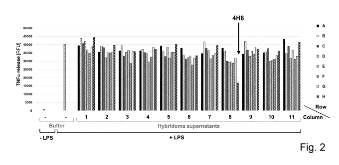
Figure 2 shows results from TNFa release assays (shedding assays) for
functional screening
of hybridoma supernatants, demonstrating that the supernatant of the hybridoma
clone 4H8
effectively interferes with LPS-induced shedding of TNFa in THP-1 cells.
Figure 3 depicts results from ELISA analyses for antibody isotype
determination
demonstrating the antibody 4H8-E3 of the invention to be of mouse IgM isotype.
Figure 4 shows results from peptide binding ELISA analyses revealing the
antibody 4H8-E3
of the invention to recognize an epitope within the section of the large
extracellular loop 1 of
human iRhom2 ("juxtamembrane domain", JMD) that is adjacent to the 1st
"transmembrane
domain" (TMD1).
The antibody 4H8-E3 of the invention recognizes peptide 3, which corresponds
to amino
acids 431 to 459 of human iRhom2, which is the JMD section of the large
extracellular loop
1 of human iRhom2 ("juxtamembrane domain") adjacent to TMD1.
3
CA 03133163 2021-09-10
WO 2020/208150 PCT/EP2020/060179
Figure 5 shows results from peptide binding ELISA analyses revealing the
antibody 4H8-E3
of the invention to recognize an epitope within the extracellular
juxtamembrane region
adjacent to the T1VID1 of human iRhom2, but not within the homologous region
of human
iRhoml. The antibody 4H8-E3 of the invention recognizes peptide 3, but not
peptide 3b,
which corresponds to the respective homologous section of human iRhoml.
Figure 6 shows results from TNFa release assays demonstrating the antibody 4H8-
E3 of the
invention to inhibit LPS-induced shedding of TNFa in THP-1 cells.
Figure 7 shows results from TNFa release assays demonstrating the
concentration-dependent
inhibition of LPS-induced TNFa shedding by the antibody 4H8-E3 of the
invention in THP-1
cells.
Figure 8 shows a schematic representation of iRhom2 with the positions of the
juxtamembrane domain adjacent to theTMD1 (A), loop 1 (B) and the C-terminus
(C) being
illustrated.
Figure 9 depicts the amino acid sequence of human iRhom2 according to SEQ ID
NO 16,
with the sequences shown which correspond to the immunization peptides used in
this
invention.
Figure 10 shows an alignment of human iRhom2 according to SEQ ID NO 16 and
human
iRhoml according to SEQ ID NO 17. The grey area shows sequence which
corresponds to
immunization peptide 3 used in this invention.
DETAILED DESCRIPTION
According to one aspect of the invention, a protein binder is provided that
binds to human
iRhom2, and inhibits and/or reduces TACE/ADAM17 activity when bound to human
iRhom2.
Rhomboid family member 2 (iRhom2) is a protein that in humans is encoded by
the
RHBDF2 gene. It is a transmembrane protein consisting of about 850 amino
acids, having
4
CA 03133163 2021-09-10
WO 2020/208150 PCT/EP2020/060179
seven transmembrane domains. The inventors of the present invention have for
the first time
demonstrated that iRhom2 can act as a target for protein binders to inhibit
TACE/ADAM17
activity.
iRhom2 comes in different isoforms. The experiments made herein have been
established
with the isoform defined as NCBI reference NP 078875.4. However, the teachings
are
transferable, without limitation, to other isoforms of iRhom2, as shown in the
following
table:
mRNA protein name
NM_024599.5 NP 078875.4 inactive rhomboid protein 2 transcript variant
1/
isoform 1
NM 001005498.3 NP 001005498.2 inactive rhomboid protein 2 transcript
variant 2/
isoform 2
As used herein, the term "inhibits and/or reduces TACE/ADAM17 activity is
meant to
describe an effect caused by a protein binder that blocks or reduces the
activity of
TACE/ADAM17, as measured e.g. in a respective shedding assay (see., e.g., Fig
2 and
example 5).
According to one or more embodiments, the protein binder is a monoclonal
antibody, or a
target-binding fragment or derivative thereof retaining target binding
capacities, or an
antibody mimetic.
As used herein, the term "monoclonal antibody (mAb)" shall refer to an
antibody
composition having a homogenous antibody population, i.e., a homogeneous
population
consisting of a whole immunoglobulin, or a fragment or derivative thereof
retaining target
binding capacities. Particularly preferred, such antibody is selected from the
group consisting
of IgG, IgD, IgE, IgA and/or IgM, or a fragment or derivative thereof
retaining target binding
capacities.
As used herein, the term "fragment" shall refer to fragments of such antibody
retaining target
binding capacities, e.g.
= a CDR (complementarity determining region)
CA 03133163 2021-09-10
WO 2020/208150 PCT/EP2020/060179
= a hypervariable region,
= a variable domain (Fv)
= an IgG or IgM heavy chain (consisting of VH, CHL hinge, CH2 and CH3
regions)
= an IgG or IgM light chain (consisting of VL and CL regions), and/or
= a Fab and/or F(ab)2.
As used herein, the term "derivative" shall refer to protein constructs being
structurally
different from, but still having some structural relationship to, the common
antibody concept,
e.g., scFv, Fab and/or F(ab)2, as well as bi-, tri- or higher specific
antibody constructs, and
further retaining target binding capacities. All these items are explained
below.
Other antibody derivatives known to the skilled person are Diabodies, Camelid
Antibodies,
Nanobodies, Domain Antibodies, bivalent homodimers with two chains consisting
of scFvs,
IgAs (two IgG structures joined by a J chain and a secretory component), shark
antibodies,
antibodies consisting of new world primate framework plus non-new world
primate CDR,
dimerized constructs comprising CH3+VL+VH, and antibody conjugates (e.g.
antibody or
fragments or derivatives linked to a toxin, a cytokine, a radioisotope or a
label). These types
are well described in the literature and can be used by the skilled person on
the basis of the
present disclosure, without adding further inventive activity.
Methods for the production of a hybridoma cell are disclosed in Kohler &
Milstein (1975).
Methods for the production and/or selection of chimeric or humanised mAbs are
known in
the art. For example, US6331415 by Genentech describes the production of
chimeric
antibodies, while US6548640 by Medical Research Council describes CDR grafting
techniques and US5859205 by Celltech describes the production of humanised
antibodies.
Methods for the production and/or selection of fully human mAbs are known in
the art. These
can involve the use of a transgenic animal which is immunized with the
respective protein or
peptide, or the use of a suitable display technique, like yeast display, phage
display, B-cell
display or ribosome display, where antibodies from a library are screened
against human
iRhom2 in a stationary phase.
6
CA 03133163 2021-09-10
WO 2020/208150 PCT/EP2020/060179
In vitro antibody libraries are, among others, disclosed in US6300064 by
MorphoSys and
US6248516 by MRC/Scripps/Stratagene. Phage Display techniques are for example
disclosed in US5223409 by Dyax. Transgenic mammal platforms are for example
described
in EP1480515A2 by TaconicArtemis.
IgG, IgM, scFv, Fab and/or F(ab)2 are antibody formats well known to the
skilled person.
Related enabling techniques are available from the respective textbooks.
As used herein, the term "Fab" relates to an IgG/IgM fragment comprising the
antigen
binding region, said fragment being composed of one constant and one variable
domain from
each heavy and light chain of the antibody
As used herein, the term "F(ab)2" relates to an IgG/IgM fragment consisting of
two Fab
fragments connected to one another by disulfide bonds.
As used herein, the term "scFv" relates to a single-chain variable fragment
being a fusion of
the variable regions of the heavy and light chains of immunoglobulins, linked
together with a
short linker, usually serine (S) or glycine (G). This chimeric molecule
retains the specificity
of the original immunoglobulin, despite removal of the constant regions and
the introduction
of a linker peptide.
Modified antibody formats are for example bi- or trispecific antibody
constructs, antibody-
based fusion proteins, immunoconjugates and the like. These types are well
described in the
literature and can be used by the skilled person on the basis of the present
disclosure, with
adding further inventive activity.
As used herein, the term "antibody mimetic" relates to an organic molecule,
most often a
protein that specifically binds to a target protein, similar to an antibody,
but is not structurally
related to antibodies. Antibody mimetics are usually artificial peptides or
proteins with a
molar mass of about 3 to 20 kDa. The definition encompasses, inter alia,
Affibody
molecules, Affilins, Affimers, Affitins, Alphabodies, Anticalins, Avimers,
DARPins,
Fynomers, Kunitz domain peptides, Monobodies, and nanoCLAMPs.
7
CA 03133163 2021-09-10
WO 2020/208150 PCT/EP2020/060179
In one or more embodiments, the protein binder is an isolated antibody, or a
target-binding
fragment or derivative thereof retaining target binding capacities, or an
isolated antibody
mimetic
In one or more embodiments, the antibody is an engineered or recombinant
antibody, or a
target binding fragment or derivative thereof retaining target binding
capacities, or an
engineered or recombinant antibody mimetic.
According to one or more embodiments of the invention, the inhibition or
reduction of
TACE/ADAM17 activity is caused by interference with iRhom2-mediated
TACE/ADAM17
activation.
According to one or more embodiments of the invention, the antibody inhibits
or reduces
TNFa shedding.
TNFa shedding, as used herein, refers to a process in which membrane-anchored
tumor
necrosis factor alpha (mTNFa/pro-TNFa) is released into the environment to
become soluble
TNFa (sTNFa or simply TNFa). This process is, inter alia, triggered by
TACE/ADAM17.
According to one or more embodiments of the invention, the human iRhom2 to
which the
protein binder binds comprises
a) the amino acid sequence set forth in SEQ ID NO 16, or
b) an amino acid sequence that has at least 80 % sequence identity with SEQ ID
NO
16, with the proviso that said sequence maintains iRhom2 activity.
In some embodiments, human iRhom2 comprises an amino acid sequence that has
>81%,
preferably >82%, more preferably >83%, >84%, >85%, >86%, >87%, >88%, >89%,
>90%, >91%, >92%, >93%, >94%, >95%, >96%, >97%, >98 or most preferably >99 %
sequence identity with SEQ ID NO 16.
SEQ ID NO 16 represents the amino acid sequence of inactive rhomboid protein 2
(iRhom2)
isoform 1 [Homo sapiens], accessible under NCBI reference NP 078875.4.
Generally,
different variants and isoforms of iRhom2 exist. Likewise, mutants comprising
conservative
8
CA 03133163 2021-09-10
WO 2020/208150 PCT/EP2020/060179
or silent amino acid substitutions exist, or may exist, which maintain full or
at least
substantial iRhom2 activity. These isoforms, variants and mutants are
encompassed by the
identity range specified above, meaning however that dysfunctional, non-active
variants and
mutants are excluded.
In this context, a "conservative amino acid substitution", has a smaller
effect on antibody
function than a non-conservative substitution. Although there are many ways to
classify
amino acids, they are often sorted into six main groups on the basis of their
structure and the
general chemical characteristics of their R groups.
In some embodiments, a "conservative amino acid substitution" is one in which
the amino
acid residue is replaced with an amino acid residue having a similar side
chain. For example,
families of amino acid residues having similar side chains have been defined
in the art. These
families include amino acids with
= basic side chains (e.g., lysine, arginine, histidine),
= acidic side chains (e.g., aspartic acid, glutamic acid),
= uncharged polar side chains (e.g., glycine, asparagine, glutamine,
serine, threonine,
tyrosine, cysteine),
= nonpolar side chains (e.g., alanine, valine, leucine, isoleucine,
proline, phenylalanine,
methionine, tryptophan),
= beta-branched side chains (e.g., threonine, valine, isoleucine) and
= aromatic side chains (e.g., tyrosine, phenylalanine, tryptophan,
histidine).
Other conserved amino acid substitutions can also occur across amino acid side
chain
families, such as when substituting an asparagine for aspartic acid in order
to modify the
charge of a peptide. Conservative changes can further include substitution of
chemically
homologous non-natural amino acids (i.e. a synthetic non-natural hydrophobic
amino acid in
place of leucine, a synthetic non-natural aromatic amino acid in place of
tryptophan).
"Percentage of sequence identity" is determined by comparing two optimally
aligned
sequences over a comparison window, wherein the portion of the polynucleotide
sequence in
the comparison window may comprise additions or deletions (i.e., gaps) as
compared to the
9
CA 03133163 2021-09-10
WO 2020/208150 PCT/EP2020/060179
reference sequence (e.g., a polypeptide), which does not comprise additions or
deletions, for
optimal alignment of the two sequences. The percentage is calculated by
determining the
number of positions at which the identical nucleic acid base or amino acid
residue occurs in
both sequences to yield the number of matched positions, dividing the number
of matched
positions by the total number of positions in the window of comparison and
multiplying the
result by 100 to yield the percentage of sequence identity.
The terms "identical" or percent "identity," in the context of two or more
nucleic acids or
polypeptide sequences, refer to two or more sequences or subsequences that are
the same
sequences. Two sequences are "substantially identical" if two sequences have a
specified
percentage of amino acid residues or nucleotides that are the same (i.e., at
least 85%, 90%,
95%, 96%, 97%, 98% or 99% sequence identity over a specified region, or, when
not
specified, over the entire sequence of a reference sequence), when compared
and aligned for
maximum correspondence over a comparison window, or designated region as
measured
using one of the following sequence comparison algorithms or by manual
alignment and
visual inspection. .. The disclosure provides polypeptides or polynucleotides
that are
substantially identical to the polypeptides or polynucleotides, respectively,
exemplified
herein. Optionally, the identity exists over a region that is at least about
15, 25 or 50
nucleotides in length, or more preferably over a region that is 100 to 500 or
1000 or more
nucleotides in length, or over the full length of the reference sequence. With
respect to amino
acid sequences, identity or substantial identity can exist over a region that
is at least 5, 10, 15
or 20 amino acids in length, optionally at least about 25, 30, 35, 40, 50, 75
or 100 amino
acids in length, optionally at least about 150, 200 or 250 amino acids in
length, or over the
full length of the reference sequence. With respect to shorter amino acid
sequences, e.g.,
amino acid sequences of 20 or fewer amino acids, substantial identity exists
when one or two
amino acid residues are conservatively substituted, according to the
conservative
substitutions defined herein.
According to one or more embodiments of the invention, the protein binder
binds to the
extracellular juxtamembrane domain adjacent to the transmembrane domain 1
(TMD1) of
human iRhom2.
CA 03133163 2021-09-10
WO 2020/208150 PCT/EP2020/060179
The juxtamembrane domain adjacent to the transmembrane domain 1 (TMD1) is a
region
that encompasses a stretch of amino acids C-terminally of the first
transmembrane domain
(TMD1). See Figures 8 and 9 for an illustration.
In one embodiment, the juxtamembrane domain adjacent to the transmembrane
domain 1
(TMD1) comprises amino acids 431 ¨459 of an amino acid sequence set forth in
SEQ ID NO
16, or of an amino acid sequence that has at least 80 % sequence identity with
SEQ ID NO
16.
In another embodiment, the juxtamembrane domain adjacent to the transmembrane
domain 1
(TMD1) comprises amino acids 431 ¨447 of an amino acid sequence set forth in
SEQ ID NO
16, or of an amino acid sequence that has at least 80 % sequence identity with
SEQ ID NO
16.
According to one or more embodiments of the invention, the protein binder
binds to an amino
acid sequence of human iRhom2 comprising
a) at least the amino acid sequence set forth in SEQ ID NO 3, or
b) an amino acid sequence that has at least 90 % sequence identity with SEQ ID
NO
3.
In some embodiments, the amino acid sequence that has >91%, preferably >92%,
more
preferably >93%, >94%, >95%, >96%, >97%, >98 or most preferably >99 % sequence
identity with SEQ ID NO 3.
In one embodiment, the antibody binds the entire amino acid sequence as set
forth above. In
another embodiment, the antibody binds also further amino acid sequences of
human iRhom2
outside of SEQ ID NO3, or outside of the amino acid sequence that has at least
90 %
sequence identity with SEQ ID NO 3.
Depending on where the further amino acids are located, the epitope that the
antibody binds
is linear or conformational.
11
CA 03133163 2021-09-10
WO 2020/208150 PCT/EP2020/060179
According to one or more embodiments of the invention, the protein binder
binds to one or
more amino acid sequences of human iRhom2 each comprising one or more amino
acids
within the amino acid sequence set forth in SEQ ID NO 3.
In one embodiment, the antibody binds one discrete subsequence within SEQ ID
NO 3,
which comprises one or more amino acids.
In one embodiment, the antibody binds to two or more discrete subsequences
within SEQ ID
NO 3, each of which comprises one or more amino acids
According to one or more embodiments of the invention, the protein binder
binds to at least
one amino acid residue selected from the group consisting of A431, Q432, H433,
V434,
T435, T436, Q437, L438, V439, L440, R441, N442, K443, G444, V445, Y446, E447,
S448,
V449, K450, Y451, 1452, Q453, Q454, E455, N456, F457, W458, V459, wherein the
numbering of the amino acid residues is relative to the amino acid sequence
set forth in SEQ
ID NO 16 (human iRhom2)
In one or more embodiments the protein binder binds to >2, >3, >4, >5, >6, >7,
>8, >9, >10,
>11, >12, >13, >14, >15, >16, >17, >18, >19, >20, >21, >22, >23, >24, >25,
>26, >27, >28,
or >29 amino acid residues from the above list. The respective amino acid
residues can be
present in a discrete, consecutive sequence, or in two or more clusters within
SEQ ID NO 3.
In another embodiment, the protein binder binds to an amino acid sequence of
human
iRhom2 comprising at least the amino acid sequence set forth in SEQ ID NO 4,
or an amino
acid sequence that has at least 90 % sequence identity with SEQ ID NO 4. The
same fallback
positions regarding the sequence identity apply.
In one embodiment, the antibody binds the entire amino acid sequence as set
forth above. In
another embodiment, the antibody binds also further amino acid sequences of
human iRhom2
outside of SEQ ID NO 4, or outside of the amino acid sequence that has at
least 90 %
sequence identity with SEQ ID NO 4.
Depending on where the further amino acids are located, the epitope that the
antibody binds
is linear or conformational.
12
CA 03133163 2021-09-10
WO 2020/208150 PCT/EP2020/060179
According to one or more embodiments of the invention, the protein binder
binds to one or
more amino acid sequences of human iRhom2 each comprising one or more amino
acids
within the amino acid sequence set forth in SEQ ID NO 4
In one embodiment, the antibody binds one discrete subsequence within SEQ ID
NO 4,
which comprises one or more amino acids.
In one embodiment, the antibody binds to two or more discrete subsequences
within SEQ ID
NO 4, each of which comprises one or more amino acids.
According to one or more embodiments of the invention, the protein binder
binds to at least
one amino acid residue selected from the group consisting of A426, P427, V428,
G429,
F430, A431, Q432, H433, V434, T435, T436, Q437, L438, V439, L440, R441, N442,
K443,
G444, V445, Y446, E447, S448, V449, K450, Y451, 1452, Q453, Q454, E455, N456,
F457,
W458, V459, wherein the numbering of the amino acid residues is relative to
the amino acid
sequence set forth in SEQ ID NO 16 (human iRhom2)
In one or more embodiments the protein binder binds to >2, >3, >4, >5, >6, >7,
>8, >9, >10,
>11, >12, 13, >14, >15, >16, >17, >18, >19, >20, >21, >22, >23, >24, >25, >26,
>27, >28,
>29, >30, >31, >32, >33 or >34 amino acid residues from the above list. The
respective
amino acid residues can be present in a discrete, consecutive sequence, or in
two or more
clusters within SEQ ID NO 3.
According to one or more embodiments of the invention, the protein binder is
not cross-
reactive with human iRhom 1, or the juxtamembrane domain adjacent to the
transmembrane
domain 1 (TMD1) thereof
According to one or more embodiments of the invention, the protein binder is
cross-reactive
with murine iRhom2, or the juxtamembrane domain adjacent to the transmembrane
domain 1
(TMD1) thereof
According to one or more embodiments of the invention, the protein binder is
an antibody in
at least one of the formats selected from the group consisting of: IgG, scFv,
Fab, (Fab)2.
13
CA 03133163 2021-09-10
WO 2020/208150 PCT/EP2020/060179
According to one or more embodiments of the invention, the protein binder is
an antibody
having an isotype selected from the group consisting of IgG, IgM.
According to one or more embodiments of the invention, the protein binder is a
murine,
chimerized, humanized, or human antibody.
According to one embodiment of the invention, the protein binder is the
antibody 4H8-E3. In
one embodiment, the protein binder is an antibody which comprises the variable
domains or
the CDRs of 4H8-E3.
According to one embodiment of the invention, the protein binder
a) comprises a set of heavy chain/light chain complementarity determining
regions
(CDR) comprised in the heavy chain/light variable region sequence pair set
forth in
SEQ ID NOs 33 and 40
b) comprises a set of heavy chain/light chain complementarity determining
regions
(CDR) comprising the following sequences
= HC CDR1 (SEQ ID NO 34 or 37)
= HC CDR2 (SEQ ID NO 35 or 38)
= HC CDR3 (SEQ ID NO 36 or 39)
= LC CDR1 (SEQ ID NO 41 0r44)
= LC CDR2 (SEQ ID NO 42 or 45), and
= LC CDR3 (SEQ ID NO 43 or 46)
c) comprises the heavy chain/light chain complementarity determining regions
(CDR) of
b), with the proviso that at least one of the CDRs has up to 3 amino acid
substitutions
relative to the respective SEQ ID NO 34 ¨ 39 or 41 ¨46, and/or
14
CA 03133163 2021-09-10
WO 2020/208150 PCT/EP2020/060179
d) comprises the heavy chain/light chain complementarity determining regions
(CDR)
of b) or c), with the proviso that at least one of the CDRs has a sequence
identity of
> 66 % to the respective SEQ ID NO 34 ¨39 or 41 ¨46,
wherein the CDRs are embedded in a suitable protein framework so as to be
capable to bind
to human iRhom2 with sufficient binding affinity and to inhibit or reduce
TACE/ADAM17
activity.
These CDRs are the CDRs sets of the antibody 4H8-E3, determined with different
approaches (SEQ ID NOs 34 ¨ 39 determined with the paratome CDR identification
tool
(http://ofranservices.biu.ac.il/site/services/paratome), and SEQ ID NOs 41 ¨
46 determined
with in house methods).
As used herein, the term "CDR" or "complementarity determining region" is
intended to
mean the non-contiguous antigen combining sites found within the variable
region of both
heavy and light chain polypeptides. These particular regions have been
described by Kabat et
al. (1977), Kabat et al. (1991), Chothia et al. (1987) and MacCallum et al.,
(1996) where the
definitions include overlapping or subsets of amino acid residues when
compared against
each other. Nevertheless, application of either definition to refer to a CDR
of an antibody or
grafted antibodies or variants thereof is intended to be within the scope of
the term as defined
and used herein. The amino acid residues which encompass the CDRs as defined
by each of
the above cited references are set forth below in Table 1 as a comparison.
Note that this
numbering may differ from the CDRs that acre actually disclosed in the
enclosed sequence
listing, because CDR definitions vary from case to case.
Kabat Chothia MacCallum
VH CDR1 31-35 26-32 30-35
VH CDR2 50-65 53-55 47-58
VH CDR3 95-102 96-101 93-101
VL CDR1 24-34 26-32 30-36
VL CDR2 50-56 50-52 46-55
VL CDR3 89-97 91-96 89-96
Table 1: CDR definitions
CA 03133163 2021-09-10
WO 2020/208150 PCT/EP2020/060179
As used herein, the term "framework" when used in reference to an antibody
variable region
is entered to mean all amino acid residues outside the CDR regions within the
variable region
of an antibody. Therefore, a variable region framework is between about 100-
120 amino
acids in length but is intended to reference only those amino acids outside of
the CDRs.
As used herein, the term "capable to bind to target X with sufficient binding
affinity" has to
be understood as meaning that respective binding domain binds the target with
a KD of 10'
or smaller. KD is the equilibrium dissociation constant, a ratio of koffikon,
between the protein
binder and its antigen. KD and affinity are inversely related. The KD value
relates to the
concentration of protein binder (the amount of protein binder needed for a
particular
experiment) and so the lower the KD value (lower concentration) and thus the
higher the
affinity of the binding domain. The following table shows typical KD ranges of
monoclonal
antibodies
KD value Molar range
104 to 10' Micromolar (04)
10-7 to 10-9 Nanomolar (nM)
10-10 to 10-12 Picomolar (pM)
10-" to 10-" Femtomolar (fM)
Table 2. KD and Molar Values
Preferably, the protein binder has up to 2 amino acid substitutions, and more
preferably up to
1 amino acid substitutions
Preferably, at least one of the CDRs has a sequence identity of > 67 %; > 68
%; > 69
%; > 70 %; > 71 %; > 72 %; > 73 %; > 74 %; > 75 %; > 76 %; > 77 %; > 78 %; >
79
%; > 80 %; > 81 %; > 82 %; > 83 %; > 84 %; > 85 %; > 86 %; > 87 %; > 88 %; >
89
%; > 90 %; > 91 %; > 92 %; > 93 %; > 94 %; > 95 %; > 96 %; > 97 %; > 98 %; >
99 %,
and most preferably? 100 % to the respective SEQ ID NO.
16
CA 03133163 2021-09-10
WO 2020/208150 PCT/EP2020/060179
As used herein, the term "% sequence identity", has to be understood as
follows: Two
sequences to be compared are aligned to give a maximum correlation between the
sequences.
This may include inserting "gaps" in either one or both sequences, to enhance
the degree of
alignment. A % identity may then be determined over the whole length of each
of the
sequences being compared (so-called global alignment), that is particularly
suitable for
sequences of the same or similar length, or over shorter, defined lengths (so-
called local
alignment), that is more suitable for sequences of unequal length. In the
above context, an
amino acid sequence having a "sequence identity" of at least, for example, 95%
to a query
amino acid sequence, is intended to mean that the sequence of the subject
amino acid
sequence is identical to the query sequence except that the subject amino acid
sequence may
include up to five amino acid alterations per each 100 amino acids of the
query amino acid
sequence. In other words, to obtain an amino acid sequence having a sequence
of at least
95% identity to a query amino acid sequence, up to 5% (5 of 100) of the amino
acid residues
in the subject sequence may be inserted or substituted with another amino acid
or deleted.
Methods for comparing the identity and homology of two or more sequences are
well known
in the art. The percentage to which two sequences are identical can for
example be
determined by using a mathematical algorithm. A preferred, but not limiting,
example of a
mathematical algorithm is integrated in the BLAST family of programs, e.g.
BLAST or
NBLAST program and FASTA. Sequences which are identical to other sequences to
a certain
extent can be identified by these programmes. Furthermore, programs available
in the
Wisconsin Sequence Analysis Package, version 9.1 for example the programs
BESTFIT and
GAP, may be used to determine the % identity between two polypeptide
sequences. If herein
reference is made to an amino acid sequence sharing a particular extent of
sequence identity
to a reference sequence, then said difference in sequence is preferably due to
conservative
amino acid substitutions. Preferably, such sequence retains the activity of
the reference
sequence, e.g. albeit maybe at a slower rate.
Preferably, at least one of the CDRs has been subject to CDR sequence
modification,
including
= affinity maturation
= reduction of immunogenicity
17
CA 03133163 2021-09-10
WO 2020/208150 PCT/EP2020/060179
Affinity maturation in the process by which the affinity of a given antibody
is increased in
vitro. Like the natural counterpart, in vitro affinity maturation is based on
the principles of
mutation and selection. It has successfully been used to optimize antibodies,
antibody
fragments or other peptide molecules like antibody mimetics. Random mutations
inside the
CDRs are introduced using radiation, chemical mutagens or error-prone PCR. In
addition, the
genetic diversity can be increased by chain shuffling. Two or three rounds of
mutation and
selection using display methods like phage display usually results in antibody
fragments with
affinities in the low nanomolar range. For principles see Eylenstein et al.
(2016), the content
of which is incorporated herein by reference.
Humanized antibodies contain murine-sequence derived CDR regions that have
been
engrafted, along with any necessary framework back-mutations, into human
sequence-
derived V regions. Hence, the CDRs themselves can cause immunogenic reactions
when the
humanized antibody is administered to a patient. Methods of reducing
immunogenicity
caused by CDRs are disclosed in Harding et al. (2010), the content of which is
incorporated
herein by reference.
According to one embodiment of the invention, the framework is a human VH/VL
framework. VH stands for heavy chain variable domain of an IgG shaped
antibody, while VL
stands for light chain variable domain (kappa or lambda)
According to one embodiment of the invention, the protein binder comprises
a) the heavy chain/light chain variable domains (VD)
= HC VD (SEQ ID NO 33), and
= LC VD (SEQ ID NO 40)
b) the heavy chain/light chain variable domains (VD) of a), with the proviso
that
= the HCVD has a sequence identity of > 80 % to the respective SEQ ID NO
33,
and/or
= the LCDVD has a sequence identity of > 80 % to the respective SEQ ID NO
40,
18
CA 03133163 2021-09-10
WO 2020/208150 PCT/EP2020/060179
c) the heavy chain/light chain variable domains (VD) of a) or b), with the
proviso that at
least one of the HCVD or LCVD has up to 10 amino acid substitutions relative
to the
respective SEQ ID NO 33 and/or 40.
said protein binder still being capable to bind to human iRhom2 with
sufficient binding
affinity and to inhibit or reduce TACE/ADAM17 activity.
Preferably, the HCVD and/or LCVD has a sequence identity of > 81 %; > 82 %; >
83 %; >
84 %; > 85 %; > 86 %; > 87 %; > 88 %; > 89 %; > 90 %; >91 %; > 92 %; > 93 %; >
94 %; >
95 %; > 96 %; > 97 %; > 98 %; > 99 %; or most preferably > 100 % to the
respective SEQ ID
NO.
A "variable domain" when used in reference to an antibody or a heavy or light
chain thereof
is intended to mean the portion of an antibody which confers antigen binding
onto the
molecule and which is not the constant region. The term is intended to include
functional
fragments thereof which maintain some of all of the binding function of the
whole variable
region. Variable region binding fragments include, for example, functional
fragments such as
Fab, F(ab)2, Fv, single chain Fv (scfv) and the like. Such functional
fragments are well
known to those skilled in the art. Accordingly, the use of these terms in
describing functional
fragments of a heteromeric variable region is intended to correspond to the
definitions well
known to those skilled in the art. Such terms are described in, for example,
Huston et al.,
(1993) or Pluckthun and Skerra (1990).
According to one embodiment of the invention, at least one amino acid
substitution discussed
above is a conservative amino acid substitution.
A õconservative amino acid substitution" has a smaller effect on protein
binder function than
a non-conservative substitution. Although there are many ways to classify
amino acids, they
are often sorted into six main groups on the basis of their structure and the
general chemical
characteristics of their R groups.
In one embodiment, a "conservative amino acid substitution" is one in which
the amino acid
residue is replaced with an amino acid residue having a similar side chain.
For example,
19
CA 03133163 2021-09-10
WO 2020/208150 PCT/EP2020/060179
families of amino acid residues having similar side chains have been defined
in the art. These
families include amino acids with
= basic side chains (e.g., lysine, arginine, histidine),
= acidic side chains (e.g., aspartic acid, glutamic acid),
= uncharged polar side chains (e.g., glycine, asparagine, glutamine,
serine, threonine,
tyrosine, cysteine),
= nonpolar side chains (e.g., alanine, valine, leucine, isoleucine,
proline, phenylalanine,
methionine, tryptophan),
= beta-branched side chains (e.g., threonine, valine, isoleucine) and
= aromatic side chains (e.g., tyrosine, phenylalanine, tryptophan,
histidine).
Other conserved amino acid substitutions can also occur across amino acid side
chain
families, such as when substituting an asparagine for aspartic acid in order
to modify the
charge of a peptide. Thus, a predicted nonessential amino acid residue in a HR
domain
polypeptide, for example, is preferably replaced with another amino acid
residue from the
same side chain family or homologues across families (e.g. asparagine for
aspartic acid,
glutamine for glutamic acid). Conservative changes can further include
substitution of
chemically homologous non-natural amino acids (i.e. a synthetic non-natural
hydrophobic
amino acid in place of leucine, a synthetic non-natural aromatic amino acid in
place of
tryptophan).
According to one embodiment of the invention, the protein binder has at least
one of
= target binding affinity of > 50 % to iRhom2, and measured by SPR,
compared to that
of the protein binder according to the above description, and/or
= > 50 % of the inhibiting or reducing effect on TACE/ADAM17 activity of
the
protein binder according to the above description
As used herein the term "binding affinity" is intended to mean the strength of
a binding
interaction and therefore includes both the actual binding affinity as well as
the apparent
binding affinity. The actual binding affinity is a ratio of the association
rate over the
disassociation rate. Therefore, conferring or optimizing binding affinity
includes altering
CA 03133163 2021-09-10
WO 2020/208150 PCT/EP2020/060179
either or both of these components to achieve the desired level of binding
affinity. The
apparent affinity can include, for example, the avidity of the interaction.
For example, a
bivalent heteromeric variable region binding fragment can exhibit altered or
optimized
binding affinity due to its valency.
A suitable method for measuring the affinity of a binding agent is through
surface plasmon
resonance (SPR). This method is based on the phenomenon which occurs when
surface
plasmon waves are excited at a metal/liquid interface. Light is directed at,
and reflected from,
the side of the surface not in contact with sample, and SPR causes a reduction
in the reflected
light intensity at a specific combination of angle and wavelength.
Biomolecular binding
events cause changes in the refractive index at the surface layer, which are
detected as
changes in the SPR signal. The binding event can be either binding association
or
disassociation between a receptor-ligand pair. The changes in refractive index
can be
measured essentially instantaneously and therefore allows for determination of
the individual
components of an affinity constant. More specifically, the method enables
accurate
measurements of association rates (k on) and disassociation rates (koff).
Measurements of k on and koff values can be advantageous because they can
identify altered
variable regions or optimized variable regions that are therapeutically more
efficacious. For
example, an altered variable region, or heteromeric binding fragment thereof,
can be more
efficacious because it has, for example, a higher konvalued compared to
variable regions and
heteromeric binding fragments that exhibit similar binding affinity. Increased
efficacy is
conferred because molecules with higher kon values can specifically bind and
inhibit their
target at a faster rate. Similarly, a molecule of the invention can be more
efficacious because
it exhibits a lower koff value compared to molecules having similar binding
affinity. Increased
efficacy observed with molecules having lower koff rates can be observed
because, once
bound, the molecules are slower to dissociate from their target. Although
described with
reference to the altered variable regions and optimized variable regions of
the invention
including, heteromeric variable region binding fragments thereof, the methods
described
above for measuring associating and disassociation rates are applicable to
essentially any
protein binder or fragment thereof for identifying more effective binders for
therapeutic or
diagnostic purposes.
21
CA 03133163 2021-09-10
WO 2020/208150 PCT/EP2020/060179
Methods for measuring the affinity, including association and disassociation
rates using
surface plasmon resonance are well known in the arts and can be found
described in, for
example, Jonsson and Malmquist, (1992) and Wu et al. (1998). Moreover, one
apparatus well
known in the art for measuring binding interactions is a BIAcore 2000
instrument which is
commercially available through Pharmacia Biosensor, (Uppsala, Sweden).
Preferably said target binding affinity is > 51%, > 52%, > 53%, > 54%, > 55%,
> 56%, >
57%, > 58%, > 59%, > 60%, > 61%, > 62%, > 63%, > 64%, > 65%, > 66%, > 67%, >
68%, >
69%, > 70%, > 71%, > 72%, > 73%, > 74%, > 75%, > 76%, > 77%, > 78%, > 79%, >
80%, >
81%, > 82%, > 83%, > 84%, > 85%, > 86%, > 87%, > 88%, > 89%, > 90%, > 91%, >
92%, >
93%, > 94%, > 95%, > 96%, > 97%, > 98%, and most preferably > 99 % compared to
that of
the reference binding agent.
As used herein, the quantification of the inhibiting or reducing effect on
TACE/ADAM17
activity, compared to a benchmark binding agent, can be carried out, e.g.,
with a respective
TNF shedding assay (see., e.g., Fig 2 and example 5).
According to another aspect of the invention, a protein binder is provided
which competes for
binding to human iRhom2 with any of the protein binders set forth above.
As regards the format or structure of such protein binders, the same preferred
embodiments
as set forth above apply. In one embodiment, said protein binder is a
monoclonal antibody,
or a target-binding fragment or derivative thereof retaining target binding
capacities, or an
antibody mimetic.
As used herein, the term "competes for binding" is used in reference to one of
the antibodies
defined by the sequences as above, meaning that the actual protein binder as
an activity
which binds to the same target, or target epitope or domain or subdomain, as
does said
sequence defined protein binder, and is a variant of the latter. The
efficiency (e.g., kinetics or
thermodynamics) of binding may be the same as or greater than or less than the
efficiency of
the latter. For example, the equilibrium binding constant for binding to the
substrate may be
different for the two antibodies.
22
CA 03133163 2021-09-10
WO 2020/208150 PCT/EP2020/060179
Such competition for binding can be suitably measured with a competitive
binding assay.
Such assays are disclosed in Finco et al. 2011, the content of which is
incorporated herein by
reference, and their meaning for interpretation of a patent claim is disclosed
in Deng et al
2018, the content of which is incorporated herein by reference.
According to another aspect of the invention, a protein binder is provided
that binds to
essentially the same, or the same, epitope on iRhom2 as the protein binder
according to the
above description.
In order to test for this characteristic, suitable epitope mapping
technologies are available,
including, inter alia,
= X-ray co-crystallography and cryogenic electron microscopy (cryo-EM)
= Array-based oligo-peptide scanning
= Site-directed mutagenesis mapping
= High-throughput shotgun mutagenesis epitope mapping
= Hydrogen¨deuterium exchange
= Cross-linking-coupled mass spectrometry
These methods are, inter alia, disclosed and discussed in Banik et al (2010),
and DeLisser
(1999), the content of which is herein incorporated by reference.
According to another aspect of the invention, a nucleic acid that encodes for
a binding agent
according to any one of the aforementioned claims.
A given sequence of the encoded binding agent provided, such nucleic acid can
have
different sequences due to the degeneracy of the genetic code.
Such nucleic acid can be used for pharmaceutic purposes. In such case, it is
an RNA-derived
molecule that is administered to a patient, wherein the protein expression
machinery of the
patient expresses the respective binding agent. The mRNA can for example be
delivered in
suitable liposomes and comprises either specific sequences or modified uridine
nucleosides to
avoid immune responses and/or improve folding and translation efficiency,
sometimes
23
CA 03133163 2021-09-10
WO 2020/208150 PCT/EP2020/060179
comprising cap modifications at the 5'- and/or 3' terminus to target them to
specific cell
types.
Such nucleic acid can be used for transfecting an expression host to then
express the actual
binding agent. In such case, the molecule can be a cDNA that is optionally
integrated into a
suitable vector.
According to another aspect of the invention, the use of the protein binder
according to the
above description is provided (for the manufacture of a medicament) in the
treatment of a
human or animal subject
= being diagnosed for,
= suffering from or
= being at risk of developing
an inflammatory condition, or for the prevention of such condition.
According to another aspect of the invention, a pharmaceutical composition
comprising the
protein binder according to the above description, and optionally one or more
pharmaceutically acceptable excipients, is provided.
According to another aspect of the invention, a combination is provided
comprising (i) the
protein binder according to the above description or the pharmaceutical
composition
according to the above description and (ii) one or more therapeutically active
compounds.
According to another aspect of the invention, a method for treating or
preventing an
inflammatory condition is provided, which method comprises administration, to
a human or
animal subject, of (i) the protein binder according to the above description,
(ii) the
pharmaceutical composition according to the above description or (iii) the
combination
according to the above description, in a therapeutically sufficient dose.
According to another aspect of the invention, a therapeutic kit of parts is
provided,
comprising:
24
CA 03133163 2021-09-10
WO 2020/208150 PCT/EP2020/060179
a) the composition according to the above description, the pharmaceutical
composition according to the above description, or the combination according
to
the above description,
b) an apparatus for administering the composition, composition or combination,
and
c) instructions for use.
EXAMPLES
While the invention has been illustrated and described in detail in the
drawings and foregoing
description, such illustration and description are to be considered
illustrative or exemplary
and not restrictive; the invention is not limited to the disclosed
embodiments. Other
variations to the disclosed embodiments can be understood and effected by
those skilled in
the art in practicing the claimed invention, from a study of the drawings, the
disclosure, and
the appended claims. In the claims, the word "comprising" does not exclude
other elements
or steps, and the indefinite article "a" or "an" does not exclude a plurality.
The mere fact that
certain measures are recited in mutually different dependent claims does not
indicate that a
combination of these measures cannot be used to advantage. Any reference signs
in the
claims should not be construed as limiting the scope.
All amino acid sequences disclosed herein are shown from N-terminus to C-
terminus; all
nucleic acid sequences disclosed herein are shown 5'->3'.
Example 1: Generation of peptides for immunization and peptide binding ELISA
analyses
Peptides were either synthesized on a parallel peptide synthesizer (peptides 1-
5, 7-9 and lb-
3b; MultiPep RSi, Intavis AG, Germany), on a microwave peptide synthesizer
(peptide 6;
Liberty Blue, CEM, USA) or on a custom made continuous flow peptide
synthesizer
(peptides 10, 11 and 4b) using Fluorenylmethoxycarbonyl (Fmoc)-based Solid
Phase Peptide
Synthesis. [Chan, W.C., White, P.D. Solid Phase Peptide Synthesis, A Practical
Approach
(Oxford University Press Inc., New York, 2000]. The sequences were assembled
in a
stepwise fashion from C to N-terminus using Fmoc-protected L-amino acids with
side chain
protection groups. Upon completion of the chain assembly peptides were cleaved
off the
CA 03133163 2021-09-10
WO 2020/208150 PCT/EP2020/060179
resin with 95% TFA, 4% triethylsilane and 1% water. The crude product was
dissolved in
15% acetonitrile in 0.1% aq TFA and purified by reversed phase HPLC using an
Orbit C18,
pm, 100 A column (MZ Analysentechnik, Germany). The resulting purified
fractions were
analyzed by analytical HPLC using a Kinetex EVO C18, 5 pm, 100 A column
(Phenomenex,
USA) and by MALDI TOF mass spectrometry (Ultraflex III, Bruker, USA). The
fractions
were lyophilized yielding the corresponding TFA salt.
For peptide 10 and lithe linear peptides as identified by mass spectrometry
were oxidized to
the corresponding cyclic disulfides by DMSO mediated oxidation. For this
purpose. the linear
peptides were dissolved in 5% acetic acid at a concentration of 1 mg/ml. The
pH was
adjusted to 6 with (NH4)2CO3 and DMSO was added to a final concentration of 10-
20%.
The oxidation was allowed to proceed for 24 hours at room temperature.
Afterwards the
reaction mixture was diluted with solvent A. The product was purified on a
reversed phase
C18 column and analyzed as described above. Fractions containing the disulfide
cyclized
peptides were pooled and lyophilized. [Chan, W.C. and White, P.D., Fmoc Solid
Phase
Peptide Synthesis, A Practical Approach (Oxford University Press Inc., New
York, 2000,
Chapter 3.3, page 97]
KLH conjugation was performed with pre-activated KLH (ImcejtTM Maleimide
Activated
mcKLH, Thermo Scientific, USA). Briefly, mcKLH was dissolved with ultrapure
water at a
concentration of 10 mg/ml. The desired peptide was dissolved at a
concentration of 5 mg/mL
in ImjectTM Maleimide Conjugation Buffer (Thermo Scientific, USA), if
necessary 8 M Urea
(pH 7.2) was added to dissolve the peptide. The peptide solution was mixed
with the mcKLH
solution and incubated for 2 to 6 hours at room temperature. The mixture was
dialyzed
overnight with a 3500-MW cut-off (MWCO) dialysis tube against 400 mL PBS.
After
dialysis the mixture was diluted with PBS to yield the desired concentration.
Biotinylation was performed with alpha-Biotin-omega-maleimido undeca(ethylene
glycol)
(Biotin-PEG(11)-mal). The peptides were dissolved in PBS pH 7,4. If necessary,
acetonitrile
was added to dissolve the peptides. Biotin-PEG(11)-mal was dissolved in DMF
and added to
the peptide solution in (weight amount =1:1). The reaction was performed
overnight and
subsequently purified on a reversed phase C18 column and analyzed as described
above.
26
CA 03133163 2021-09-10
WO 2020/208150 PCT/EP2020/060179
Figure 1 depicts the peptides used for immunization and/or peptide binding
ELISA analyses,
indicating their designation, position number and sequence of amino acids with
regard to
NCBI reference sequences NM 024599.5., NP 078875.4. for human iRhom2 and NCBI
reference sequences NM 022450.3., NP 071895.3. for human iRhoml. A terminal
cysteine
residue added to all peptides except peptides 6 and 7 for coupling to KLH (for
immunization)
and/or biotin (for peptide binding ELISA analyses) is illustrated by "-C-".
Internal cysteine
residues are replaced by alpha-aminobutyric acid (Abu) where indicated.
Peptides 1 to 4
correspond to amino acids of TMD1 (highlighted in italics) and the adjacent
extracellular
juxtamembrane region of human iRhom2 (Figure 1A). Peptides 5 to 7 resemble
sections
within the large extracellular loop 1 of human iRhom2 linking T1VID1 and
T1VID2 (Figure
1B). Peptides 8 to 11 refer to amino acids of TMD7 (highlighted in italics)
and the adjacent
C-terminal tail of human iRhom2 (Figure 1C). Peptides lb to 4b are human
iRhoml
homologues of peptides 1 to 4 and, thus, correspond to amino acids of TMD1
(highlighted in
italics) and the adjacent extracellular juxtamembrane region of human iRhoml
(Figure 1D).
Example 2: Breeding of iRhom2 knockout mice for immunization
Due to the high sequence homology of human versus mouse iRhom2 protein
(referring to the
NCBI reference sequence NP 078875.4. for human iRhom2 and the NCBI reference
sequence NP 766160.2. for mouse iRhom2, the amino acid sequence identity for
the
extracellular loops 1, 2, 3 and the C-terminal tail of human versus mouse
iRhom2 are
calculated as 89.96 %, 100.00 %, 100.00 % and 96.97 %, respectively), iRhom2
knockout
rather than wild type mice were bred for immunization.
In brief, tl,z Rhbdf2tmlb(KOMP)Wtsi mouse strain (Rhbdf2 is an alternative
name for
iRhom2) was ordered for resuscitation from the KOMI' Mouse Biology Program at
University of California, Davis, and resulted in the availability of three
heterozygous male
mice. These three animals, which were in a C57BL/6N background (C57BL/6N-
Rhbdf2tmlb(KOMP)Wtsi), were mated with wild type female mice of a 129Sva
genetic
background to produce heterozygous offspring. These heterozygous mice were
mated with
one another to generate male and female mice with homozygous knockout of the
Rhbdf2
gene. The resulting homozygous Rhbdf2 knockout mouse colony was further
expanded for
immunization.
27
CA 03133163 2021-09-10
WO 2020/208150 PCT/EP2020/060179
Example 3: Immunization of mice and serum titer analysis
Three cohorts of 8 to 10 weeks old male and female iRhom2 knockout mice (as
described in
Example 2) were immunized with peptide mixes A, B and C, respectively. Mix A
consisted
of equal amounts of the four keyhole limpet hemocyanin (KLH)-coupled peptides
1, 2, 3 and
4. Mix B was composed of equal amounts of the three KLH-coupled peptides 5, 6
and 7, and
Mix C was made up by equal amounts of the four KLH-coupled peptides 8, 9, 10
and 11.
Fifty i.tg of peptide mix were emulsified with 20 tl of GERBU Adjuvant MMTm
(GERBU
Biotechnik, Germany) and, adjusted with 10mM HEPES buffer (PH 7,6), were
applied for
intraperitoneal (IP) administration at a final volume of 100 tl per mouse per
injection. Ten
mice per cohort were injected every 10 days for five times. Ten days after the
fifth injection,
blood (serum) was collected and tested for antibody titer.
Assessment of the immune response was conducted by serum antibody titer
analysis applying
ELISA and FACS methods. With regard to FACS analysis, sera, diluted 1:50 in
PBS
containing 3% FBS, were tested on murine L929 cells stably expressing human
iRhom2
using goat F(ab')2 anti-Mouse IgG (H+L)-R-phycoerythrin (RPE) conjugate
(Dianova,
Germany) as secondary antibody. As a negative control. parental L929 cells
were used. Tests
were performed on an Accuri C6 Plus (BD Biosciences, USA) flow cytometer. Pre-
immune
serum ("PIS") taken at day 0 of the immunization protocol served as negative
control.
Complementarily, immune sera of all animals were tested in an enzyme-linked
immunosorbent assay (ELISA): Sera were diluted 1:500, 1:2,500 and 1:12,500 in
PBS
containing 1% BSA and tested for binding to plates coated with 1 i.tg/m1 of
the respective
biotinylated peptide mix through detection with horseradish peroxidase (HRP)-
conjugated
goat anti-mouse IgG secondary antibody (Southern Biotech, USA). An irrelevant
protein
(BSA) and the pre-immune sera taken at day 0 of the immunization protocol
served as
negative controls.
For further boosting of the immune response, the immunization with peptide
mixes was
extended four days after serum collection by another two injections every 2
weeks and a
booster immunization 10 days thereafter. Spleens of selected animals were
collected four
days after the final boost, lymphocytes were isolated and cryopreserved for
subsequent
fusions.
28
CA 03133163 2021-09-10
WO 2020/208150 PCT/EP2020/060179
Example 4: Recovery of lymphocytes and fusion for the generation of hybridomas
Cryopreserved splenic lymphocytes from 3 selected animals per immunization
cohort were
thawed and fused group-specifically with Ag8 mouse myeloma cells for the
generation of
hybridoma cells. Fused cells were plated and grown on 96-well plates in the
presence of
hypoxanthine-aminopterin-thymidine (HAT) medium. Group-specific fusion allowed
retrospective attribution of emerging hybridomas to the respective
immunization groups.
Example 5: Screen of hybridoma supernatants for candidate selection
After 14 days of culture, supernatants of hybridoma cells were collected and -
instead of
being selected for iRhom2 binding antibodies ¨ were subjected to an ELISA-
based functional
screen for iRhom2 activity-neutralizing antibodies. Since the crucial role of
iRhom2 in
TACE-mediated release of tumor necrosis factor alpha (TNFa) from macrophages
is very
well established (McIlwein et al., 2012, Adrain et al., 2012, Siggs et al.,
2012), the human
TNF-alpha DuoSet ELISA (R&D Systems, USA) was employed to compare the
lipopolysaccharide (LPS)-induced release of endogenous TNFa from human THP-1
macrophage cells in the presence and absence of all 5280 peptide immunization-
derived
hybridoma supernatants.
In brief, on day 1, Nunc black MaxiSorp 96-well plates (Thermo Fisher
Scientific, USA)
were coated overnight with 100 11.1 per well of mouse anti-human TNFa capture
antibody
(provided as part of the DuoSet ELISA kit) at 4 g/m1 TBS at 4 C. On day 2,
the capture
antibody solution was removed and MaxiSorp plates were blocked overnight with
300 11.1
per well of TBS, 1 % BSA at 4 C. On day 3, 20,000 THP-1 (American Type Culture
Collection, USA) cells in 80 11.1 of normal growth medium were seeded in each
well of
Greiner CELLSTAR V-bottom 96-well plates (Thermo Fisher Scientific, USA) and
pre-
incubated with 20 11.1 of hybridoma supernatants at 37 C, 5 % CO2 for 30
minutes. In case of
stimulation controls, 20 11.1 of standard growth medium instead of hybridoma
supernatants
were added. Subsequently, cells (except those for unstimulated controls) were
stimulated
with 20 11.1 per well of LPS (Sigma-Aldrich, USA) at 300 ng/ml growth medium
for a final
concentration of 50 ng/ml at 37 C, 5 % CO2 for 2 hours. Afterwards, the 96-
well plates were
centrifuged to pellet cells. In parallel, blocking buffer was removed from the
MaxiSorp
29
CA 03133163 2021-09-10
WO 2020/208150 PCT/EP2020/060179
plates and plates were washed 4 times with 350 11.1 per well of TBS-T (Carl
Roth, Germany)
on a 96-head plate washer (Tecan Group, Switzerland). To avoid drying-up, 30
11.1 of TBS
were added to each well of the MaxiSorp plates immediately, followed by the
transfer of 70
11.1 of cell-free supernatant per sample. Additionally, 100 11.1 of
recombinant human TNFa
protein (provided as part of the DuoSet ELISA kit) diluted in TB S at defined
concentrations
were added to the plate as standard references. Thereafter, 100 11.1 per well
of biotinylated
goat anti-human TNFa detection antibody (provided as part of the DuoSet ELISA
kit) at 50
ng/ml TBS were added and, protected from direct light, plates were incubated
at room
temperature for 2 hours. After 4 times washing with 350 11.1 per well of TBS-T
(Carl Roth,
Germany) on a 96-head plate washer (Tecan Group, Switzerland) and careful
removal of all
buffer traces after the fourth cycle, 100 11.1 of streptavidin-AP (R&D
Systems, USA) diluted
1:10,000 in TBS were added to each well and, again protected from direct
light, plates were
incubated at room temperature for 30 minutes. Following another round of 4
times washing
with 350 11.1 per well of TBS-T (Carl Roth, Germany) on a 96-head plate washer
(Tecan
Group, Switzerland) and careful removal of all buffer traces after the fourth
cycle, 100 11.1 of
AttoPhos substrate solution (Promega, USA) was added for incubation in the
dark at room
temperature for 1 hour. Using an infinite M1000 PRO (Tecan Group, Switzerland)
microplate
reader, the fluorescence of each well was collected at an excitation
wavelength of 435 nm
and an emission wavelength of 555 nm.
Figure 2 shows representative results of these experiments for one 96-well
plate
demonstrating the effects of peptide immunization-derived hybridoma
supernatants on LPS-
induced release of TNFa from THP-1 cells. Of the 5280 hybridoma supernatants
tested in
total, the supernatant collected from the hybridoma cell population of plate
number 4, row H,
column 8, (4H8) is the only one clearly interfering with LPS-induced TNFa
shedding in
THP-1 cells.
Example 6: Sub-cloning of the hybridoma cell population 4118
Since the hybridoma cell population 4H8 appeared to be of oligoclonal origin,
sub-cloning
applying classical liquid dilution technique was performed to isolate
monoclonal hybridoma
cell pools.
CA 03133163 2021-09-10
WO 2020/208150 PCT/EP2020/060179
In brief, cells of the hybridoma population 4H8 were counted and the dilution
factor to end
up with an average of two cells per well of 96-well plates was calculated.
Cells were diluted
accordingly and wells with growth of a single cell population were identified
through
microscopy. After expansion of these monoclonal hybridoma populations for
approximately
3 weeks, supernatants were collected and compared for inhibitory effects on
LPS-induced
release of TNFa from THP-1 cells as described in Example 5. Three 4H8 sub-
clones,
designated 4H8-D4, 4H8-E3 and 4H8-G8, turned out to significantly interfere
with TNFa
shedding and, thus, were expanded and stocked.
Example 7: Purification of antibody from the hybridoma sub-clone 4I18-E3
In this example, the purification of antibody from supernatant of the
hybridoma sub-clone
4H8-E3 applying affinity chromatography is described.
In brief, although protein G sepharose is primarily recommended for
immobilization of IgG
antibodies and described to be less suitable for binding of IgM antibodies,
protein G
sepharose columns were empirically found to result in good yields of both
antibody isotypes.
Thus, supernatants collected from the hybridoma sub-clone 4H8-E3 were pooled
and loaded
on an equilibrated protein G sepharose prepacked gravity-flow column (Protein
G
GraviTrapTm, GE Healthcare, UK) for antibody capturing. Afterwards, columns
were washed
once with binding buffer and trapped antibody was eluted with elution buffer
(both buffers
are provided as part of the Ab Buffer Kit; GE Healthcare, UK). Next, the
eluate fraction was
desalted using PD Miditrap G-25 columns (GE Healthcare, UK), and purified
samples were
concentrated via Amicon Ultra-4 Centrifugal Filter Units with a cutoff at 30
kDa (Sigma-
Aldrich, USA). Finally, the concentration of purified protein was determined
applying a
NanoDrop 2000/c spectrophotometer (Thermo Fisher Scientific, USA).
Example 8: Isotype determination of the antibody 4I18-E3 of the invention
As a next step, a mouse IgG/IgM ELISA was performed to determine the isotype
of the
purified antibody 4H8-E3 of the invention. In brief, on day 1, Nunc black
MaxiSorp 96-
well plates (Thermo Fisher Scientific, USA) were coated overnight with 100
11.1 per well of
goat anti mouse IgG+IgM (H+L) capture antibody (Sigma-Aldrich, USA) at 1 pg/m1
TBS at
4 C. On day 2, the capture antibody solution was removed and MaxiSorp plates
were
31
CA 03133163 2021-09-10
WO 2020/208150 PCT/EP2020/060179
blocked with 300 11.1 per well of PierLe protein-free (TBS) blocking buffer
(Thermo Fisher
Scientific, USA) at room temperature for 1 hour. The blocking buffer was then
removed and
plates were washed 3 times with 350 11.1 per well of TBS-T (Carl Roth,
Germany) on a 96-
head plate washer (Tecan Group, Switzerland). Afterwards, 100 11.1 per well of
TBS as blank
and negative control, mouse IgG (Thermo Fisher Scientific, USA) and mouse IgM
(Sigma-
Aldrich, USA) antibody at defined concentrations (both 1:2 titrations starting
at 1 pg/m1
TBS) as standard references, mouse IgG (Thermo Fisher Scientific, USA) and
mouse IgM
(Sigma-Aldrich, USA) antibody at 3 pg/m1 TBS each as positive and specificity
controls, and
the purified antibody 4H8-E3 of the invention at 3 pg/m1 TBS were added to
wells and
incubated at room temperature for 2 hours. Subsequently, the plates again were
washed 3
times with 350 11.1 per well of TBS-T (Carl Roth, Germany) on a 96-head plate
washer (Tecan
Group, Switzerland). For isotype detection, one half of the sample each were,
protected from
direct light, incubated with 100 11.1 per well of AP-conjugated goat anti
mouse IgM (Sigma-
Aldrich, USA) or AP-conjugated goat anti mouse IgG F(ab')2 Fragment (Dianova,
Germany)
detection antibodies diluted 1:5,000 in TBS for 1.5 hours at room temperature.
Following
another round of 3 washing steps with 350 11.1 per well of TBS-T (Carl Roth,
Germany) on a
96-head plate washer (Tecan Group, Switzerland) and careful removal of all
buffer traces
after the third cycle, 100 11.1 of AttoPhos substrate solution (Promega, USA)
were added for
incubation in the dark and at room temperature for 10 minutes. Using an
infinite M1000 PRO
(Tecan Group, Switzerland) microplate reader, the fluorescence of each well
was collected at
an excitation wavelength of 435 nm and an emission wavelength of 555 nm.
Figure 3 shows representative results of this experiment clearly demonstrating
the antibody
4H8-E3 of the invention to be of mouse IgM isotype.
Example 9: Determination of the target region recognized by the antibody 4118-
E3 of
the invention
Next, peptide binding ELISA analyses were performed to verify whether the
purified
antibody 4H8-E3 of the invention recognizes any of the peptides that were
administered to
those animals the hybridoma clone 4H8 was derived from, thereby shedding light
on the
target region being recognized by the antibody 4H8-E3 of the invention.
32
CA 03133163 2021-09-10
WO 2020/208150 PCT/EP2020/060179
In brief, on day 1, Nunc black MaxiSorp 96-well plates (Thermo Fisher
Scientific, USA)
were coated overnight with 100 11.1 per well of single biotinylated peptides 1
to 11 as well as
mixes of peptides 1 to 4 (Mix A), 5 to 7 (Mix B), and 8 to 11 (Mix C) at 10
pg/m1 TBS each
(thus, the final concentration of each peptide in mixes 1 to 4 and 8 to 11 was
2.5 pg/m1 versus
3.3 pg/m1 in mix 5 to 7) at 4 C. On day 2, peptide solutions were removed and
MaxiSorp
plates were blocked with 300 11.1 per well of Pierce protein-free (TBS)
blocking buffer
(Thermo Fisher Scientific, USA) at room temperature for 1.5 hours. The
blocking buffer was
then removed and plates were washed 4 times with 350 11.1 per well of TBS-T
(Carl Roth,
Germany) on a 96-head plate washer (Tecan Group, Switzerland). Afterwards, 100
11.1 per
well of TBS as blank control, mouse anti-biotin antibody (clone BN-34, Sigma-
Aldrich,
USA) at 0.3 pg/m1 TBS as coating control, the purified antibody 4H8-E3 of the
invention at 3
pg/m1 TBS, and mouse IgM antibody (clone MOPC 104E, Sigma-Aldrich, USA) as
isotype
control to the purified antibody 4H8-E3 of the invention at 3 pg/m1 TBS were
added to wells
pre-coated with single peptides 1 to 11 or respective mixes and incubated at
room
temperature for 4 hours. Subsequently, the plates were washed 4 times with 350
11.1 per well
of TBS-T (Carl Roth, Germany) on a 96-head plate washer (Tecan Group,
Switzerland) again
and, protected from direct light, were incubated with 100 11.1 per well of AP-
conjugated goat
anti mouse IgG/IgG/IgM F(ab')2 fragment (Sigma-Aldrich, USA) diluted 1:2,000
in TBS for
1 hour at room temperature. Following another round of 4 washing steps with
350 11.1 per well
of TBS-T (Carl Roth, Germany) on a 96-head plate washer (Tecan Group,
Switzerland) and
careful removal of all buffer traces after the fourth cycle, 100 11.1 of
AttoPhos substrate
solution (Promega, USA) were added for incubation in the dark and at room
temperature for
1 hour. Using an infinite M1000 PRO (Tecan Group, Switzerland) microplate
reader, the
fluorescence of each well was collected at an excitation wavelength of 435 nm
and an
emission wavelength of 555 nm.
Figure 4 shows representative results of this experiment. Coating controls
confirm the
abundance of biotinylated peptides immobilized individually or as peptide
mixes (Figure 4A,
C, E). In line with the clone 4H8 to originate from mice immunized with the
mix of peptides
1 to 4 (Mix A), the antibody 4H8-E3 of the invention shows no binding to
peptides 5, 6 and 7
resembling different sections of the large extracellular loop (Figure 4D) or
peptides 8, 9, 10,
and 11 reflecting the C-terminal tail of human iRhom2 (Figure 4F), regardless
whether these
peptides were coated individually or as mixes. In contrast, strong binding of
the antibody
4H8-E3 of the invention to Mix A consisting of peptides 1, 2, 3, and 4 as well
as to the single
33
CA 03133163 2021-09-10
WO 2020/208150 PCT/EP2020/060179
peptide 3 was demonstrated (Figure 4B) revealing the epitope recognized by the
antibody
4H8-E3 of the invention to be localized within amino acids 431 to 459 of the
extracellular
juxtamembrane domain of human iRhom2. Data on the antibody 4H8-E3 of the
invention are
shown after normalization to the IgM isotype control.
Example 10: Assessment of binding specificity of the antibody 4118-E3 of the
invention
Another series of peptide binding ELISA experiments was conducted to address
the
specificity of the purified antibody 4H8-E3 of the invention, i.e. to question
whether this
antibody specifically recognizes peptides, in particular peptide 3, resembling
the extracellular
juxtamembrane region adjacent to the TMD1 of human iRhom2, or whether the
antibody
4H8-E3 of the invention also binds to peptides reflecting the homologous
region of the
closely related family member human iRhoml.
In brief, on day 1, Nunc black MaxiSorp 96-well plates (Thermo Fisher
Scientific, USA)
were coated overnight with 100 11.1 per well of single biotinylated peptides 1
to 4, Mix A
consisting of peptides 1 to 4, single biotinylated peptides lb to 4b, and Mix
D consisting of
peptides lb to 4b at 10 pg/m1 PBS each (thus, the final concentration of each
peptide in both
mixes was 2.5 [tg/m1) at 4 C. On day 2, peptide solutions were removed and
MaxiSorp
plates were blocked with 300 11.1 per well of Pierce protein-free (TB S)
blocking buffer
(Thermo Fisher Scientific, USA) at room temperature for 1.5 hours. The
blocking buffer was
then removed and plates were washed 4 times with 350 11.1 per well of PBS-T
(Carl Roth,
Germany) on a 96-head plate washer (Tecan Group, Switzerland). Afterwards, 100
11.1 per
well of PBS as blank control, mouse anti-biotin antibody (clone BN-34, Sigma)
at 0.3 pg/m1
PBS as coating control, the purified antibody 4H8-E3 of the invention at 3
pg/m1 PBS, and
mouse IgM antibody (clone PFR-03, Sigma) as isotype control to the purified
antibody 4H8-
E3 of the invention at 3 pg/m1 PBS were added to wells pre-coated with single
peptides 1 to
4, lb to 4b or respective mixes and incubated at room temperature for 4 hours.
Subsequently,
the plates were washed 4 times with 350 11.1 per well of PBS-T (Carl Roth,
Germany) on a 96-
head plate washer (Tecan Group, Switzerland) again and, protected from direct
light, were
incubated with 100 11.1 per well of AP-conjugated goat anti mouse IgG/IgG/IgM
F(ab')2
fragment (Sigma-Aldrich, USA) diluted 1:2,000 in PBS for 1 hour at room
temperature.
Following another round of 4 washing steps with 350 11.1 per well of PBS-T
(Carl Roth,
Germany) on a 96-head plate washer (Tecan Group, Switzerland) and careful
removal of all
34
CA 03133163 2021-09-10
WO 2020/208150 PCT/EP2020/060179
buffer traces after the fourth cycle, 100 11.1 of AttoPhos substrate solution
(Promega, USA)
were added for incubation in the dark and at room temperature for 1 hour.
Using an infinite
M1000 PRO (Tecan Group, Switzerland) microplate reader, the fluorescence of
each well
was collected at an excitation wavelength of 435 nm and an emission wavelength
of 555 nm.
Figure 5 shows representative results of this experiment. Coating controls
again confirm the
abundance of biotinylated peptides immobilized individually or as peptide
mixes (Figure 5A,
C). Binding of the antibody 4H8-E3 of the invention to Mix A consisting of
peptides 1, 2, 3,
and 4 and, in particular, the single peptide 3 resembling amino acids 431 to
459 of the
extracellular juxtamembrane domain of human iRhom2 was confirmed (Figure 5B).
In
contrast, the antibody 4H8-E3 of the invention does not bind at all to Mix D
consisting of or
individually coated peptides lb, 2b, 3b and 4b reflecting the homologous amino
acid
sequences within the related family member human iRhom 1 (Figure 5D) providing
evidence
for the antibody 4H8-E3 of the invention to specifically bind to human iRhom2
and, thus, not
to recognize the homologous section in human iRhoml. Data on the antibody 4H8-
E3 of the
invention are shown after normalization to the IgM isotype control.
Example 11: Analysis of inhibitory effects of the antibody 4118-E3 of the
invention on
LPS-induced TNFa shedding in vitro.
In the following study, ELISA-based TNFa release assays were performed to
verify the
inhibitory effects of the purified antibody 4H8-E3 of the invention on LPS-
induced release of
endogenous TNFa from human THP-1 macrophage cells.
In brief, on day 1, Nunc black MaxiSorp 96-well plates (Thermo Fisher
Scientific, USA)
were coated overnight with 100 11.1 per well of mouse anti-human TNFa capture
antibody
(provided as part of the DuoSet ELISA kit) at 4 pg/m1 TBS at 4 C. On day 2,
the capture
antibody solution was removed and MaxiSorp plates were blocked with 300 11.1
per well of
TBS, 1 % BSA at room temperature for 3 hours. Meanwhile, 20,000 THP-1
(American Type
Culture Collection, USA) cells in 80 11.1 of normal growth medium were seeded
in each well
of Greiner CELLSTAR V-bottom 96-well plates (Thermo Fisher Scientific, USA)
and pre-
incubated with 20 11.1 per well of standard growth medium supplemented with
Batimastat
(BB94, Abcam, UK) at 50 i.tM as positive control (for a final concentration of
10 i.tM in the
resulting 100 11.1 sample volume), mouse IgM antibody (clone PFR-03, Sigma-
Aldrich, USA)
CA 03133163 2021-09-10
WO 2020/208150 PCT/EP2020/060179
at 50 pg/m1 as isotype control (for a final concentration of 10 pg/m1 in the
resulting 100 11.1
sample volume) or purified antibody 4H8-E3 of the invention at 50 pg/m1 (for a
final
concentration of 10 pg/m1 in the resulting 100 11.1 sample volume) at 37 C, 5
% CO2 for 30
minutes. In case of stimulation controls, 20 11.1 of standard growth medium
without test
articles were added. Subsequently, cells (except those for unstimulated
controls) were
stimulated with 20 11.1 per well of LPS (Sigma-Aldrich, USA) at 300 ng/ml
growth medium
for a final concentration of 50 ng/ml at 37 C, 5 % CO2 for 2 hours.
Afterwards, the 96-well
plates were centrifuged to pellet cells. In parallel, blocking buffer was
removed from the
MaxiSorp plates and plates were washed 4 times with 350 11.1 per well of TB S-
T (Carl Roth,
Germany) on a 96-head plate washer (Tecan Group, Switzerland). To avoid drying-
up, 30 11.1
of TBS were added to each well of the MaxiSorp plates immediately, followed
by the
transfer of 70 11.1 of cell-free supernatant per sample. Additionally, 100
11.1 of recombinant
human TNFa protein (provided as part of the DuoSet ELISA kit) diluted in TBS
at defined
concentrations were added to the plate as standard references. Thereafter, 100
11.1 per well of
biotinylated goat anti-human TNFa detection antibody (provided as part of the
DuoSet
ELISA kit) at 50 ng/ml TBS were added and, protected from direct light, plates
were
incubated at room temperature for 2 hours. After 4 times washing with 350 11.1
per well of
TBS-T (Carl Roth, Germany) on a 96-head plate washer (Tecan Group,
Switzerland) and
careful removal of all buffer traces after the fourth cycle, 100 11.1 of
streptavidin-AP (R&D
Systems, USA) diluted 1:10,000 in TBS were added to each well and, again
protected from
direct light, plates were incubated at room temperature for 30 minutes.
Following another
round of 4 times washing with 350 11.1 per well of TBS-T (Carl Roth, Germany)
on a 96-head
plate washer (Tecan Group, Switzerland) and careful removal of all buffer
traces after the
fourth cycle, 10011.1 of AttoPhos substrate solution (Promega, USA) was added
for incubation
in the dark at room temperature for 1 hour. Using an infinite M1000 PRO (Tecan
Group,
Switzerland) microplate reader, the fluorescence of each well was collected at
an excitation
wavelength of 435 nm and an emission wavelength of 555 nm.
Figure 6 shows representative results of this experiment demonstrating the
effects of test
articles on LPS-induced release of TNFa from THP-1 cells in absolute numbers
(Figure 6A)
and percent inhibition (Figure 6B). While Batimastat (BB94) as a small
molecule inhibitor of
metalloproteinases serves as positive control and results in 92.5 % inhibition
of LPS-induces
release of TNFa, the presence of IgM isotype control has no significant effect
on TNFa
36
CA 03133163 2021-09-10
WO 2020/208150 PCT/EP2020/060179
shedding. In contrast, the equal concentration of the purified antibody 4H8-E3
of the
invention inhibits LPS-induced release of TNFa from THP-1 cells by 62.6 %.
Example 12: Determination of the IC50 of the antibody 4118-E3 of the invention
on
LPS-induced TNFa shedding in vitro.
Expanding the functional analyses, ELISA-based TNFa release assays were
performed to
determine the half maximal inhibitory concentration (IC50) for the purified
antibody 4H8-E3
of the invention on LPS-induced release of endogenous TNFa from human THP-1
macrophage cells.
In brief, on day 1, Nunc black MaxiSorp 96-well plates (Thermo Fisher
Scientific, USA)
were coated overnight with 100 11.1 per well of mouse anti-human TNFa capture
antibody
(provided as part of the DuoSet ELISA kit) at 4 pg/m1 TBS at 4 C. On day 2,
the capture
antibody solution was removed and MaxiSorp plates were blocked with 300 11.1
per well of
TBS, 1 % BSA at room temperature for 3 hours. Meanwhile, 20,000 THP-1
(American Type
Culture Collection, USA) cells in 80 11.1 of normal growth medium were seeded
in each well
of Greiner CELLSTAR V-bottom 96-well plates (Thermo Fisher Scientific, USA)
and pre-
incubated with 20 11.1 per well of standard growth medium supplemented with
the purified
antibody 4H8-E3 of the invention at approximately 400.00 pg/ml, 307.69 pg/ml,
236.68
pg/ml, 182.06 pg/ml, 140.05 pg/ml, 107.73 pg/ml, 82.87 pg/ml, 63.74 pg/ml,
49.03 pg/ml,
37.71 pg/ml, 29.01 pg/ml, 22.31 pg/ml, 17.16 pg/ml, 13.20 pg/ml, 10.15 pg/ml,
7.81 pg/ml,
6.01 pg/ml, 4.62 pg/ml, 3.55 pg/ml, 2.73 pg/ml, 2.10 pg/ml, 1.61 pg/ml, 1.24
pg/ml, 0.95
pg/ml, 0.73 tg/ml, 0.56 pg/ml, and 0.43 pg/m1 (for a final concentration of
approximately
80.00 pg/ml, 61.53 tg/ml, 47.33 pg/ml, 36.41 pg/ml, 28.01 tg/ml, 21.54 pg/ml,
16.57
pg/ml, 12.74 pg/ml, 9.80 pg/ml, 7.54 pg/ml, 5.80 pg/ml, 4.46 pg/ml, 3.43
pg/ml, 2.64 pg/ml,
2.03 pg/ml, 1.56 pg/ml, 1.20 pg/ml, 0.92 pg/ml, 0.71 pg/ml, 0.54 pg/ml, 0.42
pg/ml, 0.32
pg/ml, 0.24 pg/ml, 0.19 pg/ml, 0.14 pg/ml, 0.11 pg/ml, and 0.08 pg/ml,
respectively, in the
resulting 100 11.1 sample volume) at 37 C, 5 % CO2 for 30 minutes.
Subsequently, cells
(except those for unstimulated controls) were stimulated with 20 11.1 per well
of LPS (Sigma-
Aldrich, USA) at 300 ng/ml growth medium for a final concentration of 50 ng/ml
at 37 C, 5
% CO2 for 3 hours. Afterwards, the 96-well plates were centrifuged to pellet
cells. In parallel,
blocking buffer was removed from the MaxiSorp plates and plates were washed 4
times
with 350 11.1 per well of TBS-T (Carl Roth, Germany) on a 96-head plate washer
(Tecan
37
CA 03133163 2021-09-10
WO 2020/208150 PCT/EP2020/060179
Group, Switzerland). To avoid drying-up, 30 11.1 of TBS were added to each
well of the
MaxiSorp plates immediately, followed by the transfer of 70 11.1 of cell-free
supernatant per
sample. Additionally, 100 11.1 of recombinant human TNFa protein (provided as
part of the
DuoSet ELISA kit) diluted in TBS at defined concentrations were added to the
plate as
standard references. Thereafter, 100 11.1 per well of biotinylated goat anti-
human TNFa
detection antibody (provided as part of the DuoSet ELISA kit) at 50 ng/ml TBS
were added
and, protected from direct light, plates were incubated at room temperature
for 2 hours. After
4 times washing with 350 11.1 per well of TBS-T (Carl Roth, Germany) on a 96-
head plate
washer (Tecan Group, Switzerland) and careful removal of all buffer traces
after the fourth
cycle, 100 11.1 of streptavidin-AP (R&D Systems, USA) diluted 1:10,000 in TBS
were added
to each well and, again protected from direct light, plates were incubated at
room temperature
for 30 minutes. Following another round of 4 times washing with 350 11.1 per
well of TBS-T
(Carl Roth, Germany) on a 96-head plate washer (Tecan Group, Switzerland) and
careful
removal of all buffer traces after the fourth cycle, 100 11.1 of AttoPhos
substrate solution
(Promega, USA) was added for incubation in the dark at room temperature for 1
hour. Using
an infinite M1000 PRO (Tecan Group, Switzerland) microplate reader, the
fluorescence of
each well was collected at an excitation wavelength of 435 nm and an emission
wavelength
of 555 nm.
Figure 7 shows representative results of this experiment. Titration of the
purified antibody
4H8-E3 of the invention leads to a concentration-dependent inhibition of TNFa
release from
THP-1 cells. Applying Prism8 software (GraphPad Software, USA), the respective
IC50
value for the antibody 4H8-E3 of the invention is calculated as 6.48 nM.
References
= Kohler, G. & Milstein, C. (1975): Continuous cultures of fused cells
secreting
antibody of predefined specificity. In: Nature. Bd. 256, S. 495-497. Jonsson
and
Malmquist, Advances in Biosnsors, 2:291-336 (1992)
= Wu et al. Proc. Natl. Acad. Sci. USA, 95:6037-6042 (1998)
= Banik, SSR; Doranz, BJ (2010). "Mapping complex antibody epitopes".
Genetic
Engineering & Biotechnology News. 3 (2): 25-8
= DeLisser, HM (1999). Epitope mapping. Methods Mol Biol. 96. pp. 11-20
38
CA 03133163 2021-09-10
WO 2020/208150 PCT/EP2020/060179
= Finco et al, Comparison of competitive ligand-binding assay and bioassay
formats for
the measurement of neutralizing antibodies to protein therapeutics. J Pharm
Biomed
Anal. 2011 Jan 25;54(2):351-8. doi: 10.1016/j jpba.2010.08.029. Epub 2010 Sep
21
= Deng et al., Enhancing antibody patent protection using epitope mapping
information
MAbs. 2018 Feb-Mar; 10(2): 204-209
= Huston et al., Cell Biophysics, 22:189-224 (1993);
= Pluckthun and Skerra, Meth. Enzymol., 178:497-515 (1989) and in Day, E.
D.,
Advanced Immunochemistry, Second Ed., Wiley-Liss, Inc., New York, N.Y. (1990)
= Harding, The immunogenicity of humanized and fully human antibodies.
MAbs. 2010
May-Jun; 2(3): 256-265.
= Eylenstein, et al, Molecular basis of in vitro affinity maturation and
functional
evolution of a neutralizing anti-human GM-CSF antibody, mAbs, 8:1, 176-186
(2016)
= Kabat et al., J. Biol. Chem. 252, 6609-6616 (1977) and Kabat et al.,
Sequences of
protein of immunological interest. (1991)
= Chothia et al., J. Mol. Biol. 196:901-917 (1987)
= MacCallum et al., J. Mol. Biol. 262:732-745 (1996)
SEQUENCES
The following sequences form part of the disclosure of the present
application. A WIPO ST
25 compatible electronic sequence listing is provided with this application,
too. For the
avoidance of doubt, if discrepancies exist between the sequences in the
following table and
the electronic sequence listing, the sequences in this table shall be deemed
to be the correct
ones.
SEQ Sequence Comment
ID
1 AQHVTTQLVLRNKGVYEC Human iRhom2 Juxtamembrane
domain of TM Dl; AA431-447
("immunization peptide 1")
2 APVGFAQHVTTQLVLRNKGVYEC Human iRhom2 Juxtamembrane
domain of TM Dl; AA426-447
("immunization peptide 2")
3 AQHVTTQLVLRNKGVYESVKYIQQENFWVC Human iRhom2 Juxtamembrane
domain of TM Dl; AA431-459
("immunization peptide 3")
39
CA 03133163 2021-09-10
WO 2020/208150
PCT/EP2020/060179
4 APVGFAQHVTTQLVLRNKGVYESVKYIQQENFWVC Human iRhom2 Juxtamembrane
domain of TM Dl; AA426-459
("immunization peptide 4")
CSPXIRKDGQIEQLVLRERDLERDSG Human iRhom2 Loop 1; AA474-498
("immunization peptide 5"); X =
aminobutyrate
6 CIQTQRKDXSETLATFVKWQDDTGPPMDKsDLGQKRTSGAV Human iRhom2 Loop 1; AA508-548
("immunization peptide 6"); X =
aminobutyrate
7 TEQARSNHTGFLHMDXEIKGRPC Human iRhom2 Loop 1; AA578-600
("immunization peptide 7"); X =
aminobutyrate
8 YIYPINWPWIEHLTXFC Human iRhom2 C-Terminus; AA824-
839 ("immunization peptide 8"); X =
aminobutyrate
9 LVLWLYIYPINWPWIEHLTXFC Human iRhom2 C-Terminus; AA819-
839 ("immunization peptide 9"); X =
aminobutyrate
YIYPINWPWIEHLTCFPFTSRFCEKYELDQVLHC Human iRhom2 C-Terminus; AA824-
856 ("immunization peptide 10")
11 LVLWLYIYPINWPWIEHLTCFPFTSRFCEKYELDQVLHC Human iRhom2 C-Terminus;
AA819-
856 ("immunization peptide 11")
12 SQHETVDSVLRNRGVYEC Human iRhoml Juxtamembrane
domain of TM Dl; AA433-
449("peptide lb")
13 APVGFSQHETVDSVLRNRGVYEC Human iRhoml Juxtamembrane
domain of TM Dl; AA428-
449("peptide 2b")
14 SQHETVDSVLRNRGVYENVKYVQQENFWIC Human iRhoml Juxtamembrane
domain of TM Dl; AA433-
461("peptide 3b")
APVGFSQHETVDSVLRNRGVYENVKYVQQENFWIC Human iRhoml Juxtamembrane
domain of TM Dl; AA428-
461("peptide 4b")
16 MASADKNGGSVSSVSSSRLQSRKPPNLSITIPPPEKETQAP >NP_078875.4 human iRhom2
GEQDSMLPEGFQNRRLKKSQPRTWAAHTTACPPSFLPKRKN isoform 1
PAYLKSVSLQEPRSRWQESSEKRPGFRRQASLSQSIRKGAA
QWFGVSGDWEGQRQQWQRRSLHHCSMRYGRLKASCQRDLEL
PSQEAPSFQGTESPKPCKMPKIVDPLARGRAFRHPEEMDRP
HAPHPPLTPGVLSLTSFTSVRSGYSHLPRRKRMSVAHMSLQ
AAAALLKGRSVLDATGQRCRVVKRSFAFPSFLEEDVVDGAD
TFDSSFFSKEEMSSMPDDVFESPPLSASYFRGIPHSASPVS
PDGVQIPLKEYGRAPVPGPRRGKRIASKVKHFAFDRKKRHY
GLGVVGNWLNRSYRRSISSTVQRQLESFDSHRPYFTYWLTF
VHVIITLLVICTYGIAPVGFAQHVTTQLVLRNKGVYESVKY
IQQENFWVGPSSIDLIHLGAKFSPCIRKDGQIEQLVLRERD
LERDSGCCVQNDHSGCIQTQRKDCSETLATFVKWQDDTGPP
MDKSDLGQKRTSGAVCHQDPRTCEEPASSGAHIWPDDITKW
PICTEQARSNHTGFLHMDCEIKGRPCCIGTKGSCEITTREY
CEFMHGYFHEEATLCSQVHCLDKVCGLLPFLNPEVPDQFYR
LWLSLFLHAGVVHCLVSVVFQMTILRDLEKLAGWHRIAIIF
ILSGITGNLASAIFLPYRAEVGPAGSQFGLLACLFVELFQS
WPLLERPWKAFLNLSAIVLFLFICGLLPWIDNIAHIFGFLS
GLLLAFAFLPYITFGTSDKYRKRALILVSLLAFAGLFAALV
LWLYIYPINWPWIEHLTCFPFTSRFCEKYELDQVLH
17 MSEARRDSTSSLQRKKPPWLKLDIPSAVPLTAEEPSFLQPL >NP_071895.3 human iRhoml
RRQAFLRSVSMPAETAHISSPHHELRRPVLQRQTSITQTIR
RGTADWFGVSKDSDSTQKWQRKSIRHCSQRYGKLKPQVLRE
CA 03133163 2021-09-10
WO 2020/208150
PCT/EP2020/060179
LDLPSQDNVSLTSTETPPPLYVGPCQLGMQKIIDPLARGRA
FRVADDTAEGLSAPHTPVTPGAASLCSFSSSRSGFHRLPRR
RKRESVAKMSFRAAAALMKGRSVRDGTFRRAQRRSFTPASF
LEEDTTDFPDELDTSFFAREGILHEELSTYPDEVFESPSEA
ALKDWEKAPEQADLTGGALDRSELERSHLMLPLERGWRKQK
EGAAAPQPKVRLRQEVVSTAGPRRGQRIAVPVRKLFAREKR
PYGLGMVGRLTNRTYRKRIDSFVKRQIEDMDDHRPFFTYWL
TFVHSLVTILAVCIYGIAPVGFSQHETVDSVLRNRGVYENV
KYVQQENFWIGPSSEALIHLGAKFSPCMRQDPQVHSFIRSA
REREKHSACCVRNDRSGCVQTSEEECSSTLAVWVKWPIHPS
APELAGHKRQFGSVCHQDPRVCDEPSSEDPHEWPEDITKWP
ICTKNSAGNHTNHPHMDCVITGRPCCIGTKGRCEITSREYC
DFMRGYFHEEATLCSQVHCMDDVCGLLPFLNPEVPDQFYRL
WLSLFLHAGILHCLVSICFQMTVLRDLEKLAGWHRIAIIYL
LSGVTGNLASAIFLPYRAEVGPAGSQFGILACLFVELFQSW
QILARPWRAFFKLLAVVLFLFTFGLLPWIDNFAHI SGFI SG
LFLSFAFLPYI SFGKFDLYRKRCQI II FQVVFLGLLAGLVV
LFYVYPVRCEWCEFLTCIPFTDKFCEKYELDAQLH
18 ACIHVITCILVLRNKGVYE Peptide
sequence in iRhom2 which
corresponds to immunization
peptide 1
19 APVGFACIHVTTQLVLRNKGVYE Peptide
sequence in iRhom2 which
corresponds to immunization
peptide 2
20 ACIHVITCILVLRNKGVYESVKYICICIENFWV Peptide
sequence in iRhom2 which
corresponds to immunization
peptide 3
21 APVGFACIHVTTQLVLRNKGVYESVKYICICIENFWV Peptide
sequence in iRhom2 which
corresponds to immunization
peptide 4
22 SPCIRKDGQIECILVLRERDLERDSG Peptide
sequence in iRhom2 which
corresponds to immunization
peptide 5
23 CICITCIRKDCSETLATFVKWCIDDTGPPMDKSDLGQKRTSGAV Peptide
sequence in iRhom2 which
corresponds to immunization
peptide 6
24 TECIARSNHTGFLHMDCEIKGRPC Peptide
sequence in iRhom2 which
corresponds to immunization
peptide 7
25 YIYPINWPWIEHLTCF Peptide
sequence in iRhom2 which
corresponds to immunization
peptide 8
26 LVLWLYIYPINWPWIEHLTCF Peptide
sequence in iRhom2 which
corresponds to immunization
peptide 9
27 YIYPINWPWIEHLTCFPFTSRFCEKYELDCWLH Peptide
sequence in iRhom2 which
corresponds to immunization
peptide 10
28 LVLWLYIYPINWPWIEHLTCFPFTSRFCEKYELDCWLH Peptide
sequence in iRhom2 which
corresponds to immunization
peptide 11
29 SCIHETVDSVLRNRGVYE Peptide
sequence in iRhoml which
corresponds to peptide lb
30 APVGFSCIHETVDSVLRNRGVYE Peptide
sequence in iRhoml which
corresponds to peptide 2b
31 SCIHETVDSVLRNRGVYENVKYVCICIENFWI Peptide
sequence in iRhoml which
corresponds to peptide 3b
41
CA 03133163 2021-09-10
WO 2020/208150
PCT/EP2020/060179
32 APVGFSCIFIETVDSVLRNRGVYENVKYVCICIENFWI Peptide
sequence in iRhom1 which
corresponds to peptide 4b
33 EVC1LCICISGPELVKPGASVKISCKASGYTFTDYYMNWVKCISHGKSL HC VD of anti JMD1
antibody 4H8-
EWIGDINPNNGGTSYNCIKFKGKATLTVDKSSNTAYMEFRSLTSEDS E3.
AVYYCARRGYYGVDYWGCIGTTLTVSS
34 DYYMN HCDR1 of
anti JMD1 antibody 4H8-
E3.
35 DINPNNGGTSYNC1KFKG HCDR2 of
anti JMD1 antibody 4H8-
E3.
36 RGYYGVDY HCDR3 of
anti JMD1 antibody 4H8-
E3.
37 YTFTDYYMN HCDR1 of
anti JMD1 antibody 4H8-
E3.
38 WIGDINPNNGGTSY HCDR2 of
anti JMD1 antibody 4H8-
E3.
39 RRGYYGVDY HCDR3 of
anti JMD1 antibody 4H8-
E3.
40 NIVMTC1SPKSMSMSVGERVTLNCKASENVGTYVSWYCICIKPECISP LC VD of anti JMD1
antibody 4H8-
KLLIFGASNRYTGVPDRFIGSGFATDFTLTISSVCIAEDLADYHCGCISY E3.
SYPYTFGGGTKLEIK
41 KASE NVGTYVS LCDR1 of
anti JMD1 antibody 4H8-
E3.
42 GASNRYT LCDR2 of
anti JMD1 antibody 4H8-
E3.
43 GC1SYSYPYT LCDR3 of
anti JMD1 antibody 4H8-
E3.
44 ENVGTYVS LCDR1 of
anti JMD1 antibody 4H8-
E3.
45 LLIFGASNRYT LCDR2 of
anti JMD1 antibody 4H8-
E3.
46 GC1SYSYPY LCDR3 of
anti JMD1 antibody 4H8-
E3.
42