Note: Descriptions are shown in the official language in which they were submitted.
CA 03133202 2021-09-10
WO 2020/185272
PCT/US2019/064631
HEMOLYSIS DETECTION BLOOD TESTING DEVICE
[001] The subject application claims benefit under 35 USC 119(e) of US
provisional Application No. 62/817,144, filed March 12, 2019. The entire
contents of
the above-referenced patent application are hereby expressly incorporated
herein by
reference.
Statement reciardind Federally Sponsored Research and Development
[002] Not Applicable.
Backdround
[003] Point-of-care testing refers generally to medical testing at or near
the
site of patient care, such as in an emergency room. A desired outcome of such
tests
is often rapid and accurate lab results to determine a next course of action
in the
patient care. A number of such point of care tests involve analysis of a blood
sample
from the patient.
Many of these tests use whole blood, plasma or serum In these samples there
may
be residual broken blood cells as a result of hemolysis due to imperfections
in
obtaining the sample from the subject, pre-analytical blood sample handling,
clinical
situations (e.g., hemolytic anemia, poisons or toxins) and the whole blood
separation
process. In certain cases, the materials release from the hemolysed cells or
cellular
membrane debris can interfere with the integrity of analytical test results.
[004] For example, if hemolysis occurs, resulting free hemoglobin in the
sample may cause interference in a number of tests, thereby leading to a
signal
reduction, reduced measurement accuracy and precision, or to false positive
results
at the other end of the spectrum. For one, it has been found that the
potassium
concentration in a corresponding sample may increase significantly and cause a
high
CA 03133202 2021-09-10
WO 2020/185272
PCT/US2019/064631
risk of misdiagnosis in a diagnostic test for potassium levels. Hemolysis can
also
interfere with readings of albumin, amylase, bilirubin, calcium, magnesium,
iron,
phosphate, hemoglobin, haptoglobin, alkaline phosphatase, total protein,
alanine
aminotransferase, aspartate amino transferase, lactate dehydrogenase,
creatinine
kinase, cardiac troponin T, RBC, HCHC and platelet count.
BRIEF DESCRIPTION OF THE DRAWINGS
[005] The accompanying drawings, which are incorporated in and constitute
a part of this specification, illustrate one or more implementations described
herein
and, together with the description, explain these implementations. In the
drawings:
[006] Figure 1 is a side elevational view of an exemplary blood testing
assembly including a receptacle and a blood testing device constructed in
accordance with the present disclosure.
[007] Figure 2 is a side elevational view of the blood testing device
constructed in accordance with one embodiment of the present disclosure.
[008] Figure 3 is a top plane view of the blood testing device of Figure 2
having a color palette on the blood testing device in accordance with one
embodiment of the present disclosure.
[009] Figure 4 is a bottom plane view of the blood testing device of Figure
2
showing a connector configured to connect the blood testing device to the
receptacle, such as a syringe, in accordance with the present disclosure.
[0010] Figure 5
is a top perspective, exploded view of the exemplary blood
testing device of Figure 2.
[0011] Figure 6
is a bottom perspective exploded view of the exemplary blood
testing device of Figure 2.
2
CA 03133202 2021-09-10
WO 2020/185272
PCT/US2019/064631
[0012] Figure 7
is a perspective, exploded view another embodiment of an
exemplary blood testing assembly constructed in accordance with the present
disclosure.
[0013] Figure 8
is a side elevational view of the embodiment of the blood
testing assembly depicted in Figure 7.
DETAILED DESCRIPTION
[0014] The
following detailed description refers to the accompanying
drawings. The same reference numbers in different drawings may identify the
same
or similar elements.
[0015] As used
herein, the terms "comprises," "comprising," "includes,"
"including," "has," "having" or any other variation thereof, are intended to
cover a
non-exclusive inclusion. For example, a process, method, article, or apparatus
that
comprises a list of elements is not necessarily limited to only those
elements, but
may include other elements not expressly listed or inherent to such process,
method,
article, or apparatus. Further, unless expressly stated to the contrary, "or"
refers to
an inclusive or and not to an exclusive or. For example, a condition A or B is
satisfied
by any one of the following: A is true (or present) and B is false (or not
present), A is
false (or not present) and B is true (or present), and both A and B are true
(or
present).
[0016] In
addition, use of the "a" or "an" are employed to describe elements
and components of the embodiments herein. This is done merely for convenience
and to give a general sense of the inventive concept. This description should
be read
to include one or more and the singular also includes the plural unless it is
obvious
that it is meant otherwise.
3
CA 03133202 2021-09-10
WO 2020/185272
PCT/US2019/064631
[0017] Further,
use of the term "plurality" is meant to convey "more than one"
unless expressly stated to the contrary.
[0018] As used
herein any reference to "one embodiment" or "an
embodiment" means that a particular element, feature, structure, or
characteristic
described in connection with the embodiment is included in at least one
embodiment.
The appearances of the phrase "in one embodiment" in various places in the
specification are not necessarily all referring to the same embodiment.
[0019] As used
herein, the term "substantially" means that the subsequently
described parameter, event, or circumstance completely occurs or that the
subsequently described parameter, event, or circumstance occurs to a great
extent
or degree. For example, the term "substantially" means that the subsequently
described parameter, event, or circumstance occurs at least 90% of the time,
or at
least 91%, or at least 92%, or at least 93%, or at least 94%, or at least 95%,
or at
least 96%, or at least 97%, or at least 98%, or at least 99%, of the time, or
means
that the dimension or measurement is within at least 90%, or at least 91%, or
at least
92%, or at least 93%, or at least 94%, or at least 95%, or at least 96%, or at
least
97%, or at least 98%, or at least 99%, of the referenced dimension or
measurement.
[0020] To
determine whether hemolysis has occurred, a number of tests have
been developed. One common reagent used for determining Hb levels in a blood
sample is referred to as Drabkin's Reagent. Drabkin's Reagent comprises a
mixture
of sodium bicarbonate, potassium ferricyanide, and potassium cyanide that
lyses red
blood cells and quantitatively converts all Hb in a sample into one form,
cyanomethaemoglobin, which is then measured on a spectrometer using a single
wavelength.
4
CA 03133202 2021-09-10
WO 2020/185272
PCT/US2019/064631
[0021] To
process a sample with Drabkin's Regent, a spectrophotometer is set
to 540 nm and absorbance is blanked to a water reference. Test tubes are
prepared
for a water reference, and samples. Drabkin's Solution (5 mL) is added to each
test
tube. Sample (20 uL) is added to test tubes as needed and pipetted up and down
multiple times to lyse the sample. The sample is left to set for 15 minute
depending
on ambient conditions to convert to cyanomethaemoglobin. The absorbance at 540
nm of the sample is then read after blanking to water. The results are then
interpreted with a calibration curve. Standard solutions may not need to
equilibrate
for 15 minutes, but will still take time requiring manual handling and result
estimations.
[0022]
Drabkin's Reagent does not provide a realistic picture of the extent of
free Hb present at a particular point in time in a sample, which is indicative
of
hemolysis. Drabkin's reagent also does not indicate whether a drawn blood
sample
has been lysed.
[0023] Some
rapid point of care hemolysis detection tests are described in
patent publications. W02015191450 describes techniques for detecting hemolysis
using a chromatographic detection pad. US patent application no. 20170248618
describes techniques for detecting hemolysis by using a membrane to separate
blood from plasma and then determining a color of the plasma.
[0024] An
analyzer sold under the trademark Cholestech separates the
plasma from the blood along the length of a filter membrane (lateral flow);
and then
presses reagent pads on a portion of the membrane to pull a sample. The
analyzer
sold under the trademark Cholestech does not detect hemolysis but may provide
an
error message if the sample is hemolyzed.
CA 03133202 2021-09-10
WO 2020/185272
PCT/US2019/064631
[0025]
Techniques for hemolysis detection are also described in the article
"Membrane-Based, Sedimentation-Assisted Plasma Separator for Point of Care
Applications", Changchun Liu et al. Analytical Chemistry 2013 85(21), 10463-
10470.
The techniques described in this article, however, requires a large sample
volume,
long wait time, and secondary steps for hemolysis detection and
quantification.
[0026] U.S.
Patent Nos. 7,896,818; and 8,444,621 disclose a sampler cap
which may be used to transfer a test sample to an analyzer without removing
the
sampler cap from a sampler. The sampler is a syringe, in a preferred
embodiment.
The sampler cap, however, does not include any manner of determining whether
hemolysis has occurred in the sample. Thus, when the sampler cap is used as
specified in U.S. Patent Nos. 7,896,818; and 8,444,621 to transfer a blood
sample
from a patient into an analyzer, hemolyzed blood may be transferred into the
analyzer, which may cause interference in a number of tests.
[0027] A need
exists for rapid, point-of-care testing of a blood sample to
determine whether hemolysis has occurred that overcomes the shortcomings of
the
present testing regimes.
[0028] In
accordance with one aspect, there are provided devices, systems,
and processes for determining a presence of hemolysis in a sample suspected of
having hemolysis (i.e., broken cell fragment, hemoglobin, etc.).
Advantageously,
devices, systems, and processes described herein determine whether hemolysis
has
occurred in a sample based upon a colorimetry assessment of a portion of the
sample. The sample may be one which could potentially create a false positive
result
in an assay from potassium levels by providing potassium levels which are
significantly larger than the associated subject's actual potassium levels in
the
absence of hemolysis. For example, potassium levels inside red blood cells may
be
6
CA 03133202 2021-09-10
WO 2020/185272
PCT/US2019/064631
25 times higher than in plasma. Thus, if hemolysis occurs, the potassium value
of
the sample in question may be increased significantly. When a subject's
potassium
levels are not actually as high as indicated, a false positive result may in
turn result
in misdiagnosis and mistreatment of a disorder characterized by elevated
potassium
levels. For example, as a result of hemolysis, a subject might be misdiagnosed
with
having hyperkalemia or any other disorder or condition characterized by
elevated
potassium levels, e.g., Addison's disease or hemolytic anemia. Further, the
subject
may be misdiagnosed as having elevated potassium levels as a side effect of
taking
medications such as water pills (diuretics) or blood pressure drugs and
unnecessarily instructed to cease taking such medications to the subject's
detriment.
In addition, a false positive result could inadvertently lead to one
unnecessarily being
provided with agents to remove potassium from the intestines before potassium
is
absorbed or other unnecessary treatments. Thus, advantageously, the blood
testing
device described herein can be utilized in a screening process for an
unacceptable
level of hemolysis prior to analysis of the sample for potassium levels or for
confirming the integrity of test results already run.
[0029] In
certain embodiments, the sample is a whole blood sample which
includes a quantity of whole blood cells, including red blood cells, white
blood cells,
and platelets. Within the sample, the extent of hemolysis may correlate to an
amount
of hemoglobin therein. As used herein, it is understood that the term
"hemoglobin"
refers to any and all hemoglobin molecules obtained from drawn blood.
Hemoglobin
is commonly known as the oxygen-carrying pigment and predominant protein of
red
blood cells. Hemoglobin is composed of four protein chains, two alpha chains
and
two beta chains, each with a ring-like heme group containing an iron atom.
Oxygen
binds reversibly to these iron atoms. In its oxygenated state, hemoglobin may
be
7
CA 03133202 2021-09-10
WO 2020/185272
PCT/US2019/064631
referred to as oxyhemoglobin and is characterized by a bright red. In the
reduced
state, hemoglobin may be referred to as deoxyhemoglobin and is characterized
by a
purple-blue color.
[0030] In
accordance with another aspect, there are provided devices,
systems, and processes for a blood collection assembly having a hemolysis
indicating feature.
[0031] In
accordance with another aspect, there are provided blood testing
devices, systems, accessories and processes having a plasma separating
feature.
[0032] In
accordance with another aspect, there are provided blood testing
devices, systems, accessories, and processes having a hemolysis indicating
feature.
[0033]
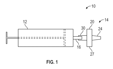
Referring now to the Figures and in particular to Figure 1, shown
therein is a diagrammatic view of a blood testing assembly 10 constructed in
accordance with the present disclosure. In general, the blood testing assembly
10
includes a receptacle 12 containing a sample of blood, and a blood testing
device
14. The receptacle 12 has a port 16 configured to transfer the blood out of
the
receptacle 12. The blood testing device 14 has a housing 20 constructed of a
fluid
impermeable material. The housing 20 has an interior space 22 which is shown
by
dashed lines in Fig. 2. The housing 20 may also include a vent 24 configured
to
allow gas to escape from the interior space 22, and prevent liquid (i.e.,
plasma) from
escaping from the interior space 22. In another embodiment, the housing 20 may
be
devoid of the vent 24. This can be accomplished by pressurizing the interior
space
22 within the housing 20 below atmospheric pressure (e.g., a vacuum) so that
the
housing 20 draws the sample of blood into the interior space 22. The housing
20 has
a top wall 25, a bottom wall 26, and a sidewall 27 extending between the top
wall 25
and the bottom wall 26. The vent 24 extends from the bottom wall 26. The blood
8
CA 03133202 2021-09-10
WO 2020/185272
PCT/US2019/064631
testing device 14 also includes a receptacle connector 30 extending from the
top wall
25 that is connected to the housing 20, and the port 16 of the receptacle 12.
The
receptacle connector 30, in one embodiment, is a tubular member that forms a
passage from the port 16 (outside of the housing 20) to the interior space 22
so that
at least a portion (e.g., 0.5 ¨ 1 mL) of the sample of blood can be
transferred from
the receptacle to the interior space 22. Smaller volumes of blood are possible
dependent upon the design of the housing 20.
[0034] In one
embodiment, the receptacle 12 is a syringe, and the receptacle
connector 30 is a device known in the art as a luer lock. It should be
understood that
the receptacle 12 and the receptacle connector 30 can be in other forms. For
example, the receptacle 12 may be a device known in the art as a vacutainer
that
can be used for collecting blood and transporting the blood for purposes of
testing. In
another embodiment, the receptacle 12 can be simultaneously connected to an
instrument, such as a blood gas analyzer, and the blood testing device 14. For
example, the housing 20 and the vent 24 can be constructed in a manner
described
for constructing the hollow body, and the analyzer connector 162 (in U.S.
Patent No.
7,896,818) to provide a venting mechanism (e.g., venting conduit) and an
analyzer
connector for attachment to an analyzer. This permits the analyzer connector
of the
blood testing device 14 to be connected to the analyzer, and used for
transferring the
sample from the receptacle 12 to the analyzer through the blood testing device
14. In
one embodiment, the analyzer may have an analyzer probe that extends through
the
analyzer connector and into the receptacle 12 to obtain a sample directly from
the
receptacle 12. The entire content of U.S. Patent No. 7,896,818 is hereby
incorporated herein by reference. The blood may be collected from an animal,
such
as a human, or a non-human (such as a cat, dog, cow, horse, fish, or the
like).
9
CA 03133202 2021-09-10
WO 2020/185272
PCT/US2019/064631
[0035] Shown in
Figure 2 is a side elevational view of an exemplary blood
testing device 14 constructed in accordance with the present disclosure. The
housing 20 is constructed of fluid impermeable material so that the housing 20
can
hold and contain a sample of blood containing blood cells, suspended within
plasma.
The housing 20 is shown as having a cylindrical shape, but it should be
understood
that the housing 20 can be provided with any shape, such as a square shape,
round
shape, rectangular shape, offset rectangular shape, triangular shape, offset
triangular shape, or the like. The housing 20 includes a window 34 (See Figure
3)
constructed of an optically transparent material that is configured to pass
light
bidirectionally from outside of the housing 20 to the interior space 22, and
from the
interior space 22 to the outside of the housing 20. In one embodiment, the
light is in
a visible part of the electromagnetic spectrum to allow a user to look through
the
window 34 and into the interior space 22 of the housing 20. As shown in Figure
3,
the window 34 can be a transparent portion of the bottom wall 26, although in
other
embodiments the window can be located on the sidewall 27 of the housing 20.
[0036] The
blood testing device 14 is also provided with a separator 40
(Figure 5) within the housing 20, and a reagent 42 (Figure 5). The
separator 40
is configured to receive blood having blood cells and plasma, separate the
blood
cells from the plasma, and direct the plasma to an inspection zone 44 within
the
interior space 22. The reagent 42 is positioned within the inspection zone 44
and
adjacent to the window 34 so that the reagent 42 is visible through the window
34.
The reagent 42 is configured to change colors in the presence of hemoglobin
within
the plasma to provide an indication of a state of hemolysis of the blood. When
the
blood testing device 14 is configured with an analyzer connector configured to
be
attached to the analyzer discussed above, the analyzer inlet probe may
approach
CA 03133202 2021-09-10
WO 2020/185272
PCT/US2019/064631
the separator 40 and pierece the separator 40 to obtain access to the sample
within
the receptacle 12.
[0037] In one
embodiment, the reagent 42 includes a pad 43 (see Figure 5)
that changes color due to an amount of hemoglobin within the plasma. The pad
43
may be a hydrophilic membrane for effecting a pinking color change so that a
user or
an external reader device can colorimetrically analyze and correlate the color
change
to a color (e.g., visual) reference. The pad 43 may be white or other color.
In some
embodiments, the pad 43 can be made of cellulose, nitrocellulose,
carboxymethylcellulose, or other material that will tend to retain protein.
[0038] The
color change of the pad 43 is visible through the window 34 to
permit a user or external reader device to view the reagent 42 and compare the
color
of the reagent 42 to regions 50a-f of a color palette 52. Each of the regions
50a-f has
a different color that has been correlated with the reagent 42 to indicate a
different
state of hemolysis, e.g., an amount of hemoglobin within the plasma. The
amount of
hemoglobin can be determined by the user comparing the color of the reagent 42
to
the regions 50a-f in the color palette 52. As will be discussed below, the
color palette
52 can be a sticker having a bonding material connecting a transparent
substrate to
the bottom wall 26, and surrounding the vent 24. In one embodiment, the
regions
50a-f are provided in a circular pattern that is coaxial with the vent 24. It
should be
understood, however, that other configurations of the regions 50a-f can be
used.
Further, the sticker can have an opening aligned with the window 34 to permit
the
user to view the reagent 42. In another example, the window 34 may be a
transparent region of the sticker. In this embodiment, the housing 20 (or a
portion
thereof) may be opaque, and have an opening aligned with the pad 43. The
sticker
may be connected to the housing 20 to form a fluid impermeable seal. In this
11
CA 03133202 2021-09-10
WO 2020/185272
PCT/US2019/064631
embodiment, a user or external reader may view the pad 43 through the
transparent
region of the sticker and the opening.
[0039]
Exemplary reagents 42 that can be used, and resulting color changes,
are set forth below in Table 1.
Assay Purpose (and simplified
Approximate TTR
formula)
Membrane (color change) 30 seconds
Blood: This test is based on the
peroxidase-like activity of hemoglobin
which catalyzes the reaction of
diisopropylbenzene di hydroperoxide
(w/w 6.8%) and <60 seconds
3,3',5,5'tetramethylbenzidine (w/w 4%).
The resulting color ranges from orange
through green. Very high levels may
continue color development to blue
Protein-Low (Albumin): This test
is based on dye binding using a high
affinity sulfonephthalein dye. At a
constant pH, the development of any
<50 seconds
color ranging from pale green to aqua
blue.
Ingredients: 1.9% w/w bis (3',3"-
diiodo-4',4"-dihydroxy-5`,5"-
12
CA 03133202 2021-09-10
WO 2020/185272
PCT/US2019/064631
dinitrophenyI)-3,4,5,6-
tetrabromosulfonephthalein
Protein-High: This test is based
on the protein-error-of-indicators
principle. At a constant pH, the
development of any green color is due
to the presence of protein. Controls <50 seconds
range from yellow, low-green, green,
and green-blue.
Ingredients: 0.3% w/w
tetrabromphenol blue
Table 1
[0040]
Referring now to Figures 5 and 6, shown therein are exploded views of
the exemplary embodiment of the blood testing device 14. In this example, the
housing 20 is provided with a housing top 60 and a housing bottom 62 that are
connected together to enclose the interior space 22. The housing top 60
includes the
top wall 25. The housing top 60 can be integrally formed as a unitary
structure, or
can be formed by separate components which are joined together via any
suitable
methodology such as sonic welding or a bonding material, such as an adhesive
or a
cohesive.
[0041] The top
wall 25 is provided with an interior surface 64, and an exterior
surface 66 generally opposite the interior surface 64. The top wall 25 is also
provided
with an opening 70 that communicates with the receptacle connector 30 to
permit the
blood to enter the interior space 22 of the housing 20. In one embodiment, the
opening 70 can be located in a central region of the top wall 25 as shown in
Figure 5.
13
CA 03133202 2021-09-10
WO 2020/185272
PCT/US2019/064631
It should be understood that the opening 70 can be placed in other parts of
the top
wall 25 so long as the opening 70 communicates with the receptacle connector
30.
The interior surface 64 is shaped so as to diffuse the blood from the opening
70 over
a separation area 71 that, in this example, encompasses a substantial part of
the
interior surface 64. For example, the interior surface 64 can be shaped to
include a
plurality of channels 72 and ribs 74. The housing top 60 also includes a
sidewall
portion 76 that extends from the top wall 25 and forms a portion of the
sidewall 27
when the housing top 60 and the housing bottom 62 are connected together. The
interior surface 64 and the sidewall portion 76 have a cup shape forming a
recess
80. The interior surface 64 may also be provided with a perimeter region 82
surrounding the separation area 71. As will be discussed below, the perimeter
region
82 is shaped so as to mate with the separator 40. For example, the perimeter
region
82 and the separator 40 may both be planar.
[0042] The
housing top 60 may be formed from any suitable liquid
impermeable material that is also inert to at least hemoglobin. For example,
without
limitation, the housing top 60 may be formed from a material comprising
polystyrene,
polyethylene, polycarbonate, polypropylene, fluoropolymer, polyester, glass,
metals,
ceramics, suitable composite materials, and combinations thereof as would be
appreciated by those skilled in art. Further, the housing top 60 may be
constructed of
a material that is opaque to light in the visible part of the electromagnetic
spectrum.
[0043] The
separator 40 may be provided with one or more stacked filters. In
the example shown, the separator 40 is provided with a first filter 90 and a
second
filter 92 that are aligned, and stacked one on top of the other. The first
filter 90 and
the second filter 92 may be the same size and shape, e.g., in this case both
the first
filter 90 and the second filter 92 are circular. In other embodiments, the
first filter 90
14
CA 03133202 2021-09-10
WO 2020/185272
PCT/US2019/064631
and the second filter 92 may be different sizes and/or shapes. In the
disclosed
embodiment, the first filter 90 is positioned on and engages the interior
surface 64,
and the second filter 92 is positioned on and engages the first filter 90.
[0044] The
first filter 90 may be designed to separate the blood cells in the
blood from the plasma, and then to pass the plasma to the second filter 92.
For
example, in the embodiment shown in Figure 5, the first filter 90 may isolate
plasma
and hemolysis products, e.g., hemoglobin, from whole blood cells in a sample
such
as a whole blood sample. In an embodiment, the first filter 90 comprises a
plasma
separation membrane as is commercially available in the art. In certain
embodiments, the plasma separation membrane comprises an asymmetric material,
which is able to retain a plurality of whole blood cells thereon while
allowing plasma
and small molecules/complexes to travel there through. A number of different
plasma
separation membranes are commercially available and may be suitable for use in
the
blood testing device 14. For example, the plasma separation membrane may
comprise an asymmetric polysulfone material as is commercially available from
Pall
Corporation (currently under the trademark VividTm). Alternatively, the first
filter 90
may comprise any other suitable material or device that can provide a sample
comprising plasma and components from hemolysis (if present) therein.
[0045] The
plasma separated by the first filter 90 is provided to the second
filter 92. The second filter 92 is provided with a predetermined color and
forms a
background or setting for the reagent 42. The second filter 92 overlaps the
reagent
42. In one embodiment, the reagent 42 is positioned between the second filter
92
and the window 34 so that the second filter 92 provides a background color for
the
reagent 42. When blood that is suspected as having hemolysis is provided into
the
interior space 22 through the receptacle connector 30 and the opening 70, the
blood
CA 03133202 2021-09-10
WO 2020/185272
PCT/US2019/064631
is diffused by the separation area 71 and passes through the first filter 90.
The first
filter 90 separates the blood cells and platelets from the plasma, and then
passes the
plasma to the second filter 92. The plasma saturates the second filter 92 and
passes
through the second filter 92 to the reagent 42. The reagent 42 reacts with the
plasma
and may change color to indicate a state of hemolysis, or an unacceptable
level of
hemolysis. The second filter 92 provides a consistent color background, and
therefore assists with the colorimetric comparison of the color of the reagent
42. In
one example, the second filter 92 is black filter paper, although it should be
understood that other colors could be used. In one embodiment, the first
filter 90 can
be a predetermined color to provide a consistent color background. In this
embodiment, the second filter 92 may be transparent, or eliminated.
[0046] As
discussed above, the vent 24 is designed to permit gas to pass
through the vent 24, but prevent the passage of a fluid through the vent 24.
In one
embodiment, the vent 24 is a tubular member having an interior opening 100.
The
vent 24 includes an overflow plug 102 positioned within and blocking the
interior
opening 100. The overflow plug 102 is constructed of a material capable of
venting
gas from the interior space 22, but preventing the passage of the fluid
through the
vent 24. Exemplary materials for making the overflow plug 102 include starch,
cellulose, and teflon.
[0047] The
housing bottom 62 includes the bottom wall 26. The bottom wall
26 is provided with an interior surface 110, and an exterior surface 112
generally
opposite the interior surface 110. The bottom wall 26 is also provided with an
opening 114 that communicates with the interior opening 100 of the vent 24 to
permit
gas and fluid to enter the interior opening 100 from the interior space 22 of
the
housing 20. In one embodiment, the opening 114 can be located in a central
region
16
CA 03133202 2021-09-10
WO 2020/185272
PCT/US2019/064631
of the bottom wall 26 as shown in Figure 6. It should be understood that the
opening
114 can be placed in other parts of the bottom wall 26 so long as the opening
114
communicates with the interior opening 100 of the vent 24. The interior
surface 110
is shaped so as to diffuse the plasma from the second filter 92 over a reagent
saturation area 120 that, in this example, encompasses a substantial part of
the
interior surface 110. For example, the interior surface 110 can be shaped to
include
a plurality of channels 122 and ribs 124. The housing bottom 62 also includes
a
sidewall portion 130 that extends from the bottom wall 26 and forms a portion
of the
sidewall 27 when the housing top 60 and the housing bottom 62 are connected
together. The interior surface 110 and the sidewall portion 130 may have a cup
shape forming a recess 132. The interior surface 110 may also be provided with
a
perimeter region 134 surrounding the reagent saturation area 120. As will be
discussed below, the perimeter region 134 is shaped so as to mate with the
separator 40. For example, the perimeter region 134 may be planar.
[0048] The
housing bottom 62 may be formed from any suitable liquid
impermeable material that is also inert to at least hemoglobin. For example,
without
limitation, the housing top 60 may be formed from a material comprising
polystyrene,
polyethylene, polycarbonate, polypropylene, fluoropolymer, polyester, glass,
metals,
ceramics, suitable composite materials, and combinations thereof as would be
appreciated by those skilled in art. Further, a portion of the housing bottom
62
forming the window 34 is constructed of a material that is transparent to
light in the
visible part of the electromagnetic spectrum.
[0049] In
operation, a sample of blood to be tested is placed within the
receptacle 12. The receptacle connector 30 of the blood testing device 14 may
be
connected to the port 16 of the receptacle 12, and an amount of blood
transferred
17
CA 03133202 2021-09-10
WO 2020/185272
PCT/US2019/064631
through the port 16 and the receptacle connector 30 into the interior space 22
of the
blood testing device 14. As the blood is transferred into the interior space
22, air
within the interior space 22 is directed through the vent 24. As the blood
enters the
interior space 22, the blood is diffused by the channels 72 and the ribs 74
and is
applied to the first filter 90. The first filter 90 separates the blood cells
and platelets
from the plasma, and passes the plasma to the second filter 92. The plasma
saturates the second filter 92, and passes the plasma into the reagent
saturation
area 120 so that the plasma can contact and saturate the reagent pad 43 of the
reagent 42. The saturated reagent pad 43 then may or may not change color
depending upon the state of hemolysis of the blood. In any event, the color of
the
reagent pad 43 can be viewed by the user, and a determination of whether the
blood
has hemolysis can be made by comparing the color of the reagent pad 43 to the
regions 50a-f of the color palette 52. Thereafter, the blood testing device 14
may be
removed from the receptacle 12 and discarded. When the blood sample does not
have an unacceptable level of hemolysis, the blood sample can be tested using
conventional techniques, such as providing the blood sample into a cartridge
of a
blood gas analyzer.
[0050] Figure 7
is a perspective, exploded view another embodiment of an
exemplary blood testing assembly 10a constructed in accordance with the
present
disclosure. The blood testing assembly 10a includes a receptacle 12a and a
blood
testing assembly 14a. Similar elements between the receptacle 12 and 12a will
be
labeled with the same reference numerals. Likewise, similar elements between
the
blood testing assembly 14 and 14a will be labeled with the same reference
numerals.
18
CA 03133202 2021-09-10
WO 2020/185272
PCT/US2019/064631
[0051] The
receptacle 12a and the blood testing assembly 14a are similar in
construction and function to the receptacle 12 and the blood testing assembly
14
described above, with the exception that the receptacle 12a and the blood
testing
assembly 14a are integrated into a single device which in this example is a
syringe.
In this regarding, the receptacle 12a has a sidewall 140, a first port 142,
and a
second port 144 (which in some embodiments can be the same as the port 16
described above). In the embodiment shown, the sidewall 140 and a top wall 25a
of
the blood testing device 14a are integrally formed as a unitary structure. The
sidewall
140 and the top wall 25a include the first port 142 formed through the
sidewall 140
and the top wall 25a so as to permit a sample, e.g., blood, to flow from the
receptacle 12a and into the interior space 22 of the blood testing device 14a.
Other
than being integrally formed with the sidewall 140, the top wall 25a is
identical to the
top wall 25 in construction and function.
[0052] The flow
of sample from the receptacle 12a and into the interior space
22 of the blood testing device 14a can be implemented in various manners. For
example, the port 142 can be designed to establish capillary action between
the
receptacle 12a and the interior space 22. In this embodiment, after a sample
is
drawn through the port 144 with the port 142 (or the vent 102) closed via a
valve,
The valve may be opened, and the sample of blood may displace the gas in the
interior space 22 of the housing 20 through capillary action. In this case,
the volume
of sample is limited by a volume of the interior space 22 that is not filled
with the first
membrane 90, the second second membrane 92, and the reagent pad 43. In either
implementation, any overfill should be stopped by the overflow plug 102.
[0053] Figure 8
is a side elevational view of the embodiment of the blood
testing assembly 10a depicted in Figure 7.
19
CA 03133202 2021-09-10
WO 2020/185272
PCT/US2019/064631
[0054] The
operation of the blood testing assembly 10a is identical to the
operation of the blood testing assembly 10 described above, with the exception
that
the blood testing assembly 14a does not have a receptacle connector 30 that
needs
to be connected to the port 16 of the receptacle 12 due to the receptacle 12a
and the
blood testing assembly 14a being integral and a single device. Further, in the
embodiments that include a valve being positioned within the first port 142,
the
operation may include opening the valve to permit the sample to flow through
the
first port 142 and into the interior space 22, and closing the valve to
prevent any
further sample from flowing through the first port 142.
[0055] From the
above description, it is clear that the inventive concepts
disclosed herein are well adapted to carry out the objects and to attain the
advantages mentioned herein as well as those inherent in the inventive
concepts
disclosed herein. While presently preferred embodiments of the inventive
concepts
disclosed herein have been described for purposes of this disclosure, it will
be
understood that numerous changes may be made which will readily suggest
themselves to those skilled in the art and which are accomplished within the
scope
and coverage of the inventive concepts disclosed and claimed herein.