Note: Descriptions are shown in the official language in which they were submitted.
CA 03133741 2021-09-15
WO 2019/191111
PCT/US2019/024093
SYSTEMS AND METHODS FOR MULTILANE VASCULATURE
CROSS-REFERENCE TO RELATED APPLICATIONS
This application claims priority to United States Provisional Application
Serial No. 62/648,209, filed on March 26, 2018, which is incorporated by
reference
herein in its entirety.
STATEMENT REGARDING FEDERALLY FUNDED RESEARCH
This invention was made with Government Support under Grant No.
1UG3TR002198-01 awarded by the National Institutes of Health. The Government
has
certain rights in the invention.
BACKGROUND
Certain organ-on-chip devices can include a micro engineered biological cell-
culture compartment in which tissue- and organ-level elements of human
physiology can
be recapitulated. The micro engineered biological cell-culture compartment can
allow
physiological in vitro modeling of functional biological units of that organ
system (e.g.
insulin-secreting islet units to mimic a pancreas, air-liquid interfaces to
mimic oxygen
transport in the lung). Such models can perform tests on live human tissue
without
requiring live human subjects.
However, certain organ-chip systems fail to create functional and/or realistic
multi-organ networks and are unable to recapitulate complex, physiological
responses
and multi-organ interactions at the systemic level. In 2D models, a monolayer
of
endothelial cells grown on a petri dish represents a vascular lumen, but this
approach can
be limited due to an absence of other cells typically involved in vessel
behavior (e.g.,
1
CA 03133741 2021-09-15
WO 2019/191111
PCT/US2019/024093
pericytes and smooth muscle cells). Certain organ-chip systems have
diffusional
constraints that limit the size of 3D tissues grown within hydrogels or other
scaffolds
because of insufficient perfusion efficiency. Larger tissues can require more
efficient
perfusion for the development, survival, regulation, and homeostasis of
tissues. Certain
tissues require microenvironmental cues from blood vessel tissue or
vasculature to
differentiate to a physiological state in vitro, or to perform biological
functions.
Accordingly, there remains a need to create multi-organ networks and
interfaces
thereto with improved perfusion efficiency for the organ-on-chip devices for
therapeutic
applications and drug screening.
SUMMARY
The disclosed subject matter provides system and methods for producing
multilane vasculatures, or perfusable vascular fluidic interfaces to, from, or
between
either single or multiple in vitro tissues, or to, from, or between multiple
in vivo tissues
within a human or animal living tissue.
In certain embodiments, a method for culturing a tissue using a
microphysiological device or organ-on-a-chip device can include embedding at
least two
tissues into a 3D structure, where the 3D structure includes a vasculogenic
element. A
vascular interface for nutrient perfusion is formed into the at least two
tissues, and is
tuned to possess a property of a native biological target tissue or a target
environment by
.. modifying at least one vessel attribute. A fluidic interface channel can be
lined and
coupled to the 3D structure to provide a growth fluid thereto, in order to
promote blood
vessel production. A corresponding system is similarly disclosed herein.
2
CA 03133741 2021-09-15
WO 2019/191111
PCT/US2019/024093
In certain embodiments, a method for culturing a tissue using a
microphysiological
device or organ-on-a-chip device can include embedding one tissue type into a
3D
structure and one or more vasculogenic elements. A vascular interface for
nutrient
perfusion is formed into the tissue, or out of the tissue, or into and out of
the tissue, and
is tuned to possess a property of a native biological target tissue or a
target environment
by modifying at least one vessel attribute. A fluidic interface channel can be
lined and
coupled to the 3D structure to provide a growth fluid thereto, in order to
promote blood
vessel production. A corresponding system is similarly disclosed herein.
In certain embodiments, the method can further include introducing a section,
an
element, or a layer of a vasculogenic tissue between the at least two tissues
to create the
vascular interface. The vasculogenic element and the layer of the vasculogenic
tissue
can include a hypoxia induced factor (HIF), a fibroblast growth factors (FGF),
and/or a
vascular endothelial growth factor (VEGF). In some embodiments, the method can
further include linking a plurality of the vascular interfaces to form a multi-
lane
vasculature culture system.
In certain embodiments, the method can include introducing a section, an
element, or a layer of a vasculogenic tissue upstream, or downstream, or
upstream and
downstream of one tissue to create one or more vascular interface to enable
fluidic
communication to the tissue through vascular vessel structures. The
vasculogenic
.. element and the layer of the vasculogenic tissue can include a hypoxia
induced factor
(HIF), a fibroblast growth factors (FGF), and/or a vascular endothelial growth
factor
(VEGF). In some embodiments, the method can further include linking a
plurality of the
vascular interfaces to form a multi-lane vasculature culture system.
3
CA 03133741 2021-09-15
WO 2019/191111 PCT/US2019/024093
In certain embodiments, vessel attributes of the vasculature can be tuned
possess
a property of a native biological target tissue by modifying one or more of
the following:
a vessel density, a vessel diameter, a vessel barrier function, a vessel
disease condition,
or a material property (including stiffness, pore size, or exposed chemical
moieties or
chemical groups) that indirectly (e.g., through mechanosensory feedback to the
vascular
cells or vascular tissues) accomplishes one or a combination of these
modifications.
BRIEF DESCRIPTION OF THE DRAWINGS
Further features and advantages of the present disclosure will become apparent
from the following detailed description taken in conjunction with the
accompanying
figures showing illustrative embodiments of the present disclosure, in which:
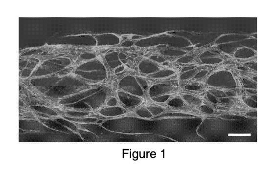
FIG. 1 is an illustration of an exemplary engineered vasculature in 3D fibrin
hydrogel in a 3D micro physiological tissue culture device. Scalebar 200 i.tm.
FIG. 2 is an illustration of an exemplary perfusion of 1 i.tm fluorescent
beads
(dots from bottom) through the hollow lumen of engineered vasculature (RFP-
expressing
vascular endothelial cells) in a micro physiological device.
FIG. 3 is an illustration of an exemplary diagram of an engineered vascular
interface to explanted human islet tissues in 3D ECM hydrogel within a micro
physiological device.
FIG. 4 is an illustration of an exemplary engineered 3D vascular interface of
endothelial-lined vessels with a GFP-expressing pancreatic islet isolated from
a mouse.
FIG. 5 is an illustration of an exemplary schematic diagram of a bone marrow
model in which human bone marrow tissue is fluidically accessible via a 3D
vascular
4
CA 03133741 2021-09-15
WO 2019/191111 PCT/US2019/024093
interface, which is in turn interfaced to a large vessel fluid interface
formed by coating
all sides of a fluid channel with vascular endothelial cells to form a
vascular vessel of
defined size.
FIG. 6 is an illustration of an exemplary perfusion of leukocytes from a
central
bone marrow tissue compartment through an engineered 3D vascular fluidic
interface as
driven by fluid flow and size-based selectivity, which is governed by the
lumen
diameters of the vessels formed therein.
FIG. 7 is an illustration of an exemplary 3D vascular interfaces (endothelium)
on
inflow and outflow sides of trapped leukocytes in fluidic suspension (middle).
FIG. 8 is an illustration of an exemplary system with two 3D vascular
interfaces
in accordance with the disclosed matter.
FIG. 9 is a diagram illustrating exemplary stages in a method in accordance
with
the disclosed matter.
FIG. 10 is a diagram illustrating exemplary stages in a method in accordance
with
the disclosed matter: Tissues form continuous and perusable vascular
connections
between the flanking flow channels.
FIG. 11 is a diagram illustrating exemplary stages in a method in accordance
with
the disclosed matter: Multiple separate tissues are in fluid communication via
multiple
vascular interface materials disposed between the separate tissues.
FIG. 12 is a diagram illustrating exemplary stages in a method in accordance
with
the disclosed matter: Multiple adjacent tissue types are interfaced to micro
chambers of
5
CA 03133741 2021-09-15
WO 2019/191111 PCT/US2019/024093
microfluidic channels by vascular interface materials to simulate a biological
interface to
the circulatory system.
FIG. 13 is a diagram illustrating exemplary stages in a method in accordance
with
the disclosed matter: Multiple separate tissues are intra-vascularized by
multiple
placements of vascular interface material. This process expedites forming of
perfusable
multi-tissue networks simulating the biological connection in blood
circulation.
FIG. 14 is a diagram illustrating exemplary stages in a method in accordance
with
the disclosed matter: Multiple adjacent tissues are intra-vascularized by
vascular
interface material to form perfusable multi-tissue material.
FIG. 15 is a diagram illustrating exemplary stages in a method in accordance
with
the disclosed matter: Discrete tissues are flowed into a channel and then
vascularized.
FIG. 16 is a diagram illustrating exemplary stages in a method in accordance
with
the disclosed matter: Discrete tissues are suspended in pre-gel vascular
material and
placed into a chamber. Vascular interface material forms interfaces with the
encapsulated tissues.
FIG. 17 is a diagram illustrating exemplary stages in a method in accordance
with
the disclosed matter: Increased area of vessel interfaces are created by
lining a
microchannel or fluidic chamber with a monolayer of cells.
FIG. 18 is a diagram illustrating exemplary stages in a method in accordance
with
the disclosed matter: Discrete tissues are flowed into a channel and then
vascularized.
Tissues capable of forming a monolayer is embedded into flow channels.
6
CA 03133741 2021-09-15
WO 2019/191111 PCT/US2019/024093
Throughout the figures, the same reference numerals and characters, unless
otherwise stated, are used to denote like features, elements, components or
portions of
the illustrated embodiments. Moreover, while the present disclosure will now
be
described in detail with reference to the figures, it is done so in connection
with the
illustrative embodiments.
DETAILED DESCRIPTION
Techniques for producing a body organ or system on an organ-on-chip using a
plurality of microfluidic devices are disclosed herein. The disclosed subject
matter can
perform a fully or partially automated organ culture using the organ-on-chip
without the
need for specialized personnel by modeling feed-forward and feedback effects
from
interfacing one functional unit to another in the organ-on-chip.
In certain embodiments, the disclosed subject matter provides a micro
physiological tissue culture system. The micro physiological tissue culture
system can
include engineered vessel networks. For example, vascular endothelial cells,
fibroblasts,
pericytes, mesenchymal stem cells, and/or smooth muscle cells can be seeded
together
into a 3D ECM scaffold or hydrogel and supplied with endothelial cell media
containing
vasculogenic factors including VEGF, FGF, and endothelial growth hormones. As
shown Fig. 1, 3D fibrin hydrogel, or collagen hydrogel, or biocompatible
hydrogel, or a
combination thereof can be used for vasculature engineering. In the presence
of these
growth factors, the cells can form patterned vessel structures with hollow,
perfusable
lumens through the process of vasculogenesis, angiogenesis, or a combination
of
vasculogenesis or angiogenesis.
In certain embodiments, the micro physiological tissue culture system can
include
a line with a layer of endothelial cells on the walls of the vascular gel. For
example, the
7
CA 03133741 2021-09-15
WO 2019/191111 PCT/US2019/024093
monolayer line of endothelial cells can drive angiogenic sprouting into
hydrogels. The
micro physiological tissue culture system with the monolayer layer can
anastomose the
vessels forming within the 3D gel matrix to the external lumen of the gel's
endothelial
lining. In some embodiments, after 2 ¨ 7 days of growth, the 3D micro
physiological
tissue culture system can have a perfusable vascular network of vessels
including a
hollow, endothelial cell-lined lumen surrounded by pericytes or fibroblasts or
a
combination thereof in the surrounding stroma (Figs. 2 and 10).
In certain embodiments, the micro physiological tissue culture system can have
vascular interfaces between multiple engineered or native tissue types. The
micro
physiological tissue culture system can be a 3D cell culture model
recapitulating body-
scale or organ-scale tissue dynamics by linking of two or more tissue types,
including the
vascular tissue produced in the vascular interfaces. Data gathered by
microscopy,
effluent sampling, integrated biosensing, or physical/structural measurements
can be
utilized to create a 3D micro physiological tissue culture system or a 3D cell
culture
system. The disclosed culture systems can be used for therapeutic
applications, culture
of human tissue biopsies (e.g., cancer biopsies, sampled microbial cultures
and/or
infections), or screening of drugs.
In certain embodiments, the 3D micro physiological microfluidic system can
provide perfusion for the development, survival, regulation, and homeostasis
of tissues
by promoting blood vessels in the tissues. Blood can carry nutrients, oxygen,
signaling
hormones, various cell types including erythrocytes, platelets, leukocytes,
and stem cells,
as well as metabolic waste products and carbon dioxide. To perfuse human
tissue, the
3D micro physiological microfluidic system can have a similar vascular system
as the
human body which can be branched from the large aorta leaving the heart (e.g.,
20 ¨ 30
8
CA 03133741 2021-09-15
WO 2019/191111 PCT/US2019/024093
mm diameter) through arteries (e.g., 0.1 ¨ 10 mm diameter), arterioles (e.g.,
0.01 ¨0.1
mm diameter), and capillaries (e.g., 0.005 ¨ 0.01 mm diameter), at which point
diffusion
and transport of bloodborne elements into and out of the surrounding tissue
can occur.
From the capillary networks, blood can circulate back through venules (0.008 ¨
0.1 mm
diameter) and veins (0.1 ¨ 15 mm diameter). Blood vessels can include an inner
endothelium formed from endothelial cells, which are surrounded by innervated
smooth
muscle cells, vascular pericytes, and fibroblasts in connective tissue.
In certain embodiments, the 3D micro physiological microfluidic system can
provide reciprocal signaling that can occur between biological tissues and the
vessels
that perfuse it. Localized hypoxia or nutrient deprivation in human tissue can
cause the
secretion of signaling molecules including hypoxia induced factors (HIFs),
fibroblast
growth factors (FGFs), and vascular endothelial growth factor (VEGF). The
secretion of
signaling molecules can promote vasculogenesis, the formation of blood vessels
from
precursor cells, and angiogenesis, the sprouting of vessels from existing
endothelial
tissue. For example, nascent vessels formed by vasculogenic endothelial cells
can be
implicated in tissue delineation and inductive signaling during embryonic
development,
in the densely vascularized islets from which insulin and other hormones that
govern
homeostasis are secreted. These islets exist in intimate contact with
fenestrated
capillaries and sample the glucose concentration of the blood therein,
releasing insulin in
response; to create an in vitro model of a pancreatic islet without including
its vascular
environment are shortsighted. Additionally, endothelial cells and smooth
muscle cells
can be the targets of damage due to drug-induced vascular injury, but the
resulting
lesions can be localized to specific structures, e.g. the branch points in
coronary arteries.
9
CA 03133741 2021-09-15
WO 2019/191111 PCT/US2019/024093
In certain embodiments, the 3D micro physiological microfluidic system can
include different vasculature structures. For example, the vasculature
structures can
include the endothelial cells lining blood vessels in the brain which can form
tight
junctions with one another to control the molecules permitted passage into the
brain (i.e.,
blood-brain barrier) in human brain. Additionally, the 3D micro physiological
microfluidic system can include vasculature structure whose walls contain
smooth
muscle cells, connective tissue, and thin micro vessels which are responsible
for alveolar
gas exchange in lung.
In certain embodiments, the disclosed micro physiological microfluidic system
.. can include at least one chamber. The shape of the chamber can be modified
based on
the purposes. For example, the chamber can be a straight lane. For example,
the
chamber can encapsulate another chamber in whole or in part. In some
embodiments, the
chamber can be patterned vertically. The chambers can be placed adjacent to
each
other. Furthermore, flow through these patterns can be networked in an
adjustable
manner. The chambers can be fabricated to create partition modeling of
substances in
the bloodstream. For example, tubing can be placed between any two or more
ports
accessing different areas of tissue of the device to create a direct
connection. Fluid
effluent flowing outwards through any of these ports can be collected. For
example, one
of the tissues can be placed in the center of the 5-lane embodiment and a flow
with a
drug candidate can flow from left to right through a tissue, through a first
vascular
interface on the left side and a second vascular interface on the right side.
The outflow in
the vascular ports before and after the tissue can be collected to measure the
metabolism
of the drug. As shown in Fig. 13, a leftward tissue with a rightward target
organ can be
interfaced with vasculature to sample the effluent before the leftward tissue,
after the
leftward tissue but before the target organ, and after the target organ; the
metabolism of a
CA 03133741 2021-09-15
WO 2019/191111 PCT/US2019/024093
drug and its effect on the secretory products of the target organ can be
measured. The
disclosed device can be expanded with more than two organs with vascular
interconnections.
In certain embodiments, the disclosed subject matter provides methods for
generating 3D vascular interfaces. The method can include interfacing at least
two
parenchymal 3D tissues. The parenchymal tissues can possess vascular or
avascular
tissues. The tissues can be interfaced by introducing additional sections,
elements, or
layers of 3D vasculogenic tissue between them to form vessels. For example, as
shown
in Fig. 9, vascular interfaces can be created to a tissue disposed in a
microfluidic device.
The interfaced tissues can form perfusable vascular structures which are
capable of
vasculogenesis or angiogenesis. The interfaced tissue can self-vascularize via
vasculogenesis and/or angiogenesis and anastomose or connect to existing
vessels or
vasculature in the parenchymal tissues, thereby forming an interface of
perfusable fluidic
connections between the opposite original tissues. In some embodiments, the
interfaced
tissue can vascularize or form new vessels in the existing tissues via
angiogenic
sprouting, thereby forming a perfusable vessel interface that fluidically
links the existing
parenchymal tissues (Fig. 10). In non-limiting embodiments, the interfaced
tissue can
anastomose with certain native vascular tissue or vascular vessels.
In certain embodiments, the vasculogenic tissue can injected prior to the
addition
of adjacent target modeled 3D tissues. For example, as shown in Fig. 18,
vasculogenic
tissues can be interfaced in scaffold, matrix, or gel (in which the vascular
interface is
formed) to stabilize apposite tissue components that are subsequently injected
or seeded
into the tissue culture device. In some embodiments, the interfaced
vasculogenic tissue
can have a structural rigidity to pin in place (e.g. after the injection port
is plugged) a
11
CA 03133741 2021-09-15
WO 2019/191111
PCT/US2019/024093
fluid suspension of injected tissue or a tissue with loose or free-flowing
elements.
Therefore, the scaffold, matrix, or gel with the interfaced vasculogenic
tissues can
prevent loose elements from escaping into bulk solution by behaving as a
filter with a
pore size governed by the diameter of the vasculature or vessels. In some
embodiment,
the interfaced vasculogenic tissue can improve a structural rigidity of the
gel to pin the
injected tissue in place until its own hydrogel, scaffold, or extracellular
matrix sets,
cures, and/or crosslinks. For example, injecting adipocytes in 3D collagen gel
can create
a "fat-on-a-chip" tissue model between two vasculogenic hydrogel extracellular
matrix
(ECM) scaffolds. The adjacent vasculogenic tissue can angiogenically sprouts
into the
.. fat model to create a vascular interface. In non-limiting embodiments, the
vasculogenic
tissue can further improve structural surfaces of the cured, set, or
crosslinked 3D
vasculogenic scaffold, matrix, or gel to seed or implant a monolayer of cells
whose
surface coverage is limited to the surfaces facing the volume enclosed by the
vasculogenic tissue boundaries. For example, a monolayer of endothelial cells
can be
seeded into a gel chamber to mimic a much larger blood vessel, such as an
artery,
interfacing with 3D vasculogenic tissue that represents a capillary bed. The
disclosed
system can be used to determine the vessel scale at which drug-induced
vascular injury
takes effect.
In certain embodiments, the method can include embedding tissues within a
scaffold, matrix, or gel that contains vasculogenic tissues. For example,
engineered
tissues (e.g., organoids or spheroids), native tissues e.g., (pancreatic beta
islets, or
biopsied cancer), or combination of thereof can be embedded within a scaffold,
matrix,
or gel. The tissue embedded-systems can have the vasculogenic elements which
can
form perfusable vessels that anastomose or connect to existing vessels or
vasculature in
.. the contained tissues. As shown in Fig. 16, discrete tissues (e.g.,
spheroids, organoids,
12
CA 03133741 2021-09-15
WO 2019/191111
PCT/US2019/024093
biopsied tissue, biopsied tumors, encapsulated tissues, cell aggregates, or
tissue
scaffolds) can be suspended in pre-gel vascular material and placed into a
chamber. The
vascular interface material can form interfaces with the encapsulated tissues.
In some
embodiments, the vasculogenic elements in the scaffold, matrix, or gel form
perfusable
vessels that sprout angiogenically into the contained tissue in order to
thereby forming a
vascular interface. In non-limiting embodiments, engineered vessels in the
scaffold,
matrix, or gel can anastomose to existing vessels native to the embedded
tissue, while
also angiogenically sprouting additional vessels into the tissue to increase
its vascularity.
In certain embodiments, the scaffold, matrix, or gel of the vasculogenic
tissue
interface can spatially restrain the interfaced tissue while still permitting
fluidic access
for nutrient perfusion. For example, the scaffold, matrix, or gel of the
vasculogenic
tissue interface can preventing the merging of multiple discrete tissues such
as two
pancreatic islets, or two kidney organoids, or multiple cancer spheroids. The
scaffold,
matrix, or gel of the vasculogenic tissue interface can further prevent the
disassociation
or loosening of the tissue elements (e.g. spheroids or organoids) into
multiple parts or
flattening in response to adhesion to a single 2D surface.
In certain embodiments, the method can include tuning engineered vasculature
interfaces to possess properties relevant to the native biological target
tissue. The tuning
can be performed by modifying vessel attributes including the density of the
engineered
vascular network, the diameter of the engineered vessels, the barrier function
of the
engineered vessels (e.g. tight endothelial junctions or fenestrated junctions
or sinusoidal
endothelium), the health of the engineered vessels (e.g. for use in an in
vitro disease
model or vascular injury model), the time to perfusability of the vasculature
(e.g. for
quickly providing fluidic access for nutrient perfusion to an explanted
tissue), and/or a
13
CA 03133741 2021-09-15
WO 2019/191111
PCT/US2019/024093
combination thereof In some embodiments, the tuned vascular interface can
allow the
interfaced tissue to differentiate to a physiological state, or behave in a
physiologically
relevant manner for purposes of biological investigation, therapeutic testing
and/or
screening. Furthermore. the tuned vascular interface can be used to determine
or affect
the partition coefficients of fluid flow into two or more adjacent tissues.
For example, a
tissue interface with vessels that are narrower can exhibit an increased
resistance to flow
than an interface with larger or denser vessels, thereby increasing fluid flow
into the
interface with larger vessels relative to the interface with more narrow,
restrictive
vessels.
In certain embodiments, environmental, mechanical and/or biological conditions
can be tuned. For example, the environmental conditions can include the
density of
vasculogenic cells placed within the vasculogenic tissue which can modulate
aspects
including vascular density and the ratios of different cells or cell types
within the
vasculogenic tissue which can modulate aspects including vessel diameter. The
biological conditions can include the biochemical profile supplied to the
cells by growth
medium, either consistently or dynamically. For example, the biological
condition can
be tune by modifying the concentrations of factors including VEGF to
accelerate or halt
vasculogenesis, Angl to promote angiogenesis, corticosteroids to promote tight
junction
formation, and/or TNF-alpha to increase vascular permeability. The mechanical
conditions can include fluid environments and structural environments. For
example,
fluid environments can be modified to expose the tissue to continuous fluid
perfusion to
create tighter endothelial junctions and an efficient vascular network with
less redundant
branching. The structural environments can be modified to create a
vasculogenic tissue
interface within a stiffer ECM scaffold or gel in order to drive the formation
of more
14
CA 03133741 2021-09-15
WO 2019/191111 PCT/US2019/024093
rigid vessels, or a more compliant or softer scaffold or gel to mimic
distended vessels,
vessel aneurysm or rupture.
In certain embodiments, the method can include lining a fluidic interface
channel
coupled to the tissues. The fluidic interface channel can include parallel,
side-by-side,
vertically stacked, or upstream/downstream spatial orientations or
configurations. For
example, as shown in Figs. 17 and 18, increased area of vessel interface can
be created
by lining a microchannel or fluidic chamber with a monolayer of cells (e.g.,
endothelial
cells). The micro physiological tissue culture system with vascular
endothelial cells can
imitate a vascular lumen and create an inter-tissue vascular interface. The
inter-tissue
vascular interface can have the diameter or cross-sectional area of the
vessels which can
be specifically defined by the geometry of the fluidic channel. The inter-
tissue vascular
interface can have patterned vessels. In some embodiments, the vessel walls
can be
mechanically actuated to simulate vascular constriction or dilation. For
example,
capsular constriction can be simulated without requiring the vessels to be
innervated
smooth muscle cells.
In certain embodiments, the method can further include linking multiple
vascular
interfaced to form an "interface to an interface" structure. In the multiple-
interface
structure, fluids including growth media can be perfused through a sequence of
vessels
with different vascular properties (e.g. diameters, densities, junction
tightness, matrix
stiffness) that vary according to the native physiological properties of
multiple tissues.
For example, a large vessel with 500 p.m diameter is formed to model an
artery, which
interfaces to an adjacent 3D vascular interface of arteriole-sized vessels
with average
diameters of 100 p.m, which in turn interfaces to a micro vessel network with
average
diameters of 10 ¨ 20 p.m that is injected via pinning. The interfaced
structure can further
CA 03133741 2021-09-15
WO 2019/191111 PCT/US2019/024093
interface to a target parenchymal or organ tissue contained in the model or
onto other
vessel types to form a full circulatory model from large-scale to small-scale
vessels. Figs
11 and 12 show multiple separate and adjacent tissues in fluid communication
via
multiple vascular interface materials. In some embodiments, fluids can be
perfused to
simulate the different scales of the circulatory system within an in vitro
model as a
sequential series of vascular interfaces to larger or smaller vessels. For
example, the
structure can include a series of 2D or 3D vascular interfaces, where one can
interface to
the next without parenchymal tissue. In non-limiting embodiments, as shown in
Figs. 13
and 14, multiple tissues can be intra-vascularized by multiple placements of
vascular
interface material and thereby form continuously perfusable multi-tissue
material
EXAMPLE I: Multi-Tissue Vascular Interface (Pancreatic Islet Model)
A model of pancreatic islet was developed by utilizing the disclosed invention
to
create a vascular interface with explanted islets that recapitulates the
densely
vascularized environment of the human islet in vivo. The purposes of this
model were to
maintain the viability of explanted islets in vitro for an extended period,
and to permit
the interrogation of islet dynamics for purposes including assessment of
glucose-
stimulated insulin secretion, which is a measure of islet function.
To achieve these goals, as shown in Fig. 3, a rapidly formed, dense, and
perfusable vascular interface was engineered from vasculogenic tissue
including primary
human vascular endothelial cells and fibroblasts. These vessels anastomosed to
the
native micro vessels of the primary human islets, and in doing so
recapitulated the rich
vasculature and resulting paracrine signaling between apposite endothelial and
endocrine
tissues native to islets in the human pancreas, which is essential to the
function of islets
16
CA 03133741 2021-09-15
WO 2019/191111 PCT/US2019/024093
in vivo¨and is thus a promising method for sustaining islet function ex vivo
in our
micro physiological, 3D tissue culture devices.
The engineered vascular interface permitted the function of the islet to be
tested
transiently, with high temporal resolution; by perfusing the engineered
vascular
interface, any hormonal secretions by the islet tissue¨including insulin¨enter
directly
into the perfusate and may be sampled in the device effluent, rather than
remaining in the
periphery of the islet tissue, or being diluted in a fluid suspension.
Furthermore, as
shown in Fig. 4, the engineered vascular interface was visualized by
fluorescence
imaging techniques.
EXAMPLE II: Multi-Tissue Vascular Interface (Bone Marrow Model)
In this example, a model of leukocyte mobilization into the bloodstream was
created by spatially patterning a section or layer of human bone marrow
between two
vascular interfaces. The leukocyte mobilization into the bloodstream can be
caused in
response to inflammatory cytokines released by infection elsewhere in the
body. The
spatial patterning of two 3D vascular interfaces in this model included room
between
them for the subsequent injection of human-derived whole bone marrow suspended
in a
3D collagen and hyaluronic acid gel or ECM scaffold, which can be spatially
pinned by
the vascular interfaces. After injection, the vasculogenic tissue in the
vascular interfaces
angiogenically sprouted into the bone marrow tissue to vascularize it, and
also
anastomosed with native vasculature in the bone marrow tissue. A second
perfusable
vascular interface was created by patterning fluid access channels to form
large vessel
models and coating them with a monolayer of endothelial cells to form a
vascular
interface. The vascular interface was anastomosed to the 3D vascular interface
previously formed to interface the bone marrow tissue.
17
CA 03133741 2021-09-15
WO 2019/191111
PCT/US2019/024093
As shown in Fig. 5, the created model utilized two 3D vascular interfaces to
pin
the subsequent injection of bone marrow tissue between them. The vascular
interfaces
self-vascularized and formed micro vessels that can selectively permit passage
of
leukocytes through their lumens while preventing the larger bulk bone marrow
tissue
from being displaced due to fluid flow. Furthermore, the outer patterned flow
channels
were seeded with a monolayer lumen of vascular endothelial cells, in order to
create a
large-vessel vascular interface that can interface to the smaller vessels in
the 3D vascular
interface. This chain of interfaces to bone marrow tissue permitted the
modeling of
leukocyte extravasation (i.e. movement into the vasculature) into bone marrow
capillaries e.g. in response to inflammatory cytokine exposure, after which
the
leukocytes were transported to larger vessels to model their transport through
the
circulatory system towards inflamed tissue. As shown in Fig. 6, perfusion of
leukocytes
from a central bone marrow tissue compartment through an engineered 3D
vascular
fluidic interface was detected. The perfusion was selectively controlled by
the lumen
.. diameters of the vessels formed therein. Fig. 7 shows 3D vascular
interfaces with
endothelium on inflow and outflow sides of trapped leukocytes in fluidic
suspension
(middle). Fig. 8 shows an alternative utilization of the device used to create
a bone
marrow model. In this experiment, two dense 3D vascular interfaces were seeded
into
patterned lanes, between which and on the outside of which monolayers of
vascular
endothelial cells were seeded to create additional layers of vascular
interfaces. On the
outside lanes (topmost and bottommost), these monolayer vascular interfaces
modeled
large vessels (arteries and veins). On the inner lane, the monolayer vessel
interface
angiogenically spouted into the 3D vascular interface, which allows the
formation of
larger, more robust connections to the lumen of vessels in the 3D vascular
interface.
Following the establishment of these connections, bone marrow tissue was
seeded into
18
CA 03133741 2021-09-15
WO 2019/191111
PCT/US2019/024093
the central channel, into which angiogenic sprouts from the monolayer can be
formed to
create a perfusable vascular interface. The perfusable vascular interface
spanned from
the top channel of the model to the bottom.
It will be understood that the foregoing is only illustrative of the
principles of the
present disclosure, and that various modifications can be made by those
skilled in the art
without departing from the scope and spirit of the present disclosure.
19