Note: Descriptions are shown in the official language in which they were submitted.
CA 03133975 2021-09-16
WO 2020/190462
PCT/US2020/019665
DIAGNOSTIC CONSUMABLES INCORPORATING COATED MICRO-
PROJECTION ARRAYS, AND METHODS THEREOF
[0001] This application claims priority to U.S. Provisional Application No.
62/819,973,
filed on March 18, 2019 and U.S. Provisional Application No. 62/875,167 filed
on July 17,
2019. The entire contents of the above-referenced patent applications are
hereby expressly
incorporated herein by reference.
FIELD
[0002] This application relates generally to fluidic devices, and in
particular to sample
preparation stages for sample fluid preparation in fluidic devices.
BACKGROUND
[0003] Fluidic devices are used to control and/or manipulate fluids for any
of a variety of
applications. A fluidic device could include channels that constrain the flow
of a fluid in the
device. A channel could also or instead be considered a microchannel if at
least one
dimension of the channel (a radius, width or height, for example) is sub-
millimeter, and/or if
the channel carries sub-milliliter volumes of fluid. A fluidic device that
includes a
microchannel, and/or other microscale components, could be considered a
microfluidic
device.
[0004] Fluidic devices could incorporate and/or be coupled to one or more
sensors to
provide sensing capabilities. For example, a sample fluid could be pumped
through channels
in a fluidic device to a sensing region of the fluidic device in order to be
exposed to a sensor.
The sensor could be incorporated into the fluidic device and/or part of a
separate device to
which the sensing region is exposed in order to measure one or more properties
of the fluid.
A fluidic device that incorporates one or more sensors or sensing regions
could be used as a
diagnostic device. In the context of medical diagnostic devices, fluidic
devices could be used
in the measurement of one or more properties of a bodily fluid. By way of
example, a blood
sample could be added to a fluidic device to control and/or manipulate the
blood sample in
order to measure the concentration of certain analytes in the blood.
[0005] In recent years, microfluidic devices have attracted attention for
use in the field as
diagnostic devices for point-of-care testing. A fluidic device in this field
usually provides
1
CA 03133975 2021-09-16
WO 2020/190462
PCT/US2020/019665
integration of multiple analytical steps into a single device. A fluidic
device may perform one
or more assays. For the purposes of the instant disclosure, an assay may be
defined as a
procedure for quantifying the amount or the functional activity of an analyte
in a liquid
sample. An assay may involve a variety of operations on the fluidic device,
such as sample
introduction, preparation, metering, sample/reagent mixing, liquid transport,
and detection,
etc.
[0006] Typical
diagnostic assays involve manipulating small volumes of fluid with precise
control, which can be challenging due to several factors, such as fluid loss
in transport,
capillary effects, impact of gravity, trapped air and others. Additionally,
several assay
processes such as mixing and incubation can also pose unique challenges in
miniature fluidic
devices. For disposable fluidic devices used as diagnostic consumables, these
challenges are
often compounded by the need for a solution that is both cost-effective and
provides the level
of precision needed to deliver the required assay performance. Improving the
efficiency,
reliability and repeatability of measurements is an important consideration in
the design of
diagnostic devices, and particularly in the context of single use diagnostic
consumables
compatible with a small form factor instrument.
SUMMARY
[0007] According to a first aspect, the present disclosure provides a
diagnostic
consumable for use in the analysis of a fluid sample, the diagnostic
consumable comprising: a
substrate having a sample preparation stage, the sample preparation stage
comprising: i) an
inlet port for receiving a fluid sample; ii) an outlet port for dispensing a
prepared fluid
sample; and iii) a channel extending from the inlet port to the outlet port,
the channel
comprising an array of micro-projections extending into the channel to define
a plurality of
flow paths therebetween along at least a portion of a length of the channel
between the inlet
port and the outlet port, the array of micro-projections having disposed
thereon a material for
mixing with the fluid sample as the fluid sample is flowed through the channel
to generate the
prepared fluid sample.
[0008] According to a second aspect, the present disclosure provides a
method for
analysis of a fluid sample on a diagnostic consumable, the method comprising:
receiving a
fluid sample at an inlet port of a sample preparation stage of the diagnostic
consumable;
mixing a material into the fluid sample by flowing the fluid sample through a
channel of the
2
CA 03133975 2021-09-16
WO 2020/190462
PCT/US2020/019665
sample preparation stage of the diagnostic consumable, the channel comprising
an array of
micro-projections extending into the channel to define a plurality of flow
paths therebetween
along at least a portion of a length of the channel, the array of micro-
projections having
disposed thereon the material for mixing with the fluid sample as the fluid
sample is flowed
through the channel to generate a prepared fluid sample.
[0009] According to a third aspect, the present disclosure provides a
method of making a
diagnostic consumable for use in analysis of a fluid sample, the method
comprising:
obtaining a substrate that includes a channel having an array of micro-
projections extending
into the channel to define a plurality of flow paths therebetween along at
least a portion of a
length of the channel; applying a fluid to the array of micro-projections in
the channel, the
fluid comprising a material for deposition on the array of micro-projections;
and drying-down
the fluid onto the array of micro-projections so that the array of micro-
projections has the
material disposed thereon.
[0010] According to a fourth aspect, the present disclosure provides a
diagnostic
consumable for use in the analysis of whole blood. The diagnostic consumable
includes a
substrate having a haemolysis stage that includes an inlet port for receiving
whole blood, an
outlet port for dispensing haemolysed blood, and a haemolysis channel
extending from the
inlet port to the outlet port. The haemolysis channel includes an array of
micro-projections
extending into the haemolysis channel to define a plurality of flow paths
therebetween along
at least a portion of a length of the haemolysis channel between the inlet
port and the outlet
port. The array of micro-projections has disposed thereon a haemolytic reagent
for
interaction with the whole blood as the whole blood is flowed through the
haemolysis
channel to generate haemolysed blood.
[0011] According to a fifth aspect of the present disclosure, the present
disclosure
provides a method for analysis of a whole blood sample on a diagnostic
consumable. The
method includes receiving a whole blood sample at an inlet port of a
haemolysis stage of the
diagnostic consumable and haemolysing the whole blood by flowing the whole
blood through
a haemolysis channel of the haemolysis stage of the diagnostic consumable. The
haemolysis
channel includes an array of micro-projections extending into the haemolysis
channel to
define a plurality of flow paths therebetween along at least a portion of a
length of the
haemolysis channel. The array of micro-projections has disposed thereon a
haemolytic
3
CA 03133975 2021-09-16
WO 2020/190462
PCT/US2020/019665
reagent for interaction with the whole blood as the whole blood is flowed
through the
haemolysis channel to generate haemolysed blood.
[0012] According to a sixth aspect of the present disclosure, the present
disclosure
provides a method of making a diagnostic consumable for use in analysis of a
whole blood
sample. The method includes obtaining a substrate that includes a haemolysis
channel having
an array of micro-projections extending into the haemolysis channel to define
a plurality of
flow paths therebetween along at least a portion of a length of the haemolysis
channel. The
method further includes applying a haemolytic reagent solution to the array of
micro-
projections in the haemolysis channel and drying-down the haemolytic reagent
solution onto
the array of micro-projections so that the array of micro-projections has
dried haemolytic
reagent disposed thereon.
[0013] Other aspects and features of embodiments of the present disclosure
will become
apparent to those ordinarily skilled in the art upon review of the following
description.
BRIEF DESCRIPTION OF THE DRAWINGS
[0014] The foregoing summary, as well as the following detailed description
of
illustrative embodiments of the present application, will be better understood
when read in
conjunction with the appended drawings. For the purposes of illustrating the
present
application, there is shown in the drawings illustrative embodiments of the
disclosure. It
should be understood, however, that the application is not limited to the
precise arrangements
and instrumentalities shown. In the drawings:
[0015] Fig. 1 is an isometric view of a haemolysis stage of a diagnostic
consumable;
[0016] Fig. 2 is a magnified view of a portion of the haemolysis channel of
the
haemolysis stage of Fig. 1;
[0017] Fig. 3 is an isometric view of an injection molded micro-pillar;
[0018] Fig. 4 is an isometric view of the haemolysis stage of Fig. 1 with a
transparent
cover layer;
[0019] Fig. 5 is a a cross-sectional view of the haemolysis stage of Fig.
4, taken along the
line illustrated in Fig. 4;
4
CA 03133975 2021-09-16
WO 2020/190462
PCT/US2020/019665
[0020] Fig. 6 is a plan view of a portion of the haemolysis channel of the
haemolysis
stage of Fig. 4;
[0021] Fig. 7 is a a cross-sectional view of the haemolysis channel of Fig.
6, taken along
the line illustrated in Fig. 6 that extends transverse to the direction of
flow along the
haemolysis channel;
[0022] Fig. 8 is a cross-sectional view of another haemolysis channel,
taken along a line
that extends transverse to the direction of flow along the haemolysis channel;
[0023] Fig. 9 is a cross-sectional view of yet another haemolysis channel,
taken along a
line that extends transverse to the direction of flow along the haemolysis
channel;
[0024] Fig. 10 is a cross-sectional view of still another haemolysis
channel, taken along a
line that extends transverse to the direction of flow along the haemolysis
channel;
[0025] Fig. 11 is a plan view of a portion of another implementation of a
haemolysis
channel;
[0026] Fig. 12 is an isometric view of the top of an example substrate for
a diagnostic
consumable that includes the haemolysis stage of Fig. 4;
[0027] Fig. 13 is an isometric view of the bottom of the substrate of Fig.
12;
[0028] Fig. 14 is a plan view of the top of the substrate of Fig. 12;
[0029] Fig. 15 is a plan view of the bottom of the substrate of Fig. 12;
[0030] Fig. 16 is a plan view of the top of an example diagnostic
consumable
incorporating the substrate of Fig. 12;
[0031] Fig. 17 is a plan view of the bottom of the diagnostic consumable of
Fig. 16;
[0032] Fig. 18 is a plan view of the haemolysis stage of the diagnostic
consumable of
Figs. 16 and 17;
[0033] Fig. 19 is a flow diagram illustrating an example method for making
a diagnostic
consumable for use in analysis of a whole blood sample; and
CA 03133975 2021-09-16
WO 2020/190462
PCT/US2020/019665
[0034] Fig. 20 is a flow diagram illustrating an example method for
analysis of a whole
blood sample on a diagnostic consumable.
DETAILED DESCRIPTION
[0035] In fluidic devices, reagent-sample fluid interaction can be provided
by flowing a
sample fluid through a channel in which a reagent has been dried-down on one
or more walls
of the channel. The incoming sample dissolves, re-suspends, and reacts with
the reagent.
[0036] However, sample fluid flow within a microfluidic channel is
generally laminar,
which means that there is little or no turbulent mixing within the channel and
the interaction
between reagent and the sample is effectively limited by diffusion. If reagent
is only dried-
down on the walls of a channel in which the fluid flow is laminar, the
concentration of
reagent in the sample may initially be highest near the walls where the
reagent was dried, and
lower towards the centre of the channel's cross-section. For example, in the
case of
haemolysis, this means blood running through the centre of the channel could
remain
unhaemolysed until the haemolysing reagent slowly diffuses to it. This can be
problematic if
complete haemolysis is necessary or desirable for a subsequent analysis, such
as an optical
measurement for co-oximetry.
[0037] Reducing the cross-section of the channel to reduce the diffusion
distance can
potentially reduce the time required for complete haemolysis, but reducing the
cross-section
of the channel makes the channel highly flow resistive, which can complicate
sample
flow/delivery downstream of the channel.
[0038] The present disclosure relates, in part, to diagnostic consumables
that include
components or structures for mixing a material, such as a reagent, with a
sample fluid. For
example, some diagnostic consumables described herein include a sample
preparation
channel that includes an array of micro-projections that are coated in the
material that is to be
mixed with the fluid sample. The array of micro-projections define a plurality
of flow paths
along a length of the channel. The material-coated micro-projection array
provides additional
surface area onto which the material can be applied and may reduce the
diffusion distance
between the material and component(s) in the sample fluid with which the
material is
intended to mix and/or interact as the sample fluid is flowed through the
sample preparation
channel. The material may be anything that mixes with and/or interacts with
the sample (or
6
CA 03133975 2021-09-16
WO 2020/190462
PCT/US2020/019665
components thereof--such as an analyte of interest). Non-limiting examples
include reagents,
antibodies, surfactants, sample conditioning compounds, and the like. For
example, in some
embodiments, these diagnostic consumables could be configured for blood
testing and/or
analysis. In such embodiments, the material could include a reagent, e.g., a
blood
haemolysing reagent, such as a detergent, that haemolyses blood cells in a
whole blood
sample, or a coagulant, such as CeliteT" (diatomaceous earth) or kaolin, that
promote blood
clotting. In some embodiments, these diagnostic consumables could be
configured for
detection of drugs of abuse. For example, in such embodiments the material may
comprise a
reagent that reacts with a drug of abuse such as barbituates, cannabinoids,
cocaine metabolite,
ethanol, ecstasy, methadone, methamphetamine and opiates. In some embodiments,
these
diagnostic consumables could be implemented in a small form factor, such as in
the form of a
diagnostic card or a test card, for example. In some embodiments, these
diagnostic
consumables are microfluidic devices.
[0039] The diagnostic consumables could include a substrate with other
channels and/or
other fluidic components formed therein. Cover layers could be applied to the
substrate to
seal top and/or bottom surfaces of the substrate. The substrate could also
include and/or be
coupled to a sensing region that includes one or more sensors. These sensors
could measure
one or more properties of a sample fluid, such as the concentration of certain
analytes in a
blood sample or the time required to reach a certain level of coagulation as
part of a
prothrombin time (PT) test, for example. To perform measurements, the
diagnostic
consumable could be inserted into an instrument such as a diagnostic
consumable reader
module. A blood sample could then be inserted into the diagnostic consumable.
The
diagnostic consumable reader module could then use and/or control the
diagnostic
consumable to perform measurements on the blood sample. The combination of the
diagnostic consumable and the diagnostic consumable reader module could be
considered a
blood analysis system.
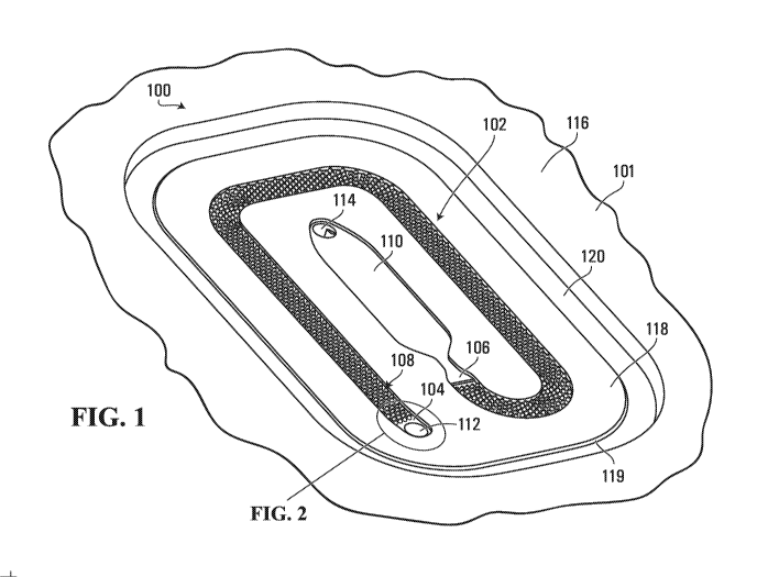
[0040] For example, Fig. 1 illustrates an isometric view of an example of a
haemolysis
stage 100 of a diagnostic consumable. The haemolysis stage 100 is implemented
as part of a
substrate 101 and includes a haemolysis channel 102 having an inlet port 104
for receiving
whole blood, an outlet port 106 for dispensing haemolysed blood, and an array
of micro-
proj ections 108 that extend into the haemolysis channel to define a plurality
of flow paths
therebetween along a length of the haemolysis channel between the inlet port
and the outlet
7
CA 03133975 2021-09-16
WO 2020/190462
PCT/US2020/019665
port. The array of micro-projections 108 has disposed thereon a haemolytic
reagent for
interaction with the whole blood as the whole blood is flowed through the
haemolysis
channel 102 to generate haemolysed blood. The inlet port 104 and/or the outlet
port of the
haemolysis channel 102 may be fluidly connected to other fluidic channels or
components on
the substrate 101. For example, in the embodiment illustrated in Fig. 1, the
inlet port 104 of
the haemolysis channel 102 is fluidly connected to another fluidic channel or
component on
the substrate 101 through a via 112, and the outlet port 106 of the haemolysis
channel is
fluidly connected to a chamber 110, which is in turn fluidly connected to
another fluidic
channel or component on the substrate 101 through a via 114. For example, in
some
embodiments, the via 114 may be fluidly connected to a vacuum port downstream
of the
chamber 110 that is configured for application of a vacuum source through
which a vacuum
can be applied as a pumping force to pull a blood sample through the
haemolysis channel 102
from the inlet port 104 to the outlet port 106 and into the chamber 110. For
example, the
vacuum source may be provided by a diagnostic device into which the diagnostic
consumable
is inserted or otherwise engaged. Such a vacuum port is merely one example of
a fluid
displacement element that may be incorporated into a diagnostic consumable in
order to
enable an external stimulus to be applied to the diagnostic consumable to pump
a fluid
sample through the channel. In the case of a downstream vacuum port, the
external stimulus
is in the form of a negative pressure or vacuum that pumps the fluid sample by
pulling the
fluid sample through the channel. In other implementations, the fluid
displacement element
may be a pumping port fluidly connected upstream of the channel and configured
for
application of a positive pressure source to push the fluid sample through the
channel. In
some embodiments, rather than directly applying a negative or positive
pressure source to the
diagnostic consumable, the external stimulus may be a mechanical or electrical
stimulus that
activates a pumping mechanism on the diagnostic consumable. For example, in
some
embodiments the fluid displacement element may be an air bladder that can be
mechanically
actuated in order to urge sample fluid through the channel. For example, the
air bladder may
have an exit port fluidly connected upstream of the channel through which air
can be expelled
from the air bladder by mechanically squeezing a flexible portion of the air
bladder in order
to push the fluid sample through the channel. Such an air bladder could be
implemented by a
cavity in the substrate 101 that is covered by a flexible cover layer affixed
to an external
surface of the substrate, for example. The mechanical stimulus to
actuate/squeeze the air
8
CA 03133975 2021-09-16
WO 2020/190462
PCT/US2020/019665
bladder could be provided by a mechanical actuator, e.g., an electrical motor,
of a diagnostic
device into which the diagnostic consumable is inserted or otherwise engaged.
[0041] The haemolysis channel 102 is illustrated in Fig. 1 as a channel
with a generally
rectangular cross-section and multiple turns along its length, however other
geometries of a
haemolysis channel are also possible. For example, in some embodiments a
haemolysis
channel could be substantially straight along its length. However, the
inclusion of multiple
turns or curves along the length of the channel may facilitate fitting a
longer channel in a
limited area on a diagnostic consumable.
[0042] Haemolysed blood dispensed into the chamber 110 may be analyzed. For
example, in the example illustrated in Fig. 1, the chamber 110 is configured
as a cuvette for
an optical assay, such as co-oximetry.
[0043] Fig. 2 is a magnified isometric view of a portion of the haemolysis
channel 102 of
Fig. 1 showing the disposition of the array of micro-projections 108 in the
channel. In
particular, Fig. 2 shows that the micro-projections in this example are
disposed in staggered
rows, each row being arranged substantially transverse to a direction of flow
through the
haemolysis channel from the inlet port 104 to the outlet port 106. Moreover,
in this example,
the micro-projections 108 are arranged with a generally uniform spacing
therebetween. In
addition, Fig. 2 shows that in this example the micro-projections 108 have the
form of micro-
pillars with a generally circular cross-sectional shape and a slight taper
from bottom to top.
In some embodiments the substrate 101 may be formed via injection moulding,
and the
slightly tapered shape of the micro-projections may facilitate removal of the
substrate 101
from an injection mould. For example, Fig. 3 is a scanning confocal microscope
image of a
portion of the array of micro-projections 108 implemented on a plastic
substrate obtained via
a plastic injection moulding process. As shown in Fig. 3, each micro-
projection has a
generally circular cross-sectional shape and a slight taper from bottom to top
to facilitate
removal of the plastic substrate from the mould. For similar reasons, in some
embodiments
the side walls 103,107 of the haemolysis channel 102 may be slightly inclined
outward from
bottom to top, as discussed in further detail below with reference to Fig. 7.
[0044] In the example shown in Figs. 1 and 2, the haemolysis channel 102
has a bottom
surface 105 and generally opposed side surfaces 103 and 107. In some
embodiments, a top
surface of the haemolysis channel 102 is formed by affixing a cover layer 130
to the substrate
9
CA 03133975 2021-09-16
WO 2020/190462
PCT/US2020/019665
101 so that the cover layer 130 covers the haemolysis channel 102. Fig. 4
shows an example
of the haemolysis stage 100 of Figs. 1 and 2 with a cover layer 130 affixed to
the substrate
101. In the example shown in Fig. 4, the cover layer 130 covers the haemolysis
channel 102
and the chamber 110. During manufacturing, the haemolysing reagent may be
deposited and
dried-down on the array of micro-projections 108 before the cover layer 130 is
affixed to the
substrate 101. For example, a solution of a haemolysing reagent, such as a
surfactant/detergent, dissolved in water and isopropyl alcohol may be
deposited on the array
of micro-projections 108 and allowed to dry-down on the micro-pillars before
the cover layer
130 is affixed to the substrate 101 to cover the haemolysis channel 102. The
array of micro-
projections 108 shown in Figs. 1 to 4 has strong capillarity properties, which
facilitates
relatively even spreading of the haemolysing reagent solution throughout the
array and the
surfaces 103, 105 and 107 of the channel (except the top surface 136 formed by
the cover
layer 130, which may be affixed after dry-down of the reagent in some
embodiments). In
some embodiments, the haemolytic reagent solution is applied to the micro-
pillars by
dispensing a predefined number of drops of the haemolytic reagent solution
onto the array
and allowing the capillarity of the array to disperse the haemolytic reagent
solution amongst
micro-projections of the array. In addition, this capillarity property tends
to retain the liquid
reagent solution in the area of the channel 102 in which the array of micro-
projections 108 is
located, rather than flowing into the chamber 110, which could potentially
affect an optical
assay in the chamber 110.
[0045] The cover layer 130 may be optically transparent in order to
facilitate optical
measurement of haemolysed blood in the chamber 110. For example, the cover
layer 130
may be made from a material with relatively high optical transparency, such as
glass or
polymethyl methacrylate (PMMA), also known as acrylic or acrylic glass.
[0046] In the example shown in Figs. 1 to 4, the haemolysis stage 100 is
formed in a
"well" 120 in the substrate 101 so that, when the cover layer 130 is affixed
to the substrate
101 to cover the haemolysis stage 100, a top surface 132 of the cover layer
130 is a short
distance below a top surface 116 of the substrate 101 to avoid potential
scratching/fouling of
top surface 132 of cover layer 130 during subsequent manufacturing steps. For
example, in
the example illustrated in Figs. 1 to 4, the haemolysis stage 100 is formed
into a top surface
118 of an "island" 119 within the "well" 120, wherein when the cover layer 130
is affixed to
the top surface 118 to cover the haemolysis stage 100, the top surface 132 of
the cover layer
CA 03133975 2021-09-16
WO 2020/190462
PCT/US2020/019665
130 is slightly below the top surface 116 of the substrate 101. Other
arrangements are
possible. For example, in some embodiments the top surface 132 of the cover
layer 130 may
be substantially co-planar with the top surface 116 of the substrate 101, In
other embodiments
the top surface 132 of the cover layer 130 may be slightly above the top
surface 116 of the
substrate 101, The cover layer 130 may be affixed to the substrate 101 by any
know affixing
means. For example, in some embodiments the cover layer 130 is adhesively
bonded to the
substrate 101.
[0047] Fig. 5 is a cross-sectional view of the haemolysis stage 100 of Fig.
4, taken along
the line illustrated in Fig. 4. As shown in Fig. 5, in this example the
haemolysis channel 102
has a bottom surface 105, a top surface 136 generally opposed to the bottom
surface 105, and
generally opposed side surfaces 103,107 extending between the bottom surface
105 and the
top surface 136, and the micro-projections 108 extend substantially the full
height of the
channel between the bottom surface 105 and the top surface 136. More
generally, micro-
projections may extend into a channel at least a portion of the height of the
channel, and may
extend from the top surface of the channel, the bottom surface of the channel,
or in some
cases from both the top and bottom surfaces of the channel, as discussed in
further detail
below with reference to Figs. 7 to 10.
[0048] Fig. 6 is a plan view of a portion of the haemolysis channel 102 of
the haemolysis
stage 100 of Fig. 4. The portion of the haemolysis channel 102 shown in Fig. 6
includes
eight staggered rows 1091, 1092, 1093, 1094, 1095, 1096, 1097 and 1098,
respectively, of
micro-projections. The staggered rows 1091-1098 of micro-projections are
arranged in the
channel 102 such that the micro-projections in each row are offset, in a
direction transverse to
the direction of flow through the haemolysis channel, relative to the micro-
projections in the
adjacent row(s). For example, the second row 1092 of micro-projections, which
includes four
micro-projections 1092,1, 1092,2, 1092,3 and 1092,4, is offset relative to the
first row 1091 of
micro-projections, which includes five micro-projections 1091,1, 1091,2,
1091,3, 1091,4 and
1091,5, such that the micro-projections in the second row 1092 are disposed
substantially
midway between the micro-projections in the first row 1091. Furthermore, as
shown in Fig.
6, every second row of micro-projections is substantially aligned in the
direction of flow
through the haemolysis channel 102. For example, the micro-projections in the
third row
1093 are substantially aligned, in the direction of flow through the
haemolysis channel, with
the micro-projections in the first row 1091, and the micro-projections in the
fourth row 1094
11
CA 03133975 2021-09-16
WO 2020/190462
PCT/US2020/019665
are substantially aligned with the micro-projections in the second row 1092,
and so on.
Moreover, in this example, the micro-projections 108 have a cross-sectional
dimension A,
measured transverse to the direction of flow through the haemolysis channel
102, that is
greater than a separation distance B between adjacent micro-projections in
each of the rows
1091-1098, which means that there are no straight flow paths through the array
of micro-
projections 108. Furthermore, the generally uniform spacing (separation
distance B) between
any two adjacent micro-pillars in each row means that as a blood cell flows
through each row
it is never more than one half of the separation distance B away from a
reagent coated
surface, thereby potentially resulting in a more consistent diffusion distance
across the cross-
section of the haemolysis channel 102. This is illustrated by way of example
in Fig. 7, which
is a a cross-sectional view of the haemolysis channel 102 of Fig. 6, taken
along the line
illustrated in Fig. 6 that extends through the first row 1091 of micro-
projections transverse to
the direction of flow.
[0049] As shown in Fig. 7, in this example the surfaces of the haemolysis
channel 102,
with the exception of the top surface 136 formed by the bottom surface of the
cover layer
130, are coated with dried-down haemolytic reagent 111. Moreover, from this
view it can be
seen that the sidewalls 103 and 107 of the haemolysis channel 102 and the five
micro-
projections 1091,1-1091,5 define six flow paths 1131,1, 1131,2, 1131,3,
1131,4, 1131,5 and 1131,6
through which a blood cell may move along the haemolysing channel 102 as it
traverses the
first row 1091 of micro-projections. As shown in Figs. 6 and 7, the offset of
the second row
1092 of micro-projections relative to the first row 1091 aligns the micro-
projections 1092,1-
1092,4 of the second row with the flow paths 1131,2-1131,5 defined by the
first row.
Furthermore, the generally uniform spacing of the micro-projections 108 in
this example
means that as a blood cell may never be more than one half of the generally
uniform
separation distance away from a reagent coated surface of a micro-projection
as the blood cell
is flowed through the array of micro-projections.
[0050] In some embodiments, the spacing between micro-projections could be
selected
based, at least in part, on a dimension of a component of the sample fluid
that is to be flowed
through the channel to interact with the reagent that has been dried-down in
the channel. For
example, in a haemolysis channel for the haemolysis of whole human blood, the
spacing
between micro-projections may be selected based, at least in part, on the
typical size of a
human red blood cell. A typical human red blood cell is generally disk-shaped
and has a disk
12
CA 03133975 2021-09-16
WO 2020/190462
PCT/US2020/019665
diameter of approximately 6-8 p.m, a thickness at its thickest point of 2-2.5
11 n.1 and a
minimum thickness in its centre of 0.8-1 him. In some embodiments, the spacing
between
micro-projections may be on the order of approximately a multiple of 10 of the
diameter of a
typical red blood cell. For example, in some embodiments the spacing between
micro-
projections may be on the order of 60-100 [tin. The 10 times multiple and the
60-100 pm
spacing are merely non-limiiing examples. Other multiples and dimensions are
possible and
are contemplated within the scope of the present disclosure.
[0051] As shown in Fig. 7, the generally opposed sidewalls 103,107 of the
haemolysing
channel 102 in this example are inclined outward, and the micro-projections
have a slight
taper from bottom to top, which, in those cases where the substrate is formed
via moulding
may facilitate removal of the substrate 101 from a mould.
[0052] The plurality of flow paths defined by the micro-projections within
the haemolysis
channel 102 means that the issue of high sample flow resistance associated
with a single thin
channel is substantially mitigated. Given its low flow resistance, the
haemolysis channel 102
can be made long enough that blood emerges from it completely haemolysed even
when
delivered at high speed by an external pressure source, provided that the time-
constant of
reagent dissolution is sufficient. This is due to numerous small-distance
diffusion events
afforded by a series of micro-projections along the length of the array.
Experimental results
obtained with a haemolysis channel implemented according to the haemolysis
channel 102
shown in Figs. 1 to 7 demonstrated complete haemolysis in less than 5 seconds.
In addition,
it has been observed that when a blood sample is flowed through the haemolysis
channel 102
it propagates with a substantially flat flow-front due to the array of micro-
projections within
the channel. In contrast, a blood sample flowed through channel without micro-
projections
typically propagates with a parabolic flow-front.
[0053] The structure of the haemolysis channel 102 of Figs. 1 to 7 is
provided by way of
example. Other haemolysis channel structures could also or instead be used in
a diagnostic
consumable. For example, in the haemolysis channel 102 shown in Figs. 4 to 7,
the micro-
projections 108 extend substantially the full height of the haemolysis channel
102 between
the bottom surface 105 formed by the substrate 101 and the top surface 136
formed by the
cover layer 130. However, as noted earlier, in other embodiments micro-
projections may
extend less than the full height of the channel, and may extend from the top
surface of the
13
CA 03133975 2021-09-16
WO 2020/190462
PCT/US2020/019665
channel, the bottom surface of the channel, or possibly both the top and
bottom surfaces of
the channel. Examples of such alternative embodiments are shown in Figs. 8 to
10.
[0054] Fig. 8 is a cross-sectional view of another haemolysis channel 202,
taken along a
transverse line that extends through a first row of micro-projections
transverse to the
direction of flow along the channel. Similar to the haemolysis channel 102
shown in Fig. 7,
the haemolysis channel 202 shown in Fig. 8 has a bottom surface 205 formed by
a substrate
201, a top surface 236 formed by the bottom surface of a cover layer 230, and
generally
opposed side walls 203, 207 extending between the bottom surface 205 and the
top surface
236. Moreover, from this view it can be seen that the first row of micro-
projections includes
five micro-projections 2091,1, 2091,2, 2091,3, 2091,4 and 2091,5 defining six
flow paths 2131,i,
2131,2, 2131,3, 2131,4, 2131,5 and 2131,6 through which a blood cell may move
along the
haemolysing channel 202 as it traverses the first row of micro-projections.
From this view it
can also be seen that a second row of micro-projections includes four micro-
projections
2092,1, 2092,2, 2092,3 and 2092,4 that are offset relative to the micro-
projections in the first row
so that they are substantially aligned with the flow paths 2131,2-2131,5
defined between the
micro-projections of the first row. However, in this example, the height 217
of the micro-
projections extends less than the full height 215 of the channel 202 so that
there is a gap 219
between the top surface 236 of the channel and the micro-projections.
[0055] Fig. 9 is a cross-sectional view of yet another haemolysis channel
302, taken
along a transverse line that extends through a first row of micro-projections
transverse to the
direction of flow along the channel. In this example, micro-projections extend
from both a
bottom surface 305A and atop surface 305B of the haemolysis channel 302. In
particular, in
this example, the bottom surface 305A of the channel 302 is formed by a first
substrate 301A
and the top surface 305B of the channel 302 is formed by a second substrate
301B. A first
array of micro-projections 308A extends into the channel 302 from the bottom
surface 305A,
and a second array of micro-projections 308B extends into the channel 302 from
the top
surface 305B. Micro-projections in each of the first and second arrays 308A,
308B are
arranged in staggered rows. In this example, each row of micro-projections in
the first array
of micro-projections 308A on the first substrate 301A is substantially aligned
with a
corresponding row of micro-projections in the second array of micro-
projections 308B on the
second substrate 301A.
14
CA 03133975 2021-09-16
WO 2020/190462
PCT/US2020/019665
[0056] An alternative arrangement of micro-projections in a haemolysis
channel 402, in
which micro-projections extend from both a bottom surface 405A and a top
surface 405B of
the haemolysis channel 402, is shown in Fig. 10. In this example, each row of
micro-
projections in a first array of micro-projections 408A on a first substrate
401A is offset
relative to a corresponding row of micro-projections in a second array of
micro-projections
408B on a second substrate 401B. In particular, in this arrangement, micro-
projections in the
first array of micro-projections 408A on the first substrate 401A extend
toward spaces
between micro-projections in the second array of micro-projections 408B on the
second
substrate 401B and vice versa.
[0057] In the embodiments shown in Figures 6 to 10, the first and last
micro-projection in
each row of micro-projections are spaced apart from the sidewalls 103 and 107
of the
haemolysis channel 102, and this spacing serves as a potential flow path. For
example,
referring again to Figures 6 and 7, in the first row 1091 it can be seen that
the flow path 1131,1
is defined between the sidewall 103 and the first micro-projection 1091,i,
while the flow path
1131,5 is defined between the sidewall 107 and the fifth micro-projection
1091,5. Similarly, in
the second row 1092 there is a first flow path defined between the sidewall
103 and the first
micro-projection 1092,1, while a fifth flow path is defined between the
sidewall 107 and the
fifth micro-projection 1092,5. As can be seen in Figure 6, the micro-
projections 109 in this
embodiment are distributed such that, in each of the odd rows 1091, 1093,
1095, 1097 the
spacing between the outermost micro-projections of the row and the sidewalls
103 and 107 of
the channel 102 is less than the separation distance between the micro-
projections in that row,
while in each of the even rows 1092, 1094, 1096, 1098 the spacing between the
outermost
micro-projections of the row and the sidewalls of the channel is greater than
the separation
distance between the micro-projections in that row. For example, the spacing
that defines the
flow path 1131,1 between the sidewall 103 and the first micro-projection
1091,1 in the first row
1091 is less than the separation distance between adjacent micro-projections
in the first row,
while the spacing that defines a flow path between the sidewall 103 and the
first micro-
projection 1092,1 in the second row 1092 is greater than the separation
distance between
adjacent micro-projections in the second row. This may cause the flow pattern
to be less
uniform near the sidewalls 103 and 107. However, although the flow paths
between the
outermost micro-projections and the sidewalls of the channel may at some
points be larger
than the separation distance between adjacent micro-projections, in general
the larger flow
path widths are not so large that the cumulative reagent-sample interaction
there becomes
CA 03133975 2021-09-16
WO 2020/190462
PCT/US2020/019665
insufficient. For example, in some embodiments the maximum spacing defining
flow paths
between the outermost micro-projections and the sidewalls of the channel may
be no more
than 200% of the separation distance between adjacent micro-projections, in
some
embodiments no more than 175% of the separation distance between adjacent
micro-
projections, in some embodiments no more than 150% of the separation distance
between
adjacent micro-projections, in some embodiments no more than 125% of the
separation
distance between adjacent micro-projections or even less.
[0058] In other embodiments, there may be no spacing between a micro-
projection and a
sidewall of a channel. For example, in some embodiments a micro-projection or
at least a
portion thereof may form part of one or more of the sidewalls of a channel. An
example of
such an embodiment is shown in Figure 11, which shows a haemolysis channel 102
that
differs from the haemolysis channel 102 shown in Figure 6 in that every second
row 1092,
1094, 1096, 1098, etc. additionally includes a first partial micro-projection
formed as part of
the first sidewall 103 and a second partial micro-projection formed as part of
the second
sidewall 107. For example, in addition to the four micro-projections 1092,1-
1092,4 of the
second row 1092 of micro-projections in Figure 6, the second row 1092 of micro-
projections
in Figure 11 further includes a first partial micro-projection 1092,0 and a
second partial micro-
projection 1092,5. The inclusion of the partial micro-projections at the
sidewalls potentially
makes the flow pattern and the sample-reagent interaction distance more
uniform near the
sidewalls of the channel, but may negatively impact manufacturability. For
example, it may
be more difficult to reliably manufacture such an embodiment repeatedly via
injection
molding.
[0059] Although the example embodiment described above and shown in Figs. 1
to 11
has been described as a haemolysis stage for haemolysing a whole blood sample,
the same or
similar structure could be used for other fluid sample preparation functions.
For example, the
same structure could be used to mix a whole blood sample with a coagulant for
an activated
clotting time (ACT) test e.g., by changing the material that is deposited on
the array of micro-
projections from a haemolytic reagent to a coagulant. In such cases, clotting
of the resulting
mix of whole blood and coagulant in the chamber 110 could be measured
optically using an
optical source and sensor external to the diagnostic device and/or or by means
of one or more
electrochemical sensors located somewhere downstream of the channel 102.
16
CA 03133975 2021-09-16
WO 2020/190462
PCT/US2020/019665
[0060] A non- limiting example of a substrate and diagnostic consumable
incorporating
the haemolysis stage of Figs. 1 to 7 will now be described with reference to
Figs. 12 to 18. It
is to be understood that this example implementation is provided for
illustrative purposes
only, and that other implementations and configurations of the haemolysis
stage, the substrate
and/or the diagnostic consumable are possible and are contemplated within the
present
disclosure.
[0061] Figs. 12 to 15 illustrate an example substrate 500 for a diagnostic
consumable that
includes multiple sensing regions. Figs. 12 and 13 are isometric views of the
substrate 500,
and Figs. 14 and 15 are plan views of the substrate. Figs. 12 and 14 are views
of a top
surface 502 of the substrate 500, and Figs. 13 and 15 are views of a bottom
surface 504 of the
substrate. The terms "top" and "bottom" are used herein for ease of reference
only, and do
not require or imply a certain orientation of the substrate 500. Although the
substrate 500
could be designed to be operated with the top surface 502 facing vertically
upwards and the
bottom surface 504 facing vertically downwards, this might not be the case in
all
implementations. Moreover, the orientation of the top surface 502 and the
bottom surface
504 of the substrate 500 could have minimal or no impact on fabrication,
storage and/or
transportation of the substrate.
[0062] The substrate 500 is illustrated as being a rectangular prism that
is approximately
the size and shape of a credit card, but this is only an example. The
substrate 500 could also
or instead be other shapes such as triangular or circular, for example. The
substrate 500
could be made out of plastics, ceramics, glass and/or metal, for example. The
substrate 500
could be a single, unitary body or part. The dimensions of the substrate 500
are not limited to
any specific ranges or values. The length and width of the substrate 500 could
be considered
to define the area of the top surface 502 and the bottom surface 504. In some
implementations, the length and/or width of the substrate 500 is on the order
of centimeters.
In some implementations, the length and/or width of the substrate 500 is on
the order of
millimeters. Other lengths and/or widths of the substrate 500 are also
possible. The
thickness of the substrate 500 could be measured as the distance between the
top surface 502
and the bottom surface 504 of the substrate. In some implementations, the
thickness of the
substrate 500 is on the order of centimeters. In some implementations, the
thickness of the
substrate 500 is on the order of millimeters. In some implementations, the
thickness of the
substrate 500 is on the order of micrometers. Other thicknesses of the
substrate 500 are also
17
CA 03133975 2021-09-16
WO 2020/190462
PCT/US2020/019665
possible. Although the top surface 502 and the bottom surface 504 of the
substrate 500 are
illustrated as being substantially flat, this might not be the case in all
embodiments. For
example, the top surface and/or the bottom surface of a substrate could also
or instead be
triangular, conical and/or hemispherical in shape. Accordingly, the thickness
of a substrate
could vary along its length and/or width. The substrate 500 is illustrated as
being transparent,
however substrates could also or instead be, in whole or in part, translucent
or opaque.
[0063] The substrate 500 includes the haemolysis stage 100 of Figs. 1 to 7,
in which a
portion of the chamber 110 functions as an optical sensing region 576. The
substrate 500
further includes a sample fluid input port 506, a sample fluid reservoir 508,
a fluid reservoir
510, a valve hole 512, two bubble traps 514, 516, another sensing region 518,
waste fluid
reservoirs 520, 543, multiple pump connection ports 522, 523, multiple vias
112, 114, 524,
526, 528, 530, 532, 534, 536, 545, and multiple channels 538, 540, 541, 542,
544, 546, 548,
550, 552, 554, 556, 558, 560, 562. In Figs. 12 to 15, solid lines are used to
illustrate
components that are directly in view in each figure, and dashed lines are used
to illustrate
components that are hidden from view by at least a portion of the substrate
500.
[0064] The channels 538, 540, 541, 542, 544, 546, 548, 550, 552, 554, 556,
558, 560, 562
are provided to carry one or more fluids in the substrate 100. The channels
540, 541, 542,
548, 552, 558 are trenches or grooves in the top surface 502 of the substrate
500. The
channels 540, 541, 542, 548, 552, 558 are illustrated as being open at the top
surface 502 of
the substrate 500 in Figs. 12 and 14. Similarly, the channels 538, 544, 546,
550, 554, 556,
560, 562 are trenches or grooves in the bottom surface 504 of the substrate
500, which are
open at the bottom surface of the substrate in Figs. 13 and 15. Any or all of
the channels 538,
540, 541, 542, 544, 546, 548, 550, 552, 554, 556, 558, 560, 562 could be
microfluidic
channels. For example, the width and/or height of any or all of the channels
538, 540, 541,
542, 544, 546, 548, 550, 552, 554, 556, 558, 560, 562 could be on the order of
micrometers.
The width and/or height of any or all of the channels 538, 540, 541, 542, 544,
546, 548, 550,
552, 554, 556, 558, 560, 562 could also or instead be on the order of
millimeters or
centimeters. The cross-sectional area of a channel or other fluidic component
is generally
measured as an area inside of the channel that is perpendicular to a direction
of fluid flow.
Although the channels 538, 540, 541, 542, 544, 546, 548, 550, 552, 554, 556,
558, 560, 562
are illustrated with generally rectangular cross-sections in Figs. 12 to 15,
one or more of these
18
CA 03133975 2021-09-16
WO 2020/190462
PCT/US2020/019665
channels could have other cross-sectional shapes as well, such as semicircular
or triangular,
for example.
[0065] The vias 112, 114, 524, 526, 528, 530, 532, 534, 536, 545 are
through-holes or
bores that extend through the substrate 500. Vias could be used to fluidly
connect two or
more components of the substrate 500. For example, via 112 fluidly connects
channel 542
and the haemolysis channel 102, via 114 fluidly connects chamber 110 and
channel 541, via
526 fluidly connects channel 538 and channel 540, via 528 fluidly connects
channel 540 and
channel 544, via 530 fluidly connects channel 552 and channel 554, via 532
fluidly connects
channel 548 and channel 556, via 534 fluidly connects channel 546 and channel
548, via 536
fluidly connects channel 560 and the waste fluid reservoir 520, and via 545
fluidly connects
channel 562 and the waste fluid reservoir 543. Vias could also or instead be
used to fluidly
connect a component of the substrate 500 to the top surface 502 and/or bottom
surface 504 of
the substrate. For example, the via 524 fluidly connects the sample fluid
reservoir 508 to the
bottom surface 504 of the substrate 500. Although illustrated as circular
holes, the vias could
also or instead be other shapes such as rectangular or triangular, for
example. The diameter
of the vias could be similar to the width of one or more of the components
that each via
connects. For example, the diameter of the via 526 could be similar to the
width of the
channel 538 and/or the channel 540. However, the diameter of the vias could be
different
from the width of the components that each via connects.
[0066] The sample fluid input port 506 is provided to deliver a blood
sample to the
substrate 500. The sample fluid input port 506 is a conical or cylindrical
opening in the top
surface 502 of the substrate 500. The sample input port 506 is coupled to the
channel 538.
The sample input port 506 could be sized and shaped to engage with an end of a
blood
sample delivery device, such as a syringe or capillary tube (not shown), that
delivers the
blood sample. For example, in the case of a syringe, this engagement between
the sample
input port 506 and the syringe could form a seal such that, when the blood
sample is
propelled or pumped out of the syringe, the blood sample is forced into the
channel 538 and
does not spill out of the sample input port. In some embodiments, a gasket
component is
installed in the sample input port 506 in order to facilitate the sealing
engagement with the
sample delivery device.
[0067] The sample fluid reservoir 508 could be a relatively wide and long
channel or
chamber that is coupled to the channel 540. The sample fluid reservoir 508 is
illustrated with
19
CA 03133975 2021-09-16
WO 2020/190462
PCT/US2020/019665
a rectangular cross-section, however other cross-sectional shapes are also
possible. The
sample fluid reservoir 508 could be provided to store a blood sample after it
is delivered into
the substrate 500. The via 524 could act as an air vent to allow air to escape
the sample fluid
reservoir 508 when it is displaced by the addition of blood sample. During
operation, the
blood sample might stay in the sample fluid reservoir 508 for an amount of
time that is on the
order of milliseconds, seconds, or minutes, for example.
[0068] The fluid reservoir 510 could be a relatively wide and long channel
or chamber
that is coupled to the channel 550. The fluid reservoir 510 is illustrated as
a U-shaped
channel with a semicircular cross-section, however other geometries are also
possible. In
some embodiments the fluid reservoir 510 could be provided to store a
calibration fluid or a
wash fluid and/or a fluid pack that seals the calibration fluid or the wash
fluid. The fluid
pack could be positioned in a shallow depression provided by the fluid pack
region 578. In
embodiments where the fluid reservoir 510 stores a calibration fluid, the
calibration fluid
could be used to calibrate one or more sensors included on and/or coupled to
the substrate
500. Calibration fluids could include fluids with known concentrations of one
or more
analytes. These analytes could correspond to analytes in the blood sample that
might be
measured using the substrate 500. In embodiments where the fluid reservoir 510
stores a
wash fluid, the wash fluid could be used to wash one or more regions of the
substrate 500.
For example, the wash fluid could be used to wash away unbound components from
an
antigen-antibody interaction region.
[0069] The valve hole 512 could be a via or bore that extends through the
thickness of the
substrate 500. The channel 550 and the channel 552 could be fluidly connected
by the valve
hole 512. The valve hole 512 could be sized and shaped to accommodate and/or
couple to a
valve (not shown). This valve could control the flow of fluid from the channel
550 to the
channel 552. When the valve is closed, the flow of fluid between the channel
550 and the
channel 552 could be blocked. When the valve is opened, the flow of fluid
between the
channel 550 and the channel 552 could be permitted. In some implementations,
the valve
could be closed until a seal in the valve is ruptured, allowing fluid to flow
into the channel
552.
[0070] The two bubble traps 514, 516 are provided to inhibit the movement
of bubbles in
the substrate 500. Each bubble that enters either of the bubble traps 514, 516
could be
prevented from moving further downstream by one or more barriers in the bubble
trap. Thus,
CA 03133975 2021-09-16
WO 2020/190462
PCT/US2020/019665
the fluid that leaves the bubble traps 514, 516 could be free of air bubbles.
The bubble trap
514 fluidly connects the channels 544, 546, and the bubble trap 516 fluidly
connects the
channels 554, 556.
[0071] The sensing region 518 includes a channel that is coupled to the
channel 548 and
to the channel 558. The sensing region 518 extends through the thickness of
the substrate
500, and is therefore illustrated as being open at the top surface 502 and
bottom surface 504
of the substrate in Figs. 12 to 15. The sensing region 518 could include
and/or be coupled to
one or more sensors that measure properties of fluids in the sensing region.
For example, the
sensors could measure the concentration of one or more analytes in a fluid
that flows from the
channel 548 to the channel 558. The sensing region 518 could also or instead
be referred to
as an assay region.
[0072] The waste fluid reservoir 520 is fluidly coupled to the channel 558,
and stores
fluid that has flowed through the sensing region 518. The waste fluid
reservoir 520 is
illustrated in Figs. 12 to 15 as a meandering channel with a rectangular cross-
section,
however other geometries of the waste fluid reservoir 520 are also possible.
[0073] The pump connection ports 522, 523 provide a connection to one or
more external
pumping systems. For example, these pumping systems could be provided in a
diagnostic
consumable reader module. The channel 560 is fluidly connected to the pump
connection
port 522, and the channel 562 is fluidly connected to the pump connection port
523. The
pumping systems could include channels or tubes that fluidly connect to the
pump connection
ports 522, 523. In some embodiments, the pumping systems could include vacuum
pumping
systems that pull fluid in one or more channels of the substrate 500 towards
the pump
connection ports 522, 523.
[0074] The optical sensing or assay region 576 provides another sensing
functionality to a
diagnostic consumable incorporating the substrate 500. The channel 542 fluidly
connects the
channel 540 to the haemolysis channel 102 through via 112. The haemolysis
channel 102 is
fluidly connected to the chamber 110 within the optical sensing region 576.
The channel 541
fluidly connects the chamber 110 and the waste fluid reservoir 543 through via
545. The
channel 562 fluidly connects the waste fluid reservoir 543 to the pump
connection port 523
through via 114. In operation, at least a portion of a blood sample could be
directed through
21
CA 03133975 2021-09-16
WO 2020/190462
PCT/US2020/019665
the channel 542, the haemolysis channel 102 and into the chamber 110 to be
optically
analyzed in the optical sensing region 576.
[0075] Figs. 16 and 17 illustrate plan views of an example diagnostic
consumable 600
that incorporates the substrate 500 shown in Figs. 12 to 15. Fig. 18 is a plan
view of the
haemolysis stage 100 of the diagnostic consumable 600 shown in Figs. 16 and
17. The
diagnostic consumable 600 could be considered an assembled diagnostic card or
test card for
blood analysis and/or testing. In some implementations, the diagnostic
consumable 600 is a
microfluidic device. The diagnostic consumable 600 could be configured, by
being sized and
shaped for example, to be received by a diagnostic consumable reader module
(not shown).
Fig. 16 is a view of the top surface 602 of the diagnostic consumable 600, and
Fig. 17 is a
view of the bottom surface 604 of the diagnostic consumable. In addition to
the substrate
500, the device 600 includes the cover layer 130 covering the haemolysis stage
100, a top
cover layer 606, a bottom cover layer 608, a sensor array 610, a calibration
fluid pack 612
(illustrated using parallel hatching) and a valve 614 (illustrated using cross-
hatching). Many
components of the substrate 500 are not labelled in Figs. 16 and 17 for the
purpose of clarity.
[0076] As described earlier, a haemolytic reagent can be deposited and
dried-down on the
array of micro-projections 108 in the haemolysis channel 102 before the cover
layer 130 is
affixed to the substrate 500. In this example, the cover layer 130 is
transparent to facilitate
optical sensing within the chamber 110 downstream of the haemolysis channel.
In other
embodiments, a cover layer for a haemolysis stage could be transparent,
translucent, opaque,
or a combination thereof
[0077] At least a portion of the top surface 502 and bottom surface 504 of
the substrate
500 are sealed using the top cover layer 606 and the bottom cover layer 608,
respectively.
The top and bottom cover layers 606, 608 could be impermeable to liquids (and
possibly
gases) to provide a liquid tight (and possibly gas tight) seal. In some
implementations, the
top and bottom cover layers 606, 608 could include plastic, metal and/or
ceramic films that
are bonded to the substrate 500 using an adhesive. For example, in some
implementations,
the top cover layer 606 and/or the bottom cover layer 608 could be implemented
as an
adhesive label or sticker. Non-limiting examples of adhesives include acrylic
adhesives and
silicone adhesives. The top and bottom cover layers 606, 608 could form a seal
around one
or more components of the substrate 600. For example, the top cover layer 606
could seal, at
least in part, the sample fluid reservoir 508, the bubble traps 514, 516, the
sensing region 518,
22
CA 03133975 2021-09-16
WO 2020/190462
PCT/US2020/019665
the waste fluid reservoir 520 and the channels 540, 541, 542, 548, 552, 558.
The bottom
cover layer 608 could seal, at least in part, the sample input port 506, the
fluid reservoir 510,
the bubble traps 514, 516 and the channels 538, 544, 546, 550, 554, 556, 560,
562. The top
cover layer 606 is illustrated as being substantially transparent and the
bottom cover layer
608 is illustrated as being substantially opaque, but this is only an example.
In general,
either or both of the top cover layer 606 and the bottom cover layer 608 could
be transparent,
translucent, opaque, or a combination thereof In Fig. 16, dashed lines are
used to illustrate
components that are under the top cover layer 606.
[0078] In this example, the sensor array 610, which could also be referred
to as an
electrode module, is bonded to the bottom surface 504 of the substrate 500.
The sensor array
610 overlaps and seals at least a portion of the sensing region 518. The
bottom cover layer
608 does not overlap the sensor array 610. The sensor array 610 could be
fabricated using
smart-card chip-module technology. In this example, the sensor array 610
includes a gold
coated copper metal foil laminated to an epoxy foil element 616 with an
optional adhesive.
The metal foil is formed into an array of electrode elements 618. Each
electrode element 618
could have a connection end for forming an electrical connection to a
measuring circuit in a
consumable reader module, for example. The connection ends of the electrode
elements 618
are not labelled for reasons of clarity. Multiple sensors 620 are coupled to
the electrode
elements 618. Each of the sensors 620 are positioned over the sensing region
518 of the
substrate 500. In use, the sensors 620 could be used to measure one or more
properties of a
calibration fluid and/or sample fluid in the sensing region 518. The sensors
620 could be
electrochemical sensors that are used for measuring concentrations of gases,
electrolytes
and/or metabolites. The sensors 620 could include potentiometric sensors to
measure
sodium, potassium, ionized calcium, chloride, urea, TCO2, pH levels and/or CO2
partial
pressure; amperometric sensors to measure 02 partial pressure, glucose,
creatinine, and/or
lactate; and/or conductometric sensors to measure hematocrit, for example. The
number and
geometry of the electrodes 618 and the sensors 620 is provided by way of
example only.
The same module fabrication technology can be used to make sensor arrays with
many
different electrode/sensor numbers and geometries.
[0079] The calibration fluid pack 612 is sandwiched between the calibration
fluid pack
region 578 of the substrate 500 and the bottom cover layer 608. The
calibration fluid pack
612 could fill the fluid reservoir 510 and the channel 550. The calibration
fluid pack 612
23
CA 03133975 2021-09-16
WO 2020/190462
PCT/US2020/019665
could be provided to seal and store a calibration fluid, in order to improve
the stability of the
calibration fluid over time. For example, the calibration fluid pack 612 could
inhibit gases,
such as carbon dioxide, from permeating into and/or out of the calibration
fluid.
[0080] The top surface 502 of the substrate 500 is substantially sealed by
the top cover
layer 606, with the exception of a hole 622 that corresponds to the location
of the sample
input port 506. The hole 622 allows a blood sample delivery device, such as a
syringe or
capillary tube, to be coupled to the sample input port 506 to deliver a blood
sample into the
diagnostic consumable 600. In addition, the top cover layer 606 also includes
a second hole
633 that corresponds to the location of the optical sensing region 576. As
discussed earlier,
the sample input port 506 may include a gasket component that facilitates a
sealing
engagement between the sample input port 506 and the sample delivery device.
For example,
the gasket component may be a rubber or silicone component installed in the
sample input
port 506 and sized and shaped to sealingly engage a sample delivery device.
[0081] The bottom surface 504 of the substrate 500 is substantially covered
by the bottom
cover layer 608, with the exception that the sensor array 610 and the via 524
are not sealed by
the bottom cover layer. The bottom cover layer 608 includes cuts or scoring
624, 626. The
scoring 624, 626 could be provided to render the bottom cover layer 608 more
malleable and
workable in the area proximate the scoring. The position of the scoring 624
corresponds to
the position of the valve 614. The scoring 624 could make the portion of the
bottom cover
layer 608 that is adjacent to the valve 614 more flexible, and could therefore
permit the valve
to be manipulated more easily. The position of the scoring 626 corresponds to
the position of
the fluid reservoir 510. The scoring 626 could make the portion of the bottom
cover layer
608 adjacent to the fluid reservoir 510 more flexible, and therefore permit
the calibration
fluid pack 612 to be manipulated more easily. The bottom cover layer 608 also
includes
pump holes 628, 630 corresponding to the location of the pump connection ports
522, 523 on
the substrate 500. The pump connection ports 522, 523 could be connected to a
pump in a
card reader module through the pump holes 628, 630. The pump holes 628, 630
could be
sized and shaped to form a seal between the pump and the pump connection ports
522, 523.
The bottom cover layer 608 overlaps the cover layer 130 of the haemolysis
stage 100, but
includes a hole 632 corresponding to the optical sensing region 576 and
generally aligned
with the hole 633 in the top cover layer 606. The holes 632, 633 and the
transparency of the
24
CA 03133975 2021-09-16
WO 2020/190462
PCT/US2020/019665
substrate 500 and the cover layer 130 in the area of the optical sensing
region 576 facilitate
optical sensing within the optical sensing region.
[0082] In this example, a 1D barcode 634 is printed on the bottom cover
layer 608. The
barcode 634 could be read by a card reader module when the diagnostic
consumable 600 is
inserted into the card reader module. The barcode 634 could authenticate the
diagnostic
consumable 600 and/or provide information regarding the diagnostic consumable.
For
example, the barcode 634 could indicate the date that the diagnostic
consumable 600 was
manufactured. The barcode 634 is one example of a machine-readable code that
could be
present on the bottom cover layer 608 or elsewhere on the diagnostic
consumable. Other
examples of machine-readable codes include 2D barcodes. Radio-frequency
identification
(RFID) chips or tags could also or instead be used.
[0083] In some embodiments, the diagnostic consumable 600 could be operated
as
follows. First, the diagnostic consumable 600 could be inserted into a
corresponding slot of a
diagnostic module, such as a portable or bench-top diagnostic card reader
module. The
diagnostic module might scan the barcode 634 to authenticate the diagnostic
consumable 600.
Second, the calibration fluid that is stored in the calibration fluid pack 612
could be propelled
or pumped into the sensing region 618. This step could include the diagnostic
module using
a first actuator element to manipulate the valve 614 by pushing on the bottom
cover layer 608
in an area proximate the scoring 624. The manipulation of the valve 614 could
cause the plug
in the valve to rupture, which opens the valve. At least a portion of the
calibration fluid could
then be pushed or pumped out of the calibration fluid pack 612, through the
channel 550, the
valve 512, the channel 552, the via 530, the channel 554, the bubble trap 516,
the channel
556, the via 532, the channel 548, and into the sensing region 518. Pushing
the calibration
fluid out of the calibration fluid pack 612 could be performed by compressing
the bottom
cover layer 608 in the area proximate the scoring 626 using a second actuator
element, such
as a plunger, in the diagnostic module. When the calibration fluid is in the
sensing region
518, it might be in contact with one or more of the sensors 620. The
diagnostic module could
include circuitry to contact the electrodes 618, which return measurements of
the calibration
fluid from the sensors 620. These measurements could be used to calibrate the
diagnostic
module for the diagnostic consumable 600, and thereby compensate for
variations between
different diagnostic consumables. The first and second actuator elements could
be controlled
by a motor-driven system in the diagnostic module. The diagnostic module could
also
CA 03133975 2021-09-16
WO 2020/190462
PCT/US2020/019665
include a form of temperature control, such as a heater in contact with the
sensor array 610,
to adjust the temperature of a fluid in the sensing region 518 and/or a heater
in contact with,
or proximal to, the optical sensing region 576, to adjust the temperature of a
fluid in the
optical sensing region. This temperature control could help provide
consistency in the
measurements made by the sensors 620 in the sensing region 518 or by an
optical sensor in a
diagnostic consumable module configured to measure one or more properties of a
sample in
the optical sensing region 576. In some implementations, the temperature of
the fluid in the
sensing region 518 and/or the fluid in the optical sensing region 576 could be
maintained at
approximately body temperature, e.g., at approximately 37 degrees Celsius.
[0084] After calibration, the diagnostic module could instruct a user to
inject a blood
sample into the sample fluid input port 506. At least a portion of the blood
sample could
flow through the channel 538, the via 526, the channel 540 and into the sample
fluid storage
reservoir 508. A vacuum pump in the diagnostic module could be coupled to the
pump
connection port 522 through the pump hole 628. When this vacuum pump is turned
on, the
vacuum pump could draw the calibration fluid from the sensing region 518 into
the waste
fluid reservoir 520. Further, the vacuum pump could draw the blood sample from
the sample
fluid reservoir 508 and/or the channel 540 (if the fluid reservoir 508 is not
vented), through
the via 528, the channel 544, the bubble trap 514, the channel 546, the via
534, the channel
548, and into the sensing region 518. The diagnostic module and sensors 620
could then
perform measurements on the blood sample to determine the concentration of
certain analytes
in the blood sample, for example. In this embodiment, the pump connection port
523
functions as a sample fluid displacement element of the diagnostic consumable
that enables
an external stimulus (vacuum pressure) to be applied to the diagnostic
consumable in order to
pump the fluid sample (whole blood) through the channel 102 and into the
chamber 110. As
noted earlier, in other embodiments other types of fluid displacement elements
may be used
to pump the blood sample through the channel 102, such as a pumping port or a
mechanically
actuatable air bladder fluidly connected upstream of the channel 102 and
configured for
application of a positive pressure source to push the blood sample through the
channel.
[0085] Optical assays can be performed on the blood sample in the optical
sensing region
576 on the diagnostic consumable 600. For example, a vacuum pump in the
diagnostic
module that is coupled to the pump connection port 523 through the pump hole
630 could be
used to apply vacuum pressure to the pump connection port 523 to draw a
portion of the
26
CA 03133975 2021-09-16
WO 2020/190462
PCT/US2020/019665
blood sample from the sample fluid reservoir 508 and/or the channel 540,
through the
channel 542, the via 112, the haemolysis channel 102, and into the optical
sensing region 576
within the chamber 110. As the blood sample is flowed through the haemolysis
channel 102,
it dissolves, re-suspends, and reacts with the haemolytic reagent that was
dried-down on the
array of micro-projections to generate haemolysed blood, which is then
dispensed into the
chamber 110. A light source and detector in the diagnostic module could then
perform
optical measurements on the haemolysed blood sample in the optical sensing
region 576. In
some embodiments, CO-oximetry could be performed in the optical sensing region
576 to
measure the concentrations of total hemoglobin (1E4 oxyhernoglobin (02HB),
carboxyhemoglobin (COHb), methernoglobin (Mettlb), deoxyhemoglobin 0-1E4
oxygen
saturation (S02) and/or total bilirubin (tBili) in the blood sample, for
example. This could
complete the testing that is performed using the diagnostic consumable 600.
The diagnostic
consumable 600 could be a disposable diagnostic device that is disposed of
after use.
However, reusable devices are also contemplated. As noted earlier, the same or
similar
structure as that of the haemolysis stage 100 could instead be used to mix a
whole blood
sample with a coagulant for an ACT test by using a coagulant rather than a
haemolytic
reagent and measuring the clotting time of the resulting mix of whole blood
and coagulant
downstream of the channel 102.
[0086] The embodiments described above relate primarily to diagnostic
consumables.
Other embodiments, including methods, are also contemplated.
[0087] Fig. 19, for example, is a flow diagram illustrating an example
method 700 for
making a diagnostic consumable for use in analysis of a whole blood sample. In
some
implementations, the diagnostic consumable could be a microfluidic device. The
method 700
includes multiple steps 702, 704, 706, 708.
[0088] Step 702 includes obtaining a substrate that includes a haemolysis
channel having
an array of micro-projections extending into the haemolysis channel to define
a plurality of
flow paths therebetween along at least a portion of a length of the haemolysis
channel. In
some embodiments, obtaining the substrate includes forming the substrate via a
molding
process, wherein the array of micro-projections is molded into the haemolysis
channel in the
molding process.
27
CA 03133975 2021-09-16
WO 2020/190462
PCT/US2020/019665
[0089] Step 704 includes applying a haemolytic reagent solution to the
array of micro-
projections in the haemolysis channel. Applying the haemolytic reagent
solution could
involve dispensing a predefined number of drops of the haemolytic reagent
solution onto the
array of micro-projections, for example. In some embodiments, capillarity of
the array of
micro-projections causes the haemolytic reagent solution to disperse amongst
the array of
micro-projections.
[0090] Step 706 includes drying-down the haemolytic reagent solution onto
the array of
micro-projections so that the array of micro-projections has dried haemolytic
reagent
disposed thereon. In some embodiments, the haemolytic reagent solution may be
dried-down
by allowing a solvent component of the haemolytic reagent solution to
passively evaporate.
[0091] Step 708 is an optional step that includes affixing a cover layer to
one side of the
substrate to form either a top surface or a bottom surface of the haemolysis
channel. The
micro-projections extend into the haemolysis channel from the other of the top
surface or the
bottom surface of the haemolysis channel.
[0092] The example operations of the method 700 are illustrative of an
example
embodiment. Various ways to perform the illustrated operations, as well as
examples of
other operations that may be performed, are described herein. Further
variations may be or
become apparent.
[0093] For example, while the method 700 is illustrative of an example for
making a
diagnostic consumable that includes a haemolysis channel for preparing a
haemolysed blood
sample, similar operations may be performed to make other types of diagnostic
consumables
that include sample preparation channels for mixing and/or interacting a
material with a fluid
sample. For example, rather than applying haemolytic reagent solution to the
array of micro-
projections at step 704, a fluid comprising another material, such as a
coagulant, may be
applied to the micro-projections and the fluid may be dried-down onto the
micro-projections
at step 706 so that the array of micro-projections has the material disposed
thereon.
[0094] Fig. 20 is a flow diagram illustrating an example method 800 for
analysis of a
whole blood sample on a diagnostic consumable. In some implementations, the
diagnostic
consumable could be a microfluidic device. The method 800 includes multiple
steps 802,
804, 806, 808.
28
CA 03133975 2021-09-16
WO 2020/190462
PCT/US2020/019665
[0095] Step 802 includes receiving a whole blood sample at an inlet port of
a haemolysis
stage of the diagnostic consumable.
[0096] Step 804 includes haemolysing the whole blood by flowing the whole
blood
through a haemolysis channel of the haemolysis stage of the diagnostic
consumable. The
haemolysis channel includes an array of haemolytic reagent-coated micro-
projections that
extend into the haemolysis channel to define a plurality of flow paths
therebetween. The
haemolytic reagent interacts with the whole blood to generate haemolysed blood
as the blood
is flowed through the haemolysis channel. The haemolysis channel could be
similar to the
haemolysis channels 102, 202, 302 and/or 402 that are discussed in detail
above, for example.
In some embodiments, flowing the whole blood through the haemolysis channel
includes
pumping the whole blood through the haemolysis channel. For example, the whole
blood
could be pumped through the haemolysis channel by applying an external
pressure source to
the diagnostic consumable. The external pressure source could be a vacuum
source that
applies a vacuum to a vacuum port on the diagnostic card that is fluidly
connected
downstream of the haemolysis channel.
[0097] Step 806 is an optional step that includes flowing the haemolysed
blood into a
chamber that is fluidly connected to the haemolysis channel. The chamber could
be similar
to the chamber 110 discussed in detail above. For example, the chamber could
be configured
as a cuvette for use in an optical assay.
[0098] Step 808 is an optional step that includes performing an optical
assay of the
haemolysed blood in the chamber through at least a portion of the chamber that
is optically
transparent. For example, the chamber could have optically transparent top and
bottom
surfaces and performing the optical assay could involve performing a
spectroscopic analysis
of light passed through the haemolysed blood in the chamber via the optically
transparent top
and bottom surfaces of the chamber. Such an optical assay could be a CO-
oximetry assay to
measure the concentrations of tHb, 02HB. COHb, MetHb, HHb arid/or tBili in the
blood
sample, for example.
[0099] The example operations of the method 800 are illustrative of an
example
embodiment. Various ways to perform the illustrated operations, as well as
examples of
other operations that may be performed, are described herein. Further
variations may be or
become apparent.
29
CA 03133975 2021-09-16
WO 2020/190462
PCT/US2020/019665
[00100] For example, while the method 800 is illustrative of an example for
analysis of a
whole blood sample on a diagnostic consumable that includes a step of
haemolysing the
whole blood sample, similar operations may be performed for other types of
analyses of
whole blood samples or other types of fluid samples that require the mixing
and/or interacting
of one or more materials with the fluid sample. For example, similar to the
operation at step
802, a fluid sample may be received at an inlet port of a sample preparation
stage of a
diagnostic consumable and then, similar to the operation at step 804, a
material may be mixed
into the fluid sample by flowing the fluid sample through a channel of the
sample preparation
stage that includes an array of micro-projections that have the material
disposed thereon. As
the fluid sample is flowed through the channel the material disposed on the
micro-projections
mixes with the fluid sample to generate a prepared fluid sample. For example,
as described
earlier, in some embodiments the fluid sample may be whole blood and the
material disposed
on the micro-projections may be a coagulant for mixing with the whole blood in
order to
perform a dotting time test.
[00101] Although the present disclosure relates primarily to haemolysis
channels in
diagnostic consumables for blood analysis systems, the embodiments described
herein could
also or instead relate to other types of fluid sample preparation channels in
diagnostic
consumables or other types of analysis systems. In particular, channels that
include reagent-
coated micro-projections could be used in any of a variety of applications
where sample fluid
preparation via reagent interaction on a diagnostic consumable would be
advantageous. For
example, an antibody reagent having an affinity for an antigen could be dried
down on an
array of micro-projections in a channel, so that when a sample fluid that may
contain the
antigen is flowed through the channel, any antigen that may be in the sample
fluid is exposed
to interact with the antibody reagent on the micro-projection array.
Downstream analysis of
the fluid dispensed from the channel may be performed to detect the antigen-
antibody
binding, for example.
ILLUSTRATIVE EMBODIMENTS
[00102] The following provides a non-limiting list of additional Illustrative
Embodiments
of the present disclosure:
Example Embodiment 1. A diagnostic consumable for use in the analysis of a
fluid
sample, the diagnostic consumable comprising:
CA 03133975 2021-09-16
WO 2020/190462
PCT/US2020/019665
a substrate having a sample preparation stage, the sample preparation stage
comprising:
i) an inlet port for receiving a fluid sample;
ii) an outlet port for dispensing a prepared fluid sample; and
iii) a channel extending from the inlet port to the outlet port, the channel
comprising
an array of micro-projections extending into the channel to define a plurality
of flow paths
therebetween along at least a portion of a length of the channel between the
inlet port and the
outlet port, the array of micro-projections having disposed thereon a material
for mixing with
the fluid sample as the fluid sample is flowed through the channel to generate
the prepared
fluid sample.
Example Embodiment 2. The diagnostic consumable of Example Embodiment 1,
wherein the micro-projections of the array are arranged with a generally
uniform spacing.
Example Embodiment 3. The diagnostic consumable of Example Embodiment 1 or
2,
wherein the micro-projections of the array are disposed in staggered rows
along at least a
portion of the length of the channel, each row being arranged substantially
transverse to a
direction of flow through the channel.
Example Embodiment 4. The diagnostic consumable of Example Embodiment 3,
wherein the staggered rows of micro-projections are disposed over
substantially the entire
length of the channel between the inlet port and the outlet port.
Example Embodiment 5. The diagnostic consumable of Example Embodiment 3,
wherein the staggered rows of micro-projections comprises a first row of micro-
projections
and a second row of micro-projections disposed adjacently downstream from the
first row of
micro-projections relative to the direction of flow through the channel, the
second row of
micro-projections being offset in a direction transverse to the direction of
flow through the
haemolysis channel, relative to the first row of micro-projections, such that
micro-projections
in the second row are disposed substantially midway between micro-projections
in the first
row.
Example Embodiment 6. The diagnostic consumable of Example Embodiment 5,
wherein:
31
CA 03133975 2021-09-16
WO 2020/190462
PCT/US2020/019665
a separation distance, measured transverse to the direction of flow through
the
haemolysis channel, between adjacent micro-projections in each of the first
and second rows
is substantially equal; and
the micro-projections in the first and second rows have a cross-sectional
dimension,
measured transverse to the direction of flow through the channel, that is
greater than or equal
to the separation distance between adjacent micro-projections in each of the
first and second
rows.
Example Embodiment 7. The diagnostic consumable of Example Embodiment 6,
wherein:
the staggered rows of micro-projections further comprises a third row of micro-
projections disposed adjacently downstream from the second row of micro-
projections; and
micro-projections in the third row are substantially aligned, in the direction
of flow
through the channel, with micro-projections in the first row.
Example Embodiment 8. The diagnostic consumable of any of Example
Embodiments 1
to 7, wherein:
the channel has a bottom surface, a top surface generally opposed to the
bottom
surface, and generally opposed side surfaces extending between the bottom
surface and the
top surface;
a height of the channel being defined as a distance between the bottom surface
of the
channel and the top surface of the channel; and
the micro-projections extend into the channel at least a portion of the height
of the
channel between the bottom surface and the top surface of the channel.
Example Embodiment 9. The diagnostic consumable of Example Embodiment 8,
wherein the micro-projections extend the height of the channel between the
bottom surface
and the top surface of the channel.
Example Embodiment 10. The diagnostic consumable of Example Embodiment 9,
wherein:
32
CA 03133975 2021-09-16
WO 2020/190462
PCT/US2020/019665
either the top surface or the bottom surface of the channel is formed by a
cover layer
affixed to one side of the substrate; and
the micro-projections extend from the other of the top surface and the bottom
surface
of the channel to the cover layer.
Example Embodiment 11. The diagnostic consumable of any one of Example
Embodiments 1 to 10, further comprising a fluid displacement element in fluid
communication with the channel, the fluid displacement element enabling an
external
stimulus to be applied to the diagnostic consumable to pump the fluid sample
through the
channel.
Example Embodiment 12. The diagnostic consumable of Example Embodiment 11,
wherein the fluid displacement element comprises a vacuum port downstream of
the channel,
the vacuum port configured for application of a vacuum source to pump the
fluid sample
through the channel.
Example Embodiment 13. The diagnostic consumable of any one of Example
Embodiments 1 to 12, wherein the material disposed on the array of micro-
projections
comprises a reagent that reacts with the fluid sample as the fluid sample is
flowed through the
channel.
Example Embodiment 14. The diagnostic consumable of Example Embodiment 13,
wherein:
the fluid sample is whole blood;
the reagent disposed on the array of micro-projections comprises a haemolytic
reagent; and
the prepared fluid sample comprises haemolysed blood.
Example Embodiment 15. The diagnostic consumable of Example Embodiment 13,
wherein:
the fluid sample is whole blood;
the reagent disposed on the array of micro-projections comprises a coagulant;
and
33
CA 03133975 2021-09-16
WO 2020/190462
PCT/US2020/019665
the prepared fluid sample comprises a mixture of the whole blood and the
coagulant.
Example Embodiment 16. The diagnostic consumable of any one of Example
Embodiments 1 to 15, wherein the substrate comprises a molded plastic
substrate.
Example Embodiment 17. The diagnostic consumable of any one of Example
Embodiments 1 to 16, wherein the micro-projections comprise micro-pillars.
Example Embodiment 18. The diagnostic consumable of any one of Example
Embodiments 1 to 17, wherein the substrate further comprises a prepared fluid
sample
collection vessel, the prepared fluid sample collection vessel comprising:
an inlet port fluidly connected to the outlet port of the sample preparation
stage for
receiving the prepared fluid sample; and
a chamber for containing the prepared fluid sample.
Example Embodiment 19. A method for analysis of a fluid sample on a
diagnostic
consumable, the method comprising:
receiving a fluid sample at an inlet port of a sample preparation stage of the
diagnostic
consumable;
mixing a material into the fluid sample by flowing the fluid sample through a
channel
of the sample preparation stage of the diagnostic consumable, the channel
comprising an
array of micro-projections extending into the channel to define a plurality of
flow paths
therebetween along at least a portion of a length of the channel, the array of
micro-projections
having disposed thereon the material for mixing with the fluid sample as the
fluid sample is
flowed through the channel to generate a prepared fluid sample.
Example Embodiment 20. The method of Example Embodiment 19, wherein the method
further comprises flowing the prepared fluid sample into a chamber on the
diagnostic
consumable that is fluidly connected to the channel.
Example Embodiment 21. The method of Example Embodiment 19 or 20, wherein
flowing the fluid sample through the channel comprises applying an external
stimulus to a
34
CA 03133975 2021-09-16
WO 2020/190462
PCT/US2020/019665
fluid displacement element in fluid communication with the channel to pump the
fluid sample
through the channel.
Example Embodiment 22. The method of Example Embodiment 21, wherein the fluid
displacement element comprises a pumping port in fluid communication with the
channel, the
pumping port being configured for application of an external pressure source
to the
diagnostic consumable to pump the fluid sample through the channel.
Example Embodiment 23. The method of Example Embodiment 22, wherein the
pumping
port comprises a vacuum port downstream of the channel, and wherein applying
an external
pressure source to diagnostic consumable comprises applying a vacuum source to
the vacuum
port to pump the fluid sample through the channel.
Example Embodiment 24. The method of any one of Example Embodiments 19 to 23,
wherein the material disposed on the array of micro-projections comprises a
reagent that
reacts with the fluid sample as the fluid sample is flowed through the
channel.
Example Embodiment 25. The method of Example Embodiment 24, wherein the
reagent
disposed on the array of micro-projections comprises a haemolytic reagent or a
coagulant.
Example Embodiment 26. A method of making a diagnostic consumable for use in
analysis of a fluid sample, the method comprising:
obtaining a substrate that includes a channel having an array of micro-
projections
extending into the channel to define a plurality of flow paths therebetween
along at least a
portion of a length of the channel;
applying a fluid to the array of micro-projections in the channel, the fluid
comprising
a material for deposition on the array of micro-projections; and
drying-down the fluid onto the array of micro-projections so that the array of
micro-
projections has the material disposed thereon.
Example Embodiment 27. The method of Example Embodiment 26, wherein applying
the
fluid to the array of micro-projections comprises dispensing a predefined
number of drops of
the fluid onto the array of micro-projections.
CA 03133975 2021-09-16
WO 2020/190462
PCT/US2020/019665
Example Embodiment 28. The method of Example Embodiment 26 or 27, wherein
capillarity of the array of micro-projections causes the fluid to disperse
amongst the array of
micro-projections.
Example Embodiment 29. The method of any of Example Embodiments 26 to 28,
wherein
drying-down the fluid comprises passively evaporating a solvent component of
the fluid.
Example Embodiment 30. The method of any of Example Embodiments 26 to 29,
further
comprising affixing a cover layer to one side of the substrate, the cover
layer forming either a
top surface or a bottom surface of the channel, the micro-projections
extending into the
channel from the other of the top surface or the bottom surface of the
channel.
Example Embodiment 31. The method of any of Example Embodiments 26 to 30,
wherein
the material disposed on the array of micro-projections comprises a reagent
that reacts with
the fluid sample as the fluid sample is flowed through the channel.
Example Embodiment 32. The method of Example Embodiment 31, wherein the
reagent
disposed on the array of micro-projections comprises a haemolytic reagent or a
coagulant.
Example Embodiment 33. The method of any one of Example Embodiments 26 to 32,
wherein obtaining the substrate comprises forming the substrate via a molding
process, the
array of micro-projections being molded into the channel in the molding
process.
Example Embodiment 34. The method of Example Embodiment 33, wherein the
substrate
comprises a plastic substrate and the molding process comprises injection
molding.
Example Embodiment 35. The method of Example Embodiment 33 or 34, wherein
forming the substrate via a molding process comprises molding the substrate
such that the
substrate comprises: an inlet port in fluid communication with the channel for
receiving a
fluid sample into the channel; and a pumping port in fluid communication with
the channel
for applying an external pressure source to the diagnostic consumable to pump
the fluid
sample through the channel.
Example Embodiment 36. The method of Example Embodiment 35, wherein the
pumping
port comprises a vacuum port formed in the substrate downstream of the
channel, so that, in
use, a vacuum source applied to the vacuum port causes the fluid sample to be
pumped
through the channel.
36
CA 03133975 2021-09-16
WO 2020/190462
PCT/US2020/019665
Example Embodiment 37. A diagnostic consumable for use in the analysis of
whole
blood, the diagnostic consumable comprising:
a substrate having a haemolysis stage, the haemolysis stage comprising:
i) an inlet port for receiving whole blood;
ii) an outlet port for dispensing haemolysed blood; and
iii) a haemolysis channel extending from the inlet port to the outlet port,
the
haemolysis channel comprising an array of micro-projections extending into the
haemolysis
channel to define a plurality of flow paths therebetween along at least a
portion of a length of
the haemolysis channel between the inlet port and the outlet port, the array
of micro-
projections having disposed thereon a haemolytic reagent for interaction with
the whole
blood as the whole blood is flowed through the haemolysis channel to generate
haemolysed
blood.
Example Embodiment 38. The diagnostic consumable of Example Embodiment 37,
wherein the micro-projections of the array are arranged with a generally
uniform spacing.
Example Embodiment 39. The diagnostic consumable of Example Embodiment 37 or
38,
wherein the micro-projections of the array are disposed in staggered rows
along at least a
portion of the length of the haemolysis channel, each row being arranged
substantially
transverse to a direction of flow through the haemolysis channel.
Example Embodiment 40. The diagnostic consumable of Example Embodiment 39,
wherein the staggered rows of micro-projections are disposed over
substantially the entire
length of the haemolysis channel between the inlet port and the outlet port.
Example Embodiment 41. The diagnostic consumable of Example Embodiment 39 or
40,
wherein the micro-projections are disposed in the haemolysis channel such
that:
in each row, a separation distance between adjacent micro-projections in the
row is
substantially equal; and
a separation distance between adjacent rows of micro-projections is
substantially
equal to the separation distance between adjacent micro-projections in each
row.
37
CA 03133975 2021-09-16
WO 2020/190462
PCT/US2020/019665
Example Embodiment 42. The diagnostic consumable of any one of Example
Embodiments 39 to 41, wherein the staggered rows of micro-projections
comprises a first
row of micro-projections and a second row of micro-projections disposed
adjacently
downstream from the first row of micro-projections relative to the direction
of flow through
the haemolysis channel, the second row of micro-projections being offset in a
direction
transverse to the direction of flow through the haemolysis channel, relative
to the first row of
micro-projections, such that micro-projections in the second row are disposed
substantially
midway between micro-projections in the first row.
Example Embodiment 43. The diagnostic consumable of Example Embodiment 42,
wherein:
a separation distance, measured transverse to the direction of flow through
the
haemolysis channel, between adjacent micro-projections in each of the first
and second rows
is substantially equal; and
the micro-projections in the first and second rows have a cross-sectional
dimension,
measured transverse to the direction of flow through the haemolysis channel,
that is greater
than or equal to the separation distance between adjacent micro-projections in
each of the
first and second rows.
Example Embodiment 44. The diagnostic consumable of Example Embodiment 43,
wherein:
the staggered rows of micro-projections further comprises a third row of micro-
projections disposed adjacently downstream from the second row of micro-
projections; and
micro-projections in the third row are substantially aligned, in the direction
of flow
through the haemolysis channel, with micro-projections in the first row.
Example Embodiment 45. The diagnostic consumable of any one of Example
Embodiments 37 to 44, wherein:
the haemolysis channel has a bottom surface, a top surface generally opposed
to the
bottom surface, and generally opposed side surfaces extending between the
bottom surface
and the top surface;
38
CA 03133975 2021-09-16
WO 2020/190462
PCT/US2020/019665
a height of the haemolysis channel being defined as a distance between the
bottom
surface of the haemolysis channel and the top surface of the haemolysis
channel; and
the micro-projections extend into the channel at least a portion of the height
of the
haemolysis channel between the bottom surface and the top surface of the
haemolysis
channel.
Example Embodiment 46. The diagnostic consumable of Example Embodiment 45,
wherein the micro-projections extend the height of the haemolysis channel
between the
bottom surface and the top surface of the haemolysis channel.
Example Embodiment 47. The diagnostic consumable of Example Embodiment 46,
wherein:
either the top surface or the bottom surface of the haemolysis channel is
formed by a
cover layer affixed to one side of the substrate; and
the micro-projections extend from the other of the top surface and the bottom
surface
of the haemolysis channel to the cover layer.
Example Embodiment 48. The diagnostic consumable of any one of Example
Embodiments 37 to 47, wherein the substrate comprises a molded plastic
substrate.
Example Embodiment 49. The diagnostic consumable of any one of Example
Embodiments 37 to 48, wherein the micro-projections comprise micro-pillars.
Example Embodiment 50. The diagnostic consumable of Example Embodiment 49,
wherein the micro-pillars have a generally circular cross-section.
Example Embodiment 51. The diagnostic consumable of any one of Example
Embodiments 37 to 50, wherein the substrate further comprises a haemolysed
blood
collection vessel, the haemolysed blood collection vessel comprising:
an inlet port fluidly connected to the outlet port of the haemolysis stage for
receiving
the haemolysed blood; and
a chamber for containing the haemolysed blood.
39
CA 03133975 2021-09-16
WO 2020/190462
PCT/US2020/019665
Example Embodiment 52. The diagnostic consumable of Example Embodiment 51,
wherein at least a portion of the chamber is optically transparent to permit
an optical assay of
the haemolysed blood.
Example Embodiment 53. The diagnostic consumable of Example Embodiment 52,
wherein the chamber comprises:
optically transparent top and bottom surfaces, one of the optically
transparent top and
bottom surfaces of the chamber being formed by a cover layer affixed to one
side of the
substrate, the cover layer having an optically transparent window
substantially aligned with
the chamber.
Example Embodiment 54. The diagnostic consumable of Example Embodiment 53,
wherein the other one of the optically transparent top and bottom surfaces of
the chamber is
molded into the substrate.
Example Embodiment 55. The diagnostic consumable of any one of Example
Embodiments 51 to 54, wherein the substrate further comprises a vacuum port
downstream of
the haemolysed blood collection vessel, the vacuum port configured for
application of a
vacuum source to generate the flow of the whole blood through the haemolysis
channel into
the haemolysed blood collection vessel.
Example Embodiment 56. The diagnostic consumable of Example Embodiment 55,
wherein the substrate further comprises a waste collection vessel for
receiving excess
haemolysed blood from the haemolysed blood collection vessel, the waste
collection vessel
being fluidly connected downstream of the haemolysed blood collection vessel
and upstream
of the vacuum port.
Example Embodiment 57. A method for analysis of a whole blood sample on a
diagnostic
consumable, the method comprising:
receiving a whole blood sample at an inlet port of a haemolysis stage of the
diagnostic
consumable;
haemolysing the whole blood by flowing the whole blood through a haemolysis
channel of the haemolysis stage of the diagnostic consumable, the haemolysis
channel
comprising an array of micro-projections extending into the haemolysis channel
to define a
CA 03133975 2021-09-16
WO 2020/190462
PCT/US2020/019665
plurality of flow paths therebetween along at least a portion of a length of
the haemolysis
channel, the array of micro-projections having disposed thereon a haemolytic
reagent for
interaction with the whole blood as the whole blood is flowed through the
haemolysis
channel to generate haemolysed blood.
Example Embodiment 58. The method of Example Embodiment 57, wherein the method
further comprises flowing the haemolysed blood into a chamber on the
diagnostic
consumable that is fluidly connected to the haemolysis channel.
Example Embodiment 59. The method of Example Embodiment 58, further comprising
performing an optical assay of the haemolysed blood in the chamber through at
least a
portion of the chamber that is optically transparent.
Example Embodiment 60. The method of Example Embodiment 59, wherein the
chamber
comprises optically transparent top and bottom surfaces and performing the
optical assay
comprises performing a spectroscopic analysis of light passed through the
haemolysed blood
in the chamber via the optically transparent top and bottom surfaces of the
chamber.
Example Embodiment 61. The method of any one of Example Embodiments 57 to 60,
wherein flowing the whole blood through the haemolysis channel comprises
pumping the
whole blood through the haemolysis channel.
Example Embodiment 62. The method of Example Embodiment 61, wherein pumping
the
whole blood through the haemolysis channel comprises applying an external
pressure source
to the diagnostic consumable.
Example Embodiment 63. The method of any one of Example Embodiments 57 to 62,
wherein applying an external pressure source to the diagnostic consumable
comprises
applying a vacuum source to a vacuum port on the diagnostic consumable that is
fluidly
connected to the haemolysis channel downstream of the haemolysis channel.
Example Embodiment 64. A method of making a diagnostic consumable for use in
analysis of a whole blood sample, the method comprising:
obtaining a substrate that includes a haemolysis channel having an array of
micro-
projections extending into the haemolysis channel to define a plurality of
flow paths
therebetween along at least a portion of a length of the haemolysis channel;
41
CA 03133975 2021-09-16
WO 2020/190462
PCT/US2020/019665
applying a haemolytic reagent solution to the array of micro-projections in
the
haemolysis channel; and
drying-down the haemolytic reagent solution onto the array of micro-
projections so
that the array of micro-projections has dried haemolytic reagent disposed
thereon.
Example Embodiment 65. The method of Example Embodiment 64, wherein applying
the
haemolytic reagent solution to the array of micro-projections comprises
dispensing a
predefined number of drops of the haemolytic reagent solution onto the array
of micro-
projections.
Example Embodiment 66. The method of Example Embodiment 64 or 65, wherein
capillarity of the array of micro-projections causes the haemolytic reagent
solution to disperse
amongst the array of micro-projections.
Example Embodiment 67. The method of any one of Example Embodiments 64 to 66,
wherein drying-down the haemolytic reagent solution comprises passively
evaporating a
solvent component of the haemolytic reagent solution.
Example Embodiment 68. The method of any one of Example Embodiments 64 to 67,
further comprising affixing a cover layer to one side of the substrate, the
cover layer forming
either a top surface or a bottom surface of the haemolysis channel, the micro-
projections
extending into the haemolysis channel from the other of the top surface or the
bottom surface
of the haemolysis channel.
Example Embodiment 69. The method of any one of Example Embodiments 64 to 68,
wherein obtaining the substrate comprises forming the substrate via a molding
process, the
array of micro-projections being molded into the haemolysis channel in the
molding process.
Example Embodiment 70. The method of Example Embodiment 69, wherein the
substrate
comprises a plastic substrate and the molding process comprises injection
molding.
Example Embodiment 71. A diagnostic consumable for use in the analysis of a
fluid
sample, the diagnostic consumable comprising:
a substrate having a sample preparation stage, the sample preparation stage
comprising:
42
CA 03133975 2021-09-16
WO 2020/190462
PCT/US2020/019665
i) an inlet port for receiving a fluid sample;
ii) an outlet port for dispensing a prepared fluid sample; and
iii) a channel extending from the inlet port to the outlet port, the channel
comprising a
plurality of micro-projections extending into the channel to define a
plurality of flow paths
therebetween along at least a portion of a length of the channel between the
inlet port and the
outlet port, the plurality of micro-projections having disposed thereon a
reagent for
interaction with the fluid sample as the fluid sample is flowed through the
channel to generate
the prepared fluid sample.
Example Embodiment 72. The diagnostic consumable of Example Embodiment 71,
wherein the micro-projections are spaced in a generally uniform array.
Example Embodiment 73. The diagnostic consumable of Example Embodiment 71 or
72,
wherein the micro-projections are disposed in staggered rows along at least a
portion of the
length of the channel between the inlet port and the outlet port.
Example Embodiment 74. The diagnostic consumable of Example Embodiment 73,
wherein the staggered rows of micro-projections are disposed over
substantially the entire
length of the channel between the inlet port and the outlet port.
Example Embodiment 75. The diagnostic consumable of Example Embodiment 73 or
74,
wherein the micro-projections are disposed in the channel such that:
in each row, a separation distance between adjacent micro-projections in the
row is
substantially equal; and
a separation distance between adjacent rows of micro-projections is
substantially
equal to the separation distance between adjacent micro-projections in each
row.
Example Embodiment 76. The diagnostic consumable of Example Embodiment 37 or
Example Embodiment 74, wherein the staggered rows of micro-projections
comprises a first
row of micro-projections and a second row of micro-projections disposed
adjacently
downstream from the first row of micro-projections relative to the direction
of flow through
the channel, the second row of micro-projections being offset in a direction
transverse to the
direction of flow through the channel, relative to the first row of micro-
projections, such that
43
CA 03133975 2021-09-16
WO 2020/190462
PCT/US2020/019665
micro-projections in the second row are disposed substantially midway between
micro-
projections in the first row.
Example Embodiment 77. The diagnostic consumable of Example Embodiment 76,
wherein:
a separation distance, measured transverse to the direction of flow through
the
channel, between adjacent micro-projections in each of the first and second
rows is
substantially equal; and
the micro-projections in the first and second rows have a cross-sectional
dimension,
measured transverse to the direction of flow through the channel, that is
greater than or equal
to the separation distance between adjacent micro-projections in each of the
first and second
rows.
Example Embodiment 78. The diagnostic consumable of Example Embodiment 77,
wherein:
the staggered rows of micro-projections further comprises a third row of micro-
projections disposed adjacently downstream from the second row of micro-
projections; and
micro-projections in the third row are substantially aligned, in the direction
of flow
through the channel, with micro-projections in the first row.
Example Embodiment 79. The diagnostic consumable of any one of Example
Embodiments 76 to 78, wherein:
the haemolysis channel has a bottom surface, a top surface generally opposed
to the
bottom surface, and generally opposed side surfaces extending between the
bottom surface
and the top surface;
a height of the channel being defined as a distance between the bottom surface
of the
channel and the top surface of the channel; and
the micro-projections extend into the channel at least a portion of the height
of the
channel between the bottom surface and the top surface of the channel.
44
CA 03133975 2021-09-16
WO 2020/190462
PCT/US2020/019665
Example Embodiment 80. The diagnostic consumable of Example Embodiment 79,
wherein the micro-projections extend the height of the channel between the
bottom surface
and the top surface of the channel.
Example Embodiment 81. The diagnostic consumable of Example Embodiment 80,
wherein:
either the top surface or the bottom surface of the channel is formed by a
cover layer
affixed to one side of the substrate; and
the micro-projections extend from the other of the top surface and the bottom
surface
of the channel to the cover layer.
Example Embodiment 82. The diagnostic consumable of any one of Example
Embodiments 71 to 81, wherein the substrate comprises a molded plastic
substrate.
Example Embodiment 83. The diagnostic consumable of any one of Example
Embodiments 71 to 82, wherein the micro-projections comprise micro-pillars.
Example Embodiment 84. The diagnostic consumable of Example Embodiment 83,
wherein the micro-pillars have a generally circular cross-section.
Example Embodiment 85. The diagnostic consumable of any one of Example
Embodiments 71 to 84, wherein the substrate further comprises a prepared
sample collection
vessel, the prepared sample collection vessel comprising:
an inlet port fluidically connected the outlet port of the sample preparation
stage for
receiving the prepared fluid sample; and
a chamber for containing the prepared fluid sample.
Example Embodiment 86. The diagnostic consumable of Example Embodiment 85,
wherein at least a portion of the chamber is optically transparent to permit
an optical assay of
the prepared fluid sample.
Example Embodiment 87. The diagnostic consumable of Example Embodiment 86,
wherein the chamber comprises:
CA 03133975 2021-09-16
WO 2020/190462
PCT/US2020/019665
optically transparent top and bottom surfaces, one of the optically
transparent top and
bottom surfaces of the chamber being formed by a cover layer affixed to one
side of the
substrate, the cover layer having an optically transparent window
substantially aligned with
the chamber.
Example Embodiment 88. The diagnostic consumable of Example Embodiment 87,
wherein the other one of the optically transparent top and bottom surfaces of
the chamber is
molded into the substrate.
Example Embodiment 89. The diagnostic consumable of any one of Example
Embodiments 85 to 88, wherein the substrate further comprises a vacuum port
downstream of
the prepared sample collection vessel, the vacuum port configured for
application of a
vacuum source to generate the flow of the fluid sample through the channel of
the sample
preparation stage into the prepared sample collection vessel.
Example Embodiment 90. The diagnostic consumable of Example Embodiment 89,
wherein the substrate further comprises a waste collection vessel for
receiving excess
prepared fluid sample from the prepared fluid sample collection vessel, the
waste collection
vessel being fluidically connected downstream of the prepared fluid sample
collection vessel
and upstream of the vacuum port.
Example Embodiment 91. The diagnostic consumable of any one of Example
Embodiments 71 to 90, wherein the sample preparation stage comprises a lysis
stage and the
reagent comprises a lysing reagent.
Example Embodiment 92. The diagnostic consumable of Example Embodiment 91,
wherein:
the fluid sample is whole blood;
the reagent comprises a haemolytic reagent; and
the prepared fluid sample comprises haemolysed blood.
Example Embodiment 93. The diagnostic consumable of any one of Example
Embodiments 71 to 90, wherein the reagent comprises at least one antibody.
46
CA 03133975 2021-09-16
WO 2020/190462
PCT/US2020/019665
[00103] The inventive concepts disclosed herein are not limited in their
application to the
details of construction and the arrangement of the components set forth in the
description or
illustrated in the drawings. The inventive concepts disclosed herein are
capable of other
embodiments or of being practiced or carried out in various ways. Also, it is
to be understood
that the phraseology and terminology employed herein is for the purpose of
description and
should not be regarded as limiting the inventive concepts disclosed and
claimed herein in any
way.
[00104] Numerous specific details are set forth in order to provide a more
thorough
understanding of the inventive concepts. However, it will be apparent to one
of ordinary skill
in the art that the inventive concepts within the instant disclosure may be
practiced without
these specific details. In other instances, well-known features have not been
described in
detail to avoid unnecessarily complicating the instant disclosure.
[00105] As used herein, the terms "comprises," "comprising," "includes,"
"including,"
"has," "having" or any other variation thereof, are intended to cover a
nonexclusive inclusion.
For example, a composition, a process, method, article, or apparatus that
comprises a list of
elements is not necessarily limited to only those elements but may include
other elements not
expressly listed or inherently present therein.
[00106] As used herein the terms "approximately," "about," "substantially" and
variations
thereof are intended to include not only the exact value qualified by the
term, but to also
include some slight deviations therefrom, such as deviations caused by
measuring error,
manufacturing tolerances, wear and tear on components or structures, stress
exerted on
structures, and combinations thereof, for example.
[00107] Unless expressly stated to the contrary, "or" refers to an inclusive
or and not to an
exclusive or. For example, a condition A or B is satisfied by anyone of the
following: A is
true (or present) and B is false (or not present), A is false (or not present)
and B is true (or
present), and both A and B are true (or present). An inclusive or may be
understood as being
the equivalent to: at least one of condition A or B.
[00108] In addition, use of the "a" or "an" are employed to describe elements
and
components of the embodiments herein. This is done merely for convenience and
to give a
general sense of the inventive concepts. This description should be read to
include one or at
47
CA 03133975 2021-09-16
WO 2020/190462
PCT/US2020/019665
least one and the singular also includes the plural unless it is obvious that
it is meant
otherwise.
[00109] Any reference to "one embodiment" or "an embodiment" means that a
particular
element, feature, structure, or characteristic described in connection with
the embodiment is
included in at least one embodiment. The appearances of the phrase "in one
embodiment" in
various places in the specification are not necessarily all referring to the
same embodiment.
48