Note: Descriptions are shown in the official language in which they were submitted.
CA 03134074 2021-09-17
I
10 Method and device for a non-invasive determination and/or
monitoring of intracranial compliance
The invention relates to a method and a device for a noninvasive
determination and/or monitoring of the intracranial compliance of a
biological material according to the preamble of the independent claims.
The development of a change in intracranial pressure (ICP) in the human
or animal skull significantly complicates numerous cerebral diseases and
considerably influences mobility and mortality and further prognoses.
For example, it has been found that 18 % of patients with severe brain
damages, such as after a traumatic brain injury and/or a stroke, have
permanent functional damages, which require a long-term occupational
and/or social rehabilitation. The extent of these damages is not only
determined by the primary severity of the respective trauma, but is
significantly influenced by secondary brain damages. As a result, a
change in intracranial pressure which is not recognized in time and
treated adequately can be of significant parthenogenetic importance.
Thus, the measurement of the intracranial pressure is an important
indicator for therapeutic decisions when treating patients with severe
brain damages. It is therefore not surprising that the state of the art
proposes a preferably continuous measurement of the intracranial
Date Recue/Date Received 2021-09-17
CA 03134074 2021-09-17
2
pressure, which is not only therapeutically relevant, but also
prognostically relevant.
Different methods and devices for measuring the intracranial pressure
are known from the state of the art. However, until now, the intracranial
pressure has been measured only by means of neurosurgically
intracranially-implanted pressure probes, the measurement being carried
out via an intraventricular or epidural pressure probe and allowing a
continuous monitoring of the intracranial pressure, even over longer
periods of time. However, it is disadvantageous that the methods and
devices known from the state of the art are invasive and that they require
a neurosurgical procedure, including the risk of infection associated
therewith.
Thus, prior art has continuously attempted to develop noninvasive
methods and devices for monitoring intracranial pressure. In this regard,
reference is made to transcranial Duplex sonography (TCD), which
allows a direct, noninvasive analysis of the cerebral hemodynamics in
the major basal cerebral arteries. However, this method is
disadvantageous in the sense that only approximate assertions regarding
the intracranial pressure of the human or animal skull can be made. This
is also disadvantageous in that transcranial Doppler sonography is
difficult to carry out and thus must be performed by specially trained
medical personnel.
Furthermore, other direct, noninvasive methods and/or devices for
monitoring the intracranial pressure are known from the state of the art.
In this regard, references include, but are not limited to, the following:
"The pulsating brain: A review of experimental and clinical studies of
intracranial pulsatility", "Pulsed Phase Lock Loop Device for Monitoring
Intracranial Pressure During Space Flight", "Noninvasive assessment of
intracranial pressure waveforms by using pulsed phase lock loop
technology: Technical note", "Detection of skull expansion with
increased intracranial pressure", "Investigation of intracranial media
Date Recue/Date Received 2021-09-17
CA 03134074 2021-09-17
3
ultrasonic monitoring model", "Intracranial Pressure Dynamics Assessed
by Noninvasive Ultrasound During 30 Days of Bed Rest", "Intracranial
Pressure Monitoring: Invasive versus Non-Invasive Methods¨A Review"
and "Noninvasive Intracranial Volume and Pressure Measurements Using
Ultrasound (Head and Spinal)". However, these methods and/or devices
known from the state of the art are disadvantageous in the sense that it is
not possible to exactly determine and/or monitor the intracranial
pressure and the brain damages associated therewith.
Additionally, Arterial Duplex Ultrasound is known from the state of the
art as a recognized diagnostic technology for monitoring the extracranial
neck vessels, in particular the A. Carotis and the A. Vertebral. Thus,
Arterial Duplex Ultrasound provides valuable information regarding
calcifications and related blood turbulences and/or anemia/lack of blood.
However, it has turned out to be a problem that the thickness of the skull
and the related sound absorption prevents a similarly detailed imaging
procedure for examination of the intracranial vessels. Thus, the imaging
ultrasound technology is, on the one hand, an important and proven
instrument for the diagnosis and therapy in the medical field, wherein
these systems unfortunately only supply information on the internal
structure of an object as a 2D or 3D image, but not on the composition of
the object.
Thus, there is a high demand for a method and a device for a noninvasive
determination and/or monitoring of the intracranial compliance of a
biologic material, which ensures a simple, quick, reliable and adequately
precise determination and/or monitoring of the state of the biological
material, in order to recognize a change in intracranial pressure in time
and/or to treat it adequately. Additionally, the method and the device
should be suitable for an inexpensive production, should work reliably
and should be suitable for a short-term or long-term determination and/or
monitoring of the biological material. A further aspect is that the
determination and/or monitoring of the intracranial compliance should be
Date Recue/Date Received 2021-09-17
CA 03134074 2021-09-17
4
carried out such that it is insusceptible to errors, error-free, low-
maintenance, low-noise, free of side effects and non-impairing for the
respective patient. Thus, the object of the invention is to provide a
method and a device for a noninvasive determination and/or monitoring
of the intracranial compliance of a biological material, in order to
overcome the difficulties mentioned above and especially in order to
timely recognize a change in intracranial pressure and/or secondary brain
damages.
This object is attained in a surprisingly simple but effective manner by a
method for a noninvasive determination and/or monitoring of the
intracranial compliance of a biological material and a corresponding
device according to the teachings of the independent main claims.
According to the invention, a method for a noninvasive determination
and/or monitoring of the intracranial compliance of a biological material
is proposed, which comprises the following steps:
a) Performing an acoustic spectroscopy of the biological material,
several acoustic transmitting signals of different frequencies
and/or amplitudes being emitted into the biological material and
corresponding reflected and/or transmitted acoustic receiving
signals of different frequencies and/or amplitudes being received
after having passed through the biological material, and the
biological material being a human or an animal skull; and
b) comparing the acoustic transmitting signals with the corresponding
acoustic receiving signals, a function in n-dimensions, which is
characteristic for the biological material, and the time-of-flight
values being determined; and
c) determining the expansion of the biological material, the linear
expansion and/or the volume expansion of the biological material
being measured, and,
Date Recue/Date Received 2021-09-17
CA 03134074 2021-09-17
d) determining the intracranial compliance of the biological
material
based on the comparisons drawn in step b) and the measurement
carried out in step c).
The fundamental concept of the method according to the invention is
5 based on the fact that for an adequately precise detection and the
associated adequate treatment of the changed intracranial pressure, it is
sufficient to determine and/or monitor the intracranial compliance of the
human or animal skull. In the course of this, it has been detected that,
based on the acoustic spectroscopy of the human or animal skull, time-
of-flight of the acoustic signal is measured and, simultaneously, its
changes of the measuring section and the speed of sound. Based on this
data, the intracranial compliance can be reliably determined in an
adequate measuring range and can thus allow conclusions to be drawn
regarding the intracranial pressure, the cerebral blood flow and/or a
pathological condition, in particular by separating the measured values.
This is based on the fact that within the scope of the present invention, it
has been detected that the concept according to the invention, namely
Acoustocerebrography (ACG), which pursues a different possible
approach of sound application, can be applied to the biological material.
It has thus been detected that the use of several frequencies shows the
dispersive character of the brain tissue and allows a specific
interpretation of the signal changes. Dispersion is an effect, in which the
nonlinear, frequency-dependent compressive modulus of the medium
leads to different propagation speeds for different sound frequencies. In
nonlinear material, such as biological tissue and, in particular, human or
animal brain tissue, an effect of the sound wave dispersion can be clearly
observed and measured. It is an effect, in which the compressive
modulus of the nonlinear frequency-depending medium leads to different
propagation speeds for different sound frequencies. Since the properties
of the compressive modulus depend on the specific features of the
medium, such as composition, mixing concentration, dispersion and/or in
some cases also the chemical composition, the pattern of the frequency-
Date Recue/Date Received 2021-09-17
CA 03134074 2021-09-17
6
dependent propagation speeds can be used for identifying the medium. In
other words, it can be seen from the following equations (Eqn. 1) and
(Eqn. 2), that the propagation speed c (f) is a function of the frequency
and/or the wavelength. It is dependent on the compressive modulus or
modulus of elasticity K u for liquid mediums and on the compressive
modulus KB for solid mediums.
c = iV * ____
dV * ¨V = V *dVdp* m (Eqn. 1)
ad(f) = K (f) (Eqn. 2)
P * 13ad
It can be seen from the equations (Eqn. 1) and Eqn. 2) that the
compressive modulus K can be split into the volume V, the volume
change dV and the corresponding pressure change dp. By analogy, the
given density p can be split into mass m and volume V.
Furthermore, it has been detected within the scope of the invention that
the equations (Eqn. 1) and (Eqn. 2) mentioned above can only be applied
to the human or animal skull if the structure of the corresponding
biological material is taken into account. As a result, it has been
detected that performing an acoustic spectroscopy on the biological
material is not sufficient by itself to determine the intracranial
compliance of the biological material in an adequate manner. Instead, it
is necessary to also consider the expansion of the human or animal skull
caused by the intracranial pressure during the systolic phase. It is
heavily dependent on different factors, such as age, intracranial pressure
and/or the presence of at least one pathological condition, and, as has
been proven by volunteers during bed rest, is in the range of up to
20 gm. It has been detected within the scope of the invention that the
expansion and contraction of the skull are caused by intracranial
pressure changes and that they are offset by the stiffness of the
Date Recue/Date Received 2021-09-17
CA 03134074 2021-09-17
7
surrounding skull. Measuring the expansion of the human or animal skull
during the systolic phase can provide valuable information regarding a
changed intracranial pressure, the cerebral blood flow and/or at least one
pathological condition.
The term "Method and device for a non-invasive determination and/or
monitoring" relates to a method for detecting the intracranial compliance
of a biological material, by means of which an adequately precise,
reliable determination of the intracranial compliance is possible in an
adequate measuring range. It is conceivable that the method is based on
the detection of the intracranial compliance and its change, which can be
an improvement or a deterioration. Preferably, this change is detected
over time. More preferably, the detection is repeated once or at a regular
or irregular interval and is carried out temporarily or permanently, in
order to be able to detect the change in intracranial compliance. This is
of particular importance because the biological material to be examined
is not a static system. Additionally, it can be monitored under which
conditions and/or influences the intracranial compliance change
progresses or decelerates. Furthermore, the origin and/or cause of this
change can be shown. The method according to the invention can also
comprise additional steps to be carried out after or between the explicitly
named essential steps a) to d). The method is preferably automatable.
The term "biological material" relates to a human or animal skull known
to the person skilled in the art. Furthermore, general and specific
features of the anatomical and/or physiological environment of the skull
and/or of the brain and the vascular system of the brain are known to the
person skilled in the art.
The term "determination of the intracranial compliance" relates to the
detection of a current value of the intracranial compliance. The
determination is preferably carried out in a semi quantitative,
quantitative, direct and/or indirect manner. By means of the detection of
Date Recue/Date Received 2021-09-17
CA 03134074 2021-09-17
8
the intracranial compliance, it is thus possible to indirectly receive
further information on the material to be examined, for example.
The term "monitoring of the intracranial compliance" relates to the
tracking and/or the prediction of the determined value of the intracranial
compliance. The monitoring is displayable numerically and/or
graphically, for example, but not exclusively. To increase the precision
of the monitoring, it is preferably carried out at a regular or irregular
interval or permanently. The advantage of a monitoring carried out over
a longer period of time is that a prediction, a prognosis and/or an
assessment of a change in intracranial compliance can be made.
It is known to a person skilled in the art that a determination and/or a
monitoring can usually not be 100 per cent correct. The term thus relates
to a statistically significant probability regarding the precision of the
detecting or the tracking and/or prediction. Whether such a
determination and/or monitoring is statistically significant can be
determined by a person skilled in the art without an inventive step by
means of methods known in professional circles. Statistical evaluation
tools are an example, such as the assessment of the confidence interval,
the p value, the student's t-test, the Mann¨Whitney U test, etc. The
corresponding intervals are at least 90 %, at least 95 %, at least 97 %, at
least 98 % or at least 99 % correct. The p values are preferably 0.1, 0.05,
0.01, 0.005 or 0.0001. The determination and/or the monitoring of the
intracranial compliance within the scope of the present invention is
preferably at least 80 %, 90 %, 95 %, 96 %, 97 %, 98 %, 99 %, 99.5 %,
99.6 %, 99.7 %, 99.8 %, 99.9 %, 99.95 %, 99.99 % or 100 % correct.
The term "intracranial compliance" is used as an interchangeable
synonym for the term "intracranial volume¨pressure relationship", both
of which are known to a person skilled in the art and which describe the
connection between intracranial volume and intracranial pressure in a
human or animal skull. Intracranial pressure increases are usually
buffered by displacing venous blood and cerebrospinal fluid from the
Date Recue/Date Received 2021-09-17
CA 03134074 2021-09-17
9
skull when the intracranial volume (ICY) increases. It is known to the
person skilled in the art that the intracranial compliance depends on
various factors, such as the pressure change in the skull, the elastance
(the inverse of compliance), the hydraulic compliance (the relationship
between a momentary change in intracranial volume and/or a
corresponding change in intracranial pressure) and/or the movement of
the skull bones at their sutures. Furthermore, it is known that the
intracranial pressure increases non-linearly with the increase of the
intracranial volume, as is described by the pressure¨volume index.
Furthermore, standard values are known to the person skilled in the art.
The method according to the invention comprises a step a) for
performing an acoustic spectroscopy of the biological material, wherein
several acoustic transmitting signals of different frequencies and/or
amplitudes are sent into the biological material and corresponding
reflected and/or transmitted acoustic receiving signals of different
frequencies and/or amplitudes are received after having passed through
the biological material. In a further step, the acoustic transmitting
signals are compared with the corresponding acoustic receiving signals,
wherein a function in n-dimensions, which is characteristic for the
biological material, and the time-of-flight values and/or the phase shift
are determined as an equivalent. It is conceivable that, in addition to the
time-of-flight values, the frequency shift of the assigned acoustic signal
is determined from each transmitting and receiving signal pair. In other
words, this means that from each transmitting and receiving signal pair
of a corresponding or specific frequency, the method according to the
invention ultimately determines a data pair from the respective time-of-
flight and, if applicable, frequency shift.
From each transmitting and receiving signal pair of a corresponding or
specific frequency, the method according to the invention ultimately
determines a data pair from the respective time-of-flight values and, if
applicable, the frequency shift, and, if necessary, stores it in a
Date Recue/Date Received 2021-09-17
CA 03134074 2021-09-17
correspondingly configured device with the corresponding frequency.
Carrying out the method accumulates very large result data records,
because for each frequency and the assigned transmitting and receiving
signal pair, one result data record with the respective time-of-flight and,
5 if applicable, frequency shift is determined, stored and/or graphically
displayed. Thus, it is preferably intended that a data reduction is carried
out; for example, a reduced data record is derived from the detected
result data record in a data reduction device, wherein the reduced result
data record characteristically displays the detected result data record and
10 has a smaller data volume. How the data reduction is carried out is
generally arbitrary and subject to the expertise of the person skilled in
the art.
The term -acoustic spectroscopy" relates to the acoustic examination of
a medium by drawing conclusions from the changes of acoustic waves
and/or vibrations in the sound frequency range (20 kHz to 1 GHz), in
particular in the range of ultrasonic waves and/or longitudinal waves,
wherein the changes are based on the interactions of the structures
contained in the biological material with the acoustic waves and/or the
vibrations. In this manner, it is possible to noninvasively examine the
biological material by means of the acoustic spectroscopy, in order to
determine changes in the structure of the medium in this manner. The
acoustic spectroscopy is preferably carried out with the aid of a suitable
means, which is partly or fully disposed on the biological material, and
which is suitable for emitting, transmitting, enhancing and/or receiving
vibrations in the material, such as an acoustic transmitting element
and/or receiving element.
It is also intended according to the invention that¨based on the
comparison of the corresponding transmitting and receiving signal pairs,
preferably based on the received corresponding result data records¨it
possible to determine a function in n-dimensions, which is characteristic
for the biological material, and the time-of-flight values and/or the phase
Date Recue/Date Received 2021-09-17
CA 03134074 2021-09-17
11
shift as an equivalent. It is known to a person skilled in the art that the
terms "n-dimensional function" and "function in n-dimensions" can be
used as interchangeable synonyms. Furthermore, suitable methods and
means for detecting the time-of-flight values, such as, for example, but
not limited to, the travel time measurement, are known to a person
skilled in the art. The terms "travel time measurement" and "time-of-
flight" are used as interchangeable synonyms for a method for an
indirect distance and/or speed measurement by measuring the time a
signal needs to pass through the measuring section. Preferably,
essentially only time differences are determined, such that the travel
time measurement constitutes a relative time system without a defined
zero point.
Within the scope of the invention, it has been detected that the speed of
the wave propagation directly depends on the characteristics of the
biological material and thus indirectly reflects its characteristics. Thus,
it is conceivable that the density of the biological material changes
because venous blood is displaced from the human or animal skull.
Furthermore, it is conceivable that the speed of the wave propagation
changes because of the cerebral blood flow (diastolic/systolic) and/or a
cerebral tissue perfusion.
Subsequently, an expansion of the biological material is determined in
step c), wherein the linear expansion and/or volume expansion of the
biological material is measured. This step is of particular importance
because it has been detected within the scope of the invention that
because of the skull expansion, a phase change and/or time dilatation or
time contraction must occur during the performance of an acoustic
spectroscopy as an equivalent of the sent signal. The expansion of the
skull to be examined is in the range of up to 20 gm and depends on
different factors, such as age, intracranial pressure and/or pre-existing
medical conditions. Preferably, the expansion is determined during the
systolic phase. The determination of the expansion is preferably carried
Date Recue/Date Received 2021-09-17
CA 03134074 2021-09-17
12
out in a semi quantitative, quantitative, direct and/or indirect manner.
Furthermore, it is conceivable that a means, which is suitable for the
determination of the expansion of the biological material, is used, by
means of which the linear expansion and/or volume expansion of the
biological material can be adequately measured in a precise manner. It is
conceivable that this suitable means directly or indirectly measures the
expansion using the means and/or methods known from the state of the
art.
The functional relation is displayable in a two-dimensional function as a
1.0 trend for example, but not exclusively, having a value progress over
time, for instance in a linear, logarithmic, exponential, logistical,
polygenic function and/or a combination of the above.
The term "comparison" relates to the comparing of corresponding values
with each other, in particular the acoustic transmitting signal with the
corresponding acoustic receiving signal. It shall be understood that
comparisons drawn in this case relate to a comparison of corresponding
parameters and/or values.
Within the scope of the invention, the comparison, the determination
and/or the detection are preferably carried out in a computer-aided
manner. For carrying out these steps, for example the steps b), c) and/or
d), in a computer-aided manner, the person skilled in the art may use all
their known tools, such as a computer and/or a computer program.
Additionally, a computer program can evaluate the corresponding result,
for example, it can automatically deliver an assessment of the value.
Furthermore, it is conceivable that the steps b) and/or d) are aided by an
analysis unit, an assessment unit and/or an evaluation unit, for example.
Preferably, it is also possible to consider successive acoustic
transmitting signals and/or acoustic receiving signals in the comparison,
such that based on this comparison, a prediction regarding how the
condition changes in relation to time can be made.
Date Recue/Date Received 2021-09-17
CA 03134074 2021-09-17
13
Within the scope of the invention, it is understood that the result of the
method, meaning the determination of the intracranial compliance,
directly or indirectly depends on the biological material to be examined.
Thus, it is conceivable that a slight and insignificant change, a large and
significant change and/or no change in intracranial compliance of the
biological material is an indicator for a change in intracranial
compliance in relation to time. A change in intracranial compliance can
preferably be an improvement and/or a deterioration of said compliance.
In this context, it is conceivable that the result of the method is
displayable as a time specification via an absolute and/or a relative
value.
In the last step, the determination of the intracranial compliance of the
biological material based on the comparisons drawn in step b) and the
measurement carried out in step c) takes place. The person skilled in the
art understands that the determination can be effected by calculating,
counting back, deriving and/or concluding, in particular based on one or
several assumptions. Furthermore, it is conceivable that the determined
result is assessed.
By means of the method according to the invention, it is thus possible to
determine and/or monitor the intracranial compliance of a human or
animal skull in a simple, quick, reliable and adequately precise manner
in order to detect the intracranial compliance temporarily or
permanently, for example. It is also possible to perform this
determination and/or monitoring live. The simplicity of the method
according to the invention allows not only specially trained medical
personnel to use the invention, but everyone ¨ be it in private households
for self-control or on the part of emergency medical technicians, nurses
and/or assistant workers. Advantageously, it has been detected within the
scope of the invention that the method has a measuring range of several
microseconds having a resolution of individual picoseconds and it thus
constitutes an adequate tool for non-invasive determination and/or
Date Recue/Date Received 2021-09-17
CA 03134074 2021-09-17
14
monitoring of the intracranial compliance of a human or animal skull,
which also significantly contributes to supporting the medical diagnosis
in the case of intracranial pressure, cerebral blood flow and/or at least
one pathological condition. In this manner, it is possible to recognize a
change in intracranial pressure in time and treat it adequately, which
particularly positively influences the mobility, mortality and/or
prognosis of the patient.
Advantageous embodiments of the invention, which can be realized on
their own or in combination, are indicated in the dependent claims.
In an embodiment of the invention, it is conceivable that the method
additionally comprises:
e) Detecting the intracranial pressure, the cerebral blood flow
and/or
a pathological condition of the biological material based on the
intracranial compliance detected in step d).
By means of this embodiment, it is possible to obtain additional
important factors from the detected intracranial compliance via
calculating, counting back, deriving and/or concluding (with
assumptions).
The term "intracranial pressure (ICP)" relates to the pressure inside the
skull and thus in the brain tissue and cerebrospinal fluid. It is known to a
person skilled in the art that the intracranial pressure is crucial for the
brain tissue perfusion and thus for the brain function in general, because
it counteracts the pressure with which the blood is pumped into the
brain. Additionally, the person skilled in the art knows about the
reciprocal relationship between the volumes of cerebrospinal fluid and
blood as Monro-Kellie doctrine, according to which the volume of brain,
blood and cerebrospinal fluid is constant with an intact skull.
Consequently, an increase in one component causes a decrease in one or
both other components. Furthermore, standard values are known to the
Date Recue/Date Received 2021-09-17
CA 03134074 2021-09-17
person skilled in the art. Preferably, the intracranial pressure is
derivable from the equations (Eqn. 1) and (Eqn. 2) mentioned above and
depends on the linear expansion and/or volume expansion of the skull.
The terms "cerebrospinal fluid (CSF)", "liquor cerebrospinalis" and
5 "liquor" are known to a person skilled in the art and are used as
interchangeable synonyms within the scope of the invention for the body
fluid that surrounds the brain and spinal cord, colloquially called brain
fluid, cerebral fluid or spinal fluid. Furthermore, standard values are
known to the person skilled in the art.
1.0 The term "cerebral blood flow (CBF)" is known to a person skilled in
the
art and relates to a measure for the blood supply to the brain in a given
period of time. Furthermore, standard values are known to the person
skilled in the art. It is known from the state of the art that the cerebral
blood flow is approx. 15 per cent of the cardiac output and amounts to
15 approx. 750 ml per minute. Additionally, the total cerebral blood flow
is
distinguishable from the actual cerebral blood flow within the scope of
the invention. Preferably, the cerebral blood flow is calculated from the
equations (Eqn. 1) and (Eqn. 2) mentioned above.
The term "pathological condition" relates to any damages to the human
or animal skull and is thus of particular importance. A pathological
condition is, for example, a traumatic brain injury, brain damage, stroke,
hyperemia, cerebral edema, insufficient blood flow, cerebral ischemia,
brain hemorrhage, in particular intracranial, intracerebral, parenchymal
and/or extracerebral brain hemorrhage, subarachnoid hemorrhage,
thrombosis, irritation and/or changes of blood vessels, decreased
perfusion and/or a tissue perfusion of the brain tissue. Preferably, the
pathological condition is derivable from the equations (Eqn. 1) and
(Eqn. 2) mentioned above. More preferably, it is possible to locate the
position of the pathological condition in the human or animal skull.
Date Recue/Date Received 2021-09-17
CA 03134074 2021-09-17
16
By means of this additional step, it is thus possible to obtain additional
crucial values for the timely detection and adequate treatment of a
patient from the previously determined intracranial compliance.
In a further embodiment of the invention, it is conceivable that the
method additionally comprises:
f) Displaying the detection carried out in step d) and/or in step
e).
By means of this embodiment, it is possible to numerically and/or
graphically display the detected values to simplify the understanding of
the detection carried out in step d) and/or in step e). A person skilled in
the art is aware of suitable means for displaying an output of a value.
Step f) can additionally be supported by an output unit.
In a further embodiment of the invention, it is conceivable that the
acoustic transmitting signals are emitted at a first position of the
biological material and that the acoustic receiving signals are received at
a second position of the biological material and that the first and second
position are identical or disposed opposite each other. By means of this
embodiment it is possible to dispose the means necessary for carrying
out the method so as to be space-saving and comfortable for the patient
to be examined, whereby the aforementioned values are simultaneously
detected in a reliable manner.
Furthermore, it is conceivable that the acoustic spectroscopy and/or the
determination of the expansion of the biological material are essentially
carried out in the area of the left and right cerebrum and the longitudinal
cerebral fissure. It has been detected within the scope of the invention
that the equations (Eqn. 1) and (Eqn. 2) mentioned above allow the best
possible detection of the previously mentioned values, if the structure of
the human or animal skull is considered. It has been detected that the
impact of skin, muscle, skull bone and/or cerebrospinal fluid on acoustic
signals can be neglected and that they can therefore be regarded as
Date Recue/Date Received 2021-09-17
CA 03134074 2021-09-17
17
constants. The areas of the left and right cerebrum and the longitudinal
cerebral fissure, including the part of the cerebrospinal fluid, however,
heavily depend on the cardiac cycle and the perfusion of the brain tissue.
Thus, these areas of the biological material are suitable for performing
the method according to the invention.
The term "essentially" means that the value or area in question is subject
to only a slight, in particular insignificant, change, shift and/or
deviation. It is conceivable, for example, that the acoustic spectroscopy
and/or the determination of the expansion of the biological material is
carried out at a position slightly deviating from the preferred area of the
left and right cerebrum and the longitudinal cerebral fissure, meaning
that it has no effect or an insignificant effect on the detection to be
carried out.
Furthermore, it is conceivable that the acoustic spectroscopy and/or the
determination of the expansion of the biological material are essentially
performed in the direction of the frontal plane (coronal) of the skull,
slightly above the external ear canal. Thus, it has been detected within
the scope of the invention that the areas most suitable for performing
this measurement method are the surfaces which are located in the
direction of the frontal plane of the skull, slightly above the external ear
canal. By means of this embodiment the intensity and/or strength of the
acoustic wave can be maximized, as this area of the cranial system is
characterized by the lowest degree of suppression of acoustic waves.
Consequently, it is very probable that a full echo is received from the
opposite skull bone, such that based on the aforementioned analyses, a
simplified, layered structure of the cranial system can be adopted.
It is assumed that the definitions and/or the explanations of the terms
stated above apply to all following aspects described in this description,
unless indicated otherwise.
Date Recue/Date Received 2021-09-17
CA 03134074 2021-09-17
18
Furthermore, the invention proposes a device for a noninvasive
determination and/or monitoring of the intracranial compliance of a
biological material according to any one of the preceding method claims.
The device according to the invention comprises a first means for
performing an acoustic spectroscopy of the biological material, wherein
the first means comprises an acoustic transmitting element for
transmitting several acoustic transmitting signals of different frequencies
and/or amplitudes into the biological material and an acoustic receiving
element for receiving corresponding reflected and/or transmitted acoustic
receiving signals of different frequencies and/or amplitudes after having
passed through the biological material, and wherein the biological
material is a human or animal skull. Furthermore, the device comprises
an evaluation unit for comparing the acoustic transmitting signals with
the corresponding acoustic receiving signals, wherein a function in n-
dimensions, which is characteristic for the biological material, and the
time-of-flight values and/or the phase shift are determinable as an
equivalent. Furthermore, the device comprises a second means for
determining the expansion of the biological material, wherein the second
means comprises a measuring device, such as, but not limited to a strain
gauge, a pressure sensor, a capacitive sensor or the like for measuring
the linear expansion and/or the volume expansion of the biological
material. Lastly, the device further comprises an analysis unit for
determining the intracranial compliance of the biological material based
on the comparisons drawn and the measurements carried out.
The device according to the invention is preferably self-learning and/or
self-calibrating in order to obtain the best possible determination and/or
monitoring of the intracranial compliance. Equally preferably, the device
can be used for Acoustocerebrography (ACG). More preferably, the
device is suitable for temporarily or permanently determining and/or
monitoring the biological material.
Date Recue/Date Received 2021-09-17
CA 03134074 2021-09-17
19
The term "first means" relates to an arbitrary means known from the
state of the art to a person skilled in the art, which is suitable for
emitting, transmitting, enhancing and/or receiving vibrations in the
biological material in the sound frequency range, in particular the range
of ultrasonic waves and the range of longitudinal waves. The means is
preferably partly or fully disposed on the biological material.
Preferably, the first means is an acoustic transmitting element for
transmitting several acoustic transmitting signals of different frequencies
and/or amplitudes into the biological material and/or an acoustic
receiving element for receiving corresponding reflected and/or
transmitted acoustic receiving signals of different frequencies and/or
amplitudes after having passed through the biological material.
The term "second means" relates to an arbitrary means known from the
state of the art to a person skilled in the art, which is suitable for
measuring the expansion of the biological material, in particular the
linear expansion or/or volume expansion of the biological material. The
measurement can be carried out using means and/or methods known from
the state of the art in a direct or indirect manner. "
The term "evaluation unit" relates to a unit which is suitable for
comparing the acoustic transmitting signals with the corresponding
acoustic receiving signals. Suitable evaluation units are known to a
person skilled in the art, for example a computer and/or a computer
program. In addition, a computer can assess the result of the comparison.
The term "analysis unit" relates to a unit which is used for assessing or
detecting the intracranial compliance of the biological material. The
analysis unit is a computer or a computer program, for example.
The device according to the invention is advantageous in the sense that it
has an adequately accurate sensitivity for a simple, quick, reliable and
adequately accurate determination and/or monitoring of the intracranial
Date Recue/Date Received 2021-09-17
CA 03134074 2021-09-17
compliance of the biological material, which can be carried out
temporarily or permanently. It is also possible to perform this
determination and/or monitoring live. Additionally, the device
advantageously has a measuring range of several microseconds having a
5 resolution of individual picoseconds and thus constitutes an adequate
instrument for non-invasive determination and/or monitoring of the
intracranial compliance of the biological material, which significantly
contributes to supporting the medical diagnosis in the case of
intracranial pressure, cerebral blood flow and/or pathological conditions.
10 Subject to everyday use, the device is also sturdy enough to last over
the
long term.
Advantageous embodiments of the invention, which can be realized on
their own or in combination, are indicated in the dependent claims.
In an embodiment of the invention, it is conceivable that the analysis
15 unit is configured to detect the intracranial pressure, the cerebral
blood
flow and/or a pathological condition of the biological material (as
described in more detail above) based on the detected intracranial
compliance.
Furthermore, it is conceivable that an output unit for illustrating the
20 detection carried out by the analysis unit is comprised. The term
"output
unit" relates to a unit which is suitable for illustrating the detected
values. By means of this embodiment, it is possible to numerically
and/or graphically illustrate the intracranial compliance and the values
relating therefrom, meaning intracranial pressure, cerebral blood flow
and/or a pathological condition, in order to simplify the understanding of
the detection. Suitable output units for illustration are known to a person
skilled in the art.
Furthermore, it is conceivable that the acoustic transmitting element is
disposed at a first position of the biological material and that the
acoustic receiving element is disposed at a second position of the
Date Recue/Date Received 2021-09-17
CA 03134074 2021-09-17
21
biological material and that the first and second position are identical or
disposed opposite each other, as previously described in more detail.
Furthermore, it is conceivable that the acoustic spectroscopy and/or the
determination of the expansion of the biological material are essentially
carried out in the area of the left and right cerebrum and the longitudinal
cerebral fissure, as previously described in more detail.
In a further embodiment, it is conceivable that the first means, the
second means, the evaluation unit, the analysis unit and/or the output
unit are disposable in one component. Preferably, the component is an
acoustic hybrid sensor, hair band, headband and/or headphones. This
embodiment has the advantage that the device is compact, easy to handle
and easy to transport.
In another embodiment, it is conceivable that the device is realized so as
to be rotatable and/or moveable in order to change the position and to
achieve an improved detection of the intracranial compliance and the
values resulting therefrom, meaning intracranial pressure, cerebral blood
flow and pathological conditions, and especially to locate the
pathological conditions.
Further details, features and advantages of the invention are apparent
from the following description of preferred embodiments in connection
with the dependent claims. The respective features can be realized on
their own or in combination with each other. The invention is not limited
to the exemplary embodiments. The exemplary embodiments are shown
schematically in the figures. Identical reference signs in the individual
figures refer to identical or functionally identical elements or elements
corresponding to each other in their function.
Date Recue/Date Received 2021-09-17
CA 03134074 2021-09-17
22
In the figures:
Fig. 1 shows a schematic view of the device according to the
invention; and
Fig. 2 shows a schematic view of the structure of the human
skull (Fig. 2A) and a correspondingly layered model of the
human skull from Fig. 2A (Fig. 2B); and
Fig. 3 shows a first (Fig. 3A) and a second (Fig. 3B) schematic view
of the most suitable area of the human skull for carrying out
the method according to the invention or for disposing the
device according to the invention; and
Fig. 4 shows an overview of the signal attenuation along the
measurement path in the human skull; and
Fig. 5 shows a graphic view of the data collected from a 72-year-old
patient; and
Fig. 6 shows a graphic view of the propagation of the cardiac pulse
pressure signal, in particular an intracranial pressure
measurement recorded by an intracranial pressure probe.
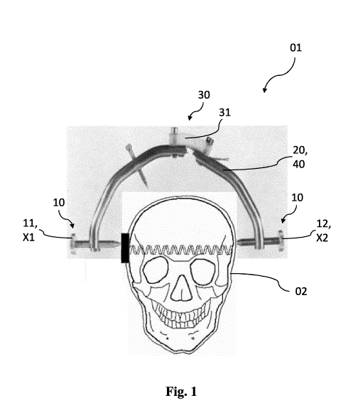
Fig. 1 schematically illustrates a device 01 according to the invention
disposed on a biological material 02, a human skull. It can clearly be
seen in Fig. 1 that device 01 has a first means 10 which comprises an
acoustic transmitting element 11, which is disposed on a first
position Xl, and an acoustic receiving element 12, which is disposed on
a second position X2. It can clearly be seen that the first and second
position Xl, X2 are disposed opposite each other and that the acoustic
spectroscopy is performed in the direction of the frontal plane (coronal)
of skull 02, slightly above the external ear canal.
Furthermore, device 01 has a second means 30 having a measuring
device 31, such as a strain gauge, a pressure sensor, a capacitive sensor
Date Recue/Date Received 2021-09-17
CA 03134074 2021-09-17
23
or the like. An evaluation unit 20 and an analysis unit 40 are also
integrated in Fig. 1. It is also conceivable that they are intended as non-
integral parts. The values recorded by device 01 can additionally be
transmitted to an output unit (now shown).
The following embodiments only serve to illustrate the invention. They
are not intended to limit the subject matter of the claims in any way.
Example 1: The basics of the concept according to the invention,
Acoustocerebrography (ACG)
As described in detail above, it has been detected within the scope of the
1.0 present invention that the concept according to the invention, namely
Acoustocerebrography (ACG), can be applied to the biological material.
It was thus detected that using several frequencies shows the dispersive
character of the brain tissue and provides some interpretation of the
signal changes. Dispersion is an effect in which the non-linear,
frequency-dependent compressive modulus of the medium results in
different propagation speeds for different sound frequencies. In non-
linear material, such as biological tissue and, in particular, human brain
tissue and animal brain tissue, an effect of longitudinal wave dispersion
can be clearly observed and measured. It is such an effect, in which the
compressive modulus of the non-linear, frequency-dependent medium
results in different propagation speeds for different sound frequencies.
As described in detail above, the properties of the compressive modulus
depend on the specific characteristics of the medium, such as
composition, mixture concentration, distribution and/or, in some cases,
chemical composition, such that the pattern of frequency-dependent
propagation speeds can be used to identify the medium.
In order to apply the equations (Eqn. 1) and (Eqn. 2) mentioned above to
the human or animal skull, the structure of the corresponding biological
material must be taken into consideration. In Fig. 2A, the structure of
Date Recue/Date Received 2021-09-17
CA 03134074 2021-09-17
24
the human skull is roughly illustrated, and in Fig. 2B, a correspondingly
layered model of the human skull from Fig. 2A is roughly illustrated.
The tissue structures of the human skull la, lb, 2, 3 (with ventricles), 4
and 5 illustrated in Figs. 2A and 2B are explained in Table 1 below:
Impact of the
Size
tissue structure on
No. Tissue structure
[mm] acoustic
examination
la + lb Skin + Muscle approx. 2.5 No
2 Skull bone approx. 2.5 No
cerebrospinal approx. 25.0
3 Partially ¨ Yes
fluid (C SF) (with ventricles)
4 left and right approx. 67.0-69.0 Yes
cerebrum
5 Cerebral fissure approx. 1.5 No
Table 1: Overview of the impact of the tissue structures from Figs. 2A
and 2B on the change of the time-of-flight of the acoustic wave
Table 1 clearly states that the structures skin (la), muscle (lb), skull
bone (2) and cerebrospinal fluid of the human skull, which are illustrated
in Figs. 3A and 3B, have no impact on the performed acoustic
spectroscopy and can therefore be regarded as constants. However, the
left and right cerebrum (4), the longitudinal cerebral fissure (5),
including the proportion of cerebrospinal fluid (3), have an impact on the
performed acoustic spectroscopy, the impact strongly depending on the
cardiac cycle and the blood circulation in the brain tissue. These zones
are the "point of interest" for further examinations.
The data should be obtained with the time-of-flight method according to
the following equation (Eqn. 3). If we have a set of tissue layers T, then
the total propagation time is obtained by summing the propagation time
for each tissue in the set.
t(f) = (Eqn. 3)
iET
Date Recue/Date Received 2021-09-17
CA 03134074 2021-09-17
The concept according to the invention and the model based on said
concept can easily be upgraded or modified, for example by adding
additional tissue layers. If precise and detailed dispersion data is
available, the dispersion for a specific tissue can be modeled as a non-
5 linear function of frequency. For a given tissue i, the propagation time
for frequency f, ti(f) can be calculated according to the following
equation (Eqn. 4).
di di
ti(f) = ¨ = __________________________________________ (Eqn. 4)
ci (f) coi + Ai
In the equation (Eqn. 4) stated above, di is the depth of the tissue
10 travelled through by the acoustic wave, co, is the basic speed defined
at a
base frequency foe, and A, is the dispersion trend of the tissue, which
characterizes the dependence of the frequency on the propagation speed.
The signal is transmitted by an ultrasound probe and is recorded either
by another acoustic wave (transmission) or by the same acoustic wave
15 (reflection). As described above, the speed of a transmitted signal
depends on the medium. Based on the anatomical analyses of the human
skull-brain-system, it can be demonstrated that depending on the region,
there are very different conditions for the propagation of acoustic waves.
This led to considerations regarding the optimization of the direction of
20 the tissue examination. It was thus detected that the direction of the
frontal plane (coronal), which is illustrated in Fig. 3A, should be chosen
for transmission or reflection measurements.
Limitations related to the minimization of the intensity of ultrasonic
waves have induced the search for such areas in the cranial system which
25 are characterized by the smallest acoustic wave suppression. The
analysis shows that the areas most suitable for the implementation of this
measurement method are the surfaces located slightly above the external
ear canal, as can be seen in Fig. 3b. Choosing such a measuring direction
very probably causes a full echo from the opposite skull bone. Based on
Date Recue/Date Received 2021-09-17
CA 03134074 2021-09-17
26
the above-stated analyses in Fig. 2A and Fig. 2B and Table 1, a
simplified, layered structure of the cranial system can be adopted.
By adopting the layer model of the human cranium (illustrated above in
Fig. 2A and Fig. 2B and in Table 1) as an input¨together with the
physical values of the different cranial tissues shown below in Table 2¨
the propagation times of the acoustic signal, as well as the signal
attenuation along the measurement path through these structures, can be
determined.
Density e.g.
coefficient
[dB/(cm * MHz)]
[kg/m3 m][ /s] [kg/(m2 * s)
1 MHz 10 MHz
Cerebrum
1030 1515 1560450.000 1 8
tissue
Skull 1900 4080 7752000.000 10 60
bones
CSF 1007.5 1498 1509235.000 0.003 0.22
Water 997 1483 1478551.000
0.003 0.22
Blood 1057 1580 1670060.000 0.2 3.8
Skin + Fat 930 1480 1376400.000 1.5
Muscle 1002 1580 1583160.000 0.7
Table 2: The basic parameter assumptions for the human skull-brain-
model
In Fig. 4, the signal attenuation along the measurement path in the
human skull is illustrated for the structures illustrated above in Figs. 2A
and 2B, namely skin (la), muscle (lb), skull bone (2), cerebrospinal
fluid (3), left cerebrum (4a), right cerebrum (4b) and the longitudinal
cerebral fissure (5). Furthermore, a human head model of the ultrasonic
signal attenuation and the expected time-of-flight along the measurement
path is illustrated in Table 3 below.
Date Recue/Date Received 2021-09-17
CA 03134074 2021-09-17
27
(IDLT* =,7
*" .
(/) - =
(/) (/) 'LT:i E
00
E v.-; 00 C7N 00 re')
= ,n) Lnl '
C4
,n
Table 3: Human head model of the ultrasonic signal attenuation and
expected time-of-flight along the measurement path
Taking into account a transmission method, the measuring process
includes the "introduction" of an acoustic wave into the central cerebral
system at the selected position X1 (shown in Fig. 3A and 3B) and,
subsequently, the reception at the opposite position X2, depending on the
direction of the spread of the acoustic beam. Thus, this method
preferably requires two ultrasound probes ¨ one for emitting and one for
receiving the acoustic signal.
The cerebrovascular system is very complex and thus, the state of the
blood supply of the brain largely influences its physical and chemical
parameters. The intracranial pressure depends on intracranial fluid
volumes, tissue volumes and the pulsating volumes, which are induced
by the arterial blood pulsation within the skull. By known normal brain
blood circulation or cerebral blood flow (CBF), e.g. of 50mL/100g/min,
it has been detected that for an average brain weight of 1,375 g, the
mean CBF value is at approx. 690 ml per minute. This results in a blood
value of approx. 11.6 ml per second (estimated as the volume per
heartbeat). Based on this, the time-of-flight measurement and the speed
of sound changes and/or acoustic wave changes can be calculated on the
basis of a standard cranial tissue perfusion CBF. The bone movement
detected with volunteers is up to 20 gm during bed rest and can be
calculated by means of the following equation (Eqn. 5).
Date Recue/Date Received 2021-09-17
CA 03134074 2021-09-17
28
2 (Knorm(1 ¨ X) + Kpath * X)
Cpath = r (Eqn. 5)
Wnorm(1 ¨ x) + Ppath * x)
Let's take a very simplified model, as shown in the equation (Eqn. 6)
below. A standard CBF with 50mL/100g/min means that with every heart
rate, e.g. 60 beats per minute (bpm), between diastole and systole,
approx. 8 % to 10 % of the mass will be exchanged.
C BF 50m/ 1.057g
¨bpm* PBlood = 60bpm* ______________ cm3 = 0,88g (Eqn. 6)
Furthermore, it can be attempted to estimate the change of speed of the
acoustic waves according to the equation (Eqn. 5) above. Assuming that
approx. 10 % of CSF is periodically exchanged with the blood according
1.0 to the normal perfusion values, it can be attempted to calculate the
time-
of-flight changes of the acoustic wave. The corresponding K values of
CSF and blood can be calculated from the known c and p according to
the following equations (Eqn. 7) to (Eqn. 10).
KCSF = cl'SF * PCSF = 14982 *1007.5 = 2.2608* 109[Pa] (Eqn. 7)
KBlood = cLlood * PBlood = 15802 * 1057 = 2.6386* 109[Pa] (Eqn. 8)
2 (KCST (1 ¨ x) + KBlood * x)
Csys = c i (Eqn. 9)
WcsE(1 ¨ x) + PBlood * x)
(2.2608* (1 ¨ 0,1) + 2.6386 *0,1)* 109 m
csys = _________________________________________________________________ =
1506.7563 [¨s1 (Eqn. 10)
(1007.5* (1 ¨ 0,1) + 1057 *0,1)
Assuming the CSF area (meaning the area, where the brain tissue
expands due to the pulsation) to be 1 cm overall, the diastolic travel time
can be calculated with the following equation (Eqn. 11):
1 0.01
tdia = ¨ = ¨ = 6.67556 ps (Eqn. 11)
cCSF 1498
Date Recue/Date Received 2021-09-17
CA 03134074 2021-09-17
29
Together with the result from equation (Eqn. 10) and based on the
assumption of a maximum expansion of the skull by 20 gm, the
following equation (Eqn. 12) allows for the calculation of an expected
systolic time-of-flight (within the faster medium, as 10 % of CSF is
exchanged with blood).
0.0102
tsys = = 1506.75 ¨ 6.76953 las (Eqn. 12)
The equations (Eqn. 11) and (Eqn. 12) listed above result in an acoustic
time-of-flight of ccFs = 1498 m/s during the diastolic phase. During the
systolic phase (with x=10%=0.1), an acoustic time-of-flight of csys =
1506.76 ms is calculated. Despite the fact that the acoustic wave is
1506.76 - 1498 = 8.76 m/s faster during the systolic phase, an increasing
time-of-flight waveform between diastolic and systolic phase can be
observed. This is because the skull is expanded because of the
intracranial pressuring during the systolic phase.
This shows that even when the speed of sound increases due to CSF vs.
blood exchange for the particular region of interest for more than
8.75 m/s, the overall acoustic travel time of the package increases either
because of the longer distance or the longer path. When subtracting tsys
from tdia, we receive the maximum difference of 94 ns shown in the
following equation (Eqn. 13). The time-of-flight measurement has an
adequate resolution which is more than ten times better than the expected
range of approx. 94 ns (better than 90 ps).
tdia tsys = 16.67556 [ts ¨ 6.76953 [ts1 = 0.09396 [ts = 93.96 ns (Eqn. 13)
The maximum difference of 94 ns shown in equation (Eqn. 13) is the
benchmark which is achieved with the method and device according to
the invention. Thus, they are an adequate tool for supporting the medical
diagnosis in the case of intracranial pressure and other pathologies for
medical diagnostics. Time around 45 ns should be measured with an
Date Recue/Date Received 2021-09-17
CA 03134074 2021-09-17
adequate resolution, meaning better than 100 times (approx. 400 ps step)
and faster than 30 measurements per second. Simultaneously, it must be
noted that the time-of-flight difference (increase/decrease) can decrease
when the skull expansion decreases, or even shift to the negative when
5 the skull stops expanding because of the increased intracranial pressure.
This can be very helpful information for urgent medical care.
Example 2: Dispersive ultrasound as a non-invasive diagnostic system
Acoustocerebrography (ACG) utilizes ultrasound quasi constant wave
packages of different frequencies to interrogate a medium in order to
10 provide propagation times for each of the transmitted frequencies. This
method provides an estimation of the dispersion patterns c(f) for a
specific contained medium. The observed propagation speed changes are
usually very small and require a very precise measurement of the
propagation speed. Instead of measuring the speed of sound in the
15 mediums it is easier to accurately measure the propagation time of
ultrasound signals.
By means of equation (Eqn. 4) shown above, the propagation speed c(f)
can be estimated very precisely from the propagation time t(f) by
assuming that the constant dimension d is known.
20 A very high sampling frequency is required for the received signal in
order to accurately measure the propagation time t(f). To achieve the
necessary accuracy, a sampling frequency in the GHz range (exactly
2.5 GHz at 400 ps resolution) is required. For the signal traveling from
the transmitter to the receiver, it is thus necessary that the time
25 resolution is in the range of sub-nanoseconds. Such a system would be
very expensive and would have unacceptable power requirements for a
portable device. Instead, it is known that an ultrasound signal can be
described not only by its frequency, but also by the phase information, as
shown in the following equation (Eqn. 14).
Date Recue/Date Received 2021-09-17
CA 03134074 2021-09-17
31
g(t) = S + A sin(ag + (p) (Eqn. 14)
Thus, the phase information of the ultrasound wave together with its
amplitude must be used to overcome the requirement for a high sampling
frequency in order to provide accurate estimations of propagation times.
It is commonly known, that the phase information only covers a range
from ¨7E to +R. Hence, it can only be used to obtain additional
information about one period of the signal. Furthermore, this information
keeps repeating itself. In this case, a phenomenon from wave theory is
used, the beat-note. In acoustics, a beat is an interference pattern
between two sounds of slightly different frequencies, perceived as a
periodic variation in volume whose rate is the difference of the two
frequencies. The beat-note is the result of the combination of two
continuous wave signals which are close in pitch but not identical. The
difference in frequency generates the beats. The frequency of the beat-
note is given by the following equation (Eqn. 15).
fbeat = ¨ f2 (Eqn. 15)
The closer fi and f2 are, the lower the resulting frequency beat f
beat and
the longer the period of the resulting beat phase Tbeat = 1/f beat. ibeat.
Using this
beat-note approach allows for the clear identification of a specific point
in the signal. Once this unique point has been found, the phase
information of the individual frequency can be used in specific situations
to accurately calculate propagation times. In addition to the observed
changes in propagation speed, different attenuation profiles can also be
observed. The interdependence between wave speed and attenuation is in
accordance with the Kramers¨Kronig relation, where the relation shown
in the following equation (Eqn. 16) is shown, inter alia.
1 1 2 r a(co)
¨ ¨ ¨ = ¨ ¨ * ¨cico (Eqn. 16)
C2 C1 TE (t)2
Date Recue/Date Received 2021-09-17
CA 03134074 2021-09-17
32
In the equation (Eqn. 16), ci, c2 are the propagation speeds (speed of
sound) for waves with circular frequencies coi or co2 and a(co) is the
attenuation for waves with circular frequency co. After introducing co ¨
27-cf, wi= 27-c*fi and co2= 27-c*f2, the following equation (Eqn. 17) applies:
1 1 1 f a(f)
= * j -2 - d f (Eqn. 17)
(01
Such patterns of frequency-dependent attenuations and the corresponding
propagation speeds can be used to identify the state of a medium or to
track possible changes to the brain tissue in real time. To achieve the
requested time resolution for a useful medical diagnostic picture (as
shown in Fig. 5), some essential requirements for the phase
determination must be met. In Fig. 5, the time-of-flight waveform
heartbeat curve of a 72-year-old patient is shown; recorded with the
ACG system as part of an authorized clinical study. The X-axis shows
the time [t] in seconds (s) and the Y-axis shows the time-of-flight in
microseconds (ts).
Assuming that the interesting acoustic measurement band for ACG is
between 0.7 MHz and 2.7 MHz, this will set a following expectation for
the signal phase resolution. We require a phase resolution better than
400 ps at a frequency of 0.7 MHz ¨higher frequencies provide a higher
time resolution while the wavelength is shorter ¨ this means that the time
resolution will be greater. Assuming that the average speed of an
acoustic quasi constant wave package in the skull is 1540 m/s, we can
receive an explanation according to the equations listed above and the
following equation (Eqn. 18).
1540
A = ¨ f = 0.7E6 = 2.2mm (Eqn. 18)
As can be seen from equation (Eqn. 18), these 2.2 mm are the length of
exactly one period (360 or 27( Phase) with the time duration of
Date Recue/Date Received 2021-09-17
CA 03134074 2021-09-17
33
1.4285714 gs. Consequently, the required phase resolution must be in the
range of 0.10 or better.
Example 3: Evaluation of the time-of-flight measurement of a patient
When using ICP monitoring in clinical practice, it is very important to
determine the validity of the obtained pressure value. Access to a high-
resolution view of the intracranial pressure waveform thus offers a more
accurate analysis of the obtained intracranial pressures. When carrying
out the method according to the invention, it is thus important to verify
whether the obtained ICP signal is truly representative of the intracranial
pressure. In this manner, the person skilled in the art should ensure that
there is in fact an oscillating pressure curve with the progressively
decreasing P1, P2 and P3 notches present, which indicate the propagation
of the cardiac pulse pressure signal. Such an oscillating pressure curve is
shown in an exemplary manner in Fig. 6, in which the propagation of the
cardiac pulse pressure signal, in particular an intracranial pressure
measurement recorded by an intracranial pressure probe, is illustrated.
The X-axis shows the time [t] in milliseconds (ms) and the Y-axis shows
the intracranial pressure (ICP).
It is understood that deviations from the pressure curve illustrated as an
example in Fig. 6 can indicate a changed intracranial compliance, a
changed intracranial pressure, a disturbed cerebral blood flow and/or a
pathological condition. For example, reversed P1 and P2 notches indicate
a state of disturbed autoregulation.
A closer look at the waveform in Fig. 5 supports the conclusion of the
above-stated example of use 1, as it shows a difference of approx. 50 ns
in the time-of-flight measurement between the diastolic and the systolic
phase. It also shows that the patient has a disturbed autoregulation, as
the P1 and P2 notches are reversed, which can be seen in seconds 6, 7, 8,
9, 14, 15 and 16.
Date Recue/Date Received 2021-09-17