Note: Descriptions are shown in the official language in which they were submitted.
CA 03134502 2021-09-21
WO 2020/198160
PCT/US2020/024289
TARGETED ACTIVE GENE EDITING AGENT AND METHODS OF USE
RELATED APPLICATIONS
This application claims priority to U.S. Provisional Application No.
62/822,529, filed on
March 22, 2019. The content of the priority application is incorporated by
reference herein.
FIELD
The present invention generally relates to methods and compositions for
editing a nucleic
acid within a cell using site-directed modifying polypeptides conjugated to an
antigen binding
polypeptide.
BACKGROUND OF THE INVENTION
CRISPR-associated RNA-guided endonucleases, such as Cas9, have become a
versatile
tool for genome engineering in various cell types and organisms (see, e.g., US
8,697,359).
Guided by a guide RNA, such as a dual-RNA complex or a chimeric single-guide
RNA, RNA-
guided endonucleases (e.g., Cas9) can generate site-specific double-strand
breaks (DSBs) or
single- stranded breaks (SSBs) within target nucleic acids (e.g., double-
stranded DNA (dsDNA),
single-stranded DNA (ssDNA), or RNA). When cleavage of a target nucleic acid
occurs within a
cell (e.g., a eukaryotic cell), the break in the target nucleic acid can be
repaired by nonhomologous
end joining (NHEJ) or homology directed repair (HDR). In addition,
catalytically inactive RNA-
guided endonucleases (e.g., Cas9) alone or fused to transcriptional activator
or repressor domains
can be used to alter transcription levels at sites within target nucleic acids
by binding to the target
site without cleavage.
However, the ability to deliver and target RNA-guided endonucleases to
specific cells or
tissues remains a challenge. A variety of methods or vehicles for delivery of
RNA-guided
endonucleases have been utilized, such as electroporation, nucleofection,
microinjection, adeno-
associated vectors (AAV), lentivirus, and lipid nanoparticles (see, e.g., in
Lino, C.A. et al., 2018.
Drug delivery, 25(1), pp.1234-1257). As described in Lino et al, certain
methods, such as
microinjection or electroporation, are limited primarily to in vitro
applications. Other modes of
delivery, such as AAVs or lipid nanoparticles, have been utilized for in vivo
delivery of RNA-guided
endonucleases, but these delivery methods have faced challenges in an in vivo
setting. For
example, AAV-based delivery vehicles present immunological barriers, packaging
size limitations,
and the risk for genotoxic genome integration events (see, e.g., Lino et al.,
2018; and Wang, D, et
aL, 2019. Nature Reviews Drug Discovery, 18(5), pp.358-378). Further, delivery
of RNA-guided
endonucleases by lipid nanoparticles has several drawbacks, including
endosomal degradation of
cargo, specific cell tropism, and bioaccumulation in the liver (see, e.g.,
Lino et al., 2018; and Finn,
J.D., et al., 2018. Cell reports, 22(9), pp.2227-2235).
1
CA 03134502 2021-09-21
WO 2020/198160
PCT/US2020/024289
Alternative methods have been attempted to improve target delivery of RNA-
guided
endonucleases by modifying the RNA-guided endonucleases themselves with a
receptor.
However, examples of such receptor-mediated RNA-guided endonucleases have
shown limited
editing in vitro and did not achieve in vivo editing (see, e.g., Rouet, R., et
al., 2018. Receptor-
mediated delivery of CRISPR-Cas9 endonuclease for cell-type-specific gene
editing. J Am Chem,
140(21), pp.6596-6603).
SUMMARY OF THE INVENTION
There is an unmet need for RNA-guided endonucleases with the capability of
targeting
desired cells or tissues, especially for in vivo editing. There is a need in
the art for effective
delivery of gene editing therapies utilizing RNA-guided endonucleases with the
capability of
targeting desired cells or tissues. Further, there is an unmet need for
compositions and methods
that provide in vivo targeted gene editing.
Provided herein are Targeted Active Gene Editing (TAGE) agents comprising an
antigen-
binding polypeptide which are able to edit specific cell types both in vivo
and ex vivo. The modular
and programmable design of TAGE agents enables rapid re-targeting and multi-
functionality to
enable flexible targeting of a variety of cell types. Further, by editing
specific nucleic acid
sequences (e.g., genes and regulatory elements) in target cells, TAGE agents
have dual
specificity and have fewer off-target effects than DNA-based delivery
approaches (Cameron, et al.
Nature methods. 14.6 (2017): 600; Kim, et al. Genome research. 24.6 (2014):
1012-1019). TAGE
agents include one or more antigen-binding polypeptides that promote cell
binding and/or cellular
internalization of the TAGE agent in the target cell. Further, in some
instances, the antigen-
binding polypeptides, not only allow for receptor-mediated entry of the TAGE
agent, but in certain
instances, the antigen-binding polypeptides also mediate the biology of the
cell (e.g., by altering
intracellular signal transduction pathways).
Accordingly, provided herein are methods and compositions relating to a gene
editing cell
internalizing agent (TAGE agent) comprising an antigen binding polypeptide
that specifically binds
to an extracellular cell membrane-bound molecule (e.g., a cell surface
molecule), and a site-
directed modifying polypeptide that recognizes a nucleic acid sequence,
wherein the antigen
binding polypeptide and the site-directed modifying polypeptide are stably
associated such that the
site-directed modifying polypeptide can be internalized into a cell displaying
the extracellular cell
membrane-bound molecule (e.g., a cell surface molecule).
In some embodiments, the antigen binding polypeptide is an antibody, an
antigen-
binding portion of an antibody, or an antibody-mimetic.
In some embodiments, the site-directed modifying polypeptide comprises a
nuclease or a
nickase. In certain embodiments, the nuclease is a DNA endonuclease, such as
Cas9 or Cas12.
2
CA 03134502 2021-09-21
WO 2020/198160
PCT/US2020/024289
In some embodiments, the TAGE agent further comprises a guide RNA that
specifically
hybridizes to a target region of the genome of the cell, wherein the guide RNA
and the site-
directed modifying polypeptide form a ribonucleoprotein.
In another aspect, the invention provides a targeted active gene editing
(TAGE) agent
comprising an antigen binding polypeptide which specifically binds to an
extracellular cell
membrane-bound molecule, and a site-directed modifying polypeptide comprising
an RNA-guided
DNA endonuclease that recognizes a CRISPR sequence, wherein the antigen
binding polypeptide
and the site-directed modifying polypeptide are stably associated such that
the site-directed
modifying polypeptide can be internalized into a cell displaying the
extracellular cell membrane-
bound molecule, and wherein the antigen binding polypeptide is an antibody, an
antigen-binding
portion of an antibody, or an antibody-mimetic.
In some embodiments, the TAGE agent comprises a guide RNA that specifically
hybridizes
to a target region of the genome of the cell, wherein the guide RNA and the
site-directed modifying
polypeptide form a ribonucleoprotein.
In some embodiments, the RNA-guided DNA endonuclease is a Cas9 nuclease. In
some
embodiments, the Cas9 nuclease is wildtype Cas9 nuclease (e.g., Streptococcus
pyogenes Cas9,
SEQ ID NO: 119). In some embodiments, the Cas9 nuclease comprises an amino
acid sequence
having at least 85%, 90%, 95%, 97%, 98%, 99%, or 100% identity to SEQ ID NO:
119. In certain
embodiments, the Cas9 nuclease comprises the amino acid substitution 080A
(e.g., SEQ ID NO:
1). In another the Cas9 nuclease comprises an amino acid sequence having at
least 85%, 90%,
95%, 97%, 98%, 99%, or 100% identity to SEQ ID NO: 1.
In some embodiments, the RNA-guided DNA endonuclease is a nuclease other than
Cas9
(e.g., such as one described in Section III). In certain embodiments, the RNA-
guided DNA
endonuclease is a CRISPR Type V nuclease. In specific embodiments, the RNA-
guided DNA
endonuclease is a Cas12 nuclease. In some embodiments, the Cas12 nuclease is
wildtype Cas12
nuclease (e.g., Acidaminococcus sp. Cas12a, SEQ ID NO: 120). In some
embodiments, the
Cas12 nuclease comprises an amino acid sequence having at least 85%, 90%, 95%,
97%, 98%,
99%, or 100% identity to SEQ ID NO: 120. Examples of Cas12a variants useful in
the TAGE
agents herein include, but are not limited to, Alt-Re Cas12a (Cpf1) Ultra
(e.g., IDT Catalog No.
10001272) or Cas12a as described in Kleinstiver, et al. Nature Biotechnology
37.3 (2019): 276-
282, which is hereby incorporated by reference.
In some embodiments, the site-directed modifying polypeptide further comprises
at least
one nuclear localization signal (NLS).
In some embodiments, the site-directed modifying polypeptide further comprises
a
conjugation moiety that binds to the antigen binding polypeptide. In certain
embodiments,
the conjugation moiety is a protein. In certain embodiments, the protein is
Protein A, SpyCatcher,
or a Halo-Tag.
3
CA 03134502 2021-09-21
WO 2020/198160
PCT/US2020/024289
In some embodiments, the site-directed modifying polypeptide and the antigen
binding
polypeptide are conjugated via a linker. In certain embodiments, the linker is
cleavable.
In some embodiments, the antibody mimetic is an adnectin (i.e., fibronectin
based binding
molecules), an affilin, an affimer, an affitin, an alphabody, an affibody, a
DARPin, an anticalin, an
.. avimer, a fynomer, a Kunitz domain peptide, a monobody, a nanoCLAMP, a
unibody, a versabody,
an aptamer, or a peptidic molecule.
In some embodiments, the antigen-binding portion of the antibody is a
nanobody, a doman
antibody, an scFv, a Fab, a diabody, a BiTE, a diabody, a DART, a minibody, a
F(ab')2, or an
intrabody.
In some embodiments, the antibody is an intact antibody or a bispecific
antibody.
In some aspects, the invention provides a targeted active gene editing (TAGE)
agent
comprising an antibody, or an antigen-binding portion thereof, which
specifically binds to an
extracellular cell membrane-bound protein, and a site-directed modifying
polypeptide comprising a
Cas9 nuclease, wherein the antibody, or antigen-binding portion thereof, and
the site-directed
modifying polypeptide are stably associated via a conjugation moiety such that
the site-directed
modifying polypeptide can be internalized into the cell expressing the
extracellular cell membrane-
bound protein via the antibody, or the antigen binding portion thereof.
In some embodiments, the site-directed modifying polypeptide further comprises
at least
one nuclear localization signal (NLS). In certain embodiments, the at least
one NLS comprises an
SV40 NLS. In certain embodiments, the SV40 NLS comprises the amino acid
sequence
PKKKRKV (SEQ ID NO: 8). In certain embodiments, the at least one NLS is at the
C-terminus,
the N-terminus, or both of the site-directed modifying polypeptide. In certain
embodiments, the
TAGE agent comprises at least two NLSs.
In certain embodiments, the TAGE agent further comprises a guide RNA that
specifically
hybridizes to a target region of the genome of a cell expressing the
extracellular cell membrane-
bound protein, wherein the guide RNA and the site-directed modifying
polypeptide form a
nucleoprotein.
In certain embodiments, the site-directed modifying polypeptide further
comprises a
conjugation moiety that can bind to the antibody, or antigen-binding portion
thereof. In certain
.. embodiments, the conjugation moiety is a protein. In some embodiments, the
protein is Protein A,
SpyCatcher, or a Halo-Tag.
In some embodiments, the Cas9 nuclease is wildtype Cas9 nuclease (e.g.,
Streptococcus
pyogenes Cas9, SEQ ID NO: 119). In some embodiments, the Cas9 nuclease
comprises an
amino acid sequence having at least 85%, 90%, 95%, 97%, 98%, 99%, or 100%
identity to SEQ
.. ID NO: 119.
4
CA 03134502 2021-09-21
WO 2020/198160
PCT/US2020/024289
In certain embodiments, the Cas9 nuclease comprises the amino acid
substitution 080A
(e.g., SEQ ID NO: 1). In certain embodiments, the Cas9 nuclease has an amino
acid sequence
that is at least 85%, 90%, 95%, 97%, 98%, 99%, or 100% identical to SEQ ID NO:
1.
In certain embodiments, the antigen-binding portion of the antibody is a
nanobody, a
doman antibody, an scFv, a Fab, a diabody, a BiTE, a diabody, a DART, a
minibody, a F(ab')2, or
an intrabody.
In certain embodiments, the antibody is an intact antibody or a bispecific
antibody.
In certain embodiments, the extracellular cell membrane-bound molecule or
protein (e.g.,
cell surface molecule or protein) is HLA-DR, 0D44, CD11 a, 0D22, CD3, 0D20,
0D33, 0D32,
0D44, 0D47, 0D59, 0D54, 0D25, AchR, CD70, 0D74, CTLA4, EGFR, HER2, EpCam,
0X40,
PD-1, PD-L1, GITR, 0D52, 0D34, 0D27, CD30, !COS, or RSV.
In some embodiments, the extracellular cell membrane-bound molecule or protein
is
CD11a. In some embodiments, the antigen binding polypeptide is an anti-CD11a
antibody, or
antigen binding fragment thereof. In certain embodiments, the anti-CD11a
antibody is efalizumab.
In some embodiments, the extracellular cell membrane-bound molecule or protein
is 0D25.
In some embodiments, the antigen binding polypeptide is an anti-0D25 antibody,
or antigen
binding fragment thereof. In certain embodiments, the anti-0D25 antibody is
daclizumab.
In another aspect, the invention provides a site-directed modifying
polypeptide comprising
an RNA-guided DNA endonuclease that recognizes a CRISPR sequence and a
conjugation
moiety that binds to an antibody, an antigen-binding portion of an antibody,
or an antibody mimetic
that specifically binds to an extracellular cell membrane-bound molecule
(e.g., cell surface
molecule).
In certain embodiments, the site-directed modifying polypeptide further
comprises a guide
RNA that specifically hybridizes to a target region of the genome of a cell.
In certain
embodiments, the RNA-guided DNA endonuclease is a Cas9 nuclease. In some
embodiments,
the Cas9 nuclease is wildtype Cas9 nuclease (e.g., Streptococcus pyogenes
Cas9, SEQ ID NO:
119). In some embodiments, the Cas9 nuclease comprises an amino acid sequence
having at
least 85%, 90%, 95%, 97%, 98%, 99%, or 100% identity to SEQ ID NO: 119. In
certain
embodiments, the Cas9 nuclease comprises the amino acid substitution 080A
(e.g., SEQ ID NO:
1). In another the Cas9 nuclease comprises an amino acid sequence having at
least 85%, 90%,
95%, 97%, 98%, 99%, or 100% identity to SEQ ID NO: 1.
In certain embodiments, the RNA-guided DNA endonuclease is a CRISPR Type V
nuclease. In specific embodiments, the RNA-guided DNA endonuclease is a Cas12
nuclease. In
some embodiments, the Cas12 nuclease is wildtype Cas12 nuclease (e.g.,
Acidaminococcus sp.
Cas12a, SEQ ID NO: 120). In some embodiments, the Cas12 nuclease comprises an
amino acid
sequence having at least 85%, 90%, 95%, 97%, 98%, 99%, or 100% identity to SEQ
ID NO: 120.
Examples of Cas12a variants useful in the TAGE agents herein include, but are
not limited to, Alt-
5
CA 03134502 2021-09-21
WO 2020/198160
PCT/US2020/024289
Re Cas12a (Cpf1) Ultra (e.g., IDT Catalog No. 10001272) or Cas12a as described
in Kleinstiver,
et al. Nature Biotechnology 37.3 (2019): 276-282, which is hereby incorporated
by reference.
In certain embodiments, the site-directed modifying polypeptide further
comprises at least
one nuclear localization signal (NLS). In certain embodiments, the at least
one NLS comprises an
SV40 NLS. In certain embodiments, the SV40 NLS comprises PKKKRKV (SEQ ID NO:
8). In
certain embodiments, the site-directed modifying polypeptide comprises at
least two NLSs. In
certain embodiments, the at least one NLS is at the C-terminus, the N-
terminus, or both of the site-
directed modifying polypeptide.
In certain embodiments, the site-directed modifying polypeptide further
comprises a
conjugation moiety that can bind to the antibody, antigen-binding portion
thereof, or antibody
mimetic. In certain embodiments, the conjugation moiety is a protein. In
certain embodiments, the
protein is Protein A, SpyCatcher, or a Halo-Tag.
In certain embodiments, the extracellular cell membrane-bound molecule is a
protein
selected from the group consisting of HLA-DR, CD44, CD11a, CD22, CD3, CD20,
CD33, CD32,
CD44, CD47, CD59, CD54, CD25, AchR, CD70, CD74, CTLA4, EGFR, HER2, or EpCam,
0X40,
PD-1, PD-L1, GITR, CD52, CD34, CD27, CD30, ICOS, or RSV.
In some embodiments, the extracellular cell membrane-bound molecule or protein
is
CD11a. In some embodiments, the antigen binding polypeptide is an anti-CD11a
antibody, or
antigen binding fragment thereof. In certain embodiments, the anti-CD11a
antibody is efalizumab.
In some embodiments, the extracellular cell membrane-bound molecule or protein
is CD25.
In some embodiments, the antigen binding polypeptide is an anti-CD25 antibody,
or antigen
binding fragment thereof. In certain embodiments, the anti-CD25 antibody is
daclizumab.
In another aspect, the invention provides a nucleoprotein comprising a site-
directed
modifying polypeptide and a guide RNA, wherein the guide RNA specifically
hybridizes to a target
region of the genome of a cell displaying the extracellular cell membrane-
bound protein.
In another aspect, the invention provides an isolated nucleic acid encoding a
site-directed
modifying polypeptide described herein. In one embodiment, a vector comprises
the nucleic acid.
In another embodiment, a cell comprises the site-directed modifying
polypeptide.
In another aspect, the invention provides a method of modifying the genome of
a target
cell, the method comprising contacting the target cell with a targeted active
gene editing (TAGE)
agent described herein. In certain embodiments, the target cell is a
eukaryotic cell. In certain
embodiments, the eukaryotic cell is a mammalian cell. In certain embodiments,
the mammalian
cell is a mouse cell, a non-human primate cell, or a human cell. In certain
embodiments, the site-
directed modifying polypeptide produces a cleavage site at the target region
of the genome,
thereby modifying the genome. In certain embodiments, the target region of the
genome is a
target gene.
6
CA 03134502 2021-09-21
WO 2020/198160
PCT/US2020/024289
In certain embodiments, a method comprising the use of a TAGE agent described
herein is
effective to modify expression of the target gene. In certain embodiments, the
method is effective
to increase expression of the target gene relative to a reference level. In
certain embodiments, the
method is effective to decrease expression of the target gene relative to a
reference level.
In another aspect, provided herein is a method of modifying a nucleic acid
sequence within
a target cell in a mammalian subject, the method comprising contacting the
target cell in the
subject with a targeted active gene editing (TAGE) agent comprising an antigen
binding
polypeptide that specifically binds to an extracellular cell membrane-bound
molecule, and a site-
directed modifying polypeptide that recognizes the nucleic acid sequence
within the target cell,
such that the nucleic acid sequence of the target cell is modified.
In another aspect, provided herein is a method of modifying a nucleic acid
sequence within
a target cell in a mammalian subject, the method comprising locally
administering to the subject a
targeted active gene editing (TAGE) agent comprising an antigen binding
polypeptide that
specifically binds to an extracellular cell membrane-bound molecule, and a
site-directed modifying
polypeptide that recognizes the nucleic acid sequence within the target cell,
such that the nucleic
acid sequence of the target cell is modified.
In some embodiments, the method comprises locally administering the TAGE agent
to the
subject by intramuscular injection, intraosseous injection, intraocular
injection, intratumoral
injection, or intradermal injection.
In some embodiments, the method is effective to increase the level of
genetically modified
target cells in the subject relative to the level achieved by treatment with a
site-directed modifying
polypeptide lacking the antigen binding polypeptide.
In some embodiments, the mammalian subject is a human subject.
In some embodiments, the subject has a disease selected from an eye disease, a
stem cell
disorder, and a cancer, and wherein the method is effective to treat the
disease.
In another aspect, provided herein is a method of modifying a nucleic acid
sequence within
a target mammalian cell, the method comprising contacting the target mammalian
cell with a
targeted active gene editing (TAGE) agent under conditions in which the TAGE
agent is
internalized into the target cell, such that the nucleic acid sequence is
modified, wherein the TAGE
agent comprises an antigen binding polypeptide that specifically binds to an
extracellular cell
membrane-bound molecule, and a site-directed modifying polypeptide that
recognizes the nucleic
acid sequence within the target cell, wherein the internalization of the TAGE
agent is not
dependent on electroporation.
In some embodiments, the target mammalian cell is a hematopoietic cell (HSC),
a
neutrophil, a T cell, a B cell, a dendritic cell, a macrophage, or a
fibroblast. In certain
embodiments, the target mammalian cell is a hematopoietic stem cell (HSC). In
certain
7
CA 03134502 2021-09-21
WO 2020/198160
PCT/US2020/024289
embodiments the target mammalian cell is a cell in the bone marrow that is not
a hematopoietic
stem cell (e.g., fibroblast, macrophages, osteoblasts, ostclasts, or
endothelial cells).
In some embodiments, the antigen binding polypeptide specifically binds an
extracellular
cell membrane-bound molecule on a human HSC. In certain embodiments, the
extracellular cell
membrane-bound molecule on the HSC is 0D34, EMCN, 0D59, CD90, ckit, 0D45, or
CD49F.
In some embodiments, the target mammalian cell is contacted with the TAGE
agent by co-
incubation ex vivo.
In some embodiments, the method provides a genetically-modified target cell
which is
administered to a subject in need thereof.
In some embodiments, the target mammalian cell is contacted with the TAGE
agent in situ
by injection into a tissue of a subject.
In some embodiments, the TAGE agent is administered to the subject by
intramuscular
injection, intraosseous injection, intraocular injection, intratumoral
injection, or intradermal
injection.
In some embodiments, the nucleic acid is a gene in the genome of the target
cell, wherein
the expression of said gene is altered following said modification.
In some embodiments, the target mammalian cell is a mouse cell, a non-human
primate
cell, or a human cell.
In some embodiments, the antigen binding polypeptide is an antibody, an
antigen-binding
portion of an antibody, or an antibody-mimetic.
In certain embodiments, the antibody mimetic is an adnectin (i.e., fibronectin
based binding
molecules), an affilin, an affimer, an affitin, an alphabody, an aptamer. an
affibody, a DARPin, an
anticalin, an avimer, a fynomer, a Kunitz domain peptide, a monobody, a
nanoCLAMP, a unibody,
a versabody, an aptamer, or a peptidic molecule.
In some embodiments, the antigen-binding portion of the antibody is a
nanobody, a doman
antibody, an scFv, a Fab, a diabody, a BiTE, a diabody, a DART, a mnibody, a
F(ab')2, or an
intrabody.
In some embodiments, the antibody is an intact antibody or a bispecific
antibody.
In some embodiments, the extracellular cell membrane-bound molecule bound by
the
antigen binding polypeptide is HLA-DR, 0D44, CD11 a, 0D22, CD3, CD20, 0D33,
0D32, 0D44,
0D47, 0D59, 0D54, 0D25, AchR, CD70, 0D74, CTLA4, EGFR, HER2, EpCam, 0X40, PD-
1, PD-
L1, GITR, 0D52, 0D34, 0D27, CD30, ICOS, or RSV.
In certain embodiments, the extracellular cell membrane-bound molecule or
protein is
CD11a. In some embodiments, the antigen binding polypeptide is an anti-CD1la
antibody, or
antigen binding fragment thereof. In certain embodiments, the anti-CD1la
antibody is efalizumab,
or an antigen binding fragment thereof.
8
CA 03134502 2021-09-21
WO 2020/198160
PCT/US2020/024289
In some embodiments, the extracellular cell membrane-bound molecule or protein
is 0D25.
In some embodiments, the antigen binding polypeptide is an anti-0D25 antibody,
or antigen
binding fragment thereof. In certain embodiments, the anti-0D25 antibody is
daclizumab.
In some embodiments, the TAGE agent further comprises at least one nuclear
localization
signal (NLS). In some embodiments, the TAGE agent comprises at least two
nuclear localization
signals (NLSs). In certain embodiments, the TAGE agent comprises four nuclear
localization
signals (NLSs). In certain embodiments, the TAGE agent comprises six nuclear
localization
signals (NLSs). In some embodiments, the TAGE agent comprises seven nuclear
localization
signals (NLSs). In some embodiments, the TAGE agent comprises eight nuclear
localization
signals (NLSs).
In some embodiments, the NLS comprises an SV40 NLS. In certain embodiments,
the
SV40 NLS comprises the amino acid sequence PKKKRKV (SEQ ID NO: 8).
In some embodiments, the target mammalian cell is a population of target
mammalian
cells. In some embodiments, the method is effective to increase the level
(number) of genetically
modified target mammalian cells in the population. In certain embodiments, the
increase is
evidenced by a response (e.g., phenotypic) in the mammalian cells. In certain
embodiments, an
increase number of mammalian cells modified by the TAGE agent can be
determined by
comparing the level in a population of mammalian cells relative to a level
achieved by treatment
with a site-directed modifying polypeptide lacking the antigen binding
polypeptide.
In some embodiments, the site-directed modifying polypeptide of the TAGE agent
has
increased cellular internalization in the target mammalian cell. In certain
embodiments, the
increase in internalization is evidenced by a response, e.g., phenotypic, in
the mammalian cell. In
certain embodiments, an increase in the internalization of the TAGE agent into
a mammalian cell
can be determined by comparing the internalization of the TAGE agent in a
population of
mammalian cells relative to cellular internalization achieved with a site-
directed modifying
polypeptide lacking the antigen binding polypeptide.
In some embodiments, the site-directed modifying polypeptide of the TAGE agent
has
increased nuclear internalization in the target mammalian cell relative to
nuclear internalization
achieved with a site-directed modifying polypeptide lacking the antigen
binding polypeptide.
In some embodiments, the site-directed modifying polypeptide comprises a
nuclease or a
nickase.
In some embodiments, the site-directed modifying polypeptide is a nucleic acid-
guided
nuclease, and the TAGE agent further comprises a guide nucleic acid that
specifically hybridizes
to a target region of the nucleic acid sequence of the target mammalian cell,
wherein the guide
nucleic acid and the nucleic acid-guided nuclease form a nucleoprotein.
In certain embodiments, the site-directed modifying polypeptide is a RNA-
guided nuclease,
and the TAGE agent further comprises a guide RNA that specifically hybridizes
to a target region
9
CA 03134502 2021-09-21
WO 2020/198160
PCT/US2020/024289
of the nucleic acid sequence of the target mammalian cell, wherein the guide
RNA and the RNA-
guided nuclease form a ribonucleoprotein. In some embodiments, the guide RNA
is a single guide
RNA (sg RNA) or a cr:trRNA.
In some embodiments, the RNA-guided nuclease is a Class 2 Cas polypeptide.
In some embodiments, the Class 2 Cas polypeptide is a Type II Cas polypeptide.
In some
embodiments, the Type II Cas polypeptide is Cas9. In some embodiments, the
Cas9 nuclease is
wildtype Cas9 nuclease (e.g., Streptococcus pyogenes Cas9, SEQ ID NO: 119). In
some
embodiments, the Cas9 nuclease comprises an amino acid sequence having at
least 85%, 90%,
95%, 97%, 98%, 99%, or 100% identity to SEQ ID NO: 119. In certain
embodiments, the Cas9
nuclease comprises the amino acid substitution C80A (e.g., SEQ ID NO: 1). In
another the Cas9
nuclease comprises an amino acid sequence having at least 85%, 90%, 95%, 97%,
98%, 99%, or
100% identity to SEQ ID NO: 1.
In some embodiments, the Class 2 Cas polypeptide is a Type V Cas polypeptide.
IN
certain embodiments, the Type V Cas polypeptide is Cas12. In some embodiments,
the Cas12
nuclease is wildtype Cas12 nuclease (e.g., Acidaminococcus sp. Cas12a, SEQ ID
NO: 120). In
some embodiments, the Cas12 nuclease comprises an amino acid sequence having
at least 85%,
90%, 95%, 97%, 98%, 99%, or 100% identity to SEQ ID NO: 120. Examples of
Cas12a variants
useful in the TAGE agents herein include, but are not limited to, Alt-Re
Cas12a (Cpf1) Ultra (e.g.,
IDT Catalog No. 10001272) or Cas12a as described in Kleinstiver, et al. Nature
Biotechnology 37.3 (2019): 276-282, which is hereby incorporated by reference.
In some embodiments, the site-directed modifying polypeptide further comprises
a
conjugation moiety that binds to the antigen binding polypeptide or a
complementary binding
moiety attached thereto. In certain embodiments, the conjugation moiety is a
protein. In some
embodiments, the protein is SpyCatcher or a Halo-Tag.
In some embodiments, the site-directed modifying polypeptide and the antigen
binding
polypeptide are conjugated via a linker. In some embodiments, the linker is a
cleavable linker.
In some embodiments, the TAGE agent further comprises an endosomal escape
agent. In
certain embodiments, the endosomal escape agent is TDP or TDP-KDEL.
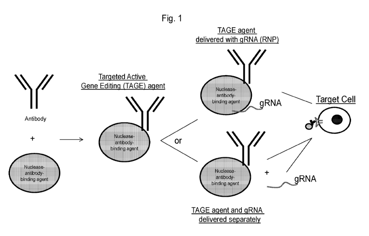
BRIEF DESCRIPTION OF THE DRAWINGS
Fig. 1 is a schematic of a nuclease antibody-binding agent described herein
complexed
with an antibody, antigen-binding agent, or antibody-like molecule to form a
targeted active gene
editing (TAGE) agent. In Figure 1, the term "nuclease antibody-binding agent"
refers to a site-
directed modifying polypeptide including a nuclease.
Fig. 2 graphically depicts the results of an in vitro DNA cleavage assay
assessing Cas9-
2xNLS-ProteinA alone ("Cas9-pA") or Cas9-2xNLS-ProteinA complexed with an anti-
CD3 antibody
CA 03134502 2021-09-21
WO 2020/198160
PCT/US2020/024289
("Cas9-pA:a-CD3"), or Cas9(080A)-2xNLS ("080A") with activity plotted relative
to Cas9(080A)-
2xNLS activity.
Fig. 3 graphically depicts the results of an ex vivo editing assay assessing
editing activity
of Cas9-2xNLS-ProteinA ("Cas9-pA") or Cas9 (080A)-2xNLS ("080A") following
nucleofection into
.. stimulated human T cells. A guide RNA targeting 0D47 was associated with
the respective TAGE
agents to form ribonucleoproteins, and the ribonucleoproteins were
nucleofected into T cells to test
for editing. Editing was measured using a phenotypic readout measuring the
loss of surface 0D47
using flow cytometry. Editing activity is plotted relative to Cas9 (080A)-
2xNLS activity.
Fig. 4 graphically depicts the results of an in vitro binding assay to assess
binding of Cas9-
2xNLS-ProteinA ("Cas9-pA") to an anti-CD3 antibody. The results for Cas9-pA
alone and anti-
CD3 antibody alone are also shown.
Figs. 5A and 5B graphically depict the results of a FACS-based internalization
assay
measuring the rate of PBMC internalization for anti-CD3 (18 nM) or anti-0D22
(100 nM) antibodies
in CD8 T cells (Fig. 5A) and in CD19 B Cells (Fig. 5B).
Figs. 6A-6C show results from binding and internalization studies of
antibodies (hulgG1,
CD22) complexed with Cas9-2xNLS-proteinA ("Cas9-pA") to form TAGE agents. Fig.
6A
graphically depicts the results of a FACS-based cell binding assay in which 10
nM of each
indicated protein was added to PBMCs and stained for 30 minutes. Fig.6B
graphically depicts the
results of a FACS-based internalization assay in which 10 nM of each indicated
protein was added
to PBMCs for the indicated temperature and time. Samples from each condition
with and without
quenching with an anti-A488 antibody were assessed by FACS analysis. Fig. 6C
further illustrates
internalization by T cells vs B cells in the pool of PBMCs.
Figs. 7A-7D graphically depicts the results of a FACS-based internalization
assays utilizing
various quench methods (heparin wash, acid wash, anti-A488 antibody, no
quench), in which
internalization of a TAGE agent including Cas9-2xNLS-proteinA ("Cas9-pA"), an
anti-CD3
antibody, or Cas9-pA complexed with an anti-CD3 antibody ("Cas9pA:CD3") was
assessed in T
cells (Figs. 7A and 7B) or myeloid cells (Fig. 7C). Fig. 7A graphically
depicts the results of the
internalization assay with an anti-CD3 antibody labelled with A488 or Cas9-
pA:anti-CD3 RNP with
guide RNA labelled with A488. Fig. 7B graphically depicts the results of the
internalization assay
in T cells with Cas9-pA:anti-CD3 RNP or Cas9-pA with guide RNA labelled with
ATT0550. Fig.
7C graphically depicts the results of the internalization assay in myeloid
cells with Cas9-pA:anti-
CD3 RNP or Cas9-pA with guide RNA labelled with ATT0550. Fig. 7D graphically
depicts the
results of a live dead FACS-based assay to evaluate the toxicity effects of
each quench method.
Fig. 8 graphically depicts the results of an in vitro DNA cleavage assay
assessing the DNA
cleavage by the TAGE agent Cas9-2xNLS-DARPin(Ec1) ("Cas9-Darpin(EC1)") (also
referred to as
Cas9-DARPin(EpCAM)) or Cas9(080A)-2xNLS ("C80A") with activity plotted
relative to
Cas9(080A)-2xNLS activity.
11
CA 03134502 2021-09-21
WO 2020/198160
PCT/US2020/024289
Fig. 9 graphically depicts the results of an ex vivo editing assay assessing
editing of the
the TAGE agents Cas9-2xNLS-ProteinA (Cas9-pA) or Cas9 (080A) following
nucleofection into
stimulated human T cells. A guide RNA targeting 0D47 was associated with the
respective TAGE
agents to form ribonucleoproteins, and the ribonucleoproteins were
nucleofected into T cells to test
for editing. Editing was measured using a phenotypic readout measuring the
loss of surface 0D47
using flow cytometry. Editing activity is plotted relative to 080A activity.
Figs. 10A-10D graphically depict the results of a FACS-based binding assay to
assess
binding of the TAGE agents Cas9-2xNLS-DARPin(EpCAM) ("Darpin") or Cas9(080A)-
2xNLS
("080A") on the cell surface of epithelial cell lines B1474 or SKBR3. Figs.
10A and 10B
graphically depict the results of the FACS-based binding assay for Cas9 (080A)-
2xNLS or Cas9-
2xNLS-DARPin(EpCAM) at 10, 25, 50, 100, or 300 nM on B1474 cells (Fig. 10A) or
SKBR3 cells
(Fig. 10B). Fig. 110 graphically depicts the results of binding by a EpCAM
antibody on SKBR3
cells or B1474 cells, demonstrating that both cell lines express EpCAM. Fig.
10D graphically
depicts the results of the FACS-based binding assay for Cas9 (080A)-2xNLS or
Cas9-2xNLS-
DARPin(EpCAM) at 25, 100, or 300 nM on B1474 cells or SKBR3 cells.
Fig. 11 graphically depicts the results of a FACS-based internalization assay
in which 100
nM or 300 nM of the TAGE agent Cas9-DARPin (EpCAM) was incubated with B1474
cells or
SKBR3 cells for the indicated time (60 min or 30 min) at 37 C or 4 C prior to
assaying with FACS,
with or without prior quenching.
Fig. 12 graphically depicts the results of an ex vivo editing assay assessing
editing
achieved by co-incubation of the TAGE agent Cas9-2xNLS-DARPin (EpCAM) RNP with
huCD47
guide RNA in B1474 cells or SKBR3 cells after the indicated time (4 days or 7
days). Results
obtained from control cells not exposed to an RNP are also shown. Editing was
measured using a
phenotypic readout measuring the loss of surface 0D47 using flow cytometry.
The percent of
edited cells as determined by flow cytometry is indicated on each graph.
Fig. 13 graphically depicts the results of an ex vivo editing assay, as
assessed by flow
cytometry, following nucleofection of the TAGE agent Cas9-2xNLS-DARPin (EpCAM)
RNP with
huCD47 guide RNA in human T cells after the indicated time (4 days or 7 days).
Editing was
measured using a phenotypic readout measuring the loss of surface 0D47 using
flow cytometry.
Figs. 14A and 14B graphically depict analyses of Cas9-2xNLS-Halo:anti-0D22
TAGE
agents ("Cas9-Halo=mCD22"). Fig. 14A graphically depicts a chromatogram from
size exchange
chromatography (S200 10/300 Increase sizing column) of a Cas9-Halo:anti-0D22
antibody TAGE
agent, in which peaks between 8.5-11mL represent antibody-Cas9 conjugated
material. Fig. 14B
is an image of an SDS-PAGE used to identify the ratio of Cas9-Antibody
conjugation. The lanes
containing material from peaks 1 through peak 3 of the size exchange analysis
are notated. "Ab-
2xCas9" refers to conjugates with two Cas9 molecules per antibody.
12
CA 03134502 2021-09-21
WO 2020/198160
PCT/US2020/024289
Figs. 15A and 15B graphically depict the results of a FACS-based
internalization assay in
which 20 nM of the indicated TAGE agent RNP (Cas9-2xNLS-Halo:anti-0D22
antibody ("Cas9-
Halo:mCD22"), Cas9-2xNLS-Halo:IgG1 ("Cas9-Halo-IgG1"), or Cas9-2xNLS-Halo
("Cas9-Halo"))
with an A488 guide RNA was incubated with total splenocytes (Fig. 15A) or
tumor infiltrating
lymphocytes (Fig. 15B) for the indicated time (15 min or 60 min) at 37 C or 4
C. Samples from
each condition with and without quenching were assessed by FACS analysis gated
on CD19+ B
cells.
Figs. 16A and 16B graphically depict the results of an in vitro DNA cleavage
assay (Fig.
16A) and an ex vivo nucleofection editing assay in human T cells (Fig. 16B)
assessing DNA
cleavage by Cas9-2xNLS-Halo alone ("Cas9-Halo"), or DNA cleavage by TAGE
agents including
Cas9-2xNLS-Halo complexed with an anti-CD22 antibody ("Cas9-Halo:mCD22"), an
anti-CTLA4
antibody ("Cas9-Halo:mCTLA4"), IgG1 ("Cas9-Halo:IgG1") with activity plotted
relative to
Cas9(C80A)-2xNLS activity. To assess ex vivo editing, a guide RNA targeting
CD47 was
associated with the respective TAGE agents to form ribonucleoproteins, and the
ribonucleoproteins were nucleofected into T cells to test for editing. Editing
was measured using a
phenotypic readout measuring the loss of surface CD47 using flow cytometry.
Fig. 16B
additionally shows editing by Halo-30a.a.-Cas9, Halo-3a.a.-Cas9, and
hIgG1:Halo-3a.a.-Cas9,
where 30 a.a. and 3 a.a. refers to the amino acid ("a.a.") length of the
peptide linker in the
construct.
Fig. 17 graphically depicts the results from a FACS-based internalization
assay in which
the indicated TAGE agent RNPs (Cas9(C80A)-2xNLS ("C80A"), Cas9-2xNLS-Halo
alone ("Cas9-
Halo"), or Cas9-2xNLS-Halo complexed with an anti-CD22 antibody ("Halo-
mCD22"), an anti-
CTLA4 antibody ("Halo-mCTLA4"), MHCII-Nb ("MHCII-Nb"), or IgG1 ("Halo-IgG1")
were assessed
for internalization into a mixed cell population isolated from B16F10 tumors.
Results are shown for
gated DC cells, non-DC myeloid cells, B cells, T cells, non-T/B cells, and
CD45- PDPN+ cells.
Figs. 18A-18C graphically depict results from an in vitro binding assay with a
TAGE agent
including Cas9-2xNLS-Halo ("Cas9-Halo") conjugated to an anti-CD22 antibody
(Fig. 18A; binding
to mouse splenocytes), an anti-FAP antibody (Fig. 18B; binding to human
fibroblasts), or an anti-
CTLA-4 antibody (Fig. 18C; binding to T cells). Fig. 18A: 20 nM of either RNP
with A488-labeled
guide or A488-labeled antibody was incubated with total mouse splenocytes for
30 minutes on ice.
Fig. 18B: Human fibroblasts were incubated with 20 nM protein for 30 minutes
on ice. Antibody is
labeled with A488 (1:1 dye:antibody) and each RNP contains a A488-labeled
guide. Fig. 18C:
Stimulated mouse T cells were incubated with 20 or 100 nM protein for 15
minutes at 37C.
Antibody was labeled with A488 (1:1 dye:antibody) and each RNP contains a A488-
labeled guide.
Fig. 18D graphically depicts the results of an ex vivo editing assay with a
TAGE agent
including human anti-FAP antibody conjugated to Cas9-2xNLS-Halo and co-
incubated with human
dermal fibroblasts. Human dermal fibroblasts were plated overnight. A guide
RNA targeting CD47
13
CA 03134502 2021-09-21
WO 2020/198160
PCT/US2020/024289
was associated with the respective TAGE agents to form ribonucleoproteins, and
the
ribonucleoproteins were co-incubated with fibroblasts to test for editing.
Editing was measured
using a phenotypic readout measuring the loss of surface 0D47 using flow
cytometry. 37.5 uM of
RNP was incubated with the cells in 2.5% FBS for 1 hour. Then complete media
was added,
diluting the RNP to 300 nM. Samples were analyzed for 0D47 expression on day 6
post
incubation.
Figs. 18E and 18F graphically depict the results of an ex vivo editing assay
with a TAGE
agent including mouse anti-CTLA-4 antibody conjugated to Cas9-Halo-2xNLS and
co-incubated
with regulatory T cells (Fig. 18E) or total stimulated T cells (Fig. 18F).
Gene editing was
measured using the TdTomato florescence reporter system. Induced Tregs or
total splenocytes
were stimulated for 3 days. 250,000 cells were incubated with 75 pmoles of RNP
(3.75 uM) for one
hour with 2.5% serum. After one hour, complete media was added to dilute RNP
to 300 nM. Cells
were analyzed by FACS on Day 6 post incubation to measure tdTomato signal.
Figs. 19A-19F graphically depict the results of ex vivo editing and binding
assays with a
TAGE agent including a human anti-FAP antibody conjugated to Cas9. The
antibody was
conjugated via a spytag (ST) moiety to SpyCatcher-Cas9(WT)-2xNLS ("FAP=SC-
Cas9"). A guide
RNA targeting CD47 was associated with the respective TAGE agents to form
ribonucleoproteins,
and the ribonucleoproteins were co-incubated with fibroblasts to test for
editing. Editing was
measured using a phenotypic readout measuring the loss of surface CD47 using
flow cytometry.
Fig. 19A graphically depicts results of an FAP=SC-Cas9 editing assay in human
dermal
fibroblasts ("C80A" refers to Cas9(C80A)-2xNLS; "FAP-LL" refers to FAP-ST-long
linker; "FAP-SL"
refers to FAP-ST-short linker). Fig. 19B and 19C graphically depicts results
of an FAP=(4x-SC-
2x)2 editing assay in human dermal fibroblasts at 3750 nM (Fig. 19B) or 5850
nm (Fig. 19C)
("C800A + FAP" refers to added FAP-ST antibody added in trans during editing
to rule out effects
of unconjugated antibody, "2x" refers to 2xNLS, "4x" refers to 4xNLS). Fig.
19D graphically
depicts the results comparing editing by hCTLA4=Cas9 ("Ipi") vs FAP=Cas9 in
human dermal
fibroblasts ("No RNP" refers to a condition where no Cas9 was added;
"C80A:BFP" refers to
Cas9(C80A)-2xNLS added with a non-targeting guide: All other conditions used
sgCD47 as a
targeting gRNA; FAP=(SC-Cas9)2 refers to a positive control for targeting Cas9
to FAP+
fibroblasts; 1pHSC-Cas9)2 refers to a negative control for Ab-Cas9; shouldn't
bind fibroblasts).
Fig. 19E show results of a fibroblast binding assay with the indicated
molecules. Fig. 19F show
results of a competition assay with excess Fe=SC-Cas9 and the indicated
molecules on human
dermal fibroblasts. "Pali" refers to palivizumab, an antibody against
respiratory syncytial virus
(RSV), used as negative control; "Ipi" refers to ipilimumab, antibody against
CTLA-4, negative
control; "Fc=(SC-Cas9)2" refers to negative control for the Fc portion of
antibody and 2 Cas9s
linked together, "FAP=(SC-Cas9)2" refers to full-length antibody, positive
control; "FAP-
F(ab)2=(SC-Cas9)2" refers to only F(ab')2, no Fe domain; positive control;
"FAP-Fab=(SC-Cas9)2"
14
CA 03134502 2021-09-21
WO 2020/198160
PCT/US2020/024289
refers to only Fab, single binding domain and no Fc domain; positive control;
"FAP=(SC-Cas9)2+
excess FAP" refers to additional control where excess FAP antibody was added
to block binding of
FAP=Cas9 conjugate (demonstrates FAP-mediated specificity).
Fig. 20A-20C graphically depict the results of an in vitro screen for TAGE
agents including
antibody-Cas9 conjugates ("Ab=Cas9") that bind T cells. Each antibody was
conjugated via a
SpyTag ("ST) to Cas9(WT)-2xNLS-Spycatcher-HTN ("A028"). Fig. 20A graphically
depicts the
level of CD4+ T cell binding by the indicated RNPs. Total PBMCs were activated
for 2 days and
were then stained with Ab=Cas9 conjugates at 7 or 70 nM. The A550 signal comes
from an A550-
labeled guide. Pali = palivizumab, negative control. An ANOVA with multiple
comparisons was
conducted to compare each antibody to palivizumab ("Pali"); antibodies were
moved to next step if
they had significantly more staining than Pali. Figs. 20B and 20C graphically
depict the results of
a blocking assay to assess whether T cell binding by the indicated
Antibody=Cas9 TAGE agents
was blocked by unconjugated ("cold") antibody in CD8+ T cells (Fig. 20B) or
CD4+ T cells (Fig.
200). The TAGE agents were complexed with a A550-labeled guide, which
generated the A550
signal notated on the Y-axis. Figs. 20D and 20E graphically depict the percent
of Ab=Cas9
binding that is blocked by an unconjugated antibody in CD4+ T cells (Fig. 20D)
and CD8+ T cells
(Fig. 20E).
Figs. 21A and 21B graphically depict the results of an ex vivo editing assay
in human
CD4+ T cells (Fig. 21A) and CD8+ T cells (Fig. 21B) with TAGE agents
identified in Example 19
including an antibody conjugated to Cas9 (Ab=Cas9). Anti-CD1la and anti-CD25a
antibodies (as
identified in the T cell screen described in Example 21) were conjugated to
0as9 (CD11a=0as9
and CD25a=0a59). Each antibody was conjugated via a SpyTag ("ST) to 0a59(WT)-
2xNLS-
Spycatcher-HTN ("A028") or 0a59(WT)-2xNLS-Spycatcher-4xNLS ("A026") to form
antibody-
based TAGE agents. A guide RNA targeting 0D47 was associated with the
respective TAGE
agents, and the TAGE agents were co-incubated with T cells to test for
editing. Editing was
measured using a phenotypic readout measuring the loss of surface 0D47 using
flow cytometry.
"2 step" indicates that 3750 nM RNP was added for 1 hour, then diluted until
300 nM, and
incubated until readout. Antibody=A026 (or A028) refers to a test article
including a full-length
antibody; Pali=A026 or Pali=A028 was used as a negative control as it does not
bind T cells.
F(ab')2 refers to an antibody fragment without the Fc domain.
Figs. 22A and 22B graphically depict the results of an assay comparing two
different
methods for detecting ex vivo editing of T cells or fibroblasts: (1) editing
measurements obtained
by flow cytometry (e.g., to detect a phenotypic readout, i.e., loss of cell
surface expression of
0D47 or 0D44) or (2) editing measurements obtained by next generation
sequencing (NGS) to
detect editing of the genes encoding 0D47 or 0D44. The same samples were
analyzed by each
approach and the measurements were compared. Fig. 22A graphically depicts a
comparison
CA 03134502 2021-09-21
WO 2020/198160
PCT/US2020/024289
between editing measurements by flow cytometry (y-axis) vs NGS (x-axis) for
samples with 0% to
50% editing. Fig. 22A graphically depicts a comparison between editing
measurements by flow
cytometry (y-axis) vs NGS (x-axis) for samples with 0% to 2% editing (same
samples as in Fig.
22B, but with different x-axis scale).
DETAILED DESCRIPTION OF THE INVENTION
Provided herein are compositions and methods relating to Targeted Active Gene
Editing
(TAGE) agents that can edit nucleic acids within specific cell types in vivo
and ex viva Further,
provided herein are compositions and methods for promoting cellular
internalization of site-
directed modifying polypeptides within cells in vivo and ex vivo. The modular
and programmable
design of TAGE agents enables rapid re-targeting and multi-functionality to
enable flexibility
targeting of a variety of desired cell types. Further, by editing specific
nucleic acids in specific
target cells, TAGE agents have dual specificity and have fewer off-target
effects than DNA-based
delivery approaches. To achieve this, TAGE agents include one or more antigen-
binding
polypeptides that promote cell binding and/or cellular internalization. The
TAGE agents of the
present compositions and methods can thereby promote delivery and
internalization of site-
directed modifying polypeptides (e.g. gene editing polypeptides), such as
Cas9, into target cell
types. Further, antigen-binding polypeptides not only allow for receptor-
mediated entry of the
TAGE agent, but in certain instances, the antigen-binding polypeptides also
mediate the biology of
the cell (e.g., by altering intracellular signal transduction pathways). TAGE
agents described
herein are particularly suited for systemic delivery.
Accordingly, provided herein are methods and compositions relating to a TAGE
agent
comprising an antigen-binding polypeptide and a site-directed modifying
polypeptide that
recognizes a nucleic acid sequence within a cell, wherein the antigen-binding
polypeptide and the
site-directed modifying polypeptide are stably associated such that the site-
directed modifying
polypeptide can be internalized into a cell.
In one aspect, provided herein is a targeted active gene editing (TAGE) agent
that
comprises an antigen binding polypeptide that specifically binds to an
extracellular cell membrane-
bound molecule (e.g., a cell surface molecule), and a site-directed modifying
polypeptide that
recognizes a nucleic acid sequence within a target cell. The antigen binding
polypeptide and the
site-directed modifying polypeptide are stably associated such that the site-
directed modifying
polypeptide can be internalized into the target cell displaying the
extracellular cell membrane-
bound molecule.
Further, provided herein are methods of modifying a genome of a cell ex vivo
or in vivo,
and methods of delivering a site-directed modifying polypeptide to a subject
via a TAGE agent.
Targeted ex vivo editing by TAGE agents enables genetic modification of cells
(e.g., hematopoietic
16
CA 03134502 2021-09-21
WO 2020/198160
PCT/US2020/024289
stem cells) for use in a variety of cellular therapies. Additionally,
administration of a TAGE agent
to a subject enables targeted editing of desired cell types in vivo.
I. Definitions
The term "targeted active gene editing" or "TAGE" agent refers to a complex of
molecules
including an antigen binding polypeptide (e.g., an antibody or an antigen-
binding portion thereof)
that specifically binds to an extracellular target molecule (e.g., an
extracellular protein or glycan,
such as an extracellular protein on the cell surface) displayed on a cell
membrane, and a site-
directed modifying polypeptide (such as, but not limited to, an endonuclease)
that recognizes a
nucleic acid sequence. The antigen binding polypeptide of a TAGE agent is
associated with the
site-directed modifying polypeptide such that at least the site-directed
modifying polypeptide is
internalized by a target cell, i.e., a cell expressing an extracellular
molecule bound by the antigen
binding polypeptide. An example of a TAGE agent is an active CRISPR targeting
(TAGE) agent
where the site directed polypeptide is a nucleic acid-guided DNA endonuclease
(e.g., RNA-guided
endonuclease or DNA-guided endonuclease), such as Cas9 or Cas12. In some
embodiments, the
TAGE agent includes at least one NLS. Notably, a TAGE agent can target any
nucleic acid within
a cell, including, but not limited to, a gene.
The term "antigen binding polypeptide" as used herein refers to a protein that
binds to a
specified target antigen, such as an extracellular cell-membrane bound protein
(e.g., a cell surface
protein). Examples of an antigen binding polypeptide include an antibody,
antigen-binding
fragments of an antibody, and an antibody mimetic. In certain embodiments, an
antigen-binding
polypeptide is an antigen binding peptide.
As used herein, a "site-directed modifying polypeptide" refers to a protein
that is targeted to
a specific nucleic acid sequence or set of similar sequences of a
polynucleotide chain via
.. recognition of the particular sequence(s) by the modifying polypeptide
itself or an associated
molecule (e.g., RNA), wherein the polypeptide can modify the polynucleotide
chain.
The terms "polypeptide" or "protein", as used interchangeably herein, refer to
any polymeric
chain of amino acids. The term "polypeptide" encompasses native or artificial
proteins, protein
fragments and polypeptide analogs of a protein sequence.
The term "conjugation moiety" as used herein refers to a moiety that is
capable of
conjugating two more or more molecules, such as an antigen binding protein and
a site-directed
modifying polypeptide. The term "conjugation," as used herein, refers to the
physical or chemical
complexation formed between a molecule (for e.g. an antibody) and the second
molecule (e.g. a
site-directed modifying polypeptide, therapeutic agent, drug or a targeting
molecule). The chemical
complexation constitutes specifically a bond or chemical moiety formed between
a functional
group of a first molecule (e.g., an antibody) with a functional group of a
second molecule (e.g., a
site-directed modifying polypeptide, therapeutic agent or drug). Such bonds
include, but are not
17
CA 03134502 2021-09-21
WO 2020/198160
PCT/US2020/024289
limited to, covalent linkages and non-covalent bonds, while such chemical
moieties include, but
are not limited to, esters, carbonates, imines phosphate esters, hydrazones,
acetals, orthoesters,
peptide linkages, and oligonucleotide linkages. In one embodiment, conjugation
is achieved via a
physical association or non-covalent complexation.
As used herein, the term "target cell" refers to a cell or population of
cells, such as
mammalian cells (e.g., human cells), which includes a nucleic acid sequence in
which site-directed
modification of the nucleic acid is desired (e.g., to produce a genetically-
modified cell in vivo or ex
vivo). In some instances, a target cell displays on its cell membrane an
extracellular molecule
(e.g., an extracellular protein such as a receptor or a ligand, or glycan)
specifically bound by an
antigen binding polypeptide of the TAGE agent.
As used herein, the term "genetically-modified cell" refers to a cell, or an
ancestor thereof,
in which a DNA sequence has been deliberately modified by a site-directed
modifying polypeptide.
As used herein, the term "nucleic acid" refers to a molecule comprising
nucleotides,
including a polynucleotide, oligonucleotide, or other DNA or RNA. In one
embodiment, a nucleic
acid is present in a cell and can be transmitted to progeny of the cell via
cell division. In some
instances, the nucleic acid is a gene (e.g., an endogenous gene) found within
the genome of the
cell within its chromosomes. In other instances, a nucleic acid is a mammalian
expression vector
that has been transfected into a cell. DNA that is incorporated into the
genome of a cell using,
e.g., transfection methods, is also considered within the scope of an "nucleic
acid" as used herein,
even if the incorporated DNA is not meant to be transmitted to progeny cells.
As used herein, the term "endosomal escape agent" or "endosomal release agent"
refers to
an agent (e.g., a peptide) that, when conjugated to a molecule (e.g., a
polypeptide, such as a site-
directed modifying polypeptide), is capable of promoting release of the
molecule from an
endosome within a cell. Polypeptides that remain within endosomes can
eventually be targeted
for degradation or recycling rather than released into the cytoplasm or
trafficked to a desired
subcellular destination. Accordingly, in some embodiments, a TAGE agent
comprises an
endosomal escape agent.
As used herein, the term "stably associated" when used in the context of a
TAGE agent
refers to the ability of the antigen binding polypeptide and the site-directed
modifying polypeptide
to complex in such a way that the complex can be internalized into a target
cell such that nucleic
acid editing can occur within the cell. Examples of ways to determine if a
TAGE agent is stably
associated include in vitro assays whereby association of the complex is
determined following
exposure of a cell to the TAGE agent, e.g., by determining whether gene
editing occurred using a
standard gene editing system. Examples of such assays are known in the art,
such as SDS-
PAGE, western blot analysis, size exclusion chromatography, and
electrophoretic mobility shift
assay to determine protein complexes and PCR amplification, direct sequencing
(e.g., next-
generation sequencing or Sanger sequencing), enzymatic cleavage of a locus
with a nuclease
18
CA 03134502 2021-09-21
WO 2020/198160
PCT/US2020/024289
(e.g., Celery) to confirm editing; and indirect phenotypic assays that measure
the downstream
effects of editing a specific gene, such as loss of a protein as measured by
Western blot or flow
cytometry or generation of a functional protein, as measured by functional
assays.
As used herein, the term "modifying a nucleic acid" refers to any modification
to a nucleic
.. acid targeted by the site-directed modifying polypeptide. Examples of such
modifications include
any changes to the amino acid sequence including, but not limited to, any
insertion, deletion, or
substitution of an amino acid residue in the nucleic acid sequence relative to
a reference sequence
(e.g., a wild-type or a native sequence). Such amino acid changes may, for
example, may lead to
a change in expression of a gene (e.g., an increase or decrease in expression)
or replacement of
.. a nucleic acid sequence. Modifications of nucleic acids can further include
double stranded
cleavage, single stranded cleavage, or binding of any RNA-guided endonuclease
disclosed herein
to a target site. Binding of a RNA-guided endonuclease can inhibit expression
of the nucleic acid
or can increase expression of any nucleic acid in operable linkage to the
nucleic acid comprising
the target site.
The term "cell-penetrating peptide" (CPP) refers to a peptide, generally of
about 5-60
amino acid residues (e.g., 5-10, 10-15, 15-20, 20-25, 25-30, 30-35, 35-40, 40-
45, 45-50, 50-55, or
55-60 amino acid resides) in length, that can facilitate cellular uptake of a
conjugated molecule,
particularly one or more site-specific modifying polypeptides. A CPP can also
be characterized in
certain embodiments as being able to facilitate the movement or traversal of a
molecular conjugate
across/through one or more of a lipid bilayer, micelle, cell membrane,
organelle membrane (e.g.,
nuclear membrane), vesicle membrane, or cell wall. A CPP herein can be
cationic, amphipathic, or
hydrophobic in certain embodiments. Examples of CPPs useful herein, and
further description of
CPPs in general, are disclosed in Borrelli, Antonella, et al. Molecules 23.2
(2018): 295; Milletti,
Francesca. Drug discovery today 17.15-16 (2012): 850-860, which are
incorporated herein by
reference. Further, there exists a database of experimentally validated CPPs
(CPPsite, Gautam et
al., 2012). The CPP of a TAGE agent can be any known CPP, such as a CPP shown
in the
CPPsite database.
As used herein, the term "nuclear localization signal" or "NLS" refers to a
peptide that,
when conjugated to a molecule (e.g., a polypeptide, such as a site-directed
modifying
polypeptide), is capable of promoting import of the molecule into the cell
nucleus by nuclear
transport. The NLS can, for example, direct transport of a protein with which
it is associated from
the cytoplasm of a cell across the nuclear envelope barrier. The NLS is
intended to encompass not
only the nuclear localization sequence of a particular peptide, but also
derivatives thereof that are
capable of directing translocation of a cytoplasmic polypeptide across the
nuclear envelope
barrier. In some embodiments, one or more NLSs (e.g., 1, 2, 3, 4, 5, 6, 7, 8,
2-6, 3-7, 4-8, 5-9, 6-
10, 7-10, 8-10 NLSs) can be attached to the N-terminus, the C-terminus, or
both the N- and C-
termini of the polypeptide of a TAGE agent herein.
19
CA 03134502 2021-09-21
WO 2020/198160
PCT/US2020/024289
The term "TAT-related peptide" as used herein, refers to a CPP that is derived
from the
transactivator of transcription (TAT) of human immunodeficiency virus. The
amino acid sequence
of a TAT peptide comprises RKKRRQRRR (SEQ ID NO: 9). Thus, a TAT-related
peptide includes
any peptide comprising the amino acid sequence of RKKRRQRRR (SEQ ID NO: 9), or
an amino
acid sequence having conservative amino acid substitutions wherein the peptide
is still able to
internalize into a cell. In certain embodiments, a TAT-related peptide
includes 1, 2, or 3 amino
acid substitutions, wherein the TAT-related peptide is able to internalize
into a target cell.
As used herein, the term "specifically binds" refers an antigen binding
polypeptide which
recognizes and binds to an antigen present in a sample, but which antigen
binding polypeptide
does not substantially recognize or bind other molecules in the sample. In one
embodiment, an
antigen binding polypeptide that specifically binds to an antigen, binds to an
antigen with an Kd of
at least about 1x10⁻⁴, 1x10⁻⁵, 1x10⁻⁶ M, 1x10⁻⁷ M, 1x10⁻⁵ M, 1x10⁻⁹ M, 1x10-19
M, 1x10-11 M,
1x10-12 M, or more as determined by surface plasmon resonance or other
approaches known in
the art (e.g., filter binding assay, fluorescence polarization, isotheramal
titration calorimetry),
including those described further herein. In one embodiment, an antigen
binding polypeptide
specifically binds to an antigen if the antigen binding polypeptide binds to
an antigen with an
affinity that is at least two-fold greater as determined by surface plasmon
resonance than its
affinity for a nonspecific antigen. When used in the context of a ligand, the
term "specifically binds"
refers to the ability of a ligand to recognize and bind to its respective
receptor(s). When used in
the context of a CPP, the term "specifically binds" refers to the ability of
CPPs to translocate a
cell's membrane. In some instances, when a CPP(s) and either an antibody or a
ligand are
combined as a TAGE agent, the TAGE agent may display the specific binding
properties of both
the antibody or ligand and the CPP(s). For example, in such instances, the
antibody or ligand of
the TAGE agent may confer specific binding to an extracellular cell surface
molecule, such as a
cell surface protein, while the CPP(s) confers enhanced ability of the TAGE
agent to translocate
across a cell membrane.
The term "antibody" is used herein in the broadest sense and encompasses
various
antibody structures, including but not limited to monoclonal antibodies,
polyclonal antibodies,
multispecific antibodies (e.g., bispecific antibodies), nanobodies,
monobodies, and antibody
fragments so long as they exhibit the desired antigen-binding activity.
The term "antibody" includes an immunoglobulin molecule comprising four
polypeptide
chains, two heavy (H) chains and two light (L) chains inter-connected by
disulfide bonds, as well
as multimers thereof (e.g., IgM). Each heavy chain (HC) comprises a heavy
chain variable region
(or domain) (abbreviated herein as HCVR or VH) and a heavy chain constant
region (or domain).
The heavy chain constant region comprises three domains, CH1, 0H2 and 0H3.
Each light chain
(LC) comprises a light chain variable region (abbreviated herein as LCVR or
VL) and a light chain
constant region. The light chain constant region comprises one domain (CL1).
Each VH and VL is
CA 03134502 2021-09-21
WO 2020/198160
PCT/US2020/024289
composed of three complementarity determining regions (CDRs) and four
framework regions
(FRs), arranged from amino-terminus to carboxy-terminus in the following
order: FR1, CDR1, FR2,
CDR2, 1-R3, CDR3, FR4 lmmunoglobulin molecules can be of any type (e.g., IgG,
IgE, IgM, IgD,
IgA and IgY), class (e.g., IgG1, IgG2, IgG3, IgG4, IgA1 and IgA2) or subclass.
Thus, the VH and
VL regions can be further subdivided into regions of hypervariability, termed
complementarity
determining regions (CDRs), interspersed with regions that are more conserved,
termed
framework regions (FR). Each VH and VL is composed of three CDRs and four FRs,
arranged
from amino-terminus to carboxy-terminus in the following order: FR1, CDR1,
FR2, CDR2, FR3,
CDR3, FR4.
As used herein, the term "CDR" or "complementarity determining region" refers
to the
noncontiguous antigen combining sites found within the variable region of both
heavy and light
chain polypeptides. These particular regions have been described by Kabat et
al., J. Biol. Chem.
252, 6609-6616 (1977) and Kabat et al., Sequences of protein of immunological
interest. (1991),
and by Chothia et al., J. Mol. Biol. 196:901-917 (1987) and by MacCallum et
al., J. Mol. Biol.
262:732-745 (1996) where the definitions include overlapping or subsets of
amino acid residues
when compared against each other. The amino acid residues which encompass the
CDRs as
defined by each of the above cited references are set forth for comparison.
Preferably, the term
"CDR" is a CDR as defined by Kabat, based on sequence comparisons.
The term "Fc domain" is used to define the C-terminal region of an
immunoglobulin heavy
chain, which may be generated by papain digestion of an intact antibody. The
Fc domain may be a
native sequence Fc domain or a variant Fc domain. The Fc domain of an
immunoglobulin
generally comprises two constant domains, a CH2 domain and a CH3 domain, and
optionally
comprises a CH4 domain Replacements of amino acid residues in the Fc portion
to alter antibody
effector function are known in the art (Winter, et al. U.S. Pat. Nos.
5,648,260; 5,624,821). The Fe
.. domain of an antibody mediates several important effector functions e.g.
cytokine induction,
ADCC, phagocytosis, complement dependent cytotoxicity (CDC) and half-
life/clearance rate of
antibody and antigen-antibody complexes. In certain embodiments, at least one
amino acid
residue is altered (e.g., deleted, inserted, or replaced) in the Fc domain of
an Fc domain-
containing binding protein such that effector functions of the binding protein
are altered.
An "intact" or a "full length" antibody, as used herein, refers to an antibody
comprising four
polypeptide chains, two heavy (H) chains and two light (L) chains. In one
embodiment, an intact
antibody is an intact IgG antibody.
The term "monoclonal antibody" as used herein refers to an antibody obtained
from a
population of substantially homogeneous antibodies, i.e., the individual
antibodies comprising the
population are identical and/or bind the same epitope, except for possible
variant antibodies, e.g.,
containing naturally occurring mutations or arising during production of a
monoclonal antibody
preparation, such variants generally being present in minor amounts. In
contrast to polyclonal
21
CA 03134502 2021-09-21
WO 2020/198160
PCT/US2020/024289
antibody preparations, which typically include different antibodies directed
against different
determinants (epitopes), each monoclonal antibody of a monoclonal antibody
preparation is
directed against a single determinant on an antigen. Thus, the modifier
"monoclonal" indicates the
character of the antibody as being obtained from a substantially homogeneous
population of
antibodies and is not to be construed as requiring production of the antibody
by any particular
method. For example, the monoclonal antibodies to be used in accordance with
the present
invention may be made by a variety of techniques, including but not limited to
the hybridoma
method, recombinant DNA methods, phage-display methods, and methods utilizing
transgenic
animals containing all or part of the human immunoglobulin loci, such methods
and other
lo exemplary methods for making monoclonal antibodies being described
herein.
The term "human antibody", as used herein, refers to an antibody having
variable regions
in which both the framework and CDR regions are derived from human germline
immunoglobulin
sequences. Furthermore, if the antibody contains a constant region, the
constant region also is
derived from human germline immunoglobulin sequences. The human antibodies of
the invention
may include amino acid residues not encoded by human germline immunoglobulin
sequences
(e.g., mutations introduced by random or site-specific mutagenesis in vitro or
by somatic mutation
in vivo). However, the term "human antibody", as used herein, is not intended
to include antibodies
in which CDR sequences derived from the germline of another mammalian species,
such as a
mouse, have been grafted onto human framework sequences.
The term "humanized antibody" is intended to refer to antibodies in which CDR
sequences
derived from the germline of one mammalian species, such as a mouse, have been
grafted onto
human framework sequences. Additional framework region modifications may be
made within the
human framework sequences. A "humanized form" of an antibody, e.g., a non-
human antibody,
refers to an antibody that has undergone humanization.
The term "chimeric antibody" is intended to refer to antibodies in which the
variable region
sequences are derived from one species and the constant region sequences are
derived from
another species, such as an antibody in which the variable region sequences
are derived from a
mouse antibody and the constant region sequences are derived from a human
antibody.
An "antibody fragment", "antigen-binding fragment" or "antigen-binding
portion" of an
antibody refers to a molecule other than an intact antibody that comprises a
portion of an intact
antibody and that binds the antigen to which the intact antibody binds.
Examples of antibody
fragments include, but are not limited to, Fv, Fab, Fab', Fab'-SH, F(ab')2;
diabodies; linear
antibodies; single-chain antibody molecules (e.g. scFv); and multispecific
antibodies formed from
antibody fragments.
A "multispecific antigen binding polypeptide" or "multispecific antibody" is
an antigen
binding polypeptide that targets and binds to more than one antigen or
epitope. A "bispecific,"
"dual-specific" or "bifunctional" antigen binding polypeptide or antibody is a
hybrid antigen binding
22
CA 03134502 2021-09-21
WO 2020/198160
PCT/US2020/024289
polypeptide or antibody, respectively, having two different antigen binding
sites. Bispecific antigen
binding polypeptides and antibodies are examples of a multispecific antigen
binding polypeptide or
a multispecific antibody and may be produced by a variety of methods
including, but not limited to,
fusion of hybridomas or linking of Fab' fragments. See, e.g., Songsivilai and
Lachmann, 1990,
Olin. Exp. lmmunol. 79:315-321; Kostelny et al., 1992, J. lmmunol. 148:1547-
1553, Brinkmann and
Kontermann. 2017. MABS. 9(2):182-212. The two binding sites of a bispecific
antigen binding
polypeptide or antibody, for example, will bind to two different epitopes,
which may reside on the
same or different protein targets.
The term "antibody mimetic" or "antibody mimic" refers to a molecule that is
not structurally
related to an antibody but is capable of specifically binding to an antigen.
Examples of antibody
mimetics include, but are not limited to, an adnectin (i.e., fibronectin based
binding molecules), an
affilin, an affimer, an affitin, an alphabody, an affibody, DARPins, an
anticalin, an avimer, a
fynomer, a Kunitz domain peptide, a monobody, a nanoCLAMP, a nanobody, a
unibody, a
versabody, an aptamer, and a peptidic molecule all of which employ binding
structures that, while
they mimic traditional antibody binding, are generated from and function via
distinct mechanisms.
Amino acid sequences described herein may include "conservative mutations,"
including
the substitution, deletion or addition of nucleic acids that alter, add or
delete a single amino acid or
a small number of amino acids in a coding sequence where the nucleic acid
alterations result in
the substitution of a chemically similar amino acid. A conservative amino acid
substitution refers to
the replacement of a first amino acid by a second amino acid that has chemical
and/or physical
properties (e.g., charge, structure, polarity, hydrophobicity/hydrophilicity)
that are similar to those
of the first amino acid. Conservative substitutions include replacement of one
amino acid by
another within the following groups: lysine (K), arginine (R) and histidine
(H); aspartate (D) and
glutamate (E); asparagine (N) and glutamine (Q); N, Q, serine (S), threonine
(T), and tyrosine (Y);
K, R, H, D, and E; D, E, N, and Q; alanine (A), valine (V), leucine (L),
isoleucine (I), proline (P),
phenylalanine (F), tryptophan (W), methionine (M), cysteine (C), and glycine
(G); F, W, and Y; H,
F, W, and Y; C, S and T; C and A; S and T; C and S; S, T, and Y; V, I, and L;
V, I, and T. Other
conservative amino acid substitutions are also recognized as valid, depending
on the context of
the amino acid in question. For example, in some cases, methionine (M) can
substitute for lysine
(K). In addition, sequences that differ by conservative variations are
generally homologous.
The term "isolated" refers to a compound, which can be e.g. an antibody or
antibody
fragment, that is substantially free of other cellular material. Thus, in some
aspects, antibodies
provided are isolated antibodies which have been separated from antibodies
with a different
specificity.
Additional definitions are described in the sections below.
Various aspects of the invention are described in further detail in the
following subsections.
23
CA 03134502 2021-09-21
WO 2020/198160
PCT/US2020/024289
Targeted Active Gene Editing (TAGE) Agent
The present invention includes a targeted active gene editing (TAGE) agent
that is useful
for delivering a gene editing polypeptide (i.e., a site-directed modifying
polypeptide) to a target cell.
In some embodiments, the TAGE agent can be a biologic. In particular
embodiments, the site-
directed modifying polypeptide contains a conjugation moiety that allows the
protein to be
conjugated to an antigen binding protein that binds to an antigen associated
with the extracellular
region of a cell membrane. This target specificity allows for delivery of the
site-directed modifying
polypeptide only to cells displaying the antigen (e.g., hematopoietic stem
cells (HSCs),
hematopotic progenitor stem cells (HPSCs), natural killer cells, macrophages,
DC cells, non-DC
myeloid cells, B cells, T cells (e.g., activated T cells), fibroblasts, or
other cells). Such cells may
be associated with a certain tissue or cell-type associated with a disease.
The TAGE agent thus
provides a means by which the genome of a target cell can be modified.
In one embodiment, a TAGE agent comprises a nucleic acid-guided endonuclease
(e.g.,
RNA-guided endonuclease or DNA-guided endonuclease), such as Cas9, that
recognizes a
.. CRISPR sequence, and an antigen binding protein that specifically binds to
an extracellular
molecule (e.g., protein, glycan, lipid) localized on a target cell membrane.
Examples of antigen
binding proteins that can be used in the TAGE agent of the invention include,
but are not limited to,
an antibody, an antigen-binding portion of an antibody, or an antibody
mimetic. The types of
antigen binding proteins that can be used in the compositions and methods
described herein are
.. described in more detail in Section IV.
Proteins within the TAGE agent (i.e., at least a site-directed polypeptide and
an antigen
binding polypeptide) are stably associated such that the antigen binding
protein directs the site-
directed modifying polypeptide to the cell surface and the site-directed
modifying polypeptide is
internalized into the target cell. In certain embodiments, the antigen binding
protein binds to the
antigen on the cell surface such that the site-directed modifying polypeptide
is internalized by the
target cell but the antigen binding protein is not internalized. In some
embodiments, the site-
directed modifying polypeptide and the antigen binding protein are both
internalized into the target
cell.
As described in more detail in Section III, in certain embodiments, when the
site-directed
.. modifying polypeptide is a nucleic acid-guided endonuclease, such as Cas9,
the nucleic acid-
guided endonuclease is associated with a guide nucleic acid to form a
nucleoprotein. For
example, the guide RNA (gRNA) binds to a RNA-guided nuclease to form a
ribonucleoprotein
(RNP) or a guide DNA binds to a DNA-guided nuclease to form a
deoxyribonucleoprotein (DNP).
In other embodiments, the nucleic acid-guided endonuclease is associated with
a guide nucleic
acid that comprises a DNA:RNA hybrid. In such instances, the ribonucleoprotein
(i.e., the RNA-
guided endonuclease and the guide RNA), deoxyribonucleoprotein (i.e., the DNA-
guided
endonuclease and the guide DNA), or the nucleic acid-guided endonuclease bound
to a DNA:RNA
24
CA 03134502 2021-09-21
WO 2020/198160
PCT/US2020/024289
hybrid guide are internalized into the target cell. In a separate embodiment,
the guide nucleic acid
(e.g., RNA, DNA, or DNA:RNA hybrid) is delivered to the target cell separately
from the nucleic
acid-guided endonuclease into the same cell. The guide nucleic acid (e.g.,
RNA,DNA, or
DNA:RNA hybrid) may already be present in the target cell upon internalization
of the nucleic acid-
guided endonuclease upon contact with the TAGE agent.
A TAGE agent specifically binds to an extracellular molecule (e.g., protein,
glycan, lipid)
localized on a target cell membrane. The target molecule can be, for example,
an extracellular
membrane-bound protein, but can also be a non-protein molecule such as a
glycan or lipid. In one
embodiment, the extracellular molecule is an extracellular protein that is
expressed by the target
cell, such as a ligand or a receptor. The extracellular target molecule may be
associated with a
specific disease condition or a specific tissue within in an organism.
Examples of extracellular
molecular targets associated with the cell membrane are described in the
sections below.
The site-directed modifying polypeptide comprises a conjugation moiety such
that the
antigen binding protein can stably associate with the site-directed modifying
polypeptide (thus
forming a TAGE agent). The conjugation moiety provides for either a covalent
or a non-covalent
linkage between the antigen binding protein and the site-directed modifying
polypeptide.
In certain embodiments, the conjugation moiety useful for the present TAGE
agents are
stable extracellularly, prevent aggregation of TAGE agents, and/or keep the
TAGE agents freely
soluble in aqueous media and in a monomeric state. Before transport or
delivery into a cell, the
TAGE agent is stable and remains intact, e.g., the antibody or antigen binding
protein thereof
remains linked to the nucleic acid-guided endonuclease.
In one embodiment, the conjugation moiety is Protein A, wherein the site-
directed
modifying polypeptide comprises Protein A and the antigen binding protein
comprises an Fc region
that can be bound by Protein A, e.g., an antibody comprising an Fe domain. In
one embodiment, a
site-directed modifying polypeptide comprises SEQ ID NO: 2, or an Fc binding
portion thereof
(SEQ ID NO: 2 corresponds to the amino acid sequence of Protein A).
In another embodiment, the conjugation moiety is Spycatcher/SpyTag peptide
system. For
example, in certain embodiments, the site-directed modifying polypeptide
comprises SpyCatcher
(e.g.., at the N-terminus or C-terminus) and the antigen binding polypeptide
comprises a SpyTag.
For example, in instances where the site-directed modifying polypeptide
comprises Cas9, the
Cas9 may be conjugated to SpyCatcher to form SpyCatcher-Cas9 (SEQ ID NO: 6) or
Cas9-
SpyCatcher (SEQ ID NO: 7). In one embodiment, the SpyTag peptide sequence is
VPTIVMVDAYKRYK (SEQ ID NO:116).
Other conjugation moieties useful in the TAGE agents provided herein include,
but are not
.. limited to, a Spycatcher tag, Snoop tag, haloalkane dehalogenase (Halo-
tag), Sortase, mono-
avidin, ACP tag, a SNAP tag, or any other conjugation moieties known in the
art. In one
embodiment, the antibody binding moiety is selected from Protein A, CBP, MBP,
GST, poly(His),
CA 03134502 2021-09-21
WO 2020/198160
PCT/US2020/024289
biotin/streptavidin, V5-tag, Myc-tag, HA-tag, NE-tag, His-tag, Flag tag, Halo-
tag, Snap- tag, Fc-tag,
Nus-tag, BCCP, thioredoxin, SnooprTag, SpyTag, SpyCatcher, lsopeptag, SBP-tag,
S- tag,
AviTag, and calmodulin.
In some embodiments, the antibody binding moiety is a chemical tag. For
example, a
chemical tag may be SNAP tag, a CLIP tag, a HaloTag or a IMP-tag. In one
example, the
chemical tag is a SNAP-tag or a CLIP-tag. SNAP and CLIP fusion proteins enable
the specific,
covalent attachment of virtually any molecule to a protein of interest. In
another example, the
chemical tag is a HaloTag. HaloTag involves a modular protein tagging system
that allows
different molecules to be linked onto a single genetic fusion, either in
solution, in living cells, or in
lo chemically fixed cells. In another example, the chemical tag is a IMP-
tag.
In some embodiments, the antibody binding moiety is an epitope tag. For
example, an
epitope tag may be a poly-histidine tag such as a hexahistidine tag (SEQ ID
NO: 25) or a
dodecahistidine (SEQ ID NO: 126), a FLAG tag, a Myc tag, a HA tag, a GST tag
or a V5 tag.
Depending on the conjugation approach, the site-directed modifying polypeptide
and the
antigen binding protein may each be engineered to comprise complementary
binding pairs that
enable stable association of the antibody-binding agent with the corresponding
antibody, antigen-
binding fragment thereof, or antibody mimetic upon contact. Exemplary binding
moiety pairings
include (i) streptavidin-binding peptide (streptavidin binding peptide; SBP)
and streptavidin (Sly),
(ii) biotin and EMA (enhanced monomeric avidin), (iii) SpyTag (ST) and
SpyCatcher (SC), (iv)
Halo-tag and Halo-tag ligand, (v) and SNAP-Tag , (vi) Myc tag and anti-Myc
immunoglobulins
(vii) FLAG tag and anti-FLAG immunoglobulins, and (ix) ybbR tag and coenzyme A
groups. In
some embodiments, the antibody binding unit is selected from SBP, biotin,
SpyTag, SpyCatcher,
halo-tag, SNAP-tag, Myc tag, or FLAG tag.
In certain embodiments, the site-directed modifying polypeptide can
alternatively be
associated with the antigen binding protein via one or more linkers as
described herein wherein
the linker is a conjugation moiety.
The term "linker" as used herein means a divalent chemical moiety comprising a
covalent
bond or a chain of atoms that covalently attaches an antigen binding protein
to a site-directed
modifying polypeptide to form an TAGE agent. Any known method of conjugation
of peptides or
macromolecules can be used in the context of the present disclosure.
Generally, covalent
attachment of the antigen binding protein and the site-directed modifying
polypeptide requires the
linker to have two reactive functional groups, i.e., bivalency in a reactive
sense. Bivalent linker
reagents which are useful to attach two or more functional or biologically
active moieties, such as
peptides, nucleic acids, drugs, toxins, antibodies, haptens, and reporter
groups are known, and
methods for such conjugation have been described in, for example, Hermanson,
G. T. (1996)
Bioconjugate Techniques; Academic Press: New York, p234-242, the disclosure of
which is
incorporated herein by reference as it pertains to linkers suitable for
covalent conjugation. Further
26
CA 03134502 2021-09-21
WO 2020/198160
PCT/US2020/024289
linkers are disclosed in, for example, Tsuchikama, K. and Zhiqiang, A. Protein
and Cell, 9(1), p.33-
46, (2018), the disclosure of which is incorporated herein by reference as it
pertains to linkers
suitable for covalent conjugation.
Generally, linkers suitable for use in the compositions and methods disclosed
are stable in
circulation, but allow for release of the antigen binding protein and/or the
site-directed modifying
polypeptide in the target cell or, alternatively, in close proximity to the
target cell. Linkers suitable for
the present disclosure may be broadly categorized as non-cleavable or
cleavable, as well as
intracellular or extracellular, each of which is further described herein
below.
Non-Cleavable Linkers
In some embodiments, the linker conjugating the antigen binding protein and
the site-directed
modifying polypeptide is non-cleavable. Non-cleavable linkers comprise stable
chemical bonds that are
resistant to degradation (e.g., proteolysis). Generally, non-cleavable linkers
require proteolytic
degradation inside the target cell, and exhibit high extracellular stability.
Non-cleavable linkers suitable
for use herein further may include one or more groups selected from a bond, -
(C=0)-, C1-C6 alkylene,
C1-C6 heteroalkylene, C2-C6 alkenylene, C2-C6 heteroalkenylene, C2-C6
alkynylene, C2-C6
heteroalkynylene, C3-C6cycloalkylene, heterocycloalkylene, arylene,
heteroarylene, and combinations
thereof, each of which may be optionally substituted, and/or may include one
or more heteroatoms
(e.g., S, N, or 0) in place of one or more carbon atoms. Non-limiting examples
of such groups include
alkylene (CH2)p, (C=0)(CH2)p, and polyethyleneglycol (PEG; (CH2CH20)p), units,
wherein p is an
integer from 1-6, independently selected for each occasion. Non-limiting
examples of non-cleavable
linker utilized in antibody-drug conjugation
include those based on
maleimidomethylcyclohexanecarboxylate, caproylmaleimide, and
acetylphenylbutanoic acid.
Cleavable Linkers
In some embodiments, the linker conjugating the antigen binding protein and
the site-directed
modifying polypeptide is cleavable, such that cleavage of the linker (e.g., by
a protease, such as
metalloproteases) releases the CRISPR targeting element or the antibody or the
antigen binding
protein thereof, or both, from the TAGE agent in the intracellular or
extracellular (e.g., upon binding of
the molecule to the cell surface) environment. Cleavable linkers are designed
to exploit the differences
in local environments, e.g., extracellular and intracellular environments, for
example, pH, reduction
potential or enzyme concentration, to trigger the release of an TAGE agent
component (i.e., the antigen
binding protein, the site-directed modifying polypeptide, or both) in the
target cell. Generally, cleavable
linkers are relatively stable in circulation in vivo, but are particularly
susceptible to cleavage in the
intracellular environment through one or more mechanisms (e.g., including, but
not limited to, activity
of proteases, peptidases, and glucuronidases). Cleavable linkers used herein
are stable outside the
target cell and may be cleaved at some efficacious rate inside the target cell
or in close proximity to the
27
CA 03134502 2021-09-21
WO 2020/198160
PCT/US2020/024289
extracellular membrane of the target cell. An effective linker will: (i)
maintain the specific binding
properties of the antigen binding protein, e.g., an antibody; (ii) allow intra-
or extracellular delivery of
the TAGE agent or a component thereof (i.e., the site-directed modifying
polypeptide); (iii) remain stable
and intact, i.e. not cleaved, until the TAGE agent has been delivered or
transported to its targeted site;
.. and (iv) maintain the gene targeting effect (e.g., CRISPR) of the site-
directed modifying polypeptide.
Stability of the TAGE agent may be measured by standard analytical techniques
such as mass
spectroscopy, size determination by size exclusion chromatography or diffusion
constant measurement
by dynamic light scattering, HPLC, and the separation/analysis technique
LC/MS.
Suitable cleavable linkers include those that may be cleaved, for instance, by
enzymatic
.. hydrolysis, photolysis, hydrolysis under acidic conditions, hydrolysis
under basic conditions, oxidation,
disulfide reduction, nucleophilic cleavage, or organometallic cleavage (see,
for example, Leriche et al.,
Bioorg. Med. Chem., 20:571-582, 2012, the disclosure of which is incorporated
herein by reference as
it pertains to linkers suitable for covalent conjugation). Suitable cleavable
linkers may include, for
example, chemical moieties such as a hydrazine, a disulfide, a thioether or a
peptide.
Linkers hydrolyzable under acidic conditions include, for example, hydrazones,
semicarbazones, thiosemicarbazones, cis-aconitic amides, orthoesters, acetals,
ketals, or the like.
(See, e.g., U.S. Pat. Nos. 5,122,368; 5,824,805; 5,622,929; Dubowchik and
Walker, 1999, Pharm.
Therapeutics 83:67-123; Neville et al., 1989, Biol. Chem. 264:14653-14661, the
disclosure of each of
which is incorporated herein by reference in its entirety as it pertains to
linkers suitable for covalent
conjugation. Such linkers are relatively stable under neutral pH conditions,
such as those in the blood,
but are unstable at below pH 5.5 or 5.0, the approximate pH of the lysosome.
Generally, linkers
including such acid-labile functionalities tend to be relatively less stable
extracellularly. This lower
stability may be advantageous where extracellular cleavage is desired.
Linkers cleavable under reducing conditions include, for example, a disulfide.
A variety of
disulfide linkers are known in the art, including, for example, those that can
be formed using SATA (N-
succinimidyl-S-acetylthioacetate), SPDP (N-succinimidy1-3-(2-
pyridyldithio)propionate), SPDB (N-
succinimidy1-3-(2-pyridyldithio)butyrate) and SMPT (N-succinimidyl-oxycarbonyl-
alpha-methyl-alpha-
(2-pyridyl-dithio)toluene), SPDB and SMPT (See, e.g., Thorpe et al., 1987,
Cancer Res. 47:5924-5931;
Wawrzynczak et al., In lmmunoconjugates: Antibody Conjugates in Radioimagery
and Therapy of
Cancer (C. W. Vogel ed., Oxford U. Press, 1987. See also U.S. Pat. No.
4,880,935, the disclosure of
each of which is incorporated herein by reference in its entirety as it
pertains to linkers suitable for
covalent conjugation. Disulfide-based linkers tend to be relatively unstable
in circulation in plasma,
however, this lower stability may be advantageous where extracellular cleavage
is desired.
Susceptibility to cleavage may also be tuned by e.g., introducing steric bulk
near the disulfide moiety
to hinder reductive cleavage.
Linkers susceptible to enzymatic hydrolysis can be, e.g., a peptide-containing
linker that is
cleaved by an intracellular peptidase or protease enzyme, including, but not
limited to, a lysosomal or
28
CA 03134502 2021-09-21
WO 2020/198160
PCT/US2020/024289
endosomal protease. In some embodiments, the peptidyl linker is at least two
amino acids long or at
least three amino acids long. Exemplary amino acid linkers include a
dipeptide, a tripeptide, a
tetrapeptide or a pentapeptide. Examples of suitable peptides include those
containing amino acids
such as Valine, Alanine, Citrulline (Cit), Phenylalanine, Lysine, Leucine, and
Glycine. Amino acid
residues which comprise an amino acid linker component include those occurring
naturally, as well as
minor amino acids and non-naturally occurring amino acid analogs, such as
citrulline. Exemplary
dipeptides include valine-citrulline (vc or val-cit) and alanine-phenylalanine
(af or ala-phe). Exemplary
tripeptides include glycine-valine-citrulline (gly-val-cit) and glycine-
glycine-glycine (gly-gly-gly). In
some embodiments, the linker includes a dipeptide such as Val-Cit, Ala-Val, or
Phe-Lys, Val-Lys, Ala-
Lys, Phe-Cit, Leu-Cit, Ile-Cit, Phe-Arg, or Trp-Cit. Linkers containing
dipeptides such as Val-Cit or Phe-
Lys are disclosed in, for example, U.S. Pat. No. 6,214,345, the disclosure of
which is incorporated
herein by reference in its entirety as it pertains to linkers suitable for
covalent conjugation. In some
embodiments, the linker includes a dipeptide selected from Val-Ala and Val-
Cit. In certain
embodiments, linkers comprising a peptide moiety may be susceptible to varying
degrees of cleavage
both intra- and extracellularly. Accordingly, in some embodiments, the linker
comprises a dipeptide,
and the TAGE agent is cleaved extracellularly. Accordingly, in some
embodiments, the linker
comprises a dipeptide, and the TAGE agent is stable extracellularly, and is
cleaved intracellularly.
Linkers suitable for conjugating the antigen binding protein as disclosed
herein to a site-directed
modifying polypeptide, as disclosed herein, include those capable of releasing
the antigen binding
protein or the site-directed modifying polypepitde by a 1,6-elimination
process. Chemical moieties
capable of this elimination process include the p-aminobenzyl (PAB) group, 6-
maleimidohexanoic acid,
pH-sensitive carbonates, and other reagents as described in Jain et al.,
Pharm. Res. 32:3526-3540,
2015, the disclosure of which is incorporated herein by reference in its
entirety as it pertains to linkers
suitable for covalent conjugation.
In some embodiments, the linker includes a "self-immolative" group such as the
afore-
mentioned PAB or PABC (para-aminobenzyloxycarbonyl), which are disclosed in,
for example, Carl
et al., J. Med. Chem. (1981) 24:479-480; Chakravarty et al (1983) J. Med.
Chem. 26:638-644; US
6214345; US20030130189; U520030096743; U56759509; U520040052793; US6218519;
U56835807; U56268488; U520040018194; W098/13059; U520040052793; U56677435;
U55621002; U520040121940; W02004/032828). Other such chemical moieties capable
of this
process ("self-immolative linkers") include methylene carbamates and
heteroaryl groups such as
aminothiazoles, aminoimidazoles, aminopyrimidines, and the like. Linkers
containing such heterocyclic
self-immolative groups are disclosed in, for example, U.S. Patent Publication
Nos. 20160303254 and
20150079114, and U.S. Patent No. 7,754,681; Hay et al. (1999) Bioorg. Med.
Chem. Lett. 9:2237; US
2005/0256030; de Groot et al (2001) J. Org. Chem. 66:8815-8830; and US
7223837. In some
embodiments, a dipeptide is used in combination with a self-immolative linker.
29
CA 03134502 2021-09-21
WO 2020/198160
PCT/US2020/024289
Linkers suitable for use herein further may include one or more groups
selected from 01-06
alkylene, 01-06 heteroalkylene, 02-06 alkenylene, 02-06 heteroalkenylene, 02-
06 alkynylene, 02-06
heteroalkynylene, 03-C6cycloalkylene, heterocycloalkylene, arylene,
heteroarylene, and combinations
thereof, each of which may be optionally substituted. Non-limiting examples of
such groups include
(CH2)p, (CH2CH20)p, and ¨(C=0)(CH2)p ¨ units, wherein p is an integer from 1-
6, independently
selected for each occasion.
In some embodiments, the linker may include one or more of a hydrazine, a
disulfide, a
thioether, a dipeptide, a p-aminobenzyl (PAB) group, a heterocyclic self-
immolative group, an optionally
substituted 01-06 alkyl, an optionally substituted 01-06 heteroalkyl, an
optionally substituted 02-06
alkenyl, an optionally substituted 02-06 heteroalkenyl, an optionally
substituted 02-06 alkynyl, an
optionally substituted 02-06 heteroalkynyl, an optionally substituted 03-06
cycloalkyl, an optionally
substituted heterocycloalkyl, an optionally substituted aryl, an optionally
substituted heteroaryl, a
solubility enhancing group, acyl, -(0=0)-, or -(CH2CH20)p- group, wherein p is
an integer from 1-6. One
of skill in the art will recognize that one or more of the groups listed may
be present in the form of a
bivalent (diradical) species, e.g., 01-06 alkylene and the like.
In some embodiments, the linker includes a p-aminobenzyl group (PAB). In one
embodiment,
the p-aminobenzyl group is disposed between the cytotoxic drug and a protease
cleavage site in the
linker. In one embodiment, the p-aminobenzyl group is part of a p-
aminobenzyloxycarbonyl unit. In one
embodiment, the p-aminobenzyl group is part of a p-aminobenzylamido unit.
In some embodiments, the linker comprises PAB, Val-Cit-PAB, Val-Ala-
PAB, Val-Lys(Ac)-PAB, Phe-Lys-PAB, Phe-Lys(Ac)-PAB, D-Val-Leu-Lys, Gly-Gly-
Arg, Ala-Ala-Asn-
PAB, or Ala-PAB. In some embodiments, the linker comprises a combination of
one or more of a peptide, oligosaccharide, -(0H2)p-, -(0H20H20)p-, PAB, Val-
Cit-PAB, Val-Ala-PAB,
Val-Lys(Ac)-PAB, Phe-Lys-PAB, Phe-Lys(Ac)-PAB, D-Val-Leu-Lys, Gly-Gly-Arg, Ala-
Ala-Asn-PAB, or
Ala-PAB.
Suitable linkers may be substituted with groups which modulate solubility or
reactivity. Suitable
linkers may contain groups having solubility enhancing properties. Linkers
including the (0H20H20)p
unit (polyethylene glycol, PEG), for example, can enhance solubility, as can
alkyl chains substituted
with amino, sulfonic acid, phosphonic acid or phosphoric acid residues.
Linkers including such moieties
are disclosed in, for example, U.S. Patent Nos. 8,236,319 and 9,504,756, the
disclosure of each of
which is incorporated herein by reference as it pertains to linkers suitable
for covalent conjugation.
Linkers containing such groups are described, for example, in U.S. Patent No.
9,636,421 and U.S.
Patent Application Publication No. 2017/0298145, the disclosures of which are
incorporated herein by
reference as they pertain to linkers suitable for covalent conjugation.
Suitable linkers for covalently conjugating an antigen binding protein and a
site-directed
modifying polypeptide as disclosed herein can have two reactive functional
groups (i.e., two reactive
termini), one for conjugation to the antigen binding protein, and the other
for conjugation to the site-
CA 03134502 2021-09-21
WO 2020/198160
PCT/US2020/024289
directed modifying polypeptide. Suitable sites for conjugation on the antigen
binding protein are, in
certain embodiments, nucleophilic, such as a thiol, amino group, or hydroxyl
group. Reactive (e.g.,
nucleophilic) sites that may be present within an antigen-binding protein as
disclosed herein include,
without limitation, nucleophilic substituents on amino acid residues such as
(i) N-terminal amine
groups, (ii) side chain amine groups, e.g. lysine, (iii) side chain thiol
groups, e.g. cysteine, (iv) side
chain hydroxyl groups, e.g. serine; or (iv) sugar hydroxyl or amino groups
where the antibody is
glycosylated. Suitable sites for conjugation on the antigen binding protein
include, without limitation,
hydroxyl moieties of serine, threonine, and tyrosine residues; amino moieties
of lysine residues;
carboxyl moieties of aspartic acid and glutamic acid residues; and thiol
moieties of cysteine residues,
as well as propargyl, azido, haloaryl (e.g., fluoroaryl), haloheteroaryl
(e.g., fluoroheteroaryl), haloalkyl,
and haloheteroalkyl moieties of non-naturally occurring amino acids.
Accordingly, the antibody
conjugation reactive terminus on the linker is, in certain embodiments, a
thiol-reactive group such as
a double bond (as in maleimide), a leaving group such as a chloro, bromo,
iodo, or an R-sulfanyl
group, or a carboxyl group.
Suitable sites for conjugation on the site-directed modifying polypeptide can
also be, in certain
embodiments, nucleophilic. Reactive (e.g., nucleophilic) sites that may be
present within a site-
directed modifying polypeptide as disclosed herein include, without
limitation, nucleophilic
substituents on amino acid residues such as (i) N-terminal amine groups, (ii)
side chain amine
groups, e.g. lysine, (iii) side chain thiol groups, e.g. cysteine, (iv) side
chain hydroxyl groups, e.g.
serine; or (iv) sugar hydroxyl or amino groups where the antibody is
glycosylated. Suitable sites for
conjugation on the site-directed modifying polypeptide include, without
limitation, hydroxyl moieties of
serine, threonine, and tyrosine residues; amino moieties of lysine residues;
carboxyl moieties of
aspartic acid and glutamic acid residues; and thiol moieties of cysteine
residues, as well as propargyl,
azido, haloaryl (e.g., fluoroaryl), haloheteroaryl (e.g., fluoroheteroaryl),
haloalkyl, and haloheteroalkyl
moieties of non-naturally occurring amino acids. Accordingly, the site-
directed modifying polypeptide
conjugation reactive terminus on the linker is, in certain embodiments, a
thiol-reactive group such as
a double bond (as in maleimide), a leaving group such as a chloro, bromo,
iodo, or an R-sulfanyl
group, or a carboxyl group.
In some embodiments, the reactive functional group attached to the linker is a
nucleophilic
group which is reactive with an electrophilic group present on an antigen
binding protein, the site-
directed modifying polypeptide, or both. Useful electrophilic groups on an
antigen binding protein or
site-directed modifying polypeptide include, but are not limited to, aldehyde
and ketone carbonyl
groups. The heteroatom of a nucleophilic group can react with an electrophilic
group on an antigen
binding protein or site-directed modifying polypeptide and form a covalent
bond to the antigen binding
protein or the site-directed modifying polypeptide. Useful nucleophilic groups
include, but are not
limited to, hydrazide, oxime, amino, hydroxyl, hydrazine, thiosemicarbazone,
hydrazine carboxylate,
and arylhydrazide.
31
CA 03134502 2021-09-21
WO 2020/198160
PCT/US2020/024289
In some embodiments, the TAGE agent as disclosed herein comprises a nucleoside
or a
nucleotide. Suitable sites for conjugation on such nucleosides or nucleotides
include -OH or
phosphate groups, respectively. Linkers and conjugation methods suitable for
use in such
embodiments are disclosed in, for example, Wang, T.P., et al., Bioconj. Chem.
21(9), 1642-55, 2010,
and Bernardinelli, G. and Hogberg, B. Nucleic Acids Research, 45(18), p. e160;
published online 16
August, 2017, the disclosure of each of which is incorporated herein by
reference as it pertains to
linkers suitable for covalent conjugation.
When the term "linker" is used in describing the linker in conjugated form,
one or both of the
reactive termini will be absent, (having been converted to a chemical moiety)
or incomplete (such as
being only the carbonyl of a carboxylic acid) because of the formation of the
bonds between the linker
and the antigen binding protein, and/or between the linker and the site-
directed modifying
polypeptide. Accordingly, linkers useful herein include, without limitation,
linkers containing a
chemical moiety formed by a coupling reaction between a reactive functional
group on the linker and
a nucleophilic group or otherwise reactive substituent on the antigen binding
protein, and a chemical
moiety formed by a coupling reaction between a reactive functional group on
the linker and a
nucleophilic group on the site-directed modifying polypeptide.
Examples of chemical moieties formed by these coupling reactions result from
reactions
between chemically reactive functional groups, including a
nucleophile/electrophile pair (e.g., a
thiol/haloalkyl pair, an amine/carbonyl pair, or a thiol/ a,13 -unsaturated
carbonyl pair, and the like), a
diene/dienophile pair (e.g., an azide/alkyne pair, or a diene/ a,13
unsaturated carbonyl pair, among
others), and the like. Coupling reactions between the reactive functional
groups to form the chemical
moiety include, without limitation, thiol alkylation, hydroxyl alkylation,
amine alkylation, amine or
hydroxylamine condensation, hydrazine formation, amidation, esterification,
disulfide formation,
cycloaddition (e.g., [4+2] DieIs-Alder cycloaddition, [3+2] Huisgen
cycloaddition, among others),
nucleophilic aromatic substitution, electrophilic aromatic substitution, and
other reactive modalities
known in the art or described herein. Suitable linkers may contain an
electrophilic functional group for
reaction with a nucleophilic functional group on the antigen binding protein,
the site-directed
modifying polypeptide, or both.
In some embodiments, the reactive functional group present within antigen
binding protein,
the site-directed modifying polypeptide, or both as disclosed herein are amine
or thiol moieties.
Certain antigen binding proteins have reducible interchain disulfides, i.e.
cysteine bridges. Antigen
binding proteins may be made reactive for conjugation with linker reagents by
treatment with a
reducing agent such as DTT (dithiothreitol). Each cysteine bridge will thus
form, theoretically, two
reactive thiol nucleophiles. Additional nucleophilic groups can be introduced
into antigen binding
proteins through the reaction of lysines with 2-iminothiolane (Traut's
reagent) resulting in conversion
of an amine into a thiol. Reactive thiol groups may be introduced into the
antigen binding protein by
introducing one, two, three, four, or more cysteine residues (e.g., preparing
mutant antibodies
32
CA 03134502 2021-09-21
WO 2020/198160
PCT/US2020/024289
comprising one or more non-native cysteine amino acid residues). U.S. Pat. No.
7,521,541 teaches
engineering antibodies by introduction of reactive cysteine amino acids.
Linkers suitable for the synthesis of the covalent conjugates as disclosed
herein include,
without limitation, reactive functional groups such as maleimide or a
haloalkyl group. These groups
may be present in linkers or cross linking reagents such as succinimidyl 4-(N-
maleimidomethyl)-
cyclohexane-L-carboxylate (SMCC), N-succinim idyl iodoacetate (S IA), sulfo-
SMCC, m-
maleimidobenzoyl-N-hydroxysuccinimidyl ester (MBS), sulfo-MBS, and
succinimidyl iodoacetate,
among others described, in for instance, Liu et al., 18:690-697, 1979, the
disclosure of which is
incorporated herein by reference as it pertains to linkers for chemical
conjugation.
In some embodiments, one or both of the reactive functional groups attached to
the linker is a
maleimide, azide, or alkyne. An example of a maleimide-containing linker is
the non-cleavable
maleimidocaproyl-based linker. Such linkers are described by Doronina et al.,
Bioconjugate Chem.
17:14-24, 2006, the disclosure of which is incorporated herein by reference as
it pertains to linkers for
chemical conjugation.
In some embodiments, the reactive functional group is ¨(0=0)- or -NH(C=0)-,
such that the
linker may be joined to the antigen binding protein or the site-directed
modifying polypeptide by an
amide or urea moiety, respectively, resulting from reaction of the ¨(0=0)- or -
NH(C=0)- group with
an amino group of the antigen binding protein or the site-directed modifying
polypeptide, or both.
In some embodiments, the reactive functional group is an N-maleimidyl group,
halogenated
N-alkylamido group, sulfonyloxy N-alkylamido group, carbonate group, sulfonyl
halide group, thiol
group or derivative thereof, alkynyl group comprising an internal carbon-
carbon triple bond, (het-
ero)cycloalkynyl group, bicyclo[6.1.0]non-4-yn-9-ylgroup, alkenyl group
comprising an internal
carbon-carbon double bond, cycloalkenyl group, tetrazinyl group, azido group,
phosphine group,
nitrile oxide group, nitrone group, nitrile imine group, diazo group, ketone
group, (0-
alkyl)hydroxylamino group, hydrazine group, halogenated N-maleimidyl group,
1,1-bis
(sulfonylmethyl)methylcarbonyl group or elimination derivatives thereof,
carbonyl halide group, or an
allenamide group, each of which may be optionally substituted. In some
embodiments, the reactive
functional group comprises a cycloalkene group, a cycloalkyne group, or an
optionally substituted
(hetero)cycloalkynyl group.
Examples of suitable bivalent linker reagents suitable for preparing
conjugates as disclosed
herein include, but are not limited to, N-succinimidyl 4-
(maleimidomethyl)cyclohexanecarboxylate
(SMCC), N-succinimidy1-4-(N-maleimidomethyl)-cyclohexane-1-carboxy-(6-
amidocaproate), which is
a "long chain" analog of SMCC (LC-SMCC), K-maleimidoundecanoic acid N-
succinimidyl ester
(KMUA), y-maleimidobutyric acid N-succinimidyl ester (GMBS), c-
maleimidocaproic acid N-
hydroxysuccinimide ester (EMCS), m-maleimidobenzoyl-N-hydroxysuccinimide ester
(MBS), N-(a-
maleimidoacetoxy)-succinimide ester (AMAS), succinimidyl-6-(8-
maleimidopropionamido)hexanoate
(SMPH), N-succinimidyl 4-(p-maleimidophenyI)-butyrate (SMPB), and N-(p-
33
CA 03134502 2021-09-21
WO 2020/198160
PCT/US2020/024289
maleimidophenyl)isocyanate (PMPI). Cross-linking reagents comprising a
haloacetyl-based moiety
include N-succinimidy1-4-(iodoacety1)-aminobenzoate (SIAB), N-succinimidyl
iodoacetate (SIA), N-
succinimidyl bromoacetate (SBA), and N-succinimidyl 3-
(bromoacetamido)propionate (SBAP).
It will be recognized by one of skill in the art that any one or more of the
chemical groups,
moieties and features disclosed herein may be combined in multiple ways to
form linkers useful for
conjugation of the antigen binding protein as disclosed herein to a site-
directed modifying polypeptide,
as disclosed herein. Further linkers useful in conjunction with the
compositions and methods described
herein, are described, for example, in U.S. Patent Application Publication No.
2015/0218220, the
disclosure of which is incorporated herein by reference as is pertain to
linkers suitable for covalent
conjugation.
Site-Directed Modifying Polypeptide of TAGE Agent
The TAGE agent comprises a site-directed modifying polypeptide, such as a
nucleic acid-
guided endonuclease (e.g., RNA-guided endonuclease (e.g., Cas9) or DNA-guided
endonuclease)
that recognizes a nucleic acid sequence in the target cell.
The site-directed modifying polypeptides used in the presently disclosed
compositions and
methods are site-specific, in that the polypeptide itself or an associated
molecule recognizes and
is targeted to a particular nucleic acid sequence or a set of similar
sequences (i.e., target
sequence(s)). In some embodiments, the site-directed modifying polypeptide (or
its associated
molecule) recognizes sequences that are similar in sequence, comprising
conserved bases or
motifs that can be degenerate at one or more positions.
In particular embodiments, the site-directed modifying polypeptide modifies
the
polynucleotide at particular location(s) (i.e., modification site(s)) outside
of its target sequence.
The modification site(s) modified by a particular site-directed modifying
polypeptide are also
generally specific to a particular sequence or set of similar sequences. In
some of these
embodiments, the site-directed modifying polypeptide modifies sequences that
are similar in
sequence, comprising conserved bases or motifs that can be degenerate at one
or more positions.
In other embodiments, the site-directed modifying polypeptide modifies
sequences that are within
a particular location relative to the target sequence(s). For example, the
site-directed modifying
polypeptide may modify sequences that are within a particular number of
nucleic acids upstream
or downstream from the target sequence(s).
As used herein with respect to site-directed modifying polypeptides, the term
"modification"
means any insertion, deletion, substitution, or chemical modification of at
least one nucleotide the
modification site or alternatively, a change in the expression of a gene that
is adjacent to the target
site. The substitution of at least one nucleotide in the modification site can
be the result of the
recruitment of a base editing domain, such as a cytidine deaminase or adenine
deaminase domain
34
CA 03134502 2021-09-21
WO 2020/198160
PCT/US2020/024289
(see, for example, Eid et al. (2018) Biochem J475(11):1955-1964, which is
herein incorporated in
its entirety).
The change in expression of a gene adjacent to a target site can result from
the
recruitment of a transcriptional activation domain or transcriptional
repression domain to the
.. promoter region of the gene or the recruitment of an epigenetic
modification domain that covalently
modifies DNA or histone proteins to alter histone structure and/or chromosomal
structure without
altering the DNA sequence, leading to changes in gene expression of an
adjacent gene. The term
"modification" also encompasses the recruitment to a target site of a
detectable label that can be
conjugated to the site-directed modifying polypeptide or an associated
molecule (e.g., gRNA) that
allows for the detection of a specific nucleic acid sequence (e.g., a disease-
associated sequence).
In some embodiments, the site-directed modifying polypeptide is a nuclease or
variant
thereof and the agent comprising the nuclease or variant thereof is thus
referred to herein as a
gene editing cell targeting (TAGE) agent. As used herein a "nuclease" refers
to an enzyme which
cleaves a phosphodiester bond in the backbone of a polynucleotide chain.
Suitable nucleases for
the presently disclosed compositions and methods can have endonuclease and/or
exonuclease
activity. An exonuclease cleaves nucleotides one at a time from the end of a
polynucleotide chain.
An endonuclease cleaves a polynucleotide chain by cleaving phosphodiester
bonds within a
polynucleotide chain, other than those at the two ends of a polynucleotide
chain. The nuclease
can cleave RNA polynucleotide chains (i.e., ribonuclease) and/or DNA
polynucleotide chains (i.e.,
deoxyribonuclease).
Nucleases cleave polynucleotide chains, resulting in a cleavage site. As used
herein, the
term "cleave" refers to the hydrolysis of phosphodiester bonds within the
backbone of a
polynucleotide chain. Cleavage by nucleases of the presently disclosed TAGE
agents can be
single-stranded or double-stranded. In some embodiments, a double-stranded
cleavage of DNA is
.. achieved via cleavage with two nucleases wherein each nuclease cleaves a
single strand of the
DNA. Cleavage by the nuclease can result in blunt ends or staggered ends.
Non-limiting examples of nucleases suitable for the presently disclosed
compositions and
methods include meganucleases, such as homing endonucleases; restriction
endonucleases,
such as Type IIS endonucleases (e.g., Fokl)); zinc finger nucleases;
transcription activator-like
effector nucleases (TALENs), and nucleic acid-guided nucleases (e.g., RNA-
guided
endonuclease, DNA-guided endonuclease, or DNA/RNA-guided endonuclease).
As used herein, a "meganuclease" refers to an endonuclease that binds DNA at a
target
sequence that is greater than 12 base pairs in length. Meganucleases bind to
double-stranded
DNA as heterodimers. Suitable meganucleases for the presently disclosed
compositions and
methods include homing endonucleases, such as those of the LAGLIDADG (SEQ ID
NO: 127)
family comprising this amino acid motif or a variant thereof.
CA 03134502 2021-09-21
WO 2020/198160
PCT/US2020/024289
As used herein, a "zinc finger nuclease" or "ZFN" refers to a chimeric protein
comprising a
zinc finger DNA-binding domain fused to a nuclease domain from an exonuclease
or endonuclease,
such as a restriction endonuclease or meganuclease. The zinc finger DNA-
binding domain is bound
by a zinc ion that serves to stabilize the unique structure.
As used herein, a "transcription activator-like effector nuclease" or "TALEN"
refers to a
chimeric protein comprising a DNA-binding domain comprising multiple TAL
domain repeats fused
to a nuclease domain from an exonuclease or endonuclease, such as a
restriction endonuclease
or meganuclease. TAL domain repeats can be derived from the TALE family of
proteins from the
Xanthomonas genus of Proteobacteria. TAL domain repeats are 33-34 amino acid
sequences
with hypervariable 12th and 13th amino acids that are referred to as the
repeat variable diresidue
(RVD). The RVD imparts specificity of target sequence binding. The TAL domain
repeats can be
engineered through rational or experimental means to produce variant TALENs
that have a
specific target sequence specificity (see, for example, Boch et al. (2009)
Science 326(5959):1509-
1512 and Moscou and Bogdanove (2009) Science 326(5959):1501, each of which is
incorporated
by reference in its entirety). DNA cleavage by a TALEN requires two DNA target
sequences
flanking a nonspecific spacer region, wherein each DNA target sequence is
bound by a TALEN
monomer. In some embodiments, the TALEN comprises a compact TALEN, which
refers to an
endonuclease comprising a DNA-binding domain with one or more TAL domain
repeats fused in
any orientation to any portion of a homing endonuclease (e.g., I-Tevl, Mmel,
EndA, End1, I-Basl, I-
Tev11,1-Tev111,1-Twol, Mspl, Mval, NucA, and NucM). Compact TALENs are
advantageous in that
they do not require dimerization for DNA processing activity, thus only
requiring a single target
site.
As used herein, a "nucleic acid-guided nuclease" refers to a nuclease that is
directed to a
specific target sequence based on the complementarity (full or partial)
between a guide nucleic
acid (i.e., guide RNA or gRNA, guide DNA or gDNA, or guide DNA/RNA hybrid)
that is associated
with the nuclease and a target sequence. The binding between the guide RNA and
the target
sequence serves to recruit the nuclease to the vicinity of the target
sequence. Non-limiting
examples of nucleic acid-guided nucleases suitable for the presently disclosed
compositions and
methods include naturally-occurring Clustered Regularly Interspaced Short
Palindromic Repeats
(CRISPR)-associated (Cas) polypeptides from a prokaryotic organism (e.g.,
bacteria, archaea) or
variants thereof. CRISPR sequences found within prokaryotic organisms are
sequences that are
derived from fragments of polynucleotides from invading viruses and are used
to recognize similar
viruses during subsequent infections and cleave viral polynucleotides via
CRISPR-associated
(Cas) polypeptides that function as an RNA-guided nuclease to cleave the viral
polynucleotides.
As used herein, a "CRISPR-associated polypeptide" or "Cas polypeptide" refers
to a naturally-
occurring polypeptide that is found within proximity to CRISPR sequences
within a naturally-
occurring CRISPR system. Certain Cas polypeptides function as RNA-guided
nucleases.
36
CA 03134502 2021-09-21
WO 2020/198160
PCT/US2020/024289
There are at least two classes of naturally-occurring CRISPR systems, Class 1
and Class
2. In general, the nucleic acid-guided nucleases of the presently disclosed
compositions and
methods are Class 2 Cas polypeptides or variants thereof given that the Class
2 CRISPR systems
comprise a single polypeptide with nucleic acid-guided nuclease activity,
whereas Class 1
CRISPR systems require a complex of proteins for nuclease activity. There are
at least three
known types of Class 2 CRISPR systems, Type II, Type V, and Type VI, among
which there are
multiple subtypes (subtype II-A, II-B, II-C, V-A, V-B, V-C, VI-A, VI-B, and VI-
C, among other
undefined or putative subtypes). In general, Type II and Type V-B systems
require a tracrRNA, in
addition to crRNA, for activity. In contrast, Type V-A and Type VI only
require a crRNA for activity.
All known Type II and Type V RNA-guided nucleases target double-stranded DNA,
whereas all
known Type VI RNA-guided nucleases target single-stranded RNA. The RNA-guided
nucleases of
Type II CRISPR systems are referred to as Cas9 herein and in the literature.
In some
embodiments, the nucleic acid-guided nuclease of the presently disclosed
compositions and
methods is a Type II Cas9 protein or a variant thereof. Type V Cas
polypeptides that function as
RNA-guided nucleases do not require tracrRNA for targeting and cleavage of
target sequences.
The RNA-guided nuclease of Type VA CRISPR systems are referred to as Cpf1; of
Type VB
CRISPR systems are referred to as C2C1; of Type VC CRISPR systems are referred
to as
Cas12C or C2C3; of Type VIA CRISPR systems are referred to as C2C2 or Cas13A1;
of Type VIB
CRISPR systems are referred to as Cas13B; and of Type VIC CRISPR systems are
referred to as
Cas13A2 herein and in the literature. In certain embodiments, the nucleic acid-
guided nuclease of
the presently disclosed compositions and methods is a Type VA Cpf1 protein or
a variant thereof.
Naturally-occurring Cas polypeptides and variants thereof that function as
nucleic acid-guided
nucleases are known in the art and include, but are not limited to
Streptococcus pyogenes Cas9,
Staphylococcus aureus Cas9, Streptococcus thermophilus Cas9, Francis&la
novicida Cpf1, or
those described in Shmakov et al. (2017) Nat Rev Microbiol 15(3):169-182;
Makarova et al. (2015)
Nat Rev Microbiol 13(11):722-736; and U.S. Pat. No. 9790490, each of which is
incorporated
herein in its entirety. Class 2 Type V CRISPR nucleases include Cas12 and any
subtypes of
Cas12, such as Cas12a, Cas12b, Cas12c, Cas12d, Cas12e, Cas12f, Cas12g, Cas12h,
and
Cas12i. Class 2 Type VI CRISPR nucleases including Cas13 can be included in
the TAGE agent
in order to cleave RNA target sequences.
The nucleic acid-guided nuclease of the presently disclosed compositions and
methods
can be a naturally-occurring nucleic acid-guided nuclease (e.g., S. pyogenes
Cas9) or a variant
thereof. Variant nucleic acid-guided nucleases can be engineered or naturally
occurring variants
that contain substitutions, deletions, or additions of amino acids that, for
example, alter the activity
of one or more of the nuclease domains, fuse the nucleic acid-guided nuclease
to a heterologous
domain that imparts a modifying property (e.g., transcriptional activation
domain, epigenetic
37
CA 03134502 2021-09-21
WO 2020/198160
PCT/US2020/024289
modification domain, detectable label), modify the stability of the nuclease,
or modify the specificity
of the nuclease.
In some embodiments, a nucleic acid-guided nuclease includes one or more
mutations to
improve specificity for a target site and/or stability in the intracellular
microenvironment. For
example, where the protein is Cas9 (e.g., SpCas9) or a modified Cas9, it may
be beneficial to
delete any or all residues from N175 to R307 (inclusive) of the Rec2 domain.
It may be found that
a smaller, or lower-molecular mass, version of the nuclease is more effective.
In some
embodiments, the nuclease comprises at least one substitution relative to a
naturally-occurring
version of the nuclease. For example, where the protein is Cas9 or a modified
Cas9, it may be
beneficial to mutate 080 or 0574 (or homologs thereof, in modified proteins
with indels). In Cas9,
desirable substitutions may include any of 080A, 080L, 080I, 080V, 080K,
0574E, 0574D,
0574N, 05740 (in any combination) and in particular 080A. Substitutions may be
included to
reduce intracellular protein binding of the nuclease and/or increase target
site specificity.
Additionally or alternatively, substitutions may be included to reduce off-
target toxicity of the
composition.
The nucleic acid-guided nuclease is directed to a particular target sequence
through its
association with a guide nucleic acid (e.g., guideRNA (gRNA), guideDNA
(gDNA)). The nucleic
acid-guided nuclease is bound to the guide nucleic acid via non-covalent
interactions, thus forming
a complex. The polynucleotide-targeting nucleic acid provides target
specificity to the complex by
comprising a nucleotide sequence that is complementary to a sequence of a
target sequence. The
nucleic acid-guided nuclease of the complex or a domain or label fused or
otherwise conjugated
thereto provides the site-specific activity. In other words, the nucleic acid-
guided nuclease is
guided to a target polynucleotide sequence (e.g. a target sequence in a
chromosomal nucleic acid;
a target sequence in an extrachromosomal nucleic acid, e.g. an episomal
nucleic acid, a
minicircle; a target sequence in a mitochondrial nucleic acid; a target
sequence in a chloroplast
nucleic acid; a target sequence in a plasmid) by virtue of its association
with the protein-binding
segment of the polynucleotide-targeting guide nucleic acid.
Thus, the guide nucleic acid comprises two segments, a "polynucleotide-
targeting
segment" and a "polypeptide-binding segment." By "segment" it is meant a
segment/section/region
of a molecule (e.g., a contiguous stretch of nucleotides in an RNA). A segment
can also refer to a
region/section of a complex such that a segment may comprise regions of more
than one
molecule. For example, in some cases the polypeptide-binding segment
(described below) of a
polynucleotide-targeting nucleic acid comprises only one nucleic acid molecule
and the
polypeptide-binding segment therefore comprises a region of that nucleic acid
molecule. In other
cases, the polypeptide-binding segment (described below) of a DNA-targeting
nucleic acid
comprises two separate molecules that are hybridized along a region of
complementarity.
38
CA 03134502 2021-09-21
WO 2020/198160
PCT/US2020/024289
The polynucleotide-targeting segment (or "polynucleotide-targeting sequence"
or "guide
sequence") comprises a nucleotide sequence that is complementary (fully or
partially) to a specific
sequence within a target sequence (for example, the complementary strand of a
target DNA
sequence). The polypeptide-binding segment (or "polypeptide-binding sequence")
interacts with a
nucleic acid-guided nuclease. In general, site-specific cleavage or
modification of the target DNA
by a nucleic acid-guided nuclease occurs at locations determined by both (i)
base-pairing
complementarity between the polynucleotide-targeting sequence of the nucleic
acid and the target
DNA; and (ii) a short motif (referred to as the protospacer adjacent motif
(PAM)) in the target DNA.
A protospacer adjacent motif can be of different lengths and can be a variable
distance
from the target sequence, although the PAM is generally within about 1 to
about 10 nucleotides
from the target sequence, including about 1, about 2, about 3, about 4, about
5, about 6, about 7,
about 8, about 9, or about 10 nucleotides from the target sequence. The PAM
can be 5' or 3' of
the target sequence. Generally, the PAM is a consensus sequence of about 3-4
nucleotides, but
in particular embodiments, can be 2, 3, 4, 5, 6, 7, 8, 9, or more nucleotides
in length. Methods for
identifying a preferred PAM sequence or consensus sequence for a given RNA-
guided nuclease
are known in the art and include, but are not limited to the PAM depletion
assay described by
Karvelis et al. (2015) Genome Biol 16:253, or the assay disclosed in
Pattanayak et al. (2013) Nat
Biotechnol 31(9):839-43, each of which is incorporated by reference in its
entirety.
The polynucleotide-targeting sequence (i.e., guide sequence) is the nucleotide
sequence
that directly hybridizes with the target sequence of interest. The guide
sequence is engineered to
be fully or partially complementary with the target sequence of interest. In
various embodiments,
the guide sequence can comprise from about 8 nucleotides to about 30
nucleotides, or more. For
example, the guide sequence can be about 8, about 9, about 10, about 11, about
12, about 13,
about 14, about 15, about 16, about 17, about 18, about 19, about 20, about
21, about 22, about
23, about 24, about 25, about 26, about 27, about 28, about 29, about 30, or
more nucleotides in
length. In some embodiments, the guide sequence is about 10 to about 26
nucleotides in length,
or about 12 to about 30 nucleotides in length. In particular embodiments, the
guide sequence is
about 30 nucleotides in length. In some embodiments, the degree of
complementarity between a
guide sequence and its corresponding target sequence, when optimally aligned
using a suitable
alignment algorithm, is about or more than about 50%, about 60%, about 70%,
about 75%, about
80%, about 81%, about 82%, about 83%, about 84%, about 85%, about 86%, about
87%, about
88%, about 89%, about 90%, about 91%, about 92%, about 93%, about 94%, about
95%, about
96%, about 97%, about 98%, about 99%, or more. In particular embodiments, the
guide sequence
is free of secondary structure, which can be predicted using any suitable
polynucleotide folding
algorithm known in the art, including but not limited to mFold (see, e.g.,
Zuker and Stiegler (1981)
Nucleic Acids Res. 9:133-148) and RNAfold (see, e.g., Gruber et al. (2008)
Cell 106(1):23-24).
39
CA 03134502 2021-09-21
WO 2020/198160
PCT/US2020/024289
In some embodiments, a guide nucleic acid comprises two separate nucleic acid
molecules
(an "activator-nucleic acid" and a "targeter-nucleic acid", see below) and is
referred to herein as a
"double-molecule guide nucleic acid" or a "two-molecule guide nucleic acid."
In other
embodiments, the subject guide nucleic acid is a single nucleic acid molecule
(single
polynucleotide) and is referred to herein as a "single-molecule guide nucleic
acid," a "single-guide
nucleic acid," or an "sgNA." The term "guide nucleic acid" or "gNA" is
inclusive, referring both to
double-molecule guide nucleic acids and to single-molecule guide nucleic acids
(i.e., sgNAs). In
those embodiments wherein the guide nucleic acid is an RNA, the gRNA can be a
double-
molecule guide RNA or a single-guide RNA. Likewise, in those embodiments
wherein the guide
nucleic acid is a DNA, the gDNA can be a double-molecule guide DNA or a single-
guide DNA.
An exemplary two-molecule guide nucleic acid comprises a crRNA-like ("CRISPR
RNA" or
"targeter-RNA" or "crRNA" or "crRNA repeat") molecule and a corresponding
tracrRNA-like
("trans-acting CRISPR RNA" or "activator-RNA" or "tracrRNA") molecule. A crRNA-
like molecule
(targeter-RNA) comprises both the polynucleotide-targeting segment (single
stranded) of the guide
RNA and a stretch ("duplex-forming segment") of nucleotides that forms one
half of the dsRNA
duplex of the polypeptide-binding segment of the guide RNA, also referred to
herein as the
CRISPR repeat sequence.
The term "activator-nucleic acid" or "activator-NA" is used herein to mean a
tracrRNA-like
molecule of a double-molecule guide nucleic acid. The term "targeter-nucleic
acid" or "targeter-NA"
is used herein to mean a crRNA-like molecule of a double-molecule guide
nucleic acid. The term
"duplex-forming segment" is used herein to mean the stretch of nucleotides of
an activator-NA or a
targeter-NA that contributes to the formation of the dsRNA duplex by
hybridizing to a stretch of
nucleotides of a corresponding activator-NA or targeter-NA molecule. In other
words, an activator-
NA comprises a duplex-forming segment that is complementary to the duplex-
forming segment of
the corresponding targeter-NA. As such, an activator-NA comprises a duplex-
forming segment
while a targeter-NA comprises both a duplex-forming segment and the DNA-
targeting segment of
the guide nucleic acid. Therefore, a subject double-molecule guide nucleic
acid can be comprised
of any corresponding activator-NA and targeter-NA pair.
The activator-NA comprises a CRISPR repeat sequence comprising a nucleotide
sequence
that comprises a region with sufficient complementarity to hybridize to an
activator-NA (the other
part of the polypeptide-binding segment of the guide nucleic acid). In various
embodiments, the
CRISPR repeat sequence can comprise from about 8 nucleotides to about 30
nucleotides, or
more. For example, the CRISPR repeat sequence can be about 8, about 9, about
10, about 11,
about 12, about 13, about 14, about 15, about 16, about 17, about 18, about
19, about 20, about
21, about 22, about 23, about 24, about 25, about 26, about 27, about 28,
about 29, about 30, or
more nucleotides in length. In some embodiments, the degree of complementarity
between a
CRISPR repeat sequence and the antirepeat region of its corresponding tracr
sequence, when
CA 03134502 2021-09-21
WO 2020/198160
PCT/US2020/024289
optimally aligned using a suitable alignment algorithm, is about or more than
about 50%, about
60%, about 70%, about 75%, about 80%, about 81%, about 82%, about 83%, about
84%, about
85%, about 86%, about 87%, about 88%, about 89%, about 90%, about 91%, about
92%, about
93%, about 94%, about 95%, about 96%, about 97%, about 98%, about 99%, or
more.
A corresponding tracrRNA-like molecule (i.e., activator-NA) comprises a
stretch of
nucleotides (duplex-forming segment) that forms the other part of the double-
stranded duplex of
the polypeptide-binding segment of the guide nucleic acid. In other words, a
stretch of nucleotides
of a crRNA-like molecule (i.e., the CRISPR repeat sequence) are complementary
to and hybridize
with a stretch of nucleotides of a tracrRNA-like molecule (i.e., the anti-
repeat sequence) to form
the double-stranded duplex of the polypeptide-binding domain of the guide
nucleic acid. The
crRNA-like molecule additionally provides the single stranded DNA-targeting
segment. Thus, a
crRNA-like and a tracrRNA-like molecule (as a corresponding pair) hybridize to
form a guide
nucleic acid. The exact sequence of a given crRNA or tracrRNA molecule is
characteristic of the
CRISPR system and species in which the RNA molecules are found. A subject
double-molecule
guide RNA can comprise any corresponding crRNA and tracrRNA pair.
A trans-activating-like CRISPR RNA or tracrRNA-like molecule (also referred to
herein as
an "activator-NA") comprises a nucleotide sequence comprising a region that
has sufficient
complementarity to hybridize to a CRISPR repeat sequence of a crRNA, which is
referred to
herein as the anti-repeat region. In some embodiments, the tracrRNA-like
molecule further
comprises a region with secondary structure (e.g., stem-loop) or forms
secondary structure upon
hybridizing with its corresponding crRNA. In particular embodiments, the
region of the tracrRNA-
like molecule that is fully or partially complementary to a CRISPR repeat
sequence is at the 5' end
of the molecule and the 3' end of the tracrRNA-like molecule comprises
secondary structure. This
region of secondary structure generally comprises several hairpin structures,
including the nexus
hairpin, which is found adjacent to the anti-repeat sequence. The nexus
hairpin often has a
conserved nucleotide sequence in the base of the hairpin stem, with the motif
UNANNC found in
many nexus hairpins in tracrRNAs. There are often terminal hairpins at the 3'
end of the tracrRNA
that can vary in structure and number, but often comprise a GC-rich Rho-
independent
transcriptional terminator hairpin followed by a string of U's at the 3' end.
See, for example, Briner
et al. (2014) Molecular Cell 56:333-339, Briner and Barrangou (2016) Cold
Spring Harb Protoc;
doi: 10.1101/pdb.t0p090902, and U.S. Publication No. 2017/0275648, each of
which is herein
incorporated by reference in its entirety.
In various embodiments, the anti-repeat region of the tracrRNA-like molecule
that is fully or
partially complementary to the CRISPR repeat sequence comprises from about 8
nucleotides to
about 30 nucleotides, or more. For example, the region of base pairing between
the tracrRNA-like
anti-repeat sequence and the CRISPR repeat sequence can be about 8, about 9,
about 10, about
11, about 12, about 13, about 14, about 15, about 16, about 17, about 18,
about 19, about 20,
41
CA 03134502 2021-09-21
WO 2020/198160
PCT/US2020/024289
about 21, about 22, about 23, about 24, about 25, about 26, about 27, about
28, about 29, about
30, or more nucleotides in length. In some embodiments, the degree of
complementarity between
a CRISPR repeat sequence and its corresponding tracrRNA-like anti-repeat
sequence, when
optimally aligned using a suitable alignment algorithm, is about or more than
about 50%, about
60%, about 70%, about 75%, about 80%, about 81%, about 82%, about 83%, about
84%, about
85%, about 86%, about 87%, about 88%, about 89%, about 90%, about 91%, about
92%, about
93%, about 94%, about 95%, about 96%, about 97%, about 98%, about 99%, or
more.
In various embodiments, the entire tracrRNA-like molecule can comprise from
about 60
nucleotides to more than about 140 nucleotides. For example, the tracrRNA-like
molecule can be
about 60, about 65, about 70, about 75, about 80, about 85, about 90, about
95, about 100, about
105, about 110, about 115, about 120, about 125, about 130, about 135, about
140, or more
nucleotides in length. In particular embodiments, the tracrRNA-like molecule
is about 80 to about
100 nucleotides in length, including about 80, about 81, about 82, about 83,
about 84, about 85,
about 86, about 87, about 88, about 89, about 90, about 91, about 92, about
93, about 94, about
95, about 96, about 97, about 98, about 99, and about 100 nucleotides in
length.
A subject single-molecule guide nucleic acid (i.e., sgNA) comprises two
stretches of
nucleotides (a targeter-NA and an activator-NA) that are complementary to one
another, are
covalently linked by intervening nucleotides ("linkers" or "linker
nucleotides"), and hybridize to form
the double stranded nucleic acid duplex of the protein-binding segment, thus
resulting in a stem-
loop structure. The targeter-NA and the activator-NA can be covalently linked
via the 3' end of the
targeter-NA and the 5' end of the activator-NA. Alternatively, the targeter-NA
and the activator-NA
can be covalently linked via the 5' end of the targeter-NA and the 3' end of
the activator-NA.
The linker of a single-molecule DNA-targeting nucleic acid can have a length
of from about
3 nucleotides to about 100 nucleotides. For example, the linker can have a
length of from about 3
nucleotides (nt) to about 90 nt, from about 3 nt to about 80 nt, from about 3
nt to about 70 nt, from
about 3 nt to about 60 nt, from about 3 nt to about 50 nt, from about 3 nt to
about 40 nt, from about
3 nt to about 30 nt, from about 3 nt to about 20 nt or from about 3 nt to
about 10 nt, including but
not limited to about 3, about 4, about 5, about 6, about 7, about 8, about 9,
about 10, about 11,
about 12, about 13, about 14, about 15, about 16, about 17, about 18, about
19, about 20, or more
nucleotides. In some embodiments, the linker of a single-molecule DNA-
targeting nucleic acid is 4
nt.
An exemplary single-molecule DNA-targeting nucleic acid comprises two
complementary
stretches of nucleotides that hybridize to form a double-stranded duplex,
along with a guide
sequence that hybridizes to a specific target sequence.
Appropriate naturally-occurring cognate pairs of crRNAs (and, in some
embodiments,
tracrRNAs) are known for most Cas proteins that function as nucleic acid-
guided nucleases that
have been discovered or can be determined for a specific naturally-occurring
Cas protein that has
42
CA 03134502 2021-09-21
WO 2020/198160
PCT/US2020/024289
nucleic acid-guided nuclease activity by sequencing and analyzing flanking
sequences of the Cas
nucleic acid-guided nuclease protein to identify tracrRNA-coding sequence, and
thus, the
tracrRNA sequence, by searching for known antirepeat-coding sequences or a
variant thereof.
Antirepeat regions of the tracrRNA comprise one-half of the ds protein-binding
duplex. The
complementary repeat sequence that comprises one-half of the ds protein-
binding duplex is called
the CRISPR repeat. CRISPR repeat and antirepeat sequences utilized by known
CRISPR nucleic
acid-guided nucleases are known in the art and can be found, for example, at
the CRISPR
database on the world wide web at crispr.i2bc.paris-saclay.fr/crispr/.
The single guide nucleic acid or dual-guide nucleic acid can be synthesized
chemically or
via in vitro transcription. Assays for determining sequence-specific binding
between a nucleic
acid-guided nuclease and a guide nucleic acid are known in the art and
include, but are not limited
to, in vitro binding assays between an expressed nucleic acid-guided nuclease
and the guide
nucleic acid, which can be tagged with a detectable label (e.g., biotin) and
used in a pull-down
detection assay in which the nucleoprotein complex is captured via the
detectable label (e.g., with
streptavidin beads). A control guide nucleic acid with an unrelated sequence
or structure to the
guide nucleic acid can be used as a negative control for non-specific binding
of the nucleic acid-
guided nuclease to nucleic acids.
In some embodiments, the DNA-targeting RNA, gRNA, or sgRNA or nucleotide
sequence
encoding the DNA-targeting RNA, gRNA, or sgRNA comprises modifications of the
nucleotide
sequence. In some cases, the sgRNA (e.g., truncated sgRNA) comprises a first
nucleotide
sequence that is complementary to the target nucleic acid and a second
nucleotide sequence that
interacts with a Cas polypeptide. In other instances, the sgRNA comprises one
or more modified
nucleotides. In some cases, one or more of the nucleotides in the first
nucleotide sequence and/or
the second nucleotide sequence are modified nucleotides.
In some embodiments, the modified nucleotides comprise a modification in a
ribose group,
a phosphate group, a nucleobase, or a combination thereof. In some instances,
the modification in
the ribose group comprises a modification at the 2' position of the ribose
group. In some cases, the
modification at the 2' position of the ribose group is selected from the group
consisting of 2'-O-
methyl, 2'-fluoro, 2'-deoxy, 2'-0-(2-methoxyethyl), and a combination thereof.
In other instances,
the modification in the phosphate group comprises a phosphorothioate
modification. In other
embodiments, the modified nucleotides are selected from the group consisting
of a 2'-ribo 3'-
phosphorothioate (S), 2'-0-methyl (M) nucleotide, a 2'-0-methyl 3'-
phosphorothioate (MS)
nucleotide, a 2'-0-methyl 3'-thioPACE (MSP) nucleotide, and a combination
thereof.
In certain embodiments, the site-directed modifying polypeptide of the
presently disclosed
compositions and methods comprise a nuclease variant that functions as a
nickase, wherein the
nuclease comprises a mutation in comparison to the wild-type nuclease that
results in the
43
CA 03134502 2021-09-21
WO 2020/198160
PCT/US2020/024289
nuclease only being capable of cleaving a single strand of a double-stranded
nucleic acid
molecule, or lacks nuclease activity altogether (i.e., nuclease-dead).
A nuclease, such as a nucleic acid-guided nuclease, that functions as a
nickase only
comprises a single functioning nuclease domain. In some of these embodiments,
additional
nuclease domains have been mutated such that the nuclease activity of that
particular domain is
reduced or eliminated.
In other embodiments, the nuclease (e.g., RNA-guided nuclease) lacks nuclease
activity
completely and is referred to herein as nuclease-dead. In some of these
embodiments, all
nuclease domains within the nuclease have been mutated such that all nuclease
activity of the
polypeptide has been eliminated. Any method known in the art can be used to
introduce
mutations into one or more nuclease domains of a site-directed nuclease,
including those set forth
in U.S. Publ. Nos. 2014/0068797 and U.S. Pat. No. 9,790,490, each of which is
incorporated by
reference in its entirety.
Any mutation within a nuclease domain that reduces or eliminates the nuclease
activity can
be used to generate a nucleic acid-guided nuclease having nickase activity or
a nuclease-dead
nucleic acid-guided nuclease. Such mutations are known in the art and include,
but are not limited
to the D10A mutation within the RuvC domain or H840A mutation within the HNH
domain of the S.
pyogenes Cas9 or at similar position(s) within another nucleic acid-guided
nuclease when aligned
for maximal homology with the S. pyogenes Cas9. Other positions within the
nuclease domains of
S. pyogenes Cas9 that can be mutated to generate a nickase or nuclease-dead
protein include
G12, G17, E762, N854, N863, H982, H983, and D986. Other mutations within a
nuclease domain
of a nucleic acid-guided nuclease that can lead to nickase or nuclease-dead
proteins include a
D917A, E1006A, E1028A, D1227A, D1255A, N1257A, D917A, E1006A, E1028A, D1227A,
D1255A, and N1257A of the Francis&la novicida Cpf1 protein or at similar
position(s) within
another nucleic acid-guided nuclease when aligned for maximal homology with
the F. novicida
Cpf1 protein (U.S. Pat. No. 9,790,490, which is incorporated by reference in
its entirety).
Site-directed modifying polypeptides comprising a nuclease-dead domain can
further
comprise a domain capable of modifying a polynucleotide. Non-limiting examples
of modifying
domains that may be fused to a nuclease-dead domain include but are not
limited to, a
.. transcriptional activation or repression domain, a base editing domain, and
an epigenetic
modification domain. In other embodiments, the site-directed modifying
polypeptide comprising a
nuclease-dead domain further comprises a detectable label that can aid in
detecting the presence
of the target sequence.
The epigenetic modification domain that can be fused to a nuclease-dead domain
serves to
covalently modify DNA or histone proteins to alter histone structure and/or
chromosomal structure
without altering the DNA sequence itself, leading to changes in gene
expression (upregulation or
downregulation). Non-limiting examples of epigenetic modifications that can be
induced by site-
44
CA 03134502 2021-09-21
WO 2020/198160
PCT/US2020/024289
directed modifying polypeptides include the following alterations in histone
residues and the
reverse reactions thereof: sumoylation, methylation of arginine or lysine
residues, acetylation or
ubiquitination of lysine residues, phosphorylation of serine and/or threonine
residues; and the
following alterations of DNA and the reverse reactions thereof: methylation or
hydroxymethylation
of cystosine residues. Non-limiting examples of epigenetic modification
domains thus include
histone acetyltransferase domains, histone deacetylation domains, histone
methyltransferase
domains, histone demethylase domains, DNA methyltransf erase domains, and DNA
demethylase
domains.
In some embodiments, the site-directed polypeptide comprises a transcriptional
activation
domain that activates the transcription of at least one adjacent gene through
the interaction with
transcriptional control elements and/or transcriptional regulatory proteins,
such as transcription
factors or RNA polymerases. Suitable transcriptional activation domains are
known in the art and
include, but are not limited to, VP16 activation domains.
In other embodiments, the site-directed polypeptide comprises a
transcriptional repressor
domain, which can also interact with transcriptional control elements and/or
transcriptional
regulatory proteins, such as transcription factors or RNA polymerases, to
reduce or terminate
transcription of at least one adjacent gene. Suitable transcriptional
repression domains are known
in the art and include, but are not limited to, IkB and KRAB domains.
In still other embodiments, the site-directed modifying polypeptide comprising
a nuclease-
dead domain further comprises a detectable label that can aid in detecting the
presence of the
target sequence, which may be a disease-associated sequence. A detectable
label is a molecule
that can be visualized or otherwise observed. The detectable label may be
fused to the nucleic-
acid guided nuclease as a fusion protein (e.g., fluorescent protein) or may be
a small molecule
conjugated to the nuclease polypeptide that can be detected visually or by
other means.
Detectable labels that can be fused to the presently disclosed nucleic-acid
guided nucleases as a
fusion protein include any detectable protein domain, including but not
limited to, a fluorescent
protein or a protein domain that can be detected with a specific antibody. Non-
limiting examples of
fluorescent proteins include green fluorescent proteins (e.g., GFP, EGFP,
ZsGreen1) and yellow
fluorescent proteins (e.g., YFP, EYFP, ZsYellow1). Non-limiting examples of
small molecule
detectable labels include radioactive labels, such as 3H and 355.
The nucleic acid-guided nuclease can be delivered as part of a TAGE agent into
a cell as a
nucleoprotein complex comprising the nucleic acid-guided nuclease bound to its
guide nucleic
acid. Alternatively, the nucleic acid-guided nuclease is delivered as a TAGE
agent and the guide
nucleic acid is provided separately. In certain embodiments, a guide RNA can
be introduced into a
.. target cell as an RNA molecule. The guide RNA can be transcribed in vitro
or chemically
synthesized. In other embodiments, a nucleotide sequence encoding the guide
RNA is introduced
into the cell. In some of these embodiments, the nucleotide sequence encoding
the guide RNA is
CA 03134502 2021-09-21
WO 2020/198160
PCT/US2020/024289
operably linked to a promoter (e.g., an RNA polymerase III promoter), which
can be a native
promoter or heterologous to the guide RNA-encoding nucleotide sequence.
In certain embodiments, the site-directed polypeptide can comprise additional
amino acid
sequences, such as at least one nuclear localization sequence (NLS). Nuclear
localization
.. sequences enhance transport of the site-directed polypeptide into the
nucleus of a cell. Proteins
that are imported into the nucleus bind to one or more of the proteins within
the nuclear pore
complex, such as importin/karypherin proteins, which generally bind best to
lysine and arginine
residues. The best characterized pathway for nuclear localization involves
short peptide sequence
which binds to the importin-a protein. These nuclear localization sequences
often comprise
stretches of basic amino acids and given that there are two such binding sites
on importin-a, two
basic sequences separated by at least 10 amino acids can make up a bipartite
NLS. The second
most characterized pathway of nuclear import involves proteins that bind to
the importin-131
protein, such as the HIV-TAT and HIV-REV proteins, which use the sequences
RKKRRQRRR
(SEQ ID NO: 9) and RQARRNRRRRWR (SEQ ID NO: 13), respectively to bind to
importin-131.
.. Other nuclear localization sequences are known in the art (see, e.g., Lange
etal., J. Biol. Chem.
(2007) 282:5101-5105). The NLS can be the naturally-occurring NLS of the site-
directed
polypeptide or a heterologous NLS. As used herein, "heterologous" in reference
to a sequence is
a sequence that originates from a foreign species, or, if from the same
species, is substantially
modified from its native form in composition and/or genomic locus by
deliberate human
.. intervention. Non-limiting examples of NLS sequences that can be used to
enhance the nuclear
localization of the site-directed polypeptides include the NLS of the 5V40
Large T-antigen and c-
Myc. In certain embodiments, the NLS comprises the amino acid sequence PKKKRKV
(SEQ ID
NO: 8).
The site-directed polypeptide can comprise more than one NLS, such as two,
three, four,
.. five, six, or more NLS sequences. Each of the multiple NLSs can be unique
in sequence or there
can be more than one of the same NLS sequence used. The NLS can be on the
amino-terminal
(N-terminal) end of the site-directed polypeptide, the carboxy-terminal (C-
terminal) end, or both the
N-terminal and C-terminal ends of the polypeptide. In certain embodiments, the
site-directed
polypeptide comprises four NLS sequences on its N-terminal end. In other
embodiments, the site-
directed polypeptide comprises two NLS sequences on the C-terminal end of the
site-directed
polypeptide. In still other embodiments, the site-directed polypeptide
comprises four NLS
sequences on its N-terminal end and two NLS sequences on its C-terminal end.
In certain embodiments, the site-directed polypeptide further comprises a cell
penetrating
peptide (CPP), which induces the absorption of a linked protein or peptide
through the plasma
.. membrane of a cell. Generally, CPPs induce entry into the cell because of
their general shape
and tendency to either self-assemble into a membrane-spanning pore, or to have
several
positively charged residues, which interact with the negatively charged
phospholipid outer
46
CA 03134502 2021-09-21
WO 2020/198160
PCT/US2020/024289
membrane inducing curvature of the membrane, which in turn activates
internalization. Exemplary
permeable peptides include, but are not limited to, transportan, PEP1, MPG, p-
VEC, MAP, CADY,
polyR (e.g., SEQ ID NO: 128), HIV-TAT (SEQ ID NO: 9), HIV-REV (SEQ ID NO: 13),
Penetratin,
R6W3, P22N, DPV3, DPV6, K-FGF, and C105Y, and are reviewed in van den Berg and
Dowdy
(2011) Current Opinion in Biotechnology 22:888-893 and Farkhani et al. (2014)
Peptides 57:78-94,
each of which is herein incorporated by reference in its entirety.
Along with or as an alternative to an NLS, the site-directed polypeptide can
comprise
additional heterologous amino acid sequences, such as a detectable label
(e.g., fluorescent
protein) described elsewhere herein, or a purification tag, to form a fusion
protein. A purification
tag is any molecule that can be utilized to isolate a protein or fused protein
from a mixture (e.g.,
biological sample, culture medium). Non-limiting examples of purification tags
include biotin, myc,
maltose binding protein (MBP), and glutathione-S-transferase (GST).
The presently disclosed compositions and methods can be used to edit genomes
through
the introduction of a sequence-specific, double-stranded break that is
repaired (via e.g., error-
prone non-homologous end-joining (NHEJ), microhomology-mediated end joining
(MMEJ), or
alternative end-joining (alt-EJ) pathway) to introduce a mutation at a
specific genomic location.
Due to the error-prone nature of repair processes, repair of the double-
stranded break can result in
a modification to the target sequence. Alternatively, a donor template
polynucleotide may be
integrated into or exchanged with the target sequence during the course of
repair of the introduced
double-stranded break, resulting in the introduction of the exogenous donor
sequence.
Accordingly, the compositions and methods can further comprise a donor
template polynucleotide
that may comprise flanking homologous ends. In some of these embodiments, the
donor template
polynucleotide is tethered to the TAGE agent via a linker as described
elsewhere herein (e.g., the
donor template polynucleotide is bound to the site-directed polypeptide via a
cleavable linker).
In some embodiments, the donor sequence alters the original target sequence
such that
the newly integrated donor sequence will not be recognized and cleaved by the
nucleic acid-
guided nuclease. The donor sequence may comprise flanking sequences that have
substantial
sequence identity with the sequences flanking the target sequence to enhance
the integration of
the donor sequence via homology-directed repair. In particular embodiments
wherein the nucleic
acid-guided nuclease generates double-stranded staggered breaks, the donor
polynucleotide can
be flanked by compatible overhangs, allowing for incorporation of the donor
sequence via a non-
homologous repair process during repair of the double-stranded break.
IV. Antigen Binding Polypeptide of the TAGE Agent
An antigen binding polypeptide targets an extracellular antigen associated
with a cell
membrane and provide specificity with which to deliver a site-directed
modifying polypeptide.
Examples of antigen binding polypeptides that may be included in the TAGE
agent described
47
CA 03134502 2021-09-21
WO 2020/198160
PCT/US2020/024289
herein include, but are not limited to, an antibody, an antigen-binding
fragment of an antibody, or
an antibody mimetic.
Antibodies and antigen binding fragments
In certain embodiments, a TAGE agent as provided herein comprises an antigen
binding
polypeptide that is an antibody, or an antigen-binding fragment thereof, that
specifically binds to an
extracellular molecule (e.g., protein, glycan, lipid) localized on a target
cell membrane or
associated with a specific tissue. The extracellular molecule specifically
bound by the antibody, or
antigen-binding fragment thereof, can be an antigen, such as, but not limited
to, HLA-DR, CD3,
CD11a, 0D20, 0D22, 0D25, 0D32, 0D33, 0D44, 0D47, 0D54, 0D59, 0D70, 0D74, AchR,
CTLA4, CXCR4, EGFR, Her2, EpCam, PD-1, or FAP1. In certain embodiments, the
antigen is
0D22. In on embodiment, the antibody or antigen binding portion thereof
specifically binds to
CD3. Other exemplary targets for the antibody, antigen-binding fragment
thereof, in the TAGE
agent of the present invention include: (i) tumor-associated antigens; (ii)
cell surface receptors, (iii)
CD proteins and their ligands, such as CD3, CD4, CD8, CD11a, CD19, 0D20, 0D22,
0D25,
0D32, 0D33, 0D34, 0D40, 0D44, 0D47, 0D54, 0D59, 0D70, 0D74, CD79a (CD79a), and
CD79P (CD79b); (iv) members of the ErbB receptor family such as the EGF
receptor, HER2,
HER3 or HER4 receptor; (v) cell adhesion molecules such as LFA-1, Mac1,
p150,95, VLA-4,
ICAM-1, VCAM and av/133 integrin including either alpha or beta subunits
thereof (e.g. anti-CD11a,
anti-CD18 or anti-CD1lb antibodies); and (vi) growth factors such as VEGF;
IgE; blood group
antigens; f1k2/f1t3 receptor; obesity (0B) receptor; mpl receptor; CTLA4;
protein C, BR3, c-met,
tissue factor, 137 etc. Other examples of antigens that can be targeted by the
antibody, or an
antigen-binding fragment thereof, include cell surface receptors such as those
described in Chen
and Flies. Nature reviews immunology. 13.4 (2013): 227, which is incorporated
herein by
.. reference.
Antigen binding polypeptides used in the TAGE agents described herein may also
be
specific to a certain cell type. For example, an antigen binding polypeptide,
such as an antibody or
antigen binding portion thereof, may bind to an antigen present on the cell
surface of a
hematopoietic cell (HSC). Examples of antigens found on HSCs include, but are
not limited to,
CD34, EMCN, CD59, CD90, c-KIT, CD45, or CD49F. Other cell types that may be
bound by the
antigen binding polypeptide via an antigen expressed or displayed on the
cell's extracellular
surface, and thus gene edited by the TAGE agent, include a neutrophil, a T
cell, a B cell, a
dendritic cell, a macrophage, and a fibroblast.
Exemplary antibodies (or antigen-binding fragments thereof) include those
selected from,
and without limitation, an anti-HLA-DR antibody, an anti-CD3 antibody, an anti-
CD20 antibody, an
anti-CD22 antibody, an anti-CD1la antibody, an anti-CD25 antibody, an anti-
CD32 antibody, an
anti-CD33 antibody, an anti-CD44 antibody, an anti-CD47 antibody, an anti-CD54
antibody, an
48
CA 03134502 2021-09-21
WO 2020/198160
PCT/US2020/024289
anti-0D59 antibody, an anti-CD70 antibody, an anti-0D74 antibody, an anti-AchR
antibody, an
anti-CTLA4 antibody, an anti-CXCR4 antibody, an anti-EGFR antibody, an anti-
Her2 antibody, an
anti-EpCam antibody, an-anti-PD-1 antibody, or an anti-FAP1 antibody.
Exemplary antibodies to
these various targets are described in the sequence table below as SEQ ID Nos:
14 to 115.
In one embodiment, the TAGE agent includes an antigen binding polypeptide that
is an
anti-0D22 antibody, or antigen-binding fragment thereof. In certain
embodiments, the anti-0D22
antibody is selected from epratuzumab (also known as hL22, see, e.g., US Pat.
No. 5789554; US.
App. No. 20120302739; sold by Novus Biologicals, Cat No. NBP2-75189 (date
March 3, 2019),
bectumomab (see, e.g., US Pat. No. U58420086), RFB4 (see, e.g., US Pat. No.
US7355012),
5M03(see, e.g., Zhao et al., Olin Drug lnvestig (2016) 36:889-902), NCI m972
(see ,e.g.,
US8591889, US9279019, U59598492), or NCI m971 (see, e.g., U57456260,
US8591889,
US9279019, U59598492).
In one embodiment, the TAGE agent comprises an anti-0D22 antibody, or antigen-
binding
portion thereof. In some embodiments, the anti-0D22 antibody, or antigen-
binding portion thereof,
comprises a variable heavy chain region comprising the amino acid residues set
forth in SEQ ID
NO: 108, and a light chain variable region comprising the amino acid residues
set forth in SEQ ID
NO: 109. In one embodiment, the anti-0D22 antibody, or antigen binding
fragment thereof,
comprises a heavy chain variable region comprising CDR1, CDR2 and CDR3 domains
as set forth
in SEQ ID NO: 108, and a light chain variable region comprising CDR1, CDR2 and
CDR3 domains
as set forth in SEQ ID NO: 109. CDRs can be determined according to Kabat
numbering.
In certain embodiments, the TAGE agent includes an antigen binding polypeptide
that is an
anti-CD1la antibody, or an antigen-binding fragment thereof. CD11a (also known
as integrin,
alpha L; lymphocyte function-associated antigen 1; alpha polypeptide; or
ITGAL; Uniprot
Accession No. P20701), is an integrin that is involved in cellular adhesion
and lymphocyte
costimulatory signaling. CD11a is one of the two components, along with CD18,
which
form lymphocyte function-associated antigen-1, which is expressed on
leukocytes. In certain
embodiments, the anti-CD11a antibody is efalizumab (described, e.g., in
W01998023761 or US
Pat. No. 6,652,855, each of which are hereby incorporated by reference).
In one embodiment, the anti-CD1la antibody comprises a heavy chain variable
region
comprising a CDR1, CDR2 and CDR3 of anti-CD1la antibody efalizumab, and a
light chain
variable region comprising a CDR1, CDR2 and CDR3 of anti-CD1la antibody
efalizumab. In one
embodiment, the anti-CD1la antibody comprises the heavy chain variable region
of anti-CD1la
antibody efalizumab, and the light chain variable region of anti-CD1la
antibody efalizumab.
In certain embodiments, the TAGE agent includes an antigen binding polypeptide
that is an
anti-0D25 antibody, or antigen-binding fragment thereof. 0D25 (also known as
Interleukin-2
receptor alpha chain, IL2RA; Uniprot Accession No. P01589), is a type I
transmembrane protein
present on activated T cells, activated B cells, some thymocytes, myeloid
precursors, and
49
CA 03134502 2021-09-21
WO 2020/198160
PCT/US2020/024289
oligodendrocytes. The interleukin 2 (IL2) receptor alpha (IL2RA) and beta
(IL2RB) chains,
together with the common gamma chain (IL2RG), form the high-affinity IL2
receptor. In certain
embodiments, the anti-0D25 antibody is daclizumab (described, e.g., in US Pat.
No. 7,361,740,
which is hereby incorporated by reference).
In one embodiment, the anti-0D25 antibody comprises a heavy chain variable
region
comprising a CDR1, CDR2 and CDR3 of anti-0D25 antibody daclizumab, and a light
chain
variable region comprising a CDR1, CDR2 and CDR3 of anti-0D25 antibody
daclizumab. In one
embodiment, the anti-0D25 antibody comprises the heavy chain variable region
of anti-0D25
antibody daclizumab, and the light chain variable region of anti-CD11a
antibody daclizumab.
In certain embodiments, the TAGE agent includes an antigen binding polypeptide
that is an
anti-FAP antibody, or fragment thereof. Fibroblast activation protein (FAP),
also known as
Seprase, is a membrane-bound serine protease of the prolyl oligopeptidase
family with post-prolyl
endopeptidase activity. FAP's restricted expression to the tumor
microenvironment (e.g., tumor
stroma) makes it an attractive therapeutic candidate to target in the
treatment of various tumors. In
certain embodiments, the anti-FAP antibody is selected from Sibrotuzumab/BIBH1
(described in
WO 99/57151, Mersmann et al., Int J Cancer 92, 240-248 (2001); Schmidt et al.,
Eur J Biochem
268, 1730-1738 (2001); WO 01/68708, WO 2007/077173), F19 (described in WO
93/05804, ATCC
Number HB 8269, sold by R&D systems, Catalog No.: MAB3715), 0S4 (described in
Wuest et al.,
J Biotech 92, 159-168 (2001)). Other anti-FAP antibodies are described, for
example, in US Pat.
No. 8568727; US Pat. No. 8999342, US. App. No. 20160060356; US. App. No.
20160060357, and
US Pat. No. U5901 1847, each of which is incorporated by reference herein.
In one embodiment, the TAGE agent comprises an anti-FAP antibody, or antigen-
binding
portion thereof. In some embodiments, the anti-FAP antibody, or antigen-
binding portion thereof,
comprises a variable heavy chain region comprising the amino acid residues set
forth in SEQ ID
NO: 100, and a light chain variable region comprising the amino acid residues
set forth in SEQ ID
NO: 101. In one embodiment, the anti-FAP antibody, or antigen binding fragment
thereof,
comprises a heavy chain variable region comprising CDR1, CDR2 and CDR3 domains
as set forth
in SEQ ID NO: 100, and a light chain variable region comprising CDR1, CDR2 and
CDR3 domains
as set forth in SEQ ID NO: 101. CDRs can be determined according to Kabat
numbering.
In certain embodiments, the TAGE agent includes an antigen binding polypeptide
that is an
anti-CTLA4 antibody, or fragment thereof. CTLA-4 (cytotoxic T-lymphocyte-
associated protein 4),
also known as CD152 (cluster of differentiation 152), is a member of the
immunoglobulin
superfamily of protein receptors and functions as an immune checkpoint to
downregulate immune
responses. CTLA4 expressed on the surface of T lymphocytes with transient
expression on the
surface of early activated CD8 T cells; and constitutive expression on
regulatory T cells. In certain
embodiments, the anti-CTLA4 antibody is selected from 1pilimumab (trade name:
YERVOY ,
described in US Pat. No. 6984720; US Pat. No. 605238, US Pat. No. 8017114, US
Pat. No.
CA 03134502 2021-09-21
WO 2020/198160
PCT/US2020/024289
8318916, and US Pat. No. 8784815.) Other anti-CTLA4 antibodies are described,
for example, in
US Pat. No. 9714290; US Pat. No. 10202453, and US. Publication No.
20170216433, each of
which is incorporated by reference herein.
In one embodiment, the anti-CTLA4 antibody, or antigen-binding portion
thereof, comprises
a variable heavy chain region comprising the amino acid residues set forth in
SEQ ID NO: 102,
and a light chain variable region comprising the amino acid residues set forth
in SEQ ID NO:
103. In one embodiment, the anti- CTLA4 antibody, or antigen binding fragment
thereof,
comprises a heavy chain variable region comprising CDR1, CDR2 and CDR3 domains
as set forth
in SEQ ID NO: 102, and a light chain variable region comprising CDR1, CDR2 and
CDR3 domains
as set forth in SEQ ID NO: 103. CDRs can be determined according to Kabat
numbering. The
foregoing sequences correspond to anti-CTLA4 antibody ipilumimab.
In one embodiment, the anti-CTLA4 antibody, or antigen-binding portion
thereof, comprises
a variable heavy chain region comprising the amino acid residues set forth in
SEQ ID NO: 104,
and a light chain variable region comprising the amino acid residues set forth
in SEQ ID NO:
105. In one embodiment, the anti- CTLA4 antibody, or antigen binding fragment
thereof,
comprises a heavy chain variable region comprising CDR1, CDR2 and CDR3 domains
as set forth
in SEQ ID NO: 104, and a light chain variable region comprising CDR1, CDR2 and
CDR3 domains
as set forth in SEQ ID NO: 105. CDRs can be determined according to Kabat
numbering. The
foregoing sequences correspond to anti-CTLA4 antibody tremelimumab.
In certain embodiments, the TAGE agent includes an antigen binding polypeptide
that is an
anti-0D44 antibody, or fragment thereof. 0D44 is a ubiquitous cell surface
glycoprotein that is
highly expressed in many cancers and regulates metastasis via recruitment of
0D44 to the cell
surface. In certain embodiments, the anti-0D44 antibody is selected from
RG7356 (described in
PCT Publication: W02013063498A1). Other anti-CTLA4 antibodies are described,
for example, in
US. Publication No. 20170216433, US. Publication No. 20070237761A1, and US.
Publication No.
US20100092484, each of which is incorporated by reference herein.
In one embodiment, the TAGE agent comprises an anti-0D44 antibody, or antigen-
binding
portion thereof. In some embodiments, the anti-0D44 antibody, or antigen-
binding portion thereof,
comprises a variable heavy chain region comprising the amino acid residues set
forth in SEQ ID
NO: 30, and a light chain variable region comprising the amino acid residues
set forth in SEQ ID
NO: 31. In one embodiment, the anti-0D44 antibody, or antigen binding fragment
thereof,
comprises a heavy chain variable region comprising CDR1, CDR2 and CDR3 domains
as set forth
in SEQ ID NO: 30, and a light chain variable region comprising CDR1, CDR2 and
CDR3 domains
as set forth in SEQ ID NO: 31. CDRs can be determined according to Kabat
numbering.
In certain embodiments, the TAGE agent includes an antigen binding polypeptide
that is an
anti-0D54 antibody, or fragment thereof. The 0D54 is a cell surface
glycoprotein that binds to the
leucocyte function¨associated antigen-1 (CD11a/0D18 [LFA-1]). 0D54 modulates
both LFA-1-
51
CA 03134502 2021-09-21
WO 2020/198160
PCT/US2020/024289
dependent adhesion of leucocytes to endothelial cells and immune functions
involving cell-to-cell
contact. Anti-0D54 antibodies are described, for example, in US. Pat No.
7943744, US. Pat No.
5773293, US. Pat No. 8623369, PCT Publication No. W091/16928, and US.
Publication No.
US20100092484, each of which is incorporated by reference herein.
In one embodiment, the TAGE agent comprises an anti-0D54 antibody, or antigen-
binding
portion thereof. In some embodiments, the anti-0D54 antibody, or antigen-
binding portion thereof,
comprises a variable heavy chain region comprising the amino acid residues set
forth in SEQ ID
NO: 86, and a light chain variable region comprising the amino acid residues
set forth in SEQ ID
NO: 87. In one embodiment, the anti-0D54 antibody, or antigen binding fragment
thereof,
comprises a heavy chain variable region comprising CDR1, CDR2 and CDR3 domains
as set forth
in SEQ ID NO: 86, and a light chain variable region comprising CDR1, CDR2 and
CDR3 domains
as set forth in SEQ ID NO: 87. CDRs can be determined according to Kabat
numbering.
In certain embodiments, the TAGE agent includes an antigen binding polypeptide
that is an
anti-0D33 antibody, or fragment thereof. 0D33 or Siglec-3 (sialic acid binding
lg-like lectin 3,
SIGLEC3, SIGLEC-3, gp67, p67) is a myeloid specific member of the sialic acid-
binding receptor
family and is expressed highly on myeloid progenitor cells but at much lower
levels in differentiated
cells. In certain embodiments, the anti-0D33 antibody is selected from
lintuzumab (also known as
clone HuM195, described in US Pat. No. 9079958,) 2H12 (described in US Pat.
No. 9587019).
Other 0D33 antibodies have been described in, for example, U.S. Pat. No.
7,342,110, U.S. Pat.
No. 7,557,189, U.S. Pat. No. 8,119,787, U.S. Pat. No. 8,337,855, U.S. Pat. No.
8,124,069, U.S.
Pat. No. 5,730,982, U.S. Pat. No. 7,695,71, W02012074097, W02004043344,
W01993020848,
W02012045752, W02007014743, W02003093298, W02011036183, W01991009058,
W02008058021, W02011038301, Hoyer et al., (2008) Am. J. Olin. Pathol. 129, 316-
323, Rollins-
Raval and Roth, (2012) Histopathology 60, 933-942), Perez-Oliva et al., (2011)
Glycobiol. 21, 757-
770), Ferlazzo et al. (2000) Eur J lmmunol. 30:827-833, Vitale et al., (2001)
Proc Natl Acad Sci
USA. 98:5764-5769, Jandus et al., (2011) Biochem. Pharmacol. 82, 323-332,
O'Reilly and
Paulson, (2009) Trends Pharmacol. Sci. 30, 240-248, Jurcic, (2012) Curr
Hematol Malig Rep 7,
65-73, and Ricart, (2011) Olin. Cancer Res. 17, 6417-6427, each of which is
incorporated by
reference herein.
In certain embodiments, the TAGE agent includes an antigen binding polypeptide
that is an
anti-0D22 antibody, or fragment thereof. In certain embodiments, the anti-0D22
antibody is the
anti-0D22 antibody epratuzumab (also known as hL22, see, e.g., US Pat. No.
5789554; US. App.
No. 20120302739; sold by Novus Biologicals, Cat No. NBP2-75189 (date March 3,
2019) or an
anti-0D22 antibody comprising antigen binding regions corresponding to the
epratuzumab
antibody. Epratuzumab antibody is a humanized antibody derived from antibody
LL2 (EPB-2), a
murine anti-0D22 IgG2a raised against Raji Burkitt lymphoma cells.
52
CA 03134502 2021-09-21
WO 2020/198160
PCT/US2020/024289
In one embodiment, the anti-0D22 antibody comprises a heavy chain comprising a
CDR1,
CDR2 and CDR3 of anti-0D22 antibody epratuzumab, and a light chain variable
region comprising
a CDR1, CDR2 and CDR3 of anti-0D22 antibody epratuzumab.
In one embodiment, the TAGE agent includes an antigen binding polypeptide that
is an
anti-CD3 antibody, or antigen binding fragment thereof. In certain
embodiments, the anti-CD3
antibody is muromonab (also known as OKT3; sold by BioLegend, Cat. No. 317301
or 317302
(date March 3, 2019)), visilizumab (see, e.g., US Pat. No. 5834597, US Pat.
No. 7381803, US
App. No. 20080025975), otelixizumab (see, e.g., W02007145941), or Dow2 (see,
e.g.,
W02014129270).
In certain embodiments, the TAGE agent comprises an anti-CD3 antibody, wherein
the
anti-CD3 antibody is the anti-CD3 antibody muromonab (also known as OKT3; sold
by BioLegend,
Cat. No. 317301 or 317302 (date March 3, 2019)) or an anti-CD3 antibody
comprising antigen
binding regions corresponding to muromonab.
In one embodiment, the anti-CD3 antibody comprises a heavy chain comprising a
CDR1,
CDR2 and CDR3 of anti-CD3 antibody muromonab, and a light chain variable
region comprising a
CDR1, CDR2 and CDR3 of anti-CD3 antibody muromonab.
In one embodiment, the TAGE agent comprises an anti-CD3 antibody, or antigen-
binding
portion thereof. In some embodiments, the anti-CD3 antibody, or antigen-
binding portion thereof,
comprises a variable heavy chain region comprising the amino acid residues set
forth in SEQ ID
NO: 76, and a light chain variable region comprising the amino acid residues
set forth in SEQ ID
NO: 77. In one embodiment, the anti-CD3 antibody, or antigen binding fragment
thereof,
comprises a heavy chain variable region comprising CDR1, CDR2 and CDR3 domains
as set forth
in SEQ ID NO: 76, and a light chain variable region comprising CDR1, CDR2 and
CDR3 domains
as set forth in SEQ ID NO: 77. CDRs can be determined according to Kabat
numbering.
In one embodiment, the TAGE agent comprises an anti-0D45 antibody, or antigen-
binding
portion thereof. In some embodiments, the anti-0D45 antibody, or antigen-
binding portion thereof,
comprises a variable heavy chain region comprising the amino acid residues set
forth in SEQ ID
NO: 14, and a light chain variable region comprising the amino acid residues
set forth in SEQ ID
NO: 15. In one embodiment, the anti-0D45 antibody, or antigen binding fragment
thereof,
comprises a heavy chain variable region comprising CDR1, CDR2 and CDR3 domains
as set forth
in SEQ ID NO: 14, and a light chain variable region comprising CDR1, CDR2 and
CDR3 domains
as set forth in SEQ ID NO: 15. CDRs can be determined according to Kabat
numbering.
In one embodiment, the TAGE agent comprises an anti-0D48 antibody, or antigen-
binding
portion thereof. In some embodiments, the anti-0D48 antibody, or antigen-
binding portion thereof,
comprises a variable heavy chain region comprising the amino acid residues set
forth in SEQ ID
NO: 16, and a light chain variable region comprising the amino acid residues
set forth in SEQ ID
NO: 17. In one embodiment, the anti-0D45 antibody, or antigen binding fragment
thereof,
53
CA 03134502 2021-09-21
WO 2020/198160
PCT/US2020/024289
comprises a heavy chain variable region comprising CDR1, CDR2 and CDR3 domains
as set forth
in SEQ ID NO: 16, and a light chain variable region comprising CDR1, CDR2 and
CDR3 domains
as set forth in SEQ ID NO: 17. CDRs can be determined according to Kabat
numbering.
In one embodiment, the TAGE agent comprises an anti-1IM3 antibody, or antigen-
binding
portion thereof. In some embodiments, the anti-1IM3 antibody, or antigen-
binding portion thereof,
comprises a variable heavy chain region comprising the amino acid residues set
forth in SEQ ID
NO: 18, and a light chain variable region comprising the amino acid residues
set forth in SEQ ID
NO:193. In one embodiment, the anti-1IM3 antibody, or antigen binding fragment
thereof,
comprises a heavy chain variable region comprising CDR1, CDR2 and CDR3 domains
as set forth
in SEQ ID NO: 18, and a light chain variable region comprising CDR1, CDR2 and
CDR3 domains
as set forth in SEQ ID NO: 19. CDRs can be determined according to Kabat
numbering.
In one embodiment, the TAGE agent comprises an anti-0D73 antibody, or antigen-
binding
portion thereof. In some embodiments, the anti-0D73 antibody, or antigen-
binding portion thereof,
comprises a variable heavy chain region comprising the amino acid residues set
forth in SEQ ID
.. NO: 20, and a light chain variable region comprising the amino acid
residues set forth in SEQ ID
NO: 21. In one embodiment, the anti-0D73 antibody, or antigen binding fragment
thereof,
comprises a heavy chain variable region comprising CDR1, CDR2 and CDR3 domains
as set forth
in SEQ ID NO: 20, and a light chain variable region comprising CDR1, CDR2 and
CDR3 domains
as set forth in SEQ ID NO: 21. CDRs can be determined according to Kabat
numbering.
In one embodiment, the TAGE agent comprises an anti-TIGIT antibody, or antigen-
binding
portion thereof. In some embodiments, the anti-TIGIT antibody, or antigen-
binding portion thereof,
comprises a variable heavy chain region comprising the amino acid residues set
forth in SEQ ID
NO: 22, and a light chain variable region comprising the amino acid residues
set forth in SEQ ID
NO: 23. In one embodiment, the anti-TIGIT antibody, or antigen binding
fragment thereof,
comprises a heavy chain variable region comprising CDR1, CDR2 and CDR3 domains
as set forth
in SEQ ID NO: 22, and a light chain variable region comprising CDR1, CDR2 and
CDR3 domains
as set forth in SEQ ID NO: 23. CDRs can be determined according to Kabat
numbering.
In one embodiment, the TAGE agent comprises an anti-CCR4 antibody, or antigen-
binding
portion thereof. In some embodiments, the anti-CCR4 antibody, or antigen-
binding portion thereof,
comprises a variable heavy chain region comprising the amino acid residues set
forth in SEQ ID
NO: 24, and a light chain variable region comprising the amino acid residues
set forth in SEQ ID
NO: 25. In one embodiment, the anti-CCR4 antibody, or antigen binding fragment
thereof,
comprises a heavy chain variable region comprising CDR1, CDR2 and CDR3 domains
as set forth
in SEQ ID NO: 24, and a light chain variable region comprising CDR1, CDR2 and
CDR3 domains
as set forth in SEQ ID NO: 25. CDRs can be determined according to Kabat
numbering.
In one embodiment, the TAGE agent comprises an anti-IL-4R antibody, or antigen-
binding
portion thereof. In some embodiments, the anti-IL-4R antibody, or antigen-
binding portion thereof,
54
CA 03134502 2021-09-21
WO 2020/198160
PCT/US2020/024289
comprises a variable heavy chain region comprising the amino acid residues set
forth in SEQ ID
NO: 26, and a light chain variable region comprising the amino acid residues
set forth in SEQ ID
NO: 27. In one embodiment, the anti-IL-4R antibody, or antigen binding
fragment thereof,
comprises a heavy chain variable region comprising CDR1, CDR2 and CDR3 domains
as set forth
in SEQ ID NO: 26, and a light chain variable region comprising CDR1, CDR2 and
CDR3 domains
as set forth in SEQ ID NO: 27. CDRs can be determined according to Kabat
numbering.
In one embodiment, the TAGE agent comprises an anti-CCR2 antibody, or antigen-
binding
portion thereof. In some embodiments, the anti-CCR2 antibody, or antigen-
binding portion thereof,
comprises a variable heavy chain region comprising the amino acid residues set
forth in SEQ ID
NO: 28, and a light chain variable region comprising the amino acid residues
set forth in SEQ ID
NO: 29. In one embodiment, the anti-CCR2 antibody, or antigen binding fragment
thereof,
comprises a heavy chain variable region comprising CDR1, CDR2 and CDR3 domains
as set forth
in SEQ ID NO: 28, and a light chain variable region comprising CDR1, CDR2 and
CDR3 domains
as set forth in SEQ ID NO: 29. CDRs can be determined according to Kabat
numbering.
In one embodiment, the TAGE agent comprises an anti-CCR5 antibody, or antigen-
binding
portion thereof. In some embodiments, the anti-CCR5 antibody, or antigen-
binding portion thereof,
comprises a variable heavy chain region comprising the amino acid residues set
forth in SEQ ID
NO: 32, and a light chain variable region comprising the amino acid residues
set forth in SEQ ID
NO: 33. In one embodiment, the anti-CCR5 antibody, or antigen binding fragment
thereof,
comprises a heavy chain variable region comprising CDR1, CDR2 and CDR3 domains
as set forth
in SEQ ID NO: 32, and a light chain variable region comprising CDR1, CDR2 and
CDR3 domains
as set forth in SEQ ID NO: 33. CDRs can be determined according to Kabat
numbering.
In one embodiment, the TAGE agent comprises an anti-CXCR4 antibody, or antigen-
binding portion thereof. In some embodiments, the anti-CXCR4 antibody, or
antigen-binding
portion thereof, comprises a variable heavy chain region comprising the amino
acid residues set
forth in SEQ ID NO: 34, and a light chain variable region comprising the amino
acid residues set
forth in SEQ ID NO: 35. In one embodiment, the anti-CXCR4 antibody, or antigen
binding
fragment thereof, comprises a heavy chain variable region comprising CDR1,
CDR2 and CDR3
domains as set forth in SEQ ID NO: 34, and a light chain variable region
comprising CDR1, CDR2
and CDR3 domains as set forth in SEQ ID NO: 35. CDRs can be determined
according to Kabat
numbering.
In one embodiment, the TAGE agent comprises an anti-SLAMF7 antibody, or
antigen-
binding portion thereof. In some embodiments, the anti-SLAMF7 antibody, or
antigen-binding
portion thereof, comprises a variable heavy chain region comprising the amino
acid residues set
forth in SEQ ID NO: 36, and a light chain variable region comprising the amino
acid residues set
forth in SEQ ID NO: 37. In one embodiment, the anti-SLAMF7 antibody, or
antigen binding
fragment thereof, comprises a heavy chain variable region comprising CDR1,
CDR2 and CDR3
CA 03134502 2021-09-21
WO 2020/198160
PCT/US2020/024289
domains as set forth in SEQ ID NO: 36, and a light chain variable region
comprising CDR1, CDR2
and CDR3 domains as set forth in SEQ ID NO: 37. CDRs can be determined
according to Kabat
numbering.
In one embodiment, the TAGE agent comprises an anti-ICOS antibody, or antigen-
binding
portion thereof. In some embodiments, the anti-ICOS antibody, or antigen-
binding portion thereof,
comprises a variable heavy chain region comprising the amino acid residues set
forth in SEQ ID
NO: 38, and a light chain variable region comprising the amino acid residues
set forth in SEQ ID
NO: 39. In one embodiment, the anti-ICOS antibody, or antigen binding fragment
thereof,
comprises a heavy chain variable region comprising CDR1, CDR2 and CDR3 domains
as set forth
.. in SEQ ID NO: 38, and a light chain variable region comprising CDR1, CDR2
and CDR3 domains
as set forth in SEQ ID NO: 39. CDRs can be determined according to Kabat
numbering.
In one embodiment, the TAGE agent comprises an anti-PD-L1 antibody, or antigen-
binding
portion thereof. In some embodiments, the anti-PD-L1 antibody, or antigen-
binding portion thereof,
comprises a variable heavy chain region comprising the amino acid residues set
forth in SEQ ID
.. NO: 40, and a light chain variable region comprising the amino acid
residues set forth in SEQ ID
NO: 41. In one embodiment, the anti-PD-L1 antibody, or antigen binding
fragment thereof,
comprises a heavy chain variable region comprising CDR1, CDR2 and CDR3 domains
as set forth
in SEQ ID NO: 40, and a light chain variable region comprising CDR1, CDR2 and
CDR3 domains
as set forth in SEQ ID NO: 41. CDRs can be determined according to Kabat
numbering.
In one embodiment, the TAGE agent comprises an anti-0X40 antibody, or antigen-
binding
portion thereof. In some embodiments, the anti-0X40 antibody, or antigen-
binding portion thereof,
comprises a variable heavy chain region comprising the amino acid residues set
forth in SEQ ID
NO: 42, and a light chain variable region comprising the amino acid residues
set forth in SEQ ID
NO: 43. In one embodiment, the anti-0X40 antibody, or antigen binding fragment
thereof,
comprises a heavy chain variable region comprising CDR1, CDR2 and CDR3 domains
as set forth
in SEQ ID NO: 42, and a light chain variable region comprising CDR1, CDR2 and
CDR3 domains
as set forth in SEQ ID NO: 43. CDRs can be determined according to Kabat
numbering.
In one embodiment, the TAGE agent comprises an anti-CD1la antibody, or antigen-
binding portion thereof. In some embodiments, the anti-CD11a antibody, or
antigen-binding portion
thereof, comprises a variable heavy chain region comprising the amino acid
residues set forth in
SEQ ID NO: 44, and a light chain variable region comprising the amino acid
residues set forth in
SEQ ID NO: 45. In one embodiment, the anti-CD1la antibody, or antigen binding
fragment
thereof, comprises a heavy chain variable region comprising CDR1, CDR2 and
CDR3 domains as
set forth in SEQ ID NO: 44, and a light chain variable region comprising CDR1,
CDR2 and CDR3
domains as set forth in SEQ ID NO: 45. CDRs can be determined according to
Kabat numbering.
In one embodiment, the TAGE agent comprises an anti-CD4OL antibody, or antigen-
binding portion thereof. In some embodiments, the anti-CD4OL antibody, or
antigen-binding portion
56
CA 03134502 2021-09-21
WO 2020/198160
PCT/US2020/024289
thereof, comprises a variable heavy chain region comprising the amino acid
residues set forth in
SEQ ID NO: 46, and a light chain variable region comprising the amino acid
residues set forth in
SEQ ID NO: 47. In one embodiment, the anti-CD4OL antibody, or antigen binding
fragment
thereof, comprises a heavy chain variable region comprising CDR1, CDR2 and
CDR3 domains as
.. set forth in SEQ ID NO: 46, and a light chain variable region comprising
CDR1, CDR2 and CDR3
domains as set forth in SEQ ID NO: 47. CDRs can be determined according to
Kabat numbering.
In one embodiment, the TAGE agent comprises an anti-IFNAR1 antibody, or
antigen-
binding portion thereof. In some embodiments, the anti-IFNAR1 antibody, or
antigen-binding
portion thereof, comprises a variable heavy chain region comprising the amino
acid residues set
forth in SEQ ID NO: 48, and a light chain variable region comprising the amino
acid residues set
forth in SEQ ID NO: 49. In one embodiment, the anti-IFNAR1 antibody, or
antigen binding
fragment thereof, comprises a heavy chain variable region comprising CDR1,
CDR2 and CDR3
domains as set forth in SEQ ID NO: 48, and a light chain variable region
comprising CDR1, CDR2
and CDR3 domains as set forth in SEQ ID NO: 49. CDRs can be determined
according to Kabat
numbering.
In one embodiment, the TAGE agent comprises an anti-transferrin antibody, or
antigen-
binding portion thereof. In some embodiments, the anti-transferrin antibody,
or antigen-binding
portion thereof, comprises a variable heavy chain region comprising the amino
acid residues set
forth in SEQ ID NO: 50, and a light chain variable region comprising the amino
acid residues set
forth in SEQ ID NO:51. In one embodiment, the anti-tranferrin antibody, or
antigen binding
fragment thereof, comprises a heavy chain variable region comprising CDR1,
CDR2 and CDR3
domains as set forth in SEQ ID NO: 50, and a light chain variable region
comprising CDR1, CDR2
and CDR3 domains as set forth in SEQ ID NO: 51. CDRs can be determined
according to Kabat
numbering.
In one embodiment, the TAGE agent comprises an anti-CD80 antibody, or antigen-
binding
portion thereof. In some embodiments, the anti-CD80 antibody, or antigen-
binding portion thereof,
comprises a variable heavy chain region comprising the amino acid residues set
forth in SEQ ID
NO: 52, and a light chain variable region comprising the amino acid residues
set forth in SEQ ID
NO: 53. In one embodiment, the anti-CD80 antibody, or antigen binding fragment
thereof,
comprises a heavy chain variable region comprising CDR1, CDR2 and CDR3 domains
as set forth
in SEQ ID NO: 52, and a light chain variable region comprising CDR1, CDR2 and
CDR3 domains
as set forth in SEQ ID NO: 53. CDRs can be determined according to Kabat
numbering.
In one embodiment, the TAGE agent comprises an anti-1L6-R antibody, or antigen-
binding
portion thereof. In some embodiments, the anti-1L6-R antibody, or antigen-
binding portion thereof,
comprises a variable heavy chain region comprising the amino acid residues set
forth in SEQ ID
NO: 54, and a light chain variable region comprising the amino acid residues
set forth in SEQ ID
NO: 55. In one embodiment, the anti-1L6-R antibody, or antigen binding
fragment thereof,
57
CA 03134502 2021-09-21
WO 2020/198160
PCT/US2020/024289
comprises a heavy chain variable region comprising CDR1, CDR2 and CDR3 domains
as set forth
in SEQ ID NO: 54, and a light chain variable region comprising CDR1, CDR2 and
CDR3 domains
as set forth in SEQ ID NO: 55. CDRs can be determined according to Kabat
numbering.
In one embodiment, the TAGE agent comprises an anti-TCRb antibody, or antigen-
binding
portion thereof. In some embodiments, the anti-TCRb antibody, or antigen-
binding portion thereof,
comprises a variable heavy chain region comprising the amino acid residues set
forth in SEQ ID
NO: 56, and a light chain variable region comprising the amino acid residues
set forth in SEQ ID
NO: 57. In one embodiment, the anti-TCRb antibody, or antigen binding fragment
thereof,
comprises a heavy chain variable region comprising CDR1, CDR2 and CDR3 domains
as set forth
in SEQ ID NO: 56, and a light chain variable region comprising CDR1, CDR2 and
CDR3 domains
as set forth in SEQ ID NO: 57. CDRs can be determined according to Kabat
numbering.
In one embodiment, the TAGE agent comprises an anti-0D59 antibody, or antigen-
binding
portion thereof. In some embodiments, the anti-0D59 antibody, or antigen-
binding portion thereof,
comprises a variable heavy chain region comprising the amino acid residues set
forth in SEQ ID
NO: 58, and a light chain variable region comprising the amino acid residues
set forth in SEQ ID
NO: 59. In one embodiment, the anti-0D59 antibody, or antigen binding fragment
thereof,
comprises a heavy chain variable region comprising CDR1, CDR2 and CDR3 domains
as set forth
in SEQ ID NO: 58, and a light chain variable region comprising CDR1, CDR2 and
CDR3 domains
as set forth in SEQ ID NO: 59. CDRs can be determined according to Kabat
numbering.
In one embodiment, the TAGE agent comprises an anti-CD4 antibody, or antigen-
binding
portion thereof. In some embodiments, the anti-CD4 antibody, or antigen-
binding portion thereof,
comprises a variable heavy chain region comprising the amino acid residues set
forth in SEQ ID
NO: 60, and a light chain variable region comprising the amino acid residues
set forth in SEQ ID
NO: 61. In one embodiment, the anti-CD4 antibody, or antigen binding fragment
thereof,
comprises a heavy chain variable region comprising CDR1, CDR2 and CDR3 domains
as set forth
in SEQ ID NO: 60, and a light chain variable region comprising CDR1, CDR2 and
CDR3 domains
as set forth in SEQ ID NO: 61. CDRs can be determined according to Kabat
numbering.
In one embodiment, the TAGE agent comprises an anti-HLA-DR antibody, or
antigen-
binding portion thereof. In some embodiments, the anti-HLA-DR antibody, or
antigen-binding
portion thereof, comprises a variable heavy chain region comprising the amino
acid residues set
forth in SEQ ID NO: 62, and a light chain variable region comprising the amino
acid residues set
forth in SEQ ID NO: 63. In one embodiment, the anti-HLA-DR antibody, or
antigen binding
fragment thereof, comprises a heavy chain variable region comprising CDR1,
CDR2 and CDR3
domains as set forth in SEQ ID NO: 62, and a light chain variable region
comprising CDR1, CDR2
and CDR3 domains as set forth in SEQ ID NO: 63. CDRs can be determined
according to Kabat
numbering.
58
CA 03134502 2021-09-21
WO 2020/198160
PCT/US2020/024289
In one embodiment, the TAGE agent comprises an anti-LAG3 antibody, or antigen-
binding
portion thereof. In some embodiments, the anti-LAG3 antibody, or antigen-
binding portion thereof,
comprises a variable heavy chain region comprising the amino acid residues set
forth in SEQ ID
NO: 64, and a light chain variable region comprising the amino acid residues
set forth in SEQ ID
NO: 65. In one embodiment, the anti-LAG3 antibody, or antigen binding fragment
thereof,
comprises a heavy chain variable region comprising CDR1, CDR2 and CDR3 domains
as set forth
in SEQ ID NO: 64, and a light chain variable region comprising CDR1, CDR2 and
CDR3 domains
as set forth in SEQ ID NO: 65. CDRs can be determined according to Kabat
numbering.
In one embodiment, the TAGE agent comprises an anti-4-1BB antibody, or antigen-
binding
portion thereof. In some embodiments, the anti-4-1BB antibody, or antigen-
binding portion thereof,
comprises a variable heavy chain region comprising the amino acid residues set
forth in SEQ ID
NO: 66, and a light chain variable region comprising the amino acid residues
set forth in SEQ ID
NO: 67. In one embodiment, the anti-4-1BB antibody, or antigen binding
fragment thereof,
comprises a heavy chain variable region comprising CDR1, CDR2 and CDR3 domains
as set forth
in SEQ ID NO: 66, and a light chain variable region comprising CDR1, CDR2 and
CDR3 domains
as set forth in SEQ ID NO: 67. CDRs can be determined according to Kabat
numbering.
In one embodiment, the TAGE agent comprises an anti-GITR antibody, or antigen-
binding
portion thereof. In some embodiments, the anti-GITR antibody, or antigen-
binding portion thereof,
comprises a variable heavy chain region comprising the amino acid residues set
forth in SEQ ID
NO: 68, and a light chain variable region comprising the amino acid residues
set forth in SEQ ID
NO: 69. In one embodiment, the anti-GITR antibody, or antigen binding fragment
thereof,
comprises a heavy chain variable region comprising CDR1, CDR2 and CDR3 domains
as set forth
in SEQ ID NO: 68, and a light chain variable region comprising CDR1, CDR2 and
CDR3 domains
as set forth in SEQ ID NO: 69. CDRs can be determined according to Kabat
numbering.
In one embodiment, the TAGE agent comprises an anti-0D27 antibody, or antigen-
binding
portion thereof. In some embodiments, the anti-0D27 antibody, or antigen-
binding portion thereof,
comprises a variable heavy chain region comprising the amino acid residues set
forth in SEQ ID
NO: 70, and a light chain variable region comprising the amino acid residues
set forth in SEQ ID
NO: 71. In one embodiment, the anti-0D27 antibody, or antigen binding fragment
thereof,
comprises a heavy chain variable region comprising CDR1, CDR2 and CDR3 domains
as set forth
in SEQ ID NO: 70, and a light chain variable region comprising CDR1, CDR2 and
CDR3 domains
as set forth in SEQ ID NO: 71. CDRs can be determined according to Kabat
numbering.
In one embodiment, the TAGE agent comprises an anti-nkg2a antibody, or antigen-
binding
portion thereof. In some embodiments, the anti-nkg2a antibody, or antigen-
binding portion thereof,
comprises a variable heavy chain region comprising the amino acid residues set
forth in SEQ ID
NO: 72, and a light chain variable region comprising the amino acid residues
set forth in SEQ ID
NO: 73. In one embodiment, the anti-nkg2a antibody, or antigen binding
fragment thereof,
59
CA 03134502 2021-09-21
WO 2020/198160
PCT/US2020/024289
comprises a heavy chain variable region comprising CDR1, CDR2 and CDR3 domains
as set forth
in SEQ ID NO: 72, and a light chain variable region comprising CDR1, CDR2 and
CDR3 domains
as set forth in SEQ ID NO: 73. CDRs can be determined according to Kabat
numbering.
In one embodiment, the TAGE agent comprises an anti-0D25 antibody, or antigen-
binding
portion thereof. In some embodiments, the anti-0D25 antibody, or antigen-
binding portion thereof,
comprises a variable heavy chain region comprising the amino acid residues set
forth in SEQ ID
NO: 74, and a light chain variable region comprising the amino acid residues
set forth in SEQ ID
NO: 75. In one embodiment, the anti-0D25 antibody, or antigen binding fragment
thereof,
comprises a heavy chain variable region comprising CDR1, CDR2 and CDR3 domains
as set forth
in SEQ ID NO: 74, and a light chain variable region comprising CDR1, CDR2 and
CDR3 domains
as set forth in SEQ ID NO: 75. CDRs can be determined according to Kabat
numbering.
In one embodiment, the TAGE agent comprises an anti-TLR2 antibody, or antigen-
binding
portion thereof. In some embodiments, the anti-TLR2 antibody, or antigen-
binding portion thereof,
comprises a variable heavy chain region comprising the amino acid residues set
forth in SEQ ID
NO: 78, and a light chain variable region comprising the amino acid residues
set forth in SEQ ID
NO: 79. In one embodiment, the anti-TLR2 antibody, or antigen binding fragment
thereof,
comprises a heavy chain variable region comprising CDR1, CDR2 and CDR3 domains
as set forth
in SEQ ID NO: 78, and a light chain variable region comprising CDR1, CDR2 and
CDR3 domains
as set forth in SEQ ID NO: 79. CDRs can be determined according to Kabat
numbering.
In one embodiment, the TAGE agent comprises an anti-PD1 antibody, or antigen-
binding
portion thereof. In some embodiments, the anti-PD1 antibody, or antigen-
binding portion thereof,
comprises a variable heavy chain region comprising the amino acid residues set
forth in SEQ ID
NO: 80, and a light chain variable region comprising the amino acid residues
set forth in SEQ ID
NO: 81. In one embodiment, the anti-PD1 antibody, or antigen binding fragment
thereof,
comprises a heavy chain variable region comprising CDR1, CDR2 and CDR3 domains
as set forth
in SEQ ID NO: 80, and a light chain variable region comprising CDR1, CDR2 and
CDR3 domains
as set forth in SEQ ID NO: 81. CDRs can be determined according to Kabat
numbering.
In one embodiment, the TAGE agent comprises an anti-CD2 antibody, or antigen-
binding
portion thereof. In some embodiments, the anti-CD2 antibody, or antigen-
binding portion thereof,
comprises a variable heavy chain region comprising the amino acid residues set
forth in SEQ ID
NO: 82, and a light chain variable region comprising the amino acid residues
set forth in SEQ ID
NO: 83. In one embodiment, the anti-CD2 antibody, or antigen binding fragment
thereof,
comprises a heavy chain variable region comprising CDR1, CDR2 and CDR3 domains
as set forth
in SEQ ID NO: 82, and a light chain variable region comprising CDR1, CDR2 and
CDR3 domains
as set forth in SEQ ID NO: 83. CDRs can be determined according to Kabat
numbering.
In one embodiment, the TAGE agent comprises an anti-0D52 antibody, or antigen-
binding
portion thereof. In some embodiments, the anti-0D52 antibody, or antigen-
binding portion thereof,
CA 03134502 2021-09-21
WO 2020/198160
PCT/US2020/024289
comprises a variable heavy chain region comprising the amino acid residues set
forth in SEQ ID
NO: 84, and a light chain variable region comprising the amino acid residues
set forth in SEQ ID
NO: 85. In one embodiment, the anti-0D52 antibody, or antigen binding fragment
thereof,
comprises a heavy chain variable region comprising CDR1, CDR2 and CDR3 domains
as set forth
in SEQ ID NO: 84, and a light chain variable region comprising CDR1, CDR2 and
CDR3 domains
as set forth in SEQ ID NO: 85. CDRs can be determined according to Kabat
numbering.
In one embodiment, the TAGE agent comprises an anti-EGFR antibody, or antigen-
binding
portion thereof. In some embodiments, the anti-EGFR antibody, or antigen-
binding portion thereof,
comprises a variable heavy chain region comprising the amino acid residues set
forth in SEQ ID
NO: 88, and a light chain variable region comprising the amino acid residues
set forth in SEQ ID
NO: 89. In one embodiment, the anti-EGFR antibody, or antigen binding fragment
thereof,
comprises a heavy chain variable region comprising CDR1, CDR2 and CDR3 domains
as set forth
in SEQ ID NO: 88, and a light chain variable region comprising CDR1, CDR2 and
CDR3 domains
as set forth in SEQ ID NO: 89. CDRs can be determined according to Kabat
numbering.
In one embodiment, the TAGE agent comprises an anti-IGF-1R antibody, or
antigen-
binding portion thereof. In some embodiments, the anti-IGF-1R antibody, or
antigen-binding
portion thereof, comprises a variable heavy chain region comprising the amino
acid residues set
forth in SEQ ID NO: 90, and a light chain variable region comprising the amino
acid residues set
forth in SEQ ID NO: 91. In one embodiment, the anti-IGF-1R antibody, or
antigen binding
fragment thereof, comprises a heavy chain variable region comprising CDR1,
CDR2 and CDR3
domains as set forth in SEQ ID NO: 90, and a light chain variable region
comprising CDR1, CDR2
and CDR3 domains as set forth in SEQ ID NO: 91. CDRs can be determined
according to Kabat
numbering.
In one embodiment, the TAGE agent comprises an anti-CD30 antibody, or antigen-
binding
portion thereof. In some embodiments, the anti-CD30 antibody, or antigen-
binding portion thereof,
comprises a variable heavy chain region comprising the amino acid residues set
forth in SEQ ID
NO: 92, and a light chain variable region comprising the amino acid residues
set forth in SEQ ID
NO: 93. In one embodiment, the anti-CD30 antibody, or antigen binding fragment
thereof,
comprises a heavy chain variable region comprising CDR1, CDR2 and CDR3 domains
as set forth
in SEQ ID NO: 92, and a light chain variable region comprising CDR1, CDR2 and
CDR3 domains
as set forth in SEQ ID NO: 93. CDRs can be determined according to Kabat
numbering.
In one embodiment, the TAGE agent comprises an anti-CD19 antibody, or antigen-
binding
portion thereof. In some embodiments, the anti-CD19 antibody, or antigen-
binding portion thereof,
comprises a variable heavy chain region comprising the amino acid residues set
forth in SEQ ID
NO: 94, and a light chain variable region comprising the amino acid residues
set forth in SEQ ID
NO: 95. In one embodiment, the anti-CD19 antibody, or antigen binding fragment
thereof,
comprises a heavy chain variable region comprising CDR1, CDR2 and CDR3 domains
as set forth
61
CA 03134502 2021-09-21
WO 2020/198160
PCT/US2020/024289
in SEQ ID NO: 94, and a light chain variable region comprising CDR1, CDR2 and
CDR3 domains
as set forth in SEQ ID NO: 95. CDRs can be determined according to Kabat
numbering.
In one embodiment, the TAGE agent comprises an anti-0D34 antibody, or antigen-
binding
portion thereof. In some embodiments, the anti-0D34 antibody, or antigen-
binding portion thereof,
comprises a variable heavy chain region comprising the amino acid residues set
forth in SEQ ID
NO: 96, and a light chain variable region comprising the amino acid residues
set forth in SEQ ID
NO: 97. In one embodiment, the anti-0D34 antibody, or antigen binding fragment
thereof,
comprises a heavy chain variable region comprising CDR1, CDR2 and CDR3 domains
as set forth
in SEQ ID NO: 96, and a light chain variable region comprising CDR1, CDR2 and
CDR3 domains
.. as set forth in SEQ ID NO: 97. CDRs can be determined according to Kabat
numbering.
In one embodiment, the TAGE agent comprises an anti-0D59 antibody, or antigen-
binding
portion thereof. In some embodiments, the anti-0D59 antibody, or antigen-
binding portion thereof,
comprises a variable heavy chain region comprising the amino acid residues set
forth in SEQ ID
NO: 98, and a light chain variable region comprising the amino acid residues
set forth in SEQ ID
NO: 99. In one embodiment, the anti-0D59 antibody, or antigen binding fragment
thereof,
comprises a heavy chain variable region comprising CDR1, CDR2 and CDR3 domains
as set forth
in SEQ ID NO: 98, and a light chain variable region comprising CDR1, CDR2 and
CDR3 domains
as set forth in SEQ ID NO: 99. CDRs can be determined according to Kabat
numbering.
In one embodiment, the TAGE agent comprises an anti-0D47 antibody, or antigen-
binding
portion thereof. In some embodiments, the anti-0D47 antibody, or antigen-
binding portion thereof,
comprises a variable heavy chain region comprising the amino acid residues set
forth in SEQ ID
NO: 114, and a light chain variable region comprising the amino acid residues
set forth in SEQ ID
NO: 115. In one embodiment, the anti-0D47 antibody, or antigen binding
fragment thereof,
comprises a heavy chain variable region comprising CDR1, CDR2 and CDR3 domains
as set forth
in SEQ ID NO: 114, and a light chain variable region comprising CDR1, CDR2 and
CDR3 domains
as set forth in SEQ ID NO: 115. CDRs can be determined according to Kabat
numbering.
In some embodiments, the antibody, antigen binding fragment thereof, comprises
variable
regions having an amino acid sequence that is at least 95%, 96%, 97% or 99%
identical to an
antibody disclosed herein, including sequences in the cited references.
Alternatively, the
antibody, or antigen binding fragment thereof, comprises CDRs of an antibody
disclosed herein
with framework regions of the variable regions described herein having an
amino acid sequence
that is at least 95%, 96%, 97% or 99% identical to an antibody disclosed
herein, including
sequences in the cited references. The sequences and disclosure specifically
recited herein are
expressly incorporated by reference.
In some embodiments, the TAGE agent comprises an antigen binding polypeptide
that
binds to a protein expressed on the surface of cells selected from
hematopoietic stem cells
(HSCs), hematopotic progenitor stem cells (HPSCs), natural kiler cells,
macrophages, DC cells,
62
CA 03134502 2021-09-21
WO 2020/198160
PCT/US2020/024289
non-DC myeloid cells, B cells, T cells (e.g., activated T cells), fibroblasts,
or other cells. In some
embodiments, the T cells are CD4 or CD8 T cells. In certain embodiments, the T
cells are
regulatory T cells (T regs) or effector T cells. In some embodiments, the T
cells are tumor
infilitrating T cells. In some embodiments, the cell is a hematopoietic stem
cell (HSCs0 or a
hematopoietic progenitor cells (HPSCs). In some embodiments, the macrophages
are M1 or M2
macrophages.
In certain embodiments, the antigen binding protein of the TAGE agent is an
antigen-
binding fragment. Examples of such fragments include, but are not limited to,
a domain antibody,
a nanobody, a unibody, an scFv, a Fab, a BiTE, a diabody, a DART, a minibody,
a F(ab')2, or an
intrabody.
In one embodiment, the antigen binding polypeptide of the TAGE agent is a
nanobody.
In one embodiment, the nanobody is an anti-MHCII nanobody. In one embodiment,
the
anti-MHCII nanobody comprises the amino acid sequence of SEQ ID NO: 110.
In one embodiment, the nanobody is an anti-EGFR nanobody. In one embodiment,
the
anti-EGFR nanobody comprises the amino acid sequence of SEQ ID NO: 111.
In one embodiment, the nanobody is an anti-HER2 nanobody. In one embodiment,
the
anti-HER2 nanobody comprises the amino acid sequence of SEQ ID NO: 112.
In one embodiment, a TAGE agent comprises a domain antibody and a site-
directed
modifying polypeptide. Domain antibodies (dAbs) are small functional binding
units of antibodies,
corresponding to the variable regions of either the heavy (VH) or light (VL)
chains of human
antibodies. Domain Antibodies have a molecular weight of approximately 13 kDa.
Domantis has
developed a series of large and highly functional libraries of fully human VH
and VL dAbs (more
than ten billion different sequences in each library), and uses these
libraries to select dAbs that are
specific to therapeutic targets. In contrast to many conventional antibodies,
domain antibodies are
well expressed in bacterial, yeast, and mammalian cell systems. Further
details of domain
antibodies and methods of production thereof may be obtained by reference to
U.S. Pat. Nos.
6,291,158; 6,582,915; 6,593,081; 6,172,197; 6,696,245; U.S. Serial No.
2004/0110941; European
patent application No. 1433846 and European Patents 0368684 & 0616640;
W005/035572,
W004/101790, W004/081026, W004/058821, W004/003019 and W003/002609, each of
which
is herein incorporated by reference in its entirety.
In one embodiment, a TAGE agent comprises a nanobody and a site-directed
modifying
polypeptide. Nanobodies are antibody-derived therapeutic proteins that contain
the unique
structural and functional properties of naturally-occurring heavy-chain
antibodies. These heavy-
chain antibodies contain a single variable domain (VHH) and two constant
domains (CH2 and
CH3). Importantly, the cloned and isolated VHH domain is a perfectly stable
polypeptide
harbouring the full antigen-binding capacity of the original heavy-chain
antibody. Nanobodies have
a high homology with the VH domains of human antibodies and can be further
humanized without
63
CA 03134502 2021-09-21
WO 2020/198160
PCT/US2020/024289
any loss of activity. Importantly, Nanobodies have a low immunogenic
potential, which has been
confirmed in primate studies with Nanobody lead compounds.
Nanobodies combine the advantages of conventional antibodies with important
features of
small molecule drugs. Like conventional antibodies, Nanobodies show high
target specificity, high
affinity for their target and low inherent toxicity. However, like small
molecule drugs they can inhibit
enzymes and readily access receptor clefts. Furthermore, Nanobodies are
extremely stable, can
be administered by means other than injection (see, e.g., WO 04/041867, which
is herein
incorporated by reference in its entirety) and are easy to manufacture. Other
advantages of
Nanobodies include recognizing uncommon or hidden epitopes as a result of
their small size,
binding into cavities or active sites of protein targets with high affinity
and selectivity due to their
unique 3-dimensional, drug format flexibility, tailoring of half-life and ease
and speed of drug
discovery.
Nanobodies are encoded by single genes and may be produced in prokaryotic or
eukaryotic hosts, e.g., E. coli (see, e.g., U.S. Pat. No. 6,765,087, which is
herein incorporated by
reference in its entirety), molds (for example Aspergillus or Trichoderma) and
yeast (for example
Saccharomyces, Kluyveromyces, Hansenula or Pichia) (see, e.g., U.S. Pat. No.
6,838,254, which
is herein incorporated by reference in its entirety). The production process
is scalable and multi-
kilogram quantities of Nanobodies have been produced. Because Nanobodies
exhibit a superior
stability compared with conventional antibodies, they can be formulated as a
long shelf-life, ready-
to-use solution.
The nanoclone method (see, e.g., WO 06/079372, which is herein incorporated by
reference in its entirety) is a proprietary method for generating nanobodies
against a desired
target, based on automated high-throughout selection of B-cells and could be
used in the context
of the instant invention.
In one embodiment, a TAGE agent comprises a unibody and a site-directed
modifying
polypeptide. UniBodies are another antibody fragment technology, however this
technology is
based upon the removal of the hinge region of IgG4 antibodies. The deletion of
the hinge region
results in a molecule that is essentially half the size of traditional IgG4
antibodies and has a
univalent binding region rather than the bivalent binding region of IgG4
antibodies. It is also well
known that IgG4 antibodies are inert and thus do not interact with the immune
system, which may
be advantageous for the treatment of diseases where an immune response is not
desired, and this
advantage is passed onto UniBodies. For example, unibodies may function to
inhibit or silence, but
not kill, the cells to which they are bound. Additionally, unibody binding to
cancer cells do not
stimulate them to proliferate. Furthermore, because unibodies are about half
the size of traditional
IgG4 antibodies, they may show better distribution over larger solid tumors
with potentially
advantageous efficacy. UniBodies are cleared from the body at a similar rate
to whole IgG4
antibodies and are able to bind with a similar affinity for their antigens as
whole antibodies. Further
64
CA 03134502 2021-09-21
WO 2020/198160
PCT/US2020/024289
details of UniBodies may be obtained by reference to patent application
W02007/059782, which is
herein incorporated by reference in its entirety.
In one embodiment, a TAGE agent comprises an affibody and a site-directed
modifying
polypeptide. Affibody molecules represent a class of affinity proteins based
on a 58-amino acid
residue protein domain, derived from one of the IgG-binding domains of
staphylococcal protein A.
This three helix bundle domain has been used as a scaffold for the
construction of combinatorial
phagemid libraries, from which affibody variants that target the desired
molecules can be selected
using phage display technology (Nord K, Gunneriusson E, Ringdahl J, Stahl S,
Uhlen M, Nygren P
A, Binding proteins selected from combinatorial libraries of an a-helical
bacterial receptor domain,
Nat Biotechnol 1997; 15:772-7. Ronmark J, Gronlund H, Uhlen M, Nygren PA,
Human
immunoglobulin A (IgA)-specific ligands from combinatorial engineering of
protein A, Eur J
Biochem 2002; 269:2647-55). The simple, robust structure of Affibody molecules
in combination
with their low molecular weight (6 kDa), make them suitable for a wide variety
of applications, for
instance, as detection reagents (Ronmark J, Harmon M, Nguyen T, et al,
Construction and
characterization of affibody-Fc chimeras produced in Escherichia coli, J
Immunol Methods 2002;
261:199-211) and to inhibit receptor interactions (Sandstorm K, Xu Z, Forsberg
G, Nygren P A,
Inhibition of the CD28-CD80 co-stimulation signal by a CD28-binding Affibody
ligand developed by
combinatorial protein engineering, Protein Eng 2003; 16:691-7). Further
details of Affibodies and
methods of production thereof may be obtained by reference to U.S. Pat. No.
5,831,012 which is
herein incorporated by reference in its entirety.
In some embodiments, the antibody, antigen-binding fragment thereof, or
antibody mimetic
may specifically bind to an extracellular molecule (e.g., protein, glycan,
lipid) localized on a target
cell membrane or associated with a specific tissue with an Kd of at least
about 1x10⁻⁴, 1x10⁻⁵,
1x10⁻⁶ M, 1x10⁻⁷ M, 1x10⁻⁸ M, 1x10⁻⁶ M, 1x10-16 M, 1x10-11 M, 1x10-12 M, or
more, and/or bind
to an antigen with an affinity that is at least two-fold greater than its
affinity for a nonspecific
antigen. Such binding can result in antigen-mediated surface interactions. It
shall be understood,
however, that the binding protein may be capable of specifically binding to
two or more antigens
which are related in sequence. For example, the binding polypeptides of the
invention can
specifically bind to both human and a non-human (e.g., mouse or non-human
primate) orthologs of
an antigen.
In some embodiments, the antibody, antigen-binding fragment thereof, or
antibody mimetic
binds to a hapten which in turn specifically binds an extracellular cell
surface protein (e.g., a Cas9-
antibody-hapten targeting a cell receptor).
Binding or affinity between an antigen and an antibody can be determined using
a variety
of techniques known in the art, for example but not limited to, equilibrium
methods (e.g., enzyme-
linked immunoabsorbent assay (ELISA); KinExA, Rathanaswami et al. Analytical
Biochemistry,
Vol. 373:52-60, 2008; or radioimmunoassay (RIA)), or by a surface plasmon
resonance assay or
CA 03134502 2021-09-21
WO 2020/198160
PCT/US2020/024289
other mechanism of kinetics-based assay (e.g., BIACORE® analysis or
Octet® analysis
(forteB10)), and other methods such as indirect binding assays, competitive
binding assays
fluorescence resonance energy transfer (FRET), gel electrophoresis and
chromatography (e.g.,
gel filtration). These and other methods may utilize a label on one or more of
the components
being examined and/or employ a variety of detection methods including but not
limited to
chromogenic, fluorescent, luminescent, or isotopic labels. A detailed
description of binding
affinities and kinetics can be found in Paul, W. E., ed., Fundamental
Immunology, 4th Ed.,
Lippincott-Raven, Philadelphia (1999), which focuses on antibody-immunogen
interactions. One
example of a competitive binding assay is a radioimmuno assay comprising the
incubation of
labeled antigen with the antibody of interest in the presence of increasing
amounts of unlabeled
antigen, and the detection of the antibody bound to the labeled antigen. The
affinity of the antibody
of interest for a particular antigen and the binding off-rates can be
determined from the data by
scatchard plot analysis. Competition with a second antibody can also be
determined using
radioimmunoassays. In this case, the antigen is incubated with antibody of
interest conjugated to a
labeled compound in the presence of increasing amounts of an unlabeled second
antibody.
The antibody or antigen-binding fragment thereof, described herein can be in
the form of
full-length antibodies, bispecific antibodies, dual variable domain
antibodies, multiple chain or
single chain antibodies, and/or binding fragments that specifically bind an
extracellular molecule,
including but not limited to Fab, Fab', (Fab')2, Fv), scFv (single chain Fv),
surrobodies (including
surrogate light chain construct), single domain antibodies, camelized
antibodies and the like. They
also can be of, or derived from, any isotype, including, for example, IgA
(e.g., IgA1 or IgA2), IgD,
IgE, IgG (e.g. IgG1, IgG2, IgG3 or IgG4), or IgM. In some embodiments, the
antibody is an IgG
(e.g. IgG1, IgG2, IgG3 or IgG4).
In one embodiment, the antibody is Abciximab (ReoPro; CD41), alemtuzumab
(Lemtrada,
Campath; CD52), abrilumab (integrin a4[37), alacizumab pegol (VEGFR2),
alemtuzumab
(Lemtrada, Campath; CD52), anifrolumab (interferon a/13 receptor), apolizumab
(HLA-DR),
aprutumab (FGFR2); aselizumab (L-selectin or CD62L), atezolizumab (Tecentriq;
PD-L1),
avelumab (Bavencio; PD-L1), azintuxizumab (CD319); basiliximab (Simulect;
CD25), BCD-100
(PD-1), bectummomab (LymphoScan; CD22), belantamab (BCMA); belimumab
(Benlysta; BAFF),
bemarituzumab (FGFR2), benralizumab (Fasenra; CD125), bersanlimab (ICAM-1),
bimagrumab
(ACVR2B), bivatuzumab (CD44 v6), bleselumab (CD40), blinatumomab (Blincyto;
CD19),
blosozumab (SOST); brentuximab (Adcentris; CD30), brontictuzumab (Notch 1),
cabiralizumab
(CSF1R), camidanlumab (CD25), camrelizumab (PD-1), carotuximab (endoglin),
catumaxomab
(Removab; EpCAM, CD3), cedelizumab (CD4); cemipilimab (Libtayo; PCDC1),
cetrelimab (PD-1),
cetuximab (Erbitux; EGFR), cibisatamab (CEACAM5), cirmtuzumab (ROR1),
cixutumumab (IGF-1
receptor, CD221), clenoliximab (CD4), coltuximab (CD19), conatumumab (TRAIL-
R2),
dacetuzumab (CD40), daclizumab (Zenapax; CD25), dalotuzumab (IGF-1 receptor,
CD221),
66
CA 03134502 2021-09-21
WO 2020/198160
PCT/US2020/024289
dapirolizumab pegol (0D154, CD4OL), daratumumab (Darzalex; 0D38), demcizumab
(DLL4),
denintuzumab (CD19), depatuxizumab (EGFR), drozitumab (DR5); DS-8201 (HER2),
deligotuzumab (ERBB3, HER3), dupilumab (IL-4Ra), durvalumab (Imfinzi; PD-L1),
duvortuxizumab (CD19, CD3E), efalizumab (CD11a), elgemtumab (ERBB3, HER3);
elotuzumab
(SLAMF7), emactuzumab (CSF1R), enapotamab (AXL), enavatuzumab (TWEAK
receptor),
enlimonomab pegol (ICAM-1, 0D54), enoblituzumab (0D276), enoticumab (DLL4),
epratuzumab
(0D22), erlizumab (ITGB2, CD18), ertumaxomab (Rexomun; HER2/neu, CD3),
etaracizumab
(Abergin; integrin av133), etigilimab (TIGIT), etrolizumab (integrin 137),
exbivirumab (hepatitis B
surface antigen), fanolesomab (NeutroSpec; CD15), faralimomab (interferon
receptor),
farletuzumab (folate receptor1), FBTA05 (Lymphomun, CD20), fibatuzumab (ephrin
receptor A3),
figitumumab (IGF-1 receptor, CD221), flotetuzumab (IL 3 receptor); foralumab
(CD3 epsilon);
futuximab (EGFR), galiximab (CD80), gancotamab (HER2/neu), ganitumab (IGF-1
receptor,
CD221), gavilimomab (CD147, basigin), gemtuzumab (Mylotarg; 0D33), gomiliximab
(0D23, IgE
receptor), ianalumab (BAFF-R), ibalizumab (Trogarzo; CD4), 1131308 (PD-1),
ibritumomab tiuxetan
(CD20), icrucumab (VEGFR-1), ifabotuzumab (EPHA3), iladatuzumab (CD97B),
imgatuzumab
(EGFR), indusatumab (GUCY2C), inebilizumab (CD19), intetumumab (CD51),
inolimomab
(0D25), inotuzumab (Besponsa; 0D22), ipilimumab (Yervoy; CD152), iomab-B
(0D45),
iratumumab (CD30), isatuximab (0D38), iscalimab (CD40), istiratumab (IGF1R,
CD221),
itolizumab (Alzumab, CD6), keliximab (CD4), laprituximab (EGFR), lemalesomab
(NCA-90,
granulocyte antigen), lenvervimab (hepatitis B surface antigen), leronlimab
(CCR5), lexatumumab
(TRAIL-R2), libivirumab (hepatitis B surface antigen), losatuxizumab (EGFR,
ERBB1, HER1),
lilotomab (0D37), lintuzumab (0D33), lirilumab (KIR2D), lorvotuzumab (0D56),
lucatumumab
(CD40), lulizumab pegol (0D28), lumiliximab (0D23, IgE receptor), lumretuzumab
(ERBB3,
HER3), lupartumab (LYPD3), mapatumumab (TRAIL-R1), margetuximab (HER2),
maslimomab (T-
cell receptor), mavrilimumab (GMCSF receptor a-chain), matuzumab (EGFR),
mirvetuximab
(folate receptor alpha), modotuximab (EGFR extracellular domain 111),
mogamulizumab (CCR4),
monalizumab (NKG2A), mosunetuzumab (CD3E, MS4A1, CD20), moxetumomab pasudotox
(0D22), muromonab-CD3 (CD3), nacolomab (0242 antigen), naratuximab (0D37),
narnatumab
(MST1R), natalizumab (Tysabri, integrin a4), naxitamab (c-Met), necitumumab
(EGFR),
nemolizumab (IL31RA), nimotuzumab (Theracim, Theraloc; EGFR), nirsevimab
(RSVFR),
nivolumab (PD-1), obinutuzumab (0D20), ocaratuzumab (0D20), ocrelizumab
(0D20),
odulimomab (LFA-1, CD11a), ofatumumab (0D20), olaratumab (PDGF-R a),
omburtamab
(0D276), onartuzumab (human scatter factor receptor kinase), ontuxizumab
(TEM1), onvatilimab
(VSIR), opicinumab (LINGO-1), otelixizumab (0D3), otlertuzumab (0D37),
oxelumab (OX-40),
panitumumab (EGFR), patitumab (ERBB3, HER3), PDR001 (PD-1), pembrolizumab
(Keytruda,
PD-1), pertuzumab (Omnitarg, HER2/neu), pidilizumab (PD-1), pinatuzumab
(0D22), plozalizumab
(00R2), pogalizumab (TNFR superfamily member 4), polatuzumab (0D79B),
prilizimab (0D4),
67
CA 03134502 2021-09-21
WO 2020/198160
PCT/US2020/024289
PRO 140 (CCR5), ramucirumab (Cyramza; VEGFR2), ravagalimab (CD40), relatlimab
(LAG3),
rinucumab (platelet-derived growth factor receptor beta); rituzimab (MabThera,
Rituzan; CD20),
robatumumab (IGF-1 receptor, 0D221), roledumab (RHD), rovelizumab (LeukArrest;
CD11,
CD18), rozanolixizumab (FCGRT), ruplizumab (Antova; 0D154, CD4OL), SA237 (IL-
6R),
sacituzumab (TROP-2), samalizumab (CD200), samrotamab (LRRC15), satralizumab
(IL6
receptor), seribantumab (ERBB3, HER3), setrusumab (SOST), SGN-CD19A (CD19),
SHP647
(mucosal addressin cell adhesion molecule), siplizumab (CD2), sirtratumab
(SLITRK6),
spartalizumab (PDCD1, 0D279), sulesomab (NCA-90, granulocyte antigen),
suptavumab
(RSVFR), tabalumab (BAFF), tadocizumab (integrin alib133) talacotuzumab
(0D123), taplitumomab
paptox (CD19), tarextumab (Notch receptor), tavolimab (0D134), telisotuzumab
(HGFR),
teneliximab (CD40), tepoditamab (dendritic cell-associated lectin 2),
teprotumomab (IGF-1
receptor, 0D221), tetulomab (0D37), TGN1412 (0D28), tibulizumab (BAFF),
tigatuzumab (TRAIL-
R2), timigutuzumab (HER2), tiragotumab (TIGIT), tislelizumab (PCDC1, 0D279),
tocilizumab
(Actemra, RoActemra; IL-6 receptor), tomuzotuximab (EGFR, HER1), toralizumab
(0D154,
CD4OL), tositumomab (Bexxar; CD20), tovetumab (PDGFRA), trastuzumab
(Herceptin;
HER2/neu); trastuzumab (Kadcyla; HER2/neu); tregalizumab (CD4), tremelimumab
(CTLA4),
ublituximab (MS4A1), ulocuplumab (CXCR4, 0D184), urelumab (4-1BB, 0D137),
utomilumab (4-
1 BB, CD137), vadastuximab talirine (0D33), vanalimab (CD40), vantictumab
(Frizzled receptor),
varlilumab (0D27), vatelizumab (I1GA2, CD49b), vedolizumab (Entyvio; integrin
a4137), veltuzumab
(CD20), vesencumab (NRP1), visilizumab (Nuvion; CD3), vobarilizumab (IL6R),
volociximab
(integrin a5131), vonlerolizumab (CD134), vopratelimab (0D278, ICOS), XMAB-
5574 (CD19),
zalutumumab (HuMax-EGFr; EGFR), zanolimumab (HuMax-CD4; CD4), zatuximab
(HER1),
zenocutuzumab (ERBB3, HER3), ziralimumab (0D147, basigin); zolbetuximab
(Claudin 18
Isoform 2), zolimomab (CD5), 3F8 (GD2 ganglioside), adecatumumab (EpCAM),
altumomab
(Hybri-ceaker; CEA), amatuximab (mesothelin), anatumomab mafenatox (TAG-72),
anetumab
(MSLN), arcitumomab (CEA), atorolimumab (Rhesus factor); bavituximab
(phosphatidylserine),
besilesomab (Scintimun; CEA-related antigen), cantuzumab (MUC1), caplacizumab
(Cablivi;
VWF), clivatuzumab tetraxetan (hPAM4-Cide; MUC1), codrituzumab (glypican 3),
crizanlizumab
(selectin P), crotedumab (GCGR), dinutuximab (Unituxin; GD2 ganglioside),
ecromeximab (GD3
ganglioside); edrecolomab (EpCAM); elezanumab (RGMA), fgatipotuzumab (MUC1),
glembatumumab (GPNMB), igovomab (Indimacis-125; CA-125), IMAB362 (CLDN18.2),
imaprelimab (MCAM), inclacumab (selectin P), indatuximab (SDC1), labetuzumab
(CEA-Cide,
CEA), lifastuzumab (phosphate-sodium co-transporter), minretumomab (TAG-72),
mitumomab
(GD3 ganglioside), morolimumab (Rhesus factor), naptumomab estafenatox (5T4),
oportuzumab
monatox (EpCAM), oregovomab (CA-125), pankomab (tumor specific glycosylation
of MUC1),
pemtumomab (Theragyn, MUC1), racotumomab (Vaxira, NGNA ganglioside),
radretumab
(fibronectina extra domain-B), refanezumab (myelin-associated glycoprotein),
sontuzumab
68
CA 03134502 2021-09-21
WO 2020/198160
PCT/US2020/024289
(episialin); TRBS07 (GD2 ganglioside), tucotuzumab celmoleukin (EpCAM),
loncastuximab
(CD19), milatuzumab (0D74), satumomab pendetide (TAG-72), sofituzumab (CA-
125), solitomab
(EpCAM), abituzumab (CD51), adalimumab (Humira; TNF-a), brodalumab (Siliq; IL-
17 receptor),
cergutuzumab amunaleukin (CEA), golimumab (Simponi; TNF-a), infliximab
(Remicade; TNF-a),
varisacumab (VEGFR2), sarilumab (Kevzara, IL-6R), siltuximab (Sylvant; soluble
IL-6, IL-6R), or
avicixizumab (DLL4, VEGFA). Antibodies or antigen binding proteins to cell
surface targets
disclosed in the previous sentence with respect to specific antibodies are
also contemplated as a
target on the cell surface, e.g., HER2.
In other embodiments, an antibody that can be used in the compositions and
methods
disclosed herein is an antibody known to internalize and be effective as an
antibody drug
conjugate (ADC). Examples of such antibodies, which can be used in TAGE agents
described
herein includes, but are not limited to, anetumab (mesothelin), aorutumab
(FGFR2), azintuxizumab
(SLAMF7), belantamab (TNFRSF17), bivatuzumab (CD44v6), brentuximab (0D30),
camidanlumab (0D25), cantuzumab (CanAg), cantuzumab (CanAg), clivatuzumab
(MUC1),
cofetuzumab (PTK7), coltuximab (0D19), denintuzumab (0D19), depatuxizumab
(EGFR),
enapotamab (AXL), enfortumab (Nectin-4), epratuzumab (0D22), gemtuzumab
(0D33),
glembatumumab (GPNMB), hertuzumab (HER2), iladatuzumab (0D79B), indatuximab
(0D138),
industuzumab (GOO), inotuzumab (0D22), labetuzumab (CEA-CAM4), ladiratuzumab
(LIV-1),
laprituximab (EGFR), lifastuzumab (SL034A2), loncastuximab (0D19),
lorvotuzumab (0D56),
losatuximab (EGFR), lupartumab (LYPD3), iratumumab (0D30), milatuzumab (0D74),
mirvetuximab (PSMA), naratuximab (0D37), pinatuzumab (0D22), polatuzumab
(0D79B),
rovalpituzumab (DLL3), sacituzumab (TACSTD2), samrotamab (LRRC15), sirtratumab
(SLTRK6),
sofituzumab (mucin 16), telisotuzumab (c-Met), tisotumab (IF), trastuzumab
(ERBB2),
vadastuximab (0D33), vandortuzumab (STEAP1), or vorsetuzumab (0D70).
Antibodies directed
to the targets referenced in the previous sentence are also contemplated
herein. Additional cell
surface targets that have been shown to be effective ADC targets include, but
are not limited to,
KAAG-1, PRLR, DLK1, ENPP3, FLT3, ADAM-9, 0D248, endothelin receptor ETB, HER3,
TM4SF1, SL044A4, 514, AXL, Ror2, 0A9, CFC1B, MT1-MMP, HGFR, CXCR4, TIM-1,
CD166,
0D163, GPC2, S. Aureus, folate receptor, FXYD5, 0D20, 0A125, AMHRI I, BCMA,
CDH-6, 0D98,
SAIL, CLDN6, 0LDN18.2, EGFRviii, alpha-V integrin, 0D123, HLA-DR, CD117, FGFR,
EphA,
0D205, 0D276, 0D99, Globo H, MTX3, MTX5, P-cadherin, 551R2, EFNA4, Notch3,
TROP2,
Ganglioside GD3, FOLH1, LY6E, CEA-CAM5, LAMP1, Le(y), 0D352, ER-a1pha36, STn,
folate
receptor alpha, P. aeruginosa antigen, 0D38, H-Ferritin, SLeA, NKA, 0D147,
OFP, SLITRK5,
EphrinA4, VEGFR2, GCL, CEACAM1, CEACAM6, or NaPi2b.
The antibody, or antigen-binding fragment thereof, described herein can be in
the form of
full-length antibodies, bispecific antibodies, dual variable domain
antibodies, multiple chain or
single chain antibodies, and/or binding fragments that specifically bind an
extracellular molecule,
69
CA 03134502 2021-09-21
WO 2020/198160
PCT/US2020/024289
including but not limited to Fab, Fab', (Fab')2, Fv), scFv (single chain Fv),
surrobodies (including
surrogate light chain construct), single domain antibodies, camelized
antibodies and the like. They
also can be of, or derived from, any isotype, including, for example, IgA
(e.g., IgA1 or IgA2), IgD,
IgE, IgG (e.g. IgG1, IgG2, IgG3 or IgG4), or IgM. In some embodiments, the
antibody is an IgG
(e.g. IgG1, IgG2, IgG3 or IgG4). In certain embodiments, the antigen binding
polypeptide is a
multispecific protein, such as a multispecific (e.g., bispecific) antibody.
In one embodiments, the antigen binding protein is a bispecific molecule
comprising a first
antigen binding site from a first antibody that binds to a target on the
extracellular cell membrane
of a cell and a second antigen binding site with a different binding
specificity, such as a binding
specificity for a second target on the extracellular cell membrane of the
cell, i.e. a bispecific
antibody wherein the first and second antigen binding sites do not cross-block
each other for
binding to either the first or the second antigen. Examples of target antigens
are provided above.
Thus, it is contemplated that a TAGE agent comprises a bispecific molecule
that binds to two
antigens, including those described herein, e.g., CTLA4 and 0D44.
Exemplary bispecific antibody molecules comprise (i) two antibodies, one with
a specificity
to a first antigen and another to a second target that are conjugated
together, (ii) a single antibody
that has one chain or arm specific to a first antigen and a second chain or
arm specific to a second
antigen, (iii) a single chain antibody that has specificity to a first antigen
and a second antigen,
e.g., via two scFvs linked in tandem by an extra peptide linker; (iv) a dual-
variable-domain
antibody (DVD-Ig), where each light chain and heavy chain contains two
variable domains in
tandem through a short peptide linkage (Wu et al., Generation and
Characterization of a Dual
Variable Domain lmmunoglobulin (DVD-IgTM) Molecule, In: Antibody Engineering,
Springer Berlin
Heidelberg (2010)); (v) a chemically-linked bispecific (Fab')2fragment; (vi) a
Tandab, which is a
fusion of two single chain diabodies resulting in a tetravalent bispecific
antibody that has two
binding sites for each of the target antigens; (vii) a flexibody, which is a
combination of scFvs with
a diabody resulting in a multivalent molecule; (viii) a so called "dock and
lock" molecule, based on
the "dimerization and docking domain" in Protein Kinase A, which, when applied
to Fabs, can yield
a trivalent bispecific binding protein consisting of two identical Fab
fragments linked to a different
Fab fragment; (ix) a so-called Scorpion molecule, comprising, e.g., two scFvs
fused to both termini
of a human Fc-region; and (x) a diabody.
Examples of platforms useful for preparing bispecific antibodies include but
are not limited
to BITE (Micromet), DART (MacroGenics), Fcab and Mab2 (F-star), Fc-
engineered IgG1
(Xencor) or DuoBody (based on Fab arm exchange, Genmab).
Examples of different classes of bispecific antibodies include but are not
limited to
asymmetric IgG-like molecules, wherein the one side of the molecule contains
the Fab region or
part of the Fab region of at least one antibody, and the other side of the
molecule contains the Fab
region or parts of the Fab region of at least one other antibody; in this
class, asymmetry in the Fc
CA 03134502 2021-09-21
WO 2020/198160
PCT/US2020/024289
region could also be present, and be used for specific linkage of the two
parts of the molecule;
symmetric IgG-like molecules, wherein the two sides of the molecule each
contain the Fab region
or part of the Fab region of at least two different antibodies; IgG fusion
molecules, wherein full
length IgG antibodies are fused to extra Fab regions or parts of Fab regions;
Fc fusion molecules,
wherein single chain Fv molecules or stabilized diabodies are fused to Fcgamma
regions or parts
thereof; Fab fusion molecules, wherein different Fab-fragments are fused
together; ScFv-and
diabody-based molecules wherein different single chain Fv molecules or
different diabodies are
fused to eachother or to another protein or carrier molecule.
Examples of asymmetric IgG-like molecules include but are not limited to the
Triomab/Quadroma (Trion Pharma/Fresenius Biotech), the Knobs-into-Holes
(Genentech),
CrossMAbs (Roche) and the electrostatically-matched (Amgen), the LUZ-Y
(Genentech), the
Strand Exchange Engineered Domain body (EMD Serono), the BicIonic (Merus) and
the DuoBody
(Genmab NS).
Example of symmetric IgG-like molecules include but are not limited to Dual
Targeting
(DT)-Ig (GSK/Domantis), Two-in-one Antibody (Genentech), Cross-linked Mabs
(Karmanos
Cancer Center), mAb2 (F-Star) and CovX-body (CovX/Pfizer).
Examples of IgG fusion molecules include but are not limited to Dual Variable
Domain
(DVD)-Ig (Abbott), IgG-like Bispecific (ImClone/Eli Lilly), Ts2Ab
(MedImmune/AZ) and BsAb
(Zymogenetics), HERCULES (Biogen ldec) and TvAb (Roche).
Examples of Fc fusion molecules include but are not limited to ScFv/Fc Fusions
(Academic
Institution), SCORPION (Emergent BioSolutions/Trubion, Zymogenetics/BMS), Dual
Affinity
Retargeting Technology (Fc-DART) (MacroGenics) and Dual(ScFv)2-Fab (National
Research
Center for Antibody Medicine--China).
Examples of class V bispecific antibodies include but are not limited to
F(ab)2
(Medarex/Amgen), Dual-Action or Bis-Fab (Genentech), Dock-and-Lock (DNL)
(ImmunoMedics),
Bivalent Bispecific (Biotecnol) and Fab-Fv (UCB-Celltech). Examples of ScFv-
and diabody-based
molecules include but are not limited to Bispecific T Cell Engager (BITE)
(Micromet), Tandem
Diabody (Tandab) (Affimed), Dual Affinity Retargeting Technology (DART)
(MacroGenics), Single-
chain Diabody (Academic), TCR-like Antibodies (AIT, ReceptorLogics), Human
Serum Albumin
ScFv Fusion (Merrimack) and COM BODY (Epigen Biotech).
Antibodies, antigen-binding fragments, or an antibody mimetic that may be used
in
conjunction with the compositions and methods described herein include the
above-described
antibodies and antigen-binding fragments thereof, as well as humanized
variants of those non-
human antibodies and antigen-binding fragments described above and antibodies
or antigen-
binding fragments that bind the same epitope as those described above, as
assessed, for
instance, by way of a competitive antigen binding assay.
71
CA 03134502 2021-09-21
WO 2020/198160
PCT/US2020/024289
The antibodies or binding fragments described herein may also include
modifications
and/or mutations that alter the properties of the antibodies and/or fragments.
Methods of
engineering antibodies to include any modifications are well known in the art.
These methods
include, but are not limited to, preparation by site-directed (or
oligonucleotide-mediated)
mutagenesis, PCR mutagenesis, and cassette mutagenesis of a prepared DNA
molecule
encoding the antibody or at least the constant region of the antibody. Site-
directed mutagenesis is
well known in the art (see, e.g., Carter et al., Nucleic Acids Res., 13:4431-
4443 (1985) and Kunkel
et al., Proc. Natl. Acad. Sci. USA, 82:488 (1987)). PCR mutagenesis is also
suitable for making
amino acid sequence variants of the starting polypeptide. See Higuchi, in PCR
Protocols, pp. 177-
183 (Academic Press, 1990); and Vallette et al., Nuc. Acids Res. 17:723-733
(1989). Another
method for preparing sequence variants, cassette mutagenesis, is based on the
technique
described by Wells et al., Gene, 34:315-323 (1985).
Antibodies or fragments thereof, may be produced using recombinant methods and
compositions, e.g., as described in U.S. Pat. No. 4,816,567. In one
embodiment, isolated nucleic
acid encoding an antibody described herein is provided. Such nucleic acid may
encode an amino
acid sequence comprising the VL and/or an amino acid sequence comprising the
VH of the
antibody (e.g., the light and/or heavy chains of the antibody). In a further
embodiment, one or more
vectors (e.g., expression vectors) comprising such nucleic acid are provided.
In a further
embodiment, a host cell comprising such nucleic acid is provided. In one such
embodiment, a host
cell comprises (e.g., has been transformed with): (1) a vector comprising a
nucleic acid that
encodes an amino acid sequence comprising the VL of the antibody and an amino
acid sequence
comprising the VH of the antibody, or (2) a first vector comprising a nucleic
acid that encodes an
amino acid sequence comprising the VL of the antibody and a second vector
comprising a nucleic
acid that encodes an amino acid sequence comprising the VH of the antibody. In
one embodiment,
the host cell is eukaryotic, e.g. a Chinese Hamster Ovary (CHO) cell or
lymphoid cell (e.g., YO,
NSO, 5p20 cell). In one embodiment, a method of making an anti-CLL-1 antibody
is provided,
wherein the method comprises culturing a host cell comprising a nucleic acid
encoding the
antibody, as provided above, under conditions suitable for expression of the
antibody, and
optionally recovering the antibody from the host cell (or host cell culture
medium).
For recombinant production of an antibody (or antibody fragment), nucleic acid
encoding
an antibody, e.g., as described above, is isolated and inserted into one or
more vectors for further
cloning and/or expression in a host cell. Such nucleic acid may be readily
isolated and sequenced
using conventional procedures (e.g., by using oligonucleotide probes that are
capable of binding
specifically to genes encoding the heavy and light chains of the antibody).
Suitable host cells for cloning or expression of antibody-encoding vectors
include
prokaryotic or eukaryotic cells described herein. For example, antibodies may
be produced in
bacteria, in particular when glycosylation and Fc effector function are not
needed. For expression
72
CA 03134502 2021-09-21
WO 2020/198160
PCT/US2020/024289
of antibody fragments and polypeptides in bacteria, see, e.g., U.S. Pat. Nos.
5,648,237, 5,789,199,
and 5,840,523. (See also Charlton, Methods in Molecular Biology, Vol. 248
(B.K.C. Lo, ed.,
Humana Press, Totowa, N.J., 2003), pp. 245-254, describing expression of
antibody fragments in
E. coli.) After expression, the antibody may be isolated from the bacterial
cell paste in a soluble
fraction and can be further purified.
Vertebrate cells may also be used as hosts. For example, mammalian cell lines
that are
adapted to grow in suspension may be useful. Other examples of useful
mammalian host cell lines
are monkey kidney CV1 line transformed by 5V40 (COS-7); human embryonic kidney
line (293 or
293 cells as described, e.g., in Graham et al., J. Gen Virol. 36:59 (1977));
baby hamster kidney
cells (BHK); mouse sertoli cells (TM4 cells as described, e.g., in Mather,
Biol. Reprod. 23:243-251
(1980)); monkey kidney cells (CV1); African green monkey kidney cells (VERO-
76); human
cervical carcinoma cells (HELA); canine kidney cells (MDCK; buffalo rat liver
cells (BRL 3A);
human lung cells (W138); human liver cells (Hep G2); mouse mammary tumor (MMT
060562); TRI
cells, as described, e.g., in Mather et al., Annals N.Y. Acad. Sci. 383:44-68
(1982); MRC 5 cells;
.. and F54 cells. Other useful mammalian host cell lines include Chinese
hamster ovary (CHO) cells,
including DHFR- CHO cells (Urlaub et al., Proc. Natl. Acad. Sci. USA 77:4216
(1980)); and
myeloma cell lines such as YO, NSO and 5p2/0. For a review of certain
mammalian host cell lines
suitable for antibody production, see, e.g., Yazaki and Wu, Methods in
Molecular Biology, Vol. 248
(B. K. C. Lo, ed., Humana Press, Totowa, N.J.), pp. 255-268 (2003).
Antibody mimetic
The TAGE agent may include an antibody mimetic capable of binding an antigen
of
interest. As detailed below, a wide variety of antibody fragment and antibody
mimetic technologies
have been developed and are widely known in the art. Generally, an antibody
mimetic, described
herein, are not structurally related to an antibody, and include adnectins,
affibodies, DARPins,
anticalins, avimers, versabodies, aptamers and SMIPS. An antibody mimetic uses
binding
structures that, while mimicking traditional antibody binding, are generated
from and function via
distinct mechanisms. Some of these alternative structures are reviewed in Gill
and Damle (2006)
17: 653-658.
In one embodiment, a TAGE agent comprises an adnectin molecule and a site-
directed
modifying polypeptide. Adnectin molecules are engineered binding proteins
derived from one or
more domains of the fibronectin protein. Fibronectin exists naturally in the
human body. It is
present in the extracellular matrix as an insoluble glycoprotein dimer and
also serves as a linker
protein. It is also present in soluble form in blood plasma as a disulphide
linked dimer. The plasma
form of fibronectin is synthesized by liver cells (hepatocytes), and the ECM
form is made by
chondrocytes, macrophages, endothelial cells, fibroblasts, and some cells of
the epithelium. As
mentioned previously, fibronectin may function naturally as a cell adhesion
molecule, or it may
73
CA 03134502 2021-09-21
WO 2020/198160
PCT/US2020/024289
mediate the interaction of cells by making contacts in the extracellular
matrix. Typically, fibronectin
is made of three different protein modules, type I, type II, and type III
modules. For a review of the
structure of function of the fibronectin, see Pankov and Yamada (2002) J Cell
Sci.; 115 (Pt
20):3861-3, Hohenester and Engel (2002) 21:115-128, and Lucena et al. (2007)
Invest Clin.
.. 48:249-262.
In one embodiment, adnectin molecules are derived from the fibronectin type
III domain by
altering the native protein which is composed of multiple beta strands
distributed between two beta
sheets. Depending on the originating tissue, fibronectin may contain multiple
type III domains
which may be denoted, e.g., 1Fn3, 2Fn3, 3Fn3, etc. The 10Fn3 domain contains
an integrin
lo .. binding motif and further contains three loops which connect the beta
strands. These loops may be
thought of as corresponding to the antigen binding loops of the IgG heavy
chain, and they may be
altered by methods discussed below to specifically bind a target of interest.
Preferably, a
fibronectin type III domain useful for the purposes of this invention is a
sequence which exhibits a
sequence identity of at least 30%, at least 40%, at least 50%, at least 60%,
at least 70%, at least
80%, at least 90%, or at least 95% to the sequence encoding the structure of
the fibronectin type
III molecule which can be accessed from the Protein Data Bank (PDB,
rcsb.org/pdb/home/home.do) with the accession code: 1ttg. Adnectin molecules
may also be
derived from polymers of 10Fn3 related molecules rather than a simple
monomeric 10Fn3
structure.
Although the native 10Fn3 domain typically binds to integrin, 10Fn3 proteins
adapted to
become adnectin molecules are altered so to bind antigens of interest. In one
embodiment, the
alteration to the 10Fn3 molecule comprises at least one mutation to a beta
strand. In a preferred
embodiment, the loop regions which connect the beta strands of the 10Fn3
molecule are altered to
bind to the antigen of interest.
The alterations in the 10Fn3 may be made by any method known in the art
including, but
not limited to, error prone PCR, site-directed mutagenesis, DNA shuffling, or
other types of
recombinational mutagenesis which have been referenced herein. In one example,
variants of the
DNA encoding the 10Fn3 sequence may be directly synthesized in vitro, and
later transcribed and
translated in vitro or in vivo. Alternatively, a natural 10Fn3 sequence may be
isolated or cloned
from the genome using standard methods (as performed, e.g., in U.S. Pat.
Application No.
20070082365), and then mutated using mutagenesis methods known in the art.
In one embodiment, a target antigen may be immobilized on a solid support,
such as a
column resin or a well in a microtiter plate. The target is then contacted
with a library of potential
binding proteins. The library may comprise 10Fn3 clones or adnectin molecules
derived from the
.. wild type 10Fn3 by mutagenesis/randomization of the 10Fn3 sequence or by
mutagenesis/randomization of the 10Fn3 loop regions (not the beta strands). In
a preferred
embodiment the library may be an RNA-protein fusion library generated by the
techniques
74
CA 03134502 2021-09-21
WO 2020/198160
PCT/US2020/024289
described in Szostak et al., U.S. Pat. No. 6,258,558 and 6,261,804; Szostak et
al., W0989/31700;
and Roberts & Szostak (1997) 94:12297-12302. The library may also be a DNA-
protein library
(e.g., as described in Lohse, US. Pat. No. 6,416,950, and WO 00/32823). The
fusion library is then
incubated with the immobilized target antigen and the solid support is washed
to remove non-
.. specific binding moieties. Tight binders are then eluted under stringent
conditions and PCR is used
to amply the genetic information or to create a new library of binding
molecules to repeat the
process (with or without additional mutagenesis). The selection/mutagenesis
process may be
repeated until binders with sufficient affinity to the target are obtained.
Adnectin molecules for use
in the present invention may be engineered using the PROfusion TM technology
employed by
Adnexus, a Briston-Myers Squibb company. The PROfusion technology was created
based on the
techniques referenced above (e.g., Roberts & Szostak (1997) 94:12297-12302).
Methods of
generating libraries of altered 10Fn3 domains and selecting appropriate
binders which may be
used with the present invention are described fully in the following U.S.
patent and patent
application documents and are incorporated herein by reference: U.S. Pat. Nos.
7,115,396;
.. 6,818,418; 6,537,749; 6,660,473; 7,195,880; 6,416,950; 6,214,553; 6623926;
6,312,927;
6,602,685; 6,518,018; 6,207,446; 6,258,558; 6,436,665; 6,281,344; 7,270,950;
6,951,725;
6,846,655; 7,078,197; 6,429,300; 7,125,669; 6,537,749; 6,660,473; and U.S.
Pat. Application Nos.
20070082365; 20050255548; 20050038229; 20030143616; 20020182597; 20020177158;
20040086980; 20040253612; 20030022236; 20030013160; 20030027194; 20030013110;
20040259155; 20020182687; 20060270604; 20060246059; 20030100004; 20030143616;
and
20020182597. The generation of diversity in fibronectin type III domains, such
as 10Fn3, followed
by a selection step may be accomplished using other methods known in the art
such as phage
display, ribosome display, or yeast surface display, e.g., Lipovsek et al.
(2007) Journal of
Molecular Biology 368: 1024-1041; Sergeeva et al. (2006) Adv Drug Deliv Rev.
58:1622-1654;
.. Petty et al. (2007) Trends Biotechnol. 25: 7-15; Rothe et al. (2006) Expert
Opin Biol Ther. 6:177-
187; and Hoogenboom (2005) Nat Biotechnol. 23:1105-1116.
It should be appreciated by one of skill in the art that the methods
references cited above
may be used to derive antibody mimics from proteins other than the preferred
10Fn3 domain.
Additional molecules which can be used to generate antibody mimics via the
above referenced
methods include, without limitation, human fibronectin modules 1Fn3-9Fn3 and
11Fn3-17Fn3 as
well as related Fn3 modules from non-human animals and prokaryotes. In
addition, Fn3 modules
from other proteins with sequence homology to 10Fn3, such as tenascins and
undulins, may also
be used. Other exemplary proteins having immunoglobulin-like folds (but with
sequences that are
unrelated to the VH domain) include N-cadherin, ICAM-2, titin, GCSF receptor,
cytokine receptor,
glycosidase inhibitor, E-cadherin, and antibiotic chromoprotein. Further
domains with related
structures may be derived from myelin membrane adhesion molecule PO, CD8, CD4,
CD2, class I
MHC, T-cell antigen receptor, CD1, 02 and l-set domains of VCAM-1, l-set
immunoglobulin fold of
CA 03134502 2021-09-21
WO 2020/198160
PCT/US2020/024289
myosin-binding protein C, l-set immunoglobulin fold of myosin-binding protein
H, l-set
immunoglobulin-fold of telokin, telikin, NCAM, twitchin, neuroglian, growth
hormone receptor,
erythropoietin receptor, prolactin receptor, GC-SF receptor, interferon-gamma
receptor, beta-
galactosidase/glucuronidase, beta-glucuronidase, and transglutaminase.
Alternatively, any other
.. protein that includes one or more immunoglobulin-like folds may be utilized
to create a adnecting
like binding moiety. Such proteins may be identified, for example, using the
program SCOP
(Murzin et al., J. Mol. Biol. 247:536 (1995); Lo Conte et al., Nucleic Acids
Res. 25:257 (2000).
In one embodiment, a TAGE agent comprises an aptamer and a site-directed
modifying
polypeptide. An "aptamer" used in the compositions and methods disclosed
herein includes
aptamer molecules made from either peptides or nucleotides. Peptide aptamers
share many
properties with nucleotide aptamers (e.g., small size and ability to bind
target molecules with high
affinity) and they may be generated by selection methods that have similar
principles to those
used to generate nucleotide aptamers, for example Baines and Colas. 2006. Drug
Discov Today.
11 (7-8):334-41; and Bickle et al. 2006. Nat Protoc. 1 (3):1066-91 which are
incorporated herein by
reference.
In certain embodiment, an aptamer is a small nucleotide polymer that binds to
a specific
molecular target. Aptamers may be single or double stranded nucleic acid
molecules (DNA or
RNA), although DNA based aptamers are most commonly double stranded. There is
no defined
length for an aptamer nucleic acid; however, aptamer molecules are most
commonly between 15
and 40 nucleotides long.
Aptamers often form complex three-dimensional structures which determine their
affinity for
target molecules. Aptamers can offer many advantages over simple antibodies,
primarily because
they can be engineered and amplified almost entirely in vitro. Furthermore,
aptamers often induce
little or no immune response.
Aptamers may be generated using a variety of techniques, but were originally
developed
using in vitro selection (Ellington and Szostak. (1990) Nature. 346 (6287):818-
22) and the SELEX
method (systematic evolution of ligands by exponential enrichment) (Schneider
et al. 1992. J Mol
Biol. 228 (3):862-9) the contents of which are incorporated herein by
reference. Other methods to
make and uses of aptamers have been published including Klussmann. The Aptamer
Handbook
.. Functional Oligonucleotides and Their Applications. ISBN: 978-3-527-31059-
3; Ulrich et al. 2006.
Comb Chem High Throughput Screen 9 (8):619-32; Cerchia and de Franciscis.
2007. Methods
Mol Biol. 361:187-200; lreson and Kelland. 2006. Mol Cancer Ther. 2006 5
(12):2957-62; U.S. Pat.
Nos. 5,582,981; 5,840,867; 5,756,291; 6,261,783; 6,458,559; 5,792,613;
6,111,095; and U.S.
patent application U.S. Pub. No. U520070009476A1; U.S. Pub. No.
U520050260164A1; U.S. Pat.
No. 7,960,102; and U.S. Pub. No. U520040110235A1 which are all incorporated
herein by
reference.
76
CA 03134502 2021-09-21
WO 2020/198160
PCT/US2020/024289
The SELEX method is clearly the most popular and is conducted in three
fundamental
steps. First, a library of candidate nucleic acid molecules is selected from
for binding to specific
molecular target. Second, nucleic acids with sufficient affinity for the
target are separated from
non-binders. Third, the bound nucleic acids are amplified, a second library is
formed, and the
process is repeated. At each repetition, aptamers are chosen which have higher
and higher affinity
for the target molecule. SELEX methods are described more fully in the
following publications,
which are incorporated herein by reference: Bugaut et al. 2006. 4 (22):4082-8;
Stoltenburg et al.
2007 Biomol Eng. 2007 24 (4):381-403; and Gopinath. 2007. Anal Bioanal Chem.
2007. 387
(1):171-82.
In one embodiment, a TAGE agent comprises a DARPin and a site-directed
modifying
polypeptide. DARPins (Designed Ankyrin Repeat Proteins) are one example of an
antibody
mimetic DRP (Designed Repeat Protein) technology that has been developed to
exploit the
binding abilities of non-antibody polypeptides. Repeat proteins such as
ankyrin or leucine-rich
repeat proteins, are ubiquitous binding molecules, which occur, unlike
antibodies, intra- and
extracellularly. Their unique modular architecture features repeating
structural units (repeats),
which stack together to form elongated repeat domains displaying variable and
modular target-
binding surfaces. Based on this modularity, combinatorial libraries of
polypeptides with highly
diversified binding specificities can be generated. This strategy includes the
consensus design of
self-compatible repeats displaying variable surface residues and their random
assembly into
repeat domains.
DARPins can be produced in bacterial expression systems at very high yields
and they
belong to the most stable proteins known. Highly specific, high-affinity
DARPins to a broad range
of target proteins, including human receptors, cytokines, kinases, human
proteases, viruses and
membrane proteins, have been selected. DARPins having affinities in the single-
digit nanomolar to
picomolar range can be obtained.
DARPins have been used in a wide range of applications, including ELISA,
sandwich
ELISA, flow cytometric analysis (FACS), immunohistochemistry (IHC), chip
applications, affinity
purification or Western blotting. DARPins also proved to be highly active in
the intracellular
compartment for example as intracellular marker proteins fused to green
fluorescent protein
(GFP). DARPins were further used to inhibit viral entry with 1050 in the pM
range. DARPins are
not only ideal to block protein-protein interactions, but also to inhibit
enzymes. Proteases, kinases
and transporters have been successfully inhibited, most often an allosteric
inhibition mode. Very
fast and specific enrichments on the tumor and very favorable tumor to blood
ratios make
DARPins well suited for in vivo diagnostics or therapeutic approaches.
Additional information regarding DARPins and other DRP technologies can be
found in
U.S. Patent Application Publication No. 2004/0132028 and International Patent
Application
Publication No. WO 02/20565, both of which are hereby incorporated by
reference in their entirety.
77
CA 03134502 2021-09-21
WO 2020/198160
PCT/US2020/024289
In one embodiment, a TAGE agent comprises an anticalin and a site-directed
modifying
polypeptide. Anticalins are an additional antibody mimetic technology, however
in this case the
binding specificity is derived from lipocalins, a family of low molecular
weight proteins that are
naturally and abundantly expressed in human tissues and body fluids.
Lipocalins have evolved to
perform a range of functions in vivo associated with the physiological
transport and storage of
chemically sensitive or insoluble compounds. Lipocalins have a robust
intrinsic structure
comprising a highly conserved 13-barrel which supports four loops at one
terminus of the protein.
These loops form the entrance to a binding pocket and conformational
differences in this part of
the molecule account for the variation in binding specificity between
individual lipocalins.
While the overall structure of hypervariable loops supported by a conserved 13-
sheet
framework is reminiscent of immunoglobulins, lipocalins differ considerably
from antibodies in
terms of size, being composed of a single polypeptide chain of 160-180 amino
acids which is
marginally larger than a single immunoglobulin domain.
In one embodiment, a TAGE agent comprises a lipocalin and a site-directed
modifying
polypeptide. Lipocalins are cloned and their loops are subjected to
engineering in order to create
anticalins. Libraries of structurally diverse anticalins have been generated
and anticalin display
allows the selection and screening of binding function, followed by the
expression and production
of soluble protein for further analysis in prokaryotic or eukaryotic systems.
Studies have
successfully demonstrated that anticalins can be developed that are specific
for virtually any
human target protein can be isolated and binding affinities in the nanomolar
or higher range can
be obtained.
Anticalins can also be formatted as dual targeting proteins, so-called
duocalins. A duocalin
binds two separate therapeutic targets in one easily produced monomeric
protein using standard
manufacturing processes while retaining target specificity and affinity
regardless of the structural
orientation of its two binding domains.
Modulation of multiple targets through a single molecule is particularly
advantageous in
diseases known to involve more than a single causative factor. Moreover, bi-
or multivalent binding
formats such as duocalins have significant potential in targeting cell surface
molecules in disease,
mediating agonistic effects on signal transduction pathways or inducing
enhanced internalization
effects via binding and clustering of cell surface receptors. Furthermore, the
high intrinsic stability
of duocalins is comparable to monomeric Anticalins, offering flexible
formulation and delivery
potential for Duocalins.
Additional information regarding anticalins can be found in U.S. Pat. No.
7,250,297 and
International Patent Application Publication No. WO 99/16873, both of which
are hereby
incorporated by reference in their entirety.
Another antibody mimetic technology useful in the context of the instant
invention are
avimers. Avimers are evolved from a large family of human extracellular
receptor domains by in
78
CA 03134502 2021-09-21
WO 2020/198160
PCT/US2020/024289
vitro exon shuffling and phage display, generating multidomain proteins with
binding and inhibitory
properties. Linking multiple independent binding domains has been shown to
create avidity and
results in improved affinity and specificity compared with conventional single-
epitope binding
proteins. Other potential advantages include simple and efficient production
of multitarget-specific
molecules in Escherichia coli, improved thermostability and resistance to
proteases. Avimers with
sub-nanomolar affinities have been obtained against a variety of targets.
Additional information regarding avimers can be found in U.S. Patent
Application
Publication Nos. 2006/0286603, 2006/0234299, 2006/0223114, 2006/0177831,
2006/0008844,
2005/0221384, 2005/0164301, 2005/0089932, 2005/0053973, 2005/0048512,
2004/0175756, all
of which are hereby incorporated by reference in their entirety.
In one embodiment, a TAGE agent comprises a versabody and a site-directed
modifying
polypeptide. Versabodies are another antibody mimetic technology that could be
used in the
context of the instant invention. Versabodies are small proteins of 3-5 kDa
with >15% cysteines,
which form a high disulfide density scaffold, replacing the hydrophobic core
that typical proteins
have. The replacement of a large number of hydrophobic amino acids, comprising
the hydrophobic
core, with a small number of disulfides results in a protein that is smaller,
more hydrophilic (less
aggregation and non-specific binding), more resistant to proteases and heat,
and has a lower
density of T-cell epitopes, because the residues that contribute most to MHC
presentation are
hydrophobic. All four of these properties are well-known to affect
immunogenicity, and together
they are expected to cause a large decrease in immunogenicity.
The inspiration for versabodies comes from the natural injectable
biopharmaceuticals
produced by leeches, snakes, spiders, scorpions, snails, and anemones, which
are known to
exhibit unexpectedly low immunogenicity. Starting with selected natural
protein families, by design
and by screening the size, hydrophobicity, proteolytic antigen processing, and
epitope density are
minimized to levels far below the average for natural injectable proteins.
Given the structure of versabodies, these antibody mimetics offer a versatile
format that
includes multi-valency, multi-specificity, a diversity of half-life
mechanisms, tissue targeting
modules and the absence of the antibody Fc region. Furthermore, versabodies
are manufactured
in E. coli at high yields, and because of their hydrophilicity and small size,
Versabodies are highly
soluble and can be formulated to high concentrations. Versabodies are
exceptionally heat stable
(they can be boiled) and offer extended shelf-life.
Additional information regarding versabodies can be found in U.S. Patent
Application
Publication No. 2007/0191272 which is hereby incorporated by reference in its
entirety.
In one embodiment, a TAGE agent comprises an SMIP and a site-directed
modifying
polypeptide. SMIPsTm (Small Modular ImmunoPharmaceuticals-Trubion
Pharmaceuticals) are
engineered to maintain and optimize target binding, effector functions, in
vivo half life, and
expression levels. SMIPS consist of three distinct modular domains. First they
contain a binding
79
CA 03134502 2021-09-21
WO 2020/198160
PCT/US2020/024289
domain which may consist of any protein which confers specificity (e.g., cell
surface receptors,
single chain antibodies, soluble proteins, etc). Secondly, they contain a
hinge domain which
serves as a flexible linker between the binding domain and the effector
domain, and also helps
control multimerization of the SMIP drug. Finally, SMIPS contain an effector
domain which may be
.. derived from a variety of molecules including Fe domains or other specially
designed proteins. The
modularity of the design, which allows the simple construction of SMIPs with a
variety of different
binding, hinge, and effector domains, provides for rapid and customizable drug
design.
More information on SMIPs, including examples of how to design them, may be
found in
Zhao et al. (2007) Blood 110:2569-77 and the following U.S. Pat. App. Nos.
20050238646;
.. 20050202534; 20050202028; 20050202023; 20050202012; 20050186216;
20050180970; and
20050175614.
The detailed description of antibody fragment and antibody mimetic
technologies provided
above is not intended to be a comprehensive list of all technologies that
could be used in the
context of the instant specification. For example, and also not by way of
limitation, a variety of
additional technologies including alternative polypeptide-based technologies,
such as fusions of
complimentary determining regions as outlined in Qui et al., Nature
Biotechnology, 25 (8) 921-929
(2007), which is hereby incorporated by reference in its entirety, as well as
nucleic acid-based
technologies, such as the RNA aptamer technologies described in U.S. Pat. Nos.
5,789,157,
5,864,026, 5,712,375, 5,763,566, 6,013,443, 6,376,474, 6,613,526, 6,114,120,
6,261,774, and
.. 6,387,620, all of which are hereby incorporated by reference, could be used
in the context of the
instant invention.
TAGE Agent Constructs
In some embodiments, the TAGE agent comprises in order from N-terminus to C-
terminus
an antigen-binding polypeptide and a site-directed modifying polypeptide
(e.g., Cas9).
In some embodiments, the TAGE agent comprises in order from N-terminus to C-
terminus
a site-directed modifying polypeptide (e.g., Cas9) and an antigen-binding
polypeptide.
In some embodiments, the TAGE agent comprises in order from N-terminus to C-
terminus
a site-directed modifying polypeptide (e.g., Cas9), two nuclear localization
signals (e.g., 2x 5V40
NLSs), and SpyCatcher. For example, the TAGE agent may comprise a Cas9-2xNLS-
SpyCatcher
construct, which may in turn be conjugated to an antigen-binding polypeptide
linked to a SpyTag.
In some embodiments, the TAGE agent comprises in order from N-terminus to C-
terminus
a SpyCatcher, a site-directed modifying polypeptide (e.g., Cas9), and two
nuclear localization
signals (e.g., 2x 5V40 NLSs). For example, the TAGE agent may comprise a
SpyCatcher-Cas9-
construct, which may in turn be conjugated to an antigen-binding polypeptide
linked to a
SpyTag.
CA 03134502 2021-09-21
WO 2020/198160
PCT/US2020/024289
In some embodiments, the TAGE agent comprises in order from N-terminus to C-
terminus
a series of polypeptides linked together by peptide linkers (e.g., a genetic
fusion) or chemical
linkers selected from Table 1. In some embodiments, a construct as set forth
in Table 1 further
includes one or more peptide linkers between the indicated polypeptides. In
certain embodiments,
a construct set forth in Table 1 further includes a peptide sequence
corresponding to a HRV 3C
Protease Cleavage site.
Table 1: Examples of TAGE Agents or Fragments Thereof
Constructs including a Site-Directed Other Constructs
Modifying Polypeptide (e.g., for conjugation to a site-
directed
polypeptide via a conjugation moiety)
"Ab" refers to antibody.
SpyCatcher-Cas9(WT)-2xNLS Anti-CD1la Ab-SpyTag or
SpyTag-Anti-CD11 a Ab
Cas9(WT)-2xNLS-Spycatcher-4xNLS Anti-CD1la F(ab')2-SpyTag or
SpyTag-Anti-CD11 a F(ab')2
Cas9(WT)-2xNLS-Spycatcher-HTN Anti-CD25 Ab-SpyTag or
SpyTag-Anti-CD25 Ab
4xNLS-Spycatcher-Cas9(WT)-2xNLS Anti-CD25 F(ab')2-SpyTag or
SpyTag-Anti-CD25 F(ab')2
HTN-Spycatcher-Cas9(WT)-2xNLS Anti-CD27-Ab SpyTag or
SpyTag-Anti-CD27 Ab
SpyCatcher-Cas9(WT)-2xNLS Anti-CD44-Ab SpyTag or
SpyTag-Anti-CD44 Ab
Cas9(WT)-2xNLS-Spycatcher-4xNLS Anti-CD52-Ab SpyTag or
SpyTag-Anti-CD52 Ab
Cas9(WT)-2xNLS-Spycatcher-HTN Anti-CD54-Ab SpyTag or
SpyTag-Anti-CD54 Ab
(SpyCatcher-Cas9(WT)-2xNLS)2 Anti-GITR-Ab SpyTag or
SpyTag-Anti-GITR Ab
SpyCatcher-TDP-Cas9 Anti-HLA-DR-Ab SpyTag or
SpyTag-Anti-HLA-DR Ab
SpyCatcher-TDP-Cas9-KDEL Anti-ICOS-Ab SpyTag or
SpyTag-Anti-ICOS Ab
Cas9 (C80A)-MHCiiNb-2XNLS Anti-0X40-Ab SpyTag or
SpyTag-Anti-0X40 Ab
81
CA 03134502 2021-09-21
WO 2020/198160
PCT/US2020/024289
Cas9-2xNLS-Darpin(EpCam) Anti-PD-L1-Ab SpyTag or
SpyTag-Anti-PD-L2 Ab
Cas9-2xNLS-ProteinA Anti-PD-1-Ab SpyTag or
SpyTag-Anti-PD-1 Ab
Anti-CTLA-4 Ab-SpyTag or
SpyTag-Anti-CTLA-4 Ab
Anti-FAP Ab-SpyTag or
SpyTag-Anti-Fap Ab
Anti-Fap(F(ab')2-SpyTag or
SpyTag- Anti-Fap(F(ab')2
Anti-0D22 Ab-Halo Tag or
Halo Tag-Anti-0D22 Ab
Anti-Fap Ab-Halo Tag or
Halo Tag-Anti-Fap Ab
Anti-CTLA-4 Ab-Halo Tag or
Halo Tag-Anti-CLTA-4 Ab
In some embodiments, the TAGE agent comprises a first series of polypeptides
(e.g., a first
genetic fusion, such as a fusion selected from Table 1) and a second series of
polypeptides (e.g.,
a second genetic fusion, such as a fusion selected from Table 1), wherein the
first and second
genetic fusions stably associate in a non-covalent manner or covalent manner,
e.g., via
complementary conjugation moieties, such as SpyCatcher/Spytag or Halo/Halo-Tag
or ligand).
In some embodiments, the TAGE comprises an Antibody-SpyTag fusion (in order
from N-
terminus to C-terminus) conjugated to SpyCatcher-Cas9(WT)-2xNLS (in order form
N-terminu to
C-terminus).
In some embodiments, the TAGE comprises an Antibody-SpyTag fusion (in order
from N-
terminus to C-terminus) conjugated to (SpyCatcher-Cas9(WT)-2xNLS)2 (in order
form N-terminu to
C-terminus).
In some embodiments, the TAGE comprises an Antibody-SpyTag fusion (in order
from N-
terminus to C-terminus) conjugated to Cas9(WT)-2xNLS-Spycatcher-4xNLS (in
order form N-
terminu to C-terminus).
In some embodiments, the TAGE comprises an Antibody -SpyTag fusion (in order
from N-
terminus to C-terminus) conjugated to Cas9(WT)-2xNLS-Spycatcher-HTN (in order
form N-terminu
to C-terminus).
82
CA 03134502 2021-09-21
WO 2020/198160
PCT/US2020/024289
In some embodiments, the TAGE comprises an Antibody -SpyTag fusion (in order
from N-
terminus to C-terminus) conjugated to 4xNLS-Spycatcher-Cas9(WT)-2xNLS (in
order form N-
terminu to C-terminus).
In some embodiments, the TAGE comprises an Antibody -SpyTag fusion (in order
from N-
terminus to C-terminus) conjugated to HTN-Spycatcher-Cas9(WT)-2xNLS (in order
form N-terminu
to C-terminus).
Methods of Use
A TAGE agent described herein can be used to modify the genome of a target
cell. The
method comprises contacting the target cell with a TAGE agent disclosed
herein, such that at least
the site-directed modifying polypeptide is internalized into the cell and
subsequently modifies the
genome (or target nucleic acid) of the targeted cell. Such methods may be used
in an in vitro
setting, ex vivo, or in vivo, including for therapeutic use where the
modification of the genome of a
subject in need thereof results in treatment of a disease or disorder.
The TAGE agent described herein can be used to target a site-directed
modifying
polypeptide to any cell displaying an antigen of interest. The cell can be a
eukaryotic cell,
including, but not limited to, a mammalian cell. Examples of mammalian cells
that can be targeted
(and have their genome's modified) by the TAGE agent of the invention include,
but are not limited
to, a mouse cell, a non-human primate cell, or a human cell.
The TAGE agent, in certain instances, can be used to edit specific cell types
ex vivo or in
vivo, such as hematopoietic stem cells (HSCs), hematopoietic progenitor stem
cells (HPSCs),
natural killer cells, macrophages, DC cells, non-DC myeloid cells, B cells, T
cells (e.g., activated T
cells), fibroblasts, or other cells. In some embodiments, the T cells are CD4
or CD8 T cells. In
certain embodiments, the T cells are regulatory T cells (T regs) or effector T
cells. In some
embodiments, the T cells are tumor infiltrating T cells. In some embodiments,
the cell is a
hematopoietic stem cell (HSC) or a hematopoietic progenitor cells (HPSCs). In
some
embodiments, the macrophages are MO, Ml, or M2 macrophages. In some
embodiments, the
TAGE agent is used to edit multiple (e.g., two or more) cell types selected
from hematopoietic
stem cells, hematopoietic progenitor stem cells (HPSCs), natural killer cells,
macrophages, DC
cells, non-DC myeloid cells, B cells, T cells (e.g., activated T cells), and
fibroblasts.
In some embodiments, the TAGE agent further comprises a CPP and the method
comprises contacting a T cell (e.g., a human T cell) with the TAGE agent.
In some embodiments, the TAGE agent further comprises a CPP and the method
comprises contacting a macrophage (e.g., a human macrophage) with the TAGE
agent.
In some embodiments, the TAGE agent further comprises a CPP and the method
comprises contacting an HSC (e.g., a human HSC) with the TAGE agent. In some
embodiments,
the TAGE agent comprisesa CPP and the method comprises contacting a cell in
the bone marrow
83
CA 03134502 2021-09-21
WO 2020/198160
PCT/US2020/024289
of a subject with the TAGE agent. In some embodiments, the cell is not a
hematopoietic stem cell
(e.g., fibroblast, macrophages, osteoblasts, ostclasts, or endothelial cells).
In some embodiments, the TAGE agent further comprises at least four NLSs and
the
method comprises contacting a T cell (e.g., a human T cell) with the TAGE
agent.
In some embodiments, the TAGE agent further comprises at least four NLSs and
the
method comprises contacting a macrophage (e.g., a human macrophage) with the
TAGE agent.
In some embodiments, the TAGE agent further comprises a CPP and the method
comprises contacting an HSC (e.g., a human HSC) with the TAGE agent.
In some embodiments, the TAGE agent further comprises at least six NLSs and
the
method comprises contacting a T cell (e.g., a human T cell) with the TAGE
agent.
In some embodiments, the TAGE agent further comprises at least four NLSs and
the
method comprises contacting a macrophage (e.g., a human macrophage) with the
TAGE agent.
In some embodiments, the TAGE agent further comprises at least six NLSs and
the
method comprises contacting an HSC (e.g., a human HSC) with the TAGE agent.
In some embodiments, the TAGE agent comprises at least six NLSs and the method
further comprises contacting a fibroblast (e.g., a human fibroblast) with the
TAGE agent.
In some embodiments, the TAGE agent further comprises a His-TAT-NLS (HTN)
peptide
and the method comprises contacting a T cell with the TAGE agent (e.g., a
human T cell).
In some embodiments, the TAGE agent further comprises an HTN peptide and the
method
comprises contacting a macrophage with the TAGE agent (e.g., a human
macrophage).
In some embodiments, the TAGE agent further comprises an HTN peptide and the
method
comprises contacting an HSC (e.g., a human HSC) with the TAGE agent. In some
embodiments,
the TAGE agent comprises an HTN peptide and the method comprises contacting a
cell in the
bone marrow of a subject with the TAGE agent. In some embodiments, the cell is
not a
hematopoietic stem cell (e.g., fibroblast, macrophages, osteoblasts,
ostclasts, or endothelial cells).
In some embodiments, the TAGE agent further comprises an HTN peptide and the
method
comprises contacting a fibroblast (e.g., a human fibroblast) with the TAGE
agent.
In some embodiments, the TAGE agent further comprises an antibody and the
method
comprises contacting a T cell (e.g., a human T cell) with the TAGE agent.
In some embodiments, the TAGE agent comprises an antibody and the method
further
comprises contacting a macrophage (e.g., a human macrophage) with the TAGE
agent.
In some embodiments, the TAGE agent comprises an antibody and the method
further
comprises contacting an HSC (e.g., a human HSC) with the TAGE agent.
In some embodiments, the TAGE agent further comprises an antibody and the
method
comprises contacting a fibroblast (e.g., a human fibroblast) with the TAGE
agent
In some embodiments, the TAGE agent further comprises an anti-FAP antibody and
the
method comprises contacting a fibroblast (e.g., a human fibroblast). with the
TAGE
84
CA 03134502 2021-09-21
WO 2020/198160
PCT/US2020/024289
In some embodiments, the TAGE agent further comprises an anti-CTLA-4 antibody
and the
method comprises contacting a T cell (e.g., a human T cell) with the TAGE
agent.
In some embodiments, the TAGE agent further comprises an anti-0D25 antibody
and the
method comprises contacting a T cell (e.g., a human T cell) with the TAGE
agent.
In some embodiments, the TAGE agent further comprises an anti-CD11a antibody
and the
method comprises contacting a T cell e.g., a human T cell). with the TAGE
agent.
In certain embodiments, the site-directed modifying polypeptide of the TAGE
agent
produces a cleavage site at the target region of the genome of the target
cell, subsequently
modifying the genome of the cell and impacting gene expression. Thus, in one
embodiment, the
target region of the genome is a target gene. The site-directed modifying
polypeptide's ability to
modify the genome of the target cell provides, in certain embodiments, a way
to modify expression
of the target gene. Expression levels of a target nucleic acid, e.g., a gene,
can be determined
according to standard methods, where in certain circumstances, the method
disclosed herein is
effective to increase expression of the target gene relative to a reference
level. Alternatively, in
other circumstances, the method disclosed herein is able to decrease
expression of the target
gene relative to a reference level. Reference levels can be determined in
standard assays using a
non-specific guide RNA/site-directed modifying polypeptide, where increases or
decreases in the
target nucleic acid, e.g., gene, may be measured relative to the control.
Internalization of the site-directed modifying polypeptide can be determined
according to
standard internalization assays, as well as those described in the Examples
below. In one
embodiment, at least 1%, at least 2%, at least 3%, at least 4%, at least 5%,
at least 8%, at least
9%, at least 10%, or at least 15% of the site-directed modifying polypeptide
is internalized by the
cell within a given time (e.g., one hour, two hours, three hours, or more than
three hours) of
contact of the TAGE agent with the extracellular cell-bound antigen. For
instance, in certain
embodiments, the site-directed modifying polypeptide is internalized by a
target cell within one
hour of contact of the TAGE agent with the extracellular cell-bound antigen at
a higher efficiency
versus a control agent, e.g., an unconjugated (i.e., without the antigen
binding polypeptide) site-
directed modifying polypeptide.
Internalization of the TAGE agent, or a component thereof, can be assessed
using any
internalization assays known in the art. For example, internalization of a
TAGE agent, or a
component thereof, can be assessed by attaching a detectable label (e.g. a
fluorescent dye) to the
peptide (and/or to the cargo to be transfected) or by fusing the peptide with
a reporter molecule,
thus enabling detection once cellular uptake has occurred, e.g., by means of
FACS analysis or via
specific antibodies. In some embodiments, one or more components of the TAGE
agent is
conjugated to a reporter molecule having a quenchable signal. For example, as
described in
Example 5, a FACS-based internalization assay can be utilized based on the
detection of Alexa-
488 labeled TAGE components (e.g., a protein component or nucleic acid guide)
following
CA 03134502 2021-09-21
WO 2020/198160
PCT/US2020/024289
incubation of the labeled component with cells for a given period of time,
after which the results
achieved with or without quenching with an anti-A488 antibody are compared.
Labeled molecules
that are internalized by a target cell are protected from quenching by the
anti-A488 antibody and
therefore retain a stronger Alexa488 signal relative to a control following
quenching. In contrast,
labeled molecules that are not internalized, and therefore remain on the cell
surface, are
susceptible to quenching by the anti-A488 antibody and therefore display a
reduced Alexa488
signal relative to an unquenched control.
The TAGE agent described herein can be used to target a site-directed
modifying
polypeptide to any cell that can be targeted by a given extracellular cell
membrane binding protein
(e.g., antigen binding protein, ligand, or CPP). The cell can be a eukaryotic
cell, including, but not
limited to, a mammalian cell. Examples of mammalian cells that can be targeted
(and have their
genome's modified) by the TAGE agent of the invention include, but are not
limited to, a mouse
cell, a non-human primate cell, or a human cell. The eukaryotic cell can be
one that exists (i) in an
organism/tissue in vivo, (ii) in a tissue or group of cells ex vivo, or (iii)
in an in vitro state. In certain
instances, the eukaryotic cell herein can be as it exists in an isolated state
(e.g., in vitro cells,
cultured cells) or a non-isolated state (e.g., in a subject, e.g., a mammal,
such as a human, non-
human primate, or a mouse). A eukaryotic cell in certain embodiments is a
mammalian cell, such
as a human cell.
The ability of a TAGE agent to edit a target nucleic acid, e.g., gene, in a
target cell can be
determined according to methods known in the art, including, for example,
phenotypic assays or
sequencing assays. Such assays may determine the presence or absence of a
marker associated
with the gene or nucleic acid of the target cell that is being edited by the
TAGE agent. For
example, as described in the examples below, a 0D47 flow cytometry assay can
be used to
determine the efficacy of a TAGE agent for gene editing. In the 0D47 flow
cytometry assay, an
endogenous 0D47 gene sequence in the target cell is targeted by the TAGE
agent, where editing
is evidenced by a lack of 0D47 expression on the cell surface of the target
cell. Levels of 0D47
can be measured in a population of cells and compared to a control TAGE agent
where a non-
targeting guide RNA is used as a negative control in the same type of target
cell. Decreases in the
level of 0D47, for example, relative to the control indicates gene editing of
the TAGE agent. In
certain instances, a decrease of at least 1%, at least 2%, at at least 5%, at
least 10%, at least
15%, at least 20%, at least 25%, at least 30%, at least 35%, and so forth,
relative to a control in a
testing assay indicates nucleic acid, e.g., gene, editing by the TAGE agent.
Ranges of the
foregoing percentages are also contemplated herein. Other ways in which
nucleic acid, e.g.,
gene, editing activity of a TAGE agent can be determined include sequence
based assays, e.g.,
amplicon sequencing, known in the art.
In alternative embodiments, an endogenous sequence in the target cell is
targeted by the
TAGE agent, where editing is evidenced by an increase in expression of a
marker on the cell
86
CA 03134502 2021-09-21
WO 2020/198160
PCT/US2020/024289
surface of the target cell or intracellular (e.g., to account for
intracellular tDtomato or fluorescent
(e.g., GFP), etc., reporters). In such embodiments, increases in the level of
a marker as detected
by flow cytometry, for example, relative to the control indicates gene editing
of the TAGE agent. In
certain instances, an increase of the cell surface marker of at least 1%, at
least 2%, at least 5%, at
least 10%, at least 15%, at least 20%, at least 25%, at least 30%, at least
35%, and so forth,
relative to a control in a testing assay indicates nucleic acid, e.g., gene,
editing by the TAGE
agent. In certain instances, an increase in expression of the cell surface
marker of at least 2-fold,
3-fold, 4-fold, 5-fold, 6-fold, 7-fold, 8-fold, 9-fold, 10-fold, or more, and
so forth, relative to a control
in a testing assay indicates nucleic acid, e.g., gene, editing by the TAGE
agent. For example, an
increase in the expression of a fluorescent marker (e.g., TdTomato fluorescent
system) can be
used to measure an increase of editing by the TAGE agent. Ranges of the
foregoing percentages
are also contemplated herein. Other ways in which nucleic acid, e.g., gene,
editing activity of a
TAGE agent can be determined include sequence based assays, e.g., amplicon
sequencing,
known in the art.
In some embodiments, the TAGE agent targets an endogenous gene sequence (e.g.,
0D47) encoding a cell surface protein in the target cell, and editing is
evidenced by the percentage
of target cells that lack expression of the cell surface protein on the cell
surface of the target
cell. In some embodiments, the percentage of target cells that lack expression
of the cell surface
protein, as detected by flow cytometry, for example, relative to the control
indicates gene editing of
the TAGE agent. In certain instances, absence of a cell surface protein (e.g.,
0D47) in at least
0.05%, at least 0.1%, at least 0.2%, at least 0.3%, at least 0.4%, at least
0.5%, at least 0.6%, at
least 0.7%, at least 0.8%, at least 0.9%, at least 1%, at least 2%, at least
5%, at least 10%, at
least 15%, at least 20%, at least 25%, at least 30%, at least 35%, and so
forth of target cells in a
population of target cells as detected by a testing assay indicates nucleic
acid, e.g., gene, editing
by the TAGE agent. Ranges of the foregoing percentages are also contemplated
herein. In some
instances, the percentage of target cells with an absence of a cell surface
protein (e.g., 0D47) is
increased by at least 2-fold, 3-fold, 4-fold, 5-fold, 6-fold, 7-fold, 8-fold,
9-fold, 10-fold, or more,
relative to a control in a testing assay indicates nucleic acid, e.g., gene,
editing by the TAGE
agent. Other ways in which nucleic acid, e.g., gene, editing activity of a
TAGE agent can be
determined include sequence based assays, e.g., amplicon sequencing, known in
the art.
In alternative embodiments, an endogenous sequence in the target cell is
targeted by the
TAGE agent, where editing is evidenced by a change in fold of the level of
gene editing relative to
a control (e.g., a non-edited target cell). In one embodiment, a certain fold
increase or decrease of
a cell surface marker as detected by flow cytometry, would indicate nucleic
acid, e.g., gene, editing
.. relative to a control, e.g., a TAGE agent with a non-targeting guide RNA,
or a TAGE agent which
lacks the antigen binding polypeptide as a negative control. In certain
instances, the fold increase
of the cell surface marker is at least 1 fold, at least 2 fold, at least 3
fold, at least 4 fold, at least 5
87
CA 03134502 2021-09-21
WO 2020/198160
PCT/US2020/024289
fold, at least 1-5 fold, at least 1-4 fold, at least 2-5 fold higher in level,
and so forth, relative to a
control. In certain instances, an increase in expression of the cell surface
marker of at least 2-fold,
3-fold, 4-fold, 5-fold, 6-fold, 7-fold, 8-fold, 9-fold, 10-fold, or more, and
so forth, relative to a control
in a testing assay indicates nucleic acid, e.g., gene, editing by the TAGE
agent. In certain
instances, a decrease in expression of the cell surface marker of at least 2-
fold, 3-fold, 4-fold, 5-
fold, 6-fold, 7-fold, 8-fold, 9-fold, 10-fold, or more, and so forth, relative
to a control in a testing
assay indicates nucleic acid, e.g., gene, editing by the TAGE agent. Such a
fold increase or
decrease (depending on result of nucleic acid editing facilitated by the TAGE
agent) would indicate
nucleic acid, e.g., gene, editing by the TAGE agent. In certain instances, an
increase in
.. expression of the cell surface marker of at least 2-fold, 3-fold, 4-fold, 5-
fold, 6-fold, 7-fold, 8-fold, 9-
fold, 10-fold, or more, and so forth, relative to a control in a testing assay
indicates nucleic acid,
e.g., gene, editing by the TAGE agent. Ranges of the foregoing fold changes
are also
contemplated herein. Other ways in which nucleic acid, e.g., gene, editing
activity of a TAGE
agent can be determined include sequence based assays, e.g., amplicon
sequencing, known in
the art.
For methods in which the proteins (e.g., antibody binding polypeptide) are
delivered to
cells, the proteins can be produced using any method known in the art, e.g.,
through covalent or
non-covalent linkages, or expression in a suitable host cell from nucleic acid
encoding the variant
protein. A number of methods are known in the art for producing proteins. For
example, the
proteins can be produced in and purified from yeast, bacteria, insect cell
lines, plants, transgenic
animals, or cultured mammalian cells; see, e.g., Palomares et al., "Production
of Recombinant
Proteins: Challenges and Solutions," Methods Mol Biol. 2004; 267:15-52. In
addition, the antigen
binding polypeptide can be linked to a moiety that facilitates transfer into a
cell, e.g., a lipid
nanoparticle, optionally with a linker that is cleaved once the protein is
inside the cell.
In some embodiments, the antigen binding polypeptide may deliver a site-
specific
modifying polypeptide into a cell via an endocytic process. Examples of such a
process might
include macropinocytosis, clathrin-mediated endocytosis, caveolae/lipid raft-
mediated endocytosis,
and/or receptor mediated endocytosis mechanisms (e.g., scavenger receptor-
mediated uptake,
proteoglycan-mediated uptake).
Once a site-specific modifying polypeptide is inside a cell, it may traverse
an organelle
membrane such as a nuclear membrane or mitochondrial membrane, for example. In
certain
embodiments, the site-specific modifying polypeptide includes at least one
(e.g., at least 1, 2, 3, 4,
or more) nuclear-targeting sequence (e.g., NLS). In other embodiments, the
ability to traverse an
organelle membrane such as a nuclear membrane or mitochondrial membrane does
not depend
on the presence of a nuclear-targeting sequence. Accordingly, in some
embodiments, the site-
specific modifying polypeptide does not include an NLS.
88
CA 03134502 2021-09-21
WO 2020/198160
PCT/US2020/024289
In some embodiments, the TAGE agent is administered to cells ex vivo, such as
hematopoietic stem cells (HSCs) or hematopoietic progenitor stem cells
(HSPCs). For example,
upon administering a TAGE agent provided herein (e.g., an anti-0D34 TAGE
agent, or a TAGE-
CPP agent) to HSCs ex vivo, TAGE-edited HSCs may then be transplanted into a
subject in need
of a hematopoietic stem cell transplant.
In certain embodiments, the TAGE agent described herein may be administered to
a
subject, e.g., by local administration. In some embodiments, the TAGE agent
may be administered
to the subject transdermally, subcutaneously, intravenously, intramuscularly,
intraocularly,
intraosseously, or intratumorally.
The TAGE agent may be administered to a subject in a therapeutically effective
amount
(e.g., in an amount to achieve a level of genome editing that treats or
prevents a disease in a
subject). For example, a therapeutically effective amount of a TAGE agent may
be administered
to a subject having a cancer (e.g., a colon carcinoma or a melanoma), an eye
disease, or a stem
cell disorder. A therapeutically effective amount may depend on the mode of
delivery, e.g.,
whether the TAGE agent is administered locally (e.g., by intradermal (e.g.,
via the flank or ear in
the case of a mouse), intratumoral, intraosseous, intraocular, or
intramuscular injection) or
systemically.
The TAGE agents described herein may be formulated to be compatible with the
intended
route of administration, such as by intradermal, intratumoral, intraosseous,
intraocular, or
intramuscular injection. Solutions, suspensions, dispersions, or emulsions may
be used for such
administrations and may include a sterile diluent, such as water for
injection, saline solution,
polyethylene glycols, glycerine, propylene glycol or other synthetic solvents;
anti-bacterial agents
such as benzyl alcohol or methylparabens; antioxidants such as ascorbic acid
or sodium bisulfate;
buffers such as acetates, citrates or phosphates, and agents for the
adjustment of tonicity such as
sodium chloride or dextrose. The pH may be adjusted with acids or bases, such
as hydrochloric
acid or sodium hydroxide. Preparations may be enclosed in ampules, disposable
syringes or
multiple dose vials made of glass or plastic. In certain embodiments, a
pharmaceutical
composition comprises a TAGE agent and a pharmaceutically acceptable carrier.
The TAGE agents can be included in a kit, container, pack or dispenser,
together with
medical devices suitable for delivering the compositions to a subject, such as
by intradermal,
intratumoral, intraosseous, intraocular, or intramuscular injection. The
compositions included in kits
may be supplied in containers of any sort such that the life of the different
components may be
preserved and may not be adsorbed or altered by the materials of the
container. For example,
sealed glass ampules or vials may contain the compositions described herein
that have been
packaged under a neutral non-reacting gas, such as nitrogen. Ampules may
consist of any
suitable material, such as glass, organic polymers, such as polycarbonate,
polystyrene, etc.,
ceramic, metal or any other material typically employed to hold reagents.
Other examples of
89
CA 03134502 2021-09-21
WO 2020/198160
PCT/US2020/024289
suitable containers include bottles that are fabricated from similar
substances as ampules, and
envelopes that consist of foil-lined interiors, such as aluminum or an alloy.
Other containers
include test tubes, vials, flasks, bottles, syringes, etc. Some containers may
have a sterile
resealable access port, such as a bottle having a stopper that may be pierced
repeatedly by a
hypodermic injection needle.
A TAGE agent may be administered to a subject by a route in accordance with
the
therapeutic goal. A variety of routes may be used to deliver a TAGE agent to
desired cells or
tissues, including systemic or local delivery.
In certain embodiments, a TAGE agent may be administered to a subject having a
cancer,
such as a colon carcinoma or a melanoma. In some embodiments, the cancer is,
for example, a
melanoma, a urogenetical cancer, a non-small cell lung cancer, a small-cell
lung cancer, a lung
cancer, a leukemia, a hepatocarcinoma, a retinoblastoma, an astrocytoma, a
glioblastoma, a gum
cancer, a tongue cancer, a neuroblastoma, a head cancer, a neck cancer, a
breast cancer, a
pancreatic cancer, a prostate cancer, a renal cancer, a bone cancer, a
testicular cancer, an
ovarian cancer, a mesothelioma, a cervical cancer, a gastrointestinal cancer,
a lymphoma, a
myeloma, a brain cancer, a colon cancer, a sarcoma or a bladder cancer. The
cancer may be a
primary cancer or a metastasized cancer. In certain embodiments, the TAGE
agent may be
injected directly into a tumor (i.e., by intratumoral injection) in a subject,
for instance, in an amount
effective to edit one or more cell types in the tumor (e.g., macrophages, CD4+
T cells, CD8+ T
cells, or fibroblasts). For example, TAGE agents of the present disclosure may
be used to treat a
solid tumor in subject (e.g., a human) by administering the TAGE agent
intratumorally.
In some embodiments, a TAGE agent may be injected directly into a solid tumor
with a
needle, such as a Turner Biopsy Needle or a Chiba Biopsy Needle. When treating
a solid tumor in
the lung, for example, a TAGE agent may be administered within the thorax
using a bronchoscope
or other device capable of cannulating the bronchial. Masses accessible via
the bronchial tree may
be directly injected by using a widely available transbronchial aspiration
needles. A TAGE agent
may also be implanted within a solid tumor using any suitable method known to
those skilled in the
art of penetrating tumor tissue. Such techniques may include creating an
opening into the tumor
and positioning a TAGE agent in the tumor.
In other embodiments, a TAGE agent may be injected into the bone marrow (i.e.,
intraosseous injection) of a subject. Intraosseous delivery may be used to
edit bone marrow cells
(e.g., hematopoietic stem cells (HSCs)) in a subject. When delivered
intraosseously, a TAGE
agent of the present disclosure may be used to treat a stem cell disorder in a
subject (e.g., a
human) where bone marrow cells, e.g., HSCS, are modified in such a way as to
provide treatment
for a stem cell disorder.
In yet further embodiments, a TAGE agent may be injected directly into the
ocular
compartment of a subject, e.g., a human, in an amount effective to edit
subretinal cells (e.g.,
CA 03134502 2021-09-21
WO 2020/198160
PCT/US2020/024289
retinal pigment epithelium (RPE) or photoreceptors). For example, TAGE agents
of the present
disclosure may be used to treat an eye disease in a subject (e.g., a human) by
administering the
TAGE agent intraocularly (e.g., by subretinal injection).
In one embodiment, a TAGE agent comprising an antigen binding polypeptide
(e.g.,
antibody), may be administered to a human subject via local delivery. Local
delivery refers to
delivery to a specific location on a body where the TAGE agent will act within
the region it is
delivered to, and not systemically. Examples of local delivery for a TAGE
agent including an
antigen binding polypeptide, include topical administration, ocular delivery,
intra-articular delivery,
intra-cardiac delivery, intradermal, intracutaneous delivery, intraosseous
delivery, intrathecal
delivery, or inhalation.
In one embodiment, a TAGE agent comprising an antigen binding polypeptide
(e.g., an
antibody or an antigen-binding fragment thereof) is administered to a human
subject via systemic
administration. Examples of systemic delivery for a TAGE agent containing an
antigen binding
polypeptide (e.g., an antibody or an antigen-binding fragment thereof) include
intravenous injection
or intraperitoneal injection.
EXAMPLES
The invention will be more fully understood by reference to the following
examples. They
should not, however, be construed as limiting the scope of the invention. All
literature and patent
citations are incorporated herein by reference.
As used throughout the Examples, the symbol "-" in a name of a construct
(e.g., Cas9-
2xNLS) refers to a genetic fusion, unless otherwise indicated. The symbol "="
or ":" in the name of
a construct (e.g., Cas9-proteinA:Antibody; Antibody-SpyTag = SpyCatcher-Cas9)
refers to
conjugation mediated by interaction between two conjugation moieties (e.g.,
ProteinA and the Fc
region of an antibody, SpyCatcher and SpyTag; or Halo and Halo-tag).
Example 1. Design and Production of Cas9-2xNLS-ProteinA
A Cas9 fusion including a 2x Nuclear Localization Signal and Protein A (Cas9
(080A)-
2xNLS-ProteinA, also referred to as "Cas9-2xNLS-ProteinA" or "Cas9-pA"
hereinafter unless
otherwise indicated; SEQ ID NO: 3; Fig. 2A) was constructed and purified from
E. coli according to
the following steps.
E. coli containing a vector expressing Cas9-2xNLS-ProteinA was cultured in
selective TB
media at 37 C with shaking at >200 rpm. At an 0D600 of 0.6-0.8, expression of
Cas9-2xNLS-
ProteinA was induced with 1mM IPTG overnight at 16 C or 3hr at 37 C. The
culture was
subsequently harvested by centrifugation at 4000xg for 20 min at 4 C. Each
liter of cells was
resuspended with 20m1 of cold lysis buffer (50 mM Tris pH 8, 500 mM NaCI, 10
mM imidazole, 1X
91
CA 03134502 2021-09-21
WO 2020/198160
PCT/US2020/024289
protease inhibitors, 0.025% TX-100) and cells were lysed by sonication. Debris
was pelleted at
15000xg for 40min at 4 C.
The lysate was applied to a 5m1 NiNTA Fastf low prepacked column. The column
was
washed with at least 5 volumes of NiNTA wash buffer (50 mM Tris pH 8, 500 mM
NaCI, 10 mM
.. imidazole). The column was then washed with at least 5 volumes of TX-100
buffer (50 mM Tris
pH8, 500 mM NaCI, 10mM imidazole, 0.025% TX-100). The column was subsequently
washed
with NiNTA wash buffer until complete. Washing was monitored by Bradford
reagent. The sample
was eluted in NiNTA elution buffer (50 mM Tris pH 8, 500 mM NaCI, 300 mM
imidazole) and
monitored by Bradford reagent. Typically, all protein was eluted with 4 column
volumes of NiNTA
lo .. Elution buffer.
The protein concentration was measured in the eluent and HRV 3C protease was
added at
1:90 w/w of protease:eluent. The eluent was transferred to dialysis cassettes,
and dialyzed
overnight in 1L of dialysis buffer (50 mM Tris pH 8, 300 mM NaCI) at 4C. The
dialysate was
applied to a 5m1 NiNTA column equilibrated in overnight dialysis buffer, and
the flowthrough was
collected. This step was repeated a second time. The column was washed with -
5m1 of overnight
dialysis buffer to ensure all flowthrough protein was collected. The sample
was then diluted with
1:1 v/v with no salt buffer (20 mM Hepes pH 7.5, 10% glycerol) to bring the
salt concentration
down to -150mM, and centrifuged for 10min and 4000rpm to pellet any
precipitated protein.
Soluble protein was applied to a HiTrap SP column equilibrated in ion exchange
(IEX)
.. buffer A (20 mM Hepes pH 7.5, 150 mM KCL, 10% glycerol) and eluted over a
linear gradient of
200V from IEX buffer A to B (20 mM Hepes pH 7.5, 1.5M KCI, 10% glycerol) at a
rate of 5m1/min
(Akta Pure). The SP column was washed in 0.5M NaOH to ensure no endotoxin
carryover from
other purifications.
Cas9-2xNLS-ProteinA eluted off the SP column with a peak at -33mS/cm or about
22%
.. IEX Buffer B. Fractions were pooled and concentrated to -0.5ml with a 30kDa
spin concentrator.
Protein was separated on an S200 Increase 10/300 column equilibrated in Size
Exclusion
Buffer (20 mM Hepes pH 7.5, 200 mM KCI, 10% glycerol). The S200 column was
washed in 0.5M
NaOH to ensure there was no endotoxin carryover from other purifications. Cas9-
ProteinA was
eluted with a peak at -12ml. Protein was concentrated and stored at -80C.
Following purification, the sample was incubated with selective endotoxin-
removal resin
until the endotoxin levels are appropriately low (e.g., generally 0.1EU/dose).
The Cas9-2xNLS-ProteinA fusion was purified at a final concentration of
approximately
1mg/L.
Example 2. In vitro DNA cleavage by Cas9-2xNLS-ProteinA
DNA cleavage by Cas9-2xNLS-ProteinA alone (Cas9-pA) or Cas9-2xNLS-ProteinA
bound
to an anti-CD3 antibody ("Cas9-pA-a-CD3") was assessed by an in vitro DNA-
cleavage assay.
92
CA 03134502 2021-09-21
WO 2020/198160
PCT/US2020/024289
500 nM of Cas9-pA:a-CD3 was reconstituted by combining 1 ul of 5X Buffer
(100mM
HEPES pH 7.5, 1M KCI, 25% glycerol, 25mM MgCl2), 2.5u1 of 1 uM Cas9, 0.6 ul of
5uM refolded
guide RNA (gRNA; 0.6 nM final concentration), and 0.9 ul water. The
reconstituted Cas9 RNP
was incubated for 10 minutes at 37 C to allow for Cas9 gRNA binding. To assess
DNA cleavage,
.. 100 nM of each Cas9 RNP was incubated for 30 minutes at 37 C with 100nM of
a dsDNA target.
Cas9 (C80A)-2xNLS ("C80A") was assessed as a control.
lul of 20mg/mIproteinase K was added to the reaction and incubated for 15min
at 50 C.
The quenching reaction was held at 4 C prior to separation on a Fragment
Analyzer capillary
electrophoresis (CE) instrument. 2u1 of the reaction was diluted with 22u1 of
TE buffer and
analyzed by capillary electrophoresis, per the manufacturer's recommendations.
Cleavage
reactions were run in triplicate, and background was subtracted from the band
intensities. Percent
cleavage was quantified with the following equation: % cleavage = (total moles
cleaved
product)/(total moles of substrate). The results are expressed as % cleavage
relative to the Cas9
(C80A) internal control.
As shown in Fig. 2, Cas9-pA:a-CD3 achieved similar levels of DNA cleavage as
Cas9
(C80A)-2xNLS.
Example 3. Ex vivo DNA editing by Cas9-2xNLS-ProteinA following nucleofection
To assess the capacity of Cas9-2xNLS-ProteinA ("Cas9-pA") to edit DNA ex vivo,
25 pM of
Cas9-2xNLS-ProteinA or Cas9(C80A)-2xNLS ("C80A") were introduced into
stimulated human T
cells by nucleofection.
To isolate stimulated human T cells, PBMCs were first isolated from buffy coat
(SepMate
isolation protocol from StemCell). T cells were then isolated from PBMCs
(EasySep isolation
protocol from StemCell) into T cell media (X-Vivo-15 media, 5% FBS, 50 uM 2-
mercaptoethanol,
.. 10 uM N-acetyl L-cysteine, and 1% Penn-Strepp). To stimulate the T cells, T
cells were
transferred to a flask at a concentration of 1x10⁶cells/mL in T cell media and
stimulation reagents
(200 U m1-1 IL-2, 5 ng m1-1 IL-7, 5 ng m1-1 IL-15, and immunocult soluble
CD3/CD28 25 ul per ml)
were added to the T cells. After 72 hours of stimulation, T cells were
prepared for nucleofection.
Next, Cas9-2xNLS-ProteinA was complexed with guide RNA by incubating 50 uM
Cas9-
2xNLS-ProteinA with 25uM refolded single guide RNA targeting the CD47 gene
(CD47SG2) in
Cas9 Buffer at 37 C for 10 minutes to prepare a Cas9-2xNLS-ProteinA:gRNA RNP.
To assess the capacity of Cas9-2xNLS-ProteinA (Cas9-pA) RNP to edit DNA ex
vivo, 25
pM of Cas9-2xNLS-ProteinA RNP or Cas9 (C80A) RNP were introduced into
stimulated human T
cells by nucleofection. Following nucleofection, CD47 downregulation was
assessed using a
phenotypic readout measuring the loss of surface CD47 using flow cytometry.
Finally, DNA was
isolated from cells and analyzed by amplicon sequencing. As shown in Fig. 3,
the Cas9-2xNLS-
ProteinA RNP displayed editing ex vivo in stimulated human T cells.
93
CA 03134502 2021-09-21
WO 2020/198160
PCT/US2020/024289
Example 4. In vitro binding assay to assess formation of Cas9-pA:antibody
agent
To assess the ability of Cas9-2xNLS-ProteinA ("Cas9-pA") to complex with an
antibody,
Cas9-proteinA (pA) was mixed with an anti-CD3 antibody at a 2:1 antibody:Cas9
ratio. Cas9pA
alone, anti-CD3 antibody alone, or a mixture of Cas9-pA and an anti-CD3
antibody were analyzed
by size exclusion chromatography on a S200 size exclusion column.
As shown in Fig. 4, Cas9-pA can bind the anti-CD3 antibody, thereby forming a
Cas9pA:antibody agent.
Example 5. Antibody and Cas9-pA:Antibody internalization assays
Antibody and TAGE agent internalization was assessed by a FACS-based
internalization
assay.
FAGS-Based Internalization Assay
The FACS-based internalization assay is based on the detection of Alexa-488
labeled
molecules (e.g., protein or RNA guides) following incubation of the labeled
molecule with cells for
a given period of time and comparing the results achieved with or without
quenching with an anti-
A488 antibody. Labeled molecules that are internalized by the cell are
protected from quenching
by the anti-A488 antibody and therefore retain a stronger Alexa488 signal
relative to a control
following quenching. In contrast, labeled molecules that are not internalized,
and therefore remain
on the cell surface, are susceptible to quenching by the anti-A488 antibody
and therefore display a
reduced Alexa488 signal relative to an unquenched control.
Alexa-488-labeled proteins (e.g., Cas9 or antibodies) described herein were
prepared
using NHSester-Alexa488 sold by Thermofisher (item #A37563) to conjugate to
accessible lysines
on the protein. To prepare the Alexa-488 labeled protein, 16000 pmol of NHS
ester-Alexa488 was
incubated with 1000pmol of protein in Size Exclusion buffer (20mM HEPES pH
7.5, 200 mM KCI,
and 10% glycerol) supplemented with 10% Sodium Bicarbonate pH8.5 for lhr at
room
temperature. Excess unconjugated NHS ester was quenched with 10mM Tris pH 8,
and excess
dye was removed using a HiTrap Desalting column.
Alexa-488-labeled guide RNA was prepared by purchasing custom tracrRNA from
IDT with
a 5' labeled Alexa488. tracrRNA is complexed to crisprRNA, First, refolded
guide RNA is prepared
by combining lx refolding buffer, 25 uM crisprRNA, and 25 uM Alexa-488-
tracrRNA. The refolding
reaction is heated to 700 for 5 min and then equilibrated to room temperature.
Subsequently, 20
mM MgCL2 is added to the reaction and heated at 500 for 5 min and then
equilibrated to room
temperature. The labeled guide RNA is then complexed with Cas9 (1.3:1
critrRNA:Cas9 ratio).
Once the labeled molecule is prepared, a titration curve with the molecule of
interest was
performed to find the optimal amount to achieve good staining without
background on irrelevant
cells. Cells were then prepared in accordance with the following method. Cells
were collected
94
CA 03134502 2021-09-21
WO 2020/198160
PCT/US2020/024289
and resuspended so they were at 500,000 - 1 million cells / 100 uL (5 million -
10 million / mL). Fc
block was added to cells (1:100 for mouse, 5 uL per sample for human) and
incubated for 15
minutes on ice. 100 uL of cells were added to each well, spun down at 300 x g
for 3 minutes, and
cells were resuspended in 80 uL of 10% RPMI. If needed, cells were stimulated
to cause an
upregulation of surface markers. Cells were then exposed to the labelled
molecule in accordance
with the wash-off method below or the continuous labeling method in the next
section.
The "wash-off" method involved first incubating all samples with the 488-
labeled molecule
at 4C to allow for surface binding. The molecule was then washed off before
moving the cells to
37C. This way, only the molecules that initially bound to the surface were
internalized. For the
wash-off method, 20 uL of the A488-molecule was added to the cells in 80 uL of
RPMI/FBS, and
incubated for 30 minutes on ice. Then, 100 uL of PBS was added on top of wells
and cells were
spun at 300 x g for 3 minutes. Cells were resuspended in 100 uL of RPM! + 10%
FBS. 4C
sample and controls were kept on ice, while 37C samples were moved to separate
plate(s) and
incubated for a set amount of time (e.g., 15 min, 60 min, or longer (e.g., 3
hr)). After the first time
point is done (i.e., 15 min), the plate or the cells were removed and kept on
ice.
In contrast, the continuous method involved moving the cells to 37C (or
keeping them at
4C) and adding the 488-labeled molecule from the beginning. This allowed for
continual uptake of
the molecule during the entire 37C incubation period. 4C samples were kept on
ice while 37C
samples were incubated at 37C. The A488-labeled molecule was then added to
samples at the
appropriate time, starting with the longest time point sample (i.e., the 488-
labeled molecule wad
added to the 3 hour sample first; after 2 hours (1 hour remaining), the 488
molecule was added to
the 60 minute sample, and then after 2.75 hours (0.25 hour remaining), the 488
molecule was
added to the 15 minute sample. After the final time point, all samples were
moved back to ice.
The continuous method was utilized in the following experiments.
Finally, the samples were quenched with an anti-A488 antibody and stained for
FACS
analysis. Before spinning down, each sample was split in half, providing two
50 uL samples for
each time point. Plates were spun down for 300 x g for 3 minutes. Then, 50 uL
of MACS buffer
(PBS, 2% FBS, 2 mM EDTA) was added to all wells that were UNQUENCHED. Next, 50
uL of
anti-A488 quenching master mix was added to all wells that were QUENCHED.
Finally, 50 uL of
FACS mix was added to all samples. Samples were then incubated on ice for 30
minutes. 100 uL
of MACS buffer was then added to each well, after which cells were spun down
at 300 x g for 3
minutes at 4C. Cells were resuspended in 170 uL MACS buffer and 10 uL 7AAD.
After incubation
for 5 minutes, the samples were run on the Attune NxT Flow Cytometer.
Alternatively, cells can be
fixed before analysis by resuspending cells in 100 uL of 4% PFA in PBS,
incubating for 10 minutes
at room temperature, adding 100 uL of PBS on top, spinning down, and
resuspending cells in 180
uL of PBS. Following this, cells can be analyzed the next day.
CA 03134502 2021-09-21
WO 2020/198160
PCT/US2020/024289
Antibody Internalization
To identify candidate antibodies that may function in a Cas9-2xNLS-ProteinA
("Cas9-
pA"):antibody agent, antibodies were first assessed for their internalization
capacity without Cas9-
pA. The internalization of a variety of antibodies having different targets
(i.e., 0D22, 0D33, CD3,
CXCR4, 0D25, 0D54, 0D44, and EGFR) was assessed in mouse and human cell
populations
(e.g., B cells, myeloid cells, T cells, activated T cells, epithelial cells).
As shown in Table 2, anti-
0D22, anti-0D33, anti-CD3, anti-CXCR4, anti-0D54, and anti-0D44 antibodies
were identified that
are internalized by a wide range of human mouse immune cells.
Table 2. Antibody Internalization
Antibody Target Population Targeted Does it Internalize?
0D22 B cell Yes
0D33 Myeloid Cells Yes
CD3 T Cells Yes, slowly (7-24
hrs)
CXCR4 Precursors, T cells, myeloid Yes
CD25 Activated T cells Not tested
CD54(ICAM1) Many cells Yes
CD44 Many cells Yes
In particular, the rate of internalization of anti-CD3 (18 nM) or anti-CD22
(100 nM)
antibodies was assessed by adding each antibody to PBMCs. After the indicated
times at the
indicated temperatures, the external A488 signal was quenched with an anti-
A488 antibody.
Specific cell populations were identified by FACS. As shown in Figs. 5A and
5B, antibodies
recognizing CD3 and CD22 internalize at different rates.
TAGE agent Internalization
Next, internalization of candidate antibodies complexed with Cas9-2xNLS-
proteinA (Cas9-
pA) was assessed in a FACS-based internalization assay. Cas9-pA complexed to
human IgG1 or
an anti-CD22 antibody was assessed with A488 labeling either on the Cas9-pA or
on the antibody.
An anti-CD22 antibody alone was assessed as a control.
First, cell binding was assessed by adding 10 nM of each protein to PBMCs and
staining
for 30 minutes on ice. As shown in Fig. 6A, complexing Cas9-pA with anti-CD22
increases binding
on B cells but not on T cells.
Next, after adding 10 nM of each protein to PBMCs and quenching, cells were
stained for
CD45, CD3, and CD19. As shown in Fig. 6B, Cas9-pA can be internalized when
complexed with
anti-CD22 while Cas9-pA is not internalized. As shown in Fig. 6C, Cas9-pA:anti-
CD22 only binds
96
CA 03134502 2021-09-21
WO 2020/198160
PCT/US2020/024289
and is internalized on B cells, but not T cells in the same cell pool.
Therefore, Cas9-pA:antibody
agents display effective internalization by B cells when delivered to bulk
PBMCs.
Example 6. Antibody and Cas9-pA:Antibody internalization assays
To assess the efficacy of different quenching methods in the FACS-based
internalization
assay described in Example 5, antibody (anti-CD3 antibody), Cas9-2xNLS-
ProteinA ("Cas9-pA")
RNP, or TAGE agent (Cas9-pA:anti-CD3 antibody RNP ("Cas9pA:CD3")
internalization was
assessed by a FACS-based internalization assay in which the reporter signal
(A488 or ATT0550)
was quenched by heparin wash (2000 U/mL), acid wash (pH 3.5), or anti-A488
antibody. For each
RNP, the reporter signal (A488 or A110500) was conjugated to guide RNA. The
toxicity of each
quenching method was further assessed on 0D45+ cells by a FACS-based live/dead
assay, in
which the level of FVDe506+ cells (dead cells) was determined by FACS (Fig.
7D). As indicated in
Fig. 7D, acid quenching and heparin quenching had more toxicity than regular
quenching.
Internalization of Cas9-pA, an anti-CD3 antibody, or Cas9-pA complexed with an
anti-CD3
antibody was assessed in T cells (Figs. 7A and 7B) or myeloid cells (Fig. 70).
As shown in, Figs.
7A and 7B, an acid wash was as effective for quenching in the internalization
assay as an anti-
A488 antibody. For myeloid populations, the acid wash was more effective for
quenching than the
anti-A488 antibody for Cas9-pA staining (Fig. 70).
Example 7. In vitro DNA cleavage by Cas9-2xNLS-ProteinA
DNA cleavage by the TAGE agent Cas9-Darpin(EC1) was assessed by an in vitro
DNA-
cleavage assay, as described in Example 2. As shown in Fig. 8, 0a59-2xNLS-
Darpin(EC1)
achieved similar levels of DNA cleavage as 0as9 (080A)-2xNLS.
Example 8. Ex vivo DNA editing by Cas9-Darpin(EC1) following nucleofection
To assess the capacity of the TAGE agent 0a59-2xNLS-Darpin(EC1) ("0a59-
Darpin(EC1)") to edit DNA ex vivo, stimulated human T cells (see Example 3)
were nucleofected
with 25 pM of 0a59-Darpin(EC1) RNP or 0as9 (080A) RNP. A guide RNA targeting
0D47 was
associated with the respective TAGE agents to form ribonucleoproteins, and the
ribonucleoproteins were nucleofected into T cells to test for editing. Editing
was measured using a
phenotypic readout measuring the loss of surface 0D47 using flow cytometry.
Following
nucleofection, 0D47 downregulation was assessed using a phenotypic readout
measuring the loss
of surface 0D47 using flow cytometry. Finally, DNA was isolated from cells and
analyzed by
amplicon sequencing. As shown in Fig. 9, the 0a59-Darpin(EC1) RNP displayed
editing ex vivo in
stimulated human T cells.
Example 9. Binding of Cas9-DARPin(EpCAM) on EpCAM+ cells
97
CA 03134502 2021-09-21
WO 2020/198160
PCT/US2020/024289
To assess the ability of the TAGE agent Cas9-2xNLS-DARPin(EpCAM) ("Cas9-
DARPin(EpCAM)") to bind EpCAM+ cells, Cas9-DARPin(EpCAM) RNP or a
Cas9(080A)2xNLS
control at 10, 25, 50, 100, or 300 nM in PBS were incubated with two different
human epithelial
breast cancer cell lines SKBR-3 and B1474. As shown in Fig. 10C, SKBR-3 and
B1474 cells
express EpCAM, as detected by EpCAM antibody staining. The indicated RNPs were
complexed
with HBB cr/tr guides labelled with A488 and incubated with the SKBR-3 or
BT474 cell line for 30
minutes on ice. The cells were then washed and analyzed by FACS.
As shown in Figs. 10A and 10B, EpCAM targeted Cas9-DARPin binds EpCAM+ cells.
Binding is detected particularly when cells are incubated with high
concentrations of Cas9-
DARPin(EpCAM), as shown in Fig. 10D.
Example 10. Internalization of Cas9-DARPin(EpCAM)
Internalization of the TAGE agent Cas9-2xNLS-DARPin(EpCAM) ("Cas9-
DARPin(EpCAM)") in EpCAM+ BT-474 cells or SKBR3 cells was assessed using a
FACS-based
internalization assay, the protocol for which is further described in Example
5. 100 nM or 300 nM
of Cas9-DARPin (EpCAM) was incubated with BT474 cells or SKBR3 cells for the
indicated time
(60 min or 30 min) at 37 C or 4 C prior to quenching.
As shown in Fig. 11, Cas9-DARPin(EpCAM) is internalized in BT474 cells.
Example 11. Ex vivo editing by Cas9-DARPin(EpCAM) following co-incubation or
nucleofection
The TAGE agent Cas9-2xNLS-DARPin(EpCAM) ("0a59-DARPin(EpCAM)") was assessed
by an ex vivo editing assay comparing the level of editing achieved with co-
incubation in BT474
cells verses that achieved in SKBR3 cells.
Ex Vivo Editing of Adherent Cells by Co-Incubation - Editing While Cells are
in Suspension
RNP complexes were prepared by combining Cas9-DARPin(EpCAM) and huCD47g2
guide RNA targeting CD47. Cells grown on tissue culture plates were lifted by
brief trypsinization.
Trypsinization was quenched by adding at least a 5x excess of complete cell
culture medium.
Cells were then counted and washed with cell culture medium. Cell culture
medium contained 0-
10% fetal bovine serum, as appropriate for desired editing condition. Cells
were then pelleted by
centrifugation and resuspended at high density in cell culture medium.
Concentrated cells
(-500,000 cells) were mixed with 3.75 uM RNP in an Eppendorf tube. Cells were
then placed in a
98
CA 03134502 2021-09-21
WO 2020/198160
PCT/US2020/024289
37 C incubator for 1 hour. After 1 hour, cells with RNP were transferred to a
tissue culture plate
that was pre-loaded with complete cell culture medium.
On the following day, cells were split when they reached 80-100% confluence
(optimal cell
density depends on the cell type being used). Day 4 and Day 7 post-co-
incubation, cells were
harvested to measure the degree of gene editing using flow cytometry.
Results
As shown in Fig. 12, Cas9-DARPin (EpCAM) exhibited approximately 1.34% editing
following co-incubation in B1474 cells and 0.7% editing following co-
incubation in SKBR3 cells.
Results achieved in a RNP-free condition are shown for comparison. As a
control, editing by
Cas9-DARPin (EpCAM) introduced by nucleofection was confirmed in human T cells
(Fig. 13).
Example 12. Production of Cas9-Halo:Antibody Conjugates
A TAGE agent including Cas9 linked to a Halo tag (Cas9-2xNLS-Halo) ("Cas9-
Halo") was
constructed and purified from E. coli according to a similar protocol used to
produce Cas9-2xNLS-
proteinA, as outlined in Example 1. Cas9-Halo can be conjugated to antibodies
of any isotype (or
any other protein) using a succinimidyl ester linked to a Halo ligand (promega
P6751). In this
example, an anti-CD22 antibody was complexed with Cas9-Halo.
First, the anti-CD22 antibody was linked to the Halo-succinimidyl ester via
the amine
reactive group to lysines on the antibody, as follows. Sodium bicarbonate
pH8.5 to 100mM was
added to the antibody. Then, 8 molar excess NHSester-Halo ligand was added to
the antibody.
The conjugation was quenched with 10mM Tris pH7.5. Increasing or decreasing
the molar excess
of halo ligand in relation to antibody can be used to change Cas9:antibody
conjugation ratio. Next,
the antibody conjugation reaction was run over a desalting column, and the
antibody was
concentrated to >50uM.
To conjugate the anti-CD22 antibody linked to the Halo ligand with Cas9-Halo,
the antibody
and Cas9-Halo were combined at a 1:1.5 molar ratio and incubated for 1 hour at
room
temperature. An S200 10/300 Increase sizing column in SEC buffer (20 mM HEPES,
pH 7.5, 200
mM KCL, and 10% glycerol) was used to separate antibody-ca59 conjugates from
unconjugated
material (Fig. 14A). Peaks between 8.5-11mL contained conjugated material. SDS-
PAGE was
used to identify the ratio of Cas9-Antibody conjugation (Fig. 14B).
Example 13. Internalization of Cas9-Halo:anti-CD22 antibody
Internalization of TAGE agents including Cas9-2xNLS-Halo ("Cas9-Halo"):anti-
CD22
antibody in mouse B cells from healthy spleen or B16 tumors was assessed using
a FACS-based
internalization assay (using a wash-off method), the protocol for which is
further described in
Example 5. 20 nM of the indicated RNP (Cas9-Halo:anti-CD22 antibody, Cas9-
Halo:IgG1, or
99
CA 03134502 2021-09-21
WO 2020/198160
PCT/US2020/024289
Cas9-Halo) with an A488 guide RNA was incubated with total splenocytes or
tumor infiltrating
lymphocytes for the indicated time (15 min or 60 min) at 37 C or 4 C. Samples
from each
condition with and without quenching were assessed by FACS analysis gated on
CD19+ B cells.
As shown in Figs. 15A and 15B, Cas9-Halo:anti-0D22 is internalized into mouse
B cells
.. from healthy spleen and B16 tumors.
Example 14. In vitro DNA cleavage and ex vivo editing by Cas9-Halo:anti-CD22
antibody
TAGE agents including Cas9-2xNLS-Halo ("Cas9-Halo") were assessed in an in
vitro DNA
cleavage and ex vivo nucleofection editing activity was assessed, as outlined
in Examples 2 and 3,
.. respectively. In particular, Cas9-halo (20181209), Cas9-Halo:anti-mCTLA4,
Cas9-Halo:IgG1,
Cas9-Halo:anti-CD22, Halo-30aa-Cas9, Halo-3aa-Cas9 were assessed for activity
in vitro by
incubating with dsDNA. Each construct displayed DNA cleavage activity in vitro
(Fig. 16A).
Next, 25 pM of each RNP was introduced by nucleofection into stimulated human
T cells.
A guide RNA targeting CD47 was associated with the respective TAGE agents to
form
ribonucleoproteins, and the ribonucleoproteins were nucleofected into T cells
to test for editing.
Editing was measured using a phenotypic readout measuring the loss of surface
CD47 using flow
cytometry. Fig. 16B shows relative efficiency of editing by Halo complexed
antibodies as
compared to Cas9(C80A)-2xNLS.
.. Example 15. Cas9-Halo:Antibody RNP differential internalization in a mixed
cell population
Internalization of TAGE agents including Cas9-2xNLS-Halo ("Cas9-Halo")
complexed with
an antibody ("Cas9-Halo:Antibody"), TAGE agent RNP internalization was
assessed in a mixed
cell population. Live cells isolated from pooled B16F10 tumors were mixed with
Cas9-Halo TAGE
agent RNPs complexed with different antibodies (anti-CTLA4 antibody, anti-CD22
antibody, IgG1,
MHCII-Nb). Cas9(C80A) RNP and Cas9-Halo RNP alone were also assessed as
controls. Each
RNP with A488-labelled guide RNA was incubated with tumor cells at 4 C and 37
C for 1 hour,
after which samples were assessed by FACS-analysis with or without quenching.
Internalization
of each RNP was assessed in gated DC cells, non-DC myeloid cells, B cells, T
cells, non-T/B
cells, or CD45- PDPN+ cells.
As shown in Fig. 17, Cas9-Halo:Antibody RNPs displayed differential
internalization
patterns in DC cells, non-DC myeloid cells, B cells, T cells, non-T/B cells,
and CD45- PDPN+ cells.
Example 16. Antibody TAGE Agent with ProteinA Conjugation - Internalization
and Editing
Assays
TAGE agents containing Cas9-2xNLS-proteinA ("Cas9-pA") linked to one of five
different
antibodies (an anti-CD33 antibody, an anti-EGFR antibody, an anti-CD25
antibody, anti-FAP
100
CA 03134502 2021-09-21
WO 2020/198160
PCT/US2020/024289
antibody, or an anti-CTLA-4 antibody) were tested in different cell types for
both internalization and
editing.
First, a FACS-based internalization assay was performed to assess cellular
internalization
of Cas9-pA:antibody complexes including an anti-0D33 antibody, an anti-EGFR
antibody, or an
.. anti-FAP antibody (data not shown; see also Example 4). A TAGE agent
containing Cas9-pA
complexed with an anti-0D33 antibody increased internalization of Cas9-pA in
US937 cells
compared with Cas9pA:hulgG1, but not to levels of internalization of the
antibody alone. Cas9-pA
complexed with an anti-EGFR antibody mediated binding and internalization in
A431 epithelial
cells compared with pA:hulgG1. Similarly, in human fibroblasts, Cas9-pA:FAP
binds more than
pA:hulgG1 (isotype control) and can drive Cas9-pA internalization on human
fibroblasts.
Cas9-pA editing (without an antibody) showed consistently less editing than
with Cas9
alone (C80A) (data not shown). Further, no detectable editing was observed
when Cas9-pA was
conjugated to an antibody across the five different cell types and antibodies
tested (Table 3). The
results from Table 3 suggest that, despite having the capacity to bind and
internalize within cells,
Cas9-pA constructs ¨ regardless of the antigen the TAGE agent is targeted to -
has reduced
editing relative to controls. Accordingly, alternative conjugation moieties
other than protein A were
assessed, as described in Examples 17-21.
Table 3.
Cell Antigen Results
U937 0D33 No editing
A431 EGFR pA edited <50% as well as Cas9(C80A); nothing with pA
+ Ab
HEK Blue 0D25 pA edited <50% as well as Cas9(C80A); nothing with pA
+ Iso;
(IL-2Ra) cells died with pA+Ab
fibroblasts FAP pA edited <50% as well as Cas9(C80A); nothing with pA
+ Ab
T cells CTLA4 No editing
(human)
Example 17. Antibody TAGE Agent with Halo Conjugation - Binding and Ex Vivo
Editing
Assays
Conjugation of antibodies to Cas9 via a Halo/Halo tag appeared to affect
antibody binding
in the context of a Cas9 TAGE agent in some antibody/cell type pairs, as shown
in the following
Example.
The antibodies described in the present Example were linked to a Halo Tag (HT)
for
conjugation to Cas9-Halo to form a Cas9-Halo:HT-Antibody conjugate
(alternatively referred to as
a Cas9-Halo:Antibody conjugate).
Initial tests with mouse anti-CD22 antibodies demonstrated equivalent B cell
binding
between a TAGE agent including Cas9-Halo conjugated to an antiCD22 antibody as
compared to
the anti-CD22 antibody alone (Fig. 18A). A subsequent fibroblast binding assay
with an anti-FAP
antibody conjugated to Cas9-Halo, or a T cell binding assay withan anti-mouse
CTLA-4 antibody
CA 03134502 2021-09-21
WO 2020/198160
PCT/US2020/024289
conjugated to Cas9-Halo (3 different clones tested) revealed less cellular
binding of the Cas9-
Halo:Antibody conjugates compared with antibody alone but increased binding
over the negative
controls (Figs. 18B and 180). Further testing indicated that the position of
the Halo Tag from the
N-terminus to the C-terminus of Cas9 did not impact binding, nor did the
number of Halo Tags.
A TAGE agent including Cas9-Halo also showed variable editing depending on the
cell
type in which the Cas9-Halo was internalized. An ex vivo editing assay
demonstrated that Cas9-
Halo conjugated to an anti-FAP antibody (0D47 guide RNA; editing assessed by
using a
phenotypic readout measuring the loss of surface 0D47 using flow cytometry.)
was able to edit
human fibroblasts via co-incubation at a similar level as Cas9(080A)-2xNLS (a
CPP-based TAGE
agent used as a positive control) (Fig. 18D). However, a TAGE agent including
Halo-Cas9
conjugated to an anti-CTLA-4 antibody and co-incubated with mouse T cells
displayed lower
editing levels (as measured by the TdTomato fluorescence reporter system)
compared to
Cas9(080A)-2xNLS (about 20% of the editing observed with Cas9(080A)-2xNLS;
Figs. 18E and
18F).
The results from the above example demonstrate binding and editing of
fibroblasts using a
TAGE agent targeting these cells (i.e., an anti-FAP TAGE agent), and suggests
that such edting
results in T cells may depend on the target or antibody.
Example 18. Anti-FAP Antibody TAGE Agent Internalization and Ex Vivo Editing
Assays
Internalization and ex vivo editing of a TAGE agent including a human anti-
Fibroblast
Activation Protein (FAP) antibody conjugated to Cas9 was assessed in this
Example.
An anti-FAP antibody linked to a SpyTag (ST) and Cas9 linked to a spycatcher
(SC) moiety
were expressed using standard methods for expression of antibodies in
mammalian cells (see,
Vazquez-Lombardi et. al., (2018). Nature protocols, 13(1), 99). The SpyTag was
genetically fused
to the C-terminal of the light chain of the antibody, while SpyCatcher was
genetically fused ot the
N-terminus of Cas9-2xNLS to form SpyCatcher-Cas9(WT)-2xNLS. Anti-FAP-SpyTag
was
conjugated to SpyCatcher-Cas9 to form anti-FAP antibody / Cas9 conjugates
("FAP=SC-Cas9").
A portion of complexes included one SpyCatcher-Cas9 per antibody (FAP-
SpyTag=SpyCatcher-
Cas9), while another portion of complexes included two SpyCatcher-Cas9
moieties per antibody
(FAP-SpyTag=(SpyCatcher-Cas9)2). Complexes with two Cas9 molecules on a single
antibody
formed due to the presence of two light chains and two SpyTags per antibody.
To assess binding of the conjugates, adherent human dermal fibroblasts were
incubated
with 270 nM of protein for 1 hour at 4 C or 37 C and then analyzed by FACS.
A488 signal comes
from labeled antibody or A488-labeled guide (where Cas9 is present). FAP=SC-
Cas9 bound
comparably to the anti-FAP antibody alone. Further, internalization of the
FAP=SC-Cas9
conjugate was evaluated in a variety of a cell types using the FACS-based
internalization assay
102
CA 03134502 2021-09-21
WO 2020/198160
PCT/US2020/024289
described herein. FAP antibody and SC-Cas9 conjugates internalized quickly in
human
fibroblasts.
Subseqeuntly, ex vivo editing of fibroblasts by an anti-FAP antibody was
assessed. A
guide RNA targeting 0D47 was associated with the respective TAGE agents to
form
ribonucleoproteins, and the ribonucleoproteins were co-incubated with
fibroblasts to test for
editing. Editing was measured using a phenotypic readout measuring the loss of
surface 0D47
using flow cytometry. Editing was compared to Cas9(080A)-2xNLS with and
without spycatcher
(SC). Further, anti-FAP antibody linked to a spy tag (ST) with a long linker
(LL) or short linker (SL)
was assessed. Human dermal fibroblasts were incubated with 3750 nM of
indicated molecules for
1 hour, then kept in 375 nM RNP for an additional 5 days. Edited cells were
detected by a loss of
0D47 surface protein. Editing values were determined as the mean of technical
triplicates for each
group.
As shown in Fig. 19A, the conjugation of the FAP-ST antibody to SC-Cas9 showed
higher
levels of editing compared with SC-Cas9 (naked control). To rule out effects
of an unconjugated
antibody, an anti-FAP-ST antibody was added in trans during editing of
Cas9(C80A)-2xNLS (C80A
+ FAP). Despite binding and internalizing similarly to conjugates with a
single Cas9 moiety per
antibody, only 2:1 Cas9:Ab conjugates (2 Cas9 per 1 Ab) edited better than
controls. In particular,
FAP=(4xNLS-SC-Cas9-2xNLS)2 showed enhanced editing over 4xNLS-SC-Cas9-2xNLS
alone at
high concentrations (Figs. 196 and 190). Further, 2:1 Cas9:Ab conjugates
edited better than the
condition in which the anti-FAP antibody was delivered in trans with an
unconjugated 0as9.
Editing of fibroblasts by an anti-CTLA4 antibody (ipilimumab, 1pHSC-0a59)2)
was
assessed as a negative (isotype) control for the FAP=(SC-0a59)2, as
fibroblasts do not express
CTLA-4. Further, a variety of antibody=SC-0a59 conjugates bound fibroblasts
similarly (Fig. 19E).
All constructs were tested on human dermal fibroblasts (donor 8194) at 50 nM
concentration. Anti-
CTLA4 control constructs edited similarly to the FAP=0as9 conjugate and bound
to fibroblasts,
suggesting that there are additional mechanisms enabling uptake and editing in
fibroblasts. For
example, while SC-0as9 and 0a59(080A) did not show substantial cell binding,
0as9 conjugates
including various non-specific (to fibroblasts) antibodies (e.g., ipilimumabj,
palivizumab, or an Fc
portion of an antibody with two 0as9s linked together) exhibited binding to
fibroblasts (Fig. 19E).
Excess FAP blocked binding of the anti-FAP antibody=SC-0a59 conjugate to
fibroblasts,
indicating that there was specificity of the anti-FAP antibody=SC-0a59 TAGE
agent for FAP
expressed on the cell surface of fibroblasts (Fig. 19E).
Next, a competition assay with excess Fc=SC-0as9 (an antibody Fc domain
conjugated to
a SC-0a59) was performed to determine whether binding by TAGE agents including
an anti-FAP-
antibody and SC-0as9 was mediated by the Fc region of the anti-FAP antibody.
Fc addition
blocked binding of the anti-FAP antibody = (SC-0a59)2TAGE agent, along with
ipilimumab and
palivizumab, to fibroblasts. This indicates that binding of the anti-FAP
antibody = (SC-0a59)2
103
CA 03134502 2021-09-21
WO 2020/198160
PCT/US2020/024289
conjugate to fibroblasts may be mediated by the Fc domain of the anti-FAP
antibody or by Cas9
itself. However, as shown in Fig. 19F, there was residual anti-FAP
antibody=(SC-Cas9)2 binding
that could not be blocked by Fc=SC-Cas9, which is consistent with FAP-mediated
binding (see
box in Fig. 19F).
Example 19. Antibody TAGE Agent Screen in Human T cells
The goal of this study was to identify antibodies for engineering T cell
targeting TAGE
agents. In this screen, antibodies were collected against targets on human T
cells that are
clinically validated. Antibodies were generated with a SpyTag on a human IgG1
backbone so that
they could be conjugated to SpyCatcher(SC)-Cas9 and validated for binding and
editing.
Antibody Screening
For this screening assay, spytagged human T-cell binding antibodies were
cloned and
expressed in Expi293 cells. Expi293 cell cultures were grown in 24-well plate
format in 4 mL of
media. On Day 0, cells at 3x10⁶/mL and at least 95% viability were transfected
with 0.5ug per mL
of cells of a vector expressing the heavy chain of the antibody and 0.5ug per
mL of cells of a
vector expressing the light chain of the antibody. Cells were harvested on day
4, or when viability
dropped below 85%, whichever came first. The cells were pelleted at 3000xg for
10 min, the
supernatant was diluted 1:1 volume:volume with PBS and filtered with a 0.44uM
filter. The
supernatant was kept at 4C overnight if not using that day.
To purify the antibodies, each antibody was expressed in 25 mLs of Expi293
cells prepared
as noted above. Cell lysate from the antibody-expressing cells was then
applied to a 5mL
MabSelect SuRe 5mL HiTrap column and washed with PBS. Antibodies were eluted
with 50mM
Citrate buffer and peak fractions were pooled. The pH of the pooled elution
was adjusted to pH
7.5 with 1 M HEPES pH8. The final antibody solution was concentrated using a
30 kD
concentrator.
To produce F(ab')2 fragments, a Genovis Frag IT Midi-Spin resin was
equilibrated in
digestion buffer (150mM NaCI and 10mM Na3PO4, pH7.5). Antibody was added at
twice the
desired amount of final F(ab')2 to account for any loss, and digested with
shaking at room
temperature for 2 hours. Digestion was confirmed by SDS-PAGE analysis.
Purified spytagged antibodies (Ab-ST) were mixed with Cas9(WT)-2xNLS-
Spycatcher-HTN
("AC28"), alternatively referred to as "SC-Cas9") in Expi293 media to form an
Ab-ST=SC-Cas9
conjugate. Unconjugated excess Cas9 was 'quenched' with spytag (5x SpyTag
solution at room
temperature for 1-2 hours) to enable blocking without excess Cas9 creating
noise. Experiments
with PBMCs were performed by treating PBMCs with up to 10% Expi293 media.
Conjugating Ab-
ST=SC-Cas9 in Expi293 media thereby obviated the need for full conjugate
purification.
104
CA 03134502 2021-09-21
WO 2020/198160
PCT/US2020/024289
For this assay, 45 antibodies that bound to receptors on human T cells were
identified and
were selected for cloning. 31 spytagged antibodies were expressed for further
testing.
T Cell Binding of Antibodies
31 spytagged T-cell binding antibodies were tested for binding to human T
cells.
Palivizumab ("Pali") and a no RNP condition with unstained cells were assessed
as negative
controls. Total PBMCs activated for zero, two, or seven days were stained with
antibodies against
indicated target for 40 for 1 hour at 70 nM. An A488-labeled anti-human
secondary was used to
detect binding. An ANOVA with multiple comparisons was conducted to compare
each antibody to
Pali; Antibodies were moved to the next step if they had significantly more
staining than Pali.
14 out of the 31 tested antibodies bound human T cells significantly above
background.
The identified antibodies targeted the following antigens: CD11a, 0D25, 0D27,
0D44, 0D52,
0D54(ICAM), 0D59, GITR, HLA-DR, ICOS, 0X40, PD-L1, and PD-1. TAGE agents
containing
anti-CTLA-4 mouse and human antibodies (including ipilimumab and tremelimumab)
using the
SpyCatcher conjugation system were previously tested on splenocytes and
stimulated PBMCs.
While these TAGE agents were able to internalize, editing was not observed.
Further investigation
of ipilimumab and tremelimumab is described below in addition to the new
antigens identified in
the T cell antibody screen.
T Cell Binding of Ab=Cas9 Conjugates
14 antibodies identified in the previous step, along with ipilimumab ("Ipi")
and
tremelimumab ("Trem") (16 antibodies total), were then selected for
conjugation to 0as9. Total
PBMCs were activated for two days and were then stained with Ab=0as9
conjugates at 7 or 70
nM. Binding was detected based on the presence of A550-labeled gRNA.
palivizumab (Pali) was
used as a negative control. An ANOVA with multiple comparisons was conducted
to compare
each antibody to Pali, and antibodies were moved to next step if they had
significantly more
staining than Pali.
As shown in Fig. 20A, 14 of the tested Ab=0as9 conjugates (antibody=0a59(WT)-
2xNLS-
Spycatcher-HTN ("A028")) bound T cells significantly more than the negative
control (Pali).
Binding of Ab=0as9 conjugates to human T cells was further assessed in a 70 nM
blocking
assay with 5X "cold" antibody to assess whether excess unconjugated antibody
blocks binding of
Ab=0as9 conjugates. Total PBMCs were activated for 2 days and were first
blocked with 350 nM
of unconjugated, SpyTagged antibody for 30 minutes. Then cells were stained
with Ab=0as9
conjugates at 70 nM. The A550 signal comes from an A550-labeled guide. Based
on the A550
signal, percent blocking was determined by comparing the amount of binding of
the antibody
conjugate with and without blocking.
105
CA 03134502 2021-09-21
WO 2020/198160
PCT/US2020/024289
As shown in Figs. 20B-20E, 14 out of the 15 tested TAGE agents (Ab=Cas9) bound
human
T cells and were blocked by an unconjugated antibody in the blocking assay.
These results
indicate that TAGE agents (Ab=Cas9) including antibodies that target CD11a,
0D25, 0D27, 0D44,
0D52, 0D54(ICAM), GITR, HLA-DR, ICOS, 0X40, PD-L1, or PD-1 can specifically
bind human T
cells.
Example 20. Anti-CD11a and Anti-CD25 Antibody TAGE Agent Ex Vivo Editing of
Human T
cells
Anti-CD11a and anti-CD25a antibodies (as identified in the T cell screen
described in
Example 19), or antigen-binding fragments thereof, were conjugated to Cas9 to
form antibody-
based TAGE agents (CD11a=Cas9 and CD25a=Cas9). In particular, anti-CD11a and
anti-CD25a
antibodies were conjugated to Cas9(WT)-2xNLS-SpyCatcher-4xNLS ("A026") or
Cas9(WT)-
2xNLS-SpyCatcher-HTN ("A028"). Conjugates were purified and tested for editing
of human T
cells by co-incubation. A guide RNA targeting CD47 was associated with the
respective TAGE
agents, and the TAGE agents were co-incubated with T cells to test for
editing. Editing was
measured using a phenotypic readout measuring the loss of surface CD47 using
flow cytometry.
Editing of human T cells from two different donors was assessed. A full length
antibody and an
antibody fragment without the Fc domain were tested to determine whether a
smaller molecule
had higher editing. Palivizumab was used as a negative control.
As shown in Figs. 21A and 21B, Cas9 (e.g., AC26 or AC28) conjugated to either
an anti-
CD11a antibody or an anti-CD25 antibody, or antigen-binding fragments thereof,
displayed
increased editing of human T cells relative to an isotype control antibody.
Similar editing was
achieved in human T cells obtained from a second donor.
Example 21. Comparison of ex vivo editing measurements by flow cytometry vs.
amplicon
sequencing
In previous Examples, ex vivo editing was, in some cases, assessed by a
phenotypic
readout using flow cytometry (see, e.g., Examples 3, 8, 14, 17, 18, 20, 23,
27, 28, 39, 45, or 47).
Flow cytometry offers a fast way to detect editing as compared to standard
amplicon sequencing
approaches. To determine the degree to which editing measurements obtained by
flow cytometry
correlate with editing measurements obtained by sequencing, T cells or
fibroblasts edited by
TAGE agents (via co-incubation or nucleofection) were assessed by both flow
cytometry and next
generation sequencing (NGS).
TAGE agents comprising Cas9(C80A)-2xNLS or 4xNLS-Cas9(C80A)-2xNLS were
complexed with a sgRNA targeting CD47 or CD44 to form ribonucleoproteins
(RNPs). A non-
targeting sgRNA was used as a negative control (sgBFP; BFP is a gene not
present in the human
genome).
106
CA 03134502 2021-09-21
WO 2020/198160
PCT/US2020/024289
Editing of fibroblasts by TAGE agents was assessed by co-incubation or
nucleofection with
each TAGE agent.
To assess editing of fibroblasts by co-incubation, human dermal fibroblasts
were grown on
tissue culture plates. RNPs were added to the wells of a 96-well round-
bottomed Ultra-Low
Attachment tissue culture plate. 30 uL of the appropriate RNP was added to
reach an RNP
concentration of 5 uM. Human dermal fibroblasts were harvested from tissue
culture plates and
resuspended in fibroblast growth media at 20x106ce115/mL. 10 uL of fibroblasts
were added to the
wells containing RNP. The final conditions in each well were: 40 uL volume;
3.75 uM RNP;
200,000 cells/well at 5x10⁶ cells/mL. The plate was incubated for 1 hour at 37
degrees Celsius.
After the incubation, each sample was transferred to one well of a 12-well
tissue culture plate
containing 960 uL of fibroblast growth medium, for a final volume of 1 mL per
well. Three days
later, cells were lifted from the plates and transferred to the wells of 6-
well tissue culture plates.
An additional three days later (six days after co-incubation), cells were
harvested and divided in
half. Half of the cells were used for genomic DNA isolation and processed for
Next-Gen
Sequencing (NGS), and half of the cells were processed for flow cytometry, as
outlined below.
To assess editing of fibroblasts by nucleofection, Human dermal fibroblasts
were grown on
tissue culture plates. RNPs were added to the wells of a 96-well round-
bottomed Ultra-Low
Attachment tissue culture plate. 5 uL of the appropriate RNP was added to each
well to reach an
RNP concentration of 5 uM. Human dermal fibroblasts were harvested from tissue
culture plates
and resuspended in Lonza Nucleofection Buffer P4 at 10x106 cells/mL. 20 uL of
fibroblasts were
added to the wells containing RNP. The final conditions in each well were: 25
uL volume; 1 uM
RNP; 200,000 cells/well at 8x10⁶cells/mL. Cells mixed with RNP were
transferred to the wells of
nucleofection cassettes for the Lonza 4D Nucleofector. Cells were nucleofected
using the Lonza
4D Nucleofector using the instrument code DS-137. After the nucleofection,
each sample was
transferred to one well of a 12-well tissue culture plate containing 975 uL of
fibroblast growth
medium, for a final volume of 1 mL per well. Three days later, cells were
lifted from the plates and
transferred to the wells of 6-well tissue culture plates. An additional three
days later (six days after
co-incubation), the cells were harvested and divided in half. Half of the
cells were used for
genomic DNA isolation and processed for Next-Gen Sequencing (NGS), and half of
the cells were
processed for flow cytometry, as described below.
Editing of T cells by TAGE agents was assessed by co-incubation with each TAGE
agent.
Human T cells were cultured for 4 days in T cell culture medium containing CD3
and CD28 cross-
linking antibodies for T cell stimulation. After 4 days of stimulation, cells
were harvested and
resuspended in T cell growth media at 20x106 cells/mL. RNPs were added to the
wells of a 96-
well round-bottomed Ultra-Low Attachment tissue culture plate. 30 uL of the
appropriate RNP was
added to each well to reach an RNP concentration of 5 uM. 10 uL of T cells
were added to the
wells containing RNP. The final conditions in each well were: 40 uL volume;
3.75 uM RNP;
107
CA 03134502 2021-09-21
WO 2020/198160
PCT/US2020/024289
200,000 cells/well at 5x10⁶ cells/mL. The plate was incubated for 1 hour at 37
degrees Celsius.
After the incubation, each sample was diluted with 160 uL of T cell growth
media. Over the next
six days, cells were fed fresh media and expanded to larger well volume as
appropriate for
standard T cell growth conditions. An additional six days after co-incubation,
the cells were
harvested and divided in half. Half of the cells were used for genomic DNA
isolation and
processed for Next-Gen Sequencing (NGS), and half of the cells were processed
for flow
cytometry.
First, editing was measured using a phenotypic readout measuring the loss of
surface
CD47 or CD44 using flow cytometry. Cells were processed by standard flow
cytometry methods
and stained with antibodies against the human CD44 and CD47 proteins. Samples
were analyzed
on a flow cytometer. Gene editing was measured by analyzing the frequency of
cells with
decreased CD44 or CD47 staining. Cells edited with CD44-targeting RNPs were
analyzed for
CD44 staining in comparison to cells treated with a non-targeting (sgBFP) RNP.
Cells edited with
CD47-targeting RNPs were analyzed for CD47 staining in comparison to cells
treated with a non-
targeting (sgBFP) RNP.
Next, editing was measured using next-generation sequencing. Genomic DNA
isolated
from at least 10,000 cells/sample was amplified by PCR. PCR primers contained
a gene-specific
region and a region containing adapters to enable IIlumina-based sequencing.
Each sample was
sequenced using an IIlumina sequencing instrument. Sequencing reads for each
sample were
aligned to the genomic DNA sequence of the target region of the human genome.
Unmodified
sequences and sequences containing insertion and deletion mutations (indels)
were counted for
each sample. Gene editing was measured as the frequency of indel mutations at
the
corresponding RNP target site for each sample.
For each sample, gene editing as measured by flow cytometry was compared to
gene
editing as measured by NGS.
As shown in Fig. 22A, the percentage of editing as measured by flow cytometry
correlated
with amplicon sequencing across genes and cell types. Editing measurements
obtained by flow
cytometry and sequencing also correlated in cells with a lower degree of
editing (Fig. 22B).
These results indicate that the phenotypic flow cytometry readout is
representative of the
amplicon sequencing assay, where either can be used to determine efficacy of a
TAGE agent for
gene editing. The results provided in Figures 22A and 22B also suggest that
the flow cytometry
assay may underestimate the level of gene editing by 2- to 4-fold as compared
to editing
measurements obtained by sequencing across different cell types, sgRNA, and
editing
efficiencies.
108
CA 03134502 2021-09-21
WO 2020/198160
PCT/US2020/024289
Table 4. Sequence Table
SE0 ID DESCRIPTION SEQUENCE
NO:
SEQ ID Cas9 (C80A) MDKKYSIGLDIGTNSVGWAVITDEYKVPSKKFKVLG
NO: 1 (amino acid NTDRHSIKKNLIGALLFDSGETAEATRLKRTARRRYT
sequence) RRKNRIAYLQEIFSNEMAKVDDSFFHRLEESFLVEE
DKKHERHPIFGNIVDEVAYHEKYPTIYHLRKKLVDST
DKADLRLIYLALAHMIKFRGHFLIEGDLNPDNSDVDK
LFIQLVQTYNQLFEENPINASGVDAKAILSARLSKSR
RLENLIAQLPGEKKNGLFGNLIALSLGLTPNFKSNFD
LAEDAKLQLSKDTYDDDLDNLLAQIGDQYADLFLAA
KNLSDAILLSDILRVNTEITKAPLSASMIKRYDEHHQD
LTLLKALVRQQLPEKYKE I FFDQSKNGYAGYI DGGA
SQEEFYKFIKPILEKMDGTEELLVKLNREDLLRKQRT
FDNGSIPHQIHLGELHAILRRQEDFYPFLKDNREKIE
KILTFRIPYYVGPLARGNSRFAWMTRKSEETITPWN
FEEVVDKGASAQSFIERMTNFDKNLPNEKVLPKHSL
LYEYFTVYNELTKVKYVTEGMRKPAFLSGEQKKAIV
DLLFKTNRKVTVKQLKEDYFKKIECFDSVEISGVEDR
FNASLGTYHDLLKIIKDKDFLDNEENEDILEDIVLTLTL
FEDREMIEERLKTYAHLFDDKVMKQLKRRRYTGWG
RLSRKLINGIRDKQSGKTILDFLKSDGFANRNFMQLI
HDDSLTFKEDIQKAQVSGQGDSLHEHIANLAGSPAI
KKGILQTVKVVDELVKVMGRHKPENIVIEMARENQT
TQKGQKNSRERMKRIEEGIKELGSQILKEHPVENTQ
LQNEKLYLYYLQNGRDMYVDQELDINRLSDYDVDHI
VPQSFLKDDSIDNKVLTRSDKNRGKSDNVPSEEVV
KKMKNYWRQLLNAKLITQRKFDNLTKAERGGLSEL
DKAGFIKRQLVETRQITKHVAQILDSRMNTKYDEND
KLIREVKVITLKSKLVSDFRKDFQFYKVREINNYHHA
HDAYLNAVVGTALIKKYPKLESEFVYGDYKVYDVRK
MIAKSEQEIGKATAKYFFYSNIMNFFKTEITLANGEIR
KRPLIETNGETGEIVWDKGRDFATVRKVLSMPQVNI
VKKTEVQTGGFSKESILPKRNSDKLIARKKDWDPKK
YGGFDSPTVAYSVLVVAKVEKGKSKKLKSVKELLGI
TIMERSSFEKNPIDFLEAKGYKEVKKDLIIKLPKYSLF
ELENGRKRMLASAGELQKGNELALPSKYVNFLYLA
SHYEKLKGSPEDNEQKQLFVEQHKHYLDEIIEQISEF
SKRVILADANLDKVLSAYNKHRDKPIREQAENIIHLFT
LTNLGAPAAFKYFDTTI DRKRYTSTKEVLDATLI HOS!
TGLYETRIDLSQLGGDGSPKKKRKVEDPKKKRKVD
SEQ ID Protein A VDNKFNKEQQNAFYEILHLPNLNEEQRNAFIQSLKD
NO: 2 DPSQSANLLAEAKKLNDAQAPKVDNKFNKEQQNAF
(amino acid YEILHLPNLNEEQRNAFIQSLKDDPSQSANLLAEAKK
sequence) LNGAQAPK
109
CA 03134502 2021-09-21
WO 2020/198160
PCT/US2020/024289
SEQ ID Cas9(WT)- MDKKYSIGLDIGTNSVGWAVITDEYKVPSKKFKVLG
NO: 3 2xNLS-proteinA NTDRHSIKKNLIGALLFDSGETAEATRLKRTARRRYT
RRKNRICYLQEIFSNEMAKVDDSFFHRLEESFLVEE
(amino acid DKKHERHPIFGNIVDEVAYHEKYPTIYHLRKKLVDST
sequence) DKADLRLIYLALAHMIKFRGHFLIEGDLNPDNSDVDK
LFIQLVQTYNQLFEENPINASGVDAKAILSARLSKSR
Protein A is RLENLIAQLPGEKKNGLFGNLIALSLGLTPNFKSNFD
underlined LAEDAKLQLSKDTYDDDLDNLLAQIGDQYADLFLAA
KNLSDAILLSDILRVNTEITKAPLSASMIKRYDEHHQD
LTLLKALVRQQLPEKYKE I FFDQSKNGYAGYI DGGA
SQEEFYKFIKPILEKMDGTEELLVKLNREDLLRKQRT
FDNGS IP HQI H LG EL HAI LRRQE DFYP FLKDN RE KI E
KILTFRIPYYVGPLARGNSRFAWMTRKSEETITPWN
FEEVVDKGASAQSFIERMTNFDKNLPNEKVLPKHSL
LYEYFTVYNELTKVKYVTEGMRKPAFLSGEQKKAIV
DLLFKTNRKVTVKQLKEDYFKKIECFDSVEISGVEDR
FNASLGTYHDLLKIIKDKDFLDNEENEDILEDIVLTLTL
FEDREMIEERLKTYAHLFDDKVMKQLKRRRYTGWG
RLSRKLINGIRDKQSGKTILDFLKSDGFANRNFMQLI
HDDSLTFKEDIQKAQVSGQGDSLHEHIANLAGSPAI
KKGILQTVKVVDELVKVMGRHKPENIVIEMARENQT
TQKGQKNSRERMKRIEEGIKELGSQILKEHPVENTQ
LQNEKLYLYYLQNGRDMYVDQELDINRLSDYDVDHI
VPQSFLKDDSIDNKVLTRSDKNRGKSDNVPSEEVV
KKMKNYWRQLLNAKLITQRKFDNLTKAERGGLSEL
DKAGFIKRQLVETRQITKHVAQILDSRMNTKYDEND
KLIREVKVITLKSKLVSDFRKDFQFYKVREINNYHHA
HDAYLNAVVGTALIKKYPKLESEFVYGDYKVYDVRK
MIAKSEQEIGKATAKYFFYSNIMNFFKTEITLANGEIR
KRPLIETNGETGEIVWDKGRDFATVRKVLSMPQVNI
VKKTEVQTGGFSKESILPKRNSDKLIARKKDWDPKK
YGGFDSPTVAYSVLVVAKVEKGKSKKLKSVKELLGI
TIMERSSFEKNP IDFLEAKGYKEVKKDL I IKLPKYSLF
ELENGRKRMLASAGELQKGNELALPSKYVNFLYLA
SHYEKLKGSPEDNEQKQLFVEQHKHYLDEIIEQISEF
SKRVILADANLDKVLSAYNKHRDKP IREQAEN I I HLFT
LTNLGAPAAFKYFDTTI DRKRYTSTKEVLDATLI HOS!
TGLYETRIDLSQLGGDGSPKKKRKVEDPKKKRKVD
NGSSGSELVDNKFNKEQQNAFYEILHLPNLNEEQR
NAFIQSLKDDPSQSANLLAEAKKLNDAQAPKVDNKF
NKEQQNAFYEILHLPNLNEEQRNAFIQSLKDDPSQS
ANLLAEAKKLNGAQAPK
SEQ ID Cas9 (C80A)- MDKKYSIGLDIGTNSVGWAVITDEYKVPSKKFKVLG
NO: 4 MHCiiNb-2XNLS NTDRHSIKKNLIGALLFDSGETAEATRLKRTARRRYT
RRKNRIAYLQEIFSNEMAKVDDSFFHRLEESFLVEE
(amino acid DKKHERHPIFGNIVDEVAYHEKYPTIYHLRKKLVDST
sequence) DKADLRLIYLALAHMIKFRGHFLIEGDLNPDNSDVDK
LFIQLVQTYNQLFEENPINASGVDAKAILSARLSKSR
RLENLIAQLPGEKKNGLFGNLIALSLGLTPNFKSNFD
LAEDAKLQLSKDTYDDDLDNLLAQIGDQYADLFLAA
110
CA 03134502 2021-09-21
WO 2020/198160
PCT/US2020/024289
KNLSDAILLSDILRVNTEITKAPLSASMIKRYDEHHQD
LTLLKALVRQQLPEKYKEIFFDQSKNGYAGYIDGGA
SQEEFYKFIKPILEKMDGTEELLVKLNREDLLRKQRT
FDNGSIPHQIHLGELHAILRRQEDFYPFLKDNREKIE
KILTFRIPYYVGPLARGNSRFAWMTRKSEETITPWN
FEEVVDKGASAQSFIERMTNFDKNLPNEKVLPKHSL
LYEYFTVYNELTKVKYVTEGMRKPAFLSGEQKKAIV
DLLFKTNRKVTVKQLKEDYFKKIECFDSVEISGVEDR
FNASLGTYHDLLKIIKDKDFLDNEENEDILEDIVLTLTL
FEDREMIEERLKTYAHLFDDKVMKQLKRRRYTGWG
RLSRKLINGIRDKQSGKTILDFLKSDGFANRNFMQLI
HDDSLTFKEDIQKAQVSGQGDSLHEHIANLAGSPAI
KKGILQTVKVVDELVKVMGRHKPENIVIEMARENQT
TQKGQKNSRERMKRIEEGIKELGSQILKEHPVENTQ
LQNEKLYLYYLQNGRDMYVDQELDINRLSDYDVDHI
VPQSFLKDDSIDNKVLTRSDKNRGKSDNVPSEEVV
KKMKNYWRQLLNAKLITQRKFDNLTKAERGGLSEL
DKAGFIKRQLVETRQITKHVAQILDSRMNTKYDEND
KLIREVKVITLKSKLVSDFRKDFQFYKVREINNYHHA
HDAYLNAVVGTALIKKYPKLESEFVYGDYKVYDVRK
MIAKSEQEIGKATAKYFFYSNIMNFFKTEITLANGEIR
KRPLIETNGETGEIVWDKGRDFATVRKVLSMPQVNI
VKKTEVQTGGFSKESILPKRNSDKLIARKKDWDPKK
YGGFDSPTVAYSVLVVAKVEKGKSKKLKSVKELLGI
TIMERSSFEKNPIDFLEAKGYKEVKKDLIIKLPKYSLF
ELENGRKRMLASAGELQKGNELALPSKYVNFLYLA
SHYEKLKGSPEDNEQKQLFVEQHKHYLDEIIEQISEF
SKRVILADANLDKVLSAYNKHRDKPIREQAENIIHLFT
LTNLGAPAAFKYFDTTIDRKRYTSTKEVLDATLIHQSI
TGLYETRIDLSQLGGDGASGASAQVQLVESGGGLV
QAGDSLRLSCAASGRTFSRGVMGWFRRAPGKERE
FVAIFSGSSWSGRSTYYSDSVKGRFTISRDNAKNTV
YLQMNGLKPEDTAVYYCAAGYPEAYSAYGRESTYD
YWGQGTQVTVSSEPKTPKPQPARQACTSGASGAS
GSPKKKRKVEDPKKKRKVD
SEQ ID 6xHis-30-Cas9 HHHHHHLEVLFQGPMDKKYSIGLDIGTNSVGWAVIT
NO: 5 C80A DEYKVPSKKFKVLGNTDRHSIKKNLIGALLFDSGETA
EATRLKRTARRRYTRRKNRIAYLQEIFSNEMAKVDD
(amino acid SFFHRLEESFLVEEDKKHERHPIFGNIVDEVAYHEK
sequence) YPTIYHLRKKLVDSTDKADLRLIYLALAHMIKFRGHFL
IEGDLNPDNSDVDKLFIQLVQTYNQLFEENPINASGV
DAKAILSARLSKSRRLENLIAQLPGEKKNGLFGNLIA
LSLGLTPNFKSNFDLAEDAKLQLSKDTYDDDLDNLL
AQIGDQYADLFLAAKNLSDAILLSDILRVNTEITKAPL
SASMIKRYDEHHQDLTLLKALVRQQLPEKYKEIFFD
QSKNGYAGYIDGGASQEEFYKFIKPILEKMDGTEEL
LVKLNREDLLRKQRTFDNGSIPHQIHLGELHAILRRQ
EDFYPFLKDNREKIEKILTFRIPYYVGPLARGNSRFA
WMTRKSEETITPWNFEEVVDKGASAQSFIERMTNF
DKNLPNEKVLPKHSLLYEYFTVYNELTKVKYVTEGM
CA 03134502 2021-09-21
WO 2020/198160
PCT/US2020/024289
RKPAFLSGEQKKAIVDLLFKTNRKVTVKQLKEDYFK
KIECFDSVEISGVEDRFNASLGTYHDLLKIIKDKDFLD
NEENEDILEDIVLTLTLFEDREMIEERLKTYAHLFDDK
VMKQLKRRRYTGWGRLSRKLINGIRDKQSGKTILDF
LKSDGFANRNFMQL1HDDSLIFKEDIQKAQVSGQG
DSLHEHIANLAGSPAIKKGILQTVKVVDELVKVMGR
HKPENIVIEMARENQTTQKGQKNSRERMKRIEEGIK
ELGSQILKEHPVENTQLQNEKLYLYYLQNGRDMYV
DQELDINRLSDYDVDHIVPQSFLKDDSIDNKVLTRSD
KNRGKSDNVPSEEVVKKMKNYWRQLLNAKLITQRK
FDNLTKAERGGLSELDKAGFIKRQLVETRQITKHVA
QILDSRMNTKYDENDKLIREVKVITLKSKLVSDFRKD
FQFYKVREINNYHHAHDAYLNAVVGTALIKKYPKLE
SEFVYGDYKVYDVRKMIAKSEQEIGKATAKYFFYSN
IMNFFKTEITLANGEIRKRPLIETNGETGEIVWDKGR
DFATVRKVLSMPQVN IVKKTEVQTGGFSKES I LPKR
NSDKLIARKKDWDPKKYGGFDSPTVAYSVLVVAKV
EKGKSKKLKSVKELLGITIMERSSFEKNPIDFLEAKG
YKEVKKDLIIKLPKYSLFELENGRKRMLASAGELQK
GNELALPSKYVNFLYLASHYEKLKGSPEDNEQKQLF
VEQHKHYLDEIIEQ1SEFSKRVILADANLDKVLSAYNK
HRDKPIREQAENIIHLFTLTNLGAPAAFKYFDTTIDRK
RYTSTKEVLDATLIHQSITGLYETRIDLSQLGGDGSP
KKKRKVEDPKKKRKVD
SEQ ID Spycatcher- MVTTLSGLSGEQGPSGDMTTEEDSATHIKFSKRDE
NO: 6 Cas9 (WT) DGRELAGATMELRDSSGKTISTWISDGHVKDFYLYP
GKYTFVETAAPDGYEVATAITFTVNEQGQVTVNGE
(amino acid ATKGDAHTGSSGSNGSSGSELDKKYSIGLDIGTNSV
sequence) GWAVITDEYKVPSKKFKVLGNTDRHSIKKNLIGALLF
DSGETAEATRLKRTARRRYTRRKNRICYLQEIFSNE
SpyCatcher MAKVDDSFFHRLEESFLVEEDKKHERHPIFGNIVDE
sequence is VAYHEKYPTIYHLRKKLVDSTDKADLRLIYLALAHMI
underlined KFRGHFLIEGDLNPDNSDVDKLFIQLVQTYNQLFEE
NPINASGVDAKAILSARLSKSRRLENLIAQLPGEKKN
GLFGNLIALSLGLTPNFKSNFDLAEDAKLQLSKDTYD
DDLDNLLAQIGDQYADLFLAAKNLSDAILLSDILRVN
TEITKAPLSASMIKRYDEHHQDLTLLKALVRQQLPEK
YKEIFFDQSKNGYAGYIDGGASQEEFYKFIKPILEKM
DGTEELLVKLNREDLLRKQRTFDNGSIPHQIHLGEL
HAILRRQEDFYPFLKDNREKIEKILTFRIPYYVGPLAR
GNSRFAWMTRKSEETITPWNFEEVVDKGASAQSFI
ERMTNFDKNLPNEKVLPKHSLLYEYFTVYNELTKVK
YVTEGMRKPAFLSGEQKKAIVDLLFKTNRKVTVKQL
KEDYFKKIECFDSVEISGVEDRFNASLGTYHDLLKIIK
DKDFLDNEENEDILEDIVLTLTLFEDREMIEERLKTYA
HLFDDKVMKQLKRRRYTGWGRLSRKLINGIRDKQS
GKTILDFLKSDGFANRNFMQL1HDDSLIFKEDIQKAQ
VSGQGDSLHEHIANLAGSPAIKKGILQTVKVVDELVK
VMGRHKPENIVIEMARENQTTQKGQKNSRERMKRI
EEG I KELGSQI LKEHPVENTQLQNEKLYLYYLQNGR
112
CA 03134502 2021-09-21
WO 2020/198160
PCT/US2020/024289
DMYVDQELDINRLSDYDVDHIVPQSFLKDDSIDNKV
LTRSDKNRGKSDNVPSEEVVKKMKNYWRQLLNAK
LITQRKFDNLTKAERGGLSELDKAGFIKRQLVETRQI
TKHVAQILDSRMNTKYDENDKLIREVKVITLKSKLVS
DFRKDFQFYKVREINNYHHAHDAYLNAVVGTALIKK
YPKLESEFVYGDYKVYDVRKMIAKSEQEIGKATAKY
FFYSNIMNFFKTEITLANGEIRKRPLIETNGETGEIVW
DKGRDFATVRKVLSMPQVNIVKKTEVQTGGFSKESI
LPKRNSDKLIARKKDWDPKKYGGFDSPTVAYSVLV
VAKVEKGKSKKLKSVKELLGITIMERSSFEKNPIDFL
EAKGYKEVKKDLIIKLPKYSLFELENGRKRMLASAG
ELQKGNELALPSKYVNFLYLASHYEKLKGSPEDNE
QKQLFVEQHKHYLDEIIEQISEFSKRVILADANLDKVL
SAYNKHRDKPIREQAENIIHLFTLTNLGAPAAFKYFD
TTIDRKRYTSTKEVLDATLIHQSITGLYETRIDLSQLG
GDGSPKKKRKVEDPKKKRKVD
SEQ ID Cas9 (WT) - MDKKYSIGLDIGTNSVGWAVITDEYKVPSKKFKVLG
NO: 7 Spycatcher NTDRHSIKKNLIGALLFDSGETAEATRLKRTARRRYT
RRKNRICYLQEIFSNEMAKVDDSFFHRLEESFLVEE
(amino acid DKKHERHPIFGNIVDEVAYHEKYPTIYHLRKKLVDST
sequence) DKADLRLIYLALAHMIKFRGHFLIEGDLNPDNSDVDK
LFIQLVQTYNQLFEENPINASGVDAKAILSARLSKSR
SpyCatcher RLENLIAQLPGEKKNGLFGNLIALSLGLTPNFKSNFD
Sequence LAEDAKLQLSKDTYDDDLDNLLAQIGDQYADLFLAA
Underlined KNLSDAILLSDILRVNTEITKAPLSASMIKRYDEHHQD
LTLLKALVRQQLPEKYKEIFFDQSKNGYAGYIDGGA
SQEEFYKFIKPILEKMDGTEELLVKLNREDLLRKQRT
FDNGSIPHQIHLGELHAILRRQEDFYPFLKDNREKIE
KILTFRIPYYVGPLARGNSRFAWMTRKSEETITPWN
FEEVVDKGASAQSFIERMTNFDKNLPNEKVLPKHSL
LYEYFTVYNELTKVKYVTEGMRKPAFLSGEQKKAIV
DLLFKTNRKVTVKQLKEDYFKKIECFDSVEISGVEDR
FNASLGTYHDLLKIIKDKDFLDNEENEDILEDIVLTLTL
FEDREMIEERLKTYAHLFDDKVMKQLKRRRYTGWG
RLSRKLINGIRDKQSGKTILDFLKSDGFANRNFMQLI
HDDSLTFKEDIQKAQVSGQGDSLHEHIANLAGSPAI
KKGILQTVKVVDELVKVMGRHKPENIVIEMARENQT
TQKGQKNSRERMKRIEEGIKELGSQILKEHPVENTQ
LQNEKLYLYYLQNGRDMYVDQELDINRLSDYDVDHI
VPQSFLKDDSIDNKVLTRSDKNRGKSDNVPSEEVV
KKMKNYWRQLLNAKLITQRKFDNLTKAERGGLSEL
DKAGFIKRQLVETRQITKHVAQILDSRMNTKYDEND
KLIREVKVITLKSKLVSDFRKDFQFYKVREINNYHHA
HDAYLNAVVGTALIKKYPKLESEFVYGDYKVYDVRK
MIAKSEQEIGKATAKYFFYSNIMNFFKTEITLANGEIR
KRPLIETNGETGEIVWDKGRDFATVRKVLSMPQVNI
VKKTEVQTGGFSKESILPKRNSDKLIARKKDWDPKK
YGGFDSPTVAYSVLVVAKVEKGKSKKLKSVKELLGI
TIMERSSFEKNPIDFLEAKGYKEVKKDLIIKLPKYSLF
ELENGRKRMLASAGELQKGNELALPSKYVNFLYLA
113
CA 03134502 2021-09-21
WO 2020/198160
PCT/US2020/024289
SHYEKLKGSPEDNEQKQLFVEQHKHYLDEIIEQISEF
SKRVILADANLDKVLSAYNKHRDKPIREQAENIIHLFT
LTNLGAPAAFKYFDTTIDRKRYTSTKEVLDATLIHQSI
TGLYETRIDLSQLGGDGSPKKKRKVEDPKKKRKVD
NGSSGSELMVTTLSGLSGEQGPSGDMTTEEDSAT
HIKFSKRDEDGRELAGATMELRDSSGKTISTWISDG
HVKDFYLYPGKYTFVETAAPDGYEVATAITFTVNEQ
GQVTVNGEATKGDAHTGSSGS
SEQ ID NLS PKKKRKV
NO: 8
(amino acid
sequence)
SEQ ID Tat RKKRRQRRR
NO: 9
(amino acid
sequence)
SEQ ID HIS-4XNLS HHHHHHLEVLFQGPMNATPKKKRKVGGSPKKKRK
NO: 10 VGGSPKKKRKVGGSPKKKRKVGIHGVPAAT
(amino acid
sequence)
SEQ ID Tat-NLS GAYGRKKRRQRRRPPAGTSVSLKKKRKVG
NO: 11
(amino acid
sequence)
SEQ ID HIS TAT-NLS HHHHHHGMGAAGRKKRRQRRRPPAGTSVSLKKKR
NO: 12 (HTN) KV
(amino acid
sequence)
SEQ ID HIV-REV RQARRNRRRRWR
NO: 13
(amino acid
sequence)
SEQ ID Anti-0D45 EVKLLESGGGLVQPGGSLKLSCAASGFDFSRYWM
NO: 14 antibody heavy SWVRQAPGKGLEWIGEINPTSSTINFTPSLKDKVFIS
chain variable RDNAKNTLYLQMSKVRSEDTALYYCARGNYYRYG
region DAMDYWGQGTSVTVSSA
(amino acid
sequence)
SEQ ID Anti-0D45 DIALTQSPASLAVSLGQRATISCRASKSVSTSGYSYL
NO: 15 antibody light HWYQQKPGQPPKLLIYLASNLESGVPARFSGSGSG
114
CA 03134502 2021-09-21
WO 2020/198160
PCT/US2020/024289
chain variable TDFTLNIHPVEEEDAATYYCQHSRELPFTFGSGTKL
region EIKR
(amino acid
sequence)
SEQ ID Anti-0D48 EVQLLESGGGLVHPGGSLRLSCAASGFTFGGYAMS
NO: 16 antibody heavy WVRQAPGKGLEWVSLISGSGGSTYYADSVKGRFTI
chain variable FRDNSKNTLYLQMISLRAEDSAVYYCAKYSNYDYFD
region PWGQGTLVTVSSA
(amino acid
sequence)
SEQ ID Anti-0D48 EIVLTQSPGTLSLSPGERVTLSCRASQSVSSSYLAW
NO: 17 antibody light YQQKPGQAPRLLIYGASSRATGIPDRFSGSGSGTD
chain variable FTLTISRLEPEDFAVYYCQQYGSSPRTFGQGTKVEI
region
(amino acid
sequence)
SEQ ID Anti-1IM3 EVQLLESGGGLVQPGGSLRLSCAAASGFTFSSYDM
NO: 18 antibody heavy SWVRQAPGKGLDWVSTISGGGTYTYYQDSVKGRF
chain variable TISRDNSKNTLYLQMNSLRAEDTAVYYCASMDYWG
region QGTIVIVSSA
(amino acid
sequence)
SEQ ID Anti-1IM3 D IQMTQSPSS LSASVG DRVT ITCRASQS I RRYLNWY
NO: 19 antibody light HQKPGKAPKLLIYGASTLQSGVPSRFSGSGSGTDF
chain variable TLTISSLQPEDFAVYYCQQSHSAP LTFGGGTKVE I K
region
(amino acid
sequence)
SEQ ID Anti-0D73 EVQLLESGGGLVQPGGSLRLSCAASGFTFSSYAYS
NO: 20 antibody heavy WVRQAPGKGLEWVSAISGSGGRTYYADSVKGRFTI
chain variable SRDNSKNTLYLQMNSLRAEDTAVYYCARLGYGRVD
region EWGRGTLVTVSSA
(amino acid
sequence)
SEQ ID Anti-0D73 QSVLTQPPSASGTPGQRVTISCSGSLSNIGRNPVN
NO: 21 antibody light WYQQLPGTAPKLLIYLDNLRLSGVPDRFSGSKSGT
chain variable SASLAISGLQSEDEADYYCATWDDSHPGWTFGGG
region TKLTVLGQPKAAPS
115
CA 03134502 2021-09-21
WO 2020/198160
PCT/US2020/024289
(amino acid
sequence)
SEQ ID Anti-TIGIT EVQLQQSGPGLVKPSQTLSLTCAISGDSVSSNSAA
NO: 22 antibody heavy WNWIRQSPSRGLEWLGKTYYRFKWYSDYAVSVKG
chain variable RITINPDTSKNQFSLQLNSVTPEDTAVFYCTRESTTY
region DLLAGPFDYWGQGTLVTVSSA
(amino acid
sequence)
SEQ ID Anti-TIGIT DIVMTQSPDSLAVSLGERATINCKSSQTVLYSSNNK
NO: 23 antibody light KYLAWYQQKPGQPPNLLIYWASTRESGVPDRESG
chain variable SGSGTDFTLTISSLQAEDVAVYYCQQYYSTPFTFGP
region GTKVEIKR
(amino acid
sequence)
SEQ ID Anti-CCR4 EVQLVESGGDLVQPGRSLRLSCAASGFIFSNYGMS
NO: 24 antibody heavy WVRQAPGKGLEWVATISSASTYSYYPDSVKGRFTI
chain variable SRDNAKNSLYLQMNSLRVEDTALYYCGRHSDGNFA
region FGYWGQGTLVTVSSA
(amino acid
sequence)
SEQ ID Anti-CCR4 DVLMTQSPLSLPVTPGEPASISCRSSRNIVHINGDTY
NO: 25 antibody light LEWYLQKPGQSPQLLIYKVSNRFSGVPDRFSGSGS
chain variable GTDFTLKISRVEAEDVGVYYCFQGSLLPWTFGQGT
region KVEIKR
(amino acid
sequence)
SEQ ID Anti-IL-4R EVQLVESGGGLEQPGGSLRLSCAGSGFTFRDYAM
NO: 26 antibody heavy TWVRQAPGKGLEWVSSISGSGGNTYYADSVKGRF
chain variable TISRDNSKNTLYLQMNSLRAEDTAVYYCAKDRLSITI
region RPRYYGLDVWGQGTTVTVSSA
(amino acid
sequence)
SEQ ID Anti-IL-4R DIVMTQSPLSLPVTPGEPASISCRSSQSLLYSIGYNY
NO: 27 antibody light LDWYLQKSGQSPQLLIYLGSNRASGVPDRFSGSGS
chain variable GTDFTLKISRVEAEDVGFYYCMQALQTPYTFGQGT
region KLEIKR
(amino acid
sequence)
116
CA 03134502 2021-09-21
WO 2020/198160
PCT/US2020/024289
SEQ ID Anti-CCR2 EVQLVESGGGLVKPGGSLRLSCAASGFTFSAYAMN
NO: 28 antibody heavy WVRQAPGKGLEWVGRIRTKNNNYATYYADSVKDR
chain variable FTISRDDSKNTLYLQMNSLKTEDTAVYYCTTFYGNG
region VWGQGTLVTVSSA
(amino acid
sequence)
SEQ ID Anti-CCR2 DVVMTQSPLSLPVTLGQPASISCKSSQSLLDSDGKT
NO: 29 antibody light FLNWFQQRPGQSPRRLIYLVSKLDSGVPDRFSGSG
chain variable SGTDFTLKISRVEAEDVGVYYCWQGTHFPYTFGQG
region TRLEIKR
(amino acid
sequence)
SEQ ID Anti-0D44 EVQLVESGGGLVKPGGSLRLSCAASGFTFSSYDMS
NO: 30 antibody heavy WVRQAPGKGLEWVSTISSGGSYTYYLDSIKGRFTIS
chain variable RDNAKNSLYLQMNSLRAEDTAVYYCARQGLDYWG
region RGTLVTVSSA
(amino acid
sequence)
SEQ ID Anti-0D44 E IVLTQS PATLS LS PG ERATLSCSASSS I NYIYWYQQ
NO: 31 antibody light KPGQAPRLLIYLTSNLASGVPARFSGSGSGTDFTLTI
chain variable SSLEPEDFAVYYCLQWSSNPLTFGGGTKVEIKR
region
(amino acid
sequence)
SEQ ID Anti-CCR5 EVQLVESGGGLVKPGGSLRLSCAASGYTFSNYWIG
NO: 32 antibody heavy WVRQAPGKGLEWIGDIYPGGNYIRNNEKFKDKTTL
chain variable SADTSKNTAYLQMNSLKTEDTAVYYCGSSFGSNYV
region FAWFTYWGQGTLVTVSSA
(amino acid
sequence)
SEQ ID Anti-CCR5 DIVMTQSPLSLPVTPGEPASISCRSSQRLLSSYGHT
NO:33 antibody light YLHWYLQKPGQSPQLLIYEVSNRFSGVPDRFSGSG
chain variable SGTDFTLKISRVEAEDVGVYYCSQSTHVPLTFGQGT
region KVEIKR
(amino acid
sequence)
SEQ ID Anti-CXCR4 EVQLVESGGGLVQPGGSLRLSCAAAGFTFSSYSMN
NO: 34 antibody heavy WVRQAPGKGLEWVSYISSRSRTIYYADSVKGRFTIS
117
CA 03134502 2021-09-21
WO 2020/198160
PCT/US2020/024289
chain variable RDNAKNSLYLQMNSLRDEDTAVYYCARDYGGQPP
region YYYYYGMDVWGQGTTVTVSSA
(amino acid
sequence)
SEQ ID Anti-CXCR4 D IQMTQSPSS LSASVG DRVT ITCRASQG ISSW LAW
NO: 35 antibody light YQQKPEKAPKSLIYAASSLQSGVPSRFSGSGSGTD
chain variable FTLTISSLQPEDFVTYYCQQYNSYPRTFGQGTKVEI
region KR
(amino acid
sequence)
SEQ ID Anti-SLAMF7 EVQLVESGGGLVQPGGSLRLSCAASGFDFSRYWM
NO: 36 antibody heavy SWVRQAPGKGLEWIGEINPDSSTINYAPSLKDKFIIS
chain variable RDNAKNSLYLQMNSLRAEDTAVYYCARPDGNYWY
region FDVWGQGTLVTVSSA
(amino acid
sequence)
SEQ ID Anti-SLAMF7 DIQMTQSPSSLSASVGDRVTITCKASQDVGIAVAWY
NO: 37 antibody light QQKPGKVPKLLIYWASTRHTGVPDRFSGSGSGTDF
chain variable TLTISSLQPEDVATYYCQQYSSYPYTFGQGTKVEIK
region
(amino acid
sequence)
SEQ ID Anti-ICOS EVQLVESGGGLVQPGGSLRLSCAASGFTFSDYWM
NO:38 antibody heavy DWVRQAPGKGLVWVSNIDEDGSITEYSPFVKGRFTI
chain variable SRDNAKNTLYLQMNSLRAEDTAVYYCTRWGRFGF
region DSWGQGTLVTVSSA
(amino acid
sequence)
SEQ ID Anti-ICOS D IVMTQS PDS LAVS LG E RAT I NCKSSQS LLSGSFNY
NO: 39 antibody light LTWYQQKPGQPPKLLIFYASTRHTGVPDRFSGSGS
chain variable GTDFTLTISSLQAEDVAVYYCHHHYNAPPTFGPGTK
region VDIKR
(amino acid
sequence)
SEQ ID Anti-PD-L1 EVQLVESGGGLVQPGGSLRLSCAASGFTFSRYWM
NO: 40 antibody heavy SWVRQAPGKGLEWVANIKQDGSEKYYVDSVKGRF
chain variable TISRDNAKNSLYLQMNSLRAEDTAVYYCAREGGWF
region GELAFDYWGQGTLVTVSSA
118
CA 03134502 2021-09-21
WO 2020/198160
PCT/US2020/024289
(amino acid
sequence)
SEQ ID Anti-PD-L1 EIVLTQSPGTLSLSPGERATLSCRASQRVSSSYLAW
NO: 41 antibody light YQQKPGQAPRLLIYDASSRATGIPDRFSGSGSGTD
chain variable FTLTISRLEPEDFAVYYCQQYGSLPWTFGQGTKVEI
region KR
(amino acid
sequence)
SEQ ID Anti-0X40 EVQLVESGGGLVQPGGSLRLSCAASGFTFSSYSMN
NO: 42 antibody heavy WVRQAPGKGLEWVSYISSSSSTIDYADSVKGRFTIS
chain variable RDNAKNSLYLQMNSLRDEDTAVYYCARESGWYLF
region DYWGQGTLVTVSSA
(amino acid
sequence)
SEQ ID Anti-0X40 DIQMTQSPSSLSASVGDRVTITCRASQGISSWLAW
NO: 43 antibody light YQQKPEKAPKSLIYAASSLQSGVPSRFSGSGSGTD
chain variable FTLTISSLQPEDFATYYCQQYNSYPPTFGGGTKVEI
region KR
(amino acid
sequence)
SEQ ID Anti-CD11a EVQLVESGGGLVQPGGSLRLSCAASGYSFTGHWM
NO: 44 antibody heavy NWVRQAPGKGLEWVGMIHPSDSETRYNQKFKDRF
chain variable TISVDKSKNTLYLQMNSLRAEDTAVYYCARGIYFYG
region TTYFDYWGQGTLVTVSSA
(amino acid
sequence)
SEQ ID Anti-CD11a DIQMTQSPSSLSASVGDRVTITCRASKTISKYLAWY
NO: 45 antibody light QQKPGKAPKLLIYSGSTLQSGVPSRFSGSGSGTDF
chain variable TLTISSLQPEDFATYYCQQHNEYPLTFGQGTKVEIK
region
(amino acid
sequence)
SEQ ID Anti-CD4OL EVQLVESGGGLVQPGGSLRLSCAVSGFSSTNYHVH
NO: 46 antibody heavy WVRQAPGKGLEWMGVIWGDGDTSYNSVLKSRFTI
chain variable SRDTSKNTVYLQMNSLRAEDTAVYYCARQLTHYYV
region LAAWGQGTLVTVSSA
(amino acid
sequence)
119
CA 03134502 2021-09-21
WO 2020/198160
PCT/US2020/024289
SEQ ID Anti-CD4OL DIQMTQSPSSLSASVGDRVTITCRASEDLYYNLAWY
NO: 47 antibody light QRKPGKAPKLLIYDTYRLADGVPSRFSGSGSGTDY
chain variable TLTISSLQPEDFASYYCQQYYKFPFTFGQGTKVE I K
region
(amino acid
sequence)
SEQ ID Anti-IFNAR1 EVQLVQSGAEVKKPG ES LKISCKGSGYI FTNYW IAW
NO: 48 antibody heavy VRQMPGKGLESMGIIYPGDSDIRYSPSFQGQVTISA
chain variable DKS ITTAYLQWSS LKAS DTAMYYCARH D I EG FDYW
region GRGTLVTVSSA
(amino acid
sequence)
SEQ ID Anti-IFNAR1 EIVLTQSPGTLSLSPGERATLSCRASQSVSSSFFAW
NO: 49 antibody light YQQKPGQAPRLLIYGASSRATGIPDRLSGSGSGTD
chain variable FTLTITRLEPEDFAVYYCQQYDSSAITFGQGTRLEIK
region
(amino acid
sequence)
SEQ ID Anti-Transferin QVQLQESGGGVVQPGRSLRLSCAASRFTFSSYAM
NO: 50 receptor HWVRQAPGKGLEWVAVISYDGSNKYYADSVKGRTI
antibody heavy SRDNSKNTLYLQMNSLRAEDTAVYYCARDLSGYGS
chain variable YPDYWGQGTLVGVS
region
(amino acid
sequence)
SEQ ID Anti-Transferin SS ELTQDPAVSVALGQTVR ITCQG DS LRSYYASWY
NO: 51 receptor QQKPGQAPVLVMYGRNERPSGVPDRFSGSKSGTS
antibody light AS LAISG LQPE DEANYYCAGW DDS LTG PVFGGGTK
chain variable LTVLG
region
(amino acid
sequence)
SEQ ID Anti-CD80 QVQLQESG PG LVKPS ETLS LTCAVSGGS ISGGYGW
NO: 52 antibody heavy GWIRQPPGKGLEWIGSFYSSSGNTYYNPSLKSQVTI
chain variable STDTSKNQFSLKLNSMTAADTAVYYCVRDRLFSVV
region GMVYNNWFDVWGPGVLVTVSSA
(amino acid
sequence)
120
CA 03134502 2021-09-21
WO 2020/198160
PCT/US2020/024289
SEQ ID F$PR1 RTIQN
SEQ ID Anti-CD80 ESALTQPPSVSGAPGQKVTISCTGSTSNIGGYDLH
NO: 53 antibody light WYQQLPGTAPKLLIYDINKRPSGISDRFSGSKSGTA
chain variable ASLAITGLQTEDEADYYCQSYDSSLNAQVFGGGTR
region LTVLG
(amino acid
sequence)
SEQ ID Anti-1L6-R QVQLQESG PG LVRPSQTLSLTCTVSGYS ITSDHAW
NO: 54 antibody heavy SWVRQPPGRGLEWIGYISYSGITTYNPSLKSRVTML
chain variable RDTSKNQFSLRLSSVTAADTAVYYCARSLARTTAM
region DYWGQGSLVTVSSA
(amino acid
sequence)
SEQ ID Anti-1L6-R DIQMTQSPSSLSASVGDRVTITCRASQDISSYLNWY
NO: 55 antibody light QQKPGKAPKLLIYYTSRLHSGVPSRFSGSGSGTDF
chain variable TFTISSLQPEDIATYYCQQGNTLPYTFGQGTKVEIKR
region
(amino acid
sequence)
SEQ ID Anti-TCRb QVQLQQSGAELARPGASVKMSCKASGYTFTSYTM
NO: 56 antibody heavy HWVKQRPGQGLEWIGYINPSSGYTNYNQKFKDKAT
chain variable LTADKSSSTAYMQLSSLTSEDSAVYYCARWRDAYY
region AMDYWGQGTSVTVSSA
(amino acid
sequence)
SEQ ID Anti-TCRb Q IVLTQS PAI MSAS PG E KVTMTCSASSSVSYM HWY
NO: 57 antibody light QQKSGTSPKRWIYDTSKLASGVPARFSGSGSGTSY
chain variable SLTISSMEAEDAATYYCQQWSSNPFTFGSGTKLEIK
region
(amino acid
sequence)
SEQ ID Anti-0D59 QVQLQQSGGGVVQPGRSLGLSCAASGFTFSSYGM
NO: 58 antibody heavy NWVRQAPGKGLEWVSYISSSSSTIYYADSVKGRFTI
chain variable SRDNSKNTLYLQMNSLRAEDTAVYYCARGPGMDV
region WGQGTTVTVSSA
(amino acid
sequence)
SEQ ID Anti-0D59 DIVLTQSPDSLAVSLGERATINCKSSQSVLYSSNNK
NO: 59 antibody light NYLAWYQQKPGQPPKLLIYWASTRESGVPDRFSGS
121
CA 03134502 2021-09-21
WO 2020/198160
PCT/US2020/024289
SEQ ID F$PR1 RTIQN
chain variable GSGTDFTPAISSLQAEDVAVYYCQQYYSTPQLTFG
region GGTKVDIKR
(amino acid
sequence)
SEQ ID Anti-CD4 QVQLQQSGPEVVKPGASVKMSCKASGYTFTSYVIH
NO: 60 antibody heavy WVRQKPGQGLDWIGYINPYNDGTDYDEKFKGKATL
chain variable TSDTSTSTAYMELSSLRSEDTAVYYCAREKDNYAT
region GAWFAYWGQGTLVTVSSA
(amino acid
sequence)
SEQ ID Anti-CD4 DIVMT0SPDSLAVSLGERVIMNCKSSQSLLYSTNQ
NO: 61 antibody light KNYLAWYQQKPGQSPKLLIYWASTRESGVPDRFSG
chain variable SGSGTDFTLTISSVQAEDVAVYYCQQYYSYRTFGG
region GTKLEIKR
(amino acid
sequence)
SEQ ID Anti-HLA-DR QVQLQQSGSELKKPGASVKVSCKASGFTFTNYGM
NO: 62 antibody heavy NWVKQAPGQGLKWMGWINTYTREPTYADDFKGRF
chain variable AFSLDTSVSTAYLQISSLKADDTAVYFCARDITAVVP
region TGFDYWGQGSLVTVSSA
(amino acid
sequence)
SEQ ID Anti-HLA-DR DIQLTQSPSSLSASVGDRVTITCRASENIYSNLAWY
NO: 63 antibody light RQKPGKAPKLLVFAASNLADGVPSRFSGSGSGTDY
chain variable TFTISS LQP ED IATYYCQH FWTTPWAFGGGTKLQ I K
region
(amino acid
sequence)
SEQ ID Anti-LAG3 QVQLQQWGAGLLKPSETLSLTCAVYGGSFSDYYW
NO: 64 antibody heavy NWIRQPPGKGLEWIGEINHRGSTNSNPSLKSRVTLS
chain variable LDTSKNQFSLKLRSVTAADTAVYYCAFGYSDYEYN
region WFDPWGQGTLVTVSSA
(amino acid
sequence)
SEQ ID Anti-LAG3 E IVLTQS PATLS LS PG ERATLSCRASQS ISSYLAWY
NO: 65 antibody light QQKPGQAPRLLIYDASNRATGIPARFSGSGSGTDFT
chain variable LTISSLEPEDFAVYYCQQRSNWPLTFGQGTNLEIKR
region
122
CA 03134502 2021-09-21
WO 2020/198160
PCT/US2020/024289
(amino acid
sequence)
SEQ ID Anti-4-1 BB QVQLQQWGAGLLKPSETLSLTCAVYGGSFSGYYW
NO: 66 antibody heavy SWIRQSPEKGLEWIGEINHGGYVTYNPSLESRVTIS
chain variable VDTSKNQFSLKLSSVTAADTAVYYCARDYGPGNYD
region WYFDLWGRGTLVTVSSA
(amino acid
sequence)
SEQ ID Anti-4-1 BB EIVLTQSPATLSLSPGERATLSCRASQSVSSYLAWY
NO: 67 antibody light QQKPGQAPRLLIYDASNRATGIPARFSGSGSGTDFT
chain variable LTISSLEPEDFAVYYCQQRSNWPPALTFCGGTKVEI
region KR
(amino acid
sequence)
SEQ ID Anti-GITR QVQLVESGGGVVQPGRSLRLSCAASGFTFSSYAM
NO: 68 antibody heavy HWVRQAPGKGLEWVAVISYDGSNKYYADSVKGRF
chain variable TISRDNSKNTLYLQMNSLRAEDTAVYYCARGIAAAG
region PPYYYYYYYMDVWGKGTTVTVSSA
(amino acid
sequence)
SEQ ID Anti-GITR DIQMTQSPSSLSASVGDRVTITCRASQTIYNYLNWY
NO: 69 antibody light QQKPGKAPKLLIYAASSLQSGVPSRFGGRGYGTDF
chain variable TLTINSLQPEDFATYFCQQSYTSPLTFGQGTKVDIK
region
(amino acid
sequence)
SEQ ID Anti-0D27 QVQLVESGGGVVQPGRSLRLSCAASGFTFSSYDM
NO: 70 antibody heavy HWVRQAPGKGLEWVAVIWYDGSNKYYADSVKGRF
chain variable TISRDNSKNTLYLQMNSLRAEDTAVYYCARGSGNW
region GFFDYWGQGTLVTVSSA
(amino acid
sequence)
SEQ ID Anti-0D27 DIQMTQSPSSLSASVGDRVTITCRASQGISRWLAW
NO: 71 antibody light YQQKPEKAPKSLIYAASSLQSGVPSRFSGSGSGTD
chain variable FTLTISSLQPEDFATYYCQQYNTYPRTFGQGTKVEI
region KR
(amino acid
sequence)
123
CA 03134502 2021-09-21
WO 2020/198160
PCT/US2020/024289
SEQ ID Anti-nkg2a QVQLVQSGAEVKKPGASVKVSCKASGYTFTSYWM
NO: 72 antibody heavy NWVRQAPGQGLEWMGRIDPYDSETHYAQKLQGR
chain variable VTMTTDTSTSTAYMELRSLRSDDTAVYYCARGGYD
region FDVGTLYWFFDVWGQGTTVTVSSA
(amino acid
sequence)
SEQ ID Anti-nkg2a D IQMTQSPSS LSASVG DRVT ITCRAS EN IYSYLAWY
NO: 73 antibody light QQKPGKAPKLLIYNAKTLAEGVPSRFSGSGSGTDFT
chain variable LTISSLQPEDFATYYCQHHYGTPRTFGGGTKVE I KR
region
(amino acid
sequence)
SEQ ID Anti-0D25 QVQLVQSGAEVKKPGSSVKVSCKASGYTFTSYRM
NO: 74 antibody heavy HWVRQAPGQGLEWIGYINPSTGYTEYNQKFKDKAT
chain variable ITADESTNTAYMELSSLRSEDTAVYYCARGGGVFD
region YWGQGTLVTVSSA
(amino acid
sequence)
SEQ ID Anti-0D25 DIQMT0SPSTLSASVGDRVTITCSASSSISYMHWY0
NO: 75 antibody light QKPGKAPKLLIYTTSNLASGVPARFSGSGSGTEFTL
chain variable TISSLQPDDFATYYCHQRSTYPLTFGQGTKVEVKR
region
(amino acid
sequence)
SEQ ID Anti-CD3 QVQLQQSGAELARPGASVKMSCKASGYTFTRYTM
NO: 76 antibody heavy HWVKQRPGQGLEWIGYINPSRGYTNYNQKFKDKA
chain variable TLTTDKSSSTAYMQLSSLTSEDSAVYYCARYYDDH
region YCLDYWGQGTTLTVSSA
(amino acid
sequence)
SEQ ID Anti-CD3 Q IVLTQS PAI MSAS PG E KVTMTCSASSSVSYMNWY
NO: 77 antibody light QQKSGTSPKRWIYDTSKLASGVPAHFRGSGSGTSY
chain variable SLTISGMEAEDAATYYCQQWSSNPFTFGSGTKLEIN
region
(amino acid
sequence)
SEQ ID Anti-TLR2 QVQLVQSGS ELKKPGASVKLSCKASG FTFTTYG I N
NO:78 antibody heavy WVRQAPGQGLEWIGWIYPRDGSTNFNENFKDRATI
124
CA 03134502 2021-09-21
WO 2020/198160
PCT/US2020/024289
SEQ ID F$PR1
chain variable TVDTSASTAYMELSSLRSEDTAVYFCARLTGGTFLD
region YWGQGTTVTVSSA
(amino acid
sequence)
SEQ ID Anti-TLR2 DIVLTQSPATLSLSPGERATLSCRASESVEYYGTSL
NO: 79 antibody light MQWYQQKPGQPPKLLIFGASNVESGVPDRFSGSG
chain variable SGTDFTLKISRVEAEDVGMYFCQQSRKLPWTFGGG
region TKVEIKR
(amino acid
sequence)
SEQ ID Anti-PD1 QVQLVQSGVEVKKPGASVKVSCKASGYTFTNYYM
NO: 80 antibody heavy YWVRQAPGQGLEWMGGINPSNGGTNFNEKFKNR
chain variable VTLTTDSSTTTAYMELKSLQFDDTAVYYCARRDYRF
region DMGFDYWGQGTTVTVSSA
(amino acid
sequence)
SEQ ID Anti-PD1 EIVLTQSPATLSLSPGERATLSCRASKGVSTSGYSY
NO: 81 antibody light LHWYQQKPGQAPRLLIYLASYLESGVPARFSGSGS
chain variable GTDFTLTISSLEPEDFAVYYCQHSRDLPLTFGGGTK
region VEIKR
(amino acid
sequence)
SEQ ID Anti-CD2 QVQLVQSGAEVKKPGASVKVSCKASGYTFTGYYM
NO: 82 antibody heavy HWVRQAPGQGLEWMGRINPNSGGTNYAQKFQGR
chain variable VTMTRDTSISTAYMELSRLRSDDTAVYYCARGRTE
region YIVVAEGFDYWGQGTLVTVSSA
(amino acid
sequence)
SEQ ID Anti-CD2 DVVMTQSPPSLLVTLGQPASISCRSSQSLLHSSGNT
NO: 83 antibody light YLNWLLQRPGQSPQPLIYLVSKLESGVPDRFSGSG
chain variable SGTDFTLKISGVEAEDVGVYYCMQFTHYPYTFGQG
region TKLEIKR
(amino acid
sequence)
SEQ ID Anti-0D52 QVQLQESGPGLVRPSQTLSLICTVSGFTFTDFYMN
NO: 84 antibody heavy WVRQPPGRGLEWIGFIRDKAKGYTTEYNPSVKGRV
chain variable TMLVDTSKNQFSLRLSSVTAADTAVYYCAREGHTA
region APFDYWGQGSLVTVSSA
125
CA 03134502 2021-09-21
WO 2020/198160
PCT/US2020/024289
(amino acid
sequence)
SEQ ID Anti-0D52 DIQMTQSPSSLSASVGDRVTITCKASQNIDKYLNWY
NO: 85 antibody light QQKPGKAPKLLIYNTNNLQTGVPSRFSGSGSGTDF
chain variable TFTISSLQPEDIATYYCLQHISRPRTFGQGTKVEIKR
region
(amino acid
sequence)
SEQ ID Anti-0D54 EVQLLESGGGLVQPGGSLRLSCAASGFTFSNAWM
NO: 86 (ICAM) antibody SWVRQAPGKGLEWVAFIWYDGSNKYYADSVKGRF
heavy chain TISRDNSKNTLYLQMNSLRAEDTAVYYCARYSGWY
variable region FDYWGQGTLVTVSSA
(amino acid
sequence)
SEQ ID Anti-0D54 QSVLTQPPSASGTPGQRVTISCTGSSSNIGAGYDV
NO: 87 (ICAM) antibody HWYQQLPGTAPKLLIYDNNNRPSGVPDRFSGSKSG
light chain TSASLAISGLRSEDEADYYCQSYDSSLSAWLFGGG
variable region TKLTV
(amino acid
sequence)
SEQ ID Anti-EGFR QVQLKQSGPGLVQPSQSLSITCTVSGFSLTNYGVH
NO: 88 antibody heavy WVRQSPGKGLEWLGVIWSGGNTDYNTPFTSRLSIN
chain variable KDNSKSQVFFKMNSLQSNDTAIYYCARALTYYDYEF
region AYWGQGTLVTVSAA
(amino acid
sequence)
SEQ ID Anti-EGFR DILLT0SPVILSVSPGERVSFSCRASQSIGTNIHWYQ
NO: 89 antibody light QRTNGSPRLLIKYASESISGIPSRFSGSGSGTDFTLS
chain variable INSVESEDIADYYCQQNNNWPTTFGAGTKLELKR
region
(amino acid
sequence)
SEQ ID Anti-IGF-1R EVQLLESGGGLVQPGGSLRLSCTASGFTFSSYAMN
NO: 90 antibody heavy WVRQAPGKGLEWVSAISGSGGITFYADSVKGRFT1
chain variable SRDNSRTTLYLQMNSLRAEDTAVYYCAKDLGWSDS
region YYYYYGMDVWGQGTTVTVSSA
(amino acid
sequence)
126
CA 03134502 2021-09-21
WO 2020/198160
PCT/US2020/024289
SEQ ID Anti-IGF-1R D IQMTQFPSSLSASVGDRVTITCRASQG I RN DLGWY
NO: 91 antibody light QQKPGKAPKRLIYAASRLHRGVPSRFSGSGSGTEF
chain variable TLTISSLQPEDFATYYCLQHNSYPCSFGQGTKLEIK
region
(amino acid
sequence)
SEQ ID Anti-CD30 QIQLQQSGPEVVKPGASVKISCKASGYTFTDYYITW
NO: 92 antibody heavy VKQKPGQGLEWIGWIYPGSGNTKYNEKFKGKATLT
chain variable VDTSSSTAFMQLSSLTSEDTAVYFCANYGNYWFAY
region WGQGTQVTVSAA
(amino acid
sequence)
SEQ ID Anti-CD30 DIVLTQSPASLAVSLGQRATISCKASQSVDFDGDSY
NO: 93 antibody light MNWYQQKPGQPPKVLIYAASNLESGIPARFSGSGS
chain variable GTDFTLNIHPVEEEDAATYYCQQSNEDPWTFGGGT
region KLEIKR
(amino acid
sequence)
SEQ ID Anti-CD19 QVQLQQSGAELVRPGSSVKISCKASGYAFSSYWM
NO: 94 antibody heavy NWVKQRPGQGLEWIGQIWPGDGDTNYNGKFKGKA
chain variable TLTADESSSTAYMQLSSLASEDSAVYFCARRETTTV
region GRYYYAMDYWGQGTTVTVSSG
(amino acid
sequence)
SEQ ID Anti-CD19 DIQLTQSPASLAVSLGQRATISCKASQSVDYDGDSY
NO: 95 antibody light LNWYQQIPGQPPKLLIYDASNLVSGIPPRFSGSGSG
chain variable TDFTLNIHPVEKVDAATYHCQQSTEDPWTFGGGTK
region LEIKR
(amino acid
sequence)
SEQ ID Anti-CD34 EVQLQQSGPELVKPGASVKISCKASGYSFIGYFMN
NO: 96 antibody heavy WVMQSHGRSLEWIGRINPYNGYTFYNQKFKGKATL
chain variable TVDKSSSTAHMELRSLASEDSAVYYCARHFRYDGV
region FYYAMDYWGQGTSVTVSSA
(amino acid
sequence)
SEQ ID Anti-CD34 QLVLTQSSSAS FSLGASAKLICTLSSQHSTFTI EWY
NO: 97 antibody light QQQPLKPPKYVMDLKKDGSHSTGDGVPDRFSGSS
127
CA 03134502 2021-09-21
WO 2020/198160
PCT/US2020/024289
chain variable SGADRYLSISNIQPEDEATYICGVGDTIKEQFVYVFG
region GGTKVTVLG
(amino acid
sequence)
SEQ ID Anti-0D59 QVQLQQSGGGVVQPGRSLGLSCAASGFTFSSYGM
NO: 98 antibody heavy NWVRQAPGKGLEWVSYISSSSSTIYYADSVKGRFTI
chain variable SRDNSKNTLYLQMNSLRAEDTAVYYCARGPGMDV
region WGQGTTVTVSSA
(amino acid
sequence)
SEQ ID Anti-0D59 DIVLTQSPDSLAVSLGERATINCKSSQSVLYSSNNK
NO: 99 antibody light NYLAWYQQKPGQPPKLLIYWASTRESGVPDRFSGS
chain variable GSGTDFTPAISSLQAEDVAVYYCQQYYSTPQLTFG
region GGTKVDIKR
(amino acid
sequence)
SEQ ID Anti-FAP EVQLLESGGGLVQPGGSLRLSCAASGFTFSSHAMS
NO: 100 antibody heavy WVRQAPGKGLEWVSAIWASGEQYYADSVKGRFTI
chain variable SRDNSKNTLYLQMNSLRAEDTAVYYCAKGWLGNF
region DYWGQGTLVTVSSA
(amino acid
sequence)
SEQ ID Anti-FAP EIVLTQSPGTLSLSPGERATLSCRASQSVSRSYLAW
NO: 101 antibody light YQQKPGQAPRLLIIGASTRATGIPDRFSGSGSGTDF
chain variable TLTISRLEPEDFAVYYCQQGQVIPPTFGQGTKVEIKR
region
(amino acid
sequence)
SEQ ID Anti-hCTLA4 QVQLVESGGGVVQPGRSLRLSCAASGFTFSSYTM
NO: 102 antibody HWVRQAPGKGLEWVTFISYDGNNKYYADSVKGRF
(ipilumimab) TISRDNSKNTLYLQMNSLRAEDTAIYYCARTGWLGP
heavy chain FDYWGQGTLVTVSSA
variable region
(amino acid
sequence)
SEQ ID Anti-hCTLA4 EIVLTQSPGTLSLSPGERATLSCRASQSVGSSYLAW
NO: 103 (ipilumimab) YQQKPGQAPRLLIYGAFSRATGIPDRFSGSGSGTD
antibody light
128
CA 03134502 2021-09-21
WO 2020/198160
PCT/US2020/024289
SEQ ID F$PR1 FPQN
chain variable FTLTISRLEPEDFAVYYCQQYGSSPWTFGQGTKVEI
region KR
(amino acid
sequence)
SEQ ID Anti-hCTLA4 QVQLVESGGGVVQPGRSLRLSCAASGFTFSSYGM
NO: 104 antibody HWVRQAPGKGLEWVAVIWYDGSNKYYADSVKGRF
(tremelimumab) TISRDNSKNTLYLQMNSLRAEDTAVYYCARDPRGA
heavy chain TLYYYYYGMDVWGQGTTVTVSSA
variable region
(amino acid
sequence)
SEQ ID Anti-hCTLA4 D IQMTQSPSS LSASVG DRVT ITCRASQS I NSYLDWY
NO: 105 (tremelimumab) QQKPGKAPKLLIYAASSLQSGVPSRFSGSGSGTDF
antibody light TLTISSLQPE DFATYYCQQYYSTPFTFG PGTKVE I KR
chain variable
region
(amino acid
sequence)
SEQ ID Anti-mCTLA4 EVQLQQSGPVLVKPGASVKMSCKASGYTFTDYYM
NO: 106 antibody heavy NWVKQSHGKSLEWIGVINPYNGDTSYNQKFKGKAT
chain variable LTVDKSSSTAYMELNSLTSEDSAVYYCARYYGSWF
region AYWGQGTLITVST
(amino acid
sequence)
SEQ ID Anti-mCTLA4 DVLMTQTPLSLPVSLGDQASISCRSSQSIVHSNGNT
NO: 107 antibody light YLEWYLQKPGQSPKLLIYKVSNRFSGVPDRFSGSG
chain variable SGTDFTLKISRVEAEDLGVYYCFQGSHVPYTFGGG
region TKLEIKR
(amino acid
sequence)
SEQ ID Anti-hCD22 QVQLVQSGAEVKKPGSSVKVSCKASGYTFTSYWL
NO: 108 antibody heavy HWVRQAPGQGLEWIGYINPRNDYTEYNQNFKDKA
chain variable TITADESTNTAYMELSSLRSEDTAFYFCARRDITTFY
region WGQGTTVTVSSA
(amino acid
sequence)
SEQ ID Anti-hCD22 DVLMTQTPLSLPVSLGDQASISCRSSQSIVHSNGNT
NO: 109 antibody light YLEWYLQKPGQSPKLLIYKVSNRFSGVPDRFSGSG
129
CA 03134502 2021-09-21
WO 2020/198160
PCT/US2020/024289
chain variable SGTDFTLKISRVEAEDLGVYYCFQGSHVPYTFGGG
region TKLEIKR
(amino acid
sequence)
SEQ ID Anti-MHCII QVQLQESGGGLVQPGGSLRLSCAASGKMSSRRCM
NO: 110 nanobody AWFRQAPGKERERVAKLLTTSGSTYLADSVKGRFTI
SQNNAKSTVYLQMNSLKPEDTAMYYCAADSFEDPT
(amino acid CTLVTSSGAFQYWGQGTQVTVSS
sequence)
SEQ ID Anti-EGFR QVKLEESGGGSVQTGGSLRLTCAASGRTSRSYGM
NO: 111 nanobody GWFRQAPGKEREFVSGISWRGDSTGYADSVKGRF
TISRDNAKNTVDLQMNSLKPEDTAIYYCAAAAGSAW
(amino acid YGTLYEYDYWGQGTQVTVSS
sequence)
SEQ ID Anti-HER2 QVQLQESGGGSVQAGGSLKLTCAASGYIFNSCGM
NO: 112 nanobody GWYRQSPGRERELVSRISGDGDTWHKESVKGRFTI
SQDNVKKTLYLQMNSLKPEDTAVYFCAVCYNLETY
(amino acid WGQGTQVTVSS
sequence)
SEQ ID Anti-mCD47 QVQLVESGGGLVEPGGSLRLSCAASGIIFKINDMG
NO: 113 nanobody WYRQAPGKRREWVAASTGGDEAIYRDSVKDRFTIS
RDAKNSVFLQMNSLKPEDTAVYYCTAVISTDRDGT
(amino acid EWRRYWGQGTQVYVSS
sequence)
SEQ ID Anti-0D47 QVQLVQSGAEVKKPGASVKVSCKASGYTFTNYNM
NO: 114 antibody heavy HWVRQAPGQRLEWMGTIYPGNDDTSYNQKFKDRV
chain variable TITADTSASTAYMELSSLRSEDTAVYYCARGGYRA
region MDYWGQGTLVTVSSA
(amino acid
sequence)
SEQ ID Anti-0D47 DIVMTQSPLSLPVTPGEPASISCRSSQSIVYSNGNT
NO: 115 antibody light YLGWYLQKPGQSPQLLIYKVSNRFSGVPDRFSGSG
chain variable SGTDFTLKISRVEAEDVGVYYCFQGSHVPYTFGQG
region TKLEIKR
(amino acid
sequence)
SEQ ID SpyTag VPTIVMVDAYKRYK
NO: 116
(amino acid
sequence)
130
CA 03134502 2021-09-21
WO 2020/198160
PCT/US2020/024289
SEQ ID SpyCatcher MVTTLSGLSGEQGPSGDMTTEEDSATHIKFSKRDE
NO: 117 DGRELAGATMELRDSSGKTISTWISDGHVKDFYLYP
(amino acid GKYTFVETAAPDGYEVATAITFTVNEQGQVTVNGE
sequence ATKGDAHTGSSGS
SEQ ID T5pr-6xHis- MHHHHHHKTEEGKLVIWINGDKGYNGLAEVGKKFE
NO: 118 MBP-HRV 3C- KDTGIKVIVEHPDKLEEKFPQVAATGDGPDIIFWAH
spCas9- DRFGGYAQSGLLAEITPDKAFQDKLYPFTWDAVRY
2xSV4ONLS- NGKLIAYPIAVEALSLIYNKDLLPNPPKTWEEIPALDK
Link8-DarpinEc1 ELKAKGKSALMFNLQEPYFTWPLIAADGGYAFKYE
NGKYDIKDVGVDNAGAKAGLTFLVDLIKNKHMNADT
(amino acid DYSIAEAAFNKGETAMTINGPWAWSNIDTSKVNYG
sequence) VTVLPTFKGQPSKPFVGVLSAGINAASPNKELAKEF
LENYLLTDEGLEAVNKDKPLGAVALKSYEEELAKDP
RIAATMENAQKGEIMPNIPQMSAFWYAVRTAVINAA
SGRQTVDEALKDAQTNLEVLFNSSSNNNNNNNNN
(Also referred to NLGIEGRISHMLEVLFQGPMDKKYSIGLDIGTNSVG
as Cas9 ¨ WAVITDEYKVPSKKFKVLGNTDRHSIKKNLIGALLFD
Darpin(EpCam) SGETAEATRLKRTARRRYTRRKNRICYLQEIFSNEM
or Cas9- AKVDDSFFHRLEESFLVEEDKKHERHPIFGNIVDEV
Darpin(Ec1)) AYHEKYPTIYHLRKKLVDSTDKADLRLIYLALAHMIKF
RGHFLIEGDLNPDNSDVDKLFIQLVQTYNQLFEENPI
NASGVDAKAILSARLSKSRRLENLIAQLPGEKKNGL
FGNLIALSLGLTPNFKSNFDLAEDAKLQLSKDTYDD
Darpin(EpCam) DLDNLLAQIGDQYADLFLAAKNLSDAILLSDILRVNTE
sequence is ITKAPLSASMIKRYDEHHQDLTLLKALVRQQLPEKYK
Underlined EIFFDQSKNGYAGYIDGGASQEEFYKFIKPILEKMDG
TEELLVKLNREDLLRKQRTFDNGSIPHQIHLGELHAI
LRRQEDFYPFLKDNREKIEKILTFRIPYYVGPLARGN
SRFAWMTRKSEETITPWNFEEVVDKGASAQSFIER
MTNFDKNLPNEKVLPKHSLLYEYFTVYNELTKVKYV
TEGMRKPAFLSGEQKKAIVDLLFKTNRKVTVKQLKE
DYFKKIECFDSVEISGVEDRFNASLGTYHDLLKIIKDK
DFLDNEENEDILEDIVLTLTLFEDREMIEERLKTYAHL
FDDKVMKQLKRRRYTGWGRLSRKLINGIRDKQSGK
TILDFLKSDGFANRNFMQL1HDDSLIFKEDIQKAQVS
GQGDSLHEHIANLAGSPAIKKGILQTVKVVDELVKV
MGRHKPENIVIEMARENQTTQKGQKNSRERMKRIE
EGIKELGSQILKEHPVENTQLQNEKLYLYYLQNGRD
MYVDQELDINRLSDYDVDHIVPQSFLKDDSIDNKVL
TRSDKNRGKSDNVPSEEVVKKMKNYWRQLLNAKLI
TQRKFDNLTKAERGGLSELDKAGFIKRQLVETRQIT
KHVAQILDSRMNTKYDENDKLIREVKVITLKSKLVSD
FRKDFQFYKVREINNYHHAHDAYLNAVVGTALIKKY
PKLESEFVYGDYKVYDVRKMIAKSEQEIGKATAKYF
FYSNIMNFFKTEITLANGEIRKRPLIETNGETGEIVWD
KGRDFATVRKVLSMPQVNIVKKTEVQTGGFSKESIL
PKRNSDKLIARKKDWDPKKYGGFDSPTVAYSVLVV
AKVEKGKSKKLKSVKELLGITIMERSSFEKNPIDFLE
AKGYKEVKKDLIIKLPKYSLFELENGRKRMLASAGEL
QKGNELALPSKYVNFLYLASHYEKLKGSPEDNEQK
131
CA 03134502 2021-09-21
WO 2020/198160
PCT/US2020/024289
QLFVEQHKHYLDEIIEQISEFSKRVILADANLDKVLSA
YNKHRDKPIREQAENIIHLFTLTNLGAPAAFKYFDTTI
DRKRYTSTKEVLDATLIHQSITGLYETRIDLSQLGGD
GSPKKKRKVEDPKKKRKVDNGSSGSELDLGKKLLE
AARAGQDDEVRILVANGADVNAYFGTTPLHLAAAH
GRLEIVEVLLKNGADVNAQDVWGITPLHLAAYNGHL
EIVEVLLKYGADVNAHDTRGWTPLHLAAINGHLEIVE
VLLKNVADVNAQDRSGKTPFDLAIDNGNEDIAEVLQ
KAAKLN
SEQ ID Darpin(EpCam) DNGSSGSELDLGKKLLEAARAGQDDEVRILVANGA
NO: 119 DVNAYFGTTPLHLAAAHGRLEIVEVLLKNGADVNAQ
(amino acid DVWGITPLHLAAYNGHLEIVEVLLKYGADVNAHDTR
sequence) GWTPLHLAAINGHLEIVEVLLKNVADVNAQDRSGKT
PFDLAIDNGNEDIAEVLQKAAKLN
SEQ ID Cas9(WT) MDKKYSIGLDIGTNSVGWAVITDEYKVPSKKFKVLG
NO: 120 NTDRHSIKKNLIGALLFDSGETAEATRLKRTARRRYT
RRKNRICYLQEIFSNEMAKVDDSFFHRLEESFLVEE
DKKHERHPIFGNIVDEVAYHEKYPTIYHLRKKLVDST
Streptococcus DKADLRLIYLALAHMIKFRGHFLIEGDLNPDNSDVDK
pyogenes LFIQLVQTYNQLFEENPINASGVDAKAILSARLSKSR
RLENLIAQLPGEKKNGLFGNLIALSLGLTPNFKSNFD
(amino acid LAEDAKLQLSKDTYDDDLDNLLAQIGDQYADLFLAA
sequence) KNLSDAILLSDILRVNTEITKAPLSASMIKRYDEHHQD
LTLLKALVRQQLPEKYKEIFFDQSKNGYAGYIDGGA
SQEEFYKFIKPILEKMDGTEELLVKLNREDLLRKQRT
FDNGSIPHQIHLGELHAILRRQEDFYPFLKDNREKIE
KILTFRIPYYVGPLARGNSRFAWMTRKSEETITPWN
FEEVVDKGASAQSFIERMTNFDKNLPNEKVLPKHSL
LYEYFTVYNELTKVKYVTEGMRKPAFLSGEQKKAIV
DLLFKTNRKVTVKQLKEDYFKKIECFDSVEISGVEDR
FNASLGTYHDLLKIIKDKDFLDNEENEDILEDIVLTLTL
FEDREMIEERLKTYAHLFDDKVMKQLKRRRYTGWG
RLSRKLINGIRDKQSGKTILDFLKSDGFANRNFMQLI
HDDSLTFKEDIQKAQVSGQGDSLHEHIANLAGSPAI
KKGILQTVKVVDELVKVMGRHKPENIVIEMARENQT
TQKGQKNSRERMKRIEEGIKELGSQILKEHPVENTQ
LQNEKLYLYYLQNGRDMYVDQELDINRLSDYDVDHI
VPQSFLKDDSIDNKVLTRSDKNRGKSDNVPSEEVV
KKMKNYWRQLLNAKLITQRKFDNLTKAERGGLSEL
DKAGFIKRQLVETRQITKHVAQILDSRMNTKYDEND
KLIREVKVITLKSKLVSDFRKDFQFYKVREINNYHHA
HDAYLNAVVGTALIKKYPKLESEFVYGDYKVYDVRK
MIAKSEQEIGKATAKYFFYSNIMNFFKTEITLANGEIR
KRPLIETNGETGEIVWDKGRDFATVRKVLSMPQVNI
VKKTEVQTGGFSKESILPKRNSDKLIARKKDWDPKK
YGGFDSPTVAYSVLVVAKVEKGKSKKLKSVKELLGI
TIMERSSFEKNPIDFLEAKGYKEVKKDLIIKLPKYSLF
132
CA 03134502 2021-09-21
WO 2020/198160
PCT/US2020/024289
ELENGRKRMLASAGELQKGNELALPSKYVNFLYLA
SHYEKLKGSPEDNEQKQLFVEQHKHYLDEIIEQISEF
SKRVILADANLDKVLSAYNKHRDKPIREQAENIIHLFT
LTNLGAPAAFKYFDTTIDRKRYTSTKEVLDATLIHQSI
TGLYETRIDLSQLGGD
SEQ ID Cas12a MTQFEGFTNLYQVSKTLRFELIPQGKTLKHIQEQGFI
NO: 121 EEDKARNDHYKELKPIIDRIYKTYADQCLQLVQLDW
Acidaminococcu ENLSAAIDSYRKEKTEETRNALIEEQATYRNAIHDYFI
s sp. (strain GRTDNLTDAINKRHAEIYKGLFKAELFNGKVLKQLG
BV3L6) TVTTTEHENALLRSFDKFTTYFSGFYENRKNVFSAE
DISTAIPHRIVQDNFPKFKENCHIFTRLITAVPSLREH
FENVKKAIGIFVSTSIEEVFSFPFYNQLLTQTQIDLYN
QLLGGISREAGTEKIKGLNEVLNLAIQKNDETAHIIAS
(amino acid LPHRFIPLFKQILSDRNTLSFILEEFKSDEEVIQSFCK
sequence) YKTLLRNENVLETAEALFNELNSIDLTHIFISHKKLETI
SSALCDHWDTLRNALYERRISELTGKITKSAKEKVQ
RSLKHEDINLQEIISAAGKELSEAFKQKTSEILSHAHA
ALDQPLPTTLKKQEEKEILKSQLDSLLGLYHLLDWFA
VDESNEVDPEFSARLTGIKLEMEPSLSFYNKARNYA
TKKPYSVEKFKLNFQMPTLASGWDVNKEKNNGAIL
FVKNGLYYLGIMPKQKGRYKALSFEPTEKTSEGFDK
MYYDYFPDAAKMIPKCSTQLKAVTAHFQTHTTPILL
SNNFIEPLEITKEIYDLNNPEKEPKKFQTAYAKKTGD
QKGYREALCKWIDFTRDFLSKYTKTTSIDLSSLRPS
SQYKDLGEYYAELNPLLYHISFQRIAEKEIMDAVETG
KLYLFQIYNKDFAKGHHGKPNLHTLYWTGLFSPENL
AKTSIKLNGQAELFYRPKSRMKRMAHRLGEKMLNK
KLKDQKTPIPDTLYQELYDYVNHRLSHDLSDEARAL
LPNVITKEVSHEIIKDRRFTSDKFFFHVPITLNYQAAN
SPSKFNQRVNAYLKEHPETPIIGIDRGERNLIYITVID
STGKILEQRSLNTIQQFDYQKKLDNREKERVAARQA
WSVVGTIKDLKQGYLSQVIHEIVDLMIHYQAVVVLEN
LNFGFKSKRTGIAEKAVYQQFEKMLIDKLNCLVLKD
YPAEKVGGVLNPYQLTDQFTSFAKMGTQSGFLFYV
PAPYTSKIDPLTGFVDPFVWKTIKNHESRKHFLEGF
DFLHYDVKTGDFILHFKMNRNLSFQRGLPGFMPAW
DIVFEKNETQFDAKGTPFIAGKRIVPVIENHRFTGRY
RDLYPANELIALLEEKGIVFRDGSNILPKLLENDDSH
AIDTMVALIRSVLQMRNSNAATGEDYINSPVRDLNG
VCFDSRFQNPEWPMDADANGAYHIALKGQLLLNHL
KESKDLKLQNGISNQDWLAYIQELRN
SEQ ID c-Myc-NLS PAAKRVKLD
NO: 122
SEQ ID TDP-KDEL GDAHTGSSGSEFGGGSGGGSGGGSEGGSLAALTA
NO: 123 HQACHLPLETFTRHRQPRGWEQLEQCGYPVQRLV
ALYLAARLSWNQVDQVIRNALASPGSGGDLGEAIR
133
CA 03134502 2021-09-21
WO 2020/198160
PCT/US2020/024289
IIIIIII111111111111111111111111111111111111111111111111111111111111111111111111
11111111111
("KDEL" EQPEQARLALTLAAAESERFVRQGTGNDEAGAANG
disclosed as GGSGGGSKLNGSSGSELDKKDEL
SEQ ID NO: 40;
underlined)
(amino acid
sequence)
SEQ ID "KDEL" KDEL
NO: 124
(amino acid
sequence)
SEQ ID Hexahistidine HHHHHH
NO: 125
(amino acid
sequence)
SEQ ID Dodecahistidine HHHHHHHHHHHH
NO: 126
(amino acid
sequence)
SEQ ID Homing LAGLIDADG
NO: 127 endonuclease
motif
(amino acid
sequence)
SEQ ID Poly R (R x 17) RRRRRRRRRRRRRRRR
NO: 128
(amino acid
sequence)
134