Note: Descriptions are shown in the official language in which they were submitted.
CA 03134900 2021-09-24
WO 2020/209787
PCT/SE2020/050380
1
TITLE: METHOD AND APPARATUS FOR RECONDITIONING KIDNEYS
FIELD OF INVENTION
The present invention relates to harvesting kidneys and preservation and
evaluation of
kidneys.
BACKGROUND
The present pool of kidneys available for transplantation is mainly restricted
to kidneys
from patients which at brain death still are exposed to mechanical respiration
and in which the heart
is still beating. In addition, kidneys from living donators are used.
Kidneys from patients which dies from cardiac arrest before or during
transport to a
hospital are normally not used for transplantation. In a few cases, such
kidneys have been used,
especially if the time from cardiac arrest to harvesting of the kidneys is
short, say less than 30 to 60
minutes. If the time from cardiac arrest to harvesting is more than 1 hour,
the kidneys are normally
not suitable for transplantation. If such a second pool of kidneys could be
used, the number of
kidneys available for transplantation could be increased ten to hundred-fold.
After cardiac arrest, the kidneys are exposed to warm ischemia, which results
in
accumulation of metabolic toxic end products in the kidneys. This is due to
the failing circulation
with oxygenated blood, resulting in the accumulating of metabolic end
products.
Early after cardiac arrest, the coagulation system is activated and fibrin
thrombi are formed
in the microcirculation, resulting in a thrombotic event that will take hours
or days to resolve if the
patient, for example should be exposed to resuscitation and survives.
If death occurs, microbial barrier functions in the bowel will fail, resulting
in bacterial
overgrowth with endotoxins like LPS and cytokine release starting to occur in
some instances within
5 minutes. The use of kidneys from donors dying of circulatory arrest are
therefore considered
marginal and in most cases used in situations where the circulatory arrest is
controlled. Kidneys with
more than two hours of warm ischemia are generally considered unsuitable for
transplantation.
If the kidneys are cooled, the metabolic process decreases with about 6% per
degree
Celsius. At 28 C, the metabolic process has decreased to about 50% and at 22 C
to about 25%.
The normal cooling of a dead body takes place by up to 2 C per hour. Thus,
after 5 hours,
the body may have a temperature of about 27 C.
Thus, there is a need in the art for a method to recondition the kidneys after
harvesting,
whereupon the second pool of kidneys could be used more extensively.
The patent publication EP0631786A1 (abstract) discloses a treatment of
ischemia and the
attendant reperfusion injury, which entails the of administration plasmin and
plasminforming
proteins, including lys-plasminogen and similar substances. Lys-plasminogen,
which can be
CA 03134900 2021-09-24
WO 2020/209787
PCT/SE2020/050380
2
obtained from the proteolytic cleavage of glu-plasminogen, has been found to
have a protective
effect on tissue that has been injured by ischemic conditions. The
administration of lys-plasminogen
can be used to treat subjects during the time of reperfusion and after
reperfusion has already
occurred. Lys-plasminogen also can be administered in conjunction with clot
lysis therapies, such as
those that employ tissue plasminogen activator and the like. It is mentioned
that the ischemic
conditions and subsequent reperfusion injury caused by surgical procedures can
be prevented or
treated with proteins having the effect of lys-plasminogen or progenitors of
lys-plasminogen. Such
proteins can even have a beneficial impact on already transplanted donor
organs or tissues, as well
as the surrounding organs and tissues of the donor and recipient. The
administration of proteins
having the effect of lys-plasminogen or progenitors of lys-plasminogen permits
organs and tissues to
tolerate prolonged periods of ischemia as weil as the physiologic stress
caused by reperfusion after
transplantation. Organ and tissue damage can be reduced or prevented
altogether by administering
proteins having the effect of lys-plasminogen or progenitors of lys-
plasminogen before the surgical
procedure is started. In the case of transplantations, proteins can be
administered to the donor before
removal of the organ or tissue. The donor can be treated systemically or
locally into an artery
supplying the organ or tissue before removal of that organ or tissue.
Likewise, a recipient of an
organ or tissue can be treated before transplantation in order to protect
organs and tissue surrounding
the transplantation area as well as the organ or tissue to be placed within
the recipient. Proteins
having the effect of lys-plasminogen or progenitors of lys-plasminogen also
can be administered
during or after reperfusion. Thus, this patent publication suggests addition
of lys-plasminogen to the
donor or recipient body, which still has circulation, otherwise there would be
no effect.
SUMMARY OF THE INVENTION
Accordingly, an object of the present invention is to mitigate, alleviate or
eliminate one or
more of the above-identified deficiencies and disadvantages singly or in any
combination.
In an aspect, there is provided a method of recovering a kidney harvested from
a donor, for
example from a cardiac arrest donor (DCD), comprising: retrieving the kidney
from the donor, at
least two hours after the donor had circulation arrest; providing lys-
plasminogen to the kidney after
harvesting, wherein the lys-plasminogen is comprised in a first hyperoncotic
solution; providing a
tissue plasminogen activator (tPa) simultaneously or after providing lys-
plasminogen, wherein the
tissue plasminogen activator is comprised in a second hyperoncotic solution;
in a first restoration
step, circirculating through the kidney a third hyperoncotic fluid comprising
albumin and
electrolytes at a low temperature of between 5 C and 25 C; in a second
restoration step, circulating
through the kidney a fourth hyperoncotic fluid comprising read blood cells
(RBC) at a temperature
of between 30 C to 37 C; evaluating the kidney by conventional criteria.
In an embodiment, the first restoration step may comprise: circulating said
third
hyperoncotic fluid through the kidney, wherein said third hyperoncotic fluid
comprises albumin at a
concentration of between 50 g/L and 120 g/L, whereby a circulation pressure is
increased, for
CA 03134900 2021-09-24
WO 2020/209787
PCT/SE2020/050380
3
example from about 20 mmHg to 90 mmHg, during 30 to 75 minutes, for example in
steps of 5
mmHg per 5 minutes. The second restoration step may comprise: circulating said
second
hyperoncotic fluid through the kidney, wherein said fourth hyperoncotic fluid
comprises albumin at
a concentration of between 50 g/L and 120 g/L, whereby a circulation pressure
is increased, for
example from about 20 mmHg to 90 mmHg, during 30 to 75 minutes, for example in
steps of 5
mmHg per 5 minutes.
In another embodiment the method may further comprise: storing the kidney at a
low
temperature of between 4 C and 16 C while circulating a preservation fluid
through the kidney at a
pressure below 30 mmHg, during a time of between one hour and 7 hours. The
storing step may be
performed after the second restoration step or between the restoration steps.
An still another embodiment, at least one of the first and second hyperoncotic
fluids
comprises electrolytes in physiological concentrations and albumin, wherein
the first and second
hyperoncotic fluid comprises albumin in a concentration of between 50 g/L and
120 g/L.
In a yet other embodiment, the at least one of the first, second, third and
fourth
hyperoncotic fluids may further comprise at least one of: a coagulation
inhibitor, such as
antithrombin III; a direct thrombin inhibitors, such as argatroban; protein C;
protein S; and a platelet
inhibitor such as abciximab.
In a still other embodiment, at least one of the third and fourth hyperoncotic
fluids are
circulated through a leucocyte-filter. In addition, at least one of the third
and fourth hyperoncotic
fluids may be contacted by a cytokine adsorber, such as Cytosorbent, for
adsorption of cytokines.
Furthermore, at least one of the third and fourth hyperoncotic fluids is
contacted by an endotoxin
adsorber, such as LPS Adsorber, for adsorption of endotoxins.
In a yet still other embodiment, the method may further comprise: retrieval of
the kidney
from the donor after the donor had circulation arrest for at least three
hours, wherein the at least
three hours included no more than two hours of topical cooling by cold saline,
ice or ice slush
installed in the abdomen of the donor.
In still another embodiment, there is provided a method of recovering a kidney
harvested
from a donor, for example from a cardiac arrest donor (DCD), comprising:
retrieving the kidney
from the donor, at least four hours after the donor had circulation arrest;
providing lys-plasminogen
to the kidney after harvesting, wherein the lysplasminogen is comprised in a
first hyperoncotic
solution comprising albumin at a concentration of between 50 g/L and 70 g/L
and a coagulation
inhibitor, such as antithrombin III; providing a tissue plasminogen activator
(tPA) to the kidney
simultaneously or after providing lys-plasminogen, wherein the tissue
plasminogen activator is
comprised in a second hyperoncotic solution comprising albumin at a
concentration of between 50
g/L and 70 g/L and a coagulation inhibitor, such as antithrombin III; in a
first restoration step,
circirculating through the kidney a third hyperoncotic fluid comprising
albumin at a concentration of
between 50 g/L and 120 g/L and electrolytes and and a coagulation inhibitor,
such as antithrombin
III, at a low temperature of between 5 C and 25 C while the pressure is
increased from 20 mmHg to
CA 03134900 2021-09-24
WO 2020/209787
PCT/SE2020/050380
4
between 70 mmHg and 90 mmHg; in a second restoration step, circulating through
the kidney a
fourth hyperoncotic fluid comprising read blood cells (RBC) albumin at a
concentration of between
50 g/L and 120 g/L and electrolytes and and a coagulation inhibitor, such as
antithrombin III, at a
temperature of between 30 C to 37 C; evaluating the kidney by conventional
criteria.
In another aspect, there is provided a device for of recovering a kidney
harvested from a
donor, for example from a circulation arrest donor (DCD), comprising: a
container for containing an
kidney to be treated; a connector for connection to an artery of the kidney
having a vein open; a
circulation pump connected between the container and said connector for
circulating fluid present in
the container through the kidney; a drain connected to the container via a
drain valve; at least one
bag connected to the container via fluid valves for providing fluids to the
container; an oxygenator
for oxygenating fluid pumped by the pump; a heater/cooler for controlling a
temperature of the fluid
pumped by the pump; a leucocyte filter for removing leucocytes in the fluid
pumped by the pump;
an endotoxin adsorber arranged to remove endotoxins in the fluid of the
container; a cytokine
adsorber arranged to remove cytokines in the fluid of the container; and a
leucocyte filter arranged
to remove leucocytes in the fluid of the container.
In a further aspect, there is provided a fluid for performing any one of the
methods
according to claims 1 to 11, comprising lys-plasminogen; tPA; electrolytes;
and albumin at a
concentration of between 50 g/L and 120 g/L.
BRIEF DESCRIPTION OF THE DRAWINGS
Further objects, features and advantages of the invention will become apparent
from the
following detailed description of embodiments of the invention with reference
to the drawings, in
which:
Fig. 1 is a schematic view of two kidneys harvested in ensemble by cutting the
aorta and
vena cava.
Fig. 2 is a schematic view of the capillary systems of a kidney.
Fig. 3 is a schematic block diagram over an embodiment of a device for
performing the
method.
Fig. 4 is a schematic block diagram over another embodiment of a device for
performing
the method.
Fig. 5 is a schematic block diagram over still another embodiment of a device
for
performing the method.
Fig. 5a is a schematic block diagram over yet another embodiment of a device
for
performing the method.
Fig. 5b is a schematic block diagram similar to Fig. 5a for separate treatment
of two
kidneys.
Fig. 6 is a diagram showing change of arterial blood flow in Example 1.
Fig. 7 is a diagram showing urine production in Example 1.
CA 03134900 2021-09-24
WO 2020/209787
PCT/SE2020/050380
Fig. 8 is a diagram showing renal arterial blood flow in Example 2.
Fig. 9 is a diagram showing renal arterial blood flow in Example 3.
Fig. 10 is a diagram showing renal artery flow after transplantation in
Example 4.
Fig. 11 is a diagram showing renal artery flow after transplantation in
Example 6.
5 Fig. 12 is a diagram showing renal artery flow after transplantation
in Example 7.
Fig. 13 is a diagram showing renal artery flow after transplantation in
Example 9.
Fig. 14 is a photograph showing the kidney in Example 9.
Figs. 15a, 15b, 15c and 15d are photographs showing a transplanted kidney
according to
Example 9.
Fig. 16a is a diagram showing changes in IL-6 levels mainly from the RBCs.
Fig. 16b is a diagram showing changes in IL-8 levels, also mainly from RBCs.
Fig. 16c is a diagram showing changes in IL-1B levels, also mainly from RBCs.
Fig. 16d is a diagram showing changes in TNF-a levels, also mainly from RBCs.
Fig. 17 is a diagram showing flows after perfusion with a modified solution,
using an
osmolality of around 300 mosm according to Example 12.
Fig. 18 is a diagram showing pressure, flow and resistance according to
Example 14.
Figs. 19 to 22 are diagrams showing creatinine before and after
transplantation according to
Example 16.
Figs. 23 and 24 are photographs showing kidneys according to Example 16.
DETAILED DESCRIPTION OF EMBODIMENTS
Below, several embodiments of the invention will be described. These
embodiments are
described in illustrating purpose in order to enable a skilled person to carry
out the invention and to
disclose the best mode. However, such embodiments do not limit the scope of
the invention.
Moreover, certain combinations of features are shown and discussed. However,
other combinations
of the different features are possible within the scope of the invention.
When the heart stops beating, the blood circulation ceases. This may result in
a cascade of
events from the body, trying to maintain blood circulation, which in the case
of brain death after
herniation of the brain is called the autonomic catecholamine storm, wherein
large quantities of
adrenalin and nor-adrenalin are released in the body in an attempt to maintain
cardiovascular
stability. The finally results are brain death and cardiac arrest. When the
cardiac arrest takes place
before herniation of the brain, other cascade events will follow affecting the
coagulation and the
inflammatory systems, without the catecholamine storm. Less is known about
events following
death due to cardiac arrest and circulation arrest.
After cardiac arrest and death, a process called pallor mortis occurs within
15 to 25
minutes, wherein the skin becomes pale. Pallor mortis results from the
cessation
of capillary circulation throughout the body.
CA 03134900 2021-09-24
WO 2020/209787
PCT/SE2020/050380
6
Then, a process called algor mortis occurs, wherein the body changes its
temperature until
the ambient temperature is matched. After about 4 hours, the body temperature
is about 27 C to
29 C or lower and after about 9 hours the body temperature is about 28 C
depending on the
surrounding temperature.
Then, a process called rigor mortis occurs, which is a post-mortem rigidity,
wherein the
muscles become stiff. After death, respiration ceases, depleting the source of
oxygen used in the
making of adenosine triphosphate (ATP). ATP is required to cause separation of
the actin-myosin
cross-bridges during relaxation of muscle. The body enters rigor mortis
because it is unable to break
those bridges. In rigor mortis myosin heads continue binding with the active
sites of actin proteins
via adenosine diphosphate (ADP), and the muscle is unable to relax until
further enzyme activity
degrades the complex.
Rigor mortis starts about one to four hours after cardiac arrest and peaks
after about 12
hours. It affects all muscles in the body and all organs including the
kidneys.
When a cardiac arrest patient is procured at a distance from the hospital, it
is difficult to
assess the duration of cardiac arrest and the impact on the kidneys. Thus,
distant cardiac arrest
bodies are normally not used for transplantation purposes. The present
invention aims at recovering
such kidneys and restore, evaluate and store such kidneys before
transplantation.
The earlier the kidneys are harvested, the better is the outcome of the
kidneys.
However, the recovery process according to embodiments of the invention is
capable of
recondition kidneys before and up to the peak of rigor mortis, with less good
outcome at longer
times after death and circulatory arrest.
All the above-mentioned actions interfere with the kidneys in the cardiac
arrest body.
Cardiac arrest may activate the coagulation system of the blood, resulting in
a
procoagulatory state which may ultimately generate microthrombi in the
capillary system. Little is
known on how a decreased body temperature influences upon the coagulation
procedures, both
activation and des-activation of the coagulation processes.
When the circulatory system of the living body is working, whether the blood
will
coagulate depends on the balance between two groups of substances, some that
promote
coagulation, called procoagulants, and some that inhibit coagulation, called
anticoagulants. In the
normal blood stream, anticoagulants predominate so that the blood does not
coagulate while it is
circulating in the blood vessels.
When the blood stops circulating after cardiac arrest, there is little known
about what
happens with the coagulation system over time. Without being bound by any
theory, it is believed
that the anticoagulants are downregulated and the procoagulants are activated
when there is a
circulatory arrest, also dependent on the cause of circulatory arrest. The
process may be slow and is
also dependent on the decrease of temperature over time.
It is known that if blood is collected in a chemically clean glass test tube,
the blood will
normally clot in 6 to 10 minutes and this is often used for determining
coagulation disorders.
CA 03134900 2021-09-24
WO 2020/209787
PCT/SE2020/050380
7
However, if the glass tube is replaced by a siliconized container, the blood
may not clot for one hour
or more, because the thrombocytes are not activated. Thus, it is believed that
the blood entrapped in
a harvested kidney will normally not clot until one hour or more. An extensive
clotting will occur
after two to four hours.
When the pressure from the heart, ceases, some of the blood vessels, notably
the capillaries,
become narrower, which may contribute to pallor mortis. After some further
time, the presence of
albumin and other oncotic substances in the blood cause an ultrafiltration of
water from the
surrounding tissue into the blood vessels, causing an expansion of the blood
vessels, notably, the
arterioles and the venules and larger vessels. Over time, the red blood cells
are separated and sinks
to the lowest portion of the blood vessels in a sedimentation reaction. A
buffy coat comprising
leucocytes and thrombocytes is formed above the red blood cells. The buffy
coat has an increased
concentration of thrombocytes and other proteins. The expansion of blood
vessels may expose
portions of the blood vessels which interact with thrombocytes and activates
the thrombocytes after
some two to four hours. In addition, because there is no circulation of blood,
the thrombocytes may
.. also interact with the endothelial cells, especially if there is an injury
to the vessel, at the surface of
the blood vessels and attach to the endothelial cells. Such interaction may
further injure the
endothelial cells and may also eventually result in formation of clots. Such
clots may be formed in
any portion of the vessels having no circulation. The decrease of temperature
also influences on the
coagulation system in different manners. Normally, a lower temperature will
slow down the
chemical reactions. Thus, after some time, normally a few hours, such as 1 to
4 hours, the blood
vessels may comprise a plurality of smaller or larger clots, which adhere to
the walls of the blood
vessels. These clots cannot be washed out by rinsing the blood vessels of the
kidneys, which
normally takes place after procurement of the kidneys from a donor. Indeed, if
the clots are
attempted to be flushed out by high pressure and high flushing flows, there
may be damages to the
endothelial cells. where the clots have been torn off. Instead, these clots
remain during the time the
kidneys are stored after harvesting and before transplantation. When the
kidneys are ultimately
transplanted into a recipient, the blood vessels are exposed to the blood of
the recipient, which may
result in formation of new clots in damaged areas of the blood vessels. Thus,
it is important to
remove clots as soon as possible, especially if the kidney has been procured
from the donor after
some time, such as 2 hours, 3 hours, 4 hours or more after circulatory arrest.
Since such clots are
produced some time after circulatory arrest, this problem is larger in kidneys
harvested from donors
after a long time of circulatory arrest, such as 4 hours or longer. On the
other side, the process is
slowed down by low temperature, which means that if the donor body is cooled
more rapidly before
harvesting, this may reduce the clotting process.
The kidney is special in that it comprises two serially connected capillary
systems, the
glomerular capillaries and the peritubular capillaries, with efferent
arterioles arranged between the
two capillary systems, as shown in Fig. 2. Thus, microclots may have been
formed in both capillary
systems. In addition, larger clots may have been formed in the efferent
arterioles, which are
CA 03134900 2021-09-24
WO 2020/209787
PCT/SE2020/050380
8
entrained between the capillary systems. The coagulation process will affect
the kidneys and block
the two capillary systems and the efferent arterioles present there between.
Thus, it is difficult to
rinse out the clots from kidneys in which microclots have been formed.
When a clot is formed, a large amount of plasminogen is trapped in the clot
along with
other plasma proteins. The plasminogen will not become plasmin or cause lysis
of the clot until it is
activated. In the living body, the injured tissues and vascular endothelium
very slowly release a
powerful activator called tissue plasminogen activator (tPA) that later
eventually converts
plasminogen to plasmin, which in turn removes the remaining blood clot. In
fact, many small blood
vessels in which blood flow has been blocked by clots, are reopened by this
mechanism in the living
body. Thus, an especially important function of the plasmin system is to
remove minute clots from
millions of tiny peripheral vessels that eventually would become occluded were
there no way to
clear them.
The plasminogen trapped in the clot is normally glu-plasminogen, which is
slowly
converted to plasmin at exposure to tPA. This causes the slowly start of the
system. However, after
some time, glu-plasminogen is converted to lys-plasminogen, which is much
faster converted to
plasmin. Thus, there is a positive amplification system, that increases the
speed of lysis of clots after
the initial time, which may be up to 48 hours.
After long experimentation, the inventor concluded that the formation of clots
during the
first few hours of ischemia may be detrimental to the kidneys. It is believed
that thrombocytes are
activated by the non-flow of blood and the clots formed may influence upon and
injury endothelial
cells in the vicinity of the clot. Since there is no circulation after death
and circulatory arrest, the
clots will have a long time to influence upon the endothelial cells, which
become damaged.
The endogenous lysis system may be used for lysis of the microclots in a donor
kidney.
However, the clot lysis is slow. Indeed, addition of large amounts of tPA will
not increase the speed
of activation of the plasminogen to plasmin.
However, it has been found that addition of lys-plasminogen and addition of
tPA will
enhance the lysis of the clot and speed up the process. Care may also be taken
to the local
environment after lysis of a clot, to prevent re-thrombosis, since the local
endothelium is more
vulnerable after a fibrinolytic treatment.
In order to verify the recondition process, kidneys from pigs exposed to warm
ischemia
during 4 hours or more have been used for experimental purposes. There is
evidence that kidneys
exposed to ischemia during 6 hours, 8 hours, 10 hours, 12 hours or more can be
recondition. In
addition, kidneys exposed to ischemia for one hour or less may also, more or
less, benefit from the
process, thus including kidneys from brain dead donors (BDD) considered
marginal either by
extended warm or cold ischemia time or other cofounding factors, such as age,
hypertension,
diabetes, hypotension, time in the intensive care unit (ICU), anuria, elevated
laboratory values,
poorly perfused kidneys on the backtable or any other cause for not primarily
accepting the donor
for transplantation.
CA 03134900 2021-09-24
WO 2020/209787
PCT/SE2020/050380
9
In an embodiment of the present invention, kidneys exposed to warm ischemia
during
prolonged and unknown time, is recovered by using the main steps mentioned
below. The steps do
not need to be performed in exactly the sequence indicated below, as will be
further explained.
A first step may comprise that lys-plasminogen and tPA are injected in the
arteries of the
kidney shortly after harvesting and at the backtable, sequentially or
combined, or it may be injected
through an extracorporeal, ex-vivo perfusion device, sequentially or combined.
The lys-plasminogen
is comprised in a carrier fluid which is a physiological iso-tonic electrolyte
fluid, possibly
comprising a hyperoncotic agent. About 5 to 20 ml lys-plasminogen may be
injected (using 5 to
100U/kidney). The lys-plasminogen will adhere to any clots present in the
blood vessel of the
kidney. About 15 minutes later (or simultaneously), about 5 to 20 ml tPA
(using 0.5 to 10
mg/kidney) may be injected in the same way in the arteries. The tPA may be
comprised in a carrier
fluid which is a physiologically iso-tonic electrolyte fluid, possibly
comprising a hyperoncotic agent.
The first step may be provided at room temperature or above, such as 20 C to
37 C.
The kidneys may additionally be exposed to a circulation or perfusion step
wherein the
kidneys are connected to a perfusion device by inserting connectors in the
arteries of the kidney and
arranging the kidney in a container for collection of fluid emerging from the
veins. The container
may comprise 500 - 1500 ml of a circulation fluid which is a physiological iso-
tonic electrolyte fluid
and further may comprise a hyperoncotic agent, for example albumin 57 g/L,
alone or in
combination with additional hyperoncotic agents. The circulation fluid is
circulated at a temperature
of 15 C to 24 C (room temperature) through the kidney for 35 minutes or
longer, starting with a
pressure of 20 mmHg and repeatedly increasing the pressure by 5 mmHg each 5-
minute period up to
a maximum of 70 mmHg, followed by a 30 minutes period at lower pressure, for
example 30
mmHg. During this time, the lys-plasminogen and
tPA are further circulated in the kidney and the resistance will progressively
decrease. The kidney is
examined for colour and the treatment at high pressure may be interrupted to
continue to lower
pressure when the kidney has a pale appearance.
There may be added one or several of:
a thrombin inhibitor, such as Antithrombin III (ATIII); argobatran, or any
other direct
thrombin inhibitor, such as inogatran, melagatran (and its prodrug
ximelagatran), dabigatran or
hirudin and derivates thereof;
allosteric inhibitors;
a platelet inhibitor, such as glycoprotein IIb/IIIa receptor antagonists
(abciximab,
eptifibatide, tirofiban);
irreversible cyclooxygenase inhibitors (aspirin, triflusal);
adenosine diphosphate (ADP) receptor inhibitors (cangrelor, clopidogrel,
prasugrel,
ticagrelor, ticlopidine);
phosphodiesterase inhibitors (cilostazol);
protease-activated receptor-1 (PAR-1) antagonists (vorapaxar);
CA 03134900 2021-09-24
WO 2020/209787
PCT/SE2020/050380
adenosine reuptake inhibitors (dipyradimol);
thromboxane inhibitors (thromboxane synthase inhibitors such as ifetroban,
picotamide,
and
thromboxane receptor antagonists such as terutroban);
5 or any other platelet inhibitor, in combination or alone, to prevent
re-thrombosis of treated
clots. These may be added during the first injection step or the first
circulation step or any of the
other steps. Addition of Heparin or low molecular heparin is optional.
A second step of the process includes perfusion and restoration of the
circulation system by
circulation of a hyperoncotic fluid through the vessels of the kidney.
Hyperoncotic is defined as a
10 pressure caused by proteins in plasma, but can be artificially
constructs such as Dextran or Poly
Ethylene Glycol (PEG). The common property is that it should have a higher
colloid oncotic
pressure than the surrounding tissue of the kidney it flows through.
Furthermore, the fluid may be
perfused at a low temperature, for example 12 C to 24 C, and at a low
pressure, for example 20
mmHg to 30 mmHg at one phase of the restoration phase, and at a higher
temperature, for example
18 C to 32 C at another phase of the restoration phase, at which perfusion
pressure could also be
higher, for example 25 mmHg to 70 mmHg or 90 mmHg. One hyperoncotic fluid
comprises
albumin at a high concentration of 40g/L to 120 g/L, such as 50g/L to 80g/L
for example 57g/L or
72 g/L, but may also contain other substances with similar properties, or
combinations thereof The
hyperoncotic fluid may comprise a high amount of potassium compared to normal
extracellular
levels, about 10 mM to 25 mM. The hyperoncotic fluid may also comprise an
osmotic membrane
impermeable agent ¨ Gluconate and Glucose, but may contain other agents with
similar function
instead or in combination of such (Lactobionate, Raffinose, Mannitol). The
fluid may be oxygenated
by being exposed to a gas composed of Oxygen (02 20%), Carbon Dioxide (CO2
6.5%) and
Nitrogen (N2 73.5%) at normal atmospheric pressure, where the percentage of
Oxygen can be
changed according to the metabolic need after blood gas analyses. The
hyperoncotic fluid removes
water from the interstitial tissue of the kidney and restores the capillary
system. In addition, toxic
products from the failing metabolic process are washed out and the pH is
restored, using a buffer
system such as Bicarbonate, but may additionally or alternatively contain
other agents or substances
with similar function (Phosphate, Histidine/histidine-HC).
Since the kidney has been exposed to ischemia during a prolonged time, the
glycogen
stores have been consumed resulting in lack of ATP. Thus, the cellular ion
pumps fail to work and
the Na/K balance inside and outside the cells is compromised. The pH decreases
and lactate and
pyruvate are produced and accumulated in the tissue. The hyperoncotic fluid
comprises glucose
and/or adenine as a substrate of metabolism, but may additionally or
alternatively contain other
agents with similar function (a-Ketoglutarate, Histidine, Glutamic acid).
However, the metabolic
rate is very slow at such low temperature.
CA 03134900 2021-09-24
WO 2020/209787
PCT/SE2020/050380
11
The circulation is performed at a low pump pressure. During the circulation,
the vascular
resistance decreases successively and the circulation is continued until the
vascular resistance is
sufficiently low, which may be several hours.
A third step of the process includes further restoration of microenvironment
and evaluation
of the kidney. The temperature is increased to about 32 C and the pressure is
increased to 30 mmHg
or up to 70 mmHg. Washed red blood cells (RBC) are added to a haematocrit of 3
to 20, such as 5 to
10, for example 6 to 8. Oxygenation is performed by an oxygenator with
increased levels of oxygen.
Normally, the kidney starts to produce urine and the ability to concentrate
creatinine is measured as
an indication of function. A known amount of creatinine may be added to the
solution as a marker of
filtration capacity of the kidneys. In addition, the kidney is visually
examined. If the kidney
comprises (large) dark areas, it may be an indication of a failing kidney. In
addition, kidney vascular
resistance is evaluated.
If the kidney is considered suitable for transplantation, the kidney is
transplanted directly,
or cooled down to a low temperature of 4 to 15 C and stored until
transplantation.
A storage period may also be arranged between the second and third step.
The different steps can be modified in many respects.
The first step may be modified by interleaving one or several first steps
after the second
step. For example, the hyperoncotic fluid may be perfused during one hour,
whereupon lys-
plasminogen and tPA and possibly ATIII is added and the perfusion may be
continued for another
one hour, whereupon lys-plasminogen and tPA and possibly AIII is added again,
etc.
The second step may be modified by perfusing the hyperoncotic fluid during a
first period
of about one hour and then lowering the colloid oncotic pressure, for example
by lowering the
albumin concentration from for example 72g/L to for example 57 g/L and
circulating the fluid for
another one to three hours. The hyperoncotic fluid may additionally comprise
Dextran 40 in a dose
of 0.1 to 10% alone or in combination with albumin or any other hyper oncotic
agent.
The third step may be modified by including a coagulation inhibitor and/or a
platelet
inhibitor, to prevent re-thrombosis of treated clots. The same products as
mentioned above in
relation to the first step may be used. The products may be added to the RBC
suspension before it is
added to the kidneys during the third step, to the perfusion solution before
the RBCs are added, or to
both solutions. It is believed that the clots formed in the microcirculation
system and vessels are
sticky and adhere to the endothelial cells. When the clots are dissolved they
will leave a damage to
the endothelial cells and the glycocalyx. This damage will activate possible
platelets and coagulation
factors in the RBC suspension, that remain in spite of washing of the red
blood cells. Such activation
may result in new formation of clots at the same place, which should be
avoided. Antithrombin III,
or any direct thrombin inhibitor, will react with thrombin and deactivate and
remove thrombin.
Abciximab, or any other platelet inhibitor, prevents platelets from sticking
together and sticking
towards damaged endothelial cells. Addition of Heparin or low molecular
heparin is optional.
The third step may be modified to be performed at 28 to 37 C and 30 to 90
mmHg.
CA 03134900 2021-09-24
WO 2020/209787
PCT/SE2020/050380
12
Rinsing steps may be performed between the steps and during the steps. A
rinsing fluid is
passed through the kidney and then discarded. Thus, the rinsing fluid is not
circulated through the
kidney
The kidneys are harvesting as soon as possible, while it is unknown how long
time the
kidney has been exposed to ischemia because of circulatory arrest or other
causes, but the ischemic
time is more than 2 hours, such as about 3 hours or 4 hours or longer. The
kidney is harvested by
making free the kidneys, aorta and vena cava and cutting the aorta and vena
cava above and below
the renal arteries and the renal veins as shown in Fig. 1. The assembly is put
on a backtable and the
aorta residue and vena cava residue are cleared from visible clots.
As soon as possible, the lys-plasminogen (5 to 100U) is injected in the renal
arteries by a
needle and syringe, whereby the renal artery is squeezed before the syringe to
ensure that all lys-
plasminogen passes into the renal arteries and into the kidney. When fluid
leaves the renal veins, it
is an indication that the kidneys have been perfused by lys-plasminogen. The
lys-plasminogen
interacts with the clots in the blood vessels and binds to the clots and
fibrin in the clots. Then, the
renal veins are clamped as well as both renal arteries until it is time for
the tPA injection.
Next, the tPA is injected in the same way as the lys-plasminogen. However, the
veins may
remain clamped so that a slight overpressure is generated inside the kidney.
The aorta is then
cannulated at one end and clamped at the other end, setting up the system for
ex-vivo machine
perfusion. Both ureters are cannulated to allow monitoring of the urine.
A thrombin inhibitor, such as Antithrombin III (ATIII) or argobatran may be
added
together with the lys-plasminogen and/or together with the tPA.
It is noted that the injected volume of lys-plasminogen, about 10 ml,
corresponds to about
half of the volume of blood normally included in the kidney, which is about 15
to 30 ml per kidney
(if the kidney has a weight of 150 g) (10 to 20 m1/100 g kidney). The volume
of tPa is also about 10
ml and the dose is 0.5 mg to 10 mg per kidney.
Finally, the kidneys are put in a closed container with the kidneys hanging in
the aorta
residue and connected to a circulatory system as shown in Fig. 3. The lower
end of vena cava is
opened so that fluid may flow freely out of the kidneys, while fluid is
provided to the aorta by the
circulatory system of the container.
Sometimes, the kidneys are treated separately, whereby the aorta is divided
longitudinally
in two portions and cannulas are connected to renal arteries still being
running off from the divided
aorta patch as shown in Fig. 3, allowing several arteries being treated as a
single artery would be and
with the possibility to monitor each kidney separately. In humans, more than
one artery per kidney
can be seen in more than 30% of all kidneys. With the suggested techniques,
this will not be any
problem.
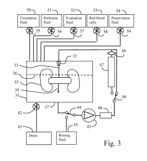
As shown in Fig. 3, an aorta residue 33 of the kidney 35 is connected to a
connector 32
arranged in a container 31. The vena cava 34 is open to emit fluid to the
bottom 37 of the container
CA 03134900 2021-09-24
WO 2020/209787
PCT/SE2020/050380
13
31 as shown by broken line 36. The fluid level 36 may be below the kidney or
above the kidney or
in between, the latter being shown in Fig. 3.
The bottom 37 of the container is connected to a drain bag 41 via a valve 42.
The bottom is
also connected to a pump 43 via a first switch valve 44. The first switch
valve 44 connects the inlet
of the pump 43 to either a rinsing fluid bag 45 or to the bottom 37 of the
container 31, in the first
position shown in Fig. 3.
The outlet of the pump 43 is connected to a heater/cooler 48 which controls
the temperature
of the fluid passing through the pump. The outlet of the heater/cooler 48
passes via a second switch
valve 46 to an oxygenator 47 and a leucocyte-filter 49, or directly to the
connector 32 and to the
kidney in the first position of the switch shown in Fig. 3. In the shown
position, the fluid is only
oxygenated by being exposed to the surrounding atmosphere (oxygen dissolved in
the fluid).
Thus, fluid present at the bottom of the container 37 is circulated via first
switch 44 to the
pump 43 and further via the heater/cooler 48 to the second switch 46 and to
the connector 32. The
fluid proceeds from the connector 32 to the aorta and to the kidney and
through the vessels of the
kidney and further to the veins of the kidney and is finally released to the
bottom of the container.
The pump 43 may be a pressure controled pump so that the pump pressure is
adjusted to a
desired value of for example 20 mmHg and the flow depends on the resistance of
the kidney.
The fluid present at the bottom 37 of the container 31 is provided via several
bags 50, 51,
52, 53 and 54 as shown in Fig. 3. Each bag is connected to the container 31
via valves 55, 56, 57, 58
and 59. By opening the valves, the contents of the bags are transported to the
bottom of the container
by gravity.
The operation may be as follows in one embodiment:
After the kidneys have been exposed to lys-plasminogen and tPA at the
backtable after
harvesting, the kidneys are moved to the container 31 and the aorta residue is
connected to the
connector 32 so that the kidneys hang inside the container 31. The veins are
open and allow the fluid
to drain directly down to the bottom 37 of the container. The kidneys may rest
on a support (not
shown) such as a net, and may have different angles of its position in regard
to the horizontal axis.
The temperature in the container is set to the desired starting temperature,
for example 18 C to 28
C. A circulation fluid comprising electrolytes, and possibly albumin at a
concentration of 57 g/L
may be provided to the container 31 from circulation fluid bag 50 by opening
the corresponding
valve 55. The kidneys are then perfused, starting with a pressure of 20 mmHg
for 5 minutes,
followed by increase of 5 mmHg each 5 minutes until 50 mmHg to 90 mmHg,
whichever has been
decided in advance. The pressure may then be lowered to for example 20 to 30
mmHg and perfused
for an additional period decided in advance. The resistance of the kidney has
decreased and the
kidney is now normally pale with no, or only small, dark areas. This is an
indication that clots in the
blood vessels of the kidneys have been dissolved and removed. The fluid inside
the container is now
passed to drain 41 by opening the drain valve 42.
CA 03134900 2021-09-24
WO 2020/209787
PCT/SE2020/050380
14
Without being bound by any theory, it is believed that dark areas of the
kidney is an
indication of areas with no circulation, which may be because of clots or
other reasons. If circulation
later is obtained in these dark areas, the kidney may recover these areas into
functioning tissue.
In a perfusion step, the drain valve 42 is closed and 1000 ml of a perfusion
fluid 51 is
passed to the container 31 via gravity by opening the valve 56. The fluid
level in container would
rise above the line 36 when 1000 ml of fluid has been accumulated at the
bottom of the container,
whereupon valve 56 is closed. The first switch valve 44 is in its first
position shown in Fig. 3 and the
pump is started and pumps at a pressure of 20 mmHg. The heater/cooler 48
adjusts the temperature
to 12 to 18 C. The fluid at the bottom of the container is circulated at a
pressure of 20 to 30 mmHg
during several hours, such as one to four hours. During the perfusion step,
the chemistry of the
kidney is restored and toxic products are removed. Now the pump is stopped and
rinsing is
performed. The perfusion step may be repeated with another composition of the
perfusion solution,
after the rinsing steps, during the hypothermic perfusion period, or the
perfusion may also continue
during another higher temperature, with or without RBCs.
In an evaluation step, the same solution from the perfusion step may be used
after the drain
valve 42 is opened to drain the contents of the container to the drain 41 or
the drain valve 42 is
closed after two rinsing steps described above and 500 ml of an evaluation
fluid 52 is passed to the
container 31 by gravity by opening the valve 57. When 500 ml of fluid has been
entered, the valve
57 is closed. The first switch valve 44 is in its first position shown in Fig.
3 and the pump is started
and pumps at a pressure of 20 mmHg. The heater/cooler 48 increases the
temperature to 28 to 32 C.
After 5 to 30 minutes under which the pH and the environment is checked, a red
blood cell
suspension (RBC) is added from bag 53 by opening valve 58 until a hematocrit
of 5 to 10 is
obtained, whereupon the valve 58 is closed. The second switch valve 46 is
moved to its second
position including the oxygenator 47 in the circuit for oxygenation of the
fluid and the red blood
cells. The circulation proceeds during 1 to 4 hours with a pressure of 30 mmHg
until the kidney is
determined to be usable for transplantation purpose. The kidneys are now
evaluated under 32 to
37 C and a pressure of 70 to 90 mmHg for 15 minutes, noting vascular
resistance, flow and visual
appearance in regards to how well the kidney is perfused. The surgeon decides
whether the kidney is
well, moderate or poor perfused, moderate being several blue/black spots and
poor being several
large dark blue or black areas not perfused. Urine production is measured and
flow is registered as
ml/min and 100g kidney tissue. Now the pump is stopped and the drain valve 42
is opened to drain
all fluid in the bottom of the container to the drain 41.
Any rinsing step may be performed by moving the first switch valve 44 to its
second
position connecting the pump 43 to the rinsing fluid bag 45. The pump is
activated and circulates
about 200 ml to the kidneys. The fluid leaving the kidney to the bottom of the
container is passed
further to the drain 41 via the open valve 42. In this manner, all red blood
cells are rinsed out of the
kidney and to the drain. Such rinsing steps may be performed several times and
between other steps.
CA 03134900 2021-09-24
WO 2020/209787
PCT/SE2020/050380
In a preservation step, the drain valve 42 is closed and 500 ml of a
preservation fluid 54 is
passed to the container 31 by gravity by opening the valve 59. When 500 ml of
fluid has been
entered, the valve 59 is closed. The first switch valve 44 is in its first
position shown in Fig. 3 and
the pump is started and pumps at a pressure of 20 to 30 mmHg. The
heater/cooler 48 adjusts the
5 temperature to 6 to 18 C, such as 12 to 15 C. The fluid at the bottom of
the container is circulated at
a pressure of 20 to 30 mmHg during several hours, up to 48 hours or more,
until a recipient has been
found and the kidney is prepared for transplantation to the recipient. The
pump is stopped and the
drain valve 42 is opened to drain all fluid in the bottom of the container to
the drain 41. The kidney
is removed from the connector 32.
10 The fluids used in the embodiment described in Fig. 3 may be the
fluids detailed in Table
A.
During the backtable procedure, the lys-plasminogen is included in a carrier
fluid which
may be identical to the base solution in Table A.
In an embodiment, the carrier fluid, the rinsing fluid, the perfusion fluid
and the
15 preservation fluid may have the same basic components. These components
include a sodium
content of 113mM to 129 mM, a potassium content of 5mM to 25 mM, a magnesium
content of
2.5mM to 5 mM and a calcium content of 1 mM to 5 mM. In addition to the above-
mentioned
electrolytes, one or several of the following substances may be included:
albumin at a concentration
of 10g/L to 120 g/L, such as 40 g/L to 75g/L, for example 57 g/L or 72 g/L;
optionally Dextran 40 at
0 g/L to 100 g/L, Hydroxy Ethyl Starch (HES) at 0 g/L to 70g/L, Poly Ethylene
Glycol (PEG 20) at
0 g/L to 25 g/L or PEG 35 at 0 g/L to 3 g/L alone or in combination. Dextrose
5 mM and
bicarbonate 10 to 35 mM are also optional. Furthermore, amino-acids such as L-
Arginine (precursor
to Nitric Oxide (NO), regulation of blood pressure, during physiological
stress), L-Leucine (protein
synthesis), L-Glutamine (synthesizes L-Arginine, needed for amino acid
production) or other, alone
or in combination, may be added in normal concentrations (see Table A). Other
substances that may
be added are I-Inositol (membrane potential stabilizer), Adenine and Ribose
(makes adenosine ¨ part
of ATP). Trace substances (cofactors and vitamins) such as stated in Table A,
may also be added.
Naturally occurring hormones may be added, such as Novorapid, T3, T4,
Progestrone and Estrogen,
in physiological concentrations (Table A). Antibiotics, such as Tienam may be
added. This basic
fluid according to Table A may be used as at least one of: the carrier fluid,
the rinsing fluid, the
circulation fluid, the perfusion fluid and the preservation fluid.
CA 03134900 2021-09-24
WO 2020/209787
PCT/SE2020/050380
16
TABLE A Vitamins mM
COMPONENT L-Ascorbic Acid = Na 0-2
Inorganic Salts mM D-Biotin 0.005
CaCl2 = 2H20 0-5 Choline Chloride 0-0.05
MgSO4 (anhydrous) 0-10 Folic Acid 0-0.01
KH2PO4 0-60 myo-lnositol 0-0.1
KCI 0-25 Niacinamide 0-0.1
NaHCO3 0-80 D-Panthothenic Acid =1/2Ca 0-0.01
NaCI 0-140 Pyridoxinhydrochloride 0-0.01
Na2H PO4 (anhydrous) 0-5 Riboflavin 0-0.001
NaGluconate 0-110 Thiamine = HCI 0-0.01
KGIuconate 0-25 Vitamin B12 0-0.01
MgGluconate 0-10 Hormones
NaLactobionate 0-110 T3 0-0.0000030
KLactobionate 0-25 T4 0-0.0000030
Ca Lactobionate 0-10 Cortisol 0-0.0000166
Amino acids Insulin Novorapid (U) 0-40
L-Alanine 0-1 Progesterone 0-0.0100
L-Arginine = HCI 0-1 Estrogen 0-0.1004
L-Asparagine = H20 0-1 Other
L-Aspartic Acid 0-1 Adenosine 0-5
L-Cysteine = HCI = H20 0-0.1 Cytidine 0-0.1
L-Cystine = 2HCI 0.5 2"Deoxyadenosine 0-0.1
L-Glutamic Acid/Ketoglutarate 0-5 2"Deoxycytidine
= HCI 0-0.1
L-Glutamine 0-15 2"Deoxyguanosine 0-0.1
Glutathione 0-5 Guanosine 0-0.1
Glycine 0-5 Pyruvic Acid 0-5
L-Histidine = HCI = H20 0-210 Thioctic Acid 0-0.01
L-Isoleucine 0-1 Thymidine 0-0.1
L-Leucine 0-1 Uridine 0-0.1
L-Lysine = HCI 0-1 Allupurinol 0-5
L-Methionine 0-1 Dextrose 0-200
L-Phenylalanine 0-1 D-Ribose 0-10
L-Proline 0-1 Raffinose 0-60
L-Serine 0-0.02 Mannitol 0-120
L-Threonine 0-2 Hyperoncotic fluid
L-Tryptophan 0-4 HES 0-5
L-Tyrosine = 2Na = 2H20 0-1 PEG35 0-0.5
L-Valine 0-2 Albumin (g/L) 0-120
Drugs Dextran 40/70 (%) 0-15
Verapamil
Heparin 0-10000 U
Minirin 0-000000009
CA 03134900 2021-09-24
WO 2020/209787
PCT/SE2020/050380
17
The perfusion fluid may have a high oncotic pressure, which is achieved by
addition of a
hyperoncotic solution such as albumin to a concentration of between 70 g/L to
120 g/L, for example
72 g/L, and perhaps Dextran 40 (or Dextran 70) at 0 g/L to 150 g/L or any
other known
hyperoncotic fluid. Potassium may be kept higher than in normal plasma in the
range 13 to 25mM,
such as 17 to 22 mM, for example 18 mM. Alternatively, the potassium
concentration may be low,
such as 1 mM to 13 mM. The potassium and sodium levels will vary during the
RBC phase due to
the normalization of the physiological environment and pH.
The evaluation fluid may further include:
a coagulation inhibitor, such as argobatran (or any other direct thrombin
inhibitor, such as
inogatran, melagatran (and its prodrug ximelagatran), dabigatran or hirudin
and derivates thereof, or
allosteric inhibitors);
and a platelet inhibitor, such as glycoprotein IIb/IIIa receptor antagonists
(abciximab,
eptifibatide, tirofiban),
Irreversible cyclooxygenase inhibitors (aspirin, triflusal),
adenosine diphosphate (ADP) receptor inhibitors (cangrelor, clopidogrel,
prasugrel,
ticagrelor, ticlopidine),
phosphodiesterase inhibitors (cilostazol),
protease-activated receptor-1 (PAR-1) antagonists (vorapaxar),
adenosine reuptake inhibitors (dipyradimol),
thromboxane inhibitors (thromboxane synthase inhibitors such as ifetroban,
picotamide,
and
thromboxane receptor antagonists such as terutroban) or
any other platelet inhibitor - in combination or alone, to prevent re-
thrombosis of treated
clots.
Heparin, Protein C and Protein S are optional.
Verapamil may also be added.
Fig. 4 shows another embodiment. This embodiment is intended to be used at a
local
hospital comprising facilities for harvesting kidneys. The kidneys are
pretreated at said local hospital
and later transported to a central hospital responsible for the
transplantation to a recipient.
At the local hospital, the victim of cardiac arrest, the donor, arrives at a
time from death
which is unknown, but lower than 12 hours, 10 hours, 8 hours, 6 hours or 4
hours. As soon as the
donor arrives, the carcass may be subjected to cooling in order to slow down
the deleterious
procedures in the body, in particular the metabolism and the coagulation. Such
cooling may be
topical in that the body is put in a room or compartment being refrigerated,
for example having an
air temperature of around 0 C. Other methods of cooling may be to arrange an
ice slurry around the
body or in the abdominal cavity, or cold fluid may be instilled in the
abdominal and/or the thoracic
cavity.
CA 03134900 2021-09-24
WO 2020/209787
PCT/SE2020/050380
18
If a harvesting team is present at the local hospital, the harvesting may
takes place as soon
as possible, with or without any cooling.
During harvesting of the kidneys, normal procedures are performed and the
kidneys are put
on a backtable, either kept en-bloc or each kidney separately. At the
backtable, the first step of
injecting lys-plasminogen in the renal arteries is performed. The lys-
plasminogen is allowed to stay
in the kidney blood vessels during approximately 15 minutes or more during
which time the kidney
is further prepared by attaching a connector to the renal arteries or the
aorta.
After about 15 minutes, or as soon as possible, the kidney is transferred to a
container
assembly 60 as shown in Fig. 4. A connector 63 of the kidney 61 is attached to
a corresponding
connector 64 at a top of a container 62, whereupon the kidney 61 hangs inside
the container 61 as
shown in Fig. 4. The vein of the kidney opens directly to the interior of the
container 61.
When the connector 64 is still open, a syringe 75 is connected and about 10 ml
of
tPA (Alteplas) is injected into the arteries of the kidney via connector 64.
It is noted that the addition
of lys-plasminogen may alternatively be performed via a syringe attached to
the connector 64 before
the addition of tPA and instead of addition at the backtable. Still
alternatively, the lys-plasminogen
and the tPA may be added simultaneously via a syringe to the connector 64 or
being injected
simultaneously at the backtable.
Instead of alteplas, another tPA could be used, such as streptokinase,
urokinase, reteplase
and tenecteplase.
Then, a tube 65 is connected to the connector 64. The tube 65 is arranged in a
spiral
configuration and connects a pressure bag 66 to the connector 64. The pressure
bag 66 is supported
by a stand 67 at a predetermined height of for example 27 cm above the
container 64 (corresponding
to a pressure of 20 mmHg). The height position of the pressure bag 66 is
adjustable along the stand
67 by a worm gear motor 68. The spiral configuration of the tube 65
accommodates the tube 65 to
the height position. Initially, the bag is empty.
A circulation fluid bag 76 comprises a circulation fluid including
electrolytes and an
hyperoncotic agent, such as albumin, (Table A). The circulation fluid in bag
76 is added to the
container 62 as indicated by arrow 77. There is about 1 L circulation fluid
and the volume of the
container 62 is about 1.3 L, which means that the container 62 is almost full
with circulation fluid.
The circulation fluid is oxygenated via an oxygenator using a gas mix of 02,
CO2 and N2 at 1L/min.
The operation is performed at room temperature. The circulation fluid
circulates together with the
lys-plasminogen and tPA in the system.
The amount of circulation fluid may be smaller than 1 L but should be larger
than the
volume of the tubes and pumps (except the container) and the kidneys. In
addition, a small volume
is needed for being enclosed in the container. Such volume may be 10 ml, 20
ml, 50 ml, or larger.
Thus, for example 150 ml to 1000 ml circulation fluid may be used.
A pump 69 is connected to the container 62 via tube 70 and pumps circulation
fluid from
the container 62 to pressure bag 66. A level detector 71 starts the pump 69
when the fluid level of
CA 03134900 2021-09-24
WO 2020/209787
PCT/SE2020/050380
19
container 62 reaches a predetermined level. The pump is for example a
peristaltic pump having a
flow rate corresponding to the revolution rate of the pump. There is no need
for the pump being
pressure controlled.
The pump 69 may be operated at a flow rate of for example 75 ml/min during one
minute,
whereupon 75 ml of fluid is transferred from container 62 to pressure bag 66.
The pump flow is
selected to correspond to a desired rate of flow through the kidney which
indicates that the
resistance of the kidney is sufficiently low. A flow of 50 ml/min per 100 g
kidney is normally
considered sufficient. A normal kidney of an adult man is about 150 g.
Since the pressure bag 66 is arranged at a height of for example 27 cm above
the container
62, a flow of circulation fluid passes through the kidney at said pressure of
27 cm water pillar, which
corresponds to 20 mmHg. If the resistance of the kidney is large, this
pressure of 27 cm water pillar
may cause a flow through the kidney of for example 7.5 ml/min, which means
that the fluid level in
container 62 is reverted to the level of the level detector 71 in 10 minutes.
Then, the pump 69 is
started by the level detector 71 and the procedure is repeated.
The pressure bag 66 may be moved to a higher position by the worm gear motor
68. In an
embodiment, the worm gear motor 68 increases the height of the pressure bag by
1 cm/min. When
the pressure increases, for example after 10 minutes to 35 cm water pillar,
the flow through the
kidney increases, for example to 15 ml/min. This means that the pump 69 is
operated again after 5
minutes. When the pressure has increased so that the flow through the kidney
is 75 ml/min, the
pump 69 will operate continuously. This is an indication that the resistance
of the kidney is
sufficiently low. The increase of the height of the pressure bag 66 will now
be stopped.
The maximum height of the pressure bag may be for example 95 cm, corresponding
to a
pressure of 70 mmHg. If the desired flow (75 ml/min for a kidney of 150 g) has
been reached before
the pressure bag is in the top position (after maximum 70 min), the procedure
is interrupted. If the
desired flow has not been reached, the operation is continued another 30
minutes at the pressure of
70 mmHg (95 cm water pillar).
The operation so far has been performed at room temperature of between 18 C to
28 C.
The container 62 is provided with a jacket 72 covering a large lower portion
of the
container. The jacket may now be provided with an ice slurry for cooling the
container 62 to a
temperature of about 5 C to 18 C, such as 12 to 15 C. The pressure chamber is
lowered to 25 cm
and the pump is operated to circulate the circulation fluid through the
kidney. The container with
jacket and pressure bag may be arranged in an insulating box (not shown) and
the assembly is
transported to a main hospital responsible for the continued handling. The
kidney may be stored in
this condition for a long time, up to 48 hours or longer.
In the main hospital, in which the transplantation may take place, the kidney
is further
treated. The kidney may stay in the same container 62 or moved to another
container 82 as shown in
Fig. 5. The container 82 comprises a connector 84 to which the kidney is
connected. A pump 89
pumps fluid from the container 82 via a tube 87 to the inlet artery of the
kidney and the fluid is
CA 03134900 2021-09-24
WO 2020/209787
PCT/SE2020/050380
emitted to the container 82 via the vein, as described earlier. The pump 89 is
pressure controlled and
keep the pressure to a desired value, such as 20 to 30 mmHg. A heater/cooler
(not shown) maintains
the temperature at a desired temperature, such as 12 to 32 C during perfusion
and preservation. A
drain bag 85 is connected to the bottom of container 82 via a valve 86.
5 The device according to Fig. 5 also comprises a red blood cell
suspension bag 91. The red
blood cell suspension bag is connected to a circulation system comprising
three pumps 92, 93, 94
arranged in parallel. A cytokine-filter 95 is arranged in series with pump 92,
an endotoxin-filter 96 is
arranged in series with pump 93 and an oxygenator 97 and a leukocyte-filter 98
are arranged in
series with pump 94. When a suspension valve 99 arranged after the red blood
cell suspension bag
10 81 is opened, the red blood cell suspension is circulated by the pumps
92, 93, 94 through the
respective filters 95, 96, 98 and through the oxygenator 97. The circulation
may be performed
during about 30 minutes at room temperature. In this manner, the red blood
cell suspension is
conditioned to have endotoxins, cytokines and leukocytes removed. In addition,
the red blood cell
suspension is oxygenated.
15 An evaluation solution is present in a bag 101 and is connected to the
container 82 via a
valve 102. When the valve is opened, the evaluation solution is passed via
gravity to the container
82, which is previous emptied to the drain 85 by opening the valve 86. The
evaluation solution is
heated to a temperature of about 32 C. The evaluation solution is circulated
through the kidney by
pump 89, whereupon the kidney assumes the temperature of 32 C. Then, the red
blood cell
20 suspension is added to the container by opening valve 99 and another two
valves 103 and 104 as
shown in Fig. 5. When the suspension has been transferred to the container 82
via gravity, the red
blood cell suspension valve 99 is closed.
Now, the pumps 92, 93, 94 pumps the fluid present in container 82 through the
open valves
104 and 103 and through the filters 95, 96, 98 and through the oxygenator 97.
In this manner it is
assured that the evaluation fluid does not include any endotoxins, cytokines
and leukocytes. In
addition, the red blood cells are oxygenated. The kidney can now be evaluated,
for example by
measuring "blood" parameters, such as pa02, PaCO2, HCO3, oxygen saturation,
Hb, Hct, Lactate,
Glucose, pH etc. In addition, the kidney can be examined optically. The
resistance can be calculated
from pump data. Urine production can be examined, including creatinine
concentration, if creatinine
is added to the evaluation fluid. In this manner, the kidney is examined for
suitability for
transplantation.
Then, the contents of the container 82 is transferred to drain 85 by opening
valve 86. A
preservation fluid 105 is introduced into the container 82 by opening valve
106. The preservation
fluid is circulated by pump 89 at a pressure of 20 to 30 mmHg and a low
temperature of 12 to 15 C
for removing all red blood cells. In addition, it happens that the kidney
increases its weight, and the
preservation fluid comprises albumin for removing excess water accumulated in
the kidney. After a
storage period of up to 7 days (or longer), the kidney is transplanted.
CA 03134900 2021-09-24
WO 2020/209787
PCT/SE2020/050380
21
A further embodiment is shown in Fig. 5a. The apparatus comprises a device 110
for
washing a red blood cell (RBC) suspension (not shown in all details). The
washed RBC in bag 111
is moved to a conditioning container 114 via a tube 112 and a valve 113. The
container 114
comprises a conditioning circuit 115, comprising a first pump 116, an
oxygenator 117, an cytokine
adsorber 118, a leucocyte filter 118, a first valve 120 and a second valve
121. In addition, a first
connector 122 with a valve is connected to the first valve 120 and a second
connector 123 with a
valve is connected to the second valve 121.
When the first and second valves 120, 121 are open and the valves in
connectors 122, 123
are closed, the conditioning circuit operates for conditioning the fluid in
container 114, by operating
pump 116 for circulating the fluid inside container 114 through the oxygenator
117, the leucocyte
filter 119 and the cytokine adsorber 118 for removing leucocytes and cytokines
in the RBC
suspension present inside the container 114. When the circulation has taken
place for at least 30
minutes, such as 60 minutes or more, the RBC suspension is prepared.
A treatment system 130 is shown to the right in Fig. 5a. The treatment system
comprises a
treatment container 131 having a third valved connector 132 which may be
connected to the first
valved connector 122 and a fourth valved connector 133 which may be connected
to the second
valved connector 123.
The treatment container 131 comprises three fluid bags 134, 135, 136 which are
connected
to the container 131 via tubes provided with valves. By opening such valves,
the fluids of the
corresponding fluid bag can be emptied into the treatment container. A waste
bag 137 is connected
to the bottom of treatment container via a tube provided with a valve. By
opening the valve, the fluid
in container 131 may be emptied to the waste bag 137.
A harvested kidney 140 (kidneys) is provided with a kidney connector 141
connecting to
the aorta residue or renal artery (arteries). The kidney connector 141 may
connect to a container
connector 142 provided in the container. The container connector 142 is
connected to a kidney
circulation system comprising a circulation pump 143 and an oxygenator 144.
The pump 143
circulates fluid in the treatment container from lower portion thereof via
said pump and oxygenator
to said container connector 142 and to the artery of the kidney 140. The fluid
emitted from the
kidney vein is let out to the interior of the container for further
circulation.
A conditioning system comprises a second pump 145 connected to the bottom of
the
treatment container 131 for pumping the fluid in the treatment container 131
through an endotoxin
adsorber 146 and a leucocyte filter 147 and back to the treatment container.
Two medical infusion pumps 148, 149 are arranged for infusing medical agents
into the
flow to the artery of the kidney. The infusion pumps may be syringe pumps. The
medical agents to
be infused may be lys-plasminogen, tPA, ATIII, a platelet inhibitor, a
thrombin inhibitor, a
coagulation inhibitor, etc. The medical infusion pumps 148, 149 may be
exchangeable, so that
further syringe pumps may be connected.
CA 03134900 2021-09-24
WO 2020/209787
PCT/SE2020/050380
22
A heater/cooler 150 is arranged for heating/cooling the fluid passing out of
the treatment
container.
The operation of the system may be the following:
An RBC suspension is washed in the washing system 110 according to known
methods.
When the washing is ready, the RBC suspension is transferred to conditioning
container 114 by
gravity by opening valve 113.
The fluid in conditioning container 114 is circulated by pump 116 by opening
valves 120
and 121 and operating pump 116 to circulate the fluid through the leucocyte
filter 119 and the
cytokine adsorber 118, thereby removing remaining leucocytes and cytokines.
In the meantime, a kidney 140 is harvested and placed in the treatment
container 131 and
connected to the container connector 142 via kidney connector 141. This
operation may be
performed at a distance from the left portion of the system shown in Fig. 5a,
for example at a remote
hospital.
The treatment container 131 is provided with treatment fluid from fluid bags
134, 135, 136
as required. When used, the fluid is expelled to waist bag 137 by opening the
waste valve.
The circulation second pump 145 is operated in order to pass the fluid in the
treatment
container through the leucocyte filter 147 and the endotoxin adsorber 146 for
continuously removing
leucocytes and endotoxin.
The circulation pump 143 is operated in order to circulate fluid from the
container via
oxygenator 144 and to the artery of the kidney and further via the kidney vein
back to the container
for treatment of the kidney, as outlined above.
The medical agent pumps 148, 149 may be used for addition of medical agents.
Then, the treatment system 130 is docked to the conditioning system 115 by
connecting
valved connector 122 to valved connector 132 and valved connector 123 to
valved connector 133
and opening the valves, except valve 123. The first valve 120 is closed and
the second valve 121 is
opened, whereupon the pump 116 is operated for pumping the fluid in
conditioning container 114
via the connectors 122, 132 to the treatment container. Then, the second valve
121 is closed and
valve 123 is opened, whereupon the pump 116 circulates the treatment fluid in
the treatment
container 131 through the oxygenator 117, the leucocyte filter 119 and the
cytokine adsorber 118.
The treatment may be according to any one of the methods mentioned above.
As shown in Fig. 5b, the treatment container 151 may be modified to treat two
kidneys
separately, although they may be arranged en-block. The two kidneys are
provided with two kidney
connectors 161, 171 connected to container connectors 162, 172, which are
connected to circulation
pumps 163, 173 as shown. An oxygenator 164 is connected in a common line for
the two circulation
systems as shown. With this system, each separate kidney can be provided with
separate medical
agents via medical infusion pumps 168, 169 and 178, 179 for different
treatments.
CA 03134900 2021-09-24
WO 2020/209787
PCT/SE2020/050380
23
Example 1.
Procedure before retrieval: 30 pigs were anaesthetized and allowed to achieve
normoventilation, after which the ventilator was turned off. Asystole and
circulatory arrest appeared
after about 15 minutes. After two hours in room temperature, cold Saline was
installed in the
abdominal and the thoracic cavities after which no further action was
undertaken during one hour.
Retrieval: Three hours after circulatory arrest, surgery was started to
retrieve kidneys,
which took a mean of 45 minutes.
Backtable procedure: The kidneys were flushed through the renal artery on
backtable with
SOLTRAN (Kidney perfusion fluid, Baxter Healthcare) after injection of about
500 U Heparin in
10m1 of Lidocaine 0.5 to 1% diluted to 20m1 with 0.9 % Saline, per kidney.
Treatment procedure: The kidneys were divided in four groups: Group A with 11
pigs,
Group B with 13 pigs, Group C with only one kidney from each 6 pigs and Group
D with the other
kidney of the same 6 pigs.
Group A was treated with cold storage for 2 hours followed by transplantation.
Group B was treated with cold storage for 2 hours followed by 90 minutes of
reconditioning at 37 C using a solution with the composition shown in Table B
and mixed with
washed RBCs to a hct of about 15 followed by transplantation.
TABLE B
COMPONENT WE. mM Vitamins el mM
irsorganic SaIts. I.-Ascorbic Add = Na OM
O.28.4
....... 21-i0 0.263 13696 0-8intin 0.0001
0.M041.
fv1g50:t {anhydrous} '3.09767 021144 Cho line
Chloride. 0.001 0,00716
KO 0.4 5,35543 Rik Adel 0.001
0,00227
Nsi-ICO. 4.72 56.18506 myc--inositoi 0.002
0,0111
116.35'269 NrtanUde 0.001
0,00319
NalHP0.4 fa rihydrotz) 0.122 0,8594 0-Panthothertic Add Iµ W.:a
0.001. 0,00210
Aminci adds Pyridoxishydrochloride 0.001
0,00486
L-Aisnine 0.07.5 0,2806 Ribofiavin 0.0001
0.000266
1.-Arginine * NCI 0.126 0,72329 Thiamine * HC I 0.001
0,00296
1.-Asparagine *1-ifi 0.05 0,17645 itd.amirs 912 Ø00136
0,00136
1.-Aspartic Add 0.03 0,22539 Other
1.-Cyst.eine * HO * 420 0.1 0,00186 Adenosine Ø01
0,01742
1.-Cyst.ine * 2140 0.0311 0,0999 Cridine 0.01
ao4112
L-Glutamic Acid 0.075 0.50975 2'Deuxyade.nusine 0,01
0.0398
L-Glutamine 0,292 2.00 Theoxyditidine = liCi 0.011
0.04841
Giycine 0.05 0,66507 Theoxygu a nosine
Ø01. 0,03742
I-Hist:Winn a NC! a H20 0.042 0,200 Cinariosine. 0.11
0,03531
1.-isoleudne 0.052 0:395 Pyruvic Add f.).11 1,249
L-Lendrie 0,052 0.396 Thioc.tir. Acid 0.0M2
0.00097
L-Lysine = HCI 0.0725 0.117 Thymidine 0.01
0.04111
L.-Meteor:kw 0.015 0,101 Liridine OM
0,04095
L-Phenyialardne 0.032 0.194 Hormones
1- Pro iine 0.04 0.34743 73 0.000000001953
0,000000030
L-Srine 0,025 0.0002164 T4 0.00000000233
0.000000030
L-Threcnine 0.048 0.403 Cortisol 0.000006
0.0000165
L.- Tryptophan '3.01 0,049t insuiin Novorapici SU
L-Tyrosine = 2Na = ay) 0.0519 0.199 Minirin 0,00000001
0.000000009
L-Valine 0.046 0.393 Drugs
VerapamiI 0.005
Tiersam 0.050
Heparin 500U
CA 03134900 2021-09-24
WO 2020/209787
PCT/SE2020/050380
24
Group C was treated by hypothermic perfusion during 4 hours directly after
retrieval of the
kidney using LifePort Kidney Transporter and KPS (Kidney Perfusion Solution)
according to the
manufacturer's (Organ Recovery Systems) instructions, followed by
transplantation.
Group D was kidneys from the same donors as Group C but treated with cold
storage for 2
hours, followed by transplantation.
Fig. 6 shows the change of arterial blood flow (ml/min) after reperfusion and
after 90
minutes' observation after transplantation.
Fig. 7 shows mean urine production in ml/min, 90 minutes after
transplantation. Using
Mann-Whitney U test, the flow was better in the reconditioned group B at
reperfusion (p<0,05) (Fig.
.. 6) and the reconditioned group B also had better urine production (p<0,05)
90 minutes after
transplantation (Fig. 7).
Example 2
6 pigs were anaesthetized and allowed to achieve normoventilation, after which
the
ventilator was turned off. Asystole appeared after about 15 minutes. After two
hours in room
temperature, cold preservation solution (Saline) was installed in the
abdominal and the thoracic
cavities.
Retrieval of the kidneys was started 4 hours after death.
The kidneys were flushed on backtable with cold Ringer solution after
injection of about
500 U of Heparin in 10 ml of Lidocaine 0.5 to 1% diluted to 20 ml with 0.9%
Saline, per kidney
through the renal artery.
The kidneys were connected to an ex-vivo perfusion device of the type shown in
Fig. 3.
0.4mg of tPA (alteplas) and 150U of apyrase (Sigma Aldrich, purinergic drug ¨
CD39/CD73) was
injected through the renal artery. The composition of the solution used can be
seen in Table C.
The solution was perfused during 30 minutes at a temperature of 15 C and a
pressure of 20
mmHg without oxygenation. Temperature was then raised to 32 C, pressure to 30
mmHg, and
perfusion was continued for another 90 minutes in a reconditioning phase. Then
washed leucocyte
filtered RBCs were added and the perfusion continued for another 90 minutes
during evaluation.
Then the kidney was transplanted.
Fig. 8 shows the renal flows after reperfusion and after 90 minutes. Kidneys
did not clear
the microcirculation completely and showed perfusion defects on the surface of
the kidneys.
Fibrinolytic drugs as well as purinergic drugs (apyrase ¨ CD39/CD73) ¨ added
to the ex-vivo
system did not substantially improve the vascular resistance and flow.
CA 03134900 2021-09-24
WO 2020/209787
PCT/SE2020/050380
TABLE C
COMPONENT di. rriM git- mNi
inorganic Salts Vitamins
CaCi,, = 21130 0.6732 4.95 Chne Chloride 0.001
0.00716
Magnesium Glocisnate ianhydrous) 1.13 5.00 Fat:Acid
0.001 0.00227
Potassium Phosphate fmonobasic) 9.68 5.00 myo-Irtositoi
0.002 0.0111
NaHCOs 4.15 49.40 Macinarnide 9.001
0.00819
5 Sodium 6-PA:mate 21.80 100.00 D-Panthethenic Acid = )Ca 0.001
0.00210
Amino adds Prid sub.:hydrochloride 0.001
0.00486
1-Gluta mine 0.292 2.00 Riboflavin 9.0001
0.000266
LArginine = HO 0.105 0443 Thiamine = HO 0.001
0,00296
L-Cystme * 21-ECE 0.0313 0,0999 Other
1.-1-ii5tithne = HO = Hp 0,042 0.200 Aden1ne 0.68 5.00
1-inoieticine ........... 0.052 0.396 = Dextrose 1.00 5.55
..
1-Leucise 0.052 3.396 D-Ribose 375 5.00
1-Lydne = i-ca 0.0725 0.397 Aiburn in 72
10 1.4.4oth ionine am 0.101 T.-Unit-in 0.00000001
L-Phertylaia nine 0.032 0,194 Veraparnii 0.005
1-Proline 0.04115 0.100 Tienam 0.050
1-Threort in e 0.048 0.403 Heparin 500 Li
1-7ryptophan 0,01 0.0490 Apyrase 150 Li
1-Tyr0s03e * 2Na = 2ft7 0.0519 0.199 Atepes 0.4m8
L=Valine 0,046 0.393 Hormones
T3
0.000000001953 0.030000030
74
0.00000000233 0.000000030
1 5 Stserile water- Cords&
3000')35 0.0000166
Insulin Novorapid 51i
Progesterorse 0.00315
0,001002
Estrogen 0.00002
0.00004
Example 3
In another experiment the protocol in Example 2 was repeated, but apyrase was
not given.
20 Instead Lidocaine was added to the perfusion solution. The idea was to
look for stabilizing effects
during the reconditioning phase, as a result of the effect on the Na + K+ pump
that Lidocaine has
shown to have. The composition of the solution can be seen in Table D.
TABU D
COMPONENT git miVI a mN1
Inorganic Salts Vitamins
25 CaClz * 2H20 0.6732 4.95 Choline
Chloride 0.001 0.00716
Magnesium Gluconate {anhydrous} 1.13 SAO Folic Add 0:001
0.00227
Potassium Phosphate (monobasic) 0.68 5.00 rnyo-inositol
0.002 0.0111
NH CO3 4.15 49,40 Niatinamide 0001
0.00819
Sodium Eiucorsate 21.80 100.00 D-Parsthothenic Add = %Ca 0.001
0.00210
Amino acids P.p. idoxinhydrechinride 0,001
4).1X86
1.-Eiutatnirse 0.292 2.00 Riboflavin 0.0001
0.000266
L-Arginine = HO 0.105 0,603 'Thiamine *1-ECI 0,001
0.00296
1-C.ystine = 2H0 0,0113 Ø0999 Other
1,-Histicline = HO = H20 0.042 0.200 Adenine 0.68 540
1.-Isoieucine 0.052 034)6 Dextrose 1.00 5.55
L-Leucine 0.052 0.396 0-Ribose 0.75 5.00
4sine = HO 0,0725 0.397 Mb wain 72
LVetidonine 0.015 0.101 Minirin 0.00000001
1.-Phenvia ia nine 0,032 0.194 Veraparsii 0.005
L-Proline 0.04115 0.100 Inman) 0.050
1.-Threonine 0,048 0.403 Heparin SOO Li
t.-Tryptophan 0.01 0.0490 Lidos:airs 0.060 0,256
1-Tyrosine = 2Na = 21420 0,0519 0.199 Hermooes
L-Vaiirse 0.046 0.393 r3
0.000000002953 0,000000030
14
0.04)4)04)000233 0.0034)00030
Cortisol 0.000006
0,0000166
Sterile water Insulin Novorapid 5L3
Pmgesteror3e 0.00315
0.001002
Estrogen 0.00001
0.00004
CA 03134900 2021-09-24
WO 2020/209787
PCT/SE2020/050380
26
Fig. 9 shows an unchanged flow during the first 90 minutes, which usually is
decreasing.
Furthermore, the weight change was minimal between the different phases of the
reconditioning
despite the fact that the osmolarity was 330 mosm and the potassium level
about 5mmo1/L. In
Example 2 kidneys gained significantly more in weight from start of perfusion
and to end after 90
min of reperfusion.
Example 4
7 pigs were anaesthetized and allowed to achieve normoventilation, after which
the ventilator
was turned off. Asystole appeared after about 15 minutes. After two hours in
room temperature, cold
preservation solution (Saline) was installed in the abdominal and the thoracic
cavities.
Retrieval of both kidneys of each pig was started 4 hours after death.
The kidneys were flushed on backtable with cold Ringer solution after
injection of about 500
U of Heparin in 10 ml of Lidocaine 0.5 to 1% diluted to 20 ml with 0.9%
Saline, per kidney through
the renal artery as described in Example 2. The composition of the solution
used can be seen in
Table E.
TAM E
COMPONENT gft rnlYi &ft mNi
inorganic Sts Vitamins
2H20 0.6732 4.95 Chne Chioride 0.001 0.00716
Magnesfum Giuconate (anhydrous) 1.18 5.00 Polk, Acid 0.001.
0.00227
Potassium Phosphate tmosubasic} 0.68 5.00 :Tlyo-J rIOSitt3/
0.002 0.0111
Na HCO3 4.15 49.40 Niacin am ide 0.001. 0.00819
Sodium Gluconate 21.80 100.00 D-Panthuthenic Acid = %Ca 0,001
0.00210
Aff31110 acids Nridoxin hyd rochlo ride 0.001
0.00486
1-Glutamine 0.202 2.430 Riboflavin 0.0001 0.000266
1-Afeinine * NC i 0.105 0.603 Thiamine 4; HO 0.001.
0.0029C)
1-Cystine = 2.14C.i 0.031.3 0.0909 Other
1,Histitiioe = Ha = H20 0.042 0.200 Adenine 0.68
5.00
1.45,1k:tone 0.052 0.396 Dt3Xtre35.e
RIACi Ile 0,052 0.396 D-Bibose 0.75 5.00
1.-Lysne * HO 0.072'3 0.n7 Abu:11in 7a
1.:Meth ion ine 0,015 0.101 :Minirin 0.00000001
1.-PhmylaWline o.(22 0.1!34 Vz1. pa:nil 0.005
1,Proline 0.04115 0.100 Tienam 0.0
I.- Th rec :lin* 0,04e a4O3 Heparin 500 U
IA ryptaphan 0,01 0.0490 Hormones
1.-Tysosine = 2Na = 21420 0.051.9 0.190 T3 0.000000001953
0.000000030
1,Vaiirse 0.046. 0.393 T4 0.00000000233
0.000C., '1)0030
Cortisol 0.00000. ;
0.0000166
insn Novor .,p id SU
Ste n id we Progesterone 0.00315 0.001002
Eeitrogen 0,00001 oxxx.v.
The kidneys were perfused during 30 minutes ex-vivo, first at 15 C and a
pressure of 20
mmHg. Then, the perfusion solution was oxygenated with start using a gas
mixture of 02 (20%),
CO2 (5,6%) and N2 (74,4%). After one hour the solution was exchanged with new
solution with the
composition according to Table E, the temperature was raised to 32 C and the
pressure was adjusted
CA 03134900 2021-09-24
WO 2020/209787
PCT/SE2020/050380
27
to 30 mmHg. Washed leucocyte filtered RBCs were added to a hct of about 10 to
15, followed by
perfusion during 5 hours. One kidney was then taken out from the perfusion
system, transplanted
(n=7) into a recipient pig and observed for 90 minutes up to 8 hours. The
remaining kidney in the
ex-vivo perfusion system was perfused for another 90 minutes for comparison.
Fig. 10 shows renal arterial blood flow after transplantation. Kidneys were
functioning,
producing urine, for more than ten hours (n=3). Only few animals were followed
up to ten hours
which explains the increased spread in flows. Experiments terminated with pigs
still under
anesthesia.
Example 5
In another experiment, treatment was given according to the same protocol as
in Example 4
(n=5), but ex-vivo perfusion was continued for 11 hours instead of 6 hours.
Flow was 104 ml/min at
reperfusion and 109 ml/min 14.5 hours after reperfusion. Thus, even extending
the perfusion time
can result in significant flows, allowing time for transportation of the
kidneys and waiting for
recipients to arrive.
TABLE F
COMPONENT gIL mit rnis/1
Inorganic Saks ..................... = .. \titarnins
c.:ai."12 = 2H20 0.6732 4.95 Chan e Chic ride 0.001
0.00716
Magnesium Giuconate (anhydrous) 1.13 5.00 Folic Add man
0.00227
2() Potassium Phosphate li.nonohasir.) 0.68 5.00
yo-inosito 1 i0.002 0.0111
NafiCO3 4:15 49.40 .. = Niacinamide
0.001. 0.00819
Sothn Glisconate 21,80 100.00 f3-Panthothenic.Add * %Ca 0.001
0.0021.0
Amino acids .Pyridoxinhydrochde 0,001
0.00486
L-Giutarnine 0.232 2.00 Riboflavin i0.0031.
0.003.266
L-Argne = HO 0.105 0.603 Thiamine = HCI .. i0.001
0.00236
1.-Cystine = 21-1C1 0.0313 0,0999 Other
1-Histidine = HCl' H20 0.042 0.200 Adenine 0.68 5.03
1..-isoie..1.:dne 0.052 0.36 .Dextrose LOO .55
L-Leucine 0.052 0.396 0-Ribose 0,75 5.00
L-Lysine = HO 0.0725 0.397 Aibumin 60
1-Methionine am 0,101 0.00000001.
1-Phenylalanine 0.032 014 Verapa rn 0.005
0,0411S 0.100 Tienam 0,050
L-Threoriine 0.048 0.403 Hepa f 500 Li
L-Tryptop ha n 0.01 0.0490 Hormones
1-Tyrosine = 2Na = 2H20 0.0519 0.199 T3 0.000000001951
0.000000030
L-Vaiine 0.046 0.393 T4 0.00033000233
0.030003030
Cortisoii 0,000006
0.0000166
in sun ISKINACH a F. id 5U
----------------------------------------- Progesterone 0.00315 0.001002
Sere water Estrogen 0.00001 0.00004
Example 6
In another experiment 7 pigs were declared dead according to the same protocol
as in
Example 4. Warm ischemia time (WIT) was 4 to 6 hours
Both kidneys per pig were retrieved starting 4 hours after death.
CA 03134900 2021-09-24
WO 2020/209787
PCT/SE2020/050380
28
Ex-vivo perfusion started at 15 C.. During the first hour, 72 g albumin/L was
used (solution
according to Table E) and the pressure during perfusion was 20 mmHg.
Oxygenation was started
after 30 minutes. After 1 hour, the solution was drained and a new solution
with 80 g albumin/L,
(solution according to Table F) was used during 2 hours after raising the
temperature to 32 C,
pressure to 30 mmHg and addition of RBCs.
Fig. 11 shows the renal artery blood flow after transplantation. All seven
kidneys showed
good flows for 4 hours (118.7 15.0 ml/min) and 8 hours (85.0 10.5 ml/min)
after kidney
transplantation and maintained urine production. Experiments terminated after
8 hours with pigs still
under anesthesia. Although the flow was declining during the observation time,
the kidneys still had
a good flow at the end of the observation. The ethical permit did not allow us
to let the pigs wake up
TABLE G
COMPONENT git. mM ea rnM
inomarcie Salts Vitamins
Caf:1;: = 2(420 0.6732 4,95 Chase Chloride 0.001
0..00716
Magnesium Glaconate (anhydrous) 1.13 5.00 Folic Add
0,001 0.00227
Pt=itassiirm Phosphate (rnonobasic.) 0.66 5,00 rnyo-inositoi
0.002 0.0111
NattC.03 4.15 49.40 Niacinamide 0.001
0.00819
Sodium 5k:cc:nate 21.80 100.00 0--Panitiothenic Acid = Me
0,00.1. 0.00210
Amino adds Pyridoxinhydrochloride 0.001
0.00486
1-Glutamine 0.202 2.00 Riboflavin 0.0001
0,000266
L=Argne = HO 0.105 0.603 'Thiamine * MCI .....
,O.Cii.11 0.00296
L-Cystine = NCI 0.0313 0.0909 Other
I.-Hist:dine * HO = 1420 0.042 0.200 Adenine 0,68 5.00
1.eucine 0.052 0.396 Dextrose 1,00 5.55
t-teudne 0.r.s2 11.396 D-Ribose 035 5.00
L-Lysine = i-iCi 0.0725 0,397 , Aiburnin 57
1.-Methne 0.015 olin Minirin o.occ000m
L-Phenylaianine 0.032 0.194 Veraparriii 0.005
1-Proline 0.04115 0.100 Tienarri 0.050
IA hreonine D.C.:48 0.403 Heparin 500
0
L-Ttyptophan D.C:1 0.0490 Hormones
1.-Tyrosine = 2Na a 21120 0.0519 0.199 13 0,000000001953
0.000000030
1.-VaDne 0.046 0.393 T4 0,0:X8X:000233
0.000000030
Cortisol 0.0V3006
0.00001.66
insulin Novora oid SU
.Strrrile water Progesterone 0,00315
0.00l002
Estrogen 0.00001
0.00004
Example 7
In another experiment, 4 pigs were declared dead according to the same
protocol as in
Example 4. Topical cooling using ice in the abdomen was used after 2 hours,
resulting a change of
body temperature from 37 C to about 20 C compared to 25 C to 29 C using fluid
as coolant.
The kidneys were retrieved, whereby the WIT varied between 4 to 5 hours.
The kidneys were rinsed with a composition of the solution described in Table
G with 57g
albumin/L as hyperoncotic agent. The kidneys were rinsed in an ex-vivo device
using a pressure of
20 mmHg up to 70 mmHg, raising the pressure 5 mmHg every 5 minutes, and a
temperature of 18 C
CA 03134900 2021-09-24
WO 2020/209787
PCT/SE2020/050380
29
until the kidneys turned white. It took on average 2 to 3 liters of solution
and up to 50 minutes to
achieve well perfused kidneys.
Using the same solution, temperature was reduced to 12 C and the kidneys were
perfused
during 1 hour at 20 mmHg. Solution was then changed to a solution comprising
80 g albumin /L as
seen in Table F, temperature 28 C and pressure of 30 mmHg. RBCs were added to
a final hct of
about 10 to 15. The kidneys were perfused with this solution for 2 hours. The
temperature was then
increased to 32 C and the kidneys were perfused for an additional hour before
draining the system
and adding a solution comprising 57g albumin/L initially used again. With this
solution, perfusion
was continued for 30 minutes at 15 C and a pressure of 20 mmHg before
transplantation.
Fig. 12 shows the renal artery flow after transplantation. High flows after
reperfusion could
be achieved after careful rinsing of the kidneys after retrieval. Urine
production could be maintained
for extended time in the recipients but declined after extended observation
time, indicating that flush
with volumes of several liters may negatively influence late outcome due to
endothelial activation.
Still, at 4 hours (145.7 29.3 ml/min), and 8 hours (96.73 12.8 ml/min) flow
was better than with
topical cooling with cold preservation solution installed in the abdominal and
the thoracic cavities as
in previous Examples 1 to 5 achieving temperatures of 25 C to 29 C. The
experiment was
terminated with pigs still under anesthesia due to the ethical permit.
Example 8
In another experiment 8 pigs were declared dead as described above in Example
4,
however, no topical cooling was given.
The kidneys were retrieved 4 hours after death and were perfused on backtable
with 200 to
500 ml of Perfadex. All showed poor to moderately poor perfusion and poorly
cleared kidneys.
The kidneys were then transplanted immediately to nephrectomized pigs. Mean
flow at
reperfusion was 14.15 ml/min and at 90 minutes 36.8 ml/min. All kidneys looked
bluish/black at 90
minutes, with no urine production at the end. This experiment served as
control, showing that
without any cooling procedure, and other treatments, for example according to
embodiments of the
present invention, a WIT of 4 hours result in poor performance in a transplant
setting, in accordance
with previous experience.
Example 9
In another experiment six pigs were declared dead using the above technique.
Topical
cooling using ice slush inserted into the abdomen was used after 2 hours,
resulting in a change of
body temperature from 37 C to about 10 to 12 C compared to 25 to 29 C using
fluid as coolant and
20 C using ice.
The kidneys were retrieved with a WIT which varied between 4 to 5 hours.
On the backtable and after retrieval, 20 ml of a solution comprising 72 g/L
albumin solution
(Table I) mixed with 10U of lys-plasminogen and 200 U of antithrombin III
(ATIII) was inject in
CA 03134900 2021-09-24
WO 2020/209787
PCT/SE2020/050380
TABLE H
COMPONENT mM
Inorganic Salts Vitamins
CC =2H20 0.6732 4,95 Cholin:? Chloride 0,001
0.0f.1716
Magnesium, Gluconatt-, {anhydrous} 1,13 5,00 Folic Acid
0.001 0,00227
5 Potassium Phosphate (rnOrlOhaSie) O. 5,00 myo-
inositol 0,002 0.0311
NeHCO3 i4.15 49.40 Niatinamide 0.001
0.00819
Sodium Giuconate 17.451 80.00 1.1=Panthothenic Acid a%Ca
0,001 0.00210
Potwi,im Glucon ate 4.6;t35 20,00 Pyridoxin
hyd rochioride 0.001 0,00486
Amino adds Riboflavin 0.0001
0.000266
........ mine 0,292 2.00 Thiamine = HC i 0.001
0,00296
1-Argirline Ha 0.105 0.603 Other
Hormones Adenine 0,68
5,00
10 13 0.000000001953 0.000000030 Dextrose ..
1.00 .. 5.55
14 0.00000000233 G,000000030 Ribie
0,75 5,00
Novorapid 511 Albumin 57
Proge5terone 0.00315 0. 00117.Kr2 -renarn
0.050
Estrogen 0.00001 0,00004 Sterile water
each kidney through the renal artery after clamping off the veins and then the
arteries after the
15 solution had been delivered.
The kidneys were moved to an ex-vivo device. After 15 minutes, 1 mg tPA
(alteplas) and
100 U of ATIII per kidney was injected, divided in 4 portions of 5m1, after
dilution into 20 ml of the
solution comprising 72 g/L albumin solution (Table I). The tPA was injected
into the renal artery
with 5 min apart at a temperature of 20 C, starting with 20 mmHg and
increasing 5 mmHg each
20 time.
After completion of infusion of the tPA, the kidneys were perfused with a
solution
comprising 72 g albumin/L and the pressure was increased every 5 minutes with
5 mmHg each time
up to 50 to 70 mmHg or until the kidneys cleared ¨ whichever came first.
Temperature was then
lowered to 12 C and the perfusion continued for 1 hour at a perfusion pressure
of 20 mmHg.
25 After rinsing with a solution comprising 57g/L albumin (Table H), a
second dose of lys-
plasminogen followed by tPA and ATIII was given, diluted in the solution with
57 g albumin/L and
given sequentially through the artery as described above.
Then, the perfusion was continued for 30 minutes, pressure 20 mmHg,
temperature 12 C.
The temperature was increased to 28 C and a pressure of 30 mmHg, before
addition of RBCs to a
30 hct of 5 to 10. The RBCs had been pretreated during 2 hours by an
external pump and circulated
through a leucocyte filter before being added to the solution. This was done
to allow leucocytes to
decrease before being in contact with the kidneys. Perfusion using the
combined albumin solution
(57 g albumin /L, Table H) and RBCs was continued for 1 hour. Temperature was
then raised to 37
C and pressure to 70 to 90 mmHg for one hour during evaluation. After rinsing
of the kidneys from
RBC containing solution, filling with new 57g/L albumin solution and lowering
the temperature to
15 C, transplantation took place.
Transplantation was performed into pigs where the native kidneys had been
nephrectomized immediately before the kidney transplant. Three animals were
allowed to wake up
CA 03134900 2021-09-24
WO 2020/209787
PCT/SE2020/050380
31
from anesthesia (new ethical permit allowing ten days' observation). One
animal was explored on
day three for inspection of the kidney and new blood samples. The only
immunosuppression given
was steroids at reperfusion. The kidney looked good, creatinine in blood was
615 mon. Another
pig was explored on the 4th day, kidney looked good as well, creatinine in
blood was 884 mon.
The third pig was taken sample from on day 5 and followed for 8 days in total.
All three pigs
showed signs of delayed graft function (DGF) in one case resulting in uremia
and death after 8 days,
in two other cases animals were sacrificed on day 3 and 4 with elevated
creatinine in blood and
higher creatinine in urine as signs of ability to concentrate urine.
Fig. 13 shows arterial flow for each of the six pigs, after reperfusion and
after 90 minutes.
Adding lys-plasminogen and tPA cleared the kidneys better than seen before,
with decrease of
resistance and improved microcirculation as result.
Fig. 14 shows that some kidneys showed patches on the surface after
reperfusion,
sometimes developing to blue/black poorly perfused kidneys a few hours after
transplantation,
indicating either a re-thrombosis due to the coagulation system or a
platelet/RBC induced action on
the endothelium.
Example10
In three more pigs the protocol in Example 9 was repeated with a few
exceptions.
After retrieval and on the backtable, injection of lys-plasminogen was
performed as
mentioned above. After 15 minutes, 2 mg of tPA (alteplas) in 500 ml of a
solution comprising 57g
albumin/L (Table H), was flushed through the kidneys before they were taken to
the ex-vivo device.
At the ex-vivo device, the kidneys were perfused in a solution comprising 72
g/L albumin
(Table I) at 24 C with increasing pressure from 20 up to between 50 and 70
mmHg. Using the same
solution, the perfusion continued for 2 hours at 12 C and pressure of 20 mmHg.
After draining and rinsing, a solution comprising 57 g albumin/L (Table H) was
added
together with washed RBCs to a hct of about 5 to 10, temperature changed to 28
C, pressure to 30
mmHg. A new dose of lys-plasminogen, tPA 2mg and ATIII 200U was added and
temperature was
increased to 32 C followed by perfusion for 15 min at a pressure of 70 mmHg
for evaluation. After
rinsing and washing, a solution comprising 57 g/L albumin (Table H) was added
and temperature
decreased to 15 C. All three pigs were taken off anesthesia and observed. Two
animals were
surviving for 10 days, one with 166 i.tmol/L and the other with 174 mon
creatinine in blood with
normal looking kidney at termination of the experiment. The third pig was
sacrificed on day 6 with
delayed graft function, having a creatinine in blood of 1231 mon but over
2000 mon in urine,
showing ability to concentrate urine. All recipients received steroids as the
only mean of
immunosuppression. Sample were collected for histological studies of
allorejection.
Fig. 15a shows a normal looking kidney transplant 10 days after
transplantation. No
macroscopic signs of rejection.
CA 03134900 2021-09-24
WO 2020/209787
PCT/SE2020/050380
32
Fig. 15b shows the cut surface of the transplanted kidney in Fig. 15a, showing
normal
macroscopic architecture.
Fig. 15c shows the surface of the second kidney surviving 10 days, showing
area of dark
spots, indicating affected microcirculation
Fig. 15d shows the cut surface of the transplanted kidney in Fig. 15c, showing
a dark area
with affected circulation in the lower pole. The data indicates that the
treatment can recover both
function and structure, although there may remain a risk for local damage of
the microcirculation
Example 11
In a further experiment comprising 19 pigs, cytokine levels were measured in
three groups.
In Group A (n=5), comprised of live donor kidneys perfused with a 72g/L
albumin solution (Table I)
without any adsorber of cytokines, samples were taken at start of perfusion,
at 3 hours and at the end
of 37 C. In Group B (n=5), comprised of live donor kidneys perfused with a 57
g/L albumin
solution (Table H) without any adsorber of cytokines, samples were taken at
start of perfusion, at 3
hours and at the end of 37 C. In Group C (n=9), comprised of DCD kidneys
perfused with 72 g/L
albumin solution for 90 minutes and 57 g/L albumin for 90 minutes before RBC
and perfusion at
37 C, all during which a cytokine adsorber (Cytosorb, Cytosorbents) was used.
An ELISA kit
(Quantikine ELISA kit, R&D Biosystems) was used for the analyzes. Levels below
detection levels
were set to 0 and mean levels for measured levels within each group was then
calculated.
Fig. 16a is a diagram showing changes in IL-6 levels mainly from the RBCs. The
adsorber
(Group C) effectively removed all signs of IL-6.
Fig. 16b is a diagram showing changes in IL-8 levels, also mainly from RBCs
and less from
the 57g/L than the 72 g/L albumin solution. The adsorber removed the IL-8
(Group C).
Fig. 16c is a diagram showing changes in IL-1B levels, also mainly from RBCs.
The
adsorber again removed all signs of cytokines contributed from either the
kidneys itself or the RBCs
added.
Fig. 16d is a diagram showing changes in TNF-a levels, also mainly from RBCs.
The
adsorber again removed all signs of cytokines contributed from either the
kidneys itself or the RBCs
added. Data not shown include analyzes of IL-10 levels, where it was not
possible to detect any
levels in any of the groups.
Example 12
In another experiment, pigs were declared death according the above protocol,
using topical
cooling with ice slush after 2 hours and start of kidney retrieval after 4
hours.
The kidneys were retrieved with a WIT of 4.5 to 5 hours.
At the backtable, the kidneys were injected with 10U lys-plasminogen and 2mg
tPA each in
15 ml solution.
CA 03134900 2021-09-24
WO 2020/209787
PCT/SE2020/050380
33
TABLE I
COMPONENT di. rnnA g/L mM
Inorganic Salts Vitamins
CaCl2 = 2H20 0.6732 4.95 Choline Chloride 0.001
0.00716
Magnesium Gluconate (anhydrous) 1.13 5.00 Folic Acid
0.001 0.00227
Potassium Phosphate (monobasic) 0.68 5.00 myo-
Inositol 0.002 0.0111
NaHCO3 4,15 49.40 Niacinamide 0.001 0.00819
Sodium Gluconate 21.80 100.00 D-Panthothenic Acid = %Ca
0.001 0.00210
Amino acids Pyridoxinhydrochloride
0.001 0.00486
L-Glutamine 0.292 2.00 Riboflavin 0.0001
0.000266
L-Arginine = HCI 0.105 0.603 Thiamine = HCI 0.001
0.00296
Hormones Other
T3 0.000000001953 0.000000030 Adenine
0.68 5.00
T4 0.00000000233 0.000000030 Dextrose
1.00 5.55
Insulin Novorapid 5U D-Ribose 0.75 5.00
Progesterone 0.00315 0.001002 Albumin 72
Estrogen 0.00001 0.00004 Tienam 0.050
i Sterile water
The kidneys were arranged in the ex-vivo device and perfused. The perfusion
solution
comprised the ingredients seen in Table J with albumin at a concentration of
57 g albumin /L and the
pressure was increased from 20 mmHg to 70 mmHg, 5 mmHg each 5 minutes. 600U of
ATIII was
used during the perfusion phase (20 C) along with 2mg abciximab (platelet
inhibitor).
TAM 1
COMPONENT gig- mM git- iniVi
inorganic Salts Vitamins
Cain,. 2H20 G.6732 4.95 Moline Chloride 0.001
0.00716
Magnesium Glut-onats, (anhydrous) 1.13 5.00 Folic Acid
0.001 0.00227
Potassium Phosphate fonobasic) 0,68 5.00 rnyo-inositol
0.002 0.0111
Na HCO3 i 4.15 49.40 Matinamide 0.001
0.00819
Sodium Gluconate 17.451 moo D-Parithothenic Acid = hCa
0.001 0,00210
Potassi Lars Glum.: ate 1982 17.00 Pyridoxishydrochioride
0.001 0,oc$8s
Amino acids Riboflavin 0.0001
0.00,0266
I:Glutamine 0,292 2.00 Thiamine = HCl 0401 0.00295
1,-Arginine a HO 0.105 0.603 Other .......
Hormones Adenine p.iaz 5.00
insulin Novorapid S Q Dextrose 1.00 S.55
D-Rihose 0.75 5.00
Albumin 570.
Tienam 0.050
wrile water
After draining and rinsing twice with 250m1 of the solution according to Table
J with 57g
albumin/L, perfusion was continued with the solution according to Table J with
57g albumin/L at
15 C for 2 hours.
Then, the pressure was raised to 30 mmHg and temperature to 28 C followed by
perfusion
during 30 minutes, whereupon RBCs were added. The RBCs had been pretreated
with 800U of
ATIII along with 2mg abciximab and perfused trough a leucocyte filter before
being mixed with the
solution. Kidneys cleared up completely with the treatment without patches or
affected circulation.
The kidneys were transplanted and two pigs were followed for 7 days before
being
sacrificed. One kidney had an infection. Both pigs had elevated creatinine at
day 7 with creatinine in
CA 03134900 2021-09-24
WO 2020/209787
PCT/SE2020/050380
34
blood 1115 i.tmol/L, 1305 mon, in urine 1790 mon, 4530 mon ¨ which may be
interpreted
as signs of DGF but improved kidney function concentrating urine.
Fig. 17 is a diagram showing the flows after perfusion with the solution
having an
osmolality of around 300mosm and the addition of ATIII and a platelet
inhibitor. The diagram
shows higher initial flows at reperfusion then seen in previous experiments.
Example 13
In another experiment, a similar protocol as in Example 12 was used.
At the backtable, kidneys were injected with 15U lys-plasminogen and 3mg tPA
each in
15m1 solution.
The kidneys were arranged in the ex-vivo device and perfused with a solution
comprising
the ingredients seen in Table J with albumin at a concentration of 57g
albumin/L. 600U of ATIII
was used during the perfusion phase (20 C) along with 3mg abciximab. The
pressure was increased
from 20 mmHg to 70 mmHg, 5 mmHg each 5 minutes.
After draining and rinsing twice with 250m1 of the solution according to Table
J with 57g
albumin/L, perfusion was continued with the solution according to Table J with
57g albumin/L at
15 C for 2 hours. An additional dose of 600U of ATIII and 3mg of abciximab was
added during this
phase.
Then, the pressure was raised to 30 mmHg and temperature to 28 C followed by
perfusion
during 30 minutes, whereupon RBCs were added. The RBCs had been pretreated
with 800U of
ATIII along with 3mg abciximab and perfused trough a leucocyte filter before
being mixed with the
solution.
After perfusion during 2,5 hours, the flow at a pressure of 30 mmHg reduced
from 147
ml/min to 138 ml/min and resistance increased. Another 800U of ATIII and 3mg
of abciximab was
added to the solution. The flow and resistance remained stagnant for 30 min
and then flow increased
and resistance reduced.
Example 14
In another experiment including 6 pigs, the following protocol was used.
Firstly, the right kidney of a living donor was removed and discarded, while
the left kidney
of the donor was left working.
Secondly, the left kidney of a recipient was retrieved and directly
transplanted to the living
donor where the right kidney of the donor had been removed.
The recipient's abdomen was closed and the recipient was left sleeping waiting
for later
transplantation and with the right kidney of the recipient still working.
In the living donor, the recipients left kidney transplanted to the donor was
reperfused with
the blood of the donor and observed until it produced urine. The abdomen of
the donor was closed.
Then, cardiac arrest of the donor was produced like in previous examples.
CA 03134900 2021-09-24
WO 2020/209787
PCT/SE2020/050380
The recipients left kidney, which had been transplanted to the donor, was
retrieved after a
WIT of 4.5 to 5 hours
On the back table, the kidneys were injected with 20 U Lys-Plasminogen and 600
U ATIII
while keeping both the arteries and veins clamped. After 15 minutes, 4mg of
tPa was injected
5 together with another 600 U ATIII. After 15 minutes, the kidneys were
connected to the ex-vivo
perfusion machine.
TABLE K
COMPONENT g/L mm g/L mm
Inorganic Salts Vitamins
10 CaCl2 = 2H20 0.6732 4.95 Choline
Chloride 0.001 0.00716
Magnesium Gluconate (anhydrous) 1.13 5.00 Folic Acid
0.001 0.00227
Potassium Phosphate (monobasic) 0.68 5.00 myo-Inositol
0.002 0.0111
Na HCO3 4.15 49.40 Niacinamide 0.001
0.00819
Sodium Gluconate 17.451 80.00 D-Panthothenic Acid = /Ca
0.001 0.00210
Potassium Gluconate 3.982 17.00 Pyridoxinhydrochloride
0.001 0.00486
Amino acids Riboflavin 0.0001
0.000266
15 L-Glutamine 0.292 2.00 Thiamine = HCI
0.001 0.00296
L-Arginine = HCI 0.105 0.603 Other
Hormones Adenine 0.68 5.00
Insulin Novorapid 40 U Dextrose 1.00 5.55
D-Ribose 0.75 5.00
Albumin 57g/L
Tienam 0.050
20 Sterile water
In the ex-vivo perfusion machine, the kidneys were perfused with a solution
according to
Table K with 57 g albumin/L. After perfusion during 5 minutes at a pressure of
70 mmHg, the
perfusion pressure was lowered to 30 mmHg and the kidneys were perfused for 30
minutes. Then,
25 the pressure was lowered to 20 mmHg and the temperature to 15 C and the
kidneys were perfused
for 2 hours.
In parallel, washed RBC was circulated through an external leukocyte filter
for 1 hour at
room temperature. 1000 U ATIII, 3mg abciximab and 4mg argatroban (direct
antithrombin
inhibitor) was added to the circulating RBC.
30 A Cytosorbg filter adsorbing cytokines and an Altecog-filter adsorbing
endotoxins were
rinsed with NaCl and the solution according to Table K and then attached to
the perfusion machine.
The temperature of the perfusion solution was increased to 28 C and the
pressure was
increased to 30 mmHg for 30 minutes. The RBC mentioned above was added to the
solution and the
filters were connected, for example as shown in Fig. 5. After stabilizing the
environment at 28 C,
35 the temperature was raised to 32 C. The kidneys were perfused at this
temperature for 3 hours.
Then, 1000U ATIII, 3mg abciximab and 8mg argatroban was added to the solution.
After
1.5-hour perfusion at 32 C, 1.5mg abciximab, 4mg argatroban, and 1000U ATIII
was added to the
solution. The temperature was then reduced to 15 C and the pressure to 20mmHg
and kidneys were
perfused for 30 minutes.
CA 03134900 2021-09-24
WO 2020/209787
PCT/SE2020/050380
36
The kidneys were then taken out and the recipient kidney was transplanted back
to the
recipient and the remaining recipient native kidney was removed. Flows and
resistance can be seen
in Fig. 18.
Example 15
In another experiment including 9 pigs, the same protocol as in Example 14 was
used.
The recipients left kidney, which had been transplanted to the donor, was
retrieved after a
WIT of 4.5 to 5 hours.
On the back table, the kidneys were injected with 30 U Lys-Plasminogen and 600
U ATIII
while keeping both the arteries and veins clamped. After 15 minutes, 6mg tPa
was injected together
with another 600 U ATIII. After another15 min, the kidneys were connected to
the perfusion
machine.
In the ex-vivo perfusion machine, the kidneys were perfused with a solution
according to
Table K with 57 g albumin/L. After perfusion during 5 minutes at a pressure of
75 mmHg, the
perfusion pressure was lowered to 25 mmHg and the kidneys were perfused for 30
minutes. The
pressure was lowered to 20 mmHg and the temperature to 15 C and the kidneys
were perfused for 2
hours.
In parallel, washed RBC was circulated through an external leukocyte filter
for 1 hour at
room temperature. 1000 U ATIII, 3mg abciximab and 4mg argatroban was added to
the circulating
RBC.
A Cytosorbg filter adsorbing cytokines and an Altecog-filter adsorbing
endotoxins were
rinsed with NaCl and the solution according to Table K and then attached to
the perfusion machine.
The temperature of the perfusion solution was increased to 28 C and the
pressure was
increased to 30 mmHg for 30 minutes. The RBC mentioned above was added to the
solution and the
filters were connected. After stabilizing the environment at 28 C, the
temperature was raised to
32 C. The kidneys were perfused at this temperature for 3 hours.
Then, 1000U ATIII, 3mg abciximab and 8mg argatroban was added to the kidneys.
After
1.5-hour perfusion at 32 C, 1.5mg abciximab, 4mg argatroban, and 1000U ATIII
was added to the
kidneys.
Example 16
In another experiment including 8 pigs, similar to experiment 15, pigs were
treated
according to the same protocol as on the previous examples 14 and 15. After
transplantation, the
pigs were then allowed to recover from anesthesia and followed for up to thee
months. The
creatinine in plasma at 10 days and three months as well as creatinine in
urine at the same time
points were followed with data as shown in Figs. 19 to 22. Creatinine was
normalized within the
first week and kept normal during three months of survival without need of
dialysis. Fig 23 shows a
typical kidney, explored three months after reconditioning and
transplantation, well perfused
CA 03134900 2021-09-24
WO 2020/209787
PCT/SE2020/050380
37
without signs of fibrosis or atrophies. In fig 24 the kidney has been removed
and cut along the
curvature, demonstrating a normal renal parenchyma.
Discussion
In Example 1 the basic principle of reconditioning kidneys from a donor
subject to warm
ischemia time (WIT) beyond 4 hours after circulatory death (DCD) was
investigated, using a
solution comprised of a minimum essential medium (MEM) and with albumin as a
hyperoncotic
agent. Ex-vivo normothermic perfusion resulted in significantly better blood
flow and urinary
production than LifePort (LP) or cold storage (CS) preserved controls.
In Example 2, extensive flushing of the retrieved DCD kidneys after applying
Heparin,
apyrase ¨ a purinergic inhibitor removing ATP from tissue ¨ and tPA (alteplas)
injected both in the
artery and in the perfusion solution, improved the vascular resistance and
blood flow marginally and
did not completely clear the kidneys, using a similar composed perfusion
solution. Apyrase may be
delivered to the kidney after the microcirculation has been cleared of fibrin
clots.
In Example 3, Lidocaine was added, which improved the blood flow at 90
minutes,
suggesting stabilized membrane function possibly through inhibition of the Na
+ K+ pump.
In Example 4, it was further noted that extended time of perfusion using RBCs
was possible
at 32 C, some kidneys were observed for 8 hours after transplantation into a
recipient pig, and in the
next Example 5, the perfusion time was extended to 11 hours with maintained
function in the
transplanted kidney.
In Example 6 an agent with higher colloid oncotic pressure ¨ albumin 80g/L ¨
was found to
produce kidneys with good renal flows above 115 ml/min with a WIT of 4 to 6
hours. Recipients
were followed for 8 hours after transplantation.
In Example 7 topical cooling was changed from cold Ringer solution to ice 2
hours after
death. This brought down core body temperature to 20 C, compared to 25 C to 29
C using cold
Ringer. Blood flow was significantly improved both at 4 and 8 hours after
transplantation.
Pigs receiving no topical cooling, see Example 8, and receiving just back
table perfusion
did not show good flows at reperfusion (14.15 ml/min) or at 90 minutes (36.8
ml/min).
In Examples 9 and 10, the temperature and the colloid oncotic pressure were
varied as well
as introducing ice slush instead of regular ice. Now body temperature down to
10 to 12 C could be
reached before harvesting. By treating the kidneys with lys-plasminogen in
combination with tPA,
kidneys were much better cleared and the vascular resistance greatly improved.
ATIII was given to
prevent re-thrombosis of vessels subject to fibrinolysis treatment. We used
alteplas, but any
fibrinolytic agent, like streptokinase, urokinase, reteplase and tenecteplase
could be used. This
modification cleared the kidneys better than seen in previous examples.
Several of these kidneys
survived 10 days with functioning kidneys after nephrectomy of the native
kidneys had been
performed at the time of transplantation, proving that kidneys from DCD
donors, 4 hours after death
can be used. It was found that the RBC treatment during the evaluation period
may contribute to
CA 03134900 2021-09-24
WO 2020/209787
PCT/SE2020/050380
38
release of cytokines, such as IL-6, IL-8, IL-1B and TNF-a, all participating
in inflammatory events
and ischemia reperfusion. By using a specific adsorber (Cytosorb), these
cytokines could be
removed completely, see Example 11.
In the following Examples 12 and 13 we noted that a platelet inhibitor, such
as abciximab,
.. added to the preservation solution and to the RBCs before they were mixed
with the perfusion
solution, produced very good flows. Furthermore, signs of increased vascular
resistance during
extended perfusion with RBCs could be improved by adding ATIII and abciximab
to the solution,
indicating that it is desired to prevent re-thrombosis by both the coagulation
system and the platelet
adhesion after the fibrinolysis have been ended. The dose of ATIII was
increased in these Examples
compared to previous Examples.
In Examples 14 and 15, results were further improved by addition of a direct
thrombin
inhibitor, as argatroban, avoiding the risk of reduced flow and increased
vascular resistance during
the RBC phase.
In example 16, we successfully transplanted kidneys reconditioned according to
Examples
.. 14 and 15 and followed them up to 3 months, proving that the kidneys
reconditioned also were
functional in a relevant clinical setting. The novel transplant model used in
Examples 14, 15 and 16,
means that allorejection mechanisms are eliminated due to the fact that the
kidney transplanted
initially was moved from the recipient to the donor before cardiac death was
induced. The pigs
survived without need of dialysis, despite the fact that the reconditioned
kidney was the sole renal
function in the recipient pig.
In the claims, the term "comprises/comprising" does not exclude the presence
of other
elements or steps. Furthermore, although individually listed, a plurality of
means, elements or
method steps may be implemented by e.g. a single unit. Additionally, although
individual features
may be included in different claims or embodiments, these may possibly
advantageously be
combined, and the inclusion in different claims does not imply that a
combination of features is not
feasible and/or advantageous. In addition, singular references do not exclude
a plurality. The terms
"a", "an", "first", "second" etc. do not preclude a plurality. Reference signs
in the claims are
provided merely as a clarifying example and shall not be construed as limiting
the scope of the
claims in any way.
Although the present invention has been described above with reference to
specific
embodiment and experiments, it is not intended to be limited to the specific
form set forth herein.
Rather, the invention is limited only by the accompanying claims and other
embodiments than those
specified above are equally possible within the scope of these appended
claims.