Note: Descriptions are shown in the official language in which they were submitted.
WO 2020/227110
PCT/US2020/031067
ANTI-BCMA ANTIBODY CONJUGATE, COIVIPOSITIONS COMPRISING THE
SAME, AND METHODS OF MAKING AND USING THE SAME
CROSS-REFERENCE TO RELATED APPLICATIONS
[0001] This application claims the benefit of U.S.
Provisional Application No.
62/843,226, filed May 3, 2019, which is incorporated by reference herein in
its entirety.
REFERENCE TO SEQUENCE LISTING SUBMITTED ELECTRONICALLY
[0002] This application incorporates by reference a
Sequence Listing submitted with this
application as text file entitled 14247-525-228_SEQ_LISTING.txt created on
April 28, 2020
and having a size of 30,207 bytes.
FIELD OF THE INVENTION
[0003] Provided herein are antibody conjugates with
binding specificity for B-cell
maturation antigen (BCMA) and compositions comprising the antibody conjugates,
including
pharmaceutical compositions, methods of producing the conjugates, and methods
of using the
conjugates and compositions for therapy. The conjugates and compositions are
useful in
methods of treatment and prevention of cell proliferation and cancer, methods
of detection of
cell proliferation and cancer, and methods of diagnosis of cell proliferation
and cancer. The
conjugates and compositions are also useful in methods of treatment,
prevention, detection,
and diagnosis of autoimmune diseases and infectious diseases.
BACKGROUND
[0004] B-cell maturation antigen (BCMA) is a member of
the tumor necrosis factor (TNF)
receptor superfamily which recognizes B-cell activating factor. The protein in
humans is
encoded by the tumor necrosis factor receptor superfamily member 17 (TNFRSF17)
gene and
is preferentially expressed in mature B lymphocytes.
[0005] BCMA plays an important role in regulating B-cell
maturation and differentiation
into plasma cells. It is closely related to BAFF receptor (BAFF-R) and
transmembrane activator
and calcium modulator and cyclophilin ligand interactor (TAC). While BCMA,
BAFF-R, and
TACT are type III transmembrane proteins that promote B-cell survival at
distinct stages of
development, BCMA is expressed exclusively in B-cell lineage cells, such as,
for example,
1
CA 03134918 2021- 10- 25
WO 2020/227110
PCT/US2020/031067
plasmablasts and differentiated plasma cells (Avery et at (2003) Clitt. Invest
112(2):286-
297; O'Connor et al. (2004) J. Exp. Med. 199(1):91-98). It is selectively
induced during plasma
cell differentiation, which occurs concurrently with loss of BAFF-R expression
in the
differentiated cells (Darce et at (2007) ,I. Jinni:mot 178(9):5612-5622). BCMA
expression
appears to support the survival of normal plasma cells and plasmablasts but is
typically absent
on naive and most memory B cells. Thus, it does not appear to be needed for
overall B-cell
homeostasis but is required for optimal survival of long-lived plasma cells in
the bone marrow
(O'Connor et (2004) supra;Ku, S. and KR Lam (2001)Mot Cell. Biol. 21(12):4067-
4074).
100061 In multiple myeloma, BCMA has been shown to be
universally and widely
expressed in malignant plasma cells at elevated levels; however, it is
typically undetected on
normal human tissues except for plasma cells. Due to its selective expression
as a cell-surface
receptor on multiple myeloma cell lines, BCMA can potentially be targeted in
therapies to treat
multiple myeloma. BCMA expression is also associated with leukemia and
lymphoma.
Accordingly, there is a need for improved methods of targeting and/ or
modulating the activity
of BCMA. Given the specific expression of BCMA on plasma cells and lower
expression in
non-cancer tissue, there is a need for improved therapeutics that can
specifically target cells
and tissues that express or overexpress BCMA. Antibody conjugates to BCMA
could be used
to deliver therapeutic or diagnostic payload moieties to target cells
expressing BCMA for the
treatment or diagnosis of such diseases.
SUMMARY
100071 Provided herein are antibody conjugates that
selectively bind B-cell maturation
antigen (BCMA). The antibody conjugates comprise an antibody, that binds BCMA,
linked to
one or more payload moieties. The antibody is linked to the payload by way of
a linker. BCMA
antibodies are described in detail herein, as are useful payload moieties, and
useful linkers.
100081 In another aspect, provided are compositions
comprising the antibody conjugates.
In some embodiments, the compositions are pharmaceutical compositions. Any
suitable
pharmaceutical composition may be used. In some embodiments, the
pharmaceutical
composition is a composition for parenteral administration In a further
aspect, provided herein
are kits comprising the antibody conjugates or pharmaceutical compositions.
100091 In another aspect, provided herein are methods of
using the anti-BCMA antibody
conjugates. In some embodiments, the methods are methods of delivering one or
more payload
2
CA 03134918 2021- 10- 25
WO 2020/227110
PCT/US2020/031067
moieties to a target cell or tissue expressing BCMA. In some embodiments, the
methods are
methods of treatment. In some embodiments, the methods are diagnostic methods.
In some
embodiments, the methods are analytical methods. In some embodiments, the
antibody
conjugates are used to treat a disease or condition. In some aspects, the
disease or condition is
selected from a cancer, autoimmune disease, and infection.
100101 In some embodiments, the antibody conjugates bind
human BCMA. In some
embodiments, the antibody conjugates also bind homologs of human BCMA. In some
aspects,
the antibody conjugates also bind cynomolgus monkey and/or mouse BCMA.
[0011] In certain embodiments, provided herein is an
antibody conjugate according to the
formula:
0
,
0 0
0 0 =T
NH
0 thl
ItSI1-13C0 N...._ =cH3
wherein n is from 1 to 4; the antibody comprises a Vn region of SEQ ID NO: 13,
and a VL
region of SEQ ID NO: 14; the antibody further comprises a heavy chain constant
region
comprising residue of p-azidomethyl-phenylalanine substituting at each of
sites HC-F404 and
HC-Y180 according to the EU numbering scheme; and each structure within the
brackets of
the formula is bonded to the antibody at one of the p-azidomethyl-
phenylalanine residues. In
other embodiments, the antibody comprises (i) a Vx region comprising a CDR1
comrpising
SEQ ID NO SEQ ID NO: 5 or 6; a CDR2 comprising SEQ ID NO: 7 or 8; a CDR3
comprising SEQ ID NO: 9; and (ii) a VL comprising a CDR1 comprising SEQ ID NO:
10; a
CDR2 comprising SEQ ID NO: 11; and a CDR3 comprising SEQ ID NO: 12. In more
specific embodiments, of the antibody conjugate, it is 1, 2, 3 or 4. In
particular embodiments,
the antibody conjugate further comprises at least one constant region domain.
For example,
in specific embodiments, the antibody conjugate comprise a human constant
region domain,
e.g. In yet other specific embodiments, the antibody conjugate comprises a
constant region
domain that comprises a human IgG1 heavy chain contant region, a human IgG1
kappa light
chain region, or a human IgG1 heavy chain constant region and a human IgG1
kappa light
3
CA 03134918 2021- 10- 25
WO 2020/227110
PCT/US2020/031067
chain region. In a more specific embodiment of the antibody conjugate, the
constant region
comprises a sequence selected from SEQ ID NO: 19 and 20, or both SEQ ID NO: 19
and
SEQ ID NO: 20. In other embodiments, the antibody conjugate comprises a heavy
chain that
comprises the amino acid sequence of SEQ ID NO: 15. For example, the antibody
conjugate
may comprise a heavy chain that comprises the amino acid sequence of SEQ ID
NO: 15,
wherein each of the amino acids corresponding to HC-F404 and HC-Y180 according
to the
EU numbering scheme have been substituted for a p-azidomethyl-phenylalanine
residue. In
other embodiments, the antibody conjugate comprises a light chain that
comprises the amino
acid sequence of SEQ ID NO: 17. In yet other embodiments, the antibody
conjugate
comprises a heavy chain that comprises the amino acid sequence of SEQ TD NO:15
and a
light chain that comprises the amino acid sequence of SEQ ID NO: 17. For
example, the
antibody conjugate may comprise a heavy chain that comprises the amino acid
sequence of
SEQ ID NO:15 and a light chain that comprises the amino acid sequence of SEQ
ID NO: 17,
wherein each of the amino acids corresponding to heavy chain (HC)-F404 and HC-
Y180
according to the EU numbering scheme have been substituted for a p-azidomethyl-
phenylalanine residue.
100121 In certain embodiments of any of the antibody
conjugates provided herein, the
antibody is a monoclonal antibody_ In certain embodiments of any of the
antibody conjugates
provided herein, the antibody is an IgA, an IgD, an IgE, an IgG, or an IgM. In
certain
embodiments of any of the antibody conjugates provided herein, the antibody is
humanized or
human. In certain embodiments of any of the antibody conjugates provided
herein, the antibody
is aglycosylated. In certain embodiments of any of the antibody conjugates
provided herein,
the antibody is an antibody fragment, e.g, an Fv fragment, a Fab fragment, a
F(ab')2 fragment,
a Fab' fragment, an scEv (sFv) fragment, or an scFv-Fc fragment. In certain
embodiments of
any of the antibody conjugates provided herein the antibody specifically binds
human BCMA
and cynomolgus BCMA. In certain embodiments of any of the antibody conjugates
provided
herein, the antibody specifically binds human BCMA and mouse BCMA.
100131 Further provided herein are kits comprising any
of the antibody conjugates
provided herein, and instructions for use of the antibody conjugate. In a
specific embodiment,
the antibody conjugate is lyophilized. In another specific embodiment, the kit
further comprises
a fluid for reconstitution of the lyophilized antibody.
4
CA 03134918 2021- 10- 25
WO 2020/227110
PCT/US2020/031067
[0014] Further provided herein are pharmaceutical
compositions comprising any of the
antibody conjugates provided herein, and a pharmaceutically acceptable
carrier.
[0015] Further provided herein are methods of treating
or preventing a disease or
condition in a subject in need thereof, comprising administering to the
subject an effective
amount of any of the antibody conjugates provided herein, or a pharmaceutical
composition of
any of the antibody conjugates provided herein. In certain embodiments, the
disease or
condition is a cancer. In certain embodiments, the disease or condition is
leukemia or
lymphoma. In certain embodiments, the disease or condition is multiple
myeloma. In specific
embodiments, said multiple myeloma is Stage I, Stage II, or Stage In according
to the
International Staging System or the Revised International Staging System. In
certain
embodiments, said multiple myeloma is newly-diagnosed multiple myeloma. In
other
embodiments, said multiple myeloma is relapsed or refractory multiple myeloma.
100161 Further provided herein are methods of diagnosing
a disease or condition in a
subject in need thereof, comprising administering to the subject an effective
amount of any of
the antibody conjugates provided herein. In certain embodiments, the disease
or condition is a
cancer. In certain embodiments, the disease or condition is leukemia or
lymphoma. In certain
embodiments, the disease or condition is multiple myeloma. In specific
embodiments, said
multiple myeloma is Stage I, Stage II, or Stage III according to the
International Staging System
or the Revised International Staging System. In certain embodiments, said
multiple myeloma
is newly-diagnosed multiple myeloma. In other embodiments, said multiple
myeloma is
relapsed or refractory multiple myeloma.
BRIEF DESCRIPTION OF THE FIGURES
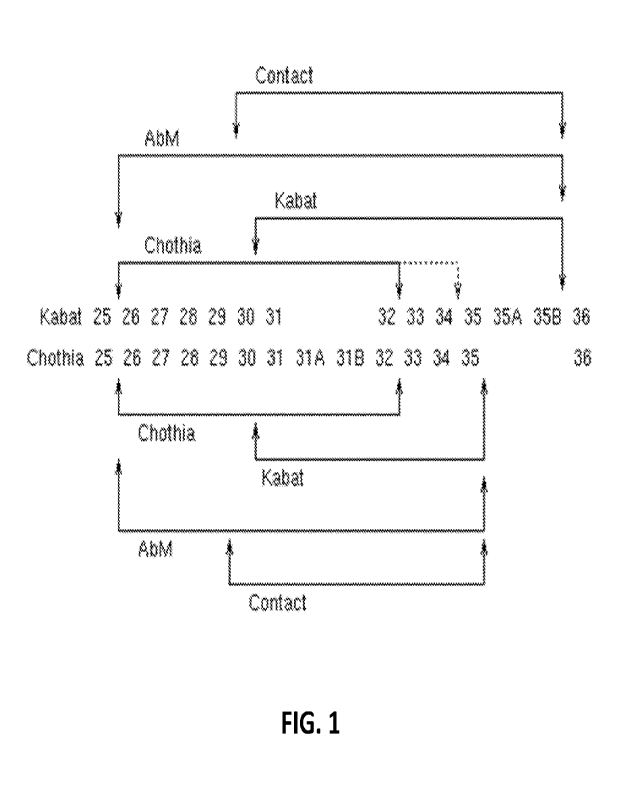
[0017] FIG. 1 provides a comparison of the Kabat and
Chothia numbering systems for
CDR-H1. Adapted from Martin A.C.R. (2010). Protein Sequence and Structure
Analysis of
Antibody Variable Domains. In R. Kontermann & S. Dilbel (Eds.), Antibody
Engineering vol.
2 (pp. 33-51). Springer-Verlag, Berlin Heidelberg.
100181 FIG. 2 is a graph illustrating body weight
changes in mice implanted with ARP-1
multiple myeloma tumors after being administered a single dose of different
BCMA antibody-
drug conjugates as disclosed herein.
CA 03134918 2021- 10- 25
WO 2020/227110
PCT/US2020/031067
[0019] FIGS. 3A and 3B are graphs illustrating tumor
growth curves and tumor size in
mice implanted with ARP-1 multiple myeloma tumors after being administered a
single dose
of different BCMA antibody-drug conjugates as disclosed herein.
[0020] FIG. 4 is a graph illustrating body weight
changes in mice implanted with MM. 1 S
multiple myeloma cells after being administered a single dose of different
BCMA antibody-
drug conjugates as disclosed herein.
[0021] FIG. 5 is a graph illustrating Kaplan-Meier
survival plots in mice implanted with
M114.15 multiple myeloma cells after being administered a single dose of
different BCMA
antibody-drug conjugates as disclosed herein.
[0022] FIG. 6 is a graph illustrating Kaplan-Meier
survival plots in mice implanted with
MM. 1 S multiple myeloma cells after being administered a single dose of a
BCMA antibody-
drug conjugate, Daratumumab, Velcade, or different combinations thereof as
disclosed herein.
[0023] FIGS. 7A-7C are graphs illustrating survival
plots in mice implanted with MM.1 S
multiple myeloma cells after being administered a single dose of a BCMA
antibody-drug
conjugate along with either Daratumumab or Velcade as disclosed herein
[0024] FIGS. 8A and 8B are graphs illustrating a Kaplan-
Meier survival plot and a
survival plot of mice implanted with MM 1S multiple myeloma cells after being
administered
a single dose of a BCMA antibody-drug conjugate at different concentrations as
disclosed
herein.
[0025] FIG. 9 is a graph illustrating body weight
changes in mice implanted with ARP-1
multiple myeloma tumors after being administered a single dose of a BCMA
antibody-drug
conjugate at different doses as disclosed herein.
100261 FIGS. 10A and 10B are graphs illustrating tumor
growth curves and tumor size in
mice implanted with ARP-1 multiple myeloma tumors after being administered a
single dose
of a BCMA antibody-drug conjugate at different doses as disclosed herein.
[0027] FIGS. 11 is a graph illustrating the average DAR
of Conjugate 4 over time in
PBS, human, mouse, and cynomolgus plasma.
[0028] FIG. 12 provides graphs illustrating cell binding
of Conjugate 4 and Conjugate
1 to cells expressing human BCMA, BAFF-R, and TACT receptors.
6
CA 03134918 2021- 10- 25
WO 2020/227110
PCT/US2020/031067
DETAILED DESCRIPTION OF THE EMBODIMENTS
1. Definitions
100291 Unless otherwise defined, all terms of art,
notations and other scientific
terminology used herein are intended to have the meanings commonly understood
by those of
skill in the art to which this invention pertains. In some cases, terms with
commonly understood
meanings are defined herein for clarity and/or for ready reference, and the
inclusion of such
definitions herein should not necessarily be construed to represent a
difference over what is
generally understood in the art The techniques and procedures described or
referenced herein
are generally well understood and commonly employed using conventional
methodologies by
those skilled in the art, such as, for example, the widely utilized molecular
cloning
methodologies described in Green & Sambrook, Molecular Cloning: A Laboratory
Manual 4th
ed. (2012), Cold Spring Harbor Laboratory Press, Cold Spring Harbor, NY; and
Ausubel et al.,
Current Protocols in Molecular Biology, John Wiley & Sons. As appropriate,
procedures
involving the use of commercially available kits and reagents are generally
carried out in
accordance with manufacturer-defined protocols and conditions unless otherwise
noted.
100301 As used herein, the singular forms "a," "an," and
"the" include the plural referents
unless the context clearly indicates otherwise.
00311 The term "about" indicates and encompasses an
indicated value and a range above
and below that value. In certain embodiments, the term "about" indicates the
designated
value 10%, 5%, or 1%. In certain embodiments, the term "about" indicates
the designated
value one standard deviation of that value.
00321 The term "combinations thereof' includes every
possible combination of elements
to which the term refers to. For example, a sentence stating that "if a2 is A,
then a3 is not D; as
is not S; or a6 is not S; or combinations thereof' includes the following
combinations when a2
is A: (1) a3 is not D; (2) as is not S; (3) a6 is not S; (4) a3 is not D; as
is not S; and a6 is not S;
(5)423 is not D and as is not S; (6) a3 is not D and as is not S; and (7) as
is not Sand as is not
S.
00331 The terms "BCMA" and "B-cell maturation antigen"
are used interchangeably
herein. BCMA is also known by synonyms, including BCM, tumor necrosis factor
receptor
7
CA 03134918 2021- 10- 25
WO 2020/227110
PCT/US2020/031067
superfamily member 17 ("TNFRSF17"), CD269, TNFRSF13A, and TNF receptor
supeifamily
member 17, among others. Unless specified otherwise, the terms include any
variants, isoforms
and species homologs of human BCMA that are naturally expressed by cells, or
that are
expressed by cells transfected with a BCMA or BCMA gene. BCMA proteins
include, for
example, human BCMA isoform 1 (SEQ ID NO: 1) and human BCMA isoform 2 (SEQ ID
NO: 2). In some embodiments, BCMA proteins include cynomolgus monkey BCMA (SEQ
ID
NO: 3). hi some embodiments, BCMA proteins include murine BCMA (SEQ ID NO: 4).
100341 The term "immunoglobulin" refers to a class of
structurally related proteins
generally comprising two pairs of polypeptide chains: one pair of light (L)
chains and one pair
of heavy (H) chains. In an "intact immunoglobulin," all four of these chains
are interconnected
by disulfide bonds. The structure of immunoglobulins has been well
characterized. See, e.g.,
Paul, Fundamental Immunology 7th ed., Ch. 5 (2013) Lippincott Williams &
Wilkins,
Philadelphia, PA. Briefly, each heavy chain typically comprises a heavy chain
variable region
(VH or WI) and a heavy chain constant region (CH or CH). The heavy chain
constant region
typically comprises three domains, abbreviated CH1 (or CH1), CH2 (or CH2), and
CH3 (or
CH3). Each light chain typically comprises a light chain variable region (VL
or VL) and a light
chain constant region. The light chain constant region typically comprises one
domain,
abbreviated CL or CL.
100351 The term "antibody" describes a type of
immunoglobulin molecule and is used
herein in its broadest sense. An antibody specifically includes intact
antibodies (e.g., intact
immunoglobulins), and antibody fragments. Antibodies comprise at least one
antigen-binding
domain. One example of an antigen-binding domain is an antigen binding domain
formed by
a VH-VL dimer. A "BCMA antibody," "anti-BCMA antibody," "BCMA Ab," "BCMA-
specific
antibody," "anti-BCMA Ab," "BCMA antibody," "anti-BCMA antibody," "BCMA Ab,"
"BCMA-specific antibody," or "anti-BCMA Ab," or any iteration of these phrases
where
"BCMA" is substituted by "TNFSF17," is an antibody, as described herein, which
binds
specifically to BCMA. In some embodiments, the antibody binds the
extracellular domain of
BCMA.
100361 The VH and VL regions may be further subdivided
into regions of hypervariability
("hypervariable regions (HVRs);" also called "complementarity determining
regions" (CDRs))
interspersed with regions that are more conserved. The more conserved regions
are called
framework regions (FRs). Each VH and VL generally comprises three CDRs and
four FRs,
8
CA 03134918 2021- 10- 25
WO 2020/227110
PCT/US2020/031067
arranged in the following order (from N-terminus to C-terminus): FRI - CDR]: -
FR2 - CDR2
- FR) - CDR3 - FR4. The CDRs are involved in antigen binding, and influence
antigen
specificity and binding affinity of the antibody. See Kabat et at., Sequences
of Proteins of
Immunological Interest 5th ed. (1991) Public Health Service, National
Institutes of Health,
Bethesda, MD, incorporated by reference in its entirety.
100371 The light chain from any vertebrate species can
be assigned to one of two types,
called kappa and lambda, based on the sequence of the constant domain.
100381 The heavy chain from any vertebrate species can
be assigned to one of five
different classes (or isotypes): IgA, IgD, IgE, IgG, and IgM. These classes
are also designated
a, 8, E, y, and rt, respectively. The IgG and IgA classes are further divided
into subclasses on
the basis of differences in sequence and function. Humans express the
following subclasses:
IgGl, IgG2, IgG3, IgG4, IgAl, and IgA2.
100391 The amino acid sequence boundaries of a CDR can
be determined by one of skill
in the art using any of a number of known numbering schemes, including those
described by
Kabat et al., supra ("Kabat" numbering scheme); Al-Lazikani et al., 1997, J.
Mol. Biol.,
273:927-948 ("Chothia" numbering scheme); MacCallum et al., 1996, J. Mot BioL
262:732-
745 ("Contact" numbering scheme); Lefranc et al., Dev. Comp. ImmunoL, 2003,
27:55-77
("[MGT" numbering scheme); and Honegge and Pliickthun, J. illoL BioL, 2001,
309:657-70
("AHo" numbering scheme), each of which is incorporated by reference in its
entirety.
00401 Table 1 provides the positions of CDR-L1, CDR-L2,
CDR-L3, CDR-H1, CDR-
H2, and CDR-H3 as identified by the Kabat and Chothia schemes. For CDR-H1,
residue
numbering is provided using both the Kabat and Chothia numbering schemes.
Table 1. Residues in CDRs according to Kabat and Chothia numbering schemes.
CDR Kabat
Chothia
L1 L24-L34
L24-L34
L2 L50-L56
L50-L56
L3 L89-L97
L89-L97
H1 (Kabat Numbering) H31-1135B
H26-H32 01 1134*
H1 (Chothia Numbering) 1131-1135
1126-H32
H2 1150-1165
1152-H56
113 1495-H102
1495-H102
* The C-terminus of CDR-H1, when numbered using the Kabat numbering
convention, varies
between H32 and H34, depending on the length of the CDR, as illustrated in
FIG. 1.
9
CA 03134918 2021- 10- 25
WO 2020/227110
PCT/US2020/031067
[0041] Unless otherwise specified, the numbering scheme
used for identification of a
particular CDR herein is the Kabat/Chothia numbering scheme. Where the
residues
encompassed by these two numbering schemes diverge (e.g.. CDR-H1 and/or CDR-
H2), the
numbering scheme is specified as either Kabat or Chothia. For convenience, CDR-
H3 is
sometimes referred to herein as either Kabat or Chothia. However, this is not
intended to imply
differences in sequence where they do not exist, and one of skill in the art
can readily confirm
whether the sequences are the same or different by examining the sequences.
[0042] CDRs may be assigned, for example, using antibody
numbering software, such as
Abnum, available at www.bioinf org.uk/abs/abnum/, and described in Abhinandan
and Martin,
Immunology, 2008, 45:3832-3839, incorporated by reference in its entirety.
[0043] The "EU numbering scheme" is generally used when
referring to a residue in an
antibody heavy chain constant region (e.g., as reported in Kabat et al.,
supra). Unless stated
otherwise, the EU numbering scheme is used to refer to residues in antibody
heavy chain
constant regions described herein.
[0044] An "antibody fragment" comprises a portion of an
intact antibody, such as the
antigen binding or variable region of an intact antibody. Antibody fragments
include, for
example, Fv fragments, Fab fragments, F(ab')2 fragments, Fab' fragments, scFv
(sFv)
fragments, and scFv-Fc fragments.
[0045] "Fv" fragments comprise a non-covalently-linked
dimer of one heavy chain
variable domain and one light chain variable domain.
[0046] "Fab" fragments comprise, in addition to the
heavy and light chain variable
domains, the constant domain of the light chain and the first constant domain
(CHI) of the heavy
chain. Fab fragments may be generated, for example, by recombinant methods or
by papain
digestion of a full-length antibody.
[0047] "F(a13)2" fragments contain two Fab' fragments
joined, near the hinge region, by
disulfide bonds. F(ab 1)2 fragments may be generated, for example, by
recombinant methods or
by pepsin digestion of an intact antibody. The F(abl) fragments can be
dissociated, for example,
by treatment with 13-mercaptoethanol.
[0048] "Single-chain Fv" or "sFv" or "scFv" antibody
fragments comprise a Vii domain
and a Vt. domain in a single polypeptide chain. The Vii and VI_ are generally
linked by a peptide
CA 03134918 2021- 10- 25
WO 2020/227110
PCT/US2020/031067
linker. See Phackthun A. (1994). In some embodiments, the linker is SEQ ID NO:
26. hi some
embodiments, the linker is SEQ ID NO: 27. Antibodies from Escherichia colt In
Rosenberg
M. & Moore G. P. (Eds.), The Pharmacology of Monoclonal Antibodies vol. 113
(pp. 269-315).
Springer-Verlag, New York, incorporated by reference in its entirety.
[0049] "scFv-Fc" fragments comprise an scFv attached to
an Fc domain. For example, an
Fc domain may be attached to the C-terminus of the seFv. The Fe domain may
follow the VH
or VL, depending on the orientation of the variable domains in the scFv (i.e.,
VH-Vi, or W-VH).
Any suitable Fc domain known in the art or described herein may be used. In
some cases, the
Fc domain comprises an IgG1 Fc domain. In some embodiments, the IgG1 Fc domain
comprises SEQ ID NO: 19, or a portion thereof SEQ ID NO: 19 provides the
sequence of CHI,
CH2, and CH3 of the human IgGil constant region.
[0050] The term "monoclonal antibody" refers to an
antibody from a population of
substantially homogeneous antibodies. A population of substantially
homogeneous antibodies
comprises antibodies that are substantially similar and that bind the same
epitope(s), except for
variants that may normally arise during production of the monoclonal antibody.
Such variants
are generally present in only minor amounts. A monoclonal antibody is
typically obtained by
a process that includes the selection of a single antibody from a plurality of
antibodies. For
example, the selection process can be the selection of a unique clone from a
plurality of clones,
such as a pool of hybridoma clones, phage clones, yeast clones, bacterial
clones, or other
recombinant DNA clones. The selected antibody can be further altered, for
example, to
improve affinity for the target ("affinity maturation"), to humanize the
antibody, to improve its
production in cell culture, and/or to reduce its immunogenicity in a subject.
[0051] The term "chimeric antibody" refers to an
antibody in which a portion of the heavy
and/or light chain is derived from a particular source or species, while the
remainder of the
heavy and/or light chain is derived from a different source or species.
100521 "Humanized" forms of non-human antibodies are
chimeric antibodies that contain
minimal sequence derived from the non-human antibody. A humanized antibody is
generally
a human immunoglobulin (recipient antibody) in which residues from one or more
CDRs are
replaced by residues from one or more CDRs of a non-human antibody (donor
antibody). The
donor antibody can be any suitable non-human antibody, such as a mouse, rat,
rabbit, chicken,
or non-human primate antibody having a desired specificity, affinity, or
biological effect. In
11
CA 03134918 2021- 10- 25
WO 2020/227110
PCT/US2020/031067
some instances, selected framework region residues of the recipient antibody
are replaced by
the corresponding framework region residues from the donor antibody. Humanized
antibodies
may also comprise residues that are not found in either the recipient antibody
or the donor
antibody. Such modifications may be made to further refine antibody function.
For further
details, see Jones et al., Nature, 1986, 321:522-525; Riechmann et al.,
Nature, 1988, 332:323-
329; and Presta, Curr. Op. Struct. Biol., 1992, 2:593-596, each of which is
incorporated by
reference in its entirety.
100531 A "human antibody" is one which possesses an
amino acid sequence
corresponding to that of an antibody produced by a human or a human cell, or
derived from a
non-human source that utilizes a human antibody repertoire or human antibody-
encoding
sequences (e.g., obtained from human sources or designed de novo). Human
antibodies
specifically exclude humanized antibodies.
[0054] An "isolated antibody" is one that has been
separated and/or recovered from a
component of its natural environment. Components of the natural environment
may include
enzymes, hormones, and other proteinaceous or nonproteinaceous materials. In
some
embodiments, an isolated antibody is purified to a degree sufficient to obtain
at least 15 residues
of N-terminal or internal amino acid sequence, for example by use of a
spinning cup
sequenator. In some embodiments, an isolated antibody is purified to
homogeneity by gel
electrophoresis (e.g., SDS-PAGE) under reducing or nonreducing conditions,
with detection
by Coomassie blue or silver stain. An isolated antibody includes an antibody
in situ within
recombinant cells, since at least one component of the antibody's natural
environment is not
present. In some aspects, an isolated antibody is prepared by at least one
purification step.
[0055] In some embodiments, an isolated antibody is
purified to at least 80%, 85%, 90%,
95%, or 99% by weight. In some embodiments, an isolated antibody is purified
to at least 80%,
85%, 90%, 95%, or 99% by volume. In some embodiments, an isolated antibody is
provided
as a solution comprising at least 85%, 90%, 95%, 98%, 99% to 100% by weight.
In some
embodiments, an isolated antibody is provided as a solution comprising at
least 85%, 90%,
95%, 98%, 99% to 100% by volume.
[0056] "Affinity" refers to the strength of the sum
total of non-covalent interactions
between a single binding site of a molecule (e.g., an antibody) and its
binding partner (e.g., an
antigen). Unless indicated otherwise, as used herein, "binding affinity"
refers to intrinsic
12
CA 03134918 2021- 10- 25
WO 2020/227110
PCT/US2020/031067
binding affinity, which reflects a 1:1 interaction between members of a
binding pair (e.g.,
antibody and antigen). The affinity of a molecule X for its partner Y can be
represented by the
dissociation constant (KD). Affinity can be measured by common methods known
in the art,
including those described herein. Affinity can be determined, for example,
using surface
plasmon resonance (SPR) technology, such as a Biacore instrument. In some
embodiments,
the affinity is determined at 25 C.
[0057] With regard to the binding of an antibody to a
target molecule, the terms "specific
binding," "specifically binds to," "specific for," "selectively binds," and
"selective for" a
particular antigen (e.g., a polypeptide target) or an epitope on a particular
antigen mean binding
that is measurably different from a non-specific or non-selective interaction.
Specific binding
can be measured, for example, by determining binding of a molecule compared to
binding of a
control molecule. Specific binding can also be determined by competition with
a control
molecule that mimics the antibody binding site on the target. In that case,
specific binding is
indicated if the binding of the antibody to the target is competitively
inhibited by the control
molecule.
[0058] The term "IQ" or "kd" (see), as used herein,
refers to the dissociation rate constant
of a particular antibody-antigen interaction. This value is also referred to
as the koff value.
[0059] The term "ka" or "ka" (M-Ixsec-1), as used
herein, refers to the association rate
constant of a particular antibody-antigen interaction. This value is also
referred to as the 6.
value
[0060] The term "KD" (also referred to as "Kd" or "ICD,"
M or nM), as used herein, refers
to the dissociation equilibrium constant of a particular antibody-antigen
interaction. KD = kilka.
The value of KD is typically equal in magnitude to the concentration of ligand
at which half the
protein molecules are bound to ligand at equilibrium.
[0061] The term "KA" or "Ka" (W), as used herein, refers
to the association equilibrium
constant of a particular antibody-antigen interaction. KA = ka/b.
[0062] An "affinity matured" antibody is one with one or
more alterations in one or more
CDRs or FRs that result in an improvement in the affinity of the antibody for
its antigen,
compared to a parent antibody which does not possess the alteration(s). In one
embodiment, an
affinity matured antibody has nanomolar or picomolar affinity for the target
antigen. Affinity
matured antibodies may be produced using a variety of methods known in the
art. For example,
13
CA 03134918 2021- 10- 25
WO 2020/227110
PCT/US2020/031067
Marks et at. (]31o/Technology, 1992, 10:779-783, incorporated by reference in
its entirety)
describes affinity maturation by VH and VI_ domain shuffling. Random
mutagenesis of CDR
and/or framework residues is described by, for example, Barbas et al. (Proc.
Nat Acad. So.
U.S.A., 1994, 91:3809-3813); Schier et al., Gene, 1995, 169:147-155; Yelton et
al., J.
Immunot, 1995, 155:1994-2004; Jackson et at.,
ImmunoL, 1995, 154:3310-33199;
and
Hawkins et al, J. MoL Biot, 1992, 226:889-896, each of which is incorporated
by reference in
its entirety.
100631
When used herein in the
context of two or more antibodies, the term "competes
with" or "cross-competes with" indicates that the two or more antibodies
compete for binding
to an antigen (e.g., BCMA). In one exemplary assay, BCMA is coated on a plate
and allowed
to bind a first antibody, after which a second, labeled antibody is added. If
the presence of the
first antibody reduces binding of the second antibody, then the antibodies
compete. In another
exemplary assay, a first antibody is coated on a plate and allowed to bind the
antigen, and then
the second antibody is added. The term "competes with" also includes
combinations of
antibodies where one antibody reduces binding of another antibody, but where
no competition
is observed when the antibodies are added in the reverse order. However, in
some
embodiments, the first and second antibodies inhibit binding of each other,
regardless of the
order in which they are added. In some embodiments, one antibody reduces
binding of another
antibody to its antigen by at least 50%, at least 60%, at least 70%, at least
80%, or at least 90%.
[0064]
The term "epitope" means a
portion of an antigen capable of specific binding to an
antibody. Epitopes frequently consist of surface-accessible amino acid
residues and/or sugar
side chains and may have specific three dimensional structural
characteristics, as well as
specific charge characteristics. Conformational and non-conformational
epitopes are
distinguished in that the binding to the former but not the latter is lost in
the presence of
denaturing solvents. An epitope may comprise amino acid residues that are
directly involved
in the binding, and other amino acid residues, which are not directly involved
in the binding.
The epitope to which an antibody binds can be determined using known
techniques for epitope
determination such as, for example, testing for antibody binding to variants
of BCMA with
different point-mutations.
100651
Percent "identity" between a
polypeptide sequence and a reference sequence, is
defined as the percentage of amino acid residues in the polypeptide sequence
that are identical
to the amino acid residues in the reference sequence, after aligning the
sequences and
14
CA 03134918 2021- 10- 25
WO 2020/227110
PCT/US2020/031067
introducing gaps, if necessary, to achieve the maximum percent sequence
identity. Alignment
for purposes of determining percent amino acid sequence identity can be
achieved in various
ways that are within the skill in the art, for instance, using publicly
available computer software
such as BLAST, BLAST-2, ALIGN, MEGALIGN (DNASTAR), CLUSTALW, CLUSTAL
OMEGA, or MUSCLE software. Those skilled in the art can determine appropriate
parameters
for aligning sequences, including any algorithms needed to achieve maximal
alignment over
the full length of the sequences being compared.
10111661 A "conservative substitution" or a "conservative
amino acid substitution," refers
to the substitution of an amino acid with a chemically or functionally similar
amino acid.
Conservative substitution tables providing similar amino acids are well known
in the art.
Polypeptide sequences having such substitutions are known as "conservatively
modified
variants." By way of example, the groups of amino acids provided in Tables 2-4
are, in some
embodiments, considered conservative substitutions for one another.
Table 2. Selected groups of amino acids that are considered conservative
substitutions for
one another, in certain embodiments.
kCIdICReSIdueSiD and E
!Basic Residues R,
and H
'
Wydrophilic Uncharged Residues iS, T,
N, and Q
kliphatic Uncharged Residues IG, A,
V. L, and I
NonzeglazUncharKed Residues --------------------------------------- C,M, and
P -----------
Aromatic Residues F, Y,
and W
Alcohol Group-Containing Residues S and T
hatic Residues I, L, V,
and M
yeloalkenyl-ctssociated Residues ---------------------------------------- H,
W, and Y
Wydrophobic Residues A, C, F,
G, H, I, L, M, it, T, V. W, and Y
Negatively Charged Residues D and E
___
rPolar Residues C, D, E, H, K, N, Q, R, S. and T
!Positive' Char ed Residues
and R
;Small Residues ---------------------------------------------------- A, C, D,
G, N, P, S, T, and V
'IVery Small Residues A, G,
and S
Residues Involved in Turn Formation A, C, D,
E, G, H, K, N, Q, R, S, P, and T ------
Flexible Residues Q, T, K,
S. G, P, D, E, and R
CA 03134918 2021- 10- 25
WO 2020/227110
PCT/US2020/031067
Table 3. Additional selected groups of amino acids that are considered
conservative
substitutions for one another, in certain embodiments.
laroup I S,
and T
Group 2 band E -
-----------------
Grou 3 4N4 and
Q
Groy 4 ___________________________________________________________ R and K
[Group 5
IEL,aIIdM
[Group 6 Y,
and W
Table 4. Further selected groups of amino acids that are considered
conservative
substitutions for one another, in certain embodiments.
:Group A and
G --------------
Group B and
E
1Grou C and
Group D -------------------------------------------------------------- 1, K,
and H ------
Group_f_ --------------------------------------------------------------- L, M,
V _____________________
Groy F µr, Y,
and W
[GroupG S and T
Group H r and M
-----------------
[0067] Additional conservative substitutions may be
found, for example, in Creighton,
Proteins: Structures and Molecular Properties 2nd ed. (1993) W. H. Freeman &
Co., New
York, NY. An antibody generated by making one or more conservative
substitutions of amino
acid residues in a parent antibody is referred to as a "conservatively
modified variant."
[0068] The term "amino acid" refers to the twenty common
naturally occurring amino
acids. Naturally occurring amino acids include alanine (Ma; A), arginine (Arg;
R), asparagine
(Asn; N), aspartic acid (Asp; D), cysteine (Cys; C); glutamic acid (Glu; E),
glutamine (Gln;
Q), Glycine (Gly; G); histidine (His; H), isoleucine (Ile; I), leucine (Leu;
L), lysine (Lys; K),
methionine (Met; M), phenylalanine (Phe; F), proline (Pro; P), serine (Ser;
S), threonine (Thr;
T), tryptophan (Tip; W), tyrosine (Tyr; Y), and valine (Val; V).
[0069] Naturally encoded amino acids are the
proteinogenic amino acids known to those
of skill in the art. They include the 20 common amino acids (alanine,
arginine, asparagine,
aspartic acid, cysteine, glutamine, glutamic acid, glycine, histidine,
isoleucine, leucine, lysine,
methionine, phenylalanine, proline, serine, threonine, tryptophan, tyrosine,
and valine) and the
less common pyrrolysine and selenocysteine. Naturally encoded amino acids
include post-
16
CA 03134918 2021- 10- 25
WO 2020/227110
PCT/US2020/031067
translational variants of the 22 naturally occurring amino acids such as
prenylated amino acids,
isoprenylated amino acids, myrisoylated amino acids, palmitoylated amino
acids, N-linked
glycosylated amino acids, 0-linked glycosylated amino acids, phosphorylated
amino acids and
acylated amino acids.
[0070] The term "non-natural amino acid" refers to an
amino acid that is not a
proteinogenic amino acid, or a post-translationally modified variant thereof.
In particular, the
term refers to an amino acid that is not one of the 20 common amino acids or
pyrrolysine or
selenocysteine, or post-translationally modified variants thereof
[0071] The term "conjugate" or "antibody conjugate"
refers to an antibody linked to one
or more payload moieties. The antibody can be any antibody described herein.
The payload
can be any payload described herein. The antibody can be directly linked to
the payload via a
covalent bond, or the antibody can be linked to the payload indirectly via a
linker. Typically,
the linker is covalently bonded to the antibody and also covalently bonded to
the payload. The
term "antibody drug conjugate" or "ADC" refers to a conjugate wherein at least
one payload
is a therapeutic moiety such as a drug.
[0072] The term "payload" refers to a molecular moiety
that can be conjugated to an
antibody. In particular embodiments, payloads are selected from the group
consisting of
therapeutic moieties and labelling moieties.
[0073] The term "linker" refers to a molecular moiety
that is capable of forming at least
two covalent bonds. Typically, a linker is capable of forming at least one
covalent bond to an
antibody and at least another covalent bond to a payload. In certain
embodiments, a linker can
form more than one covalent bond to an antibody. In certain embodiments, a
linker can form
more than one covalent bond to a payload or can form covalent bonds to more
than one payload.
After a linker forms a bond to an antibody, or a payload, or both, the
remaining structure, i.e.
the residue of the linker after one or more covalent bonds are formed, may
still be referred to
as a "linker" herein. The term "linker precursor" refers to a linker having
one or more reactive
groups capable of forming a covalent bond with an antibody or payload, or
both. In some
embodiments, the linker is a cleavable linker. For example, a cleavable linker
can be one that
is released by an bio-labile function, which may or may not be engineered. In
some
embodiments, the linker is a non-cleavable linker. For example, a non-
cleavable linker can be
one that is released upon degradation of the antibody.
17
CA 03134918 2021- 10- 25
WO 2020/227110
PCT/US2020/031067
100741
"Treating" or "treatment" of
any disease or disorder refers, in certain
embodiments, to ameliorating a disease or disorder that exists in a subject.
In another
embodiment, "treating" or "treatment" includes ameliorating at least one
physical parameter,
which may be indiscernible by the subject. In yet another embodiment,
"treating" or
"treatment" includes modulating the disease or disorder, either physically
(e.g., stabilization of
a discernible symptom) or physiologically (e.g., stabilization of a physical
parameter) or both.
In yet another embodiment, "treating" or "treatment" includes delaying or
preventing the onset
of the disease or disorder.
100751
As used herein, the term
"therapeutically effective amount" or "effective amount"
refers to an amount of an antibody or composition that when administered to a
subject is
effective to treat a disease or disorder. In some embodiments, a
therapeutically effective
amount or effective amount refers to an amount of an antibody or composition
that when
administered to a subject is effective to prevent or ameliorate a disease or
the progression of
the disease, or result in amelioration of symptoms.
100761
As used herein, the term
"inhibits growth" (e.g. referring to cells, such as tumor
cells) is intended to include any measurable decrease in cell growth (e.g.,
tumor cell growth)
when contacted with a BCMA antibody, as compared to the growth of the same
cells not in
contact with a BCMA antibody. In some embodiments, growth may be inhibited by
at least
about 10%, 20%, 30%, 40%, 50%, 60%, 70%, 80%, 90%, 99%, or 100%. The decrease
in cell
growth can occur by a variety of mechanisms, including but not limited to
antibody
internalization, apoptosis, necrosis, and/or effector function-mediated
activity.
100771
As used herein, the term
"subject" means a mammalian subject. Exemplary
subjects include, but are not limited to humans, monkeys, dogs, cats, mice,
rats, cows, horses,
camels, avians, goats, and sheep. In certain embodiments, the subject is a
human. In some
embodiments, the subject has a disease that can be treated or diagnosed with
an antibody
provided herein. In some embodiments, the disease is leukemia, lymphoma, or
multiple
myeloma, a plasmacytoid dendritic cell tumor, a B-cell lineage malignancy, a
plasma cell
neoplasm, diffuse large B-cell lymophoma (DLBCL), a low-grade B-cell lymphoma,
Burkitt's
lymphoma, a plasmablastic lymphoma, or a follicular lymphoma.
100781
In some chemical structures
illustrated herein, certain substituents, chemical
groups, and atoms are depicted with a curvy/wavy line (e.g.,
) that intersects a bond or
18
CA 03134918 2021- 10- 25
WO 2020/227110
PCT/US2020/031067
bonds to indicate the atom through which the substituents, chemical groups,
and atoms are
REG
\C tO
bonded. For example, in some structures, such as but not limited to
0
N:irk
HNS/
H2N
, or
, this curvy/wavy line
indicates the atoms in the
backbone of a conjugate or linker-payload structure to which the illustrated
chemical entity is
N.N *
1101
bonded. In some structures, such as but not limited to
, this curvy/wavy line
indicates the atoms in the antibody or antibody fragment as well as the atoms
in the backbone
of a conjugate or linker-payload structure to which the illustrated chemical
entity is bonded.
100791
The term "site-specific"
refers to a modification of a polypeptide at a
predetermined sequence location in the polypeptide. The modification is at a
single, predictable
residue of the polypeptide with little or no variation. In particular
embodiments, a modified
amino acid is introduced at that sequence location, for instance recombinantly
or synthetically.
Similarly, a moiety can be "site-specifically" linked to a residue at a
particular sequence
location in the polypeptide. In certain embodiments, a polypeptide can
comprise more than one
site-specific modification.
2. Conjugates
100801
Provided herein are conjugates
of antibodies to BCMA. The conjugates comprise
an antibody to BCMA covalently linked via a linker to a payload. In certain
embodiments, the
antibody is linked to one payload. In further embodiments, the antibody is
linked to more than
one payload. In certain embodiments, the antibody is linked to two, three,
four, five, six, seven,
eight, or more payloads.
19
CA 03134918 2021- 10- 25
WO 2020/227110
PCT/US2020/031067
100811 In the conjugates provided herein, the antibody
can be from any species. In certain
embodiments, the BCMA is a vertebrate BCMA. In certain embodiments, the BCMA
is a
mammalian BCMA. In certain embodiments, the BCMA is human BCMA. In certain
embodiments, the BCMA is mouse BCMA. In certain embodiments, the BCMA is
cynomolgus
BCMA.
100821 The antibody is typically a protein comprising
multiple polypeptide chains. In
certain embodiments, the antibody is a heterotetramer comprising two identical
light (L) chains
and two identical heavy (H) chains. Each light chain can be linked to a heavy
chain by one
covalent disulfide bond. Each heavy chain can be linked to the other heavy
chain by one or
more covalent disulfide bonds. Each heavy chain and each light chain can also
have one or
more intrachain disulfide bonds As is known to those of skill in the art, each
heavy chain
typically comprises a variable domain (VH) followed by a number of constant
domains. Each
light chain typically comprises a variable domain at one end (VL) and a
constant domain. As is
known to those of skill in the art, antibodies typically have selective
affinity for their target
molecules, i.e. antigen&
100831 The antibodies provided herein can have any
antibody form known to those of skill
in the art. They can be full-length, or fragments. Exemplary full length
antibodies include IgA,
IgAl, IgA2, IgD, IgE, IgG, IgGl, IgG2, IgG3, IgG4, IgNI, etc. Exemplary
fragments include
Fv, Fab, Fc, scFv, scFv-Fc, etc.
100841 In certain embodiments, the antibody of the
conjugate comprises six of the CDR
sequences described herein. In certain embodiments, the antibody of the
conjugate comprises
a heavy chain variable domain (VH) described herein. In certain embodiments,
the antibody of
the conjugate comprises a light chain variable domain (VL) described herein.
In certain
embodiments, the antibody of the conjugate comprises a heavy chain variable
domain (VH)
described herein and a light chain variable domain (VL) described herein. In
certain
embodiments, the antibody of the conjugate comprises a paired heavy chain
variable domain
and a light chain variable domain described herein (VH - VL pair).
100851 In certain embodiments, the antibody conjugate
can be formed from an antibody
that comprises one or more reactive groups. In certain embodiments, the
antibody conjugate
can be formed from an antibody comprising all naturally encoded amino acids.
Those of skill
in the art will recognize that several naturally encoded amino acids include
reactive groups
CA 03134918 2021- 10- 25
WO 2020/227110
PCT/US2020/031067
capable of conjugation to a payload or to a linker. These reactive groups
include cysteine side
chains, lysine side chains, and amino-terminal groups. In these embodiments,
the antibody
conjugate can comprise a payload or linker linked to the residue of an
antibody reactive group.
In these embodiments, the payload precursor or linker precursor comprises a
reactive group
capable of forming a bond with an antibody reactive group. Typical reactive
groups include
maleimide groups, activated carbonates (including but not limited to, p-
nitrophenyl ester),
activated esters (including but not limited to, N-hydroxysuccinimide, p-
nitrophenyl ester, and
aldehydes). Particularly useful reactive groups include maleimide and
succinimide, for instance
N-hydroxysuccinimide, for forming bonds to cysteine and lysine side chains.
Further reactive
groups are described in the sections and examples below.
[0086] In further embodiments, the antibody comprises
one or more modified amino acids
having a reactive group, as described herein. Typically, the modified amino
acid is not a
naturally encoded amino acid. These modified amino acids can comprise a
reactive group
useful for forming a covalent bond to a linker precursor or to a payload
precursor. One of skill
in the art can use the reactive group to link the polypeptide to any molecular
entity capable of
forming a covalent bond to the modified amino acid. Thus, provided herein are
conjugates
comprising an antibody comprising a modified amino acid residue linked to a
payload directly
or indirectly via a linker. Exemplary modified amino acids are described in
the sections below.
Generally, the modified amino acids have reactive groups capable of forming
bonds to linkers
or payloads with complementary reactive groups.
[0087] In certain embodiments, the non-natural amino
acids are positioned at select
locations in a polypeptide chain of the antibody. These locations were
identified as providing
optimum sites for substitution with the non-natural amino acids. Each site is
capable of bearing
a non-natural amino acid with optimum structure, function and/or methods for
producing the
antibody.
[0088] In certain embodiments, a site-specific position
for substitution provides an
antibody that is stable. Stability can be measured by any technique apparent
to those of skill in
the art.
[0089] In certain embodiments, a site-specific position
for substitution provides an
antibody that has optimal functional properties. For instance, the antibody
can show little or no
loss of binding affinity for its target antigen compared to an antibody
without the site-specific
21
CA 03134918 2021- 10- 25
WO 2020/227110
PCT/US2020/031067
non-natural amino acid. In certain embodiments, the antibody can show enhanced
binding
compared to an antibody without the site-specific non-natural amino acid.
[0090] In certain embodiments, a site-specific position
for substitution provides an
antibody that can be made advantageously. For instance, in certain
embodiments, the antibody
shows advantageous properties in its methods of synthesis, discussed below. In
certain
embodiments, the antibody can show little or no loss in yield in production
compared to an
antibody without the site-specific non-natural amino acid. In certain
embodiments, the antibody
can show enhanced yield in production compared to an antibody without the site-
specific non-
natural amino acid. In certain embodiments, the antibody can show little or no
loss of tRNA
suppression compared to an antibody without the site-specific non-natural
amino acid. In
certain embodiments, the antibody can show enhanced tRNA suppression in
production
compared to an antibody without the site-specific non-natural amino acid.
100911 In certain embodiments, a site-specific position
for substitution provides an
antibody that has advantageous solubility. In certain embodiments, the
antibody can show little
or no loss in solubility compared to an antibody without the site-specific non-
natural amino
acid. In certain embodiments, the antibody can show enhanced solubility
compared to an
antibody without the site-specific non-natural amino acid.
[0092] In certain embodiments, a site-specific position
for substitution provides an
antibody that has advantageous expression. In certain embodiments, the
antibody can show
little or no loss in expression compared to an antibody without the site-
specific non-natural
amino acid. In certain embodiments, the antibody can show enhanced expression
compared to
an antibody without the site-specific non-natural amino acid.
[0093] In certain embodiments, a site-specific position
for substitution provides an
antibody that has advantageous folding. In certain embodiments, the antibody
can show little
or no loss in proper folding compared to an antibody without the site-specific
non-natural
amino acid. In certain embodiments, the antibody can show enhanced folding
compared to an
antibody without the site-specific non-natural amino acid.
[0094] In certain embodiments, a site-specific position
for substitution provides an
antibody that is capable of advantageous conjugation. As described below,
several non-natural
amino acids have side chains or functional groups that facilitate conjugation
of the antibody to
a second agent, either directly or via a linker. In certain embodiments, the
antibody can show
22
CA 03134918 2021- 10- 25
WO 2020/227110
PCT/US2020/031067
enhanced conjugation efficiency compared to an antibody without the same or
other non-
natural amino acids at other positions. In certain embodiments, the antibody
can show enhanced
conjugation yield compared to an antibody without the same or other non-
natural amino acids
at other positions. In certain embodiments, the antibody can show enhanced
conjugation
specificity compared to an antibody without the same or other non-natural
amino acids at other
positions.
[0095] The one or more non-natural amino acids are
located at selected site-specific
positions in at least one polypeptide chain of the antibody. The polypeptide
chain can be any
polypeptide chain of the antibody without limitation, including either light
chain or either
heavy chain. The site-specific position can be in any domain of the antibody,
including any
variable domain and any constant domain.
[0096] In certain embodiments, the antibodies provided
herein comprise one non-natural
amino acid at a site-specific position. In certain embodiments, the antibodies
provided herein
comprise two non-natural amino acids at site-specific positions. In certain
embodiments, the
antibodies provided herein comprise three non-natural amino acids at site-
specific positions. In
certain embodiments, the antibodies provided herein comprise more than three
non-natural
amino acids at site-specific positions.
[0097] In certain embodiments, the antibodies provided
herein comprise non-natural
amino acids each at the positions HC-F404 and HC-Y180, according to the Kabat
or Chothia
or EU numbering scheme, or a post-translationally modified variant thereof. In
these
designations, HC indicates a heavy chain residue, and LC indicates a light
chain residue. Those
of skill will recognize tht the non-natural amino acids substitute for the
residues HC-F404 and
HC-Y180 in the antibody amino acid sequence. In certain embodiments, the non-
natural amino
acids are residues of Formula (30), herein.
3. Conjugating Groups ant/Residues Thereof
[0098] Conjugating groups facilitate conjugation of the
payloads described herein to a
second compound, such as an antibody described herein. In certain embodiments,
the
conjugating group is designated R herein. Conjugating groups can react via any
suitable
reaction mechanism known to those of skill in the art. In certain embodiments,
a conjugating
group reacts through a [3+2] alkyne-azide cycloaddition reaction, inverse-
electron demand
Diels-Alder ligation reaction, thiol-electrophile reaction, or carbonyl-
oxyamine reaction, as
23
CA 03134918 2021- 10- 25
WO 2020/227110
PCT/US2020/031067
described in detail herein. In certain embodiments, the conjugating group
comprises an alkyne,
for instance a strained alkyne. In certain embodiments, the conjugating group
is:
*
11
---/
1111
. Additional conjugating
groups are described in, for example, U.S. Patent
Publication No, 2014/0356385, U.S. Patent Publication No, 2013/0189287, U.S.
Patent
Publication No, 2013/0251783, U.S. Patent No, 8,703,936, U.S. Patent No.
9,145,361, U.S.
Patent No. 9,222,940, and U.S. Patent No. 8,431,558.
100991
After conjugation, a divalent
residue of the conjugating group is formed and is
bonded to the residue of a second compound. The structure of the divalent
residue is determined
by the type of conjugation reaction employed to form the conjugate.
1001001
In certain embodiments when a
conjugate is formed through a [3+2] alkyne-azide
cycloaddition reaction, the divalent residue of the conjugating group
comprises a triazole ring
or fused cyclic group comprising a triazole ring. In certain embodiments when
a conjugate is
formed through a strain-promoted [3+2] alkyne-azide cycloaddition (SPAAC)
reaction, the
divalent residue of the conjugating group is:
N *
T *
Nc=
N'
h / -1
ri / 1
aland/or . .
1001011
In an embodiment, provided
herein is a conjugate according to any of Formulas
101a-1056, where COMP indicates a residue of the anti-BCMA antibody and PAY
indicates
the payload moiety:
24
CA 03134918 2021- 10- 25
WO 2020/227110
PCT/US2020/031067
COMP-4 i
0
0 0
. =
N.A....,eThrN.,..õ)Cce./"Nor.õ,õ.0õ,.../....wk.,,--=====-,..ApAy
I
I
(105a)
COMP
N-Nt ___
=
NI Agit
a
1111 = 0
0 0
I
I
(105b).
[00102] In any of the foregoing embodiments, the
conjugate comprises n number of PAY
moieties, wherein n is an integer from 1 to S. In some embodiments, n is 2. In
some
embodiments, n is 3. hi some embodiments, n is 4. In some embodiments, n is 5.
In some
embodiments, n is 6. In some embodiments, n is 7. In some embodiments, n is 8.
[00103] In particular embodiments, provided herein are
anti-BCMA conjugates according
to any of Formulas 105a-105b wherein COMP indicates a residue of the non-
natural amino
acid according to Formula (30), below. In particular embodiments, provided
herein are
anti-BCMA conjugates according to any of Formulas 105a-105b wherein COMP
indicates a
residue of the non-natural amino acid according to Formula (30), below, at
heavy chain
position 404 according to the EU numbering system. In particular embodiments,
provided
herein are anti-BCMA conjugates according to any of Formulas 105a-105b wherein
COMP
indicates a residue of the non-natural amino acid according to Formula (30),
below, at heavy
chain position 180 according to the EU numbering system.
N3
OS
H' _
F1H2
(30)
Those of skill will recognize that amino acids such as Formula (30) are
incorporated into
polypeptides and antibodies as residues. For instance, a residue of Formula
(30) can be
according to the following Formula:
CA 03134918 2021- 10- 25
WO 2020/227110
PCT/US2020/031067
N3
0*
atC0 .
HFI,
7
(30')
Further modification, for instance at -N3 is also encompassed within the term
residue herein.
1001041 In an embodiment, provided herein is a conjugate according to any of
Formulas
105c-105d, where COMP indicates a residue of the anti-BCMA antibody and PAY
indicates
the payload moiety:
N=N
COMP¨N
c
..,1
r.lrõ (105c)
COMP
N¨Nr
NO 41
PAY
(105d).
[00105] In any of the foregoing embodiments, the conjugate comprises n number
of PAY
moieties, wherein n is an integer from 1 to 8. In some embodiments, n is 2. In
some
embodiments, n is 3. hi some embodiments, n is 4. In some embodiments, n is 5.
In some
embodiments, n is 6. In some embodiments, n 1s7. In some embodiments, n is 8.
[00106] Iii particular embodiments, provided herein are anti-BCMA conjugates
according
to any of Formulas 105c-105d wherein COMP indicates a residue of the non-
natural amino
acid according to Formula (30), below. In particular embodiments, provided
herein are
anti-BCMA conjugates according to any of Formulas 105c-105d wherein COMP
indicates a
residue of the non-natural amino acid according to Formula (30), below, at
heavy chain
position 404 according to the EU numbering system. In particular embodiments,
provided
herein are anti-BCMA conjugates according to any of Formulas 105c-105d wherein
COMP
26
CA 03134918 2021- 10- 25
WO 2020/227110
PCT/US2020/031067
indicates a residue of the non-natural amino acid according to Formula (30),
below, at heavy
chain position 180 according to the EU numbering system.
N3
OS
H = _
EIH2
(30)
Those of skill will recognize that amino acids such as Formula (30) are
incorporated into
polypeptides and antibodies as residues. For instance, a residue of Formula
(30) can be
according to the following Formula:
N3
0*
'Co.
y
(30')
Further modification, for instance at -N3 is also encompassed within the term
residue herein.
1001071 In particular embodiments, provided herein are
anti-BCMA conjugates having the
structure of Conjugate M:
=iskr\[\11,
o
=
141H
'
_
I Ag CI 'o OH
H3C0
=cH3
_ n
where n is an integer from 1 to 6. In some embodiments, n is an integer from 1
to 4. In some
embodiments, n is 2. For example, in particualr embodiments, the anti-BCMA
conjugate has
the structure:
27
CA 03134918 2021- 10- 25
WO 2020/227110
PCT/US2020/031067
Too
00
0 0 =
NH
thl
CI
H3C0 api
= CH3
¨2
1001081 In some embodiments, n is 4. For example, in
particular embodiments, the anti-
BCMA conjugate has the structure:
(Ct
o
= o_ro
NH
'0H
CI
H3C0
CH3
---
.
¨4
1001091 In any of the foregoing embodiments wherein the
anti-BCMA conjugate has a
structure according to Conjugate M, the bracketed structure can be covalently
bonded to one
or more non-natural amino acids of the antibody at sites HC-F404 and HC-Yl 80,
according to
the Kabat or EU numbering scheme of Kabat. In particular embodiments, each non-
natural
amino acid is a residue according to Formula (30).
1001101 In one embodiment, the anti-BCMA conjugate is
Conjugate 4, having the
structure of:
1 0 0
414S. 1. 0 0
0 0 =
TH
CI
= H
Mir
H3C0 = H3
¨4
,or
28
CA 03134918 2021- 10- 25
WO 2020/227110
PCT/US2020/031067
r[
C N .
Nit
r---
+'N a
Mir 0 0 0
y,õ
H
I I = !
1/1/41H
CI ot 7:: -
H3co gas ..., bcH3
¨4
,
wherein the antibody comprises a heavy chain sequence provided in SEQ ID NO:
15, and a
light chain sequence provided in SEQ ID NO: 17;
wherein the antibody further comprises residues of p-azidomethyl-phenylalanine
substituting
at each of sites HC-F404 and HC-Y180 according to the EU numbering scheme; and
each structure within the brackets of the formulas is bonded to the antibody
at one of the p-
azidomethyl-phenylalanine residues.
1001111 In one embodiment, the anti-BCMA conjugate is
Conjugate 4, wherein the
predominant species is:
Ist- Nr i i, o t o
o 0 7N-L-----c.--------g-------0-------Q-----N-L--------1-N-Ixo:
H
I I 0 10FI
H3C0
14,.... exH3
¨4
, or
N
tle' 0,0 ¨
I 1.
N
1/11-1
CI
H3C0 -
bCH3
¨4
,
wherein the antibody comprises a heavy chain sequence provided in SEQ ID NO:
15, and a
light chain sequence provided in SEQ ID NO: 17;
wherein the antibody further comprises residues of p-azidomethyl-phenylalanine
substituting
at each of sites HC-F404 and HC-Y180 according to the EU numbering scheme; and
29
CA 03134918 2021- 10- 25
WO 2020/227110
PCT/US2020/031067
each structure within the brackets of the formulas is bonded to the antibody
at one of the p-
azidomethyl-phenylalanine residues.
[00112] In one embodiment, the anti-BCMA conjugate is
Conjugate 4, wherein the
predominant species is:
0 0
0 0
NI
ri
E
2 00
C H
I I -
14H
ci 0= t"
TI
H3C0 NI, tcH3
WI ---
-4
,
wherein the antibody comprises a heavy chain sequence provided in SEQ ID NO:
15, and a
light chain sequence provided in SEQ 1D NO: 17;
wherein the antibody further comprises residues of p-azidomethyl-phenylalanine
substituting
at each of sites HC-F404 and HC-Y180 according to the EU numbering scheme; and
each structure within the brackets of the formulas is bonded to the antibody
at one of the p-
azidomethyl-phenylalanine residues.
[00113] In one embodiment, the anti-BCMA conjugate is
Conjugate 4, wherein the
predominant species is:
_
1 'r Nil . j'Cr.1)0
c
L
cl,Ni )))0yõ,.. 7 0T:
t
b1 as
.1
lir H I I ci 0 %OH
H3C0 isiõ ...,.
'Cl-I3
IP
-----
-4
,
wherein the antibody comprises a heavy chain sequence provided in SEQ ID NO:
15, and a
light chain sequence provided in SEQ ID NO: 17;
wherein the antibody further comprises residues of p-azidomethyl-phenylalanine
substituting
at each of sites HC-F404 and HC-Y180 according to the EU numbering scheme; and
each structure within the brackets of the formulas is bonded to the antibody
at one of the p-
azidomethyl-phenylalanine residues.
CA 03134918 2021- 10- 25
WO 2020/227110
PCT/US2020/031067
4. Antibody Speccity
[00114] The conjugates comprise antibodies that
selectively bind human BCMA. In some
aspects, the antibody selectively binds to the extracellular domain of human
BCMA (human
BCMA).
[00115] In some embodiments, the antibody binds to a
homolog of human BCMA. In some
aspects, the antibody binds to a homolog of human BCMA from a species selected
from
monkeys, mice, dogs, cats, rats, cows, horses, goats and sheep. In some
aspects, the homolog
is a cynomolgus monkey homolog. In some aspects, the homolog is a mouse or
murine
homolog.
[00116] In some embodiments, the antibody comprises a
light chain. In some aspects, the
light chain is a kappa light chain. In some aspects, the light chain is a
lambda light chain. In
specific embodiments, the kappa light chain comprises a constant region
comprising the amino
acid sequence provided SEQ ID NO: 20.
1001171 In some embodiments, the antibody comprises a
heavy chain. In some aspects, the
heavy chain is an IgA. In some aspects, the heavy chain is an IgD. In some
aspects, the heavy
chain is an IgE. In some aspects, the heavy chain is an IgG. In some aspects,
the heavy chain
is an Ig.M. In some aspects, the heavy chain is an IgGl. In some aspects, the
heavy chain is an
IgG2. In some aspects, the heavy chain is an IgG3. In some aspects, the heavy
chain is an IgG4.
In some aspects, the heavy chain is an IgAl . In some aspects, the heavy chain
is an IgA2.
1001181 In some embodiments, the antibody is an antibody
fragment. In some aspects, the
antibody fragment is an Fv fragment. In some aspects, the antibody fragment is
a Fab fragment.
In some aspects, the antibody fragment is a F(abr)2 fragment. In some aspects,
the antibody
fragment is a Fab' fragment. In some aspects, the antibody fragment is an scFv
(sFv) fragment.
In some aspects, the antibody fragment is an scFv-Fc fragment.
[00119] In some embodiments, the antibody is a monoclonal
antibody. In some
embodiments, the antibody is a polyclonal antibody.
[00120] In some embodiments, the antibody is a chimeric
antibody. In some embodiments,
the antibody is a humanized antibody. In some embodiments, the antibody is a
human antibody.
31
CA 03134918 2021- 10- 25
WO 2020/227110
PCT/US2020/031067
[00121] In some embodiments, the antibody is an affinity
matured antibody. In some
aspects, the antibody is an affinity matured antibody derived from an
illustrative sequence
provided in this disclosure.
[00122] The antibody conjugates provided herein may be
useful for the treatment of a
variety of diseases and conditions including cancers. In some embodiments, the
antibody
conjugates provided herein may be useful for the treatment of cancers of solid
tumors. For
example, the antibody conjugates provided herein can be useful for the
treatment of colorectal
cancer.
[00123] In some embodiments, the antibody comprises,
consists of, or consists essentially
of a VH sequence provided in SEQ ID NO: 13. In some embodiments, the antibody
comprises,
consists of, or consists essentially of a VL sequence provided in SEQ ID NO:
14. In some
embodiments, the antibody comprises a VH sequence and a VL sequence. In some
aspects, the
Vii sequence is a Vii sequence comprising, consisting of, or consisting
essentially of any one
of SEQ ID NO: 13, and the VL sequence is a VL sequence comprising, consisting
of, or
consisting essentially of any one of SEQ ID NO: 14. In certain embodiments,
the antibody
comprises, consists of, or consists essentially of, a heavy chain sequence
provided in SEQ ID
NO: 15. In a specific embodiments, the heavy chain sequence, e.g., heavy chain
sequence
provided in SEQ ID NO: 15, additionally comprises an N-terminal methionine. An
certain
embodiments, such heavy chain sequence is encoded by the nucleotide sequence
provided in
SEQ 113 NO: 16. In certain embodiments, the antibody comprises, consists of,
or consists
essentially of, a light chain sequence provided in SEQ ID NO: 17. In a
specific embodiments,
the light chain sequence, e.g., light chain sequence provided in SEQ ID NO:
17, additionally
comprises an N-terminal methionine_ An certain embodiments, such light chain
sequence is
encoded by the nucleotide sequence provided in SEQ ID NO: 18.
[00124] In some embodiments, the antibodies comprise six
of the CDRs indicated in Table
below. In particular embodiments, Chothia CDRs are selected. In particular
embodiments,
Kabat CDRs are selected.
32
CA 03134918 2021- 10- 25
WO 2020/227110
PCT/US2020/031067
[00125] Table 5. Antibody 2265-F02 CDRs.
Kaba
ICabat
Chothia CDR Chothia CDR
CDR 111 CDR 112 CDR CDR CDR CDR
111 SEQ 112 SEO 113 Li L2 L3
SEQ ID ID SEQ ID ID SEQ SEQ SEQ SEQ
NO NO NO NO ID
NO ID NO ID NO ID NO
2265-1102 5 6 7 8
9 10 11 12
[00126] In some embodiments, the antibody comprises three
of: a CDR-H1 comprising one
of SEQ ID NOs: 5 and 6; a CDR-H2 comprising one of SEQ ID NOs: 7 and 8; a CDR-
H3
comprising SEQ ID NO: 9; and one, two, or all three of: a CDR-L1 comprising
SEQ ID NO:
10; a CDR-L2 comprising SEQ ID NO: 11; and a CDR-L3 comprising SEQ ID NO: 12.
In
particular embodiments, the CDRs are according to Chothia. In particular
embodiments, the
CDRs are according to Kabat.
5. Germline
[00127] In some embodiments, the antibody that
specifically binds BCMA is an antibody
comprising a variable region that is encoded by a particular germline gene, or
a variant thereof
The illustrative antibodies provided herein comprise variable regions that are
encoded by the
heavy chain variable region germline genes VH1-18, V113-33, VH2-5, VH2-70, and
VH4-30-
4. or variants thereof; and the light chain variable region germline genes VKI-
5, VK3-I 1, VK2-
20, VK1-33, and VK1-16, or variants thereof
[00128] One of skill in the art would recognize that the
CDR sequences provided herein
may also be useful when combined with variable regions encoded by other
variable region
germline genes, or variants thereof. In particular, the CDR sequences provided
herein may be
useful when combined with variable regions encoded by variable region germline
genes, or
variants thereof, that are structurally similar to the variable region
germline genes recited
above. For example, in some embodiments, a CDR-H sequence provided herein may
be
combined with a variable region encoded by a variable region germline gene
selected from the
Vii 1, VH 2, VH 3, or VH 4 families, or a variant thereof In some embodiments,
a CDR-L
sequence provided herein may be combined with a variable region encoded by a
variable region
germline gene selected from the Vkl, VK2, or VK3, or a variant thereof
33
CA 03134918 2021- 10- 25
WO 2020/227110
PCT/US2020/031067
6 Glycosylation Variants
[00129] In certain embodiments, an antibody may be
altered to increase, decrease or
eliminate the extent to which it is glycosylated. Glycosylation of
polypeptides is typically either
"N-linked" or "0-linked."
[00130] "N-linked" glycosylation refers to the attachment
of a carbohydrate moiety to the
side chain of an asparagine residue. The tripeptide sequences asparagine-X-
serine and
asparagine-X-threonine, where X is any amino acid except proline, are the
recognition
sequences for enzymatic attachment of the carbohydrate moiety to the
asparagine side chain.
Thus, the presence of either of these tripeptide sequences in a polypeptide
creates a potential
glycosylation site.
[00131] "0-linked" glycosylation refers to the attachment
of one of the sugars
N-acetylgalactosamine, galactose, or xylose to a hydroxyamino acid, most
commonly serine or
threonine, although 5-hydroxyproline or 5-hydroxylysine may also be used.
[00132] Addition or deletion of N-linked glycosylation
sites to the antibody may be
accomplished by altering the amino acid sequence such that one or more of the
above-described
tripeptide sequences is created or removed. Addition or deletion of 0-linked
glycosylation sites
may be accomplished by addition, deletion, or substitution of one or more
serine or threonine
residues in or to (as the case may be) the sequence of an antibody.
7. Fc Variants
[00133] In certain embodiments, amino acid modifications
may be introduced into the Fc
region of an antibody provided herein to generate an Fc region variant. In
certain embodiments,
the Fc region variant possesses some, but not all, effector functions. Such
antibodies may be
useful, for example, in applications in which the half-life of the antibody in
vivo is important,
yet certain effector functions are unnecessary or deleterious. Examples of
effector functions
include complement-dependent cytotoxicity (CDC) and antibody-directed
complement-
mediated cytotoxicity (ADCC). Numerous substitutions or substitutions or
deletions with
altered effector function are known in the art.
[00134] In some embodiments, the Fc comprises one or more
modifications in at least one
of the CH3 sequences. In some embodiments, the Fe comprises one or more
modifications in at
least one of the CH2 sequences. For example, the Fc can include one or
modifications selected
from the group consisting of: V262E, V262D, V262K, V262R, V2625, V2645, V303R,
and
34
CA 03134918 2021- 10- 25
WO 2020/227110
PCT/US2020/031067
V305R. In some embodiments, an Fc is a single polypeptide. In some
embodiments, an Fc is
multiple peptides, e.g., two polypeptides. Exemplary modifications in the Fc
region are
described, for example, in International Patent Application No.
PCT/US2017/037545, filed
June 14, 2017.
1001351 An alteration in in CDC and/or ADCC activity can
be confirmed using in vitro
and/or in vivo assays. For example, Fc receptor (FcR) binding assays can be
conducted to
measure FcyR binding. The primary cells for mediating ADCC, NK cells, express
FcyRM only,
whereas monocytes express FcyRI, FcyRII and FcyRIII. FcR expression on
hematopoietic cells
is summarized in Ravetch and Kinet, Ann. Rev. Immunot, 1991, 9:457-492,
incorporated by
reference in its entirety.
1001361 Non-limiting examples of in vitro assays to
assess ADCC activity of a molecule of
interest are provided in U.S. Patent Nos. 5,500,362 and 5,821,337; Hellstrom
et al., Proc. Natl.
Acted Sc!. U.S.A., 1986, 83:7059-7063; Hellstrom et al., Proc. Natl. Acad.
Sc!. USA., 1985,
82:1499-1502; and Bruggemann et al., J Exp. Med., 1987, 166:1351-1361; each of
which is
incorporated by reference in its entirety. Useful effector cells for such
assays include peripheral
blood mononuclear cells (PBMC) and Natural Killer (NK) cells. Alternatively,
or additionally,
ADCC activity of the molecule of interest may be assessed in vivo, using an
animal model such
as that disclosed in Clynes et al. Proc. Natl. Acad. Sc!. U.S.A., 1998, 95:652-
656, incorporated
by reference in its entirety.
[00137] Clq binding assays may also be carried out to
confirm that the antibody is unable
to bind Clq and hence lacks CDC activity. Examples of C 1 q binding assays
include those
described in WO 2006/029879 and WO 2005/100402, each of which is incorporated
by
reference in its entirety.
[00138] Complement activation assays include those
described, for example, in
Gazzano-Santoro et al., .1. Inanunot Methods, 1996, 202:163-171; Cragg et al.,
Blood, 2003,
101:1045-1052; and Cragg and Glennie, Blood, 2004, 103:2738-2743; each of
which is
incorporated by reference in its entirety.
[00139] FcRn binding and in vivo clearance (half-life
determination) can also be measured,
for example, using the methods described in Petkova et al., Intl. Immunol.,
2006, 18:1759-
1769, incorporated by reference in its entirety.
CA 03134918 2021- 10- 25
WO 2020/227110
PCT/US2020/031067
8. Modified Amino Acids
[00140] When the antibody conjugate comprises a modified
amino acid, the modified
amino acid can be any modified amino acid deemed suitable by the practitioner.
In particular
embodiments, the modified amino acid is p-azido-methyl-L-phenylalanine (also
referred to as
p-methylazido phenylalanine). In particular embodiments, the non-natural amino
acid is
compound (30):
N3
1.1
He _
I9112
(30);
or a salt thereof Such non-natural amino acids may be in the form of a salt.
It will be
understood by one of ordinary skill in the art that the azido moiety of the p-
azido-methyl-L-
phenylalanine residue reacts with a conjugating group to form the triazole of
the fused cyclic
group formed through the strain-promoted [3+2] alkyne-azide cycloaddition
reaction used to
make certain of the conjugates described herein.
9. Preparation of Antibody Conjugates
9.1. Antigen Preparation
[00141] The BCMA protein to be used for isolation of the
antibodies may be intact BCMA
or a fragment of BCMA. The intact BCMA protein, or fragment of BCMA, may be in
the form
of an isolated protein or protein expressed by a cell. Other forms of BCMA
useful for
generating antibodies will be apparent to those skilled in the art.
9.2. Monoclonal Antibodies
[00142] Monoclonal antibodies may be obtained, for
example, using the hybridoma method
first described by Kohler et al., Nature, 1975, 256:495-497 (incorporated by
reference in its
entirety), and/or by recombinant DNA methods (see e.g., U.S. Patent No.
4,816,567,
incorporated by reference in its entirety). Monoclonal antibodies may also be
obtained, for
36
CA 03134918 2021- 10- 25
WO 2020/227110
PCT/US2020/031067
example, using phage or yeast-based libraries. See e.g., U.S. Patent Nos.
8,258,082 and
8,691,730, each of which is incorporated by reference in its entirety.
[00143] In the hybridoma method, a mouse or other
appropriate host animal is immunized
to elicit lymphocytes that produce or are capable of producing antibodies that
will specifically
bind to the protein used for immunization. Alternatively, lymphocytes may be
immunized in
vitro. Lymphocytes are then fused with myeloma cells using a suitable fusing
agent, such as
polyethylene glycol, to form a hybridoma cell. See Goding J.W., Monoclonal
Antibodies:
Principles and Practice 3'd ed. (1986) Academic Press, San Diego, CA,
incorporated by
reference in its entirety.
[00144] The hybridoma cells are seeded and grown in a
suitable culture medium that
contains one or more substances that inhibit the growth or survival of the
unfused, parental
myeloma cells. For example, if the parental myeloma cells lack the enzyme
hypoxanthine
guanine phosphoribosyl transferase (HGPRT or HPRT), the culture medium for the
hybridomas typically will include hypoxanthine, aminopterin, and thymidine
(HAT medium),
which substances prevent the growth of HGPRT-deficient cells.
1001451 Useful myeloma cells are those that fuse
efficiently, support stable high-level
production of antibody by the selected antibody-producing cells, and are
sensitive media
conditions, such as the presence or absence of HAT medium. Among these,
preferred myeloma
cell lines are murine myeloma lines, such as those derived from MOP-21 and MC-
11 mouse
tumors (available from the Salk Institute Cell Distribution Center, San Diego,
CA), and SP-2
or X63-Ag8-653 cells (available from the American Type Culture Collection,
Rockville, MD).
Human myeloma and mouse-human heteromyeloma cell lines also have been
described for the
production of human monoclonal antibodies. See e.g., Kozbor, J. linmunol.,
1984, 133:3001,
incorporated by reference in its entirety.
[00146] After the identification of hybridoma cells that
produce antibodies of the desired
specificity, affinity, and/or biological activity, selected clones may be
subcloned by limiting
dilution procedures and grown by standard methods. See Goding, supra. Suitable
culture media
for this purpose include, for example, D-MEM or RPMI-1640 medium. In addition,
the
hybridoma cells may be grown in vivo as ascites tumors in an animal.
[00147] DNA encoding the monoclonal antibodies may be
readily isolated and sequenced
using conventional procedures (e.g., by using oligonucleotide probes that are
capable of
37
CA 03134918 2021- 10- 25
WO 2020/227110
PCT/US2020/031067
binding specifically to genes encoding the heavy and light chains of the
monoclonal
antibodies). Thus, the hybridoma cells can serve as a useful source of DNA
encoding antibodies
with the desired properties. Once isolated, the DNA may be placed into
expression vectors,
which are then transfected into host cells such as bacteria (e.g., E. coil),
yeast (e.g.,
Saccharomyces or Pichia sp.), COS cells, Chinese hamster ovary (CHO) cells, or
myelorna
cells that do not otherwise produce antibody, to produce the monoclonal
antibodies.
9.3. Humanized Antibodies
[00148]
Humanized antibodies may be
generated by replacing most, or all, of the structural
portions of a non-human monoclonal antibody with corresponding human antibody
sequences.
Consequently, a hybrid molecule is generated in which only the antigen-
specific variable, or
CDR, is composed of non-human sequence. Methods to obtain humanized antibodies
include
those described in, for example, Winter and Milstein, Nature, 1991, 349:293-
299; Rader et al.,
Proc. Nat Acad. Set U.S.A., 1998, 95:8910-8915; Steinberger et al., J BioL
Chem., 2000,
275:36073-36078; Queen et at., Proc. Nag Aead Sci. U.S.A., 1989, 86:10029-
10033; and U.S.
Patent Nos. 5,585,089, 5,693,761, 5,693,762, and 6,180,370; each of which is
incorporated by
reference in its entirety.
9.4. Human Antibodies
[00149]
Human antibodies can be
generated by a variety of techniques known in the art,
for example by using transgenic animals (e.g., humanized mice). See, e.g.,
Jakobovits et at.,
Proc. Natl. Acad. Sc!. U.S.A., 1993, 90:2551; Jakobovits et al., Nature, 1993,
362:255-258;
Bruggermann et al., Year in Immuno., 1993, 7:33; and U.S. Patent Nos.
5,591,669, 5,589,369
and 5,545,807; each of which is incorporated by reference in its entirety.
Human antibodies
can also be derived from phage-display libraries (see e.g., Hoogenboom et al.,
Alol. Biol.,
1991, 227:381-388; Marks et al.,
Mol. Biol., 1991, 222:581-597;
and U.S. Pat.
Nos. 5,565,332 and 5,573,905; each of which is incorporated by reference in
its entirety).
Human antibodies may also be generated by in vitro activated B cells (see
e.g., U.S. Patent.
Nos. 5,567,610 and 5,229,275, each of which is incorporated by reference in
its entirety).
Human antibodies may also be derived from yeast-based libraries (see e.g.,
U.S. Patent
No. 8,691,730, incorporated by reference in its entirety).
38
CA 03134918 2021- 10- 25
WO 2020/227110
PCT/US2020/031067
9.5. Conjugation
1001501 The antibody conjugates can be prepared by
standard techniques. In certain
embodiments, an antibody is contacted with a payload precursor under
conditions suitable for
forming a bond from the antibody to the payload to form an antibody-payload
conjugate. In
certain embodiments, an antibody is contacted with a linker precursor under
conditions suitable
for forming a bond from the antibody to the linker. The resulting antibody-
linker is contacted
with a payload precursor under conditions suitable for forming a bond from the
antibody-linker
to the payload to form an antibody-linker-payload conjugate. In certain
embodiments, a
payload precursor is contacted with a linker precursor under conditions
suitable for forming a
bond from the payload to the linker. The resulting payload-linker is contacted
with an antibody
under conditions suitable for forming a bond from the payload-linker to the
antibody to form
an antibody-linker-payload conjugate. Suitable linkers for preparing the
antibody conjugates
are disclosed herein, and exemplary conditions for conjugation are described
in the Examples
below.
1001511 In some embodiments, an anti-BCMA conjugate is
prepared by contacting an anti-
BCMA antibody as disclosed herein with a linker precursor having a structure
(M) :
7
o 0
o o 00
=
g1H
wk,õ.......N)....õ---0,..--,,,õ0õ.......Thy.,__õ0õ....,---tNIC,,
11
H
I I 0 thl
CI
H3C0 s. .,
= CH3
41111
-
MI.
Such a linker precursor can be prepared by standard techniques, or obtained
from commercial
sources, e.g. WO 2019/055931, WO 2019/055909, WO 2017/132617, WO 2017/132615,
each
incorporated by reference in its entirety.
1001521 It will be understood that the conjugates from the conjugation
reaction disclosed
herein may result in a mixture of conjugates with a distribution of one or
more drugs (e.g.,
PAY moieties) attached to an antibody. Individual conjugates may be identified
in the mixture
by, for example, mass spectroscopy and separated by HPLC, e.g., hydrophobic
interaction
chromatography, including such methods known in the art. In certain
embodiments, the
39
CA 03134918 2021- 10- 25
WO 2020/227110
PCT/US2020/031067
mixture of conjugates comprises a predominant conjugate species. In certain
embodiments, a
homogeneous conjugate with a single drug to antibody ratio (DAR) value may be
isolated from
the conjugation mixture, for example by electrophoresis or chromatography.
1001531 DAR may range from 1 to 8 units per conjugate.
The quantitative distribution of
DAR in terms of n may also be determined. In some instances, separation,
purification, and
characterization of homogeneous conjugate where n is a certain value may be
achieved by
means such as electrophoresis.
1001541 In certain embodiments, the DAR for a conjugate provided herein ranges
from 1 to
8. In certain embodiments, the DAR for a conjugate provided herein ranges from
about 2 to
about 6; from about 3 to about 5.
1001551 In some embodiments, the DAR for a conjugate
provided herein is about 1. In
some embodiments, the DAR for a conjugate provided herein is about 2. In some
embodiments, the DAR for a conjugate provided herein is about 2.5. In some
embodiments,
the DAR for a conjugate provided herein is about 3. In some embodiments, the
DAR for a
conjugate provided herein is about 3.5. In some embodiments, the DAR for a
conjugate
provided herein is about 4. In some embodiments, the DAR for a conjugate
provided herein is
about 3.0, about 3.1, about 3.2, about 3.3, about 3.4, about 3.5, about 3.6,
about 3.7, about 3.8,
or about 3.9. In some embodiments, the DAR for a conjugate provided herein is
about 5. In
some embodiments, the DAR for a conjugate provided herein is about 6. In some
embodiments, the DAR for a conjugate provided herein is about 7. In some
embodiments, the
DAR for a conjugate provided herein is about 8.
[00156] In some preferred embodiments, the DAR for a
conjugate provided herein is
about 4.
10. Vectors, Host Cells, and Recombinant Methods
[00157] Embodiments are also directed to the provision of
isolated nucleic acids encoding
anti-BCMA antibodies, vectors and host cells comprising the nucleic acids, and
recombinant
techniques for the production of the antibodies_
[00158] For recombinant production of the antibody, the
nucleic acid(s) encoding it may
be isolated and inserted into a replicable vector for further cloning (i.e.,
amplification of the
DNA) or expression. In some aspects, the nucleic acid may be produced by
homologous
CA 03134918 2021- 10- 25
WO 2020/227110
PCT/US2020/031067
recombination, for example as described in U.S. Patent No. 5,204,244,
incorporated by
reference in its entirety.
[00159] Many different vectors are known in the art. The
vector components generally
include, but are not limited to, one or more of the following: a signal
sequence, an origin of
replication, one or more marker genes, an enhancer element, a promoter, and a
transcription
termination sequence, for example as described in U.S. Patent No. 5,534,615,
incorporated by
reference in its entirety.
[00160] Illustrative examples of suitable host cells are
provided below. These host cells are
not meant to be limiting.
[00161] Suitable host cells include any prokaryotic
(e.g., bacterial), lower eukaryotic (e.g.,
yeast), or higher eukaryotic (e.g., mammalian) cells. Suitable prokaryotes
include eubacteria,
such as Gram-negative or Gram-positive organisms, for example,
Enterobacteriaceae such as
Escherichia (E coli), Ettterobacter, Erwinia, Klebsiella, Proteus, Salmonella
(S.
typhimurium), Serrano' (S. marcescans), Shigella, Bacilli (B. subtilis and B.
licheniformis),
Pseudomonas (P. aeruginosa), and Streptomyces. One useful E. cold cloning host
is E. coli 294,
although other strains such as E. coil B, E coil X1776, and E. coil W3110 are
suitable.
[00162] In addition to prokaryotes, eukaryotic microbes
such as filamentous fungi or yeast
are also suitable cloning or expression hosts for anti-BCMA antibody-encoding
vectors.
Saccharomyces cerevisiae, or common baker's yeast, is a commonly used lower
eukaryotic
host microorganism. However, a number of other genera, species, and strains
are available and
useful, such as Spodoptera fnigiperda (e.g., SF9), Schizosaccharomyces potnbe,
Kluyveromyces (K lactis, K fragilis, K bulgaricus K wickerantii, K waltii ,K
drosophilarum,
K. thermotolercms, and K marxianus), Yarrowia, Pichia pastor/s. Candida (C.
albicans),
Trichoderma reesia, Neurospora crcissa, Schwanniomyces (S. occidental's), and
filamentous
fungi such as, for example Penicillium, Tolypocladium, and Aspergillus (A.
nidulans and A.
niger).
[00163] Useful mammalian host cells include COS-7 cells,
HEK293 cells; baby hamster
kidney (BHK) cells; Chinese hamster ovary (CHO); mouse sertoli cells; African
green monkey
kidney cells (VERO-76), and the like.
[00164] The host cells used to produce the anti-BCMA
antibody of this invention may be
cultured in a variety of media. Commercially available media such as, for
example, Ham's F10,
41
CA 03134918 2021- 10- 25
WO 2020/227110
PCT/US2020/031067
Minimal Essential Medium (1VIEM), RPMI-1640, and Dulbecco's Modified Eagle's
Medium
(DMEM) are suitable for culturing the host cells. In addition, any of the
media described in
Ham et al., Meth. Enz., 1979, 58:44; Barnes et at., Anal. Biochem., 1980,
102:255; and U.S.
Patent Nos. 4,767,704, 4,657,866, 4,927,762, 4,560,655, and 5,122,469, or WO
90/03430 and
WO 87/00195 may be used. Each of the foregoing references is incorporated by
reference in
its entirety.
[00165] Any of these media may be supplemented as
necessary with hormones and/or other
growth factors (such as insulin, transferrin, or epidermal growth factor),
salts (such as sodium
chloride, calcium, magnesium, and phosphate), buffers (such as HEPES),
nucleotides (such as
adenosine and thymidine), antibiotics, trace elements (defined as inorganic
compounds usually
present at final concentrations in the micromolar range), and glucose or an
equivalent energy
source. Any other necessary supplements may also be included at appropriate
concentrations
that would be known to those skilled in the art.
[00166] The culture conditions, such as temperature, pH,
and the like, are those previously
used with the host cell selected for expression, and will be apparent to the
ordinarily skilled
artisan.
[00167] When using recombinant techniques, the antibody
can be produced intracellularly,
in the periplasmic space, or directly secreted into the medium. If the
antibody is produced
intracellularly, as a first step, the particulate debris, either host cells or
lysed fragments, is
removed, for example, by centrifugation or ultrafiltration. For example,
Carter et al.
(Bio/Technology, 1992, 10:163-167) describes a procedure for isolating
antibodies which are
secreted to the periplasmic space of E. coll. Briefly, cell paste is thawed in
the presence of
sodium acetate (p113.5), EDTA, and phenylmethylsulfonylfluoride (PMSF) over
about 30 min.
Cell debris can be removed by centrifugation.
[00168] In some embodiments, the antibody is produced in
a cell-free system. In some
aspects, the cell-free system is an in vitro transcription and translation
system as described in
Yin et al., tnAhs, 2012, 4:217-225, incorporated by reference in its entirety.
In some aspects,
the cell-free system utilizes a cell-free extract from a eukaryotic cell or
from a prokaryotic cell.
In some aspects, the prokaryotic cell is E. coil. Cell-free expression of the
antibody may be
useful, for example, where the antibody accumulates in a cell as an insoluble
aggregate, or
42
CA 03134918 2021- 10- 25
WO 2020/227110
PCT/US2020/031067
where yields from periplasmic expression are low. The antibodies produced in a
cell-free
system may be aglycosylated depending on the source of the cells.
1001691 Where the antibody is secreted into the medium,
supernatants from such
expression systems are generally first concentrated using a commercially
available protein
concentration filter, for example, an Amicon or Millipore Peneon
ultrafiltration unit. A
protease inhibitor such as PMSF may be included in any of the foregoing steps
to inhibit
proteolysis and antibiotics may be included to prevent the growth of
adventitious contaminants.
1001701 The antibody composition prepared from the cells
can be purified using, for
example, hydroxylapatite chromatography, gel electrophoresis, dialysis, and
affinity
chromatography, with affinity chromatography being a particularly useful
purification
technique. The suitability of protein A as an affinity ligand depends on the
species and isotype
of any immunoglobulin Fc domain that is present in the antibody. Protein A can
be used to
purify antibodies that are based on human yl, 72, or 74 heavy chains (Lindmark
et al., J.
Inttnunol. Meth., 1983, 62:1-13, incorporated by reference in its entirety).
Protein G is useful
for all mouse isotypes and for human 73 (Cuss et al., EA1130 J., 1986, 5:1567-
1575,
incorporated by reference in its entirety).
1001711 The matrix to which the affinity ligand is
attached is most often agarose, but other
matrices are available. Mechanically stable matrices such as controlled pore
glass or
poly(styrenedivinyl)benzene allow for faster flow rates and shorter processing
times than can
be achieved with agarose. Where the antibody comprises a CH3 domain, the
BakerBond ABX*
resin is useful for purification.
1001721 Other techniques for protein purification, such
as fractionation on an ion-exchange
column, ethanol precipitation, Reverse Phase HPLC, chromatography on silica,
chromatography on heparin Sepharose , chromatofocusing, SDS-PAGE, and ammonium
sulfate precipitation are also available, and can be applied by one of skill
in the art.
1001731 Following any preliminary purification step(s),
the mixture comprising the
antibody of interest and contaminants may be subjected to low pH hydrophobic
interaction
chromatography using an elution buffer at a pH between about 2.5 to about 4.5,
generally
performed at low salt concentrations (e.g., from about 0 to about 0.25 M
salt).
43
CA 03134918 2021- 10- 25
WO 2020/227110
PCT/US2020/031067
11. Pharmaceutical Compositions and Methods of
Administration
[00174] The antibody conjugates provided herein can be
formulated into pharmaceutical
compositions using methods available in the art and those disclosed herein.
Any of the antibody
conjugates provided herein can be provided in the appropriate pharmaceutical
composition and
be administered by a suitable route of administration.
[00175] The methods provided herein encompass administering pharmaceutical
compositions comprising at least one antibody conjugate provided herein and
one or more
compatible and pharmaceutically acceptable carriers. In this context, the term
"pharmaceutically acceptable" means approved by a regulatory agency of the
Federal or a state
government or listed in the U.S. Pharmacopeia or other generally recognized
pharmacopeia for
use in animals, and more particularly in humans. The term "carrier" includes a
diluent, adjuvant
(e.g., Freund's adjuvant (complete and incomplete)), excipient, or vehicle
with which the
therapeutic is administered. Such pharmaceutical carriers can be sterile
liquids, such as water
and oils, including those of petroleum, animal, vegetable or synthetic origin,
such as peanut
oil, soybean oil, mineral oil, sesame oil and the like. Water can be used as a
carrier when the
pharmaceutical composition is administered intravenously. Saline solutions and
aqueous
dextrose and glycerol solutions can also be employed as liquid carriers,
particularly for
injectable solutions. Examples of suitable pharmaceutical carriers are
described in Martin,
E.W., Remington 's Pharmaceutical Sciences.
[00176] In clinical practice the pharmaceutical
compositions or antibody conjugates
provided herein may be administered by any route known in the art. Exemplary
routes of
administration include, but are not limited to, the inhalation, intraarterial,
intradermal,
intramuscular, intraperitoneal, intravenous, nasal, parenteral, pulmonary, and
subcutaneous
routes. In some embodiments, a pharmaceutical composition or antibody
conjugate provided
herein is administered parenterally.
[00177] The compositions for parenteral administration
can be emulsions or sterile
solutions. Parenteral compositions may include, for example, propylene glycol,
polyethylene
glycol, vegetable oils, and injectable organic esters (e.g., ethyl oleate).
These compositions can
also contain wetting, isotonizing, emulsifying, dispersing and stabilizing
agents. Sterilization
can be carried out in several ways, for example using a bacteriological
filter, by radiation or by
heating. Parenteral compositions can also be prepared in the form of sterile
solid compositions
which can be dissolved at the time of use in sterile water or any other
injectable sterile medium.
44
CA 03134918 2021- 10- 25
WO 2020/227110
PCT/US2020/031067
[00178] In some embodiments, a composition provided
herein is a pharmaceutical
composition or a single unit dosage form. Pharmaceutical compositions and
single unit dosage
forms provided herein comprise a prophylactically or therapeutically effective
amount of one
or more prophylactic or therapeutic antibody conjugates.
[00179] The pharmaceutical composition may comprise one
or more pharmaceutical
excipients. Any suitable pharmaceutical excipient may be used, and one of
ordinary skill in the
art is capable of selecting suitable pharmaceutical excipients. Non-limiting
examples of
suitable excipients include starch, glucose, lactose, sucrose, gelatin, malt,
rice, flour, chalk,
silica gel, sodium stearate, glycerol monostearate, talc, sodium chloride,
dried skim milk,
glycerol, propylene, glycol, water, ethanol and the like. Whether a particular
excipient is
suitable for incorporation into a pharmaceutical composition or dosage form
depends on a
variety of factors well known in the art including, but not limited to, the
way in which the
dosage form will be administered to a subject and the specific antibody in the
dosage form.
The composition or single unit dosage form, if desired, can also contain minor
amounts of
wetting or emulsifying agents, or pH buffering agents. Accordingly, the
pharmaceutical
excipients provided below are intended to be illustrative, and not limiting.
Additional
pharmaceutical excipients include, for example, those described in the
Handbook of
Pharmaceutical Excipients, Rowe et al. (Eds.) 6th Ed. (2009), incorporated by
reference in its
entirety.
[00180] In some embodiments, the pharmaceutical
composition comprises an anti-foaming
agent. Any suitable anti-foaming agent may be used. In some aspects, the anti-
foaming agent
is selected from an alcohol, an ether, an oil, a wax, a silicone, a
surfactant, and combinations
thereof. In some aspects, the anti-foaming agent is selected from a mineral
oil, a vegetable oil,
ethylene bis stearamide, a paraffin wax, an ester wax, a fatty alcohol wax, a
long chain fatty
alcohol, a fatty acid soap, a fatty acid ester, a silicon glycol, a
fluorosilicone, a polyethylene
glycol-polypropylene glycol copolymer, polydi methyl sil oxane-sili con
dioxide, ether, octyl
alcohol, captyl alcohol, sorbitan trioleate, ethyl alcohol, 2-ethyl-hexanol,
dimethicone, oleyl
alcohol, simethicone, and combinations thereof.
[00181] In some embodiments, the pharmaceutical
composition comprises a co-solvent.
Illustrative examples of co-solvents include ethanol, poly(ethylene) glycol,
butylene glycol,
dimethylacetamide, glycerin, and propylene glycol.
CA 03134918 2021- 10- 25
WO 2020/227110
PCT/US2020/031067
[00182] In some embodiments, the pharmaceutical
composition comprises a buffer.
Illustrative examples of buffers include acetate, borate, carbonate, lactate,
malate, phosphate,
citrate, hydroxide, diethanolamine, monoethanolamine, glycine, methionine,
guar gum, and
monosodium glutamate.
[00183] In some embodiments, the pharmaceutical
composition comprises a carrier or
filler. Illustrative examples of carriers or fillers include lactose,
maltodextrin, mannitol,
sorbitol, chitosan, stearic acid, xanthan gum, and guar gum.
[00184] In some embodiments, the pharmaceutical
composition comprises a surfactant.
Illustrative examples of surfactants include d-alpha tocopherol, benzalkonium
chloride,
benzethonium chloride, cetrimide, cetylpyridinium chloride, docusate sodium,
glyceryl
behenate, glyceryl monooleate, lauric acid, macrogol 15 hydroxystearate,
myristyl alcohol,
phospholipids, polyoxyethylene alkyl ethers, polyoxyethylene sorbitan fatty
acid esters,
polyoxyethylene stearates, polyoxylglycerides, sodium lauryl sulfate, sorbitan
esters, and
vitamin E polyethylene(glycol) succinate.
[00185] In some embodiments, the pharmaceutical
composition comprises an anti-caking
agent. Illustrative examples of anti-caking agents include calcium phosphate
(tribasic),
hydroxymethyl cellulose, hydroxypropyl cellulose, and magnesium oxide.
[00186] Other excipients that may be used with the
pharmaceutical compositions include,
for example, albumin, antioxidants, antibacterial agents, antifungal agents,
bioabsorbable
polymers, chelating agents, controlled release agents, diluents, dispersing
agents, dissolution
enhancers, emulsifying agents, gelling agents, ointment bases, penetration
enhancers,
preservatives, solubilizing agents, solvents, stabilizing agents, and sugars.
Specific examples
of each of these agents are described, for example, in the Handbook of
Pharmaceutical
Excipients, Rowe et al. (Eds.) 6th Ed. (2009), The Pharmaceutical Press,
incorporated by
reference in its entirety.
[00187] In some embodiments, the pharmaceutical
composition comprises a solvent. In
some aspects, the solvent is saline solution, such as a sterile isotonic
saline solution or dextrose
solution. In some aspects, the solvent is water for injection.
[00188] In some embodiments, the pharmaceutical
compositions are in a particulate form,
such as a microparticle or a nanoparticle. Microparticles and nanoparticles
may be formed from
46
CA 03134918 2021- 10- 25
WO 2020/227110
PCT/US2020/031067
any suitable material, such as a polymer or a lipid. In some aspects, the
microparticles or
nanoparticles are micelles, liposomes, or polymersomes.
[00189] Further provided herein are anhydrous
pharmaceutical compositions and dosage
forms comprising an antibody conjugate, since, in some embodiments, water can
facilitate the
degradation of some antibodies.
[00190] Anhydrous pharmaceutical compositions and dosage
forms provided herein can be
prepared using anhydrous or low moisture containing ingredients and low
moisture or low
humidity conditions. Pharmaceutical compositions and dosage forms that
comprise lactose and
at least one active ingredient that comprises a primary or secondary amine can
be anhydrous if
substantial contact with moisture and/or humidity during manufacturing,
packaging, and/or
storage is expected.
[00191] An anhydrous pharmaceutical composition can be
prepared and stored such that
its anhydrous nature is maintained. Accordingly, anhydrous compositions can be
packaged
using materials known to prevent exposure to water such that they can be
included in suitable
formulary kits. Examples of suitable packaging include, but are not limited
to, hermetically
sealed foils, plastics, unit dose containers (e.g., vials), blister packs, and
strip packs.
[00192] Lactose-free compositions provided herein can
comprise excipients that are well
known in the art and are listed, for example, in the U.S. Pharmocopia (USP) SP
(XXI)/NF
(XVI). In general, lactose-free compositions comprise an active ingredient, a
binder/filler, and
a lubricant in pharmaceutically compatible and pharmaceutically acceptable
amounts.
Exemplary lactose-free dosage forms comprise an active ingredient,
microcrystalline cellulose,
pre gelatinized starch, and magnesium stearate.
[00193] Also provided are pharmaceutical compositions and
dosage forms that comprise
one or more excipients that reduce the rate by which an antibody or antibody-
conjugate will
decompose. Such excipients, which are referred to herein as "stabilizers,"
include, but are not
limited to, antioxidants such as ascorbic acid, pH buffers, or salt buffers.
11.1. Parenteral Dosage Forms
[00194] In certain embodiments, provided are parenteral
dosage forms. Parenteral dosage
forms can be administered to subjects by various routes including, but not
limited to,
subcutaneous, intravenous (including bolus injection), intramuscular, and
intraarterial . Because
47
CA 03134918 2021- 10- 25
WO 2020/227110
PCT/US2020/031067
their administration typically bypasses subjects' natural defenses against
contaminants,
parenteral dosage forms are typically, sterile or capable of being sterilized
prior to
administration to a subject. Examples of parenteral dosage forms include, but
are not limited
to, solutions ready for injection, dry products ready to be dissolved or
suspended in a
pharmaceutically acceptable vehicle for injection, suspensions ready for
injection, and
emulsions.
[00195] Suitable vehicles that can be used to provide
parenteral dosage forms are well
known to those skilled in the art. Examples include, but are not limited to:
Water for Injection
USP; aqueous vehicles such as, but not limited to, Sodium Chloride Injection,
Ringer's
Injection, Dextrose Injection, Dextrose and Sodium Chloride Injection, and
Lactated Ringer's
Injection; water miscible vehicles such as, but not limited to, ethyl alcohol,
polyethylene glycol,
and polypropylene glycol; and non-aqueous vehicles such as, but not limited
to, corn oil,
cottonseed oil, peanut oil, sesame oil, ethyl oleate, isopropyl myristate, and
benzyl benzoate.
[00196] Excipients that increase the solubility of one or
more of the antibodies disclosed
herein can also be incorporated into the parenteral dosage forms.
11.2. Dosage and Unit Dosage Forms
[00197] In human therapeutics, the doctor will determine
the posology which he considers
most appropriate according to a preventive or curative treatment and according
to the age,
weight, condition and other factors specific to the subject to be treated.
[00198] In certain embodiments, a composition provided
herein is a pharmaceutical
composition or a single unit dosage form_ Pharmaceutical compositions and
single unit dosage
forms provided herein comprise a prophylactically or therapeutically effective
amount of one
or more prophylactic or therapeutic antibodies.
[00199] The amount of the antibody conjugate or
composition which will be effective in
the prevention or treatment of a disorder or one or more symptoms thereof will
vary with the
nature and severity of the disease or condition, and the route by which the
antibody is
administered. The frequency and dosage will also vary according to factors
specific for each
subject depending on the specific therapy (e.g., therapeutic or prophylactic
agents)
administered, the severity of the disorder, disease, or condition, the route
of administration, as
well as age, body, weight, response, and the past medical history of the
subject. Effective doses
48
CA 03134918 2021- 10- 25
WO 2020/227110
PCT/US2020/031067
may be extrapolated from dose-response curves derived from in vitro or animal
model test
systems.
1002001 In certain embodiments, exemplary doses of a
composition include milligram or
microgram amounts of the antibody per kilogram of subject or sample weight
(e.g., about 10
micrograms per kilogram to about 50 milligrams per kilogram, about 100
micrograms per
kilogram to about 25 milligrams per kilogram, or about 100 microgram per
kilogram to about
milligrams per kilogram). In certain embodiment, the dosage of the antibody
conjugate
provided herein, based on weight of the antibody, administered to prevent,
treat, manage, or
ameliorate a disorder, or one or more symptoms thereof in a subject is 0.1
mg/kg, 1 mg/kg, 2
mg/kg, 3 mg/kg, 4 mg/kg, 5 mg/kg, 6 mg/kg, 10 mg/kg, or 15 mg/kg or more of a
subject's
body weight. In another embodiment, the dosage of the composition or a
composition provided
herein administered to prevent, treat, manage, or ameliorate a disorder, or
one or more
symptoms thereof in a subject is 0.1 mg to 200 mg, 0..1 mg to 100 mg, 0.1 mg
to 50 mg, 0.1
mg to 25 mg, 0.1 mg to 20 mg, 0.1 mg to 15 mg, 0.1 mg to 10 mg, 0.1 mg to 7.5
mg, 0.1 mg
to 5 mg, 0.1 to 2.5 mg, 0.25 mg to 20 mg, 0.25 to 15 mg, 0.25 to 12 mg, 0.25
to 10 mg, 0.25
mg to 7.5 mg, 0.25 mg to 5 mg, 0.25 mg to 2.5 mg, 0.5 mg to 20 mg, 0.5 to 15
mg, 0.5 to 12
mg, 0.5 to 10 mg, 0.5 mg to 7.5 mg, 0.5 mg to 5 mg, 0.5 mg to 2.5 mg, 1 mg to
20 mg, 1 mg
to 15 mgõ 1 mg to 12 mg, 1 mg to 10 mg, 1 mg to 7.5 mg, 1 mg to 5 ing, or 1 mg
to 2.5 mg.
[00201] The dose can be administered according to a
suitable schedule, for example, once,
two times, three times, or for times weekly. It may be necessary to use
dosages of the antibody
conjugate outside the ranges disclosed herein in some cases, as will be
apparent to those of
ordinary skill in the art. Furthermore, it is noted that the clinician or
treating physician will
know how and when to interrupt, adjust, or terminate therapy in conjunction
with subject
response.
1002021 Different therapeutically effective amounts may
be applicable for different
diseases and conditions, as will be readily known by those of ordinary skill
in the art. Similarly,
amounts sufficient to prevent, manage, treat or ameliorate such disorders, but
insufficient to
cause, or sufficient to reduce, adverse effects associated with the antibodies
provided herein
are also encompassed by the herein described dosage amounts and dose frequency
schedules.
Further, when a subject is administered multiple dosages of a composition
provided herein, not
all of the dosages need be the same. For example, the dosage administered to
the subject may
49
CA 03134918 2021- 10- 25
WO 2020/227110
PCT/US2020/031067
be increased to improve the prophylactic or therapeutic effect of the
composition or it may be
decreased to reduce one or more side effects that a particular subject is
experiencing.
[00203] In certain embodiments, treatment or prevention
can be initiated with one or more
loading doses of an antibody conjugate or composition provided herein followed
by one or
more maintenance doses.
[00204] In certain embodiments, a dose of an antibody
conjugate or composition provided
herein can be administered to achieve a steady-state concentration of the
antibody in blood or
serum of the subject. The steady-state concentration can be determined by
measurement
according to techniques available to those of skill or can be based on the
physical characteristics
of the subject such as height, weight and age.
[00205] In certain embodiments, administration of the
same composition may be repeated
and the administrations may be separated by at least 1 day, 2 days, 3 days, 5
days, 10 days, 15
days, 30 days, 45 days, 2 months, 75 days, 3 months, or 6 months. In other
embodiments,
administration of the same prophylactic or therapeutic agent may be repeated
and the
administration may be separated by at least 1 day, 2 days, 3 days, 5 days, 10
days, 15 days, 30
days, 45 days, 2 months, 75 days, 3 months, or 6 months.
11.3. Combination Therapies and Formulations
1002061 In certain embodiments, provided are
compositions, therapeutic formulations, and
methods of treatment or uses comprising any of the antibody conjugates
provided herein in
combination with one or more chemotherapeutic agents disclosed herein, and
methods of
treatment comprising administering such combinations to subjects in need
thereof. Examples
of chemotherapeutic agents include, but are not limited to, Bendamustine
(TREANDA ,
Cephalon), Venetoclax (VENCLEXTA , Abbvie, Genentech), Denosumab (XGEVA ,
Amgen; PROLIA , Amgen), Carfilzomib (KYPROLIS , Amgen), Ixazomib (NlNLAROO,
Takeda), Erlotinib (TARCEVA , Genentech/OSI Phartn.), Bortezomib (VELCADE ,
Millennium Pharm.), Fulvestrant (FASLODEX , AstraZeneca), Sutent (SU11248,
Pfizer),
Letrozole (FEMARA , Novartis), Irnatinib mesyl ate (GLEEVEC , Novartis),
PTK787/ZK
222584 (Novartis), Oxaliplatin (Eloxatin , Sanofi), 5-FU (5-fluorouracil),
Leucovorin,
Rapamycin (Sirolimus, RAPAMUNE , Wyeth), Lapatinib (TYKERB , GSK572016, Glaxo
Smith Kline), Lonafarnib (SCH 66336), Sorafenib (BAY43-9006, Bayer Labs), and
Gefitinib
(IRESSAO, AstraZeneca), AG1478, AG1571 (SU 5271; Sugen), alkylating agents
such as
CA 03134918 2021- 10- 25
WO 2020/227110
PCT/US2020/031067
thiotepa and CYTOXAN cyclosphosphamide; alkyl sulfonates such as busulfan,
improsulfan
and piposulfan; aziridines such as benzodopa, carboquone, meturedopa, and
uredopa;
ethyl enimines and methylamelamines including altretamine,
triethylenemelamine,
triethylenephosphorami de,
ttiethylenethiophosphoramide and
trimethylomelamine;
acetogenins (especially bullatacin and bullatacinone); a camptothecin
(including the synthetic
analog topotecan); bryostatin; callystatin; CC-1065 (including its adozelesin,
carzelesin and
bizelesin synthetic analogs); cryptophycins (particularly cryptophycin 1 and
cryptophycin 8);
dolastatin; duocarmycin (including the synthetic analogs, KW-2189 and CB1-
TM1);
eleutherobin; pancratistatin; a sarcodictyin; spongistatin; nitrogen mustards
such as
chlorambucil, chlomaphazine,
chlorophosphamide, estramustine, ifosfa mi
de,
mechlorethamine, mechlorethamine oxide hydrochloride, mei phal an, novembi
chin,
phenesterine, prednimustine, trofosfamide, uracil mustard; nitrosureas such as
carmustine,
chlorozotocin, fotemustine, lomustine, nimustine, and ranimnustine;
antibiotics such as the
enediyne antibiotics (e.g., calicheamicin, especially uncialamycin,
calicheamicin gammall, and
calicheamicin omegall (Angew Chem. Intl Ed. Engl. (1994) 33:183-186);
dynemicin,
including dynemicin A; bisphosphonates, such as clodronate; an esperamicin; as
well as
neocarzinostatin chromophore and related chromoprotein enediyne antibiotic
chromophores),
aclacinomysins, actinomycin, authramycin, azaserine, bleomycins, cactinomycin,
carabicin,
caminomycin, carzinophilin, chromomycinis, dactinomycin, daunorubicin,
detorubicin, 6-
diazo-5-oxo-L-norleucine, A DRIAlvIYC1NO (doxorubicin), morpholino-
doxorubicin,
cyanomorpholino-doxorubicin, 2-pyrrolino-doxorubicin and deoxydoxorubicin),
epirubicin,
esorubicin, idarubicin, marcellomycin, mitomycins such as mitomycin C,
mycophenolic acid,
nogalamycin, olivomycins, peplomycin, porfiromycin, puromycin, quelamycin,
rodorubicin,
streptonigrin, streptozocin, tubercidin, ubenimex, zinostatin, zorubicin; anti-
metabolites such
as methotrexate and 5-fluorouracil (5-FU); folic acid analogs such as
denopterin, methotrexate,
pladienolide B, pteropterin, trimetrexate; purine analogs such as fludarabine,
6-
mercaptopurine, thiamnipiine, thioguanine; pyrimidine analogs such as
ancitabine, azacitidine,
6-azauridine, carmofur, cytarabine, dideoxyuridine, doxiflinidine,
enocitabine, floxuridine;
androgens such as calusterone, dromostanolone propionate, epitiostanol,
mepitiostane,
testolactone; anti-adrenals such as atninoglutethimide, mitotane, trilostane;
folic acid
replenisher such as frolinic acid; aceglatone; aldophosphamide glycoside;
aminolevulinic acid;
eniluracil; amsacrine; bestrabuci I ; bisantrene; edatraxate; defofamine;
demecolcine;
51
CA 03134918 2021- 10- 25
WO 2020/227110
PCT/US2020/031067
diaziquone; elfonmithine; elliptinium acetate; an epothilone; etog,lucid;
gallium nitrate;
hydroxyurea; lentinan; lonidainine; maytansinoids such as maytansine and
ansamitocins;
mitoguazone; mitoxantrone; mopidanmol; nitraerine; pentostatin; phenamet;
pirarubicin;
losoxantrone; podophyllinic acid; 2-ethylhydrazide; procarbazine; PSK
polysaccharide
complex (JHS Natural Products, Eugene, Oreg.); razoxane; rhizoxin; sizofuran;
spirogermanium; tenuazonic acid; triaziquone; 2,T,2"-trichlorotriethylamine;
trichothecenes
(especially T-2 toxin, verracurin A, roridin A and anguidine); urethan;
vindesine; dacarbazine;
mannomustine; mitobronitol; mitolactol; pipobroman; gacytosine; arabinoside
("Ara-C");
cyclophosphamide; thiotepa; taxoids, e.g., TAXOL (paclitaxel; Bristol-Myers
Squibb
Oncology, Princeton, N.J.), ABRAXANE (Cremophor-free), albumin-engineered
nanoparticle formulations of paclitaxel (American Pharmaceutical Partners,
Schaumberg,
and TAXOTERE (doxetaxel; Rhone-Poulenc Rorer, Antony, France); chloranmbucil;
GEMZAR (gemcitabine); 6-thioguanine; mercaptopurine; methotrexate; platinum
analogs
such as cisplatin and carboplatin; vinblastine; etoposide (VP-16); ifosfamide;
mitoxantrone;
vincristine; NAVELB1NE (vinorelbine); novantrone; teniposide; edatrexate;
daunomycin;
aminopterin; capecitabine (XELODA0); ibandronate; CPT-11; topoisomerase
inhibitor RFS
2000; difluoromethylomithine (DM-10); retinoids such as retinoic acid; and
pharmaceutically
acceptable salts, acids and derivatives of any of the above.
1002071 In certain embodiments, provided are
compositions, therapeutic formulations, and
methods of treatment or uses comprising any of the antibody conjugates
provided herein in
combination with one or more PD-1 or PD-Li inhibitors, and methods of
treatment comprising
administering such combinations to subjects in need thereof. In some
embodiments, the one or
more PD-1 or PD-Li inhibitors comprise a small molecule blocker of the PD-1 or
PD-Li
pathway. In some embodiments, the one or more PD-1 or PD-Li inhibitors
comprise an
antibody that inhibits PD-1 or PD-Li activity. In some embodiments, the one or
more PD-1 or
PD-Li inhibitors are selected from the group consisting of: CA-170, BMS-8, BMS-
202, BMS-
936558, CK-301, and AUNP12. In some embodiments, the one or more PD-1 or PD-L1
inhibitors are selected from the group consisting of: avelumab, nivolumab,
pembrolizumab,
atezolizumab, durvalumab, AMP-224 (GlaxoSmithKline), MEDI0680/AMP-514
(AstraZeneca), PDR001 (Novartis), cemiplimab, TSR-042 (Tesaro,
GlaxoSmithKline),
Tizlelizumab/BGB-A317 (Beigene), CK-301 (Checkpoint Therapeutics), BMS-936559
(Bristol-Meyers Squibb), cemiplimab (Regeneron), camrelizumab, sintilimab,
toripalimab,
52
CA 03134918 2021- 10- 25
WO 2020/227110
PCT/US2020/031067
genolimzumab, and A167 (Sichuan Kelun-Biotech Biopharmaceutical). In some
embodiments,
the one or more PD-1 or PD-Li inhibitors are selected from the group
consisting of: MGA012
(Incyte/MacroGenics), PF-06801591 (Pfizer/Merck KGaA), LY3300054 (Eli Lilly),
FAZ053
(Novartis), PD-11 (Novartis), CX-072 (CytomX), BGB-A333 (Beigene), BI 754091
(Boehringer Ingelheim), INJ-63723283 (Johnson and Johnson/Jannsen), AGEN2034
(Agenus), CA-327 (Curis), CX-188 (CytomX), STI ¨A1110 (Servier), JTX-4014
(Jounce),
AM0001 (Armo Biosciences, Eli Lilly), CDT-502 (CDT Pharmaceuticals), FS118 (F-
Star/Merck KGaA), XmAb20717 (Xencor), XmAb23104 (Xencor), AB122 (Arcus
Biosciences), KY1003 (Kymab), RXI-762 (RX0. In some embodiments, the one or
more PD-
1 or PD-L1 inhibitors are selected from the group consisting of: PRS-332
(Pieris
Pharmaceuticals), ALPN-202 (Alpine Immune Science), TSR-075 (Tesaro/Anaptys
Bio),
MCLA-145 (Merus), MGD013 (Macrogenics), MGD019 (Macrogenics), R07121661
(Hoffman-La Roche), LY3415244 (Eli Lilly). In some embodiments, the one or
more PD-1 or
PD-Li inhibitors are selected from an anti-PD1 mono-specific or bi-specific
antibody
described in, for example, WO 2016/077397, WO 2018/156777, and International
Application
No. PCT/0S2013/034213, filed May 23, 2018.
[00208] In certain embodiments, provided are
compositions, therapeutic formulations, and
methods of treatment or uses comprising any of the antibody conjugates
provided herein in
combination with one or more LAG3 inhibitors, and methods of treatment
comprising
administering such combinations to subjects in need thereof In some
embodiments, the one or
more LAG3 inhibitors comprise a small molecule blocker of the LAG3 pathway. In
some
embodiments, the one or more LAG3 inhibitors comprise an antibody that
inhibits LAG3
activity. In some embodiments, the one or more LAG3 inhibitors are selected
from the group
consisting of: IMP321 (Eftilagimod alpha, Immutep), relatilimab (Brisol-Myers
Squibb),
LAG525 (Novartis), MK4280 (Merck), BI 754111 (Boehringer Ingelheim), REGN3767
(Regeneron/Sanofi), Sym022 (Symphogen) and TSR-033 (Tesaro/GSK).
[00209] In certain embodiments, provided are
compositions, therapeutic formulations, and
methods of treatment or uses comprising any of the antibody conjugates
provided herein in
combination with one or more TIM3 inhibitors, and methods of treatment
comprising
administering such combinations to subjects in need thereof. In some
embodiments, the one or
more TIM3 inhibitors comprise a small molecule blocker of the TIM3 pathway. In
some
embodiments, the one or more TIM3 inhibitors comprise an antibody that
inhibits TIM3
53
CA 03134918 2021- 10- 25
WO 2020/227110
PCT/US2020/031067
activity. In some embodiments, the one or more TIM3 inhibitors are selected
from the group
consisting of: TSR-022 (Tesaro), LY3321367 (Eli Lilly), Sym023 (Symphogen) and
MBG453
(Novarti s).
[00210] In certain embodiments, provided are
compositions, therapeutic formulations, and
methods of treatment or uses comprising any of the antibody conjugates
provided herein in
combination with one or more CD73 inhibitors, and methods of treatment
comprising
administering such combinations to subjects in need thereof. In some
embodiments, the one or
more CD73 inhibitors comprise a small molecule blocker of the CD73 pathway. In
some
embodiments, the one or more CD73 inhibitors comprise an antibody that
inhibits CD73
activity. In some embodiments, the one or more CD73 inhibitors are selected
from the group
consisting of: MEDI9447 (Medimmune), AB680 (Arcus), and BMS-986179 (Bristol-
Myers
Squibb).
[00211] In certain embodiments, provided are
compositions, therapeutic formulations, and
methods of treatment or uses comprising any of the antibody conjugates
provided herein in
combination with one or more CD39 inhibitors, and methods of treatment
comprising
administering such combinations to subjects in need thereof In some
embodiments, the one or
more CD39 inhibitors comprise a small molecule blocker of the CD39 pathway. In
some
embodiments, the one or more CD39 inhibitors comprise an antibody that
inhibits CD39
activity. In some embodiments, the one or more CD39 inhibitors are selected
from the group
consisting of CPI-444 (Corvus), PBF-509 (Pablobio, Novartis), MK-3814 (Merck),
and
AZD4635 (AstraZeneca).
[00212] In certain embodiments, the antibody conjugates
provided herein are administered
in combination with VELCADE (bortezomib), KYPROLIS (Carfilzomib), NINLARO
(Ixazomib). In certain embodiments, the antibody conjugates provided herein
are administered
in combination with FARYDAK (panobinostat). In certain embodiments, the
antibody
conjugates provided herein are administered in combination with DARZALEX
(daratumumab). In certain embodiments, the antibody conjugates provided herein
are
administered in combination with EMPLICITIO (elotuzumab). In certain
embodiments, the
antibody conjugates provided herein are administered in combination with
AREDIA
(pamidronate) or ZOMETA (zolendronic acid). In certain embodiments, the
antibody
conjugates provided herein are administered in combination with XGEVA
(denosumab) or
PROLIA (denosumab).
54
CA 03134918 2021- 10- 25
WO 2020/227110
PCT/US2020/031067
[00213] In certain embodiments, the antibody conjugates
provided herein are administered
in combination with a gamma secretase inhibitor (GSI), e.g., avagacestat (BMS-
708163;
Bristol-Myers Squib), MK-0752 (Merck & Co.), R04929097 (Roche), semagacestat
(LY-
450139; Eli Lilly & Co.), DAPT (N4N-(3,5-Difluorophenylacetyl-L-alanyl)]-S-
phenylglycine
t-Butyl ester), L685,458, compound E ((s,$)-2-(3,5-Difluoropheny1)-
acetylaminol-N-(1-
methy1-2-oxo-5-phenyl-2,3- -di hydro-1H-benzo[e] [1,4]di azepi n-3 -y1)-propi
onami de), DBZ
(dibenzazepine), JLK6 (7-amino-4-chloro-3-methoxyisocoumarin), or [11-endo1-N-
(5,6, 7,8,9,10-hexahydro-6,9-methano benzo[9] [8]annul en-11-y1)-thiophene-2-
sulfonami de.
[00214] The agents administered in combination with the
antibody conjugates disclosed
herein can be administered just prior to, concurrent with, or shortly after
the administration of
the antibody conjugates. In certain embodiments, the antibody conjugates
provided herein are
administered on a first dosing schedule, and the one or more second agents are
administered
on their own dosing schedules. For purposes of the present disclosure, such
administration
regimens are considered the administration of an antibody conjugate "in
combination with" an
additional therapeutically active component. Embodiments include
pharmaceutical
compositions in which an antibody conjugate disclosed herein is co-formulated
with one or
more of the chemotherapeutic agents, PD-1 inhibitors, or PD-L1 inhibitors
disclosed herein_
12. Therapeutic Applications
1002151 For therapeutic applications, the antibody
conjugates of the invention are
administered to a mammal, generally a human, in a pharmaceutically acceptable
dosage form
such as those known in the art and those discussed above. For example, the
antibody conjugates
of the invention may be administered to a human intravenously as a bolus or by
continuous
infusion over a period of time, by intramuscular, intraperitoneal, intra-
cerebrospinal,
subcutaneous, intra-articular, intrasynovial, intrathecal, or intratumoral
routes. The antibody
conjugates also are suitably administered by peritumoral, intralesional, or
perilesional routes,
to exert local as well as systemic therapeutic effects. The intraperitoneal
route may be
particularly useful, for example, in the treatment of ovarian tumors.
[00216] The antibody conjugates provided herein may be
useful for the treatment of any
disease or condition involving BCMA. In some embodiments, the disease or
condition is a
disease or condition that can be diagnosed by overexpression of BCMA. In some
embodiments,
the disease or condition is a disease or condition that can benefit from
treatment with an anti-
CA 03134918 2021- 10- 25
WO 2020/227110
PCT/US2020/031067
BCMA antibody. In some embodiments, the disease or condition is a cancer. In
some
embodiments, the disease or condition is a leukemia, a lymphoma, or multiple
myeloma.
1002171 Any suitable cancer may be treated with the
antibody conjugates provided herein.
Illustrative suitable cancers include, for example, acute lymphoblastic
leukemia (ALL), acute
myeloid leukemia (AML), adrenocortical carcinoma, anal cancer, appendix
cancer,
astrocytoma, basal cell carcinoma, brain tumor, bile duct cancer, bladder
cancer, bone cancer,
breast cancer, bronchial tumor, carcinoma of unknown primary origin, cardiac
tumor, cervical
cancer, chordoma, colon cancer, colorectal cancer, craniopharyngioma, ductal
carcinoma,
embryonal tumor, endometrial cancer, ependymoma, esophageal cancer,
esthesioneuroblastoma, fibrous histiocytoma, Ewing sarcoma, eye cancer, germ
cell tumor,
gallbladder cancer, gastric cancer, gastrointestinal carcinoid tumor,
gastrointestinal stromal
tumor, gestational trophoblastic disease, glioma, head and neck cancer,
hepatocellular cancer,
histiocytosis, Hodgkin lymphoma, hypopharyngeal cancer, intraocular melanoma,
islet cell
tumor, Kaposi sarcoma, kidney cancer, Langerhans cell histiocytosis, laryngeal
cancer, lip and
oral cavity cancer, liver cancer, lobular carcinoma in situ, lung cancer,
macroglobulinemia,
malignant fibrous histiocytoma, melanoma, Merkel cell carcinoma, mesothelioma,
metastatic
squamous neck cancer with occult primary, midline tract carcinoma involving
NUT gene,
mouth cancer, multiple endocrine neoplasia syndrome, multiple myeloma, mycosis
fungoides,
myelodysplastic syndrome, myelodysplastic/myeloproliferative neoplasm, nasal
cavity and par
nasal sinus cancer, nasopharyngeal cancer, neuroblastoma, non-small cell lung
cancer,
oropharyngeal cancer, osteosarcoma, ovarian cancer, pancreatic cancer,
papillomatosis,
paraganglioma, parathyroid cancer, penile cancer, pharyngeal cancer,
pheochromocytomas,
pituitary tumor, pleuropulmonary blastoma, primary central nervous system
lymphoma,
prostate cancer, rectal cancer, renal cell cancer, renal pelvis and ureter
cancer, retinoblastoma,
rhabdoid tumor, salivary gland cancer, Sezary syndrome, skin cancer, small
cell lung cancer,
small intestine cancer, soft tissue sarcoma, spinal cord tumor, stomach
cancer, T-cell
lymphoma, teratoid tumor, testicular cancer, throat cancer, thymoma and thymic
carcinoma,
thyroid cancer, urethral cancer, uterine cancer, vaginal cancer, vulvar
cancer, and Wilms tumor.
1002181 In some embodiments, the disease to be treated
with the antibody conjugates
provided herein is gastric cancer, colorectal cancer, renal cell carcinoma,
cervical cancer, non-
small cell lung carcinoma, ovarian cancer, uterine cancer, endometrial
carcinoma, prostate
cancer, breast cancer, head and neck cancer, brain carcinoma, liver cancer,
pancreatic cancer,
56
CA 03134918 2021- 10- 25
WO 2020/227110
PCT/US2020/031067
mesothelioma, and/or a cancer of epithelial origin. In particular embodiments,
the disease is
colorectal cancer. In some embodiments, the disease is ovarian cancer. In some
embodiments,
the disease is breast cancer. In some embodiments, the disease is lung cancer.
In some
embodiments, the disease is head and neck cancer. In some embodiments, the
disease is renal
cell carcinoma. In some embodiments, the disease is brain carcinoma. In some
embodiments,
the disease is endometrial carcinoma.
[00219] In certain embodiments, the disease to be treated
with the antibody conjugates
provided herein is multiple myeloma. In specific embodiments, the multiple
myeloma is Stage
I, Stage II, or Stage III according to the International Staging System or the
Revised
International Staging System. In certain embodiments, said multiple myeloma is
newly-
diagnosed multiple myeloma. In other embodiments, said multiple myeloma is
relapsed or
refractory multiple myeloma.
[00220] Under the International Staging System (ISS), the
stages of multiple myeloma are
as follows: Stage I: Serum beta-2 microglobulin < 15 mg/L and serum albumin
3.5 g/dL;
Stage II: Not stage I or stage III; Stage III: Serum beta-2 microglobulin i
5.5 mg/L. Under
the Revised International Staging System (R-ISS), the stages of multiple
myeloma are as
follows: Stage I: ISS stage I and standard-risk chromosomal abnormalities by
fluorescence in
situ hybridization (FISH)(that is, no high-risk) and serum lactate
dehydrogenase (LDH) level
at or below the upper limit of normal; Stage II: Not R-ISS stage I or III;
Stage III: ISS stage III
and either high-risk chromosomal abnormalities by FISH (for example, presence
of del(17p)
and/or translocation t(4;14) and/or translocation t(14;16)) or serum LDH level
above the upper
limit of normal.
[00221] Multiple myeloma may also be staged using the
Dune-Salmon system. Under this
system, multiple myeloma is classified as stage I, II, or 111(1, 2, or 3).
Each stage is further
classified into A or B, depending on whether kidney function has been
affected, with the B
classification indicating significant kidney damage. Stage I: Patients show no
symptoms;
however, if the cancer has affected kidney function, the prognosis may be
worse regardless of
the stage. Factors characteristic of stage I include: Number of red blood
cells is within or
slightly below normal range; normal amount of calcium in the blood; low levels
of M protein
in the blood or urine; M protein <5 gAIL for IgG; <3 g/dL for IgA; <4 g/24 h
for urinary light
chain; and/or no bone damage on x-rays or only 1 bone lesion is visible. Stage
II: More cancer
57
CA 03134918 2021- 10- 25
WO 2020/227110
PCT/US2020/031067
cells are present in the body in stage II, and if kidney function is affected,
then the prognosis
worsens regardless of the stage. Criteria for stage II are defined as those
that fit neither stage I
nor stage III. Stage HI: Many cancer cells are present in the body at stage
Ill. Factors
characteristic of this stage include: Anemia, with a hemoglobin <8.5 g/dL;
hypercalcemia;
advanced bone damage (3 or more bone lesions); high levels of M protein in the
blood or urine;
and/or M protein >7 g/dL for IgG; >5 g/dL for IgA; >12 g/24 h for urinary
light chain.
13. Diagnostic Applications
[00222] In some embodiments, the antibody conjugates
provided herein are used in
diagnostic applications. For example, an anti-BCMA antibody conjugate may be
useful in
assays for BCMA protein. In some aspects the antibody conjugate can be used to
detect the
expression of BCMA in various cells and tissues. These assays may be useful,
for example, in
making a diagnosis and/or prognosis for a disease, such as a cancer.
[00223] In some diagnostic and prognostic applications,
the antibody conjugate may be
labeled with a detectable moiety. Suitable detectable moieties include, but
are not limited to
radioisotopes, fluorescent labels, and enzyme-substrate labels. In another
embodiment, the
anti-BCMA antibody conjugate need not be labeled, and the presence of the
antibody conjugate
can be detected using a labeled antibody which specifically binds to the anti-
BCMA antibody
conjugate.
14. Affinity Purification Reagents
[00224] The antibody conjugates provided herein may be
used as affinity purification
agents. In this process, the antibody conjugates may be immobilized on a solid
phase such a
resin or filter paper, using methods well known in the art. The immobilized
antibody conjugate
is contacted with a sample containing the BCMA protein (or fragment thereof)
to be purified,
and thereafter the support is washed with a suitable solvent that will remove
substantially all
the material in the sample except the BCMA protein, which is bound to the
immobilized
antibody. Finally, the support is washed with another suitable solvent, such
as glycine buffer,
pH 5.0 that will release the BCMA protein from the antibody.
15. Kits
[00225] In some embodiments, an anti-BCMA antibody
conjugate provided herein is
provided in the form of a kit, i.e., a packaged combination of reagents in
predetermined
58
CA 03134918 2021- 10- 25
WO 2020/227110
PCT/US2020/031067
amounts with instructions for performing a procedure. In some embodiments, the
procedure is
a diagnostic assay. In other embodiments, the procedure is a therapeutic
procedure.
1002261 In some embodiments, the kit further comprises a
solvent for the reconstitution of
the anti-BCMA antibody conjugate. In some embodiments, the anti-BCMA antibody
conjugate
is provided in the form of a pharmaceutical composition.
EXAMPLES
EXAMPLE 1
GENERATION OF ANTI-BCMA ANTIBODIES
Generation and Phage Display Selection
1002271 Phage display was used to discover initial human
antibody leads 2190-B01 and
2213-A06. Antibody Fab libraries were constructed using an optimized
trastuzumab Fab
sequence codon optimized in a modified, commercially available p3 phagemid
vector
(Antibody Design Labs). Briefly, the phagemid vector was modified to express
Fab heavy
chains as C-terminal p3 fusion proteins, and regulatory regions (start codons,
restriction
enzyme sites, periplasmic leader sequences) were optimized for Fab display
levels. Libraries
were constructed using a standard overlap extension PCR protocol with
mutagenic primers
targeting heavy chain complementary determining regions (CDRs). See Heckman
and Pease,
Nat Protoc., 2007, 2:924-932. Libraries were rescued through electroporation
in M13-K07
infected SS320 K con cells. Library selections were performed using standard
phage display
protocols. See Rajan & Sidhu, Methods Eraymol., 2012, 502:3-23; Marks &
Bradbury,
Methods Mol Biol., 2004, 248:161-76. Following multiple selection rounds, Fab
heavy chain
pools were transferred into cell-free expression vectors for expression as
11is6 and FLAG-
tagged IgGl.
Ribosome Display Selections
1002281 Ribosome display was used to discover initial
human antibody leads 2137-A05
and 2137-007. Ribosome display was also used to affinity mature 2137-007, 2137-
A05, 2190-
1101, and 2213-A06 to generate improved derivative 2265, among others.
59
CA 03134918 2021- 10- 25
WO 2020/227110
PCT/US2020/031067
[00229] Antibody Fab libraries were constructed using a
standard overlap extension PCR
protocol with mutagenic primers targeting complementary determining regions
(CDRs). See
Heckman & Pease, supra. Selections for novel antibodies were performed using
standard
ribosome display protocols. See Hanes & Pliickthun, Proc. Natl. Acad. Set U.
S. A., 1997,
94:4937-4942. Specifically, Fab-based ribosome display selections were
performed according
to published protocols. See Stafford et al., 2014, Protein Eng. Des. Se!.
27:97-109; Dreier and
Pltickthun, 2011, Methods Mol Bid 687:283-306. After multiple rounds of
selection, the DNA
from RT-PCR output was cloned into an optimized vector for cell-free
expression using
standard molecular biology techniques_ See Yin et aL, 2012, mAbs 4:217-225.
All constructs
were HIS- and FLAG-tagged to streamline purification and testing during
screening.
[00230] Exemplary antibodies are reported in Table 6.
Antibody 4 is also referred to as
"Antibody 2265-F02" herein.
Table6. Antibodies produced by ribosome and phage-display
Antibody Vii SEQ ID NO.
V.. SEQ ID NO.
4 2265-F02 13
Trastuzumab 14
EXAMPLE 2
PRIMARY SCREENING OF ANTIBODIES
Primary ELISA Screening of Antibody Variants
[00231] Libraries of antibody variants generated by
selection workflow were transformed
into E coh and grown on agar plates with antibiotic (Kanamycin). Individual
colonies were
grown in liquid broth (TB + antibiotic Kanamycin), and used as a template for
DNA
amplification via rolling circle amplification (RCA). The variants were then
expressed in a cell-
free protein synthesis reaction as described. See Yin et al., mAbs, 2012,
4:217-225. Briefly,
cell-free extracts were treated with 50 pM iodoacetamide for 30 min at RT (20
C) and added
to a premix containing cell-free components (see Cal et al., Biotechnol Prg,
2015, 3:823-831),
10% (v/v) RCA DNA template (approximately 10 pg/mL DNA) for HC variants of
interest,
and 2.5 pg/mL of the trastuz-umab LC. 60 itL cell free (CF) reactions were
incubated at 30 C
for 12 hr on a shaker at 650 rpm in 96-well plates. 400-1500 colonies were
screened, depending
on the predicted diversity of different selection campaigns. Following
synthesis, each reaction
CA 03134918 2021- 10- 25
WO 2020/227110
PCT/US2020/031067
was diluted 1:200 and tested for binding to human or cynomolgus BCMA-Fc
protein by
ELISA. Briefly, BCMA-Fc (R&D Systems, Minneapolis, MN) was coated to 384-well
Maxisorp plates in 0.1M bicarbonate (pH 8.9) and blocked with 1% BSA in PBST.
Antibodies
from a 1:200 diluted CF reaction were incubated on the plates, washed, and
detected with HRP-
conjugated anti-human Fab antibodies (Jackson ImmunoResearch, West Grove, PA)
and Pierce
Pico Supersignal ELISA substrate (ThermoFisher Scientific).
High-throughput Cell Binding
1002321 A high-throughput primary screen was performed to
rapidly assess cell binding of
antibodies produced in small-scale (60 AL) cell-free reactions. In this
screen, four components
were combined in equal volumes to a final volume of 100 gL/well in a U-bottom
96-well plate
(Greiner Cat #650201) or flat bottom 384-well plate (Greiner Cat #781201).
These components
are: 1) BCMA-expressing NCI-H929 cells diluted in assay buffer (1X PBS + 0.2%
BSA,
sterile filtered) to achieve a final concentration of 500,000 cells/well, 2)
BCMA-negative
MOLT-4 cells stained with CellTrace Oregon Green (Invitrogen Cat #34555) and
diluted in
assay buffer to achieve a final concentration of 500,000 cells/well, 3) a 1:50
dilution of cell-
free reaction producing the antibody of interest diluted in assay buffer, and
4) a secondary anti-
human antibody (Alexanuor 647 AffiniPure F(a131)2 Donkey anti-human IgG, Fc
specific;
Jackson ImmunoResearch Cat#709-606-098) diluted 1:100 in assay buffer. Plates
were then
incubated on ice for one hour. Cells were pelleted by spinning at 1500 x g for
5 minutes and
resuspended in assay buffer. High-throughput flow cytometry was then performed
on
resuspended cells on a FACS instrument (BD Biosciences FACSCanto H or BD
Biosciences
LSR II), and data was analyzed with FlowJo software. Antibody binding was
assessed by the
proportional level of secondary antibody signal (presumably due to binding to
the antibody of
interest) on NCIH929 BCMA-positive cells compared to the signal on MOLT-4 BCMA-
negative cells.
EXAMPLE 3
SECONDARY SCREENING OF ANTIBODIES
Preparation of IgGs
[00233] The top leads from the initial round of screening
were cultured and miniprepped
via the Qiaprep 96 Turbo miniprep kit (Qiagen) according to manufacturer's
instructions. 7.5
p.g/mL miniprepped HC DNA and 2_5 itg/mL of the trastuzumab LC was added to 4
mL cell-
61
CA 03134918 2021- 10- 25
WO 2020/227110
PCT/US2020/031067
free reactions and incubated overnight for 12 hr at 30 C, 650 rpm. Expressed
variants from
clarified cell-free reactions were purified via IMAC purification using a semi-
automated high
throughput batch purification method. Briefly, purifications were performed in
a 96-well plate
format where 50 L/well of IMAC resin (Ni Sepharose High Performance, GE
Healthcare) was
equilibrated in IMAC binding buffer (50 mM Tris pH 8.0, 300 mM NaCl, 10 mM
imidazole),
incubated with 1 mL cell-free reaction for 15 minutes followed by two washes
in [MAC
binding buffer. His-tagged antibody variants were then eluted using 200 ri.L
'MAC elution
buffer (50 mM Tris pH 8.0,300 mM NaCI, 500 mM imidazole) and buffer exchanged
into PBS
using a 96-well Zeba plate (7 kD MVVCO, Thermofisher). Purified antibodies
were quantified
via high throughput capillary electrophoresis using the Labchip GXII (Perkin
Elmer) against a
Herceptin standard curve, according to manufacturer's instructions.
Preparation of scFvs
[00234] A single-chain antibody is made in either the
VHVL or VLVH orientation with a
linker sequence between the VH and VL domains. Typically scFv linkers are
composed of
(GGGGS)n (SEQ ID NO: 28) repeats where n = 3, 4, 5, or 6 for linkers of 15,
20, 25, or 30
residues respectively. For cell-free expression, an N-terminal Met is added,
but for mammalian
expression a leader peptide is added. On the C-terminal end of the scFv, an Fe
sequence can be
added to extend in vivo half-life or the scFv can be used directly. An
optional linker sequence
can be incorporated between the scFv and the Fe. An exemplary scFv-Fe linker
sequence is
AAGSDQEPKSS (SEQ ID NO: 27). C-terminal affinity tags can optionally be added
to
facilitate purification and assay development. An exemplary affinity tag is a
C-terminal
FlagHis tag GSGDYKDDDDKGSGHTIFIREH (SEQ ID NO: 25). A stop codon is typically
inserted at the end of the sequence. An exemplary scFv can include an N-
terminal Met residue,
a VH domain, a GGGGSGGGGSGGGGS (SEQ ID NO: 26) linker, a VL domain, an
AAGSDQEPKSS (SEQ ID NO: 27) linker, an Fe domain, a FlagHis tag, and a stop
codon.
Differential Scanning Fluorimetry
[00235] A protein thermal shift assay was carried out by
mixing the protein to be assayed
with an environmentally sensitive dye (SYPRO Orange, Life Technologies Cat #5-
6650) in a
phosphate buffered solution (PBS), and monitoring the fluorescence of the
mixture in real time
as it underwent controlled thermal denaturation. Protein solutions between 0.2-
2 mWmL were
mixed at a 1:1 volumetric ratio with a 1:500 PBS-diluted solution of SYPRO
Orange (SYPRO
62
CA 03134918 2021- 10- 25
WO 2020/227110
PCT/US2020/031067
Orange stock dye is 5000X in DMS0). 10 p.L, aliquots of the protein-dye
mixture were
dispensed in quadruplicate in a 384-well microplate (Bio-Rad Cat #MSP-3852),
and the plate
was sealed with an optically clear sealing film (Bio-Rad Cat #MSB-1001) and
placed in a 384-
well plate real-time thermocycler (Bio-Rad CFX384 Real Time System). The
protein-dye
mixture was heated from 25 C to 95 C, at increments of 0.1 C per cycle (-1.5 C
per minute),
allowing 3 seconds of equilibration at each temperature before taking a
fluorescence
measurement. At the end of the experiment, the transition melting temperatures
(TM1 and
TM2) were determined using the Bio-Rad CFX manager software. TM1 represents
the melting
temperature of the Fc domain. TM2 represents the melting temperature of the
Fab domain.
Biacore Off-Rate and Kinetic Analysis
[00236] Anti-Fab or anti-Fc polyclonal antibodies were
immobilized onto a CM5 chip (GE
Life Sciences) using amine coupling chemistry (from Amine Coupling Kit, GE
Life Sciences).
The immobilization steps were carried out at a flow rate of 25 plimin in lx
1413S-EP-F buffer
(GE Life Sciences; 10x Stock diluted before use). The sensor surfaces were
activated for 7 min
with a mixture of NHS (0.05 M) and EDC (0.2 M). The anti-Fab or anti-Fc
antibodies were
injected over all 4 flow cells at a concentration of 25 pg/m1 in 10 mM sodium
acetate, pH 4.5,
for 7 min. Ethanolamine (1 M, pH 8.5) was injected for 7 min to block any
remaining activated
groups. An average of 12,000 response units (RU) of capture antibody was
immobilized on
each flow cell.
[00237] Off-rate and kinetic binding experiments were
performed at 25 C using lx HBS-
EP+ buffer. Test and control antibodies were injected over the anti-Fab or
anti-Fc surface at
concentrations of 5-10 gg/mL for 12 seconds at a flow rate of 10 p.L/min on
flow cells 2, 3 and
4, followed by a buffer wash for 30 seconds at the same flow rate. Kinetic
characterization of
antibody samples was carried out with a range of antigen concentrations from 1-
100 nM and 1
injection of 0 nM antigen (for example, 100, 50, 25, 6.25, 1.56 and 0 nM).
After capturing
ligand (antibody) on the anti-Fab or anti-Fc surface, the analyte (human BCMA-
Fc, cyno
BCMA-Fc, or human BCMA from R&D Systems, custom protein production, or Sigma
Aldrich, respectively) was bound for 180 seconds, followed by a 600 second
dissociation phase
at a flow rate of 50 pL/min. Between each ligand capture and analyte binding
cycle,
regeneration was carried out using 2 injections of 10 mM glycine pH 2.0 for 30
seconds at 30
L/min, followed by a 30 second buffer wash step.
63
CA 03134918 2021- 10- 25
WO 2020/227110
PCT/US2020/031067
1002381 The data was fit with the Biacore T200 Evaluation
software, using a 1-1 Langmuir
binding model. KD (affinity, n114) was determined as a ratio of the kinetic
rate constants
calculated from the fits of the association and dissociation phases.
Cell Lines and Cell Culture Conditions
1002391 NCI-11929, 11266B1, MOLT-4 and ARP-1, were obtained from ATCC and the
Keats Lab (Tgen, Phoenix, AZ). 293T-cynoBCMA and 293T-ratBCMA recombinant
cells
were generated by transfecting 293T cells with a plasmid containing cynomolgus
or rat BCMA
cDNA sequences and selecting for the highest stable expression of cynomolgus
BCMA or rat
BCMA on the cell surface. NCI-F1929, U266111, and MOLT-4 cells were maintained
in RPMI-
1640 (Cellgro-Mediatech; Manassas, VA) supplemented with 20% heat-inactivated
fetal
bovine serum (Hyclone; Thermo Scientific; Waltham, MA), 1%
Penicillin/Streptomycin
(Cellgro-Mediatech; Manassas, VA), and 2 mmol/L-glutamax (Life Technology;
Carlsbad,
CA). 293T-cynoBCMA and 293T-ratBCMA cells were maintained in Ham's F-12- high
glucose DMEM (50-50) (Cellgro-Mediatech; Manassas, VA) supplemented with 10%
heat-
inactivated fetal bovine serum (Hyclone; Thermo Scientific; Waltham, MA), 1%
Penicillin/Streptomycin (Cellgro-Mediatech; Manassas, VA), and 2 mmol/L-
glutamax (Life
Technology; Carlsbad, CA).
Cell Binding Experiments
1002401 Variants for which sufficient protein was
purified in secondary screening were
tested in a fluorescence-activated cell sorting (FACS) cell-binding assay.
BCMA positive
NCI-H929 and 293T-cynoBCMA cells and BCMA negative 293T cells were used to
screen
for FACS binders. 293T cells were treated with 1 pM DAPT 24 hours prior to
cell binding to
prevent BCMA shedding. 6-12 point dilutions of anti-BCMA variants starting
from
concentrations of about 100-200 nM antibody were dispensed into each well
using a
BioMekFX (Beckman Coulter). Cells were then incubated on ice for 1 hr, washed
with FACS
buffer and incubated for 1 hr on ice with 50 mL FACS buffer containing 2.5
ps/m1 Alexa647-
conjugated Goat Anti-Human 1gG dispensed using BioMekFX (Beckman Coulter).
Cells were
then washed 2x with FACS buffer and fixed for 10 minutes in 200 ml PBS with 2%
paraformaldehyde (PFA) prior to fluorescence detection. Samples were acquired
using a
Beckton Dickinson LSRII FACS. Geometric Mean Fluorescence Intensity of BCMA
antibody
binding was analyzed using FlowJo software (Tree Star, Inc.).
64
CA 03134918 2021- 10- 25
WO 2020/227110
PCT/US2020/031067
Cell-killing Analysis
[00241] The internalization of the antibodies was
evaluated by drugs conjugated to
secondary antibodies in a cell killing assay on BCMA positive cells. BCMA-
positive cell lines
ARP-1 and U266B1 were used to screen for internalizing leads. Cells were
washed twice with
calcium and magnesium-free Dulbecco's phosphate-buffered saline (DPBS),
harvested with
Accutase (Innovative Cell Technologies; San Diego, CA) and counted by the Vi-
CELL Cell
Viability Analyzers (Beckman Coulter, Brea, CA). A total of 12,500 cells in a
volume of 25
microliter were seeded in a 384-well flat bottom white polystyrene plate
(Greiner Bio-One,
Monroe, NC) on the day of assay. Lead antibodies were formulated at 4x
starting concentration
in the cell culture medium and filtered through MultiScreenHTS 96-Well Filter
Plates
(Millipore; Billerica, MA). 12.5 1.11, of the serial diluted antibody (1:3
serial dilution starting
from 100 riM) was added into treatment wells and 12.5 [IL of an anti-human
nanobody
conjugated to according to Conjugate P (hemiasterlin via a cleavable linker)
or according to
Conjugate M (maytansinoid via a non-cleavable linker) was then added into each
well at a fixed
final concentration of 20 nM. Assay plates were cultured at 37 C in a CO2
incubator for 72 hrs
before assay. For cell viability measurement, 30 itL of Cell Titer-Glo
reagent (Promega Corp.
Madison, WI) was added into each well, and plates were processed as per
product instructions.
Relative luminescence was measured on an ENVISION plate reader (Perkin-Elmer;
Waltham, MA). Relative luminescence readings were converted to % viability
using untreated
cells as controls. Data was fitted with non-linear regression analysis, using
a log(inhibitor) vs.
response-variable slope, 4 parameter fit with GraphPad Prism (GraphPad v 5.0,
Software; San
Diego, CA). Data was expressed as relative cell viability (ATP content) % vs.
dose of antibody.
EXAMPLE 4
CHARACTERISTICS OF ILLUSTRATIVE ANTI-BCMA ANTIBODIES
[00242] Tables 7A and M show results obtained with
antibodies produced by ribosome
and phage-display of initial leads and after affinity maturation.
Table 7A. Antibodies from ribosome and phage-display.
CA 03134918 2021- 10- 25
WO 2020/227110
PCT/US2020/031067
NCI-11929
U266B1,
293T-
ARP-1,
(BCMA-F
Conjugate M 2
cynoBCMA
Conjugate M 2
cells) cell
antibody cell
Fab-HC cell binding
antibody cell killing
binding
killing
Variant ID
Kd
Bmax Kd Bmax M ECso Span ECM Span
(n
(MFI) (nM) (NIFI)
(nM) (%) (nM) (%)
2265-F02 11728 5.2 23759 6.2 1.9
55 0.9 58
NK = no killing
Table 7B. Antibodies from ribosome and phage-display.
Therm
o-
B acore, human BCMA-Fc
Biacore, cyno BCMA-Fc
stabiht
Fab
Fab-HC ka
KD ka KD
kd (1M)
lid (1/s)
Variant ID TM2 (1/N1s)
(M) (1/Ms) (M)
( C)
5.87E+0
4.99E- 31 5E+0 1.16E- 3.68E-
2265-F02
85.0 5 2.93E-04
10 5 03 09
ND = not detected
EXAMPLE 5
ANTIBODY-DRUG CONJUGATION AND DAR RATIO DETERMINATION
[00243] Antibody-drug conjugation is described in
Zimmerman ES, et al. 2014,
Bioconjugate Chem., 25 (2), pp 351-361. Briefly, purified anti-BCMA antibody
variants were
conjugated to a cytotoxic agent. Stock drug was dissolved in DMS0 to a final
concentration of
mM. The compound was diluted with PBS to 1 mM and then added to the purified
protein
sample in to final drug concentration of 100 M. Mixture was incubated at RT
(20 C) for 17
hours. Unincorporated drug was removed by passing the reaction sample through
a 7000
MWCO resin in Zeba plates (Thermo Scientific) equilibrated in formulation
buffer_ Filtrate
was then passed through a MUSTANG Q plate (Pall Corp.) to remove endotoxin.
[00244] Following purification, the purified antibody or
antibody drug conjugate samples
were quantified on a Caliper GXII system by comparing with by mass standards
of
HERCEPTINO run on the same Protein Express LabChip (Caliper Life Sciences #
760499).
Samples were prepared for analysis as specified in the Protein Express Reagent
Kit (Caliper
Life Sciences # 760328) with the exception that the samples (mixed in sample
buffer + 50mM
NEM) were heated at 65 C for 10 minutes prior to analysis on the Caliper
system.
66
CA 03134918 2021- 10- 25
WO 2020/227110
PCT/US2020/031067
[00245] Antibody drug conjugates were reduced in with
10mM TCEP (Pierce) for 10min
at 37 C. Add 30uL of TA30 (30% Acetonitrile, 70% of 0.1% Trifluoroacetic acid)
to the
reduced sample. Dissolve 20mg of super-DHB (Sigma, part No. 50862) into TA50
(50%
acetonitrile, 50% of 0.1% trifluoroacetic acid) to generate a sample matrix.
Next add 0.5uL of
sample in TA30 to 0.8uL of super-DHB matrix in TA50 and deposit onto MALDI
sample plate.
Spectra were acquired on a Bruker Autollex Speed MALDI instrument with the
following
initial settings: Mass range 7000 ¨ 70000Da, sample rate and digitizer
settings of 0.05, 0.1,
0.5, 1, 2, with realtime smoothing set at High and no baseline offset
adjustment. High voltage
switched On and Ion source 1 adjusted to 20kV. Pulse ion extraction at 200ns,
matrix
suppression on deflection and suppress up to 6000Da. Peak detection algorithm
is centroid with
signal to noise threshold at 20, peak width at 150m/z height at 80% with
baseline subtraction
TopHat. Smoothing algorithm is SavtzkyGolay with width of 10m/z and cycles of
10. The
drug-antibody ratio (DAR) for all samples was determined as a weighted average
of the
deconvoluted mass spectrum area under the curve for each conjugate.
EXAMPLE 6
IN VITRO PLASMA STABILITY
[00246] In this example, the in vitro stability of
conjugate 4 was evaluated in plasma from
human, cynomolgus monkey and mouse. The linker-warhead stability was measured
by a
LC/MS based-assay utilizing affinity-captured antibody. ADCs (50 Lit at 100 pg
/mL) were
incubated with PBS or plasma (lithium-heparin) samples from human, cynomolgus
monkey or
mouse for different lengths of time (0, 2, 24, 72, 168, 336 and 504 hrs). The
samples were taken
out at predetermined time points and added to Streptavidin Mag Sepharose Beads
(GE
Healthcare, Catil 28-9857-99,) that have been coated with Biotin- (Fab)2 Goat
Anti-Human
IgG, Fey fragment specific (Jackson Immnoresearch, catti 109-066-098)
antibodies (for PBS,
cyno and mouse plasma samples) or Biotinylated human BCMA ECD (for human
plasma
samples) (bug/sample). The plasma sample/bead mixtures were incubated at room
temperature for 2 hours with gentle rotation. The beads were then washed three
times in lmL
HBS-E buffer, followed by two washes with lmL water. Elution of the captured
ADCs was
performed with addition of 25 tit of 1% formic acid solution at room
temperature for 5 min.
The released antibody was removed from the beads and neutralized with 15 pL of
1M Tiis-
HC1 (pH 9.0).
67
CA 03134918 2021- 10- 25
WO 2020/227110
PCT/US2020/031067
[00247] The DARs of the pull-down ADCs were acquired on an Agilent 6520A
Accurate
Mass Q-TOF MS connected to an Agilent 1200 series HPLC system with a Binary SL
pump.
Additional chromatographic traces were acquired on an Agilent DAD at 278 and
214 nm. The
pull-down method loading was optimized to that the entire volume of sample (40
pt) was
injected onto an Agilent Advance Bio Desalting HPLC cartridge (2.1x12.50 mm)
at 80 C and
0.4 mL/min. Standard mobile phases for LC-MS were employed: A: 0.1% formic
acid in water;
B: 0.1% formic acid in acetonitrile. After a 1 min desalting time at 10% B
protein was eluted
from the cartridge from 15 ¨ 4.5 min from 65 ¨ 80% B. Carry over was prevented
by running
a cleaning grading between each injection.
[00248] All spectra were extracted and combined from a
single TIC peak using MassHunter
Qualitative (8.06.00) from Agilent. The spectra were deconvoluted using the
Maximum
Entropy algorithm in MassHunter Qualitative and identity confirmed from the
observed neutral
mass. Deconvolution was restricted to the ions originating from the fully
assembled antibody,
a mass range of 140,000-160,000 Da was searched with a mass step of 1.0 Da.
[00249] Peak areas were assigned in DAR Calculator B.1.0
(Agilent Technologies). Where
automatic peak picking failed, peaks were defined manually. The resulting peak
table was
exported to an Excel worksheet and the DAR values reassigned as appropriate.
In cases where
drug-linker degradation was observed, only the remaining drugs on the product
species were
counted as active. For example, an antibody with one full drug-linker and just
a linker
(degraded from a 2-drug species) was considered equivalent to a one-drug
species. The overall
DAR value was calculated as a weighted average of deconvoluted peak areas.
Overall DAR.
values for replicate samples were averaged together.
[00250] Exemplary plasma stability results are provided
for Conjugate 4 in FIG. 11.
EXAMPLE 7
EVALUATION OF DOSE RESPONSE RELATIONSHIP OF BCMA ADC VARIANTS IN
ARP-1 MULTIPLE MYELOMA TUMORS
[00251] A study was conducted to compare the efficacy of
Conjugate 4 (described in Table
8) in subcutaneous ARP-1 multiple myeloma tumors.
Table 8. List of test articles
Conjugate Description
68
CA 03134918 2021- 10- 25
WO 2020/227110
PCT/US2020/031067
4 Antibody 2265-F02 conjugated to a non-
cleavable maytansine warhead
(Conjugate M) at Y180/F404 site (DA1t4)
[00252] Anti-BCMA ADCs were generated by conjugating linker payload to para-
Azido-
Methyl-Phenylalanine (pAMF) at the F404 site of antibodies described herein.
Conjugate 1, a
surrogate ADC for GSK2857916 (GSK, Trude] et al., 2018, Lancet Oncol. 19:1641-
1653;
Trudel et al., 2019, Blood Cancer Journal 9:37), was generated by conjugating
a maleimido-
caproyl monomethyl autistatin F (mc-MMAF) linker-warhead to the anti-BCMA
antibody
J6M0. The J6M0 antibody was made with a CHO cell line, CHOEBNALT (Icosagen),
and
purified by ProA. The mc-MMAF linker-warhead and conjugated to J6M0 to produce
Conjugate 1. Unlike GSK2857916, Conjugate 1 does not use an afucosylated
antibody, which
might enhance Fc-gammaRM interactions.
[00253] Female severe combined immune deficient (SOD)
Beige mice 9 weeks of age
were anesthetized with isoflurane and implanted subcutaneously into the right
hind flank with
a 1:1 mixture of 1 x 10⁷ human A1tP-1 MM cells and matrigel. Randomization and
start of
treatment was initiated when the average tumor size was approximately 150 mm3
(corresponding to 15 days post-implantation). The treatment groups are
outlined in Table 9.
All test articles were formulated in 10 mM citrate pH 6.0, 10% sucrose. Body
weight and tumor
size were monitored 1 - 2x per week. Primary study endpoint was when the mean
tumor size
of the vehicle control group was > 1,500 mm3.
Table 9. List of Treatment Groups
Dose
Dosing
Group Treatment
Route N
(mg/kg) frequency
1 PBS
single IV 8
2 Conjugate 4
0.1 single IV 8
3 Conjugate 4
0.5 single IV 8
4 Conjugate 4
2 single IV 8
Conjugate 4 8 single IV 8
Conjugate 1 2 single IV 8
[00254] Body weight and tumor size were analyzed using a
one-way analysis of variance
(ANOVA) with Dunnett's multiple comparison test. A probability of less than 5%
(p <0.05)
was considered statistically significant.
69
CA 03134918 2021- 10- 25
WO 2020/227110
PCT/US2020/031067
[00255] In this study, animals bearing established ARP-1
tumors were treated once with 4
dose levels of Conjugate 4 ranging from 0.1 to 8 mg/kg or 2 mg/kg Conjugate 1.
All test
articles were well tolerated and none exhibited any toxicity based on body
weight loss FIG. 2.
[00256] The effects of treatment on ARP-1 tumor growth
are illustrated in FIG. 3A and
FIG. 3B and show a positive correlation between increasing activity and dose
for both drugs.
Both BCMA ADC variants had little to no activity, similar to vehicle control,
at the two lower
doses (0.1 and 0.5 mg), while moderate activity was observed with 2 mg/kg
(FIG. 3A). The
highest Conjugate 4 dose at 8 mg/kg resulted in tumor stasis with tumor
regrowth observed
approximately 10 days after treatment (FIG. 3A).
[00257] Results from this study show that activity of
Conjugate 4 was not statistically
different compared to Conjugate 1 in this model.
EXAMPLE 8
EVALUATING THE DOSE RESPONSE RELATIONSHIP OF BCMA ADC VARIANTS
CONJUGATES 4 AND SIN THE DISSEMINATED MM.1 S MULTIPLE MYELOMA
MODEL
[00258] A study was conducted to evaluate the efficacy of
Conjugate 4 in the disseminated
MM.1S model in NSG mice.
[00259] Female NOD severe combined immune deficient (SCID) gamma (NSG) mice 8-
9
weeks of age were inoculated with 5 x 10⁶ multiple myeloma MMIS cells into the
tail vein.
Randomization by body weight and start of treatment was initiated 7 days post
tumor
inoculation. The treatment groups are outlined in Table 10. All
investigational test articles
were formulated in 10 mM citrate pH 6,0, 10% sucrose. Groups 1-10 (n=6/group)
were
monitored for survival endpoint characterized by > 20% body weight loss and
clinical signs
including lethargy, hind limb paralysis or moribundity. Groups 11-20
(n=3/group) were used
for bone marrow harvest and analysis of tumor burden on day 28 post tumor cell
inoculation.
For all groups, body weights were monitored 1 - 2x/week.
Table 10. List of Treatment Groups
Dose
Dosing
Group Treatment
Route N
(mg/kg) frequency
1 PBS
single IV 6
2 Conjugate 4
0.02 single IV 6
CA 03134918 2021- 10- 25
WO 2020/227110
PCT/US2020/031067
3 Conjugate 4
0.1 single IV 6
4 Conjugate 4
0.5 single IV 6
Conjugate 4 2,5 single IV 6
Conjugate 1 0.5 single IV 6
11 PBS
single IV 3
12 Conjugate 4
0.02 single IV 3
13 Conjugate 4
0.1 single IV 3
14 Conjugate 4
0.5 single IV 3
Conjugate 4 2.5 single IV 3
Conjugate 1 0.5 single IV 3
1002601
Tumor burden was assessed and
quantified by detection of hCD138 positive
(hCD138+) cells in the bone marrow. Bone marrow cells from mouse femur and
tibia were
pooled and assessed for human CD138+ expression using the Alexa Fluor 647
mouse anti-
human CD138 clone MI15 (BD Biosciences # 562097) according to the
manufacturer's
protocol. CD138 is a specific surface antigen for MM and plasma cells in the
bone marrow
(Chilosi M et. Al. Mod. Pathol. Off. J. U. S. Can. Acad. Pathol. Inc (1999):
12, 1101-1106).
Direct immunofluorescence flow cytometric analysis was performed using an
LSRII flow
cytometer and FACS Diva Software. Data was analyzed using Flowjo (Tree Star,
Inc., Ashland,
OR).
1002611
Mean survival, survival delay,
and tumor burden, during and at the study endpoint,
were analyzed using a one-way analysis of variance (ANOVA) with Dunnett's
multiple
comparison test. A probability of less than 5% (p < 0.05) was considered
statistically
significant.
1002621
In this study, animals bearing
established MM. 1S tumors were treated once with 4
dose levels of Conjugate 4 ranging from 0.02 to 2.5 mg/kg, or 0.5 mg/kg of
surrogate
Conjugate 1 on day 7 post inoculation.
1002631
FIG. 4 shows all treatment
groups induced minimal body weight loss (-5% body
weight loss) and were well tolerated. Body weight loss in vehicle control
animals started on
day 30, followed by progressive body weight loss (until > 20%) coincident with
development
of clinical signs including hind-limb paralysis, piloerection, and lethargy.
Survival curves are
illustrated in FIG. 5. The mean survival for the vehicle group was 34.2 days.
A linear increase
in mean survival was observed with increasing Conjugate 4 doses starting at
approximately
71
CA 03134918 2021- 10- 25
WO 2020/227110
PCT/US2020/031067
day 43 with 0.1 mg/kg and up to approximately 77 days with 2.5 mg/kg (FIG. 5).
All doses?
0.1 mg/kg significantly increased survival compared to vehicle control (FIG.
3).
[00264] Results from this study show that Conjugate 4 in the disseminated
MM. 1S model
was significantly more efficacious than an equivalent dose of Conjugate 1 in
reducing tumor
burden and prolonging survival.
EXAMPLE 9
EVALUATING THE EFFICACY OF CONJUGATE 4 IN COMBINATION WITH MM
SOC VELCADE/BORTEZOMIB OR DARZALEX/DARATLTMUMAB IN THE
DISSEMINATED MM.15 MODEL IN NSG MICE
1002651 A study was conducted to evaluate the efficacy of Conjugate 4 in
combination
with MM standard of care (SOC) agents Velcade and Daratumumab in the
disseminated
MMAS model in NSG mice.
[00266] Female NOD severe combined immune deficient (SC1D) gamma (NSG) mice 9-
12 weeks of age were inoculated with 5 x 10⁶ multiple myeloma MM.1S cells into
the tail vein.
Randomization by body weight and start of treatment was initiated 7 days post
tumor
inoculation. The treatment groups are outlined in Table 11. All Swim
investigational test
articles were formulated in 10 mM citrate pH 6.0, 10% sucrose. Clinical grade
Daratumumab
and Velcade (Pharmaceutical Buyers International) were formulated as per
manufacturer's
recommendations, Test articles were administered by intraperitoneal (IF) or
intravenous (IV)
injection. Body weights were monitored 1 - 2x/week. Study endpoint was
survival and
characterized by > 20% body weight loss and clinical signs including lethargy,
hind limb
paralysis or moribundity.
Table 11. List of Treatment Groups
Dose
Dosing
Group Treatment
Route N
(mg/kg) frequency
1 Vehicle/PBS
NA single IV 5
2 Conjugate 4
0.25 single IV 5
3 Daratumumab
3 single lP 5
4 Daratumumab
10 single IP 5
Velcade
0.8 q7dx2 IP 5
72
CA 03134918 2021- 10- 25
WO 2020/227110
PCT/US2020/031067
Dose
Dosing
Group Treatment
Route N
(mg/kg) frequency
0.25 mg/kg Conjugate 4 1- 3 mg/kg
6 See single agent treatments 5
Daratumumab
0.25 mg/kg Conjugate 4 + 10 mg/kg
7 See single agent treatments 5
Daratumumab
0.25 mg/kg Conjugate 4 + 0.8 mg/kg
8 See single agent treatments 5
Velcade
9 Conjugate 4
10 single IV 5
[00267] Mean survival (days) was analyzed to compare the effect of
treatment versus
vehicle or relevant treatment groups to each other using one-way analysis of
variance
(ANOVA) with the Dunnett's and Sidak's multiple comparison tests,
respectively. A
probability of less than 5% (p <0M5) was considered as significant.
[00268] In this study, animals bearing established MMAS tumors were treated
on day 7
post-inoculation with 0.25 mg/kg Conjugate 4 (single dose), 3 mg/kg
Daratumumab (single
dose), 10 mg/kg Daratumumab (single dose), 0.8 mg/kg Velcade (q7dx2), or a
combination of
0.25 mg/kg Conjugate 4 with each dose of Daratumumab or Velcade. In addition,
a single
high dose of Conjugate 4 at 10 mg/kg was administered.
[00269] FIG. 6 shows all treatments initially induced minimal body weight
loss (-5% body
weight loss) and were well tolerated. As expected in this model, body weight
loss in vehicle
control animals started on approximately day 24, followed by progressive body
weight loss
(until > 20%) coincident with development of clinical signs including hind-
limb paralysis,
piloerection, and lethargy. FIG. 7A-7C shows Kaplan-Meier survival curves in
response to
0.25 mg/kg Conjugate 4 and MM SOC therapeutics as single agents or
combinations. The
mean survival for the vehicle group was 30.6 days (FIG. 7A-7C). Single agent
treatment with
0.25 mg/kg Conjugate 4 or 0.8 mg/kg Velcade resulted in significantly longer
mean survival
(50.2 and 40.6 days, respectively) compared to vehicle control (FIG. 7A). Co-
administration
of Conjugate 4 + Velcade appeared to have an additive effect on mean survival
at 61.2 days
which was significantly different compared to either single agent. Meanwhile,
single agent
Daratumumab at 3 or 10 mg/kg had no significant effect on survival compared to
vehicle
control (FIG. 7A, FIG. 7B and FIG 7C). However, Conjugate 4+ Daratumumab at
either dose
resulted in significantly prolonged mean survival (71.6 and 75.6 days,
respectively) compared
73
CA 03134918 2021- 10- 25
WO 2020/227110
PCT/US2020/031067
to single agents alone (FIG. 7B and FIG. 7C). The lack of single agent
Daratumumab efficacy
suggests a synergistic effect in combination with Conjugate 4.
[00270] FIG. 8A shows Kaplan-Meier survival curves in
response to a higher dose of
Conjugate 4 at 10 mg/kg. Mean survival of animals treated with 10 mg/kg
Conjugate 4 was
89.4 days, which was extended significantly compared to vehicle control or
0.25 mg,/kg
Conjugate 4 (FIG. 8B).
[00271] Results from this study show that Conjugate 4 in
combination with Velcade or
Daratumumab significantly potentiated efficacy compared to Conjugate 4 or MM
SOC single
agents alone. It should be noted that since NSG mice lack NK cells, the
combination benefit
observed with Daratumumab in this model may be attributed to its NK-
independent functions
(Phipps C et al., 2015, Ther. Adv. Hem. 63:120-127). In addition, treatment
with 10 mg/kg
Conjugate 4 markedly extended survival compared to vehicle or 0.25 mg/kg
Conjugate 4.
EXAMPLE 10
ASSESSING THE EFFICACY OF BCMA ADC VARIANTS WITH DIFFERENT ANTI-
BCMA ANTIBODIES IN SUBCUTANEOUS ARP-1 TUMORS
[00272] This example evaluates the activity of BCMA ADC
variants in subcutaneous ARP-
1.
[00273] Female SOD beige mice 10 weeks of age were
anesthetized with isoflurane and
implanted subcutaneously into the right hind flank with a 1:1 mixture of 8 x
106 human ARP-
1 MM cells and matrigel. Randomization and start of treatment (Day 0 post
treatment) was
initiated when the average tumor size was approximately 150 mm3(14 days post-
implantation).
The test articles and treatment groups are outlined in Table 12. All
investigational test articles
were formulated in 10 mM citrate pH 6.0, 10% sucrose. Body weight and tumor
size were
monitored at least 1-2x/week. Primary study endpoint was when the mean tumor
size of the
vehicle control group was > 1,200 mm3.
Table 12. List of treatment groups
Group Treatment Dose
DosingRoute N
(mg/kg)
frequency
1 PBS
single IV 8
2 Conjugate 4 3
single IV 8
74
CA 03134918 2021- 10- 25
WO 2020/227110
PCT/US2020/031067
1002741 Tumor size was analyzed using a one-way analysis of variance
(ANOVA) with
Dunnett's multiple comparison test. A probability of less than 5% (p <0.05)
was considered
statistically significant.
[00275] In this study, animals bearing established ARP-1 tumors were
treated once with 3
mg/kg of BCMA ADC variants with different anti-BCMA antibodies and Conjugate
1. All
test articles were well tolerated and did not exhibit any substantial toxicity
defined as a >20%
decrease in body weight
[00276] Statistical analysis of tumor size on day 14 (when mean of the
vehicle control
tumors was > 1,200 mm3) showed that all treatment groups were significantly
efficacious
compared to control. Conjugates 4 (-70% TGL p <0.001) was efficacious based on
p values.
Continued monitoring showed that Conjugates 4 was potent. Conjugate 1 was the
most potent
inducing tumor regression and stasis until ¨ day 17.
EXAMPLE 11
ASSESSING THE RESPONSE OF SUBCUTANEOUS ARP-1 MULTIPLE MYELOMA
TUMORS TO HIGHER DOSES OF CONJUGATE 4
[00277] A study was conducted to assess the response of subcutaneous ARP-1
multiple
myeloma tumors to higher doses of Conjugate 4.
1002781 .. Female severe combined immune deficient (SCID) Beige mice 9 weeks
of age
were anesthetized with isoflurane and implanted subcutaneously into the right
hind flank with
a 1:1 mixture of 1 x 10⁷ human ARP-1 MM cells and matrigel. Randomization and
start of
treatment (Day 0 post treatment) was initiated when the average tumor size was
approximately
150 mm3 (14 days post-implantation). The treatment groups are outlined in
Table 13. All Sutro
investigational test articles were formulated in 10 inM citrate pH 6.0, 10%
sucrose. Body
weight and tumor size were monitored 1 - 2x per week. Primary study endpoint
was when the
mean tumor size of the vehicle control group was > 1,500 mm3.
Table 13. List of Treatment Groups
Dose
Dosing
Group Treatment
Route N
(mg/kg) frequency
CA 03134918 2021- 10- 25
WO 2020/227110
PCT/US2020/031067
1 Vehicle
single W 8
2 Conjugate 4
5 single IV 8
3 Conjugate 4
10 single IV 8
4 Conjugate 4
15 single IV 8
Conjugate 4 20 single IV 8
6 Conjugate 1
5 single IV 8
[00279] Body weight and tumor size were analyzed using a
one-way analysis of variance
(ANOVA) with Dunnett's multiple comparison test. A probability of less than 5%
(p <005)
was considered statistically significant.
[00280] In this study, animals bearing established ARP-1
tumors were treated once with 4
dose levels of Conjugate 4 ranging from 5 to 20 mg/kg or 5 mg/kg of Conjugate
1. All test
articles were well tolerated and none exhibited any toxicity based on body
weight loss (FIG.
9). However, as the study progressed, an increase in body weight was observed
in all the
remaining treatment groups, with the most body weight change in animals
treated with 5 mg/kg
Conjugate 1. The continuous increase in body weight, as well as distended
abdomens noted
in some animals, suggested formation of internal ARP-1 tumors typically
observed in this
model. For this reason, the study was terminated on day 52.
[00281] The effects of BCMA ADC Conjugate 4 and Conjugate 1 treatment on ARP-1
tumor growth are illustrated in FIG. 10A and 10B. Increasing potency at
escalating Conjugate
4 doses was observed indicating a linear dose-response relationship (FIG.
10A). Analysis of
tumor size on day 11, when mean tumor size of the vehicle group reached study
endpoint (>
1,500 mm3), showed that Conjugate 4 exhibited significant efficacy compared to
vehicle
control starting at 10 mg/kg (FIG. 10B). Doses ? 10 mg/kg Conjugate 4 and 5
mg/kg
Conjugate 1 induced tumor regression. Tumor re-growth for 4 out of 8 animals
was seen
starting at approximately day 11 for the 10 mg/kg Conjugate 4 group, while
growth
suppression was maintained up to day 52 for higher doses of Conjugate 4 or 5
mg/kg
Conjugate 1 (FIG. 10A and FIG. 10B).
[00282] The results of this study show that Conjugate 4
at doses > 15 mg/kg induced tumor
regression and prolonged growth suppression for > 50 days post treatment.
76
CA 03134918 2021- 10- 25
WO 2020/227110
PCT/US2020/031067
EXAMPLE 12
RECEPTOR CROSS-REACTIVITY ANALYSIS
1002831 The present example evaluates Conjugate 4
potential cross-reactive binding and
recognition of human BCMA, BAFF-R and TACI receptors on engineered stable 293T
cells.
Results demonstrate that Conjugate 4 binds specifically to BMCA, but not to
BAFF-R or
TACI on engineered 293T cell lines. The control was Conjugate 1.
[00284] BCMA, B-cell activating factor receptor (BAFF-R,
also referred to as
TNFRSF13C) and transmembrane activator and calcium-modulator and cyclophilin
ligand
interactor (TACI, also referred to as TNFRSF13B) are homology-related type HI
transmembrane receptors with differential expression profiles and affinities
for TNF (tumor
necrosis factor) ligands, B-cell activating factor (BAFF, also referred to as
BLyS) and a
proliferation-inducing ligand (APRIL) to promote B cell survival and
maturation (Hengeveld
and Kerstan, 2015, Blood Cancer Journal 2015 Feb 27, 5:e282).
[00285] 293T cells were purchased from ATCC (American
Type Culture Collection) and
transfected with plasmids encoding human BCMA, BAFF-R and TACI using the
Lipofectamine LTX Reagent with PLUS Reagent (ThermoFisher Scientific).
Expression of
human BCMA, BAFF-R and TACI on the stable cell lines were confirmed with
commercial
antibodies from BioLegend, anti-BCMA (clone 19F2), BAFF-R (clone 11C I) and
TACI (clone
1A1).
[00286] Engineered 293T cells stably expressing human
BCMA were treated with 1 p.M
DAPT, a secretase inhibitor (Santa Cruz Biotechnology), overnight prior to
cell binding studies
to maintain high level of BCMA expression. Parental and engineered 293T cells
stably
expressing BCMA, BAFF-R and TACI were collected, washed and resuspended in
FACS
buffer (DPBS buffer with 1% bovine serum albumin and 0.05% v/v sodium azide).
Cells were
plated in 96-well plates (100K per well) and incubated with Abs. Anti-human
BCMA ADCs
at 67nM were incubated for 1 hour on ice. ADC binding was detected with
phycoerythrin-
conjugated anti-human Fc Ab (Jackson ImmunoResearch, West Grove, PA) for 1
hour on ice.
Cells were analyzed using a BD FACS Canto system. FACS data were analyzed
using Flowjo
software to generate cell binding histograms.
[00287] Both Conjugate 4 and the Conjugate 1 surrogate
benchmark ADC, tested at a
saturation concentration (67aM), showed specific binding on 293T cells
expressing human
77
CA 03134918 2021- 10- 25
WO 2020/227110
PCT/US2020/031067
BCMA, but not BAFF-R and TACI (FIG. 12). These results indicated that
Conjugate 4 binds
specifically to BCMA, but not BAFF-R and TACI.
EXAMPLE 13
IN VITRO CYTOTOXICITY OF ADCS VERSUS FREE DRUG CATABOLITES
[00288] The present example compares the relative cell
killing activity of Conjugate 4 and
Conjugate 1 (Maleimidocaproyl monomethylauristatin F) and their respective
free-drug
catabolites against a panel of different multiple myeloma cell lines.
[00289] Cytotoxic effects of ADCs and their respective
free-drug catabolites were assessed
in a tumor cell proliferation assay in two separate experiments. Twenty
thousand cells per well
were plated in 96-well flat-bottom half-area plates and ADC or free-drug
catabolite was added
to cells in cell culture media (n = 3 replicates for each experiment) starting
from 12.5 nM to
0.049 n1VI (2-fold dilutions) and from 2 RM to 0.03 nIvI for free-drug
catabolites (4-fold
dilutions). Cells were cultured at 37 C in a CO2 incubator for 3 days. For
cell viability
measurement, Cell Titer-Glo reagent (Promega Corp, Madison, WI) was added and
plates
were processed and read accordingly to the manufacturer's protocol. Relative
luminescence
was measured on an ENVISION plate reader (Perkin-Elmer, Waltham, MA).
Relative
luminescence readings were converted to % viability using untreated cells as
controls. Data
was fitted with non-linear regression analysis, using log (inhibitor) vs.
response, variable slope,
4-parameter fit equation using GraphPad Prism statistical software. Data was
expressed as %
viability relative to untreated control cells vs. dose of ADC in nM with error
bars indicating
the Standard Deviation (SD) of triplicates.
[00290] In two independent experiments, Conjugate 4
(Table 14) shows similar potent
activity against three BCMA-positive MM cell lines (NCI-I1929, OPM2 and
U26681) (Table
14) with EC50 values ranging from 0.8 to 1.8 nM. In comparison, Conjugate 1,
the J6M0-
mcMIVIAF surrogate benchmark ADC (Table 14), shows slightly greater cell
killing potency
based on ECK) values (0.2 to 0.9 nM), but with similar % span cell killing as
Conjugate 4.
Both ADCs do not show activity against the BCMA-negative K562 cell line.
[00291] The active catabolites of Conjugate 4 as free-
drug compounds, 4-1 and 4-2 (Table
14), showed much weaker activity than the Conjugate 4 against all three BCMA-
positive MM
cell lines, including the BCMA-negative K562 cell line. In addition, the
active catabolite of
78
CA 03134918 2021- 10- 25
WO 2020/227110
PCT/US2020/031067
Conjugate 1 as a free-drug compound, 1-1 (Table 14), also showed weaker cell
killing activity
compared to Conjugate 4 on all four cell lines.
002921
Data from these experiments
indicate that anti-BCMA ADC Conjugate 4 is more
potent than the released catabolite, which suggests that the cytotoxicity of
Conjugate 4 is
mainly due to BCMA-targeting and internalization in MM cells.
Table 14: In vitro cell-killing: ADCs and catabolites
Experiment 1
NCI-11929 U266B1 OPM2 IC562
ADC/ ECso Span ECso Span ECso Span ECso Span
catabolite (nM) (%) (nM) (%) (nM) (%) (nM) (%)
Conjugate 4 0.8 85 0.8 80
1.7 91 NI( NK
Catabolite
4-1 135 86 61 84
183 97 NC NC
Catabolite
4-2 73 90 59 85
165 97 NC NC
Conjugate 1 0.2 85 0.2 85
0.8 95 NIC NK
Catabolite
1-1 398* 100* 106 94 480* 100* NC NC
Experiment 2
NCI-H929 U266B1 OPM2 1(562
ADC/ ECso Span ECso Span ECso Span ECso Span
catabolite (nM) (%) (nM) (%) (nM) (%) (nM) (%)
Conjugate 4 0.8 85 0.9 81
1.8 90 NK MC
Catabolite
4-1 86 83 39 82
161 97 NC NC
Catabolite
4-2 70 92 54 85 159 96 NC NC
Conjugate 1 0.2 86 0.3 84
0.9 94 NK NK
Catabolite
1-1 393* 94* 110 89
514* 100* NC NC
* : Estimated value
NC: Not calculable due to incomplete dilution curve
NK: No killing observed
ADC: Antibody drug conjugate
79
CA 03134918 2021- 10- 25
WO 2020/227110
PCT/US2020/031067
EXAMPLE 14
IN VITRO CYTOTOXICTTY COMPARISON ON MULTIPLE MYELOMA CELL LINES
VERSUS GFP CONTROL
1002931 The present example evaluates the cell killing
activity of Conjugate 4 compared
to the respective anti-GFP negative control conjugate Conjugate 20 at DAR4 on
three BCMA-
positive MM cell lines (NCI-11929, U266131 and OPM-2) and one BCMA-negative
cell line
(K562).
1002941 As a negative control ADC for this experiment, an
anti-GFP IgG was generated as
a cell free (CF)-produced antibody. The antibody was conjugated to the same
drug linker, see
Conjugate M, at the same Y180 and F404 sites on the anti-GFP heavy chain to
yield
Conjugate 20.
1002951 Cytotoxic effects of Conjugate 4 and the
respective anti-GFP negative control
ADC, Conjugate 20, were assessed in a tumor cell proliferation assay in two
separate
experiments. In both experiments, Conjugate 4 showed potent cell killing
activity on all three
BCMA-positive MM cell lines (NCI-H929, OPM-2 and U266B1) with EC50 values
ranging
from 0.7 to 2.0 n11/I (Table 15). No cell killing was observed for Conjugate 4
on the BCMA-
negative K562 cell line. In comparison, the anti-GFP Conjugate 20 negative
control ADC did
not show any cell killing activity against any of the four cell lines tested.
Data from these
experiments suggests that the in vitro cell killing effect of Conjugate 4 is
mediated through
BCMA-target mediated internalization of the ADC in BCMA-positive MM cell
lines.
Table 15: Summary of Cell Killing ECso and Span Against
Different Cell Lines
Experiment 1
NCI-H929 OPM2
U266B1 1(562
Conjugate
ECso Span ECso Span ECso Span ECso Span
No.
(nM) (%) (nM) (%)
(nM) (%) (nM) (V )
4 0.7 89 L7 87
0.7 78 NK NK
Experiment 2
NCI-11929 0PM2
U266B1 1(562
Conjugate
ECso Span ECso Span ECso Span ECso Span
No.
(nM) (%) (nM) (%)
(nM) (%) (nM) (%)
4 0.8 89 2 88
0.8 79 NK NK
NK=No Killing
CA 03134918 2021- 10- 25
WO 2020/227110
PCT/US2020/031067
EXAMPLE 15
SPECIFICITY OF CONJUGATE CELL KILLING ACTIVITY
[00296] The example evaluates the specific cell killing
activity of Conjugate 4 for BCMA-
expressing multiple myeloma cells.
[00297] C3rtotoxic effects of ADCs (Conjugate 4,
Conjugate 1) in the absence or presence
of excess unconjugated anti-BCMA antibody, 2265-F02, and recombinant human
BCMA
Extra Cellular Domain (ECD) protein (catalog 310-16, PeproTech, NJ, USA) were
assessed in
a tumor cell proliferation assay. Twenty thousand cells per well were plated
in 96-well flat-
bottom half-area plates. Recombinant human BCMA ECD protein at 2 idyl
concentration (100-
fold excess of the highest ADC concentration) was pre-incubated with ADCs for
1 hour at
room temperature prior to adding it to cells to block the BCMA binding sites
on the ADCs.
Unconjugated anti-BCMA antibody, 2265-F02, was added to cells at 500 TIM
concentration
(25-fold excess of the highest ADC concentration) for 1 hour at room
temperature. 2-fold serial
dilutions of ADCs were then added into the well with the starting
concentration of 20n.M and
the final concentration of 0.078n.M. Cells were cultured at 37 C in a CO2
incubator for 3 days.
For cell viability measurement, Cell Titer-Glo reagent (Promega Corp,
Madison, WI) was
added and plates were processed and read accordingly to the manufacturer's
protocol. Relative
luminescence was measured on an ENVISION plate reader (Perkin-Elmer; Waltham,
MA).
Relative luminescence readings were converted to % viability using untreated
cells as controls.
Data (mean of the duplicates) was fined with non-linear regression analysis,
using log
(inhibitor) vs. response, variable slope, 4-parameter fit equation using
GraphPad Prism
statistical software. Data was plotted as % of cell viability relative to
untreated control well vs.
dose of ADC in nanomolar (nN1) with error bars indicating the Standard
Deviation (SD) of
duplicates.
[00298] Conjugate 4 and Conjugate 1 surrogate benchmark
ADC (Table 16) showed
potent cell killing activity on all four BCMA-positive MM cell lines tested
(Table 16) with
EC50 values ranging from 0.4 to 3.3 nN1 (Table 16). No cell killing was
observed for Conjugate
4 or Conjugate 1 in the presence of excess unconjugated anti-BCMA Ab, 2265-
F02, or
recombinant human BCMA ECD protein across all four BCMA-positive cell lines.
Data from
this experiment indicates that the in vitro cell killing effect of Conjugate 4
is specific for
BCMA.
81
CA 03134918 2021- 10- 25
WO 2020/227110
PCT/US2020/031067
Table 16: Summary of Cell Killing ECso and Span Against Different Cell Lines
NCI-11929
OPM2 U266B1 ARP-1
Conjugate Competing
No Reagent EC50 Span ECso Span ECso Span EC50 Span
.
(nM) (%) (nM) (%) (nM) (%) (nM) (%)
none 0.9 85
3.3 85 0.9 84 0.7 95
2pM BCMA
4 NK MC MC MC MC MC MC MC
ECD
0.5pM 2265-F02 NK NK NK NK NK NK NK NK
none 04 86
2.3 92 0.4 90 0.6 97
1 2 p.M BCMA
NK MC MC MC MC MC MC MC
ECD
NK=No Killing
EXAMPLE 16
IN VITRO CELL BINDING AND CELL KILLING: MULTIPLE MYELOMA CELL
LINES
1002991
This example compares in vitro cell binding and cell
killing potency of Conjugate
4 versus the Conjugate 1 (Maleimidocaproyl monomethylauristatin F) surrogate
benchmark
ADC across a large panel of multiple myeloma (MM) cell lines expressing BCMA.
In this
experiment, Conjugate 4 shows better cell binding and similar potent cell
killing compared to
the surrogate benchmark ADC.
[00300] NCI-H929, U266B1, RPMI-8226, MAUS, MC/CAR and K-562 cells were
purchased from ATCC (American Type Culture Collection, Manassas, VA, USA). OPM-
2
cells were purchased from The Leibniz Institute DSMZ (German Collection of
Microorganisms
and Cell Cultures GmbH, Braunschweig, Germany). ARP-1 cells were liscensed
from the
laboratory of Dr. Jonathan J. Keats from the Translational Genomics Research
Institute
(Phoenix, Arizona, USA). All cell lines were maintained in RPMI high glucose
media
(Coming, Corning, NY) supplemented with 20% heat-inactivated fetal bovine
serum (Thermo
Scientific, Grand Island, NY), 2mM glutamax (Thermo Scientific, Grand Island,
NY), and lx
Penicillin/streptomycin (Coning, Coming, NY).
1003011
Tumor cells were collected, washed and resuspended in
FACS buffer (DPBS
buffer with 1% bovine serum albumin and 0.05% v/v sodium azide). MM cells pre-
incubated
with 2.5pg of Human Fc Block (BD Biosciences, cat 564220) for 10 minutes at
room
82
CA 03134918 2021- 10- 25
WO 2020/227110
PCT/US2020/031067
temperature were plated in 96-well plates (100-200K per well) and incubated
with antibodies
(titrated from 66.7nM with 3-fold serial dilutions) for 1 hour on ice.
Antibody binding was
detected with phycoerythrin-conjugated anti-human Fc Ab (Jackson
ImmunoResearch, West
Grove, PA) for 1 hour on ice. Cells were analyzed using a BD FACS Canto
system.
Fluorescence-activated cell sorting (FACS) data were analyzed using Flowjo
software to
calculate mean fluorescence intensity (MFI) (n=3 replicates) and data (mean
MFI +/- standard
error of the mean [SEM] versus nIVI of the antibody) was generated using the
GraphPad Prism
software.
[00302] Cytotoxic effects of Conjugate 4, 2265-F02 (as
the negative unconjugated
antibody version of Conjugate 4) and the Conjugate 1 surrogate benchmark ADC
were
assessed in a tumor cell proliferation assay.
[00303] Both Conjugate 4 and its unconjugated antibody
version, 2265-F02, showed
similarly high affinity binding on six MM cell lines (NCI-H929, ARP-1, OPM-2,
U266B1,
IVIM.1S and RPMI-8226) with KD ranging from 0.9 to 3.9 nM. In comparison, the
Conjugate
1 mcMMAF surrogate benchmark ADC showed weaker binding. The binding curves for
2265-
F02 were not saturated at 66.7nM. All three Abs tested showed no significant
binding on
BCMA-negative myeloma MC/CAR cells (Table 17). Results indicate that drug-
linker
conjugation on F404/Y180 sites does not affect binding of the anti-BCMA
antibody and that
Conjugate 4 ADC has high affinity binding for BCMA-expressing MM cell lines.
[00304] Both Conjugate 4 and Conjugate 1 surrogate
benchmark ADCs showed similar
potent cell killing activity across five of the six MM cell lines expressing
BCMA. Cell killing
potency EC50 ranged from 0.70 to 2.1 for Conjugate 4 ADC and 0.29 to 1.4 nM
for Conjugate
1 surrogate benchmark ADC, respectively (Table 18). Low cell killing activity
was observed
for both ADCs on the low BCMA-expressing RPMI-8226 MIVI cell line. Results
indicate that
Conjugate 4 has potent cell killing potential against multiple MM cell lines.
[00305] Conjugate 4 binds to BCMA-expressing MM cell
lines with high affinity and
shows potent cell killing activity, similar to the Conjugate 1 surrogate
benchmark ADC, across
five of the six MM cell lines expressing BCMA.
83
CA 03134918 2021- 10- 25
WO 2020/227110
PCT/US2020/031067
Table 17: Summary of KD and Bmax Binding on
Different AIM Cell Lines
Cell Line BCMA 2265-F02
Conjugate 4 Conjugate 1
Copyff/Cell KD (nM) Bmax KJ) (nM) Bmax Km (nM) Bmax
NCI-H929 171,234 0.9 450 2.4 469 NC NC
ARP-1 47,937 1 112
1,2 115 14 45
OPM-2 47,221 1.3 389
2.3 381 NC NC
U266B1 29,649 1.4 151
3.9 133 NC NC
M1I.1 S 21,447 0.9 162
1.6 141 NC NC
RPM I-
20,640 1.3 193 1.8 249 NC
NC
8226
MC/CAR <LOD NSB NSB NSB NSB NSB NSB
CLOD = Below limit of detection
NC = Binding observed, but KD and Bmax Not Calculable due to incomplete
dilution curve
NSB = No significant binding
Table 18: Summary of ECso and Cell Killing
Span on Different MM Cell
Lines
BCMA 2265-F02 Conjugate 4 Conjugate 1
Cell Line Copy#/Cell ECso Span
ECso Span ECso Span
(nM) (%)
(nM) (0/0) (nM) (%)
NCI-H929 171,234 NK NK 0.8 89 0.29 91
ARP-1 47,937 NK NK
0.7 95 0,52 95
OPM-2 47,221 NK NK
2.1 88 1.4 93
U266B1 29,649 NK NK
0.86 84 0.32 90
M1VL1S 21,447 NK NK
0.82 77 0.68 86
RPMI-
20,640 NK NK NC 15 NC
40
8226
MC/CAR <LOD MC MC MC MC MC MC
K-562 CLOD MC NK NK NK NK MC
CLOD = Below limit of detection
NC = Cell killing observed, but ECso and span Not Calculable due to imcomplete
dilution
curve
NK =No Killing
EXAMPLE 17
IN VITRO CELL BINDING AND CELL KILLING:
SPECIES CROSS-REACTIVITY
1003061
This example compares in vitro
cell binding and cell killing potency of Conjugate
4 versus the Conjugate 1 (Maleimidocaproyl monomethylauristatin F) surrogate
benchmark
ADC on stable 293T cells overexpressing human, cynomolgus primate, rat, or
mouse BCMA.
84
CA 03134918 2021- 10- 25
WO 2020/227110
PCT/US2020/031067
1003071 293T cells were purchased from ATCC (American
Type Culture Collection) and
transfected with plasmids encoding human, cynomolgus primate or rat BCMA using
the
Lipofectamine LTX Reagent with PLUS Reagent (ThermoFisher Scientific). 293T-
mouse
BCMA cells were generated by transfecting ITEK293T cells with plasmids
encoding mouse
BCMA (Invivogen) using FUGENE HD reagent (Promega).
1003081 Engineered 293T cells stably expressing human,
cynomolgus primate or rat
BCMA were treated with 1 !AM DAPT, a y-secretase inhibitor (Santa Cruz
Biotechnology),
overnight prior to cell binding studies to maintain high level of BCMA
expression. Cells were
collected, washed and resuspended in FACS buffer (DPBS buffer with 1% bovine
serum
albumin and 0.05% v/v sodium azide). Cells were plated in 96-well plates (100K
per well) and
incubated with Abs (titrated from 200 nM with 2-fold serial dilutions) for 1
hour on ice. Ab
binding was detected with phycoerythrin-conjugated anti-human Fc Ab (Jackson
ImmunoResearch, West Grove, PA) for 1 hour on ice. Cells were analyzed using a
BD FACS
Canto system.
1003091 293T-mouse BCMA cells were collected, washed and
suspended in FACS buffer
(DPBS buffer with 1% bovine serum albumin and 0.05% v/v sodium azide). Cells
were plated
in 96-well plates (100k per well) and incubated with antibodies (titrated half-
log serial dilutions
from 200nM) for 1 hour on ice. Cells were washed then antibody binding was
detected with
phycoerythrin-conjugated anti-human Fe secondary antibody (Jackson
ImmunoResearch, West
Grove, PA) for 1 hour on ice. Cells were analyzed using a BD LSR-Fortessa X-20
flow
cytometry system. FACS data were analyzed using Flowjo software to calculate
geometric
fluorescence intensity (gMFI) (n=3 replicates) and data (geo. Mean MFI +1- SEM
versus log
nM Ab) were generated using GraphPad Prism software.
1003101 Cytotoxic effects of SP8919 ADC and the J6M0-
mcMIVIAF surrogate benchmark
ADC were assessed in a tumor cell proliferation assay. 500 cells per well were
plated in 96-
well flat-bottom half-area plates overnight and ADCs were added to cells the
next day in cell
culture media (n = 3 replicates) starting at 20 nM (2-fold dilutions). Cells
were cultured at 37 C
in a CO2 incubator for 5 days For cell viability measurement, Cell Titer-Gle
reagent
(Promega Corp, Madison, WI) was added and plates were processed and read
accordingly to
the manufacturer's protocol. Relative luminescence was measured on an
ENVISION!' plate
reader (Perkin-Elmer; Waltham, MA). Relative luminescence readings were
converted to %
CA 03134918 2021- 10- 25
WO 2020/227110
PCT/US2020/031067
viability using untreated cells as controls. Data was fitted with non-linear
regression analysis,
using log (inhibitor) vs. response, variable slope, 4-parameter fit equation
using GraphPad
Prism statistical software. Data was expressed as % relative cell viability
vs. dose of ADC
(mean -FR SEM).
[00311]
Both Conjugate 4 and its
unconjugated Ab version, 2265-F02 Y180/F404, showed
similarly high affinity binding on 293T cells overexpressing human and
cynomolgus, but not
parental 293T cells or cells stably transfected to express rat BCMA or mouse
BCMA. Kd
binding to human and cynomolgus BCMA-expressing 293T cells ranged from lA to
2_8 nIV1
(Table 19). In comparison, the Conjugate 1 surrogate benchmark ADC showed
slightly weaker
binding activity with Ka values ranging from 7.1 to 8.6 nM (Table 19). Results
indicate that
linker payload conjugation at F404/Y180 sites does not affect binding of the
anti-BCMA
Conjugate 4 compared to the unconjugated Ab control and that Conjugate 4 binds
to human
and cynomolgus primate BCMA, but not rat or mouse BCMA.
[00312]
Based on the positive species
cross-reactive cell binding results, cell killing
activity of Conjugate 4 and the Conjugate 1 surrogate benchmark ADC was
compared on
293T cells expressing human or cynomolgus primate BCMA. Both Conjugate 4 and
the
Conjugate 1 surrogate benchmark ADCs showed similar cell killing activity on
stably-
transfected 293T cells expressing human and cynomolgus primate BCMA, but not
parental
293T cells. Results indicate that Conjugate 4 has cynomolgus primate BMCA
binding
reactivity similar to the Conjugate 1 surrogate benchmark ADC, which was
confirmed by the
cell killing assay.
[00313]
Overall, results from this
experiment indicates that Conjugate 1 and Conjugate 4
showed specific cell binding recognition and cell killing sensitivity against
293T cells
overexpressing human and cynomolgus primate BCMA but did not bind rat or mouse
BCMA.
This suggests that similar to the Conjugate 1 surrogate benchmark ADC,
Conjugate 4 can be
tested for toxicity assessment in cynomolgus primates.
Table 19:
Summary of Kd and Boum Binding
on 293T Cells Stably Expressing Human,
Cynomolgus Primate, Rat or Mouse BCMA
2265-F02 Y180/F404
Conjugate 4
Conjugate 1
Cell Line (unconjugated Ab)
Kd (nM) Bmax Kd (nM)
Bmax Kd (nM) Bmax
293T NB NB
NB NB NB NB
86
CA 03134918 2021- 10- 25
WO 2020/227110
PCT/US2020/031067
293T-hBCMA 2.1 1186
1.4 994 7.1 856
293T-cBCMA 2.8 1104
2.7 913 8.6 981
293T-rBCMA NB NB
NB NB NB NB
293T-mBCMA NB NB
NB NB NB NB
hBCMA: human BCMA, cBCMA: cynomolgous BCMA, rBCMA: rat BCMA, mBCMA:
mouse BCMA, NB: No binding
EXAMPLE 18
ADC BLOCKADE OF BCMA BINDING TO BAFF AND APRIL LIGANDS
[00314] This example compares Conjugate 4 ADC and the
Conjugate 1 surrogate
benchmark ADC in blocking BCMA receptor binding to ligands BAFF (B cell
activating
factor) and APRIL (a proliferation inducing ligand).
[00315] BCMA binds to ligands, BAFF and APRIL to mediate survival of bone
marrow
plasma cells and plasmablasts, as well as MM cell growth and survival. Tai
flat, 2014, Blood
123(20):3128-38. The J6M0 Ab was reported to block BAFF and APRIL binding as
an
additional therapeutic mechanism of action, in addition to being an ADC to
target BCMA-
expressing MM cells. Tai et at, supra.
[00316] Recombinant human BCMA ECD protein (Acro Biosystems) was coated at 0.5
rig/m1 in carbonate/bicarbonate pH 9.6 buffer (Sigma-Aldrich) overnight at 4 C
in 96-well
Nunc MaxiSorp plates. All following steps were performed at room temperature.
Plates were
washed with PBST buffer (DPBS +0.05% Tween-20) and blocked with ELISA blocking
buffer
(DPBS + 1% BSA) for 1 hour. Abs and ligands were diluted in ELISA diluent
buffer
(DPBS + 0.5% BSA + 0.05% Tween-20) and mixed in a 1:1 volume ratio starting at
a final
concentration of 200nM with two-fold serial dilutions for test Abs with
recombinant ligands,
BAFF or APRIL, at 1 ng/ml and 10 ng/ml final concentrations, respectively.
Mixed Ab and
ligand was added to human BCMA coated plates for binding for 2 hours. Plates
were washed
and streptavidin-conjugated HRP Ab (Jackson ImmunoResearch) was diluted 1,000-
fold in
ELISA diluent buffer and added to plates for 1 hour in the dark. Plates were
washed and TMB
substrate (SureBlue Reserve, ICPL) was added for 20 minutes in the dark.
Substrate reaction
was quenched with an equal volume of 1M phosphoric acid and plates were read
at 450 nm on
the M5 SpectraMax plate reader (Molecular Devices). OD values were plotted and
GraphPad
Prism software was used to create one site, specific binding with Hill slope
curves (log
transform) to determine IC9) values (mean + SEM, n = 2).
87
CA 03134918 2021- 10- 25
WO 2020/227110
PCT/US2020/031067
[00317] Both Conjugate 4 ADC and the Conjugate 1 surrogate benchmark ADC
showed
equivalent activity in blocking both BAFF (Table 20) and APRIL (Table 21)
ligand binding to
recombinant BCMA by ELISA with IC50 values ranging from 6.8 to 8.9 nM. Anti-
Her2
antibody Trastuzumab was added as negative control in the assays and did not
block BAFF nor
APRIL binding to BCMA.
1003181
Results indicate that
Conjugate 4 ADC blocks both BAFF and APRIL ligand
binding to BCMA and suggest that Conjugate 4 ADC may share the same additional
mechanism of action as Conjugate 1 in potentially reducing MIVI cell
proliferation.
Table 20: Summary BAFF IC50
Experiment No.!
Experiment No.2
Conjugate No.
IC50 (nM) IC50 (nM)
Conjugate 4 6.8
6.9
Conjugate 1 7
7.4
Table 21:
Summary APRIL IC50
Experiment No.!
Experiment No.2
Conjugate No.
IC50 (nM) IC50 (nM)
Conjugate 4 8.3
8.9
Conjugate 1 6.8
8.5
EXAMPLE 19
CHEMICAL CHARACTERISTICS OF CONJUGATE 4
[00319]
Conjugate 4 is a conjugate of
antibody and drug-linker. Conjugate 4 is an
aglycosylated anti-B-cell maturation antigen (anti-BCMA) humanized IgG1
antibody drug
conjugate (ADC) comprised of an anti-BCMA IgG1 humanized antibody
(aglycosylated 2265-
F02) conjugated covalently at the non-natural amino acid (nnAA) para-
azidomethyl-L-
phenylalanine (pAIvIF) residue at nominal positions 180 and 404 by EU
numbering (actual
positions 186 and 410) to a
20-methy1-1-(3 -methyl -3,9-di
hydro-
8Hdibenzo[b,f][1,2,3]triazolo[4,5-diazocin-8-yl)-1,5,21-trioxo-8,11,14,17-
tetraoxa-4,20-
diazapentacosan-25-oyl (desacetyl) maytansinoid drug-linker. The ADC,
Conjugate 4, is a
single predominant conjugated species (existing as a ¨1:1 mixture of two
regioisomers) with a
88
CA 03134918 2021- 10- 25
WO 2020/227110
PCT/US2020/031067
drug to antibody ratio (DAR) of 4. The molecular weight of Conjugate 4 is
approximately
151 lcDa. A sample of Conjugate 4, prepared using the methods described
herein, exhibited a
DAR. of 3.9 to 4, as measured and calculated using the methods described
herein (see, e.g.,
Example 6).
1003201 Disulfide bonds in Conjugate 4 are as follows:
Inter chain (LC 1): Cys 24-Cys 89;
Cys 135-Cys 195. Inter Chain (HC1): Cys 23-Cys 97; Cys 150-Cys 206; Cys 267-
Cys 327;
Cys 373-Cys 431. Inter Chain (HC2): Cys 23-Cys 97; Cys 150-Cys 206; Cys 267-
Cys 327;
Cys 373-Cys 431. Inter chain (LC2): Cys 24-Cys 89; Cys 135-Cys 195. Intra-LC1-
HC-1: Cys
215-Cys 226. Intra-LC2-HC-2: Cys 215-Cys 226. Intra-HC-HC¨Hinge-1: Cys 232-Cys
232.
Intra-HC-HC-Hinge-2: Cys 235 - Cys 235.
EXAMPLE 20
SEQUENCES
1003211 Table 22 provides sequences referred to herein.
Table 22. Sequences
SEO ID Molecule Region Scheme
Sequence
NO:
MLQMAGQC SQNEYFDSLLHACIP
CQLRC S SNTPPLTCQRYCNAS VTN
Human BCMA
SVICGTNAILWTCLGLSLIISLAVFV
1 (Isoform 1,
LIVIELLRICINSEPLICDEFICNTGSGL
UniprotKB -
LGMANIDLEKSRTGDEI1LPRGLEY
Q02223) TVEECTCEDOKSKPKVDSDHCFP
LPAMEEGATILVTTKTNDYCKSLP
AALSATEIEKSISAR
MLQMAGQC SQNEYFDSLLHACIP
Human BCMA
CQLRC SSNTPPLTCQRYCNARSGL
2 (Isoform 2,
LGMANIDLEKSRTGDEI1LPRGLEY
UniprotICB -
TVEECTCEDCIKSKPKVDSDHCFP
Q02223) LPAMEEGATILVTTKTNDYCKSLP
AALSATEIEKSISAR
MLQMARQCSQNEYFDSLLITDCKP
CQLRC SSTPPLTCQRYCNASMTNS
Cynomolgus
VKGMNAILWTCLGLSLIISLAVFV
BCMA (Predicted
LTFLLRKMSSEPLICDEFKNTGSGL
3 NCBI Reference
LGMANIDLEKGRTGDEIVLPRGLE
Sequence:
XP 001106892.1)
YTVEECTCEDCIKNKPKVDSDHCF
_
PLPAMEEGAT1LVTTKTNDYCNSL
SAALSVTEIEKSISAR
89
CA 03134918 2021- 10- 25
WO 2020/227110
PCT/US2020/031067
SD) ID Molecule Region Scheme
Sequence
NO:
MAQQCFHSEYFDSLLHACKPCHL
RC SNPPATCQPYCDP SVT S SVKGT
Murine BCMA
YTVLWIFLGLTLVLSLALFTISFLL
4 (NBCI Reference
RKMNPEALKDEPQSPGQLDGSAQ
Sequence:
LDKADTELTRIRAGDDRIFPRSLEY
NP_035738.1)
TVEECTCEDCVKSKPKGDSDHFFP
LPAMEEGATILVTTKTGDYGKSSV
PTALQSVMGMEKPTHTR
2265-F02 CDR-H1 Chothi a GFNISAP
6 2265-F02 CDR-H1 Kabat APGTH
7 2265-F02 CDR-H2 Chothi a NPAGGY
8 2265-F02 CDR-H2 Kabat FINPAGGYTDYADSVKG
9 2265-F02 CDR-H3
DYIRQYWTYVLDY
trastuzumab CDR-L1 RASQDVNTAVA
11 trastuzumab CDR-L2
SASFLYS
12 trastuzumab CDR-L3
QQHYTTPPT
EVQLVESGGGLVQPGGSLRLSC A
ASGFNISAPGIHWVRQAPGKGLE
WVGFINPAGGYTDYADSVKGRFTI
13 2265-F02 VH
SADTSKNTAYLQMNSLRAEDTAV
YYCARDY1RQYWTYVLDYWGQG
TLVTVSS
DIQMTQ SP S SLSASVGDRVTITCRA
SQDVNTAVAWYQQKPGKAPKLLI
14 trastuzumab VL
YSASFLYSGVPSRFSGSRSGTDFTL
TISSLQPEDFATYYCQQHYTTPPTF
GQGTKVELK
CA 03134918 2021- 10- 25
WO 2020/227110
PCT/US2020/031067
SD) ID Molecule Region Scheme
Sequence
NO:
EVQLVESGGGLVQPGGSLRLSCA
ASGFNISAPGIFIWVRQAPGKGLE
WVGF1NPAGGYTDYADSVKGRFTI
SADT SKNTAYLQMNSLRAEDTAV
YYCARDY1RQYWTYVLDYWGQG
TLVTVSSASTKGPSVFPLAPSSKST
SGGTAALGCLV1CDYFPEPVTVSW
NSGALTSGVHTFPAVLQSSGLYSL
SSVVTVPSSSLGTQTYICNVNIBCPS
NTKVD1CKVEPKSCDKTHTCPPCP
APELLGGP SVFLFPPKP1CDTLMISR
Antibody 2265- Heavy
TPEVTCVVVDVSKEDPEVKFNWY
F02 Chain
VDGVEVHNAKTKPREEQYNSTYR
VVSVLTVLHQDWLNGKEYKCKV
SN1CALPAPIEKTISKAKGQPREPQV
YTLPPSREEMTKNQVSLTCLVKGF
YP SDIAVEWESNGQPENNYKTTPP
VLDSDGSFFLYSKLTVDKSRWQQ
GNVFSCSVMHEALENHYTQKSLS
LSPGK
Residues in bold are replaced with p-
azidomethyl-phenylalanine in
Antibody 2265-F02.
91
CA 03134918 2021- 10- 25
WO 2020/227110
PCT/US2020/031067
GAAGTTCAGTTAGTGGAATCAGG
CGGCGGTTTAGTTCAACCAGGCG
GTTCATTGCGTCTGTCATGCGCG
GCTTCCGGTTTCAACATCAGTGC
GCCTGGGATCCATTGGGTGCGTC
AGGCCCCAGGCAAGGGTCTGGA
GTGGGTCGGTTTTATCAATCCTG
CTGGCGGTTATACCGACTATGCG
GACTCTGTGAAGGGTCGCTTCAC
CATTAGCGCGGATACCTCGAAGA
ATACGGCGTATTTACAGATGAAT
TCCCTGCGTGCAGAGGACACTGC
CGTCTACTATTGTGCGCGCGATT
ACATTCGGCAGTACTGGACCTAC
GTTCTTGACTACTGGGGCCAGGG
TACGCTGGTCACCGTGTCGTCGG
CGTCAACCAAGGGTCCGTCGGTT
TTTCCGCTGGCGCCGTCGTCAAA
ATCTACGTCCGGTGGTACCGCCG
CTCTGGGTTGCCTGGTTAAAGAC
TACTTTCCGGAGCCGGTCACGGT
TTCGTGGAACTCTGGTGCCCTGA
CTTCTGGCGTCCACACGTTCCCA
16 Antibody 2265- Heavy
GCCGTTTTGCAGTCATCCGGTCT
F02 Chain
GTAGTCGTTGTCCTCTGTGGTCA
CGGTGCCGTCATCGTCTCTGGGC
ACCCAAACCTATATCTGCAATGT
CAACCACAAACCGTCCAATACG
AAAGTTGACAAAAAAGTCGAGC
CGAAATCTTGCGACAAGACCCAC
ACGTGCCCTCCGTGCCCGGCACC
GGAACTGCTGGGCGGTCCGTCGG
TGTTCCTGTTCCCGCCGAAGCCG
AAAGATACTCTGATGATCTCACG
TACCCCGGAAGTCACGTGTGTTG
TTGTTGACGTGTCACACGAAGAT
CCAGAGGTGAAATTCAATTGGTA
TGTGGACGGTGTCGAAGTGCATA
ATGCCAAAACCAAACCGCGCGA
GGAACAGTACAACTCCACCTACC
GCGTCGTGTCGGTGTTGACCGTC
CTGCATCAAGACTGGCTGAACGG
TAAAGAGTACAAGTGCAAGGTTT
CAAATAAGGCACTGCCTGCGCCG
ATTGAAAAGACCATCTCTAAGGC
AAAGGGCCAGCCGCGTGAGCCA
CAGGTGTATACCCTGCCGCCGTC
GCGTGAAGAAATGACCAAGAAC
92
CA 03134918 2021- 10- 25
WO 2020/227110
PCT/US2020/031067
SD) ID Molecule Region Scheme
Sequence
NO:
CAAGTTTCACTGACGTGTCTGGT
CAAGGGCTTTTATCCGTCCGATA
TTGCGGTGGAGTGGGAGTCTAAT
GGCCAGCCGGAAAACAATTACA
AAACGACTCCGCCGGTGCTGGAT
TCCGACGGTTCGTAG'FICCTGTA
TTCCAAGCTGACCGTTGACAAAT
CACGTTGGCAGCAAGGCAACGTT
TITTCTIGTTCGGTAATGCACGA
AGCGCTGCACAATCATTACACCC
AGAAATCACTGTCGTTGTCTCCG
GGCAAA
DIQMTQSPSSLSASVGDRVTITCRA
SQDVNTAVAWYQQKPGKAPKLLI
YSASFLYSGVPSRFSGSRSGTDFTL
TISSLQPEDFATYYCQQHYTTPPTF
Antibody 2265- Light
17
GQGTKVE1KRTVAAPSVFIFPPSDE
F02 Chain
QLKSGTASVVCLLNNFYPREAKV
QWKVDNALQSGNSQESVTEQDSK
DSTYSLSSTLTLSKADYEKHKVYA
CEVTHQGLSSPVTKSFNRGEC
93
CA 03134918 2021- 10- 25
WO 2020/227110
PCT/US2020/031067
SE0 ID Molecule Region Scheme
Sequence
NO:
GACATTCAAATGACCCAGTCTCC
GTCGTCACTGTCCGCATCCGTTG
GC GACC GCGTTACCATC AC GTGC
CGTGCGTCGCAAGATGTGAACAC
CGCCGTGGCGTGGTATCAGCAAA
AACCGGGCAAAGCTCCGAAGCT
GCTGATCTATTCAGCCTCTTTCCT
GTACTCGGGTGTTCCGTCCCGTT
TCTCAGGC TCTCGC TCGGGTAC G
GATTTCACCCTGACTATTTCTTCA
CTGCAACCGGAAGATTTTGCGAC
GTACTACTGTCAGCAGCATTACA
CGACTCCGCCGACCTTTGGTCAG
Antibody 2265- Light
GGTACCAAGGTCGAGATTAAGC
18
F02 Chain
GTACCGTGGCTGCACCATCCGTG
TTTATCTTCCCTCCGTCTGATGAG
CAGCTGAAATCCGGTACGGCGTC
GGTCGTCTGCTTGCTGAATAACT
TCTATCCGCGTGAAGCGAAGGTG
CAATGGAAGGTTGACAATGCCCT
GCAGTCAGGTAACTCCCAAGAGT
CTGTTACCGAACAAGATTCGAAA
GACTCAACCTACTCCCTGTCTTC
GACGCTGACGTTGTCCAAAGCGG
ACTATGAGAAACACAAGGTTTAC
GCATGTGAAGTGACCCACCAGG
GCCTGTCATCTCCGGTCACCAAA
TCATTTAATCGCGGTGAGTGC
ASTKGPSVFPLAPSSKSTSGGTAAL
GCLVKDYFPEPVTVSWNSGALTS
GVHTFPAVLQS S GLY SLS SVVTVP
SSSLGTQTYICNVNHICPSNTKVDK
KVEPKSCDKTHTCPPCPAPELLGG
PSVFLFPPKPKDTLMISRTPEVTCV
19 Human IgG1 HC
VVDVSHEDPEVICFNWYVDGVEV
Constant
HNAKTICPREEQYNSTYRVVSVLT
VLHQDWLNG10EYKCKVSNKALP
APIEKTISKAKGQPREPQVYTLPPS
REEMTKNQVSLTCLVICGFYPSDIA
VEWESNGQPENNYKTTPPVLDSD
GSFFLYSKLTVDKSRWQQGNWS
CSVMHEALHNHYTQKSLSLSPGK
94
CA 03134918 2021- 10- 25
WO 2020/227110
PCT/US2020/031067
SE0 ID Molecule Region Scheme
Sequence
NO:
RTVAAPSVFIFPPSDEQLKSGTASV
VCLLNNFYPREAKVQWKVDNAL
Human IgG LC
20
QSGNSQESVTEQDSKDSTYSLSST
Constant Ckappa
LTLSKADYEKHKVYACEVTHQGL
SSPVTKSFNRGEC
AKTTPPSVYPLAPGSAAQTNSMVT
LGCLVKGYFPEPVTVTWNSGSLSS
GVHTFPAVLQSDLYTLSSSVTVPS
STWPSETVTCNVAHPASSTKVDK
KIVPRDCGCICPCICTVPEVSSW1FP
PKPICDVLTITLTPKVTCVVVDISK
21 Mouse IgG1 HC
DDPEVQFSWFVDDVEVHTAQTQP
Constant
REEQFNSTERSVSELPIMHQDWLN
GICEFKCRVNSAAFPAPIEKTISKTIC
GRPKAPQVYTIPPPICEQMAKDKVS
LTCMITDFFPEDITVEWQWNGQPA
ENYK.NTQPIMDTDGSYFVYSICLN
VQKSNWEAGNTFTCSVLHEGLHN
HHTEKSLSHSPG
RADAAPTVSIEPPSSEQLTSGGASV
VCFLNNFYPKDINVKWKIDGSERQ
Mouse IgG LC
22
NGVLNSWTDQDSICDSTYSMSSTL
Constant Ckappa
TLTKDEYERHNSYTCEATHKTSTS
PIVKSFNRNEC
HMTVAAPSVFIFPPSDEQLKSGTA
SVVCLLNNFYPREAKVQWKVDN
23 Kappa LC
ALQSGNSQESVTEQDSICDSTYSLS
STLTLSKADYEKHKVYACEVTHQ
GLSSPV'TKSFNRGEC
GQPKAAPSVTLFPPSSEELQANKA
TLVCLISDFYPGAVTVAWKADSSP
24 Lambda LD
VKAGVETTTPSKQSNNKYAASSY
LSLTPEQWKSHRSYSCQVTHEGST
VEKTVAPTECS
25 FlagHis Tag
GSGDYKDDDDKGSGHHHHHH
26 Linker
GGGGSGGGGSGGGGS
27 Linker
AAGSDQEPKSS
Equivalents
[00322] The disclosure set forth above may encompass
multiple distinct inventions with
independent utility. Although each of these inventions has been disclosed in
its preferred
form(s), the specific embodiments thereof as disclosed and illustrated herein
are not to be
considered in a limiting sense, because numerous variations are possible. The
subject matter of
CA 03134918 2021- 10- 25
WO 2020/227110
PCT/US2020/031067
the inventions includes all novel and nonobvious combinations and
subcombinations of the
various elements, features, functions, and/or properties disclosed herein. The
following claims
particularly point out certain combinations and subcombinations regarded as
novel and
nonobvious. Inventions embodied in other combinations and subcombinations of
features,
functions, elements, and/or properties may be claimed in this application, in
applications
claiming priority from this application, or in related applications. Such
claims, whether directed
to a different invention or to the same invention, and whether broader,
narrower, equal, or
different in scope in comparison to the original claims, also are regarded as
included within the
subject matter of the inventions of the present disclosure.
1003231
One or more features from any
embodiments described herein or in the figures may
be combined with one or more features of any other embodiments described
herein or in the
figures without departing from the scope of the invention.
[00324]
All publications, patents and
patent applications cited in this specification are
herein incorporated by reference as if each individual publication or patent
application were
specifically and individually indicated to be incorporated by reference.
Although the foregoing
invention has been described in some detail by way of illustration and example
for purposes of
clarity of understanding, it will be readily apparent to those of ordinary
skill in the art in light
of the teachings of this invention that certain changes and modifications may
be made thereto
without departing from the spirit or scope of the appended claims.
96
CA 03134918 2021- 10- 25