Note: Descriptions are shown in the official language in which they were submitted.
CA 03135377 2021-09-28
WO 2020/205859 PCT/US2020/025955
METHODS OF PROMOTING THYMIC EPITHELIAL CELL AND THYMIC EPITHELIAL CELL
PROGENITOR DIFFERENTIATION OF PLURIPOTENT STEM CELLS
CROSS REFERENCE TO RELATED APPLICATION
The present application claims priority to U.S. patent application serial no.
62/827,383
filed April 1, 2019, which is hereby incorporated by reference in its
entirety.
STATEMENT OF GOVERNMENT SUPPORT
This invention was made with government support under grant numbers DK104207,
DK103585 and AI045897, awarded by the National Institutes of Health. The
government has
certain rights in this invention.
FIELD
The current disclosure provides for methods of promoting differentiation of
pluripotent
stem cells into thymic epithelial cells or thymic epithelial cell progenitors
as well as the cells
obtained from the methods, and solutions, compositions, and pharmaceutical
compositions
comprising such cells. The current disclosure also provides for methods of
using the thymic
epithelial cells or thymic epithelial cell progenitors for treatment and
prevention of disease,
generating organs, as well as other uses, and kits.
BACKGROUND
The thymus is the primary lymphoid organ responsible for T cell development
and
education. Thymic epithelial cells (TECs) are a key component of the thymic
stroma. TECs in
the thymic cortex (cTECs) are specialized for T cell positive selection, while
medullary TECs
(mTECs) are involved in T cell negative selection. TEC-mediated selection
promotes a self-
tolerant and highly diverse T cell repertoire that can recognize foreign
antigens presented by
self-MHC molecules. Normal thymopoiesis involves a highly organized network of
stromal
and hematopoietic cell types in addition to TECs.
In vitro generation of functional TECs or TECs progenitors (TEPs) from human
pluripotent stem cells (hPSCs) could generate cells, tissues or organs which
aid in T cell
reconstitution in patients with thymic dysfunction due to congenital disorders
such as DiGeorge
syndrome and acquired dysfunction due to HIV infection, high dose chemotherapy
and
radiotherapy treatment, graft-vs-host disease and long-term immunosuppressive
therapy
CA 03135377 2021-09-28
WO 2020/205859
PCT/US2020/025955
combined with advanced age, which in itself results in poor thymopoietic
function. As the
number of TECs in human adult thymi is limited and reliable methods of
expanding them from
post-natal thymi have been elusive, generating TECs from pluripotent stem
cells (PSCs) is an
important goal. Creating an in vitro protocol for tightly controlled
differentiation of hPSCs to
TECs requires precise knowledge and application of developmental temporal and
cytokine
cues. While the generation of functional TEPs from murine or human PSCs that
support murine
(Parent et al. 2013; Sun et al. 2013; Soh et al. 2014; Bredenkamp et al.
2014)) or human (Su
et al. 2015) T cell development has been described, reconstitution of high
levels of naïve human
T cells has not been demonstrated. Thus, there is a need in the art for a
method to generate
human TEPs and TECs.
SUMMARY
Shown herein is an efficient method to induce differentiation of human
pluripotent
stem cells (hPSCs) including embryonic stem cells (ESCs) and induced
pluripotent stem
cells (iPSCs) into thymic epithelial cell progenitors (TEC progenitors) in
vitro,
wherein the thymic epithelial cells (TECs) or thymic epithelial cell
progenitors (TEPs)
are capable of generating thymic organs and T cells in vivo.
This protocol achieved the highest in vitro expression of FOXN1 described so
far
without protein transduction or genetic modification. After culture, the cells
expressed
epithelial markers EpCam, Keratin 5 and Keratin 8. When mixed with human
thymic
mesenchymal cells (ThyMES), the cells implanted in vivo supported naïve human
T cell
reconstitution in thymectomized NOD-scid IL2Rgamma"11 (NSG) mice (Khosravi-
Mahrarlooei et al. 2020) receiving human hematopoietic stem cells (HSCs)
intravenously.
One embodiment of the present disclosure is a method of inducing
differentiation of
human pluripotent stem cells (hPSCs) including embryonic stem cells (ESCs) and
induced
pluripotent stem cells (iPSCs) into thymic epithelial cells (TECs) or thymic
epithelial cell
progenitors (TEC progenitors) (TEPs) including the steps of:
1. differentiating human pluripotent stem cells into endoderm cells;
2. culturing the resulting endoderm cells and differentiating the endoderm
cells into
anterior foregut cells by contacting or incubating the endoderm cells with an
agent which
inhibits BMP and an agent which inhibits TGFI3 signaling, and further
contacting or
incubating the cells with an agent which stimulates the expression of HOXA3
and an agent
which stimulates the expression of TBX1;
3. further culturing the resulting anterior foregut cells and
differentiating the anterior
2
CA 03135377 2021-09-28
WO 2020/205859
PCT/US2020/025955
foregut cells into pharyngeal endoderm cells by contacting or incubating the
anterior foregut
cells with an agent which stimulates the expression of TBX1 and an agent which
stimulates
the expression of PAX9 and PAX];
4. further culturing the resulting pharyngeal endoderm cells and
differentiating the
pharyngeal endoderm cells into distal pharyngeal pouch (PP) specification
cells, thymic
epithelial cells or thymic epithelial cell progenitors by contacting or
incubating the
pharyngeal endoderm cells with an agent which inhibits BMP and subsequently
contacting
or incubating the pharyngeal endoderm cells with BMP; and
5. contacting or incubating the TECs or TEPs at the end of the method with
a survivin
inhibitor.
A further embodiment is a method of obtaining thymic epithelial cells (TECs)
or
thymic epithelial cell progenitors (TEPs) from human pluripotent stem cells
(hPSCs)
including embryonic stem cells (ESCs) and induced pluripotent stem cells
(iPSCs) including
the steps of:
1. differentiating human pluripotent stem cells into endoderm cells;
2. culturing the resulting endoderm cells and differentiating the endoderm
cells into
anterior foregut cells by contacting or incubating the endoderm cells with an
agent which
inhibits BMP and an agent which inhibits TGFI3 signaling, and further
contacting or
incubating the cells with an agent which stimulates the expression of HOXA3
and an agent
which stimulates the expression of TBX1;
3. further culturing the resulting anterior foregut cells and
differentiating the anterior
foregut cells into pharyngeal endoderm cells by contacting or incubating the
anterior foregut
cells with an agent which stimulates the expression of TBX1 and an agent which
stimulates
the expression of PAX9 and PAX];
4. further culturing the resulting pharyngeal endoderm cells and
differentiating the
pharyngeal endoderm cells into distal pharyngeal pouch (PP) specification
cells, thymic
epithelial cells by contacting or incubating the pharyngeal endoderm cells
with an agent
which inhibits BMP and subsequently contacting or incubating the pharyngeal
endoderm
cells with BMP; and
5. contacting or incubating the TECs or TEPs at the end of the method with
a survivin
inhibitor.
A further embodiment of the present disclosure is a method of inducing
differentiation
of human pluripotent stem cells (hPSCs) including embryonic stem cells (ESCs)
and induced
pluripotent stem cells (iPSCs) into thymic epithelial cells (TECs) or thymic
epithelial cell
3
CA 03135377 2021-09-28
WO 2020/205859
PCT/US2020/025955
progenitors (TEC progenitors) (TEPs) including the steps of:
1. differentiating the pluripotent stem cells into endoderm cells by
culturing the
pluripotent stem cells in serum-free differentiation medium and contacting or
incubating the
cells with human Bone Morphogenic Protein (BMP), human b Fibroblast Growth
Factor
(bFGF) and human Activin A;
2. differentiating the endoderm cells from the first step into anterior
foregut cells by
culturing the endoderm cells in differentiation medium and contacting or
incubating the cells
with Noggin, SB431542, retinoic scid and FGF8b;
3. differentiating the anterior foregut cells from the second step into
pharyngeal
endoderm cells by culturing the cells in differentiation medium, and
contacting or incubating
the cells with FGF8b and retinoic acid followed by FGF8b and Sonic Hedgehog
(Shh);
4. differentiating the pharyngeal endoderm cells from step 3 into 3rd
pharyngeal pouch
specification by culturing the cells in differentiation medium and contacting
or incubating
the cells with Noggin;
5. further differentiating the pharyngeal endoderm cells from step 3 or
step 4 into 3rd
pharyngeal pouch specification cells, TEPs or TECs, by culturing the cells in
differentiation
medium and contacting or incubating the cells with BMP; and
6. exposing the cells to a survivin inhibitor.
A further embodiment is a method of obtaining thymic epithelial cells (TECs)
or
thymic epithelial cell progenitors (TEPs) from human pluripotent stem cells
(hPSCs)
including embryonic stem cells (ESCs) and induced pluripotent stem cells
(iPSCs) including
the steps of:
1. differentiating the pluripotent stem cells into endoderm cells by
culturing the
pluripotent stem cells in serum-free differentiation medium and contacting or
incubating the
cells with human Bone Morphogenic Protein (BMP), human b Fibroblast Growth
Factor
(bFGF) and human Activin A;
2. differentiating the endoderm cells from the first step into anterior
foregut cells by
culturing the endoderm cells in differentiation medium and contacting or
incubating the cells
with Noggin, SB431542, retinoic acid and FGF8b;
3. differentiating the anterior foregut cells from the second step into
pharyngeal
endoderm cells by culturing the cells in differentiation medium, and
contacting or incubating
the cells with FGF8b and retinoic acid followed by FGF8b and Sonic Hedgehog
(Shh);
4. differentiating the pharyngeal endoderm cells from step 3 into 3rd
pharyngeal pouch
specification by culturing the cells in differentiation medium and contacting
or incubating
4
CA 03135377 2021-09-28
WO 2020/205859
PCT/US2020/025955
the cells with Noggin;
5. further differentiating the pharyngeal endoderm cells from step 3 or
step 4 into 3rd
pharyngeal pouch specification cells, TEPs or TECs, by culturing the cells in
differentiation
medium and contacting or incubating the cells with BMP; and
6. exposing the cells to a surviving inhibitor.
In some embodiments, the contacting or incubating of the cells with the
various agents
is accomplished by culturing the cells in media comprising the agents.
The current disclosure also provides for cells obtained using the methods
described
herein, and solutions, compositions, and pharmaceutical compositions
comprising the cells
obtained using the methods described herein.
In some embodiments, these cells express FOXN1, EpCAM, Keratin 5, and Keratin
8. In some embodiments, these cells are thymic epithelial cells (TECs). In
some
embodiments, these cells are thymic epithelial cell progenitors (TEC
progenitors) (TEPs).
All of the foregoing embodiments including cells, solutions, compositions, and
pharmaceutical compositions comprising the cells can be used to treat and/or
prevent disease.
In some embodiments, the disease is a disease of the thymus.
In further embodiments, the disease is an autoimmune disease, including but
not limited
to Type 1 diabetes, rheumatoid arthritis (RA), psoriasis, psoriatic arthritis,
multiple sclerosis,
systemic lupus erythematosus (SLE), inflammatory bowel disease, Addison's
disease, Graves'
disease, Sjogren' s syndrome, Hashimoto's thyroiditis, myasthenia gravis,
autoimmune
vasculitis, pernicious anemia, celiac disease, vitiligo and alopecia areata.
All of the foregoing embodiments including cells, solutions, compositions, and
pharmaceutical compositions comprising the cells can be used to recover or
restore impairment
of the function of the thymus wherein the impaired functionality is due to
aging or injury or
infectious diseases such as HIV.
All of the foregoing embodiments including cells, solutions, compositions, and
pharmaceutical compositions comprising the cells can be used to reconstitute T
cells after a
bone marrow transplant.
All of the foregoing embodiments including cells, solutions, compositions, and
pharmaceutical compositions comprising the cells can be used to generate a
hybrid thymus
comprising the cells and a thymus or other cells or tissues which comprise a
thymus. In some
embodiments, the thymus is from a different individual. In some embodiments,
the thymus is
from a different species. In some embodiments, the thymus is from a swine. In
some
embodiments, the swine is a fetal swine. In some embodiments, the swine is a
juvenile swine.
5
CA 03135377 2021-09-28
WO 2020/205859
PCT/US2020/025955
All of the foregoing embodiments including cells, solutions, compositions, and
pharmaceutical compositions comprising the cells can be used to develop mouse
models and
perform drug testing.
All of the foregoing embodiments including cells, solutions, compositions, and
pharmaceutical compositions comprising the cells can be used to develop a
thymus for the
treatment of individuals with congenital abnormalities, where the thymus
function is partially
or totally impaired, like DiGeorge Syndrome, 22q.11.2 deletion syndrome or
nude syndrome.
In yet additional embodiments, the disclosure relates to kits for practicing
the methods
of the disclosure to obtain cells, solutions, compositions, and pharmaceutical
compositions
disclosed herein. The disclosure also includes kits comprising the cells,
solutions,
compositions, and pharmaceutical compositions.
As described herein, the methods, systems and kits are suitable for the large-
scale,
reproducible production of thymic epithelial cells or thymic epithelial cell
progenitors (TEPs).
BRIEF DESCRIPTION OF THE FIGURES
For the purpose of illustrating the invention, there are depicted in drawings
certain
embodiments of the invention. However, the invention is not limited to the
precise
arrangements and instrumentalities of the embodiments depicted in the
drawings.
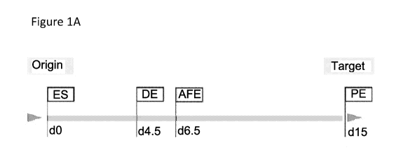
Figure 1 - Establishment of a protocol for direct differentiation of hESCs to
3rd PP
biased Pharyngeal Endoderm. Figure 1A is a schematic of the representation of
postulated
hESC differentiation steps towards desired cell-fates, mirroring the aims of
the treatments
shown in Figure 1B. Figure 1B is a schematic of the tested protocols for hESCs
differentiation
to 3rd PP biased pharyngeal endoderm until day 15. Protocol #1 (indicated as
"1" in Figure
1B) (FGF8b+RA25o) was considered the reference protocol to which protocol #2
(indicated as
"2" in Figure 1B) (FGF8b) (#1 vs #2) and #3 (indicated as "3" in Figure 1B)
(FGF8b+ RA250
to FGF8b+Shh) (#1 vs #3), are compared in Figure 1D. In Figure 1B, "NS"
indicates Noggin
and 5B431542. Figure 1C shows representative flow cytometric analysis of EpCAM
and
CXCR4 (endodermal markers) expression on dissociated embryoid bodies at day
4.5. Figure
1D is a graph showing the comparative analyses of gene expression in
differentiated hESCs at
day 15 under protocol conditions shown in Figure 1B. The graphs represent fold
change in
RNA expression as measured by qPCR. (n=3-11, values represent mean SEM, *p
<0.05,
**p < 0.01, ***p < 0.001, two-tailed ratio paired t-test). Figure lE shows the
comparison of
PP markers' expression at day 15 in hESCs differentiated using protocol #1
with hESCs
differentiated to 'liver' ('hepatic conditions' (Gouon-Evans et al. 2006)).
Bar graphs represent
6
CA 03135377 2021-09-28
WO 2020/205859
PCT/US2020/025955
fold change in RNA expression as measured by qPCR (n = 6, values represent
mean + SEM,
*p < 0.05, **p < 0.01, ***p < 0.001, two-tailed ratio paired t-test). Figure
1F is a graph showing
the comparative analyses of gene expression in differentiated hESCs at day 15
under protocol
conditions shown in Figure 1B. The graphs represent fold change in RNA
expression as
measured by qPCR. (n=9-11, values represent mean SEM, *p < 0.05, **p < 0.01,
***p <
0.001, two-tailed ratio paired t-test).
Figure 2 - Development of a protocol for distalization of 3rd PP and/or TEC.
Figure
2A is a schematic of the tested protocols for distalization of 3rd PP biased
cells until day 30.
In Figure 2A "3b" and "3c" indicate modifications based on Protocol #3 in
Figure 1B; "4b"
and "4c" indicate modifications based on Protocol #4 in Figure 1B. Figure 2B
shows a
schematic representation of multiple hESC differentiation protocols tested
under divergent
culture conditions from day 6.5 onwards. hESCs were differentiated to
definitive endoderm
(DE) for 4.5 days and subsequently anteriorized with Noggin+SB (NS) and
retinoic acid (RA).
Then the cells were patterned for 8.5 days with RA and different combinations
of the indicated
.. factors, until day 15. Figure 2C are graphs of expression analysis of
FOXA2, HOXA3, SIX],
TBX1, EYA1, PAX9 and PAX] in the hESC-derived cells from cultures containing
RA and
FGF8b (protocol #1) vs RA + factors substituting FGF8b as shown in Figure 2B.
Bar graphs
represent fold change in RNA expression as measured by qPCR (n = 3, values
represent mean
SEM, *p <0.05, **p < 0.01, ***p < 0.001, one-way ANOVA with Dunnett's multiple
comparisons test). Figure 2D show the effect of Noggin exposure on PAX9
expression at day
30. The bar graphs represent fold change in PAX9 expression between protocol
#3b vs #3c and
#4b vs #4c. (n =4, values represent mean SEM, *p < 0.05, **p < 0.01, ***p
<0.001, two-
tailed ratio paired t-test). Figure 2E shows the fold change of FOX1V1
expression at day 30
upon initiation of FGF8b treatment at day 4.5 vs day 6.5 (protocol #3c vs #4c)
as measured by
qPCR (n =4-8, values represent mean SEM, *p < 0.05, **p < 0.01, ***p <
0.001, two-tailed
ratio paired t-test). Figure 2F shows the fold change in FOXN1 expression at
day 21 vs day 30
(before and after BMP4 exposure) of protocol #4c as measured by qPCR (n =4-8,
values
represent mean SEM, *p < 0.05, **p < 0.01, ***p < 0.001, two-tailed ratio
paired t-test).
Figure 2G shows the fold change in FOXN1 expression at day 15 vs day 30 of
protocol #4c as
measured by qPCR (n =4-8, values represent mean SEM, *p <0.05, **p < 0.01,
***p <
0.001, two-tailed ratio paired t-test).
Figure 3 - Characterization of in vitro differentiated TEC progenitors at day
30. Figure
3A shows the TEC marker expression in cultured cells (d30; protocol #4c)
compared to fetal
thymus (FTHY). (Ct relative to I3-actin, n = 3-22, values represent mean +
SEM, *p < 0.05,
7
CA 03135377 2021-09-28
WO 2020/205859
PCT/US2020/025955
**p < 0.01, ***p < 0.001, two-tailed unpaired Welch's t-test). Every dot
represents an
independent experiment. Figure 3B shows the 3rd PP marker expression in H9
cells cultured
under protocol #4c conditions for 30 days compared to fetal thymus. Bar graphs
represent mean
Ct values relative to I3-actin + SEM (n = 3-6). Two-tailed unpaired Welch's t-
test. Each dot
represents an independent experiment. Figure 3C are graphs of Pearson
correlation analysis of
gene expression levels of FOXN1 and GCM2, FOXN1 and IL7, and FOXN1 and CD205.
Both
axes depict Ct values relative to I3-actin. Every dot represents an
independent experiment.
Figure 4 - Treatment of day 30 hES-TEP cultures with survivin inhibitor YM155
depletes multipotent cells. Figure 4A is a schematic representation of
protocol #4c showing
time period of YM155 treatment. This schematic also shows the complete
differentiation
protocol. Figure 4B is a graph of Pearson correlation analysis of FOXN1 and
OCT4 expression.
Both axes depict Ct values relative to I3-actin. Every dot represents an
independent experiment.
Figure 4C is a graph of the fold change in OCT4 expression at day 30 following
depletion of
multipotent cells (protocol #4c vs #4c+YM155; n =5, values represent mean +
SEM, *p <0.05,
two-tailed ratio paired t-test). Figure 4D is a graph showing percent survival
free from overt
teratoma formation in weeks post hES-TEP transplantation. hES-TEP-grafted mice
from
protocol #4c day 15 (n=8, grey line), compared to hES-TEP-grafted mice from
day 30 cultures
treated with (n=15, black dotted line) or without (n=12, solid black line)
YM155. Log-rank
Mantel Cox test showed p<0.005 for hES-TEP day 15 survival compared to either
hES-TEP
day 30 alone or hES-TEP day 30 + YM155 treatment.
Figure 5 - Reaggregate hES-TEP prepared using the protocol shown in Figure 4A
and
thymic mesenchyme cells form a thymic organoid that supports thymopoiesis.
Figure 5A shows
the percentage of T cells when the native thymic rudiment was surgically
removed (ATX) or
not from NSG mice injected with human HSCs. ACK lysis of peripheral blood
produced white
blood cells (WBCs) that were stained for HuCD45+CD3+ T cells at the indicated
weeks post-
HSC injection. NSG n=12, ATX NSG n=4. Figure 5B are representative FACS plots
gated on
HuCD45+CD19-CD14- cells. NSG n=10, ATX n=14. Figures 5C-5F show the frequency
of
various cells when cultured hES-TEPs clusters mixed with thymic mesenchyme
cells (TMC)
or TMCs alone were grafted under the renal capsule of ATX NSG mice injected
with human
HSCs. Figure 5C shows the frequency of HuCD45+ cells among total mouse + human
CD45+
cells in PBMCs for individual hES-TEC/TMC mice and the average (grey line) for
TMC
grafted mice (n=6). Figure 5D shows the frequency of CD3+ cells among total
mouse + human
CD45+ cells in PBMCs for individual hES-TEP/TMC mice and the average (grey
line) for
TMC grafted mice (n=6). Figure 5E shows the frequency of CD4+ cells among
total mouse +
8
CA 03135377 2021-09-28
WO 2020/205859
PCT/US2020/025955
human CD45+ cells in PBMCs for individual hES-TEP/TMC mice and the average
(grey line)
for TMC grafted mice (n=6). Figure 5F shows the frequency of CD4+ cells
stained for
CD45RA+CD45R0- naive cells. Timepoints with fewer than 100 CD4+ events were
excluded.
Figure 5G show human T cells in PBMCs from a healthy human (left), hES-TEC/TMC
(middle) and TMC mouse (right) 30 weeks post-humanization. hES-TEC/TMC plot is
representative of n=4 mice that developed CD4+ and CD8+ T cells and TMC plot
is
representative of n=6. Figure 5H shows the CD4+ and CD8+ expression on cells
from hES-
TEC/TMC (n=3). Cell suspensions were gated on HuCD45+CD19-CD14- cells.
Figure 6 - hES-TECs generated from TEPs prepared using the protocol shown in
Figure
4A persist in swine thymus and promote thymopoiesis. Figure 6A is schematic of
the protocol
to test the hES-TECs in vivo. The swine thymus was injected or not with hES-
TEPs and grafted
under the renal capsule of ATX NSG mice injected i.v. with human HSCs. Figure
6B shows
the results of flow cytometry analysis of the thymic grafts 18-22 weeks post-
transplant. Single
cell suspension from liberase digested stromal fraction of half the thymus
graft was stained and
analyzed by flow cytometry. Human pediatric thymus was prepared as a control.
Non-
hematopoietic cells were gated as huCD45-HLA-ABC+. Markers for thymic
fibroblasts
(CD105+) and epithelial cell marker EpCAM are shown. Figure 6C is a graph of
the frequency
of huCD45- HLA-ABC+CD105-EpCAM+ epithelial cells in SwTHY+hES-TECs (left bar,
squares) and SwTHY (right bar, triangles) grafts. Figure 6D are representative
flow cytometry
plots of thymocytes gated as huCD45+CD19-CD14- cells for CD4/CD8 distribution
for human
pediatric thymus, and swine thymus injected or not injected with hES-TEPs
(left to right).
Figure 6E are graphs of absolute count of thymocytes from half of the thymus
graft in double
positive CD4+CD8+, single positive CD4+CD8- and CD4-CD8+ with further division
into
immature CD45R0+ compared to more mature CD45RA+ thymocytes are shown. Average
+
SEM are shown for SwTHY+hES-TEC (n=6, squares) and SwTHY (n=5, triangles) from
two
independent experiments. Thymic grafts yielding fewer than 6x10⁵ (n=1 each
from
SwTHY+hES-TEC and SwTHY) cells were eliminated from analysis. Mann-Whitney
test was
used to determine p-values comparing SwTHY+hES-TEC to SwTHY groups with p<0.05
considered significant. +p=0.05, *p<0.05, "p<0.005. Figure 6F is a graph of
human immune
cells assayed for total human (huCD45+) cells in PBMCs at the indicated weeks
post-
humanization. Average + SEM are shown for swine thymus alone (n=9, black line
with
triangles) and swine thymus injected with hES-TEP (n=11, green line with
squares) from two
independent hES-TEC differentiations. Figure 6G is a graph of human immune
cells assayed
for total B cells (huCD19+) cells in PBMCs at the indicated weeks post-
humanization. Average
9
CA 03135377 2021-09-28
WO 2020/205859
PCT/US2020/025955
+ SEM are shown for swine thymus alone (n=9, black line with black triangles)
and swine
thymus injected with hES-TEP (n=11, green line with squures) from two
independent hES-
TEP differentiations. Figure 6H is a graph of 18-22 weeks post humanization
total human
CD45+immune cells in the spleen analyzed by flow cytometry. Average + SEM are
shown for
swine thymus injected with hES-TEP (n=7, squares) and swine thymus alone (n=6,
triangles)
from two independent hES-TEC differentiations. Figure 61 is a graph of 18-22
weeks post
humanization total human CD19+ B cells in the spleen analyzed by flow
cytometry. Average
+ SEM are shown for swine thymus injected with hES-TEP (n=7, squares) and
swine thymus
alone (n=6, triangles) from two independent hES-TEP differentiations. Figure
6J is a graph of
18-22 weeks post humanization total human CD14+ myeloid cells in the spleen
analyzed by
flow cytometry. Average + SEM are shown for swine thymus injected with hES-TEP
(n=7,
squares) and swine thymus alone (n=6, triangles) from two independent hES-TEP
differentiations.
Figure 7 - hES-TEP prepared using the protocol shown in Figure 4A injected
into swine
thymus promotes an increase in the proportion of CD4+ T cells in the blood and
increased
number of naive T cells and CD4+ recent thymic emigrants in spleen compared to
swine
thymus-grafted control mice. Figures 7A-7C show the results of human immune
cells assayed
in PBMCs at the indicated weeks post-humanization. Average + SEM are shown for
swine
thymus alone (n=9, black line with triangles) and swine thymus injected with
hES-TEP (n=11,
green line with squares) from two independent hES-TEP differentiations. Figure
7A shows
CD3+ cells. Figure 7B shows CD8+ cells. Figure 7C shows CD4+ cells.
Significant effect of
TEP injection was revealed by two-way ANOVA with p<0.05 considered significant
in CD3+
and CD4+ kinetics. Post-hoc Bonferroni multiple comparison at each time point
p<0.05
indicated by *. Figure 7D shows the absolute number of CD3+ T cells in the
spleen 18-22
weeks post-humanization. Figure 7E shows the absolute number of CD8+ T cells
in the spleen
18-22 weeks post-humanization. Figure 7F shows the absolute number of CD4+ T
cells in the
spleen 18-22 weeks post-humanization. Figure 7G shows CD45RA versus CCR7 used
to
distinguish naive, effector memory (EM), central memory (CM) and terminally
differentiation
effector memory cells re-expressing CD45RA (EMRA) (left panel) among CD8+
(middle
panel) or CD4+ T cells (right panel). Figure 7H shows the absolute number of
recent thymic
emigrant CD31+CD4+ naive cells as defined CD45RA+CCR7+ cells in mononuclear
cells of
the spleen. Average + SEM are shown for swine thymus injected with hES-TEP
(n=7, squares)
and swine thymus alone (n=6, triangles) from two independent hES-TEP
differentiations.
Mann-Whitney test was used to determine p-values comparing SwTHY alone to
SwTHY hES-
CA 03135377 2021-09-28
WO 2020/205859
PCT/US2020/025955
TEP injected groups with p<0.05 considered significant. *p<0.05.
DETAILED DESCRIPTION
Definitions
The terms used in this specification generally have their ordinary meanings in
the art,
within the context of this invention and the specific context where each term
is used. Certain
terms are discussed below, or elsewhere in the specification, to provide
additional guidance to
the practitioner in describing the methods of the invention and how to use
them. Moreover, it
will be appreciated that the same thing can be said in more than one way.
Consequently,
alternative language and synonyms may be used for any one or more of the terms
discussed
herein, nor is any special significance to be placed upon whether or not a
term is elaborated or
discussed herein. Synonyms for certain terms are provided. A recital of one or
more synonyms
does not exclude the use of the other synonyms. The use of examples anywhere
in the
specification, including examples of any terms discussed herein, is
illustrative only, and in no
way limits the scope and meaning of the invention or any exemplified term.
Likewise, the
invention is not limited to its preferred embodiments.
As used herein, the term "induced pluripotent stem cells" commonly abbreviated
as iPS
cells or iPSCs, refers to a type of pluripotent stem cell artificially
generated from a non-
pluripotent cell, typically an adult somatic cell, or terminally
differentiated cell, such as
fibroblast, a hematopoietic cell, a myocyte, a neuron, an epidermal cell, or
the like.
As used herein, the terms "differentiation" and "cell differentiation" refer
to a process
by which a less specialized cell (i.e., stem cell) develops or matures or
differentiates to possess
a more distinct form and/or function into a more specialized cell or
differentiated cell, (i.e.,
thymic epithelial cell).
As used herein, the expressions "cell," "cell line," and "cell culture" are
used
interchangeably and all such designations include progeny. Thus, the words
"transformants"
and "transformed cells" include the primary subject cell and cultures derived
therefrom without
regard for the number of transfers. It is also understood that not all progeny
will have precisely
identical DNA content, due to deliberate or inadvertent mutations. Mutant
progeny that have
the same function or biological activity as screened for in the originally
transformed cell are
included. Where distinct designations are intended, it will be clear from the
context.
With respect to cells, the term "isolated" refers to a cell that has been
isolated from its
natural environment (e.g., from a tissue or subject). The term "cell line"
refers to a population
of cells capable of continuous or prolonged growth and division in vitro.
Often, cell lines are
11
CA 03135377 2021-09-28
WO 2020/205859
PCT/US2020/025955
clonal populations derived from a single progenitor cell. It is further known
in the art that
spontaneous or induced changes can occur in karyotype during storage or
transfer of such
clonal populations. Therefore, cells derived from the cell line referred to
may not be precisely
identical to the ancestral cells or cultures, and the cell line referred to
includes such variants.
As used herein, the terms "recombinant cell" refers to a cell into which an
exogenous DNA
segment, such as DNA segment that leads to the transcription of a biologically-
active
polypeptide or production of a biologically active nucleic acid such as an
RNA, has been
introduced.
Abbreviations
hPSC- human pluripotent stem cell
ES or ESC- embryonic stem cell
iPSC- induced pluripotent stem cells
TEC- thymic epithelial cell
TEP- thymic epithelial cell progenitor
PE- pharyngeal endoderm
DE- definitive endoderm
AFE- anterior foregut or anterior foregut endoderm
PA- pharyngeal arches
3rd PP- third pharyngeal pouch
Shh- sonic hedgehog
RA- retinoic acid
SP- single positive
DP- double positive
To differentiate DE to third PP, the co-expression of TBX1 and HOXA3 was
induced
using a combination of FGF8 and retinoic acid (RA). RA treatment was
previously shown to
boost HOXA3 activity (Parent et al. 2013; Diman et al. 2011), but the TBX1
upregulating
potential of FGF8 was a novel finding disclosed herein. It is believed that
FGF8 plays a two-
fold role in the disclosed differentiation protocol: i) FGF8 signaling
immediately after activin
exposure drives Tbxl, anteriorizing the DE into a pharyngeally biased AFE
(Green et al. 2011).
Early exposure to FGF8 (day 4.5 vs day 6.5; protocol #3c vs #4c) strongly
pushed the culture
towards pharyngeal AFE, significantly increasing the number of FOXN1+ cells at
day 30; ii)
12
CA 03135377 2021-09-28
WO 2020/205859
PCT/US2020/025955
After anteriorization, FGF8b contributes to development of PE, now acting
downstream of and
in conjunction with TBX1 (Vitelli et al. 2002; Vitelli et al. 2010).
Another cytokine playing a key role in PE development is sonic hedgehog (Shh)
(Moore-Scott and Manley 2005). RA exposure was reduced and replaced with Shh
(protocol
#1 vs #3) as another innovation. This upregulated PAX9, PAX], and TBX1, but
downregulated
HOXA consistent with previous reports showing that Shh signaling induces Tbxl
in PE (Garg
et al. 2001). High levels of HOXA3 are critical to early pharyngeal region
patterning but its
expression diminishes in later stages. Indeed, Paxl expression is reduced in
Hoxa3 null
mutants, while Hoxa3 expression is normal in Paxl;Pax9 double mutant embryos
(Moore-
Scott and Manley 2005). Hoxa3 expression is also unaffected in Shh-/- mutants.
Thus, the
contribution of temporally opposite gradients of HOXA3 and Pax 1 -Pax9 to
third PP
development further justifies the initial use of RA followed by the treatment
with Shh in the
disclosed protocol.
In the last part of the protocol, the cells were exposed to Noggin and then
BMP4.
Although it has been shown that BMP signaling is required for FOXN1 expression
(Patel et al.
2006; Swann et al. 2017), this is the first report of using Noggin, a BMP4
antagonist and/or
inhibitor, for in vitro thymic differentiation. Noggin's presence in the 3rd
PP endoderm has
been associated with the parathyroid domain rather than the thymus, where BMP4
is expressed
(Patel et al. 2006). In the disclosed protocol, adding ectopic Noggin to the
culture further
enhanced the expression of PAX9 at day 30. Since BMP4 expression starts at
E10.5 in cells of
the 3rd PP endoderm right after Noggin expression at E9.5 (Patel et al. 2006),
the cells were
exposed to BMP4 from day 21 to 30 (immediately after Noggin). This led to
increases in
FOXN1 at day 30 compared to day 21 and day 15. Interestingly, BMP4 treatment
without prior
exposure to Noggin did not lead to any FOXN1 increase, confirming the need for
Noggin
exposure to develop sensitivity to BMP4.
Several groups have reported the ability to generate murine and human TEPs
from PSCs
(Parent et al. 2013; Sun et al. 2013; Soh et al. 2014; Su et al. 2015; Lai and
Jim 2009). In three
reports, grafts consisting of these cells, often along with supporting
mesenchyme or EPCAM-
cells from TEP cultures, have reconstituted murine T cells in nude mice, and
though robust,
continuous thymopoiesis in a normal-appearing thymic structures was not
demonstrated.
Indeed, the possibility that a wave of thymopoiesis was followed by peripheral
lymphopenia-
driven expansion of mature T cells was not ruled out. In one report, human T
cell repopulation
of peripheral tissues and human thymopoiesis in the grafted tissue was
demonstrated, though
thymic structure was not demonstrated for the grafted cells. Again, peripheral
markers of recent
13
CA 03135377 2021-09-28
WO 2020/205859
PCT/US2020/025955
thymic emigration were not included in the study, so it is unclear how robust
or durable the
thymopoiesis was.
Described herein, the hPSC-TEC-dependent appearance of naive human T cells in
the
periphery of the mice implanted with hPSC-TEPs plus thymic mesenchymal cells
and receiving
human HSCs was clearly demonstrated. Since the NSG mouse thymus is also
capable of
supporting human thymopoiesis, all NSG mice were thymectomized before
implanting the
hPSC-TEPs (Khosravi et al. 2020), thereby assuring that all peripheral T cells
arose from the
grafted tissue. The phenotype of peripheral human T cells in these mice
eventually converted
to the memory type.
The inability to generate durable, structured thymi from "stand-alone"
cellular grafts
led to the development of a novel approach to assess thymopoietic function of
hPSC-TEPs in
vivo. It has been previously demonstrated that fetal porcine thymic tissue
supports
phenotypically normal human thymopoiesis (Nikolic and Sykes 1999) with a
diverse TCR
repertoire (Shimizu et al. 20008) and robust population of peripheral naive T
cells in NSG
mice, though with some subtle differences from that observed for T cells
developing in a human
thymus graft (Kalscheuer et al. 2014). These fetal pig thymus fragments grow
markedly and
contain up to hundreds of millions of human thymocytes in a normal-appearing
thymic
structure (Nikolic and Sykes 1999; Kalscheuer et al. 2014). Disclosed herein
is a methodology
for injecting hPSC-TEPs into fragments of fetal pig thymus tissue that
maintained the human
cells in close proximity to the pig thymus tissue and ultimately resulted in
their incorporation
into the pig thymus as it grew. The human TEPs incorporated into the pig
thymus clearly
expressed human cTEC and mTEC-associated cytokeratins and appeared integrated
into the
highly organized thymic structure of the grafts. Most importantly, they had a
notable functional
effect, significantly increasing the total number of human thymocytes and the
number of
peripheral naive human T cells, including CD4+CD34RA+ T cells with the CD31+
RTE
phenotype.
Methods and Systems of Obtaining Thymic Epithelial Cells and/or Thymic
Epithelial Cell
Progenitors
The methods and systems described herein not only provide a reproducible
method to
obtain thymic epithelial cells (TEC s) or TEC progenitors (TEPs) by inducing
differentiation
of human pluripotent stem cells into thymic epithelial cells (TECs) or TEC
progenitors
(TEPs) but also provide an increase the purity and homogeneity of the thymic
epithelial cells
(TEC s), or TEC progenitors (TEPs) thus increasing function.
14
CA 03135377 2021-09-28
WO 2020/205859
PCT/US2020/025955
The methods and systems set forth herein generate a defined and reproducible
cell
population that is fully functional upon transplantation. Furthermore, the
methods and systems
set forth herein provide a substantially homogenous population of thymic
epithelial cells
(TEC s) or TEC progenitors.
A human pluripotent stem cell is the starting material of the methods of the
invention.
The human pluripotent stem cell (hPSCs) can be an embryonic stem cells (ESCs)
or an
induced pluripotent stem cell (iPSCs).
The steps of the method and the timing are set forth in Table 1 and Figure 4A.
Table 1 - Timeline of the Differentiation Method
STEP TIMING GENERAL
DESCRIPTION
1 Performed from about day 1 Differentiate hPSCs to
to about day 6 definitive endoderm cells
2 Starting from about day 3 to Differentiate
definitive
about day 5 and performed endoderm cells to anterior
for about 48 to about 72 foregut endoderm cells by
hours, ending at about day 5 inhibiting BMP and/or TGFI3
to day 8 signaling and stimulating
expression of TBX1 and/or
optionally stimulating
HOXA3
3 Starting from about day 5 to Differentiate anterior
foregut
about day 8 and performed endoderm cells to pharyngeal
for about 6 days to about 10 endoderm cells by
days ending at about day 11 continuing to stimulate
to about day 18 expression of HOXA3 (for
about one day to three days)
and/or later stimulating
expression of PAX] and
PAX9 and/or stimulating
TBX1 throughout
4 Starting from about day 11 Differentiate
pharyngeal
to about day 18 and endoderm cells into distal
performed for about 4 days third PP, thymic epithelial
to about 7 days ending at cells, or thymic epithelial
about day 19 to about day progenitor cells by
inhibiting
25 BMP
5 Starting from about day 13 Continue to
differentiate
to about day 25 and pharyngeal endoderm cells
performed about 5 days to into distal third PP, thymic
about 15 days ending at epithelial cells, or thymic
CA 03135377 2021-09-28
WO 2020/205859
PCT/US2020/025955
about day 18 to about day epithelial progenitor cells
by
40 adding BMP
6 Starting from about day 21 Add survivin
inhibitor
to about day 23 or at the end
of the protocol for about 24
to about 72 hours
The first step of the method is differentiating the hPSCs to definitive
endoderm (DE)
cells using any method known in the art. Exemplified here was the use of
previously
published protocols using serum-free differentiation medium containing BMP4,
bFGF and
Activin A. However, other protocols known in the art can be used.
The next step of the method is the culturing the resulting definitive endoderm
cells
from the first step to further differentiate into anterior foregut endoderm
(AFE). Any medium
used for differentiation protocols can be used for culturing the cells at this
step. A serum-
free differentiation medium is preferred. Additionally, growth factors such as
EGF and FGF
can be added to the medium to promote cellular growth.
The endoderm cells are then contacted or incubated with an agent that inhibits
BMP
and an agent that inhibits TGFI3 signaling to promote differentiation of the
definitive
endoderm cells to anterior foregut progenitor cells. The most efficient method
to accomplish
this is by adding the agents to the medium in which the cells are being
cultured. However,
any other method known in the art that would contact or incubate the cells
with the agents
can be used. The cells can be contacted or incubated with the agents
simultaneously or
concurrently.
Agents that inhibit BMP include but are not limited to Noggin and
Dorsomorphin.
Agents that inhibit TGFI3 signaling include but are not limited to SB431542.
Dorsomorphin can be used in an amount ranging from about 0.5 [tM to about 2
M.
Noggin can be used in an amount ranging from about 25 ng/ml to about 500
ng/ml,
or ranging from about 50 ng/ml to about 400 ng/ml, or ranging from about 100
ng/ml to
about 300 ng/ml, with about 200 ng/ml being a preferred amount.
An agent for the inhibition of TGFI3 signaling is SB431542 in an amount
ranging
from about 1 [tM to about 50 M, or ranging from about 2 [tM to about 30 M,
or ranging
from about 5 [tM to about 20 M. In some embodiments, the agent used for the
inhibition of
TGFI3 signaling is 5B431542 in the amount of about 10 M.
However, other agents that inhibit TGFI3 signaling can be used in the method.
Additionally, it was found that the combined stimulation of expression of TBX1
and
16
CA 03135377 2021-09-28
WO 2020/205859
PCT/US2020/025955
HOXA3 at the AFE stage was essential for the physiological 3r1 PP endoderm
development.
Thus, the cells are further contacted or incubated with agents which stimulate
expression of
these genes. An agent for the stimulation of TBX1 is FGF8b, which may be used
in an
amount ranging from about 10 ng/ml to about 200 ng/ml, or ranging from about
20 ng/ml to
about 150 ng/ml, or ranging from about 30 ng/ml to about 100 ng/ml. In some
embodiments,
the FGF8b may be used at about 50 ng/ml.
The cells are contacted or incubated with this agent from about day 4.5 to
about day
15.
An agent for the stimulation of HOXA3 is retinoic acid (RA) used in an amount
ranging from about 0.1 [tM to about 0.6 M, or ranging from about 0.2 [tM to
about 0.5 M.
In some embodiments, the retinoic acid may be used in the amount of about 0.6
M. The
cells can be contacted or incubated with this agent from about day 4.5 to
about day 7.5.
Stimulation of HOXA3 can be performed at any other period of 3 days during the
first 15
days, other than day 4.5 to 7.5.
As shown in Figures 1D-1F, this protocol yields AFE with high efficiency.
The cells continue to be cultured in any serum-free medium used for
differentiation
of cells (herein referred to as the "differentiation medium" or "serum-free
differentiation
medium). Additionally, growth factors such as EGF and FGF can be added to the
differentiation medium to promote cellular growth. At the beginning of this
step for about
one to two days, the cells are contacted or incubated with RA in an amount of
ranging from
about 0.1 [tM to about 0.6 M, or ranging from about 0.2 [tM to about 0.5 M.
In some
embodiments, the cells are contacted or incubated with about 0.25 [tM RA. Also
the cells
continue to be contacted or incubated with FGF8b throughout this step, in an
amount ranging
from about 10 ng/ml to about 200 ng/ml, or ranging from about 20 ng/ml to
about 150 ng/ml,
or ranging from about 30 ng/ml to about 100 ng/ml. As a non-limiting example,
the cells
may be contacted with about 50 ng/ml FGF8b.
The next step promotes differentiation of the anterior foregut cells into
pharyngeal
endoderm (PE) cells.
In this step, the cells are contacted or incubated with an agent that induces
expression
of PAX9 and PAX]. The most efficient method to accomplish this is by adding
the agents to
the medium in which the cells are being cultured. However, any other method
known in the
art that would contact or incubate the cells with the agents can be used. The
cells can be
contacted or incubated with the agents simultaneously or concurrently. An
agent for the
stimulation of both PAX9 and PAX] is sonic hedgehog (Shh) in an amount ranging
from
17
CA 03135377 2021-09-28
WO 2020/205859
PCT/US2020/025955
about 10 ng/ml to about 400 ng/ml, or ranging from about 25 ng/ml to about 300
ng/ml, or
ranging from about 50 ng/ml to about 200 ng/ml. In some embodiments, Shh may
be used at
about 100 ng/ml.
Also the cells are continued to be contacted or incubated with FGF8b
throughout at
an amount ranging from about 10 ng/ml to about 200 ng/ml, or ranging from
about 20 ng/ml
to about 150 ng/ml, or ranging from about 30 ng/ml to about 100 ng/ml. In some
embodiments, cells may be contacted or incubated with about 50 ng/ml FGF8b.
Noggin can also be used to induce expression of PAX9 and PAX]. Noggin can be
used in an amount ranging from about 50 ng/ml to about 400 ng/ml, or ranging
from about
60 ng/ml to about 300 ng/ml, or ranging from about 75 ng/ml to about 200
ng/ml. In some
embodiments, Noggin may be used in the amount of about 100 ng/ml.
This step is performed for about 4 to about 10 days.
The next step is the differentiation of the PE cells to distal third PP/ TECs.
This step
is divided into two steps: the first where the cells are contacted or
incubated with an agent
which inhibits BMP. Agents which inhibit BMP include but are not limited to
Noggin and
Dorsomorphin.
Dorsomorphin can be used in an amount ranging from about 0.5 [tM to about 2
M.
Noggin can be used in an amount ranging from about 50 ng/ml to about 400
ng/ml,
or ranging from about 60 ng/ml to about 300 ng/ml, or ranging from about 75
ng/ml to about
200 ng/ml. As a non-limiting example, Noggin may be used in the amount of
about 100
ng/ml.
This part of the step is performed for about 5 days to about 7 days.
The second part of the step the cells are contacted or incubated with BMP4 in
an
amount ranging from about 5 ng/ml to about 300 ng/ml, or ranging from about 15
ng/ml to
about 200 ng/ml, or ranging from about 25 ng/ml to about 100 ng/ml, or with
about 50 ng/ml.
This part of the step is performed for about 5 days to about 10 days.
The final cells obtained following the method may show gene expression of TEC
markers including FOX1V1, PAX9, PAX], DLL4, ISL1, EYA1, SIX], IL7, K5, K8 and
AIRE.
See Figures 3A and 3B.
While the method set forth above is a novel, reproducible and robust method to
induce the differentiation of hPSCs to TECs or TEPs, the present method also
provides for
further steps to reduce and eliminate pluripotent cells which can cause
teratomas in the final
grafted cells. In this step the cells are contacted or incubated with a
survivin inhibitor such
as YM155 for about the last 24 hours of the method in an amount ranging from
about 5nM
18
CA 03135377 2021-09-28
WO 2020/205859
PCT/US2020/025955
to about 50nM. As a non-limiting example, cells may be contacted or incubated
with 20nM
of YM155. The cells may also be contacted or incubated with a survivin
inhibitor concurrent
with the BMP4 treatment. In some embodiments, the cells may be contacted or
incubated
with a survivin inhibitor during the first 24 to 48 hours of concurrent BMP4
incubation.
The present invention also includes systems for practicing the disclosed
methods for
obtaining TECs or TEPs from hPSCs. These systems can include subsystems
wherein the
subsystems include differentiation medium, and agents which inhibit BMP and
TGFI3
signaling, agents which stimulate expression of HOXA3, TBX1, PAX] and PAX9,
agents
which inhibit surviving, and BMP4. These systems can include subsystems
wherein the
subsystems include differentiation medium, and Noggin, retinoic acid, FGF8b,
sonic
hedgehog, BMP, and YM155.
Cells
A further embodiment of the present disclosure are the thymic epithelial cells
(TECs) or TEC progenitors (TEPs) generated by the differentiation protocol set
forth herein.
In some embodiments, these cells express FOXN1, EpCAM, Keratin 5, and Keratin
8.
In some embodiments, these cells are thymic epithelial cells (TECs). In some
embodiments,
these cells are thymic epithelial cell progenitors (TEC progenitors) (TEPs).
Thus, one aspect of the present disclosure is thymic epithelial cells (TECs)
or TEC
progenitors (TEPs) suitable for administration, transplantation and grafting
into a subject
produced by the methods as described herein.
In another aspect, provided herein is a composition comprising the thymic
epithelial
cells or TEC progenitors (TEPs) produced by the methods as described herein.
In some
embodiments, these cells are suitable for administration, transplantation and
grafting into a
subject. In some embodiments, the composition is a pharmaceutical composition
further
comprising any pharmaceutically acceptable carrier or excipient.
In certain embodiments, the composition or pharmaceutical composition
comprises at
least 10,000, at least 50,000, at least 100,000, at least 500,000, at least 1
x 106, at least 5 x 10⁶,
at least 1 x 10⁷, at least 5 x 10⁷, at least 1 x 10⁸, at least 5 x 10⁸, at
least 1 x 10⁹, at least 5 x 10⁹,
or at least 1 x 10¹⁰ thymic epithelial cells (TECs) or TEC progenitors (TEPs)
produced by
the methods as described herein. In some embodiments, these cells are suitable
for
administration, transplantation and grafting into a subject.
In certain embodiments, the disclosure provides a cryopreserved composition or
solution of the thymic epithelial cells (TECs) or TEC progenitors (TEPs)
produced by the
19
CA 03135377 2021-09-28
WO 2020/205859
PCT/US2020/025955
methods as described herein. In some embodiments, these cells are suitable for
administration,
transplantation and grafting into a subject.
In certain embodiments, the cryopreserved composition or solution comprises at
least
10,000, at least 50,000, at least 100,000, at least 500,000, at least 1 x 10⁶,
at least 5 x 10⁶, at
least 1 x 10⁷, at least 5 x 10⁷, at least 1 x 10⁸, at least 5 x 10⁸, at least
1 x 10⁹, at least 5 x 10⁹,
or at least 1 x 10¹⁰ thymic epithelial cells (TECs) or TEC progenitors (TEPs)
produced by
the methods as described herein. In some embodiments, these cells are suitable
for
administration, transplantation and grafting into a subject.
In certain embodiments, the disclosure provides for cell culture comprising
thymic
epithelial cells (TECs) or TEC progenitors (TEPs) produced by the methods as
described
herein. In certain embodiments, the cell culture comprises at least 1 x 10⁷,
at least 5 x 10⁷, at
least 1 x 10⁸, at least 5 x 10⁸, at least 1 x 10⁹, at least 5 x 10⁹, or at
least 1 x 10¹⁰ thymic
epithelial cells (TECs) or TEC progenitors (TEPs) produced by the methods as
described
herein. In some embodiments, these cells are suitable for administration,
transplantation and
grafting into a subject.
In certain embodiments, the disclosure provides the therapeutic use of the
thymic
epithelial cells (TECs) or TEC progenitors (TEPs) suitable for administration,
transplantation and grafting into a subject produced by the methods as
described herein, and
compositions, solutions and cell cultures comprising such cells.
In other embodiments, the disclosure provides for a population of
substantially
homogenous thymic epithelial cells (TECs) or TEC progenitors (TEPs) produced
by the
methods as described herein. In some embodiments, these cells are suitable for
administration,
transplantation and grafting into a subject. In some embodiments, the
population of cells
comprises at least about 90%, 91%, 92%, 93%, 94%, 95%, 96%, 97%, 98%, or 99%
thymic
epithelial cells (TECs) or TEC progenitors (TEPs).
In another aspect, provided herein is a composition comprising the population
of
substantially homogenous thymic epithelial cells (TECs) or TEC progenitors
(TEPs)
produced by the methods as described herein. In some embodiments, these cells
are suitable
for administration, transplantation and grafting into a subject. In some
embodiments, the
composition is a pharmaceutical composition further comprising any
pharmaceutically
acceptable carrier or excipient.
In certain embodiments, the population or composition or pharmaceutical
composition
comprises at least 10,000, at least 50,000, at least 100,000, at least
500,000, at least 1 x 10⁶, at
least 5 x 10⁶, at least 1 x 10⁷, at least 5 x 10⁷, at least 1 x 10⁸, at least
5 x 10⁸, at least 1 x 10⁹,
CA 03135377 2021-09-28
WO 2020/205859
PCT/US2020/025955
at least 5 x 10⁹, or at least 1 x 10¹⁰ thymic epithelial cells (TECs) or TEC
progenitors
(TEPs) produced by the methods as described herein. In some embodiments, these
cells are
suitable for administration, transplantation and grafting into a subject.
In certain embodiments, the disclosure provides a cryopreserved composition or
solution of the population of substantially homogenous thymic epithelial cells
(TECs) or
TEC progenitors (TEPs) produced by the methods as described herein. In certain
embodiments, the cryopreserved composition or solution comprises at least
10,000, at least
50,000, at least 100,000, at least 500,000, at least 1 x 10⁶, at least 5 x
106, at least 1 x 10⁷, at
least 5 x 10⁷, at least 1 x 10⁸, at least 5 x 10⁸, at least 1 x 10⁹, at least
5 x 10⁹, or at least 1 x
1010 thymic epithelial cells (TECs) or TEC progenitors (TEPs) produced by the
methods as
described herein. In some embodiments, these cells are suitable for
administration,
transplantation and grafting into a subject.
In certain embodiments, the disclosure provides for cell culture comprising
population
of substantially homogenous thymic epithelial cells (TECs) or TEC progenitors
(TEPs)
produced by the invention as described herein. In certain embodiments, the
cell culture
comprises at least 1 x 10⁷, at least 5 x 10⁷, at least 1 x 10⁸, at least 5 x
108, at least 1 x 10⁹, at
least 5 x 10⁹, or at least 1 x 10¹⁰ thymic epithelial cells (TECs) or TEC
progenitors (TEPs)
produced by the methods as described herein. In some embodiments, these cells
are suitable
for administration, transplantation and grafting into a subject.
In certain embodiments, the disclosure provides the therapeutic use of the
population
of substantially homogenous thymic epithelial cells (TECs) or TEC progenitors
(TEPs)
suitable for transplantation and grafting into a subject produced by the
methods as described
herein, and compositions, solutions and cell cultures comprising such cells.
A further embodiment is a thymic organ comprising the TECs or TEPs disclosed
herein
combined with other cells which make up a thymus.
Therapeutic Uses
The novel method described herein for the generation of TECs or TEC
progenitors
(TEPs) from stem cells and the cells and substantially homogenous population
of cells
generated from this method, provide new therapies for diseases.
The ability to generate functional TECs from human pluripotent stem cells,
would have
important applications in modeling human immune responses in mice, and in
modeling and
treating thymus deficiency syndromes, such as DiGeorge syndrome, Nude
syndrome, and
immunodeficiency complicating bone marrow transplantation for leukemia. Cells
could also
21
CA 03135377 2021-09-28
WO 2020/205859
PCT/US2020/025955
be used clinically for cell-therapy and transplanted in patients to achieve T
cell reconstitution,
or generating immune tolerance to prevent graft rejection after an organ
transplantation, or for
recovering an impaired thymic functionality due to injuries or aging
Thus, one embodiment is a method of treating or preventing a disease of the
thymus in
a subject in need thereof comprising the steps of administering, transplanting
or grafting a
therapeutically effective amount of the cells of the present disclosure, a
solution comprising
the cells of the present disclosure, a composition comprising the cells of the
present disclosure,
or a pharmaceutical composition comprising the cells of the present
disclosure, to the subject
in need thereof. The subject is preferably a mammal, and most preferably
human.
A further embodiment is a method of treating or preventing an autoimmune
disease in
a subject in need thereof comprising the steps of administering, transplanting
or grafting a
therapeutically effective amount of the cells of the present disclosure, a
solution comprising
the cells of the present disclosure, a composition comprising the cells of the
present disclosure,
or a pharmaceutical composition comprising the cells of the present
disclosure, to the subject
in need thereof. The subject is preferably a mammal, and most preferably
human.
Another embodiment is a method of recovering or restoring impairment of the
function
of the thymus in a subject in need thereof comprising the steps of
administering, transplanting
or grafting a therapeutically effective amount of the cells of the present
disclosure, a solution
comprising the cells of the present disclosure, a composition comprising the
cells of the present
disclosure, or a pharmaceutical composition comprising the cells of the
present disclosure, to
the subject in need thereof. The subject is preferably a mammal, and most
preferably human.
In some embodiments the impairment is due to injury. In some embodiments, the
impairment
is due to aging. In some embodiments, the impairment is due to congenital
abnormalities.
Yet a further embodiment is a method of reconstituting T cells after a bone
marrow
transplant in a subject in need thereof comprising the steps of administering,
transplanting or
grafting a therapeutically effective amount of the cells of the present
disclosure, a solution
comprising the cells of the present disclosure, a composition comprising the
cells of the present
disclosure, or a pharmaceutical composition comprising the cells of the
present disclosure, to
the subject in need thereof. The subject is preferably a mammal, and most
preferably human.
The cells obtained using the methods disclosed herein can be used to generate
a hybrid
thymus. In some embodiments the hybrid thymus comprises thymic epithelial
cells obtained
using the methods disclosed herein and thymic tissue from a second individual
of the same
species. In some embodiments the hybrid thymus comprises thymic epithelial
cells obtained
using the methods disclosed herein and thymic tissue from a second species. In
some
22
CA 03135377 2021-09-28
WO 2020/205859
PCT/US2020/025955
embodiments, the second species is a swine. In some embodiments, the second
species is a
miniature swine. In some embodiments, the swine a juvenile swine. In some
embodiments, the
swine is fetal. A method of obtaining such a hybrid swine is disclosed in
commonly owned
patent application no. PCT/US2019/051865.
A further embodiment is the use of the cells to develop mice models. Since
cellular
reprogramming was discovered (iPSCs), a new era of disease modelling with
pluripotent stem
cells representing a myriad of genetic diseases can now be produced from
patient tissue. IPSCs
from patients with different autoimmune diseases where the central tolerance
is involved can
be differentiated to TECs (or TEPs), then injected or grafted into mice where
the cells can
reproduce and develop into the various conditions or disorders. Humanized
mouse models can
be generated from TECs from patients with an autoimmune disease such as
multiple sclerosis,
or type I diabetes, or a congenic abnormality such as DiGeorge Syndrome. The
mouse, in vivo
environment can then be used to study the progress of a disorder that,
otherwise, could not be
developed in vitro.
Additionally, personalized humanized mouse models can be generated using the
cells
described herein. Thus far, the most developed humanized mouse model contains
human
hematopoietic stem cells (HSCs), and a sample of a pediatric or human fetal
thymus sample
grafted under the kidney capsule. The limitation of these mouse models is that
the HLA from
both type of cell populations (HSCs and TECs) do not match because they
originate from two
different individuals. With the differentiation protocol disclosed herein,
TECs (or TEPs) could
be differentiated from the same iPSCs as the HSCs, so the immune system cells
HLA will
match with the ones on the human TECs transplanted on the mouse. This
technology could be
used for individual patients resulting in a Personalized Immune (PI) mouse.
A further embodiment is the use of the cells for drug testing in vivo (with
the previously
described mouse models including but not limited to the Personalized Immune
(PI) mouse
model) or in vitro. In vitro, differentiated TECs cultures, can be used to
test drugs against
different conditions that affect to TECs, such as cancer (thymomas), or
infectious, or
autoimmune diseases.
Kits
The present disclosure also provides kits.
In one embodiment, the kit includes one or more components including human
pluripotent stem cells, medium for culturing and differentiation the hPSCs,
such medium
23
CA 03135377 2021-09-28
WO 2020/205859
PCT/US2020/025955
including growth factors and inhibit BMP and TGFI3 signaling, agents which
stimulate
expression of HOXA3, TBX1, PAX] and PAX9, agents which inhibit surviving, and
BMP4.
In another embodiment, the kit includes one or more components including human
pluripotent stem cells, medium for culturing and differentiation the hPSCs,
such medium
including growth factors and Noggin, retinoic acid, FGF8b, sonic hedgehog,
BMP, and
YM155.
In further embodiments, a kit can include the TECs or TEC progenitors (TEPs)
obtained by the current methods and systems of the disclosure. The kit can
also comprise
reagents for culturing the cells.
In further embodiments, a kit can include a pharmaceutical composition
comprising the
TECs or TEC progenitors (TEPs) obtained by the current methods and systems of
the
disclosure.
In further embodiments, a kit can include a cryopreserved composition
comprising the
TECs or TEC progenitors (TEPs) obtained by the current methods and systems of
the
disclosure.
The kits can further include a package insert including information concerning
the
pharmaceutical compositions and dosage forms in the kit. For example, the
following
information regarding a combination of the invention may be supplied in the
insert: how
supplied, proper storage conditions, references, manufacturer/distributor
information and
patent information.
EXAMPLES
The present invention may be better understood by reference to the following
non-
limiting examples, which are presented in order to more fully illustrate the
preferred
embodiments of the invention. They should in no way be construed to limit the
broad scope of
the invention.
Example 1- Materials and Methods
Maintenance of hPSCs
RUES2 (Rockefeller University Embryonic Stem Cell Line 2, NIH approval number
NIHhESC-09-0013, Registration number 0013; passage 13-24) were cultured on
mouse
embryonic fibroblasts as previously described (Green et al. 2011). Mouse
embryonic
fibroblasts (GlobalStem, Rockville, MD) were plated at a density of
approximately 25,000
24
CA 03135377 2021-09-28
WO 2020/205859
PCT/US2020/025955
cells/cm2. hPSCs were cultured in DMEM/F12 with 20% knockout serum replacement
[Gibco
(Life Technologies, Grand Island, NY)], 0.1 mM 13-mercaptoethanol (Sigma-
Aldrich, St.
Louis, MO), and 20 ng/ml FGF-2 (R&D Systems, Minneapolis, MN). Medium was
changed
daily and cells were passaged with accutase/EDTA (Innovative Cell
Technologies, San Diego,
CA) every 4 days at 1:24 dilution. Undifferentiated hPSCs were maintained in a
5% CO2/air
environment. Human H9 ES cell line was also treated with protocol #4c. Lines
were karyotyped
and verified for mycoplasma contamination using PCR every 6 months.
Induction of endoderm
The differentiation was performed as described Huang et al. 2014 in serum-free
differentiation (SFD) medium consisting of DMEM/F12 (3:1) (Life Technologies)
supplemented with N2 [Gibco (Life Technologies)], B27 (Gibco), ascorbic acid
(50 [tg/ml,
Sigma), Glutamax (2 mM, Life Technologies), monothioglycerol (0.4 M, Sigma),
0.05%
bovine serum albumin (BSA) (Life Technologies) and 1% penicillin-streptomycin
(Thermo
Fisher Scientific, Waltham, MA). Cells were then briefly trypsinized (0.05%, 1
min at 37 C)
into single cell suspension and plated onto low attachment 6-well plates
[Costar 2 (Corning
Incorporated, Tewksbury MA)] to form embryoid bodies in serum-free
differentiation medium
containing human BMP4, 0.5 ng/ml, human bFGF, 2.5 ng/ml (R&D Systems) and
human
activin A, 100 ng/ml (R&D Systems) for 84 hours (3.5 days approximately) on
low-adherence
plates. Embryoid bodies were then collected, briefly trypsinized (0.05%, 1 min
at 37 C) into
3-10 small cell clumps and resuspended again in endoderm induction medium for
another 24
hours. Cells were fed every 24-48 hr (depending on the density) and maintained
in a 5%
CO2/5% 02/90% N2 environment.
Induction of anterior foregut endoderm, pharyngeal endoderm and distal 3rd
pharyngeal pouch
After 108 hours in total on low-adherence plates with endoderm induction media
(described above), embryoid bodies were collected and, without being
trypsinized, plated on
matrigel-coated, 24-well tissue culture plates (approximately 50,000-70,000
cells/well) in SFD
medium supplemented with 200 ng/mL recombinant human (rh) Noggin and 10 [LM
SB431542
(NS) (as described in established protocols Green et al. 2011), with Retinoic
Acid (0.25 M)
and FGF8b 50 ng/mL (as a novel modification of this protocol) for 48 hours.
For pharyngeal
endoderm, the resulting cells were then treated for 24 hours with FGF8b
(50ng/mL) and
Retinoic Acid (0.25 M) followed by 8 days with FGF8b (50ng/mL) and Sonic
Hedgehog
(Shh) (10Ong/mL) (Figure 1B). For the 3rd pharyngeal pouch specification,
cells were then
exposed to rhNoggin (200 ng/mL) for 6 days, and then to BMP4 (10 ng/mL) until
day 30 of
differentiation (Figure 2A). To avoid the formation of teratomas after
engrafting in mice, cells
CA 03135377 2021-09-28
WO 2020/205859
PCT/US2020/025955
were also exposed to survivin inhibitor YM155 (20nM) (Lee et al. 2013)) for 24
hours during
the last step in the later experiments (Figure 4A). During the entire process,
cell cultures were
maintained in a 5% CO2/air environment at 37 C. Cells were fed every 24 hours.
Quantitative Real-Time PCR
Total RNA from clusters of ES cells differentiated for the indicated time with
the
indicated culture method was extracted using Trizol (Invitrogen), and Direct-
zol RNA
Miniprep Kit (Zymo Research) according to the manufacturer' s instructions.
NanoDrop 2000
spectrophotometer (ThermoFisher Scientific) was used to determine RNA
concentration.
500ng RNA was amplified with random hexamers by reverse transcription using
Superscript
III kit (Invitrogen) according to the manufacturer's instructions. Real-time
quantitative PCR
was performed in 20u1 volume using ABI Power SYBR Green PCR Master Mix on an
ABI
ViiA7 Thermocycler (Applied Biosystems Life Technologies). PCR cycling
conditions were
set at 50 C for 2 minutes, 95 C for 10 minutes followed by 95 C for 15
seconds, and 60 C for
1 minute for 40 cycles. Single peak dissociation/melting curve was verified
for all reactions
and primer pairs. Quantification of each gene transcript was obtained by
comparing the average
of triplicate experimental CT values to a standard curve of serially diluted
genomic DNA for
each primer target and then normalized by dividing by the CT housekeeping gene
b-Actin.
Primer sequences are listed in Table 2.
Table 2 - Quantitative PCR Primers
Gene Forward Primer (5'-3') Reverse Primer (5'-3')
ACTB TTTGAATGATGAGCCTTCGT GGTCTCAAGTCAGTGTACAGGTA
GCCC (SEQ ID NO: 1) AGC (SEQ ID NO: 2)
K8 CACCACAGATGTGTCCGAGA
(SEQ ID NO: 3) AGGGCTGACCGACGAGAT (SEQ
ID NO: 4)
EYA1
ACCTCTGCCTTTGTGGTGAAT GGCAGACACATAACGCTGTGCTA
GGA (SEQ ID NO: 5) AA (SEQ ID NO: 6)
FOXA2
TTCAACCACCCGTTCTCCATC CTGTTCGTAGGCCTTGAGGTCCAT
AAC (SEQ ID NO: 7) TT (SEQ ID NO: 8)
26
CA 03135377 2021-09-28
WO 2020/205859
PCT/US2020/025955
FOXN1
CGGCACAACCTATCCCTCAA TTGTCGATCTTGGCCGGATT (SEQ
(SEQ ID NO: 9) ID NO: 10)
HOXA3
AGAGTTCCACTTCAACCGCT ATGCCCTTGCCCTTCTGATCCTTT
ACCT (SEQ ID NO: 11) (SEQ ID NO: 12)
OCT3/4 ATGCACAACGAGAGGATTTT
GA (SEQ ID NO: 13) CTTTGTGTTCCCAATTCCTTCC
(SEQ ID NO: 14)
PAX1
AAACCCTCCATGAACTGTCC CCCTGTGCTCCCTACTCCTACC
TCTCC (SEQ ID NO:15) (SEQ ID NO: 16)
PAX9
GGAAGCCGTGACAGAATGAC TGGTTATGTTGCTGGACATGGGT
TACCT (SEQ ID NO:17 ) G (SEQ ID NO: 18)
SIX1
CTATTCTCTCCCGGGCTTAAC CAGAGAGTCTTGGAGCTGATG
(SEQ ID NO: 19) (SEQ ID NO: 20)
TBX1
CCCGGCTCCTACGACTATTG GGAACGTATTCCTTGCTTGCCCTT
C (SEQ ID NO: 21) (SEQ ID NO: 22)
AIRE
CCTGGATGCACTTCTTGGA CAGAGAGCTGTGGCCATGT (SEQ
(SEQ ID NO: 23) ID NO:24 )
CD205
ATTGCTGGCACAGTACAGGA TGGAATTCATGGACCTCCACTT
(SEQ ID NO: 25) (SEQ ID NO: 26)
DLL4
GGGCACCTACTGTGAACTCC GCTGCCCACAAAGCCATAAG
(SEQ ID NO: 27) (SEQ ID NO: 28)
27
CA 03135377 2021-09-28
WO 2020/205859
PCT/US2020/025955
K5
TACCAGGACTCGGCTTCTGT ATCATCGCTGAGGTCAAGGC
(SEQ ID NO: 29) (SEQ ID NO: 30)
IL7 TCCTCCACTGATCCTTGTTC CTTCAACTTGCGAGCAGCAC (SEQ
(SEQ ID NO: 31) ID NO: 32)
ISL1
TTGTACGGGATCAAATGCGC CCACACAGCGGAAACACTCGAT
CAAG (SEQ ID NO: 33) (SEQ ID NO: 34)
Immunohistochemistry and Immunofluorescence
hES-cultures in 24-well tissue culture plates were fixed with paraformaldehyde
in PBS
(4%) for 10 minutes at room temperature. Cells were washed in PBS twice,
permeabilized in
PBS with 0.1% triton for 20 min, and blocked in 5% fetal donkey serum for 1
hour at room
temperature.
Thymic grafts were extracted, embedded in OCT (Tissue-Tec, Torrance CA) media,
frozen and 5-7um thick sections cut for immune staining. Sections were stained
with H&E to
visualize gross histology and interface of the thymic graft with the mouse
renal tissue. For
immunofluorescent staining, tissue sections were fixed and permeabilized in
100% ice-cold
acetone and allowed to dry completely. Tissue sections were blocked in PBS
supplemented
with 0.1% Tween and 0.1% Bovine Serum Albumin. Slides were washed in PBS 0.1%
Tween
and stained with primary antibody for 2 hours at room temperature, and then
washed and
incubated in secondary antibodies for 2 hours at room temperature.
Cultures or tissue sections were incubated with one, or a combination of two
or three
of the following primary antibodies and appropriate secondary antibodies
listed in Table 3.
Images were collected on a Leica SCN 400 whole slide scanning platform for H&E
stained
sections and immunofluorescent images were collected on a Leica TCS 5P8 2
photon laser
scanning microscope.
28
Table 3 - Antibodies used in Examples
0
t.)
o
Antigen Conjugation Clone Catalogue
Company 1'12 Source t.)
o
i-J
Target #
o
vi
oe
vi
Cytokeratin-8 AlexaFluor 647 EP1628Y ab192468
Abeam 1 rabbit
monoclonal
Cytokeratin- Biotin LL002 MA5-11596
ThermoFisher 10 mouse IgG3
14
EpCAM none EpCAM/TROP1 AF960
R&D System 1 goat
polyclonal
P
IgG
0
,
r.) EYA1 none L-19 5c15094
Santa Cruz 10 goat ,
,
CO
iv
Biotechnology
polyclonal 2
,
,
IgG
0
0
FOXA2 none M-20 Sc-6554
Santa Cruz 10 goat
(HNF-3beta)
Biotechnology polyclonal
IgG
HLA-DR none L243 307602
Biolegend 10 mouse IgG2a
kappa
Iv
n
,-i
HOXA3 none H-165 Sc-28598
Santa Cruz 1 rabbit cp
t.)
o
t.)
Biotechnology
polyclonal o
'a
t.)
vi
vi
vi
ISL1/2 none 39.4D5 39.4D5
Devi S 10 Mouse IgG3 0
Hybridoma
tµ.)
o
tµ.)
o
Keratin 5 none A-16 Sc-17090
Santa Cruz 10 goat =
vi
oe
(K5)
Biotechnology polyclonal vi
IgG
Keratin 8 none A-9 Sc-374275
Santa Cruz 10 mouse IgG2b
(K8)
Biotechnology kappa
OCT4 none C-10 Sc-5279
Santa Cruz 10 mouse IgG2b
Biotechnology
kappa P
,D
,
,
w
,
0 SIX1 none A-20 Sc-9709
Santa Cruz 10 goat
2
Biotechnology
polyclonal ,
,
IgG
,
.3
50X2 none Y-17 Sc-17320
Santa Cruz 10 goat
Biotechnology
polyclonal
IgG
TBX1 none TBX1 34-9800
Invitrogen 10 rabbit
polyclonal
Iv
n
IgG
cp
tµ.)
UEA-1 biotin N/A B-1065
Vector 10 N/A =
tµ.)
o
Laboratories
'a
tµ.)
vi
vi
vi
Donkey Anti- AlexaFluor 488 N/A A-11055
ThermoFisher 2 donkey 0
t.)
goat IgG
polyclonal o
t.)
(H+L)
IgG o
o
vi
oe
Donkey Anti- AlexaFluor 546 N/A A-11056
ThermoFisher 2 donkey vi
goat IgG
polyclonal
(H+L)
IgG
Donkey Anti- AlexaFluor 488 N/A A-21202
ThermoFisher 2 donkey
mouse IgG
polyclonal
(H+L)
IgG
Donkey Anti- Cy TM 3 N/A A-10036
ThermoFisher 2 donkey P
,D
mouse IgG
polyclonal
(H+L)
IgG
,
w
,
,,,
2
Donkey Anti- AlexaFluor 488 N/A A-21206
ThermoFisher 2 donkey
,
rabbit IgG
polyclonal ,
.3
(H+L)
IgG
Rat Anti- AlexaFluor 488 SB84a Ab172324
Abeam 2 rat
mouse IgG2a
monoclonal
IgG1 kappa
Streptavidin AlexaFluor 555 N/A S32355
ThermoFisher 2 N/A Iv
n
,-i
cp
t..,
=
t..,
=
'a
t..,
u,
u,
u,
CA 03135377 2021-09-28
WO 2020/205859
PCT/US2020/025955
Animals and Human Tissues
NOD-scid IL2Rgamma"11(NSG, stock 005557) mice were obtained from the Jackson
Laboratory and bred and housed in microisolator cages in a Helicobacter- and
Pasteurella
pneumotropica-free SPF barrier. Human fetal thymus and liver tissues
(gestational age 17 to
20 weeks) were obtained from Advanced Biosciences Resource. Fetal liver
tissues were cut
into small pieces and incubated at 37 C in Medium 199 (Corning) supplemented
with
0.01mg/m1 DNase I from bovine pancreas (Sigma), 2.5mM HEPES, 4ug/m1 Gentamicin
(Gibco) and 1WU/m1LiberaseTm (Roche) to create a single cell suspension. Cells
were filtered
through a 70um mesh cell strainer into up to 100m1 of Medium 199 supplemented
as listed
above without Liberase. Human mononuclear cells were enriched by density
gradient
centrifugation by layering liver cell suspension over 15ml Ficoll (Histopaque-
1077 Sigma).
Mononuclear cells were collected, washed, resuspended in MACS buffer and CD34+
cells
enriched by magnetic-activated cell sorting (MACS) to purity of approximately
80% CD34+
according to the manufacturer's protocol (Miltenyi). CD34+ cells were frozen
in aliquots in
10% DMSO (Sigma) in Human serum AB (GEMCell).
Three to five mm3 fragments of human pediatric thymus from patients undergoing
cardiac surgery were cryopreserved in 10% DMSO in human AB serum. To generate
primary
thymic mesenchyme, thymic pieces were thawed, dissociated with LiberaseTM
digestion as
described above and plated at approximately 2x10⁴ cells per cm2 in DMEM media
supplemented with 10% fetal calf serum (Gemini Bio-Products). Medium was
changed 48
hours later to remove non-adherent cells and every 3-4 days up to 3 weeks
until cells were
confluent. Cells at passage 7-10 were used for experiments and identity of
cells verified by
flow cytometry (CD45-CD105+CD9O+EpCAM-) (Siepe et al. 2009). Use of human
tissues/cells was approved by the Columbia University Irving Medical Center
(CUIMC)
Institutional Review Board and all experiments were performed in accordance
with approved
protocols.
Humanized Mice
Six to ten week old NSG mice were thymectomized as described (Khosravi-
Maharlooei
et al. 2020) and allowed to recover for at least 3 weeks. After recovery,
animals were
conditioned with 1.8 Gy total body irradiation (TBI) via X-rays. Cryopreserved
fetal swine
thymus (60-90 days gestation) was thawed in Medium 199 supplemented with
DNAse,
gentamicin and HEPES as above. Fetal swine fragments (1-2mm3) were injected or
not with
2x10⁵ hES-derived TEPs with a 28 gauge syringe and coated with 50% matrigel
(Corning) in
Medium 199. Four to 24 hours after TBI, 1-2x10⁶ hES-derived TEPs mixed with 1-
2x10⁶
32
CA 03135377 2021-09-28
WO 2020/205859
PCT/US2020/025955
thymic mesenchymal cells, 1-2x10⁶ thymic mesenchymal cells alone, fetal swine
thymus
injected with hES-TEPs or fetal swine thymus alone were implanted beneath the
kidney capsule
and 2x10⁵ fetal human CD34+ cells were injected intravenously. Peripheral
human immune
reconstitution was assayed every 2-3 weeks post-grafting after full recovery
as indicated. Blood
was collected from the tail vein and immune populations enriched by density
gradient
centrifugation with Ficoll as described above. At the time of euthanasia,
thymus, spleen and
peripheral blood were collected for analysis. Thymic grafts were dissected
from the mouse
kidney and divided into two pieces. One thymic fragment was crushed to evolve
thymocytes
and remaining stromal components were digested with LiberaseTM as described
above to create
a single cell suspension for flow cytometric analysis. The second thymic
fragment was
embedded in OCT. Spleen was crushed, filtered through 70um nylon filter and
red blood cells
lysed with hypotonic lysis buffer (ACK Gibco). Peripheral blood from cardiac
puncture was
enriched for white blood cells by density gradient centrifugation over Ficoll.
All animal
experiments were performed under protocols approved by the Columbia University
Institutional Animal Care and Use Committee.
Flow Cytometry
Human immune reconstitution and differentiation efficiency of hES-TEP cultures
were
determined by multi-parametric flow cytometry. To assay human immune
reconstitution,
single cell suspensions prepared from thymus graft, tissue from the anterior
mediastinum,
spleen and peripheral blood were prepared as described above. Day 4.5 embryoid
bodies from
hES-TEP cultures were dissociated into single cells with 0.05% trypsin/EDTA.
Cells were
stained with fluorochrome-labeled monoclonal antibodies against mouse and
human cell
surface antigens (Table 4). Cells were acquired on an LSRII or Fortessa (BD
Biosciences) and
data analysis completed with FlowJo software (TreeStar, Ashland OR).
33
CA 03135377 2021-09-28
WO 2020/205859
PCT/US2020/025955
Table 4¨ Monoclonal antibodies against mouse and human cell surface antigens
Antigen Conjugation Species Clone Catalogue Company
Target Specificity #
CD3 PerCP-Cy5.5 human SP34-2 552852 BD
Pharmingen
CD4 V500 human RPA-T4 560768 BD
Pharmingen
CD8 AlexaFluor 700 human RPA-T8 557945 BD
Pharmingen
CD14 PE human HCD14 325606 Biolegend
CD19 APC human HIB19 302212 Biolegend
CD31 Brilliant Violet human WM59 562855 BD
695 Pharmingen
CD45 PE-FR594 human 1130 562279 BD
Pharmingen
CD45 APC-Cy7 mouse 30-F11 557659 BD
Pharmingen
CD45RA FITC human HI100 555488 BD
Pharmingen
CD45R0 Brilliant Violet human UCHL1 304236 Biolegend
711
CD105 PE-Cy7 human SN6 25-1057 eBiosciences
CD184 PE-Cy5 human 12G5 15-9999 eBiosciences
(CXCR7)
CD197 Brilliant Violet human G430H7 353208 Biolegend
(CCR7) 421
CD326 biotin human 1B7 13-9326 eBiosciences
(EpCAM)
CD326 Brilliant Violet human EBA-1 563182 BD
(EpCAM) 605 Pharmingen
HLA-ABC APC human G46-2.6 555555 BD
Pharmingen
34
CA 03135377 2021-09-28
WO 2020/205859
PCT/US2020/025955
G46-2.6 PE-Cy7 mouse TER-119 557853 BD
(Erythro Pharmingen
cells)
streptavidin AlexaFluor 647 N/A N/A S21374 Life
Technologies
Statistics
Statistical analysis and comparisons were performed with Graph-Pad Prism 7.0
(GraphPad Software). Values for individual mice are shown in bar graphs with
the height of
.. the bar depicting the average + standard error of the mean. For qPCR data,
Ct values normalized
to internal control I3-actin were graphed and two-tailed ratio paired student
t-test was used to
compare relative gene expression. For multiple comparisons (more than two)
from several
experimental groups against a single control group, one-way ANOVA with
Dunnett's test was
used. Gene correlations were evaluated with Pearson Correlation Coefficient
with p<0.05
considered significant, linear regression was also performed and r-square
determined.
Euthanasia due to teratoma growth was plotted on a Kaplan-Meyer plot and
analyzed by Mantel
Cox Log-rank test to determine p-value. Comparisons between groups of mice
were made with
the nonparametric Mann-Whitney U test. Effects between transplant groups were
resolved by
calculating a two-way analysis of variance (ANOVA). When the two-way ANOVA was
significant (p<0.05), Bonferonni's multiple comparison test was run at
individual time points.
P < 0.05 was considered significant.
Example 2 - Direct differentiation of hESCs to 3rd PP-biased pharyngeal
endoderm
The thymus is derived from the pharyngeal endoderm (PE), the anterior-most
part of
.. the endoderm germ layer. Directed differentiation of TECs from ESCs
requires sequential
induction of definitive endoderm (DE), anterior foregut (AFE) and PE, followed
by
specification of the thymus domain of the third pharyngeal pouch (3rd PP)
(Gordon and Manley
2011) (Figures 1A and 2A). ESCs were differentiated to DE to AFE as described
previously,
using Activin A, and then Noggin plus 5B431542 (NS) (Kubo et al. 2004; D'
Amour et al.
2005; Green et al. 2011) (Figure 1B). Flow cytometric analysis showed co-
expression of
endoderm markers EpCAM and CXCR4 in 98.3% of cells at day 4.5 from dissociated
embryoid
bodies (Figure 1C). Dual BMP/TGF-I3 inhibition after induction of DE yielded
AFE with high
efficiency (>90%) (Soh et al. 2014). Consistently, day 9 immunofluorescent
staining showed
CA 03135377 2021-09-28
WO 2020/205859
PCT/US2020/025955
that the majority of the cells expressed FOXA2 (endoderm) and SOX2 (foregut),
confirming
efficient specification to AFE (FOXA2+SOX2+) (results not shown).
Next, differentiation of AFE toward thymic fate was focused on HOXA3, TBX1,
PAX9,
PAX], SIX] and EYA1 are genes involved in the development of PE and formation
of the 3rd
PP (Manley and Condie 2010). Hence their expression was used as read-outs at
culture day 15.
In humans, HOXA3 is observed throughout the 3rd PP endoderm and surrounding
mesenchyme, while TBX1 is expressed in the core mesenchyme of the 1st, 2nd and
3'
pharyngeal arches (PA) and in the 3rd PP endoderm (Farley et al. 2013). In the
PE the
expression of these two genes only overlap in the 3' PP (Farley et al. 2013).
Retinoic acid
(RA), a factor essential for morphogenesis of PA (Kopinke et al. 2006) and PP
(Wendling et
al. 2000), has been correlated with the expression of Hoxa3 (Diman et al.
2011) while Fgf8
prevalence in the PP overlaps with Tbxl at E10.5 in mice (Vitelli et al.
2002). To mimic the
physiological 3rd PP endoderm development, simultaneous expression of TBX1 and
HOXA3
was induced by combined RA and FGF8b stimulation of AFE cells in protocol #1
(Figure 1B).
To confirm the role of RA, the protocol was tested without it in protocol #2.
Addition of RA
was essential for HOXA3 expression (Figure 1D, protocol #1 vs #2), consistent
with the results
shown by Parent et al. 2013.
FGF10, FGF7, CHIR (Wnt signaling activator) and BMP4 are also factors known to
regulate the read-out genes (Parent et al. 2013; Sun et al. 2013; Soh et al.
2014; Su et al. 2015).
The effect of substituting FGF8 with these cytokines individually was
investigated in protocol
#1. Not only did FGF8b+RA bring about the highest expression for most read-out
genes, it was
the only combination (Figure 2B) that could drive TBX1 expression (Figures 2B
and 2C). The
addition of BMP4, CHIR, FGF7, and FGF10 to the protocol using FGF8b+RA did not
improve
the expression of any 3rd PP markers (not shown).
Despite the expression of most read-out genes, FOXN1, the master regulator for
TEC
differentiation (Romano et al. 2013), was barely detectable at culture day 15
(not shown),
leaving room for improvement. In mice, Pax9 and Pax] are expressed in the four
PPs and
become restricted to subpopulations of TECs postnatally (Wallin et al. 1996;
Hetzer-Egger et
al. 2002). Thus, besides being AFE markers, Pax] and Pax9 are also TEC
markers. Although
the expression of PAX9 and PAX] was statistically higher in protocols #1 and
#2 than the
negative control (liver, 'hepatic conditions' (Gouon-Evans et al. 2006))
(Figure 1E), Shh was
introduced at culture day 7.5 as a strategy for further upregulation of PAX9
and PAX], as Shh
induces the expression of Pax] and Pax9 in ventral somites (Furumoto et al.
1999). Both Shh
36
CA 03135377 2021-09-28
WO 2020/205859
PCT/US2020/025955
and its receptor, PTC1, are expressed in human TECs and have been reported to
contribute to
TEC differentiation (Saldana et al. 2016; Sacedon et al. 2003).
Since Shh enhances RA clearance (Probst et al. 2002), RA exposure was reduced
and
replaced with Shh at day 6.5 (Figure 1B). This led to an increase in PAX9 that
was significant
(2.5-fold; p<0.0001) and also in PAX] approaching significance (5-fold;
p=0.053) (Figure 1D
protocol #1 vs #3). TBX1 expression was also increased significantly,
consistent with the
reports showing that Shh induces Tbxl expression in PE (Figure 1D) (Garg et
al. 2001).
Next it was tested whether increasing the exposure to FGF8b, starting at
culture day
4.5, would serve to bias AFE development towards PE and enhance the expression
of the 3rd
PP genes. An equivalent expression of 3rd PP markers was observed in both
protocols #3 and
#4, which led to continued efforts to optimize both in parallel, to explore
their potential beyond
day 15 of the differentiation (Figure 1F).
Example 3- Distalization of 3rd PP
Despite improved expression of 3rd PP markers with the addition of FGF8b
during the
anteriorization and/or culture with Shh, day 15 cultures showed low FOXN1
expression (results
not shown). In mice, Bmp4 is co-expressed with FoxN1 in the ventral/posterior
prospective
thymus domain of the 3rd PP endoderm at El 1.5 (Moore-Scott and Manley 2005;
Bleul and
Boehm 2005). It was therefore hypothesized that addition of BMP4 might lead to
better
expression of FOXN1. Thus, the day 15 cultures were exposed to BMP4 (Figure 2A
protocols
#3b and #4b). However, addition of BMP4 failed to induce expression of FoxN1
assayed at
culture day 22 and 30 for protocols #3b and #4b (results not shown). It was
hypothesized that
insufficient expression of PAX9, which is also expressed in TECs after thymus
organ formation
(Manley and Condie 2010; Hetzer-Egger et al. 2002), might be the cause of poor
FOXN1
expression.
Next it was tested whether the addition of Noggin would increase PAX9
expression.
Noggin is a BMP4 antagonist and/or inhibitor expressed throughout the
mesenchyme of the
3rd PA at E9.5 in mice, immediately adjacent to the early 3rd PP endoderm
(Patel et al. 2006).
BMP4 expression begins at E10.5 in cells of the 3rd PP endoderm (Patel et al.
2006). It was
hypothesized that Noggin may diffuse from the mesenchyme to the 3rd PP
endodermal cells
right before BMP4 signaling arises in this area. To mimic this event, BMP4 was
substituted
with Noggin from day 16 to day 22 in protocols #3c and #4c (Figure 2A). PAX9
expression
was significantly increased in both protocols with the addition of Noggin
(Figure 2D).
37
CA 03135377 2021-09-28
WO 2020/205859
PCT/US2020/025955
Five-fold greater levels of FOXN1 expression levels were observed in protocol
#4c
(FGF8b during anteriorization) as compared to protocol #3c (Figure 2E). Thus,
protocol #4c
was further optimized. To confirm that the cells were producing FOXN1 after
addition of
BMP4, FOXN1 expression was compared at day 21 vs day 30 using protocol #4c.
Figure 2F
shows that FOXN1 expression was significantly higher at day 30 than day 21,
confirming that
BMP4 exposure had the potential to enhance FOXN1 expression after day 21. In
protocol #4c,
FOXN1 levels at day 30 were 8 times higher than at day 15 (Figure 2G).
Gene expression of TEC markers at culture day 30 as compared to whole human
fetal
thymus lysates is shown in Figure 3A. Although the thymus stromal sample was
diluted by the
presence of thymocytes, protocol 4c achieved 76% of the expression of FOXN1
seen in thymic
lysates. This was markedly higher than the levels reported by other groups
doing the same
comparison (Parent et al. 2013; Sun et al. 2013; Su et al. 2015). Furthermore,
PAX9, PAX],
DLL4, ISL1, EYA1, SIX], IL7, K5, K8 and AIRE mRNA was detectable at comparable
or higher
levels than fetal thymus. To establish the reproducibility of this protocol in
other hESC lines,
the human H9 ES cell line was treated with protocol #4c. The expression of TEC
markers ISL1,
FOXN1, K5, K8, DLL4, AIRE and IL7 (Figure 3B) was demonstrable in H9 cells
differentiated
with this protocol.
Immunostaining of protocol #4c cultures at day 15 revealed colonies positive
for PE
markers TBX1, EYA, ISL1 and SIX, that also co-stained with 3rd PP marker EpCAM
(results
not shown). At culture day 30 these colonies remained positive for EpCAM, a
general epithelial
marker, K5 and UEA-1, which are associated with mTECs and K8, which is
associated with
cTECs (results not shown). A strong correlation between the expression levels
of FOXN1 and
GCM2, a parathyroid marker that is also found in the 3rd PP, was also found
(Figure 3C). This
suggested the presence of cells destined to mature towards parathyroid
progenitors despite
BMP4 exposure, showing an incomplete distalization of the 3rd PP (Gordon et
al. 2001). IL7
is an essential cytokine produced by TECs that promotes the survival,
differentiation, and
proliferation of thymocytes (Zamisch et al. 2005), as well as CD205, which
functions as an
endocytic receptor in cTECs (Shakib et al. 2009). It was found that IL7 and
CD205 expression
was correlated to that of FOXN1 (Figure 3C).
Example 4 - Determining functional competence of hES-TEPs
hES-TEPs differentiated with protocol #4c were tested for their ability to
support
thymopoiesis from human hematopoietic stem cells grafted in a humanized mouse.
Persistence
of undifferentiated pluripotent cells in cultures is a major clinical
translational barrier to use of
38
CA 03135377 2021-09-28
WO 2020/205859
PCT/US2020/025955
ES and iPSC derivatives. Grafting experiments revealed the presence of
pluripotent cells at the
time of transplant resulting in rapid uncontrolled outgrowth of cells from the
graft and teratoma
formation (results not shown). Consistent with these results, OCT4, a marker
for pluripotent
cells, was detected in hES-TEP cultures at day 30 (Figure 4C) (Pan et al.
2002). However,
TEPs at culture day 30 showed co-expression of OCT4 in EpCAM+ cells (results
not shown)
and qPCR analysis showed a correlation between the expression levels of FOXN1
and OCT4
(Figure 4B) suggesting OCT4 expression could be part of TEC differentiation
program.
Survivin inhibitor YM155 has been reported to selectively eliminate
pluripotent cells
(Lee et al. 2013). Treatment with YM155 in the final 24 hours culture was
tested to see if it
was sufficient to eliminate pluripotent cells (Figure 4A). OCT4 expression was
significantly
reduced with YM155 treatment (Figure 4C). Engraftment of untreated day 15 hES-
TEPs
resulted in teratomas in all of animals by 11 weeks post-transplant (Figure
4D). hES-TEPs
cultured to 30 days with and without YM155 showed decreased teratoma formation
compared
to day 15 TEP grafted untreated controls, with only 3 of 15 animals developing
teratomas in
the group that received YM155-treated cells (results not shown).
The native thymic rudiment of the NSG host was able to support low levels of
thymopoiesis from human fetal liver-derived HSCs. A method to surgically
remove both lobes
of the native thymic rudiment from NSG mice was developed preventing T cell
development
in thymectomized (ATX) NSG animals grafted with human HSCs (Khosravi-
Maharlooei et al.
2020). Complete removal of the native thymic rudiment in ATX mice was
confirmed by
collecting the connective tissue from the anterior mediastinum and assaying
for the absence of
CD4+CD8+ developing thymocytes (Figures 5A and 5B). Therefore, to assess the
functional
capacity of grafted hES-derived TEPs, all subsequent recipients were
thymectomized.
Example 5 - Functional thymic organ formation with hES-TEP/TMCs
To test the functional capacity of cultured hES-TEPs to support thymopoiesis,
hES-
TEP clusters (generated using protocol #4c) mixed with human thymic
mesenchymal cells
(TMCs), or TMCs alone, were grafted under the renal capsule of ATX NSG mice
injected with
i.v. 2x10⁵ human HSCs. Total human CD45+ cells in peripheral blood were shown
for all mice,
with human chimerism averaging 61% + 21% among hES-TEP/TMC and 81% + 13% among
TMCs grafted mice from 11 to 31 weeks post-humanization (Figure 5C). Human HSC
engraftment resulted in dominant B cell production (data not shown). As early
as 9 weeks post-
TEP grafting under the renal capsule, human CD3+ T cells were detected at
greater than 1% of
total human blood cells in two mice grafted with hES-TEP/TMCs and were
eventually detected
39
CA 03135377 2021-09-28
WO 2020/205859
PCT/US2020/025955
in 6 of 7 hES-TEP-grafted mice, while TMC grafted controls did not show
peripheral T cell
reconstitution (Figure 5D). T cells were skewed toward the CD4+ rather than
the CD8+ lineage,
however, 4 of 7 hES-TEP/TMC grafted mice developed both CD4+ and CD8+ cells
(Figure
5E and 5G). CD4+ cells were further assayed for the expression of the naïve T
cell marker
CD45RA and the effector/memory T cell marker CD45RO. In the 4 mice that
developed CD4+
and CD8+ T cells, CD4+ T cells had a predominantly naïve phenotype
(CD45RA+CD45R0-
), consistent with de novo thymopoiesis (Figure 5F). Over time CD4+ T cells
converted to an
effector/memory phenotype (CD45RA-CD45R0+), consistent with arrest of
thymopoiesis and
lymphopenic expansion.
A low frequency of CD4+CD8+ double positive cells was present in the hES-
TEP/TMC
(Figure 5H). hES-TEP/TMC grafts expanded slightly in volume and presented a
disorganized
architecture with no discernable cortical or medullary regions in hematoxylin
and eosin stains
(results not shown). In addition, cells from the hES-TEC/TMC graft appeared to
penetrate the
renal parenchyma, suggesting the presence of multiple cell types
differentiating from TEP
cultured cells in vivo. Despite disorganized architecture, some cells in the
hES-TEC/TMC
grafts co-tamed with the TEC markers EpCAM, Pancytokeratin and human MHC II
(HLA-
DR), suggesting terminal differentiation and survival of the hES-TECs long-
term (results not
shown).
Example 6 - A strategy for testing the impact of hES-TECs: evidence for
integration into
porcine thymus grafts
It was hypothesized that the ability of hES-TEPs to generate bona fide thymic
tissue in
vivo might be limited by the absence of a thymic structural scaffold or other
cell types needed
to generate a functioning thymus. To address this possibility, the survival
and function of hES-
TEPs (generated by protocol #4c) injected into a porcine fetal thymus graft in
humanized mice
was investigated. See Figure 6A. Previously, it was shown that fetal swine
thymus (SwTHY)
supports robust thymopoiesis from human fetal liver-derived HSCs in NOD-scid
or NSG mice
(Kalcheuer et al. 2014; Nikolic and Sykes 1999; Nauman et al. 2019)
The presence of hES-TECs was analyzed by flow cytometry and immunofluorescence
in injected SwTHY grafts 18-22 weeks post-transplant. Stromal cells from half
of the thymus
graft were dissociated with LiberaseTM and stained for markers of human cells
(huCD45 and
HLA-ABC), thymic fibroblasts (CD105) and epithelial cells (EpCAM).
Distribution of CD105
and EpCAM cells for SwTHY+hES-TEC and SwTHY are shown for huCD45- HLA-ABC+
cells (Figure 6B). HuCD45-HLA-ABC+CD105-EpCAM+ were detected at a frequency of
CA 03135377 2021-09-28
WO 2020/205859
PCT/US2020/025955
1.6%+2.3% in the hES-TEC injected thymi, whereas they were undetectable in non-
injected
SwTHY, as expected (Figures 6B and 6C). Intact thymic grafts were stained with
epithelial
cell marker cytokeratin 14 and anti-human pan-MHCII (HLADR). Cytokeratin 14 is
expressed
on human and swine epithelial cells (red). HLA-DR is expressed on human
antigen presenting
cells seeding the thymic graft differentiated from human HSCs in the bone
marrow and on
terminally differentiated human TECs (green). Confocal microscopy showed
colocalized
HLA-DR and cytokeratins expressed by hES-TECs (yellow) in the injected SwTHY
but not in
uninjected SwTHY (results not shown). hES-TECs were detected in 6 of 7
SwTHY+hES-TEC
thymic grafts.
Example 7 - hES-TEP injection into swine thymus improved human thymopoiesis
Thymocytes in the terminal stages of differentiation were assayed by flow
cytometry
to determine if hES-TECs supported improved human thymopoiesis. Distribution
of single
positive (SP) CD4+, CD8+ and double positive (DP) CD4+CD8+ cells in the
SwTHY+hES-
.. TEC and SwTHY grafts were similar to those in human pediatric thymus
(Figure 6D). hES-
TECs in SwTHY led to a significant increase in the total number of thymocytes
and
CD4+CD8+ DP cells compared to SwTHY grafts (Figure 6E). The frequency and
absolute
number of CD4+ SP, CD4+CD45RA+ and CD4+CD45R0+ developing T cells were
significantly increased in SwTHY+hES-TEC compared to SwTHY grafts (Figure 6E).
These
data suggested that hES-TECs may facilitate human thymopoiesis by providing
human MHC
interactions necessary for thymocyte survival from the double positive stage
through terminal
differentiation.
Next it was tested if the injection of hES-TEPs into SwTHY compared to
uninjected
SwTHY grafted under the renal capsule altered T cell frequency and phenotype
in the periphery
of HSC injected mice (Figure 6A). Animals grafted with SwTHY or SwTHY injected
with
hES-TEPs (SwTHY+hES-TEC) developed robust human chimerism with a similar
frequency
of B cells, averaging approximately 30%+14% from 11 to 21 weeks post-
humanization in
peripheral blood (Figures 6F and 6G). Comparative kinetics of T cell
reconstitution
demonstrated a significant increase in the proportion of CD3+ T cells due to
an increase in the
.. frequency of CD4+ T cells in the blood of the SwTHY+hES-TEC group compared
to the
SwTHY group (Figure 7A).
As a primary immune organ, splenic immune populations were assayed to
determine if
hES-TEC injection altered the frequency or absolute number cells. Frequencies
and total
numbers of human immune cells were comparable between SwTHY+hES-TEC and SwTHY
41
CA 03135377 2021-09-28
WO 2020/205859
PCT/US2020/025955
groups (Figure 6H). Similarly, there was no difference between groups in the
number of CD19+
B cells and CD14+ monocytes (Figures 61 and 6J). Frequency and total number of
CD3+ T
cells was increased in the SwTHY+hES-TEC group compared to the SwTHY grafted
animals
(Figure 7E). Both CD8+ cytotoxic and CD4+ helper T cells were elevated in
percentage and
absolute number in the SwTHY+hES-TEP injected group compared to SwTHY controls
(Figure 7F).
Phenotypic and functional subgroups of CD4 and CD8 T cells were defined based
on
expression of chemokine receptor CCR7 and CD45RA to delineate naive
(CD45RA+CCR7+),
central memory (Tcm) (CD45RA-CCR7+), effector memory (Tem) (CD45RA-CCR7-) and
terminally differentiated effector memory cells re-expressing CD45RA (TEMRA)
(CD45RA+CCR7-) populations (Figure 7G) (Thome et al. 2014). Consistent with an
increase
in the number of T cells in the SwTHY+hES-TEP grafted animals, naive, Tcm, Tem
and
TEMRA were significantly increased in both the CD4+ and CD8+ T cell
compartments (Figure
7G). CD31 (platelet/endothelial cell adhesion molecule-1 or PECAM-1) is
expressed by new
naive CD4+ T cells recently emigrating from the thymus. SwTHY+TEP injected
animals
showed a significant increase in the number of CD31+ cells among naive CD4+ T
cells
compared to SwTHY controls (Figure 7H), consistent with the interpretation
that hES-TECs
contribute to human T cell development.
REFERENCES
Bleul and Boehm, BMP Signaling Is Required for Normal Thymus Development. The
Journal of Immunology, 2005. 175(8): p. 5213.
Bredenkamp et al., An organized and functional thymus generated from FOXN1-
reprogrammed fibroblasts. Nature cell biology, 2014. 16(9): p. 902-908.
D'Amour et al., Efficient differentiation of human embryonic stem cells to
definitive
endoderm. Nature Biotechnology, 2005. 23: p. 1534.
Diman et al., A Retinoic Acid Responsive Hoxa3 Transgene Expressed in
Embryonic
Pharyngeal Endoderm, Cardiac Neural Crest and a Subdomain of the Second Heart
Field. PLoS
ONE, 2011. 6(11): p. e27624.
Farley et al., Dynamics of thymus organogenesis and colonization in early
human
development. Development (Cambridge, England), 2013. 140(9): p. 2015-2026.
42
CA 03135377 2021-09-28
WO 2020/205859
PCT/US2020/025955
Furumoto et al., Notochord-Dependent Expression of MFH1 and PAX1 Cooperates to
Maintain the Proliferation of Sclerotome Cells during the Vertebral Column
Development.
Developmental Biology, 1999. 210(1): p. 15-29.
Garg et al., Tbx 1 , a DiGeorge Syndrome Candidate Gene, Is Regulated by Sonic
Hedgehog during Pharyngeal Arch Development. Developmental Biology, 2001.
235(1): p.
62-73.
Green et al., Generation of anterior foregut endoderm from human embryonic and
induced pluripotent stem cells. Nature Biotechnology, 2011. 29: p. 267.
Gordon et al., Gcm2 and Foxnl mark early parathyroid- and thymus-specific
domains
in the developing third pharyngeal pouch. Mechanisms of Development, 2001.
103(1): p. 141-
143.
Gordon, Hox genes in the pharyngeal region: how Hoxa3 controls early embryonic
development of the pharyngeal organs. Int J Dev Biol, 2018. 62(11-12): p. 775-
783.
Gordon and Manley, Mechanisms of thymus organogenesis and morphogenesis.
Development (Cambridge, England), 2011. 138(18): p. 3865-3878.
Gouon-Evans et al., BMP-4 is required for hepatic specification of mouse
embryonic
stem cell¨derived definitive endoderm. Nature Biotechnology, 2006. 24(11): p.
1402-1411.
Hetzer-Egger et al., Thymopoiesis requires Pax9 function in thymic epithelial
cells.
European Journal of Immunology, 2002. 32(4): p. 1175-1181.
Huang et al., Efficient generation of lung and airway epithelial cells from
human
pluripotent stem cells. Nat Biotechnol, 2014. 32(1): p. 84-91.
Kischeuer et al., Xenograft tolerance and immune function of human T cells
developing
in pig thymus xenografts. J Immunol, 2014. 192(7): p. 3442-50.
Khosravi-Maharlooei et al., Rapid thymectomy of NSG mice to analyze the role
of
native and grafted thymi in humanized mice. Eur J Immunol, 2020. 50(1): p. 138-
141.
Kopinke et al., Retinoic acid is required for endodermal pouch morphogenesis
and not
for pharyngeal endoderm specification. Developmental Dynamics, 2006. 235(10):
p. 2695-
2709.
Kubo et al., Development of definitive endoderm from embryonic stem cells in
culture.
Development, 2004. 131(7): p. 1651.
Lai and Jin, Generation of thymic epithelial cell progenitors by mouse
embryonic stem
cells. Stem Cells, 2009. 27(12): p. 3012-20.
43
CA 03135377 2021-09-28
WO 2020/205859
PCT/US2020/025955
Lee et al., Inhibition of pluripotent stem cell-derived teratoma formation by
small
molecules. Proceedings of the National Academy of Sciences of the United
States of America,
2013. 110(35): p. E3281-E3290.
Manley and Condie, Transcriptional Regulation of Thymus Organogenesis and
Thymic
Epithelial Cell Differentiation, in Progress in Molecular Biology and
Translational Science, A.
Liston, Editor. 2010, Academic Press. p. 103-120.
Scott and Manley, Differential expression of Sonic hedgehog along the
anterior¨
posterior axis regulates patterning of pharyngeal pouch endoderm and
pharyngeal endoderm-
derived organs. Developmental Biology, 2005. 278(2): p. 323-335.
Nauman et al., Reduced positive selection of a human TCR in a swine thymus
using a
humanized mouse model for xenotolerance induction. Xenotransplantation, 2019:
p. e 1 2558.
Nikolic and Sykes, Porcine thymus supports development of human T cells that
are
tolerant to porcine xenoantigens. Transplant Proc, 1999. 31(1-2): p. 924.
Pan et al., Stem cell pluripotency and transcription factor 0ct4. Cell
Research, 2002.
12(5): p. 321-329.
Parent et al., Generation of Functional Thymic Epithelium from Human Embryonic
Stem Cells that Supports Host T Cell Development. Cell stem cell, 2013. 13(2):
p.
10.1016/j.stem.2013.04.004.
Patel et al., Bmp4 and Noggin expression during early thymus and parathyroid
organogenesis. Gene Expression Patterns, 2006. 6(8): p. 794-799.
Probst et al., SHH propagates distal limb bud development by enhancing CYP26B1-
mediated retinoic acid clearance via AER-FGF signalling. Development
(Cambridge,
England), 2011. 138(10): p. 1913-1923.
Romano et al., FOXN1: A Master Regulator Gene of Thymic Epithelial Development
Program. Frontiers in immunology, 2013. 4: p. 187-187.
Saced6n et al., Expression of Hedgehog Proteins in the Human Thymus. Journal
of
Histochemistry and Cytochemistry, 2003. 51(11): p. 1557-1566.
Saldaria et al., Sonic Hedgehog regulates thymic epithelial cell
differentiation. Journal
of Autoimmunity, 2016. 68: p. 86-97.
Shakib et al., Checkpoints in the Development of Thymic Cortical Epithelial
Cells. The
Journal of Immunology, 2009. 182(1): p. 130.
Shimizu et al., Comparison of human T cell repertoire generated in xenogeneic
porcine
and human thymus grafts. Transplantation, 2008. 86(4): p. 601-10.
44
CA 03135377 2021-09-28
WO 2020/205859
PCT/US2020/025955
Siepe et al., Human neonatal thymus-derived mesenchymal stromal cells:
characterization, differentiation, and immunomodulatory properties. Tissue Eng
Part A, 2009.
15(7): p. 1787-96.
Soh et al., FOXN1(GFP/w) Reporter hESCs Enable Identification of Integrin-I34,
HLA-
DR, and EpCAM as Markers of Human PSC-Derived FOXN1(+) Thymic Epithelial
Progenitors. Stem Cell Reports, 2014. 2(6): p. 925-937.
Su et al., Efficient in vitro generation of functional thymic epithelial
progenitors from
human embryonic stem cells. Sci Rep, 2015. 5: p. 9882.
Sun et al., Directed Differentiation of Human Embryonic Stem Cells into Thymic
Epithelial Progenitor-like Cells Reconstitutes the Thymic Microenvironment In
Vivo. Cell
Stem Cell, 2013. 13(2): p. 230-236.
Swann et al., Cooperative interaction of BMP signalling and Foxnl gene dosage
determines the size of the functionally active thymic epithelial compartment.
Scientific reports,
2017. 7(1): p. 8492-8492.
Sykes and Sachs, Transplanting organs from pigs to humans. Sci Immunol, 2019.
4(41).
Tanabe et al., Role of Intrinsic (Graft) Versus Extrinsic (Host) Factors in
the Growth
of Transplanted Organs Following Allogeneic and Xenogeneic Transplantation. Am
J
Transplant, 2017. 17(7): p. 1778-1790.
Thome et al., Spatial map of human T cell compartmentalization and maintenance
over
decades of life. Cell, 2014. 159(4): p. 814-28.
Vitelli et al., A genetic link between Tbx 1 and fibroblast growth factor
signaling.
Development, 2002. 129(19): p. 4605.
Vitelli et al., Partial Rescue of the Tbxl mutant Heart Phenotype by Fgf8:
genetic
evidence of impaired tissue response to Fgf8. Journal of molecular and
cellular cardiology,
2010. 49(5): p. 836-840.
Wallin et al., Pax 1 is expressed during development of the thymus epithelium
and is
required for normal T-cell maturation. Development, 1996. 122(1): p. 23.
Wendling et al., Retinoid signaling is essential for patterning the endoderm
of the third
and fourth pharyngeal arches. Development, 2000. 127(8): p. 1553.
Yamada et al., Marked prolongation of porcine renal xenograft survival in
baboons
through the use of alphal,3-galactosyltransferase gene-knockout donors and the
cotransplantation of vascularized thymic tissue. Nat Med, 2005. 11(1): p. 32-
4.
Zamisch et al., Ontogeny and Regulation of IL-7-Expressing Thymic Epithelial
Cells.
The Journal of Immunology, 2005. 174(1): p. 60.