Note: Descriptions are shown in the official language in which they were submitted.
CA 03136471 2021-10-07
WO 2020/210710 PCT/US2020/027783
MINIMALLY INVASIVE CELL TRANSPLANT PROCEDURE TO INDUCE
THE
DEVELOPMENT OF IN VIVO ORGANOGENESIS
PRIORITY CLAIM
This application claims priority to United States Provisional Application
Serial No.
62/832,492 filed April 11, 2019, the contents of which are hereby incorporated
by
reference in its entirety.
INTRODUCTION
The present disclosure relates to minimally invasive methods for transplanting
cells
into a lymph node of a subject to generate functional ectopic tissues and
organs.
BACKGROUND
There is a great demand for organ transplant and/or regeneration. However, the
shortage of organs available for transplant to terminally ill patients
represents a major
worldwide medical, social and economic challenge. In
addition, whole organ
transplantation can have stringent requirements as to the recipient's own
health status. For
instance, there are currently around 30,000 patients/year with end-stage liver
disease
(ESLD) in the US that do not qualify for standard liver transplantation.
An alternative approach to whole-organ transplant can involve the
transplantation of
cells to regenerate failing organs. For instance, hepatocyte transplantation
(HT) can
prolong and enhance the quality of life of patients with ESLD who would be
considered
unsuitable for standard liver transplantation and have no additional
therapeutic option.
However, orthotopic cell-based therapy directed at a diseased organ may not be
feasible
for many reasons, ranging from a possible lack of an appropriate environment
in cirrhotic
and fibrotic liver during end-stage disease to the lack of a thymus in
complete DiGeorge
syndrome.
For patients suffering from ESLD, there can be a significant challenge: most
cellular
therapies have been directed to promote cell engraftment into the native
diseased liver.
Transplanted liver cells are generally injected into the spleen (splenic
artery in patients or
splenic parenchyma in rodents) or intrahepatic via the portal vein. Liver
cells transplanted
into the splenic artery can rapidly migrate, actively or passively, to the
diseased liver after
-1-
CA 03136471 2021-10-07
WO 2020/210710 PCT/US2020/027783
the initial splenic injection, where hepatic regeneration by the transplanted
hepatocytes is
expected to occur. However, this approach has significant limitations because
of the
anatomical site of transplantation. Most of these patients with ESLD
(Takahashi et al.,
2014), have splenomegaly and hypersplenism, where aggressive cell trapping
followed by
phagocytosis and cell destruction can take place in the spleen regarding all
cells (e.g., red
blood cells, leukocytes and platelets) entering the splenic parenchyma through
the splenic
artery circulation. In addition, the transplanted hepatocytes can be further
directed to the
liver through the splenic vein as a major component of the portal vein supply
into the liver
parenchyma. These cells can circulate from the portal triads into the hepatic
sinusoids
where partial occlusion of small portal vein branches and hepatic sinusoids by
hepatocytes
leads to transient portal hypertension, initial ischemia and the death of many
of the
transplanted cells (da Fonseca et al., 2008). The transplanted hepatocytes can
have very
limited ability to overcome the sinusoidal endothelial cell barrier and
engraft within the
hepatic parenchyma. Moreover, patients with ESLD can have significant degrees
of
hepatic fibrosis and cirrhosis, which can be major limiting factors for
subsequent cell
growth within the hepatic lobule already restricted by progressive
cytoarchitecture
disarray.
Thus, there remains a need for novel cell-based organ regeneration methods
capable
to generate functional organs with reasonable anatomic features. There also
remains a
need for novel treatments for ESLD, metabolic liver diseases and acute liver
failure.
SUMMARY
In one aspect, the present disclosure provides a minimally invasive method of
transplanting one or more cells and growing an ectopic tissue in a subject. In
certain
embodiments, the method comprises advancing an endoscope through an
endoluminal
approach, e.g., into the gastrointestinal, respiratory, or urinary tract of
the subject, utilizing
a transluminal approach to insert a needle attached to the endoscope through a
visceral
wall into a lymph node of the subject, and delivering one or more cells into
the lymph node
via the needle, thereby allowing the one or more cells to engraft and produce
the ectopic
tissue in the lymph node.
In certain embodiments, advancing the endoscope, inserting the needle, or both
are
performed with the aid of radiological imaging or ultrasound imaging. In
certain
embodiments, radiological imaging comprises dynamic radiological imaging,
computed
tomography (CT), magnetic resonance imaging (MRI) or both. In certain
embodiments,
-2-
CA 03136471 2021-10-07
WO 2020/210710 PCT/US2020/027783
the lymph node is in the abdominal or thoracic cavity of the subject. In
certain
embodiments, the lymph node is in the mediastinal or retroperitoneal region of
the subject.
In certain embodiments, a minimally invasive method of transplanting one or
more
cells and growing an ectopic tissue in a subject of the present disclosure
includes inserting
a needle into lymph node(s) in the abdominal or thoracic cavity of the subject
with the aid
of ultrasound or radiological imaging, and delivering one or more cells into
the lymph
node via the needle, thereby allowing the one or more cells to engraft, expand
and
differentiate into an ectopic tissue in the lymph node. In certain
embodiments, the method
further comprises advancing an endoscope through the gastrointestinal,
respiratory, or
urinary tract of the subject, and utilizing a transluminal approach to insert
the needle
through a visceral wall to reach the lymph node of the subject, wherein the
needle is
attached to the endoscope. In certain embodiments, advancing the endoscope,
inserting
the needle or both are performed with the aid of ultrasound imaging of the
lymph node. In
certain embodiments, the radiological imaging comprises dynamic radiological
imaging,
computed tomography (CT), magnetic resonance imaging (MRI) or both. In certain
embodiments, the endoscope comprises an ultrasound probe configured to detect
the
lymph node.
In certain embodiments, the one or more cells comprise hepatocytes, pancreatic
cells
or islets, kidney cells or fragments, thymic cells or fragments, or lung cells
or fragments.
In certain embodiments, the one or more cells are autologous, allogenic, or
xenogeneic to
the subject. In certain embodiments, the one or more cells are syngeneic to
the subject.
In certain embodiments, the methods disclosed herein further comprises
isolating the
one or more cells from a live donor tissue. In certain embodiments, the
methods disclosed
herein further comprises recovering the one or more cells from
cryopreservation prior to
the delivering. In certain embodiments, the method further comprises
administering an
immunosuppressant to the subject to reduce immune rejection of the one or more
cells.
In certain embodiments, the method comprises delivering the one or more cells
into at
least 2, 3, 4, 5, 6, 7, 8, 9, 10, or more lymph nodes in the abdominal or
thoracic cavity.
In certain embodiments, the one or more cells comprise cells of an average
diameter
of about 20 p.m. In certain embodiments, the one or more cells comprise at
least about 10
million, 20 million, 30 million, 40 million, 50 million, 60 million, 70
million, 80 million,
90 million, or 100 million cells per delivery into one single lymph node. In
certain
embodiments, the needle delivers the one or more cells in a suspension
solution that has
at least about 10 million, 20 million, 25 million, 30 million, 40 million, 45
million, 50
-3-
CA 03136471 2021-10-07
WO 2020/210710 PCT/US2020/027783
million, 55 million, 60 million, 70 million, 80 million, 90 million, or 100
million cells per
mL. In certain embodiments, the needle delivers the one or more cells in a
suspension
solution that has at least about 10 million, 20 million, 30 million, 40
million, or 50 million
viable cells per mL. In certain embodiments, the one or more cells are
delivered into the
.. lymph node in a population of cells, and wherein the population of cells
has at least about
50%, 55%, 60%, 65%, 66%, 67%, 68%, 69%, 70%, 71%, 72%, 73%, 74%, 75%, 80%,
85%, 90%, 95%, or about 100% viable cells. In certain embodiments, the one or
more
cells are delivered into the lymph node in a population of cells, and wherein
the delivering
leads to less than about 20%, 15%, 10%, 9%, 8.5%, 8%, 7.5%, 7%, 6.5%, 6%,
5.5%, 5%,
4.5%, 4%, 3.5%, 3%, 2.5%, 2%, 1.5%, 1%, or 0.5% reduction in cell viability
percentage
in the population of cells when the one or more cells pass through the needle.
In certain embodiments, an inner diameter of the needle is at most about 700
p.m, 600
p.m, 500 p.m, 450 p.m, 400 p.m, 300 p.m, 260 p.m, 250 p.m, or 200 pm. In
certain
embodiments, an inner diameter of the needle is at most about 260 p.m. In
certain
embodiments, an outer diameter of the needle is at most about lmm, 900 p.m,
800 p.m, 750
p.m, 700 p.m, 650 p.m, 600 p.m, 550 p.m, 520 p.m, 510 p.m, 500 p.m, 480 p.m,
450 p.m, or
400 p.m. In certain embodiments, an outer diameter of the needle is at most
about 510 p.m.
In certain embodiments, the needle is at most about 19, 19.5, 20, 20.5, 21,
21.5, 22, 22.5,
23.5, 24, 24.5, 25, 25.5, 26, 26.5, or 27 gauge. In certain embodiments, the
needle is at
most about 25 gauge. In certain embodiments, the needle is at most about 25
gauge, and
the needle delivers the one or more cells in a suspension solution that has at
least about 50
million viable cells per mL, and wherein the one or more cells comprise at
least about 65%
viable cells.
In certain embodiments, the one or more cells comprise hepatocytes and the
ectopic
.. tissue is ectopic liver tissue. In certain embodiments, the method treats a
liver disease or
condition in the subject. In certain embodiments, the liver disease or
condition is end stage
liver disease or liver fibrosis related to alcohol consumption, Hepatitis A,
B, C or D
infection, nonalcoholic fatty liver disease, autoimmune hepatitis, primary
biliary cirrhosis,
primary sclerosing cholangitis, biliary atresia, cystic fibrosis, Alagille
syndrome, syphilis,
.. brucellosis, parasitic infections, chemical exposure, chronic biliary
diseases, Budd-Chiary
Syndrome, Osler Disease, or right heart failure. In certain embodiments, the
liver disease
or condition is associated with a metabolic disorder comprising tyrosinemia,
maple syrup
urine disease, phenylketonuria, Crigler-Najjar syndrome, oxalosis,
hyperoxaluria,
hemochromatosis, Alpha-1 antitrypsin deficiency, Wilson disease, familial
intrahepatic
-4-
CA 03136471 2021-10-07
WO 2020/210710 PCT/US2020/027783
cholestasis syndromes, galactosemia, glycogen storage disease, or familial
amyloid
polyneuropathy. In certain embodiments, the subject has received a portacaval
shunt
surgical procedure or Transjugular Intrahepatic Portosystemic Shunt (TIPS)
that reduces
blood supply to liver of the subject and induces hepatocellular dysfunction in
the subject,
and wherein the portacaval shunt surgical procedure comprises end-to-side
portacaval
shunt, side-to-side portacaval shunt, mesocaval shunt with interposition H- or
C- grafts, or
central or distal splenorenal shunt. In certain embodiments, the method treats
end-stage
liver disease in the subject.
In certain embodiments, the one or more cells comprise kidney cells or kidney
fragments and the ectopic tissue is ectopic kidney tissue. In certain
embodiments, the
method treats a renal disease or condition in the subject. In certain
embodiments, the renal
disease or condition is an end stage renal disease.
In certain embodiments, the one or more cells comprise pancreatic cells or
islets and
the ectopic tissue is ectopic pancreatic tissue. In certain embodiments, the
method treats
an endocrine pancreatic disease or condition that leads to reduction or
absence of insulin
secretion in the subject. In certain embodiments, the pancreatic disease or
condition is
type I diabetes, type II diabetes, or chronic pancreatitis that leads to
reduction of insulin
secretion in the subject.
In certain embodiments, the one or more cells comprise lung cells or lung
fragments
and the ectopic tissue is ectopic lung tissue. In certain embodiments, the
method treats a
lung disease or condition in the subject. In certain embodiments, the lung
disease or
condition is chronic obstructive pulmonary disease (COPD). In certain
embodiments, the
COPD is caused by: cigarette smoke, pollutions and fumes, alpha-l-
antiytrypsin, cystic
fibrosis, chronic asthma, emphysema, chronic bronchitis, or idiopathic
pulmonary fibrosis.
In certain embodiments, the one or more cells comprise thymic cells or thymus
fragments, and the ectopic tissue is ectopic thymus tissue. In certain
embodiments, the
thymic cells or fragments are obtained from a donor subject and the ectopic
thymus tissue
induces donor-specific tolerance in the subject to transplantation of cells
from the donor
subject. In certain embodiments, the disease or condition is age-related
immune system
malfunction, and the ectopic thymus tissue modulates immune function of the
subject.
In certain embodiments, the lymph node is in proximity to the gastrointestinal
tract,
and the endoscope is advanced along the gastrointestinal tract. In certain
embodiments,
the lymph node comprises one or more of a periduodenal lymph node, a
perigastric lymph
node, a peripancreatic lymph node, a mesenteric lymph node, an ileocolic lymph
node, a
-5-
CA 03136471 2021-10-07
WO 2020/210710 PCT/US2020/027783
mesocolic lymph node, a gastric lymph node, a hepatic splenic lymph node, a
splenic hilar
lymph node, a paraoesophageal lymph node, a paracardial lymph node, a
paraaortic lymph
node, a retroaortic lymph node, a lateral aortic lymph node, a preaortic lymph
node, a
lesser curve lymph node, a common hepatic lymph node, a splenic artery lymph
node, a
coeliac axis lymph node, an iliac lymph node, or a retroperitoneal lymph node.
In certain
embodiments, the lymph node comprises a periduodenal lymph node. In certain
embodiments, the lymph node is in proximity to the respiratory tract, and the
endoscope
is advanced along the respiratory tract. In certain embodiments, the lymph
node comprises
one or more lymph nodes in mediastinal region. In certain embodiments, the
lymph node
comprises one or more of a parasternal lymph node, an intercostal lymph node,
a superior
diaphragmatic lymph node, a superior tracheobronchi lymph node, an inferior
tracheobronchi lymph node, a bronchopulmonary lymph node, a paratracheal lymph
node,
or an intrapumonary lymph node. In certain embodiments, the lymph node is in
proximity
to the urinary tract and the endoscope is advanced along the urinary tract. In
certain
embodiments, the lymph node comprises one or more lymph nodes in
retroperitoneal
region. In certain embodiments, the lymph node comprises one or more of an
external
iliac lymph node, an internal iliac lymph node, a caval lumbar lymph node, an
aortic
lumbar lymph node, a superficial inguinal lymph node, a profound inguinal
lymph node,
an interaortocaval peri-bladder lymph node, an obturator pen-bladder lymph
node, or a
pre-sacral peri-bladder lymph node.
In certain embodiments, the subject is a human. In certain embodiments, the
subject
is a non-human animal.
In certain embodiments, the ectopic tissue is formed within about 5 days, 10
days, 15
days, 20 days, 25 days, 30 days, 35 days, 40 days, 45 days, 50 days, 55 days,
60 days, 65
days, 70 days, 75 days, 80 days, 85 days, 90 days, 95 days, 100 days, 110
days, 120 days,
130 days, 140 days, 150 days, 160 days, 170 days, 180 days, 190 days, or about
200 days
after delivering the one of more cells into a lymph node.
The present disclosure further provides a method of treating a liver disease
in a subject
in need thereof. In certain embodiments, the method comprises advancing an
endoscope
through an endoluminal approach into the gastrointestinal, respiratory, or
urinary tract of
the subject, utilizing a transluminal approach to insert a needle attached to
the endoscope
through a visceral wall into a lymph node of the subject with the aid of
ultrasound imaging,
and delivering the one or more cells into the lymph node via the needle,
thereby allowing
the one or more cells to engraft and produce an ectopic liver tissue in the
lymph node. In
-6-
CA 03136471 2021-10-07
WO 2020/210710 PCT/US2020/027783
non-limiting embodiments, the ectopic tissue is formed within about 5 days, 10
days, 15
days, 20 days, 25 days, 30 days, 35 days, 40 days, 45 days, 50 days, 55 days,
60 days, 65
days, 70 days, 75 days, 80 days, 85 days, 90 days, 95 days, 100 days, 110
days, 120 days,
130 days, 140 days, 150 days, 160 days, 170 days, 180 days, 190 days, or about
200 days
after delivering the one of more cells into a lymph node
The present disclosure provides a system for transplanting hepatocytes and
growing
an ectopic liver in a subject, comprising an endoscope and an injector having
a needle and
a population of cells in a suspension solution contained therein, wherein the
suspension
solution has from about 25 million to about 100 million viable hepatocytes per
mL, and
wherein the needle is at most about 25 gauge. In certain embodiments, the
endoscope and
the needle are configured to advance together along gastrointestinal,
respiratory, or urinary
tract of the subject. In certain embodiments, the injector is configured to
deliver the one
or more cells via the needle. In certain embodiments, the endoscope comprises
an
ultrasound probe. In certain embodiments, the ultrasound probe is configured
to detect a
lymph node in abdominal cavity of the subject. In certain embodiments, the
suspension
solution has at least about 30 million, 40 million, 45 million, 50 million, 55
million, 60
million, 70 million, 80 million, 90 million, or 100 million cells per mL. In
certain
embodiments, the population of cells in the injector has at least about 50%,
55%, 60%,
65%, 66%, 67%, 68%, 69%, 70%, 71%, 72%, 73%, 74%, 75%, 80%, 85%, 90%, 95%, or
about 100% viable cells. In certain embodiments, the population of cells in
the injector
has at least about 65% viable cells. In certain embodiments, the population of
cells in the
injector comprises at least about 10 million, 20 million, 30 million, 40
million, 50 million,
60 million, 70 million, 80 million, 90 million, or 100 million cells. In
certain
embodiments, the population of cells in the injector comprises at least about
50 million
viable cells per mL, and the population of cells comprise at least about 65%
viable cells.
INCORPORATION BY REFERENCE
All publications, patents, and patent applications mentioned in this
specification are
herein incorporated by reference to the same extent as if each individual
publication,
patent, or patent application was specifically and individually indicated to
be incorporated
by reference.
-7-
CA 03136471 2021-10-07
WO 2020/210710 PCT/US2020/027783
BRIEF DESCRIPTION OF THE DRAWINGS
The novel features of the disclosure are set forth with particularity in the
appended
claims. A better understanding of the features and advantages of the present
disclosure
will be obtained by reference to the following detailed description that sets
forth
illustrative embodiments, in which the principles of the disclosure are
utilized, and the
accompanying drawings of which:
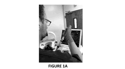
FIGURE 1A is a picture of an endoscopist conducting ultrasound endoscopy on an
experimental animal for EUS-guided cell delivery into lymph node.
FIGURE 1B is a picture of sonography showing the ultrasound image of a Fine
Needle
Aspiration needle reaching a nearby lymph node (LN).
FIGURE 2 shows pictures of direct injection of hepatocytes into periduodenal
lymph
node of an experimental animal conducted by an operative surgeon.
FIGURE 3 is a bar graph summarizing cell viability percentage of different
batches
of hepatocytes that were isolated from donor animals and passed through
needles of
different gauges.
FIGURE 4 shows pictures of opened abdominal cavity of an experimental animal
before and after a portacaval shunt procedure, as well as a diagram
(rightmost) of the
portacaval shunt procedure.
FIGURE 5 is a diagram showing the experimental design of a preclinical study
according to the present disclosure.
FIGURE 6A shows positive staining of CK-18, a marker of hepatocytes, in normal
liver tissue.
FIGURE 6B shows the presence of CK-18 immunostaining signal in lymph nodes 6
days after receiving endoscopic ultrasound (EUS) injection of hepatocytes in a
preclinical
study.
FIGURE 6C shows the presence of CK-18 immunostaining signal in lymph nodes 6
days after receiving direct injection of hepatocytes in a preclinical study.
FIGURE 7A shows the presence of liver tissue in a lymph node about 90 days
after
transplantation of autologous hepatocytes into the lymph node by endoscopic
ultrasound
(EUS) injection.
FIGURE 7B shows the presence of liver tissue in a lymph node about 90 days
after
transplantation of allogenic hepatocytes into the lymph node by endoscopic
ultrasound
(EUS) injection.
-8-
CA 03136471 2021-10-07
WO 2020/210710 PCT/US2020/027783
FIGURE 7C shows the presence of liver tissue and fumarylacetoacetate-hydrolase
(FAH) positive hepatocytes in a lymph node about 60 days after transplantation
of
autologous hepatocytes into the lymph node by endoscopic ultrasound (EUS)
injection.
FIGURE 7D shows the presence of liver tissue and FAH positive hepatocytes in a
lymph node about 150 days after transplantation of allogenic hepatocytes into
the lymph
node by endoscopic ultrasound (EUS) injection.
DETAILED DESCRIPTION
For clarity, but not by way of limitation, the detailed description of the
presently
disclosed subject matter is divided into the following subsections:
1. Overview;
2. Definitions;
3. Cell Delivery Procedures;
4. Production of Ectopic Tissue from Transplanted Cells;
5. Diseases and Conditions;
6. Subjects;
7. Systems; and
8. Kits.
1. Overview
As an overview, the present disclosure relates to methods and systems for
minimally
invasive procedures to induce the development of in vivo organogenesis.
Provided herein
are methods and systems for transplanting cells and growing an ectopic tissue
in a subject
by delivering cells into a lymph node of the subject. The process of growing
the ectopic
tissue from the delivered cells or tissue fragments inside the lymph node with
proper
anatomical and functional features is termed in vivo organogenesis. The
minimally
invasive procedures enabled by the present disclosure can lead to production
of ectopic
tissue(s) that can supplement or augment normal function of one or more organs
for the
subj ect.
As discussed above, one problem associated with current transplantation
therapies,
particularly orthotopic organ transplant, can be that many patients with end-
stage diseases,
such as end-stage liver or renal diseases, are no longer suitable for major
surgeries that can
be required for orthotopic organ transplant or other types of cells
transplant. There can be
significant risks associated with the major surgeries and the prognostication
in these
-9-
CA 03136471 2021-10-07
WO 2020/210710 PCT/US2020/027783
patients can be poor given their deteriorated health conditions. In one
aspect, the present
disclosure provides a minimally invasive procedure (e.g., endoscopic
ultrasound (EUS))
to solve this problem. The methods and systems can generate anatomically
intact and fully
or at least partially functional organs in ectopic sites using the lymph node
as natural
bioreactors. The methods and systems provided herein can minimize the
morbidity and
the mortality for patients with end organ disease that can be often associated
with
additional surgical procedures. The present disclosure can also allow at least
some of the
surgical procedures described herein to be conducted on an outpatient basis,
therefore can
decrease the costs of the initial procedure.
As used herein, the term "ectopic tissue" can refer to a tissue existing at an
ectopic
(non-native) location of a body and having one or more morphological and/or
functional
properties similar to or same as a healthy organ or tissue that can normally
be found at a
native location of the body. The term "functional ectopic tissue" can refer to
an ectopic
tissue that has one or more of the functions of a healthy organ or tissue that
can normally
be found at a native location of the body. The term "ectopic location" can be
used to
describe a location relative to a native location of the subject, e.g., an
ectopic location can
refer to a location that is different than the native location in the
subject's body. For
instance, a liver in a lymph node is ectopic, i.e., an ectopic liver, in a
healthy normal
mammalian body because liver tissue is typically not found in a lymph node.
According
to some aspects of the present disclosure, ectopic tissue can grow in a lymph
node that
contains a population of cells that morphologically resemble hepatocytes and
collectively
can perform one or more functions that a healthy native liver can perform.
In certain embodiments, the methods include advancing an endoscope into a body
lumen or a closed body cavity of a subject. In certain embodiments, the
methods include
advancing an endoscope into the gastrointestinal (GI), respiratory, or urinary
tract of the
subj ect.
In certain embodiments, the endoscope is advanced into a body lumen of the
subject
through an endoluminal approach. As used herein the term "endoluminal
approach" as
used herein refers any approaches, e.g., methods and devices, known in the art
for inserting
an endoscope into a body lumen of the subject. For example, but not by way of
limitation,
the methods include advancing an endoscope through an endoluminal approach
into the
gastrointestinal (GI), respiratory, or urinary tract of the subject.
In certain embodiments, the methods can further include inserting a needle
into a
lymph node of the subject, where the needle is attached to the endoscope. In
certain
-10-
CA 03136471 2021-10-07
WO 2020/210710 PCT/US2020/027783
embodiments, the needle attached to the endoscope is inserted through a
visceral wall into
a lymph node. Non-limiting examples of visceral walls include anatomical
structures
enclosing a hollow viscera and/or an organ along the tract, for instance,
stomach,
duodenum, trachea, bronchi, or bladder. In certain embodiments, the methods
further
include inserting the needle through a transluminal approach. As used herein,
the term
"transluminal approach" refers to any approaches, e.g., methods and devices,
known in the
art for inserting a working instrument (e.g., a needle) across a lumen via the
use of an
endoscope.
In certain embodiments, the methods further include delivering one or more
cells into
the lymph node via the needle. In certain embodiments, the methods include
delivering
one single cell into the lymph node. In certain embodiments, the methods
include
delivering a population of cells into the lymph node. In certain embodiments,
the
population of cells includes one cell type. In certain embodiments, the
population of cells
includes at least two cell types. In certain embodiments, the one or more
cells delivered
into the lymph node can engraft and produce the ectopic tissue in the lymph
node.
In certain embodiments, advancing the endoscope, inserting the needle, or both
are
performed by a minimally invasive or non-invasive method. For example,
ultrasound-
mediated imaging or other detection methods can be applied in the methods
provided
herein for location of the target lymph node. During the process, ultrasound
imaging or
other methods, e.g., minimally invasive or noninvasive detection approaches,
can be
applied for any of advancing the endoscope, locating the suitable target lymph
node, or
monitoring the insertion of the needle through the visceral wall or into the
target lymph
node.
In another aspect, the present disclosure provides methods that include
inserting a
needle into a lymph node in the abdominal, pelvis or thoracic cavity of the
subject with
the aid of ultrasound. The methods can further include delivering one or more
cells into
the lymph node via a needle. Ultrasound-guided location of a lymph node can be
performed by any technology available. For example, but not by way of
limitation, one
can use ultrasound imaging to identify and locate a suitable lymph node for
injection of
the cells. In certain embodiments, ultrasound spectrometry can be utilized for
detecting
the lymph node. In certain embodiments, ultrasound imaging or spectrometry can
be used
in conjunction with other detection methods for localization of the lymph
node.
In certain embodiments, the methods include delivering a population of cells
into a
lymph node of the subject, where the population of hepatocytes engrafts and
produces an
-11-
CA 03136471 2021-10-07
WO 2020/210710 PCT/US2020/027783
ectopic liver in the lymph node. In certain embodiments, the methods further
include
reducing blood supply to the liver of the subject. It has been discovered by
the present
disclosure that a reduction in the blood supply to the liver of the subject
benefits the growth
of the ectopic liver in the lymph node.
2. Definitions
In this application, the use of the singular includes the plural unless
specifically stated
otherwise. It must be noted that, as used in the specification, the singular
forms "a," "an"
and "the" include plural referents unless the context clearly dictates
otherwise.
In this application, the use of "or" means "and/or" unless stated otherwise.
The terms
"and/or" and "any combination thereof' and their grammatical equivalents as
used herein,
can be used interchangeably. These terms can convey that any combination is
specifically
contemplated. Solely for illustrative purposes, the following phrases "A, B,
and/or C" or
"A, B, C, or any combination thereof' can mean "A individually; B
individually; C
individually; A and B; B and C; A and C; and A, B, and C." The term "or" can
be used
conjunctively or disjunctively, unless the context specifically refers to a
disjunctive use.
Furthermore, use of the term "including" as well as other forms, such as
"include",
"includes," and "included," is not limiting.
Reference in the specification to "some embodiments," "certain embodiments,"
"an
embodiment," "one embodiment" or "other embodiments" means that a particular
feature,
structure, or characteristic described in connection with the embodiments is
included in at
least some embodiments, but not necessarily all embodiments, of the present
disclosures.
As used in this specification and claim(s), the words "comprising" (and any
form of
comprising, such as "comprise" and "comprises"), "having" (and any form of
having, such
as "have" and "has"), "including" (and any form of including, such as
"includes" and
"include") or "containing" (and any form of containing, such as "contains" and
"contain")
are inclusive or open-ended and do not exclude additional, unrecited elements
or method
steps. It is contemplated that any embodiment discussed in this specification
can be
implemented with respect to any method or composition of the present
disclosure, and vice
versa. Furthermore, compositions of the present disclosure can be used to
achieve methods
of the present disclosure.
The term "about" in relation to a reference numerical value and its
grammatical
equivalents as used herein can include the numerical value itself and a range
of values plus
or minus 10% from that numerical value.
-12-
CA 03136471 2021-10-07
WO 2020/210710 PCT/US2020/027783
The term "about" or "approximately" means within an acceptable error range for
the
particular value as determined by one of ordinary skill in the art, which will
depend in part
on how the value is measured or determined, i.e., the limitations of the
measurement
system. For example, "about" can mean within 1 or more than 1 standard
deviation, per
the practice in the art. Alternatively, "about" can mean a range of up to 20%,
up to 10%,
up to 5%, or up to 1% of a given value. In another example, the amount "about
10"
includes 10 and any amounts from 9 to 11. In yet another example, the term
"about" in
relation to a reference numerical value can also include a range of values
plus or minus
10%, 9%, 8%, 7%, 6%, 5%, 4%, 3%, 2%, or 1% from that value. Alternatively,
particularly with respect to biological systems or processes, the term "about"
can mean
within an order of magnitude, preferably within 5-fold, and more preferably
within 2-fold,
of a value. Where particular values are described in the application and
claims, unless
otherwise stated the term "about" meaning within an acceptable error range for
the
particular value should be assumed.
3. Cell Delivery Procedures
In certain embodiments, methods and systems of the present disclosure make use
of an
endoscope for transluminal delivery of cells into a lymph node of a subj ect.
In certain
embodiments, the lymph node is located in proximity to or within a body lumen
or a closed
body cavity that can be reached through the use of the endoscope. In certain
embodiments,
the lymph node is located in the abdominal, pelvis or thoracic cavity of a
subject.
As used herein, the term "endoscope" can refer to any instrument that can be
introduced into the body of a subject, e.g., a human subject or a non-human
mammalian
subject, e.g., dog, pig, horse, donkey, rabbit, ox, mouse, or rat, and provide
a view of the
internal parts of the subject. Sometimes, the view provides visual inspection,
for instance,
when the endoscope is equipped with illuminated optics or otherwise aided with
illumination. In certain embodiments, an endoscope of the present disclosure
includes or
is attached to or associated with one or more detection probes and offer
different detection
modes for examination of the internal parts of the subject. For instance, the
endoscope
can have illuminated optics for visual inspection, ultrasound probe for
ultrasound-
mediated detection (e.g., ultrasound imaging), or detectors for radiation,
infrared signal,
radiofrequency signal, or fluorescence signal.
An endoscope described herein can include one or more of the following parts:
a rigid
or flexible tube to travel along the luminal tract of the subject (e.g., GI
tract, respiratory
-13-
CA 03136471 2021-10-07
WO 2020/210710 PCT/US2020/027783
tract, or urinary tract); a light delivery system to illuminate the organ or
object under
inspection; a lens system transmitting the image from the objective lens to a
viewer, an
eyepiece or a video system that displays the images captured by the endoscope
inside the
body; one or more channels to allow entry of medical instruments or
manipulators. The
light source can be outside the body or of a mini size and equipped inside the
endoscope.
When the light source is outside the body, the light can be transmitted via an
optical fiber
system. There can be an optic system, e.g., fiber optics, for transmitting the
light images
captured inside the body. Additionally or alternatively, an endoscope provided
herein can
be equipped with (e.g., include as a part or attached to) other detection
instrument for
different purposes.
The methods provided herein can include advancing the endoscope in a body
lumen or
a closed body cavity of a subject. Non-limiting examples of body lumens
include
gastrointestinal (GI) tract (e.g., esophagus, stomach, duodenum, small
intestine, large
intestine, colon, bile duct, rectum, anus), respiratory tract (e.g., nose,
lower respiratory
tract), ear, urinary tract, cervix, uterus, and fallopian tube. Non-limiting
examples of
closed body cavities include abdominal cavity, and pelvic cavity. In certain
embodiments,
the methods disclosed herein include advancing the endoscope in the
gastrointestinal,
respiratory, or urinary tract of the subject.
In certain embodiments, the methods disclosed herein include advancing the
endoscope along a body lumen or in a closed body cavity of a subject for
delivering cells
into at least one lymph node that is located in proximity to or in the body
lumen or the
closed body cavity. In certain embodiments, the location of the lymph node is
chosen
based on the purpose of the cell transplantation. In certain embodiments, the
lumen and/or
closed body cavity is chosen based on the lymph node that is suitable for cell
transplantation.
In certain embodiments, the methods disclosed herein includes advancing the
endoscope along the GI tract for delivering cells into one or more lymph nodes
in
proximity to the GI tract. An endoscope (e.g., an esophagoscope or a
gastroscope) can be
used to deliver cells into lymph nodes in proximity to the upper part or lower
part of the
GI tract. For instance, an esophagoscope can be used to deliver cells into
lymph nodes
close to the esophagus, and a gastroscope can be used to deliver cells into
the perigastric
and/or periduodenal lymph nodes. Non-limiting examples of lymph nodes in
proximity to
the GI tract in which cells can be delivered into using the methods provided
herein include
periduodenal lymph node, perigastric lymph node, peripancreatic lymph node,
mesenteric
-14-
CA 03136471 2021-10-07
WO 2020/210710 PCT/US2020/027783
lymph node, ileocolic lymph node, mesocolic lymph node, gastric lymph node,
hepatic
splenic lymph node, splenic hilar lymph node, paraoesophageal lymph node,
paracardial
lymph node, paraaortic lymph node, retroaortic lymph node, lateral aortic
lymph node,
preaortic lymph node, lesser curve lymph node, common hepatic lymph node,
splenic
artery lymph node, coeliac axis lymph node, iliac lymph node, and
retroperitoneal lymph
node. The endoscope can be introduced from the mouth of the subj ect, or in
certain
embodiments, from the nose of the subject. Alternatively, the endoscope can be
introduced
from the anus of the subj ect, for instance, a proctoscope, sigmoidoscope, or
colonoscope
can be used to deliver cells into lymph nodes close to rectum or colon. In
certain
embodiments, the endoscope is flexible to travel along the GI tract, and an
investigative
examination is conducted in order to locate the suitable lymph node for the
purpose of cell
transplantation.
In certain embodiments, the methods disclosed herein include advancing the
endoscope along the respiratory tract of the subject for delivering cells into
one or more
lymph nodes in proximity to the respiratory tract. For example, but not by way
of
limitation, a bronchoscope or a laryngoscope is used to target lymph nodes
close to trachea
or bronchi of the lungs, or the larynx of the subject. Non-limiting examples
of lymph
nodes in proximity to the respiratory tract in which cells can be delivered
into using
methods provided herein include parasternal lymph node, an intercostal lymph
node,
superior diaphragmatic lymph node, superior tracheobronchi lymph node,
inferior
tracheobronchi lymph node, bronchopulmonary lymph node, paratracheal lymph
node,
and intrapumonary lymph node. The bronchoscope or laryngoscope can be
introduced
from the mouth of the subject, or in certain embodiments, from the nose of the
subject.
In certain embodiments, the methods disclosed herein includes advancing the
endoscope along the urinary tract of the subject for delivering cells into one
or more lymph
modes in proximity to the urinary tract. For example, but not by way of
limitation, a
cystoscope is used to target lymph nodes close to urethra or bladder. The
cystoscope can
be introduced from the urethra of the subject. Non-limiting examples of lymph
nodes in
proximity to the urinary tract in which cells can be delivered into using
methods provided
herein include one or more of an external iliac lymph node, an internal iliac
lymph node,
a caval lumbar lymph node, an aortic lumbar lymph node, a superficial inguinal
lymph
node, a profound inguinal lymph node, an interaortocaval pen-bladder lymph
node, an
obturator pen-bladder lymph node, or a pre-sacral pen-bladder lymph node.
-15-
CA 03136471 2021-10-07
WO 2020/210710 PCT/US2020/027783
In certain embodiments, the methods disclosed herein include advancing the
endoscope in a closed body cavity (e.g., abdominal cavity, pelvis cavity) for
delivering
cells into at least one lymph node inside or near the closed body cavity. In
certain
embodiments, the endoscope is introduced into the closed body cavity through a
small
incision, e.g., a small incision on the surface of the abdomen. In certain
embodiments, the
closed body cavity is an abdominal cavity or a pelvis cavity. Non-limiting
examples of
lymph nodes inside or near the abdominal cavity or pelvis cavity include
splenic lymph
nodes, hepatic lymph nodes, cystic lymph nodes, foraminal lymph nodes,
right/left gastric
lymph nodes, pyloric lymph nodes, super pyloric lymph nodesõ sub pyloric lymph
nodes,
retro pyloric lymph nodes, superior pancreatic lymph nodes, inferior lymph
nodes,
superior/inferior pancreaticoduodenal lymph nodes, inferior mesenteric lymph
nodes,
sigmoid lymph nodes, superior rectal lymph nodes, mesocolic lymph nodes, left
colic
lymph nodes, right colic lymph nodes, middle colic lymph nodes, appendicular
lymph
nodes, ileocolic lymph nodes, retrocaecal lymph nodes, pretectal lymph nodes,
superior
mesenteric lymph nodes, left lumbar lymph nodes, lateral aortic lymph nodes,
and
preaortic lymph nodes.
In certain embodiments, the methods and systems disclosed herein use
ultrasound for
locating a lymph node in which cells will be transplanted, and/or for guiding
the steps of
advancing the endoscope through the body lumen or closed body cavity,
inserting the
needle, or any combination thereof Ultrasound is sound waves with frequencies
higher
than the upper audible limit of human hearing, which can be about more than 20
kHz to
several gigahertz. Ultrasound imaging (or sonography) can be performed in a
variety of
fashions depending on the intended purpose of applying the methods and systems
provided
herein. Non-limiting examples of sonography can include Doppler
ultrasonography,
contrast ultrasonography, molecular ultrasonography, elastography,
interventional
ultrasonography, and compression ultrasonography. In certain embodiments, the
ultrasound imaging technology used herein has high spatial and/or temporal
resolution for
the purpose of locating the suitable lymph node for injection, using
technologies such as
those described in International Patent Publication Nos. W02018222724A1 and
W02018134729A1, each of which is incorporated herein by reference.
In certain embodiments of the present disclosure, the methods disclosed herein
uses
endoscopy in combination with ultrasound imaging, where the endoscope is
attached to an
ultrasound probe (e.g., as part of the endoscope or as a separate piece). The
term
"ultrasound endoscopy," "endoscopic ultrasound," or "EUS," used
interchangeably
-16-
CA 03136471 2021-10-07
WO 2020/210710 PCT/US2020/027783
herein, can refer to a medical procedure in which endoscopy is combined with
ultrasound
to detect (e.g., to obtain images of), and/or manipulate the internal organs
in the chest,
abdomen and pelvis, or any other internal structures of a body. EUS instrument
and
technology used in the methods and systems provided herein can be those
commercially
available currently, and/or those as described in U.S. Patent Publication Nos.
US20060106306A1 and US20070237373A1, and International Patent Publication No.
W01998009247A1, each of which is incorporated herein in its entirety by
reference.
In certain embodiments, ultrasound imaging of the target lymph node (e.g., the
lymph
node into which one or more cells are to be delivered) is conducted with the
aid of other
imaging technologies, such as radiological imaging. Radiological imaging can
include
dynamic radiological imaging (fluoroscopy), computed tomography (CT), or
magnetic
resonance imaging (MRI). For example, but not by way of limitation, computed
tomography (CT) is performed in order to gain anatomical information of the
whole body
or some local organ(s) or tissue(s) of the subject. In certain embodiments,
magnetic
resonance or any other medical arts available is used in conjunction with or
in lieu of the
ultrasound imaging for locating the target lymph node in the abdominal, pelvis
or thoracic
cavity of the subject.
The methods and systems provided herein can include using a needle for
delivering
the one or more cells into a lymph node. The needle can be part of an
injector, which can
be configured to receive the cells to be delivered and push the cells out
through the needle
for delivery. The needle provided herein can be configured to have certain a
degree of
sharpness and stiffness. For instance, the needle is to be inserted into the
lymph node. In
certain embodiments, the needle is also configured to penetrate the wall of
the GI tract
(e.g., the wall of esophagus, stomach, intestine, or colon), the respiratory
tract, the urinary
tract (e.g., the wall of the urethra or the bladder), so that the needle can
reach the lymph
node outside the tract. Any suitable needles known in the art can be used with
the methods
disclosed herein.
In certain embodiments, the needle is attached to the endoscope, e.g., as a
part of the
endoscope or as a separate instrument. The needle can be those used in Fine
Needle
Aspiration (FNA) procedures. FNA needles can be commercially available or
specifically
designed for the purpose of applying the teachings of the present disclosure.
FNA needles
are typically used for biopsy purpose, e.g., making cut into tissue and
aspirating the tissue
fragment for diagnostic purposes. In the methods and systems provided herein,
FNA
needles can be used for cell delivery purpose instead. In certain embodiments,
the FNA
-17-
CA 03136471 2021-10-07
WO 2020/210710 PCT/US2020/027783
needle is operated through a linear array echoendoscope EUS/FNA equipment. An
exemplary EUS/FNA equipment can be configured for the controlled and measured
advancement of the FNA needle (e.g., a hollow needle with a solid removable
stylet)
within a semirigid protective sheath. The EUS/FNA equipment can also have a
handle
with a port for stylet insertion or withdrawal and for attachment of a syringe
from where
live cells can be inserted. In certain embodiments, the needle is inserted
within the
protective sheath into the lymph node identified acoustically with the EUS
probe from the
echoendoscope equipment. The needle can then be advanced out of the sheath and
inserted
transluminally into the target lymph node under direct ultrasound guidance. A
stopcock
attached to the tip of the syringe can assist in creating and holding vacuum
within the
needle body. Once the needle tip is in the target lymph node and the stylet is
removed, a
syringe containing live cells in a culture medium can be connected onto the
needle handle.
Once the stopcock is opened, the needle placed within the lymph node
parenchyma allows
the cells to be infused promptly and under direct view. The needles can have
adjustable
spacers/sliders at the distal portion of the handle to allow modification of
the length of
sheath exiting the scope, which can assure an additional level of safety and
accuracy when
injecting nearby lymph nodes.
In certain embodiments, the needle is configured, e.g., the size of the needle
is
configured, for ease of injection of cells into the lymph node. In certain
embodiments, the
size (e.g., the gauge) of the needle is selected based on the location of the
target lymph
node, the type of cells to be delivered, and the amount of cells to be
delivered. In certain
embodiments, the smaller the needle is, the more flexible it is to reach and
insert into a
target lymph node. In certain embodiments, it is easier to insert a relatively
small needle
into a small target lymph node, which can otherwise tent around a relatively
large needle.
In certain embodiments, the size of the needle, e.g., the inner diameter of
the needle, is
configured to allow the cells being pushed out without clogging up the needle.
As
described herein, the size of the needle refers to the size of the tip of the
needle, not the
hub of the needle where the needle is joined with other part of the injector.
As disclosed
herein, the outer diameter of the needle, refers to the first full diameter
outside the wall of
the needle from the tip, and the inner diameter of the needle refers to the
first full diameter
inside the wall of the needle from the tip.
In certain embodiments, the needle has an inner diameter of at most about 700
m,
600 m, 500 m, 450 m, 400 m, 300 m, 260 m, 250 i_tm or 200 m. In certain
-18-
CA 03136471 2021-10-07
WO 2020/210710 PCT/US2020/027783
embodiments, the needle has an inner diameter of at most about 260 m. In
certain
embodiments, the inner diameter of the needle can be about 700 m, 600 m, 500
m,
450 m, 400 m, 300 m, 260 m, 250 m, or 200 m. In certain embodiments, the
inner
diameter of the needle is about 260 Jim. The needle can have an outer diameter
of at most
about lmm, 900 m, 800 m, 750 m, 700 m, 650 m, 600 m, 550 m, 520 m, 510
m, 500 m, 480 m, 450 m, or 400 m. In certain embodiments, an outer
diameter of
the needle is at most about 510 m. In certain embodiments, the outer diameter
of the
needle is about lmm, 900 m, 800 m, 750 m, 700 m, 650 m, 600 m, 550 m,
520
m, 510 m, 500 m, 480 m, 450 m, or 400 m. In certain embodiments, an outer
diameter of the needle is about 510 m. In certain embodiments, the needle is
of a certain
gauge, as prescribed according to ISO 7864:2016. For example, but not by way
of
limitation, the needle is about 19, 19.5, 20, 20.5, 21, 21.5, 22, 22.5, 23.5,
24, 24.5, 25,
25.5, 26, 26.5, or 27 gauge (ga). In certain embodiments, the needle is at
most about 19,
19.5, 20, 20.5, 21, 21.5, 22, 22.5, 23.5, 24, 24.5, 25, 25.5, 26, 26.5, or 27
ga. The needle
can be at most about 25 ga. As described herein, when a comparison is made for
the size
of needle relative to a particular gauge size, the comparison is made between
the outer
diameter of the needle and the outer diameter of a standardized hypodermic
needle of that
particular gauge as prescribed by ISO 7864:2016. In certain other embodiments,
the
needle has a relatively small outer diameter and a relatively large inner
diameter. In certain
embodiments, the needle has a non-standard size, for instance, having a thin
wall while
maintaining a large inner diameter and a small outer diameter.
The total number of cells and the cell concentration inside the injector of
the needle
are selected based on a number of factors, for instance, the size of the cells
to be injected,
the type of ectopic tissue to produce, the proliferation capability of the
cells, the target
mass of the ectopic tissue, the size of the needle for injection, and the size
of the target
lymph node.
In certain embodiments, the injector as described herein receives at least
about 10
million, 20 million, 30 million, 40 million, 50 million, 60 million, 70
million, 80 million,
90 million, 100 million, 200 million, 300 million, 400 million, 500 million,
600 million,
700 million, 800 million, 900 million, 1 billion, 3 billion, 5 billion, 8
billion, 10 billion, 20
billion, 50 billion, or 100 billion cells for injection. In certain
embodiments, the injector
as described herein has about 10 million, 20 million, 30 million, 40 million,
50 million, 60
million, 70 million, 80 million, 90 million, 100 million, 200 million, 300
million, 400
-19-
CA 03136471 2021-10-07
WO 2020/210710 PCT/US2020/027783
million, 500 million, 600 million, 700 million, 800 million, 900 million, 1
billion, 3 billion,
billion, 8 billion, 10 billion, 20 billion, 50 billion, or 100 billion cells
for injection. In
certain embodiments, the injector as described herein has at most about 10
million, 20
million, 30 million, 40 million, 50 million, 60 million, 70 million, 80
million, 90 million,
5 .. 100 million, 200 million, 300 million, 400 million, 500 million, 600
million, 700 million,
800 million, 900 million, 1 billion, 3 billion, 5 billion, 8 billion, 10
billion, 20 billion, 50
billion, or 100 billion cells for injection. In certain embodiments, the
injector receives
from about 50 million to about 200 million cells for injection.
In certain embodiments, the injector as described herein receives at least
about 10
million, 20 million, 30 million, 40 million, 50 million, 60 million, 70
million, 80 million,
90 million, or 100 million hepatocytes for production of an ectopic liver. In
certain
embodiments, the injector as described herein has about 10 million, 20
million, 30 million,
40 million, 50 million, 60 million, 70 million, 80 million, 90 million, or 100
million
hepatocytes for production of an ectopic liver. In certain embodiments, the
injector as
described herein has at most about 10 million, 20 million, 30 million, 40
million, 50
million, 60 million, 70 million, 80 million, 90 million, or 100 million
hepatocytes for
production of an ectopic liver. In certain embodiments, the injector receives
from about
50 million to about 200 million cells for production of an ectopic liver.
In certain embodiments, the injector as described herein has a suspension
solution of
cells having at least about 30 million, 40 million, 45 million, 50 million, 55
million, 60
million, 70 million, 80 million, 90 million, 100 million, 200 million, 300
million, 400
million, 500 million, 600 million, 700 million, 800 million, 900 million, 1
billion, 3 billion,
5 billion, 8 billion, or 10 billion cells per mL. In certain embodiments, the
injector as
described herein has a suspension solution of cells having about 30 million,
40 million, 45
million, 50 million, 55 million, 60 million, 70 million, 80 million, 90
million, 100 million,
200 million, 300 million, 400 million, 500 million, 600 million, 700 million,
800 million,
900 million, 1 billion, 3 billion, 5 billion, 8 billion, or 10 billion cells
per mL. In certain
embodiments, the injector as described herein has a suspension solution of
cells having at
most about 30 million, 40 million, 45 million, 50 million, 55 million, 60
million, 70
million, 80 million, 90 million, 100 million, 200 million, 300 million, 400
million, 500
million, 600 million, 700 million, 800 million, 900 million, 1 billion, 3
billion, 5 billion, 8
billion, or 10 billion cells per mL.
In certain embodiments, the injector as described herein receives a suspension
solution
of hepatocytes having at least about 30 million, 40 million, 45 million, 50
million, 55
-20-
CA 03136471 2021-10-07
WO 2020/210710 PCT/US2020/027783
million, 60 million, 70 million, 80 million, 90 million, or 100 million cells
per mL for
production of an ectopic liver. In certain embodiments, the injector as
described herein
receives a suspension solution of hepatocytes having about 30 million, 40
million, 45
million, 50 million, 55 million, 60 million, 70 million, 80 million, 90
million, or 100
million cells per mL for production of an ectopic liver. In certain
embodiments, the
injector as described herein receives a suspension solution of hepatocytes
having at most
about 30 million, 40 million, 45 million, 50 million, 55 million, 60 million,
70 million, 80
million, 90 million, or 100 million cells per mL for production of an ectopic
liver.
In certain embodiments, the needle for injection of hepatocytes is selected to
be at most
about 25 gauge, and the needle delivers the cells including hepatocytes in a
suspension
solution that has at least about 50 million viable cells per mL. In certain
embodiments,
the population of cells inside the injector has at least about 65% viable
cells.
Without wishing to be bound to a certain theory, the viability of the cells
delivered into
the lymph node can be important for the formation and function of the ectopic
tissue. In
certain embodiments, the viability percentage in the population of cells needs
to be
controlled. The injector, e.g., the needle, can be configured to maintain the
viability of the
cells during the delivery process. For instance, the needle size and the cell
concentration
of the suspension solution can be configured so that the viability percentage
does not
significantly decrease when the cells pass through the needle during cell
delivery process.
In certain embodiments, the cell delivery leads to less than about 20%, 15%,
10%, 9%,
8.5%, 8%, 7.5%, 7%, 6.5%, 6%, 5.5%, 5%, 4.5%, 4%, 3.5%, 3%, 2.5%, 2%, 1.5%,
1%, or
0.5% reduction in cell viability percentage in the population of cells when
the cells pass
through the needle. In certain embodiments, the reduction of cell viability
percentage is
less than about 10% when the cells pass through the needle during cell
delivery (e.g., as
compared to the viability of the cells prior to injection).
In certain embodiments, the methods provided herein include reducing blood
supply
to the liver of the subject, and delivering one or more hepatocytes into a
lymph node of
the subject. The reduction of blood supply to the liver of the subject can
induce
hepatocellular dysfunction, e.g., malfunction or death of at least some of the
liver cells,
and consequentially reduction or loss of function of the liver. Without
wishing to be bound
to a particular theory, the reduction or loss of function of liver in the
subject after reduction
of blood supply to the liver can have a compensatory impact on the growth of
the ectopic
liver in the lymph node after hepatocyte transplantation into the lymph node.
In other
words, the step of reducing blood supply to the liver, in certain embodiments,
facilitates
-21-
CA 03136471 2021-10-07
WO 2020/210710 PCT/US2020/027783
the formation of the ectopic liver in the lymph node, and/or, in certain
embodiments,
improves the functional performance of the ectopic liver in the lymph node. In
certain
embodiments of the methods, cell delivery is carried out by direct injection
(e.g., via major
surgery or percutaneous injection) of the hepatocytes or EUS-guided injection
of the
.. hepatocytes, when performed in combination with the step of reducing blood
supply to the
liver.
A surgical method can be used for reducing blood supply to the liver, which
can
involve redirecting the blood inflow from the portal vein. For instance, a
transjugular
intrahepatic portosystemic shunt (TIPS) procedure is performed to establish
direct
communication between the inflow portal vein and the outflow hepatic vein,
thus the blood
flow through the liver can be reduced. TIPS refers to an artificial channel
within the liver
that establishes communication between the portal vein and the hepatic vein.
TIPS has
been used as a stand-alone therapy to alleviate the detrimental splanchnic and
systemic
hemodynamic impact of progressive portal hypertension due to end stage liver
disease. In
.. certain embodiments, the methods disclosed herein include using TIPS in
combination
with hepatocyte transplant in a lymph node to produce an ectopic liver in the
lymph node.
Transjugular intrahepatic portosystemic shunts can be surgically placed in the
liver
through a major surgery involving opening of the abdominal cavity, or a
minimally
invasive procedure. In certain embodiments, transjugular intrahepatic
portosystemic
shunts is placed under fluoroscopic guidance. Access to the liver can be
obtained via the
internal jugular vein in the neck. Once access to the jugular vein is
confirmed, a guidewire
and introducer sheath can be placed to facilitate the shunt's placement. This
step can
enable the access to the patient's hepatic vein by traveling from the superior
vena cava
into the inferior vena cava and finally the hepatic vein. Once the catheter is
in the hepatic
.. vein, a wedge pressure can be obtained to calculate the pressure gradient
in the liver.
Following this, carbon dioxide can be injected to locate the portal vein.
Then, a special
needle known as a Colapinto can be advanced through the liver parenchyma to
connect the
hepatic vein to the large portal vein, near the center of the liver. The
channel for the shunt
can be next created by inflating an angioplasty balloon within the liver along
the tract
.. created by the needle. The shunt can be completed by placing a special mesh
tube known
as a stent or endograft to maintain the tract between the higher-pressure
portal vein and
the lower-pressure hepatic vein. After the procedure, fluoroscopic images can
be made to
show placement.
-22-
CA 03136471 2021-10-07
WO 2020/210710 PCT/US2020/027783
In certain other embodiments, portacaval shunt procedure is performed to
establish
communication between the portal vein and the inferior vena cava, in order to
reduce blood
supply to the liver. Portacaval shunt can be placed by surgical methods such
as, but not
limited to, the procedure described in Example 2. Other surgical procedures
that can be
used can include end-to-side or side-to-side portocaval shunt (SSPCS),
mesocaval shunts
(MCS) with interposition H-or C-grafts and splenorenal shunts (SRS) (either
central or
distal).
In certain embodiments, hepatocytes are delivered to a lymph node after blood
supply
to the liver is reduced. In certain embodiments, hepatocyte transplant is
performed
immediately after the step of reducing blood supply to the liver. In certain
embodiments,
hepatocyte transplant is performed a period after the step of reducing blood
supply to the
liver, for instance, about 1 hour, 2 hours, 6 hours, 12 hours, 18 hours, 24
hours, 36 hours,
2 days, 3 days, 4 days, 5 days, 7 days, 10 days, 2 weeks, 3 weeks, or 4 weeks.
In certain
embodiments, such an interval cannot be longer than a certain amount of time,
in order to
avoid complete liver failure, which can be life-threatening. For example, but
not by way
of limitation, the interval is shorter than about 12 hours, 18 hours, 24
hours, 36 hours, 2
days, 3 days, 4 days, 5 days, 7 days, 10 days, 2 weeks, 3 weeks, or 4 weeks.
Alternatively,
hepatocyte transplant can also be performed shortly before the step of
reducing blood
supply to the liver. In certain embodiments, the interval can be shorter than
about 1 hour,
2 hours, 6 hours, 12 hours, 18 hours, 24 hours, 36 hours, 2 days, 3 days, 4
days, 5 days, 7
days, 10 days, 2 weeks, or 3 weeks. The interval between the two steps can
depend on the
extent of the reduction in blood supply to the liver and the health condition
of the subject
receiving the procedures. Doctor's medical assessment on a case-by-case basis
can be
involved in determining the interval between the two steps as described
herein.
Transplant rejection can be a problem associated with any transplantation
procedure
involving implant of non-autologous organs or cells. Immunosuppression can be
applied
to prevent or ameliorate the transplant rejection induced by procedures
according the
methods of the present disclosure. For example, immunosuppressant drugs can be
administered to the subject shortly before or immediately after cell
transplantation is
completed, or at about 1 hour, 2 hours, 6 hours, 12 hours, 18 hours, 24 hours,
36 hours, 2
days, 3 days, 4 days, 5 days, 7 days, 10 days, 2 weeks, 4 weeks, 6 weeks, 2
months, 3
months, 4 months, 5 months, 6 months, 9 months, 12 months, 18 months, 2 years,
3 years,
4 years, or even longer after the procedure.
-23-
CA 03136471 2021-10-07
WO 2020/210710 PCT/US2020/027783
Different approaches of immunosuppression can be applied herein. For example,
but
not by way of limitation, the subject is administered an immunosuppressant for
induction
immunosuppression, which can include all medications given immediately after
transplantation in intensified doses for the purpose of preventing acute
rejection. Non-
limiting examples of associated medications can include Methylprednisolone,
Atgam,
Thymoglobulin, OKT3, Basiliximab, Solumedrol, and Daclizumab. The subject can
also
be administered an immunosuppressant for maintenance immunosuppression, which
can
include all immunosuppressive medications given before, during or after
transplant with
the intention to maintain them long-term. For example, but not by way of
limitation,
Prednisone, Cyclosporine, Tacrolimus, Mycophenolate Mofetil, Azathioprine,
Prograf, or
Rapamycin can be used for maintenance immunosuppression as described herein.
In
certain embodiments, the subject can also be administered an immunosuppressant
for anti-
rejection immunosuppression, which can include all immunosuppressive
medications
given for the purpose of treating an acute rejection episode during the
initial post-
transplant period or during a specific follow-up period, e.g., up to 30 days
after the
diagnosis of acute rejection. Non-limiting examples of associated medications
can include
Methylprednisolone, Atgam, OKT3, Thymoglobulin, Basiliximab, or Daclizumab.
Other
examples of immunosuppressant that can be used in the subject methods can also
include
other steroids (such as corticosteroids, dexamethasone, and prednisone), Cox-1
and Cox-
2 inhibitors, macrolide antibiotics (such as rapamycin and tacrolimus), and
other
substances that limit, reduce, or suppress B-cell, T-cell, and/or other innate
immune
activity.
4. Production of Ectopic Tissue from Transplanted Cells
The methods and systems provided herein can be applied to transplant various
types
of cells, including, but not limited to, hepatocytes, kidney cells or kidney
tissue fragments,
pancreatic cells or islets, thymic cells or thymus fragments, or lung cells or
lung tissue
fragments. The cells transplanted into the lymph node can engraft and form
ectopic tissue
that can supplement or augment one or more functions of a normal organ of a
subject.
The cells to be transplanted into a lymph node can be either a homogenous cell
population or a heterogeneous cell population, depending on the purpose of the
cell
transplant. For example, in certain embodiments, a population of homogeneous
hepatocytes is delivered into a lymph node to produce an ectopic liver. In
certain
embodiments, when growing an ectopic liver, a heterogeneous population of
cells, e.g.,
-24-
CA 03136471 2021-10-07
WO 2020/210710 PCT/US2020/027783
hepatocytes with additional other liver parenchymal cells, is delivered into a
lymph node.
In certain embodiments, a population of heterogeneous kidney cells or kidney
fragments
is delivered into a lymph node for the purpose of generating an ectopic kidney
in the lymph
node. In certain embodiments, an embryonic kidney from a donor subject or a
kidney
.. organoid that are generated by in vitro differentiation methods (such as
those described in
International Patent Publication Nos. W02014182885A2, W02019006127A1, and
W02018227101A1, each of which is incorporated herein by reference) is
obtained, and is
processed (e.g., minced or grinded) into small fragments and resuspended in a
liquid to
form a solution to be delivered into a lymph node. The solution includes
different types
of cells that constituting the embryonic kidney or the kidney organoid. In
certain
embodiments, a population of heterogeneous pancreatic cells is delivered into
the lymph
node for generating an ectopic pancreas.
The methods and systems provided herein can involve delivering a
therapeutically-effective amount of cells into the lymph node of a subject.
The term
"therapeutically-effective amount" as used herein when referring to a
population of cells
to be transplanted or delivered can mean the amount of relevant cells in the
population of
cells, e.g., the cells to be transplanted, or composition including the cells
to be transplanted,
that is effective for producing a desired therapeutic effect in the subject
receiving the cell
transplant in the lymph node at a reasonable benefit/risk ratio applicable to
any medical
treatment. For example, but not by way of limitation, the amount of a
population of cells
transplanted into a subject is sufficient to produce a statistically
significant, measurable
change in one or more symptoms of the disease or condition the transplantation
is intended
to treat, e.g., end-stage liver disease or renal disease. Determination of a
therapeutically
effective amount is dependent on the intended medical purpose of applying the
subject
methods or systems. A therapeutically effective amount can vary with the
subject's
history, age, condition, sex, as well as the severity and type of the medical
condition in the
subject, and administration of other pharmaceutically active agents.
Depending on the intended purpose of applying the methods and systems provided
herein, different amounts of cells can be delivered into a lymph node for
growing an
ectopic tissue. There can be at least about 1 million, 2 million, 3 million, 4
million, 5
million, 7 million, 9 million, 10 million, 15 million, 20 million, 30 million,
40 million, 45
million, 50 million, 55 million, 60 million, 70 million, 80 million, 90
million, 100 million,
150 million, 200 million, 300 million, 500 million, 750 million, 800 million,
900 million,
1 billion, 5 billion, 10 billion, 20 billion, 30 billion, 50 billion, or even
more cells delivered
-25-
CA 03136471 2021-10-07
WO 2020/210710 PCT/US2020/027783
per lymph node. In certain embodiments, about 10 million, 15 million, 20
million, 30
million, 40 million, 45 million, 50 million, 55 million, 60 million, 70
million, 80 million,
90 million, or 100 million cells can be delivered into a single lymph node. In
certain
embodiments, about 50 million to about 200 million of cells can be delivered
in a single
lymph node.
The population of cells to be delivered can have at least about 40%, 45%, 50%,
55%,
60%, 65%, 66%, 67%, 68%, 69%, 70%, 71%, 72%, 73%, 74%, 75%, 80%, 85%, 90%,
95%, or about 100% viable cells. Measures available to promote cell viability
can be
applied in order to maintain the cell viability level in the population of
cells to be delivered.
In certain embodiments, prior to cell delivery, the viability status of the
population of cells
(e.g., viability percentage or other cell viability parameters) can be
measured in order to
ensure viable cell population to be delivered and consequently functional
ectopic tissue
can be produced in the lymph node.
In non-limiting embodiments, the ectopic tissue can be formed at least about 5
days,
10 days, 15 days, 20 days, 25 days, 30 days, 35 days, 40 days, 45 days, 50
days, 55 days,
60 days, 65 days, 70 days, 75 days, 80 days, 85 days, 90 days, 95 days, 100
days, 110 days,
120 days, 130 days, 140 days, 150 days, 160 days, 170 days, 180 days, 190
days, or about
200 days after transplantation of the disclosed various types of cells into a
lymph node.
For example, but not by way of limitation, hepatocytes can be delivered into
lymph nodes
via EUS, and the engrafted hepatocytes can form a liver tissue about 60, about
90, or about
150 days after the transplantation of the hepatocytes. In non-limiting
embodiments, the
ectopic tissue can be formed within about 5 days, 10 days, 15 days, 20 days,
25 days, 30
days, 35 days, 40 days, 45 days, 50 days, 55 days, 60 days, 65 days, 70 days,
75 days, 80
days, 85 days, 90 days, 95 days, 100 days, 110 days, 120 days, 130 days, 140
days, 150
days, 160 days, 170 days, 180 days, 190 days, or about 200 days after
transplantation of
the disclosed various types of cells into a lymph node. For example, but not
by way of
limitation, hepatocytes can be delivered into lymph nodes via EUS, and the
engrafted
hepatocytes can start forming a liver tissue within about 60, about 90, or
about 150 days
after the transplantation of the hepatocytes.
The cells to be delivered can be obtained from different sources and via a
number of
different methods. The cells can be allogeneic, xenogeneic, or autologous to
the subject.
The cells can be obtained directly from a live donor tissue. For example,
hepatocytes are
isolated and prepared for transplant according to the methods described in
U.S. Patent Nos.
9125891B2 and 6610288B1, U.S. Patent Publication No. 20120045764A1 and
-26-
CA 03136471 2021-10-07
WO 2020/210710 PCT/US2020/027783
20040110289A1, each of which is incorporated herein by reference. In certain
embodiments, the cells to be delivered are obtained from in vitro sources.
Stem cells, such
as embryonic stem cells, induced pluripotent stem cells, trophoblast stem
cells, or any
other type of pluripotent or multipotent stem cells, can be cultured in vitro
and
differentiated into certain types of cells or cell populations that are
suitable for the purposes
of applying the methods and systems provided herein. For example, as described
above,
kidney organoids or pancreatic islets are obtained using in vitro
differentiation methods
and they are prepared for transplantation procedures described herein. The
cells to be
delivered can also be stored, e.g., cryopreserved, prior to transplantation.
Standard
cryopreservation and recovery protocols can be used so along as the cell
viability level cis
suitable for the subsequent cell delivery and ectopic tissue formation in the
lymph node.
In certain embodiments, the cells to be delivered are genetically modified so
that one or
more genes of the cells are modified, or the cells are modified to express one
or more
exogenous genes. Any available gene editing methods, such as homologous
recombination, transposase/transposon, Zinc Finger nuclease, TALEN, and
CRIPSR, e.g.,
CRISPR-Cas9, technologies, can be applied for gene modification of the cells
to be
delivered.
The cells can be prepared and suspended in solutions for transplant. In
certain
embodiments, the suspension solution containing the cell population can
further include
pharmaceutically acceptable excipients, diluents, or carriers. As used herein,
the term
"pharmaceutically acceptable" can refer to those compounds, materials,
compositions,
and/or dosage forms which are, within the scope of sound medical judgment,
suitable for
use in contact with the tissues of the subject (e.g., human subject or
nonhuman animals)
without excessive toxicity, irritation, allergic response, or other problem or
complication,
commensurate with a reasonable benefit/risk ratio.
As used herein, the term "solution" can include a pharmaceutically acceptable
carrier
or diluent in which the cells of the invention remain viable. Pharmaceutically
acceptable
carriers and diluents include saline, aqueous buffer solutions, solvents
and/or dispersion
media. The use of such carriers and diluents is well known in the art. The
solution is
preferably sterile and fluid to the extent that easy syringability exists. The
solution can be
stable under the conditions of manufacture and storage and preserved against
the
contaminating action of microorganisms such as bacteria and fungi through the
use of, for
example, parabens, chlorobutanol, phenol, ascorbic acid, thimerosal, and the
like.
Solutions of the present disclosure can be prepared by incorporating viable,
functional
-27-
CA 03136471 2021-10-07
WO 2020/210710 PCT/US2020/027783
cells as described herein in a pharmaceutically acceptable carrier or diluent
and, as
required, other ingredients enumerated above, followed by filtered
sterilization.
Non-limiting examples of substances or materials that can serve as
pharmaceutically-acceptable excipients for the purpose of the present
disclosure can
include: sugars, such as lactose, glucose and sucrose; starches, such as corn
starch and
potato starch; cellulose, and its derivatives, such as sodium carboxymethyl
cellulose,
methylcellulose, ethyl cellulose, microcrystalline cellulose and cellulose
acetate;
powdered tragacanth; malt; gelatin; lubricating agents, such as magnesium
stearate,
sodium lauryl sulfate and talc; cocoa butter and suppository waxes; oils, such
as peanut
oil, cottonseed oil, safflower oil, sesame oil, olive oil, corn oil and
soybean oil; glycols,
such as propylene glycol; polyols, such as glycerin, sorbitol, mannitol and
polyethylene
glycol (PEG); esters, such as ethyl oleate and ethyl laurate; agar; buffering
agents, such as
magnesium hydroxide and aluminum hydroxide; alginic acid; pyrogen-free water;
isotonic
saline; Ringer's solution; ethyl alcohol; pH buffered solutions; polyesters,
polycarbonates
and/or polyanhydrides; bulking agents, such as polypeptides and amino acids;
serum
component, such as serum albumin, HDL and LDL; C2-C12 alcohols, such as
ethanol; and
other non-toxic compatible substances employed in pharmaceutical formulations.
Wetting
agents, coloring agents, release agents, coating agents, sweetening agents,
flavoring
agents, perfuming agents, preservative and antioxidants can also be present in
the
formulation. The terms such as "excipient", "carrier", "pharmaceutically
acceptable
carrier" or the like are used interchangeably herein. As used herein, the term
"pharmaceutically-acceptable excipient" can refer to a pharmaceutically-
acceptable
material, composition or vehicle, such as a liquid or solid filler, diluent,
carrier,
manufacturing aid (e.g., lubricant, talc magnesium, calcium or zinc stearate,
or stearic
acid), or solvent encapsulating material, involved in carrying or transporting
the subject
compound, materials, or cells, to an organ or portion of the body. Each
excipient must be
"acceptable" in the sense of being compatible with the other ingredients of
the formulation,
e.g., the cells to be transplanted, and not injurious to the subject.
After being delivered into the lymph node, the cells of the present disclosure
can
engraft, proliferate, and produce an ectopic tissue in the lymph node. As used
herein, the
term "engraft" or grammatically equivalents thereof can refer to the process
that the one
or more cells are implanted in a target lymph node and survive and becomes
biologically
active (e.g., performing cellular function) within the lymph node. As used
herein, the term
"proliferate" or grammatically equivalents thereof can refer to the process
that a cell
-28-
CA 03136471 2021-10-07
WO 2020/210710 PCT/US2020/027783
experiences one or more series of mitosis and generate a number of offspring
cells, the
proliferation process can result in exponential increase in cell count. In
certain
embodiments, the cells proliferate at a high level, such that the mass of the
ectopic tissue
eventually produced is significantly higher than the original mass of the
cells delivered to
the lymph node. In certain embodiments, the cell proliferation is at a
moderate level. In
certain embodiments, the mass increase is at least about 1.2 times, 1.5 times,
2 times, 2.5
times, 3times, 3.5 times, 4 times, 4.5 times, 5 times, 6 times, 7 times, 7.5
times, 8 times,
9 times, 10 times, 12 times, 15 times, 17.5 times, 20 times, 30 times, 40
times, 50 times,
60 times, 70 times, 80 times, 90 times, 100 times, 120 times, 150 times, 200
times, 300
times, 400 times, 500 times, or 1000 times, e.g., compared to the mass of the
cells as
originally transplanted. In certain embodiments, the number of cells increases
by at least
about 1.2 times, 1.5 times, 2 times, 2.5 times, 3times, 3.5 times, 4 times,
4.5 times, 5 times,
6 times, 7 times, 7.5 times, 8 times, 9 times, 10 times, 12 times, 15 times,
17.5 times, 20
times, 30 times, 40 times, 50 times, 60 times, 70 times, 80 times, 90 times,
100 times, 120
times, 150 times, 200 times, 300 times, 400 times, 500 times, or 1000 times,
e.g., compared
to the number of the cells as originally transplanted.
In certain embodiments, vascularization can take place in the lymph node
receiving
the cell transplant, e.g., there can be blood vessels infiltrating and forming
vasculature
network within the lymph node, in certain embodiments, within the ectopic
tissue. In
certain embodiments, the infiltrating vasculature network can form the basis
of blood
supply to the ectopic tissue. In certain embodiments, the infiltrating
vasculature network
can help transport metabolic products or materials to and/or from the ectopic
tissue. For
example, but not by way of limitation, an ectopic liver can produce glycogen,
glucose,
and/or bile, which can be transported into the main blood stream by the
vasculature
network formed inside the transplantation site (the lymph node). In certain
embodiments,
an ectopic pancreas can produce and secrete insulin, glucagon, and/or
somatostatin, which
can also be introduced into the main blood stream of the subject's body via
the vasculature
network formed inside the lymph node. In certain embodiments, the lymphatic
circulation
system also serves as a transportation channel for the substances produced by
the ectopic
tissue. The lymphatic system, in certain embodiments, can thus supply provide
such
substances to the blood circulation system via their crosstalk in other parts
of the body.
-29-
CA 03136471 2021-10-07
WO 2020/210710 PCT/US2020/027783
5. Diseases and Conditions
The methods and systems can be applied to treat a disease or condition in a
subject. In
certain embodiments, the methods and systems find use in growing one or more
ectopic
tissues in lymph nodes, which supplement or augment function of an organ to
the subject
and, as a result, can ameliorate one or more symptoms of or cure the disease
or condition
the subject is suffering from.
A. Liver Diseases and Conditions
In certain embodiments, the methods and systems can be used to deliver
hepatocytes
into a lymph node of the subject, allowing the hepatocytes to engraft and
produce an
ectopic liver in the lymph node. The ectopic liver can have one or more
functions that a
normal healthy liver organ can perform, such as, but not limited to,
production of bile,
which can help clear waste and break down fats in the small intestine during
digestion;
production of plasma proteins, e.g., albumin; production of cholesterol,
phospholipids, and
lipoproteins to help carry fats through the body; regulation of blood glucose
by conversion
of excess glucose into glycogen for storage (glycogenesis) and
depolymerization of
glycogen (glyconolysis) when glucose is needed; conversion of excess
carbohydrates and
proteins into fatty acids and triglyceride; deamination and transamination of
amino acids;
conversion of the non-nitrogenous part of amino acids to glucose or lipids;
oxidization of
triglycerides to produce energy; processing of hemoglobin for use of its iron
content (the
liver stores iron); conversion of poisonous ammonia to urea; blood dialysis to
clear certain
drugs and other poisonous substances; synthesis of clotting factors necessary
for blood
coagulation; resisting infection by producing immune factors and removing
bacteria from
the bloodstream; clearance of bilirubin from red blood cells.
In certain embodiments, the methods and systems can be used for treating a
number of
different liver diseases or conditions. Such liver diseases or conditions can
involve liver
failure or reduction in one or more of liver functions. Non-limiting examples
of liver
diseases and/or disorders that can be treated by the methods of the present
disclosure
include acute liver failure, cirrhosis, liver cancer, hepatitis, fatty liver
disease and non-
alcoholic fatty liver disease. The liver condition can be associated with
metabolic
disorders (e.g., pediatric metabolic disorders), including, but not limited
to, tyrosinemia,
maple syrup urine disease, phenylketonuria, Crigler-Najjar syndrome, oxalosis,
hyperoxaluria, hemochromatosis, Alpha-1 antitrypsin deficiency, Wilson
disease, familial
intrahepatic cholestasis syndromes, and familial amyloid polyneuropathy. The
present
methods and systems can find particular use in treating end stage liver
disease and/or liver
-30-
CA 03136471 2021-10-07
WO 2020/210710 PCT/US2020/027783
fibrosis, for instance, those caused by Hepatitis B infection, Hepatitis C
infection, alcohol
consumption, cirrhosis, nonalcoholic steatohepatitis, or hemochromatosis.
In certain embodiments, the methods and systems provided herein when applied
to a
subject in need thereof ameliorate one or more symptoms associated with the
liver disease
.. or condition, e.g., recovery of one or more liver functions, and/or in
certain embodiments,
prolong the survival of the subject experience life-threatening liver disease
or condition
before the cell transplant. For example, the subjects receiving the hepatocyte
transplant
according to the present disclosure can find improvement in the results of
their liver
function tests, such as alanine transaminase (ALT) test, aspartate
aminotransferase (AST)
test, alkaline phosphatase (ALP) test, albumin test, bilirubin test. Such
improvement can
be at least about 10%, 20%, 30%, 40%, 50%, 60%, 70%, 80%, 90%, or about 100%
recovery as compared to other healthy subject or as compared to the test
results of the same
subject before having the liver disease or condition. In certain embodiments,
the lifespan
of the subject receiving the hepatocyte transplantation is prolonged for at
least about 6
months, 9 months, 1 year, 2 years, 3 years, 4 years, 5 years, 6 years, 7
years, 8 years, 9
years, 10 years, 12 years, 15 years, 18 years, 20 years, 25 years, 30 years,
40 years, 50
years, 60 years, or even longer.
B. Kidney Diseases and Conditions
In certain embodiments, the methods and systems can be used to deliver kidney
cells
or kidney tissue fragments into a lymph node of the subject, allowing the
kidney cells to
engraft and produce an ectopic kidney in the lymph node. The ectopic kidney
can have
one or more functions that a normal healthy kidney organ can perform. For
instance, the
ectopic kidney can perform, to some extent or to the full extent, the main
function of a
kidney: production of urine, which can involve filtration of substances of
small molecular
weights (e.g., metabolic waste) from blood to produce an ultrafiltrate that
eventually
becomes urine while retaining cells and large proteins in blood; and
reabsorption of certain
substances (e.g., ions, glucose) from the ultrafiltrate into the peritubular
capillary. In
certain embodiments, the ectopic kidney can also participate in the
maintenance of whole-
body homeostasis as a normal kidney can do, such as, but not limited to, acid-
base balance,
electrolyte concentrations, extracellular fluid volume, and blood pressure.
In certain embodiments, the methods and systems find use in treating a number
of
different renal diseases or conditions. Such renal diseases or conditions can
involve renal
failure or reduction in one or more of renal functions. Non-limiting examples
of renal
diseases and/or disorders that can be treated by the methods of the present
disclosure
-31-
CA 03136471 2021-10-07
WO 2020/210710 PCT/US2020/027783
include acute kidney failure, chronic kidney disease, glomerulonephritis,
Lupus,
polycystic kidney disease, nephropathy, nephrosis, kidney malformations and
kidney
cancer. The present methods and systems can find particular use in treating
end stage renal
disease (kidney failure), for instance, those caused by diabetes, autoimmune
diseases (e.g.,
lupus and IgA nephropathy), genetic diseases (e.g., polycystic kidney
disease), nephrotic
syndrome, and urinary tract problems.
In certain embodiments, the methods and systems provided herein when applied
to a
subject in need thereof ameliorate one or more symptoms associated with the
kidney
disease or condition, e.g., recovery of one or more kidney functions, and/or
in certain
embodiments, prolong the survival of the subject experience life-threatening
kidney
disease or condition before the cell transplant. For example, but not by way
of limitation,
the subjects receiving the transplant of kidney cell or fragments according to
the present
disclosure find improvement in the results of their kidney function tests,
such as blood
tests for serum creatinine, glomerular filtration rate (GFR), blood urea
nitrogen (BUN).
Such improvement can be at least about 10%, 20%, 30%, 40%, 50%, 60%, 70%, 80%,
90%, or about 100% recovery as compared to other healthy subject or as
compared to the
test results of the same subject before having the renal disease or condition.
In certain
embodiments, the lifespan of the subject receiving the transplant of kidney
cells or
fragments can be prolonged for at least about 6 months, 9 months, 1 year, 2
years, 3 years,
4 years, 5 years, 6 years, 7 years, 8 years, 9 years, 10 years, 12 years, 15
years, 18 years,
20 years, 25 years, 30 years, 40 years, 50 years, 60 years, or even longer.
C. Pancreatic Diseases and Conditions
In certain embodiments, the methods and systems are used to deliver pancreatic
cells
(e.g., pancreatic (3, a, 6, or y cells) or islets into a lymph node of the
subject, allowing
the pancreatic cells or islets to engraft and produce an ectopic pancreas in
the lymph node.
The ectopic pancreas can have one or more functions that a normal healthy
pancreas organ
can perform. For example, but not by way of limitation, the ectopic pancreas
secretes one
or more of insulin, glucagon, somatostatin or pancreatic polypeptide. In
certain
embodiments, the ectopic pancreas secretes the one or more endocrine hormones
properly
in response to physiological stimuli. In certain embodiments, the ectopic
pancreas secretes
insulin in response to increase in blood glucose concentration and/or secrete
glucagon in
response to decrease in blood glucose concentration, which can help maintain
the
homeostasis of blood glucose level. Such secretion response can be
proportional to the
change in physiological stimuli. For example, the more the increase is in
blood glucose,
-32-
CA 03136471 2021-10-07
WO 2020/210710 PCT/US2020/027783
the more insulin secreted by the ectopic pancreas, the ratio of which can be,
at least
partially equivalent to or similar to a normal healthy pancreas in other
healthy subject or
the pancreas of the same subject before suffering the pancreatic disease.
In certain embodiments, the methods and systems find use in treating a number
of
different pancreatic diseases or conditions. Such pancreatic diseases or
conditions can
involve pancreatic failure or reduction in one or more of pancreatic
functions. The
pancreatic diseases or conditions treated by the methods and systems provided
herein can
be endocrine pancreatic diseases or conditions. Non-limiting examples of
pancreatic
diseases and/or disorders that can be treated by the methods of the present
disclosure
include acute pancreatitis, chronic pancreatitis, hereditary pancreatitis, and
pancreatic
cancer. The present methods and systems can find particular use in treating
pancreas
failure or other situations where pancreas transplant is needed.
In certain embodiments, the methods and systems provided herein when applied
to a
subject in need thereof ameliorate one or more symptoms associated with the
pancreatic
disease or condition, e.g., recovery of one or more pancreatic functions,
and/or in certain
embodiments, prolong the survival of the subject experience life-threatening
pancreatic
disease or condition before the cell transplant. For example, but not by way
of limitation,
the subjects receiving the transplant of pancreatic cells or islets according
to the present
disclosure can find improvement in blood glucose control. Such improvement can
be at
least about 10%, 20%, 30%, 40%, 50%, 60%, 70%, 80%, 90%, or about 100%
recovery
as compared to other healthy subject or as compared to the test results of the
same subject
before having the pancreatic disease or condition. In certain embodiments, the
lifespan of
the subject receiving the transplant of pancreatic cells or islets can be
prolonged for at least
about 6 months, 9 months, 1 year, 2 years, 3 years, 4 years, 5 years, 6 years,
7 years, 8
years, 9 years, 10 years, 12 years, 15 years, 18 years, 20 years, 25 years, 30
years, 40 years,
50 years, 60 years, or even longer.
D. Immune System Dysfunction/Immunodeficiency Disorders and Conditions
In certain embodiments, the methods and systems are used to deliver thymic
cells or
thymus fragments into a lymph node of the subject, allowing the thymic cells
or fragments
to engraft and produce an ectopic thymus in the lymph node. In certain
embodiments, the
ectopic thymus supplements or augments one or more functions that a normal
healthy
thymus organ can perform. For example, but not by way of limitation, the
ectopic thymus
can participate in immunomodulation of the body for its participation of T
cell growth,
development, maturation, and selection. Production of ectopic thymus according
to the
-33-
CA 03136471 2021-10-07
WO 2020/210710 PCT/US2020/027783
present disclosure can find use in augmenting or modulating immune system
function in
subjects having immune system dysfunction, for instance, in aged or elderly
subject (e.g.,
older than 50, 55, 60, 65, 70, 75, 80, or 85 years old), or in subjects having
immunodeficiency disorders or conditions, such as X-linked agammaglobulinemia
(XLA),
common variable immunodeficiency (CVID), severe combined immunodeficiency
(SCID), severe burns, chemotherapy, radiation, diabetes, malnutrition,
adaptive
immunodeficiency syndrome (AIDS), leukemia, severe viral infections, and
multiple
myeloma.
In certain embodiments, thymic cells or fragments from a donor subject are
introduced
into a lymph node of the recipient subject prior to organ or other cell
transplant from the
same donor subject. The production of the ectopic thymus in the lymph node can
induce
tolerance n the recipient subject to the donor subject, which can be
beneficial for the
subsequent organ or cell transplant. The thymic cell transplant, in these
embodiments, can
be performed at least about 5 days, 1 week, 2 weeks, 3 weeks, 4 weeks, 1
month, 2 months,
3 months, 4 months, 6 months, 9 months, or 1 year before the organ or other
cell transplant,
so that the proper immune tolerance to the donor subject can be established in
the recipient.
The thymic cell transplant can be described herein can be applied in
conjunction with any
type of organ transplant or transplant of any other cell types, and can reduce
the transplant
rejection response seen in a recipient receiving otherwise the same organ or
cell transplant
but without the thymic cell transplant according to the present disclosure.
The lifespan of the subject receiving the transplant of cells according to the
present
disclosure can be prolonged for at least about 6 months, 9 months, 1 year, 2
years, 3 years,
4 years, 5 years, 6 years, 7 years, 8 years, 9 years, 10 years, 12 years, 15
years, 18 years,
20 years, 25 years, 30 years, 40 years, 50 years, 60 years, or even longer.
E. Lung Diseases and Conditions
In certain embodiments, the methods and systems are used to deliver lung cells
or lung
tissue fragments into a lymph node of the subject, allowing the lung cells or
fragments to
engraft and produce an ectopic lung in the lymph node. The ectopic lung can
have one or
more functions that a normal healthy pancreas lung can perform. For instance,
the ectopic
lung can patients with chronic obstructive pulmonary disease (COPD). The
ectopic lung
can increase the pulmonary functional mass, which can be severely decreased by
the
progressive fibrosis in patients with COPD. In certain embodiments, patients
experiencing
lung reduction procedures that wouldn't be candidates for standard lung
transplantation
can be suitable for the transplantation of lung cells within the peribronchial
lymph nodes.
-34-
CA 03136471 2021-10-07
WO 2020/210710 PCT/US2020/027783
In certain embodiments, the methods and systems find use in treating a number
of
different lung diseases or conditions. Such lung diseases or conditions can
involve lung
failure or reduction in one or more of lung functions. Non-limiting examples
of lung
diseases and/or disorders that can be treated by the methods of the present
disclosure
include chronic obstructive pulmonary disease (COPD). COPD can be caused by:
cigarette smoke and further tobacco use, pollutions (chemical, dust or toxic
substances)
and fumes, genetic disorders (e.g., alpha-l-antiytrypsin, cystic fibrosis),
chronic asthma,
emphysema, chronic bronchitis, or idiopathic pulmonary fibrosis.
In certain embodiments, the methods and systems provided herein can ameliorate
one
or more symptoms associated with the lung disease or condition, e.g., recovery
of one or
more lung functions, and/or in certain embodiments, prolong the survival of
the subject
experience life-threatening lung disease or condition before the cell
transplant.
6. Subjects
Subject that can receive cell transplant according to the present disclosure
can be any
human patient, such as an ESLD patient, a patient with kidney failure, a
patient with type
I diabetes, or a patient awaiting organ transplant. In certain embodiments,
the subject is
in a particular stage of medical treatment.
A subject receiving a cell transplant according to the present disclosure can
be of any
age and can be an adult, newborn, infant or child. In certain embodiments, the
subject is
0, 1,2, 3,4, 5, 6, 7, 8, 9, 10, 11, 12, 13, 14, 15, 16, 17, 18, 19, 20, 21,
22, 23, 24, 25, 26,
27, 28, 29, 30, 31, 32, 33, 34, 35, 36, 37, 38, 39, 40, 41, 42, 43, 44, 45,
46, 47, 48, 49, 50,
51, 52, 53, 54, 55, 56, 57, 58, 59, 60, 61, 62, 63, 64, 65, 66, 67, 68, 69,
70, 71, 72, 73, 74,
75, 76, 77, 78, 79, 80, 81, 82, 83, 84, 85, 86, 87, 88, 89, 90, 91, 92, 93,
94, 95, 96, 97, 98,
or 99 years old, or within a range therein (e.g., between 2 and 20 years old,
between 20
and 40 years old, or between 40 and 90 years old). Furthermore, a subject can
be male or
female.
The suitability of a subject to receive a cell transplant according to the
present
disclosure can be determined by a certified doctor. In
certain embodiments,
comprehensive assessment of the subject's health condition can be required
prior to
performing the cell transplantation. In certain embodiments, the requirement
for the
subject's health condition can be significantly lower than other organ or cell
transplant
procedure. Without wishing to be bound to a particular theory, the minimally
invasive
procedure according to the present disclosure can significantly reduce the
risk of the
-35-
CA 03136471 2021-10-07
WO 2020/210710 PCT/US2020/027783
surgical procedure as compared to major surgeries that are typically conducted
for organ
transplant. In addition, given that the cells are to be transplanted into one
or more lymph
nodes, rather than the diseased organ, the requirement can be further
significantly lower
as there can be no prerequisite conditions for the diseased organ.
Any of the methods and systems disclosed herein can also be used on a non-
human
subject, such as a laboratory or farm animal for research purpose or
veterinary medicine
purpose. Non-limiting examples of a non-human subject include a dog, a goat, a
guinea
pig, a hamster, a mouse, a pig, a non-human primate (e.g., a gorilla, an ape,
an orangutan,
a lemur, or a baboon), a rat, a sheep, or a cow.
7. Systems
The present disclosure further provides systems for transplanting cells and
growing a
functional ectopic tissue in lymph node. The system can include an endoscope
and an
injector having a needle and one or more cells in a suspension solution
contained therein.
The endoscope and the needle can be configured to advance together along a
body lumen
(e.g., GI tract, respiratory tract, or urinary tract) or a closed body cavity
(e.g., abdominal
cavity, pelvis cavity, or thoracic cavity) of the subject, and the injector
can be configured
to deliver the one or more cells via the needle.
As discussed above, in certain embodiments, the suspension solution can have
at least
about 30 million, 40 million, 45 million, 50 million, 55 million, 60 million,
70 million, 80
million, 90 million, 100 million, 200 million, 300 million, 400 million, 500
million, 600
million, 700 million, 800 million, 900 million, 1 billion, 3 billion, 5
billion, 8 billion, or
10 billion cells per mL. In certain embodiments, the injector as described
herein has a
suspension solution of cells having about 30 million, 40 million, 45 million,
50 million,
55 million, 60 million, 70 million, 80 million, 90 million, 100 million, 200
million, 300
million, 400 million, 500 million, 600 million, 700 million, 800 million, 900
million, 1
billion, 3 billion, 5 billion, 8 billion, or 10 billion cells per mL. In
certain embodiments,
the injector as described herein has a suspension solution of cells having at
most about 30
million, 40 million, 45 million, 50 million, 55 million, 60 million, 70
million, 80 million,
90 million, 100 million, 200 million, 300 million, 400 million, 500 million,
600 million,
700 million, 800 million, 900 million, 1 billion, 3 billion, 5 billion, 8
billion, or 10 billion
cells per mL.
In certain embodiments, the injector as described herein has a suspension
solution of
hepatocytes having at least about 30 million, 40 million, 45 million, 50
million, 55 million,
-36-
CA 03136471 2021-10-07
WO 2020/210710 PCT/US2020/027783
60 million, 70 million, 80 million, 90 million, or 100 million cells per mL
for production
of an ectopic liver. In certain embodiments, the injector as described herein
has a
suspension solution of hepatocytes having about 30 million, 40 million, 45
million, 50
million, 55 million, 60 million, 70 million, 80 million, 90 million, or 100
million cells per
mL for production of an ectopic liver. In certain embodiments, the injector as
described
herein has a suspension solution of hepatocytes having at most about 30
million, 40
million, 45 million, 50 million, 55 million, 60 million, 70 million, 80
million, 90 million,
or 100 million cells per mL for production of an ectopic liver.
As discussed above, in certain embodiments, the needle can have an inner
diameter of
at most about 700 p.m, 600 p.m, 500 p.m, 450 p.m, 400 p.m, 300 p.m, 260 p.m,
250 p.m, or
200 p.m. In certain embodiments, the needle has an inner diameter of at most
about 260
p.m. In certain embodiments, the inner diameter of the needle is about 700
p.m, 600 p.m,
500 p.m, 450 p.m, 400 p.m, 300 pm, 260 p.m, 250 p.m, or 200 p.m. In certain
embodiments,
the inner diameter of the needle is about 260 p.m. The needle can have an
outer diameter
of at most about lmm, 900 p.m, 800 p.m, 750 p.m, 700 p.m, 650 p.m, 600 p.m,
550 p.m, 520
p.m, 510 p.m, 500 p.m, 480 p.m, 450 p.m, or 400 p.m. In certain embodiments,
an outer
diameter of the needle is at most about 510 p.m. In certain embodiments, the
outer diameter
of the needle is about lmm, 900 p.m, 800 p.m, 750 p.m, 700 p.m, 650 p.m, 600
p.m, 550 p.m,
520 p.m, 510 p.m, 500 p.m, 480 p.m, 450 p.m, or 400 p.m. In certain
embodiments, an outer
__ diameter of the needle is about 510 p.m. In certain embodiments, the needle
is of a certain
gauge, as prescribed according to ISO 7864:2016. For example, but not by way
of
limitation, the needle is about 19, 19.5, 20, 20.5, 21, 21.5, 22, 22.5, 23.5,
24, 24.5, 25,
25.5, 26, 26.5, or 27 gauge (ga). In certain embodiments, the needle is at
most about 19,
19.5, 20, 20.5, 21, 21.5, 22, 22.5, 23.5, 24, 24.5, 25, 25.5, 26, 26.5, or 27
ga. For example,
but not by way of limitation, the needle of the system can be at most about 25
ga. In
certain, embodiments, the needle has a non-standardized size, for instance,
having a thin
wall while maintaining a large inner diameter and a small outer diameter.
The systems and methods provided herein can be particular useful for cells of
an
average diameter of about 20 p.m, for instance, hepatocytes. Without wishing
to be bound
by a certain theory, the size of the cells to be delivered can a contributing
factor for
determining the working cell concentration in the suspension solution, the
inner diameter
of the needle used for cell delivery, and cell viability level in the injector
before cell
delivery and post cell delivery. For cells of an average diameter of less than
20 p.m, the
needle size can be smaller than 25 ga, while the cell concentration and total
cell number
-37-
CA 03136471 2021-10-07
WO 2020/210710 PCT/US2020/027783
in the suspension solution can be higher as compared to cells of average
diameter of about
20 p.m, for instance, the parameters as discussed above. On the other hand,
for cells of an
average diameter of more than 20 p.m, the needle size can be, for example,
bigger than 25
ga, while the cell concentration and total cell number in the suspension can
be lower as
compared to cells of average diameter of about 20 p.m, for instance, the
parameters as
discussed above.
8. Kits
The present disclosure further provides kits containing materials useful for
performing
any of the methods disclosed herein. In certain embodiments, the kit includes
a container
containing one or more cells disclosed herein. In certain embodiments, the
cells can be
provided in the container frozen. In certain embodiments, the cells can be
provided in a
solution, e.g., a media. In certain embodiments, the container can include
from about 10
million to about 500 million cells. In certain embodiments, the container can
comprise a
homogenous cell population. Alternatively, the container can contain a
heterogenous cell
population. Non-limiting examples of cells that can be provided in the
container include
hepatocytes, kidney cells or kidney tissue fragments, pancreatic cells or
islets, thymic cells
or thymus tissue fragments, or lung cells or lung tissue fragments.
In certain embodiments, the kit can further include a second container that
comprises
a solution for introducing the cells into a lymph node, as disclosed herein.
In certain embodiments, the kit can further include a device (or system) for
delivering
the cells to a lymph node as disclosed herein. In certain embodiments, the kit
can further
include an endoscope as disclosed herein. In certain embodiments, the
endoscope is
coupled to the needle. For example, but not by way of limitation, the kit can
include a
needle and/or endoscope for delivering the cells to a lymph node as disclosed
herein. Non-
limiting examples of such delivery devices are disclosed in the Systems
section above.
EXAMPLES
The following examples are provided to further illustrate some embodiments of
the
present invention, but are not intended to limit the scope of the invention;
it will be
understood by their exemplary nature that other procedures, methodologies, or
techniques
known to those skilled in the art can alternatively be used.
-38-
CA 03136471 2021-10-07
WO 2020/210710 PCT/US2020/027783
Example 1. Hepatocyte Isolation for Transplantation and Viability Test
This example demonstrates that the viability of the isolated hepatocytes is
not
significantly affected when the cell suspension is delivered through needles
of certain sizes
according to the present disclosure. This example demonstrates that
hepatocytes isolated
.. and delivered according to one exemplary method of the present disclosure
can have
viability suitable for transplantation.
Liver cells were isolated according to the following protocol. First, the left
hepatic
lobe was obtained surgically from a donor dog and transported under cold
storage
technique (static preservation with Belzer solution at 4 C) in a double bag
container with
ice for further processing. Liver cells were isolated using the two-step
collagenase
perfusion method described by Seglen (Seglen PO, Preparation of isolated rat
liver cells.
Methods Cell Biol, 1976; 13:29-63). Briefly, a portal cannula or cannula in
any large
vessel was placed. The liver was perfused with EDTA solution (0.02%) at 37 C,
at a flow
rate of 50 ml per minute for 10 minutes in a culture dish. Subsequently, the
collagenase
solution (37 C) was recirculated through the liver sample at the same flow
rate. Ten
minutes later, the liver capsule was disrupted, and the digested liver
parenchyma was
suspended in the ice-cold Hanks' solution or Plasma-Lyte A solution containing
calcium
gluconate and human serum albumin. The resulting liver cell suspension was
filtered and
washed for three times.
Trypan blue exclusion test was used to ascertain the viability of isolated
hepatocytes
in the suspension solutions at different steps throughout the experiments
described below.
In these experiments, different batches of suspension solutions that have
different cell
concentrations were tested for the cell viability before and after passing
through the EUS
FNA needles of different gauges. FIGURE 3 is a plot summarizing the cell
viability
results obtained from the experiments of the two batches of cells. As shown in
the figure,
most, if not all tested solutions, before and after passing through any tested
EUS needle,
had similar cell viability levels.
Needles used in the experiments:
19 gauge (19ga) needle (Covidien: Ref# DSN-19-01): approximate dead volume is
1.2mL.
22 gauge (22ga) needle (Covidien: Ref# N22-05): approximate dead volume is
0.5mL.
25 gauge (25ga) needle (Covidien: Ref# DSN-25-01): approximate dead volume is
1.1mL.
-39-
CA 03136471 2021-10-07
WO 2020/210710 PCT/US2020/027783
Batch #1:
Donor ID#: 6303.
Initial viability after liver digestion and single cell hepatocyte isolation:
59.5%. For a
total of 4.24 billion live hepatocytes (5.3 M/mL with a total of 800 mL
solution).
Cell viability post 3-wash cycle and ready for transplantation: 72.04%. For a
total of
429 million live hepatocytes (2.86 I\4/mL with a total of 150 mL solution used
out of
800mL).
The 429 million live hepatocytes were resuspended in around 20m1 and tested
for
viability using 19, 22 and 25 ga EUS needles. Table 1 summarizes the viability
scores
obtained in different groups tested in this experiment. Control represents the
solution that
was not pushed through any EUS needle but was handled otherwise the same
before the
post-test viability assessment.
Table 1. Viability Results of Batch #1
EUS Needle Cell Viability Concentration of
Gauge _______________________________________________ cells out of needle
Pre-Test Post-Test
Control 72.04% 20 M/mL
19 68.2% 15 M/mL
22 ________________________ 72.4% 67% 18.6 M/mL
25 72.2% 20.1M/mL
Batch #2:
Donor ID#: 6302.
Initial viability after liver digestion and single cell hepatocyte isolation:
67.5%. For a
total of about 5 billion live isolated hepatocytes (5.5M/mL with a total of
900 mL solution),
600m1 solution (about 3.3 billion cells) was used for the experiment.
Cell viability post 3-wash cycle and ready for transplantation:
71.7% viability for a total of 624 Million live hepatocytes (4.16M/mL in 150mL
solution).
Store in UAV overnight for transplantation next day.
76.3% viability for a total of 567 Million live hepatocytes (8.1M/m1 in 70m1
solution).
14 tubes at 107 hepatocytes/ml, 2m1 per tube cryopreserved.
69.7% viability for a total of 1.3 billion cells (9.1M/m1 in 150m1 solution).
Hepatocytes were resuspended at 25M/m1 and 50M/m1 and tested for viability
using
19, 22 and 25ga EUS needles. Tables 2 and 3 summarizes the viability scores
obtained in
-40-
CA 03136471 2021-10-07
WO 2020/210710 PCT/US2020/027783
different groups tested in this experiment. Control represents the solution
that was not
pushed through any EUS needle but was handled otherwise the same before the
post-test
viability assessment.
Table 2. Viability Results of Batch #2 with 25M/mL Solution
EUS Needle Cell Viability Concentration of
Gauge cells out of needle
Pre-Test Post-Test
Control 64.5% 16. 8M/mL
19 71.6% 15.4M/mL
22 69.7% 67.4% 18.2M/mL
25 70.7% 13.6M/mL
Table 3. Viability Results of Experiment #2 with 50M/mL Solution
EUS Needle Cell Viability Concentration of
Gauge cells out of needle
Pre-Test Post-Test
Control no
73.1% 43.9M/mL
needle
19 77.3% 42.9M/mL
69.7 A
22 78.7% 50.3M/mL
25 77.2% 40.6M/mL
Example 2. Portacaval Shunt in Dog as a Model of Liver Failure
This Example describes an exemplary surgical procedure that creates an animal
model
for studying regeneration medicine for treatment of liver failure.
As illustrated in FIGURE 4, complete portacaval shunt can be performed to shut
off
major blood flow into the liver, which can induce liver damage. The use of
appropriate
surgical staplers will allow this procedure to be conducted with almost no
blood loss. The
mobilization of the main portal vein (PV) within the hepatic hilum by is
followed by the
isolation of the common bile duct (CBD) and the main hepatic artery (HA).
Proximal
dissection of the main PV towards the confluence between the superior
mesenteric vein
(SMV) and the splenic vein (SV) are also performed. Further mobilization of
the infra-
hepatic inferior vena cava (IVC) with a distal dissection towards the renal
veins is
conducted. A complete ligation and transection of the main PV before the
bifurcation
(right and left branches) is conducted with articulated endovascular staples
prior to the
mobilization of the PV caudally towards the IVC. A complete caudal
mobilization of the
PV towards the IVC is conducted and an end to side anastomosis between the PV
to the
-41-
CA 03136471 2021-10-07
WO 2020/210710 PCT/US2020/027783
IVC (running sutures of 6-0 Prolene) is performed. This step involves partial
clamping of
the IVC and total clamping of the proximal PV. After full hemostasis was
achieved, the
abdominal cavity is then closed. The animals were extubated in the OR after
full recovery
from the general anesthesia.
Example 3. Pre-clinical Study of EUS-Guided Delivery of Hepatocytes for
Organogenesis in Lymph Node
This Example illustrates treatment of liver failure in a large animal (canine)
model by
EUS-guided delivery of hepatocytes into periduodenal lymph node (LN).
The pre-clinical experiments in a large animal model (canine) described herein
are
approved by an IACUC protocol under USDA guidelines and are conducted at a
fee-for-service lab at the Allegheny Health Network, Highmark, Pittsburgh, PA.
In these
experiments, hepatocyte transplants are performed in a canine model where
liver failure
has been induced after a complete portacaval shunt (PCS) has been surgically
developed.
Briefly, two groups of animals are tested to confirm that hepatocyte
transplantation
into the periduodenal lymph nodes (PDLN) induces the generation of new ectopic
liver
buds (organogenesis) with normal hepatic cytoarchitecture and full
functionality. The
control group undergoes a complete surgical PCS followed by sham infusion of
normal
saline into the LN. The study groups undergo the same initial PCS procedure
prior to
either autologous or allogeneic hepatocyte transplantation into the PDLN
through a EUS
approach. Both groups receive nonspecific immunosuppressive therapy composed
of
Tacrolimus and Prednisone. Both groups are followed up for 6 months after the
initial
procedures. The control group animals experience progressive liver failure
with 30%
mortality over the duration of the study. The study group animals do not show
prolonged
signs of hepatic failure and have no mortality. These animals show signs of
enlargement
of the PDLN sites where hepatocytes are initially transplanted. During the end
of study
necropsy these animals show the development of ectopic hepatic tissue in these
PDLN
with normal anatomical and histological features. The ectopic livers in the
PDLN display
clinical and laboratorial evidence of sustainable hepatic function for the
duration of the
study.
As shown in the study diagram in FIGURE 5, Group 1 serves as the donor group
for
Groups 4-7, which receive allogeneic transplants of hepatocytes (HTs). Group 2
undergoes a PCS procedure along with a placebo infusion (saline) into their
LNs. Group
3 is the autologous control group. Groups 4 and 5 receive direct injection of
HTs (as
shown in FIGURE 2) into LNs at low (75M HTs; 25M hepatocytes/mL, with 1 mL of
-42-
CA 03136471 2021-10-07
WO 2020/210710 PCT/US2020/027783
solution injected into each of 3 LNs) or medium dose levels (150M HTs; 50M
hepatocytes/mL, with 1 mL of solution injected into each of 3 LNs). Groups 6
and 7
receive their HT transplants through a minimally invasive endoscopic
ultrasound (EUS)
approach (as shown in FIGURES 1A and 1B), with both the low and medium doses
outlined above.
The primary endpoint is at 90 days (N=2 sacrifices from each of Groups 2-7),
with
additional long-term safety follow-ups from Days 91-180, with interim
sacrifices of the
study animals at Day 120 (N=1 from each of Groups 2-7), Day 150 (N=1 from each
of
Groups 2-7) and end of study at Day 180 (N=1 from each of Groups 2-7).
Lymph nodes are harvested from animals sacrificed at each time point, fixed in
paraformaldehyde, and embedded in optimal cutting temperature compound or
paraffin.
Sections are prepared, and stained with Hematoxylin and Eosin (H&E) or
Hematoxylin
with anti-fumarylacetoacetate-hydrolase (FAH) immunostaining. FAH is highly
expressed
in hepatocytes and thus can be used to identify hepatocytes. Sections are
imaged, and
histologically evaluated for the presence of engrafted hepatocytes and the
formation of
ectopic liver tissue.
Statistical Analyses
The primary endpoint is the size and weight (g) of the lymph nodes with the
newly
engrafted hepatocytes at 90 days. A full-size liver for a medium-large canine
(23.7 kg)
weighs approximately 767 48 g, which also serve as estimates for the weight
of a fully
engrafted ectopic liver and the standard deviation of the liver weights within
a treatment
group. Four of the six treatment groups receive allogenic hepatocytes in doses
of either
75M (low dose) or 150M (medium dose) by direct injection or endoscopic
infusion into
their lymph nodes. A two-way analysis of covariance (ANCOVA) of the 4
allogenic
groups, adjusting for the animal's total weight (kg), is used to test for
endpoint differences
in the main effects and interaction of dose level and infusion type. Five (5)
dogs per
treatment group provide 92% power to detect a 77 g difference in the endpoint
(0.8 effect
size) between the two levels of each main effect, using an F test with a =
0.05 (2-sided),
and 75% power to detect an interaction between dose level and infusion type
when the
effect size is 0.6. Without wishing to be bound by a particular theory, there
can be an
underlying linear relationship as well as significant correlation between
liver weight and
total weight based on previously reported findings (Kam et al., 1987,
Sohlenius-Sternbeck,
2006). This is a conservative estimate of the overall power since it does not
account for
the covariate adjustment.
-43-
CA 03136471 2021-10-07
WO 2020/210710 PCT/US2020/027783
A one-way ANOVA with the same endpoint and covariate is also used to perform
an
overall F test for treatment group differences as well as relevant pairwise
comparisons
between the six groups. The 6 groups include the 4 allogenic groups described
above plus
a group receiving 75M autologous hepatocytes via endoscopic infusion, and a
control
group receiving an endoscopic infusion of saline solution. Five (5) dogs per
group can
provide 100% power to detect at least one difference of 154 g (20%
engraftment) against
the null hypothesis of equal means using an overall F test with a=0.05 (2-
sided). This
sample size can achieve 90% power to detect a difference of at least 230 g
(30%
engraftment) for all stepdown tests using the Tukey-Kramer (pairwise) multiple
.. comparison procedure to preserve a 0.05 significance level. These power
calculations are
derived through PASS 12 (Hintze, 2013).
Surgical and Experimental Methods
(I) Line Placements and Animal Support
The right jugular vein is cannulated with a PE catheter and 0.9% NaCl IV
supportive
fluids are initiated. A paralytic agent is administered and maintained via a
syringe pump
or bolus therapy throughout the duration of the procedure. A peripheral artery
is
cannulated with a PE catheter. A double lumen, long term central venous access
(superior
vena cava) is further placed through a right cervicotomy and direct access to
the external
right jugular vein. The permanent central line access is inserted and
exteriorized at the
posterior cervical region after the completion of the abdominal procedure. The
electro
cardiac register is monitored through electrodes placed on the animal's body
surface.
Rectal temperature is continuously monitored. Animal support is in accordance
with SOP
ANI-017 Guidelines for Performing Survival Surgery in USDA Regulated Species
and
ANI-032 Thermal Regulation of the Anesthetized Patient.
(II) Left Liver Lobe Segmentectomy for subsequent hepatocyte isolation
The animals are prepped and draped in a sterile fashion after being stable
under general
anesthesia. The abdominal cavity is entered through a mid-line incision. The
liver hilum
is initially dissected and the left lateral segment (LLS) is isolated. The
left portal vein
(PV) and the left hepatic artery (HA) are isolated and encircled with a
vascular tape. The
LLS is excised through a controlled parenchymal transection in combination
with the use
of articulated endovascular staples.
The left lateral segmentectomy results in
approximately 20% removal of the total liver volume. Once the hepatic left
lateral segment
is removed from the operative field, the specimen is processed in a back table
(BT)
procedure under sterile conditions. The BT involves flushing of the left PV
and left HA
-44-
CA 03136471 2021-10-07
WO 2020/210710 PCT/US2020/027783
with cold lactate ringer (LR) to remove all the blood (flushing step),
followed by the
infusion of Belzer Solution for subsequent hepatic preservation. The hepatic
segment is
packed in a double plastic bag and placed on an ice cooler for subsequent
transportation
to the lab where subsequent cell isolation is conducted (to obtain
hepatocytes).
(III) Portacaval Shunt
Extended surgical dissection of the hepatic hilum is performed after the
completion of
the left lateral segmentectomy. The complete portacaval shunt is performed
according to
the procedure as described in Example 2.
(IV) Hepatocyte Isolation for transplantation
Liver cells are isolated according to the procedure described in Example 1.
The
resulting liver cell suspension is filtered and washed. The trypan blue
exclusion test is
used to ascertain the viability of isolated hepatocytes. In the auto-
transplant group, where
the animals received their own hepatocytes, the animals remain under general
anesthesia
for 5 additional hours while the cells are isolated and prepared for
subsequent infusion.
(V) Hepatocyte transplantation through direct surgical infusion
Hepatocyte cell transplantation is conducted by the operative surgeons after
samples
for quality control (cell viability, cell count, culture and sensitivity) are
obtained.
Mesenteric and periduodenal lymph nodes receive direct intra parenchymal cell
infusions
(cell yield from 25x106 to 50x106 viable cells/ml within a small volume (1 to
3m1) in
infusion media) through either a direct surgical approach, or via endoscopic
injection
(described below), as determined by assigned experimental group. Proper
hemostasis is
fully achieved after the heterotopic hepatocyte infusion into the lymph nodes.
The
abdominal cavity is profusely irrigated with antibiotics and antifungal
(Neomycin
500mg/L, Polymyxin 15,000 units/kg/L, Bacitracin 1000 units/kg/L and
Amphotericin B
4 mg/kg/L) solutions after meticulous hemostasis is achieved. The abdominal
wall is
closed in a three-layer fashion and no drains are utilized. A sealing surgical
dressing is
placed over the skin closure. The animals are properly recovered from the
general
anesthesia procedure and further extubated in the operative room. The animals
are
subsequently transferred to a properly equipped animal facility to receive
their post-
operative care.
(VI) Hepatocyte transplantation through an intraluminal endoscopic approach
The endoscopic injection is performed in the animal under general anesthesia
after the
completion of the PCS. A certified biliary-pancreatic endoscopist with
extensive
experience in EUS conducts these procedures. A linear echoendoscope (Olympus
GF-
-45-
CA 03136471 2021-10-07
WO 2020/210710 PCT/US2020/027783
UCT 180) is utilized for these experiments. The scope is introduced through
the animal's
mouth and advanced to the stomach and the duodenum. The endoscopic ultrasound
(EUS)
probe assists and guides the localization of the periduodenal lymph nodes
(LN). The LN
is directly reached through a transgastric and/or transduodenal approach. Once
the LN is
properly entered with fine needle aspiration (FNA) needles (Boston scientific,
ranging
from 19 to 25 G needles), the operative surgeon assists the endoscopist to
conduct the
hepatocyte transplants after the successful needle insertion into the LN
through this EUS
guided procedure. The previously isolated hepatocytes are kept in a solution
containing
25x106 cells/mL or 50x106 cells/mL. The hepatocytes in solution are
transplanted directly
into the periduodenal LN, using fine needle aspiration (FNA) needle (Boston
scientific,
ranging from 19 to 25 G needles). Once the surgical procedure is completed the
animals
are properly recovered from the general anesthesia procedure and further
extubated in the
operative room. The animals are subsequently transferred to a properly
equipped animal
facility to receive their postoperative care.
(VII) Recovery and Post-Operative Care
The animals are allowed to recover from anesthesia with appropriate monitoring
while
resuming spontaneous ventilation. Animals are monitored 24 hours per day for
the first 2-
3-days post-surgery (longer if indicated) by trained Preclinical Facility
Staff, then at least
daily for the duration of the study. The postoperative care of the animals is
under the
direction of the Study Director in consultation with the veterinarians.
The animals are closely and frequently monitored. Each animal is assessed
based on
the following species-specific criteria for the evaluation and alleviation of
pain:
Vocalizations, depression, respiration > 50% increase (based on 20 /min, the
average
respiration rate in canine). In addition, heart rate is monitored. Temperature
is also
monitored.
Pain Management and Monitoring
The animals are initially monitored hourly for the following indicators of
postsurgical
pain and distress, using a pain scoring system which includes but is not
limited to: overall
level of activity, surgical wound, appetite and attitude towards their diet.
Postoperative pain is treated with the described analgesic administrations
based on the
following indicators: increase in heart rate of ¨10-15%; and respiratory rate
increases of
¨40%. Pain is managed with the scheduled administration of: buprenorphine or
butorphanol and ketoprofen, or other adjunct analgesia determined in
consultation with the
veterinarians.
-46-
CA 03136471 2021-10-07
WO 2020/210710 PCT/US2020/027783
All animals receive postoperative buprenorphine (0.01mg/kg IV q6-8h)
analgesia.
This is supplemented with ketoprofen (1-2mg/kg IV) and butorphanol (0.1mg/kg
IV q6h)
if additional or alternative analgesia is required.
Additional Supportive and Preventive Measures
The animals receive daily IV antibiotics for the 1st week and additional
medication
during the post-operative period. Postoperative animals receive maintenance IV
fluids
which are adjusted according to clinical and laboratory parameters.
In addition, throughout the post-op period, gastric motility (ileus, etc.) is
monitored.
A central line is placed at the end of the operative procedure. This IV access
is cleaned
and maintained for the duration of the study, unless there are signs of
infection, distress to
the animal, or dog-inflicted trauma.
Immunosuppressive (IS) therapy:
The animals receive Solumedrol (1g IV) in the operative room prior to
hepatocyte
transplantation. The IS regimen post-operative are the following:
Prograf (approximately 0.3mg/kg) Po q 12 hours for the duration of this
study¨monitored
and adjusted accordingly to prevent toxicity.
Prednisone 20 mg po qd x 1 week
Prednisone 10 mg po qd x 1 week
Prednisone 5 mg po qd for the duration of this study.
Example 4. Pre-clinical Study of EUS-Guided Delivery of Hepatocytes for
Organogenesis in Lymph Node
This Example illustrates the treatment of liver failure in a large animal
(canine) model
by EUS-guided delivery of hepatocytes into periduodenal lymph node (LN).
The pre-clinical experiments in a large animal model (canine) described herein
were
approved by an IACUC protocol under USDA guidelines and were conducted at a
fee-for-service lab at the Allegheny Health Network, Highmark, Pittsburgh, PA.
In these
experiments, hepatocyte transplants were performed in a canine model where
liver failure
has been induced after a complete portacaval shunt (PCS) has been surgically
developed.
Briefly, groups of animals were tested to confirm that hepatocyte
transplantation into
the periduodenal lymph nodes (PDLN) can induce the generation of new ectopic
liver buds
(organogenesis) with normal hepatic cytoarchitecture and full functionality.
The study
groups underwent a complete surgical PCS procedure prior to either autologous
or
allogeneic hepatocyte transplantation into the PDLN through an EUS approach,
or via
direct injection. All groups received nonspecific immunosuppressive therapy
composed
-47-
CA 03136471 2021-10-07
WO 2020/210710 PCT/US2020/027783
of Tacrolimus and Prednisone. The groups were followed up for 150 days after
the initial
procedures.
Animals received HTs via direct injection into LNs (as shown in FIGURE 2) or
through a minimally invasive endoscopic ultrasound (EUS) approach (as shown in
FIGURES 1A and 1B). The animals received either 75 million HTs (25M
hepatocytes/mL, with 1 mL of solution injected into each of 3 LNs), or 150
million HTs
(50M hepatocytes/mL, with 1 mL of solution injected into each of 3 LNs).
Animals were sacrificed on day 6, 60, 90, or 150.
FIGURE 6A shows positive staining of CK-18, a marker of hepatocytes, in normal
liver tissue, while FIGURES 6B and 6C show the presence of CK-18 immunostained
hepatocytes in lymph nodes 6 days after transplantation by EUS and direct
injection,
respectively. These early results demonstrate the presence of hepatocytes in
lymph nodes
in both direct injection and EUS injection groups. Primary hepatocytes
delivered into
lymph nodes via EUS successfully formed liver tissue.
FIGURES 7A-7D show the formation of liver tissue after transplantation of
hepatocytes by EUS. Lymph nodes were harvested from animals sacrificed at each
time
point, fixed in paraformaldehyde, and embedded in paraffin. Sections were
prepared, and
stained with Hematoxylin and Eosin (H&E) or Hematoxylin with anti-
fumarylacetoacetate-hydrolase (FAH) immunostaining. FAH is highly expressed in
hepatocytes and thus can be used to identify hepatocytes. Sections were
imaged, and
histologically evaluated for the presence of engrafted hepatocytes and the
formation of
ectopic liver tissue.
FIGURE 7A shows histology images of a lymph node after transplantation of
autologous hepatocytes by EUS. About 25x106 autologous hepatocytes per lymph
node
were transplanted by EUS, and the lymph node was collected about 90 days after
the
transplantation. Sections of the lymph node were stained with hematoxylin and
eosin
(H&E). Higher magnification of H&E staining (right) shows liver tissue
(arrows) was
formed from the engrafted hepatocytes.
FIGURE 7B shows histology images of a lymph node after transplantation of
allogenic hepatocytes by EUS. About 50x106 allogenic hepatocytes per lymph
node were
transplanted by EUS, and the lymph node was collected about 90 days after the
transplantation. Sections of the lymph node were stained with H&E. Liver
tissue (arrows)
was formed from the engrafted hepatocytes in various parts of the lymph node.
-48-
CA 03136471 2021-10-07
WO 2020/210710 PCT/US2020/027783
FIGURE 7C shows histology images of a lymph node after transplantation of
autologous hepatocytes by EUS. About 50x106 autologous hepatocytes per lymph
node
were transplanted by EUS, and the lymph node was collected about 60 days after
the
transplantation. Sections of the lymph node were stained with H&E (right
panels) or
Hematoxylin with anti-fumarylacetoacetate-hydrolase (FAH) immunostaining (left
panels). Liver tissue (arrows) was formed in various parts of the lymph node,
and included
FAH-positive hepatocytes.
FIGURE 7D shows histology images of a lymph node after transplantation of
allogeneic hepatocytes by EUS. About 25x106 allogenic hepatocytes per lymph
node were
transplanted by EUS, and the lymph node was collected about 150 days after the
transplantation. Sections of the lymph node were stained with H&E (right
panels) or
Hematoxylin with anti-fumarylacetoacetate-hydrolase (FAH) immunostaining (left
panels). Liver tissue (arrows) was formed in various parts of the lymph node,
and included
FAH-positive hepatocytes.
The study group animals did not show prolonged signs of hepatic failure or
mortality.
These animals showed signs of enlargement of the PDLN sites where hepatocytes
were
initially transplanted. During the end of study necropsy, these animals showed
the
development of ectopic hepatic tissue in the PDLN with normal anatomical and
histological features. The ectopic livers in the PDLN displayed evidence of
sustainable
hepatic tissue for the duration of the study.
Surgical and Experimental Methods
(I) Line Placements and Animal Support
The right jugular vein was cannulated with a PE catheter, and 0.9% NaCl IV
supportive
fluids were initiated. A paralytic agent was administered and maintained via a
syringe
pump or bolus therapy throughout the duration of the procedure. A peripheral
artery was
cannulated with a PE catheter. A double lumen, long term central venous access
(superior
vena cava) was further placed through a right cervicotomy and direct access to
the external
right jugular vein. The permanent central line access was inserted and
exteriorized at the
posterior cervical region after the completion of the abdominal procedure. The
electro
cardiac register was monitored through electrodes placed on the animal's body
surface.
Rectal temperature was continuously monitored. Animal support was in
accordance with
SOP ANI-017 Guidelines for Performing Survival Surgery in USDA Regulated
Species
and ANI-032 Thermal Regulation of the Anesthetized Patient.
(II) Left Liver Lobe Segmentectomy for subsequent hepatocyte isolation
-49-
CA 03136471 2021-10-07
WO 2020/210710 PCT/US2020/027783
The animals were prepped and draped in a sterile fashion after being stable
under
general anesthesia. The abdominal cavity was entered through a mid-line
incision. The
liver hilum was initially dissected, and the left lateral segment (LLS) was
isolated. The
left portal vein (PV) and the left hepatic artery (HA) were isolated and
encircled with a
vascular tape. The LLS was excised through a controlled parenchymal
transection in
combination with the use of articulated endovascular staples.
The left lateral
segmentectomy resulted in approximately 20% removal of the total liver volume.
Once
the hepatic left lateral segment was removed from the operative field, the
specimen was
processed in a back table (BT) procedure under sterile conditions. The BT
involved the
flushing of the left PV and left HA with cold lactate ringer (LR) to remove
all the blood
(flushing step) followed by the infusion of Belzer Solution for subsequent
hepatic
preservation. The hepatic segment was packed in a double plastic bag and
placed on an
ice cooler for subsequent transportation to the lab where subsequent cell
isolation is
conducted (to obtain hepatocytes).
(III) Portacaval Shunt
Extended surgical dissection of the hepatic hilum was performed after the
completion
of the left lateral segmentectomy. The complete portacaval shunt was performed
according to the procedure, as described in Example 2.
(IV) Hepatocyte Isolation for transplantation
Liver cells were isolated according to the procedure described in Example 1.
The
resulting liver cell suspension was filtered and washed. The trypan blue
exclusion test was
used to ascertain the viability of isolated hepatocytes. In the auto-
transplant group, where
the animals received their own hepatocytes, the animals remained under general
anesthesia
for 5 additional hours while the cells were isolated and prepared for
subsequent infusion.
(V) Hepatocyte transplantation through direct surgical infusion
Hepatocyte cell transplantation was conducted by the operative surgeons after
samples
for quality control (cell viability, cell count, culture, and sensitivity)
were obtained.
Mesenteric and periduodenal lymph nodes received direct intra parenchymal cell
infusions
(cell yield from 25x106 to 50x106 viable cells/ml within a small volume (1 to
3m1) in
infusion media) through either a direct surgical approach or via endoscopic
injection
(described below), as determined by assigned experimental group. Proper
hemostasis was
fully achieved after the heterotopic hepatocyte infusion into the lymph nodes.
The
abdominal cavity was profusely irrigated with antibiotics and antifungal
(Neomycin
500mg/L, Polymyxin 15,000 units/kg/L, Bacitracin 1000 units/kg/L, and
Amphotericin B
-50-
CA 03136471 2021-10-07
WO 2020/210710 PCT/US2020/027783
4 mg/kg/L) solutions after meticulous hemostasis was achieved. The abdominal
wall was
closed in a three-layer fashion, and no drains was utilized. A sealing
surgical dressing was
placed over the skin closure. The animals were properly recovered from the
general
anesthesia procedure and further extubated in the operative room. The animals
were
subsequently transferred to a properly equipped animal facility to receive
their post-
operative care.
(VI) Hepatocyte transplantation through an intraluminal endoscopic approach
(EUS)
The endoscopic injection was performed in the animal under general anesthesia
after
the completion of the PCS. A certified biliary-pancreatic endoscopist with
extensive
experience in EUS conducted these procedures. A linear echoendoscope (Olympus
GF-
UCT 180) was utilized for these experiments. The scope was introduced through
the
animal's mouth and advanced to the stomach and the duodenum. The endoscopic
ultrasound probe (EUS) probe assisted and guided the localization of the
periduodenal
lymph nodes (LN). The LN was directly reached through a transgastric and/or
transduodenal approach. Once the LN was properly entered with fine needle
aspiration
(FNA) needles (Boston scientific, ranging from 19 to 25 G needles), the
operative surgeon
assisted the endoscopist to conduct the hepatocyte transplants after the
successful needle
insertion into the LN through this EUS guided procedure. The previously
isolated
hepatocytes were kept in a solution containing 25x106 or 50x106 cells/mL. The
hepatocytes in solution were transplanted directly into the periduodenal LN,
using fine
needle aspiration (FNA) needle (Boston scientific, ranging from 19 to 25 G
needles). Once
the surgical procedure was completed, the animals were properly recovered from
the
general anesthesia procedure and further extubated in the operative room. The
animals
were subsequently transferred to a properly equipped animal facility to
receive their post-
operative care.
(VII) Recovery and Post-Operative Care
The animals were allowed to recover from anesthesia with appropriate
monitoring
while resuming spontaneous ventilation. Animals were monitored 24 hours per
day for
the first 2-3-days post-surgery (longer if indicated) by trained Preclinical
Facility Staff,
then at least daily for the duration of the study. The postoperative care of
the animals was
under the direction of the Study Director in consultation with the
veterinarians.
The animals were closely and frequently monitored. Each animal was assessed
based
on the following species-specific criteria for the evaluation and alleviation
of pain:
Vocalizations, depression, respiration > 50% increase (based on 20 /min, the
average
-51-
CA 03136471 2021-10-07
WO 2020/210710 PCT/US2020/027783
respiration rate in canine). In addition, heart rate was monitored. The
temperature was
also monitored.
Pain Management and Monitoring
The animals were initially monitored hourly for the following indicators of
postsurgical pain and distress, using a pain scoring system which includes but
is not limited
to: overall level of activity, surgical wound, appetite, and attitude towards
their diet.
Postoperative pain was treated with the described analgesic administrations
based on
the following indicators: increase in heart rate of ¨10-15%; and respiratory
rate increases
of ¨40%. Pain was managed with the scheduled administration of: buprenorphine
or
butorphanol and ketoprofen, or other adjunct analgesia determined in
consultation with the
veterinarians.
All animals received postoperative buprenorphine (0.01mg/kg IV q6-8h)
analgesia.
This was supplemented with ketoprofen (1-2mg/kg IV) and butorphanol (0.1mg/kg
IV
q6h) if additional or alternative analgesia is required.
Additional Supportive and Preventive Measures
The animals received daily IV antibiotics for the 1st week and additional
medication
during the post-operative period. Postoperative animals received maintenance
IV fluids,
which was be adjusted according to clinical and laboratory parameters.
In addition, throughout the post-op period, gastric motility (ileus, etc.) was
monitored.
A central line was placed at the end of the operative procedure. This IV
access was
cleaned and maintained for the duration of the study unless there are signs of
infection,
distress to the animal, or dog-inflicted trauma.
Immunosuppressive (IS) therapy:
The animals received Solumedrol (1g IV) in the operative room prior to
hepatocyte
transplantation. The IS regimen post-operative are the following:
Prograf (approximately 0.3mg/kg) Po q 12 hours for the duration of this
study¨monitored
and adjusted accordingly to prevent toxicity.
Prednisone 20 mg po qd x 1 week
Prednisone 10 mg po qd x 1 week
Prednisone 5 mg po qd for the duration of this study.
While preferred embodiments of the present disclosure have been shown and
described
herein, it will be obvious to those skilled in the art that such embodiments
are provided by
way of example only. Numerous variations, changes, and substitutions will now
occur to
those skilled in the art without departing from the disclosure. It should be
understood that
-52-
CA 03136471 2021-10-07
WO 2020/210710 PCT/US2020/027783
various alternatives to the embodiments of the present disclosure may be
employed in
practicing the present disclosure. It is intended that the following claims
define the scope
of the present disclosure and that methods and structures within the scope of
these claims
and their equivalents be covered thereby.
Various patents, patent applications, publications, product descriptions,
protocols, and
sequence accession numbers are cited throughout this application, the
inventions of which
are incorporated herein by reference in their entireties for all purposes.
-53-