Note: Descriptions are shown in the official language in which they were submitted.
CA 03138584 2021-10-28
WO 2020/227726 PCT/US2020/035016
MODULATING ANTIBODY EFFECTOR FUNCTIONS
FIELD OF THE INVENTION
[1] The present invention relates generally to modulating effector
functions of therapeutic
antibodies.
BACKGROUND
[2] Monoclonal antibodies have become widely used as therapeutic agents for
treatment of a
wide range of metabolic, inflammatory and oncology disease states. The most
common human
antibody subclasses used as biotherapeutics, IgG1 and IgG2, have very
different immunological
properties and are usually selected for a drug candidate based on the desired
mechanism of action.
Target cell killing, as might be desirable for a cancer indication, would seek
to take advantage of IgG1
mediated effector functions such as antibody-dependent cell-mediated
cytotoxicity (ADCC), antibody
dependent cellular phagocytosis (ADCP), and complement dependent cytotoxicity
(CDC). While these
can be easily mediated by IgG1 antibodies, IgG2's are traditionally thought to
be incapable of bringing
about such effects (Ravetch, J. V., and S. Bolland. 2001, Annu. Rev. Immunol.
19: 275-290).
However, it was recently found that panitumumab, a human IgG2 EGFR antagonist
indicated for the
treatment of metastatic colorectal cancer, can mediate cytotoxicity (Schneider-
Merck, J Immunol
2010; 184:512-520). This was shown to be mediated primarily through cells of
the myeloid lineage
(monocytes and neutrophils) and mediated by FcyRIla, which stands in contrast
to traditional ADCC,
which is mediated by IgG1's through lymphoid-derived natural killer (NK) cells
and associated with
FcyRIlla.
[3] In the manufacturing of therapeutic monoclonal antibodies, ensuring the
necessary product
quality requires defining and monitoring key quality attributes that impact
the product safety and
efficacy. It has become well established that the specific glycan structures
of IgG1 associated the
conserved glycan in the Fc CH2 domain can strongly influence the interaction
with the FcyRs that
mediated ADCC, ADCP as well as C1q binding that initiates CDC (Reusch D,
Tejada ML.,
Glycobiology 2015; 25:1325-34). However, there are no studies that investigate
the influence of
product quality attributes of an IgG2 molecule that influence immune mediated
cytotoxic activity. Fc
receptors are key immune regulatory receptors connecting the antibody mediated
(Humoral) immune
response to cellular effector functions. Fc gamma receptors on the surface of
effector cells (like
natural killer cells, macrophages or monocytes) bind to the Fc region of an
IgG, which itself is bound
to a target cell. Upon Fc-binding, a signaling pathway is triggered which
results in the secretion of
various substances that mediate the destruction of the targets cells. The
level of cytotoxic effector
function varies for human IgG subtypes. Human IgG1 and IgG3 bind better to
FcyR's and thereby
mediate higher effector functions as compared to IgG2 or IgG4 (Jefferis, R.
2007, Expert Opin. Biol.
Ther. 7: 1401-1413; Daeron M, Fc receptor biology; Annu Rev Immunol.
1997;15:203-234).
1
CA 03138584 2021-10-28
WO 2020/227726
PCT/US2020/035016
[4] The contribution of IgG2-mediated cytotoxicity in therapeutic efficacy
is not well understood,
nor are the quality attributes that influence IgG2-mediated cytotoxicity. The
quality attributes that are
impactful and predictive of IgG2-mediated cytotoxicity, and therefore suitable
to monitor during IgG2
antibody manufacturing, are not well established. Therefore, there is a need
to understand how
certain quality attributes influence IgG2-mediated cytotoxicity, and modulate
such quality attributes
accordingly.
SUMMARY
[5] Based on the disclosure provided herein, those skilled in the art will
recognize, or be able to
ascertain using no more than routine experimentation, many equivalents to the
specific embodiments
of the invention described herein. Such equivalents are intended to be
encompassed by the following
embodiments (E).
El. A
method of modulating Fc gamma Receptor (FcyR)-mediated cytotoxicity of
panitumumab,
comprising increasing or decreasing the amount of terminal [3-galactose at the
N-297 glycosylation
site of panitumumab, or increasing or decreasing the amount of panitumumab
molecules that
comprise Gl, Gl a, Gl b, and/or G2 galactosylated glycan at the N-297 site.
E2. A method of increasing Fc gamma Receptor (FcyR)-mediated cytotoxicity
of panitumumab,
comprising increasing the amount of terminal [3-galactose at the N-297
glycosylation site of
panitumumab, or increasing the amount of panitumumab molecules that comprise
Gl, Gl a, Gl b,
and/or G2 galactosylated glycan at the N-297 site.
E3. The method of El or E2, wherein an increase of about 1 percent of [3-
galactose increases
FcyR-mediated cytotoxicity by about 0.55 percent to about 0.75 percent, such
as about 0.55 percent,
about 0.6 percent, about 0.65 percent, about 0.7 percent, or about 0.75
percent.
E4. A method of decreasing Fc gamma Receptor (FcyR)-mediated cytotoxicity
of panitumumab,
comprising decreasing the amount of terminal [3-galactose at the N-297
glycosylation site of
panitumumab, or decreasing the amount of panitumumab molecules that comprise
Gl, Gl a, Gl b,
and/or G2 galactosylated glycan at the N-297 site.
E5. The method of El or E4, wherein a decrease of about 1 percent of [3-
galactose decreases
FcyR-mediated cytotoxicity by about 0.55 percent to about 0.75 percent, such
as about 0.55 percent,
about 0.6 percent, about 0.65 percent, about 0.7 percent, or about 0.75
percent.
E6. The method of any one of El-E5, wherein said FcyR is FcyRIla.
E7. The method of any one of El-E6, wherein said FcyR-mediated cytotoxicity
is measured by an
in vitro cytotoxicity assay, such as KILRTM Cytotoxicity Assay.
2
CA 03138584 2021-10-28
WO 2020/227726 PCT/US2020/035016
E8. The method of any one of E1-E7, wherein said FcyR-mediated cytotoxicity
is FcyRIla-
mediated cellular cytotoxicity.
E9. A method of matching the Fc gamma Receptor (FcyR)-mediated cytotoxicity
of a
panitumumab sample to a reference value, comprising:
(1) obtaining a reference value of FcyR-mediated cytotoxicity;
(2) determining the FcyR-mediated cytotoxicity of said panitumumab sample; and
(3) changing the FcyR-mediated cytotoxicity of said panitumumab sample by
increasing or
decreasing the amount of terminal 13-galactose at the N-297 glycosylation site
of
panitumumab, or increasing or decreasing the amount of panitumumab molecules
that
comprise Gl, Gl a, Gl b, and/or G2 galactosylated glycan at the N-297 site;
such that the
difference in FcyR-mediated cytotoxicity between the panitumumab sample and
the reference
value is about 35% or less.
El O. The method of E9, wherein the difference in FcyR-mediated
cytotoxicity between the
panitumumab sample and the reference value is about 30% or less, about 25% or
less, about 20% or
less, about 15% or less, about 10% or less, or about 5% or less.
El 1. The method of E9 or El 0, wherein the FcyR-mediated cytotoxicity of
the panitumumab
sample is increased by increasing the amount of terminal 13-galactose at the N-
297 glycosylation site
of panitumumab, or increasing the amount of panitumumab molecules that
comprise Gl, Gl a, Gl b,
and/or G2 galactosylated glycan at the N-297 site.
E12. The method of any one of E9-Ell, wherein an increase of about 1
percent of 3-galactose
increases FcyR-mediated cytotoxicity by about 0.55 percent to about 0.75
percent, such as about 0.55
percent, about 0.6 percent, about 0.65 percent, about 0.7 percent, or about
0.75 percent.
El 3. The method of E9 or El 0, wherein the FcyR-mediated cytotoxicity of
the panitumumab
sample is decreased by decreasing the amount of terminal 13-galactose at the N-
297 glycosylation site
of panitumumab, or decreasing the amount of panitumumab molecules that
comprise Gl, Gl a, Gl b,
and/or G2 galactosylated glycan at the N-297 site.
E14. The method of any one of E9, El 0, or E13, wherein a decrease of about
1 percent of 13-
galactose decreases FcyR-mediated cytotoxicity by about 0.55 percent to about
0.75 percent, such as
about 0.55 percent, about 0.6 percent, about 0.65 percent, about 0.7 percent,
or about 0.75 percent.
E15. The method of any one of E9-E14, wherein said FcyR is FcyRIla.
E16. The method of any one of E9-E15, wherein said FcyR-mediated
cytotoxicity is measured by
an in vitro cytotoxicity assay, such as KILRTM Cytotoxicity Assay.
3
CA 03138584 2021-10-28
WO 2020/227726 PCT/US2020/035016
E17. The method of any one of E9-E16, wherein said FcyR-mediated
cytotoxicity is FcyRIla-
mediated cellular cytotoxicity.
E18. A method of modulating Fc gamma Receptor (FcyR)-mediated cytotoxicity of
panitumumab,
comprising increasing or decreasing the amount of panitumumab molecules that
comprise
fucosylated glycan at the N-297 site.
E19. A method of modulating Fc gamma Receptor (FcyR)-mediated cytotoxicity of
panitumumab,
comprising increasing or decreasing the amount of panitumumab molecules that
comprise
afucosylated glycan at the N-297 site.
E20. A method of increasing Fc gamma Receptor (FcyR)-mediated cytotoxicity of
panitumumab,
comprising increasing the amount of panitumumab molecules that comprise
fucosylated glycan at the
N-297 site.
E21. A method of increasing Fc gamma Receptor (FcyR)-mediated cytotoxicity of
panitumumab,
comprising decreasing the amount of panitumumab molecules that comprise
afucosylated glycan at
the N-297 site.
E22. The method of any one of E18-E21, wherein an increase of about 1
percent of fucosylated
panitumumab molecules increases FcyR mediated cytotoxicity by about 2.70
percent to about 3
percent, such as about 3.0 percent, about 2.95 percent, about 2.90 percent,
about 2.85 percent, or
about 2.70 percent.
E23. The method of any one of E18-E21, wherein a decrease of about 1
percent of afucosylated
panitumumab molecules increases FcyR mediated cytotoxicity by about 2.70
percent to about 3
percent, such as about 3.0 percent, about 2.95 percent, about 2.90 percent,
about 2.85 percent, or
about 2.70 percent.
E24. A method of decreasing Fc gamma Receptor (FcyR)-mediated cytotoxicity of
panitumumab,
comprising decreasing the amount of panitumumab molecules that comprise
fucosylated glycan at the
N-297 site.
E25. A method of decreasing Fc gamma Receptor (FcyR)-mediated cytotoxicity of
panitumumab,
comprising increasing the amount of panitumumab molecules that comprise
afucosylated glycan at
the N-297 site.
E26. The method of any one of E18-E19 and E24-E25, wherein a decrease of about
1 percent of
fucosylated panitumumab molecules decreases FcyR mediated cytotoxicity by
about 2.70 percent to
about 3 percent, such as about 3.0 percent, about 2.95 percent, about 2.90
percent, about 2.85
percent, or about 2.70 percent.
4
CA 03138584 2021-10-28
WO 2020/227726 PCT/US2020/035016
E27. The method of any one of E18-E19 and E24-E25, wherein an increase of
about 1 percent of
afucosylated panitumumab molecules increases FcyR mediated cytotoxicity by
about 2.70 percent to
about 3 percent, such as about 3.0 percent, about 2.95 percent, about 2.90
percent, about 2.85
percent, or about 2.70 percent.
E28. The method of any one of E18-E27, wherein said FcyR is FcyRIla.
E29. The method of any one of E18-E28, wherein said FcyR mediated
cytotoxicity is measured by
an in vitro cytotoxicity assay, such as KILRTM Cytotoxicity Assay.
E30. The method of any one of E18-E29, wherein said FcyR mediated
cytotoxicity is FcyRIla-
mediated cellular cytotoxicity.
E31. A method of matching the Fc gamma Receptor (FcyR)-mediated cytotoxicity
of a
panitumumab sample to a reference value, comprising:
(1) obtaining a reference value of FcyR-mediated cytotoxicity;
(2) determining the FcyR-mediated cytotoxicity of said panitumumab sample; and
(3) changing the FcyR-mediated cytotoxicity of said panitumumab sample by
increasing or
decreasing the amount of panitumumab molecules that comprise fucosylated
glycan at the N-
297 site; such that the difference in FcyR-mediated cytotoxicity between the
panitumumab
sample and the reference value is about 35% or less.
E32. A method of matching the Fc gamma Receptor (FcyR)-mediated cytotoxicity
of a
panitumumab sample to a reference value, comprising:
(1) obtaining a reference value of FcyR-mediated cytotoxicity;
(2) determining the FcyR-mediated cytotoxicity of said panitumumab sample; and
(3) changing the FcyR-mediated cytotoxicity of said panitumumab sample by
increasing or
decreasing the amount of panitumumab molecules that comprise afucosylated
glycan at the
N-297 site; such that the difference in FcyR-mediated cytotoxicity between the
panitumumab
sample and the reference value is about 35% or less.
E33. The method of E31 or E32, wherein the difference in FcyR-mediated
cytotoxicity between the
panitumumab sample and the reference value is about 30% or less, about 25% or
less, about 20% or
less, about 15% or less, about 10% or less, or about 5% or less.
E34. The method of any one of E31-E33, wherein the FcyR-mediated
cytotoxicity of the
panitumumab sample is increased by increasing the amount of panitumumab
molecules that
comprise fucosylated glycan at the N-297 site.
E35. The method of any one of E31-E34, wherein an increase of about 1
percent of fucosylated
panitumumab molecules increases FcyR mediated cytotoxicity by about 2.70
percent to about 3
CA 03138584 2021-10-28
WO 2020/227726 PCT/US2020/035016
percent, such as about 3.0 percent, about 2.95 percent, about 2.90 percent,
about 2.85 percent, or
about 2.70 percent.
E36. The method of any one of E31-E33, wherein the FcyR-mediated
cytotoxicity of the
panitumumab sample is increased by decreasing the amount of panitumumab
molecules that
comprise afucosylated glycan at the N-297 site.
E37. The method of any one of E31-E34 and E36, wherein a decrease of about 1
percent of
afucosylated panitumumab molecules increases FcyR mediated cytotoxicity by
about 2.70 percent to
about 3 percent, such as about 3.0 percent, about 2.95 percent, about 2.90
percent, about 2.85
percent, or about 2.70 percent.
E38. The method of any one of E31-E33, wherein the FcyR-mediated
cytotoxicity of the
panitumumab sample is decreased by decreasing the amount of panitumumab
molecules that
comprise fucosylated glycan at the N-297 site.
E39. The method of any one of E31-E33 and E38, wherein a decrease of about 1
percent of
fucosylated panitumumab molecules decreases FcyR mediated cytotoxicity by
about 2.70 percent to
about 3 percent, such as about 3.0 percent, about 2.95 percent, about 2.90
percent, about 2.85
percent, or about 2.70 percent.
E40. The method of any one of E31-E33, wherein the FcyR-mediated
cytotoxicity of the
panitumumab sample is decreased by increasing the amount of panitumumab
molecules that
comprise afucosylated glycan at the N-297 site.
E41. The method of any one of E31-E33 and E40, wherein an increase of about
1 percent of
afucosylated panitumumab molecules decreases FcyR mediated cytotoxicity by
about 2.70 percent to
about 3 percent, such as about 3.0 percent, about 2.95 percent, about 2.90
percent, about 2.85
percent, or about 2.70 percent.
E42. The method of any one of E31-E41, wherein said FcyR is FcyRIla.
E43. The method of any one of E31-E42, wherein said FcyR-mediated
cytotoxicity is measured by
an in vitro cytotoxicity assay, such as KILRTM Cytotoxicity Assay.
E44. The method of any one of E31-E43, wherein said FcyR-mediated
cytotoxicity is FcyRIla-
mediated cellular cytotoxicity.
E45. A method of modulating Fc gamma Receptor (FcyR)-mediated cytotoxicity of
panitumumab,
comprising increasing or decreasing the amount of panitumumab molecules that
comprise a high-
mannose glycan at the N-297 site.
6
CA 03138584 2021-10-28
WO 2020/227726 PCT/US2020/035016
E46. A method of increasing Fc gamma Receptor (FcyR)-mediated cytotoxicity of
panitumumab,
comprising decreasing the amount of panitumumab molecules that comprise a high-
mannose glycan
at the N-297 site.
E47. The method of E45 or E46, wherein a decrease of about 1 percent of
high-mannose glycan
increases FcyR-mediated cytotoxicity by about 1.20 percent to about 1.40
percent, such as about 1.2
percent, about 1.25 percent, about 1.3 percent, about 1.35 percent, or about
1.40 percent.
E48. A method of decreasing Fc gamma Receptor (FcyR)-mediated cytotoxicity of
panitumumab,
comprising increasing the amount of panitumumab molecules that comprise a high-
mannose glycan
at the N-297 site.
E49. The method of E45 or E48, wherein a decrease of about 1 percent of
high-mannose glycan
decreases FcyR-mediated cytotoxicity by about 1.20 percent to about 1.40
percent, such as about 1.2
percent, about 1.25 percent, about 1.3 percent, about 1.35 percent, or about
1.40 percent.
E50. The method of any one of E45-E49, wherein said high-mannose is Mannose-
5 (Man-5).
E51. The method of any one of E45-E50, wherein said FcyR is FcyRIla.
E52. The method of any one of E45-E51, wherein said FcyR-mediated
cytotoxicity is measured by
an in vitro cytotoxicity assay, such as KILRTM Cytotoxicity Assay.
E53. The method of any one of E45-E52, wherein said FcyR-mediated
cytotoxicity is FcyRIla-
mediated cellular cytotoxicity.
E54. A method of matching the Fc gamma Receptor (FcyR)-mediated cytotoxicity
of a
panitumumab sample to a reference value, comprising:
(1) obtaining a reference value of FcyR-mediated cytotoxicity;
(2) determining the FcyR-mediated cytotoxicity of said panitumumab sample; and
(3) changing the FcyR-mediated cytotoxicity of said panitumumab sample by
increasing or
decreasing the amount of panitumumab molecules that comprise a high-mannose
glycan at
the N-297 site; such that the difference in FcyR-mediated cytotoxicity between
the
panitumumab sample and the reference value is about 35% or less.
E55. The method of E54, wherein the difference in FcyR-mediated
cytotoxicity between the
panitumumab sample and the reference value is about 30% or less, about 25% or
less, about 20% or
less, about 15% or less, about 10% or less, or about 5% or less.
E56. The method of E54 or E55, wherein the FcyR-mediated cytotoxicity of
the panitumumab
sample is increased by decreasing the amount of panitumumab molecules that
comprise a high-
mannose glycan at the N-297 site.
7
CA 03138584 2021-10-28
WO 2020/227726 PCT/US2020/035016
E57. The method of any one of E54-E56, wherein a decrease of about 1 percent
of high-mannose
glycan increases FcyR-mediated cytotoxicity by about 1.2 percent to about 1.40
percent, such as
about 1.2 percent, about 1.25 percent, about 1.3 percent, about 1.35 percent,
or about 1.40 percent.
E58. The method of E54 or E55, wherein the FcyR-mediated cytotoxicity of
the panitumumab
sample is decreased by increasing the amount of panitumumab molecules that
comprise a high-
mannose glycan at the N-297 site.
E59. The method of any one of E54, E55, or E58, wherein an increase of
about 1 percent of high-
man nose glycan decreases FcyR-mediated cytotoxicity by about 1.2 percent to
about 1.40 percent,
such as about 1.2 percent, about 1.25 percent, about 1.3 percent, about 1.35
percent, or about 1.40
percent.
E60. The method of any one of E54-E59, wherein said high-mannose is Mannose-
5 (Man-5).
E61. The method of any one of E54-E60, wherein said FcyR is FcyRIla.
E62. The method of any one of E54-E61, wherein said FcyR-mediated
cytotoxicity is measured by
an in vitro cytotoxicity assay, such as KILRTM Cytotoxicity Assay.
E63. The method of any one of E54-E62, wherein said FcyR-mediated
cytotoxicity is FcyRIla-
mediated cellular cytotoxicity.
E64. A method of modulating Fc gamma Receptor (FcyR)-mediated cytotoxicity of
panitumumab,
comprising:
(i) increasing or decreasing the amount of terminal 13-galactose at the N-297
glycosylation site
of panitumumab, or increasing or decreasing the amount of panitumumab
molecules that
comprise G1, G1 a, G1 b, and/or G2 galactosylated glycan at the N-297 site;
(ii) increasing or decreasing the amount of panitumumab molecules that
comprise fucosylated
glycan at the N-297 site, or increasing or decreasing the amount of
panitumumab molecules
that comprise afucosylated glycan at the N-297 site; and/or
(iii) increasing or decreasing the amount of panitumumab molecules that
comprise a high-
mannose glycan at the N-297 site.
E65. A method of increasing Fc gamma Receptor (FcyR)-mediated cytotoxicity of
panitumumab,
comprising:
(i) increasing the amount of terminal 13-galactose at the N-297 glycosylation
site of
panitumumab, or increasing the amount of panitumumab molecules that comprise
G1, G1 a,
G1 b, and/or G2 galactosylated glycan at the N-297 site;
(ii) increasing the amount of panitumumab molecules that comprise fucosylated
glycan at the
N-297 site, or decreasing the amount of panitumumab molecules that comprise
afucosylated
glycan at the N-297 site; and/or
8
CA 03138584 2021-10-28
WO 2020/227726 PCT/US2020/035016
(iii) decreasing the amount of panitumumab molecules that comprise a high-
mannose glycan
at the N-297 site.
E66. A method of decreasing Fc Receptor (FcyR)-mediated cytotoxicity of
panitumumab,
comprising:
(i) decreasing the amount of terminal [3-galactose at the N-297 glycosylation
site of
panitumumab, or decreasing the amount of panitumumab molecules that comprise
G1, G1 a,
G1 b, and/or G2 galactosylated glycan at the N-297 site;
(ii) decreasing the amount of panitumumab molecules that comprise fucosylated
glycan at the
N-297 site, or increasing the amount of panitumumab molecules that comprise
afucosylated
glycan at the N-297 site; and/or
(iii) increasing the amount of panitumumab molecules that comprise a high-
mannose glycan
at the N-297 site.
E67. An antibody composition produced by the method of any one of E1-E66.
E68. A pharmaceutical composition comprising the antibody composition of
E67 and a
pharmaceutically acceptable carrier, diluent or excipient.
BRIEF DESCRIPTION OF THE DRAWINGS
[6] FIG. 1A is an illustration of the three types of N-glycans
(oligomannose, complex and hybrid)
and commonly used symbols for such saccharides. FIG. 1B is an illustration the
major N-Linked
glycans found in human IgGs at the N-glycosylation site asparagine (Asn) 297
with a representative
attachment of an oligosaccharide structure. These glycans commonly comprises a
core
heptasaccharide and outer arms constructed by variable addition of fucose, N-
acetylglucosamine
(GIcNAc), galactose, sialic acid (SA), and bisecting N-GIcNAc. FIG. 1C is a
summary of the structures
of the three major glycan species evaluated in the cytotoxicity reporter gene
assay, including
afucosylated species (i.e. species lacking core-fucose, including GO or G1),
high mannose species
(including M5 species) or terminal [3-galactose species (i.e. terminal beta-
galactose, including G1F or
G2F) species. FIG. 1D provides a basic schematic flow chart summarizing glycan
enrichment and
engineered sample preparation. FIG. 1E is a diagram of the salvage pathway and
the de novo
pathway of fucose metabolism. In the salvage pathway, free L -fucose is
converted to GDP-fucose,
while in the de novo pathway, GDP-fucose is synthesized via three reactions
catalyzed by GMD and
FX. GDP-fucose is then transported from the cytosol to the Golgi lumen by GDP-
Fuc Transferase and
transferred to acceptor oligosaccharides and proteins. The other reaction
product, GDP, is converted
by a luminal nucleotide diphosphatase to guanosine 5 -monophosphate (GMP) and
inorganic
phosphate (Pi). The former is exported to the cytosol (via an antiport system
that is coupled with the
transport of GDP-fucose), whereas the latter is postulated to leave the Golgi
lumen via the Golgi
anion channel, GOLAC. See, e.g., Nordeen et al. 2000; Hirschberg et al. 2001.
9
CA 03138584 2021-10-28
WO 2020/227726 PCT/US2020/035016
[7] FIG. 2A shows the FcyRIla signaling activity as a function of 13-
galactosylation level. FIG. 2B
shows representative dose-response curve overlay. A linear regression line
(with the equations
shown) was fit for a plot of the measured activity (FIG. 2A), with
representative dose-response curves
provided for the various activity levels within the correlated line graphs
(FIG. 2B). This data
demonstrated the quantitative nature and range of the ADCC reporter gene
assay, and its suitability
for the assessment of quality attributes that impact ADCC activity. Higher
levels of 13-galactose
generally result in higher cytotoxicity.
[8] FIG. 3A shows the FcyRIla signaling activity as a function of
afucosylation level. FIG. 3B
shows representative dose-response curve overlay. A linear regression line
(with the equations
shown) was fit for a plot of the measured activity (FIG. 3A), with
representative dose-response curves
provided for the various activity levels within the correlated line graphs
(FIG. 3B). Higher levels of
afucosylation generally result in lower cytotoxicity.
[9] FIG. 4A shows the FcyRIla signaling activity as a function of high-
mannose level. FIG. 4B
shows representative dose-response curve overlay. A linear regression line
(with the equations
shown) was fit for a plot of the measured activity (FIG. 4A), with
representative dose-response curves
provided for the various activity levels within the correlated line graphs
(FIG. 4B). Higher levels of
high-mannose generally result in lower cytotoxicity.
[10] FIG. 5 shows representative dose curve for PBMC mediated cell
cytotoxicity with the enriched
glycan samples using KILR assay.
[11] FIGs. 6A-6D are graphs showing PBMC mediated cytotoxicity as a
function of panitumumab
afucosylation levels from donors with (A) HHVV, (B) HHFF, (C) RRFV, and (D)
HHFV polymorphisms
for FcyRIla and FcyRIlla receptors, respectively.
[12] FIGs. 7A-7D are graphs showing PBMC mediated cytotoxicity as a
function of panitumumab
high mannose levels from donors with (A) HHVV, (B) HHFF, (C) RRFV, and (D)
HHFV polymorphisms
for FcyRIla and FcyRIlla receptors, respectively.
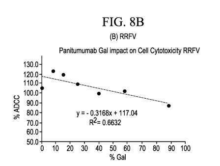
[13] FIGs. 8A-8D are graphs showing PBMC mediated cytotoxicity as a
function of panitumumab
13-galactose levels from donors with (A) HHVF, (B) RRVF, (C) HHVV, and (D)
RRFF polymorphisms
for FcyRIla and FcyRIlla receptors, respectively.
[14] FIG. 9A is a representative panitumumab dose curve for PBMCs
cytotoxicity activity. FIG. 9B
is a plot showing FcyR blocking using antibodies against indicated receptors
or controls.
[15] FIGs. 10A-10C are SPR sensograms showing panitumumab or controls
binding to (A) FcyRI
at up to 10 uM antibody; (B) FcyRIlla-158F at up to 10 uM antibody; and (C)
FcyRIla-131H at up to 10
uM antibody.
CA 03138584 2021-10-28
WO 2020/227726 PCT/US2020/035016
[16] FIGs. 11A-11B are SPR equilibrium binding curves for panitumumab in
(A) fucose enriched
and (B) afucose enriched samples bind to huFcgRIla-131H with apparent KID ¨7.9
pM and 8 pM
respectively.
DETAILED DESCRIPTION
1. OVERVIEW
[17] The most common human antibody subclasses used as biotherapeutics,
IgG1 and IgG2,
have very different immunological properties. Common effector functions, such
as antibody-
dependent cell-mediated cytotoxicity (ADCC), can be an important mechanism of
action for IgG1
antibodies. Previously, IgG2 antibodies were not known to exhibit effector
functions. However,
recently, it has been shown that panitumumab can mediate cytotoxic effect
similar to ADCC by
engaging FcyRIla. This is in contrast to conventional ADCC mediated by IgG1
engaging with FcyRIlla.
[18] Panitumumab is a human IgG2 monoclonal antibody that binds to human
epidermal growth
factor receptor (also known as EGF receptor, EGFR, ErbB-1 and HER1).
Panitumumab has an
approximate molecular weight of 147 kD. The heavy chain and light chain
sequences are shown in
Table 1 as SEQ ID Nos. 1 and 2, respectively. Panitumumab has two N-
glycosylation sites located in
the 2nd constant domain of each heavy chain. The N-glycosylation site is
commonly referred to as
residue N-297 according to the Kabat EU numbering. The actual residue number
is residue 295 of
SEQ ID NO:1.
[19] As described and exemplified herein, the inventors conducted an
extensive study into
mechanisms by which cytotoxicity of panitumumab is mediated, and product
quality attributes that
affect the FcyR-mediated cytotoxicity levels of panitumumab. The inventors
discovered that
cytotoxicity was primarily mediated by the myeloid lineage cells (monocytes,
macrophages and
neutrophils). The inventors then investigated the impact of the different
glycans in this IgG2 molecule
on effector functions. Using sensitive cytotoxicity assays in combination with
glycoengineered forms
of panitumumab, the inventor discovered that the impact of the different
glycans on FcyRIla-mediated
cell killing can be substantial and variable depending on the glycoforms.
[20] For example, galactosylation at the N-297 site showed a positive
correlation in the reporter
genes, while the afucosylation levels and high-mannose levels at the N-297
site showed an inverse
correlation to cell killing. Therefore, the FcyR-mediated cytotoxicity of
panitumumab can be increased
by (1) increasing the galatosylation level at the N-297 site; (2) decreasing
the afucosylation level at
the N-297 site; and/or (3) decreasing the high-mannose level at the N-297
site. Conversely, the FcyR-
mediated cytotoxicity of panitumumab can be decreased by (1) decreasing the
galatosylation level at
the N-297 site; (2) increasing the afucosylation level at the N-297 site;
and/or (3) increasing the high-
man nose level at the N-297 site.
11
CA 03138584 2021-10-28
WO 2020/227726 PCT/US2020/035016
[21] Panitumumab is currently produced in genetically engineered mammalian
(Chinese hamster
ovary) cells. During recombination production process, glycan moieties are
attached to the antibody
through post-translational modification. The discoveries made by the inventors
herein provide a
quantifiable relationship between glycoform profiles of panitumumab and its
cytotoxicity. The
discovery can be used to modulate the glycosylation pattern during the CHO-
cell production process,
such that the cytotoxicity level meets a desired reference level.
2. DEFINITIONS
[20] "Panitumumab" (trade names Vectibixe) refers to a human monoclonal
antibody comprising a
heavy chain comprising SEQ ID NO:1, and a light chain comprising SEQ ID NO:2.
The amino acid
sequences of the heavy and light chains of denosumab is shown in Table 1.
Nucleic acid sequences
encoding SEQ ID Nos: 1 and 2 are shown as SEQ ID Nos. 3 and 4, respectively.
As illustrated in the
examples, glycan profiles of panitumumab may vary.
Table 1 Sequences of panitumumab
Sequence
Heavy chain QVQLQESGPG LVKPSETLSL TCTVSGGSVS SGDYYWTWIR QSPGKGLEWI
amino acid
GHIYYSGNTN YNPSLKSRLT ISIDTSKTQF SLKLSSVTAA DTAIYYCVRD
sequence
RVTGAFDIWG QGTMVTVSSA STKGPSVFPL APCSRSTSES TAALGCLVKD
(SEQ ID NO:1)
YFPEPVTVSW NSGALTSGVH TFPAVLQSSG LYSLSSVVTV PSSNFGTQTY
TCNVDHKPSN TKVDKTVERK CCVECPPCPA PPVAGPSVFL FPPKPKDTLM
N-297
glycosylation site ISRTPEVTCV VVDVSHEDPE VQFNWYVDGV EVHNAKTKPR EEQ MMBIERV
is shown VSVLTVVHQD WLNGKEYKCK VSNKGLPAPI EKTISKTKGQ PREPQVYTLP
PSREEMTKNQ VSLTCLVKGF YPSDIAVEWE SNGQPENNYK TTPPMLDSDG
SFFLYSKLTV DKSRWQQGNV FSCSVMHEAL HNHYTQKSLS LSPGK
Light chain DIQMTQSPSS LSASVGDRVT ITCQASQDIS NYLNWYQQKP GKAPKLLIYD
amino acid
ASNLETGVPS RFSGSGSGTD FTFTISSLQP EDIATYFCQH FDHLPLAFGG
sequence
GTKVEIKRTV AAPSVFIFPP SDEQLKSGTA SVVCLLNNFY PREAKVQWKV
(SEQ ID NO:2)
DNALQSGNSQ ESVTEQDSKD STYSLSSTLT LSKADYEKHK VYACEVTHQG
LSSPVTKSFN RGEC
Heavy chain 1 40
nucleotide (1) AT GGACCT
CCT GT GCAAGAACAT GAAACACCT GT GGTT CT
sequence 41 80
(41) TCCTCCTCCTGGTGGCAGCTCCCAGATGGGTCCTGTCCCA
(SEQ ID NO:3) 81 120
(81) GGTACAGCTGCAGGAGTCGGGCCCAGGACTGGTGAAGCCT
121 160
121) TCGGAGACCCTGTCCCTCACCTGCACTGTCTCTGGTGGCT
161 200
161) CCGTCAGCAGTGGTGATTACTACTGGACCTGGATCCGGCA
201 240
201) GTCCCCAGGGAAGGGACTGGAGTGGATTGGACACATCTAT
241 280
241) TACAGTGGGAACACCAATTATAACCCCTCCCTCAAGAGTC
281 320
12
CA 03138584 2021-10-28
WO 2020/227726
PCT/US2020/035016
281) GACTCACCATATCAATTGACACGTCCAAGACTCAGTTCTC
321 360
321) CCT GAAGCT GAGTTCT GT GACCGCT GCGGACACGGCCATT
361 400
361) TATTACT GT GT GCGAGATCGAGT GACT GGT GCTTTT GATA
401 440
401) TCTGGGGCCAAGGGACAATGGTCACCGTCTCTTCAGCTAG
441 480
441) CACCAAGGGCCCATCGGTCTTCCCCCTGGCGCCCTGCTCC
481 520
481) AGGAGCACCTCCGAGAGCACAGCGGCCCTGGGCTGCCTGG
521 560
521) TCAAGGACTACTTCCCCGAACCGGTGACGGTGTCGTGGAA
561 600
561) CTCAGGCGCTCTGACCAGCGGCGTGCACACCTTCCCAGCT
601 640
601) GTCCTACAGTCCTCAGGACTCTACTCCCTCAGCAGCGTGG
641 680
641) TGACCGTGCCCTCCAGCAACTTCGGCACCCAGACCTACAC
681 720
681) CT GCAAC GTAGAT CACAAGCCCAGCAACACCAAGGT GGAC
721 760
721) AAGACAGTT GAGCGCAAAT GTT GT GT CGAGT GCCCACCGT
761 800
761) GCCCAGCACCACCT GT GGCAGGACCGTCAGTCTTCCTCTT
801 840
801) CCCCCCAAAACCCAAGGACACCCTCATGATCTCCCGGACC
841 880
841) CCTGAGGTCACGTGCGTGGTGGTGGACGTGAGCCACGAAG
881 920
8 8 1 ) ACCCCGAGGTCCAGTTCAACTGGTACGTGGACGGCGTGGA
921 960
921) G GT GCATAAT G C CAAGACAAAG C CAC G G GAG GAG CAGT T C
961 1000
961) AACAGCACGTTCCGT GT GGTCAGCGTCCTCACCGTT GT GC
1001 1040
1001) ACCAGGACTGGCTGAACGGCAAGGAGTACAAGTGCAAGGT
1041 1080
1041) CTCCAACAAAGGCCTCCCAGCCCCCATCGAGAAAACCATC
1081 1120
1081) TCCAAAACCAAAGGGCAGCCCCGAGAACCACAGGTGTACA
1121 1160
1121) CCCTGCCCCCATCCCGGGAGGAGATGACCAAGAACCAGGT
1161 1200
1161) CAGCCTGACCTGCCTGGTCAAAGGCTTCTACCCCAGCGAC
1201 1240
1201) ATCGCCGTGGAGTGGGAGAGCAATGGGCAGCCGGAGAACA
1241 1280
1241) ACTACAAGACCACACCTCCCATGCTGGACTCCGACGGCTC
1281 1320
1281) CTTCTTCCTCTACAGCAAGCTCACCGTGGACAAGAGCAGG
1321 1360
1321) T GGCAGCAGGGGAACGTCTTCTCAT GCTCCGT GAT GCAT G
1361 1400
1361) AGGCTCTGCACAACCACTACACGCAGAAGAGCCTCTCCCT
1401 1416
1401) GTCTCCGGGTAAAT GA
13
CA 03138584 2021-10-28
WO 2020/227726 PCT/US2020/035016
Light chain 1 40
nucleotide 1) AT GGACAT
GAGGGT CCCT GCT CAGCT CCT GGGGCT CCT GC
sequence 41 80
41) T GCT CT GGCT CT CAGGT GCCAGAT GT GACAT CCAGAT GAC
(SEQ ID NO:4) 81 120
81) CCAGT CT CCAT CCT CCCT GT CT GCAT CT GTAGGAGACAGA
121 160
121) GT CAC CAT CACT T GC CAGGC GAGT CAGGACAT CAGCAACT
161 200
161) ATTTAAATTGGTATCAGCAGAAACCAGGGAAAGCCCCTAA
201 240
201) ACT CCT GAT CTACGAT GCAT CCAATTT GGAAACAGGGGT C
241 280
241) CCAT CAAGGTT CAGT GGAAGT GGAT CT GGGACAGATTTTA
281 320
281) CTTTCACCATCAGCAGCCTGCAGCCTGAAGATATTGCAAC
321 360
321) ATATTT CT GT CAACACTTT GAT CAT CT CCCGCT CGCTTT C
361 400
361) GGCGGAGGGACCAAGGT GGAGAT CAAACGAACT GT GGCT G
401 440
401) CACCAT CT GT CTT CAT CTT CCCGCCAT CT GAT GAGCAGTT
441 480
441) GAAAT CT GGAACT GCCT CT GTT GT GT GCCT GCT GAATAAC
481 520
481) TT CTAT CCCAGAGAGGCCAAAGTACAGT GGAAGGT GGATA
521 560
521) ACGCCCT CCAAT CGGGTAACT CCCAGGAGAGT GT CACAGA
561 600
561) GCAGGACAGCAAGGACAGCAC CTACAGC CT CAGCAGCAC C
601 640
601) CT GACGCT GAG CAAAG CAGAC TAC GAGAAACACAAAGT CT
641 680
641) ACGCCTGCGAAGTCACCCATCAGGGCCTGAGCTCGCCCGT
681 711
681) CACAAAGAGCTT CAACAGGGGAGAGT GT TAG
[22] The term "glycan", "glycans", "glycoform" or "glycoforms" refers to
oligomers of
monosaccharide species that are connected by various glycosidic bonds.
Examples of
monosaccharides commonly found in mammalian N-linked glycans include hexose
(Hex), glucose
(Glc), galactose (Gal), mannose (Man) and N-acetylglucosamine (GIcNAc). The
major N-glycan
species found on recombinant IgG2 antibodies include fucose, galactose,
mannose, sialic acid and
GIcNAc, as depicted in FIGs. 1B, 1C, and Table 2. In case of panitumumab, the
glycan
oligosaccharide structures are linked to the N-glycosylation site at Asn-297
(Kabat EU numbering),
and are generally composed of a core heptasaccharide with outer arms
constructed by variable
addition of fucose, N-acetylglucosamine (GIcNAc), galactose, sialic acid (SA),
and bisecting N-
GIcNAc. Each of the potential oligosaccharide structures may be abbreviated as
follows: GO, G1, or
G2 referring to the core GIcNAc and mannose oligosaccharide structure having
zero, one or two
terminal galactose molecules, respectively. Within G1, two additional
structures, abbreviated G1a and
G1 b, may be present with G1a or G1b referring to whether the terminal
galactose group is attached to
either the 6-arm or the 3-arm of the core structure. See FIGs. 1B and 1C. When
fucosylated (i.e. a
14
CA 03138584 2021-10-28
WO 2020/227726 PCT/US2020/035016
fucose group is attached to the core glycan structure), the GO, G1 (G1a/G1b),
or G2 forms may be
abbreviated GOF, G1F (G1aF/G1bF), or G2F. When sialic acid is present, these
abbreviations contain
a "S" such that, for example, G2FS2 refers to a glycan having two galactose, a
fucose and two sialic
acid groups. Additional glycans linked to antibodies may also exist including
high mannose (HM)
structures, which are formed by the incorporation of additional mannose
groups, including the high
mannose species (e.g., "Man 5" or "M5" as shown in FIG. 1C and Table 2). As
used herein, the term
"glycan" or "glycans" refers to any of the oligomers of monosaccharide species
described herein or
any other oligomers of monosaccharaide species linked to an antibody.
[23] The N-glycosylation sites of an IgG2 (located at the 2nd constant
domain of the heavy chain)
is typically referred to as N-297 based on EU numbering system. A full chart
comparing different
numbering systems is provided by the International Immunogenetics Information
System ("IMGT
Scientific chart"). The IMGT Scientific chart refers to IgG1, the
corresponding numbers in IgG2 can be
readily obtained by aligning the respective sequences.
[24] The terms "terminal 13-galactose, "galactosylated glycans" or "G1, G1
a, G1 b, and/or G2
galactosylated glycans" refers to a glycan comprising one or two galactose
molecules linked to an IgG
antibody at the N-glycosylation site (Asn-297) through the N-
acetylglucoseamine moieties that attach
to the core mannose structure. Exemplary glycans comprising "terminal 13-
galactose" "galactosylated
glycans" or "G1, G1 a, G1 b, and/or G2 galactosylated glycans" are depicted in
FIGs. 1B and 1C. In
some embodiments, the G1, G1 a, G1 b, and/or G2 galactosylated glycans may or
may not contain
core fucose.
[25] The term "core fucose" or "fucosylated species" refers to a glycan
comprising a fucose
molecule (alpha 1-6) linked to an IgG antibody at the N-glycosylation site
(Asn-297) through the N-
acetylglucoseamine moieties that attach to the core mannose structure.
Exemplary glycans
comprising "core fucose" or "fucosylated glycans" are depicted in FIGs. 1B and
1C. In some
embodiments, antibodies containing core fucose and/or a fucosylated glycans
may or may not contain
other glycans (including terminal 13-galactose and/or high mannose glycans).
[26] The terms "afucosylated", "afucosylated glycans" or "afucosylation"
refers to the removal or
lack of a core fucose on an antibody. Exemplary afucosylated glycans are
depicted in FIGs 1B and
1C. In some embodiments, antibodies lacking core fucose may or may not contain
other glycans
(including terminal 13-galactose and/or high mannose glycans). Afucosylated
glycoforms include, but
are not limited to, A1GO, A1G1a, A2GO, A2G1a, A2G1b, A2G2, and Al G1 M5. See,
e.g., Reusch and
Tejada, Glycobiology 25(12): 1325-1334 (2015).
[27] The term "high mannose", "high mannose glycans" or "HM" refers to a
glycan comprising
more than 3 mannose molecules linked to an IgG antibody at the N-glycosylation
site (Asn-297).
Exemplary high mannose antibodies are depicted in FIG. 1C and Table 2,
including the "Man-5 high
mannose glycans" which contains two additional mannose molecules. High mannose
glycans
encompass glycans comprising 5, 6, 7, 8, or 9 mannose residues, abbreviated as
Man 5 or M5, Man
CA 03138584 2021-10-28
WO 2020/227726 PCT/US2020/035016
6 or M6, Man 7 or M7, Man 8 or M8, and Man 9 or M9, respectively. Exemplary
structures of Man 6,
Man 7, and Man 8 are shown below.
Table 2. Exemplary high-mannose Structures
Man 5 (M5)
C53036N4H84
=\ = =
Man 5-GIcNAc
C61041N5H97
HS \== =
Man 7-Fuc
C65046N4H 104
=\ = =
Man 6
C50041N4H94
\ = =
Man 6-GIcNAc
C67046N5H 107
= =
Man 7
C65046N4H 104
Man 8
71 51 4H 114
= =
[28] "FcyR" or "Fc-gamma receptor" is a protein belonging to the IgG
superfamily involved in
inducing phagocytosis of opsonized cells or microbes. See, e.g., Fridman WH.
Fc receptors and
immunoglobulin binding factors. FASEB Journal. 5 (12): 2684-90 (1991). Members
of the Fc-gamma
receptor family include: FcyRI (CD64), FcyRIIA (CD32), FcyRIIB (CD32),
FcyRIIIA (CD16a), and
FcyRIIIB (CD16b). The sequences of FcyRI, FcyRIIA, FcyRIIB, FcyRIIIA, and
FcyRIIIB can be found in
many sequence databases, for example, at the Uniprot database
(www.uniprot.org) under accession
16
CA 03138584 2021-10-28
WO 2020/227726 PCT/US2020/035016
numbers P12314 (FCGR1_HUMAN), P12318 (FCG2A_HUMAN), P31994 (FCG2B_HUMAN),
P08637 (FCG3A_HUMAN), and P08637 (FCG3A_HUMAN), respectively.
[29] As used herein, the terms "a," "an," and "the" and similar referents
in the context of describing
the disclosure (especially in the context of the following claims) are to be
construed to cover both the
singular and the plural, unless otherwise indicated herein or clearly
contradicted by context. The
terms "comprising," "having," "including," and "containing" are to be
construed as open-ended terms
(i.e., meaning "including, but not limited to," and permit the presence of one
or more features or
components) unless otherwise noted. The terms "a" (or "an"), as well as the
terms "one or more," and
"at least one" can be used interchangeably herein. Furthermore, "and/or" where
used herein is to be
taken as specific disclosure of each of the two specified features or
components with or without the
other. Thus, the term "and/or" as used in a phrase such as "A and/or B" herein
is intended to include
"A and B," "A or B," "A" (alone), and "B" (alone). Likewise, the term "and/or"
as used in a phrase such
as "A, B, and/or C" is intended to encompass each of the following aspects: A,
B, and C; A, B, or C; A
or C; A or B; B or C; A and C; A and B; B and C; A (alone); B (alone); and C
(alone).
[30] The term "about" as used in connection with a numerical value
throughout the specification
and the claims denotes an interval of accuracy, familiar and acceptable to a
person skilled in the art.
In general, such interval of accuracy is 10%.
3. POST-TRANSLATIONAL GLYCOSYLATION AND PCyR-MEDIATED CYTOTOXICITY
3.1 Post-translational Glycosylation
[31] Many secreted proteins undergo post-translational glycosylation, a
process by which sugar
moieties (e.g., glycans, saccharides) are covalently attached to specific
amino acids of a protein. In
eukaryotic cells, two types of glycosylation reactions occur: (1) N-linked
glycosylation, in which
glycans are attached to the asparagine of the recognition sequence Asn-X-
Thr/Ser, where "X" is any
amino acid except proline, and (2) 0-linked glycosylation in which glycans are
attached to serine or
threonine. Regardless of the glycosylation type (N-linked or 0-linked),
microheterogeneity of protein
glycoforms exists due to the large range of glycan structures associated with
each site (0 or N). For
an IgG2 antibody, N-linked glycosylation occurs at Asparigine-297 (N-297) site
(Eu numbering
system). For panitumumab, the actual position of this Asparagine occurs at
residue number 295, but
in general, the N-glycosylation site is nonetheless referred to as N-297 to be
consistent with the EU
numbering system.
[32] All N-glycans have a common core sugar sequence: Manoc1-6(Mana1-
3)Man131-
4G1cNAc131-4G1cNAc131-Asn-X-Ser/Thr (Man3G1cNAc2Asn) and are categorized into
one of three
types: (A) a high mannose (HM) or oligomannose (OM) type, which consists of
two N-
acetylglucosamine (GaINAc) moieties and a large number (e.g., 5, 6, 7, 8 or 9)
of mannose (Man)
residues; (B) a complex type, which comprises more than two GIcNAc moieties
and any number of
other sugar types; or (C) a hybrid type, which comprises a Man residue on one
side of the branch and
17
CA 03138584 2021-10-28
WO 2020/227726 PCT/US2020/035016
GIcNAc at the base of a complex branch. FIG. 1A (based on Stanley et al.,
Chapter 8: N-Glycans,
Essentials of Glycobiology, 2nd ed., Cold Spring Harbor Laboratory Press;
2009) shows the three
types of N-glycans.
[33] N-linked glycans typically comprise one or more monosaccharides of
galactose (Gal), N-
acetylgalactosamine (GaINAc), N-acetylglucoasamine (GIcNAc), mannose (Man), N-
Acetylneuraminic
acid (Neu5Ac), fucose (Fuc). The commonly used symbols for such saccharides
are shown in
FIG. 1A.
[34] The sugar composition and the structural configuration of a glycan
structure varies,
depending on the glycosylation machinery in the ER and the Golgi apparatus,
the accessibility of the
machinery enzymes to the glycan structure, the order of action of each enzyme
and the stage at
which the protein is released from the glycosylation machinery, among other
factors. Controlling the
glycan structure is important in recombinant production of therapeutic
monoclonal antibodies, as the
glycan structure attached to the Fc domain influences the interaction with the
FcyRs that mediate
cytotoxicity.
3.2 Glycans that affect Fc7R-mediated cytotoxicity
[35] The present disclosure identifies the impact of various glycans
(including, e.g., 13-galactose,
core-fucose, and/or high mannose) on FcyR-mediated cytotoxicity of IgG2
antibodies, such as
panitumumab. Accordingly, the present disclosure provides a method of
modulating Fc gamma
Receptor (FcyR)-mediated cytotoxicity of an IgG2 antibody (such as
panitumumab), or a composition
comprising the antibody (an antibody composition). In exemplary embodiments,
the method
comprises modulating the amount of (a) galactosylated glycans of the antibody;
(b) afucosylated
glycans of the antibody; (c) high mannose glycans of the antibody; or (d) a
combination thereof.
Without being bound to a particular theory, it is believed that the methods
disclosed herein provide
means for tailor-made compositions comprising specific amounts of particular
glycoforms of a given
antibody, which exhibit targeted levels of FcyR-mediated cytotoxicity.
Particularly relevant glycan
structures are illustrated in FIG. 1C.
[36] In exemplary aspects, the methods provided by the present disclosure
relate to modulation of
an IgG2 antibody (such as panitumumab), or a composition comprising the
antibody (an antibody
composition), wherein steps are taken to achieve a desired or predetermined
level of glycoforms of
the IgG2 antibody, such that the antibody or antibody composition exhibits a
desired or pre-
determined reference level of FcyR-mediated cytotoxicity. In exemplary
embodiments, the method
comprises modulating (increasing or decreasing) the amount of (a)
galactosylated glycans; (b)
afucosylated glycans; (c) high mannose glycans; or (d) a combination thereof
of the IgG2 antibody
(such as panitumumab), in order to modulate (increase or decrease) the FcyR-
mediated cytotoxicity
that is induced or stimulated by the antibody. In exemplary embodiments, the
method comprises
modulating (increasing or decreasing) the amount of glycoforms, e.g., (a)
galactosylated glycoforms;
18
CA 03138584 2021-10-28
WO 2020/227726 PCT/US2020/035016
(b) afucosylated glycoforms; (c) high mannose glycoforms; or (d) a combination
thereof, to modulate
(increase or decrease) the FcyR-mediated cytotoxicity that is induced or
stimulated by the antibody.
[37] The term "amount" when referring the amount of a particular glycan
(including, e.g., (1) the
amount of terminal 13-galactose, (2) the amount of G1, G1 a, G1 b, and/or G2
galactosylated glycan, (3)
the amount of core fucose, (4) the amount of fucosylated glycan, (5) the
amount of afucosylated
glycan, (6) the amount of high mannose glycan, and/or (7) the amount of Man-5
glycan), refers to a
relative percentage of a particular glycan at the N-297 site, compared to the
total amount of glycans at
N-297 site. Because counting glycan species at individual molecule level is
impractical/impossible, the
amount of a glycan content described herein is generally calculated based on
relative percentage
according to commonly used analytical methods. For example, as exemplified in
Example 2.2, an
enzyme is used to release all N-glycans from the protein; then glycans are
separated by hydrophilic
interaction liquid chromatography (HILIC). HILIC results in various peaks,
each peak representing a
glycan species. The amount of a particular glycan is calculated as a relative
percentage, based on the
area of its peak, out of the total areas of all peaks. Therefore, unless
otherwise specified, the amount
of a glycan refers to the relative percentage of that particular glycan
species, out of total N-glycans at
the N-297 site, using any of the commonly used analytical method (such as
HPAEC, CE-SDS, HILIC,
or LC-MS).
[38] Methods for measuring and determining the amount or relative
percentage of a glycan
(including, e.g., terminal 13-galactose, G1, G1a, Gib, and/or G2
galactosylated glycans, core fucose,
fucosylated glycans, afucosylated glycans, high mannose glycans, and/or Man-5
glycans) are well
known in the art, and include, e.g., Hydrophilic Interaction Liquid
Chromatography (HILIC) as
described in the Examples. See also, Pace et al., Characterizing the Effect of
Multiple Fc Glycan
Attributes on the Effector Functions and FcyRIlla Receptor Binding Activity of
an IgG1 Antibody,
Biotechnol.Prog., 2016, Vol.32, No.5 pages 1181-1192; and Shah, B. et al. LC-
MS/MS Peptide
Mapping with Automated Data Processing for Routine Profiling of N-Glycans in
Immuno globulins J.
Am. Soc. Mass Spectrom. (2014) 25: 999, herein each incorporated by reference
for all purposes. In
some embodiments, amount can be determined or calculated as mole percent
incorporation.
[39] In some aspects, the methods disclosed herein comprise modulating the
amount of terminal
13-galactose, core fucose, or high mannose, or a combination thereof, attached
to particular IgG2
molecules (such as panitumumab).
[40] For example, the method may comprise increasing the amount of terminal
13-galactose on an
IgG2 (such as panitumumab) by, e.g., effectively changing the glycan from a GO
to a G1 or G2, or
from a G1 to a G2, to increase the FcyR-mediated cytotoxicity. Alternatively,
the FcyR-mediated
cytotoxicity may be increased by increasing amount of antibody molecules that
comprise G1, G1 a,
G1 b, and/or G2 galactosylated glycan at the N-297 site. Also, for example,
the method may comprise
decreasing the amount of terminal 13-galactose on an IgG2 (such as
panitumumab) by, e.g., effectively
changing the glycan from a G2 to a G1 or GO, or from a G1 to a GO, to decrease
the FcyR-mediated
19
CA 03138584 2021-10-28
WO 2020/227726 PCT/US2020/035016
cytotoxicity. Alternatively, the FcyR-mediated cytotoxicity may be decreased
by decreasing amount of
antibody molecules that comprise G1, G1 a, G1 b, and/or G2 galactosylated
glycan at the N-297 site.
[41] In other exemplary aspects, the method may comprise increasing the
amount of core fucose
on an IgG2 (such as panitumumab) to increase the FcyR-mediated cytotoxicity.
The FcyR-mediated
cytotoxicity may be increased by increasing amount of antibody molecules that
comprise fucosylated
glycan at the N-297 site, or by decreasing amount of antibody molecules that
comprise afucosylated
glycan at the N-297 site. Also, the method may comprise decreasing the amount
of core fucose on an
IgG2 (such as panitumumab) to decrease the FcyR-mediated cytotoxicity. The
FcyR-mediated
cytotoxicity may be decreased by decreasing amount of antibody molecules that
comprise fucosylated
glycan at the N-297 site, or by increasing amount of antibody molecules that
comprise afucosylated
glycan at the N-297 site.
[42] In other exemplary aspects, the method may comprise decreasing the
amount of high-
mannose (e.g., Man-5) on an IgG2 (such as panitumumab) to increase the FcyR-
mediated
cytotoxicity. The FcyR-mediated cytotoxicity may be increased by decreasing
amount of antibody
molecules that comprise high-mannose glycan at the N-297 site. Also, the
method may comprise
increasing the amount of mannose (e.g., Man-5) on an IgG2 (such as
panitumumab) to decrease the
FcyR-mediated cytotoxicity. The FcyR-mediated cytotoxicity may be decreased by
increasing amount
of antibody molecules that comprise mannose (e.g., Man-5) at the N-297 site.
3.3 Modulating Fc7R-mediated cytotoxicity
[43] Fc-gamma receptors are present in two distinct classes ¨ those that
activate cells upon their
crosslinking ("activation FcRs") and those that inhibit activation upon co-
engagement ("inhibitory
FcRs"). In human, there are two low-affinity activation FcRs for IgG ¨ FcyRIla
and FcyRIlla. FcyRIla
(or FcyRIIA) is a single-chain low affinity receptor for IgG, with an ITAM
sequence located in its
cytoplasmic tail. It is expressed on macrophages, mast cells, monocytes,
neutrophils and some B
cells. It is 90% homologous in its extracellular domain to the human
inhibitory FcRIlb molecule, which
has an ITIM sequence in its cytoplasmic domain, expressed on B cells,
macrophages, mast cells,
neutrophils, monocytes but not NK cells or T cells. FcyRIlla (or FcyRIIIA) is
an oligomeric activation
receptor consisting of a ligand binding a. subunit and an ITAM containing
gamma or zeta subunit. It is
expressed on NK cells, macrophages and mast cells. It is not expressed on
neutrophils, B cells or T
cells. In addition, a receptor with greater than 95% sequence identity in its
extracellular domain called
FcRIllb is found on human neutrophils as a GPI-anchored protein. It is capable
of binding immune
complexes but not activating cells in the absence of association with an ITAM
containing receptor like
FcRlIa. FcRII and FcRIII are about 70% identical in their ligand binding
extracellular domains.
[44] Thus, in human, IgG cytotoxic antibodies interact with four distinct
low- affinity receptors - two
of which are capable of activating cellular responses, FcRIla and FcRIlla, one
of which is inhibitory,
FcRIlb, and one of which will bind IgG complexes but not trigger cellular
responses, FcRIllb.
CA 03138584 2021-10-28
WO 2020/227726
PCT/US2020/035016
Macrophages expresses FcRIla, FcRIlb and FcRIlla, neutrophils express FcRIla,
FcRIlb and FcRIllb,
while NK cells express only FcRIlla. The efficacy of a therapeutic anti-tumor
antibody will thus depend
on the specific interactions with activation, inhibition and inert low-
affinity FcRs, differentially
expressed on distinct cell types.
[45] Well-defined tumor models to study cytotoxicity of therapeutic anti-
tumor antibodies are
known. For example, Matui et al. described an in vitro system using A431
cells, as well as an in vivo
system using A431 cell xenografts in athymic mice, to study the cytotoxicities
of IgG1 and IgG2
antibodies that bind to EGFR.
[46] In certain aspects, the FcyR-mediated cytotoxicity described herein is
mediated by FcyRIla.
[47] In certain aspects, the FcyR-mediated cytotoxicity FcyRIla-mediated
cellular cytotoxicity.
[48] In certain aspects, the FcyR-mediated cytotoxicity described herein is
measured or
determined using a FcyR reporter gene assay. In certain aspects, the reporter
gene assay comprises
Jurkat cells. In certain aspects, the reporter gene assay comprises a Jurkat
cell expressing a FcyR
receptor, a NFAT-response element, and/or a reporter gene. A reporter gene can
be any gene whose
expression provides a measurable signal. Exemplary reporter genes include the
genes encoding
green fluorescent protein (GFP), antibiotic resistance proteins (e,g.,
chloramphenicol transferase),
toxic proteins GATA-
1 DNA binding don-loins, cocn ysis proteins), r-galactosidase, E. coli r-
galactosidase (LacZ), Halobacteriurn [1-galactosidase, Neuropsora tyrosinase,
human placental
alkaline phosphatase, chloramphenicol acetyl transferase (CAT), Aequorin
(jellyfish
bioluminescence), Firefly luciferase (EC 1,13.12.7) form the American firefly,
Photinus pyralis, Renilla
luciferase (EC 1,13.12.5) from the sea pansy Rena reniformis, and Bacterial
luciferase (EC
1.14,14.3) from Photobacteriurn fischeri. Various other reporter genes are
well known by those haying
ordinary skill in the art. In an exemplary embodiment, the reporter gene
encodes a luciferase.
[49] In certain aspects, the FcyR-mediated cytotoxicity described herein is
measured using an
ADCC assay kit. ADCC assay kits are commercially available, such as "ADCC
Reporter Bioassays"
by Promega (Catalog No. G7010 or G7018).
[50] In certain aspects, the present disclosure provides a method of
increasing FcyR-mediated
cytotoxicity of an IgG2 antibody (such as panitumumab) or a composition
comprising the antibody, as
compared to a control or a reference value. In exemplary embodiments, the
increase is at least or
about 0.1% to about 100% increase (e.g., at least or about a 0.1% increase, at
least or about a 0.2%
increase, at least or about a 0.3% increase, at least or about a 0.4%
increase, at least or about a
0.5% increase, at least or about a 0.55% increase, at least or about a 0.6%
increase, at least or about
a 0.65% increase, at least or about a 0.7% increase, at least or about a 0.75%
increase, at least or
about a 0.8% increase, at least or about a 0.9% increase, at least or about a
1% increase, at least or
about a 1.2% increase, at least or about a 1.25% increase, at least or about a
1.3% increase, at least
or about a 1.35% increase, at least or about a 1.4% increase, at least or
about a 1.5% increase, at
least or about a 2% increase, at least or about a 2.5% increase, at least or
about a 2.7% increase, at
least or about a 2.75% increase, at least or about a 2.8% increase, at least
or about a 2.85%
21
CA 03138584 2021-10-28
WO 2020/227726 PCT/US2020/035016
increase, at least or about a 2.9% increase, at least or about a 2.95%
increase, at least or about a 3%
increase, at least or about a 4% increase, at least or about a 5% increase, at
least or about a 6%
increase, at least or about a 7% increase, at least or about a 8% increase, at
least or about a 9%
increase, at least or about a 9.5% increase, at least or about a 10% increase,
at least or about a 15%
increase, at least or about a 20% increase, at least or about a 25% increase,
at least or about a 30%
increase, at least or about a 35% increase, at least or about a 40% increase,
at least or about a 45%
increase, at least or about a 50% increase, at least or about a 55% increase,
at least or about a 60%
increase, at least or about a 65% increase, at least or about a 70% increase,
at least or about a 75%
increase, at least or about a 80% increase, at least or about a 85% increase,
at least or about a 90%
increase, at least or about a 95% increase, or at least or about a 100%
increase), as compared to a
control or a reference value. In exemplary embodiments, the increase is over
100%, e.g., at least or
about 125%, at least or about 150%, at least or about 175%, at least or about
200%, at least or about
300%, at least or about 400%, at least or about 500%, at least or about 600%,
at least or about 700%,
at least or about 800%, at least or about 900%, or at least or about 1000%, as
compared to a control
or a reference value. In exemplary embodiments, the FcyR-mediated cytotoxicity
of the antibody, or
composition comprising the antibody, increases by at least about 1.1-fold, at
least about 1.2-fold, at
least about 1.3-fold, at least about 1.4-fold, at least about 1.5-fold, at
least about 1.6-fold, at least
about 1.7-fold, at least about 1.8-fold, or at least about 1.9-fold, as
compared to a control or a
reference value. In exemplary embodiments, the FcyR-mediated cytotoxicity of
the antibody, or
composition comprising the antibody, increases by at least about 2-fold, at
least about 2.5-fold, at
least about 3-fold, at least about 3.5-fold, at least about 4-fold, at least
about 4.5-fold, at least about 5-
fold, at least about 5.5-fold, at least about 6-fold, at least about 6.5-fold,
at least about 7-fold, at least
about 7.5-fold, at least about 8-fold, at least about 8.5-fold, at least about
9-fold, at least about 9.5-
fold, or at least about 10-fold, relative to a control or a reference value.
In exemplary embodiments,
the FcyR-mediated cytotoxicity of the antibody, or composition comprising the
antibody, increases by
from about 1.1-fold to about 10-fold, from about 1.2-fold to about 10-fold,
from about 1.3-fold to about
10-fold, from about 1.4-fold to about 10-fold, from about 1.5-fold to about 10-
fold, from about 1.1-fold
to about 5-fold, from about 1.2-fold to about 5-fold, from about 1.3-fold to
about 5-fold, from about 1.4-
fold to about 5-fold, or from about 1.5-fold to about 5-fold, as compared to a
control or a reference
value.
[51] In certain aspects, the present disclosure provides a method of
decreasing FcyR-mediated
cytotoxicity of an IgG2 antibody (such as panitumumab) or a composition
comprising the antibody, as
compared to a control or a reference value. In exemplary embodiments, the
decrease is at least or
about 0.1% to about 100% decrease (e.g., at least or about a 0.1% decrease, at
least or about a 0.2%
decrease, at least or about a 0.3% decrease, at least or about a 0.4%
decrease, at least or about a
0.5% decrease, at least or about a 0.55% decrease, at least or about a 0.6%
decrease, at least or
about a 0.65% decrease, at least or about a 0.7% decrease, at least or about a
0.75% decrease, at
least or about a 0.8% decrease, at least or about a 0.9% decrease, at least or
about a 1% decrease,
at least or about a 1.2% decrease, at least or about a 1.25% decrease, at
least or about a 1.3%
22
CA 03138584 2021-10-28
WO 2020/227726 PCT/US2020/035016
decrease, at least or about a 1.35% decrease, at least or about a 1.4%
decrease, at least or about a
1.5% decrease, at least or about a 2% decrease, at least or about a 2.5%
decrease, at least or about
a 2.7% decrease, at least or about a 2.75% decrease, at least or about a 2.8%
decrease, at least or
about a 2.85% decrease, at least or about a 2.9% decrease, at least or about a
2.95% decrease, at
least or about a 3% decrease, at least or about a 4% decrease, at least or
about a 5% decrease, at
least or about a 6% decrease, at least or about a 7% decrease, at least or
about a 8% decrease, at
least or about a 9% decrease, at least or about a 9.5% decrease, at least or
about a 10% decrease,
at least or about a 15% decrease, at least or about a 20% decrease, at least
or about a 25%
decrease, at least or about a 30% decrease, at least or about a 35% decrease,
at least or about a
40% decrease, at least or about a 45% decrease, at least or about a 50%
decrease, at least or about
a 55% decrease, at least or about a 60% decrease, at least or about a 65%
decrease, at least or
about a 70% decrease, at least or about a 75% decrease, at least or about a
80% decrease, at least
or about a 85% decrease, at least or about a 90% decrease, at least or about a
95% decrease, or at
least or about a 100% decrease), as compared to a control or a reference
value. In exemplary
embodiments, the decrease is over 100%, e.g., at least or about 125%, at least
or about 150%, at
least or about 175%, at least or about 200%, at least or about 300%, at least
or about 400%, at least
or about 500%, at least or about 600%, at least or about 700%, at least or
about 800%, at least or
about 900%, or at least or about 1000%, as compared to a control or a
reference value. In exemplary
embodiments, the FcyR-mediated cytotoxicity of the antibody, or composition
comprising the antibody,
decreases by at least about 1.1-fold, at least about 1.2-fold, at least about
1.3-fold, at least about 1.4-
fold, at least about 1.5-fold, at least about 1.6-fold, at least about 1.7-
fold, at least about 1.8-fold, or at
least about 1.9-fold, as compared to a control or a reference value. In
exemplary embodiments, the
FcyR-mediated cytotoxicity of the antibody, or composition comprising the
antibody, decreases by at
least about 2-fold, at least about 2.5-fold, at least about 3-fold, at least
about 3.5-fold, at least about 4-
fold, at least about 4.5-fold, at least about 5-fold, at least about 5.5-fold,
at least about 6-fold, at least
about 6.5-fold, at least about 7-fold, at least about 7.5-fold, at least about
8-fold, at least about 8.5-
fold, at least about 9-fold, at least about 9.5-fold, or at least about 10-
fold, as compared to a control or
a reference value. In exemplary embodiments, the FcyR-mediated cytotoxicity of
the antibody, or
composition comprising the antibody, decreases by from about 1.1-fold to about
10-fold, from about
1.2-fold to about 10-fold, from about 1.3-fold to about 10-fold, from about
1.4-fold to about 10-fold,
from about 1.5-fold to about 10-fold, from about 1.1-fold to about 5-fold,
from about 1.2-fold to about
5-fold, from about 1.3-fold to about 5-fold, from about 1.4-fold to about 5-
fold, or from about 1.5-fold to
about 5-fold, as compared to a control or a reference value.
[52] As used herein, the "control" or "reference value" here is the level
of FcyR-mediated
cytotoxicity of the antibody, or a composition comprising the antibody, prior
to an experimental
intervention directed at modulating the glycan profile (such as the level of
cytotoxicity when first
measured). If an antibody, or a composition comprising the antibody, has
undergone experimental
intervention directed at modulating the glycan profile, but additional
modulation is desired, then the
23
CA 03138584 2021-10-28
WO 2020/227726 PCT/US2020/035016
"control" or "reference value" can be the level of FcyR-mediated cytotoxicity
prior to any additional
experimental intervention directed at further modulating the glycan profile.
[53] In certain aspects, the reference value is the level of the FcyR-
mediated cytotoxicity exhibited
by commercially available panitumumab samples at the same dose (e.g., same
amount of antibody
molecules). In certain aspects, the reference value is a pre-determined level
that provides therapeutic
benefit.
[54] In certain aspects, the present disclosure provides a method
comprising modulating (i.e.
increasing or decreasing) the amount of a specific glycan species (e.g.,
galactosylated glycans, G1,
G1 a, G1 b, and/or G2 galactosylated glycans, fucosylated glycans,
afucosylated glycans, core fucose,
high mannose glycans, Man-5 glycans, or a combination thereof) of the antibody
to a total amount of
at least or about 0.5%, at least or about 1%, at least or about 2%, at least
or about 3%, at least or
about 5%, at least or about 7%, at least or about 10%, at least or about 15%,
at least or about 20%,
at least or about 25%, at least or about 30%, at least or about 35%, at least
or about 40%, at least or
about 45%, at least or about 50%, at least or about 55%, at least or about
60%, at least or about 65%,
at least or about 70%, at least or about 75%, at least or about 80%, at least
or about 85%, at least or
about 90%, at least or about 95%, at least or about 96%, at least or about
97%, at least or about 98%,
from about 0.5% to about 98%, from about 0.5% to about 98%, from about 0.5% to
about 98%, from
about 0.1% to about 99%, from about 0.5% to about 98%, from about 0.5% to
about 95%, from about
1% to about 90%, from about 1% to about 85%, from about 5% to about 85%, from
about 10% to
about 85%, or from about 10% to about 80%. As described above, the percentage,
when describing
specific glycan species, generally refers to the relative percentage of a
particular glycan species, out
of total glycan content at the N-297 site, calculated according to any of the
art-recognized analytical
methods (such as HILIC, LC-MS). In one exemplary embodiment, the relative
percentage is
calculated according to the areas of chromatographic peaks.
[55] In certain aspect, the disclosure provides a method of modulating Fc
gamma Receptor
(FcyR)-mediated cytotoxicity of panitumumab, comprising increasing or
decreasing the amount of
terminal 13-galactose at the N-297 glycosylation site of panitumumab, or
increasing or decreasing the
amount of panitumumab molecules that comprise G1, G1 a, G1 b, and/or G2
galactosylated glycan at
the N-297 site.
[56] In certain embodiments, the disclosure provides a method of increasing
Fc gamma Receptor
(FcyR)-mediated cytotoxicity of panitumumab, comprising increasing the amount
of terminal 13-
galactose at the N-297 glycosylation site of panitumumab, or increasing the
amount of panitumumab
molecules that comprise G1, G1 a, G1 b, and/or G2 galactosylated glycan at the
N-297 site. In certain
embodiments, an increase of about 1 percent of 3-galactose increases FcyR-
mediated cytotoxicity by
from about 0.55 percent to about 0.75 percent, such as about 0.55 percent,
about 0.6 percent, about
0.65 percent, about 0.7 percent, or about 0.75 percent. As described above,
the percentages of 13-
24
CA 03138584 2021-10-28
WO 2020/227726 PCT/US2020/035016
galactose, or G1, G1a, Gib, G2 galactosylated glycans, refer to relative
percentages of the respective
glycan species, out of the total glycan content at the N-297 site.
[57] When quantifying the relationship between FcyR-mediated cytotoxicity
and various glycans
(e.g., change of the percentage level of a particular glycan, and the
corresponding changes in
cytotoxicity level), the FcyR-mediated cytotoxicity is often expressed as a
relative value, quantified
against a standard. For example, "percent relative activity" (against a
standard) can be used to
express FcyR-mediated cytotoxicity level. "Percent relative activity" can be
calculated as: (i) cytotoxic
activity of the sample / cytotoxic activity of the standard ("/" means
divide); or (ii) cytotoxic activity of
the standard / cytotoxic the activity of sample ("1' means divide). For
example, if sample A exhibits
50% cytotoxicity level, as compared to a standard, and sample B exhibits 51%
cytotoxicity level, as
compared to the same standard, then it can be said that the FcyR-mediated
cytotoxicity is increased
by 1% from sample A to sample B.
[58] In certain embodiments, the standard is the level of the FcyR-mediated
cytotoxicity exhibited
by commercially available panitumumab samples at the same dose (e.g., same
amount of antibody
molecules). Therefore, in certain embodiments, a quantitative relationship is
established using relative
cytotoxicity level. For example, referring to FIG. 2A, when terminal 13-
galactose is about 0%, the
relative cytotoxicity level (calculated against commercially available
panitumumab samples at the
same dose) is about 88%. When terminal 13-galactose is increased to about 10%,
the relative
cytotoxicity level (calculated against commercially available panitumumab
samples at the same dose)
is about 95%. Thus, an increase of about 1 percent of terminal 13-galactose
correlates to an increase
of FcyR-mediated cytotoxicity by about 0.67 percent. This means that for every
1 percent increase of
terminal 13-galactose, the relative cytotoxicity level of the panitumumab
sample increases by 0.67
percent.
[59] In certain embodiments, the relative cytotoxicity level can be
calculated based on EC50
values measured in a bioassay. For example, if a reporter gene is used to
determine the EC50 of the
cytotoxicity exhibited by a sample antibody, then the relative cytotoxicity
level can be calculated as
EC50 sample / EC50 standard, or EC50 standard / EC50 sample ("1' means
divide).
[60] If preferred, the relative cytotoxicity value of a sample can be
measured multiple times (e.g.,
twice, three times, four times), and the result can be reported as the mean of
these multiple values.
[61] In certain embodiments, the disclosure provides a method of decreasing
Fc gamma Receptor
(FcyR)-mediated cytotoxicity of panitumumab, comprising decreasing the amount
of terminal 13-
galactose at the N-297 glycosylation site of panitumumab, or decreasing the
amount of panitumumab
molecules that comprise G1, G1 a, G1 b, and/or G2 galactosylated glycan at the
N-297 site. In certain
embodiments, a decrease of about 1 percent of 13-galactose decreases FcyR-
mediated cytotoxicity by
from about 0.55 percent to about 0.75 percent, such as about 0.55 percent,
about 0.6 percent, about
CA 03138584 2021-10-28
WO 2020/227726 PCT/US2020/035016
0.65 percent, about 0.7 percent, or about 0.75 percent. Again, changes in
cytotoxicity level is
generally calculated based on relative cytotoxicity value as described above.
[62] In certain aspects, the disclosure provides a method of modulating Fc
gamma Receptor
(FcyR)-mediated cytotoxicity of panitumumab, comprising increasing or
decreasing the amount of
panitumumab molecules that comprise fucosylated glycan at the N-297 site, or
increasing or
decreasing the amount of panitumumab molecules that comprise afucosylated
glycan at the N-297
site
[63] In certain embodiments, the disclosure provides a method of increasing
Fc gamma Receptor
(FcyR)-mediated cytotoxicity of panitumumab, comprising increasing the amount
of panitumumab
molecules that comprise fucosylated glycan at the N-297 site. In certain
embodiments, an increase of
about 1 percent of fucosylated panitumumab molecules increases FcyR mediated
cytotoxicity by from
about 2.70 percent to about 3.0 percent, such as about 3.0 percent, about 2.95
percent, about 2.90
percent, about 2.85 percent, or about 2.70 percent. In certain embodiments,
the disclosure provides a
method of increasing Fc gamma Receptor (FcyR)-mediated cytotoxicity of
panitumumab, comprising
decreasing the amount of panitumumab molecules that comprise afucosylated
glycan at the N-297
site. In certain embodiments, a decrease of about 1 percent of afucosylated
panitumumab molecules
increases FcyR mediated cytotoxicity by from about 2.70 percent to about 3.0
percent, such as about
3.0 percent, about 2.95 percent, about 2.90 percent, about 2.85 percent, or
about 2.70 percent.
Again, changes in cytotoxicity level is generally calculated based on relative
cytotoxicity value as
described above.
[64] In certain embodiments, the disclosure provides a method of decreasing
Fc gamma Receptor
(FcyR)-mediated cytotoxicity of panitumumab, comprising decreasing the amount
of panitumumab
molecules that comprise fucosylated glycan at the N-297 site. In certain
embodiments, a decrease of
about 1 percent of fucosylated panitumumab molecules increases FcyR mediated
cytotoxicity by from
about 2.70 percent to about 3.0 percent, such as about 3.0 percent, about 2.95
percent, about 2.90
percent, about 2.85 percent, or about 2.70 percent. In certain embodiments,
the disclosure provides a
method of decreasing Fc gamma Receptor (FcyR)-mediated cytotoxicity of
panitumumab, comprising
increasing the amount of panitumumab molecules that comprise afucosylated
glycan at the N-297
site. In certain embodiments, an increase of about 1 percent of afucosylated
panitumumab molecules
decreases FcyR mediated cytotoxicity by from about 2.70 percent to about 3.0
percent, such as about
3.0 percent, about 2.95 percent, about 2.90 percent, about 2.85 percent, or
about 2.70 percent.
Again, changes in cytotoxicity level is generally calculated based on relative
cytotoxicity value as
described above.
[65] In exemplary embodiments, the fucosylated glycans modulated (increased
or decreased) on
the antibody include one or more of the fucosylated glycans selected from the
group consisting of:
A1GO, A1G1, A2GO, A2G1a, A2G1b, A2G2, and A1G1M5. In exemplary embodiments,
the
afucosylated glycans modulated (increased or decreased) on the antibody
include one or more of the
26
CA 03138584 2021-10-28
WO 2020/227726 PCT/US2020/035016
afucosylated glycans selected from the group consisting of: Al GO, Al Gl,
A2GO, A2G1a, A2G1b,
A2G2, and Al G1M5.
[66] In certain aspects, the disclosure provides a method of modulating Fc
gamma Receptor
(FcyR)-mediated cytotoxicity of panitumumab, comprising increasing or
decreasing the amount of
panitumumab molecules that comprise a high-mannose glycan at the N-297 site.
In exemplary
embodiments, the high mannose glycan can be Man-5, Man-6, Man-7, Man-8, or Man-
9. In exemplary
embodiments, the high mannose glycan is Man-5.
[67] In certain aspects, the disclosure provides a method of increasing Fc
gamma Receptor
(FcyR)-mediated cytotoxicity of panitumumab, comprising decreasing the amount
of panitumumab
molecules that comprise a high-mannose glycan at the N-297 site. In certain
embodiments, a
decrease of about 1 percent of high-mannose glycan increases FcyR-mediated
cytotoxicity by from
about 1.2 percent to about 1.4 percent, such as about 1.2 percent, about 1.25
percent, about 1.3
percent, about 1.35 percent, or about 1.40 percent. Again, changes in
cytotoxicity level is generally
calculated based on relative cytotoxicity value as described above. In certain
embodiments, the high-
mannose is Mannose-5 (Man-5).
[68] In certain aspects, the disclosure provides a method of decreasing Fc
gamma Receptor
(FcyR)-mediated cytotoxicity of panitumumab, comprising increasing the amount
of panitumumab
molecules that comprise a high-mannose glycan at the N-297 site. In certain
embodiments, an
increase of about 1 percent of high-mannose glycan decreases FcyR-mediated
cytotoxicity by from
about 1.2 percent to about 1.4 percent, such as about 1.2 percent, about 1.25
percent, about 1.3
percent, about 1.35 percent, or about 1.40 percent. Again, changes in
cytotoxicity level is generally
calculated based on relative cytotoxicity value as described above. In certain
embodiments, the high-
mannose is Mannose-5 (Man-5).
[69] The methods provided herein also include methods of matching the FcyR-
mediated
cytotoxicity of an IgG2 antibody sample (such as a panitumumab sample) to a
reference value, by
modulating the amount of glycans (e.g., galactosylated glycans, terminal 13-
galactose, Gl, Gl a, Gl b,
and/or G2 galactosylated glycans, fucosylated glycans, afucosylated glycans,
core fucose, high
mannose glycans, Man-5 glycans, or a combination thereof) in the sample
antibody to match the
reference value. In certain aspects, the reference value is the level of the
FcyR-mediated cytotoxicity
exhibited by commercially available panitumumab samples at the same dose
(e.g., same amount of
antibody molecules). In certain aspects, the reference value is a pre-
determined level that provides
therapeutic benefit. In exemplary embodiments, the method comprises measuring
the cytotoxic
activity of the sample antibody and/or a reference sample using the methods
described herein. In
exemplary aspects, determining or measuring the cytotoxic activity of the
antibody sample and/or
reference sample occurs: (i) before modulating the amount of glycans in the
antibody, (ii) after
27
CA 03138584 2021-10-28
WO 2020/227726 PCT/US2020/035016
modulating the amount of glycans in the antibody; or (iii) before and after
modulating the amount of
glycans in the antibody.
[70] In certain aspects, the disclosure provides a method of matching the
Fc gamma Receptor
(FcyR)-mediated cytotoxicity of an IgG2 antibody sample (such as a panitumumab
sample) to a
reference value, comprising: (1) obtaining a reference value of FcyR-mediated
cytotoxicity; (2)
determining the FcyR-mediated cytotoxicity of said IgG2 antibody sample (such
as panitumumab
sample); and (3) changing the FcyR-mediated cytotoxicity of said IgG2 antibody
sample (such as
panitumumab sample) by increasing or decreasing the amount of terminal 13-
galactose at the N-297
glycosylation site of the antibody, or increasing or decreasing the amount of
IgG2 molecules that
comprise G1, G1 a, G1 b, and/or G2 galactosylated glycan at the N-297 site;
such that the difference in
FcyR-mediated cytotoxicity between the antibody sample and the reference value
is about 35% or
less. In certain embodiments, the difference in FcyR-mediated cytotoxicity
between the IgG2 antibody
sample (such as a panitumumab sample) and the reference value is about 30% or
less, about 25% or
less, about 20% or less, about 15% or less, about 10% or less, or about 5% or
less. In some
instances, step (1) ("obtaining a reference value of FcyR-mediated
cytotoxicity") occurs before, after
or at the same time as step (2) ("determining the FcyR-mediated cytotoxicity
of said IgG2 sample or
panitumumab sample") and/or step (3) ("changing the FcyR-mediated cytotoxicity
of said IgG2 sample
or panitumumab sample"); while in other instances, step (2) occurs before,
after or at the same time
as step (1) and/or step (3).
[71] In certain aspects, the FcyR-mediated cytotoxicity of the IgG2 sample
or panitumumab
sample is increased by increasing the amount of terminal 13-galactose at the N-
297 glycosylation site
of the antibody, or increasing the amount of panitumumab molecules that
comprise G1, G1a, Gib,
and/or G2 galactosylated glycan at the N-297 site. In certain embodiments, an
increase of about 1
percent of 3-galactose increases FcyR-mediated cytotoxicity by from about 0.55
percent to about 0.75
percent, such as about 0.55 percent, about 0.6 percent, about 0.65 percent,
about 0.7 percent, or
about 0.75 percent. The calculations of glycan level and cytotoxicity level
are described above, and in
general changes in cytotoxicity level is generally calculated based on
relative cytotoxicity value as
described above.
[72] In certain aspects, the FcyR-mediated cytotoxicity of the IgG2 sample
or panitumumab
sample is decreased by decreasing the amount of terminal 13-galactose at the N-
297 glycosylation site
of the antibody, or decreasing the amount of antibody molecules that comprise
G1, G1 a, G1 b, and/or
G2 galactosylated glycan at the N-297 site. In certain embodiments, a decrease
of about 1 percent of
13-galactose increases FcyR-mediated cytotoxicity by from about 0.55 percent
to about 0.75 percent,
such as about 0.55 percent, about 0.6 percent, about 0.65 percent, about 0.7
percent, or about 0.75
percent. Again, changes in cytotoxicity level is generally calculated based on
relative cytotoxicity
value as described above.
28
CA 03138584 2021-10-28
WO 2020/227726 PCT/US2020/035016
[73] In certain aspect, the disclosure provides a method of matching the Fc
gamma Receptor
(FcyR)-mediated cytotoxicity of an IgG2 antibody sample (such as a panitumumab
sample) to a
reference value, comprising: (1) obtaining a reference value of FcyR-mediated
cytotoxicity; (2)
determining the FcyR-mediated cytotoxicity of said IgG2 antibody sample (such
as panitumumab
sample); and (3) changing the FcyR-mediated cytotoxicity of said IgG2 antibody
sample (such as
panitumumab sample) by increasing or decreasing the amount of IgG2 molecules
that comprise
fucosylated glycan at the N-297 site, or by increasing or decreasing the
amount of IgG2 molecules
that comprise afucosylated glycan at the N-297 site; such that the difference
in FcyR-mediated
cytotoxicity between the antibody sample and the reference value is about 35%
or less. In certain
embodiments, the difference in FcyR-mediated cytotoxicity between the IgG2
antibody sample (such
as a panitumumab sample) and the reference value is about 30% or less, about
25% or less, about
20% or less, about 15% or less, about 10% or less, or about 5% or less. In
some instances, step (1)
("obtaining a reference value of FcyR-mediated cytotoxicity") occurs before,
after or at the same time
as step (2) ("determining the FcyR-mediated cytotoxicity of said IgG2 sample
or panitumumab
sample") and/or step (3) ("changing the FcyR-mediated cytotoxicity of said
IgG2 sample or
panitumumab sample"); while in other instances, step (2) occurs before, after
or at the same time as
step (1) and/or step (3).
[74] In certain aspects, the FcyR-mediated cytotoxicity of the IgG2 sample
or panitumumab
sample is increased by increasing the amount of panitumumab molecules that
comprise fucosylated
glycan at the N-297 site, or by decreasing the amount of panitumumab molecules
that comprise
afucosylated glycan at the N-297 site. In certain embodiments, an increase of
about 1 percent of
fucosylated panitumumab molecules increases FcyR mediated cytotoxicity by from
about 2.7 percent
to about 3.0 percent, such as about 3.0 percent, about 2.95 percent, about
2.90 percent, about 2.85
percent, or about 2.70 percent. In certain embodiments, a decrease of about 1
percent of
afucosylated panitumumab molecules increases FcyR mediated cytotoxicity by
from about 2.7 percent
to about 3.0 percent, such as about 3.0 percent, about 2.95 percent, about
2.90 percent, about 2.85
percent, or about 2.70 percent. Again, changes in cytotoxicity level is
generally calculated based on
relative cytotoxicity value as described above.
[75] In certain aspects, the FcyR-mediated cytotoxicity of the IgG2 sample
or panitumumab
sample is decreased by decreasing the amount of panitumumab molecules that
comprise fucosylated
glycan at the N-297 site, or by increasing the amount of panitumumab molecules
that comprise
afucosylated glycan at the N-297 site. In certain embodiments, a decrease of
about 1 percent of
fucosylated panitumumab molecules decreases FcyR mediated cytotoxicity by from
about 2.7 percent
to about 3.0 percent, such as about 3.0 percent, about 2.95 percent, about
2.90 percent, about 2.85
percent, or about 2.70 percent. In certain embodiments, an increase of about 1
percent of
afucosylated panitumumab molecules decreases FcyR mediated cytotoxicity by
from about 2.7
percent to about 3.0 percent, such as about 3.0 percent, about 2.95 percent,
about 2.90 percent,
29
CA 03138584 2021-10-28
WO 2020/227726 PCT/US2020/035016
about 2.85 percent, or about 2.70 percent. Again, changes in cytotoxicity
level is generally calculated
based on relative cytotoxicity value as described above.
[76] In certain aspect, the disclosure provides a method of matching the Fc
gamma Receptor
(FcyR)-mediated cytotoxicity of an IgG2 antibody sample (such as a panitumumab
sample) to a
reference value, comprising: (1) obtaining a reference value of FcyR-mediated
cytotoxicity; (2)
determining the FcyR-mediated cytotoxicity of said IgG2 antibody sample (such
as panitumumab
sample); and (3) changing the FcyR-mediated cytotoxicity of said IgG2 antibody
sample (such as
panitumumab sample) by increasing or decreasing the amount of IgG2 molecules
that comprise high-
mannose glycan at the N-297 site; such that the difference in FcyR-mediated
cytotoxicity between the
antibody sample and the reference value is about 35% or less. In certain
embodiments, the difference
in FcyR-mediated cytotoxicity between the IgG2 antibody sample (such as a
panitumumab sample)
and the reference value is about 30% or less, about 25% or less, about 20% or
less, about 15% or
less, about 10% or less, or about 5% or less. In some instances, step (1)
("obtaining a reference value
of FcyR-mediated cytotoxicity") occurs before, after or at the same time as
step (2) ("determining the
FcyR-mediated cytotoxicity of said IgG2 sample or panitumumab sample") and/or
step (3) ("changing
the FcyR-mediated cytotoxicity of said IgG2 sample or panitumumab sample");
while in other
instances, step (2) occurs before, after or at the same time as step (1)
and/or step (3).
[77] In certain aspects, the FcyR-mediated cytotoxicity of the IgG2 sample
or panitumumab
sample is increased by decreasing the amount of panitumumab molecules that
comprise high-
mannose glycan at the N-297 site. In certain embodiments, a decrease of about
1 percent of high-
mannose glycan increases FcyR-mediated cytotoxicity by from about 1.2 percent
to about 2.4 percent,
such as about 1.2 percent, about 1.25 percent, about 1.3 percent, about 1.35
percent, or about 1.40
percent. Again, changes in cytotoxicity level is generally calculated based on
relative cytotoxicity
value as described above.
[78] In certain aspects, the FcyR-mediated cytotoxicity of the IgG2 sample
or panitumumab
sample is decreased by increasing the amount of panitumumab molecules that
comprise high-
mannose glycan at the N-297 site. In certain embodiments, an increase of about
1 percent of high-
mannose glycan decreases FcyR-mediated cytotoxicity by from about 1.2 percent
to about 2.4
percent, such as about 1.2 percent, about 1.25 percent, about 1.3 percent,
about 1.35 percent, or
about 1.40 percent. Again, changes in cytotoxicity level is generally
calculated based on relative
cytotoxicity value as described above.
3.4 Methods of modulating glycans
[79] Suitable methods of modulating the amount of glycans, such as
galactosylated glycans
(including, e.g., terminal 13-galactose, G1, G1a, Gib, and/or G2
galactosylated glycans), afucosylated
glycans, fucosylated or glycans containing core fucose, and/or high mannose
glycans (including, e.g.,
CA 03138584 2021-10-28
WO 2020/227726 PCT/US2020/035016
Man-5 glycan), on glycoproteins, including antibodies, are known in the art.
See, e.g., Zhang et al.,
Drug Discovery Today 21(5): 2016). Thus, in some aspects, glycosylation-
competent cells¨which
can be used to recombinantly produce a glycoprotein, including antibodies ¨
are cultured under
particular conditions to achieve the desired level of glycans.
[80] For example, International Patent Publication Nos. WO 2013/114164; WO
2013/114245; WO
2013/114167; WO 2015128793; and WO 2016/089919 each teach recombinant cell
culturing
techniques useful to modulate glycans, such as galactosylated glycans
(including, e.g., terminal 13-
galactose or G1, G1a, Gib, and/or G2 galactosylated glycans), afucosylated
glycans, fucosylated
glycans or glycans containing core fucose, and/or high mannose glycans
(including, e.g., Man-5
glycans), including: methods of obtaining glycoproteins having increased
percentage of total
afucosylated glycans (VVO 2013/114164); methods of obtaining glycoproteins
having increased
percentage of Man5 glycans and/or afucosylated glycans (WO 2013/114245);
methods of obtaining
glycoproteins having specific amounts of high mannose glycans, afucosylated
glycans and GOF
glycans (WO 2013/114167); methods of obtaining glycoproteins having high-
mannose glycan and
reduced galactosylation and/or high galactosylated glycans (WO 2015128793);
and methods of
manipulating the fucosylated glycan content on a recombinant protein (WO
2016/089919). The cell
culture methods described by WO 2013/114164; WO 2013/114245; WO 2013/114167;
WO
2015128793; and WO 2016/089919 include modifying one or more cell culture
parameters such as
temperature, pH, culturing cells with manganese ion or salts thereof (e.g.,
0.35 pM to about 20 pM
Manganese) and/or culturing cells with copper (e.g., 10 to 100) and manganese
(e.g., 50t0 1000 nM)
to modulate specific glycans.
[81] Additionally, International Patent Publication No. WO 2015/140700
describes culturing cells in
the presence of betaine to increase afucosylated glycans, or culturing cells
with manganese,
galactose and betaine to obtain target values of mannosylated, galactosylated
and afucosylated
glycans. U.S. Patent Application Publication No. 2014/0356910 teaches methods
of increasing high
mannose glycoforms by manipulating the mannose to total hexose ratio in the
cell culture media
formulation. Pacis et al., Biotechnology and Bioengineering 108(10): 2348-2358
(2011) teaches
obtaining high levels of Man5 glycans by increasing cell culture medium
osmolality levels and
extending culture duration. Similarly, Konno et al., Cytotechnology 64: 249-
3+6 (2012) describes
methods of controlling antibody fucose content through culture medium
osmolality. Wong et al.,
Biotechnology and Bioengineering 89(2): 164-177 (2004) teaches methods of
decreasing
recombinant protein sialylation and increasing high mannose glycans by using
low glutamine fed-
batch cultures. International Patent Publication No. WO 2017/079165 describes
methods of
increasing or decreasing afucosylated or fucosylated forms of recombinant
proteins by using host
cells genetically modified to have no GMD or FX and culturing the host cell
with fucose. International
Patent Publication No. WO 2017/134667 describes culturing cells with
nicotinamide and fucose to
produce antibodies having decreased levels of afucosylation. Sha et al., TIBs
34(10): 835-846 (2016)
also reviews several methods of modulating glycans, including, for example,
culturing with uridine,
31
CA 03138584 2021-10-28
WO 2020/227726 PCT/US2020/035016
manganese, and galactose to increase galactosylation levels on antibodies, and
using mannose as a
carbon source to increase high mannose glycoforms.
[82] Accordingly, the methods of the present disclosure, in exemplary
aspects, comprises
adopting one or more of the practices, cell culture media and/or cell culture
conditions taught in any
one or more of the above references or other reference described herein, in
order to modulate the
amounts of the galactosylated glycans (including, e.g., terminal 13-galactose
or G1, G1a, Gib and/or
G2 galactosylated glycans), afucosylated glycans, fucosylated glycans or
glycans containing core
fucose, and/or high mannose glycans (including, e.g., Man-5 glycans). In
exemplary aspects, the
method comprises culturing glycosylation-competent cells expressing the
antibody in a cell culture
medium under conditions which modulate the level(s) of the galactosylated
glycans (including, e.g.,
terminal 13-galactose or G1, G1 a, G1b and/or G2 galactosylated glycans),
afucosylated glycans,
fucosylated glycans or glycans containing core fucose, and/or high mannose
glycans (including, e.g.,
M5 high mannose species). For example, the method, in some aspects, comprises
culturing
glycosylation-competent cells expressing the antibody in a cell culture medium
under conditions which
modulate the level(s) of the glycan(s), wherein the cell culture medium
comprises fucose or fucose
and glucose.
[83] In the methods comprising maintaining or culturing cells in cell
culture, the cell culture may be
maintained according to any set of conditions suitable for a recombinant
glycosylated protein or
antibody production. For example, in some aspects, the cell culture is
maintained at a particular pH,
temperature, cell density, culture volume, dissolved oxygen level, pressure,
osmolality, and the like. In
exemplary aspects, the cell culture prior to inoculation is shaken (e.g., at
70 rpm) at 5% CO2 under
standard humidified conditions in a CO2 incubator. In exemplary aspects, the
method comprises
culturing glycosylation-competent cells expressing the antibody in a cell
culture medium under
conditions which modulate the level(s) of the glycan(s), wherein the
osmolality of the cell culture
medium is increased to decrease the level of afucosylated glycans of the
antibody, e.g., as taught by
Konno et al., supra. In exemplary aspects, the method comprises culturing
glycosylation-competent
cells expressing the antibody in a cell culture medium under conditions which
modulate the level(s) of
the glycan(s), wherein the pH and the temperature of the cell culture are
adjusted, e.g., as taught by
W02013/114164, W02013/114245, W02013/114167, or WO 2015/128793, each herein
incorporated by reference.
[84] In exemplary aspects, the methods of the disclosure comprise
maintaining the glycosylation-
competent cells in a cell culture medium at a pH, temperature, osmolality, and
dissolved oxygen level
suitable for recombinant glycosylated protein or antibody production, as well-
known in the art. In
exemplary aspects, the cell culture is maintained in a medium suitable for
cell growth and/or is
provided with one or more feeding media according to any suitable feeding
schedule as well-known in
the art.
32
CA 03138584 2021-10-28
WO 2020/227726 PCT/US2020/035016
[85] In exemplary aspects, the glycosylation-competent cells are eukaryotic
cells, including, but
not limited to, yeast cells, filamentous fungi cells, protozoa cells, algae
cells, insect cells, or
mammalian cells. Such host cells are described in the art. See, e.g., Frenzel,
et al., Front Immunol 4:
217 (2013). In exemplary aspects, the eukaryotic cells are mammalian cells. In
exemplary aspects,
the mammalian cells are non-human mammalian cells. In some aspects, the cells
are Chinese
Hamster Ovary (CHO) cells and derivatives thereof (e.g., CHO-K1, CHO pro-3),
mouse myeloma cells
(e.g., NSO, GS-NSO, 5p2/0), cells engineered to be deficient in
dihydrofolatereductase (DHFR)
activity (e.g., DUKX-X11, DG44), human embryonic kidney 293 (HEK293) cells or
derivatives thereof
(e.g., HEK293T, HEK293-EBNA), green African monkey kidney cells (e.g., COS
cells, VERO cells),
human cervical cancer cells (e.g., HeLa), human bone osteosarcoma epithelial
cells U2-0S,
adenocarcinomic human alveolar basal epithelial cells A549, human fibrosarcoma
cells HT1080,
mouse brain tumor cells CAD, embryonic carcinoma cells P19, mouse embryo
fibroblast cells NIH
3T3, mouse fibroblast cells L929, mouse neuroblastoma cells N2a, human breast
cancer cells MCF-7,
retinoblastoma cells Y79, human retinoblastoma cells SO-Rb50, human liver
cancer cells Hep G2,
mouse B myeloma cells J558L, or baby hamster kidney (BHK) cells (Gaillet et
al. 2007; Khan, Adv
Pharm Bull 3(2): 257-263 (2013)).
[86] Cells that are not glycosylation-competent can also be transformed
into glycosylation-
competent cells, e.g. by transfecting them with genes encoding relevant
enzymes necessary for
glycosylation. Exemplary enzymes include but are not limited to
oligosaccharyltransferases,
glycosidases, glucosidase I, glucosidease II, calnexin/calreticulin,
glycosyltransferases,
mannosidases, GIcNAc transferases, galactosyltransferases, and
sialyltransferases.
[87] In additional or alternative aspects, the glycosylation-competent
cells which recombinantly
produce the antibody are genetically modified in a way to modulate the glycans
(such as the
galactosylated glycans (including, e.g., terminal 13-galactose or G1, G1a, Gib
and/or G2
galactosylated species), afucosylated glycans or glycans containing core
fucose, and/or high
mannose glycans (including, e.g., M5 high mannose species) of the antibody. In
exemplary aspects,
the glycosylation-competent cells are genetically modified to alter activity
of an enzyme of the de novo
pathway or the salvage pathway. Optionally, the glycosylation-competent cells
are genetically
modified to knock-out a gene encoding GDP-keto-6-deoxymannonse-3,5-epimerase,
4-reductase. In
exemplary embodiments, the glycosylation-competent cells are genetically
modified to alter the
activity of an enzyme of the de novo pathway or the salvage pathway. These two
pathways of fucose
metabolism are well-known in the art and shown in Figure 1E. In exemplary
embodiments, the
glycosylation-competent cells are genetically modified to alter the activity
of any one or more of: a
fucosyl-transferase (FUT, e.g., FUT1, FUT2, FUT3, FUT4, FUT5, FUT6, FUT7,
FUT8, FUT9), a
fucose kinase, a GDP-fucose pyrophosphorylase, GDP-D-mannose-4,6-dehydratase
(GMD), and
GDP-keto-6-deoxymannose-3,5-epimerase, 4-reductase (FX). In exemplary
embodiments, the
glycosylation-competent cells are genetically modified to knock-out a gene
encoding FX. In exemplary
embodiments, the glycosylation-competent cells are genetically modified to
alter the activity 13(1,4)-N-
acetylglucosaminyltransferase III (GNTIII) and/or GDP-6-deoxy-D-Iyxo-4-
hexulose reductase (RMD).
33
CA 03138584 2021-10-28
WO 2020/227726 PCT/US2020/035016
In exemplary aspects, the glycosylation-competent cells are genetically
modified to overexpress
GNTIII and/or RMD. In exemplary embodiments, the glycosylation-competent cells
are genetically
modified to have altered beta-galactosyltransferase activity. In some
embodiments, the glycosylation-
competent cells are genetically modified to modulate the expression level of
the gene encoding GDP-
keto-6-deoxymannonse-3,5-epimerase, 4-reductase, 131-4 galactosyltransferase,
and/or 131-4 N-
acetylgalactosaminyltransferase.
[88] Several ways are known in the art for reducing or abolishing
fucosylation of Fc-containing
molecules, e.g., antibodies. These include recombinant expression in certain
mammalian cell lines
including a FUT8 knockout cell line, variant CHO line Led 3, rat hybridoma
cell line YB2/0, a cell line
comprising a small interfering RNA specifically against the FUT8 gene, and a
cell line coexpressing 13-
1,4-N-acetylglucosaminyltransferase III and Golgi oc-mannosidase II.
Alternatively, the Fc-containing
molecule may be expressed in a non-mammalian cell such as a plant cell, yeast,
or prokaryotic cell,
e.g., E. co/i.
[89] In exemplary aspects, targeted glycan amounts are achieved through
post-production
chemical or enzyme treatment of the antibody. In exemplary aspects, the method
of the present
disclosure comprises treating the antibody with a chemical or enzyme after the
antibody is
recombinantly produced. In exemplary aspects, the chemical or enzyme is
selected from the group
consisting of EndoS; Endo-52; Endo-D; Endo-M; endoLL; oc-fucosidase;13-(1-4)-
Galactosidase; Endo-
H; Endo F1; Endo F2; Endo F3; 13-1,4-galactosyltransferase; kifunensine, and
PNGase F. In
exemplary aspects, the chemical or enzyme is incubated with the antibody at
various times to
generate antibodies having different amounts of glycans. In some aspects, the
antibody is incubated
with 13-1,4-galactosyltransferase as described in the Examples. In some
additional aspects, antibodies
having different levels of galactose can be generated by incubating the
antibody with 13-1,4-
galactosyltransferase fora set period of time, including, but not limited to,
about 10 minutes, about 20
minutes, about 30 minutes, about 1 hour, about 2 hours, about 4 hours, about 9
hours or for a period
of time falling in the range between about 10 minutes and about 9 hours.
3.5 Methods of Measuring Glycans
[90] Various methods are known in the art for assessing glycoforms present
in a glycoprotein-
containing composition, including antibodies, or for determining, detecting or
measuring a glycoform
profile of a particular sample comprising glycoproteins. Suitable methods
include, but are not limited
to, Hydrophilic Interaction Liquid Chromatography (HILIC), Liquid
chromatography-tandem mass
spectrometry (LC-MS), positive ion MALDI-TOF analysis, negative ion MALDI-TOF
analysis, HPLC,
weak anion exchange (WAX) chromatography, normal phase chromatography (NP-
HPLC),
exoglycosidase digestion, Bio-Gel P-4 chromatography, anion-exchange
chromatography and one-
dimensional NMR spectroscopy, and combinations thereof. See, e.g., Pace et
al., Biotechnol.Prog.,
2016, Vol.32, No.5 pages 1181-1192; Shah, B. et al. J. Am. Soc. Mass Spectrom.
(2014) 25: 999;
Mattu et al., JBC 273: 2260-2272 (1998); Field et al., Biochem J 299(Pt 1):
261-275 (1994); Yoo et al.,
34
CA 03138584 2021-10-28
WO 2020/227726 PCT/US2020/035016
MAbs 2(3): 320-334 (2010) Wuhrer M. et al., Journal of Chromatography B, 2005,
Vol.825, Issue 2,
pages 124-133; Ruhaak L.R., Anal Bioanal Chem, 2010, Vol. 397:3457-3481;
Kurogochi et al., PLOS
One 10(7): e0132848; doi:10.1371/journal.pone.0132848; Thomann et al., PLOS
One 10(8):
e0134949. Doi:10.1371/journal.pone.0134949; Pace et al., Biotechnol. Prog.
32(5): 1181-1192
(2016); and Geoffrey, R. G. et. al. Analytical Biochemistry 1996, Vol. 240,
pages 210-226. Also, the
examples set forth herein describe a suitable method for assessing glycoforms
present in a
glycoprotein containing composition such as an antibody.
[91] For example, glycan content can be measured by high pH anion exchange
chromatography
(HPAEC), as described in Wuhrer et al. (Journal of Chromatography B Vol.
825:124-133, 2005) and
Dell et al. (Science Vol. 291:2351-2356). Briefly, N-glycans are removed
enzymatically from the
recombinant glycoproteins, such as a recombinant monoclonal antibody, and
labeled with a
fluorescent tag (e.g., 2-Aminobenzamide or 2-aminobenzoic acid) at the
reducing terminus. The
fluorescent N-glycans are separated by HPAEC, and detected using fluorescence
detection.
Separation of the neutral N-glycans is generally based on the increasing
complexity in the N-glycan
structures. Separation of the charged N-glycans is based on the number and
type of sialic acid,
sulfate, or other modifications present from which a charge number can be
derived. These glycan
profiles of test samples are compared visually to an appropriate standard.
[92] Example 2.2 uses Hydrophilic Interaction Liquid Chromatography
(HILIC). Briefly, the glycan
species can be analyzed based on the following steps: (i) release of the N-
glycans (e.g., by an
enzyme such as PNGase F), (ii) labeling (e.g., with 2-aminobenzoic acid or 2-
aminobenzamide), (iii)
removal of the free label (e.g., by gel filtration or solid-phase extraction);
(iv) separation of glycan
species by HILIC; and (v) detection (e.g., by fluorescence spectrometry).
Additional details of HILIC is
provided by Melmer et. al., Analytical and Bioanalytical Chemistry, September
2010, Volume 398,
Issue 2, pp 905-914.
[93] Another commonly used method is liquid chromatography-tandem mass
spectrometry (LC-
MS). After the release of the N-glycans, labeling, and removal of free label,
the samples can be
analyzed by techniques that combine the physical separation capabilities of
liquid chromatography (or
HPLC) with the mass analysis capabilities of mass spectrometry (MS). See,
e.g., Wang et. al., Biotech
Method, 17 January 2018, doi.org/10.1002/biot.201700185.
3.6 Antibody Compositions
[94] Provided herein are also compositions comprising recombinant
glycosylated proteins and
antibodies produced by the methods described herein. In exemplary embodiments,
the compositions
are prepared by methods which modulate the amount of glycans (e.g.,
galactosylated glycans,
terminal 13-galactose, G1, G1 a, G1b and/or G2 galactosylated glycans,
afucosylated glycans,
fucosylated glycans, core fucose, high mannose glycans, Man-5 glycans, or a
combination thereof) in
an antibody. In exemplary aspects, the recombinant glycosylated protein is an
IgG2 antibody, such as
panitumumab. Accordingly, antibody compositions are provided herein, including
IgG2 antibodies
(e.g., panitumumab) having increased or decreased FcyR-mediated cytotoxicity,
wherein the IgG2
CA 03138584 2021-10-28
WO 2020/227726 PCT/US2020/035016
antibody (e.g., panitumumab) have been engineered to have increased or
decreased FcyR-mediated
cytotoxicity, as compared to a control or reference value, by modulating
glycan profiles as described
above.
[95] In exemplary embodiments, the antibody compositions provided herein
are combined with a
pharmaceutically acceptable carrier, diluent or excipient. Accordingly,
provided herein are
pharmaceutical compositions comprising the recombinant glycosylated protein
composition (e.g., the
antibody composition) described herein and a pharmaceutically acceptable
carrier, diluent or
excipient. As used herein, the term "pharmaceutically acceptable carrier"
includes any of the standard
pharmaceutical carriers, such as a phosphate buffered saline solution, water,
emulsions such as an
oil/water or water/oil emulsion, and various types of wetting agents.
[96] The following examples are given merely to illustrate the present
disclosure and not in any
way to limit its scope.
EXAMPLES
1. INTRODUCTION
[97] To expand on the understanding of IgG2 mediated cytotoxicity, we
developed highly sensitive
cytotoxicity assays with panitumumab as a model IgG2 using specific responding
cell types. A FcyRIla
signaling assay using an engineered cell line and reporter gene, and primary
cells derived from
PBMCs isolated from the whole blood of genotyped donors were deployed to study
the cytotoxicity
activity. We used donors expressing the common FcyRIla and FcyRIlla receptor
allotypes. To
understand the influence of quality attributes that can vary as a function of
the production process, we
generated panitumumab species that contained a wide range of the major glycan
species, namely
galactosylation, afucosylation and mannosylation and evaluated the impact to
activity of each on
panitumumab in various assays.
2. MATERIALS AND METHODS
[98] Panitumumab was produced in CHO cells by standard manufacturing
processes at Amgen
(Thousand Oaks, CA).
2.1 Enrichment and enzymatic remodeling of glycan species
[99] High mannose containing species were enriched from mAbs using ProSwift
ConA-1S affinity
column (5x50 mm, ThermoFisher, PN 074148) on an Agilent 1100 series HPLC
system with a flow
rate of 0.5 mL/min. The column was first kept at initial condition with 100%
buffer A (50mM sodium
acetate, 0.2M NaCI, 1mM CaCl2, 1mM MgCl2, pH 5.3) for 10.5 min, and then
eluted with 100% buffer
B (50 mM sodium acetate, 0.2 M NaCI, 1 mM CaCl2, 1 mM MgCl2. 100 mM a-methyl-
mannopyranoside pH 5.3) for 17.5 min. Both flow-through and eluted fractions
were collected and
treated with 6-(1-4)-Galactosidase (QA Bio, PN E-BG07) to remove terminal
galactose. Specifically,
36
CA 03138584 2021-10-28
WO 2020/227726 PCT/US2020/035016
mAb fractions were incubated with 13-(1-4)-galactosidase at a ratio of 1/50
(14/14) in the presence of
a reaction buffer containing 50 mM sodium phosphate (pH 6.0) for 1 hour at 37
C. Reactions were
terminated by flash freezing.
[100] Afucosylated species were prepared from mAbs by enzymatic treatment with
Endo-H (QA-
Bio, PN E-EH02). Specifically, mAbs were incubated with Endo-H for 24 hrs at
37 C in a reaction
buffer of 50 mM sodium phosphate (pH 5.5). The final mAb concentration is 4
mg/mL. Subsequently,
afucosylated mAbs were separated by affinity chromatography using customized
glycap-3A column
(low density Fcyllla, 3 x 150 mm, Zepteon, PN R3AVD1P1ML) on an Agilent 1100
series HPLC. The
mobile phase A contained 20 mM Tris, 150 mM NaCI, pH 7.5 and the mobile phase
B was 50 mM
sodium citrate (pH 4.2). A gradient (hold at 0% B for 8 min, 0% to 18% B for
22 min) at a flow rate of
0.5 mL/min was used to separate both afucose- depleted (flow-through) and
enriched (eluate) mAbs.
Enzymatic treatment with 13-(1-4)-galactosidase (as described above) was also
carried out to remove
any potential impact from terminal galactose.
[101] Galactose remodeled samples were generated through the in vitro
activity of13-1,4-
galactosyltransferase (Sigma/Roche). First, fucosylated mAbs (mainly GOF) were
prepared by
collecting the flow-through fraction from Fcyllla column and treated with
galactosidase to remove
terminal Galactose. Then, GOF enriched mAbs were incubated vvith 13-1,4-
galactosyltransferase at
37 C in a reaction buffer containing 10 mM UDP-galactose, 100 mM MES (pH 6.5),
20 mM MnC12 and
0.02% sodium azide. The final enzyme to mAb ratio is 6 (pUrng) with a mAb
concentration of 2
rngirnL. MAbs with different level of galactose were obtained by taking sample
out of the reaction
mixture at different tirne points (10 min, 20 min, 30 min, 1 hr, 2 hr, 4 hr
and 9 hr) followed by flash
freezing to halt the reaction.
[102] Protein A chromatography purification was performed for all the enriched
and remodeled
samples to remove enzymes and other components. The purification was carried
out with a
prepacked protein A column (Poros A/20, 4.6X 100 mm, Applied Biosystems, PN 1-
5022-26) on an
Agilent 1100 series HPLC system with a flow rate of 3 mL/min. After loading
the appropriate amount
of each sample, the column was first kept at initial condition with 100%
buffer A (20 mM Tris¨HCl/150
mM NaCI, pH 7.0) for 1.4 min, and then eluted with 100% buffer B (0.1% acetic
acid) for 2.9 min. All
eluted mAbs were diafiltered into formulation buffer using Amicon Ultra
centrifugal filters with a 3 kDa
cutoff membrane. Protein concentration was typically ¨1 mg/mL for all
enriched/remodeled mAb
samples.
2.2 Characterization of enriched and remodeled of glycan species
[103] All the enriched and remodeled samples were characterized with
Hydrophilic Interaction
Liquid Chromatography (HILIC) and Size Exclusion Chromatography to ensure
desired glycan
properties and minimal level of high molecular weight species. Glycans from
mAbs were released
using PNGase F (New England BioLabs) with an E/S ratio of 1/25 (4/14) and
labeled with 12 mg/mL
2-aminobenzoic acid (2-AA, Sigma-Aldrich) by incubating the reaction mixture
at 80 C for 75 min. 2-
37
CA 03138584 2021-10-28
WO 2020/227726 PCT/US2020/035016
AA labeled glycans were separated with BEH glycan column (1.7 pm, 2.1 x100 mm,
Waters) on a
Waters Acuity or H-Class UPLC system equipped with a fluorescence detector.
The column
temperature was maintained at 55 C. The mobile phase A contained 100 mM
ammonium format
(pH3.0) and the mobile phase B was 100% acetonitrile. Glycans were bound to
the column in high
organic solvent then eluted with an increasing gradients of aqueous ammonium
formate buffer (76%
B was held for 5 min, followed by a gradient from 76 to 65.5% B over 14 min).
Confirmation that the
required manipulations didn't result in the formation of high molecular weight
species was assessed
using a size exclusion column (SEC) TSK-Gel G3000SWLXL (7.8 x 300 mm, Tosoh
Bioscience) on
an Agilent 1100 HPLC system with a flow rate of 0.5 mL/min. Sample loads of 20-
40 g of sample
were typically separated isocratically with a mobile phase containing 100 mM
sodium phosphate (pH
6.8) and 250 mM NaCI.
2.3 FcyRIla reporter gene assay
[104] The FcyRIla reporter luciferase reporter gene assay employs engineered
Jurkat T cells as the
effector cells. The Jurkat reporter cells express IgG Fc receptor FcyRIla
(H131 variant) on the cell
surface as well as a luciferase reporter gene with a response element for the
nuclear factor of
activated T cell (NFAT). Concurrent binding between an antibody on target
cells and of the antibody
Fc domain with stably expressed FcyRIla on Jurkat effector cells activates the
transcription factor
NFAT. Activated NFAT translocates into the nucleus of Jurkat cells and induces
luciferase reporter
gene expression. After addition of a luciferase substrate that contains
luciferin and surfactant, a
luminescence signal generation enables detection of FcyRIla reporter activity.
For panitumumab, the
reference standard, assay control, and test samples are serially diluted over
8 concentration levels in
RPM! 1640 assay medium with low IgG FBS to the range of (0.004 pg/mL -2 pg/mL)
of the final plate
well concentration to serve as a dose response curve. Effector Jurkat reporter
and target (A431) cells
are prepared in a combined cell suspension at an effector to target (E to T)
cell ratio of 3:2. The plate
is then incubated in a humidified incubator at 5% CO2 and 37 C for about 5.5
hours. At the end of the
incubation, cells are lysed by the surfactant in the luciferase assay buffer.
Luminescence signal
generated by the luciferase reaction with its substrate luciferin in the
luciferase assay buffer is
detected by an EnVision plate reader. Data were fitted to the mean emission
values using a 4
parameter fit using SoftMaxPro and reported as percent activity as calculation
by EC50
standard/EC50 sample. Each sample is tested in 3 independent assays and the
sample final result is
reported as the mean of the 3 determinations.
2.4 Donor Allotyping and Cell Cytotoxicity assays using PBMCs
[105] Allotyping PBMC Donors. PBMC's were isolated from the blood of healthy
donors using
Becton Dickinson Cell Preparation Tubes (BD-CPT). 8 mL of blood was collected
from each donor
using veni-puncture into BD CPT tubes. The tubes were then centrifuged for 30
mins at 1500 RPM to
separate the blood into different layers. The plasma layer was aspirated, and
the lymphocytes were
collected into a 15 mL centrifuge tube. The lymphocytes were then washed 2X
with PBS to remove
any plasma and the cells are counted before DNA isolation. DNA was extracted
from cells using
38
CA 03138584 2021-10-28
WO 2020/227726 PCT/US2020/035016
QIAGEN Blood and Cell Culture DNA Kit. DNA was then subjected to Taqman single
nucleotide
polymorphism (SNP) genotyping analysis with a qualified set specific for each
receptor (FcyRIla and
FcyR111a) in a 7900 HT RealTime PCR system. qPCR assay was set up with master
mix, DNA, and
assay Oligo mix (fluorescently labeled probes) for 40 cycles. Each probe
anneals specifically to a
complementary sequence if present. The exonuclease activity of the DNA
polymerase cleaves probes
that have been hybridized to the target, releasing the reporter dye, resulting
in increased
fluorescence. If the specific sequence doesn't exist the probe doesn't attach
during amplification, and
thereby no dye will be released, so the presence of the dye is indicative of a
particular polymorphism.
SDS software gives a read-out of each well, and the call determines the
genotype. The software also
provides an allelic discrimination plot, where clustering is indicative of
individual genotypes. TaqMane
5'-nuclease assay chemistry provided a way to get single nucleotide
polymorphism (SNP) genotyping
results. Each predesigned TaqMane SNP Genotyping Assay included two allele-
specific TaqMane
MGB probes containing distinct fluorescent dyes and a PCR primer pair to
detect specific SNP
targets. These TaqMane probe and primer sets (assays) uniquely align with the
genome to provide
unmatched specificity for the allele of interest. The SNP assay for FcyRIIA
131 histidine or arginine
polymorphism (H/R) is C_9077561_20. The SNP assay for FcyRIIIA 158
phenylalanine or valine (F/V)
is C_25815666_10.
[106] KILRT" Cell Cytotoxicity Assay. This assay utilized U2OS target cells
that overexpress EGFR
and a proprietary housekeeping protein fused to an inactive fragment of the 13-
galactosidase (13-gal)
reporter that is a component of the Eurofins DiscoverX KILRTM Cytotoxicity
Assay. Modified target
cells were mixed with PBMC at a 1:200 ratio, respectively, in the presence of
varying concentrations
of panitumumab or glycoengineered samples. When target cells were lysed the
tagged housekeeping
protein was released into the media. The tagged housekeeping protein is
detected in the media by
addition of reagents containing another fragment of the 13-gal reporter which
leads to the formation of
an active 13-gal enzyme. Upon 13-gal-dependent hydrolysis of a
chemiluminescent reagent, a dose
dependent increase in luminescence occurs. Luminescence response data was
directly proportional
to the amount of cytotoxicity. Fresh PBMC from healthy donors was used in this
assay as effector
cells. The luminescence signal was detected with a plate reader. The
luminescence response was
plotted relative to the test concentration and dose response curves generated.
[107] PBMC isolated from healthy volunteers with known FcyRIla and FcyRIlla
genotypes were
procured by Amgen (Thousand Oaks, CA). KILRT" ADCC assays were performed by
isolating PBMC
using BD-CPT tubes. PBMC were harvested, washed in D-PBS, and 1.2x10⁶ cells
were dispensed
per well of a 96-well plate. KILRTM U2OS target cells (6,000/well) were added
to the wells containing
PBMC and were incubated with increasing concentrations of panitumumab or
glycoengineered
samples (0.148 ¨200 ng/mL) for 12 hours. The dose dependent increase in
luminescence signal is
detected by reading the assay plates on a Perkin Elmer Envision plate reader.
Data analysis was
performed using SoftMax Pro v5.4.1 and dose response curves are reported.
2.5 FcyR Blocking Assay to show specificity
39
CA 03138584 2021-10-28
WO 2020/227726 PCT/US2020/035016
[108] Receptor Antibody blocking studies were performed by individually
blocking CD16 (FcyR111a),
CD32 (FcyRIla) and CD64 (FcyRI) with antibodies that specifically bind and
block these receptors and
the resulting cytotoxic activity was measured. Panitumumab was used at a
constant concentration of
200 mg/mL and varying concentrations (2000 ng/mL - 1 ng/mL) of the different
blocking mAbs (anti
FcyRI [mouse monoclonal, BioLegend cat# 360701], anti-FcyRIla [goat
polyclonal, R&D Systems cat#
AF1330] and anti FcyRIlla [goat polyclonal, R&D Systems cat# AF1257]). Goat
lsotype control:
polyclonal goat, R&D Systems cat# AB-108-C; Mouse IgG1/k isotype control:
mouselgG1/k, BD
Biosciences cat# 550979.
[109] U205 target cells engineered with the KILR housekeeping gene from
Eurofins DiscoverX
were harvested and plated in a 96 well plate at a density of 6000 cells/well.
A constant concentration
of panitumumab (200 ng/mL) was mixed with blocking reagent over a range of
concentrations from
2000 ng/mL to 1 ng/mL and added to the target cells. Healthy donor PBMC were
used as effector
cells by taking whole blood and isolating PBMCs with BD-CPT tubes. These PBMCs
were then added
to the mixture of target cells and antibody mixtures at a density of 1.2e6
cells/well for an effector to
target ratio of 200:1. Assay plates were co-cultured at 37 C for approximately
18 hours before the
addition of KILR Detection reagent and reading of the luminescent signal on
an Envision plate
reader.
2.6 FOR binding by SPR
[110] Surface plasmon resonance experiments were performed using the SPR T-200
instrument.
His-tagged human FcyRs were expressed in CHO cells and purified in house mouse
anti-his capture
antibody was immobilized at approximately 5000 RU on a Series S Sensor Chip
CMS (GE
Healthcare) using Instrument Buffer (0.005% P20 in PBS). FcyRs were diluted to
3.3 ¨ 10 nM in
running buffer (0.005% P20, 0.1 mg/mL BSA in PBS) and injected at 10 pL/min
for 1.5 min for the
capture step. Panitumumab samples were diluted in running buffer (PBS + 0.005%
P20 + 0.1 mg/mL
BSA) over a range of concentrations from 0.4 nM-20000 nM and injected over the
captured FcyR with
association and dissociation times of 3 minutes at 50 pL/min. The chip surface
was regenerated by
injecting 10 mM glycine, pH 1.7 at 30 pL/min for 30 s.
3. RESULTS
3.1 Influence of glycan species on panitumumab mediated cytotoxicity
[111] As has been previous described, panitumumab can mediate a cell mediated
cytotoxicity
activity that had not been previously described for human IgG2s therapeutic
antibodies. To ascertain
which product quality attributes can influence that activity, a series of
enrichment and enzymatic
treatments (see materials and methods) were used to alter the glycan profile
of panitumumab to
produce a wide range of each of the major glycan species: terminal galactose,
core fucose, and high
mannose. As this activity has previously been attributed to FcyRIla, we also
devised a sensitive
FcyRIla reporter gene assay to read out the impact of quality attributes on
the activity.
CA 03138584 2021-10-28
WO 2020/227726 PCT/US2020/035016
[112] The first glycan species evaluated for an impact was terminal 13-
galactose. Panitumumab
samples were enzymatically treated as described in the methods section to
display a wide range of
terminal galactose from 0.4% to 88.3%. As shown in FIGs. 2A-2B, over that
range of galactose levels,
the activity as measured by the reporter gene assay ranged nearly 60%, with
activity levels showing a
very linear response to the galactose level. We quantified the relative impact
of 3-galactose on
FcyRIla signaling activity by expressing it in terms of the slope of the
activity/attribute correlation plot,
which can be taken to represent a response coefficient. Using this approach
for panitumumab, the
FcyRIla signaling impact can be calculated as 0.6681 for 3-galactose, with an
R2 value of 0.98.
[113] Next, we examined the influence of the level of the core fucose species
on the FcyRIla
reporter gene assay. Panitumumab also demonstrated a linear response to
varying fucose
(afucosylation) levies. The dose response curve for FcyRIla signaling activity
for afucosylation is
shown in FIGs. 3A-3B. The calculated activity yielded a very linear negative
response to the amount
of afucosylation. The data shows that the panitumumab mediates higher
cytotoxicity at lower
afucosylation levels and lower cytotoxicity at higher afucosylation levels
thereby inversely corelating
with percent afucosylation on the mAb. We note that this is an interesting
reversal of the impact that
afucose levels have on FcyRIlla mediated ADCC activity by IgG1.
[114] The last component of this glycan study involved examining the effect of
high mannose on the
ability of panitumumab to impact the FcyRIla reporter gene assay. The FcyRIla
signaling activity
response as a function high mannose level is shown in FIGs. 4A-4B. Here again
panitumumab has a
linear but inverse response to high mannose.
[115] A summary of the impacts of the different glycan species can be found in
Table 3.
Table 3. Summary of the impact of the various glycan attributes on panitumumab
activity
Afucose High Mannose (3-Galactose
Responding Cell Type Slope R2 Slope R2 Slope R2
Value Value Value
FcyRIla 131H Signaling -2.88 0.9896 -1.31 0.9758 0.66 0.9698
activity
HHVV PBMC NV NV -0.45 0.7944 NV 0.4647
HHFF PBMC -0.723 0.8958 -1.35 0.7999 ND
RRFV PBMC -0.448 1.000* -0.194 1.000* NV 0.6632
HHFV PBMC -0.538 0.7652 -0.178 0.9076 NV 0.0013
RRFF PBMC NV 0.0715
* Data from two dose points only
NV: No value due to very low R2 values
NA: None Apparent
ND: Not Done
3.2 PBMC Allotyping
41
CA 03138584 2021-10-28
WO 2020/227726 PCT/US2020/035016
[116] In order to extend these observations to an additional assay format
more reflective of a
physiological context, we developed a primary PBMC assay to assess the impact
of panitumumab
mediated cytotoxicity, see materials and methods. And to assess the FcyR
allotype impact in this
method, we genotyped DNA of several donors to determine the allele at amino
acid position 131(H/R)
of the FcyRIIA and the allele at amino acid position 158 (V/F) of the FcyRIIIA
receptor. The allelic
clusters for FcyRIlla receptor polymorphism were 52% homozygous for FF
genotype, 36%
heterozygous for FV and 12% homozygous for VV at amino acid position 158 and
the allelic
discrimination plot for FcyRIla found 26% homozygous for RR genotype, 58%
heterozygous for HR
and 16% homozygous for HH at amino acid position 131. Primary PBMCs from these
donors were
used in subsequent cytotoxicity activity assays.
3.3 PBMC Cytotoxicity Data
[117] PBMC's from donors with HHVV, HHFF, RRFV and HHFV allotypes for FcyRIla
and FcyRIlla
respectively were used to test the wide ranging afucosylated, mannosylated and
galactosylated
Panitumumab samples in cytotoxicity assays as described in materials and
methods. A representative
dose-response curve overlay for the method with varying levels of a particular
glycan is shown in FIG.
5. Panitumumab again showed a linear inverse relationship response to a range
of fucose
(afucosylation) from 0.4%-27.4%. The calculated activity yielded a very linear
negative response to
the amount of afucosyltion (FIGs. 6A-6D) when tested with donors with
different allotypes (HHVV,
HHFF, RRFV and HHFV respectively). All donors with the exception of HHVV
showed a linear
negative correlation of cell killing to the percent afucosylation.
[118] Panitumumab also again showed a linear response to a range of high
mannose from 2.9%-
75.6%. The calculated activity yielded a very linear negative response to the
amount of high mannose
(FIGs. 7A-7D) when tested with donors with different allotypes (HHVV, HHFF,
RRFV and HHFV
respectively). All 4 donors showed a strong negative correlation of cell
killing to high mannose levels
in this assay.
[119] Panitumumab samples possessing a wide range of terminal galactose from
0.4% to 88.3%
were also tested in PBMC mediated cytotoxicity assays. In this case, the
results showed a lack of a
substantial correlation between cytotoxicity activity with the varying levels
of galactosylation, unlike
what had been seen with the FcyRIla reporter gene assay. We were unable to
quantify the relative
impact of 3-galactose on cell cytotoxicity using this approach for
panitumumab. The assays were
done with PBMC's from 4 donors with different allotypes as shown in FIGs. 8A-
8D with HHFV, RRFV,
HHVV and RRFF, respectively.
3.4 Fc7RIla is the only receptor involved in IgG2 mediated cell killing
[120] Receptor antibody blocking studies were performed by individually
blocking CD16 (FcyR111a),
CD32 (FcyRIla) and CD64 (FcyRI) with antibodies that specifically bind and
block these receptors and
42
CA 03138584 2021-10-28
WO 2020/227726 PCT/US2020/035016
the resulting cytotoxic activity was measured. Blocking experiment was set up
using the KILR assay
with PBMC's and Panitumumab. Panitumumab was used at a constant concentration
of 200 mg/mL
and varying concentrations (2000 ng/mL - 1 ng/mL) of the different blocking
mAbs showed that only
cells incubated with anti-FcyRIla showed reduction in cell death with
increasing concentrations of the
blocking mAb. Cells treated with Panitumumab (0.1-200ng/mL) without any
blocking antibodies
mediated cell cytotoxicity as expected in a dose dependent manner (FIG. 9A).
The cells incubated
with anti FcyRI or anti FcyRIlla showed no difference in percent cell death at
any concentration of the
blocking mAb similar to isotype control mAbs (FIG. 9B). This demonstrates that
the IgG2
(Panitumumab) mediated cytotoxicity is mainly through the involvement of
FcyRIla receptor and not
FcyRI or FcyRIlla.
3.5 Panitumumab binding to Fc7Rs
[121] To try to assess the affinity of panitumumab and the various enriched
glycan species for
FcyRs, we measured the biding of panitumumab samples to all three human Fcy
Receptors by
surface plasmon resonance (SPR). Panitumumab at 10 pM did not show any
detectable binding to
human FcyRI or FcyRIlla-158F but did bind to huFcyRIla-131H (FIG. 10A-10C).
Panitumumab and
hulgG2 control bound to huFcyRIla-131H with apparent KD ¨20 pM and 25 M,
respectively (FIG.
10C). The binding was further assessed for the fucose and afucose enriched
panitumumab samples
by equilibrium binding (FIGs.11A-11B). The difference in binding by SPR
between the two glycan
enriched samples (fucose enriched and afucose enriched KID values of ¨7.9 M
and 8 M
respectively) was not nearly as pronounced as the differences found in the
FcyRIla signaling activity
or PBMC cytotoxicity assays. This is likely attributable to the signal
amplification afforded by cell-
based assays through the receptor clustering, signal transduction and gene
expression aspects of the
binding response (see, e.g., Unkeless et al, Semin Immunol. 1995;7(1):37-44;
Amigorena et al.,
Science. 1992;256(5065):1808-1812; Amigorena et al., Nature.
1992;358(6384):337-341; Regnault et
al., J Exp Med. 1999;189(2):371-380).
[122] To summarize the impact of the various glycans on panitumumab cytotoxic
activity, Table 3
shows the slope values and R2 for each donor when tested with either
afucosylated samples, high
mannose samples or 3-galactose samples for their ability to mediate cell
cytotoxicity.
4. DISCUSSION
[123] The goal of this study to understand the mechanism and product quality
attributes that affect
therapeutic human IgG2 monoclonal antibody mediated cytotoxicity. To assess
the impact of quality
attributes, it is necessary to have highly sensitive, reproducible,
quantitative functional assays. To
understand the influence of quality attributes that can vary as a function of
the production process, we
generated panitumumab species that contained a wide range of the major glycan
species, namely
galactosylation, afucosylation and mannosylation and evaluated the impact to
activity of each on
panitumumab in various assays. Through the engineering process we were able to
achieve ranges
43
CA 03138584 2021-10-28
WO 2020/227726 PCT/US2020/035016
substantially wider than would be possible by process modifications to more
accurately discern the
relationship between attribute and activity. Gal levels ranged from 0.4% to
88.3%, afucose levels
ranged from 0.4% to 27.4% and high mannose levels ranged from 2.9% to 75.6%.
[124] Many assays using primary cells to determine phagocytic activity is
prone to inconsistency in
effector type present in the population derived from the donor at the time of
the assay, as well as
receptor allotypes and other background genetic variability that could affect
the assay activity. Also, it
is generally more difficult to discern the relevant receptors on phagocytes
due to the diversity of
receptors expressed as others have also noted (Parren et al., J. Clin. Invest.
90: 1537-1546, 1992;
Salmon et al, 1992, J. Clin. Invest. 89(4):1274-81; Ackerman et al., J Immunol
Methods. 2011; 366:8-
19). From an operational perspective, assay throughput is also limited by the
number of cells that can
be harvested. Consequently, these types of assays are ill-suited to drug
development and
characterization in a quality-control setting. In recognizing of these
challenges, Tada et el (PLOS
ONE, 2 April 2014, Volume 9, Issue 4, e95787) developed a reporter gene assay
for FcyRIla signaling
activity which overcomes many of the above limitations.
[125] Effector functions are also dependent on receptor polymorphisms (158V or
F for FcyRIlla or
131H or R for FcyRIla). To further study the effect of quality attributes in a
more physiologically
relevant setting, we used PBMC donors expressing the common FcyRIla and
FcyRIlla receptor
allotypes to assess if there is any impact on receptor allotype on the
panitumumab mediated
cytotoxicity and conclusions about glycan impact. We were able to develop a
reliable functional assay
using PBMC's that was able to tolerate long read out essays as the kinetics of
IgG2 is much slower
than IgG1 thereby increasing the duration of incubation for these assays. KILR
(killing immuno lysis
reaction) by DisCoverX is a non-radioactive assay for kinetically slow
cytotoxicity. KILR assays were
set up using panitumumab coated U205 target cells overexpressing EGFR
transduced with KILR
Housekeeping gene and PBMC's as effector cells.
[126] These studies demonstrate for the first time that the glycans in IgG2
panitumumab have
substantial and differential impact on cell cytotoxicity activity. We observed
afucosylation and
mannosylation have a negative impact on cell cytotoxicity both in the FcyRIla
signaling activity and in
PBMC mediated cell killing assays, a phenomenon completely opposite to IgG1.
Increase in
afucosylation significantly reduces cell killing ability of the antibody and
similarly increase in high
mannose content in the antibody decreases cell cytotoxicity mediated by
panitumumab. A very linear
negative response is observed in both the primary cell and reporter gene assay
for these two glycans.
Galactosylation on the other hand seems to have a more modest but positive
correlation between 13-
galactose content and cell killing. This was more pronounced in the FcyRIla
signaling activity assays
than in PBMC mediated cell killing assays. It should be mentioned that there
is as yet no definitive
mechanistic explanation available for the observed association of FcyRIla to
IgG2, because the
affinity of fucosylated panitumumab to FcyRIla-H131 (KD¨ 7.9 pM) appears only
slightly higher than
that for afucosylated panitumumab to FcyRIla-H131 (KD¨ 8 pM), as measured by
surface plasmon
resonance.
44
CA 03138584 2021-10-28
WO 2020/227726 PCT/US2020/035016
[127] It is acknowledged that PMBCs are a complex and variable population. To
confirm that
FcyRIla was mediating the activity, receptor blocking studies were performed
to confirm the specificity
to show Panitumumab mediates cytotoxicity through FcyRIla and not FcyRIlla.
This was accomplished
by using blocking antibodies against FcyRI, FcyRIla and FcyRIlla. Cytotoxicity
was inhibited only with
increasing concentrations of anti-FcyRIla and had no impact on cytotoxicity
when blocked with the
other antibodies against FcyRI or FcyRIlla. The difference in cytotoxicity
levels between the different
donors with the same FcyRIla alleles could be due to various reasons, such as
receptor density,
membrane mobility or interactions/cooperation with other molecules that could
potentially affect
intracellular signaling and consequently cell cytotoxicity. It remains to be
discerned if and how the role
of inhibitory receptors affects the overall cell cytotoxicity in PBMCs.
[128] The FcyR involvement was further examined by SPR binding assays where
panitumumab
samples upto 10 pM was tested for binding to FcyRI, FcyRIla 131H and FcyRIlla
158V. Binding was
detected only to FcyRIla 131H with an apparent KD of 20 pM. IgG2 control mAb
used in this study
also demonstrated similar binding activity to only FcyRIla at 25 uM KD.
Additionally, fucose enriched
and afucose enriched samples were generated to detect differences in binding
activity. In the context
of the SPR binding assay, a significant difference in binding between the two
glycan enriched
samples and the fucose and afucose enriched was not as dramatic as was seen in
the functional
assays, with samples binding with 7.9 pM and 8 pM KD respectively. Engagement
of immunoreceptor
tyrosine-based activation motif (ITAM)-bearing type FcyRs by IgG complexes
initiates a number of
signaling cascades that lead to cellular activation and subsequent induction
of effector functions.
Cellular responses to Fc-FcyR interactions vary between myeloid cell types;
however, FcyR
aggregation typically leads to rapid internalization of FcyRs and activation
of different signaling
pathways that influence cell activation (Unkekess et al., Semin Immunol.
1995;7(1):37-44. PubMed
PMID: 7612894; Amigorena et al., Science. 1992;256(5065):1808-1812; Amigorena
et al., Nature.
1992;358(6384):337-341; Regnault et al., J Exp Med. 1999;189(2):371-380). The
differences between
the afucose and fucosylated Panitumumab might be less obvious in a Biacore
binding assay due to
these missing components like clustering and signal amplification that a cell-
based assay might bring
about.
[129] In this study, the wide-ranging attributes combined with highly
responsive functional assays
revealed a significant impact to a novel IgG2 mediated cytotoxicity activity
by the conserved Fc
glycan. This understanding should be taken into consideration during the
design and characterization
of therapeutic IgG2 drug candidates.
[130] All publications, patents and patent applications cited in this
specification are herein
incorporated by reference as if each individual publication or patent
application were specifically and
individually indicated to be incorporated by reference. Although the foregoing
invention has been
described in some detail by way of illustration and example for purposes of
clarity of understanding, it
will be readily apparent to those of ordinary skill in the art in light of the
teachings of this disclosure
CA 03138584 2021-10-28
WO 2020/227726 PCT/US2020/035016
that certain changes and modifications may be made thereto without departing
from the spirit or
scope of the disclosed embodiments. The section headings used herein are for
organizational
purposes only and are not to be construed as limiting the subject matter
described.
[131] Recitation of ranges of values herein are merely intended to serve as a
shorthand method of
referring individually to each separate value falling within the range and
each endpoint, unless
otherwise indicated herein, and each separate value and endpoint is
incorporated into the
specification as if it were individually recited herein.
[132] All methods described herein can be performed in any suitable order
unless otherwise
indicated herein or otherwise clearly contradicted by context. The use of any
and all examples, or
exemplary language (e.g., "such as") provided herein, is intended merely to
better illuminate the
disclosure and does not pose a limitation on the scope of the disclosure
unless otherwise claimed. No
language in the specification should be construed as indicating any non-
claimed element as essential
to the practice of the disclosure.
[133] Preferred embodiments of this disclosure are described herein,
including the best mode
known to the inventors for carrying out the disclosure. Variations of those
preferred embodiments may
become apparent to those of ordinary skill in the art upon reading the
foregoing description. The
inventors expect skilled artisans to employ such variations as appropriate,
and the inventors intend for
the disclosure to be practiced otherwise than as specifically described
herein. Accordingly, this
disclosure includes all modifications and equivalents of the subject matter
recited in the claims
appended hereto as permitted by applicable law. Moreover, any combination of
the above-described
elements in all possible variations thereof is encompassed by the disclosure
unless otherwise
indicated herein or otherwise clearly contradicted by context.
[134] The present invention relates in particular to the following
embodiments:
1. A method of modulating Fc gamma Receptor (FcyR)-mediated cytotoxicity of
panitumumab,
comprising increasing or decreasing the amount of terminal 13-galactose at the
N-297 glycosylation
site of panitumumab, or increasing or decreasing the amount of panitumumab
molecules that
comprise G1, G1 a, G1b and/or G2 galactosylated glycan at the N-297 site.
2. The method of claim 1, wherein the FcyR-mediated cytotoxicity of
panitumumab is increased
by increasing the amount of terminal 13-galactose at the N-297 glycosylation
site of panitumumab, or
increasing the amount of panitumumab molecules that comprise G1, G1 a, G1b
and/or G2
galactosylated glycan at the N-297 site.
3. The method of claim 1, wherein the FcyR-mediated cytotoxicity of
panitumumab is decreased
by decreasing the amount of terminal 13-galactose at the N-297 glycosylation
site of panitumumab, or
increasing the amount of panitumumab molecules that comprise G1, G1 a, G1b
and/or G2
galactosylated glycan at the N-297 site.
4. The method of claim 1, wherein said FcyR is FcyRIla.
46
CA 03138584 2021-10-28
WO 2020/227726 PCT/US2020/035016
5. The method of claim 1, wherein said FcyR-mediated cytotoxicity is
FcyRIla-mediated cellular
cytotoxicity.
6. A method of matching the Fc gamma Receptor (FcyR)-mediated cytotoxicity
of a
panitumumab sample to a reference value, comprising:
(1) obtaining a reference value of FcyR-mediated cytotoxicity;
(2) determining the FcyR-mediated cytotoxicity of said panitumumab sample; and
(3) changing the FcyR-mediated cytotoxicity of said panitumumab sample by
increasing or decreasing
the amount of terminal 13-galactose at the N-297 glycosylation site of
panitumumab, or increasing or
decreasing the amount of panitumumab molecules that comprise G1, G1 a, G1b
and/or G2
galactosylated glycan at the N-297 site; such that the difference in FcyR-
mediated cytotoxicity
between the panitumumab sample and the reference value is about 35% or less.
7. The method of claim 6, wherein the FcyR-mediated cytotoxicity of the
panitumumab sample is
increased by increasing the amount of terminal 13-galactose at the N-297
glycosylation site of
panitumumab, or increasing the amount of panitumumab molecules that comprise
G1, G1 a, G1b
and/or G2 galactosylated glycan at the N-297 site.
8. The method of claim 6, wherein the FcyR-mediated cytotoxicity of the
panitumumab sample is
decreased by decreasing the amount of terminal 13-galactose at the N-297
glycosylation site of
panitumumab, or decreasing the amount of panitumumab molecules that comprise
G1, G1 a, G1b
and/or G2 galactosylated glycan at the N-297 site.
9. The method of claim 6, wherein said FcyR is FcyRIla.
10. The method of claim 6, wherein said FcyR-mediated cytotoxicity is
FcyRIla-mediated cellular
cytotoxicity.
11. A method of modulating Fc gamma Receptor (FcyR)-mediated cytotoxicity
of panitumumab,
comprising increasing or decreasing the amount of panitumumab molecules that
comprise
fucosylated glycan at the N-297 site, or increasing or decreasing the amount
of panitumumab
molecules that comprise afucosylated glycan at the N-297 site.
12. The method of claim 11, wherein the FcyR-mediated cytotoxicity of
panitumumab is increased
by increasing the amount of panitumumab molecules that comprise fucosylated
glycan at the N-297
site, or decreasing the amount of panitumumab molecules that comprise
afucosylated glycan at the
N-297 site.
13. The method of claim 11, wherein the FcyR-mediated cytotoxicity of
panitumumab is
decreased by decreasing the amount of panitumumab molecules that comprise
fucosylated glycan at
the N-297 site, or increasing the amount of panitumumab molecules that
comprise afucosylated
glycan at the N-297 site.
14. The method of claim 11, wherein said FcyR is FcyRIla.
47
CA 03138584 2021-10-28
WO 2020/227726 PCT/US2020/035016
15. The method of claim 11, wherein said FcyR-mediated cytotoxicity is
FcyRIla-mediated cellular
cytotoxicity.
16. A method of matching the Fc gamma Receptor (FcyR)-mediated cytotoxicity
of a
panitumumab sample to a reference value, comprising:
(1) obtaining a reference value of FcyR-mediated cytotoxicity;
(2) determining the FcyR-mediated cytotoxicity of said panitumumab sample; and
(3) changing the FcyR-mediated cytotoxicity of said panitumumab sample by
increasing or decreasing
the amount of panitumumab molecules that comprise fucosylated glycan at the N-
297 site, or by
increasing or decreasing the amount of panitumumab molecules that comprise
afucosylated glycan at
the N-297 site; such that the difference in FcyR-mediated cytotoxicity between
the panitumumab
sample and the reference value is about 35% or less.
17. The method of claim 16, wherein the FcyR-mediated cytotoxicity of the
panitumumab sample
is increased by increasing the amount of panitumumab molecules that comprise
fucosylated glycan at
the N-297 site, or decreasing the amount of panitumumab molecules that
comprise afucosylated
glycan at the N-297 site.
18. The method of claim 16, wherein the FcyR-mediated cytotoxicity of the
panitumumab sample
is decreased by decreasing the amount of panitumumab molecules that comprise
fucosylated glycan
at the N-297 site, or increasing the amount of panitumumab molecules that
comprise afucosylated
glycan at the N-297 site.
19. The method of claim 16, wherein said FcyR is FcyRIla.
20. The method of claim 16, wherein said FcyR-mediated cytotoxicity is
FcyRIla-mediated cellular
cytotoxicity.
21. A method of modulating Fc gamma Receptor (FcyR)-mediated cytotoxicity
of panitumumab,
comprising increasing or decreasing the amount of panitumumab molecules that
comprise a high-
mannose glycan at the N-297 site.
22. The method of claim 21, wherein the FcyR-mediated cytotoxicity of
panitumumab is increased
by decreasing the amount of panitumumab molecules that comprise a high-mannose
glycan at the N-
297 site.
23. The method of claim 21, wherein the FcyR-mediated cytotoxicity of
panitumumab is
decreased by increasing the amount of panitumumab molecules that comprise a
high-mannose
glycan at the N-297 site.
24. The method of claim 21, wherein said FcyR is FcyRIla.
25. The method of claim 21, wherein said FcyR-mediated cytotoxicity is
FcyRIla-mediated cellular
cytotoxicity.
48
CA 03138584 2021-10-28
WO 2020/227726 PCT/US2020/035016
26. A method of matching the Fc gamma Receptor (FcyR)-mediated cytotoxicity
of a
panitumumab sample to a reference value, comprising:
(1) obtaining a reference value of FcyR-mediated cytotoxicity;
(2) determining the FcyR-mediated cytotoxicity of said panitumumab sample; and
(3) changing the FcyR-mediated cytotoxicity of said panitumumab sample by
increasing or
decreasing the amount of panitumumab molecules that comprise a high-mannose
glycan at
the N-297 site; such that the difference in FcyR-mediated cytotoxicity between
the
panitumumab sample and the reference value is about 35% or less.
27. The method of claim 26, wherein the FcyR-mediated cytotoxicity of the
panitumumab sample
is increased by decreasing the amount of panitumumab molecules that comprise a
high-mannose
glycan at the N-297 site.
28. The method of claim 26, wherein the FcyR-mediated cytotoxicity of the
panitumumab sample
is decreased by increasing the amount of panitumumab molecules that comprise a
high-mannose
glycan at the N-297 site.
29. The method of claim 26, wherein said FcyR is FcyRIla.
30. The method of claim 26, wherein said FcyR-mediated cytotoxicity is
FcyRIla-mediated cellular
cytotoxicity.
31. A method of increasing Fc gamma Receptor (FcyR)-mediated cytotoxicity
of panitumumab,
comprising:
(i) increasing the amount of terminal 13-galactose at the N-297 glycosylation
site of
panitumumab, or increasing the amount of panitumumab molecules that comprise
G1, G1 a,
G1b and/or G2 galactosylated glycan at the N-297 site;
(ii) increasing the amount of panitumumab molecules that comprise fucosylated
glycan at the
N-297 site, or decreasing the amount of panitumumab molecules that comprise
afucosylated
glycan at the N-297 site; and/or
(iii) decreasing the amount of panitumumab molecules that comprise a high-
mannose glycan
at the N-297 site.
32. A method of decreasing Fc Receptor (FcyR)-mediated cytotoxicity of
panitumumab,
comprising:
(i) decreasing the amount of terminal 13-galactose at the N-297 glycosylation
site of
panitumumab, or decreasing the amount of panitumumab molecules that comprise
G1, G1 a,
G1b and/or G2 galactosylated glycan at the N-297 site;
(ii) decreasing the amount of panitumumab molecules that comprise fucosylated
glycan at the
N-297 site, or increasing the amount of panitumumab molecules that comprise
afucosylated
glycan at the N-297 site; and/or
49
CA 03138584 2021-10-28
WO 2020/227726
PCT/US2020/035016
(iii) increasing the amount of panitumumab molecules that comprise a high-
mannose glycan
at the N-297 site.