Note: Descriptions are shown in the official language in which they were submitted.
WO 2020/237222 PCT/US2020/034404
1
METHOD AND APPARATUS FOR SIMULTANEOUS TARGETED SEQUENCING
OF DNA, RNA AND PROTEIN
FIELD
[0001] This invention relates generally to methods
and systems for the simultaneous targeted
detection and sequencing of DNA, RNA, and Protein, and more particularly
performing this analysis from
a single cell.
RELATED APPLICATIONS
[0002] This application takes priority to the
following U.S. Provisional Application U.S.S.N.
62/851,448 filed May 22, 2019 by D. Dhingra et at., and entitled 'Method and
Apparatus For Simultaneous
Targeted Sequencing Of DNA, RNA And Protein'; and U.S.S.N., all incorporated
by reference herein.
BACKGROUND
[0003] The development of multiomic approaches to
studying analytes in a human cell holds many
promises for increasing our understanding and for developing new therapies.
However, these technologies
have yet to fulfil this promise yet due to the complexity of such systems,
limitations, road blocks and
problems with current methods and systems, such as the low amount of
biological sample available from
small samples (e.g. cells, or a cell). There is a need for method, system and
apparatus to provide high-
throughput, single-cell analysis that incorporates targeted, DNA, RNA and
protein detection and
characterization. There is also a need for a system that can be customized for
the detection of particular
analytes. The inventions provided here address these unmet needs.
BRIEF SUMMARY
[0004] The inventions described and claimed herein have many attributes and
embodiments
including, but not limited to, those set forth or described or referenced in
this Brief Summary. The
inventions described and claimed herein are not limited to, or by, the
features or embodiments identified in
this Summary, which is included for purposes of illustration only and not
restriction.
[0005] In a first aspect, embodiments of the invention are directed to methods
for the simultaneous
targeted detection and sequencing of DNA, RNA, and Protein. In preferred
embodiments, the DNA, RNA,
and proteins are detected, characterized, and sequenced using just a single
cell. A multiomic detection and
characterization method provided herein may utilize the following novel
strategy: i) proteins are tagged
with antibodies, ii) RNA is reverse transcribed, iii) DNA is released from the
cell nucleus, and iv) each of
the preceding is tagged with a cell identifier (e.g. a barcode) so that DNA,
RNA, and proteins from the
same cell will have the same identifier that is unique to that cell.
[0006] One embodiment of a multiomic detection and characterization method for
detecting DNA,
RNA, or protein from a single cell includes, independent of order, the
following steps: encapsulating a cell
CA 03138806 2021- 11- 19
WO 2020/237222 PCT/US2020/034404
2
in a drop comprising a reaction mixture comprising a protease; performing a
protease digest on the
encapsulated cell drop with the protease to produce a cell lysate; providing a
reverse transcriptase and
performing a reverse transcription reaction; perfonning a droplet merger with
barcoding PCR reagents and
barcoding beads; performing a PCR reaction to attach the cell barcodes to the
DNA targeted amplicons,
RNA targeted amplicons, and protein tag amplicons, wherein all amplicons from
the same emulsion contain
the same cell barcode; performing a capture of DNA and RNA amplicons to a
solid phase, wherein protein
tag amplicons are separated from DNA and RNA amplicons; and detecting and
characterizing a DNA,
RNA, or protein amplicon by sequencing the cell barcode incorporated into each
amplicon. In a preferred
embodiment, DNA, RNA, and proteins are detected and characterized. Also, the
DNA, RNA, and proteins
are detected and characterized from a single cell in preferred embodiments.
[0007] In some embodiments, the reverse transcription reaction is performed in
the same drop as
the protease digest. In other embodiments, a droplet merger with the cell ly
sate and reverse transcriptase
is performed before the reverse transcription reaction is performed.
Typically, the protease and reverse
transcriptase reactions are performed on a PCR thennocycler and later
transferred to another instrument for
processing and analysis. The preferred instrument for processing and analysis
comprises a Mission Bio,
TapestriTm system and the droplet merger with barcoding PCR reagents and
barcoding beads is performed
on this instrument. In some embodiments, a nucleic acid concentration step is
performed.
[0008] In one particular exemplary embodiment, a sample of cells is obtained
and the cells are
stained. The single cells are encapsulated with a buffer containing components
for lysis, reverse
transcription, and a protease treatment. The reactions are performed on a
thermocycler and the encapsulated
cells transferred to suitable platform, preferably a Mission Bio, Tapestri
system, to perform a droplet
merger with barcoding PCR reagents and barcoding beads. A PCR is performed
which attaches the cell
barcodes to the DNA targeted amplicons, RNA targeted amplicons, and protein
tag amplicons. All
amplicons from the same emulsion contain the same cell barcode. After
emulsions are broken, an
exonuclease reaction, and a SPRI cleanup may be performed. The supernatant is
kept to process into protein
libraries. The SPRI beads contain both the DNA and RNA amplicons that are then
separated using a biotin
capture oligo and slieptavidin beads. The DNA and RNA amplicons are typically,
but not always, processed
separately to form sequencing libraries. This embodiment may use a two-droplet
process where cells are
individually encapsulated in a first droplet where analyte preparation can
occur, including aggressive
protease digestion and subsequent protease heat inactivation. Afterwards,
additional reagents and enzymes
can be merged into a second drop in which a barcoding and amplification
reaction takes place.
[0009] In some embodiments, one or more DNA, RNA, and protein listed in Figure
9 is detected
and characterized. In some embodiments, two or more DNA, RNA, and protein
listed in Figure 9 are
detected and characterized. In some embodiments, three or more DNA, RNA, and
protein listed in Figure
9 are detected and characterized, lir some embodiments, four or more DNA, RNA,
and protein listed in
Figure 9 are detected and characterized. In some embodiments, five or more
DNA, RNA, and protein listed
in Figure 9 are detected and characterized. In some embodiments, at least five
DNA, RNA, and proteins
CA 03138806 2021- 11- 19
WO 2020/237222 PCT/US2020/034404
3
listed in Figure 9 are detected and characterized. In some embodiments, at
least ten DNA, RNA, and
proteins listed in Figure 9 are detected and characterized. In some
embodiments, at least twenty DNA,
RNA, and proteins listed in Figure 9 are detected and characterized.
[0010] In another aspect, a method for preparing a protein library and a DNA
library which can
be paired based on the cell barcode is provided.
[0011] In another aspect, a method for preparing a protein library and an RNA
library which can
be paired based on the cell barcode is provided_ These embodiments do not
typically utilize a protease
treatment as part of the cell preparation.
[0012] In another aspect, a method for preparing a protein library, DNA
library, and RNA library
which can be paired based on the cell barcode is provided.
BRIEF DESCRIPTION OF THE DRAWINGS
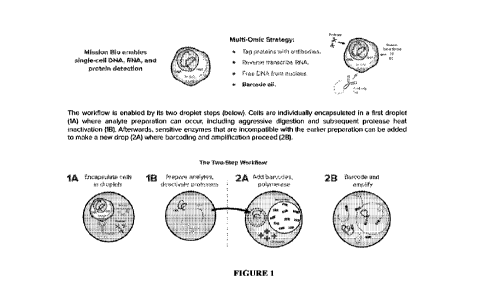
[0013] Figure 1 is a schematic diagram of a multiomic strategy used in some
embodiments to
detect DNA, RNA, and protein from a single cell.
[0014] Figure 2 is a schematic diagram of the first droplet of an exemplary
embodiment of the
invention.
100151 Figure 3 is a schematic diagram of the second droplet of an exemplary
embodiment of the
invention.
[0016] Figure 4 is a schematic diagram of the mutliomics workflow. Stained
cells are input onto
the Tapestri platform. The single cells are encapsulated with a buffer
containing components for lysis,
reverse transcription, and a protease treatment. These reactions are performed
on a thennocycler and the
encapsulated cells returned to the Tapestri platform to for droplet merger
with barcoding PCR reagents and
barcoding beads. A PCR is performed which attaches the cell barcodes to the
DNA targeted amplicons,
RNA targeted amplicons, and protein tag amplicons. All amplicons from the same
emulsion would contain
the same cell barcode. After emulsions are broken, an exonuclease reaction and
a SPRI cleanup are
performed. The supernatant is kept to process into protein libraries. The SPR1
beads contain both the DNA
and RNA amplicons that are then separated using a biotin capture ohgo and
streptavidin beads. The DNA
and RNA amplicons are then processed separately to form sequencing libraries.
[0017] Figure 5 is Bioanalyzer trace of a DNA library from the untreated cells
in tubes 4-8 from
the multiomics experiment from Example I where 3 cell lines (Jurkat, K-562,
and KCL-22) were untreated
or treated 3 different doses of imatinib (10 uM, 100 uM, and 250 uM). 1 uL of
undiluted library was loaded
onto a HS DNA chip.
100181 Figure 6 is Bioanalyzer trace of a protein library from the untreated
cells in tubes 4-8 from
the multiomics experiment from Example I where 3 cell lines (Jurkat, K-562,
and KCL-22) were untreated
CA 03138806 2021- 11- 19
WO 2020/237222 PCT/US2020/034404
4
or treated 3 different doses of imatinib (10 uM, 100 uM, and 250 uM), 1 uL of
undiluted library was loaded
onto a HS DNA chip.
[0019] Figure 7 is a Bioanalyzer trace of a RNA library from the untreated
cells in tubes 4-8 from
the multiomics experiment from Example I where 3 cell lines (Jurkat, K-562,
and KCL-22) were untreated
or treated 3 different doses of imatinib (10 uM, 100 uM, and 250 tiM). 1 uL of
a 1:5 library dilution was
loaded onto a HS DNA chip.
[0020] Figure 8 shows the results from the multiomics experiment from Example
1 where 3 cell
lines (Jurkat, K-562, and KCL-22) were untreated or treated 3 different doses
of imatinib (10 uM, 100 uM,
and 250 uM). The DNA variants are used to make a heat map where the Y-axis are
the cell barcodes are
shown in Fig. 9. This heat map separates into 3 clusters (green, red, and
black, which alternately can be
represented as grey shades) which corresponds to each of the cell lines.
Figure 8 also shows the clustering
with t-SNE and tunap where the colors are the same as in the heat map.
[0021] Figure 9 depicts the results from the multiomics experiment from
Example 1 where 3 cell
lines (Jurkat, K-562, and KCL-22) were untreated or treated 3 different doses
of imatinib (10 uM, 100 uM,
and 250 uM). These cells were stained then input onto the Tapestri for the
multiomics workflow. DNA
libraries, RNA libraries, and protein libraries from single cells were
produced where the libraries produced
from the same cell shared a cell barcode. By looking at the mutations found in
the DNA libraries, the 3 cell
lines were identified. Each cell barcode also has corresponding RNA reads and
protein reads. Figure 9 also
shows those same cells clustered with umap based on SNV, CNV, protein, and RNA
where the color of
each datapoint corresponds to the cell identified by the mutations found in
DNA. The doses of imatinib
were differentiated by the tags on the cell hashing antibodies resulting in
more clusters for the protein
expression.
[0022] Figure 10 depicts the results from the multiomics experiment from
Example II where
PBMCs were untreated or treated with PHA. A spike-in of the Jurkat cell line
was also included. DNA
libraries, RNA libraries, and protein libraries from single cells were
produced where the libraries produced
from the same cell shared a cell barcode. By looking at the mutations found in
the DNA libraries, Jurkat
cells, cell doublets, and merged droplets were identified. Each cell barcode
also has corresponding RNA
reads and protein reads (top). Figure 11 shows the fraction of PBMCs that had
low RNA expression and
high RNA expression. The cells were identified as being treated by PHA or
untreated by the protein tags
used for cell hashing.
DETAILED DESCRIPTION
[0023] Various aspects of the invention will now be described with reference
to the following
section which will be understood to be provided by way of illustration only
and not to constitute a limitation
on the scope of the invention.
[0024] "Complementarity" refers to the ability of a nucleic acid to form
hydrogen bond(s) or
hybridize with another nucleic acid sequence by either traditional Watson-
Crick or other non-traditional
types. As used herein "hybridization," refers to the binding, duplexing, or
hybridizing of a molecule only
CA 03138806 2021- 11- 19
WO 2020/237222 PCT/US2020/034404
to a particular nucleotide sequence under low, medium, or highly stringent
conditions, including when that
sequence is pmsent in a complex mixture (e.g., total cellular) DNA or RNA. See
e.g. Ausubel, et al., Current
Protocols In Molecular Biology, John Wiley & Sons, New York, N.Y., 1993. If a
nucleotide at a certain
position of a polynucleotide is capable of forming a Watson-Crick pairing with
a nucleotide at the same
position in an anti-parallel DNA or RNA strand, then the polynucleotide and
the DNA or RNA molecule
are complementary to each other at that position. The polynucleotide and the
DNA or RNA molecule are
"substantially complementary" to each other when a sufficient number of
corresponding positions in each
molecule are occupied by nucleotides that can hybridize or anneal with each
other in order to affect the
desired process. A complementary sequence is a sequence capable of annealing
under stringent conditions
to provide a 3'-terminal serving as the origin of synthesis of complementary
chain.
[0025] "Identity," as known in the art, is a relationship between two or more
polypeptide
sequences or two or more polynucleotide sequences, as determined by comparing
the sequences. In the art,
"identity" also means the degree of sequence relatedness between polypeptide
or polynucleotide sequences,
as determined by the match between strings of such sequences. "Identity" and
"similarity" can be readily
calculated by known methods, including, but not limited to, those described in
Computational Molecular
Biology, Lesk, A. M., ed., Oxford University Press, New York, 1988;
Biocomputing: Infortnatics and
Genome Projects, Smith, D. W., at., Academic Press, New York, 1993; Computer
Analysis of Sequence
Data, Part I, Griffm, A. M., and Griffin, H. G., eds., Humana Press, New
Jersey, 1994; Sequence Analysis
in Molecular Biology, von Heinje, G., Academic Press, 1987; and Sequence
Analysis Primer, Gribskov,
M. and Devereux, J., eds., M Stockton Press, New York, 1991; and Carillo, H.,
and Lipman, D., Siam J.
Applied Math., 48:1073 (1988). In addition, values for percentage identity can
be obtained from amino acid
and nucleotide sequence alignments generated using the default settings for
the AlignX component of
Vector NT! Suite 8.0 (Informax, Frederick, Md.). Preferred methods to
determine identity are designed to
give the largest match between the sequences tested. Methods to determine
identity and similarity are
codified in publicly available computer programs. Preferred computer program
methods to determine
identity and similarity between two sequences include, but are not limited to,
the GCG program package
(Dcycreux, J., et al., Nucleic Acids Research 12(1): 387 (1984)), BLASTP,
BLASTN, and FASTA
(Atschul, S. F. et al., J. Molec. Biol. 215:403410 (1990)). The BLAST X
program is publicly available
from NCBI and other sources (BLAST Manual, Altschul, S., et al, NCBINLM NIH
Bethesda, Md. 20894:
Altschul, S., et al., J. Mol. Biol. 215:403410 (1990). The well-known Smith
Waterman algorithm may also
be used to determine identity.
[0026] The terms "amplify", "amplifying", "amplification reaction" and their
variants, refer
generally to any action or process whereby at least a portion of a nucleic
acid molecule (referred to as a
template nucleic acid molecule) is replicated or copied into at least one
additional nucleic acid molecule.
The additional nucleic acid molecule optionally includes sequence that is
substantially identical or
substantially complementary to at least some portion of the template nucleic
acid molecule. The template
nucleic acid molecule can be single-stranded or double-stranded and the
additional nucleic acid molecule
can independently be single-stranded or double-stranded. In some embodiments,
amplification includes a
CA 03138806 2021- 11- 19
WO 2020/237222 PCT/US2020/034404
6
template-dependent in vitro enzyme-catalyzed reaction for the production of at
least one copy of at least
some portion of the nucleic acid molecule or the production of at least one
copy of a nucleic acid sequence
that is complementary to at least some portion of the nucleic acid molecule.
Amplification optionally
includes linear or exponential replication of a nucleic acid molecule. In some
embodiments, such
amplification is performed using isothemtal conditions; in other embodiments,
such amplification can
include themocycling. In some embodiments, the amplification is a multiplex
amplification that includes
the simultaneous amplification of a plurality of target sequences in a single
amplification reaction. At least
some of the target sequences can be situated, on the same nucleic acid
molecule or on different target
nucleic acid molecules included in the single amplification reaction. In some
embodiments, "amplification"
includes amplification of at least some portion of DNA- and RNA-based nucleic
acids alone, or in
combination. The amplification reaction can include single or double-stranded
nucleic acid substrates and
can further including any of the amplification processes known to one of
ordinary skill in the art. In some
embodiments, the amplification reaction includes polymerase chain reaction
(PCR). In the present
invention, the terms "synthesis" and "amplification" of nucleic acid are used.
The synthesis of nucleic acid
in the present invention means the elongation or extension of nucleic acid
from an oligonucleotide serving
as the origin of synthesis. If not only this synthesis but also the formation
of other nucleic acid and the
elongation or extension reaction of this formed nucleic acid occur
continuously, a series of these reactions
is comprehensively called amplification. The polynucleic acid produced by the
amplification technology
employed is generically referred to as an "amplicon" or "amplification
product"
[0027] A number of nucleic acid polymerases can be used in the amplification
reactions utilized
in certain embodiments provided herein, including any enzyme that can catalyze
the polymerization of
nucleotides (including analogs thereof) into a nucleic acid strand. Such
nucleotide polymerization can occur
in a template-dependent fashion. Such polymerases can include without
limitation naturally occurring
polymerases and any subunits and truncations thereof, mutant polymerases,
variant polymerases,
recombinant, fusion or otherwise engineered polymerases, chemically modified
polymerases, synthetic
molecules or assemblies, and any analogs, derivatives or fragments thereof
that retain the ability to catalyze
such polymerization. Optionally, the polymerase can be a mutant polymerase
comprising one or more
mutations involving the replacement of one or more amino acids with other
amino acids, the insertion or
deletion of one or more amino acids from the polymerase, or the linkage of
parts of two or more
polymerases. Typically, the polymerase comprises one or more active sites at
which nucleotide binding
and/or catalysis of nucleotide polymerization can occur. Some exemplary
polymerases include without
limitation DNA polymerases and RNA polymerases. The term "polymerase" and its
variants, as used herein,
also includes fusion proteins comprising at least two portions linked to each
other, where the first portion
comprises a peptide that can catalyze the polymerization of nucleotides into a
nucleic acid strand and is
linked to a second portion that comprises a second polypeptide. In some
embodiments, the second
polypeptide can include a reporter enzyme or a processivity -enhancing domain.
Optionally, the polymerase
can possess 5' exonuclease activity or terminal transferase activity. In some
embodiments, the polymerase
can be optionally reactivated, for example through the use of heat, chemicals
or re-addition of new amounts
CA 03138806 2021- 11- 19
WO 2020/237222
PCT/US2020/034404
7
of polymerase into a reaction mixture, hi some embodiments, the polymerase can
include a hot-start
polymerase or an aptamer-based polymerase that optionally can be reactivated.
[0028] The terms "target primer" or "target-specific primer" and variations
thereof refer to primers
that are complementary to a binding site sequence. Target printers are
generally a single stranded or double-
stranded polynucleotide, typically an oligonucleotide, that includes at least
one sequence that is at least
partially complementary to a target nucleic acid sequence.
[0029] "Forward primer binding site" and "reverse primer binding site" refers
to the regions on
the template DNA and/or the amplicon to which the forward and reverse primers
bind. The primers act to
delimit the region of the original template polynucleotide which is
exponentially amplified during
amplification. In some embodiments, additional primers may bind to the region
5' of the forward primer
and/or reverse primers. Where such additional primers are used, the forward
primer binding site and/or the
reverse primer binding site may encompass the binding regions of these
additional primers as well as the
binding regions of the primers themselves. For example, in some embodiments,
the method may use one
or more additional primers which bind to a region that lies 5' of the forward
and/or reverse primer binding
region. Such a method was disclosed, for example, in W00028082 which discloses
the use of "displacement
primers" or "outer primers".
[0030] A `barcode' nucleic acid identification sequence can be incorporated
into a nucleic acid
primer or linked to a primer to enable independent sequencing and
identification to be associated with one
another via a barcode which relates information and identification that
originated from molecules that
existed within the same sample. There are numerous techniques that can be used
to attach barcodes to the
nucleic acids within a discrete entity. For example, the target nucleic acids
may or may not be first amplified
and fragmented into shorter pieces. The molecules can be combined with
discrete entities, e.g., droplets,
containing the barcodes. The barcodes can then be attached to the molecules
using, for example, splicing
by overlap extension. In this approach, the initial target molecules can have
"adaptor" sequences added,
which are molecules of a known sequence to which primers can be synthesized.
When combined with the
barcodes, primers can be used that are complementary to the adaptor sequences
and the barcode sequences,
such that the product amplicons of both target nucleic acids and barcodes can
anneal to one another and,
via an extension reaction such as DNA polymerization, be extended onto one
another, generating a double-
stranded product including the target nucleic acids attached to the barcode
sequence. Alternatively, the
primers that amplify that target can themselves be barcoded so that, upon
annealing and extending onto the
target, the amplicon produced has the barcode sequence incorporated into it.
This can be applied with a
number of amplification strategies, including specific amplification with PCR
or non-specific amplification
with, for example, MDA. An alternative enzymatic reaction that can be used to
attach barcodes to nucleic
acids is ligation, including blunt or sticky end ligation. In this approach,
the DNA barcodes are incubated
with the nucleic acid targets and ligase enzyme, resulting in the ligation of
the barcode to the targets. The
ends of the nucleic acids can be modified as needed for ligation by a number
of techniques, including by
using adaptors introduced with ligase or fragments to enable greater control
over the number of barcodes
added to the end of the molecule_
CA 03138806 2021- 11- 19
WO 2020/237222 PCT/US2020/034404
8
[0031] A barcode sequence can additionally be incorporated into microfluidic
beads to decorate
the bead with identical sequence tags. Such tagged beads can be inserted into
microfluidic droplets and via
droplet PCR amplification, tag each target amplicon with the unique bead
barcode. Such barcodes can be
used to identify specific droplets upon a population of amplicons originated
from. This scheme can be
utilized when combining a microfluidic droplet containing single individual
cell with another microfluidic
droplet containing a tagged bead. Upon collection and combination of many
microfluidic droplets,
amplicon sequencing results allow for assignment of each product to unique
microfluidic droplets. In a
typical implementation, we use barcodes on the Mission Bio TapestriTm beads to
tag and then later identify
each droplet's amplicon content. The use of barcodes is described in US Patent
Application Serial No.
15/940,850 filed March 29, 2018 by Abate, A. et al., entitled 'Sequencing of
Nucleic Acids via Barcoding
in Discrete Entities', incorporated by reference herein.
[0032] In some embodiments, it may be advantageous to introduce barcodes into
discrete entities,
e.g., microdroplets, on the surface of a bead, such as a solid polymer bead or
a hydrogel bead. These beads
can be synthesized using a variety of techniques. For example, using a mix-
split technique, beads with
many copies of the same, random barcode sequence can be synthesized. This can
be accomplished by, for
example, creating a plurality of beads including sites on which DNA can be
synthesized. The beads can be
divided into four collections and each mixed with a buffer that will add a
base to it, such as an A, T, G, or
C. By dividing the population into four subpopulations, each subpopulation can
have one of the bases added
to its surface. This reaction can be accomplished in such a way that only a
single base is added and no
further bases are added. The beads from all four subpopulations can be
combined and mixed together, and
divided into four populations a second time. In this division step, the beads
from the previous four
populations may be mixed together randomly. They can then be added to the four
different solutions, adding
another, random base on the surface of each bead. This process can be repeated
to generate sequences on
the surface of the bead of a length approximately equal to the number of times
that the population is split
and mixed. If this was done 10 times, for example, the result would be a
population of beads in which each
bead has many copies of the same random 10-base sequence synthesized on its
surface. The sequence on
each bead would be determined by the particular sequence of reactors it ended
up in through each mix-spit
cycle.
100331 A barcode may further comprise a 'unique identification sequence' (UMW
A UMI is a
nucleic acid having a sequence which can be used to identify and/or
distinguish one or more first molecules
to which the UMI is conjugated from one or more second molecules. UMIs are
typically short, e.g., about
to 20 bases in length, and may be conjugated to one or more target molecules
of interest or amplification
products thereof. UMIs may be single or double stranded. In some embodiments,
both a nucleic acid
barcode sequence and a UMI are incorporated into a nucleic acid target
molecule or an amplification
product thereof. Generally, a UMI is used to distinguish between molecules of
a similar type within a
population or group, whereas a nucleic acid barcode sequence is used to
distinguish between populations
or groups of molecules. In some embodiments, where both a UMI and a nucleic
acid barcode sequence are
utilized, the UMI is shorter in sequence length than the nucleic acid barcode
sequence.
CA 03138806 2021- 11- 19
WO 2020/237222
PCT/US2020/034404
[0034] The terms "identity" and "identical" and their variants, as used
herein, when used in
reference to two or more nucleic acid sequences, refer to similarity in
sequence of the two or more
sequences (e.g., nucleotide or polypeptide sequences). In the context of two
or more homologous
sequences, the percent identity or homology of the sequences or subsequences
thereof indicates the
percentage of all monomeric units (e.g., nucleotides or amino acids) that are
the same (i.e., about 70%
identity, preferably 75%, 80%, 85%, 900/u, 95%, 97%, 98% or 99% identity). The
percent identity can be
over a specified region, when compared and aligned for maximum correspondence
over a comparison
window, or designated region as measured using a BLAST or BLAST 2.0 sequence
comparison algorithms
with default parameters described below, or by manual aligmnent and visual
inspection. Sequences are said
to be "substantially identical" when there is at least 85% identity at the
amino acid level or at the nucleotide
level. Preferably, the identity exists over a region that is at least about
25, 50, or 100 residues in length, or
across the entire length of at least one compared sequence. A typical
algorithm for determining percent
sequence identity and sequence similarity are the BLAST and BLAST 2.0
algorithms, which are described
in Altschul et al, Nuc. Acids Res. 253389-3402 (1977). Other methods include
the algoritluns of Smith &
Waterman, Adv, Appl. Math. 2:482 (1981), and Needleman & Wunsch, J. Mel. Biol.
48:443 (1970), etc.
Another indication that two nucleic acid sequences are substantially identical
is that the two molecules or
their complements hybridize to each other under stringent hybridization
conditions.
[0035] The tenns "nucleic acid," "polynucleotides," and "oligonucleotides"
refers to biopolymers
of nucleotides and, unless the context indicates otherwise, includes modified
and unmodified nucleotides,
and both DNA and RNA, and modified nucleic acid backbones. For example, in
certain embodiments, the
nucleic acid is a peptide nucleic acid (PNA) or a locked nucleic acid (LNA).
Typically, the methods as
described herein are performed using DNA as the nucleic acid template for
amplification. However, nucleic
acid whose nucleotide is replaced by an artificial derivative or modified
nucleic acid from natural DNA or
RNA is also included in the nucleic acid of the present invention insofar as
it functions as a template for
synthesis of complementary chain. The nucleic acid of the present invention is
generally contained in a
biological sample. The biological sample includes animal, plant or microbial
tissues, cells, cultures and
excretions, or extracts therefrom. In certain aspects, the biological sample
includes intracellular parasitic
genomic DNA or RNA such as virus or mycoplasma. The nucleic acid may be
derived from nucleic acid
contained in said biological sample. For example, genomic DNA, or cDNA
synthesized from mRNA, or
nucleic acid amplified on the basis of nucleic acid derived from the
biological sample, are preferably used
in the described methods. Unless denoted otherwise, whenever a oligonucleotide
sequence is represented,
it will be understood that the nucleotides are in 5' to 3' order from left to
right and that "A" denotes
deoxyadenosine, "C" denotes deoxycytidine, "G" denotes deoxyguanosine, "T"
denotes thymidine, and "U'
denotes deoxyuridine. Oligonucleotides are said to have "5' ends" and "3'
ends" because mononucleotides
are typically reacted to form oligonucleotides via attachment of the 5'
phosphate or equivalent group of one
nucleotide to the 3' hydroxyl or equivalent group of its neighboring
nucleotide, optionally via a
phosphodiester or other suitable linkage.
CA 03138806 2021- 11- 19
WO 2020/237222
PCT/US2020/034404
[0036] A template nucleic acid is a nucleic acid serving as a template for
synthesizing a
complementary chain in a nucleic acid amplification technique. A complementary
chain having a nucleotide
sequence complementary to the template has a meaning as a chain corresponding
to the template, but the
relationship between the two is merely relative. That is, according to the
methods described herein a chain
synthesized as the complementary chain can function again as a template. That
is, the complementary chain
can become a template. In certain embodiments, the template is derived from a
biological sample, e.g.,
plant, animal, virus, micro-organism, bacteria, fungus, etc. In certain
embodiments, the animal is a
mammal, e.g., a human patient. A template nucleic acid typically comprises one
or more target nucleic acid.
A target nucleic acid in exemplary embodiments may comprise any single or
double-stranded nucleic acid
sequence that can be amplified or synthesized according to the disclosure,
including any nucleic acid
sequence suspected or expected to be present in a sample.
[0037] Primers and oligonucleotides used in embodiments herein comprise
nucleotides. A
nucleotide comprises any compound, including without limitation any naturally
occurring nucleotide or
analog thereof, which can bind selectively to, or can be polymerized by, a
polymerase. Typically, but not
necessarily, selective binding of the nucleotide to the polymerase is followed
by polymerization of the
nucleotide into a nucleic acid strand by the polymerase; occasionally however
the nucleotide may dissociate
from the polymerase without becoming incorporated into the nucleic acid
strand, an event referred to herein
as a "non-productive" event. Such nucleotides include not only naturally
occurring nucleotides but also any
analogs, regardless of their structure, that can bind selectively to, or can
be polymerized by, a polymerase.
While naturally occurring nucleotides typically comprise base, sugar and
phosphate moieties, the
nucleotides of the present disclosure can include compounds lacking any one,
some or all of such moieties.
For example, the nucleotide can optionally include a chain of phosphorus atoms
comprising three, four,
five, six, seven, eight, nine, ten or more phosphorus atoms. In some
embodiments, the phosphorus chain
can be attached to any carbon of a sugar ring, such as the 5' carbon. The
phosphorus chain can be linked to
the sugar with an intervening 0 or S. hi one embodiment, one or more
phosphorus atoms in the chain can
be part of a phosphate group having P and 0. In another embodiment, the
phosphorus atoms in the chain
can be linked together with intervening 0, NH, 5, methylene, substituted
methylene, ethylene, substituted
ethylene, CNF12, C(0), C(CH2), CH2CH2, or C(OH)CH2R (where R can be a 4-
pyridine or 14midazole). In
one embodiment, the phosphorus atoms in the chain can have side groups having
0, I3H3, or S. In the
phosphorus chain, a phosphorus atom with a side group other than 0 can be a
substituted phosphate group.
In the phosphorus chain, phosphorus atoms with an intervening atom other than
0 can be a substituted
phosphate group. Some examples of nucleotide analogs are described in Xu, U.S.
Pat. No. 7,405,281.
[0038] In some embodiments, the nucleotide comprises a label and referred to
herein as a "labeled
nucleotide"; the label of the labeled nucleotide is referred to herein as a
"nucleotide label". In some
embodiments, the label can be in the form of a fluorescent moiety (e.g. dye),
luminescent moiety, or the
like attached to the terminal phosphate group, i.e., the phosphate group most
distal from the sugar. Some
examples of nucleotides that can be used in the disclosed methods and
compositions include, but are not
limited to, ribonucleotides, deoxyribonucleotides, modified ribonucleotides,
modified
CA 03138806 2021- 11- 19
WO 2020/237222 PCT/US2020/034404
11
deoxyribonucleotides, ribonucleofide polyphosphates, deoxyribottucleofide
polyphosphates, modified
ribonucleotide polyphosphates, modified deoxyfibonucleotide polyphosphates,
peptide nucleotides,
modified peptide nucleotides, metallonucleosides, phosphonate nucleosides, and
modified phosphate-sugar
backbone nucleotides, analogs, derivatives, or variants of the foregoing
compounds, and the like. In some
embodiments, the nucleotide can comprise non-oxygen moieties such as, for
example, thio- or borano-
moieties, in place of the oxygen moiety bridging the alpha phosphate and the
sugar of the nucleotide, or the
alpha and beta phosphates of the nucleotide, or the beta and gamma phosphates
of the nucleotide, or
between any other two phosphates of the nucleotide, or any combination
thereof. "Nucleotide 5'-
triphosphate" refers to a nucleotide with a triphosphate ester group at the 5'
position, and are sometimes
denoted as "NTP", or "dNTP" and "ddNTP" to particularly point out the
structural features of the ribose
sugar. The triphosphate ester group can include sulfur substitutions for the
various oxygens, e.g. a-titio-
nucleotide 5'-triphosphates. For a review of nucleic acid chemistry, see:
Shabarova, Z. and Bogdanov, A.
Advanced Organic Chemistry of Nucleic Acids, VCH, New York, 1994.
[0039] Any nucleic acid amplification method may be utilized, such as a PCR-
based assay, e.g.,
quantitative PCR (qPCR), or an isothermal amplification may be used to detect
the presence of certain
nucleic acids, e.g., genes, of interest, present in discrete entities or one
or more components thereof, e.g.,
cells encapsulated therein. Such assays can be applied to discrete entities
within a microfluidic device or a
portion thereof or any other suitable location. The conditions of such
amplification or PCR-based assays
may include detecting nucleic acid amplification over time and may vary in one
or more ways.
[0040] The number of amplification/PCR primers that may be added to a
microdroplet may vary.
The number of amplification or PCR primers that may be added to a microdmplet
may range from about 1
to about 500 or more, e.g., about 2 to 100 primers, about 2 to 10 primers,
about 10 to 20 primers, about 20
to 30 primers, about 30 to 40 primers, about 40 to 50 primers, about 50 to 60
primers, about 60 to 70
primers, about 70 to 80 primers, about 80 to 90 primers, about 90 to 100
primers, about 100 to 150 primers,
about 150 to 200 primers, about 200 to 250 primers, about 250 to 300 primers,
about 300 to 350 primers,
about 350 to 400 primers, about 400 to 450 primers, about 450 to 500 primers,
or about 500 primers or
more.
[0041] One or both primer of a primer set may also be attached or conjugated
to an affinity reagent
that may comprise anything that binds to a target molecule or moiety.
Nonlimiting examples of affinity
reagent include ligands, receptors, antibodies and binding fragments thereof,
peptide, nucleic acid, and
fusions of the preceding and other small molecule that specifically binds to a
larger target molecule in order
to identify, track, capture, or influence its activity. Affinity reagents may
also be attached to solid supports,
beads, discrete entities, or the like, and are still referenced as affmity
reagents herein.
[0042] One or both primers of a primer set may comprise a barcode sequence
described herein.
In some embodiments, individual cells, for example, are isolated in discrete
entities, e.g., droplets. These
cells may be lysed and their nucleic acids barcoded. This process can be
performed on a large number of
single cells in discrete entities with unique barcode sequences enabling
subsequent deconvolution of mixed
sequence reads by barcode to obtain single cell information. This approach
provides a way to group together
CA 03138806 2021- 11- 19
WO 2020/237222
PCT/US2020/034404
12
nucleic acids originating from large numbers of single cells. Additionally,
affinity reagents such as
antibodies can be conjugated with nucleic acid labels, e.g., oligonucleotides
including barcodes, which can
be used to identify antibody type, e.g., the target specificity of an
antibody. These reagents can then be used
to bind to the proteins within or on cells, thereby associating the nucleic
acids carried by the affinity reagents
to the cells to which they are bound. These cells can then be processed
through a barcoding workflow as
described herein to attach barcodes to the nucleic acid labels on the affinity
reagents. Techniques of library
preparation, sequencing, and bioinfomintics may then be used to group the
sequences according to
cell/discrete entity barcodes. Any suitable affinity reagent that can bind to
or recognize a biological sample
or portion or component thereof, such as a protein, a molecule, or complexes
thereof, may be utilized in
connection with these methods. The affinity reagents may be labeled with
nucleic acid sequences that
relates their identity, e.g., the target specificity of the antibodies,
permitting their detection and quantitation
using the barcoding and sequencing methods described herein. Exemplary
affinity reagents can include, for
example, antibodies, antibody fragments, Fabs, scFvs, peptides, drugs, etc. or
combinations thereof. The
affinity reagents, e.g., antibodies, can be expressed by one or more organisms
or provided using a biological
synthesis technique, such as phage, mRNA, or ribosome display. The affinity
reagents may also be
generated via chemical or biochemical means, such as by chemical linkage using
N-Hydroxysuccinimide
(NETS), click chemistry, or streptavidin-biotin interaction, for example. The
oligo-affmity reagent
conjugates can also be generated by attaching oligos to affinity reagents and
hybridizing, ligating, and/or
extending via polymerase, etc., additional oligos to the previously conjugated
oligos. An advantage of
affinity reagent labeling with nucleic acids is that it permits highly
multiplexed analysis of biological
samples. For example, large mixtures of antibodies or binding reagents
recognizing a variety of targets in
a sample can be mixed together, each labeled with its own nucleic acid
sequence. This cocktail can then be
reacted to the sample and subjected to a barcoding workflow as described
herein to recover information
about which reagents bound, their quantity, and how this varies among the
different entities in the sample,
such as among single cells. The above approach can be applied to a variety of
molecular targets, including
samples including one or more of cells, peptides, proteins, macromolecules,
macromolecular complexes,
etc. The sample can be subjected to conventional processing for analysis, such
as fixation and
permeabilization, aiding binding of the affinity reagents. To obtain highly
accurate quantitafion, the unique
molecular identifier (UMI) techniques described herein can also be used so
that affinity reagent molecules
are counted accurately. This can be accomplished in a number of ways,
including by synthesizing UMIs
onto the labels attached to each affinity reagent before, during, or after
conjugation, or by attaching the
UMIs microfluidically when the reagents are used. Similar methods of
generating the barcodes, for
example, using combinatorial barcode techniques as applied to single cell
sequencing and described herein,
are applicable to the affinity reagent technique. These techniques enable the
analysis of proteins and/or
epitopes in a variety of biological samples to perform, for example, mapping
of epitopes or post
translational modifications in proteins and other entities or performing
single cell proteomics. For example,
using the methods described herein, it is possible to generate a library of
labeled affinity reagents that detect
an epitope in all proteins in the proteome of an organism, label those
epitopes with the reagents, and apply
CA 03138806 2021- 11- 19
WO 2020/237222
PCT/US2020/034404
13
the barcoding and sequencing techniques described herein to detect and
accurately quantitate the labels
associated with these epitopes.
[0043] Primers may contain primers for one or more nucleic acid of interest,
e.g. one or more
genes of interest. The number of primers for genes of interest that are added
may be from about one to 500,
e.g., about 1 to 10 primers, about 10 to 20 primers, about 20 to 30 primers,
about 30 to 40 primers, about
40 to 50 primers, about 50 to 60 primers, about 60 to 70 primers, about 70 to
80 primers, about 80 to 90
primers, about 90 to 100 primers, about 100 to 150 primers, about 150 to 200
primers, about 200 to 250
primers, about 250 to 300 primers, about 300 to 350 primers, about 350 to 400
primers, about 400 to 450
primers, about 450 to 500 primers, or about 500 primers or more. Primers
and/or reagents may be added to
a discrete entity, e.g., a microdroplet, in one step, or in more than one
step. For instance, the primers may
be added in two or more steps, three or more steps, four or more steps, or
five or more steps. Regardless of
whether the primers are added in one step or in more than one step, they may
be added after the addition of
a lysing agent, prior to the addition of a lysing agent, or concomitantly with
the addition of a lysing agent.
When added before or after the addition of a lysing agent, the PCR primers may
be added in a separate step
from the addition of a lysing agent. In some embodiments, the discrete entity,
e.g., a microdroplet, may be
subjected to a dilution step and/or enzyme inactivation step prior to the
addition of the PCR reagents.
Exemplary embodiments of such methods are described in PCT Publication No, WO
2014/028378, the
disclosure of which is incorporated by reference herein in its entirety and
for all purposes.
[0044] A primer set for the amplification of a target nucleic acid typically
includes a forward
primer and a reverse primer that are complementary to a target nucleic acid or
the complement thereof. In
some embodiments, amplification can be performed using multiple target-
specific primer pairs in a single
amplification reaction, wherein each primer pair includes a forward target-
specific primer and a reverse
target-specific primer, where each includes at least one sequence that
substantially complementary or
substantially identical to a corresponding target sequence in the sample, and
each primer pair having a
different corresponding target sequence. Accordingly, certain methods herein
are used to detect or identify
multiple target sequences from a single cell sample.
[0045] In some implementations, solid supports, beads, and the like are coated
with affmity
reagents. Affmity reagents include, without limitation, antigens, antibodies
or aptamers with specific
binding affmity for a target molecule. The affinity reagents bind to one or
more targets within the single
cell entities. Affmity reagents are often detectably labeled (e.g., with a
fluorophore). Affinity reagents are
sometimes labeled with unique barcodes, oligonucleotide sequences, or UMI's.
[0046] In some implementations, a RT/PCR polymerase reaction and amplification
reaction are
performed, for example in the same reaction mixture, as an addition to the
reaction mixture, or added to a
portion of the reaction mixture.
[0047] In one particular implementation, a solid support contains a plurality
of affmity reagents,
each specific for a different target molecule but containing a common sequence
to be used to identify the
unique solid support. Affinity reagents that bind a specific target molecule
are collectively labeled with the
same oligonucleotide sequence such that affmity molecules with different
binding affmities for different
CA 03138806 2021- 11- 19
WO 2020/237222
PCT/US2020/034404
14
targets are labeled with different oligonucleotide sequences. In this way,
target molecules within a single
target entity are differentially labeled in these implements to determine
which target entity they are from
but contain a common sequence to identify them from the same solid support.
Targeted detection and sequencing of DNA, RNA, and Protein
[0048] A first objective of some implementations is to provide methods for the
simultaneous
targeted detection and sequencing of DNA, RNA, and Protein. In preferred
embodiments, the DNA, RNA,
and Protein is detected, characterized, and sequenced using single cells.
Single-cell analysis is conducted
herein by sequencing either genomic DNA targets, RNA/cDNA transcripts or
protein detection. Genomic
DNA can be assessed by first amplifying whole genomic DNA or targeted
approaches.
[0049] One such example is amplifying whole genomic DNA or targeted portions
of the DNA in
a Mission Bio's Tapestri platform. In some embodiments using this approach,
RNA/cDNA transcripts
are often accessed by first priming with 3' mRNA end oligo dT extension
strategies or random primers.
Targeted cDNA extension can be conducted using transcript specific primers and
is amenable to these
workflows. Surface protein markers are readily detected using dye-labelled
antibodies and a fluorescence-
activated cell sorting platfortn (PACS). In preferred embodiments, DNA
barcoded antibodies can be
employed in cell staining and readout by next generation sequencing (NGS). We
have combined three
different methodologies to simultaneously detect, by targeted sequencing, DNA,
RNA, and surface protein
markers as our so-called `triomic' methodology. Our novel approach represents
a great advancement in
single-cell analysis.
[0050] Typically, methods are optimized to efficiently detect and amplify
genomic DNA amplicon
targets. Additionally, some implementations use reverse transcriptase enzyme
with RNA-specific primers
to extend RNA targets and generate cDNA templates for efficient RNA detection.
Additionally, an
antibody-oligonucleotide tagging system for surface protein detection on
single-cells using microfluidic
droplets has been developed. In certain embodiments provided herein, a Mission
Bio Tapestri workflow
scheme has been developed that applies central concepts from all three
'tunics' to thereby provide for a
triomic interrogation process for single-cells. While the disclosed
embodiments relate to using the
Tapestri workflow, it should be noted that the disclosed principles are not
limited thereto and may be
applied to other instrumentations and/or workflow.
[0051] Certain methods provided herein utilize specific antibodies to detect
epitopes of interest.
hi some embodiments, antibodies are labeled with sequence tags that can be
read out with microfluidic
barcoding and DNA sequencing. This and related implementations are used herein
to characterize cell
surface proteins of different cell types at the single-cell level.
[0052] In some embodiments, a barcode identity is encoded by its full
nucleobase sequence and
thus confers a combinatorial tag space far exceeding what is possible with
conventional approaches using
fluorescence. A modest tag length of ten bases provides over a million unique
sequences, sufficient to label
an antibody against every epitope in the human proteome. Indeed, with this
approach, the limit to
CA 03138806 2021- 11- 19
WO 2020/237222
PCT/US2020/034404
multiplexing is not the availability of unique tag sequences but, rather, that
of specific antibodies that can
detect the epitopes of interest in a multiplexed reaction.
[0053] In some implementations, cells are bound with antibodies against the
different target
epitopes, as in conventional immtmostaining, except that the antibodies are
labeled with barcodes.
[0054] In practice, when an antibody binds its target the DNA barcode tag is
carried with it and
thus allows the presence of the target to be inferred based on the presence of
the barcode. In some
implementations, counting barcode tags provides an estimate of the different
epitopes present in the cell.
[0055] Other embodiments implementations are used to distinguish particular
cells by their protein
expression profiles. Some embodiments of DNA-tagged antibodies provided herein
have multiple
advantages for profiling proteins in single cells.
[0056] A primary advantage of these implementations is the ability to amplify
low-abundance tags
to make them detectable with sequencing. Another advantage in some
implementations is the capability of
using molecular indices for quantitative results. Some implementations also
have essentially limitless
multiplexing capabilities.
[0057] Some embodiments utilize solid beads having an alternate chemistry
where the primers to
be used are in solution and contain a PCR annealing sequence embedded, or
'handle', that allows
hybridization to primers, In some implementations, the handle is a specific
tail 5' upstream of the target
sequence and this handle is complimentary to bead barcoded oligo and serves as
a PCR extension bridge to
link the target amplicon to the bead barcode library primer sequence. The
solid beads may contain primers
that can anneal to the PCR handle on the primers.
[0058] Some embodiments are used to detect and characterize cell surface
proteins, DNA, and
RNA. Such a workflow can begin with an antibody-oligonucleotide staining and
washing of a single-cell
suspension. The stained cells are loaded onto a Mission, Bio TapestriTm
system, cells are lysed, RNA is
converted to cDNA by reverse transcriptase, PCR cycling is used to amplify
antibody-oligonucleotides,
targeted cDNA species, and targeted genomic DNA regions. All three omic
libraries are purified and
quantified, and sequencing is conducted to determine the identity of an
analyte. This workflow is only
exemplary, and it is understood that certain steps can be removed and other
steps can be added.
100591 Other aspects of the invention may be described in the follow
embodiments:
1. An apparatus or system for performing a method described herein.
2. A composition or reaction mixture for performing a method described
herein.
3. An antibody library generated by methods described herein.
4. A genomic library generated by methods described herein.
5. A transcriptome library generated according to a method described
herein.
CA 03138806 2021- 11- 19
WO 2020/237222
PCT/US2020/034404
16
6. An antibody library, genomic, and transcriptome library generated
according to a method described
herein.
7. A kit for perfonning a method described herein.
8. A cell population selected by the methods described herein.
9. A method of determining and characterizing the protein expression
pattern of a single cell, the
method comprising the steps of:
a) conjugating barcode sequences flanked by PCR priming sites onto
antibodies, wherein a
barcode sequence is specific to an antibody;
b) performing a cell identification step using the barcode conjugated
antibodies;
c) partitioning or separating individual cells and encapsulating one or
more individual
cell(s) in a reaction mixture comprising a protease;
d) incubating the encapsulated cell with the protease in the drop to
produce a cell ly sate;
e) performing a reverse transcriptase reaction, wherein a reverse
transcriptase is in the
reaction mixture or added to the reaction mixture
providing one or more nucleic acid amplification primer sets targeting nucleic
acids present in a
cell, wherein one or more primer of a primer set includes a barcode
identification sequence associated
with an antibody;
8) providing one or more nucleic acid
amplification primer sets targeting nucleic acids
present in a cell, wherein one or more primer of a primer set includes a
barcode identification sequence
unique to each cell;
h) performing a nucleic acid amplification
reaction to produce one or more amplicons;
i)providing an affinity reagent that comprises a nucleic acid sequence
complementary to the
identification barcode sequence of one of more nucleic acid primer of a primer
set, wherein said affinity
reagent comprising said nucleic acid sequence complementary to the
identification barcode sequence is
capable of binding to a nucleic acid amplification primer set comprising a
barcode identification
sequence;
1) contacting an affinity reagent to the
amplification product comprising amplicons of one
or more target nucleic acid sequence under conditions sufficient for binding
of the affinity reagent to the
target nucleic acid to form an affinity reagent bound target nucleic acid; and
k) determining the identity and characterizing
one or more protein by sequencing a barcode
of an amplicon.
10. A method for adding a barcode identification sequence linked to an
antibody, the method
comprising the steps:
i) performing a barcoding PCR reaction of a
target gDNA using a) a primer containing a cell
barcode sequence and a PCR handle; b) a primer containing sequence
complementary to the target genomic
DNA and a PCR handle that is complementary to the printer containing the cell
barcode and c) a reverse
primer comprising a sequence complementary to the target genomic DNA, an
antibody tag sequence, a
CA 03138806 2021- 11- 19
WO 2020/237222
PCT/US2020/034404
17
second PCR handle, and could include a unique molecular tag , to produce an
amplicon comprising a cell
barcode, a target DNA sequence, an antibody tag with a PCR handle on both the
5' end and 3' end; and
ii) performing a library creation PCR reaction
using a first primers comprising sequencing
adapters , sample indexes, and sequences complementary to the two PCR handles
produced on the amplicon
to produce library comprising sequencing adapters, dual or single sample
indexes, a cell barcode, a target
DNA sequence, an antibody tag, and could include a unique molecular tag.
11. A method for adding a barcode identification sequence linked to an
antibody, the method
comprising the steps:
1) performing a barcoding PCR reaction of a
target gDNA using a) a primer containing a cell
barcode sequence and a PCR handle; b) a primer containing sequence
complementary to the target genomic
DNA and a PCR handle that is complementally to the primer containing the cell
barcode and c) a reverse
primer comprising a sequence complementary to the target genomic DNA, an
antibody tag sequence, a
second PCR handle, and could include a unique molecular tag , to produce an
amplicon comprising a cell
barcode, a target DNA sequence, an antibody tag with a PCR handle on both the
5' end and 3' end, a first
read sequence a first cell barcode, a constant region 1, a second cell bar
code, a constant region 2, the
forward primer sequence, an insert sequence of length 'n', a reverse primer
comprising a sequence
complementary to the target genomic DNA, a unique molecular identifier, an
antibody tag sequence, to a
second unique molecular identifier; a second read sequence; and
i) performing a library creation PCR reaction
using a first primers comprising sequencing
adapters , sample indexes, and sequences complementary to the two PCR handles
produced on the amplicon
comprising a P5 sequence and a second read sequence and a second primer
comprising a second read
sequence, and index sequence, and a P7 sequence to produce library comprising
sequencing adapters, dual
or single sample indexes, a cell barcode, a target DNA sequence, an antibody
tag, and could include a
unique molecular tag.
[00601 The following Examples are included for illustration and not
limitation,
Example I
[0061] In this Example, we provide an embodiment for a single workflow for the
simultaneous
detection of DNA, RNA, and protein as described in Figures 1. Fresh Jurkat
cells, K-562 cells, and KCL-
22 cells were treated with imatinib at doses of 10 uM, 100 uM, and 250 uM or
left untreated. The cells from
each dose were then mixed in equal ratios and stained with the antibodies
listed in Table 1 as well as two
antibodies for cell hashing, B2M and CD298.
CD14 CD117 CD45RA
CD33 CD123 Mouse IgG
CA 03138806 2021- 11- 19
WO 2020/237222
PCT/US2020/034404
18
CD44 HLA-DR Annexin V
CD38 CD90
CD4 CD34
CD3 CD7
EPCAM CD45
Table 1: The 17 protein targets used in the multi-omics experiment from
Example I.
[0062] After staining, -400,000 cells were loaded onto a Tapestrind instrument
for each
encapsulation. The encapsulation partitioned single cells along with reagents
for cell lysis, reverse
transcription, and a protease treatment. The reverse transcription targets are
listed in Table 2. Gene specific
priming was used for reverse transcription with a reverse transcriptase, such
as SuperScript IV First-
Strand Synthesis System. After protease treatment, the encapsulation droplets
were then reloaded onto the
Tapestri instrument for barcoding PCR. The gDNA from each cell were targeted
by the 88 amplicons
listed in Table 3. Barcoding PCR was performed using the reagents from the
Tapestri Single-Cell DNA
Sequencing V2 kit.
[0063] After barcoding PCR, sequencing libraries were made separating out the
protein libraries,
RNA libraries, and DNA libraries as described in Figure 4. The bioanalyzer
traces from the untreated cells
are shown in Figures 5 (DNA), 6 (protein), and 7 (RNA). These libraries were
pooled and sequenced. Each
sequencing read with the same cell barcode sequence was identified as being
from the same cell. Reads
from DNA, RNA, and protein were then combined for each cell as seen in Figure
9. The cell lines were
identified using their SNVs as seen in Figure 8.
CCND3 CREB5 HIFI A NFKB I
SIRTI SRF
CD44 CREW HSPB1 MYC NCL TP53
CCND1 ELKI IKBKG PIK3C B RHOA
CASP9
CD33 FOS IRF9 PIM1 MCM4 CASP3
CDK6 FHL I BCL2 PIAS1
NASP CASP8
CDK4 FASLG BCL2L11 PRKCB SOSI
UBB
CDKN1B GNG12 MAP2K1 PTEN
TCL1B IVIRPL1 6
CA 03138806 2021- 11- 19
WO 2020/237222
PCT/US2020/034404
19
CREB3L4 ________________________ GSK3B MAPK1 HSPA1A SOCS3 MRPL21
CDKN1A BAD BCL2L1 HS PA2
SOCS2 FAIVI32A
CREBBP FOX04 MYB IL2RB STAT4 ABCB7
CREB3L1 FOX01 NF1 IL2RA STAT6
PCBP1
Table 2: The 66 genes targeted by 66 RNA amplicons in the multi-omics
experiment from Example L
EPS15 PIK3CA WRN AFtHGEF12 BUB1 B
METTL23 IEP300
NRAS MAP3KI3 JAK2 KRAS
PALB2 SRSF2 SSX1
RP S27A NSD1 GATA3 COL2 Al
FANCA MFSD I 1
AFF3 PTPRK DIUC I KMT2D
NCORI DNM2
PAX3 CARD11 P OLA2 CLIP 1
ERBB2 CIC
CMTM6 EGFR CCNDI FLT3
KAT2A BCR
RHOA EZH2 ATM BRC A2
RAB5C MYH9
Table 3: The 44 genes targeted by 88 DNA amplicons in the multi-omics
experiment from Example I.
Example II
[0064] In this Example, we provide an embodiment for a single workflow for the
simultaneous
detection of DNA, RNA, and protein with PBMCs. PBMCs were untreated or treated
with PHA. The cells
were combined with a small percentage of Jurkat cells then stained with tagged
antibodies. These stained
cells were then encapsulated on the TapestriTm instrument with reagents for
cell lysis, reverse transcription
and protease treatment. Following these reactions, the droplets were then
merged with PCR reagents on the
TapestriTm instrument. This reaction attached the cell barcodes onto each
amplicon for DNA, RNA, and
protein. These amplicons were then used to produce separate sequencing
libraries using the workflow
described in Figure 4. Following sequencing, the cell barcodes were used to
link the reads from DNA,
RNA, and protein to the same cells as seen in Figure 10. Shown in Figure 11,
these reads could then be
used for cluster analysis and expression level analysis.
[0065] In an exemplary embodiment, the workflow consists of three discrete
processes to access,
tag and amplify PCR amplicons for each omic. The general outlay of the
exemplary process is summarized
below (and reference to Figures IA and 1B):
CA 03138806 2021- 11- 19
WO 2020/237222
PCT/US2020/034404
[0066] A bulk suspension of cells (typically from a mammalian source such as a
cultured cells,
blood-derived and bone marrow-derived cells) are incubated with
oligonucleotide-tagged antibodies. The
oligonucleotide is attached to the antibody by use of click chemistry. The 5'-
end nucleotide sequence
comprise a "handle" sequence for hybridization to a "forward" PCR primer. The
mid-section of the
oligonucleotide contains an antibody-specific barcode and random nucleotide
sequence. The 3'-end
contains a sequence for the "reverse" PCR primer.
[0067] Typically, 0.1-50 nM of oligo-tagged antibody is incubated with 1,000-
1,000,000 cells in
a volume of 1-1000 uL. After 0.1-20 hours incubation at 4-37C temperature, the
cells are wash with 0.1-
100 inL of wash buffer. After 2-8 washes, the cells are resuspended in 10-1000
uL for loading onto a
Tapestri" cartridge.
[0068] The single cells are combined into the first droplet merger containing
reagents for reverse
transcription and a protease lysis buffer. After a brief reverse transcription
and protease incubation, the
protease is heat inactivated at 70-95C for 2-60 minutes.
[0069] The first droplet is then merged with a second droplet containing the
cell barcoded bead,
PCR hot start enzyme and complete buffer, multiplex forward and reverse
primers for both targeted DNA
and RNA,. After a 1-60 minute incubation to prime and extend RNA templates,
the droplets are heated to
80-95C 1-30 minutes to activate the thermostable PCR enzyme.
[0070] PCR amplification of antibody, RNA/cDNA and DNA targets is commenced by
the
standard Mission Bio v2 cycling conditions, with tolerances added.
[0071] After the first round of PCR, the emulsion is eliminated and the PCR
product aqueous
phase carried forward.
[0072] A biotin-labeled oligonucleotide directed to hybridize to the antibody
reverse primer
sequence region and thereby selectively isolates the antibody tags away from
the RNA and DNA amplicons.
[0073] Both the DNA, RNA and protein amplicon fractions are selectively
purified by AMPure
bead process (beads and instructions available from Beckman Coulter, Fullerton
CA),
[0074] A second round of PCR with 11lumina compatible sequencing primers is
commenced on
the single-cell libraries. Different sample indexes can be used for each
analyte.
[0075] The DNA, RNA and Protein libraries are combined together at a range
varying from 1:100
to 100:1 ratio, depending on the desired read distribution of DNA to RNA to
protein. The mixture is loaded
onto an Illumina sequencing platform.
[0076] All patents, publications, scientific articles, web sites, and other
documents and materials
referenced or mentioned herein are indicative of the levels of skill of those
skilled in the art to which the
invention pertains, and each such referenced document and material is hereby
incorporated by reference to
the same extent as if it had been incorporated by reference in its entirety
individually or set forth herein in
its entirety. Applicants reserve the right to physically incorporate into this
specification any and all
CA 03138806 2021- 11- 19
WO 2020/237222 PCT/US2020/034404
21
materials and information from any such patents, publications, scientific
articles, web sites, electronically
available information, and other referenced materials or documents.
[0077] The specific methods and compositions described herein are
representative of preferred
embodiments and are exemplary and not intended as limitations on the scope of
the invention. Other
objects, aspects, and embodiments will occur to those skilled in the art upon
consideration of this
specification, and are encompassed within the spirit of the invention as
defined by the scope of the claims.
It will be readily apparent to one skilled in the art that varying
substitutions and modifications may be made
to the invention disclosed herein without departing from the scope and spirit
of the invention. The invention
illustratively described herein suitably may be practiced in the absence of
any element or elements, or
limitation or limitations, which is not specifically disclosed herein as
essential. Thus, for example, in each
instance herein, in embodiments or examples of the present invention, any of
the terms "comprising",
"consisting essentially or, and "consisting of' may be replaced with either of
the other two tenns in the
specification. Also, the terms "comprising", "including", containing", etc.
are to be read expansively and
without limitation. The methods and processes illustratively described herein
suitably may be practiced in
differing orders of steps, and that they are not necessarily restricted to the
orders of steps indicated herein
or in the claims. It is also that as used herein and in the appended claims,
the singular forms "a," "an," and
"the" include plural reference unless the context clearly dictates otherwise.
Under no circumstances may
the patent be interpreted to be limited to the specific examples or
embodiments or methods specifically
disclosed herein. Under no circumstances may the patent be interpreted to be
limited by any statement
made by any Examiner or any other official or employee of the Patent and
Trademark Office unless such
statement is specifically and without qualification or reservation expressly
adopted in a responsive writing
by Applicants.
[0078] The terms and expressions that have been employed are used as terms of
description and
not of limitation, and there is no intent in the use of such terms and
expressions to exclude any equivalent
of the features shown and described or portions thereof, but it is recognized
that various modifications are
possible within the scope of the invention as claimed. Thus, it will be
understood that although the present
invention has been specifically disclosed by preferred embodiments and
optional features, modification and
variation of the concepts herein disclosed may be resorted to by those skilled
in the art, and that such
modifications and variations are considered to be within the scope of this
invention as defined by the
appended claims.
[0079] The invention has been described broadly and generically herein. Each
of the narrower
species and subgeneric groupings falling within the generic disclosure also
form part of the invention. This
includes the generic description of the invention with a proviso or negative
limitation removing any subject
matter from the genus, regardless of whether or not the excised material is
specifically recited herein.
[0080] Other embodiments are within the following claims. In addition, where
features or aspects
of the invention are described in terms of Markush groups, those skilled in
the art will recognize that the
invention is also thereby described in terms of any individual member or
subgroup of members of the
Markush group.
CA 03138806 2021- 11- 19