Note: Descriptions are shown in the official language in which they were submitted.
WO 2020/243663
PCT/US2020/035447
A BIOCOMPATIBLE MEMBRANE COMPOSITE
FIELD
[0001] The present disclosure relates generally to the
field of implantable
medical devices and, in particular, to a biocompatible membrane composite and
uses thereof.
BACKGROUND
[0002] Biological therapies are increasingly viable
methods for treating
peripheral artery disease, aneurysm, heart disease, Alzheimer's and
Parkinson's
diseases, autism, blindness, diabetes, and other pathologies.
[0003] With respect to biological therapies in general,
cells, viruses, viral
vectors, bacteria, proteins, antibodies, and other bioactive entities may be
introduced into a patient by surgical or interventional methods that place the
bioactive moiety into a tissue bed of a patient. Often the bioactive entities
are
first placed in a device that is then inserted into the patient.
Alternatively, the
device may be inserted into the patient first with the bioactive entity added
later.
The device is formed of one or more biocompatible membranes or other
biocompatible materials that permit the passage of nutrients through but
prevent
the passage of the cells encapsulated therethrough.
[0004] To maintain a viable and productive population of
bioactive entities
(e.g., cells), the bioactive entities must maintain access to nutrients, such
as
oxygen, which are delivered through the blood vessels of the host. To maximize
the viability and productivity of the implanted, encapsulated cells, it is
necessary
to maximize access to the source of oxygen and nutrients by ensuring that the
formation of blood vessels be as close as possible to the cells such that the
diffusion distance and time needed for transport of the oxygen and nutrients
to
the implanted, encapsulated cells is minimized.
[0005] The implantation of external devices, such as, for
example, cell
encapsulation devices, into a body triggers an immune response in which
foreign
body giant cells form and at least partially encapsulate the implanted device.
1
CA 03139591 2021- 11-25
WO 2020/243663
PCT/US2020/035447
The presence of foreign body giant cells at or near the cell impermeable
interface
makes it difficult, if not impossible for blood vessels to form in close
proximity to
the encapsulated cells, thereby restricting access to the oxygen and nutrients
needed to maintain the viability and health of the encapsulated cells.
[0006] Thus, there remains a need in the art for a
material that provides
the encapsulated cells sufficient immune isolation from the host's immune
cells
while providing an environment that is able to mitigate or tailor the foreign
body
response such that sufficient vascularization occurs at or near the surface of
a
cell encapsulation device, thereby permitting the encapsulated cells to
survive
and secrete a therapeutically useful substance.
SUMMARY
[0007] According to one Aspect ("Aspect 1"), a
biocompatible membrane
composite includes (1) a first layer has an MPS (maximum pore size) less than
about 1 micron, (2) a second layer has first solid features with a majority of
first
solid feature spacing less than about 50 microns, where a majority of the
first
solid features has a representative minor axis from about 3 microns to about
20
microns, and (3) a third layer that has a pore size greater than about 5
microns in
effective diameter and second solid features having a majority of a second
solid
feature spacing greater than about 50 microns. The second layer is positioned
between the first layer and the third layer.
[0008] According to another Aspect ("Aspect 2") further to
Aspect 1, the
first layer has a mass per area (MpA) less than about 5 g/m2.
[0009] According to another Aspect ("Aspect 3") further to
Aspect 1 or
Aspect 2, the first layer has a first thickness less than about 10 microns.
[0010] According to another Aspect ("Aspect 4") further to
any one of
Aspects 1 to 3, the second layer has a second thickness less than about 60
microns.
[0011] According to another Aspect ("Aspect 5") further to
any one of
Aspects 1 to 4, the biocompatible membrane composite has a maximum tensile
load in the weakest axis greater than 40 N/m.
2
CA 03139591 2021- 11-25
WO 2020/243663
PCT/US2020/035447
[0012] According to another Aspect ("Aspect 6") further to
any one of
Aspects 1 to 5, the first layer has a first porosity greater than about 50%.
[0013] According to another Aspect ("Aspect 7") further to
any one of
Aspects 1 to 6, the second layer has a second porosity greater than about 60%.
[0014] According to another Aspect ("Aspect 8") further to
any one of
Aspects 1 to 7, the biocompatible membrane composite has a measured
composite z-strength greater than 100 KPa.
[0015] According to another Aspect ("Aspect 9") further to
any one of
Aspects 1 to 8, the solid features of the second layer each includes solid
features
each with a representative minor axis, a representative major axis and a solid
feature depth where a majority of at least two of the representative minor
axis,
the representative major axis, and the solid feature depth of the second layer
is
greater than about 5 microns.
[0016] According to another Aspect ("Aspect 10") further
to any one of
Aspects 1 to 9, at least a portion of the first solid features in contact with
the first
layer are bonded solid features.
(0017] According to another Aspect ("Aspect 11") further
to any one of
Aspects 1 to 10, the second layer has a pore size from about 1 micron to about
9
microns in effective diameter.
[0018] According to another Aspect ("Aspect 12") further
to any one of
Aspects 1 to 11, the solid features are connected by fibrils and the fibrils
are
deformable.
[0019] According to another Aspect ("Aspect 13") further
to any one of
Aspects 1 to 12, the third layer has a third thickness from about 30 microns
to
about 200 microns.
[0020] According to another Aspect ("Aspect 14") further
to any one of
Aspects 1 to 13, a majority of the second solid feature spacing of the third
layer is
greater than about 50 microns.
[0021] According to another Aspect ("Aspect 15") further
to any one of
Aspects 1 to 14, a majority of the second solid features in the third layer
has a
representative minor axis that is less than about 40 microns_
3
CA 03139591 2021- 11-25
WO 2020/243663
PCT/US2020/035447
[0022] According to another Aspect ("Aspect 16") further
to any one of
Aspects 1 to 15, at least two of the first layer, the second layer, and the
third
layer are intimately bonded.
[0023] According to another Aspect ("Aspect 17) further to
any one of
Aspects 1 to 16, the first layer and the second layer are intimately bonded.
[0024] According to another Aspect ("Aspect 18") further
to any one of
Aspects 1 to 17, the third thickness of the third layer is greater than a sum
of the
first thickness of the first layer and the second thickness of the second
layer.
[0025] According to another Aspect ("Aspect 19") further
to any one of
Aspects 1 tol 8, the third thickness of the third layer is at least two times
a
combined thickness of the first layer and the second layer.
[0026] According to another Aspect ("Aspect 20") further
to any one of
Aspects 1 to 19, at least one of the first layer, the second layer, and the
third
layer includes a polymer, a fluoropolymer membrane, a non-fluoropolymer
membrane, a woven textile, a non-woven textile, woven or non-woven collections
of fibers or yarns, fibrous matrices, and combinations thereof.
[0027] According to another Aspect ("Aspect 21") further
to any one of
Aspects 1 to 20, at least one of the first layer, the second layer, and the
third
layer is a polymer.
[0028] According to another Aspect ("Aspect 22") further
to Aspect 21, the
polymer is a fluoropolymer membrane selected from an expanded
polytetrafluoroethylene (ePTFE) membrane, a fluorinated ethylene propylene
(FEP) membrane, and a modified ePTFE membrane.
[0029] According to another Aspect ("Aspect 23") further
to any one of
Aspects 1 to 22, at least one of the first layer, the second layer, and the
third
layer is an expanded polytetrafluoroethylene membrane.
[0030] According to another Aspect ("Aspect 24") further
to any one of
Aspects 1 to 23, the third layer is a spunbound non-woven polyester material.
[0031] According to another Aspect ("Aspect 25") further
to any one of
Aspects 1 to 24, the second solid features of the third layer include fibers
of a
non-woven or a woven textile.
4
CA 03139591 2021- 11-25
WO 2020/243663
PCT/US2020/035447
[0032] According to another Aspect ("Aspect 26") further
to any one of
Aspects 1 to 25, the second solid features of the third layer includes a woven
or a
non-woven textile, and a second representative minor axis is a diameter of a
fiber
in the woven or non-woven textile.
[0033] According to another Aspect ("Aspect 27") further
to any one of
Aspects 1 to 26, including a reinforcing component thereon.
[0034] According to another Aspect ("Aspect 28") further
to Aspect 27, the
reinforcing component has a stiffness from about 0.01 N/cm to about 5 N/cm.
[0035] According to another Aspect, ("Aspect 29") further
to Aspect 27 or
Aspect 28, the reinforcing component is a woven or non-woven textile.
[0036] According to another Aspect, ("Aspect 30") further
to any one of
Aspects 1 to 29, including a first reinforcing component and a second
reinforcing
component.
[0037] According to another Aspect ("Aspect 31") further
to any one of
Aspects 1 to 30 the first solid features of the second layer include a member
selected from thermoplastic polymers, polyurethanes, silicones, rubbers,
epoxies, and combinations thereof.
[0038] According to another Aspect ("Aspect 32") further
to any one of
Aspects 1 to 31, including a surface coating thereon, the surface coating
including one or more members selected from antimicrobial agents, antibodies,
pharmaceuticals, and biologically active molecules.
[0039] According to another Aspect ("Aspect 33") further
to any one of
Aspects 1 to 32, including a hydrophilic coating thereon.
[0040] According to another Aspect ("Aspect 34") further
to according to
any one of Aspects 1 to 33, the biocompatible membrane composite is in the
form of a cell encapsulation device.
[0041] According to one Aspect ("Aspect 35") a
biocompatible membrane
composite includes (1) a first layer, (2) a second layer has a pore size from
1
micron to 9 microns in effective diameter, a first thickness less than about
60
microns, and first solid features with a majority of a first solid feature
spacing less
than about 50 microns, where a majority of the first solid features has a
first
CA 03139591 2021- 11-25
WO 2020/243663
PCT/US2020/035447
representative minor axis from about 3 microns to about 20 microns, and (3) a
third layer. The second layer is positioned between the first layer and the
third
layer.
[0042] According to another Aspect ("Aspect 36") further
to Aspect 35, the
first layer has an MPS (maximum pore size) less than about 1 micron in
diameter.
[0043] According to another Aspect ("Aspect 37") further
Aspect 35 or
Aspect 36, the first layer has a mass per area (MpA) less than about 5 g/m2.
[0044] According to another Aspect ("Aspect 38") further
to any one of
Aspects 35 to 37, the first layer has a second thickness less than about 10
microns.
[0045] According to another Aspect ("Aspect 39") further
to any one of
Aspects 35 to 38, the biocompatible membrane composite has a maximum
tensile load in the weakest axis greater than about 40 N/m.
[0046] According to another Aspect ("Aspect 40") further
to any one of
Aspects 35 to 39, the first layer has a first porosity greater than about 50%.
[0047] According to another Aspect ("Aspect 41") further
to any one of
Aspects 35 to 40, the second layer has a second porosity greater than about
60%.
[0048] According to another Aspect ("Aspect 42") further
to any one of
Aspects 35 to 41, the biocompatible membrane composite has a measured
composite z-strength greater than 100 KPa.
[0049] According to another Aspect ("Aspect 43") further
to any one of
Aspects 35 to 42, the solid features of the second layer each includes a
representative minor axis, a representative major axis, and a solid feature
depth,
where a majority of at least two of the representative minor axis, the
representative major axis, and the solid feature depth of the second layer is
greater than about 5 microns.
[0050] According to another Aspect ("Aspect 44") further
to any one of
Aspects 35 to 42, the third layer has a pore size greater than about 9 microns
in
effective diameter.
6
CA 03139591 2021- 11-25
WO 2020/243663
PCT/US2020/035447
[0051] According to another Aspect ("Aspect 45") further
to any one of
Aspects 35 to 44, at least a portion of the first solid features in contact
with the
first layer are bonded solid features.
[0052] According to another Aspect ("Aspect 46") further
to any one of
Aspects 35 to 45, the first solid features of the second layer are connected
by
fibrils and the fibrils are deformable.
[0053] According to another Aspect ("Aspect 47") further
to any one of
Aspects 35 to 46, the third layer has a third thickness from about 30 microns
to
about 200 microns.
[0054] According to another Aspect ("Aspect 48") further
to any one of
Aspects 35 to 47, the third layer includes second solid features with a
majority of
a second solid feature spacing greater than about 50 microns.
[0055] According to another Aspect ("Aspect 49") further
to any one of
Aspects 35 to 48, a majority of the second solid features in the third layer
has a
representative minor axis that is less than about 40 microns.
[0056] According to another Aspect ("Aspect 50") further
to Aspect 48 or
Aspect 49, the second solid features of the third layer include fibers of a
non-
woven or woven textile_
[0057] According to another Aspect ("Aspect 51") further
to any one of
Aspects 48 to 50, the second solid features of the third layer include a woven
or
a non-woven textile, and where a second representative minor axis is a
diameter
of a fiber in a woven or non-woven textile.
[0058] According to another Aspect ("Aspect 52") further
to any one of
Aspects 35 to 51, at least two of the first layer, the second layer, and the
third
layer are intimately bonded.
[0059] According to another Aspect ("Aspect 53") further
to any one of
Aspects 35 to 52, the first layer and the second layer are intimately bonded.
[0060] According to another Aspect ("Aspect 54") further
to any one of
Aspects 35 to 53, the third thickness of the third layer is greater than a sum
of the
second thickness of the first layer and the first thickness of the second
layer.
7
CA 03139591 2021- 11-25
WO 2020/243663
PCT/US2020/035447
[0061] According to another Aspect ("Aspect 55") further
to any one of
Aspects 35 to 54, the third thickness of the third layer is at least two times
a
combined thickness of the second thickness of the first layer and the first
thickness of the second layer.
[0062] According to another Aspect ("Aspect 56") further
to any one of
Aspects 35 to 55, at least one of the first layer, the second layer, and the
third
layer includes a polymer, a fluoropolymer membrane, a non-fluoropolymer
membrane, a woven textile, a non-woven textile, woven or non-woven collections
of fibers or yarns, fibrous matrices, and combinations thereof.
[0063] According to another Aspect ("Aspect 57") further
to any one of
Aspects 35 to 56, at least one of the first layer, the second layer, and the
third
layer is a polymer.
[0064] According to another Aspect ("Aspect 58") further
to Aspect 57, the
polymer is a fluoropolymer membrane selected from an expanded
polytetrafluoroethylene (ePTFE) membrane, a fluorinated ethylene propylene
(FEP) membrane and a modified ePTFE membrane.
[0065] According to another Aspect ("Aspect 59") further
to any one of
Aspects 35 to 58, at least one of the first layer, the second layer, and the
third
layer is an expanded polytetrafluoroethylene membrane.
[0066] According to another Aspect ("Aspect 60") further
to any one of
Aspects 35 to 59, the third layer is a spunbound non-woven polyester material.
[0067] According to another Aspect ("Aspect 61") further
to any one of
Aspects 35 to 60, including a reinforcing component.
[0068] According to another Aspect ("Aspect 62") further
to Aspect 61, the
reinforcing component has a stiffness from about 0.01 N/cm to about 5 N/cm.
[0069] According to another Aspect ("Aspect 63") further
to Aspects 35 to
62, including an external reinforcing component and an internal reinforcing
component.
[0070] According to another Aspect ("Aspect 64") further
to any one of
Aspects 35 to 63, the first solid features of the second layer include a
member
8
CA 03139591 2021- 11-25
WO 2020/243663
PCT/US2020/035447
selected from thermoplastic polymers, polyurethanes, silicones, rubbers,
epoxies
and combinations thereof.
[0071] According to another Aspect ("Aspect 65") further
to any one of
Aspects 35 to 64, including a surface coating thereon, the surface coating is
selected from antimicrobial agents, antibodies, pharmaceuticals, and
biologically
active molecules.
[0072] According to another Aspect ("Aspect 66") further
to any one of
Aspects 35 to 65, including a hydrophilic coating thereon.
[0073] According to another Aspect ("Aspect 67") further
to any one of
Aspects 35 to 66, the biocompatible membrane composite is in the form of a
cell
encapsulation device.
[0074] According to one Aspect ("Aspect 68") a cell
encapsulation device
includes (1) a first biocompatible membrane composite sealed along at least a
portion of its periphery to a second biocompatible membrane composite sealed
along at least a portion of its periphery to define a lumen therebetween, and
(2)
at least one filling tube in fluid communication with the lumen, where at
least one
of the first and second biocompatible membranes include a first layer having
an
MPS (maximum pore size) less than about 1 micron, a second layer having first
solid features with a majority of a first solid feature spacing less than
about 50
microns, where a majority of the first solid features has a first minor axis
from
about 3 microns to about 20 microns, and a third layer that has a pore size
greater than about 9 microns in effective diameter and second solid features
where the second solid features has a majority of a second solid feature
spacing
greater than about 50 microns. The second layer is positioned between the
first
layer and the third layer.
[0075] According to another Aspect ("Aspect 69") further
to Aspect 68, the
first layer has a mass per area (MpA) less than about 5 g/m2.
[0076] According to another Aspect ("Aspect 70") further
to any one of
Aspects 68 to 69, the first layer has a first thickness less than about 10
microns.
9
CA 03139591 2021- 11-25
WO 2020/243663
PCT/US2020/035447
[0077] According to another Aspect ("Aspect 71") further
to any one of
Aspects 68 to 70, the second layer has a second thickness less than about 60
microns.
[0078] According to another Aspect ("Aspect 72") further
to any one of
Aspects 68 to 71, the biocompatible membrane composite has a maximum
tensile load in the weakest axis greater than about 40 N/m.
[0079] According to another Aspect ("Aspect 73") further
to any one of
Aspects 68 to 72, the first layer has a first porosity greater than about 50%.
[0080] According to another Aspect ("Aspect 74") further
to any one of
Aspects 68 to 73, the second layer has a second porosity greater than about
60%.
[0081] According to another Aspect ("Aspect 75") further
to any one of
Aspects 68 to 74, the first solid features of the second layer each have a
representative minor axis, a representative major axis and a solid feature
depth
where a majority of at least two of the representative minor axis, the
representative major axis, and the solid feature depth of the second layer is
greater than about 5 microns.
[0082] According to another Aspect ("Aspect 76") further
to any one of
Aspects 68 to 75, at least a portion of the first solid features are bonded
solid
features intimately bonded to the first layer.
[0083] According to another Aspect ("Aspect 77") further
to Aspect 76, the
first solid features of the second layer are connected by fibrils and the
fibrils are
deformable.
[0084] According to another Aspect ("Aspect 78") further
to any one of
Aspects 68 to 77, the second layer has a pore size from about 1 micron to
about
9 microns in effective diameter.
[0085] According to another Aspect ("Aspect 79") further
to any one of
Aspects 68 to 78, the third layer has a third thickness from about 30 microns
to
about 200 microns.
CA 03139591 2021- 11-25
WO 2020/243663
PCT/US2020/035447
[0086] According to another Aspect ("Aspect 80") further
to any one of
Aspects 68 to 79, a majority of the second solid feature spacing of the third
layer
is from about 50 microns to about 90 microns.
[0087] According to another Aspect ("Aspect 81") further
to any one of
Aspects 68 to 80, a majority of the second solid features in the third layer
has a
representative minor axis that is less than about 40 microns.
[0088] According to another Aspect ("Aspect 82") further
to any one of
Aspects 68 to 81, at least two of the first layer, the second layer, and the
third
layer are intimately bonded.
[0089] According to another Aspect ("Aspect 83") further
to any one of
Aspects 68 to 82, a third thickness of the third layer is greater than a sum
of a
first thickness of the first layer and a second thickness of the second layer.
[0090] According to another Aspect ("Aspect 84") further
to any one of
Aspects 68 to 83, a third thickness of the third layer is at least two times a
combined thickness of the first thickness of the first layer and the second
thickness of the second layer.
[0091] According to another Aspect ("Aspect 85") further
to any one of
Aspects 68 to 84, at least one of the first layer, the second layer, and the
third
layer includes a polymer, a fluoropolymer membrane, a non-fluoropolymer
membrane, a woven textile, a non-woven textile, woven or non-woven collections
of fibers or yarns, fibrous matrices, and combinations thereof.
[0092] According to another Aspect ("Aspect 86") further
to any one of
Aspects 68 to 85, at least one of the first layer, the second layer, and the
third
layer is a polymer.
[0093] According to another Aspect ("Aspect 87") further
to Aspect 86, the
polymer is a fluoropolymer membrane selected from an expanded
polytetrafluoroethylene (ePTFE) membrane, a fluorinated ethylene propylene
(FEP) membrane, and a modified ePTFE membrane.
[0094] According to another Aspect ("Aspect 88") further
to any one of
Aspects 68 to 87, at least one of the first layer, the second layer, and the
third
layer is an expanded polytetrafluoroethylene membrane.
11
CA 03139591 2021- 11-25
WO 2020/243663
PCT/US2020/035447
[0095] According to another Aspect ("Aspect 89") further
to any one of
Aspects 68 to 88, the third layer is a spunbound non-woven polyester material.
[0096] According to another Aspect ("Aspect 90") further
to any one of
Aspects 68 to 89, the second solid features of the third layer include fibers
of a
non-woven or woven textile.
[0097] According to another Aspect ("Aspect 91") further
to any one of
Aspects 68 to 90, the second solid features of the third layer include a woven
or
non-woven textile, and a second representative minor axis of the first solid
features is a diameter of a fiber in a woven or non-woven textile.
[0098] According to another Aspect ("Aspect 92") further
to any one of
Aspects 68 to 91, the first solid features of the second layer include a
member
selected from thermoplastic polymers, polyurethanes, silicones, [libbers,
epoxies, and combinations thereof.
[0099] According to another Aspect ("Aspect 93") further
to any one of
Aspects 68 to 92, including a surface coating thereon, the surface coating
includes one or more members selected from antimicrobial agents, antibodies,
pharmaceuticals and biologically active molecules.
[0100] According to another Aspect ("Aspect 94") further
to any one of
Aspects 68 to 93, including a hydrophilic coating thereon.
[0101] According to another Aspect ("Aspect 95") further
to Aspects 68 to
94, including a reinforcing component external to at least one of the first
biocompatible membrane composite and the second biocompatible membrane
composite.
[0102] According to another Aspect ("Aspect 96") further
to Aspects 68 to
95, including an internal reinforcing component.
[0103] According to another Aspect ("Aspect 97") further
to Aspect 96, the
internal reinforcing component includes a filling tube.
[0104] According to another Aspect ("Aspect 98") further
to any one of
Aspects 68 to 97, at least one of the first biocompatible membrane composite
and the second biocompatible membrane composite includes both an internal
reinforcing component and an external reinforcing component
12
CA 03139591 2021- 11-25
[0105] According to another Aspect ("Aspect 99") further to any one of
Aspects 68 to 98, the reinforcing component is a woven or non-woven textile.
[0106] According to another Aspect ("Aspect 100") further to any one
of
Aspects 68 to 99, the cell encapsulation device includes a first weld film
positioned between the first biocompatible membrane composite and a first
reinforcing component positioned externally on the first biocompatible
membrane
composite and a second weld film positioned between the second biocompatible
membrane composite and a second reinforcing component positioned externally
on the second biocompatible membrane composite.
[0107] According to another Aspect ("Aspect 101") further to any of
the
preceding Aspects, a method for lowering blood glucose levels in a mammal
includes transplanting a cell encapsulated device including a biocompatible
membrane composite of any of the previous Aspects, where cells encapsulated
therein include a population of PDX1-positive pancreatic endoderm cells, and
where the pancreatic endoderm cells mature into insulin secreting cells,
thereby
lowering blood glucose.
[0108] According to another Aspect ("Aspect 102") further to any of
the
preceding Aspects, the PDX1-positive pancreatic endoderm cells include a
mixture of cells further including endocrine and/or endocrine precursor cells,
where the endocrine and/or endocrine precursor cells express chromogranin A
(CHGA).
[0109] According to another Aspect ("Aspect 103") further to any of
the
preceding Aspects, a method for lowering blood glucose levels in a mammal
includes transplanting a cell encapsulation device as in Aspect 1, where cells
encapsulated therein include a population of PDX1-positive pancreatic endoderm
cells, and where the pancreatic endoderm cells mature into insulin secreting
cells, thereby lowering blood glucose.
[0110] According to another Aspect ("Aspect 104") further to any of
the
preceding Aspects, the PDX1-positive pancreatic endoderm cells include a
mixture of cells further including endocrine and/or endocrine precursor cells,
13
Date recue/Date received 2023-05-15
WO 2020/243663
PCT/US2020/035447
where the endocrine and/or endocrine precursor cells express chromogranin A
(CHGA).
[0111]
According to another Aspect ("Aspect 105") further to any of the
preceding Aspects, a method for lowering blood glucose levels in a mammal
includes transplanting a cell encapsulation device including a first layer
having a
MPS (maximum pore size) less than about 1 micron, a second layer has first
solid features with a majority of a first solid feature spacing less than
about 50
microns, where a majority of the first solid features has a representative
minor
axis from about 3 microns to about 20 microns, and a third layer that has a
pore
size greater than about 5 microns in effective diameter and second solid
features
having a majority of a second solid feature spacing greater than about 50
microns, where the second layer is positioned between the first layer and the
third layer, where at least a portion of the bonded features are intimately
bonded
to the first layer, and a cell population including PDX1-positive pancreatic
endoderm cells, and where the pancreatic endoderm cells mature into insulin
secreting cells, thereby lowering blood glucose.
[0112]
According to another Aspect ("Aspect 106") further to any of the
preceding Aspects, the PDX1-positive pancreatic endoderm cells include a
mixture of cells further including endocrine and/or endocrine precursor cells,
where the endocrine and/or endocrine precursor cells express chromogranin A
(CHGA).
[0113]
According to another Aspect ("Aspect 107") further to any of the
preceding Aspects, a method for lowering blood glucose levels in a mammal
includes transplanting a biocompatible membrane composite that includes a
first
layer having a MPS (maximum pore size) less than about 1 micron, a second
layer has first solid features with a majority of a first solid feature
spacing less
than about 50 microns, where a majority of the first solid features has a
representative minor axis from about 3 microns to about 20 microns, and a
third
layer that has a pore size greater than about 5 microns in effective diameter
and
second solid features having a majority of a second solid feature spacing
greater
than about 50 microns, where the second layer is positioned between the first
14
CA 03139591 2021- 11-25
WO 2020/243663
PCT/US2020/035447
layer and the third layer, and a cell population including PDX1-positive
pancreatic
endoderm cells, and where the pancreatic endoderm cells mature into insulin
secreting cells, thereby lowering blood glucose.
P114]
According to another Aspect ("Aspect 108") further to any of the
preceding Aspects, the PDX1-positive pancreatic endoderm cells include a
mixture of cells further including endocrine and/or endocrine precursor cells,
where the endocrine and/or endocrine precursor cells express chromogranin A
(CHGA).
[0115]
According to another Aspect ("Aspect 109") further to any of the
preceding Aspects, an encapsulated in vitro PDX1-positive pancreatic endoderm
cells include a mixture of cell sub-populations including at least a
pancreatic
progenitor population co-expressing PDX-1/NKX6.1.
[0116]
According to another Aspect ("Aspect 110") further to any of the
preceding Aspects, an encapsulated in vitro PDX1-positive pancreatic endodemri
cells includes a mixture of cell sub-populations including at least a
pancreatic
progenitor population co-expressing PDX-1/NKX6.1 and a pancreatic endocrine
and/or endocrine precursor population expressing PDX-1/NKX6.1 and CHGA.
[0117]
According to another Aspect ("Aspect 111") further to any of the
preceding Aspects, at least 30% of the population includes pancreatic
progenitor
population co-expressing PDX-1/NKX6.1.
[0118]
According to another Aspect ("Aspect 112") further to any of the
preceding Aspects, at least 40% of the population includes pancreatic
progenitor
population co-expressing PDX-1/NKX6.1.
[0119]
According to another Aspect ("Aspect 113") further to any of the
preceding Aspects, at least 50% of the population includes pancreatic
progenitor
population co-expressing PDX-1/NKX6.1.
[0120]
According to another Aspect ("Aspect 114") further to any of the
preceding Aspects, at least 20% of the population endocrine and/or endocrine
precursor population express PDX-1INKX6.1/CHGA..
CA 03139591 2021- 11-25
[0121] According to another Aspect ("Aspect 115") further to any of
the
preceding Aspects, at least 30% of the population endocrine and/or endocrine
precursor population express PDX-1/NKX6.1/CHGA.
[0122] According to another Aspect ("Aspect 116") further to any of
the
preceding Aspects, at least 40% of the population endocrine and/or endocrine
precursor population express PDX-1/NKX6.1/CHGA.
[0123] According to another Aspect ("Aspect 117") further to any of
the
preceding Aspects, the pancreatic progenitor cells and/or endocrine or
endocrine
precursor cells are capable of maturing into insulin secreting cells in viva
[0124] According to another Aspect ("Aspect 118") further to any of
the
preceding Aspects, a method for producing insulin in vivo includes
transplanting
a cell encapsulated device including a biocompatible membrane composite of
any of the previous Aspects and a population of PDX-1 pancreatic endoderm
cells
mature into insulin secreting cells, where the insulin secreting cells secrete
insulin in response to glucose stimulation.
[0125] According to another Aspect ("Aspect 119") further to any of
the
preceding Aspects, the PDX1-positive pancreatic endoderm cells include a
mixture of cells further including endocrine and/or endocrine precursor cells,
where the endocrine and/or endocrine precursor cells express chromogranin A
(CHGA).
[0126] According to another Aspect ("Aspect 120") further to any of
the
preceding Aspects, at least about 30% of the population are endocrine and/or
endocrine precursor population expressing PDX-1/NKX6.1/CHGA.
[0127] According to another Aspect ("Aspect 121") further to any of
the
preceding Aspects, an in vitro human PDX1-positive pancreatic endoderm cell
culture includes a mixture of PDX-1 positive pancreatic endoderm cells and at
least a transforming growth factor beta (TGF-beta) receptor kinase inhibitor.
[0128] According to another Aspect ("Aspect 122") further to any of
the
preceding Aspects, further including a bone morphogenetic protein (BMP)
inhibitor.
16
Date recue/Date received 2023-05-15
WO 2020/243663
PCT/US2020/035447
[0129] According to another Aspect ("Aspect 123") further
to any of the
preceding Aspects, the TGF-beta receptor kinase inhibitor is TGF-beta receptor
type 1 kinase inhibitor.
[0130] According to another Aspect ("Aspect 124") further
to any of the
preceding Aspects, the TGF-beta receptor kinase inhibitor is ALK5i.
[0131] According to another Aspect ("Aspect 125") further
to any of the
preceding Aspects, the BMP inhibitor is noggin.
BRIEF DESCRIPTION OF THE DRAWINGS
[0132] The accompanying drawings are included to provide a
further
understanding of the disclosure and are incorporated in and constitute a part
of
this specification, illustrate embodiments, and together with the description
serve
to explain the principles of the disclosure.
[0133] FIG. 1A is a schematic illustration depicting the
determination of
solid feature spacing where three neighboring solid features represent the
corners of a triangle whose circumcircle has an interior devoid of additional
solid
features and the solid feature spacing is the straight distance between two of
the
solid features forming the triangle in accordance with embodiments described
herein;
[0134] FIG. 1B is a schematic illustration depicting the
determination of
non-neighboring solid features where the solid features form the corners of a
triangle whose circumcircle contains at least one additional solid feature in
accordance with embodiments described herein;
[0135] FIG. 2 is a scanning electron micrograph of the
spacing (white
lines) between solid features (white shapes) in an ePTFE membrane in
accordance with embodiments described herein;
[0136] FIG. 3A is a schematic illustration depicting the
method to
determine the major axis and the minor axis of a solid feature in accordance
with
embodiments described herein;
[0137] FIG. 3B is a schematic illustration depicting the
depth of a solid
feature in accordance with embodiments described herein;
17
CA 03139591 2021- 11-25
WO 2020/243663
PCT/US2020/035447
[0138] FIG. 4 is a schematic illustration of the effective
diameter of a pore
in accordance with embodiments described herein;
[0139] FIG. 5 is a scanning electron micrograph (SEM)
showing a pore
size in accordance with embodiments described herein;
[0140] FIG. 6A is a schematic illustration of a
thermoplastic polymer in the
form of solid features positioned on the surface of a cell impermeable layer
in
accordance with embodiments described herein;
[0141] FIGS. 6B-6H are schematic illustrations of sample
geometries for
forming solid features on a cell impermeable layer in accordance with
embodiments described herein;
[0142] FIG. 7 is a schematic illustration of a
biocompatible membrane
composite having therein bonded solid features intimately bonded to the
surface
of the cell impermeable layer in accordance with embodiments described herein;
[0143] FIG. 8 is a schematic illustration of a
biocompatible membrane
composite where the mitigation layer has therein solid features with differing
heights and widths in accordance with embodiments described herein;
[0144] FIG. 9 is a schematic illustration of a
biocompatible membrane
composite having a mitigation layer containing therein solid features that are
nodes in accordance with embodiments described herein;
[0145] FIGS. 10A-10D are schematic illustrations of a
biocompatible
membrane composites showing various locations of a reinforcing component in
accordance with embodiments described herein;
[0146] FIG. 11A is a schematic illustration of a cross-
sectional view of a
mitigation layer positioned on a cell impermeable layer where the mitigation
layer
is characterized at least by solid feature size, solid feature spacing, solid
feature
depth, and thickness in accordance with embodiments described herein;
[0147] FIG. 11B is a schematic illustration of a cross-
sectional view of a
mitigation layer positioned on a cell impermeable layer where the mitigation
layer
is characterized at least by solid feature size, solid feature spacing, solid
feature
depth, thickness, and pore size in accordance with embodiments described
herein;
18
CA 03139591 2021- 11- 25 RECTIFIED SHEET (RULE 91) - ISA/US
WO 2020/243663
PCT/US2020/035447
[0148] FIG. 12 is a schematic illustration of a cross-
sectional view of a
biocompatible membrane composite containing a vascularization layer, a
mitigation layer, and a cell impermeable layer where the vascularization layer
is
characterized at least by thickness, pore size, solid feature size, and solid
feature
spacing in accordance with embodiments described herein;
[0149] FIG. 13A is a schematic illustration of a top view
of a cell
encapsulation device in accordance with embodiments described herein;
[0150] FIG. 138 is a schematic illustration of a cross-
section of the cell
encapsulation device of FIG. 13A depicting the orientation of the layers of
the
biocompatible membrane composite and placement of cells in accordance with
embodiments described herein;
[0151] FIG. 14 is a scanning electron micrograph (SEM) of
the top surface
of a comparable cell impermeable layer formed of an expanded
polytetrafluoroethylene (ePTFE) membrane in accordance with embodiments
described herein;
[0152] FIG. 15 is an SEM of the top surface of a
vascularization layer
formed of a non-woven polyester in accordance with embodiments described
herein;
[0153] FIG. 16 is a schematic illustration of exploded
view of an
encapsulation device in accordance with embodiments described herein;
[0154] FIG. 17 is a representative histology image showing
the presence
of foreign body giant cells on the surface of a cell impermeable layer in
Comparative Example 1 in accordance with embodiments described herein;
[0155] FIG. 18 is an SEM of the top surface of the
mitigation layer with a
discontinuous layer of fluorinated ethylene propylene (FEP) on the mitigation
layer in Comparative Example 2 in accordance with embodiments described
herein;
[0156] FIG. 19 is an SEM of the top surface of the ePTFE
cell
impermeable layer used in Comparative Example 2, Example 2, Example 4, and
Example 5 in accordance with embodiments described herein;
19
CA 03139591 2021- 11-25
WO 2020/243663
PCT/US2020/035447
[0157] FIG. 20 is an SEM of the top surface of the ePTFE
mitigation layer
used in Comparative Example 2 in accordance with embodiments described
herein;
[0158] FIG. 21 is an SEM of the cross-section of the two
layer ePTFE
composite formed in Comparative Example 2 in accordance with embodiments
described herein;
[0159] FIG. 22 is a representative histology image of
foreign body giant
cells forming on the cell impermeable layer of Comparative Example 2 in
accordance with embodiments described herein;
[0160] FIG. 23 is an SEM of the top surface of the ePTFE
cell
impermeable layer used in Example 1 in accordance with embodiments
described herein;
[0161] FIG. 24 is an SEM of the top surface of the ePTFE
mitigation layer
used in Example 1 in accordance with embodiments described herein;
[0162] FIG 25 is an SEM of the cross-section of a two-
layer ePTFE
composite formed in Example 1 in accordance with embodiments described
herein;
[0163] FIG. 26 is a representative histology image
depicting the absence
of the formation of foreign body giant cells on the cell impermeable layer of
Example 1 in accordance with embodiments described herein;
[0164] FIG. 27 is an SEM of the top surface of the ePTFE
mitigation layer
with a discontinuous layer of fluorinated ethylene propylene (FEP) thereon in
Example 2 in accordance with embodiments described herein;
[0165] FIG. 28 is an SEM of the top surface of the ePTFE
mitigation layer
of Example 2 in accordance with embodiments described herein;
[0166] FIG. 29 is an SEM of the cross-section of the two-
layer ePTFE
composite formed in Example 2 in accordance with embodiments described
herein;
[0167] FIG. 30 is an SEM of the top surface of the ePTFE
cell
impermeable layer formed in Example 3 in accordance with embodiments
described herein;
CA 03139591 2021- 11-25
WO 2020/243663
PCT/US2020/035447
[0168] FIG. 31 is an SEM of the top surface of the ePTFE
mitigation layer
formed in Example 3 in accordance with embodiments described herein;
[0169] FIG. 32 is an SEM of the cross-section of the two-
layer ePTFE
composite formed in Example 3 in accordance with embodiments described
herein;
[0170] FIG. 34 is an SEM of the top surface of the ePTFE
mitigation layer
with a discontinuous layer of FEP thereon formed in Example 4 in accordance
with embodiments described herein;
[0171] FIG. 34 is an SEM of the top surface of the ePTFE
mitigation layer
formed in Example 4 in accordance with embodiments described herein;
[0172] FIG. 35 is an SEM of the cross-section of the two-
layer ePTFE
composite formed in Example 4 in accordance with embodiments described
herein;
[0173] FIG. 36 is a representative histology image showing
the absence of
the formation of foreign body giant cells on the cell impermeable layer of
Example 4 in accordance with embodiments described herein;
[0174] FIG. 37 is an SEM of the top surface of the ePTFE
mitigation layer
with a discontinuous layer of FEP thereon in Example 5 in accordance with
embodiments described herein;
[0175] FIG. 38 is an SEM of the top surface of the ePTFE
vascularization
layer utilized in Example 5 in accordance with embodiments described herein;
[0176] FIG. 39 is an SEM of the cross-section of the three
layer composite
formed in Example 5 in accordance with embodiments described herein;
[0177] FIG. 40 is a schematic illustration of a top view
of an insert
reinforcing component in accordance with embodiments described herein;
[0178] FIG. 41 is a schematic illustration of an exploded
view of a planar
device in accordance with embodiments described herein;
[0179] FIG. 42 is an image of a top view of a surface of a
planar device in
accordance with embodiments described herein;
21
CA 03139591 2021- 11-25
WO 2020/243663
PCT/US2020/035447
[0180] FIG. 43A is an image of a cross-section of the
planar device of FIG.
42 taken along line A-A showing a single point bond and the lumen in
accordance with embodiments described herein;
[0181] FIG. 438 is an image of a cross-section the planar
device of FIG.
42 taken along line B-B showing two point bonds and the lumen in accordance
with embodiments described herein;
[0182] FIG. 44 is a representative histology image showing
the absence of
the formation of foreign body giant cells on the cell impermeable layer of
Example 6 in accordance with embodiments described herein;
[0183] FIG. 45 is a representative histology image showing
the absence of
the formation of foreign body giant cell on the surface of the impermeable
layer of
Example 2 in accordance with embodiments described herein;
[0184] FIG. 46 is a representative histology image showing
the absence of
the formation of foreign body giant cell on the surface of the impermeable
layer of
Example 3 in accordance with embodiments described herein;
[0185] FIG. 47 is a representative histology image showing
the absence of
the formation of foreign body giant cell on the surface of the impermeable
layer of
Example 5 in accordance with embodiments described herein;
[0186] FIG. 48 is a representative histology image showing
the absence of
the formation of foreign body giant cell on the surface of the impermeable
layer of
Example 6 in accordance with embodiments described herein;
[0187] FIG. 49A is a representative histology image
showing in vivo cell
viability in Construct A of Example 7 in accordance with embodiments described
herein;
[0188] FIG. 498 is a representative histology image
showing in vivo cell
viability in Construct B of Example 7 in accordance with embodiments described
herein;
[0189] FIG. 49C is a representative histology image
showing in vivo cell
viability in Construct C of Example 7 in accordance with embodiments described
herein;
22
CA 03139591 2021- 11-25
WO 2020/243663
PCT/US2020/035447
[0190] FIG. 50 is a representative SEM image of the node
and fibril
structure of the third ePTFE membrane in Construct A of Example 7 in
accordance with embodiments described herein;
[0191] FIG. 51 is a representative SEM image of the node
and fibril
structure of the third ePTFE membrane in Construct B of Example 7 in
accordance with embodiments described herein;
[0192] FIG. 52 is a representative SEM image of the node
and fibril
structure of the third ePTFE membrane in Construct C of Example 7 in
accordance with embodiments described herein;
[0193] FIG. 53 is an SEM image of the cross-section of the
third ePTFE
membrane of Construct A of Example 7 in accordance with embodiments
described herein;
[0194] FIG. 54 is an SEM image of the cross-section of the
third ePTFE
membrane of Construct B of Example 7 in accordance with embodiments
described herein;
[0195] FIG. 55 is an SEM image of the cross-section of the
third ePTFE
membrane of Construct C of Example 7 in accordance with embodiments
described herein; and
[0196] FIG. 56 is an SEM image depicting a representative
surface
microstructure of the second ePTFE layer of Constructs A, B, and C having
thereon FEP in accordance with embodiments described herein.
DETAILED DESCRIPTION
[0197] Persons skilled in the art will readily appreciate
that various aspects
of the present disclosure can be realized by any number of methods and
apparatus configured to perform the intended functions. It should also be
noted
that the accompanying figures referred to herein are not necessarily drawn to
scale, and may be exaggerated to illustrate various aspects of the present
disclosure, and in that regard, the figures should not be construed as
limiting.
Directional references such as "up," "down," "top," "left," "right," "front,"
and
"back," among others are intended to refer to the orientation as illustrated
and
23
CA 03139591 2021- 11-25
described in the figure (or figures) to which the components and directions
are
referencing. It is to be appreciated that the terms "biocompatible membrane
composite" and "membrane composite" are used interchangeably herein. It is to
be noted that all ranges described herein are exemplary in nature and include
any and all values in between.
[0198] The present disclosure is directed to a biocompatible membrane
composite. The membrane composite contains a first layer, a second layer, and
a third layer. Each layer is distinct and serves a necessary function for the
survival of encapsulated cells. In certain embodiments, the first layer
functions
as a cell impermeable layer, the second layer functions as a mitigation layer,
and
the third layer functions as a vascularization layer. Herein, the term "first
layer" is
used interchangeably with "cell impermeable layer", the term "second layer" is
used interchangeably with "mitigation layer", and the term "third layer" is
used
interchangeably with "vascularization layer" for ease of convenience. Each
layer
is distinct, serving a unique function that supports the survival of the
encapsulated cells The mitigation layer is positioned between the cell
impermeable layer and the vascularization layer and reduces the formation of
foreign body giant cells on the surface of the cell impermeable layer. In at
least
one embodiment, the mitigation layer includes solid features (e.g., nodes)
that
are present in the membrane forming the mitigation layer. In other
embodiments,
the mitigation layer includes solid features (e.g., printed solid features)
that are
provided and/or formed on a surface of the cell impermeable layer. In some
embodiments, the cell impermeable layer and the mitigation layer are
intimately
bonded or otherwise connected to each other to form a composite layer having a
tight/open structure. As used herein, "intimate bond" and "intimately bonded"
refer to layers of the biocompatible composite or to solid features within the
biocompatible composite that are not readily separable or detachable at any
point on their surface. A reinforcing component may optionally be positioned
on
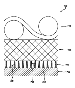
either side of the biocompatible membrane composite (i.e., external to) or
within
the biocompatible membrane composite (i.e., internal to) to provide support to
24
Date recue/Date received 2023-05-15
WO 2020/243663
PCT/US2020/035447
and prevent distortion of the membrane composite. Herein, a "reinforcing
component" may be further described as being external or internal to a cell
encapsulation device and may be nutrient impermeable or nutrient permeable.
The biocompatible membrane composite may be used in or to form a device for
encapsulating biological entities and/or cell populations. It is to be
appreciated
that the term "about" as used herein denotes +1- 10% of the designated unit of
measure.
[0199] Biological entities suitable for use with the
biocompatible
membrane composite include, but are not limited to, cells, viruses, viral
vectors,
gene therapies, bacteria, proteins, polysaccharides, antibodies, and other
bioactive entities. It is to be appreciated that if a biological entity other
than a cell
is selected for use herein, the bioactive component or product of the
biological
entity needs to be able to pass through the cell impermeable layer, but not
the
entity itself. For simplicity, herein the biological entity is referred to as
a cell, but
nothing in this description limits the biological entity to cells or to any
particular
type of cell, and the following description applies also to biological
entities that
are not cells.
[0200] Various types of prokaryotic cells, eukaryotic
cells, mammalian
cells, non-mammalian cells, and/or stem cells may be used with the
biocompatible membrane composite described herein. In some embodiments,
the cells secrete a therapeutically useful substance. Such therapeutically
useful
substances include hormones, growth factors, trophic factors,
neurotransmitters,
lymphokines, antibodies, or other cell products which provide a therapeutic
benefit to the device recipient. Examples of such therapeutic cell products
include, but are not limited to, insulin and other pancreatic hormones, growth
factors, interleukins, parathyroid hormone, erythropoietin, transferrin,
collagen,
elastin, tropoelastin, exosomes, vesicles, genetic fragments, and Factor VIII.
Non-limiting examples of suitable growth factors include vascular endothelial
growth factor, platelet-derived growth factor, platelet-activating factor,
transforming growth factors bone morphogenetic protein, activin, inhibin,
fibroblast growth factors, granulocyte-colony stimulating factor, granulocyte-
CA 03139591 2021- 11-25
WO 2020/243663
PCT/US2020/035447
macrophage colony stimulating factor, glial cell line-derived neurotrophic
factor,
growth differentiation factor-9, epidermal growth factor, and combinations
thereof.
[0201] As discussed above, the biocompatible membrane
composite
includes a first layer (i.e., cell impermeable layer). The cell impermeable
layer
serves as a microporous, immune isolation barrier, and is impervious to
vascular
ingrowth and prevents cellular contact from the host. Herein, layers that do
not
have openings large enough to allow cellular ingrowth may be referred to as
"tight" layers. The pores of the cell impermeable layer are sufficiently small
so as
to allow the passage therethrough of cellular nutrients, oxygen, waste
products,
and therapeutic substances while not permitting the passage of any cells.
Because the cell impermeable layer has an MPS that is sufficiently small so as
to
prevent vascular ingrowth, it is necessary to balance the parameters of the
cell
impermeable layer that could also negatively impact the mass transport and
diffusion properties of the cell impermeable layer. For instance, while the
MPS is
small enough to prevent cell ingress or vascular ingrowth, the cell
impermeable
layer is sufficiently open so as to allow the passage of molecules (i.e.
nutrients
and therapeutic molecules) therethrough. Layers that have openings large
enough to allow cellular ingrowth may be referred to as "open layers".
[0202] Diffusion resistance is further minimized by
keeping the cell
impermeable layer thin, porous, and low in mass. It is to be appreciated that
sufficient porosity of the cell impermeable layer be maintained so as to allow
the
passage of molecules. In certain embodiments, the porosity of the cell
impermeable layer is greater than about 50%, greater than about 60%, greater
than about 70%, or greater than about 80%. Additionally, the porosity may
range
from about 50% to about 98%, from about 50% to about 90%, from about 50% to
about 80%, or from about 60% to about 90%. It is also to be appreciated that
sufficient durability and strength of the cell impermeable layer be maintained
so
that immune isolation can be provided in vivo through an intended use by
ensuring the integrity of this tight layer. It is therefore necessary to
balance the
tradeoffs of the competing properties of strength and diffusion resistance. In
26
CA 03139591 2021- 11-25
WO 2020/243663
PCT/US2020/035447
certain embodiments, the maximum tensile load of the weakest axis of the cell
impermeable layer is greater than about 40 N/m, greater than about 130 N/m,
greater than about 260 N/m, greater than about 600 N/m, or greater than about
1000 N/m. Additionally, the maximum tensile load of the weakest axis may range
from about 40 N/m to about 2000 N/m, 40 N/m to about 780 N/m, 40 N/m to
about 350 N/m, from about 130 N/m to about 2000 N/m, from about 130 N/rri to
about 450 N/m, or from about 260 N/m to about 2000 N/m.
[0203] In certain embodiments, the cell impermeable layer
has a
combination of tensile strengths in orthogonal directions (D1, D2) that result
in a
geometric mean tensile strength that is greater than about 20 MPa, greater
than
about 50 MPa, greater than about 100 MPa, or greater than about 150 MPa
when the geometric mean tensile strength is defined by the following equation:
Geometric Mean .J (Tensile Strengthm)2 + (Tensile StrengthD2)2.
[0204] The geometric mean tensile strength of the cell
impermeable layer
may range from about 20 MPa to about 180 MPa, from about 30 MPa to about
150 MPa, from about 50 MPa to about 150 MPa, or from about 100 MPa to about
150 MPa.
[0205] In some embodiments, the cell impermeable layer has
an MPS that
is less than about 1 micron, less than about 0.50 microns, less than about
0.30
microns, or less than about 0.10 microns as measured by porometry. The MPS
may be from about 0.05 microns to about 1 micron, from about 0.1 microns to
about 0.80 microns, from about 0.1 microns to about 0.6 microns, from about
0.1
microns to about 0.5 microns, or from about 0.2 microns to about 0.5 microns
as
measured by porornetry.
[0206] In addition, the cell impermeable layer has a
thickness that is less
than about 30 microns, less than about 20 microns, less than about 10 microns,
or less than about 5 microns. The thickness may range from about 1 micron to
about 30 microns, from about 1 micron to about 20 microns, from about 1 micron
27
CA 03139591 2021- 11-25
WO 2020/243663
PCT/US2020/035447
to about 10 microns, from about 5 microns to about 10 microns, or from about 1
micron to about 5 microns. The mass per area (MpA) of the cell impermeable
layer may be less than about 25 g/m2, less than about 20 g/m2, less than about
g/m2, less than about 5 g/m2, or less than about 3 g/m2. The MpA may range
from about 3 g/m2 to about 25 g/m2, from about 0.5 g/m2 to about 20 g/m2, from
about 0.5 g/m2 to about 10 g/m2, or from about 0.5 g/m2 to about 5 g/m2.
[0207] As discussed previously, the biocompatible membrane
composite
includes a second layer (i.e., mitigation layer) which is sufficiently porous
to
permit growth of vascular tissue into the mitigation layer, and in some
instances,
also acts as an initial vascularization layer. The mitigation layer creates a
suitable environment to minimize, reduce, inhibit, or even prevent the
formation
of foreign body giant cells while allowing for access to blood vessels at the
cell
impermeable layer. Ingrowth of vascular tissues into the mitigation layer
facilitates nutrient transfer through the cell impermeable layer. Herein,
layers
that have openings large enough to allow vascular ingrowth may be referred to
as "open" layers. Blood vessels, which are the source of oxygen and nutrients
for implanted cells, need to form in the mitigation layer so that they are
sufficiently close to the cell impermeable layer such that the distance for
nutrient
diffusion to any encapsulated cells is minimized. The thinness of the cell
impermeable layer helps to reduce the distance over which diffusion must
occur.
[0208] The ingrowth of vascular tissue through the
mitigation layer up to
the cell impermeable layer facilitates nutrient transfer across the cell
impermeable layer. The mitigation layer creates an environment that enables a
sufficient formation of blood vessels into the mitigation layer positioned
adjacent
to the cell impermeable layer instead of the formation of foreign body giant
cells.
As a result, and as shown in the Examples, foreign body giant cells do not
form
at the interface of the cell impermeable layer and the mitigation layer such
that
foreign body cells impede sufficient vascularization. It is to be noted that
foreign
body giant cells may individually form at the interface of the cell
impermeable
layer and the mitigation layer, but they do not impede or prevent the
vascularization needed for growth of encapsulated cells_
28
CA 03139591 2021- 11-25
WO 2020/243663
PCT/US2020/035447
[0209] The mitigation layer is characterized at least in
part by the inclusion
of a plurality of solid features that have a solid feature spacing, which is
discussed in detail below. "Solid features" as used herein may be defined as
three dimensional components within the mitigation layer and/or
vascularization
layer that are generally immovable and resistant to deformation when exposed
to
environmental forces, such as, but not limited to, cell movement (e.g.,
cellular
migration and ingrowth, host vascularization/ endothelial blood vessel
formation).
To facilitate the reduction or mitigation of the formation of a barrier of
foreign
body giant cells at the cell impermeable layer, the solid features abutting
the
surface of the cell impermeable layer adjacent to the mitigation layer help
prevent
the fusion of multiple macrophages into multinucleated foreign body giant
cells at
this interface. In some embodiments, the solid features in the mitigation
layer
abutting the cell impermeable layer are intimately bonded to the cell
impermeable
layer and are herein referred to as "bonded solid features". "Non-bonded solid
features" are those solid features within the mitigation layer that are not
bonded
(intimately bonded or otherwise) to the cell impermeable layer. The solid
features in the mitigation layer may be formed of, for example, thermoplastic
polymers, polyurethanes, silicones, rubbers, epoxies, and combinations
thereof.
[0210] In some embodiments, the solid features of the
mitigation layer
project outwardly from a plane defined by the cell impermeable layer. In such
embodiments, the solid features of the mitigation layer projecting from the
cell
impermeable layer may be intimately bonded with the cell impermeable layer and
spaced such that they provide blockades or barriers to the formation of
foreign
body giant cells at this tight, cell impermeable interface. In some
embodiments,
the solid features may be a feature of the mitigation layer (e.g. nodes), and
may
be connected to each other, such as by fibrils or fibers. In another
embodiments,
the solid features may be provided and/or otherwise formed on the surface of
the
cell impermeable layer (e.g., printed solid features) such that the solid
features
project outwardly from the plane defined by a plane defined by the cell
impermeable layer.
29
CA 03139591 2021- 11-25
WO 2020/243663
PCT/US2020/035447
[0211] In embodiments where the mitigation layer has a
node and fibril
microstructure (e.g., formed from a fibrillated polymer), the nodes are the
solid
features and the fibrils are not the solid features. Indeed, in some
embodiments,
the fibrils may be removed, leaving only the nodes in the mitigation layer. In
embodiments where the nodes within the mitigation layer are the solid
features,
those nodes which are bonded to the cell impermeable layer are bonded solid
features. In at least one embodiment, the mitigation layer is formed of an
expanded tetrafluoroethlyene (ePTFE) membrane having a node and fibril
microstructure.
[0212] The solid features of the mitigation layer do not
negatively impact
the overall diffusion resistance of the biocompatible membrane composite for
applications that require a rapid time course of diffusion. The solid features
of
the mitigation layer are of a sufficiently small size such that they do not
interfere
with the amount of porous area needed for diffusion across the cell
impermeable
layer. Also, the thickness of the mitigation layer is sufficiently thin so as
to
maximize mass transport of oxygen and nutrients to encapsulated cells from the
interstitium during the acute period post implantation. The space between the
solid features are sufficiently open to allow for easy and rapid
penetration/integration of host tissue up to the cell impermeable layer (i.e.,
tight
layer) to decrease the duration of the acute period. "Acute period" is defined
herein as the time period prior to host cell/vascular infiltration.
[0213] The majority of the solid feature spacing of the
solid features
adjacent to the cell impermeable layer is less than about 50 microns, less
than
about 40 microns, less than about 30 microns, less than about 20 microns, or
less than about 10 microns. As used herein, the term "majority" is meant to
describe an amount over half (i.e., greater than 50%) of the measured values
for
the parameter being measured. In addition, the phrase "solid feature spacing"
is
defined herein as the straight-line distance between two neighboring solid
features. In this disclosure, solid features are considered neighboring if
their
centroids represent the corners of a triangle whose oircumcircle has an empty
interior. As shown pictorially in FIG. 1A, the designated solid feature (P) is
CA 03139591 2021- 11-25
WO 2020/243663
PCT/US2020/035447
connected to neighboring solid features (N) to form a triangle 100 where the
circumcircle 110 contains no solid features within. Solid features (X)
designate
the solid features that are not neighboring solid features. Thus, in the
instance
depicted in FIG. 1A, the solid feature spacing 130 is the straight distance
between the designated solid features (P), (N). In contrast, the circumcircle
150
shown in FIG. 1B drawn from the triangle 160 contains therein a solid feature
(N),
and as such, cannot be utilized to determine the solid feature spacing in the
mitigation layer (or the vascularization layer). FIG. 2 is a scanning electron
micrograph depicting measured distances, e.g., the white lines 200 between the
solid features 210 (white shapes) in a mitigation layer formed of an expanded
polytetrafluoroethylene membrane. In some embodiments, the majority of the
solid feature spacing may range from about 5 microns to about 45 microns, from
about 10 microns to about 40 microns, from about 10 microns to about 35
microns, or from about 15 microns to about 35 microns.
[0214] The solid features also include a representative
minor axis, a
representative major axis, and a solid feature depth. The representative minor
axis of a solid feature is defined herein as the length of the minor axis of
an
ellipse fit to the solid feature where the ellipse has the same area,
orientation,
and centroid as the solid feature. The representative major axis of a solid
feature
is defined herein as the length of the major axis of an ellipse fit to the
solid
feature where the ellipse has the same area, orientation, and centroid as the
solid feature. The major axis is greater than or equal to the minor axis in
length.
The representative minor axis and representative major axis of a layer are the
respective median values of all measured representative minor axes and
representative major axes of features in the layer. The minor and major axes
of
an ellipse 320 to fit the solid feature 310 is shown pictorially in FIG. 3A.
The
representative minor axis of the solid feature 310 is depicted by arrow 300,
and
the representative major axis of the solid feature 310 is depicted by arrow
330. A
majority of the solid features in the mitigation layer has a minor axis that
range in
size from about 3 microns to about 20 microns, from about 3 microns to about
15
microns, or from about 3 microns to about 10 microns. The solid feature depth
is
31
CA 03139591 2021- 11-25
WO 2020/243663
PCT/US2020/035447
the length of the projection of the solid feature in the axis perpendicular to
the
surface of the layer (e.g., mitigation layer or vascularization layer). The
solid
feature depth of the solid feature 310 is shown pictorially in FIG 3B. The
depth of
the solid feature 310 is depicted by line 340. In at least one embodiment, the
depth of the solid features is equal to or less than the thickness of the
mitigation
layer. The solid feature depth of a layer is the median value of all measured
solid
feature depths in the layer. In at least one embodiment, the majority of at
least
two of the representative minor axis, representative major axis, and solid
feature
depth in a layer is greater than 5 microns.
[0215] In embodiments where the solid features are
interconnected by
fibrils or fibers, the boundary connecting the solid features creates a pore.
It is
necessary that these pores are open enough to allow rapid cellular ingrowth
and
vascularization and not create a resistance to mass transport of oxygen and
nutrients. The pore effective diameter is measured by quantitative image
analysis (QIA) and performed on a scanning electron micrograph (SEM) image.
The "effective diameter" of a pore is defined as the diameter of a circle that
has
an area equal to the measured area of the surface pore. This relationship is
defined by the following equation:
Effective Diameter = 2 x Arz.
[0216] Turning to FIG. 4, the effective diameter is the
diameter of the circle
400 and the surface pore is designated by reference numeral 420. The total
pore
area of a surface is the sum of the area of all pores at that surface. The
pore
size of a layer is the effective diameter of the pore that defines the point
where
roughly half the total pore area consists of pores with diameters smaller than
the
pore size and half the total pore area consists of pores with diameters
greater
than or equal to the pore size. FIG. 5 illustrates a pore size 500 (white in
color),
pores smaller in size 510 (shown in light grey), and pores larger in size 520
(shown in dark grey). Pores that intersect with the edge of the image 530 were
excluded from analysis and are shown in black.
32
CA 03139591 2021- 11-25
WO 2020/243663
PCT/US2020/035447
[0217] The pore size of the mitigation layer may range
from about 1
micron to about 9 microns in effective diameter, from about 3 microns in
effective
diameter to about 9 microns in effective diameter, or from about 4 micron in
effective diameter to about 9 microns in effective diameter as measured by
quantitative image analysis (QUA) performed on an SEM image. Also, the
mitigation layer has a thickness that is less than about 60 microns, less than
about 50 microns, less than about 40 microns, less than about 30 microns, or
less than about 20 microns. The thickness of the mitigation layer may range
from about 3 microns to about 60 microns, from about 10 microns to about 50
microns, from about 10 microns to about 40 microns, or from about 15 microns
to
about 35 microns. In some embodiments, the mitigation layer has a porosity
greater than about 60%. In other embodiments, the mitigation layer has a
porosity greater than about 70%, greater than about 80%, greater than about
90%, or greater than about 95%. In some embodiments, the porosity may be
about 98% or about 99%. The porosity of the mitigation layer may range from
about 60% to about 98%, from about 70% to about 98%, or from about 80% to
about 98%.
[0218] As discussed previously, the biocompatible membrane
composite
also includes a third layer (i.e., vascularization layer). The vascularization
layer
is an "open" layer that permits additional vascular penetration from the host
and
rapid anchoring and attachment of the biocompatible membrane composite
within the tissue of the host. Additionally, the vascularization layer
provides a
porous matrix to harbor the growth of a sufficient quantity of additional, new
blood vessels to feed the encapsulated cells. The vascularization layer is
designed such that there are solid features to enable host integration and
attachment. As the vascularization layer does not meet the same criteria as
the
mitigation layer, the two are separate and distinct layers. The solid features
of
the vascularization layer have increased spacing and pore sizes therebetween
compared to the solid features of the mitigation layer to facilitate a more
rapid
ingrowth of the tissue or blood vessels into the vascularization layer. In
some
embodiments, the majority of the solid feature spacing of the solid features
in the
33
CA 03139591 2021- 11-25
WO 2020/243663
PCT/US2020/035447
vascularization layer is greater than about 50 microns, greater than about 60
microns, greater than about 70 microns, or greater than about 80 microns_ A
majority of the solid features in the vascularization layer has a solid
feature
spacing that range from about 50 microns to about 90 microns, from about 60
microns to about 90 microns, or from about 70 microns to about 90 microns.
[0219] The pore size and overall thickness of the
vascularization layer is
sufficient to provide space to harbor the necessary quantities of additional
blood
vessels to provide nutrients and oxygen to cells. A pore size of the
vascularization layer may be greater than about 9 microns in effective
diameter,
greater than about 25 microns in effective diameter, greater than about 50
microns in effective diameter, greater than about 75 microns in effective
diameter, greater than about 100 microns in effective diameter, greater than
about 125 microns in effective diameter, greater than about 150 microns in
effective diameter, greater than about 175 microns in effective diameter, or
greater than about 200 microns in effective diameter as measured by Q IA
performed on an SEM image. In some embodiments, the pore size of the
vascularization layer may range from about 9 microns in effective diameter to
about 200 microns in effective diameter, from about 9 microns in effective
diameter to about 50 microns in effective diameter, from about 15 microns in
effective diameter to about 50 microns in effective diameter from about 25
microns in effective diameter to about 50 microns in effective diameter, from
about 50 microns in effective diameter to about 200 microns in effective
diameter, from about 75 microns in effective diameter to about 175 microns in
effective diameter as measured by QIA performed on an SEM image.
[0220] Additionally, the vascularization layer may have a
thickness that is
greater than about 30 microns, greater than about 50 microns, greater than
about
75 microns, greater than about 100 microns, greater than about 125 microns,
greater than about 150 microns, or greater than about 200 microns. In
addition,
the thickness of the vascularization layer may range from about 30 microns to
about 300 microns, from about 30 microns to about 200 microns, from about 30
microns to about 100 microns, from about 100 microns to about 200 microns, or
34
CA 03139591 2021- 11-25
WO 2020/243663
PCT/US2020/035447
from about 100 microns to about 150 microns. In at least one embodiment, the
thickness of the vascularization layer is at least two times the combined
thickness of the cell impermeable layer and the mitigation layer. In some
embodiments, the thickness of the vascularization layer is greater than a sum
of
a thickness of the cell impermeable layer and a thickness of the mitigation
layer.
In addition, a majority of the solid features in the vascularization layer has
a
representative minor axis that is less than 50 microns, less than about 40
microns, less than about 30 microns, less than about 20 microns, less than
about
microns, less than about 5 microns, or less than about 3 microns. In some
embodiments, a majority of the solid features in the vascularization layer has
a
representative minor axis that ranges in size from about 3 microns to about 40
microns, from about 3 microns to about 30 microns, from about 3 microns to
about 20 microns, from about 3 microns to about 10 microns, or from about 20
microns to about 40 microns. The solid features present in the vascularization
layer also have a major axis that is greater in length than the minor axis and
may
effectively be unlimited in length, such as a continuous fiber of a non-woven.
The solid features in the vascularization layer also have a depth that is less
than
or equal to the total thickness of the vascularization layer.
[0221]
In some embodiments, the biocompatible membrane composite,
including the cell impermeable layer, is perforated with discretely placed
holes.
The perforation size, number, and location can be selected to optimize cell
function. As few as one (1) perforated hole may be present. The perforations
are of a sufficient size to allow host vascular tissue (such as capillaries)
to pass
through the biocompatible membrane composite in order to support, for example,
encapsulated pancreatic cell types. While the cell impermeable layer maintains
its function as a microporous, immune isolation barrier in locations where no
perforations are present, due to the discrete perforations where portions of
the
cell impermeable layer have been removed, the cell impermeable layer in its
entirety is no longer cell impermeable because the discrete perforations allow
vascular ingrowth and cellular contact from the host to pass through the
biocompatible membrane composite. Because cell encapsulation device
CA 03139591 2021- 11-25
WO 2020/243663
PCT/US2020/035447
embodiments that contain a perforated cell impermeable layer allow for host
immune cell contact with cells, the cells are no longer protected from immune
rejection unless the host is immunocompromised or treated with
immunosuppressant drugs.
[0222] An optional reinforcing component may be provided
to the
biocompatible membrane composite to minimize distortion in vivo so that the
cell
bed thickness is maintained (e.g., in an encapsulated device). This
additional,
optional reinforcing component provides a stiffness to the biocompatible
membrane composite that is greater than the biocompatible membrane
composite itself to provide mechanical support. This optional reinforcing
component could be continuous in nature or it may be present in discrete
regions
on the biocompatible membrane composite, e.g., patterned across the entire
surface of the biocompatible membrane composite or located in specific
locations
such as around the perimeter of the biocompatible membrane composite. Non-
limiting patterns suitable for the reinforcing component on the surface of the
membrane composite include dots, straight lines, angled lines, curved lines,
dotted lines, grids, etc. The patterns forming the reinforcing component may
be
used singly or in combination. In addition, the reinforcing component may be
temporary in nature (e.g., formed of a bioabsorbable material) or permanent in
nature (e.g., a polyethylene terephthalate (PET) mesh or Nitinol). As is
understood by one of ordinary skill in the art, the impact of component
stiffness
depends not just on the stiffness of a single component, but also on the
location
and restraint of the reinforcing component in the final device form. In order
for a
component (e.g., a reinforcing component) to be practically useful for adding
stiffness to the biocompatible composite membrane, the reinforcement
component should have a stiffness greater than about 0.01 N/cm, although a
final determination of the stiffness needed will depend on location and
restraint in
the finished cell encapsulation device. In some embodiments, the reinforcement
component may have a stiffness from about 0.01 N/cm to about 5 N/cm, from
about 0.05 N/cm to about 4 N/cm, from about 0.1 N/cm to about 3 N/cm, or from
about 0.3 N/cm to about 2 N/cm.
36
CA 03139591 2021- 11-25
WO 2020/243663
PCT/US2020/035447
[0223] In at least one embodiment, the reinforcing
component may be
provided on the external surface of the vascularization layer to strengthen
the
biocompatible membrane composite against environmental forces. In this
orientation, the reinforcing component has a pore size sufficient to permit
vascular ingrowth, and is therefore is considered an "open" layer. Materials
useful as the reinforcing component include materials that are significantly
stiffer
than the biocompatible membrane composite. Such materials include, but are
not limited to, open mesh biomaterial textiles, woven textiles, non-woven
textiles
(e.g., collections of fibers or yarns), and fibrous matrices, either alone or
in
combination. In another embodiment, patterned grids, screens, strands, or rods
may be used as the reinforcing component. The reinforcing component may be
positioned on the outer surface of the biocompatible membrane adjacent to the
cell impermeable layer (see, e.g. FIG. 10C). In this orientation, the
reinforcing
component may be a cell impermeable and nutrient impermeable dense layer as
long as there is sufficient spacing for cells to reside between the
reinforcing
component and the cell impermeable layer. Additionally, the reinforcing
component may be oriented within or between the composite layers at discrete
regions or the composite layers themselves could also be reinforcing
components (see, e.g. FIGS. 10A, 10B, and 10D). It is to be appreciated that
the
reinforcing component could be located externally, internally, or within the
biocompatible membrane, or combinations thereof.
[0224] In at least one embodiment, the cell impermeable
layer, the
mitigation layer, and the vascularization layer are bonded together by one or
more biocompatible adhesive to form the biocompatible membrane composite.
The adhesive may be applied to the surface of one or more of the cell
impermeable layer, the mitigation layer, and the vascularization layer in a
manner
to create a discrete or intimate bond between the layers. As used herein, the
phrases "discrete bond" or "discretely bonded" are meant to include bonding or
bonds in intentional patterns of points and/or lines around a continuous
perimeter
of a defined region. Non-limiting examples of suitable biocompatible adhesives
include fluorinated ethylene propylene (FEP), a polycarbonate urethane, a
37
CA 03139591 2021- 11-25
WO 2020/243663
PCT/US2020/035447
thermoplastic fluoropolymer comprised of TFE and PAVE, EFEP (ethylene
fluorinated ethylene propylene), PEBAX (a polyether amide), PVDF (poly
vinylidene fluoride), Carbosil (a silicone polycarbonate urethane), Elasthane
TM
(a polyether urethane), PurSil (a silicone polyether urethane), polyethylene,
high
density polyethylene (1-IDPE), ethylene chlorotetrafluoroethylene (ECTFE),
perfluoroalkoxy (PFA), polypropylene, polyethylene terephthalate (PET), and
combinations thereof. The mitigation layer may be intimately bonded to the
cell
impermeable layer. The vascularization layer may be intimately or discretely
bonded to the mitigation layer. In at least one embodiment, the mitigation
layer is
intimately bonded to the cell impermeable layer. In some embodiments, the cell
impermeable layer and the mitigation layer are co-expanded as a composite
layer. In embodiments where the cell impermeable layer and mitigation layer or
the cell impermeable layer, mitigation layer, and vascularization layer,
measured
composite z-strengths may be greater than 100 kPa. Additionally, the measured
composite z-strength may range from about 100 kPa to about 1300 kPa, from
about 100 kPa to about 1100 kPa, from about 100 kPa to about 900 kPa, from
about 100 kPa to about 700 kPa, from about 100 kPa to about 500 kPa, from
about 100 kPa to about 300 kPa, or from about 100 kPa to about 200 kPa.
[0225] At least one of the cell impermeable layer, the
mitigation layer, and
the vascularization layer may be formed of a polymer membrane or woven or
non-woven collections of fibers or yams, or fibrous matrices, either alone or
in
combination. Non-limiting examples of polymers that may be used any one or all
of the cell impermeable layer, the mitigation layer, and the vascularization
layer
include, but are not limited to, alginate; cellulose acetate; polyalkylene
glycols
such as polyethylene glycol and polypropylene glycol; panvinyl polymers such
as
polyvinyl alcohol; chitosan; polyacrylates such as
polyhydroxyethylmethacrylate;
agarose; hydrolyzed polyacrylonitrile; polyacrylonitrile copolymers; polyvinyl
acrylates such as polyethylene-co-acrylic acid, polyalkylenes such as
polypropylene, polyethylene; polyvinylidene fluoride; fluorinated ethylene
propylene (FEP); perfluoroalkoxy alkane (PFA); polyester sulfone (PES);
polyurethanes; polyesters; and copolymers and combinations thereof.
38
CA 03139591 2021- 11-25
WO 2020/243663
PCT/US2020/035447
[0226] In some embodiments, the polymer(s) forming the
polymer
membrane forming the cell impermeable layer, mitigation layer, and/or
vascularization layer is a fibrillatable polymer. Fibrillatable, as used
herein, refers
to the ability to introduce fibrils to a polymer membrane, such as, but not
limited
to, converting portions of the solid features into fibrils. For example, the
fibrils
are the solid elements that span the gaps between the solid features. Fibrils
are
generally not resistant to deformation upon exposure to environmental forces,
and are therefore deformable. The majority of deformable fibrils may have a
diameter less than about 2 microns, less than about 1 micron, less than about
0.75 microns, less than about 0.50 microns, or less than about 0.25 microns.
In
some embodiments, the fibrils may have a diameter from about 0.25 microns to
about 2 microns, from about 0.5 microns to about 2 microns, or from about 0.75
microns to about 2 microns.
[0227] Non-limiting examples of fibrillatable polymers
that may be used to
form one or more of the cell impermeable layer, the mitigation layer, and the
vascularization layer include, but are not limited to, tetrafluoroethylene
(TFE)
polymers such as polytetrafluoroethylene (PTFE), expanded PTFE (ePTFE),
modified PTFE, TFE copolymers, polyvinylidene fluoride (PVDF), poly (p-
xylylene) (ePPX) as taught in U.S. Patent Publication No. 2016/0032069 to
Sbriglia, porous ultra-high molecular weight polyethylene (eUHMVVPE) as taught
in U.S. Patent No. 9,926,416 to Sbriglia, porous ethylene tetrafluoroethylene
(eETFE) as taught in U.S. Patent No. 9,932,429 to Sbriglia, and porous
vinylidene fluoride-co-tetrafluoroethylene or trifluoroethylene [VDF-co-(TFE
or
TrFE)] polymers as taught in U.S. Patent No. 9,441,088 to Sbriglia, and
combinations thereof.
[0228] In some embodiments, the fibrillatable polymer is a
fluoropolymer
membrane such as an expanded polytetrafluoroethylene (ePTFE) membrane.
Expanded polytetrafluoroethylene (ePTFE) membranes (and other fibrillated
polymers) have a node and fibril microstructure where the nodes are
interconnected by the fibrils and the pores are the space located between the
nodes and fibrils throughout the membrane. As used herein, the term "node" is
39
CA 03139591 2021- 11-25
WO 2020/243663
PCT/US2020/035447
meant to denote a solid feature consisting largely of polymer material. When
deformable fibrils are present, nodes reside at the junction of multiple
fibrils_ In
some embodiments the fibrils may be removed from the membrane, such as, for
example, by plasma etching. In at least one embodiment, an expanded
polytetrafluoroethylene membrane is used in one or more of the cell
impermeable
layer, the mitigation layer, and the vascularization layer. Expanded
polytetrafluoroethylene membranes such as, but not limited to, those prepared
in
accordance with the methods described in U.S. Patent No. 3,953,566 to Gore,
U.S. Patent No. 7,306,729 to Bacino et al., U.S. Patent No. 5,476,589 to
Bacino,
WO 94/13469 to Bacino, U.S. Patent No. 5,814,405 to Branca et al. or U.S.
Patent No. 5,183,545 to Branca et al. may be used herein.
[0229] In some embodiments, one or more of the cell
impermeable layer,
the mitigation layer, and the vascularization layer is formed of a
fluoropolyrner
membrane, such as, but not limited to, an expanded polytetrafluoroethylene
(ePTFE) membrane, a modified ePTFE membrane, a tetrafluoroethylene (TFE)
copolymer membrane, a polyvinylidene fluoride (PVDF) membrane, or a
fluorinated ethylene propylene (FEP) membrane. In further embodiments, the
vascularization layer may include biocompatible textiles, including woven and
non-woven fabrics (e.g., a spunbound non-woven, melt blown fibrous materials,
electrospun nanofibers, etc.), non-fluoropolymer membranes such as
polyvinylidene difluoride (PVDF), nanofibers, polysulfones, polyethersulfones,
polyarlysulfones, polyether ether ketone (PEEK), polyethylenes,
polypropylenes,
and polyimides. In some embodiments, the vascularization layer is a spunbound
polyester or an expanded polytetrafluoroethylene (ePTFE) membrane.
[0230] In some embodiments at least one of the mitigation
layer,
vascularization layer or reinforcing component is formed of a non-woven
fabric.
There are numerous types of non-woven fabrics, each of which may vary in
tightness of the weave and the thickness of the sheet. The filament cross-
section may be trilobal. The non-woven fabric may be a bonded fabric, a formed
fabric, or an engineered fabric that is manufactured by processes other than
weaving or knitting. In some embodiments, the non-woven fabric is a porous,
CA 03139591 2021- 11-25
WO 2020/243663
PCT/US2020/035447
textile-like material, usually in flat sheet form, composed primarily or
entirely of
fibers, such as staple fibers assembled in a web, sheet, or batt. The
structure of
the non-woven fabric is based on the arrangement of, for example, staple
fibers
that are typically randomly arranged. In addition, non-woven fabrics can be
created by a variety of techniques known in the textile industry. Various
methods
may create carded, wet laid, melt blown, spunbonded, or air laid non-woven
materials. Non-limiting methods and substrates are described, for example, in
U.S. Patent Publication No 2010/0151575 to Colter, et al. In one embodiment,
the non-woven fabric is polytetrafluoroethylene (PTFE). In another embodiment,
the non-woven fabric is a spunbound polyester. The density of the non-woven
fabric may be varied depending upon the processing conditions. In one
embodiment, the non-woven fabric is a spunbound polyester with a basis weight
from about 0.40 to about 1.00 (oz/yd2) a nominal thickness of about 127 to
about
228 microns and a fiber diameter of about 0.5 microns to about 26 microns. The
filament cross-section is trilobal. In some embodiments, the non-woven fabrics
are bioabsorbable.
[0231] In some embodiments, it may be desirable for one or
more of the
vascularization layer and reinforcing component to be non-permeant (e.g.,
biodegradable). In such an instance, a biodegradable material may be used to
form the vascularization layer and/or the reinforcing component. Suitable
examples of biodegradable materials include, but are not limited to,
polyglycolide:trimethylene carbonate (PGA:TMC), polyalphahydroxy acid such as
polylactic acid, polyglycolic acid, poly (glycolide), and poly(lactide-co-
caprolactone), poly(caprolactone), poly(carbonates), poly(dioxanone), poly
(hydroxybutyrates), poly(hydroxyvalerates), poly (hydroxybutyrates-co-
valerates),
expanded polyparaxylylene (ePLLA), such as is taught in U.S. Patent
Publication
No. 2016/0032069 to Sbriglia, and copolymers and blends thereof.
Alternatively,
the vascularization layer may be coated with a bio-absorbable material or a
bio-
absorbable material may be incorporated into or onto the vascularization layer
in
the form of a powder. Coated materials may promote infection site reduction,
vascularization, and favorable type 1 collagen deposition.
41
CA 03139591 2021- 11-25
WO 2020/243663
PCT/US2020/035447
[0232] The biocompatible membrane composite may have at
least partially
thereon a surface coating, such as a Zwitterion non-fouling coating, a
hydrophilic
coating, or a CBAS /Heparin coating (commercially available from W.L. Gore &
Associates, Inc.). The surface coating may also or alternatively contain
antimicrobial agents, antibodies (e.g., anti-CD 47 antibodies (anti-
fibrotic)),
pharmaceuticals, and other biologically active molecules (e.g., stimulators of
vascularization such as FGF, VEGF, endoglin, PDGF, angiopoetins, and
integrins; Anti-fibrotic such as TGFb inhibitors, sirolimus, CSF1R inhibitors,
and
anti CD 47 antibody; anti-inflammatory/immune modulators such as CXCL12,
and corticosteroids), and combinations thereof.
[0233] In some embodiments, the solid features of one or
both of the
mitigation layer and the vascularization layer may be formed by micro
lithography,
micro-molding, machining, or printing (or otherwise laying down) a polymer
(e.g.,
thermoplastic) onto a cell impermeable layer to form at least a part of a
solid
feature. Any conventional printing technique such as transfer coating, screen
printing, gravure printing, ink-jet printing, patterned imbibing, and knife
coating
may be utilized to place the thermoplastic polymer onto the cell impermeable
layer. FIG. 6A illustrates a thermoplastic polymer in the form of solid
features
620 positioned on a cell impermeable layer 610 (after printing is complete),
where the solid features 620 have a feature spacing 630. Non-limiting examples
of geometries for forming the solid features include, but are not limited to,
dashed
lines (see FIG. 6B), dots and/or dotted lines (see FIGS. 6C, 6G), geometric
shapes (see FIG. 6H), straight lines (see FIG. 6D), angled lines (see FIG.
6F),
curved lines, grids (see FIG. 6E), etc., and combinations thereof.
[0234] Materials used to form the solid features of the
mitigation layer
include, but are not limited to, polyurethane, polypropylene, polyethylene,
polyether amide, polyetheretherketone, polyphenylsulfone, polysulfone,
silicone
polycarbonate urethane, polyether urethane, polycarbonate urethane, silicone
polyether urethane, polyester, polyester terephthalate, melt-processable
fluoropolymers, such as, for example, fluorinated ethylene propylene (FEP),
tetrafluoroethylene-(periluoroalkyl) vinyl ether (P FA), an alternating
copolymer of
42
CA 03139591 2021- 11-25
WO 2020/243663
PCT/US2020/035447
ethylene and tetrafluoroethylene (ETFE), a terpolymer of tetrafluoroethylene
(TFE), hexafluoropropylene (HFP) and vinyl idene fluoride (THV),
polyvinylidene
fluoride (PVDF), and combinations thereof. In some embodiments,
polytetrafluoroethylene may be used to form the pattern features. In further
embodiments, the solid features may be separately formed and adhered to the
surface of the cell impermeable layer (not illustrated).
[0235] Biocompatible membrane composite 700 is depicted in
FIG. 7,
which includes a cell impermeable layer 710, a mitigation layer 720, a
vascularization layer 730, and the optional reinforcement layer 740. In the
depicted embodiment, the solid features 750 are bonded to the surface of the
cell
impermeable layer 710 to form bonded features within the mitigation layer 720.
In
some embodiments, the solid features 750 do not penetrate into the pores of
the
vascularization layer 730. The solid features 750 are depicted in FIG. 7 as
being
essentially the same height and width and extending between the cell
impermeable layer 710 and the vascularization layer 730, although it is to be
appreciated that this an example and the solid features 750 may vary in height
and/or width. The distance between solid features 750 is the solid feature
spacing 760.
[0236] FIG. 8 is another biocompatible membrane composite
800 that
includes a cell impermeable layer 810, a mitigation layer 820, a
vascularization
layer 830, and the optional reinforcement layer 840. In the depicted
biocompatible membrane composite, the solid features 850, 880 are nodes that
differ in height and width, and may or may not extend the distance between the
cell impermeable layer 810 and the vascularization layer 830. The solid
features
850, 880 are connected by fibrils 870. In FIG. 8, the majority of the solid
feature
depth is less than the total thickness of the mitigation layer 820. Solid
features
880 are bonded solid features.
[0237] Turning to FIG. 9, an biocompatible membrane
composite 900
containing a cell impermeable layer 910, a mitigation layer 920, a
vascularization
layer 930, and an optional reinforcement layer 940 is depicted. In this
embodiment, solid features within the mitigation layer 920 are the nodes of a
43
CA 03139591 2021- 11-25
WO 2020/243663
PCT/US2020/035447
mitigation layer 920 that are formed in an ePTFE membrane. The nodes 950,
980 are interconnected by fibrils 970. Nodes 950, 980 are positioned within
the
mitigation layer 920. Nodes 980, however, are not only within the mitigation
layer
920, but are also in contact with, and are intimately bonded to, the cell
impermeable layer 910.
[0238] As discussed above, the reinforcing component may
be oriented
within or between the composite layers at discrete regions. In one non-
limiting
embodiment shown in FIG. 10A, the reinforcing component 1030 is formed as
discrete regions on the cell impermeable layer 1000 and are positioned within
the
mitigation layer 1010 of the biocompatible membrane composite 1050. The
vascularization layer 1020 is shown for reference only. In the embodiment
depicted in FIG. 10B, the reinforcing component 1030 is positioned on the
mitigation layer 1010 as discrete regions and are positioned within the
vascularization layer 1020 of the biocompatible membrane composite 1050. The
cell impermeable layer 1000 is shown for reference only. In yet another non-
limiting embodiment depicted in FIG. 10C, the reinforcing component 1030 is
external to the biocompatible membrane composite 1050. Specifically, the
reinforcing component 1030 is positioned on a side of the cell impermeable
layer
1000 opposing the mitigation layer 1010. The vascularization layer 1020 is
shown for reference only. Turning to FIG. 10D, the reinforcing component 1030
is located between the mitigation layer 1010 and the vascularization layer
1020
of the biocompatible membrane composite 1050. The cell impermeable layer
1000 is shown for reference only.
[0239] In the embodiments described herein, the mitigation
layer 1100
may be formed by placing or otherwise depositing a polymer in a pattern (as
described above) which is characterized by one or more of the following: the
solid feature size (i.e., minor axis) 1110, solid feature spacing 1120, solid
feature
depth 1160, thickness 1130, the absence of fibrils and/or the pore size (as
measured by quantitative image analysis (0IA) performed on an SEM image), as
depicted generally in FIG. 11A. A cell impermeable layer 1150 is shown for
reference only.
44
CA 03139591 2021- 11-25
WO 2020/243663
PCT/US2020/035447
[0240] FIG. 11B depicts a mitigation layer 1200 that is
formed of a polymer
having a node and fibril microstructure that is characterized by one or more
of the
following: the solid feature size (i.e., minor axis) 1210, solid feature
spacing
1220, solid feature depth 1270, thickness 1230, the presence of fibrils 1260,
and/or the pore size (as measured by quantitative image analysis (0 IA)
performed on an SEM image) 12210. A cell impermeable layer 1250 is shown in
FIG. 11B for reference only.
[0241] Also, in the embodiments described herein, the
vascularization
layer 1300 may be characterized by one or more of the following: thickness
1310, pore size 1320, solid feature size (i.e., minor axis) 1340, and solid
feature
spacing 1330 as depicted generally in FIG. 12. A cell impermeable layer 1350
and a mitigation layer 1360 are shown for reference only.
[0242] The biocompatible membrane composite can be
manufactured into
various forms including, but not limited to, a housing, a chamber, a pouch, a
tube, or a cover. In one embodiment, the biocompatible membrane composite
forms a cell encapsulating device as illustrated in FIG. 13A. FIG. 13A is a
top
view of a cell encapsulating device 1400 formed of two layers of the
biocompatible membrane composite that are sealed along a portion of their
periphery 1410_ Only the outer layer of the biocompatible membrane composite
1420 is shown in FIG. 13A. The cell encapsulating device 1400 includes an
internal chamber (not shown) for containing cells and a port 1430 that extends
into the internal chamber and is in fluid communication therewith.
[0243] FIG. 13B is a cross-sectional illustration of the
cell encapsulation
device of FIG. 13A. As shown, a first biocompatible membrane composite 1450
is positioned adjacent to a second biocompatible membrane composite 1460.
The biocompatible membrane composites 1450, 1460 each include a cell
impermeable layer 1470, a mitigation layer 1480, and a vascularization layer
1490. The optional reinforcing component is not depicted in FIG. 13B, although
it
could be utilized in this embodiment. A chamber 1435 (i.e., lumen) is located
between the two membrane composites 1450, 1460 for the placement of cells
(and/or other biological entities).
CA 03139591 2021- 11-25
WO 2020/243663
PCT/US2020/035447
[0244] Having generally described this disclosure, a
further understanding
can be obtained by reference to certain specific examples illustrated below
which
are provided for purposes of illustration only and are not intended to be all
inclusive or limiting unless otherwise specified.
Test Methods
Porosity
[0245] The porosity of a layer is defined herein as the
proportion of layer
volume consisting of pore space compared to the total volume of the layer. The
porosity is calculated by comparing the bulk density of a porous construct
consisting of solid fraction and void fraction to the density of the solid
fraction
using the following equation:
Densitymdk
Porosity = ( L'
1 ri x100%
ensaYsoud Fraction
Thickness
[0246] The thickness of the layers in the composites was
measured by
quantitative image analysis (QUA) of cross-sectional SEM images. Cross-
sectional SEM images were generated by fixing membranes to an adhesive,
cutting the film by hand using a liquid-nitrogen-cooled razor blade, and then
standing the adhesive backed film on end such that the cross-section was
vertical. The sample was then sputter coated using an Ernitech K550X sputter
coater (commercially available from Quorum Technologies Ltd, UK) and platinum
target. The sample was then imaged using a FEI Quanta 400 scanning electron
microscope from Thermo Scientific.
[0247] Layers within the cross-section SEM images were
then measured
for thickness using ImageJ 1.51h from the National Institutes of Health (NIH).
The image scale was set per the scale provided by the SEM. The layer of
interest was isolated and cropped using the free-hand tool. A number of at
least
ten equally spaced lines were then drawn in the direction of the layer
thickness.
The lengths of all lines were measured and averaged to define the layer
thickness.
46
CA 03139591 2021- 11-25
WO 2020/243663
PCT/US2020/035447
Maximum Tensile Load
[0248] Materials were tested for maximum tensile load
using a 5500
Series Instrone Electromechanical Testing System. Samples were cut oriented
in the axis of interest using a D412F or D638-V dogbone die. The samples were
then loaded into the Instron tester grips and tested at a constant rate of 20
in/min (for D412F samples) or 3 in/min (for D683-V samples) until failure. The
maximum load sustained during testing was normalized by specimen gauge
width (6.35mm for D412F samples and 3.175mm for D638-V samples) to define
maximum tensile load.
Tensile Strength
[0249] Materials were tested for tensile strength using a
5500 Series
Instroe Electromechanical Testing System. Unless otherwise noted, materials
were testing for tensile strength prior to the application of any coatings.
Samples
were cut using a D412F or D638-V dogbone die. The samples were then loaded
into the Instron tester grips and tested at a constant rate of 20 in/min (for
D412F
samples) or 3 in/min (for D683-V samples) until failure. Maximum load was
normalized by test area (defined as gauge width times material thickness) to
define tensile stress. Materials were tested in perpendicular directions (D1
and
D2) and the maximum stress in each direction was used to calculate the
geometric mean tensile strength of the material per the below equation:
Geometric Mean = /(Tensile Strengthm)2 + (Tensile 5trengthD2)2.
Composite Bond Strength (Z-Strength)
[0250] Materials were tested for composite bond strength
using a 5500
Series Instrone' Electromechanical Testing System. Unless otherwise noted,
materials were testing for tensile strength prior to the application of any
coatings.
Samples were fixed to a 1"x1" steel platen using 3M 9500PC double sided tape
and loaded into the Instroe with an opposing 1"x1" steel platen with 3M 9500PC
47
CA 03139591 2021- 11-25
WO 2020/243663
PCT/US2020/035447
double sided tape on its surface. A characteristic compressive load of 1001 N
was applied for 60 s to allow adhesive to partially penetrate the structure.
After
this bonding, the platens were separated at a constant rate of 20 in/s until
failure.
The maximum load was normalized by the test area (defined as the 1" x 1" test
area) to define the composite bond.
Mass/Area
[0251] Samples were cut (either by hand, laser, or die) to
a known
geometry. Unless otherwise noted, materials were testing for tensile strength
prior to the application of any coatings. The dimensions of the sample were
measured or verified and the area was calculated in m2. The sample was then
weighed in grams on a calibrated scale. The mass in grams was divided by the
area in m2 to calculate the mass per area in g/m2.
SEM Sample Preparation
[0252] SEM samples were prepared by first fixing the
membrane
composite or membrane composite layer(s) to an adhesive for handling, with the
side opposite the side intended for imaging facing the adhesive. The film was
then cut to provide an approximately 3mm x 3mm area for imaging. The sample
was then sputter coated using an Emitech K550X sputter coater and platinum
target. Images were then taken using a FEI Quanta 400 scanning electron
microscope from Thermo Scientific at a magnificent and resolution that allowed
visualization of a sufficient number of features for robust analysis while
ensuring
each analyzed feature's minimum dimension was at least five pixels in length.
Solid Feature Spacing
[0253] Solid feature spacing was determined by analyzing
SEM images in
ImageJ 1.51h from the National Institute of Health (NIH). The image scale was
set based on the scale provided by the SEM image. Features were identified
and isolated through a combination of thresholding based on size/shading
and/or
manual identification. In instances where the structure consists of a
continuous
48
CA 03139591 2021- 11-25
WO 2020/243663
PCT/US2020/035447
structure, such as a nonwoven or etched surface, as opposed to a structure
with
discrete solid features, solid features are defined as the portion of the
structure
surrounding voids the their corresponding spacing extending from one side of
the
void to the opposing side. After isolating the features, a Delaunay
Triangulation
was performed to identify neighboring features. Triangulations whose
circum circle extended beyond the edge of the image were disregarded from the
analysis. Lines were drawn between the nearest edges of neighboring features
and measured for length to define spacing between neighboring features (see,
e.g., FIG. 1A). The median of all measured solid feature spacings marks the
value that is less than or equal to half of the measured solid feature
spacings and
greater than or equal to half of the measured solid feature spacings_
Therefore, if
the measured median is above or below some value, the majority of
measurements is similarly above or below the value. As such, the median is
used
as summary statistic to represent the majority of solid feature spacings.
Measurement of Representative Minor Axis and Representative Major Axis
[0254]
The representative minor axis was measured by analyzing SEM
images of membrane surfaces in IrriageJ 1.51h from the NIH. The image scale
was set based on the scale provided by the SEM image. Features were
identified and isolated through a combination of thresholding based on
size/shading and/or manual identification. After isolating the features, the
built in
particle analysis capabilities were leveraged to determine the major and minor
axis of the representative ellipse. The minor axis of this ellipse is the
representative minor axis of the measured feature. The major axis of this
ellipse
is the representative major axis of the measured feature. The median of all
measured minor axes marks the value that is less than or equal to half of the
measured minor axes and greater than or equal to half of the measured minor
axes. Similarly, the median of all measured major axes marks the value that is
less than or equal to half of the measured major axes and greater than or
equal
to half of the measured major axes. In both cases, if the measured median is
above or below some value, the majority of measurements is similarly above or
49
CA 03139591 2021- 11-25
WO 2020/243663
PCT/US2020/035447
below the value. As such, the median is used as summary statistic to represent
the majority of solid feature representative minor axes and representative
major
axes.
Solid Feature Depth
[0255] Solid feature depth was determined by using
quantitative image
analysis (QIA) of SEM images of membrane cross-sections. Cross-sectional
SEM images were generated by fixing films to an adhesive, cutting the film by
hand using a liquid-nitrogen-cooled razor blade, and then standing the
adhesive
backed film on end such that the cross-section was vertical. The sample was
then sputter coated using an Emitech K550X sputter coater (commercially
available from Quorum Technologies Ltd, UK) and platinum target. The sample
was then imaged using a FEI Quanta 400 scanning electron microscope from
Thermo Scientific.
[0256] Features within the cross-section SEM images were
then measured
for depth using ImageJ 1.51h from the National Institutes of Health (NIH). The
image scale was set per the scale provided by the SEM_ Features were
identified and isolated through a combination of thresholding based on
size/shading and/or manual identification. After isolating features, built in
particle
analysis capabilities were leveraged to calculate the Feret diameter and angle
formed by the axis defined by the Feret diameter axis and horizontal plane for
each solid feature. The Feret diameter is the furthest distance between any
two
points on a feature's boundary in the plane of the SEM image. The Feret
diameter axis is the line defined by these two points. The projection of the
Feret
diameter of each solid feature in the direction of the layer thickness was
calculated per the equation:
ProjectionTlifekness = sine * LengthLõgest Axis -
CA 03139591 2021- 11-25
WO 2020/243663
PCT/US2020/035447
[0257] The projection of the longest axis in the direction
of the layer
thickness is the solid feature depth of the measured feature. The median of
all
measured solid feature depths marks the value that is less than or equal to
half
of the measured solid feature depths and greater than or equal to half of the
measured solid feature depths. Therefore, if the measured median is above or
below some value, the majority of measurements is similarly above or below the
value As such, the median is used as summary statistic to represent the
majority
of solid feature depths.
Pore Size
[0258] The pore size was measured by analyzing SEM images
of
membrane surfaces in ImageJ 1.51h from the NIH. The image scale was set
based on the scale provided by the SEM image. Pores were identified and
isolated through a combination of thresholding based on size/shading and/or
manual identification. After isolating the pores, the built in particle
analysis
capabilities were leveraged to determine the area of each pore. The measured
pore area was converted to an "effective diameter" per the below equation:
Area
Effective Diameter = 2 x I-
1T
[0259] The pore areas were summed to define the total area
of the surface
defined by pores. This is the total pore area of the surface. The pore size of
a
layer is the effective diameter of the pore that defines the point where
roughly
half the total pore area consists of pores with diameters smaller than the
pore
size and roughly half the total pore area consists of pores with diameters
greater
than or equal to the pore size.
MPS (Maximum Pore Size)
[0260] Maximum Pore Size or MPS was measured per ASTM F316
using
a Quantachrome 3Gzh porometer from Anton Paar and silicone oil (20.1
dyne/cm) as a wetting solution.
51
CA 03139591 2021- 11-25
WO 2020/243663
PCT/US2020/035447
Stiffness
[0261] A stiffness test was performed based on ASTM D790-
17 Standard
test method for flexural properties of unreinforced and reinforced plastics
and
electrical insulating material. This method was used to determine the
stiffness
for biocompatible membrane composite layers and/or the final device.
[0262] Procedure B of the ASTM method was followed and
includes
greater than 5% strain and type 1 crosshead position for deflection. The
dimensions of the fixture were adjusted to have a span of 16 mm and a radius
of
support and nosepiece of 1.6 mm. The test parameters used were a deflection
of 3.14 mm and a test speed of 96.8 mm/mm. In cases where the sample width
differed from the standard 1 cm, the force was normalized to a 1 cm sample
width by the linear ratio.
[0263] The load was reported in N/cm at maximum
deflection.
Integration of Blocompatible Membrane Composite into a Device Form
[0264] In order to evaluate the in vivo utility, various
biocompatible
membrane composites were manufactured into a device form suitable for use as
an implantable encapsulation device for the delivery of a cell therapy. In
this test
form, two identical membrane composites were sealed around a perimeter region
to form an open internal lumen space accessed by a fill tube or port to enable
the
loading of cells.
[0265] A thermoplastic film acted as the bonding component
that created
the perimeter seal around the device during the welding operation. The film
used
was a polycarbonate urethane film. The extruded tube had an outer diameter of
1.60 mm and an inner diameter of 0.889 mm.
[0266] Additionally, a reinforcing mechanical support
having a suitable
stiffness was added to the exterior of the encapsulation device. In
particular, a
polyester monofilament woven mesh with 120 microns fibers spaced
approximately 300 microns from each other was positioned on the outside of
52
CA 03139591 2021- 11-25
WO 2020/243663
PCT/US2020/035447
both composite membranes (i.e., the exterior of the device). The stiffness of
this
layer was 0.097 N/cm.
[0267] All layers were cut to an approximate 22 mm x 11 mm
oval outer
dimension size using a laser cutting table. The film was cut into oval ring
profiles
with a 2 mm width and placed in an intercalating stack up pattern on both
sides
of the biocompatible membrane composite as well as around the polyester mesh
(reinforcing component). This intercalating stack-up pattern of the components
allowed for a melted film bond around each of the composite layers as well as
the mesh at a perimeter location. The layers of the biocompatible membrane
composite were stacked symmetrically opposing the filling tube such that the
cell
impermeable tight layer of the biocompatible membrane composite was facing
internally towards the inner lumen. An exploded view of the encapsulation
device is shown in FIG. 16. As shown in FIG. 16, the cell encapsulation device
is
formed a first biocompatible membrane composite 1600 sealed along a portion of
its periphery to a second biocompatible membrane composite 1610 along a
portion of its periphery. An inner chamber is formed between the two
biocompatible membranes 1600, 1610 with access through a filling tube 1630.
The cell encapsulation device may further include at least one weld film 1640
positioned at least between the first biocompatible membrane composite 1600
and a reinforcing component 1650 and between the second biocompatible
membrane composite 1610 and another reinforcing component 1650. A weld
film 1640 may also be used to adhere the first biocompatible membrane
composite to the second biocompatible membrane composite around the
peripheries thereof.
[0268] An integral perimeter seal around the device was
formed by using
either an ultrasonic welder (Herrmann Ultrasonics) or a thermal staking
welder.
With both processes, thermal or vibrational energy and force was applied to
the
layered stack to melt and flow the thermoplastic film above its softening
temperature to weld all the layers together. The device was constructed in a
two
step welding process where the energy or heat was applied from one side such
that the first composite membrane was integrated into one side of the device
53
CA 03139591 2021- 11-25
WO 2020/243663
PCT/US2020/035447
followed by the second composite membrane onto the opposing side of the
device. The final suitability of the weld was assessed by testing the device
for
integrity using a pressure decay test with a USON Sprint iQ Leak Tester at a
test
pressure of 5 psi.
In Vivo Porcine Study to Evaluate Host Tissue Response
[0269] Sterilized, empty encapsulation devices (i.e., no
cells) were sealed
at the fill tube using a radio frequency (RF) welder and implanted
subcutaneously
in the dorsum of swine using a trocar delivery technique. After 30 days, the
animals were euthanized and devices with surrounding tissue were retrieved for
histological imaging.
[0270] The tissue samples were processed such that the
skin and
subcutaneous tissue were reflected to expose the implanted encapsulation
devices. The devices were identified using digital radiography (Faxitron
UltraFocus System) when needed prior to removing the encapsulation device
and surrounding tissue en bloc. Device orientation was marked with staples.
All
explanted devices and surrounding tissue were immersed in 10% neutral
buffered formalin. Each device specimen was assigned a unique accession
number.
[0271] Three cross-sections were taken from each specimen.
The three
sections from each device were embedded together in paraffin, cut into 5-10
microns thick sections, placed on a slide and stained with hematoxylin and
eosin
(H&E) and Masson's Trichrome.
[0272] Images of the slide were captured using a Nikon DS-
Fi Series
camera and Nikon NIS Elements Microscope Imaging software. At least three
magnification images of each slide were captured. Measurements were taken
using the Nikon NIS Elements Microscope Imaging software which is calibrated
using a certified microscope micrometer and scale bars are included on each
image.
54
CA 03139591 2021- 11-25
WO 2020/243663
PCT/US2020/035447
In Vitro Production of Human PDXI-Positive Pancreatic Endoderm and
Endocrine Cells
[0273] The directed differentiation methods herein for
pluripotent stem
cells, for example, hES and iPS cells, can be described into at least four or
five
or six or seven stages, depending on end-stage cell culture or cell population
desired (e.g. PDX1-positive pancreatic endoderm cell population (or PEC), or
endocrine precursor cell population, or endocrine cell population, or immature
beta cell population or mature endocrine cell population).
[0274] Stage 1 is the production of definitive endoderm
from pluripotent
stem cells and takes about 2 to 5 days, preferably 2 or 3 days. Pluripotent
stem
cells are suspended in media comprising RPM! , a TGFr3 superfamily member
growth factor, such as Activin A, Activin B, GDF-8 or GDF-11 (100ng/mL), a Wnt
family member or VVnt pathway activator, such as Wnt3a (25ng/mL), and
alternatively a rho-kinase or ROCK inhibitor, such as Y-27632 (10 pM) to
enhance growth, and/or survival and/or proliferation, and/or cell-cell
adhesion..
After about 24 hours, the media is exchanged for media comprising RPM! with
serum, such as 0.2% FBS, and a TG93 superfamily member growth factor, such
as Activin A, Activin B, GDF-8 or GDF-11 (100ng/mL), and alternatively a rho-
kinase or ROCK inhibitor for another 24 (day 1) to 48 hours (day 2).
Alternatively, after about 24 hours in a medium comprising Activin / Wnt3a,
the
cells are cultured during the subsequent 24 hours in a medium comprising
Activin
alone (i.e., the medium does not include Wnt3a). Importantly, production of
definitive endoderm requires cell culture conditions low in serum content and
thereby low in insulin or insulin-like growth factor content. See McLean et
al.
(2007) Stem Cells 25: 29-38. McLean et al. also show that contacting hES cells
with insulin in concentrations as little as 0.2 pg/mL at Stage 1 can be
detrimental
to the production of definitive endoderm. Still others skilled in the art have
modified the Stage 1 differentiation of pluripotent cells to definitive
endoderm
substantially as described here and in D'Amour et al. (2005), for example, at
least, Again/al et al., Efficient Differentiation of Functional Hepatocytes
from
CA 03139591 2021- 11-25
WO 2020/243663
PCT/US2020/035447
Human Embryonic Stem Cells, Stem Cells (2008) 26:1117-1127; Borowiak et al.,
Small Molecules Efficiently Direct Endodermal Differentiation of Mouse and
Human Embryonic Stem Cells, (2009) Cell Stem Cell 4:348-358; Brunner et al.,
Distinct DNA methylation patterns characterize differentiated human embryonic
stem cells and developing human fetal liver, (2009) Genome Res. 19:1044-1056,
Rezania et al. Reversal of Diabetes with Insulin-producing Cells Derived In
Vitro
from Human Pluripotent Stem Cells (2014) Nat Biotech 32(11): 1121-1133
(GDF8 & GSK3beta inhibitor, e.g. CHIR99021); and Pagliuca et al. (2014)
Generation of Function Human Pancreatic B-cell In Vitro, Cell 159: 428-439
(Activin A & CHIR)Proper differentiation, specification, characterization and
identification of definitive are necessary in order to derive other endoderm-
lineage cells. Definitive endoderm cells at this stage co-express SOX17 and
HNF3I3 (FOXA2) and do not appreciably express at least HNF4alpha, HNF6,
PDX1, SOX6, PROX1, PTF1A, CPA, cMYC, NKX6.1, NGN3, PAX3, ARK,
NKX2.2, INS, GSC, GHRL, SST, or PP. The absence of HNF4alpha expression
in definitive endoderm is supported and described in detail in at least Duncan
et
al. (1994), Expression of transcription factor HNF-4 in the extraembryonic
endoderm, gut, and nephrogenic tissue of the developing mouse embryo: HNF-4
is a marker for primary endoderm in the implanting blastocyst," Proc. Natl.
Acad.
Sci, 91:7598-7602 and Si-Tayeb et al. (2010), Highly Efficient Generation of
Human Hepatocyte-Like cells from Induced Pluripotent Stem Cells," Hepatology
51:297-305.
[0275] Stage 2 takes the definitive endoderm cell culture
from Stage 1 and
produces foregut endoderm or PDX1-negative foregut endoderm by incubating
the suspension cultures with RPM! with low serum levels, such as 0.2% FBS, in
a 1:1000 dilution of ITS, 25ng KGF (or FGF7), and alternatively a ROCK
inhibitor
for 24 hours (day 2 to day 3). After 24 hours (day 3 to day 4), the media is
exchanged for the same media minus a TGFP inhibitor, but alternatively still a
ROCK inhibitor to enhance growth, survival and proliferation of the cells, for
another 24 (day 4 to day 5) to 48 hours (day 6). A critical step for proper
specification of foregut endoderm is removal of TGFI3 family growth factors.
56
CA 03139591 2021- 11-25
WO 2020/243663
PCT/US2020/035447
Hence, a TGFI3 inhibitor can be added to Stage 2 cell cultures, such as 2.5 M
TGFp inhibitor no.4 or 51.tM SB431542, a specific inhibitor of activin
receptor-like
kinase (ALK), which is a TGFI3 type I receptor. Foregut endoderm or PDX1-
negative foregut endoderm cells produced from Stage 2 co-express SOX17,
HNF1p and HNF4alpha and do not appreciably co-express at least SOK17 and
HNF313 (FOXA2), nor HNF6, PDX1, SOX6, PROX1, PTF1A, CPA, cMYC,
NKX6.1, NGN3, PAX3, ARK, NKX2.2, INS, GSC, GHRL, SST, or PP, which are
hallmark of definitive endoderm, PDX1-positive pancreatic endoderm or
pancreatic progenitor cells or endocrine progenitor/precursors as well as
typically
poly hormonal type cells.
[0276] Stage 3 (days 5-8) for PEC production takes the
foregut endoderm
cell culture from Stage 2 and produces a PDX1-positive foregut endoderm cell
by
DMEM or RPM! in 1% B27, 0.25p.M KAAD cyclopamine, a retinoid, such as 0.2
p.M retinoic acid (RA) or a retinoic acid analog such as 3nM of TTNPB (or
CTT3,
which is the combination of KAAD cyclopamine and TTNPB), and 50ng/mL of
Noggin for about 24 (day 7) to 48 hours (day 8). Specifically, Applicants have
used DMEM-high glucose since about 2003 and all patent and non-patent
disclosures as of that time employed DMEM-high glucose, even if not mentioned
as "DMEM-high glucose" and the like. This is, in part, because manufacturers
such as Gibco did not name their DMEM as such, e.g. DMEM (Cat.No 11960)
and Knockout DMEM (Cat. No 10829). It is noteworthy, that as of the filing
date
of this application, Gibco offers more DMEM products but still does not put
"high
glucose" in certain of their DMEM products that contain high glucose e.g.
Knockout DMEM (Cat. No. 10829-018). Thus, it can be assumed that in each
instance DMEM is described, it is meant DMEM with high glucose and this was
apparent by others doing research and development in this field. Again, a ROCK
inhibitor or rho-kinase inhibitor such as Y-27632 can be used to enhance
growth,
survival, proliferation and promote cell-cell adhesion. Additional agents and
factors include but are not limited to ascorbic acid (e.g. Vitamin C), BMP
inhibitor
(e.g. Noggin, LDN, Chordin), SHH inhibitor (e.g. SANT, cyclopamine, HIP1);
and/or PKC activator (e.g. PdBu, TBP, ILV) or any combination thereof.
57
CA 03139591 2021- 11-25
WO 2020/243663
PCT/US2020/035447
Alternatively, Stage 3 has been performed without an SHH inhibitor such as
cyclopamine in Stage 3. PDX1-positive foregut cells produced from Stage 3 co-
express PDX1 and HNF6 as well as SOX9 and PROX, and do not appreciably
co-express markers indicative of definitive endoderm or foregut endoderm
(PDX1-negative foregut endoderm) cells or PDX1-positive foregut endoderm
cells as described above in Stages 1 and 2.
[0277] The above stage 3 method is one of four stages for
the production
of PEC populations. For the production of endocrine progenitor/precursor and
endocrine cells as described in detail below, in addition to Noggin, KAAD-
cyclopamine and Retinoid; Activin, Wnt and Heregulin, thyroid hormone, TGFb-
receptor inhibitors, Protein kinase C activators, Vitamin C, and ROCK
inhibitors,
alone and/or combined, are used to suppress the early expression NGN3 and
increasing CHGA-negative type of cells.
[0278] Stage 4 (about days 8-14) PEC culture production
takes the media
from Stage 3 and exchanges it for media containing DMEM in 1% vol/vol B27
supplement, plus 50ngirriL KGF and 50ng/mL of EGF and sometimes also
5Ong/mL Noggin and a ROCK inhibitor and further includes Activin alone or
combined with Heregulin. Alternatively, Stage 3 cells can be further
differentiated using KGF, RA, SANT, PKC activator and/or Vitamin C or any
combination thereof. These methods give rise to pancreatic progenitor cells co-
expressing at least PDX1 and NKX6.1 as well as PTF1A. These cells do not
appreciably express markers indicative of definitive endoderm or foregut
endoderm (PDX1-negative foregut endoderm) cells as described above in
Stages 1, 2 and 3.
[0279] Stage 5 production takes Stage 4 PEC cell
populations above and
further differentiates them to produce endocrine progenitor/precursor or
progenitor type cells and / or singly and poly-hormonal pancreatic endocrine
type
cells in a medium containing DMEM with 1% vol/vol B27 supplement, Noggin,
KGF, EGF, RO (a gamma secretase inhibitor), nicotinamide and/or ALK5
inhibitor, or any combination thereof, e.g. Noggin and ALK5 inhibitor, for
about 1
to 6 days (preferably about 2 days, i.e. days 13-15). Alternatively, Stage 4
cells
58
CA 03139591 2021- 11-25
WO 2020/243663
PCT/US2020/035447
can be further differentiated using retinoic acid (e.g. RA or an analog
thereof),
thyroid hormone (e.g. T3, T4 or an analogue thereof), TGFb receptor inhibitor
(ALK5 inhibitor), BMP inhibitor (e.g. Noggin, Chordin, LDN), or gamma
secretase
inhibitor (e.g., XXI, XX, DAPT, XVI, L685458), and/or betacellulin, or any
combination thereof. Endocrine progenitor/precursors produced from Stage 5 co-
express at least PDX1/NKX6.1 and also express CHGA, NGN3 and Nkx2.2, and
do not appreciably express markers indicative of definitive endoderm or
foregut
endoderm (PDX1-negative foregut endoderm) as described above in Stages 1, 2,
3 and 4 for PEC production.
[0280] Stage 6 and 7 can be further differentiated from
Stage 5 cell
populations by adding any of a combination of agents or factors including but
not
limited to PDGF + SSH inhibitor (e.g. SANT, cyclopamine, HIP1 ), BMP inhibitor
(e.g. Noggin, Chordin, LDN), nicotinamide, insulin-like growth factor (e.g.
IGF1,
IGF2), TTNBP, ROCK inhibitor (e.g. Y27632), TGFb receptor inhibitor (e.g.
ALK5i), thyroid hormone (e.g. T3, T4 and analogues thereof), and/or a gamma
secretase inhibitor (XXI, XX, DAPT, XVI, L685458) or any combination thereof
to
achieve the cell culture populations or appropriate ratios of endocrine cells,
endocrine precursors and immature beta cells.
[0281] Stage 7 or immature beta cells are considered
endocrine cells but
may or may not me sufficiently mature to respond to glucose in a physiological
manner. Stage 7 immature beta cells may express MAFB, whereas MAFA and
MAFB expressing cells are fully mature cells capable of responding to glucose
in
a physiological manner.
[0282] Stages 1 through 7 cell populations are derived
from human
pluripotent stem cells (e.g. human embryonic stem cells, induced pluripotent
stem cells, genetically modified stem cells e.g. using any of the gene editing
tools
and applications now available or later developed) and may not have their
exact
naturally occurring corresponding cell types since they were derived from
immortal human pluripotent stem cells generated in vitro (i.e. in an
artificial tissue
culture) and not the inner cell mass in vivo (i.e. in vivo human development
does
not have an human ES cell equivalent).
59
CA 03139591 2021- 11-25
WO 2020/243663
PCT/US2020/035447
[0283] Pancreatic cell therapy replacements as intended
herein can be
encapsulated in the described herein devices consisting of herein described
membranes using any of Stages 4, 5, 6 or 7 cell populations and are loaded and
wholly contained in a macro-encapsulation device and transplanted in a
patient,
and the pancreatic endoderm lineage cells mature into pancreatic hormone
secreting cells, or pancreatic islets, e.g., insulin secreting beta cells, in
vivo (also
referred to as "in vivo function") and are capable of responding to blood
glucose
normally.
[0284] Encapsulation of the pancreatic endoderm lineage
cells and
production of insulin in vivo is described in detail in U.S. Application No.
12/618,659 (the '659 Application), entitled ENCAPSULATION OF PANCREATIC
LINEAGE CELLS DERIVED FROM HUMAN PLURIPOTENT STEM CELLS, filed
November 13, 2009. The '659 Application claims the benefit of priority to
Provisional Patent Application Number 61/114,857, entitled ENCAPSULATION
OF PANCREATIC PROGENITORS DERIVED FROM HES CELLS, filed
November 14, 2008; and U.S. Provisional Patent Application Number
61/121,084, entitled ENCAPSULATION OF PANCREATIC ENDODERM CELLS,
filed December 9, 2008; and now U.S. Patent 8,278,106 and 8,424,928. The
methods, compositions and devices described herein are presently
representative of preferred embodiments and are exemplary and are not
intended as limitations on the scope of the invention. Changes therein and
other
uses will occur to those skilled in the art which are encompassed within the
spirit
of the invention and are defined by the scope of the disclosure. Accordingly,
it
will be apparent to one skilled in the art that varying substitutions and
modifications may be made to the invention disclosed herein without departing
from the scope and spirit of the invention.
[0285] Additionally, embodiments described herein are not
limited to any
one type of pluripotent stem cell or human pluripotent stem cell and include
but
are not limited to human embryonic stem (hES) cells and human induced
pluripotent stem (iPS) cells or other pluripotent stem cells later developed.
It is
also well known in the art, that as of the filing of this application, methods
for
CA 03139591 2021- 11-25
WO 2020/243663
PCT/US2020/035447
making human pluripotent stems may be performed without destruction of a
human embryo and that such methods are anticipated for production of any
human pluripotent stem cell.
[0286] Methods for producing pancreatic cell lineages from
human
pluripotent cells were conducted substantially as described in at least the
listed
publications assigned to ViaCyte, Inc. including but not limited to:
PCT/US2007/62755 (W02007101130), PCT/US2008/80516 (W02009052505),
PCT/U82008/82356 (W02010053472), PCT/U82005/28829 (W02006020919),
PCT/U82014/34425 (W02015160348), PCT/US2014/60306 (W02016080943),
PCT/US2016/61442 (W02018089011), PCT/US2014/15156 (W02014124172),
PCT/US2014/22109 (W02014138691), PCT/US2014/22065 (W02014138671),
PCT/U82005/14239 (W02005116073), PCT/U52004/43696 (W02005063971),
PCT/US2005/24161 (W02006017134), PCT/US2006/42413 (W02007051038),
PCT/U52007/15536 (W02008013664), P C T/U S2007/05541 (W02007103282),
PCT/US2008/61053 (W02009131568), PCT/US2008/65686 (W02009154606),
PCT/U82014/15156 (W02014124172), PCT/U82018/41648 (W02019014351),
PCT/US2014/26529 (W02014160413), PCT/US2009/64459 (W02010057039);
and d'Amour et al. 2005 Nature Biotechnology 23:1534-41; D'Amour et al. 2006
Nature Biotechnology 24(11):1392-401; McLean et al., 2007 Stem Cells 25:29-
38, Kroon et al. 2008 Nature Biotechnology 26(4): 443-452, Kelly et al. 2011
Nature Biotechnology 29(8): 750-756, Schulz et al., 2012 PLos One
7(5):e37004;, and/or Agulnick et al. 2015 Stem Cells Trans!. Med. 4(10):1214-
22.
[0287] Methods for producing pancreatic cell lineages from
human
pluripotent cells were conducted substantially as described in at least the
listed
below publications assigned to Janssen including but not limited to:
PCT/US2008/68782 (W0200906399), PCT/US2008/71775 (W0200948675),
PCT/US2008/71782 (W0200918453), PCT/U52008/84705 (W0200970592),
PCT/US2009/41348 (W02009132063), PCT/US2009/41356 (W02009132068),
PCT/US2009/49183 (W02010002846), PCT/U52009/61635 (W02010051213),
PCT/U52009/61774 (W02010051223), PCT/US2010/42390 (W02011011300),
PCT/US2010/42504 (W02011011349), PCT/US2010/42393 (W02011011302),
61
CA 03139591 2021- 11-25
WO 2020/243663
PCT/US2020/035447
PCT/US2010/60756 (W02011079017), PCT/US2011/26443 (W02011109279),
PCT/US2011/36043 (W02011143299), PCT/US2011/48127 (W02012030538),
PCT/US2011/48129 (W02012030539), PCT/US2011/48131 (W02012030540),
PCT/US2011/47410 (W02012021698), PCT/U52012/68439 (W02013095953),
PCT/US2013/29360 (W02013134378), PCT/US2013/39940 (W02013169769),
PCT/US2013/44472 (W02013184888), PCT/US2013/78191 (W02014106141),
PCTU/S2014/38993 (W02015065524), PCT/US2013/75939 (W02014105543),
PCT/US2013/75959 (W02014105546), PCT/US2015/29636 (W02015175307),
PCT/US2015/64713 (W02016100035), PCT/US2014/41988 (W02015002724),
PCT/U82017/25847 (W02017180361), PCT/US2017/37373 (W02017222879),
PCT/US2017/37373 (W02017222879); PCT/US2009/049049
(W02010/002785), PCT/US2010/060770 (W02011/079018),
PCT/US2014/042796, (W02015/065537), PCT/U52008/070418
(W02009/012428); Bruin et al. 2013 Diabetologia. 56(9): 1987-98, Fryer et al.
2013 Curr. Opin. Endocrinol. Diabetes Obes. 20(2): 112-7, Chetty et al. 2013
Nature Methods. 10(6):553-6, Rezania et al. 2014 Nature Biotechnologyy
32(11):1121-33, Bruin et al_ 2014 Stem Cell Res.12(1): 194-208, Hrvatin 2014
Proc. Natl. Acad. Se'. U S A. 111(8): 3038-43, Bruin et al. 2015 Stem Cell
Reports. 5, 1081-1096, Bruin et al.2015 Science Trans!. Med., 2015, 7,
316ps23,
and/or Bruin et al. 2015 Stem Cell Reports. 14;4(4):605-20.
[0288] In one embodiment, human pluripotent cells were
differentiated to
PDX1-positive pancreatic endodermcells including pancreatic progenitors and
endocrine precursors according to one of the preferred following conditions A
and/or B.
62
CA 03139591 2021- 11-25
WO 2020/243663
PCT/US2020/035447
Table I
Media Conditions for PDX1 -positive Pancreatic
Endoderm Cell Production
Stage A B
r0.2FBS-ITS1:5000 A100 W50
1
r0.2FBS-ITS1:5000 A100
r0.2FBS-ITS1:1000 K25 IV
2 r0.2FBS-ITS1:1000 K25
r0.2FBS-ITS1:1000 K25
db-TT3 N50
3 db-TT3 N50
db-TT3 N50
db-N50 K50 E50
4 db-N50 K50 E50
db-N50 K50 E50
db-N50 K50 E50 --> Cryopreserved
db-N50 K50 E50 db-N100 A5i (luM)
db-N50 K50 E50 db-N100 A5i (1uM)
Thaw
db-N50 K50 E50 db-N100 A5i (1uM)
A6) (S5-
db-N100 A5i (10uM)
db-A5i (10uM)
db-A5i (10uM)
[0289] Table 1 Legend: r0.2FBS: RPM! 1640 (Mediatech);
0.2% FBS
(HyClone), lx GlutaMAX-1 (Life Technologies), 1% vN penicillin/streptomycin;
db: DMEM Hi Glucose (HyClone) supplemented with 0.5x B-27 Supplement (Life
Technologies); A100, A50, A5: 100 ng/mL recombinant human Activin A (R&D
Systems); A5i: 1uM, 5uM, 10uM ALK5 inhibitor; TT3: 3 nM TTNPB (Sigma-
Aldrich); E50: 50 ng/mL recombinant human EGF (R&D Systems); ITS: Insulin-
Transferrin-Selenium (Life Technologies) diluted 1:5000 or 1:1000; IV: 2.5 mM
TGF-b RI Kinase inhibitor IV (EMD Bioscience); K50, K25: 50ng/mL, 25ng/mL
recombinant human KGF (R&D Systems, or Peprotech); N50, N100: 50 ng/mL or
63
CA 03139591 2021- 11-25
WO 2020/243663
PCT/US2020/035447
100ng/mL recombinant human Noggin (R&D Systems); W50: 50 ng/mL
recombinant mouse VVnt3A (R&D Systems).
[0290] One of ordinary skill in the art will appreciate
that there may exist
other methods for production of PDX1-positive pancreatic endoderm cells or
PDX1-positive pancreatic endoderm lineage cells including pancreatic
progenitors or even endocrine and endocrine precursor cells; and at least
those
PDX1-positive pancreatic endoderm cells described in Kroon et al. 2008,
Rezania et al. 2014 supra and Pagliuca et al. 2014 Ce11159(2):428-439, supra.
[0291] One of ordinary skill in the art will also
appreciate that the
embodiments described herein for production of PDX1-positive pancreatic
endoderm cells consists of a mixed population or a mixture of subpopulations.
And because unlike mammalian in vivo development which occurs along the
anterior-posterior axis, and cells and tissues are named such accordingly,
cell
cultures in any culture vessels lack such directional patterning and thus have
been characterized in particular due to their marker expression. Hence, mixed
subpopulations of cells at any stage of differentiation does not occur in viva
The
PDX1-positive pancreatic endoderm cell cultures therefore include, but are not
limited to: i) endocrine precursors (as indicated e.g. by the early endocrine
marker, Chromogranin A or CHGA); ii) singly hormonal polyhormonal cells
expressing any of the typical pancreatic hormones such as insulin (INS),
somatostatin (SST), pancreatic polypeptide (PP), glucagon (GCG), or even
gastrin, incretin, secretin, or cholecystokinin; iii) pre-pancreatic cells,
e.g. cells
that express PDX-1 but not NKX6.1 or CHGA; iv) endocrine cells that co-express
PDX-1/NKX6.1 and CHGA (PDX-1/NKX6.1/CHGA), or non-endocrine e.g., PDX-
1/NKX6.1 but not CHGA (PDX-1+/NKX6.1+/CHA-); and v) still there are cells that
do not express PDX-1, NKX6.1 or CHGA (e.g. triple negative cells).
[0292] This PDX1-positive pancreatic endoderm cells
population with its
mixed subpopulations of cells mostly express at least PDX-1, in particular a
subpopulation that expresses PDX-1/NKX6.1. The PDX1/NKX6.1 subpopulation
has also been referred to as "pancreatic progenitors", "Pancreatic Epithelium"
or
"PEC" or versions of PEG, e.g. PEC-01. Although Table 1 describes a stage 4
64
CA 03139591 2021- 11-25
WO 2020/243663
PCT/US2020/035447
population of cells, these various subpopulations are not limited to just
stage 4_
Certain of these subpopulations can be for example found in as early as stage
3
and in later stages including stages 5, 6 and 7 (immature beta cells). The
ratio of
each subpopulation will vary depending on the cell culture media conditions
employed. For example, in Agulnick et al. 2015, supra, 73-80% of PDX-
1/NKX6.1 cells were used to further differentiate to islet-like cells (ICs)
that
contained 74-89% endocrine cells generally, and 40-50% of those expressed
insulin (INS). Hence, different cell culture conditions are capable of
generating
different ratios of subpopulations of cells and such may effect in vivo
function and
therefore blood serum c-peptide levels. And whether modifying methods for
making PDX1-positive pancreatic endoderm lineage cell culture populations
effects in vivo function can only be determined using in vivo studies as
described
in more detail below. Further, it cannot be assumed and should not be assumed
that just because a certain cell type has been made and has well
characterized,
that such a method produces the same cell intermediates, unless this is also
well
characterized.
[0293] In one aspect, a method for producing mature beta
cells in vivo is
provided. The method consisting of making human definitive endoderm lineage
cells derived from human pluripotent stem cells in vitro with at least a TGFI3
superfamily member and/or at least a TGFI3 superfamily member and a Wnt
family member, preferably a TGFP superfamily member and a Wnt family
member, preferably Activin A, B or GDF-8, GDF-11 or GDF-15 and Wnt3a,
preferably Actvin A and Wnt3a, preferably GDF-8 and Wnt3a. The method for
making PDX1-positive pancreatic endoderm cells from definitive endoderm cells
with at least KGF, a BMP inhibitor and a retinoic acid (RA) or RA analog, and
preferably with KGF, Noggin and RA. The method may further differentiate the
PDX1-positive pancreatic endoderm cells into immature beta cells or MAFA
expressing cells with a thyroid hormone and/or a TGFb-RI inhibitor, a BMP
inhibitor, KGF, EGF, a thyroid hormone, and/or a Protein Kinase C activator;
preferably with noggin, KGF and EGF, preferably additionally with T3 or T4 and
ALK5 inhibitor or T3 or T4 alone or ALK5 inhibitor alone, or T3 or T4, ALK5
CA 03139591 2021- 11-25
WO 2020/243663
PCT/US2020/035447
inhibitor and a PKG activator such as ILV, TPB and PdBu. Or preferably with
noggin and ALK51 and implanting and maturing the PDX1-positive pancreatic
endoderm cells or the MAFA immature beta cell populations into a mammalian
host in vivo to produce a population of cells including insulin secreting
cells
capable of responding to blood glucose.
[0294] In one aspect, a unipotent human immature beta cell
or PDX1-
positive pancreatic endoderm cell that expresses INS and NKX6.1 and does not
substantially express NGN3 is provided. In one embodiment, the unipotent
human immature beta cell is capable of maturing to a mature beta cell. In one
embodiment, the unipotent human immature beta cell further expresses MAFB in
vitro and in vivo. In one embodiment, the immature beta cells express INS,
NKX6.1 and MAFA and do not substantially express NGN3.
[0295] In one aspect, pancreatic endoderm lineage cells
expressing at
least CHGA (or CHGA+) refer to endocrine cells; and pancreatic endoderm cells
that do not express CHGA (or CHGA-) refer to non-endocrine cells. In another
aspect, these endocrine and non-endocrine sub-populations may be multipotent
progenitor/precursor sub-populations such as non-endocrine multipotent
pancreatic progenitor sub-populations or endocrine multipotent pancreatic
progenitor sub-populations; or they may be unipotent sub-populations such as
immature endocrine cells, preferably immature beta cells, immature glucagon
cells and the like.
[0296] In one aspect, more than 10% preferably more than
20%, 30%,
40% and more preferably more than 50%, 60%, 70%, 80%, 90%, 95%, 98% or
100% of the cells in the pancreatic endoderm or PDX1-positive pancreatic
endoderm cell population (stage 4) are the non-endocrine (CHGA-) multipotent
progenitor sub-population that give rise to mature insulin secreting cells and
respond to glucose in vivo when implanted into a mammalian host.
[0297] One embodiment provides a composition and method
for
differentiating pluripotent stem cells in vitro to substantially pancreatic
endoderm
cultures and further differentiating the pancreatic endoderm culture to
endocrine
or endocrine precursor cells in vitro. In one aspect, the endocrine precursor
or
66
CA 03139591 2021- 11-25
WO 2020/243663
PCT/US2020/035447
endocrine cells express CHGA. In one aspect, the endocrine cells can produce
insulin in vitro. In one aspect, the in vitro endocrine insulin secreting
cells may
produce insulin in response to glucose stimulation. In one aspect, more than
10% preferably more than 20%, 30%, 40% and more preferably more than 50%,
60%, 70%, 80%, 90%, 95%, 98% or 100% of the cells in the cells population are
endocrine cells.
[0298] Embodiments described herein provide for
compositions and
methods of differentiating pluripotent human stem cells in vitro to endocrine
cells.
In one aspect, the endocrine cells express CHGA. In one aspect, the endocrine
cells can produce insulin in vitro. In one aspect, the endocrine cells are
immature endocrine cells such as immature beta cells. In one aspect, the in
vitro
insulin producing cells may produce insulin in response to glucose
stimulation.
[0299] One embodiment provides a method for producing
insulin in vivo in
a mammal, the method comprising: (a) loading apancreatic endoderm cell or
endocrine or endocrine precursor cell population into an implantable semi-
permeable device; (b) implanting the device with the cell population into a
mammalian host; and (c) maturing the cell population in the device in vivo
wherein at least some of the endocrine cells are insulin secreting cells that
produce insulin in response to glucose stimulation in vivo, thereby producing
insulin in vivo to the mammal. In one aspect the endocrine cell is derived
from a
cell composition comprising PEG with a higher non-endocrine multipotent
pancreatic progenitor sub-population (CHGA-). In another aspect, the endocrine
cell is derived from a cell composition comprising PEG with a reduced
endocrine
sub-population (CHGA+). In another aspect, the endocrine cell is an immature
endocrine cell, preferably an immature beta cell.
[0300] In one aspect the endocrine cells made in vitro
from pluripotent
stem cells express more PDX1 and NKX6.1 as compared to PDX-1 positive
pancreatic endoderm populations, or the non-endocrine (CHGA-) subpopulations
which are PDX1/NKX6.1 positive. In one aspect, the endocrine cells made in
vitro from pluripotent stem cells express PDX1 and NKX6.1 relatively more than
the PEC non-endocrine multipotent pancreatic progenitor sub-population
67
CA 03139591 2021- 11-25
WO 2020/243663
PCT/US2020/035447
[0301] (CHGA-). In one aspect, a Bone Morphogenic Protein
(BMP) and a
retinoic acid (RA) analog alone or in combination are added to the cell
culture to
obtain endocrine cells with increased expression of PDX1 and NKX6.1 as
compared to the PEC non-endocrine multipotent progenitor sub-population
(CHGA-). In one aspect BMP is selected from the group comprising BMP2,
BMP5, BMP6, BMP7, BMP8 and BMP4 and more preferably BMP4. In one
aspect the retinoic acid analog is selected from the group comprising all-
trans
retinoic acid and TTNPB (4-[(E)-2-(5,6,7,8-Tetrahydro-5,5,8,8-tetramethy1-2-
naphthaleny1)-1-propenylibenzoic acid Arotinoid acid), or 0.1-10pM AM-580 (4-
[(5,6,7,8-Tetrahydro-5,5,8,8-tetramethy1-2- naphthalenyl)carboxamido]benzoic
acid) and more preferably TTNPB.
[0302] One embodiment provides a method for
differentiating pluripotent
stem cells in vitro to endocrine and immature endocrine cells, preferably
immature beta cells, comprising dissociating and re-associating the
aggregates.
In one aspect the dissociation and re-association occurs at stage 1, stage 2,
stage 3, stage 4, stage 5, stage 6 or stage 7 or combinations thereof. In one
aspect the definitive endoderm, PDX1-negative foregut endoderm, PDX1-positive
foregut endoderm, PEC, and / or endocrine and endocrine progenitor/precursor
cells are dissociated and re-associated. In one aspect, the stage 7
dissociated
and re-aggregated cell aggregates consist of fewer non-endocrine (CHGA-) sub-
populations as compared to endocrine (CHGA+) sub-populations. In one aspect,
more than 10% preferably more than 20%, 30%, 40% and more preferably more
than 50%, 60%, 70%, 80%, 90%, 95%, 98% or 100% of the cells in the cell
population are endocrine (CHGA+) cells.
[0303] One embodiment provides a method for
differentiating pluripotent
stem cells in vitro to endocrine cells by removing the endocrine cells made
during
stage 4 PEC production thereby enriching for non-endocrine multipotent
pancreatic progenitor (CHGA-) sub-population which is PDX1+ and NKX6.1+.
[0304] In one embodiment, PEC cultures enriched for the
non-endocrine
multipotent progenitor sub-population (CHGA-) are made by not adding a Noggin
family member at stage 3 and / or stage 4. In one embodiment, PEC cultures
68
CA 03139591 2021- 11-25
WO 2020/243663
PCT/US2020/035447
which are relatively replete of cells committed to the endocrine lineage
(CHGA+)
are made by not adding a Noggin family member at stage 3 and / or stage 4. In
one aspect the Noggin family member is a compound selected from the group
comprising Noggin, Chordin, Follistatin, Folistatin-like proteins, Cerberus,
Coco,
Dan, Gremlin, Sclerostin, PRDC (protein related to Dan and Cerberus).
[0305] One embodiment provides a method for maintaining
endocrine cells
in culture by culturing them in a media comprising exogenous high levels of
glucose, wherein the exogenous glucose added is about 1mM to 25mM, about
1mM to 20mM, about 5mM to 15mM, about 5mM to 10mM, about 5mM to 8mM.
In one aspect, the media is a DMEM, CMRL or RPM! based media.
[0306] One embodiment provides a method for
differentiating pluripotent
stem cells in vitro to endocrine cells with and without dissociating and re-
associating the cell aggregates. In one aspect the non-dissociated or the
dissociated and re-associated cell aggregates are cryopreserved or frozen at
stage 6 and/or stage 7 without affecting the in vivo function of the endocrine
cells. In one aspect, the cryopreserved endocrine cell cultures are thawed,
cultured and, when transplanted, function in vivo.
[0307] Another embodiment provides a culture system for
differentiating
pluripotent stem cells to endocrine cells, the culture system comprising of at
least
an agent capable of suppressing or inhibiting endocrine gene expression during
early stages of differentiation and an agent capable of inducing endocrine
gene
expression during later stages of differentiation. In one aspect, an agent
capable
of suppressing or inhibiting endocrine gene expression is added to the culture
system consisting of pancreatic PDX1 negative foregut cells. In one aspect, an
agent capable of inducing endocrine gene expression is added to the culture
system consisting of PDX1-positive pancreatic endoderm progenitors or PEG. In
one aspect, an agent capable of suppressing or inhibiting endocrine gene
expression is an agent that activates a TGFbeta receptor family, preferably it
is
Activin, preferably, it is high levels of Activin, followed by low levels of
Activin. In
one aspect, an agent capable of inducing endocrine gene expression is a gamma
secretase inhibitor selected from a group consisting of NAN-(3,5-
69
CA 03139591 2021- 11-25
WO 2020/243663
PCT/US2020/035447
Diflurophenacetyl-L-alany1A-S-phenylglycine t-Butyl Ester (DAPT), R044929097,
DAPT (N-4N-(3,5-Difluorophenacetyl-L-alany1)]-S-phenylglycine t-Butyl Ester),
1-
(S)-endo-N-(1,3,3)-Trimethylbicyclo[2.2.1]hept-2-y1)-4-fluorophenyl
Sulfonamide,
WPE-III31C, S-3-[N'-(3,5-difluorophenyl-alpha-hydroxyacety1)-L-alanilygamino-
2,3-dih- ydro-1-methy1-5-pheny1-1H-1,4-benzodiazepin-2-one, (N)-[(S)-2-hydroxy-
3-methyl-butyry1]-1-(L-alaniny1)-(S)-1-am ino-3-methyl-- 4,5, 6,7-tetrahydro-
2H-3-
benzazepin-2-one, BMS-708163 (Avagacestat), BMS-708163, Semagacestat
(LY450139), Semagacestat (LY450139), MK-0752, MK-0752, Y0-01027, YO-
01027 (Dibenzazepine, DBZ), LY-411575, LY-411575, or LY2811376. In one
aspect, high levels of Activin is meant levels greater than 40 ng/mL, 50
ng/mL,
and 75ng/mL. In one aspect, high levels of Activin are used during stage 3 or
prior to production of pancreatic foregut endoderm cells. In one aspect, low
levels of Activin means less than 30 ng/mL, 20 ng/mL, 10 ng/mL and 5 ng/mL. In
one aspect, low levels of Activin are used during stage 4 or for production of
PEG. In one aspect, the endocrine gene that is inhibited or induced is NGN3.
In
another aspect, Activin A and Wnt3A are used alone or in combination to
inhibit
endocrine expression, preferably to inhibit NGN3 expression prior to
production
of pancreatic foregut endoderm cells, or preferably during stage 3. In one
aspect,
a gamma secretase inhibitor, preferably R044929097 or DAPT, is used in the
culture system to induce expression of endocrine gene expression after
production of PEG, or preferably during stages 5, 6 and/or 7.
[0308] An in vitro cell culture comprising endocrine cells
wherein at least
5% of the human cells express an endocrine marker selected from the group
consisting of, insulin (INS), NK6 homeobox 1(NKX6.1), pancreatic and duodenal
homeobox 1 (PDX1), transcription factor related locus 2 (NKX2.2), paired box 4
(PAX4), neurogenic differentiation 1 (NEUROD), forkhead box Al (FOXA1),
forkhead box A2 (FOXA2), snail family zinc finger 2 (SNAIL2), and
musculoaponeurotic fibrosarcom a oncogene family A and B (MAFA and MAFB),
and does not substantially express a marker selected from the group consisting
of neurogenin 3 (NGN3), islet 1 (ISL1), hepatocyte nuclear factor 6 (HNF6),
GATA binding protein 4 (GATA4), GATA binding protein 6 (GATA6), pancreas
CA 03139591 2021- 11-25
WO 2020/243663
PCT/US2020/035447
specific transcription factor la (PTF1A) and SRY (sex determining region Y)-9
(SOX9), wherein the endocrine cells are unipotent and can mature to pancreatic
beta cells.
In Vivo Nude Rat Study to Evaluate Functional Response
[0309] The encapsulation devices were loaded ex vivo with
6-7 x10⁶ cells
(or about 20 pL) of pancreatic progenitor cells as described in at least the
teachings of U.S. Patent No. 8,278,106 to Martinson, et. a/. After being held
in
media for less than 24-96 hours, two devices were implanted subcutaneously in
each male immunodeficient athymic nude rat. The pancreatic progenitor cells
were allowed to develop and mature in vivo and functional performance of the
grafts was measured by performing glucose stimulated insulin secretion (GSIS)
assays at 12, 16, 20 and 23-24 weeks post-implant.
Nude Rat Explant Histology
[0310] At indicated time points post implant, nude rats
were euthanized
and devices were explanted. Excess tissue was trimmed away and devices were
placed in neutral buffered 10% formalin for 6-30 hours. Fixed devices were
processed for paraffin embedding in a Leica Biosystems ASP300S tissue
processor. Processed devices were cut into 4-6 pieces of approximately 5 mm
each and embedded together in paraffin blocks. Multiple 3-10 micron cross
sections were cut from each block, place on slides and stained with
hematoxylin
and eosin (H&E). Images of the slides were captured using a Hamamatsu
Nanozoomer 2.0-HT Digital Slide Scanner.
GSIS Assay and Measurement of C-peptide Secretion
[0311] Animals that had been implanted with encapsulated
pancreatic
progenitor cells underwent glucose stimulated insulin secretion assays at 12,
16,
20 and 23-24 weeks post device implantation to monitor graft function. Animals
were fasted for 4-16 hours and blood samples were taken via jugular vein
venupuncture prior to glucose administration at a dose of 3g/kg body weight
via
71
CA 03139591 2021- 11-25
WO 2020/243663
PCT/US2020/035447
intraperitoneal injection of a sterile 30% glucose solution. Blood samples
were
again drawn at 90 minutes, or 60 and 90 minutes, or 30 and 60 minutes after
glucose administration. Serum was separated from the whole blood and then
assayed for human c-peptide using a commercially available ELISA kit
(Mercodia, catalog #10-1141-01, Uppsala Sweden). Beta-cells co-release c-
peptide with insulin from pro-insulin in an equimolar ratio and c-peptide is
measured as a surrogate for insulin secretion due to its longer half-life in
blood.
EXAMPLES
Comparable Example I
Manufacturing of A Membrane Composite
[0312] A composite was constructed having two distinct
layers. The first
layer (Cell Impermeable Layer) was a commercially available microporous,
hydrophilic ePTFE membrane with a MPS of 0.4 micron sold under the trade
name Biopore from Millipore (Cork, Ireland). This first layer provided a
tight, cell
impermeable interface while still enabling mass transport of oxygen and
nutrients
therethrough. A representative scanning electron micrograph (SEM) of the
surface of the ePTFE membrane forming the cell impermeable layer is shown in
FIG. 14.
[0313] The second layer (Vascularization Layer) was a
commercially
available spunbound polyester non-woven material. This second layer was an
open layer that provided tissue anchoring and enabled sufficient
vascularization
of the biocompatible membrane composite. A representative SEM of the surface
of the non-woven material forming the vascularization layer is shown in FIG.
15_
[0314] The two layers (Cell Impermeable and
Vascularization Layers)
were assembled into a composite using a heated lamination process. The fibers
of the non-woven material were heated to a temperature above their melt
temperature so that they adhered to the ePTFE membrane across the entire
surface area of the ePTFE membrane where the fibers of the spunbound non-
woven made contact with the surface of the ePTFE membrane. Two examples
of laminators used are a Galaxy Flatbed Laminator and a HPL Flatbed
72
CA 03139591 2021- 11-25
WO 2020/243663
PCT/US2020/035447
Laminator. The conditions were adjusted so that a sufficient pressure and
temperature heated and melted the polyester fibers into the ePTFE membrane at
a given run speed. Suitable temperature ranges were identified between 150 C -
170 C, nip pressures between 35 kPA and 355 kPA and run speeds of 1-3
meters per minute.
Characterization of the Biocompatible Membrane Composite
[0315] Each layer of the two-layer composite was evaluated
and
characterized for the relevant parameters necessary for the function of each
layer. Parameters for layers are marked as "N/A" if they are not relevant for
that
layers specific function. Parameters for layers are marked as "¨" if they are
practically unobtainable as a result of how the layers of the composite were
processed. The methods used for the characterization of the relevant
parameters
were performed in accordance with the methods described in "Test Methods"
section set forth above. The results of Comparable Example 'I are summarized
in Table 2.
Table 2
Cell Layer Function
Im
FBGCpermeable Mitigation Vascularizatiol
Biopore
PET Non-
Description None
ePTFE
woven
MPS (pm) 0.43 none
N/A
Pore Size (pm) 0.43 none
101.77
Thickness (pm) 25.7 none
77.4
Mass (g/m2) 20.6 none
12.4
Porosity (%) 63.6 None
92.7
Solid Feature Spacing (pm) N/A none
77.9
Solid Feature Minor Axis (pm) N/A none
28.8
Solid Feature Major Axis (pm) N/A none
¨
Solid Feature Depth (pm) N/A none
27.0
Weakest Axis Tensile Strength (N/m) 404.2 none
270.4
Geometric Mean Tensile Strength none
37.0
6.3
(MPa)
Composite Bond (kPa) ¨
73
CA 03139591 2021- 11-25
WO 2020/243663
PCT/US2020/035447
Evaluation of the Composite Membrane Performance In Vivo
[0316] The biocompatible membrane composite was
ultrasonically welded
into a device form in accordance with the Integration of Biocompatible
Membrane
Composite into a Device Form set forth in the Test Methods section above and
evaluated in vivo.
[0317] The host tissue response was evaluated in
accordance with the In
Vivo Porcine Study set forth in the Test Methods section set forth above. The
host tissue response at the device interface demonstrated host tissue
penetration
through all layers of the device up to the cell impermeable layer. At this
interface, the presence of foreign body giant cells were observed at the cell
impermeable layer, creating a barrier for neovascularization. As shown in FIG.
17, foreign body giant cells (depicted by arrows) 1710 rest on the cell
impermeable layers 1720.
[0318] The functional response was evaluated in vivo in
accordance with
the In Vivo Nude Rat Study set forth in the Test Methods section set forth
above.
The results in Table 3 shows in vivo function of the grafted device in the
animals
at about 12, 16, 20, and 23 weeks. Human C-peptide levels at the various time
points are indicative of the levels of insulin producing cells present in the
device.
[0319] In Comparable Example 1, levels of c-peptide peaked
about week
20 post-implant. The low c-peptide levels at the later time points indicate a
low
level of insulin producing cells present in the device.
74
CA 03139591 2021- 11-25
WO 2020/243663 PCT/US2020/035447
Table 3
Mean Human c-peptide serum levels for each time
Sample size (n)
point
for each time
12 weeks 16 weeks 20 weeks 23-24 weeks
point
GSIS Time # #
0 90 0 90 0 90 0 90
animals device
Comparative
12" 48 30 98 29 154 62 124
5 10
Example 1
Comparative
33 46.5 20 51 n.d. n.d. n.d. n.d.
6 12
Example 2
Example 1 27** 196 68 437 79 420 132 488.8* 6
12
Example 2 26 297.7 43 490 91 594.7 118 615
6 12
Example 3 141 818 90 830 91 676.9 103 556
7 14
Example 4 8 247 34 283 56 298 35 208 6-7 12-
14
Example 5 21 246 25 306 51 304 77 337 5-6 10-
12
** 60 min GSIS Time
** rats were not fasted prior
to GSIS assay
Comparable Example 2
Manufacturing of Biocompatible Membrane Composite
[0320] A composite was constructed with three distinct
layers. A first layer
of an ePTFE membrane (Cell Impermeable Layer) was formed according to the
teachings of U.S. Patent No. 3,953,566 to Gore.
[0321] A second ePTFE membrane (Mitigation Layer) was
prepared
according the teachings of U.S. Patent No. 5,814,405 to Branca, et. al. During
an initial machine direction (MD) expansion step, a fluorinated ethylene
propylene (FEP) film was applied to the second ePTFE membrane. Through
subsequent co-processing of the second ePTFE membrane and FEP through the
machine direction (MD) expansion and transverse direction (TD) expansion, the
FEP became discontinuous on the surface of the second ePTFE membrane as
per the teachings of W0194/13469 to Bacino. FIG. 18 is a representative image
of the second ePTFE layer 1800 surface with discontinuous layer of FEP 1810
thereon.
[0322] The second ePTFE layer including the discontinuous
FEP thereon
was laminated to the first layer by bringing the materials (with the FEP
positioned
CA 03139591 2021- 11-25
between the two layers) into contact at a temperature above the melting point
of
the FEP. Both ePTFE layers were held under tension to prevent unintentional
deformation during this lamination process. The composite was subsequently
rendered hydrophilic per the teachings in U.S. Patent No. 5,902,745 to Butler,
et.
al. The SEM image shown in FIG. 19 is a representative image of the node and
fibril structure of the first ePTFE layer 1900 (Cell Impermeable Layer). The
SEM
image shown in FIG. 20 is a representative image of the node and fibril
structure
of the second ePTFE layer 2000 (Mitigation Layer). FIG. 21 is an SEM image of
a
representative image of the cross-section structure of the two-layer composite
2100 including the first ePTFE layer 2110 (Cell Impermeable Layer) and the
second ePTFE layer 2120 (Mitigation Layer).
[0323] The third layer (Vascularization Layer) was a commercially
available spunbound polyester non-woven material. A representative surface
microstructure of the third layer is shown in the SEM image in FIG. 15. This
third
layer was assembled into a composite with the first and second layers by
placing
the third layer on the top of the second layer and discretely welding to the
composite at only a perimeter location during integration of the composite
into a
device form as described in the Test Methods section set forth above.
Characterization of the Biocompatible Membrane Composite
[0324] Each individual layer of the biocompatible membrane composite
was evaluated and characterized for the relevant parameters necessary for the
function of each layer. Parameters for layers are marked as "N/A" if they are
not
relevant for that layer's specific function. Parameters for layers are marked
as "if they are practically unobtainable as a result of how the layers of the
composite were processed. The methods used for the characterization of
relevant parameters were performed in accordance with the methods set forth in
the Test Methods section set forth above. The results are summarized in Table
4.
76
Date recue/Date received 2023-05-15
WO 2020/243663
PCT/US2020/035447
Table 4
Layer Function Cell FBGC
Vascularization
Impermeable Mitigation
Descri tion ePTFE Tight ePTFE Open PET Non-
1
Layer Layer
woven
MPS (microns) 0.20
N/A
Pore Size (microns) 0.38 9.74
101.77
Thickness (microns) 8.4 95.7
77.4
Mass (g/m2) 4.2 6.6
12.4
Porosity (%) 77.4 96.9
92.7
Solid Feature Spacing (microns) N/A 63.2
77.9
Solid Feature Minor Axis (microns) N/A 4.2
28.8
Solid Feature Major Axis (microns) N/A 24.6
Solid Feature Depth (microns) N/A 24.5
27.0
Weakest Axis Tensile Strength
799.3
270.4
(MPa)
Geometric Mean Tensile Strength
12.6
6.3
(MPa)
Composite Bond (kPa) 251.4
Evaluation of the Biocompatible Membrane Composite In Vivo
[0325] The biocompatible membrane composite was thermally
welded into
a device form in accordance with the Integration of Biocompatible Membrane
Composite into a Device Form set forth in the Test Methods section above and
evaluated in vivo.
[0326]
The host tissue response was evaluated in accordance with the In
Vivo Porcine Study set forth in the Test Methods section set forth above. The
host tissue response at the device interface demonstrated host tissue
penetration
through all layers of the device up to the cell impermeable ePTFE tight layer.
At
this interface foreign body giant cells were still visible at the cell
impermeable
layer, creating a barrier for neovascularization as seen in Comparable Example
t FIG. 22 is a representative histology image of the observation of foreign
body
giant cells (indicated by arrows 2210) abutting cell impermeable layers 2220.
77
CA 03139591 2021- 11-25
WO 2020/243663
PCT/US2020/035447
[0327] The functional response was evaluated in vivo in
accordance with
the In Vivo Nude Rat Study set forth in the Test Methods section set forth
above.
The results are shown in Table 3. The low levels of c-peptide indicate a low
level
of insulin producing cells present in the device. There was no marked increase
in function as compared to Comparative Example 1.
Example 1
Manufacturing of Biocompatible Membrane Composite
[0328] A biocompatible membrane composite having three
distinct layers
was constructed. First, a two-layer ePTFE composite was prepared by layering
and then co-expanding a first ePTFE layer consisting of a dry, biaxially-
expanded
membrane (Cell Impermeable Layer) prepared according to the teachings of U.S.
Patent No. 3,953,566 to Gore and a second ePTFE layer consisting of a paste
extruded calendered tape (Mitigation Layer) prepared according to the
teachings
of U.S. Patent No. 3,953,566 to Gore. The two-layer ePTFE composite was
biaxially expanded and then rendered hydrophilic according to the teachings of
U.S. Patent No. 5,902,745, to Butler, et al. The first ePTFE layer provided a
tight, cell impermeable interface while still enabling mass transport of
oxygen and
nutrients. A representative surface microstructure of the first layer is shown
in
the SEM image of FIG. 23. The second ePTFE membrane (Mitigation Layer)
reduced the formation of foreign body giant cells at the interface of the
first
ePTFE layer. A representative surface microstructure of the second ePTFE
membrane is shown in FIG. 24_ A representative cross-section showing the
microstructure of the composite 2500 including the first ePTFE membrane 2510
(Cell Impermeable Layer) and the second ePTFE membrane 2520 (Mitigation
Layer) is shown in the SEM image of FIG. 25.
[0329] The third layer (Vascularization Layer) was a
commercially
available spunbound polyester non-woven material. A representative surface
microstructure of the third layer is shown in the SEM image in FIG 15. This
third
layer was assembled into a composite with the first and second layers by
placing
the spunbound polyester non-woven on the top of the second layer and
78
CA 03139591 2021- 11-25
WO 2020/243663
PCT/US2020/035447
discretely welding the spunbound polyester non-woven to the composite at only
the perimeter during integration of the composite into a device form as
described
in the Method section set forth above.
Characterization of the Biocompatible Membrane Composite
[0330] Each individual layer of the biocompatible membrane
composite
was evaluated and characterized for the relevant parameters necessary for the
function of each layer. Parameters for layers are marked as "N/A" if they are
not
relevant for that layer's specific function. Parameters for layers are marked
as "¨
"if they are practically unobtainable as a result of how the layers of the
composite were processed. The methods used for this characterization of
relevant parameters were performed in accordance with the methods described
in the Test Methods section set forth above. The results are summarized in
Table 5.
Table 5
Cell FBGC
Layer Function
Vascularization
Impermeable Mitigation
Description
ePTFE Tight ePTFE Open PET Non-
Layer Layer
woven
MPS (microns) 0.35 ¨ N/A
Pore Size (microns) 0.51 4.87
101.77
Thickness (microns) 6.6 24.7
77.4
Mass (g/m2) 2.3 2.2
12.4
Porosity (%) 83.8 95.9
92.7
Solid Feature Spacing (microns) N/A 24.4
77.9
Solid Feature Minor Axis (microns) N/A 4.2
28.8
Solid Feature Major Axis (microns) N/A 7.5 ¨
Solid Feature Depth (microns) N/A 5.2
27.0
Weakest Axis Tensile Strength
210.9
270.4
(N/m)
Geometric Mean Tensile Strength
38.1 6.3
(MPa)
Composite Bond (kPa) 170.2 ¨
79
CA 03139591 2021- 11-25
WO 2020/243663
PCT/US2020/035447
Evaluation of the Composite Membrane Performance
[0331] The biocompatible membrane composite was thermally
welded into
a device form in accordance with the Integration of Biocompatible Membrane
Composite into a Device Form set forth in the Test Methods section above and
evaluated in vivo.
[0332] The host tissue response was evaluated in
accordance with the In
Vivo Porcine Study set forth in the Test Methods section set forth above. The
host tissue response at the device interface demonstrated host tissue
penetration
through the polyester woven mesh reinforcing component, the polyester non-
woven vascularization layer, and open ePTFE mitigation layer up to the tight
ePTFE cell impermeable layer. While foreign body giant cells were present
within the polyester woven mesh (reinforcing component) and polyester non-
woven layer (Vascularization Layer), there was no observation of foreign body
giant cells along the tight, ePTFE layer (Cell Impermeable Layer). The
histology
image shown in FIG. 26 is a representative image of this observation, with
arrows 2610 indicating the location of the foreign body giant cells in
relation to
each layer of the biocompatible membrane composite 2600. Additionally, as
shown in FIG. 26, foreign body giant cells (indicated by arrows 2610) did not
form
on the surface of the cell impermeable layer 2620.
[0333] It was concluded that the biocompatible membrane
composite 2600
formed of the cell impermeable layer, the mitigation layer, and the
vascularization
layer described in this Example reduced the formation of foreign body giant
cells
(indicated by arrows 2610) on the surface of the cell impermeable layers 2620.
[0334] The functional response was evaluated in vivo in
accordance with
the In Vivo Nude Rat Study set forth in the Test Methods section set forth
above.
The results shown in Table 3 demonstrate a step change in functional response
as compared to the comparative examples, indicating a significant increase in
viability of insulin producing cells. At 23 weeks after implantation, the c-
peptide
blood serum concentration was measured in response to glucose stimulated
CA 03139591 2021- 11-25
WO 2020/243663
PCT/US2020/035447
insulin secretion and was, on average, 488.8 pM, which is 3.9x greater that of
Comparative Example 1 where no mitigation layer is present.
Example 2
Manufacturing of Biocompatible Membrane Composite
[0335] A composite was constructed with three distinct
layers. A first
ePTFE membrane (Cell Impermeable Layer) was formed according to the
teachings of U.S. Patent No 3,953,566 to Gore.
[0336] A second ePTFE membrane (Mitigation Layer) was
prepared
according to the teachings of U.S. Patent No. 5,814,405 to Branca, et al.
During
machine direction (MD) expansion processing, a fluorinated ethylene propylene
(FEP) film was applied to the second ePTFE membrane. Through subsequent
co-processing of the second ePTFE membrane and FEP through machine
direction (MD) expansion and transverse direction (TD) expansion, the FEP
became discontinuous on the second ePTFE membrane as per the teachings of
WO/94/13469 to Bacino. The SEM image shown in FIG. 27 is a representative
image of the second ePTFE membrane surface 2700 with the discontinuous
layer of FEP 2710 thereon.
[0337] The second ePTFE layer including the discontinuous
layer of FEP
thereon was laminated to the first ePTFE layer by bringing the materials (with
the
FEP positioned between the two ePTFE membranes) into contact at a
temperature above the melting point of the FEP. The two ePTFE layers were left
unrestrained in the transverse direction during lamination. The laminate was
then transversely expanded above the melting point of polytetrafluoroethylene
(PTFE) such that each ePTFE layer was returned to its width prior to any
necking
sustained through lamination. The composite was subsequently rendered
hydrophilic per the teachings of U.S. Patent No. 5,902,745, to Butler, et al.
The
SEM image shown in FIG. 19 is a representative image of the node and fibril
structure of the first ePTFE membrane (Cell Impermeable Layer). The SEM
image shown in FIG. 28 is a representative image of the node and fibril
structure
of the second ePTFE membrane (Mitigation Layer). The SEM image shown in
81
CA 03139591 2021- 11-25
WO 2020/243663
PCT/US2020/035447
FIG. 29 is a representative image of the cross-section structure of the two-
layer
composite 2900 (i.e., the first ePTFE membrane 2910 (Cell Impermeable Layer)
and the second ePTFE membrane 2920 (Mitigation Layer)).
[0338] The third layer (Vascularization Layer) was a
commercially
available spunbound polyester non-woven material. A representative surface
microstructure of the third layer is shown in the SEM image of FIG. 15. This
third
layer was assembled into a composite with the first and second layers by
placing
the spunbound polyester non-woven material on the top of the second ePTFE
layer and discretely welding the spunbound polyester material at a perimeter
location during integration of the composite into a device form as described
in the
Test Methods section set forth above.
Characterization of the Biocompatible Membrane Composite
[0339] Each individual layer of the biocompatible membrane
composite
was evaluated and characterized for the relevant parameters necessary for the
function of each layer. Parameters for layers are marked as "N/A" if they are
not
relevant for that layer's specific function. Parameters for layers are marked
as "¨
"if they are practically unobtainable as a result of how the layers of the
composite were processed_ The methods used for the characterization of
relevant parameters were performed in accordance with the methods set forth
above. The results are summarized in Table 6.
82
CA 03139591 2021- 11-25
WO 2020/243663
PCT/US2020/035447
Table 6
Cell FBGC
Layer Function
Vascularization
Impermeable Mitigation
Descr ePTFE Tight ePTFE Open PET Non-
iption
Layer Layer
woven
MPS (microns) 0.18 ¨
N/A
Pore Size (microns) 0.34 8.06
101.77
Thickness (microns) 6.1 44.6
77.4
Mass (g/m2) 3.8 6.2
12.4
Porosity (%) 71.7 93.7
92.7
Solid Feature Spacing (microns) N/A 24.2
77.9
Solid Feature Minor Axis
N/A 4.7
28.8
(microns)
Solid Feature Major Axis
N/A 31.9 ¨
(microns)
Solid Feature Depth (microns) N/A 11.5
27.0
Weakest Axis Tensile Strength
768.8
270.4
(N/m)
Geometric Mean Tensile
22.8
6.3
Strength (MPa)
Composite Bond (kPa) 1231.9 ¨
Evaluation of the Composite Membrane Performance
[0340] The biocornpatible membrane composite was thermally
welded into
a device form in accordance with the Integration of Bioconnpatible Membrane
Composite into a Device Form set forth in the Test Methods section above and
evaluated in vivo.
[0341] The host tissue response was evaluated in accordance
with the In
Vivo Porcine Study set forth in the Test Methods section set forth above. The
host tissue response at the device interface demonstrated host tissue
penetration
through the polyester woven mesh reinforcing component, the polyester non-
woven vascularization layer, and open ePTFE mitigation layer up to the tight
ePTFE cell impermeable layer. While foreign body giant cells were present
within the polyester woven mesh (reinforcing component) and polyester non-
woven layer (Vascularization Layer), there was no observation of foreign body
83
CA 03139591 2021- 11-25
WO 2020/243663
PCT/US2020/035447
giant cells along the tight, ePTFE layer (Cell Impermeable Layer). The
histology
image shown in FIG. 45 is a representative image of this observation, with
arrows 4510 indicating the location of the foreign body giant cells in
relation to
each layer of the biocompatible membrane composite 4500. Additionally, as
shown in FIG. 45, foreign body giant cells (indicated by arrows 4510) did not
form
on the surface of the cell impermeable layer 4520. It was concluded that the
biocompatible membrane composite 4500 formed of the cell impermeable layer,
the mitigation layer, and the vascularization layer described in this Example
reduced the formation of foreign body giant cells (indicated by arrows 4510)
on
the surface of the cell impermeable layer 4520.
[0342] The functional response was evaluated in vivo in
accordance with
the In Vivo Nude Rat Study set forth in the Test Methods section set forth
above.
The results shown in Table 3 demonstrate a step change in functional response
as compared to the Comparative Examples, which indicated a significant
increase in viability of insulin producing cells. At 24 weeks after
implantation, the
c-peptide blood serum concentration measured in response to glucose
stimulated insulin secretion was, on average, 615 pM, which is significantly
greater than that of Comparative Example 1 where no mitigation layer is
present.
It was concluded that in order to achieve such an increase in the degree of
functional response, the mitigation layer was able to successfully mitigate
the
formation of the formation of foreign body giant cells at the cell impermeable
interface.
Example 3
Manufacturing of Biocompatible Membrane Composite
[0343] A biocompatible membrane composite having three
distinct layers
was constructed. First, a two-layer ePTFE composite was prepared by layering
and then co-expanding a first ePTFE membrane consisting of a dry, biaxially-
expanded membrane (Cell Impermeable Layer) prepared according to the
teachings of U.S. Patent No. 3,953,566 to Gore and a second ePTFE layer
consisting of a paste extruded calendered tape (Mitigation Layer) prepared
84
CA 03139591 2021- 11-25
WO 2020/243663
PCT/US2020/035447
according to the teachings of U.S. Patent No. 3,953,566 to Gore. The two-layer
ePTFE composite was biaxially expanded and then rendered hydrophilic
according to the teachings of U.S. Patent No. 5,902,745 to Butler, et al. The
first
ePTFE membrane provided a tight, cell impermeable interface that still enabled
mass transport of oxygen and nutrients. A representative surface
microstructure
of the first ePTFE membrane is shown in the SEM image of FIG. 30. A
representative surface microstructure of the second ePTFE membrane is shown
in FIG. 31. A representative cross-section of the two-layer ePTFE composite
3200 containing the first ePTFE membrane 3210 (Cell Impermeable Layer) and
the second ePTFE membrane 3220 (Mitigation Layer) is shown in the SEM
image shown in FIG. 32.
[0344] The third layer (Vascularization Layer) was a
commercially
available spunbound polyester non-woven material. A representative surface
microstructure of the spunbound polyester non-woven material is shown in the
SEM image of FIG. 15. This third layer was assembled into a composite with the
two-layer composite by placing the spunbound polyester non-woven material on
the top of the second ePTFE membrane of the two-layer composite and
discretely welding at a perimeter location during integration of the composite
into
a device form as described in the Test Methods section set forth above.
Characterization of the Biocompatible Membrane Composite
[0345] Each individual layer of the biocompatible membrane
composite
was evaluated and characterized for the relevant parameters necessary for the
function of each layer. Parameters for layers are marked as "N/A" if they are
not
relevant for that layer's specific function. Parameters for layers are marked
as "¨
"if they are practically unobtainable as a result of how the layers of the
composite were processed. The methods used for the characterization of
relevant parameters were performed in accordance with the methods described
in the Test Methods section set forth above. The results are summarized in
Table 7.
CA 03139591 2021- 11-25
WO 2020/243663
PCT/US2020/035447
Table 7
Cell FBGC
Layer Function
Vascularization
Impermeable Mitigation
ePTFE Tight ePTFE Open
Description
PET Non-woven
Layer Layer
MPS (microns) 0.33 ¨
N/A
Pore Size (microns) 0.51 5.18
101.77
Thickness (microns) 5.6 16.3
77.4
Mass (g/m2) 2.0 1.9
12.4
Porosity (%) 83.9 94.7
92.7
Solid Feature Spacing (microns) N/A 9.2
77.9
Solid Feature Minor Axis (microns) N/A 2.6
28.8
Solid Feature Major Axis (microns) N/A 4.3 ¨
Solid Feature Depth (microns) N/A 4.8
27.0
Weakest Axis Tensile Strength
208.1
270.4
(Wm)
Geometric Mean Tensile Strength
47.1
6.3
(MPa)
Composite Bond (kPa) 307.5 ¨
Evaluation of the Composite Membrane Performance
[0346] The biocompatible membrane composite was thermally
welded into
a device form in accordance with the Integration of Bioconwatible Membrane
Composite into a Device Form set forth in the Test Methods section above and
evaluated in vivo.
[0347] The host tissue response was evaluated in the In Vivo
Porcine
Study_set forth in the Method section set forth above. The host tissue
response at
the device interface demonstrated host tissue penetration through the
polyester
woven mesh reinforcing component, the polyester non-woven vascularization
layer, and open ePTFE mitigation layer up to the tight ePTFE cell impermeable
layer. While foreign body giant cells were present within the polyester woven
mesh (reinforcing component) and polyester non-woven layer (Vascularization
Layer), there was no observation of foreign body giant cells along the tight,
86
CA 03139591 2021- 11-25
WO 2020/243663
PCT/US2020/035447
ePTFE layer (Cell Impermeable Layer). The histology image shown in FIG. 46 is
a representative image of this observation, with arrows 4610 indicating the
location of the foreign body giant cells in relation to each layer of the
biocompatible membrane composite 4600. Additionally, as shown in FIG. 46,
foreign body giant cells (indicated by arrows 4610) did not form on the
surface of
the cell impermeable layer 4620. It was concluded that the biocompatible
membrane composite 4600 formed of the cell impermeable layer, the mitigation
layer, and the vascularization layer described in this Example reduced the
formation of foreign body giant cells (indicated by arrows 4610) on the
surface of
the cell impermeable layer 4620.
[0348] The functional response was evaluated in vivo in
accordance with
the In Vivo Nude Rat Study set forth in the Test Methods section set forth
above.
The results shown in Table 3 demonstrate a step change in functional response
as compared to the comparative examples and indicated a significant increase
in
viability of insulin producing cells. At 24 weeks after implantation, the c-
peptide
blood serum concentration measured in response to glucose stimulated insulin
secretion was, on average, 556 pM, which is 4.5x greater than that of
Comparative Example 1 where no mitigation layer is present. In order to
achieve
this degree of functional response, it was concluded that the mitigation layer
was
able to successfully mitigate the formation of foreign body giant cells at the
cell
impermeable interface.
Example 4
Manufacturing of Biocompatible Membrane Composite
[0349] A biocompatible membrane composite was constructed
with three
distinct layers. A first layer consisting of an ePTFE membrane (Cell
Impermeable Layer) was formed according to the teachings of U.S. Patent No.
3,953,566 to Gore.
[0350] A second ePTFE membrane (FBGC Mitigation Layer) was
prepared according the teachings of U.S. Patent No. 5,814,405 to Branca, et
al.
During the initial machine direction (MD) expansion step, a fluorinated
ethylene
87
CA 03139591 2021- 11-25
WO 2020/243663
PCT/US2020/035447
propylene (FEP) film was applied to the second ePTFE membrane. Through
subsequent co-processing of the second ePTFE membrane and FEP through
machine direction (MD) expansion and transverse direction (TD) expansion, the
FEP became discontinuous on the second ePTFE membrane as per the
teachings of WO/94/13469 to Bacino. The SEM image shown in FIG. 33 is a
representative image of the surface or the second ePTFE membrane 3300
having thereon discontinuous FEP 3310.
[0351] The second ePTFE layer that included the
discontinuous FEP layer
was laminated to the first ePTFE layer by bringing the two ePTFE membranes
materials into contact (with the FEP positioned between the two ePTFE
membranes) at a temperature above the melting point of the FEP. Both ePTFE
layers were held under tension to prevent unintentional deformation during
this
lamination process. The laminate was subsequently rendered hydrophilic per the
teachings of U.S. Patent No. 5,902,745 to Butler, et al. The SEM image shown
in FIG. 19 is a representative image of the node and fibril structure of the
first
ePTFE layer (Cell Impermeable Layer). The SEM image shown in FIG. 34 is a
representative image of the node and fibril structure of the second ePTFE
membrane (Mitigation Layer). The SEM image shown in FIG. 35 is a
representative image of the cross-section structure of the two layer ePTFE
laminate 3500 having the first ePTFE membrane 3510 (Cell Impermeable Layer)
and the second ePTFE membrane 3520 (Mitigation Layer).
[0352] The third layer (Vascularization Layer) was a
commercially
available spunbound polyester non-woven material. A representative surface
microstructure of the third layer is shown in the SEM image of FIG. 15. The
third
layer and the ePTFE laminate was assembled into a biocompatible membrane
composite with the first and second ePTFE layers by placing the spunbound
polyester non-woven material on the top of the second ePTFE membrane and
discretely welding the spunbound polyester non-woven material to the second
ePTFE membrane of the two-layer ePTFE composite at the perimeter during
integration of the biocompatible membrane composite into a device form as
described in the Method section set forth above.
88
CA 03139591 2021- 11-25
WO 2020/243663
PCT/US2020/035447
Characterization of the Biocompatible Membrane Composite
[0353] Each individual layer of the biocompatible membrane
composite
was evaluated and characterized for the relevant parameters necessary for the
function of each layer. Parameters for layers are marked as "N/A" if they are
not
relevant for that layers specific function. Parameters for layers are marked
as
"¨" if they are practically unobtainable as a result of how the layers of the
composite were processed. The methods used for the characterization of
relevant parameters were performed in accordance with the Test Methods
section set forth above. The results are summarized in Table 8.
Table 8
Layer Function Cell FBGC
Vascularization
Impermeable Mitigation
Description
ePTFE Tight ePTFE Open PET Non-
Layer Layer
woven
MPS (microns) 0.18 ¨ N/A
Pore Size (microns) 0.38 2.40
101.77
Thickness (microns) 8.7 44.5
77.4
Mass (g/m2) 4.1 3.3
12.4
Porosity (%) 78.3 96.6
92.7
Solid Feature Spacing (microns) N/A 12.0
77.9
Solid Feature Minor Axis (microns) N/A 3.2
28.8
Solid Feature Major Axis (microns) N/A 32.9 ¨
Solid Feature Depth (microns) N/A 12.5
27.0
Weakest Axis Tensile Strength
867.9
270.4
(Wm)
Geometric Mean Tensile Strength
24.1
6.14
(MPa)
Composite Bond (kPa) 288.6 ¨
Evaluation of the Composite Membrane Performance
[0354] The biocompatible membrane composite was thermally
welded into
a device form in accordance with the Integration of Biocompatible Membrane
89
CA 03139591 2021- 11-25
WO 2020/243663
PCT/US2020/035447
Composite into a Device Form set forth in the Test Methods section above and
evaluated in vivo.
[0355] The host tissue response was evaluated in
accordance with the In
Vivo Porcine Study set forth in the Method section set forth above. The host
tissue response at the device interface demonstrated host tissue penetration
through the polyester woven mesh reinforcing component, the polyester non-
woven vascularization layer, and open ePTFE mitigation layer up to the tight
ePTFE cell impermeable layer. While foreign body giant cells were present
within the polyester woven mesh (reinforcing component) and polyester non-
woven layer (Vascularization Layer), there was no formation of foreign body
giant
cells observed along the tight, ePTFE layer (Cell Impermeable Layer). The
histology image of FIG. 36 is a representative image of this observation, with
arrow 3610 indicating the location of a foreign body giant cell in relation to
each
layer of the biocompatible membrane composite. Additionally, as shown in FIG.
36, foreign body giant cells 3610 did not form on the surface of the cell
impermeable layer 3620. It was concluded that the biocompatible membrane
composite formed of the cell impermeable layer, the mitigation layer, and the
vascularization layer described in this Example reduced the formation of
foreign
body giant cells on the surface of the cell impermeable layer.
[0356] The functional response was evaluated in vivo in
accordance with
the In Vivo Nude Rat Study set forth in the Test Methods section set forth
above.
The results in Table 3 demonstrate a step change in functional response as
compared to the comparative examples, indicating a significant increase in
viability of insulin producing cells. At 24 weeks after implantation, the c-
peptide
blood serum concentration was measured in response to glucose stimulated
insulin secretion and was, on average, 208 pM, which is significantly greater
than
that of Comparative Example 1 where no mitigation layer was present.
CA 03139591 2021- 11-25
WO 2020/243663
PCT/US2020/035447
Example 5
Manufacturing of Biocompatible Membrane Composite
[0357] A biocompatible membrane composite having three
distinct layers
was constructed. A first layer formed of an ePTFE membrane (Cell Impermeable
Layer) was formed according to the teachings of U.S. Patent No. 3,953,566 to
Gore.
[0358] A two-layer composite consisting of a second ePTFE
membrane
(Mitigation Layer) and a third ePTFE layer (Vascularization Layer) was formed
The second ePTFE membrane was prepared according to the teachings of U.S.
Patent No. 5,814,405 to Branca, et al. The ePTFE tape precursor of the second
ePTFE layer was processed per the teachings of U.S. Patent No. 5,814,405 to
Branca, et al. through the below-the-melt MD expansion step. During the below-
the-melt MD expansion step of the second ePTFE tape precursor, an FEP film
was applied per the teachings of WO/94/13469 to Bacino. The ePTFE tape
precursor of the third ePTFE layer was processed per the teachings of U.S.
Patent No. 5,814,405 to Branca, et al. through an amorphous locking step.
During the first below-the-melt MD expansion step of the third ePTFE tape
precursor, an FEP film was applied per the teachings of WO/94/13469 to Bacino.
The expanded ePTFE tape precursor of the third ePTFE membrane was
laminated to the expanded ePTFE tape precursor of the second ePTFE
membrane such that the FEP side of the third ePTFE tape was in contact with
the PTFE side of the ePTFE tape precursor of the second ePTFE membrane.
The two layer composite was then co-expanded in the machine direction and
transverse direction above the melting point of PTFE. A representative surface
microstructure of the second ePTFE layer 3700 having thereon FEP 3710 is
shown in the SEM image of FIG. 37.
[0359] The two-layer composite consisting of the second
ePTFE
membrane (Mitigation Layer) and third ePTFE membrane (Vascularization Layer)
was laminated to the first ePTFE membrane (Cell Impermeable Layer). The side
of the second ePTFE membrane comprising a discontinuous layer of FEP
91
CA 03139591 2021- 11-25
WO 2020/243663
PCT/US2020/035447
thereon was laminated to the first ePTFE layer by first bringing two-layer
ePTFE
composite into contact with the third ePTFE layer (with the FEP positioned
between the two layers) at a tern perature above the melting point of the FEP
with
the ePTFE membranes unrestrained in the transverse direction. The laminate
was then transversely expanded above the melting point of PTFE so each layer
was returned to its width prior to any necking sustained through lamination.
The
composite was subsequently rendered hydrophilic per the teachings of U.S.
Patent No. 5,902,745 to Butler, et al. The SEM image shown in FIG. 19 is a
representative image of the node and fibril structure of the first ePTFE
membrane
(Cell Impermeable Layer). The SEM image shown in FIG. 38 is a representative
image of the node and fibril structure of the third ePTFE membrane
(Vascularization Layer). The SEM image shown in FIG. 39 is a representative
image of the cross-section structure 3900 of the three layer biocompatible
membrane composite including the first ePTFE membrane 3910 (Cell
Impermeable Layer), the second ePTFE membrane 3920 (Mitigation Layer) and
the third ePTFE membrane 3930 (Vascularization Layer).
Characterization of the Biocompatible Membrane Composite
[0360]
Each individual layer of the biocompatible membrane composite
was evaluated and characterized for the relevant parameters necessary for the
function of each layer. Parameters for layers are marked as "N/A" if they are
not
relevant for that layer's specific function. Parameters for layers are marked
as
"¨" if they are practically unobtainable as a result of how the layers of the
composite were processed. The methods used for the characterization of
relevant parameters were performed in accordance with the methods described
in the Test Methods section set forth above. The results are summarized in
Table 9.
92
CA 03139591 2021- 11-25
WO 2020/243663
PCT/US2020/035447
Table 9
Layer Function Cell FBGC
Vascularization
Impermeable Mitigation
Description ePTFE Tight ePTFE Open ePTFE
Open
Layer Layer
Layer
MPS (microns) 0.19 ¨ ¨
Pore Size (microns) 0.34 8.06
10.15
Thickness (microns) 6.7 42.8
80.5
Mass (g/m2) 3.0 4.8
5.5
Porosity (%) 79.3 94.9
96.9
Solid Feature Spacing (microns) N/A 24.2
58.4
Solid Feature Minor Axis
N/A 4.7
8.6
(microns)
Solid ons) Feature Major Axis
N/A 31.9
83.7
(micr
Solid Feature Depth (microns) N/A 16.3
11.7
Weakest Axis Tensile Strength
814.7
(N/m)
Geometric Mean Tensile
13.3
Strength (MPa)
Composite Bond (kPa) 235.0
Evaluation of the Composite Membrane Performance
[0361] The biocompatible membrane composite was thermally
welded into
a device form in accordance with the Integration of Biocompatible Membrane
Composite into a Device Form set forth in the Test Methods section above and
evaluated for functional performance in vivo.
[0362] The host tissue response was evaluated in the In
Vivo Porcine
Study_set forth in the Method section set forth above. The host tissue
response at
the device interface demonstrated host tissue penetration through the
polyester
woven mesh reinforcing component, the open ePTFE vascularization layer, and
open ePTFE mitigation layer up to the tight ePTFE cell impermeable layer.
While
foreign body giant cells were present within the polyester woven mesh
(reinforcing component) and there was no observation of foreign body giant
cells
along the tight, ePTFE layer (Cell Impermeable Layer). The histology images
93
CA 03139591 2021- 11-25
WO 2020/243663
PCT/US2020/035447
shown in FIG. 47 is are representative images of this observation, with arrows
4710 indicating the location of the foreign body giant cells in relation to
each
layer of the biocompatible membrane composite 4700. Additionally, as shown in
FIG. 47, foreign body giant cells (indicated by arrows 4710) did not form on
the
surface of the cell impermeable layer 4720. It was concluded that the
biocompatible membrane composite 4700 formed of the cell impermeable layer,
the mitigation layer, and the vascularization layer described in this Example
reduced the formation of foreign body giant cells (indicated by arrows 4710)
on
the surface of the cell impermeable layer 4720.
[0363] The functional response of the device loaded with
cells was
evaluated in vivo in accordance with the In Vivo Nude Rat Study set forth in
the
Test Methods section set forth above. The results in Table 3 demonstrate a
step
change in functional response as compared to the comparative examples, which
indicated a significant increase in viability of insulin producing cells. At
24 weeks
after implantation, the c-peptide blood serum concentration measured in
response to glucose stimulated insulin secretion was, on average, 337 pM,
which
is 2.7x greater than that of Comparative Example '1 where no mitigation layer
was present. It was concluded that in order to achieve this increased degree
of
functional response, the mitigation layer was able to successfully mitigate
the
formation of foreign body giant cells at the cell impermeable surface.
Example 6
Manufacturing of Biocompatible Membrane Composite
[0364] A biocompatible membrane composite as described in
Example 5
was made and formed into a planar device 4100 that included a reinforcing
component 4130, shown generally in FIG. 41. The planar device described in
this Example differs from the previously described devices (i.e., the devices
in
Examples 1-5) in that the planar device is based on a reinforcing component,
depicted in FIG. 40, that is located adjacent to the cell impermeable layers
of the
biocompatible membrane composites. The reinforcing component is located
within the lumen of the planar device (e.g., endoskeleton) as opposed to the
94
CA 03139591 2021- 11-25
WO 2020/243663
PCT/US2020/035447
external reinforcing component that was provided by the woven polyester mesh
in the previous Examples. The reinforcing component 4000 includes a
reinforcing component 4010 and an integrated filling tube 4020 with a flow
through hole 4030 to access both sides of the reinforcing component 4000.
[0365] The planar device 4100 is shown generally in FIG.
41 (in an
exploded view). As shown in FIG. 41, the planar device 4100 includes a first
biocompatible membrane composite 4110, a second biocompatible membrane
composite 4140, a reinforcing component 4130 that includes a reinforcing
component 4120 and an integrated filling tube 4150 with a flow through hole
4160 to access dual internal lumens (not shown) formed on both sides of the
reinforcing component 4130 when the biocompatible membranes 4110, 4140 are
integrated into a final device form.
[0366] The reinforcing component was constructed by
placing a sheet of a
fluorothermoplastic terpolymer of TFE, HFP, and VDF into a mold cavity and
compressing the terpolymer in an heated press (Wabash C30H-15-CPX) set at a
temperature above the softening temperature of the polymer so that it conforms
to a final dimension and shape. The resulting reinforcing component had a
thickness of approximately 270 microns and a stiffness of 0.7 N.
[0367] Two biocompatible membrane composites were cut to
approximately 1"x 2" (2.54 cm X 5.08 cm) and arranged on both sides of the
reinforcing component with the Cell Impermeable Layer of each membrane
composite facing inwardly towards the lumen and the planar reinforcing
component. An exploded view of the individual components of the planar device
4100 is shown in FIG. 41.
[0368] The planar device is shown in FIG. 42. To create
the planar device
4200, a weld was formed by compressing the material stack shown in FIG. 41
using an impulse welder along the perimeter 4210 and applying a temperature
and pressure such that the reinforcing component thermoplastic softened enough
to form a bond into each composite membrane. Internal points of the
reinforcing
component were bonded to each membrane composite surface by applying light
manual pressure with a thermal head to create internal point bonds 4220 of
CA 03139591 2021- 11-25
WO 2020/243663
PCT/US2020/035447
approximately 1 mm diameter spaced at least 1.45 mm apart at 12 locations on
each side. The integrity of the welds were evaluated for suitability by
testing for
the presence of leaks visually detected as a stream of bubbles when submerged
in isopropyl alcohol at an internal pressure of 5 psi. The internal geometry
of the
reinforcing component 4310 and internal lumen 4330 is shown in FIGS. 43A and
43B. FIG. 43A depicts a cross-section of the planar device 4200 taken along
line
A-A showing a single point bond 4320 and the lumen 4330. FIG. 43B is a cross-
section image of the planar device 4200 taken along line B-B showing two point
bonds 3620 and the lumen 3630. The finished planar device shown in FIG. 42
was filled with a low viscosity silastic to allow for better visualization and
imaging
of the reinforcing component 4210 shown in FIGS. 42A and 42B.
Evaluation of Composite Membrane Performance In Vivo
[0369] The biocompatible composite membrane integrated
into the planar
device described above was evaluated for functional performance in the In Vivo
Porcine Study to Evaluate Host Tissue Response set forth in the Method section
set forth above. The host tissue response at the planar device interface with
the
host's tissue demonstrated host tissue penetration through the open ePTFE
vascularization and mitigation layers up to the tight ePTFE cell impermeable
layer. There were very few instances of foreign body giant cells observed in
the
membrane composite. The histology image shown in FIG. 44 is a representative
image of an observation where there is no host penetration through the ePTFE
vascularization layer 4430 and the ePTFE mitigation layer 4420, and no obvious
observations of foreign body giant cell formation in or around the membrane
composite, including at the cell impermeable interface 4410. It was concluded
that the biocompatible membrane composite 4400 formed of the cell
impermeable layers 4410, the mitigation layers 4420, and the vascularization
layers 4430 described in this Example reduced the formation of foreign body
giant cells on the surface of the cell impermeable layer 4410. The lumen 4440
is
also shown for reference.
[0370] The functional performance of planar device 4200
loaded with cells
was evaluated in accordance with the Nude Rat Explant Histology set forth in
the
96
CA 03139591 2021- 11-25
WO 2020/243663
PCT/US2020/035447
Test Methods section above. A representative histology image of a cross-
section
of the device is shown in FIG. 48. From the evaluation of the histology
images, it
can be concluded that the inclusion of an internal reinforcing component
positioned in the lumen of planar device 4200 successfully enabled in vivo
cell
viability at 24 weeks as evidenced by viable cells 4810 in FIG. 48.
Example 7
Manufacturing of Biocompatible Membrane Composite
[0371] Three different composite membranes having three
layers were
used to construct the cell encapsulation device form described in Example 6.
The
first layer (Cell Impermeable Layer) and second layer (Mitigation Layer) were
similar across all three constructs. However, the third layer (Vascularization
Layer) was different across the three constructs. These constructs will be
referred to as construct A, construct B, and construct C in this section.
[0372] For all three constructs, a first layer formed of
an ePTFE membrane
(Cell Impermeable Layer) was formed according to the teachings of U.S. Patent
No. 3,953,566 to Gore.
[0373] For all three constructs, a two-layer composite
consisting of a
second ePTFE membrane (Mitigation Layer) and a third ePTFE layer
(Vascularization Layer) was formed. The second ePTFE membrane was
prepared according to the teachings of U.S. Patent No. 5,814,405 to Branca, et
al. The ePTFE tape precursor of the second ePTFE layer was processed per the
teachings of U.S. Patent No. 5,814,405 to Branca, et al. through the below-the-
melt MD expansion step. During the below-the-melt MD expansion step of the
second ePTFE tape precursor, an FEP film was applied per the teachings of WO
94/13469 to Bacino. The ePTFE tape precursor of the third ePTFE layer was
processed per the teachings of U.S. Patent No. 5,814,405 to Branca, et al.
through an amorphous locking step and above-the-melt MD expansion. Each
construct's third layer was subjected to different process conditions during
processing prior to layering to achieve the desired microstructure in the
third
layer of construct A, construct B, and construct C. During the first below-the-
melt
97
CA 03139591 2021- 11-25
WO 2020/243663
PCT/US2020/035447
MD expansion step of the third ePTFE tape precursor, an FEP film was applied
per the teachings of WO 94/13469 to Bacino. The expanded ePTFE tape
precursor of the third ePTFE membrane was laminated to the expanded ePTFE
tape precursor of the second ePTFE membrane such that the FEP side of the
third ePTFE tape was in contact with the PTFE side of the ePTFE tape precursor
of the second ePTFE membrane. The two layer composite was then co-
expanded in the machine direction and transverse direction above the melting
point of PTFE. A representative surface microstructure of the second ePTFE
layer of Construct A, Construct B, and Construct C having thereon FEP 5620 is
shown in the scanning electron micrograph (SEM) image of FIG. 56.
[0374] The two-layer composite consisting of the second
ePTFE
membrane (Mitigation Layer) and third ePTFE membrane (Vascularization Layer)
was laminated to the first ePTFE membrane (Cell Impermeable Layer). The side
of the second ePTFE membrane comprising a discontinuous layer of FEP
thereon was laminated to the first ePTFE layer by first bringing two-layer
ePTFE
composite into contact with the third ePTFE layer (with the FEP positioned
between the two layers) at a tern perature above the melting point of the FEP
with
the ePTFE membranes unrestrained in the transverse direction. The laminate
was then transversely expanded above the melting point of PTFE so each layer
was returned to its width prior to any necking sustained through lamination.
The
composite was subsequently rendered hydrophilic per the teachings of U.S.
Patent No. 5,902,745 to Butler, et al. The SEM image shown in FIG. 19 is a
representative image of the node and fibril structure of the first ePTFE
membrane
(Cell Impermeable Layer). The SEM images shown in FIG. 50, FIG. 51, and FIG.
52 are each a representative image of the node and fibril structure of the
third
ePTFE membrane in each of Construct A, B, and C (Vascularization Layers).
The SEM images shown in FIG. 53, FIG. 54, and FIG. 55 are representative
images of the cross-section structures of the three layer biocompatible
membrane composite including the first ePTFE membrane 5320, 5420 and 5520
(Cell Impermeable Layer), the second ePTFE membrane 5340, 5440, and 55.40
98
CA 03139591 2021- 11-25
WO 2020/243663
PCT/US2020/035447
(Mitigation Layer) and the third ePTFE membrane 5360, 5460, and 5560
(Vascularization Layer).
[0375] Characterization of the Biocompatible Membrane
Composites
[0376] Each biocompatible membrane composite was evaluated
and
characterized for the relevant properties for each layer. Parameters for
layers
are marked as "N/A" if they are not relevant for that layer's specific
function.
Parameters for layers are marked as "¨" if they are practically unobtainable
as a
result of how the layers of the composite were processed. The methods used for
the characterization of the relevant properties were performed in accordance
with
the methods described in the Test Methods section set forth above.
[0377] Table 10 illustrates three (3) different
biocompatible membrane
composites. All three biocompatible membrane composites had the same Cell
Impermeable Layer and FBGC Mitigation Layer but the Vascularization Layer
was varied across Construct A (Vascularization A), Construct B
(Vascularization
B), and Construct C (Vascularization C). The properties of the components of
the
three biocompatible membrane composites are shown in Table 10.
99
CA 03139591 2021- 11-25
WO 2020/243663
PCT/US2020/035447
Table 10
Construct
Construct Construct Construct
All All
ID A B
C
Layer Cell FBGC Vasculariza Vasculariza Vasculariza
Function Impermeable Mitigation tion A tion B
tion C
ePTFE
ePTFE Tight ePTFE ePTFE
ePTFE
Description Open
Layer Open Layer Open Layer Open Layer
Layer
MPS (pm) 0.21-0.31 ¨ ¨ ¨ ¨
Pore Size 19.69
18.96
0.34 8.06 16.38
(pm)
Thickness 63.1 30.2
8.2 ¨ 12.0 32.3 ¨ 44.4 43.1
(pm)
Mass (g/m2) Pending Pending Pending Pending
Pending
Porosity Pending
Pending
Pending Pending Pending
(%)
Solid 163.3 86.0
Feature
N/A 24.2 69.4
Spacing
(pm)
Solid 18.5 8.8
Feature N/A 4.7 7.5
Minor Axis
(pm)
Solid 38.7 54.2
Feature N/A 31.9 24.6
Major Axis
(pm)
Solid 16.5 7.3
Feature N/A 10.3 ¨ 19.2 18.1
Depth (pm)
Geometric Pending
Pending
Mean
Tensile N/A N/A Pending
Strength
(MPa)*
Composite
N/A N/A Pending Pending
Pending
Bond (kPa)*
[0378] Note that the values listed under each Construct for these
properties are for the bulk values of all three layers in each construct and
not just
the third layer (Vascularization Layer)
100
CA 03139591 2021- 11-25
WO 2020/243663
PCT/US2020/035447
Evaluation of Composite Membrane Performance In Vivo
[0379] The three biocompatible membrane composites were
integrated
into cell encapsulation devices as described in Example 6.
[0380] The functional performances of the devices
(Constructs A, B and C)
loaded with cells were evaluated in accordance with the Nude Rat Explant
Histology set forth in the Test Methods section above. Representative
histology
images of cross-sections of the devices with varied vascularization layers
Constructs A, B and C are shown in FIGS. 49A, 49B and 49C, respectively.
From the evaluation of the histology images, it can be observed that the
formation of foreign body giant cells (FBGC) on the cell impermeable layer
were
mitigated and that the inclusion of an internal reinforcing component
positioned in
the lumen of planar devices Construct A 4900, Construct B 4910, and Construct
C 4930 successfully enabled in vivo cell viability as evidenced by viable
cells
4920, 4940, and 4960 in FIGS. 49A, 49B, and 49C.
[0381] The invention of this application has been
described above both
generically and with regard to specific embodiments. It will be apparent to
those
skilled in the art that various modifications and variations can be made in
the
embodiments without departing from the scope of the disclosure. Thus, it is
intended that the embodiments cover the modifications and variations of this
invention provided they come within the scope of the appended claims and their
equivalents.
101
CA 03139591 2021- 11-25