Note: Descriptions are shown in the official language in which they were submitted.
CA 03140416 2021-11-12
WO 2020/236494
PCT/US2020/032748
PRESSURE BASED STRUCTURAL HEART ASSESSMENT SYSTEMS AND
METHODS
INCORPORATION BY REFERENCE TO ANY PRIORITY APPLICATIONS
[0001] This
application claims benefit of U.S. Provisional Patent Application
Serial No. 62/849,768 entitled "Pressure Sensing Guidewires, Systems and
Methods for
Structural Heart Procedures" filed May 17, 2019, U.S. Provisional Patent
Application
Serial No. 62/849,806 entitled "Heart Valve Assessment Systems and User
Interfaces" filed
May 17, 2019, and U.S. Provisional Patent Application Serial No. 62/849,798
entitled
"Pressure Based Structural Heart Assessment Systems and Methods" filed May 17,
2019,
each of which are hereby incorporated by reference in their entireties.
BACKGROUND
Field
[0002] This
application is directed to devices, user interfaces, algorithms, and
systems associated with a structural heart guidewire that is configured to
sense blood
pressure to provide information about blood flow through a heart valve before,
during
and/or immediately after a structural heart procedure.
Description of the Related Art
[0003]
Guidewires are known for delivering catheters to many vascular
locations in the body. Access to vascular locations is facilitated by a
combination of
mechanical properties such as flexibility, pushability and torqueability. It
is known for
coronary procedures to include a pressure sensor to enable a measure of blood
flow through
a static occlusion to help a cardiologist determine whether to treat a
patient.
[0004] While
pressure sensing around static lesions in coronary vessels is
known such concepts have not been applied to structural heart procedures, such
as for
treatment of heart valves and improving heart pumping function. Pumping
function has
been addressed with mechanical pumps of various sorts. Heart valves have
historically
been treated by open heart surgery. Presently, however, heart valves are more
and more
replaced by cardiologists using catheters upon which percutaneous heart valves
are
mounted and by which such valves are delivered.
SUMMARY
[0005] For
purposes of summarizing the disclosure, certain aspects, advantages
and novel features are discussed herein. It is to be understood that not
necessarily all such
-1-
CA 03140416 2021-11-12
WO 2020/236494
PCT/US2020/032748
aspects, advantages or features will be embodied in any particular embodiment
of the
invention and an artisan would recognize from the disclosure herein a myriad
of
combinations of such aspects, advantages or features.
[0006]
According to an embodiment, a method for determining a heart valve
condition during deployment of a replacement heart valve is disclosed
comprising:
calibrating a second pressure sensor relative to a first pressure sensor while
both sensors
are positioned in a heart; determining a first plurality of pressure values
from a first pressure
sensor positioned in a first portion of the heart; determining a second
plurality of pressure
values from the second pressure sensor positioned in a cardiovascular region
adjacent to
the first portion of the heart; adjusting the second plurality of pressure
values based at least
in part on the calibrating; detecting a first feature in the first plurality
of pressure values;
detecting a second feature in the adjusted plurality of pressure values;
determining a heart
valve condition based at least in part on the first feature and the second
feature; and
displaying the heart valve condition on a user interface.
[0007]
According to an aspect, calibrating the second pressure sensor relative
to the first pressure sensor may further comprise: receiving a first
calibration pressure value
corresponding to a first calibration signal received from the first pressure
sensor measuring
a first cardiovascular region; receiving a second calibration pressure value
corresponding
to a second calibration signal received from the second pressure sensor
measuring the first
cardiovascular region; and calculating a calibration parameter based at least
in part on the
first calibration pressure value and the second calibration pressure value,
wherein adjusting
the second plurality of pressure values further comprises applying the
calibration parameter
to the second plurality of pressure values.
[0008]
According to another aspect, receiving the first calibration pressure
value may further comprise receiving a first plurality of calibration pressure
values, the
first plurality of calibration pressure values may comprise the first
calibration pressure
value, the first plurality of calibration pressure values can correspond to a
first vector,
receiving the second calibration pressure value may further comprise receiving
a second
plurality of calibration pressure values, the second plurality of calibration
pressure values
may comprise the second calibration pressure value, the second plurality of
calibration
pressure values can correspond to a second vector, and wherein calculating the
calibration
parameter may further comprises determining a linear fit between the first
vector and the
second vector.
-2-
CA 03140416 2021-11-12
WO 2020/236494
PCT/US2020/032748
[0009]
According to yet another aspect, the first vector can correspond to [P1],
the second vector can correspond to [P2], the calibration parameter may
comprise K and b,
and wherein determining the linear fit comprises a determining relationship
substantially
as:
[P1] = K = [P2] + b.
[0010]
According to yet another aspect, the first feature may comprise at least
one of a first systolic phase or a first diastolic phase in the first
plurality of pressure values.
[0011]
According to yet another aspect, detecting the at least one of the first
systolic phase or the first diastolic phase may further comprise: detecting a
first dicrotic
notch feature in the first plurality of pressure values; and identifying the
at least one of the
first systolic phase or the first diastolic phase according to the first
dicrotic notch feature.
[0012]
According to yet another aspect, detecting the first dicrotic notch feature
may further comprise: calculating a plurality of second derivative values from
the first
plurality of pressure values; and identifying a point of zero crossing based
at least in part
on the plurality of second derivative values, wherein the point of zero
crossing corresponds
to the first dicrotic notch feature.
[0013]
According to yet another aspect, detecting the first dicrotic notch feature
may further comprise: calculating, from the first plurality of pressure
values, a first angle
for a first point based at least in part on a first preceding point and a
first following point;
calculating, from the first plurality of pressure values, a second angle for a
second point
based at least in part on a second preceding point and a second following
point; determining
that the second angle is less than the first angle; and identifying the second
point as the first
dicrotic notch feature.
[0014]
According to yet another aspect, the second feature may comprise at
least one of a second systolic phase or a second diastolic phase in the
adjusted plurality of
pressure values.
[0015]
According to yet another aspect, the heart valve condition may comprise
a regurgitation index, and determining the heart valve condition may further
comprise:
calculating the regurgitation index based at least in part on: a first subset
of the first plurality
of pressure values according to the at least one of the first systolic phase
or the first diastolic
phase; and a second subset of adjusted plurality of pressure values according
to the at least
one second systolic phase or the second diastolic phase.
[0016]
According to yet another aspect, the heart valve condition may comprise
a gradient value, and wherein determining the heart valve condition may
further comprise:
-3-
CA 03140416 2021-11-12
WO 2020/236494
PCT/US2020/032748
calculating the gradient value based at least in part on a difference between:
a first subset
of the first plurality of pressure values during the first systolic phase; and
a second subset
of adjusted plurality of pressure values during second systolic phase.
[0017]
According to yet another aspect, detecting the at least one of the first
systolic phase or the first diastolic phase may further comprise: identifying
a first subset of
rising pressure values from the first plurality of pressure values;
identifying a local
minimum pressure value from the first plurality of pressure values;
determining a tangent
from the first subset; identifying a horizontal line intersecting the local
minimum pressure
value; identifying a first intersection between the tangent and the horizontal
line; and
identifying a first point from the first plurality of pressure values as an
end of the first
diastolic phase or a beginning of the first systolic phase based at least in
part on the first
intersection.
[0018]
According to yet another aspect, identifying the first point may further
comprise: adjusting the first intersection by a predetermined time period.
[0019]
According to yet another aspect, the predetermined time period may
comprise approximately 60 milliseconds.
[0020]
According to yet another aspect, the predetermined time period may
comprise between approximately 40 milliseconds and approximately 100
milliseconds.
[0021]
According to yet another aspect, identifying the first point may further
comprise: adjusting the first intersection by a percentage of a heartbeat
period.
[0022]
According to yet another aspect, the percentage may comprise between
approximately 8 percent and 12 percent of the heartbeat period.
[0023]
According to yet another aspect, the percentage may comprise between
approximately 5 percent and 8 percent of the heartbeat period.
[0024]
According to yet another aspect, calibrating the second pressure sensor
relative to the first pressure sensor may occur while (i) the first pressure
sensor is positioned
in the first portion of the heart and (ii) the second pressure sensor is
positioned in the
cardiovascular region adjacent to the first portion of the heart.
[0025]
According to yet another aspect, calibrating the second pressure sensor
relative to the first pressure sensor may further comprise: determining a
third plurality of
pressure values from the first pressure sensor positioned in the first portion
of the heart;
determining a fourth plurality of pressure values from the second pressure
sensor in the
cardiovascular region adjacent to the first portion of the heart; detecting a
value at a
substantially beginning of a systolic phase in the third plurality of pressure
values; and
-4-
CA 03140416 2021-11-12
WO 2020/236494
PCT/US2020/032748
calculating a time adjustment to the fourth plurality of pressure values such
that a value
from the fourth plurality of pressure values corresponds to the value at the
substantially
beginning of the systolic phase in the third plurality of pressure values,
wherein adjusting
the second plurality of pressure values further comprises applying the time
adjustment to
the second plurality of pressure values.
[0026]
According to yet another aspect, calibrating the second pressure sensor
relative to the first pressure sensor may further comprise: detecting a
dicrotic notch feature
in the third plurality of pressure values; identifying a timestamp
corresponding to the
dicrotic notch feature; determining, from the third plurality of pressure
values, a first value
at the timestamp; determining, from the fourth plurality of pressure values, a
second value
at the timestamp; and calculating a gain adjustment based at least in part on
the first value
and the second value, wherein adjusting the second plurality of pressure
values further
comprises applying the gain adjustment to the second plurality of pressure
values.
[0027]
According to yet another aspect, calibrating the second pressure sensor
relative to the first pressure sensor may further comprise: determining a
third plurality of
pressure values from the first pressure sensor positioned in the first portion
of the heart;
determining a fourth plurality of pressure values from the second pressure
sensor in the
cardiovascular region adjacent to the first portion of the heart; detecting a
value at a
substantially beginning of a systolic phase in the third plurality of pressure
values;
calculating a time adjustment to the fourth plurality of pressure values such
that a value
from the fourth plurality of pressure values corresponds to the value at the
substantially
beginning of the systolic phase in the third plurality of pressure values;
detecting a dicrotic
notch feature in the third plurality of pressure values; identifying a
timestamp
corresponding to the dicrotic notch feature; determining, from the third
plurality of pressure
values, a first value at the timestamp; determining, from the fourth plurality
of pressure
values and the time adjustment, a second value at the timestamp; and
calculating a gain
adjustment based at least in part on the first value and the second value,
wherein adjusting
the second plurality of pressure values further comprises applying the time
adjustment and
the gain adjustment to the second plurality of pressure values.
[0028]
According to yet another aspect, calibrating the second pressure sensor
relative to the first pressure sensor may further comprise: identifying the
substantially
beginning of the systolic phase within a percentage of a heartbeat period
before or after an
end of a diastolic phase in the third plurality of pressure values. According
to yet another
aspect, the percentage may comprise between approximately 0 percent and 1
percent of the
-5-
CA 03140416 2021-11-12
WO 2020/236494
PCT/US2020/032748
heartbeat period. According to yet another aspect, the percentage may comprise
between
approximately 0 percent and 2 percent of the heartbeat period. According to
yet another
aspect, the percentage may comprise between approximately 0 percent and 5
percent of the
heartbeat period. According to yet another aspect, the percentage may comprise
between
approximately 0 percent and 10 percent of the heartbeat period.
[0029] According to yet another aspect, identifying the timestamp
corresponding to the dicrotic notch feature may further comprise: identifying
the timestamp
within a percentage of a heartbeat period before or after the dicrotic notch
in the third
plurality of pressure values. According to yet another aspect, the percentage
may comprise
between approximately 0 percent and 1 percent of the heartbeat period.
According to yet
another aspect, the percentage may comprise between approximately 0 percent
and 2
percent of the heartbeat period. According to yet another aspect, the
percentage may
comprise between approximately 0 percent and 5 percent of the heartbeat
period. According
to yet another aspect, the percentage may comprise between approximately 0
percent and
percent of the heartbeat period.
[0030]
According to yet another aspect, the first value may correspond to 171,
the second value may correspond to 172, the gain adjustment may correspondg,
and wherein
calculating the gain adjustment may further comprise a determining
relationship
substantially as: g = ¨vvl
[0031]
According to another embodiment, a system is disclosed comprising: a
non-transitory computer storage medium configured to at least store computer-
executable
instructions; and one or more hardware processors in communication with the
non-
transitory computer storage medium, the one or more hardware processors
configured to
execute the computer-executable instructions to at least: determine a first
plurality of
pressure values from a first pressure sensor positioned in a first portion of
a heart; determine
a second plurality of pressure values from a second pressure sensor positioned
in a
cardiovascular region adjacent to the first portion of the heart; detect a
first feature in the
first plurality of pressure values; detect a second feature in the second
plurality of pressure
values; determine a heart valve condition based at least in part on the first
feature and the
second feature; and display the heart valve condition on a user interface.
[0032]
According to an aspect, the one or more hardware processors may be
further configured to: calibrate the second pressure sensor relative to the
first pressure
sensor while both sensors are positioned in the heart.
-6-
CA 03140416 2021-11-12
WO 2020/236494
PCT/US2020/032748
[0033]
According to another aspect, to calibrate the second pressure sensor
relative to the first pressure sensor may further comprise: receive a first
calibration pressure
value corresponding to a first calibration signal received from the first
pressure sensor
measuring a first cardiovascular region; receive a second calibration pressure
value
corresponding to a second calibration signal received from the second pressure
sensor
measuring the first cardiovascular region; and calculate a calibration
parameter based at
least in part on the first calibration pressure value and the second
calibration pressure value,
wherein to determine the second plurality of pressure values further
comprises: apply the
calibration parameter to an initial plurality of pressure values.
[0034]
According to yet another aspect, to receive the first calibration pressure
value may further comprise: receive a first plurality of calibration pressure
values, the first
plurality of calibration pressure values comprises the first calibration
pressure value, the
first plurality of calibration pressure values corresponding to a first
vector, wherein to
receive the second calibration pressure value further comprises: receive a
second plurality
of calibration pressure values, the second plurality of calibration pressure
values comprises
the second calibration pressure value, the second plurality of calibration
pressure values
corresponding to a second vector, and wherein to calculate the calibration
parameter further
comprises: determine a linear fit between the first vector and the second
vector.
[0035]
According to yet another aspect, to calibrate the second pressure sensor
relative to the first pressure sensor may occur while (i) the first pressure
sensor is positioned
in the first portion of the heart and (ii) the second pressure sensor is
positioned in the
cardiovascular region adjacent to the first portion of the heart.
[0036]
According to yet another aspect, to calibrate the second pressure sensor
relative to the first pressure sensor may further comprise: determine a third
plurality of
pressure values from the first pressure sensor positioned in the first portion
of the heart;
determine a fourth plurality of pressure values from the second pressure
sensor in the
cardiovascular region adjacent to the first portion of the heart; detect a
value at a
substantially beginning of a systolic phase in the third plurality of pressure
values; and
calculate a time adjustment to the fourth plurality of pressure values such
that a value from
the fourth plurality of pressure values corresponds to the value at the
substantially
beginning of the systolic phase in the third plurality of pressure values,
wherein to
determine the second plurality of pressure values further comprises: apply the
time
adjustment to an initial plurality of pressure values.
-7-
CA 03140416 2021-11-12
WO 2020/236494
PCT/US2020/032748
[0037]
According to yet another aspect, to calibrate the second pressure sensor
relative to the first pressure sensor may further comprise: detect a dicrotic
notch feature in
the third plurality of pressure values; identify a timestamp corresponding to
the dicrotic
notch feature; determine, from the third plurality of pressure values, a first
value at the
timestamp; determine, from the fourth plurality of pressure values, a second
value at the
timestamp; and calculate a gain adjustment based at least in part on the first
value and the
second value, wherein to determine the second plurality of pressure values
further
comprises: apply the gain adjustment to an initial plurality of pressure
values.
[0038]
According to yet another aspect, to calibrate the second pressure sensor
relative to the first pressure sensor may further comprise: determine a third
plurality of
pressure values from the first pressure sensor positioned in the first portion
of the heart;
determine a fourth plurality of pressure values from the second pressure
sensor in the
cardiovascular region adjacent to the first portion of the heart; detect a
value at a
substantially beginning of a systolic phase in the third plurality of pressure
values; calculate
a time adjustment to the fourth plurality of pressure values such that a value
from the fourth
plurality of pressure values corresponds to the value at the substantially
beginning of the
systolic phase in the third plurality of pressure values; detect a dicrotic
notch feature in the
third plurality of pressure values; identify a timestamp corresponding to the
dicrotic notch
feature; determine, from the third plurality of pressure values, a first value
at the timestamp;
determine, from the fourth plurality of pressure values and the time
adjustment, a second
value at the timestamp; and calculate a gain adjustment based at least in part
on the first
value and the second value, wherein to determine the second plurality of
pressure values
further comprises: apply the time adjustment and the gain adjustment to an
initial plurality
of pressure values.
[0039]
According to yet another aspect, to calibrate the second pressure sensor
relative to the first pressure sensor may further comprise: identify the
substantially
beginning of a systolic phase within a percentage of a heartbeat period before
or after an
end of a diastolic phase in the third plurality of pressure values. According
to yet another
aspect, the percentage may comprise between approximately 0 percent and 1
percent of the
heartbeat period. According to yet another aspect, the percentage may comprise
between
approximately 0 percent and 2 percent of the heartbeat period. According to
yet another
aspect, the percentage may comprise between approximately 0 percent and 5
percent of the
heartbeat period. According to yet another aspect, the percentage may comprise
between
approximately 0 percent and 10 percent of the heartbeat period.
-8-
CA 03140416 2021-11-12
WO 2020/236494
PCT/US2020/032748
[0040] According to yet another aspect, to identify the timestamp
corresponding to the dicrotic notch feature may further comprise: identify the
timestamp
within a percentage of a heartbeat period before or after the dicrotic notch
in the third
plurality of pressure values. According to yet another aspect, the percentage
may comprise
between approximately 0 percent and 1 percent of the heartbeat period.
According to yet
another aspect, the percentage may comprise between approximately 0 percent
and 2
percent of the heartbeat period. According to yet another aspect, the
percentage may
comprise between approximately 0 percent and 5 percent of the heartbeat
period. According
to yet another aspect, the percentage may comprise between approximately 0
percent and
percent of the heartbeat period.
[0041]
According to yet another aspect, the first value may correspond to 171,
the second value may correspond to 172, the gain adjustment may comprise g,
and to
calculate the gain adjustment may further comprise a determining relationship
substantially
as: g =
V2
[0042]
According to yet another aspect, the first feature may comprise at least
one of a first systolic phase or a first diastolic phase in the first
plurality of pressure values.
[0043]
According to yet another aspect, to detect the at least one of the first
systolic phase or the first diastolic phase may further comprise: detect a
first dicrotic notch
feature in the first plurality of pressure values; and identify the at least
one of the first
systolic phase or the first diastolic phase according to the first dicrotic
notch feature.
[0044]
According to yet another aspect, to detect the first dicrotic notch feature
may further comprises: calculate a plurality of second derivative values from
the first
plurality of pressure values; and identify a point of zero crossing based at
least in part on
the plurality of second derivative values, wherein the point of zero crossing
corresponds to
the first dicrotic notch feature.
[0045]
According to yet another aspect, to detect the first dicrotic notch feature
may further comprise: calculate, from the first plurality of pressure values,
a first angle for
a first point based at least in part on a first preceding point and a first
following point;
calculate, from the first plurality of pressure values, a second angle for a
second point based
at least in part on a second preceding point and a second following point; and
determine
that the second angle is less than the first angle; and identify the second
point as the first
dicrotic notch feature.
-9-
CA 03140416 2021-11-12
WO 2020/236494
PCT/US2020/032748
[0046]
According to yet another aspect, the second feature may comprise at
least one of a second systolic phase or a second diastolic phase in the
adjusted plurality of
pressure values.
[0047]
According to yet another aspect, the heart valve condition may comprise
a regurgitation index, and wherein to determine the heart valve condition may
further
comprise: calculate the regurgitation index based at least in part on: a first
subset of the
first plurality of pressure values according to the at least one of the first
systolic phase or
the first diastolic phase; and a second subset of adjusted plurality of
pressure values
according to the at least one second systolic phase or the second diastolic
phase.
[0048]
According to yet another aspect, the heart valve condition may comprise
a gradient value, and wherein to determine the heart valve condition may
further comprise:
calculate the gradient value based at least in part on a difference between: a
first subset of
the first plurality of pressure values during the first systolic phase; and a
second subset of
adjusted plurality of pressure values during second systolic phase.
[0049]
According to yet another aspect, to detect the at least one of the first
systolic phase or the first diastolic phase may further comprise: identify a
first subset of
rising pressure values from the first plurality of pressure values; identify a
local minimum
pressure value from the first plurality of pressure values; determine a
tangent from the first
subset; identify a horizontal line intersecting the local minimum pressure
value; identify a
first intersection between the tangent and the horizontal line; and identify a
first point from
the first plurality of pressure values as an end of the first diastolic phase
or a beginning of
the first systolic phase based at least in part on the first intersection.
[0050]
According to yet another aspect, to identify the first point may further
comprise: adjust the first intersection by a predetermined time period.
According to yet
another aspect, the predetermined time period may comprise approximately 60
milliseconds. According to yet another aspect, the predetermined time period
may comprise
between approximately 40 milliseconds and approximately 100 milliseconds.
[0051]
According to yet another aspect, to identify the first point may further
comprise: adjust the first intersection by a percentage of a heartbeat period.
According to
yet another aspect, the percentage may comprise between approximately 8
percent and 12
percent of the heartbeat period. According to yet another aspect, the
percentage may
comprise between approximately 5 percent and 8 percent of the heartbeat
period.
[0052]
According to yet another embodiment, a system is disclosed comprising:
a pressure guidewire configured to be positioned at a first cardiovascular
region; a second
-10-
CA 03140416 2021-11-12
WO 2020/236494
PCT/US2020/032748
pressure sensing device configured to be positioned at a second cardiovascular
region
adjacent to the first cardiovascular region; and one or more hardware
processors configured
to at least: determine a first plurality of pressure values from the pressure
guidewire;
determine a second plurality of pressure values from the second pressure
sensing device;
and detect a first feature in the first plurality of pressure values; detect a
second feature in
the second plurality of pressure values; determine a heart valve condition
based at least in
part on the first feature and the second feature; and display the heart valve
condition on a
user interface.
[0053]
According to an aspect, wherein the one or more hardware processors
are further configured to: calibrate one of the pressure guidewire or the
second pressure
sensing device relative to the other one of the pressure guidewire or the
second pressure
sensing device while both the pressure guidewire or the second pressure
sensing device are
positioned in a same cardiovascular region.
[0054]
According to another aspect, to calibrate one of the pressure guidewire
or the second pressure sensing device may further comprise: receive a first
calibration
pressure value corresponding to a first calibration signal received from the
pressure
guidewire measuring the first cardiovascular region; receive a second
calibration pressure
value corresponding to a second calibration signal received from the second
pressure
sensing device measuring the first cardiovascular region; and calculate a
calibration
parameter based at least in part on the first calibration pressure value and
the second
calibration pressure value, wherein to determine the second plurality of
pressure values
further comprises: apply the calibration parameter to an initial plurality of
pressure values.
[0055]
According to yet another aspect, to receive the first calibration pressure
value may further comprise: receive a first plurality of calibration pressure
values, the first
plurality of calibration pressure values comprises the first calibration
pressure value, the
first plurality of calibration pressure values corresponding to a first
vector, to receive the
second calibration pressure value may further comprise: receive a second
plurality of
calibration pressure values, the second plurality of calibration pressure
values comprises
the second calibration pressure value, the second plurality of calibration
pressure values
corresponding to a second vector, and wherein to calculate the calibration
parameter further
comprises: determine a linear fit between the first vector and the second
vector.
[0056]
According to yet another embodiment, a method for determining a heart
valve condition during deployment of a replacement heart valve is disclosed
comprising:
detecting a first feature from a first plurality of pressure values responsive
to measurements
-11-
CA 03140416 2021-11-12
WO 2020/236494
PCT/US2020/032748
by a first pressure sensor positioned in a first portion of a heart; detecting
a second feature
from a second plurality of pressure values responsive to measurements by a
second sensor
positioned in a cardiovascular region adjacent to the first portion of the
heart; determining
a heart valve condition based at least in part on the first feature and the
second feature; and
displaying the heart valve condition on a user interface.
[0057]
According to yet another embodiment a method for calibrating pressure
waveforms used for determining a heart valve condition during deployment of a
replacement heart valve is disclosed comprising: receiving a first calibration
pressure value
corresponding to a first calibration signal received from a first pressure
sensor measuring a
first cardiovascular region; receiving a second calibration pressure value
corresponding to
a second calibration signal received from a second pressure sensor measuring
the same first
cardiovascular region; calculating a calibration parameter based at least in
part on the first
calibration pressure value and the second calibration pressure value;
determining a first
plurality of pressure values from a first pressure sensor positioned in a
first portion of a
heart; determining a second plurality of pressure values from a second
pressure sensor
positioned in a cardiovascular region adjacent to the first portion of the
heart; adjusting the
second plurality of pressure values based at least in part on the calculated
calibration
parameter; and determining a heart valve condition using the adjusted second
plurality of
pressure values.
[0058]
According to an aspect, determining the heart valve condition may
further comprise using the first plurality of pressure values.
[0059]
According to another aspect, the heart valve condition may comprise a
valve stenosis severity index.
[0060]
According to another aspect, the heart valve condition may comprise a
corrected aortic regurgitation index.
[0061]
According to an embodiment, a method for presenting an interactive
graphical user interface of a patient monitor during deployment of a
replacement heart
valve is disclosed comprising: receiving a first plurality of pressure values,
wherein each
pressure value from the first plurality of pressure values corresponds to a
first signal
received from a first pressure sensor measuring a first portion of a heart;
receiving a second
plurality of pressure values, wherein each pressure value from the second
plurality of
pressure values corresponds to a second signal received from a second pressure
sensor
measuring a cardiovascular region adjacent to the first portion of the heart;
presenting a
first user interface for a first gradient type, the first user interface
comprising: a first graph
-12-
CA 03140416 2021-11-12
WO 2020/236494
PCT/US2020/032748
based at least in part on the first plurality of pressure values; a second
graph based at least
in part on the second plurality of pressure values; and a first gradient
representation that
visually presents an area between the first graph and the second graph, the
area indicating
a difference in pressure between the first portion of the heart and the second
portion of the
heart and a first gradient of a valve; receiving, via the first user
interface, a user selection
of a second gradient type; and presenting, instead of the first user
interface, a second user
interface for the second gradient type, the second user interface comprising:
the first graph
and the second graph; and a second gradient representation that visually
presents a gradient
measurement between a first peak in the first graph and a second peak in the
second graph.
[0062]
According to an aspect, the first user interface may further comprise: a
numerical value indicating an amount of regurgitation of the valve.
[0063]
According to another aspect, the first user interface may further
comprise: a regurgitation representation that visually presents a
regurgitation measurement
between a first point in the first graph and a second point in the second
graph, the
regurgitation measurement indicating an amount regurgitation of the valve.
[0064]
According to yet another aspect, the first user interface may further
comprise: a numerical value for the first gradient of the valve according to a
statistical
measure.
[0065]
According to yet another aspect, the method may further comprise:
receiving, via the second user interface, a second user selection of a third
gradient type; and
presenting, instead of the second user interface, a third user interface for
the third gradient
type, the third user interface comprising: the first graph and the second
graph; and a third
gradient representation that visually presents a second gradient measurement
between a
first point in the first graph and a second point in the second graph.
[0066]
According to yet another aspect, the first user interface may further
comprise: a first numerical value for the first gradient and a second
numerical value for a
second gradient.
[0067]
According to yet another aspect, the first numerical value and the second
numerical value are presented on a display comprising the first graph and the
second graph.
[0068]
According to yet another aspect, the method may further comprise:
presenting a third user interface comprising an electrocardiography graph.
[0069]
According to yet another aspect, the method may further comprise:
detecting rapid pacing from at least one of the first plurality of pressure
values or the second
plurality of pressure values; and presenting a warning of the rapid pacing in
a user interface.
-13-
CA 03140416 2021-11-12
WO 2020/236494
PCT/US2020/032748
[0070]
According to yet another aspect, wherein the first user interface may
further comprise a first numerical value for the first gradient, the method
may further
comprise: receiving a user heartbeat selection; and calculating the first
numerical value
based at least in part on the user heartbeat selection.
[0071]
According to yet another aspect, the user heartbeat selection may further
comprise a quantity of heartbeats, and wherein calculating the first numerical
value may
further comprise determining the first numerical value according to a
statistical measure
for the quantity of heartbeats.
[0072]
According to yet another aspect, the user heartbeat selection may
comprise a selection of a particular heartbeat.
[0073]
According to yet another aspect, calculating the first numerical value
may further comprise determining the first numerical value for the particular
heartbeat.
[0074]
According to yet another aspect, calculating the first numerical value
may further comprise determining the first numerical value for one or more
other heartbeats
that excludes the particular heartbeat.
[0075]
According to another embodiment, a system is disclosed comprising: a
non-transitory computer storage medium configured to at least store computer-
executable
instructions; and one or more hardware processors in communication with the
non-
transitory computer storage medium, the one or more hardware processors
configured to
execute the computer-executable instructions to at least: determine a first
plurality of
pressure values from a first pressure sensor positioned in a first portion of
a heart; determine
a second plurality of pressure values from a second pressure sensor positioned
in a
cardiovascular region adjacent to the first portion of the heart; and present
a first user
interface for a first gradient type, the first user interface comprising: a
first graph based at
least in part on the first plurality of pressure values; a second graph based
at least in part
on the second plurality of pressure values; and a first gradient
representation that visually
presents a first gradient measurement between a first peak in the first graph
and a second
peak in the second graph.
[0076]
According to an aspect, the one or more hardware processors may be
further configured to: receive, via the first user interface, a user selection
of a second
gradient type; and present, instead of the first user interface, a second user
interface for the
second gradient type, the second user interface comprising: the first graph
and the second
graph; and a second gradient representation that visually presents an area
between the first
-14-
CA 03140416 2021-11-12
WO 2020/236494
PCT/US2020/032748
graph and the second graph, the area indicating a difference in pressure
between the first
portion of the heart and the second portion of the heart and a second gradient
of a valve.
[0077]
According to another aspect, the one or more hardware processors may
be further configured to: receive, via the first user interface, a user
selection of a second
gradient type; and present, instead of the first user interface, a second user
interface for the
second gradient type, the second user interface comprising: the first graph
and the second
graph; and a second gradient representation that visually presents a second
gradient
measurement between a first point in the first graph and a second point in the
second graph.
[0078]
According to yet another aspect, the first user interface may further
comprise: a numerical value indicating an amount of regurgitation of the
valve.
[0079]
According to yet another aspect, the first user interface may further
comprise: a regurgitation representation that visually presents a
regurgitation measurement
between a first point in the first graph and a second point in the second
graph, the
regurgitation measurement indicating an amount regurgitation of the valve.
[0080]
According to yet another aspect, the first user interface may further
comprise: a numerical value for a first gradient of the valve according to a
statistical
measure.
[0081]
According to yet another aspect, the first user interface may further
comprise: a first numerical value for a first gradient and a second numerical
value for a
second gradient.
[0082]
According to yet another aspect, the first numerical value and the second
numerical value may be presented on a display comprising the first graph and
the second
graph.
[0083]
According to yet another aspect, the one or more hardware processors
may be further configured to: present a third user interface comprising an
electrocardiography graph.
[0084]
According to yet another aspect, the one or more hardware processors
may be further configured to: detect rapid pacing from at least one of the
first plurality of
pressure values or the second plurality of pressure values; and present a
warning of the
rapid pacing in a user interface.
[0085]
According to yet another aspect, the first user interface may further
comprise a first numerical value for the first gradient, wherein the one or
more hardware
processors may be further configured to: receive a user heartbeat selection;
and calculate
the first numerical value based at least in part on the user heartbeat
selection.
-15-
CA 03140416 2021-11-12
WO 2020/236494
PCT/US2020/032748
[0086]
According to yet another embodiment, a system is disclosed comprising:
a pressure guidewire configured to be positioned at a first cardiovascular
region; a second
pressure sensing device configured to be positioned at a second cardiovascular
region
adjacent to the first cardiovascular region; one or more hardware processors
configured to
at least: determine a first plurality of pressure values from the pressure
guidewire;
determine a second plurality of pressure values from the second pressure
sensing device;
and present a first user interface for a first gradient type, the first user
interface comprising:
a first graph based at least in part on the first plurality of pressure
values; a second graph
based at least in part on the second plurality of pressure values; and a first
numerical value
for a first gradient of a valve.
[0087]
According to an aspect, the first user interface may further comprise: a
first gradient representation that visually presents a first gradient
measurement between a
first point in the first graph and a second point in the second graph.
[0088]
According to another aspect, the one or more hardware processors may
be further configured to: receive, via the first user interface, a user
selection of a second
gradient type; and present, instead of the first user interface, a second user
interface for the
second gradient type, the second user interface comprising: the first graph
and the second
graph; and a second gradient representation that visually presents an area
between the first
graph and the second graph, the area indicating a difference in pressure
between the first
portion of the heart and the second portion of the heart and a second gradient
of a valve.
[0089]
According to yet another aspect, the one or more hardware processors
may be further configured to: receive, via the first user interface, a user
selection of a second
gradient type; and present, instead of the first user interface, a second user
interface for the
second gradient type, the second user interface comprising: the first graph
and the second
graph; and a second gradient representation that visually presents a second
gradient
measurement between a first peak in the first graph and a second peak in the
second graph.
[0090]
According to yet another aspect, the first user interface may further
comprise: a second numerical value indicating an amount of regurgitation of
the valve.
[0091]
According to yet another aspect, the first user interface may further
comprise: a regurgitation representation that visually presents a
regurgitation measurement
between a first point in the first graph and a second point in the second
graph, the
regurgitation measurement indicating an amount regurgitation of the valve.
-16-
CA 03140416 2021-11-12
WO 2020/236494
PCT/US2020/032748
[0092]
According to yet another aspect, the first user interface may further
comprise: a second numerical value for the first gradient of the valve
according to a
statistical measure.
[0093]
According to yet another aspect, the first user interface may further
comprise: a second numerical value for a second gradient.
[0094]
According to yet another aspect, the first numerical value and the second
numerical value are presented on a display comprising the first graph and the
second graph.
[0095]
According to yet another aspect, the one or more hardware processors
may be further configured to: present a second user interface comprising an
electrocardiography graph.
[0096] While
pressure measuring coronary guidewires have been described and
marketed for many years, structural heart guidewires have not been developed.
Accordingly, structural heart guidewires are needed for enabling a
cardiologist to improve
structural heart procedures.
[0097] During
structural heart procedures, a downstream pressure curve and an
upstream pressure curve can be used to determine a condition of a heart valve,
a status of
blood flow through a heart valve and in some cases to determine how and when
to treat a
patient. Depending on the valve to be treated and the approach, in some
implementations,
the downstream pressure curve can be provided by a guide catheter pressure
sensor, a
pressure guidewire or another device capable of sensing pressure. The upstream
pressure
curve can be provided by a pressure guidewire or other device capable of
sensing pressure
upstream to the downstream pressure measurement. In other implementations, the
upstream pressure curve can be provided by a guide catheter pressure sensor, a
pressure
guidewire or another device capable of sensing pressure. The downstream
pressure curve
can be provided by a pressure guidewire or other device capable of sensing
pressure
downstream to the upstream pressure measurement.
[0098] For
example, some methods for evaluating a heart valve include
accessing a blood flow passage of a patient at an access point. The access
point may be a
femoral artery, radial artery, femoral vein, radial vein, left ventricle apex,
or otherwise. A
pressure guidewire may be advanced through the access point to a location
adjacent to a
treatment site of the patient, for example the heart valve to be assessed,
treated, or replaced.
A pressure sensing device separate from the pressure guidewire may be advanced
to the
opposite side of the treatment site, e.g., to the side of a heart valve
opposite to the side of
the valve where a pressure sensing device located toward a distal tip of the
pressure
-17-
CA 03140416 2021-11-12
WO 2020/236494
PCT/US2020/032748
guidewire is located. The pressure sensing device may comprise or may be
disposed in an
aortic pigtail catheter, a guide catheter, a pressure guidewire, or another
device capable of
sensing pressure. Treatment devices, such as a balloon or replacement heart
valve, may be
advanced over the pressure guidewire. In some implementations, the pressure
sensing
device may sense pressure on a first side of the heart valve, e.g. in the
aorta or atrium, and
the pressure guidewire may sense pressure on a second side of the heart valve,
e.g. in the
left ventricle or the right ventricle. In some implementations, the pressure
sensing device
may sense pressure in a heart chamber and the pressure guidewire may sense
pressure in a
blood flow passage on an opposite of a heart valve, e.g., in a second heart
chamber or in
the aorta. A specific example includes positioning the pressure sensing device
in the left
ventricle to sense pressure therein and positioning the pressure guidewire in
the aorta to
sense pressure therein to evaluate the aortic valve from a transapical heart
access approach.
Another specific example includes positioning the pressure sensing device in
the left
ventricle to sense pressure therein and positioning the pressure guidewire in
the left atrium
to sense pressure therein to evaluate the mitral valve from a transapical
heart access
approach. The pressure measurements may be used to measure a valve state
condition,
such as pressure gradient across the heart valve and/or valve regurgitation.
[0099] The
methods described herein may include equalizing pressure
measurements between the pressure sensing device and the pressure guidewire.
Pressure
equalization may take place in any location such as the aorta or the left
ventricle.
Equalizing pressure measurements may include automatically or manually
adjusting a
phase delay between the pressure curves generated from the pressure sensing
device and
the pressure guidewire.
[0100] Some
methods described herein are directed towards assessing and/or
treating a cardiac and/or a cardiovascular condition. In some cases, the
method invovles
treating a structural heart condition. For example, the method may include:
accessing a
blood flow passage of a patient at an access point, advancing an access
catheter through the
access point to a location in the heart, advancing a pressure guidewire
through the access
catheter, and/or sensing pressure using the pressure guidewire. The method may
also
include inducing rapid pacing through the pressure guidewire. For example,
current may
be delivered from a proximal segment of the pressure guidewire and through a
core wire of
the pressure guidewire to a distal segment of the pressure guidewire. The
access catheter
or other delivery catheter may insulate the patient from current in the rapid
pacing pressure
guidewire. In some configurations, the pressure guidewire may include an
insulator along
-18-
CA 03140416 2021-11-12
WO 2020/236494
PCT/US2020/032748
at least a portion of the pressure guidewire, for example a polymeric layer
such as a PTFE
layer can insulate the patient from the rapid pacing pressure guidewire where
the current
application is not desired. By combining pressure sensing with rapid pacing
capabilities,
these methods eliminate the need for a separate pacing device and/or for
exchange of such
devices to sequentially provide these capabilities.
[0101] Various
pressure guidewire configurations are suitable for the pressure
sensing methods described herein. These pressure guidewires may guide other
catheters
advanced over the pressure guidewires. A distal segment of the catheter may
include a
curvature to provide an atraumatic tip. The pressure guidewire may include a
distal tip to
enclose a distal end of the pressure guidewire, e.g., to prevent fluid flow or
passage of
structures through the distal end of the pressure guidewire.
[0102] Some of
the pressure guidewires described herein may include an outer
tube having a lumen extending through the outer tube. At least a portion of
the outer tube
includes a coil portion and/or connector tube. The pressure guide wire may
also include a
core wire extending through at least a portion of the lumen of the outer tube.
In some
configurations, the core wire may extend substantially the entire length or
the entire length
of the lumen of the outer tube. The core wire may include a reduced diameter
portion, such
as a tapered portion. The pressure guide wire may also include a pressure
sensor assembly
having a pressure sensor and one or more pressure wires leads extending from
the pressure
sensor toward a proximal end of the pressure guidewire. For example, the
pressure sensor
may be an optical sensor, electrical, MEMS, or a membrane-based sensor, and
the pressure
wire lead(s) may be an optical fiber or an electrical wire. The pressure
sensor may be
positioned radially between the reduced diameter portion of the core wire and
the coil
portion of the outer tube. The pressure sensor may be disposed within a sensor
housing or
the outer tube itself may provide a sensor housing. The pressure sensor may be
exposed to
or in pressure communication with blood flow outside the pressure guidewire
through the
spacing in the coil portion and/or through one or more openings in the sensor
housing.
[0103] At least
a portion of at least one pressure wire lead may not be concentric
with the outer tube. For example, a first section of the pressure wire lead
may be concentric
with the outer tube and a second section of the pressure wire lead may be off-
axis relative
to a longitudinal axis of the outer tube. The second section may be positioned
radially
outward of the core wire. For example, in the distal region of the pressure
guidewire where
the core wire has a reduced diameter, there may be space between the core wire
and the
outer tube for the pressure sensor to be positioned off-axis relative to the
longitudinal axis
-19-
CA 03140416 2021-11-12
WO 2020/236494
PCT/US2020/032748
of the outer tube. When the pressure sensor is located in the distal region of
the pressure
guidewire, the pressure guidewire is capable of measuring pressure at a
position more
centrally located in the chamber of the heart while the core wire maintains
structural
integrity in the distal region. However, it may be beneficial for at least a
portion of the
pressure wire lead to be concentric with the outer tube to facilitate
connection to an optical
or other connector at a proximal end of the pressure guidewire.
[0104] The
outer tube may include an opening configured to permit at least one
pressure wire lead to transition from the first section that is concentric
with the outer tube
to the second section that is not concentric with the outer tube. The opening
may be a
partial thickness cut out or extend through the full thickness of the outer
tube. If the opening
extends through the full thickness of the outer tube, the opening may be
sealed, e.g. using
adhesive, to prevent fluid from flowing into the pressure guidewire through
the opening.
[0105] In some
implementations, current may be delivered through the core
wire to a conductive surface on an outside of the guidewire to induce rapid
pacing. When
the core wire extends the substantially entire or entire working length of the
pressure
guidewire, the current generator may deliver current directly to the core wire
or to an
exposed conductor in contact directly or indirectly with a proximal portion of
the core wire.
Additionally or alternatively, the current may be delivered to a conductive
tube and/or coil
and then directly or indirectly transferred to the core wire, for example
through a separate
conductive connector. In some configurations, the outer tube of the pressure
guidewire
may include an insulator along at least a portion of the pressure guidewire,
for example a
polymer layer such as PTFE, to insulate the patient from the core wire.
[0106] Some of
the pressure guidewires described herein include connector
tube, a core wire, a coil portion, and/or a pressure sensor assembly. The
connector tube
may extend from a proximal end of the pressure guidewire such that a current
generator
may be connected to the connector tube. The core wire may extend distally of a
distal end
of the connector tube, for example through the distal end of the connector
tube or distal of
the distal end of the connector tube. The core wire may include a reduced
diameter portion
such as a tapered portion. In some implementations, current may be directly or
indirectly
delivered from the connector tube to the core wire for rapid pacing. For
example, current
may be delivered from the connector tube to the core wire via a separate
connector from
the connector for the optical connection when using optical sensing.
[0107] The coil
portion may be positioned distal to the distal end of the
connector tube and surround at least a portion of the core wire. The coil
portion may
-20-
CA 03140416 2021-11-12
WO 2020/236494
PCT/US2020/032748
include a sensor housing section, e.g. a tube or weld, that is stiffer than
another section or
remainder of the coil portion. The pressure sensor of the pressure sensor
assembly may be
disposed within the sensor housing section of the coil portion. In this
configuration, the
sensor housing section of the coil portion may include one or more openings to
allow the
blood or another fluid in pressure communication with the blood to reach the
pressure
sensor.
[0108] The
pressure sensor assembly may include a pressure sensor and one or
more pressure wires leads extending from the pressure sensor toward the
proximal end of
the pressure guidewire. For example, the pressure sensor may be an optical
sensor,
electrical, MEMS, or a membrane-based sensor. The pressure sensor may be
positioned
radially between the reduced diameter portion of a core wire and a coil
portion such that
fluid may flow through a space in the coil portion to the pressure sensor. In
some
configurations, the pressure sensor assembly may include a separate pressure
housing
disposed over the pressure sensor.
[0109] The
pressure wire(s) lead(s) may be an optical fiber or an electrical wire.
A first section of at least one pressure wire lead may be concentric with the
connector tube
and a second section of the pressure wire lead may be off-axis relative to a
longitudinal
axis of the connector tube. The second section of the pressure wire lead may
be positioned
radially outward of the core wire. The tube wall of the connector tube may
include an
opening to permit the pressure wire lead to transition from the first section
that is concentric
with the connector tube to the second section that is off-axis relative to the
longitudinal axis
of the connector tube. The opening may be a partial thickness cut out or
extend through
the full thickness of the connector tube. If the opening extends through the
full thickness
of the outer tube, the opening may be sealed to prevent fluid from flowing
into the pressure
guidewire through the opening. In other configurations, the pressure guidewire
may
include a separate connector with an opening to permit the pressure wire lead
to transition
from the first section that is concentric with the connector tube to the
second section that is
off-axis relative to the longitudinal axis of the connector tube.
[0110] Some of
the pressure guidewire discussed herein include an outer tube,
connector tube positioned radially inward of the outer tube, a pressure sensor
assembly,
and/or a distal tip at the distal end of the outer tube. The outer tube may
have a uniform or
substantially uniform diameter. A core wire may be positioned distal to the
connector tube.
The core wire may have a reduced diameter portion such as a tapered portion.
The pressure
sensor assembly may include a pressure sensor positioned distal of the
connector tube, for
-21-
CA 03140416 2021-11-12
WO 2020/236494
PCT/US2020/032748
example radially between a coil portion of the outer tube and the core wire.
The pressure
sensor assembly may also include one or more pressure wires leads extending
from the
pressure sensor and through the connector tube lumen.
[0111] The
pressure guidewire may also include a sensor housing, for example
in the outer tube or over the pressure sensor but within the outer tube. The
sensor housing
may include at least one opening to allow blood or other fluid to flow to the
pressure sensor.
In this configuration, the pressure guidewire may include a second coil
portion extending
proximally from the sensor housing toward a proximal end of the pressure
guidewire. The
coil portions of the outer tube may extend along a majority of a working
length of the
pressure guidewire or substantially the entire working length of the pressure
guidewire. A
proximal end of the connector tube may be exposed from a proximal end of the
second coil
portion to facilitate rapid pacing. For example, less than ten percent, or
less than five
percent, of a length of the connector tube may be exposed from the proximal
end of the
second coil portion.
[0112] In various embodiments, systems and/or computer systems are disclosed
that comprise a computer readable storage medium having program instructions
embodied
therewith, and one or more processors configured to execute the program
instructions to
cause the one or more processors to perform operations comprising one or more
aspects of
the above- and/or below-described embodiments (including one or more aspects
of the
appended claims).
[0113] In various embodiments, computer-implemented methods are disclosed in
which, by one or more processors executing program instructions, one or more
aspects of
the above- and/or below-described embodiments (including one or more aspects
of the
appended claims) are implemented and/or performed.
[0114] In
various embodiments, computer program products comprising a
computer readable storage medium are disclosed, wherein the computer readable
storage
medium has program instructions embodied therewith, the program instructions
executable
by one or more processors to cause the one or more processors to perform
operations
comprising one or more aspects of the above- and/or below-described
embodiments
(including one or more aspects of the appended claims).
BRIEF DESCRIPTION OF THE DRAWINGS
[0115] These
and other features, aspects and advantages are described below
with reference to the drawings, which are intended for illustrative purposes
and should in
no way be interpreted as limiting the scope of the embodiments. Furthermore,
various
-22-
CA 03140416 2021-11-12
WO 2020/236494
PCT/US2020/032748
features of different disclosed embodiments can be combined to form additional
embodiments, which are part of this disclosure. In the drawings, like
reference characters
denote corresponding features consistently throughout similar embodiments. The
following is a brief description of each of the drawings.
[0116] Figures 1A-1F are schematic diagrams of a pressure guidewire
deployed
in a heart;
[0117] Figure 2A is a schematic view of a system including a console
and a
guidewire adapted for facilitating delivery of a structural heart device;
[0118] Figure 2B is a plan view of a coiled distal tip of a pressure
sensing
guidewire that can be incorporated into the system of Figure 2;
[0119] Figure 2C is a transverse cross-sectional view a system
including a
system including an aortic pigtail catheter and a guide catheter for a TAVR
delivery system;
[0120] Figure 2D is a transverse cross-sectional view a system
including a
system including a guide catheter for a TMVR delivery system;
[0121] Figure 3 is a schematic view of one of the variations of the
pressure
sensing guidewire shown in Figure 2B;
[0122] Figure 4 is a cross-sectional view of another one of the
variations of the
pressure sensing guidewire shown in Figure 2B;
[0123] Figure 5 is a schematic view of another one of the variations
of the
pressure sensing guidewire shown in Figure 2B;
[0124] Figure 6 is a cross-sectional view of another one of the
variations of the
pressure sensing guidewire shown in Figure 2B;
[0125] Figure 7 is a schematic view of another one of the variations
of the
pressure sensing guidewire shown in Figure 2B;
[0126] Figure 8 is a schematic view of another one of the variations
of the
pressure sensing guidewire shown in Figure 2B;
[0127] Figure 9 is a cross-sectional view of another one of the
variations of the
pressure sensing guidewire shown in Figure 2B;
[0128] Figures 10A-10E are user interfaces for a heart valve
assessment system;
[0129] Figures 11A-11C are additional user interfaces for a heart
valve
assessment system;
[0130] Figure 12 is a configuration user interface for a heart valve
assessment
system;
[0131] Figure 13 is a flowchart of a user interface generation
process;
-23-
CA 03140416 2021-11-12
WO 2020/236494
PCT/US2020/032748
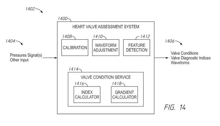
[0132] Figure 14 is a diagram of a heart valve assessment system;
[0133] Figure 15 is a flowchart of a heart valve assessment process;
[0134] Figure 16 is a flowchart of a calibration process;
[0135] Figure 17 is a diagram of example waveform analyses;
[0136] Figure 18 is a diagram of additional example waveform
analyses;
[0137] Figure 19 is a flowchart of a calibration process;
[0138] Figures 20-26 are diagrams of additional example waveform
analyses;
[0139] Figure 27 is a flowchart of another calibration process; and
[0140] Figure 28 is another diagram of the heart valve assessment
system with
which various methods and systems discussed herein may be implemented.
DETAILED DESCRIPTION
[0141] This application is directed to systems and methods for
providing
pressure curves during surgical heart procedures, including valvuloplasty
procedures,
transcatheter aortic valve replacement (TAVR) procedures sometimes also called
transcatheter aortic valve implantation (TAVI) procedures, and transcatheter
mitral valve
replacement (TAMR) procedures. The systems and methods can be used to aid a
cardiologist in completing critical aspects of a structural heart procedure.
The
embodiments herein can be used to convey by a user interface output, e.g.,
graphically, the
condition of a heart valve before, during and/or immediately after the
deployment of
structural heart device such as an aortic valve, a mitral valve or another
heart valve. The
embodiments herein can be used to convey the nature of blood flow through a
heart valve
before, during and/or immediately after the deployment of structural heart
device such as
an aortic valve, mitral valve or another heart valve. Novel displays provide
an intuitive
and/or immediate sense of a condition of the patient to simplify and to
expedite procedures
and to increase the success thereof. Further discussion of the user interface
output can be
found in Section III of the present application.
[0142] The pressure measurements obtained from the systems and
methods
described herein may be used to calculate a heart valve or blood flow index,
such as a valve
regurgitation index or a pressure gradient across a natural heart valve, a
previous placed
replacement heart valve or a replacement heart valve being currently
implanted. The valve
regurgitation index and pressure gradient enable the cardiologist to properly
evaluate the
heart valve. During systole, a higher pressure gradient across the aortic
valve (or lower
pressure in the aorta) may be indicative of greater valve calcification. A
lower regurgitation
-24-
CA 03140416 2021-11-12
WO 2020/236494
PCT/US2020/032748
index at the end of diastole may be indicative of greater regurgitation.
Further discussion
of such calculations can be found in Sections III and IV of the present
application.
I. EXAMPLE METHODOLOGIES
[0143] Figures
1A-1F illustrate various methods of accessing a heart during a
structural heart procedure. One of the pressure guidewire 30 or the pressure
sensing device
(e.g., a pigtail catheter 10 or access catheter 20) may be used to calculate
an upstream
pressure curve (with respect to flow) and the other of the pressure guidewire
30 or the
pressure sensing device may be used to calculate a downstream pressure curve
(with respect
to flow). Although certain methods are described below with respect to
particular heart
valves and approaches to access, similar systems may be used to evaluate other
valves such
as the tricuspid valve or the pulmonary valve.
[0144] Figure
1A illustrates a system and method for measuring the
performance of an existing or a replacement aortic heart valve. An existing
heart valve can
be a natural but diseased valve or a previously implanted replacement heart
valve that is
being assessed in a subsequent procedure. As shown, a pigtail catheter 10 may
be
positioned downstream of a treatment site, for example downstream of an aortic
valve in
an aorta A, to provide the downstream pressure curve. The pigtail catheter 10
may also be
used to deliver contrast media to facilitate visualization of the treatment
site. An access
catheter 20 may be delivered to the heart from the same or different access
site as the pigtail
catheter 10. The access catheter 20 or a separate delivery catheter exchanged
with the
access catheter 20 may be used to advance a valve dilation balloon,
replacement valve, and
or other device to the treatment site. A pressure guidewire 30 may extend
through the
access catheter 20 to a position upstream of the treatment site, for example
in left ventricle
LV, to provide the upstream pressure curve. The pressure guidewire 30 may
include a
pressure sensor 40 anywhere along a distal segment of the pressure guidewire
30, for
example within an atraumatic curvature, at the transition to the atraumatic
curvature, or
proximal of the atraumatic curvature (see Figure 2B). Prior to entering the
heart, access is
provided using an arterial approach, such as a femoral or a radial approach.
Figure 1B
illustrates a similar configuration to Figure 1A except one or both of the
pigtail catheter 10
and/or the access catheter 20 can be used to provide pressure reading with the
use of the
external pressure sensing. Catheter 20 can allow the measurement of the
downstream
pressure, similar to the pressure read by the pressure guidewire 30. This
configuration can
be used to equalize the external pressure sensor with the pressure guidewire.
Alternatively,
any other delivery catheter exchanged with the access catheter may be used to
provide the
-25-
CA 03140416 2021-11-12
WO 2020/236494
PCT/US2020/032748
downstream pressure curve. In some cases, the downstream pressure output can
be
received by a console that can be coupled with the pressure signal of either
or both of the
pigtail catheter 10 and the access catheter 20.
[0145] It can
be important to equalize the pressure readings between the
downstream and the upstream pressure sensing devices. Equalization may be done
in term
of pressure accuracy (gain and offset), but also in term of phase delay
between the two
pressure curves. For example, pressure readings may be taken from the
downstream and
upstream pressure sensing devices in the same general anatomical region and
the pressure
measurements may be manually or automatically adjusted for the phase delay
between the
two pressure curves. As shown in Figure 1B, the pressure measurements for
equalization
may be taken from the left ventricle LV. In this approach, the downstream
pressure output
is provided by the access catheter 20 and the upstream pressure output is
provided by the
pressure guidewire 30. The sensing feature of the access catheter 20 (e.g., a
distal end of a
column of fluid in the catheter 20) is advanced to be adjacent to the sensing
feature of the
pressure guidewire 30. The sensing features of the access catheter 20 and the
pressure
guidewire 30 can be confirmed to be placed in the left ventricle LV. The
sensing features
of the access catheter 20 and the pressure guidewire 30 can be confirmed to be
in a similar
position in the left ventricle LV.
[0146] Figure
1C illustrates a similar configuration to Figure 1A except the
pressure sensor 40 is located proximal of the atraumatic curvature of the
pressure guidewire
30. For example, the sensing feature of the pigtail catheter 10 (e.g., a
distal end of a column
of fluid in the catheter 10) is advanced to be adjacent to the sensing feature
of the pressure
guidewire 30. The sensing features of the pigtail catheter 10 and the pressure
guidewire 30
can be confirmed to be placed in the aorta A. In this configuration, pressure
equalization
may be performed in the aorta A. After pressure equalization, the pressure
guidewire 30
may be advanced into the left ventricle LV to provide the upstream pressure
curve while
the pigtail catheter 10 remains in the aorta A to provide the downstream
pressure curve.
[0147] In
Figure 1D, pigtail catheter 10 may be positioned in the aorta A to
provide the downstream pressure curve. The pressure guidewire 30 extends
through the
pigtail catheter 10 in this embodiment to provide the upstream pressure curve.
In this
configuration, pressure equalization may be performed in the aorta A. For
example, the
sensing feature of the pressure guidewire 30 can be advanced to the end of a
fluid column
in the pigtail catheter 10 or just distal thereto. The signals from the
sensing feature of the
pressure guidewire 30 and the fluid column can be compared to equalize them
(as discussed
-26-
CA 03140416 2021-11-12
WO 2020/236494
PCT/US2020/032748
further in Section IV below). After pressure equalization, the pressure
guidewire 30 may
be retrieve from the aortic pigtail and insert in the left ventricle via the
access catheter like
it is usually done while the pigtail catheter 10 remains in the aorta A to
provide the
downstream pressure curve.
[0148] The
systems described herein may also be used to measure the
performance of an existing or replacement mitral valve. For example, as shown
in Figure
1E, the access catheter 20 may be advanced through the venous vasculature,
e.g., through
an inferior or superior vena cava VC, e.g., from a femoral approach, to a
right atrium RA.
The access catheter 20 may then be advanced through an atrial septum to a
position in a
left atrium LA. In some variations, the access catheter 20 may be configured
to provide
access through a patent foramen ovale or may be configured to track a
guidewire or device
that has provided such access. The access catheter 20 or a separate delivery
catheter
exchanged with the access catheter 20 may be used to advance a valve dilation
balloon,
replacement valve, and or other device to the treatment site. The pressure
guidewire 30
may extend through the access catheter 20 to the left ventricle LV. The access
catheter 20
may provide pressure signals that can be used to generate an upstream pressure
curve, while
the pressure guidewire 30 provides pressure signals that can be used to
generate a
downstream pressure curve. Alternatively, any other delivery catheter
exchanged with the
access catheter may be used to provide the upstream pressure curve.
[0149] Similar
systems may be used in an apical approach for aortic or mitral
valve procedures. For example, as shown in Figure 1F, the access catheter 20
may access
the left ventricle LV through the apex P of a heart. A separate device (not
shown) can be
used to open a pathway through the apex P. The access catheter 20 can be
advanced through
such as device. The access catheter 20 or a separate delivery catheter
exchanged with the
access catheter 20 may be used to advance a valve dilation balloon,
replacement valve, and
or other device to the treatment site. The pressure guidewire 30 may extend
through the
access catheter 20 to the aorta A in an aortic valve procedure. The access
catheter 20 may
provide the pressure signals that can be used to calculate an upstream
pressure curve, while
the pressure guidewire 30 can provide signals that can be used to calculate a
downstream
pressure curve. Alternatively, any other delivery catheter exchanged with the
access
catheter may be used to provide the upstream pressure curve.
[0150] Although
FIG. 1F shows an aortic valve assessment or procedure via the
apex P of the heart, the pressure guidewire 30 can be advanced through the
mitral valve M
such that the sensing feature thereof is in the left atrium. In this way, the
pressure guidewire
-27-
CA 03140416 2021-11-12
WO 2020/236494
PCT/US2020/032748
can provide pressure signals that can be used to calculate a left atrial
pressure curve
(proximal or upstream pressure curve from the perspective of flow). The access
catheter 20
can generate pressure signals that can be used to calculate a left ventricle
pressure curve
(distal or downstream pressure curve from the perspective of flow).
[0151] During
valve dilation procedures, sometimes called a valvuloplasty, or
a valve implantation procedure, natural circulation through the heart valve
may be blocked
by the valvuloplasty balloon, valve replacement delivery system, or other
treatment device.
However, when the heart is pumping, pressure from the left ventricle LV or
compression
of the heart muscle may drive the treatment device back into the aorta A
making it difficult
to properly position the treatment device. Rapid pacing or defibrillating the
left ventricle
LV can reduce the pressure gradient between the aorta A and the left ventricle
LV and also
heart muscle forces and allow the clinician to complete the procedure.
Conventional rapid
pacing may involve introducing a temporary pace maker to the heart, but this
usually
requires a separate access point, for example a venous access point. Temporary
pace
makers may also burn the heart causing other complications. Instead, the
pressure
guidewire 30 may be used to perform the rapid pacing. As explained above, the
pressure
guidewire 30 may be introduced through the same access point as the access
catheter 20 or
other delivery catheter, which reduces the total number of access points. A
current may be
delivered to a proximal segment pressure guidewire and transmitted to a distal
segment of
the pressure guidewire via connector tube and/or the core wire, as explained
in further detail
below. The access catheter 20 or other delivery catheter may insulate at least
an
intermediate segment of the rapid pacing pressure guidewire 30 from the
patient to prevent
burns. Alternatively or additionally, the pressure guidewire 30 may include an
insulator
portion to isolate the pressure guidewire 30. As shown in Figure 2B, the
distal segment of
the pressure guidewire may include a curvature allowing the current to contact
ventricle
walls in multiple locations.
II. OVERVIEW OF PRESSURE WIRE SYSTEMS AND THEIR USE
[0152] Figure
2A illustrates a diagnostic system 200 that can be used in the
vasculature of a patient. The diagnostic system 200 is configured to determine
whether the
extent of valve damage is great enough to indicate that a balloon dilation
(e.g.,
valvuloplasty), valve replacement or other catheter intervention ought to be
performed.
[0153] The
diagnostic system 200 can include a monitor assembly 204 that is
configured to be coupled to the pressure guidewire 208. The diagnostic system
200 may
-28-
CA 03140416 2021-11-12
WO 2020/236494
PCT/US2020/032748
include a connection (indicated by the dashed line A) that facilitates
connection to and
disconnection of the pressure guidewire 208 from the monitor assembly 204. The
connection to and disconnection from the monitor assembly 204 is useful in
allowing a
clinician to use the pressure guidewire 208 initially for assessing the effect
of the heart
valve damage. The pressure guidewire 208 may also be used for delivering a
treatment
device such as a balloon catheter or valve delivery system.
[0154] A fiber
optic interface cable 202 can be used to couple the pressure
guidewire 208 with the monitor assembly 204 by way of a handle 207. In some
embodiments, the system 200 receives an input from a tubular catheter body
used to access
the vasculature. For example, the access catheter 20 may be an access
catheter. A distal
tip of the pressure sensing of or in the access catheter 20 can be positioned
adjacent the
treatment site such that pressure signals corresponding to the pressure on a
first side of the
treatment site, e.g., in the aorta, can be obtained. This pressure measurement
is sometimes
referred to herein as Pa. In other configurations, the system 200 may include
a pressure
sensing device, such as a pigtail catheter, delivered separate from the
pressure guidewire to
obtain Pa.
[0155] The
pressure guidewire 208 can take any suitable form. For example,
the pressure guidewire 208 may include a proximal segment that has a proximal
end that is
positioned outside the patient and a distal end that may be advanced through
the access
catheter 20 to the vasculature. The pressure guidewire 208 can be configured
to have the
flexibility to navigate the tortuous vasculature while maintaining structural
integrity for
pushability and torqueability. For example, at least proximal section of the
pressure
guidewire 208 may be supported by a connector tube and/or core for structural
integrity,
while a distal section of the pressure guidewire 208 can be formed to include
an atraumatic
curvature 250, such as the coiled end shown in Figure 2B, to provide more
flexibility and
prevent puncture. In other configurations, a curved distal section may be
joined to the
pressure guidewire 208 to provide the atraumatic curvature 250.
[0156] Any
sensing modality can be used. For example, an optical sensor can
be configured to sense pressure when exposed to blood. The optical sensor can
be disposed
within an interior space of the pressure guidewire 208 in fluid communication
with an
exterior of the pressure guidewire 208. The sensor may be an optical or
electrical pressure
sensor. The sensor can be selectively placed in communication with the monitor
assembly
204 by pressure wire lead(s) disposed between the sensor and a proximal end of
the pressure
guidewire 208. The pressure wire lead(s) may be an optical fiber or an
electrical wire.
-29-
CA 03140416 2021-11-12
WO 2020/236494
PCT/US2020/032748
[0157] As shown
in Figure 2B, the pressure sensor may be located anywhere
along the distal section of the pressure guidewire 208. For example, the
pressure sensor
may be positioned near the distal-most tip of the guidewire at location 206D,
along the
curvature 250 of the guidewire at location 206C, at the transition to the
curvature 250 of
the guidewire at location 206B, or proximal of the curvature 250 of the
guidewire at
location 206A. For example, location 206C may be at about 270 degrees around
the
curvature 250 from the straight region (around location 206A) of the pressure
guidewire
and location 206D may be about 540 degrees around the curvature from the
straight region
of the pressure guidewire. However, the pressure sensor may be located any
position in
the curved distal region of the pressure guidewire, for example between and
including about
0 degrees to about 90 degrees, between and including about 90 degrees to about
180
degrees, between and including about 180 degrees to about 270 degrees, between
and
including about 270 degrees to about 360 degrees, between and including about
360
degrees to about 450 degrees, or between and including about 450 degrees to
about 540
degrees from the straight region of the pressure guidewire.
[0158] When the
distal section is curled up, pressure sensor may be positioned
about 270 degrees along the curvature 250 from the straight section of the
pressure
guidewire 208. The location of the pressure sensor within the distal section
of the
guidewire may influence the accuracy of the pressure measurements. For
example, when
the pressure sensor is in the more distal locations 206C, 206D, the pressure
sensor may be
more centrally located within the chamber of the heart, e.g. the left
ventricle LV, and
displaced from the chamber walls. Also, in the more distal locations 206C,
206D the
pressure sensor is less likely to be obstructed by the access catheter or
other delivery
catheter during the valvuloplasty or heart replacement procedure. In the more
proximal
positions 206A, 206B, the pressure measurements will be taken closer to the
heart valve
but it is possible to perform equalization in the aorta A while maintaining
the distal tip of
the pressure guidewire 208 within the left ventricle LV. In some procedures,
performing
equalization in the aorta A requires less manipulation of the pigtail catheter
or other
pressure sensing device is required. For example, during an aortic valve
procedure, the
pigtail catheter is already located in the aorta. Although the pressure sensor
may be
proximal to the curvature 250, the pressure sensor is sufficiently distal to
take pressure
measurements distal to the heart valve. Leaving the distal tip of the pressure
guidewire 208
within the left ventricle LV maintains access to the left ventricle LV.
-30-
CA 03140416 2021-11-12
WO 2020/236494
PCT/US2020/032748
[0159] Figure
2C illustrates a cross-section of a TAVR system within a
descending aorta of a patient, with the anatomy removed for clarity. The TAVR
system
could be used in connection with the monitor display 204. For example, the
pressure
guidewire 208 extends through an access catheter 210. The same access catheter
210 can
be used to advance a delivery system 212 over the pressure guidewire 208. The
delivery
system 212 may be used to advance a valve replacement or other treatment
device. Other
configurations are also possible. For example, the catheter for the access
catheter 210 could
be exchanged with the delivery system 212 and thereafter advanced over the
pressure
guidewire 208. As shown, the pressure sensing device that is used to provide
pressure
signals for the pressure of blood in the aorta is an aortic pigtail catheter
214 delivered
separately from the access catheter 210, although possibly from the same
access point.
[0160] In other
configurations, the access catheter 210 or the delivery system
212 may be used to obtain pressure signals for the pressure of blood in the
aorta and thus
may be the pressure sensing device for aortic pressure. As shown in Figure 2D,
for a mitral
valve replacement, the access catheter 211 may be the pressure sensing device.
The
pressure guidewire 208 extends through the access catheter 211 and the
delivery system
213 may be advanced over the pressure guidewire 208. The delivery system 213
may be
used to deliver a mitral valve or other replacement or treatment device.
a. WIRE-BASED PRESSURE GUIDE WIRES
[0161] Figures
3 and 4 illustrate different pressure guidewires 308, 408 that
may be used in any of the above-described methods. Numerals used to identify
features of
the pressure guidewire 308 are incremented by a factor of one hundred (100) to
identify
like features of the pressure guidewire 408. This numbering convention
generally applies
to the remainder of the figures. Any component of the pressure guidewires 308,
408 can
be interchanged.
[0162] In
general, the pressure guidewires 308, 408 include an outer tube 310,
410 defining a lumen, a core wire 316, 416 extending at least partially
through the lumen
of the outer tube 310, 410, a pressure sensor assembly 318, 418 disposed
within the lumen
of the outer tube 310, 410, and/or a distal tip 432. The pressure guidewire
308 also can
include a distal tip that can be the same as or similar to the tip 432 or any
of the other tips
disclosed herein. An outer diameter of the pressure guidewire 308, 408 may be
uniform or
substantially uniform along substantially the entire or entire working length
of the pressure
guidewire 308, 408. For example, the outer diameter of the pressure guidewire
308 may
be uniform or substantially uniform along the entire working length, excluding
distal tip
-31-
CA 03140416 2021-11-12
WO 2020/236494
PCT/US2020/032748
432 or atraumatic curvature 250. The pressure guidewire 308, 408 may include
an outer
diameter of up to 0.035 inches, for example between 0.018 inches and 0.035
inches. In
some configurations, the distal portion of the pressure guidewire 308, 408 may
be form an
atraumatic curvature 250 such as the coiled portion shown in Figure 2B. In
other
configurations, the distal portion of the pressure guidewire 308, 408 may
remain straight
from at least the pressure sensor of the pressure sensor assembly to the
distal tip of the
pressure guidewire.
[0163] Figure 3
is a schematic view of one variation of the pressure sensing
guidewire 308. As illustrated, at least a distal portion of the outer tube 310
may be coiled.
For example, the coil portion 312 may be a flat ribbon coil or a round coil.
The coil portion
312 may extend along a majority of the working length of the pressure
guidewire 308, along
substantially the entire working length of the pressure guidewire 308, or
along the entire
working length of the pressure guidewire 308. With a substantial length of the
outer tube
310 being coiled, the coil portion 312 provides sufficient flexibility and
softness to avoid
any trauma during use (e.g. perforation and/or dissection). The coil portion
312 also
promotes safety in case of distal tip failure. When rapid pacing, the coil
portion 312 may
also ensure electrical contact with the heart.
[0164] As shown
in Figure 3, at least a proximal portion 328 of the core wire
316 may be concentric with the outer tube 310 and extend through at least a
portion of the
lumen of the outer tube 310. For example, the core wire 316 may extend along a
majority
of the working length of the pressure guidewire 308, along substantially the
entire working
length of the pressure guidewire 308, or along the entire working length of
the pressure
guidewire 308. The core wire 316 provides the pressure guidewire 308 with
sufficient
rigidity for pushability and to prevent kinking. It also provides sufficient
rigidity to support
the delivery catheter during valve implementation.
[0165] At least
a portion of the core wire 316 may include a reduced diameter
portion 326 to provide space in the lumen outer tube 310 for a pressure sensor
322. For
example, as shown in Figure 3, the reduced diameter portion 326 may be tapered
toward
the distal end of the pressure guidewire 308. The transition between the
proximal
portion 328 and the reduced diameter portion 326 of the core wire 316 may be
positioned
proximal of at least a portion or the entirety of the atraumatic curvature 250
in the distal
section of the pressure guidewire 308 (shown in Figure 2B) to promote a
flexible transition
to the atraumatic curvature 250 of the pressure guidewire 308. The core wire
316 would
continue to extend through at least a portion of the atraumatic curvature 250.
This flexible
-32-
CA 03140416 2021-11-12
WO 2020/236494
PCT/US2020/032748
transition acts as a force absorber and ensures no kink is formed in the
proximal section of
the atraumatic curvature 250 of the pressure guidewire 308. A kink could
complicate a
procedure, such as advancing another catheter over the guidewire 308 or
removing the
guidewire 308 from the patient without trauma.
[0166] The
proximal portion 328 of the core wire 316 may include an outer
diameter of up to 0.03 inches, for example between 0.015 inches and 0.03
inches. A
reduced diameter portion 326 of the core wire 316 may include an outer
diameter that is
less than one-third, or less than one-fourth, of the outer diameter of the
proximal portion
328 of the core wire 316. For example, the reduced diameter portion 326 of the
core wire
316 may include an outer diameter of less than 0.01 inches or less than 0.0075
inches.
[0167] The core
wire 316 may include a conductive material such as stainless
steel to provide a conductive path for current applied to the guidewire 308 in
connection
with a rapid pacing technique as described above. A proximal end of the core
wire 316
may be exposed from the proximal end of the outer tube 310 for connection to
the monitor
display 204 and/or connection to a current generator. Less than ten percent,
or less than
five percent, of a length of the core wire 316 may be exposed from the
proximal end of the
outer tube 310 for connection to a current source for rapid pacing.
[0168] The
pressure sensor assembly 318 may include a pressure sensor 322
and one or more pressure wire leads 320 extending from the pressure sensor
322. The
pressure wire leads 320 may extend along the core wire 316. For example, the
pressure
sensor 322 may be an optical or electrical sensor, membrane-based sensor, a
MEMS sensor
or other device that can generate a signal in response to pressure levels or
fluctuations. The
one or more pressure wire leads 320 may be an optical fiber or electrical
wire. As shown
in Figure 3, the pressure sensor assembly 318 may also include a sensor
housing 324
disposed over the pressure sensor 322 and positioned between the outer tube
310 and the
core wire 316. The sensor housing 324 can include a ring or short tubular
member or a
cylinder in which a membrane is supported. The sensor housing 324 can enhance
handling
during assembly in the coil portion 312.
[0169] The
pressure sensor assembly 318 may be disposed radially between the
core wire 316 and the outer tube 310 with the pressure sensor 322 disposed
radially between
the reduced diameter portion 326 of the core wire 316 and the coiled portion
312 of the
outer tube 310. At least a portion of the pressure sensor assembly 318 may be
off-axis
relative to a longitudinal axis L of the pressure guidewire 308. In some
configurations, the
-33-
CA 03140416 2021-11-12
WO 2020/236494
PCT/US2020/032748
entire pressure sensor assembly 318 may be off-axis relative to the
longitudinal axis of the
pressure guidewire 308.
[0170] The
pressure sensor 322 may be exposed to blood or other fluid through
the spacing or gaps 314 in the coil portion 312. Although, in other
variations, the outer
tube 310 may include a sensor housing section with one or more openings to
expose the
pressure sensor 322 to blood or other fluid. The sensor housing section may be
stiffer than
the remainder of the coil portion 312. For example, the sensor housing section
may be a
metallic tube splitting the coil portion 312 into two sections. The sensor
housing section
may be mounted to a distal portion of a first coil section of the coil portion
312 and to a
proximal portion of a second coil section of the coil portion 312. As another
example, the
coil portion 312 may include two coils welded together to create a stiffened
section.
[0171] At least
a portion of the pressure guidewire 308 may be covered by a
lubricious insulator, for example a polymeric layer such as PTI4E,. The
insulator may secure
one or more pressure wire(s) lead(s) 320 in place. When rapid pacing is
induced through
the core wire 316, the insulator may also electrically isolate the core wire
316 from the
patient along the length of the insulator. The insulator may replace the need
for a separate
catheter to electrically isolate the pressure guidewire 308.
[0172] Figure 4
illustrates another variation of the pressure guidewire 408. The
pressure guidewire 408 can include any of the features described with respect
to the
pressure guidewire 308. In this variation, a distal portion of the outer tube
410 may be
formed by the coil portion 412. A proximal portion of the outer tube 410 may
be formed
by a connector tube 430. The connector tube 430 may include a conductive
material to
facilitate rapid pacing. For example, the connector tube 430 may be formed
with a metal
structure such as a stainless steel tube. The connector tube 430 is not
covered with a coating
or other insulator to allow for rapid pacing. In some configurations, current
may
alternatively or additionally flow through the one or more pressure wire leads
420. The
connector tube 430 may be connected directly or indirectly to the coil portion
412 and/or
to the distal tip 432. For example, the coil portion 412 may be indirectly
connected to the
connector tube 430 by an insulated portion. The insulated portion can provide
a length that
is insulated from the patient and thus may be an insulator portion 434 in some
embodiments. The insulator portion 434 may insulate the patient from the core
wire 416.
In some configurations, the insulator portion 434 may include a polymeric
layer such as
PTFE.
-34-
CA 03140416 2021-11-12
WO 2020/236494
PCT/US2020/032748
[0173] At least
a non-reduced diameter portion of the core wire 416 may be
concentric with the outer tube 410. The core wire 416 may extend through at
least the coil
portion 412, but may also extend through at least a portion of the insulator
portion 434
and/or the connector tube 430 of the outer tube 410. For example, a proximal
end of the
core wire 416 may be sealed, for example using adhesive 436, to a distal end
of the
connector tube 430 and extend distally from a distal end of the connector tube
430.
[0174] The core
wire 416 may include any of the features of the core wire 316.
For example, a distal portion of the core wire 416 may include a reduced
diameter
portion 426. A proximal end of the coil portion 412 may be distal of a
transition between
the non-reduced diameter portion 428 and the reduced diameter portion 426 of
the core
wire 416.
[0175] The
pressure sensor assembly 418 may be disposed radially between the
core wire 416 and the outer tube 410 with the pressure sensor 422 positioned
radially
between the reduced diameter portion 426 of the core wire 416 and the coil
portion 412.
At least a portion of the pressure sensor assembly 418 may be off-axis
relative to a
longitudinal axis L of the pressure guidewire 408. For example, a first
section 438a of at
least one pressure wire lead 420 may be concentric with the outer tube 410 and
a second
section 438b of the pressure wire lead 420 may be off-axis relative to a
longitudinal axis of
the outer tube 410. The outer tube 410 may include an opening 440 to permit
the pressure
wire lead 420 to transition from the first section 438a that is concentric
with the outer tube
410 to the second section 438b that is off-axis relative to the longitudinal
axis of the outer
tube 410. The opening 440 may be a partial thickness cut out or extend through
the full
thickness of the outer tube 410. If the opening 430 extends through the full
thickness of
the outer tube 410, the opening 440 may be sealed, for example with adhesive
436, to
prevent blood or other fluids from flowing into the pressure guidewire through
the opening
440. As shown in Figure 4, the opening 440 is disposed in the connector tube
430.
However, in other configurations, the opening 440 may be disposed in the
insulator portion
434.
[0176]
Alternative to the opening 440, the core wire 416 may be sized or offset
relative to a longitudinal axis of the pressure guidewire 408 to permit the
pressure wire lead
420 to transition from the first section 438a that is concentric with the
outer tube 410 to the
second section 438b that is off-axis relative to the longitudinal axis of the
outer tube 410.
The core wire 416 can have a groove in one side configured to receive a span
of the pressure
-35-
CA 03140416 2021-11-12
WO 2020/236494
PCT/US2020/032748
wire lead 420 such that the lead can transition from the first section 438a to
the second
section 438b.
[0177] The
pressure guidewire 408 may include a distal tip 432 that is rounded
to form an atraumatic tip. For example, the distal tip 432 may have a
hemispherical shape.
The tip 432 may also reduce or even to prevent ingress of unwanted foreign
matter through
a distal end of the pressure guidewire 408.
[0178] In some
configurations, the distal tip 432 is a separate component
adhered, welded, and/or otherwise joined to the coil portion 412 and/or the
core wire 416.
The distal tip may be joined to an inner surface of the coil portion 412
and/or the distal
most edge of the coil portion 412. The core wire 416 may be bent up to 180
degrees within
the outer tube 410 to strengthen the adhesive joint to the distal tip 432. In
other
configurations, the distal tip 432 may be an enlarged distal end of the core
wire 416 that is
distal of the reduced diameter portion 426. The distal end of the core wire
416 may be
adhered, welded, and/or otherwise joined to the inner surface and/or distal
most edge of the
coil portion 412. In one method, the distal tip 432 is formed by transforming
an enlarged
segment of the core wire 416 into a hemispherical member. The enlarged segment
can be
melted to form the hemispherical member. The hemispherical member can be
joined to a
distal portion of the coil portion 412. In any of these configurations, the
atraumatic portion
of the distal tip 432 may be formed from the core wire 416, adhesive, and/or
welding.
b. TUBE-BASED PRESSURE GUIDE WIRES
[0179] Figures
5 to 9 illustrate further variations of pressure guidewires that
may be used in any of the above-described methods. The pressure guidewires
described
below may include any of the features of the above-described pressure
guidewires 308,
408. In general, the pressure guidewires shown in Figures 5 to 9 include an
outer tube
defining a lumen, connector tube positioned radially inward of the outer tube,
a pressure
sensor assembly disposed within the lumen of the outer tube, and/or a distal
tip. An outer
diameter of the pressure guidewire may be uniform or substantially uniform
along
substantially the entire or entire working length of the pressure guidewire.
For example,
the outer diameter of the pressure guidewire may be uniform or substantially
uniform along
the entire working length, excluding distal tip or grinded down curvature. The
pressure
guidewire may include an outer diameter of up to 0.035 inches, for example
between 0.018
inches and 0.035 inches. In some configurations, the distal portion of the
pressure
guidewire may be formed to an atraumatic curvature 250 as shown in Figure 2B.
In other
configurations, the distal portion of the pressure guidewire may remain
straight.
-36-
CA 03140416 2021-11-12
WO 2020/236494
PCT/US2020/032748
[0180] The
connector tube may include an inner diameter that is less than one-
third, or less than one-fourth an outer diameter of the connector tube. For
example, the
connector tube may include an outer diameter of up to 0.035 inches, for
example between
0.018 inches and 0.035 inches, and an inner diameter of less than 0.01 inches,
for example
less than 0.007 inches. The connector tube may have a uniform outer diameter
(see Figure
5) or a non-uniform diameter (see Figure 6). In the non-uniform
configurations, a reduced
diameter portion of the connector tube may have an outer diameter of less than
or equal to
about 0.027 inches. The connector tube may extend along a majority of or
substantially
the entire working length of the pressure guidewire. For example, the
connector tube may
extend at least eighty percent, or at least ninety percent, of the working
length of the
pressure guidewire.
[0181] The
connector tube may be constructed of a conductive metal. For
example, the connector tube may be a stainless steel tube. A proximal end of
the connector
tube may be exposed from the proximal end of the outer tube for connection to
the monitor
display and/or connection to a current generator. Thus, at least the proximal
end of the
connector tube may be uncoated.
[0182] The
pressure guidewire may also include a core wire distal to the
connector tube. In a venous or trans-apical aortic valve application, the
portion with the
core wire may be disposed in blood flow downstream of a portion with the
connector tube.
In an arterial or trans-apical mitral valve application, the portion with the
core wire may be
disposed in blood flow upstream of a portion with the connector tube. The core
wire may
include an outer diameter of up to 0.03 inches, for example between 0.018
inches and 0.03
inches. A reduced diameter portion of the core wire may include an outer
diameter that is
less than one-third, or less than one-fourth, of the outer diameter of the
remainder of the
core wire. For example, the reduced diameter portion of the core wire may
include an outer
diameter of less than 0.01 inches or less than 0.0075 inches. The core wire
may extend
along only a distal portion of the pressure guidewire, for example along less
than twenty
percent or less than ten percent or less than 5 percent of a working length of
the pressure
guidewire.
[0183] Figure 5
is a schematic view another variation of the pressure sensing
guidewire 508. As illustrated, at least a distal portion of the outer tube 510
may be coiled.
For example, the coiled portion may be a flat ribbon coil or a round coil. As
shown in
Figure 5, the coiled portion may include two coiled sections 512a, 512b
separated from
each other by a sensor housing 542. Together, the coil portions 512a, 512b may
extend
-37-
CA 03140416 2021-11-12
WO 2020/236494
PCT/US2020/032748
along a majority of the working length of the pressure guidewire 508 or along
substantially
the entire working length of the pressure guidewire 508. For example,
together, the coiled
portions 512a, 512b may extend at least eighty percent, or at least ninety
percent, of the
working length of the pressure guidewire 508. With a substantial length of the
outer tube
510 being coiled, the coil portions 512a, 512b provides sufficient flexibility
to navigate
tortuous vasculature. The distal coil portion 512a also promotes safety in
case of failure
along a coiled portion, e.g., distal tip failure. When used for rapid pacing,
the distal coil
portion 512a may also ensures electrical contact with the inner walls of the
patient's heart,
e.g., with inner walls of the left ventricle.
[0184] As shown
in Figure 5, at least a proximal portion 528 of the core wire
516 may be concentric with the outer tube 510 and extend through at least a
portion of the
lumen of the outer tube 510. The diameter of the proximal portion 528 of the
core wire
516 may be the same as the outermost diameter of the connector tube 530. At
least a portion
of the core wire 516 may include a reduced diameter portion 526 such as a
tapered portion
that is tapered toward the distal end of the pressure guidewire 508. The
transition between
the proximal portion 528 and the reduced diameter portion 526 of the core wire
516 may
be positioned proximal of the atraumatic curvature 250 in the distal section
of the pressure
guidewire 508 to promote a flexible transition to the atraumatic curvature 250
of the
pressure guidewire 508. This flexible transition acts as a force absorber and
ensures no
kink is formed in the proximal section of the atraumatic curvature 250 of the
pressure
guidewire 508. A kink could complicate a procedure, such as advancing another
catheter
over the guidewire 508 or removing the guidewire 308 from the patient without
trauma.
The core wire 516 may include a conductive material such as stainless steel to
provide rapid
pacing as described above.
[0185] The
pressure sensor assembly 518 may include a pressure sensor 522
and one or more pressure wires leads 520 extending from the pressure sensor
522. For
example, the pressure sensor 522 may be an optical or electrical sensor,
membrane-based
sensor, or otherwise. The pressure wire(s) lead(s) 520 may be an optical fiber
or electrical
wires. The pressure wire(s) lead(s) 520 may extend through the lumen of the
connector
tube 530. The connector tube 530 locates the pressure wire(s) lead(s) 520
along the central
longitudinal axis L of the pressure guidewire 508. The pressure wire(s)
lead(s) 520 may
be secured to and in some cases also sealed to the connector tube 530, for
example using
adhesive. In some cases, the adhesive provides a seal to prevent fluid from
flowing
proximally through the connector tube 530. Adhesive may also be used in the
proximal
-38-
CA 03140416 2021-11-12
WO 2020/236494
PCT/US2020/032748
end of the connector tube 530 to secure the optical fiber 520 concentrically
to the connector
tube 530.
[0186] As shown
in Figure 5, the pressure sensor 522 may be disposed within
the pressure sensor housing 542 of the outer tube 510. The sensor housing 542
protects the
pressure sensor 522 but also provides a connection between the coil portions
512a, 512b.
The pressure sensor 522 may be exposed to blood or other fluid through the at
least one
opening 544 in the sensor housing 542. As illustrated, the sensor housing 542
may be a
metal tube joining the two coil portions 512a, 512b, but in other variations,
the sensor
housing 542 may be formed by welding several coils together to form a welded
portion
joining the coil portions 512a, 512b.
[0187] The
sensor housing 542 and the pressure sensor 522 may be positioned
proximal of the atraumatic curvature 250 shown in Figure 2B, for example at
location
206A. However, as explained above, the pressure sensor may also be positioned
anywhere
along the curvature 250 in the distal section of the pressure guidewire 508.
[0188] At least
a portion of the pressure guidewire 508 may be covered by a
lubricious insulator, for example a polymeric layer such as PTFE. When rapid
pacing is
induced through the connector tube 530 and/or the core wire 516, the insulator
may also
electrically isolate portions of the pressure guidewire 508. The insulator may
replace the
need for a separate catheter body to electrically isolate the pressure
guidewire 508.
[0189] Figure 6
is a cross-sectional view of another variation of the pressure
sensing guidewire 608. The pressure sensing guidewire 608 is similar to the
pressure
sensing guidewire 508 except as described differently below. The disclosure in
connection
with Figure 6 can be seen to supplement that of Figure 5. The pressure sensing
guidewire
608 includes a distal tip 632. The distal tip 632 is similar to the distal tip
432 except as
described differently below. The distal tip 632 provide for atraumatic
interaction with
blood vessels, valves and heart wall chambers. The tip 632 also may reduce or
prevent
ingress of foreign matter, e.g., components or fluid, through a distal end of
the pressure
guidewire 608. The distal tip 632 may have a hemispherical shape.
[0190] In some
configurations, the distal tip 632 is a separate component
adhered, welded, and/or otherwise joined to the coil portion 612a and/or the
core wire 616.
The distal tip 632 may be joined to an inner surface of the coil portion 612a
and/or the distal
most edge of the coil portion 612a. The core wire 616 may be bent up to 180
degrees within
the outer tube 610 to strengthen the adhesive joint to the distal tip 632. In
other
configurations, the distal tip 632 may be an enlarged distal end of the core
wire 616 that is
-39-
CA 03140416 2021-11-12
WO 2020/236494
PCT/US2020/032748
distal of the reduced diameter portion 626. The distal end of the core wire
616 may be
adhered, welded, and/or otherwise joined to an inner surface and/or distal
most edge of the
coil portion 612a. In any of these configurations, the atraumatic portion of
the distal tip
632 may be formed from the core wire 616, such as by melting or otherwise re-
forming an
enlarged segment of the core wire 616 to create the desired shape.
[0191] Figure 7
is a schematic view of another variation of the pressure sensing
guidewire 708. The pressure sensing guidewire 708 is similar to the pressure
sensing
guidewire 508 except that sensor housing 742 and pressure sensor 722 may be
positioned
more distally into the distal curvature 250 of the pressure guidewire 708, for
example at
locations 206B or 206C shown in Figure 2B. However, as discussed above, it can
be
beneficial to reduce the diameter of the inner core wire to promote
flexibility at the
transition to the distal curvature 250. Thus, the sensor housing 742 and the
pressure
sensor 722 may be positioned in a region in which the connector tube 730
and/or core wire
716 have transitioned to a reduced diameter. For example, as shown in Figure
7, the
connector tube 730 may have a reduced diameter section 746 at the distal end
of the
connector tube 730. The connector tube 730 may be tapered toward the reduced
diameter
section 746 at tapered portion 754. A diameter of the proximal end of the core
wire 716
may also be less than an outermost diameter of the connector tube 730, e.g. at
a distal end
or distal region thereof. In this configuration, an outer diameter of the
sensor housing 742
may also be reduced compared to the sensor housing 542.
[0192] Figure 8
is a schematic view of another variation of the pressure sensing
guidewire 808. The pressure sensing guidewire 808 is similar to the pressure
sensing
guidewire 708 except that sensor housing 842 and pressure sensor 822 may be
positioned
more distally into the distal curvature 250 of the pressure guidewire 808, for
example at
location 206D shown in Figure 2B. However, as discussed above, it can be
beneficial to
reduce the diameter of the inner core wire to promote flexibility at the
transition to the distal
curvature 250. Thus, in the region of 206D in the distal curvature 250, the
reduced diameter
portion 826 of the core wire 816 may have a sufficiently reduced diameter to
permit the
positioning of the sensor 822 radially between the distal coil portion 812a
and the reduced
diameter portion 826 of the core wire 816. As shown in Figure 8, the sensor
822 may have
a separate sensor housing 842 positioned around the sensor 822.
[0193] Instead
of a sensor housing along the outer tube 810, the pressure
guidewire 808 includes a connector 848 extending between the coil portions
812a, 812b.
The connector 848 may include an opening 852 to permit at least one pressure
wire lead
-40-
CA 03140416 2021-11-12
WO 2020/236494
PCT/US2020/032748
820 to transition from the first section 838a that is concentric with the
outer tube 410 and
within the connector tube 830 to the second section 838b that is off-axis
relative to the
longitudinal axis L of the outer tube 810. The opening 840 may be a partial
thickness cut
out or extend through the full thickness of the outer tube 810. If the opening
830 extends
through the full thickness of the outer tube 810, the opening 840 may be
sealed, for example
with adhesive, to prevent fluid from flowing into the pressure guidewire
through the
opening 840.
[0194] Figure 9
is a cross-sectional view of another variation of the pressure
sensing guidewire 908. The pressure sensing guidewire 908 is similar to the
pressure
sensing guidewire 808 except that except that Figure 9 includes distal tip
932. The distal
tip 932 may include any of the features of the distal tip 632 shown in Figure
6.
[0195] The
outer tube 910 includes an insulator portion 934 and a coil portion
912 joined by the connector 948. The insulator portion 934 surrounds at least
a portion of
the connector tube 930. The insulator portion 934 may include a polymeric
layer such as
PTFE to electrically isolate the connector tube 930 from the patient during
rapid pacing. A
proximal end 956 of the connector tube 930 may be exposed from the proximal
end of the
insulator portion 934 for connection to the monitor display and/or connection
to a current
generator. Thus, at least the proximal end of the connector tube 930 may be
uncoated.
[0196] As
illustrated, the connector 948 may be a metal tube joining the
insulator portion 934 and the coil portion 912, but in other variations, the
connector 948
may be a welded portion joining the insulator portion 934 and the coil portion
912.
[0197] The one
or more pressure wires leads 920 may be sealed to the inner
lumen of the connector tube 930, for example using adhesive, to prevent fluid
from flowing
proximally and ensuring concentricity of the optical fiber for signal
transmission.
[0198] The
pressure sensor 922 may be exposed to blood or other fluid through
the spacing or gaps in the coil portion 912. The outer tube 910 may also
include sensor
housing section 924. The sensor housing section may be stiffer than the
remainder of the
coil portion 912. For example, the sensor housing section 924 may be a
metallic tube
splitting the coil portion 912 into two sections. The sensor housing section
924 may be
mounted to a distal portion of a first coil section of the coil portion 912
and to a proximal
portion of a second coil section of the coil portion 912. The sensor housing
section 924
may include one or more openings to expose the pressure sensor 922 to blood or
other fluid.
As another example, the coil portion 912 may include two coils welded together
to create
a stiffened section that serves as the sensor housing section 924.
-41-
CA 03140416 2021-11-12
WO 2020/236494
PCT/US2020/032748
III. HEART VALVE ASSESSMENT USER INTERFACE SYSTEMS
[0199] Existing
user interfaces may be incapable of or deficient at indicating a
condition of a heart valve before, during, and/or immediately after the
deployment of
structural heart device. During a structural heart procedure, existing user
interfaces may
be unable to provide one or more pressure curves or indicators of a heart
valve condition,
such as a valve regurgitation index or a pressure gradient. Further, existing
user interfaces
for structural heart procedures may have limited the user interaction
capabilities, such as a
lack of options to allow a user to customize one or more user interfaces.
Existing patient
monitors and/or displays may have limited visual space to present indicators,
such as heart
valve conditions, diagnostics, physiological parameters, or other data.
[0200]
Accordingly, the user interfaces of heart valve assessment systems
disclosed herein can improve over existing user interfaces. During a
structural heart
procedure, one or more indicators of a heart valve condition can be provided
to a clinician
via a user interface. The user interface can be organized to provide
information in an
efficient manner. Specific graphical representations or indicators can be
presented or
selected by a user that allow a clinician to quickly assess a heart valve
condition or issue.
The systems and techniques described herein can enable clinicians to access
data faster,
perform analyses faster, and/or interact with one or more user interfaces
faster than existing
graphical user interface systems (such as by reducing the number of clicks or
selections by
a user). The user interfaces described herein can improve over existing user
interfaces by
providing more efficient use of limited visual space on small monitors or
displays. For
example, the visual indicators, graphical representations, and/or combinations
thereof can
provide information to users related to heart valve conditions in an efficient
manner that is
configured for monitors or displays with limited space. Thus, the systems and
techniques
described herein can improve over conventional user interfaces.
[0201] As used
herein, in addition to its ordinary meaning, a "cardiovascular
region" refers broadly to any area with or around the heart, such as the left
or right ventricle,
aorta, the left or right atrium, the vena cava, and/or a blood flow passage
adjacent to a
portion of the heart (such as a blood flow chamber, a blood vessel, a
pulmonary artery).
[0202] The heart valve assessment systems described herein can
advantageously provide indicators of a heart valve condition, such as a
pressure gradient or
a valve regurgitation index. As used herein, in addition to its ordinary
meaning, a "pressure
gradient" or "gradient" can refer to a severity or measurement of the
narrowing (or stenosis)
of a valve by the increase in pressure behind it. Example gradients are
provided herein
-42-
CA 03140416 2021-11-12
WO 2020/236494
PCT/US2020/032748
such as peak to peak gradient, an area gradient, or an instantaneous gradient.
A peak to
peak gradient can indicate a difference in pressure between a maximum or local
maximum
systolic pressure of a first cardiovascular region (such as the left ventricle
LV) and a second
cardiovascular region (such as the aorta Ao). An area gradient can indicate an
area between
two graphs such as pressure curves. An instantaneous gradient can indicate a
maximum or
local maximum pressure between a first cardiovascular region and a second
cardiovascular
region in a heartbeat cycle. As used herein, in addition to its ordinary
meaning, "valve
regurgitation index," "regurgitation index," or "regurgitation," can refer to
a leakiness
measurement of a valve. A regurgitation calculation can include a difference
in pressure
at the end of a diastolic cycle divided or normalized by a systolic pressure.
An aortic
regurgitation calculation can correspond to the following equation: aortic
regurgitation
index = (aortic diastolic blood pressure - left ventricular diastolic
pressure) / aortic systolic
blood pressure. Another heart valve condition can include rapid pacing of a
heart. The
systems, techniques, and/or graphical user interfaces described herein can
provide
clinicians additional data on which to base treatment/operation decisions. For
example, the
heart valve conditions and/or related user interfaces can provide additional
information for
a clinician to address a valve disease, modify a replacement valve during a
procedure,
and/or to make a recommendation following a valve procedure.
[0203]
Regurgitation can occur when blood leaks back through the valve.
Regurgitation may be caused by valve disease or in the case of prosthetic
replacement it
may be caused by malapposition of the replacement valve against the native
valve.
a. EXAMPLE USER INTERFACES
[0204] Figures
10A-10E, 11A-11C, and 12 depict example heart valve
assessment user interfaces. A heart valve assessment system can be the same
as, similar
to, or can include similar components as the diagnostic system 200 described
above in
Figure 2A. For convenience, the user interfaces will be described as being
presented by
the diagnostic system 200 or the monitor 204, although other computing systems
may
present the user interfaces. These user interfaces can be presented by the
monitor 204
described above using, for example, data received from a pressure guidewire
208, a
pressure sensing access catheter 20, or a pressure sensing pigtail catheter
10. Thus, each
of the user interfaces shown may be output for presentation by electronic
hardware as
graphical user interfaces.
[0205] Each of
the user interfaces shown includes one or more user interface
elements or controls that can be selected by a user. The user interfaces can
enable the
-43-
CA 03140416 2021-11-12
WO 2020/236494
PCT/US2020/032748
receiving of user input. The user interface elements shown are merely
illustrative examples
and can be varied in other embodiments. For instance, aspects of the user
interfaces may
be rearranged from what is shown and described below, and/or particular
aspects may or
may not be included. Further, the user interfaces shown may be combined or
divided into
other user interfaces such that similar functionality or the same
functionality may be
provided. The user interfaces of Figures 10A-10E such as, the user interfaces
1000, 1020,
1040, 1060, and/or 1080 may have similar user interface elements and/or
capabilities.
Moreover, each of the user interface elements may be selected by a user using
one or more
input options, such as a mouse, touch screen input (e.g., finger or pen), or
keyboard input,
among other user interface input options.
[0206] Figures
10A-10E depict example user interfaces that may be presented
by the monitor 204 described above. In Figure 10A, the user interface 1000 can
be
presented before, during, and/or immediately after a heart procedure. The user
interface
1000 can include one or more graphs 1002, 1004, 1006, 1008 and one or more
physiological
parameters 1010, 1012. The example graphs 1002, 1004, 1006, 1008 can include
or be
pressure waves. The graphs 1002, 1004, 1006, 1008 can represent pressure
values that
correspond to measurements from a cardiovascular region. The pressure values
can include
a series of numerical pressure values over time. A cardiovascular region can
include a
portion of a heart (such as a left ventricle LV, a right ventricle RV, or a
mitral valve) and/or
a blood flow passage adjacent to a portion of the heart (such as the aorta Ao,
vena cava, or
a pulmonary artery). The one or more graphs 1002, 1004, 1006, 1008 and/or the
one or
more physiological parameters 1010, 1012 can update in near or real time as
pressure
measurements are captured from a patient.
[0207] As
shown, the user interface 1000 can include a first graph 1002 for a
first cardiovascular region, such as the aorta Ao, and a second graph 1006 for
a second
cardiovascular region, such as the left ventricle LV. The additional graphs
1004, 1008 can
correspond to a statistical measure of pressure values from a cardiovascular
region such as
a mean or average pressure value for the aorta Ao or the left ventricle LV.
The statistical
measure can be based on a configuration parameter, which can be user selected,
that
indicates the statistical measure period, such as a quantity of heartbeats or
a period of time
to calculate the statistical measure. In some embodiments, the one or more
graphs 1002,
1004, 1006, 1008 can have indicators to indicate the corresponding
cardiovascular region
for the graph (for example, the aorta Ao graphs 1002, 1004 can be color-coded
red and the
left ventricle LV graphs 1006, 1008 can be color-coded blue).
-44-
CA 03140416 2021-11-12
WO 2020/236494
PCT/US2020/032748
[0208] As
shown, the user interface 1000 can include first physiological
parameters 1010 for a first cardiovascular region, such as the aorta Ao, and
second
physiological parameters 1012 for a second cardiovascular region, such as the
left ventricle
LV. The physiological parameters 1010, 1012 can include a systolic blood
pressure, a
diastolic blood pressure, and/or a statistical measure for blood pressure such
as a mean or
average systolic or diastolic blood pressure or some combination thereof for a
particular
cardiovascular region. The statistical measure physiological parameter can
correspond to
the additional graphs 1004, 1008.
[0209] The user
interface 1000 can include one or more user interface options,
such as the record option 1014. A clinician can select the record option 1014
to record the
blood pressure values, other measurements, and/or other values associated with
the
procedure. The clinician can then playback the recorded data. In some
embodiments, a
heart valve condition such as, but not limited to, a gradient or a
regurgitation index, may
be presented to a user during the playback mode.
[0210] Turning
to Figure 10B, another user interface 1020 is depicted. The
additional user interface 1020 can be similar to the user interface 1000 of
Figure 10A.
However, the additional user interface 1020 can include a stop recording
option 1022 that
can enable a user to stop the recording of patient data. In some embodiments,
once stopped,
a user can enter a playback mode to view a heart valve condition such as, but
not limited
to, a gradient or a regurgitation index.
[0211] Turning
to Figure 10C, yet another user interface 1040 is depicted. The
additional user interface 1040 can be similar to the user interface 1000 of
Figure 10A. The
additional user interface 1040 can include a first graph 1002 and a second
graph 1006
similar to the first and second graphs of Figure 10A. However, the additional
user interface
1040 can present one or more gradient representations 1042a, 1042b, 1042c that
visually
presents a gradient measurement between a first peak in the first graph 1002
and a second
peak in the second graph 1006. The one or more gradient representations 1042a,
1042b,
1042c can correspond to a gradient type such as a peak to peak gradient type.
[0212] The use
interface 1040 can include a first numerical value 1046 that
corresponds to a gradient type such as a peak to peak gradient type. The first
numerical
value 1046 can correspond to a difference in pressure between a maximum or
local
maximum systolic pressure of a first cardiovascular region (such as the left
ventricle LV)
and a second cardiovascular region (such as the aorta Ao). In some
embodiments, the first
numerical value 1046 can include a statistical measure, such as an average or
mean
-45-
CA 03140416 2021-11-12
WO 2020/236494
PCT/US2020/032748
difference in pressure between a maximum or local maximum systolic pressure,
for
multiple heartbeat cycles. As shown, the first numerical value 1046 (here 28)
can be a
statistical measure of peak-to-peak measurements for multiple heartbeats that
correspond
to the three graphical peak-to-peak measurements 1042a, 1042b, 1042c.
[0213] The user
interface 1040 can present one or more regurgitation
representations 1044a, 1044b, 1044c. As shown, the one or more regurgitation
representations 1044a, 1044b, 1044c can visually present a regurgitation
measurement
between a first point in the first graph 1002 and a second point in the second
graph 1006.
The one or more regurgitation representations 1044a, 1044b, 1044c can
correspond to a
calculation in a difference in pressure at the end of a diastolic cycle (here
aortic A end-
diastolic blood pressure minus left ventricular LV end-diastolic pressure)
divided or
normalized by a systolic pressure (here aortic systolic blood pressure).
[0214] The use
interface 1040 can include a second numerical value 1048 that
corresponds to a regurgitation index. The second numerical value 1048 can
correspond to
a difference in pressure at the end of a diastolic cycle divided or normalized
by a systolic
pressure. In some embodiments, the second numerical value 1048 can include a
statistical
measure, such as an average or mean regurgitation for multiple heartbeat
cycles. As shown,
the second numerical value 1048 (here 22) can be a statistical measure of
regurgitation
calculations for multiple heartbeats that correspond to the three
regurgitation
representations 1044a, 1044b, 1044c.
[0215] The use
interface 1040 can include an electrocardiography graph 1050.
The electrocardiography graph 1050 can be disabled or enabled by a user.
Accordingly,
the electrocardiography graph 1050 can be removed or omitted from the use
interface 1040.
While not illustrated, in some embodiments, if the electrocardiography graph
1050 is
removed or omitted, the pressure graph display 1051 can expand in size in the
user interface
1040.
[0216] The use
interface 1040 can include a playback control 1052 and a
gradient type selector 1056. As shown, the playback control 1052 can present a
time (here
0:01:18) and a current playback position 1054. In some embodiments, a user can
interact
with the playback control 1052 to advance or rewind playback of one or more
graphs and
corresponding indicators of heart valve condition(s). A user can change the
gradient type
of the user interface 1040 by selecting the gradient type selector 1056, which
can cause an
updated user interface to be presented instead of the present use interface
1040.
-46-
CA 03140416 2021-11-12
WO 2020/236494
PCT/US2020/032748
[0217] Turning
to Figure 10D, yet another user interface 1060 is depicted. The
additional user interface 1060 can be similar to the user interface 1040 of
Figure 10C. The
additional user interface 1040 can include a first graph 1002 and a second
graph 1006
similar to the first and second graphs of Figure 10C in addition to other
similar user
interface elements. However, the additional user interface 1060 can present
one or more
gradient representations 1062a, 1062b, 1062c that visually presents a gradient
measurement
between a first point in the first graph 1002 and a second point in the second
graph 1006.
The one or more gradient representations 1062a, 1062b, 1062c of the additional
user
interface 1060 can be presented in response to a user selection, such as a
user selection of
the gradient type selector 1056 of Figure 10C.
[0218] The one
or more gradient representations 1062a, 1062b, 1062c can
correspond to a gradient type such as an instantaneous gradient type. The
instantaneous
gradient representations 1062a, 1062b, 1062c can indicate a maximum or local
maximum
pressure difference between a first cardiovascular region and a second
cardiovascular
region in a heartbeat cycle. The use interface 1060 can include a numerical
value 1064
(here 64) that corresponds to a gradient type such as an instantaneous
gradient type. The
numerical value 1064 can indicate a maximum or local maximum pressure
difference
between a first cardiovascular region (such as the left ventricle LV) and a
second
cardiovascular region (such as the aorta Ao) in a heartbeat cycle. In some
embodiments,
the numerical value 1064 can include a statistical measure, such as an average
or mean
maximum or local maximum pressure difference for multiple heartbeat cycles. As
shown,
the numerical value 1064 can be a statistical measure of instantaneous
gradient
measurements for multiple heartbeats that correspond to the three graphical
instantaneous
measurements 1062a, 1062b, 1062c.
[0219] Turning
to Figure 10E, yet another user interface 1080 is depicted. The
additional user interface 1080 can be similar to the user interface 1040 of
Figure 10C. The
additional user interface 1080 can include a first graph 1002 and a second
graph 1006
similar to the first and second graphs of Figure 10C in addition to other
similar user
interface elements. However, the additional user interface 1080 can present
one or more
gradient representations 1082a, 1082b, 1082c that visually presents an area
between the
first graph 1002 and the second graph 1006. The one or more gradient
representations
1082a, 1082b, 1082c of the additional user interface 1060 can be presented in
response to
a user selection, such as one or more user selections of the gradient type
selector 1056 of
Figure 10C.
-47-
CA 03140416 2021-11-12
WO 2020/236494
PCT/US2020/032748
[0220] The one
or more gradient representations 1082a, 1082b, 1082c can
correspond to a gradient type such as an area gradient type. The area gradient
representations 1082a, 1082b, 1082c can indicate a difference in pressure
between a first
cardiovascular region and a second cardiovascular region. The use interface
1080 can
include a numerical value 1084 (here 56) that corresponds to a gradient type
such as an area
gradient type. The numerical value 1084 can indicate an area between two
graphs
corresponding to a first cardiovascular region (such as the left ventricle LV)
and a second
cardiovascular region (such as the aorta Ao). In some embodiments, the
numerical value
1084 can include a statistical measure, such as an average or mean area
between two graphs
or pressure curves for multiple heartbeat cycles. As shown, the numerical
value 1084 can
be a statistical measure of area gradient measurements for multiple heartbeats
that
correspond to the three graphical instantaneous measurements 1082a, 1082b,
1082c.
[0221] Figures
11A-11C depict additional example user interfaces that may be
presented by the monitor 204 described above. The user interfaces 1100, 1120,
1140 of
Figures 11A, 11B, 11C may be similar to the user interfaces 1040, 1060, 1080
of Figures
10C, 10D, 10E, respectively. In particular, the user interfaces 1100, 1120,
1140 of Figures
11A, 11B, 11C may present alternative gradient representations than the
gradient
representations of the user interfaces 1040, 1060, 1080 of Figures 10C, 10D,
10E,
respectively. Further, the user interfaces 1100, 1120, 1140 of Figures 11A,
11B, 11C can
depict user interfaces that present a heart valve condition(s) for a mitral
valve.
[0222] In
Figure 11A, the user interface 1100 can include a gradient
representation 1102. The gradient representation 1102 can be for a peak to
peak gradient
type, which can be similar to the peak to peak gradient type of Figure 10C.
However,
instead of a measurement visualization between two peaks, the gradient
representation
1102 can depict one or more pressure values corresponding to peak to peak
gradients in a
graph format. An advantage of the gradient representation 1102 of Figure 11A
is that it
can enable a clinician to quickly review relative peak-top-peak pressure
changes over time
that can include past and present measurements.
[0223] The
regurgitation representation 1104 of Figure 11A can be similar to
the regurgitation representations 1044a, 1044b, 1044c of Figure 10C. However,
similar to
the gradient representation 1102, the regurgitation representation 1104 can
depict one or
more pressure values corresponding to valve regurgitation in a graph format
instead of a
measurement visualization between two points.
-48-
CA 03140416 2021-11-12
WO 2020/236494
PCT/US2020/032748
[0224] Turning
to Figure 11B, the user interface 1120 can include another
gradient representation 1122. The gradient representation 1122 can be for an
instantaneous
gradient type, which can be similar to the instantaneous gradient type of
Figure 10D.
However, instead of a measurement visualization between two points, the
gradient
representation 1122 can depict one or more pressure values corresponding to
peak to peak
gradients in a graph format. Similar to the gradient representation 1102 of
Figure 11A, an
advantage of the gradient representation 1122 of Figure 11B is that it can
enable a clinician
to quickly review relative instantaneous pressure changes over time that can
include past
and present measurements.
[0225] Turning
to Figure 11C, the user interface 1140 can include another
gradient representation 1142. The gradient representation 1142 can be for an
area gradient
type, which can be similar to the area gradient type of Figure 10E. However,
instead of or
in addition to depicting an area between two graphs as the exclusive
visualization, the
gradient representation 1142 can depict one or more pressure values
corresponding to area
gradients in a graph format. Similar to the gradient representation 1102 of
Figure 11A, an
advantage of the gradient representation 1142 of Figure 11C is that it can
enable a clinician
to quickly review relative pressure changes over time that can include past
and present
measurements.
[0226] Figure
12 depicts a configuration user interface 1200 for a heart valve
assessment system. A clinician can use the configuration user interface 1200
to configured
one or more user interfaces. The configuration user interface 1200 can enable
a clinician
to select a procedure type, a default regurgitation type, a time scale, a
pressure scale, and/or
other customizable user interface options. The configuration user interface
1200 can
include a statistical measure period selector 1202 that can allow a user to
select a quantity
of heartbeats for a statistical measure calculation.
b. USER INTERFACE GENERATION PROCESSES
[0227] Turning
to Figure 13, an example user interface generation process 1300
is shown. Although the process 1300 is described in conjunction with a heart
valve
assessment system, such as the system 200 of Figure 2A or the system 1400 of
Figure 14
described below, any system configured to perform the process, in any order,
is within the
scope of this disclosure. The process 1300 may be performed by the various
components
of the system of Figure 2A as discussed herein, including the monitor 204, or
the system
1400 of Figure 14 described below. Depending on the embodiment, the process
1300 may
include fewer or additional blocks and/or the blocks may be performed in an
order different
-49-
CA 03140416 2021-11-12
WO 2020/236494
PCT/US2020/032748
than is illustrated. Other embodiments of the process 1300 may include less
blocks than
illustrated or the blocks may be performed in a different order than as
illustrated.
[0228]
Beginning at block 1302, pressure values can be received. In particular,
the monitor 204 can receive pressure values. The monitor 204 can receive a
first set of
pressure values and a second set of pressure values. Each pressure value from
the first set
pressure values can correspond to a first signal received from a first
pressure sensor
measuring a first cardiovascular region, such as a first portion of a heart.
Each pressure
value from the second set of pressure values can correspond to a second signal
received
from a second pressure sensor measuring a second cardiovascular region, such
as a blood
flow passage adjacent to the first portion of the heart. Thus, the monitor 204
can determine
first and second sets of pressure values from first and second sensors,
respectively. As
described above in Sections I and/or II, the pressure sensors can be included
within a
pressure guidewire, an access catheter, a pigtail catheter, or a therapy
device such as a heart
valve dilation balloon, a heart valve delivery device adapted to sense
pressure, or other
pressure sensing devices. Additional details regarding receiving pressure
values can be
described in further detail below with respect to process 1500 of Figure 15,
such as with
respect to blocks 1502 and/or 1504 of the process 1500.
[0229] At block
1304, configuration parameters can be received. In particular,
the monitor 204 can receive configuration parameters. Example configuration
parameters
can include a quantity of heartbeats or a default gradient type to present in
a user interface.
Additional details regarding configuration parameters are described above in
further detail
with respect to Figure 12.
[0230] At block
1306, a user selection can be received. An example user
selection can include a change in gradient type. A user can select the
gradient type selector
1056 of Figure 10C to change between gradient types, such as the instantaneous
gradient
type of Figure 10D or the area gradient type of Figure 10E. Additional user
selections can
include changes to configuration parameters of the configuration user
interface 1200
describe above in Figure 12. For example, a user selection can include a user
heartbeat
selection. The user heartbeat selection can specify a quantity of heartbeats
to be used for a
statistical measure (such as 2, 3, or 4 heartbeats, for example). The user
heartbeat selection
can also include a selection of one or more particular heartbeats. For
example, a user can
interact with a user interface described herein to select a portion of a graph
corresponding
to a particular heartbeat and/or can select identifier(s) for a particular
heartbeat(s).
-50-
CA 03140416 2021-11-12
WO 2020/236494
PCT/US2020/032748
[0231] At block
1308, a heart valve condition can be determined. As described
herein, example heart valve conditions can include a regurgitation index or a
gradient
pressure. The monitor 204 can determine the heart valve condition based on the
data of the
previous blocks 1302, 1304, 1306. For example, the monitor 204 can calculate
specific
heart valve conditions from the received pressure values according to the
configuration
parameters or the user selections that can specify a particular gradient type,
number of
heartbeats for a calculation, and/or specific heartbeats to use or to exclude.
As described
herein, a quantity of heartbeats can be used to calculate a statistical
measure for a particular
heart valve condition, such as a regurgitation index or a gradient. The
monitor 204 can
detect rapid pacing from one of the first set pressure values or the second
set of pressure
values, such as by detecting that a number of beats have exceeded a threshold
period of
time. Additional details regarding determining a heart valve condition are
described below
in Section IV, such as with respect to the process 1500 of Figure 15. Some of
the blocks
of the process 1500 of Figure 15 can further describe determining a heart
valve condition,
such as the blocks 1504, 1506, 1508, 1510.
[0232] At block
1310, a user interface can be presented. The monitor 204 can
present the user interface. Example use interfaces are described above with
respect to
Figures 10A-10E and 11A-11C. A first presented user interface can include a
first graph
based at least in part on the first set of pressure values and a second graph
based at least in
part on the second set of pressure values. The first presented user interface
can correspond
to any of the user interfaces 1040, 1060, 1080, 1100, 1120, 1140 of Figures
10C, 10D, 10E,
11A, 11B, 11C, respectively. The first presented user interface can also
include a gradient
representation that indicates a gradient of a valve, such as a peak to peak
gradient, an
instantaneous gradient, and/or an area gradient. For example, a first gradient
representation
can visually depict an area between the first graph and the second graph (such
as the
gradient representations 1082a, 1082b, 1082c of Figure 10E). The presented
area can
indicate a difference in pressure between the first cardiovascular region and
the second
cardiovascular region. The first user interface can also include a numerical
value indicating
an amount of regurgitation of the valve (such as the second numerical value
1048 of Figure
10C). The first user interface can also include a regurgitation representation
that visually
presents a regurgitation measurement between a first point in the first graph
and a second
point in the second graph (such as the regurgitation representations 1044a,
1044b, 1044c
of Figure 10C). Accordingly, the regurgitation measurement can indicate an
amount
regurgitation of the valve. The first user interface can also include a
numerical value for
-51-
CA 03140416 2021-11-12
WO 2020/236494
PCT/US2020/032748
the first gradient of the valve according to a statistical measure (such as
the first numerical
value 1084 of Figure 10E that can be a mean or average gradient value). The
first user
interface can also include an electrocardiography graph, which is described in
further detail
above with respect to Figure 10C. In some embodiments, the first user
interface can present
a warning of rapid pacing if rapid pacing is detected.
[0233] In some
embodiments, the first user interface can include multiple
numerical values for different gradient types but on the same graph display.
For example,
two or more numerical values can be selected from: a peak to peak gradient
numerical
value, an instantaneous gradient numerical value, and/or an area gradient
numerical value,
and can be presented on the same graph display at the same time.
[0234] As
shown, after the presentation block 1310 executes, the previous
blocks can be revisited to receive additional pressure value data, user
selections, and/or
updates to configuration parameters that causes one or more user interfaces to
update. For
example, the monitor 204 can receive, via the first user interface, a user
selection of a
second gradient type (such as a peak to peak gradient type). Accordingly, the
monitor 204
can present, instead of the first user interface, a second user interface for
the second gradient
type (such as a peak to peak gradient type). The second user interface can
include the first
graph and the second graph and a second gradient representation that visually
presents a
gradient measurement between a first peak in the first graph and a second peak
in the second
graph (such as the gradient representations 1042a, 1042b, 1042c of Figure
10C).
[0235] A user
can make any number of changes to the user interfaces. For
example, another user interface selection can be received for a third gradient
type (such as
an instantaneous gradient type). Accordingly, the monitor 204 can present,
instead of the
second user interface, a third user interface for the third gradient type
(such as the
instantaneous gradient type). The third user interface can include the first
graph and the
second graph and a third gradient representation that visually presents a
second gradient
measurement between a first point in the first graph and a second point in the
second graph
(such as the gradient representations 1062a, 1062b, 1062c of Figure 10D).
While particular
orders of gradient type changes have been described herein any order of
gradient type
changes can be accepted by the heart valve assessment system.
IV. HEART VALVE ASSESSMENT SYSTEMS AND METHODS
[0236] The
systems and methods described herein can assess a heart valve.
Pressure values can be used to assess a valve. A valve can be diagnosed by way
of various
signal processing methods that involve the pressure gradient across the valve.
The pressure
-52-
CA 03140416 2021-11-12
WO 2020/236494
PCT/US2020/032748
gradient across a valve during a systolic phase can be indicative of a
pressure loss caused
by the blood flowing through the valve, which can be indicative of a
limitation in blood
flow. A pressure gradient at the end of diastole, e.g., after the valve has
closed, can be
indicative of the amount of blood leaking through the valve while being
closed. As
described herein, normalizing or dividing this gradient with the systolic
pressure from a
cardiovascular region such as the aorta can be called regurgitation. Various
techniques
described herein can be used to improve the accuracy of valve assessment or
diagnostic
methods, such as, but not limited to, pressure sensor calibration, waveform
adjustment,
feature detection, and/or valve condition generation.
a. HEART VALVE ASSESSMENT SYSTEM AND METHOD
OVERVIEW
[0237] Turning
to Figure 14, a block diagram of a heart valve system 1400 is
depicted. In Figure 14, the heart valve assessment environment 1402 includes
input 1404
such as pressure signal(s), a heart valve assessment system 1400, and output
1406, such as
valve conditions, valve diagnostic indices, and/or waveforms. Example
waveforms can
include time series data, such as a series of respective pressure and
timestamp pair values.
The heart valve assessment system 1400 can be similar to or can be embodied in
the monitor
204 and/or components of the heart valve assessment system 1400 can be
embodied in the
monitor 204. The pressure signal(s) 1406 can be received from one or more
pressure
sensors described herein, such as a pressure guidewire 208, a pressure sensing
access
catheter 20, or a pressure sensing pigtail catheter 10.
[0238] The
heart valve assessment system 1400 can include a calibration
service 1408, a waveform adjustment service 1410, a feature detection service
1412, and/or
valve condition determination service 1414. The calibration service 1408 can
calibrate one
pressure sensor against another pressure sensor. The waveform adjustment
service 1410
can adjust one or more pressure waveforms, such that two or more pressure
waveforms can
generally be synchronized. The feature detection service 1412 can detect one
or more
features from pressure waveforms, such as detection of a systolic phase, a
diastolic phase,
a dicrotic notch, the end of diastole, and/or the beginning of systole. The
valve condition
determination service 1414 can determine one or more valve conditions. The
valve
condition determination service 1414 can include an index calculator 1416
and/or a
gradient calculator 1418. The index calculator 1416 can generate indices such
as a valve
regurgitation index. The gradient calculator 1418 can generate a pressure
gradient and/or
a statistical measure of a pressure gradient. The generated output data 1406,
such as valve
-53-
CA 03140416 2021-11-12
WO 2020/236494
PCT/US2020/032748
conditions, valve diagnostic indices, and/or waveforms, can be provided in a
user interface
as described herein or can be provided to another device or system.
[0239] Turning
to Figure 15, an example valve assessment process 1500 is
shown. Although the process 1500 is described in conjunction with a heart
valve
assessment system, such as the system 200 of Figure 2A or the system 1400 of
Figure 14,
any system configured to perform the process, in any order, is within the
scope of this
disclosure. The process 1500 may be performed by the various components of the
system
of Figure 2A as discussed herein, including the monitor 204, or the system
1400 of Figure
14. Depending on the embodiment, the process 1500 may include fewer or
additional
blocks and/or the blocks may be performed in an order different than is
illustrated. Other
embodiments of the process 1500 may include less blocks than illustrated or
the blocks
may be performed in a different order than as illustrated.
[0240]
Beginning at block 1502, pressure values or signals can be received or
determined. In particular, the heart valve assessment system 1400 can receive
pressure
signals from a pressure sensor, such as a pressure guidewire 208, a pressure
sensing access
catheter 20, or a pressure sensing pigtail catheter 10. The heart valve
assessment system
1400 can determine a first set of pressure values and a second set of pressure
values from
received pressure signals. Each pressure value from the first set pressure
values can
correspond to a first signal received from a first pressure sensor measuring a
first
cardiovascular region. Each pressure value from the second set of pressure
values can
correspond to a second signal received from a second pressure sensor measuring
a second
cardiovascular region that can be the same as or different from the first
cardiovascular
region. The heart valve assessment system 1400 can determine first and second
sets of
pressure values from first and second sensors, respectively. As described
above in Sections
I and/or II, the pressure sensors can be included within a pressure guidewire
or other
pressure sensing devices. In some embodiments, such as where calibration is
performed,
the first pressure sensor and the second pressure sensor can be located at a
same or different
cardiovascular region. In such a case, the process can proceed to block 1504.
[0241] At block
1504, calibration can be performed. The calibration service
1408 can perform the calibration. A second sensor can be calibrated against a
first sensor
to determine more accurate pressure measurements. Similarly, a first sensor
can be
calibrated against a second sensor to determine more accurate pressure
measurements. In
some embodiments, each of a first sensor and second sensor can both be
calibrated together.
Pressure values determined from the second sensor can be adjusted based on the
calibrating.
-54-
CA 03140416 2021-11-12
WO 2020/236494
PCT/US2020/032748
Calibration of a first sensor and a second sensor can result in the generation
of one or more
calibration parameters. The one or more calibration parameters can be used to
adjust one
or more pressure values determined from a calibrated pressure sensor.
Additional details
regarding calibration are described in further detail below with respect to
the process 1600
of Figure 16 and/or the process 2700 of Figure 27. As used herein, the terms
"calibration"
and "equalization" can be used interchangeably.
[0242] The
process can return to block 1502. Once calibration has been
finished, one or more pressure sensors can be moved to a different
cardiovascular region,
and additional pressure signals can be received at block 1502. Pressure
signals can be
received from two or more pressure sensors that are located in different
cardiovascular
regions. Examples of different cardiovascular regions can include adjacent
blood passages,
such as, but not limited to, opposite sides of a heart valve, such the left
ventricle and the
aorta, the left ventricle and the left atrium, the right ventricle and the
pulmonary artery, the
right atrium and the right ventricle, the vena cava and the right atrium, etc.
The heart valve
assessment system 1400 can determine first and second sets of pressure values
from the
pressure signals, such as by applying the determined calibration parameter.
[0243] At block
1506, waveform adjustment can be performed. Since some
valve conditions (such as diagnostics) can be based on intra-beat waveform
analysis where
specific portions within the heartbeat cycle can be used, it can be important
to adjust one
or more pressure waveforms to have two or more pressure waveforms to be
generally
synchronized or aligned. The adjustment of the waveforms can correspond to a
time shift
of one or more of the waveforms such that corresponding features thereof are
aligned. In
some embodiments, the waveform adjustment service 1410 can automatically
adjust one or
more waveforms. In other embodiments, some aspects of the waveform adjustment
can
include receiving user input, such as an operator manually adjusting one or
more
waveforms.
[0244] The
waveform adjustment service 1410 can adjust the phase between
one or both pressure waveforms by adding a delay to one or both of the
pressure waveforms
during installation, maintenance, or use with a specific patient. This
technique can assume
that the set-up process during installation or maintenance is representative
of the set-up
process during a procedure such as TAVI. For example, the aortic pressure
signal time
delay can be representative of the aortic pressure line delay induced in a
TAVI procedure.
The waveform adjustment service 1410 can adjust the phase for each specific
patient.
-55-
CA 03140416 2021-11-12
WO 2020/236494
PCT/US2020/032748
[0245] The
waveform adjustment service 1410 can include or communicate
with automatic phase delay recognition system that may advise the operator to
verify and
adjust the phase delay between both pressure signals. The waveform adjustment
service
1410 can verify such time delay while equalization is requested or occurs,
such as when
both pressure signals have the same origin (e.g., are positioned at the same
location such
that they should experience similar pressure) or when the pressure signals are
from different
locations.
[0246] The
waveform adjustment service 1410 can detect phase delay based on
time delay between one or more pressure waveforms features. Pressure waveform
features
can include one or more of the systolic pressure relative position, dicrotic
notch relative
position, or end of diastole relative position. The waveform adjustment
service 1410 can
use the feature of the relative position of the maximum slope of systolic
raising edges,
which can be a reliable feature.
[0247]
Additionally or alternatively, the waveform adjustment service 1410 can
systematically adjust the time delay when pressure equalization is requested.
Similar to the
technique for detecting the phase delay, time adjustment can be measured by
comparing
the relative position of specific pressure waveform features. For example, the
waveform
adjustment service 1410 can delay the timing of pressure sampling during
equalization
based on the recognition of pressure waveform features. The waveform
adjustment service
1410 can use the cross-correlation between both signals, i.e., such as by
calculating the
correlation between one signal against the other while being shifted in time.
The time shift
can result in an enhanced or even the maximal correlation value between both
signals that
can correspond to the time shift that can be added to one pressure signal or
the other.
[0248] At block
1508, feature detection can be performed. The feature
detection service 1412 can perform feature detection. Example features that
can be
detected from a pressure waveform can include a systolic phase, a diastolic
phase, a dicrotic
notch, the end of diastole, and/or the beginning of systole. Additional
details regarding
feature detection are described in further detail below with respect to
process 1900 of
Figure 19.
[0249] At block
1510, a heart valve condition can be determined. The valve
condition determination service 1414 can determine a valve condition. In
particular, the
index calculator 1416 can generate indices, such as a valve regurgitation
index, and the
gradient calculator 1418 can generate a pressure gradient and/or a statistical
measure of a
pressure gradient. The index calculator 1416 and/or the gradient calculator
1418 can use
-56-
CA 03140416 2021-11-12
WO 2020/236494
PCT/US2020/032748
the calibrated or adjusted waveforms or the detected features of the previous
blocks to
generate a valve condition. The index calculator 1416 can calculate a
regurgitation index
based at least in part on a first subset of the first set of pressure values
according to a systolic
phase or diastolic phase and a second subset of adjusted pressure values
according to a
systolic phase or diastolic phase. For example, the index calculator 1416 can
calculate a
regurgitation index with the following equation: aortic regurgitation index =
(aortic
diastolic blood pressure - left ventricular diastolic pressure) / aortic
systolic blood pressure.
The gradient calculator 1418 can calculate a gradient value based at least in
part on a
difference between a first subset of pressure values during a systolic phase
and a second
subset of adjusted pressure values during a systolic phase (such as an area
gradient value,
a peak to peak gradient value, and/or an instantaneous gradient value, which
are described
in further detail above in Section III). Additional details regarding
determination of a valve
condition are described in further detail above in Section III.
[0250] At block
1512, the heart valve condition can be presented in a user
interface. The heart valve assessment system 1400 and/or a monitor 204 can
present the
heart valve condition. Additional details regarding presentation of a valve
condition are
described in further detail above with respect to block 1310 of Figure 13 and
the user
interfaces of Figures 10A-10E and 11A-11C.
b. PRESSURE SENSOR CALIBRATION
[0251] As
described herein, a first sensor can be calibrated against a second
sensor to determine more accurate pressure measurements, which can also be
referred to as
equalization. For example, a pressure guidewire can be at or nearby valve
location along
with another pressure instrument. The other pressure instrument can be a
catheter, a pigtail
or other instrument used to deliver the valve and comprising a lumen connected
to a
pressure transducer. The pressure instrument may be another pressure guidewire
or a
catheter comprising a tip pressure sensor. The pressure guidewire and the
pressure
instrument are positioned to measure the same pressure. The pressure sensors
can be
positioned in the same cardiovascular region, such as the aorta, in the
ventricle, atrium, or
some other location. Although it can be understood that the pressure sensors
at the same
location should display the same pressure, there can be a difference between
the first and
second pressures (such as the aortic pressure Pa and the distal pressure Pd).
Accordingly,
once at the same position, one pressure sensor can be calibrated against the
other.
[0252] The
systems and methods described herein for pressure sensor
calibration can improve the accuracy of pressure instruments. For example, as
mentioned
-57-
CA 03140416 2021-11-12
WO 2020/236494
PCT/US2020/032748
above, even if two pressure instruments are located in the same cardiovascular
region there
can be differences in pressure based on the proximal or distal location of
each pressure
instrument. Moreover, differences in pressure can arise from other factors,
such as different
device types among the multiple pressure instruments. Therefore, the systems
and methods
described herein for calibration can improve pressure instrument technology by
being able
to provide more accurate pressure readings.
[0253] Turning
to Figure 16, an example calibration process 1600 is shown.
Although the process 1600 is described in conjunction with a heart valve
assessment
system, such as the system 200 of Figure 2A or the system 1400 of Figure 14,
any system
configured to perform the process, in any order, is within the scope of this
disclosure. The
process 1600 may be performed by the various components of the system of
Figure 2A as
discussed herein, including the monitor 204, or the system 1400 of Figure 14.
Depending
on the embodiment, the process 1600 may include fewer or additional blocks
and/or the
blocks may be performed in an order different than is illustrated. Other
embodiments of
the process 1600 may include less blocks than illustrated or the blocks may be
performed
in a different order than as illustrated.
[0254]
Beginning at block 1602, one or more calibration pressure values can be
determined from one or more pressure sensors. The calibration service 1600 can
determine
one or more calibration pressure values from one or more pressure sensors. The
calibration
service 1600 can receive a first calibration pressure value corresponding to a
first
calibration signal received from a first pressure sensor measuring a
cardiovascular region
and a second calibration pressure value corresponding to a second calibration
signal
received from the second pressure sensor measuring the same cardiovascular
region. The
calibration service 1600 can receive a first set of calibration pressure
values determined
from a first pressure sensor and a second set of calibration pressure values
determined from
a second pressure sensor.
[0255] At block
1604, a calibration parameter can be calculated. The
calibration service 1600 can calculate one or more calibration parameters. The
calibration
service 1600 can use one or more techniques to calculate the calibration
parameters, such
as an offset or gain (G). The calibration service 1600 can determine an offset
or gain (G).
The calibration service 1600 can use the following offset equation to
calculate an offset: Pi
= P2+ offset, where Pi can be Pd and P2 can be Pa. The offset can be used by
the calibration
service 1600 to determine that pressure is equal between two pressure sensors
(such as a
mean pressure). Additionally or alternatively, the calibration service 1600
can adjust the
-58-
CA 03140416 2021-11-12
WO 2020/236494
PCT/US2020/032748
gain (G) of one pressure sensor such that the pressure (such as a mean
pressure) between
the two sensors becomes equal. The calibration service 1600 can use the
following gain
equation to calculate gain (G): Pi = G * P2, where Pi can be Pd and P2 can be
Pa.
[0256] The
calibration service 1600 can use a linear fit to determine calibration
parameters. The first set and the second set of pressure values can be or can
include a first
vector or a second vector, respectively. The calibration service 1600 can
determine a linear
fit between the first vector and the second vector. The first vector can
correspond to [Pi]
(such as [Pd1) and the second vector can correspond to lP21 (such as [Pap. The
calibration
service 1600 can use the following offset equation to calculate an offset:
[Pi] = K * [P21 +
b. The calibration service 1600 can apply a linear fit between multiple
pressure
measurements of one pressure sensor against another to determine the
calibration
parameters K and b. A linear fit calibration can be desirable when equalizing
both pressure
measurements when positioned in a heart chamber, e.g., the ventricle. As
opposed to the
aortic pressure, ventricular pressure (and atrial pressure) changes over an
extended range
of pressure, from nearly venous pressure to aortic systolic pressure (and even
to higher
pressures in view of the pressure loss across the aortic valve) and,
therefore, the risk of
getting a linear fit with a significant offset (b) is minimized. [Pi] can
correspond to a vector
including multiple Pi pressure values (such as Pd) and lP21 can correspond to
a vector
including multiple P2 pressure values (such as Pa). The pressure measurements
that
calibration service 1600 can use to calculate the linear fit can be a subset
of pressure
measurements, for example, it may include systolic pressure measurements only,
diastolic
pressure measurements, or other portion of the heartbeat cycle.
[0257] At block
1604, the calibration parameter can be applied. The calibration
service 1600 can apply one or more calibration parameters to a pressure value.
The
calibration service 1600 can apply an offset, a gain (G), or linear fit
parameters (K and b)
to one or more pressure values to determine one or more adjusted pressure
values.
c. FEATURE DETECTION
[0258] The
heart valve assessment system 1400 may rely on a determination of
the phases of a heartbeat cycle to determine a valve condition, such as a
regurgitation index.
Thus, the heart valve assessment system 1400 can detect one or more features,
such as, but
not limited to, a systolic phase, a diastolic phase, a dicrotic notch, the end
of diastole, and/or
the beginning of systole. A dicrotic notch is a feature that can be indicative
of a phase
change from a diastolic phase to a systolic phase. The end of diastole or the
beginning of
systole is another feature that can be detected. The end of the diastole can
be identified by
-59-
CA 03140416 2021-11-12
WO 2020/236494
PCT/US2020/032748
way of an electrocardiogram (ECG). In some embodiments, it may be desirable to
identify
the end of diastole using pressure because ECG signals may not be available or
ECG signals
may often not be clean enough. The transition from diastole to systole is
often not clearly
distinguishable as it may be quite rounded and/or the transition may also
include various
pressure features that may lead to inadequate localization. The heart valve
assessment
system 1400 may rely on a feature such as the systolic pressure rising region.
The heart
valve assessment system 1400 can identify the position of the maximum or local
maximum
slope of the systolic pressure rising portion, which can be more reliable than
other features.
In particular, the heart valve assessment system 1400 can identify the
position of the
maximum slope of a conditioned pressure signal.
[0259] Turning
to Figure 17, a waveform analyses environment 1700 is
depicted. The waveform analyses environment 1700 includes a first set of a
pressure data
points 1702 and a second set of pressure data points 1704. The heart valve
analysis system
1400 can analyze the data points in the environment 1700 to detect a dicrotic
notch feature.
The heart valve analysis system 1400 can detect a dicrotic notch feature by
calculating and
identifying a data point with a smallest angle formed with nearby data points.
An example
of angular calculation is shown in Figure 17. With respect to the first set of
data points
1702, the heart valve analysis system 1400 can obtain the angle a(i) by
calculating the angle
formed by a first line that extends from a central point P(i) and the
preceding point P(i-1),
and a second line that extends from the same central point P(i) and the
following point
P(i+1). With respect to the first and second set of data points 1702, 1704,
the angle a(i+1)
around point P(i+1) can be smaller than the angle a(i) around P(i). Therefore,
the heart
valve analysis system 1400 can identify the dicrotic notch feature at the
point P(i+1). In
this example, although the angle is calculated using adjacent points, this
technique can
include using points that are not adjacent. In some embodiments, the technique
can include
the use of n more data points (such as 2 or 3 more data points) for the
calculation of the line
forming on part of the angle. Depending on the embodiment, the signal for the
data points
may or may not be pre-conditioned.
[0260] Turning
to Figure 18, another waveform analyses environment 1800 is
depicted. The heart valve analysis system 1400 can analyze the data points in
the
environment 1800 to detect an end of diastole and/or a beginning of systole.
The
environment 1800 can include a waveform 1806. The slope 1802 extending from
the
position of a maximum or local maximum slope of the rising portion of systole
1801 is
illustrated in Figure 18. The heart valve analysis system 1400 can trace a
horizontal line
-60-
CA 03140416 2021-11-12
WO 2020/236494
PCT/US2020/032748
1804 that intersects with a minimum or local minimum pressure value 1803. The
intersection 1805 between the slope 1802 and the horizontal line 1804 may
provide a
reliable position for an intermediate position of the transition from the end
of diastole to
the beginning of systole. In some embodiments, when using the aortic pressure
to identify
the end of diastole or the beginning of systole, a more accurate positioning
of the end of
diastole can be obtained by moving the position by a time period 1807, which
can be pre-
determined. The end of diastole 1807 can be obtained by moving about 40 to 100
ms from
the intersection 1805. In some embodiments, moving the position of 60 ms can
provide a
good estimation of the end of diastole. The heart valve analysis system 1400
can use other
techniques such as moving the intersect of a percentage of the heartbeat
period, such as
between 8% to 12%, or 5% to 8%.
[0261] The
technique(s) described herein can be adapted for identifying the end
of diastole when using the aortic pressure. This technique can be adapted in
case the
pressure being processed is the ventricular pressure. Ventricular pressure can
result in a
more accurate determination of the end of diastole as there is no need to move
the position
of the intersect 1805. Once the position of a first intersection 1805 is
determined, a more
accurate determination of the end of diastole may include changing the slope
extending
from the position of maximum slope to a new slope extending from a position
located
between the position of maximum slope and the first intersection 1805. In
particular, the
determination of the end of diastole may include changing the slope extending
from the
position of maximum slope to a new slope extending from a position closer to
the first
intersect 1805. The new slope extending from the position closer to the first
intersect 1805
can thereafter be extended to intersect with the horizontal line. A new
intersect can be
found and used as the end of diastole.
[0262] Turning
to Figure 19, a feature detection process 1900 is shown.
Although the process 1900 is described in conjunction with a heart valve
assessment
system, such as the system 200 of Figure 2A or the system 1400 of Figure 14,
any system
configured to perform the process, in any order, is within the scope of this
disclosure. The
process 1900 may be performed by the various components of the system of
Figure 2A as
discussed herein, including the monitor 204, or the system 1400 of Figure 14.
Depending
on the embodiment, the process 1900 may include fewer or additional blocks
and/or the
blocks may be performed in an order different than is illustrated. Other
embodiments of
the process 1900 may include less blocks than illustrated or the blocks may be
performed
in a different order than as illustrated.
-61-
CA 03140416 2021-11-12
WO 2020/236494
PCT/US2020/032748
[0263]
Beginning at block 1902, a signal can be conditioned. The heart valve
analysis system 1400 can condition a signal. The heart valve analysis system
1400 can
filter the pressure signal, such as by convoluting the signal with a window.
The window
can be a square or other forms and the duration may be two samples or more.
Conditioning
a signal may be preferred in certain situations, such as finding the position
of the maximum
slope, because there may be oscillatory features caused by air bubbles or
other factors.
[0264] At block
1904, a dicrotic notch feature can be detected. The feature
detection service 1412 can detect a dicrotic notch feature. Various techniques
can be used
to determine a dicrotic notch feature. The feature detection service 1412 can
calculate the
second derivative of one of the pressure signals and can identify the position
of zero
crossing. More specifically, the feature detection service 1412 can calculate
second
derivative values from a first set of pressure values and can identify a point
of zero crossing
based at least in part on the second derivative values, where the point of
zero crossing
corresponds to the first dicrotic notch feature. These techniques can allow
for the
localization of the dicrotic notch feature in the absence of a clear notch,
i.e., a notch that is
visible on a graphical representation of the pressure waveform itself. When a
clear notch
is present, i.e., when the notch includes a short pressure signal feature
going back up that
is visible in a graphical representation of the pressure waveform, the feature
detection
service 1412 may search for a nearby first derivative zero crossing. These
techniques may
be performed on non-conditioned pressure signal, but it may also be performed
on a
conditioned pressure signal.
[0265] The
feature detection service 1412 can detect a dicrotic notch feature by
calculating and identifying the data point with the smallest angle formed with
nearby data
points. The feature detection service 1412 can calculate, from a set of
pressure values, a
first angle for a first point based at least in part on a first preceding
point and a first
following point. The feature detection service 1412 can calculate, from the
first set of
pressure values, a second angle for a second point based at least in part on a
second
preceding point and a second following point. The feature detection service
1412 can
determine that the second angle is less than the first angle and can identify
the second point
as the first dicrotic notch feature. Additional details regarding detecting a
dicrotic notch
feature are described above with respect to Figure 17.
[0266] At block
1906 an end of diastole feature or a beginning of systole feature
can be detected. The feature detection service 1412 can detect the end of
diastole feature
or the beginning of systole feature. The feature detection service 1412 can
identify a first
-62-
CA 03140416 2021-11-12
WO 2020/236494
PCT/US2020/032748
subset of rising pressure values from a first set of pressure values. The
feature detection
service 1412 can identify a local minimum pressure value from the first
plurality of pressure
values. The feature detection service 1412 can determine a tangent from the
first subset.
The feature detection service 1412 can then identify a horizontal line
intersecting the local
minimum pressure value and identify a first intersection between the tangent
and the
horizontal line. The feature detection service 1412 can identify a first point
from the first
set of pressure values as an end of the first diastolic phase or the beginning
of the first
systolic phase based at least in part on the first intersection. Identifying
the first point can
further include adjusting the first intersection by a predetermined time
period. The
predetermined time period can be or can include approximately 60 milliseconds.
The
predetermined time period can include between approximately 40 milliseconds
and
approximately 100 milliseconds. Identifying the first point can further
include adjusting
the first intersection by a percentage of a heartbeat period. The percentage
can include or
can be between approximately 8 percent and 12 percent of the heartbeat period.
The
percentage can include or can be 5 percent and 8 percent of the heartbeat
period. Additional
details regarding detecting an end of diastole feature or a beginning of
systole are described
above with respect to Figure 18.
[0267] At block
1908, a systolic or a diastolic phase can be determined. The
feature detection service 1412 can detect a diastolic phase feature or a
systolic phase
feature. The feature detection service 1412 can use the dicrotic notch feature
to identify a
phase change from a diastolic phase to a systolic phase. The end of diastole
or the
beginning of systole is another feature that can be detected.
d. ADDITIONAL VALVE CONDITIONS
[0268] The
heart valve assessment system 1400 can determine additional valve
conditions. Additional example valve conditions can include, but are not
limited to,
transvalvular dysfunction diagnostics, valve stenosis severity indices, aortic
regurgitation
indices, and/or corrected aortic regurgitation indices.
i. VALVE STENOSIS SEVERITY INDICES
[0269] While
the aortic valve is used to describe certain embodiments, it will
be understood that the techniques described herein can apply to other valves
such as the
mitral, pulmonary, and tricuspid valves. Turning to Figure 20, a left
ventricular pressure
waveform (LVEP) 2010 and aortic pressure waveform (AOP) 2011 are depicted. In
the
example of Figure 20, aortic stenosis may be present. Aortic stenosis can
obstruct the blood
from crossing through the valve, which in turn can cause a pressure loss or
drop. The
-63-
CA 03140416 2021-11-12
WO 2020/236494
PCT/US2020/032748
pressure loss can occur during systole. In particular, the pressure loss can
occur during the
period when the blood is ejected from the left ventricle to the aorta (the
ejection period).
The ejection period 2012 is defined as the period delimited by the points
where the left
ventricular pressure crosses the aortic pressure.
[0270] In a
normal healthy subject, the left ventricular and aortic pressure
should be equal during the ejection period. However, in presence of aortic
stenosis, the left
ventricular pressure can be higher than the aortic pressure. The pressure loss
can increase
with the severity of the aortic stenosis. A technique to assess the severity
of aortic stenosis
(AS) can include calculating the pressure gradient between the LVP and AOP
during the
whole ejection period. In particular, the severity of aortic stenosis (AS) can
be determined
based on the gradient between mean LVP and mean AOP during the ejection
period ((LVSP) ¨ (ASP)), as illustrated by the area 2013. As mentioned below,
(LVSP)
can be the mean left ventricular systolic pressure during the ejection period
and (ASP) can
be the mean aortic systolic pressure during the ejection period. An improved
technique can
include eliminating the edge portions of the ejection period, calculating the
pressure
gradient in the region where the instantaneous pressure gradient is more
constant. This can
be done by calculating the gradient of mean pressures of the central 50% of
the ejection
period, hence rejecting 25% of the period on both edges. Other percentages of
central
portions are also possible such as 30%, 40%, 60%, 70%, or 80%.
[0271] However,
this technique may be sensitive to the pressure amplitude.
Another technique consists normalizing the aortic stenosis index (AS) by
dividing the
gradient of mean pressures by the mean LVEP, as indicated by the below
equation.
(LVSP) ¨ (ASP) (ASP)
AS = ________________________________ = 1 _____
(LVSP) (LVSP)
(LVSP) can be the mean left ventricular systolic pressure during the ejection
period and
(ASP) can be the mean aortic systolic pressure during the ejection period.
[0272] At rest,
it can be assumed that the total amount of blood supply is
adequate, i.e., the aortic pressure during the ejection period allows adequate
total perfusion.
In the absence of aortic stenosis, it can be reasonable to assume that the
left ventricular
pressure during the ejection period might be equal to the aortic pressure in
presence of the
aortic stenosis. The total vascular resistance either in the presence or the
absence of aortic
stenosis may not change. A value of AS = 0.2 = 20% corresponds to the loss of
available
perfusion caused by the presence of the aortic stenosis. In a normal healthy
subject without
-64-
CA 03140416 2021-11-12
WO 2020/236494
PCT/US2020/032748
aortic stenosis, AS would be equal to 0. An equivalent to fractional flow
reserve (FFR)
would be to modify AS index as follow AS* = 1¨ AS, in which case AS* can
express the
percentage of available perfusion of the stenotic valve relative to the normal
valve. AS and
AS* can be calculated by taking the mean pressures over the whole ejection
period, or
pressures can be calculated by taking a portion of the ejection period.
Another index
consists in taking the maximum instantaneous gradient between LVSP and ASP
within the
ejection period.
ii. AORTIC REGURGITATION INDICES
[0273]
Regurgitation can occur when blood leaks back through the valve.
Regurgitation may be caused by valve disease or in the case of prosthetic
replacement it
may be caused by malapposition of the replacement valve against the native
valve. The
post-procedural outcome for a patient following a valve replacement can be
negatively
affected by valve regurgitation. Therefore, it can be important to diagnose
post-TA VI valve
regurgitation, and possibly pre-TAVI regurgitation for valve adjustment.
[0274] Figure
21 illustrates the pressure waveforms of a normal healthy subject.
Figure 22 illustrates similar pressure waveforms but with aortic valve
regurgitation. The
blood flowing though the valve and back within the left ventricle can cause
the left
ventricular diastolic pressure to increase. It also can cause the aortic
diastolic pressure to
drop as a result of blood volume lost through the closed aortic valve. The
systolic pressure
can increase to compensate for the loss of available blood perfusion.
[0275] The
aortic regurgitation index (AR) can consist of calculating the
gradient between the end of diastolic ventricular pressure (LVEDP) and the end
of diastolic
aortic pressure (AEDP) 2220, normalized with the aortic systolic pressure
(ASP), which is
shown in the below equation.
(LVEDP ¨ AEDP)
AR= _________________________________________
ASP
[0276] Another
index that may provide better stability and reproducibility
consists in calculating the gradient of mean left ventricular diastolic
pressure (LVDP) and
mean aortic diastolic pressure (ADP) 2221, divided by the aortic systolic
pressure (ASP).
Another index consists of calculating the same gradient over a portion of the
diastolic, for
example, calculating the gradient over the period where the left ventricular
pressure is
below a certain value 2222, more specifically in the region where the LVDP is
relatively
flat. A pre-determined portion of the diastole can also be used to calculate
the regurgitation
index, such as for example taking 75% of the left portion of the diastole.
-65-
CA 03140416 2021-11-12
WO 2020/236494
PCT/US2020/032748
iii. CORRECTED AORTIC REGURGITATION INDEX
[0277] Arterial
stiffness, which can be common for patients with a valve
replacement, affects the pressure waveforms in a way similar to aortic
regurgitation. In
these cases, the above aortic regurgitation index may lead to a false
determination of
positive regurgitation, which in turn may lead to unnecessary valve
adjustment. More
specifically, arterial stiffness has the effect of increasing the systolic
pressure. In Figure
23, this pressure augmentation 2330 can be caused by reflected pressure waves
from a
stiffened vascular system that occurs earlier than in a normal vascular
system. In a normal
vascular system, reflected waves occur later, during diastole, and, therefore,
there can be
an increase in the early stage of diastolic pressure.
[0278] The
pressure during the diastole can be sustained by the compliance of
the vascular system. The extensibility of the arteries, predominantly the
aorta, keeps
exerting a pressure within the vascular system by retracting back toward an
unsolicited
state. Stiffened arteries do not have the same degree of compliance and,
therefore, the
arteries do not have the ability to sustain extended pressure during diastole.
As the diastolic
pressure drops more rapidly, so does the aortic end of diastolic pressure
(AEDP) and,
therefore, the aortic regurgitation is lower.
[0279]
Diastolic pressure may be represented by a 2-element model that
includes the vascular system compliance (C) and the total vascular resistance
(R). Diastolic
pressure relaxes as expressed by the below Diastolic Equation.
dP/c/t = (¨ ¨
Ric ) = P during diastole
[0280] If
compliance is known, the equation would allow calculating the total
vascular resistance (R). Aortic end of diastolic pressure could be
recalculated using a
generic normal compliance along with the previously calculated total vascular
resistance.
[0281]
Compliance can refer to the gain in arterial volume caused by a given
pressure change, as indicated by the below equation.
AV
C = ¨
AP
AV can be obtained by measuring the relative change in artery diameter. It can
more easily
be measured by measuring the left ventricular stroke volume obtained from
angiographic
left ventricular contour measurement difference between systole and diastole.
AP is the
aortic systolic to diastolic gradient.
-66-
CA 03140416 2021-11-12
WO 2020/236494
PCT/US2020/032748
[0282] Arterial
stiffness can be well correlated with the augmentation pressure
(AP) 2330 or inversely, compliance is well correlated with the inverse of
augmentation
pressure. Because augmentation pressure may be easier to implement in a
clinical set-up,
it may be preferred to correct the aortic regurgitation by way of the
augmentation pressure.
Augmentation pressure can be accompanied by a visible change in the raising
portion of
systole 2331, allowing the estimation of the augmentation pressure. Another
method may
consist in injecting nitroglycerine to the patient as it relaxes the arterial
system, causing the
augmentation pressure to drop. Aortic systolic pressure change caused by the
nitroglycerine can give the augmentation pressure. Compliance can be estimated
with
relation of the form of the below Compliance Equation, or any relation f(AP)
adjusting an
assumed generic normal compliance Cõ, using the augmentation pressure.
C = f (AP) = C ¨k = AP
[0283] R can be
calculated by applying C of the above equation into the
Diastolic Equation using diastolic aortic pressure measurements. Corrected
aortic diastolic
pressure (CADP) is calculated using calculated R and Cr, back into the
Compliance
Equation. Corrected aortic regurgitation is calculated by replacing aortic
diastolic pressure
(ADP) by corrected aortic diastolic pressure (CADP), as shown in the below
equation.
( VDP ¨ CADP)
AR= ________________________________________
ASP
[0284] Another
method consists of replacing the measured aortic systolic
pressure with corrected aortic systolic pressure, i.e., removing the
augmentation pressure
contribution from ASP. Corrected aortic regurgitation may also use mean
diastolic
calculations as described above rather than using end of diastolic sole
pressure values.
[0285] Left
ventricular diastolic elevated pressure caused by abnormal venous
pressure may also lead to erroneous calculated, aortic regurgitation index.
Another
improved method consists in subtracting the contribution of venous pressure,
or atrial
pressure, from the left ventricular diastolic pressure components, as shown in
the below
equation.
AR*
[(LVDP ¨ VP) ¨ CADP]
¨
ASP
e. ADDITIONAL PRESSURE SENSOR CALIBRATION
[0286] As
described herein, a first sensor can be calibrated against a second
sensor to determine more accurate pressure measurements, which can also be
referred to as
-67-
CA 03140416 2021-11-12
WO 2020/236494
PCT/US2020/032748
equalization. For example, a pressure guidewire can be at or nearby valve
location along
with another pressure instrument. The other pressure instrument can be a
catheter, a pigtail
or other instrument used to deliver the valve and comprising a lumen connected
to a
pressure transducer. The pressure instrument may be another pressure guidewire
or a
catheter comprising a tip pressure sensor. Additional pressure instruments can
include a
piezoelectric sensor and/or an optical sensor. Example pressure instruments
that can be
equalized can be selected from, but are not limited to, a pressure guidewire,
a catheter, a
pigtail, a tip pressure sensor, piezoelectric sensor, and/or an optical
sensor. Accordingly,
example combinations of pressure instruments that can be equalized can include
two
pigtails, two piezo-electrics sensors, two optical sensors, and/or any other
combination of
pressure instruments. However, unlike some of the calibration techniques
described herein
where the pressure instruments are located in the same cardiovascular region
for calibration
purposes, other calibration techniques described herein can be performed while
pressure
instruments that are located in different cardiovascular regions, such as a
first instrument
located in the left ventricle and a second instrument located in the aorta. As
described
herein, calibration of the pressure instruments while the instruments are
located in different
cardiovascular regions in the heart can be accomplished by detecting one or
more features
from the pressure waveforms. In particular, the detected one or more features
in the
pressure waveforms can be used to perform a time adjustment and/or a gain
adjustment to
a pressure waveform for purposes of equalization.
[0287] The
systems and methods described herein for pressure sensor
calibration while the pressure instruments are in different locations can
improve the
efficiency of pressure readings. For example, as mentioned above, pressure
calibration can
occur when the pressure instruments are positioned in the same cardiovascular
region.
However, ensuring that both pressure instruments are positioned in the same
cardiovascular
region can add an additional step to a heart procedure. The techniques
described herein
related to calibrating with a time adjustment and/or a gain adjustment from
waveform
features can advantageously occur while the pressure instruments are in the
different
locations as necessitated by a heart procedure. Therefore, the systems and
methods
described herein for calibration while the pressure instruments are in the
different locations
can improve pressure instrument technology by being able to provide more
accurate
pressure readings without adding an additional step to a heart procedure.
[0288] An
aortic pressure waveform can be different than a ventricular pressure
waveform. For example, with respect to Figure 20 described above, the aortic
pressure can
-68-
CA 03140416 2021-11-12
WO 2020/236494
PCT/US2020/032748
start to increase at the beginning of a systolic phase, as shown by the aortic
pressure
waveform 2011. In particular, the aortic pressure can start to increase when
the aortic valve
opens at the beginning of the time period 2012 where the aortic valve is open.
The aortic
pressure waveform 2011 increases until it reaches at least a local maximum in
the time
period 2012, which is the systolic pressure, and then abruptly decreases until
it reaches the
dicrotic notch at the end of the time period 2012. The dicrotic notch
represents the moment
when the aortic valve closes. Those two moments, when the aortic valve opens
and closes,
can be useful for equalization because they can represent the only points
where the
ventricular pressure and the aortic pressure may be equal. Those two moments,
the aortic
valve opening and closing, can generally correspond to the beginning and
ending of the
time period 2012, respectively. The left ventricular pressure waveform 2010
can cross the
aortic pressure waveform 2011 at the beginning and ending of the time period
2012
[0289] In
Figure 24, pressure waveforms are depicted that can include phase
delay. In particular, a left ventricular pressure waveform 2402 and aortic
pressure
waveform 2404 are depicted. In contrast to the crossings of the left
ventricular pressure
waveform 2010 and the aortic pressure waveform 2011 in Figure 20, the left
ventricular
pressure waveform 2402 can cross the aortic pressure waveform 2042 in Figure
24 at
different moments with respect to the features of the aortic pressure waveform
2042. In
particular, in Figure 24, the left ventricular pressure waveform 2402 does not
cross the
aortic pressure waveform 2042 at the time of the valve opening 2406 or at the
time of the
dicrotic notch 2408. The calibration techniques described herein can be
applied to the
pressure waveforms 2402, 2404 of Figure 24. In particular, time adjustment can
be applied
to the pressure waveform(s) of Figure 24.
[0290] In
Figure 25, pressure waveforms are depicted that can include gain
error. In particular, a left ventricular pressure waveform 2502 and aortic
pressure waveform
2504 are depicted. In the pressure waveforms of Figure 25, an error of gain
can be present,
which can cause amplitude variation. For example, the left ventricular
pressure waveform
2502 can include gain error of approximately 1.3, which can cause a widening
of the point
where the left ventricular pressure waveform 2502 crosses the aortic pressure
waveform
2504. In particular, the pressure value 2508 of the left ventricular pressure
waveform 2502
can be higher than the pressure value 2506 of the aortic pressure waveform
2504 at the
dicrotic notch feature of the aortic pressure waveform 2504. In other
examples, if gain is
less than one, then the crossing points would be closer together (not
illustrated). The
calibration techniques described herein can be applied to the pressure
waveforms 2502,
-69-
CA 03140416 2021-11-12
WO 2020/236494
PCT/US2020/032748
2504 of Figure 25. In particular, gain adjustment can be applied to the
pressure
waveform(s) of Figure 25.
[0291] In
Figure 26, pressure waveforms are depicted that can include phase
delay and gain error. In particular, a left ventricular pressure waveform 2602
and aortic
pressure waveform 2604 are depicted. The calibration techniques described
herein can be
applied to the pressure waveforms 2602, 2604 of Figure 26. In particular, time
adjustment
and/or gain adjustment can be applied to the pressure waveform(s) of Figure
26.
[0292] In
Figure 27, another example calibration process 2700 is shown.
Although the process 2700 is described in conjunction with a heart valve
assessment
system, such as the system 200 of Figure 2A or the system 1400 of Figure 14,
any system
configured to perform the process, in any order, is within the scope of this
disclosure. The
process 2700 may be performed by the various components of the system of
Figure 2A as
discussed herein, including the monitor 204, or the system 1400 of Figure 14.
Depending
on the embodiment, the process 2700 may include fewer or additional blocks
and/or the
blocks may be performed in an order different than is illustrated. Other
embodiments of
the process 2700 may include less blocks than illustrated or the blocks may be
performed
in a different order than as illustrated.
[0293] In some
embodiments, one set of pressure values can be from a pressure
sensor (such as a pressure fluid filled line) positioned in a left ventricle
and another set of
pressure values can be from a different pressure sensor (such as a pressure
guidewire)
positioned in the aorta. Moreover, in some embodiments, the heart valve
assessment
system 1400 can adjust the pressure values for the left ventricle based on the
output of the
calibration process 2700. In some cases, a pressure fluid filled line have a
relatively greater
delay compared to a pressure guidewire due to the pressure propagation time in
the fluid
filed line. Accordingly, it may be advantageous to adjust the left ventricle
pressure instead
of the aorta pressure. As described herein, multiple options are possible. For
example,
additionally or alternatively, the heart valve assessment system 1400 can
adjust the pressure
values for the aorta based on the output of the calibration process 2700.
[0294]
Beginning at block 2702, pressure instrument(s) can be zeroed. In
particular, the heart valve assessment system 1400 can zero one or more
pressure
instruments. As used herein, "zeroing" can refer to the process by which
external pressures,
such as atmospheric pressure, on the system 1400 can be negated. The system
1400 can
zero the one or more pressure instruments to exclude other pressure signals
other than the
-70-
CA 03140416 2021-11-12
WO 2020/236494
PCT/US2020/032748
actual pressures from the patient. Zeroing can result in more accurate data on
which to
base treatment/operation decisions.
[0295] At block
2704, pressure values or signals can be received or determined.
In particular, the heart valve assessment system 1400 can receive pressure
signals from a
pressure sensor, such as a pressure guidewire 208, a pressure sensing access
catheter 20, a
pressure sensing pigtail catheter 10, a tip pressure sensor, a piezoelectric
sensor, and/or an
optical sensor. The heart valve assessment system 1400 can determine a first
set of pressure
values and a second set of pressure values from received pressure signals.
Each pressure
value from the first set pressure values can correspond to a first signal
received from a first
pressure sensor measuring a first cardiovascular region. Each pressure value
from the
second set of pressure values can correspond to a second signal received from
a second
pressure sensor measuring a second cardiovascular region that is different
from the first
cardiovascular region. For example, the first pressure sensor can be
positioned in the first
portion of the heart and the second pressure sensor is positioned in the
cardiovascular
region adjacent to the first portion of the heart. The heart valve assessment
system 1400
can determine first and second sets of pressure values from first and second
sensors,
respectively. The present block 2704 for receiving pressure values or signals
can be similar
to the block 1502 of Figure 15 for receiving pressure values or signals.
[0296] At block
2706, feature detection can be performed. The feature
detection service 1412 can perform feature detection. Example features that
can be
detected from a pressure waveform can include a systolic phase, a diastolic
phase, a dicrotic
notch, the end of diastole, and/or the beginning of systole. The feature
detection service
1412 can identify features in the example pressure waveforms of Figures 24-26.
In
particular, the feature detection service 1412 can determine a substantially
beginning of a
systolic phase in a set of pressure values. As another example, the feature
detection service
1412 can determine a dicrotic notch feature in a set of pressure values.
Additional details
regarding feature detection are described in further detail above with respect
to process
1900 of Figure 19. The present block 2706 for performing feature detection can
be similar
to the block 1508 of Figure 15 for performing feature detection.
[0297] In some
embodiments, the feature detection service 1412 can perform
feature detection by applying one or more threshold(s). In some pressure
waveforms, the
valve opening crossing point may not be completely at the beginning of the
systole, but
slightly later or before. For example, the pressure required to open a highly
calcified leaflet
may delay the pressure transmission in the aorta leading to a flatter aortic
pressure curve,
-71-
CA 03140416 2021-11-12
WO 2020/236494
PCT/US2020/032748
which can change the crossing point. For example, the feature detection
service 1412 can
identify a substantially beginning of a systolic phase within a percentage of
a heartbeat
period before or after an end of a diastolic phase in a set of pressure
values. As another
example, the feature detection service 1412 can identify a timestamp
corresponding to the
dicrotic notch feature where the timestamp can be within a percentage of a
heartbeat period
before or after the dicrotic notch in a set of pressure values. Example
threshold percentages
can include between approximately 0 percent and 1 percent, 0 percent and 2
percent, 0
percent and 5 percent, and 0 percent and 10 percent of the heartbeat period.
[0298] At block
2708, a time adjustment can be calculated. In particular, the
calibration service 1408 can calculate a time adjustment. The calibration
service 1408 can
calculate a time adjustment that causes a set of pressure values to cross a
base set of pressure
values at the substantially beginning of the systolic phase in the base set of
pressure values.
For example, in the context of the pressure waveforms of Figure 24, the
calibration service
1408 can calculate a time adjustment that causes the left ventricular pressure
waveform
2402 to cross the aortic pressure waveform 2404 at the time of the
substantially beginning
of the systolic phase / the valve opening 2406. An example time adjustment can
be a time
value in a unit of time, such as a millisecond or a second. At block 2710 the
time adjustment
can be applied. In particular, the calibration service 1408 can apply the time
adjustment to
a set of pressure values such that a value from the set of pressure values
corresponds to the
value at the substantially beginning of the systolic phase in the base set of
pressure values.
[0299] At block
2712, a gain adjustment can be calculated. In particular, the
calibration service 1408 can calculate a gain adjustment. The calibration
service 1408 can
measure the pressure difference at the dicrotic notch position and calculate
the gain
correction needed. In particular, the calibration service 1408 can use the
detected dicrotic
notch feature in a base set of pressure values. The calibration service 1408
can identify a
timestamp corresponding to the dicrotic notch feature and determine, from the
base set of
pressure values, a first value at the timestamp. The calibration service 1408
can further
determine, from another set of pressure values, a second value at the
timestamp. Where
first value corresponds to V1, the second value corresponds to 172, and the
gain adjustment
can include g, an example equation to calculate the gain adjustment can
include a
determining relationship substantially as: g = ¨vvl. For example, in the
context of the
pressure waveforms of Figure 25, calibration service 1408 can calculate the
gain adjustment
from the pressure value 2506 of the aortic pressure waveform 2504 divided by
the pressure
-72-
CA 03140416 2021-11-12
WO 2020/236494
PCT/US2020/032748
value 2508 of the left ventricular pressure waveform 2502 (i.e., Gaincorrect
ion
A0Dicroac I
LVDicrotid=
[0300] At block
2714, the gain adjustment can be applied. In particular, the
calibration service 1408 can apply the gain adjustment to a set of pressure
values to achieve
a gain correction. In some embodiments, the calibration service 1408 can
multiply the gain
adjustment to each value form a set of pressure values to modify the amplitude
of the
pressure values.
[0301] A block
2716, it can be determined whether an end condition for the
calibration process 2700 has been satisfied. In particular, the calibration
service 1408 can
determine whether an end condition has been satisfied. An example end
condition can
include a determination whether the crossing point(s) for a set of pressure
values is within
a threshold of a base set of pressure values at one or more detected features.
For example,
the calibration service 1408 can determine that the crossing from a left
ventricle pressure
waveform is within a threshold from the substantially beginning of the
systolic phase in the
aortic pressure values. As another example, the calibration service 1408 can
determine that
the crossing from a left ventricle pressure waveform is within a threshold
from the dicrotic
notch feature in aortic pressure values. If the condition is not satisfied,
the process can
return to the previous blocks 2708, 2710, 2712, 2714 to recalculate and apply
the calibration
parameters such as the time adjustment and/or the gain adjustment. Thus, the
calibration
service 1408 can operate in a loop until the end condition is satisfied. For
example, the
calibration service 1408 can operate in a loop until, in some embodiments, the
pressure
waveform crossings match a valve opening as indicated by features in a
pressure waveform.
If the end condition is satisfied, the process continues to block 2718.
[0302] At block
2718, the determined calibration parameter(s) can be stored
and/or used. In particular, the calibration service 1408 can store and/or use
the time
adjustment and/or the gain adjustment. For example, the calibration service
1408 apply
the time adjustment and/or the gain adjustment to a set pressure values and/or
signals that
are received from pressure instruments after calibration has been completed.
Implementation Mechanisms
[0303] Figure
28 is a block diagram that illustrates example components of the
heart valve assessment system 1400. While the heart valve assessment system
1400 of
Figure 28 is depicted as a single device, the heart valve assessment system
1400 may be
implemented in a server cluster, server farm, data center, mainframe, cloud
computing
-73-
CA 03140416 2021-11-12
WO 2020/236494
PCT/US2020/032748
environment, or the like. The heart valve assessment system 1400 can include
any number
of devices that operate as distributed computing resources that provides
services, such as
storage, computing, networking, and so on.
[0304] The
heart valve assessment system 1400 can include a hardware
processor 2802, a data storage device 2804, a memory device 2806, a bus 2808,
a display
2812, and one or more input/output devices 2814. A processor 2802 can also be
implemented as a combination of computing devices, e.g., a combination of a
digital signal
processor and a microprocessor, a plurality of microprocessors, one or more
microprocessors in conjunction with a digital signal processor, or any other
such
configuration. The processor 2802 can be configured, among other things, to
process data
or to execute instructions to perform one or more functions. The data storage
device 2804
can include a magnetic disk, optical disk, or flash drive, etc., and may be
provided and
coupled to the bus 2808 for storing information and instructions. The memory
2806 can
include one or more memory devices that store data, including without
limitation, random
access memory (RAM) and read-only memory (ROM). The heart valve assessment
system
1400 may be coupled via the bus 2808 to a display 2812, such as a LCD display
or touch
screen, for displaying information to a user, such as a patient. The heart
valve assessment
system 1400 may be coupled via the bus 2808 to one or more input/output
devices 2814.
The input device 2814 can include, but is not limited to, a keyboard, mouse,
digital pen,
microphone, touch screen, gesture recognition system, voice recognition
system, imaging
device (which may capture eye, hand, head, or body tracking data and/or
placement),
gamepad, accelerometer, or gyroscope.
[0305] The
heart valve assessment system 1400 can include one or more
software engines (or services) for performing the processes and functions
described
herein. The software engines can include programming instructions for
performing
processes as discussed herein (and illustrated in flowcharts) for detection of
input
conditions, such as pressure signals and generation of output conditions, such
as heart valve
condition. The engines can be executed by the one or more hardware processors
2802. The
programming instructions can be stored in the data storage device 2804 and/or
loaded into
the memory 2806. The programming instructions can be implemented in C, C++,
JAVA,
or any other suitable programming languages. In some embodiments, some or all
of the
portions of the heart valve assessment system 1400 including the engines can
be
implemented in hardware processors of application specific circuitry such as
ASICs and
FPGAs. Some aspects of the functionality of the heart valve assessment system
1400 can
-74-
CA 03140416 2021-11-12
WO 2020/236494
PCT/US2020/032748
be executed remotely on a server (not shown) over a network. Furthermore, some
aspects
of the functionality of the heart valve assessment system 1400 can be executed
in one or
more sensors or external devices.
[0306] The
heart valve assessment system 1400 can be in communication with
one or more sensor devices 2816 as described herein, such as a pressure
guidewire 208, a
pressure sensing access catheter 20, or a pressure sensing pigtail catheter
10.
Terminology
[0307] As used
herein, the relative terms "proximal" and "distal" shall be
defined from the perspective of the user of the system. Thus, proximal refers
to the
direction toward the user of the system and distal refers to the direction
away from the user
of the system.
[0308] As used
herein, the relative terms "upstream" and "downstream" shall
be defined from the perspective of blood flow. Thus, downstream refers to the
direction
toward the aorta relative to the left ventricle.
[0309]
Conditional language, such as "can," "could," "might," or "may," unless
specifically stated otherwise, or otherwise understood within the context as
used, is
generally intended to convey that certain embodiments include, while other
embodiments
do not include, certain features, elements, and/or steps. Thus, such
conditional language is
not generally intended to imply that features, elements, and/or steps are in
any way required
for one or more embodiments.
[0310] The
terms "comprising," "including," "having," and the like are
synonymous and are used inclusively, in an open-ended fashion, and do not
exclude
additional elements, features, acts, operations, and so forth. Also, the term
"or" is used in
its inclusive sense (and not in its exclusive sense) so that when used, for
example, to connect
a list of elements, the term "or" means one, some, or all of the elements in
the list.
[0311] The
terms "approximately," "about," "generally," and "substantially" as
used herein represent an amount close to the stated amount that still performs
a desired
function or achieves a desired result. For example, the terms "approximately,"
"about,"
"generally," and "substantially" may refer to an amount that is within less
than 5% of the
stated amount, as the context may dictate.
[0312] The
ranges disclosed herein also encompass any and all overlap, sub-
ranges, and combinations thereof. Language such as "up to," "at least,"
"greater than,"
"less than," "between" and the like includes the number recited. Numbers
preceded by a
-75-
CA 03140416 2021-11-12
WO 2020/236494
PCT/US2020/032748
term such as "about" or "approximately" include the recited numbers. For
example, "about
four" includes "four."
[0313] Any
methods disclosed herein need not be performed in the order
recited. The methods disclosed herein include certain actions taken by a
practitioner;
however, they can also include any third-party instruction of those actions,
either expressly
or by implication. For example, actions such as "distally moving a locking
element"
include "instructing distal movement of the locking element."
[0314] Although
certain embodiments and examples have been described
herein, it will be understood by those skilled in the art that many aspects of
the humeral
assemblies shown and described in the present disclosure may be differently
combined
and/or modified to form still further embodiments or acceptable examples. All
such
modifications and variations are intended to be included herein within the
scope of this
disclosure. A wide variety of designs and approaches are possible. No feature,
structure,
or step disclosed herein is essential or indispensable.
[0315] Some
embodiments have been described in connection with the
accompanying drawings. However, it should be understood that the figures are
not drawn
to scale. Distances, angles, etc. are merely illustrative and do not
necessarily bear an exact
relationship to actual dimensions and layout of the devices illustrated.
Components can be
added, removed, and/or rearranged. Further, the disclosure herein of any
particular feature,
aspect, method, property, characteristic, quality, attribute, element, or the
like in connection
with various embodiments can be used in all other embodiments set forth
herein.
Additionally, it will be recognized that any methods described herein may be
practiced
using any device suitable for performing the recited steps.
[0316] For
purposes of this disclosure, certain aspects, advantages, and novel
features are described herein. It is to be understood that not necessarily all
such advantages
may be achieved in accordance with any particular embodiment. Thus, for
example, those
skilled in the art will recognize that the disclosure may be embodied or
carried out in a
manner that achieves one advantage or a group of advantages as taught herein
without
necessarily achieving other advantages as may be taught or suggested herein.
[0317]
Moreover, while illustrative embodiments have been described herein,
the scope of any and all embodiments having equivalent elements,
modifications,
omissions, combinations (e.g., of aspects across various embodiments),
adaptations and/or
alterations as would be appreciated by those in the art based on the present
disclosure. The
limitations in the claims are to be interpreted broadly based on the language
employed in
-76-
CA 03140416 2021-11-12
WO 2020/236494
PCT/US2020/032748
the claims and not limited to the examples described in the present
specification or during
the prosecution of the application, which examples are to be construed as non-
exclusive.
Further, the actions of the disclosed processes and methods may be modified in
any manner,
including by reordering actions and/or inserting additional actions and/or
deleting actions.
It is intended, therefore, that the specification and examples be considered
as illustrative
only, with a true scope and spirit being indicated by the claims and their
full scope of
equivalents.
-77-