Note: Descriptions are shown in the official language in which they were submitted.
SYSTEM AND METHOD FOR DETECTION OF FLOATERS
TECHNICAL FIELD
[0001] The current disclosure relates to a system and method for the detection
of floaters and
in particular to the detection of floaters for subsequent treatment using
lasers.
BACKGROUND
[0002] Floaters in a patient's eye can impact the patient's vision and/or
comfort. Floaters are
microscopic fibers that can tend to clump together within the vitreous of the
eye that cast
shadows over the patients retina. Current treatment for floaters incudes
removing the
vitreous fluid that has the floaters and replacing it with a solution. New
treatments may use
lasers to breakup the debris within the vitreous. The lasers may be targeted
at the debris by
an ophthalmologist using a targeting laser. The manual targeting process may
risk targeting
non-floater elements within the patient's eye. Further, the manual targeting
limits the
minimum size of the floaters that can be targeted and treated using existing
techniques.
[0003] An additional, alternative and or improved system and method for the
treatment of
floaters is desirable.
SUMMARY
[0004] In accordance with the present disclosure there is provided a system
for use in
treatment of floaters in an eye of a patient comprising: a first imaging
system for capturing
real-time images of the patient's eye; a laser treatment system for focusing
and firing a
treatment laser; and a controller for controlling the first imaging system and
the laser
treatment system, the controller configured to: detect a floater in an image
captured by the
first imaging system; track a position of the detected floater across images
subsequently
captured by the first imaging system; and focus the treatment laser of the
laser treatment
system at the tracked position of the detected floater for subsequent firing
of treatment laser
to treat the floater.
[0005] In a further embodiment of the system, the first imaging system
comprises a scanning
laser ophthalmoscopy imaging system.
[0006] In a further embodiment of the system, the treatment laser comprises a
femtosecond
laser.
1
Date recue / Date received 2021-11-30
[0007] In a further embodiment of the system, detecting the floater is done
using a machine
learning algorithm using large kernels for object detection.
[0008] In a further embodiment of the system, detecting the floater further
comprises
removing non-floater features of the eye from the image prior to using the
machine learning
algorithm.
[0009] In a further embodiment of the system, the non-floater features
comprise veins in the
eye.
[0010] In a further embodiment of the system, the system further comprises a
second imaging
system for capturing real-time images of the patient's eye.
[0011] In a further embodiment of the system, the second imaging system
comprises an
optical coherence tomography (OCT) imaging system.
[0012] In a further embodiment of the system, a location within the eye that
the OCT imaging
system images is adjusted based on the tracked location of the floater.
[0013] In a further embodiment of the system, the OCT imaging system is used
to determine
a depth of the floater.
[0014] In a further embodiment of the system, tracking the position of the
detected floater
comprises stabilizing images subsequently captured by the first imaging
system.
[0015] In a further embodiment of the system, the controller determines one or
more of: a
number of floaters; a surface area of floaters; a volume of floaters; a
location of floaters; an
opacity of floaters; a refractive index of floaters; a speed of movement of
floaters; a direction
of movement of floaters; and a concentration of floaters.
[0016] In a further embodiment of the system, detecting the floater uses a
convolutional
neural network (CNN) that takes as input a sequence of a number (M) of image
frames
captured by the first imaging system and determines a sequence of M floater
detection masks
corresponding to floater locations in each image frame of the input sequence.
[0017] In a further embodiment of the system, detecting the floater comprises:
applying the
CNN to a plurality of input sequences of M image frames, each of the plurality
of input
2
Date recue / Date received 202 1-1 1-30
sequences including a frame of interest to provide a plurality of floater mask
sequences each
including a floater detection mask for the frame of interest; and summing the
floater detection
masks for the frame of interest from each of the plurality of floater mask
sequences.
[0018] In a further embodiment of the system, detecting the floater further
comprises: applying
a threshold value to the summation of the floater detection masks.
[0019] In accordance with the present disclosure there is further provided a
system for use in
treatment of floaters in an eye of a patient comprising: a first imaging
system for capturing
real-time images of the patient's eye; a laser treatment system for focusing
and firing a
treatment laser; and a controller for controlling the first imaging system and
the laser
treatment system, the controller configured to: send an image captured by the
first imaging
system to a remote server for detecting a floater in the image; buffer
subsequently captured
images from the first imaging system; receive a position of the floater
detected in the image
by the remote server; track a position of the detected floater across the
buffered images; and
focus the treatment laser of the laser treatment system at the tracked
position of the detected
floater for subsequent firing of treatment laser to treat the floater.
[0020] In a further embodiment of the system, the first imaging system
comprises a scanning
laser ophthalmoscopy imaging system.
[0021] In a further embodiment of the system, the treatment laser comprises a
femtosecond
laser.
[0022] In a further embodiment of the system, detecting the floater is done
using a machine
learning algorithm using large kernels for object detection.
[0023] In a further embodiment of the system, detecting the floater further
comprises
removing non-floater features of the eye from the image prior to using the
machine learning
algorithm.
[0024] In a further embodiment of the system, the non-floater features
comprise veins in the
eye.
[0025] In a further embodiment of the system, the system further comprises a
second imaging
system for capturing real-time images of the patient's eye.
3
Date recue / Date received 202 1-1 1-30
[0026] In a further embodiment of the system, the second imaging system
comprises an
optical coherence tomography (OCT) imaging system.
[0027] In a further embodiment of the system, a location within the eye that
the OCT imaging
system images is adjusted based on the tracked location of the floater.
[0028] In a further embodiment of the system, the OCT imaging system is used
to determine
a depth of the floater.
[0029] In a further embodiment of the system, tracking the position of the
detected floater
comprises stabilizing images subsequently captured by the first imaging
system.
[0030] In a further embodiment of the system, the controller determines one or
more of: a
number of floaters; a surface area of floaters; a volume of floaters; a
location of floaters; an
opacity of floaters; a refractive index of floaters; a speed of movement of
floaters; a direction
of movement of floaters; and a concentration of floaters.
[0031] In a further embodiment of the system, detecting the floater uses a
convolutional
neural network (CNN) that takes as input a sequence of a number (M) of image
frames
captured by the first imaging system and determines a sequence of M floater
detection masks
corresponding to floater locations in each image frame of the input sequence.
[0032] In a further embodiment of the system, detecting the floater comprises:
applying the
CNN to a plurality of input sequences of M image frames, each of the plurality
of input
sequences including a frame of interest to provide a plurality of floater mask
sequences each
including a floater detection mask for the frame of interest; and summing the
floater detection
masks for the frame of interest from each of the plurality of floater mask
sequences.
[0033] In a further embodiment of the system, detecting the floater further
comprises: applying
a threshold value to the summation of the floater detection masks.
[0034] In accordance with the present disclosure there is further provided a
method for use in
treatment of a floater, the method comprising: detecting a floater in a
captured image;
tracking a position of the detected floater across subsequently captured
images; and focusing
a treatment laser at the tracked position of the detected floater for
subsequent firing of a
treatment laser to treat the floater.
4
Date recue / Date received 2021-11-30
[0035] In a further embodiment of the method, detecting the floater is
performed at a
controller of an imaging system.
[0036] In a further embodiment of the method, detecting the floater is
performed at remote
server separate from a controller of an imaging system.
[0037] In a further embodiment of the method, the method further comprises
buffering the
subsequently captured images.
[0038] In a further embodiment of the method, the method further comprises
capturing real-
time images of the patient's eye using a second imaging system.
[0039] In a further embodiment of the method, the second imaging system
comprises an
optical coherence tomography (OCT) imaging system.
[0040] In a further embodiment of the method, the method further comprises
adjusting a
location within the eye that the OCT imaging system images based on the
tracked location of
the floater.
[0041] In a further embodiment of the method, the method further comprises
using the OCT
images to determine a depth of the floater.
[0042] In a further embodiment of the method, the position of the detected
floater comprises
stabilizing images subsequently captured by the first imaging system.
[0043] In a further embodiment of the method, the controller determines one or
more of: a
number of floaters; a surface area of floaters; a volume of floaters; a
location of floaters; an
opacity of floaters; a refractive index of floaters; a speed of movement of
floaters; a direction
of movement of floaters; and a concentration of floaters.
[0044] In a further embodiment of the method, detecting the floater uses a
convolutional
neural network (CNN) that takes as input a sequence of a number (M) of image
frames
captured by the first imaging system and determines a sequence of M floater
detection masks
corresponding to floater locations in each image frame of the input sequence.
[0045] In a further embodiment of the method, detecting the floater comprises:
applying the
CNN to a plurality of input sequences of M image frames, each of the plurality
of input
Date recue / Date received 202 1-1 1-30
sequences including a frame of interest to provide a plurality of floater mask
sequences each
including a floater detection mask for the frame of interest; and summing the
floater detection
masks for the frame of interest from each of the plurality of floater mask
sequences.
[0046] In a further embodiment of the method, detecting the floater further
comprises:
applying a threshold value to the summation of the floater detection masks.
[0047] In accordance with the present disclosure there is further provided a
non-transitory
computer readable medium having stored thereon instructions, which when
executed by a
processor of a computing device, configure the device to provide a method as
described
above.
BRIEF DESCRIPTION OF THE DRAWINGS
[0048] Further features and advantages of the present disclosure will become
apparent from
the following detailed description, taken in combination with the appended
drawings, in which:
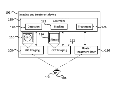
[0049] FIG. 1 depicts a system for the treatment of floaters;
[0050] FIG. 2 depicts a method for targeting a laser for use in the treatment
of floaters;
[0051] FIG. 3 depicts a floater detection process;
[0052] FIG. 4 depicts a further floater detection process;
[0053] FIG. 5 depicts a distributed system for the treatment of floaters;
[0054] FIG. 6 depicts a further method for targeting a laser for use in the
treatment of floaters;
[0055] FIG. 7 depicts a distributed system for the detection of floaters;
[0056] FIG. 8A depicts an image of an eye with a floater; and
[0057] FIG. 8B depicts the image of the eye of FIG. 8A with the floater
identified.
DETAILED DESCRIPTION
[0058] Floaters in a patient's eye may be tracked in real-time using a
combination of imaging
devices. A first imaging device, such as a scanning laser ophthalmoscopy (SLO)
imaging
device, may capture an image of the eye or portion of the eye within which a
floater is visible.
6
Date recue / Date received 2021-11-30
The image from the first imaging device may provide an X-Y image that allows a
position of
the floater to be partially determined, although no depth information about
the position of the
floater may not be determined by first imaging device. The X-Y position of the
floater may be
used to control an imaging location of a second imaging device capable of
capturing depth
information, such as an optical coherence tomography (OCT) imaging device. The
images
from the first and second imaging devices allow the 3D location of floaters
within the eye can
be determined. The imaging devices may capture images in real-time which may
allow the
tracking of floaters to be done in real-time. The tracking information can be
used for various
purposes including for example measuring details of the floater(s) as well as
possibly treating
the floater(s) with a laser.
[0059] FIG. 1 depicts a system for the treatment of floaters. The system
comprises an
imaging and treatment device 102 that can be used for imaging a patient's eye,
depicted as
eye 104. The patient's eye may have one or more floaters 106. The imaging and
treatment
device 102 is depicted a single device in FIG. 1, however it will be
appreciated that the
components may be provided in multiple separate devices. International patent
application
No. PCT/CA2021/051451 filed October 15, 2021 entitled "OPHTHALMOLOGICAL
IMAGING
AND LASER DELIVERY DEVICE, SYSTEM AND METHODS," which is incorporated herein
by reference in its entirety describes an imaging and treatment device that
could be used as
the imaging and treatment device 102. The imaging and treatment device 102
comprises an
SLO imaging device 108 that can capture a X-Y image 110 of the patients eye
and an OCT
imaging device 112 that captures a depth image 114 of the patient's eye. The
OCT imaging
device 112 may capture a depth 'slice' image at a particular horizontal
location in the eye.
Both of the imaging devices 108, 112 may be able to capture multiple frames of
images to
provide real-time images, or videos of the patient's eye.
[0060] Imaging and treatment device 102 may also include a treatment laser 116
that can be
targeted and fired at a particular location within the patient's eye, such as
at a floater. The
laser may be one of various known treatment lasers, including for example a
femtosecond
laser. Other lasers may be used including for example nanosecond lasers,
picosecond lasers,
microsecond lasers, milisecond lasers, or cw lasers. The SLO imaging device
108, the OCT
imaging device 110 and the treatment laser 116 can be calibrated so that all
of the coordinate
systems of devices are aligned and a location in one of the device's
coordinate system can
7
Date recue / Date received 202 1-1 1-30
be aligned with the same location in the coordinate system of the other
devices. Although not
depicted in detail in FIG. 1, it will be appreciated that each of the imaging
devices 108, 112 as
well as the treatment laser 116 will include an optical pathway and other
components, such
as light sources, sensors, etc. The optical pathways of the imaging devices
and treatment
later may include at least a portion of the optical pathways that are common
to all of the
devices. For example, the last portion of the optical pathway before the
patient's eye may be
common to all of the devices.
[0061] The imaging and treatment components 108, 112, 116 may be controlled by
a
controller 118 that is configured to provide various functionality including
floater detection
functionality 120, floater tracking functionality and floater treatment
functionality 124. The
floater detection functionality 120 uses image processing techniques to detect
floaters within
the SLO images. Floater detection can be difficult using current techniques.
Current object
detection techniques perform well when detecting object with relative sharp
edges. The
object detection techniques typically use kernels for feature
extraction/detection with a
relatively small kernel size, such as 3x3 or 4x4. The floaters in the captured
SLO images are
shadows of the actual floaters and typically do not include sharp edges. In
order to improve
the floater detection, the object detection may be modified to use relatively
large kernel sizes
of for example, 8x8, 16x16, 32x32, and larger.
[0062] Additionally, floater detection may be further complicated by other
features within the
image. For example, features such as veins within the eye may make the floater
detection
difficult. It is possible to identify the non-floater features within the
images and then remove
or mask those features from within the images prior to attempting to detect
the floaters. The
non-floater features may be detected using various image processing techniques
including
machine learning image classification techniques and/or object detection
techniques.
[0063] The controller 118 may further include floater tracking functionality
122. Regardless of
the particular details on the image processing used to detect floaters, once
detected the
floaters can be tracked across subsequently captured images. The tracking can
be done
using conventional image processing or tracking techniques such as optical
flow. These
conventional techniques may be modified to use additional information from
previous
tracking. For example, the floater tracking may be used to predict floater
locations in future
frames, with the predicted locations used to speed detection/tracking of the
floaters.
8
Date recue / Date received 202 1-1 1-30
[0064] The tracking functionality 122 may track the floater's X-Y position
across the SLO
images. The OCT image, or images may be used to track the depth, or Z,
position
information of the floater. The tracked X-Y position of the floater may be
used to control the
location that is imaged by the OCT imaging device. The OCT imaging device may
provide a
depth window that is insufficient to image the entire depth of the patient's
eye and as such
multiple OCT images may need to be captured covering different depths in order
to detect the
depth of the floater. Once detected, the depth of the floater may be tracked
and predicted.
The predicted floater depth location may be used to control, at least the
initial, imaging depth
of the OCT images to increase the likelihood that the floater is captured by
the OCT images.
Further, multiple OCT images of adjacent depth slices may be captured to
capture depth
information for the entire extent of the floater.
[0065] As described above, the SLO and OCT imaging devices 108,112 may be used
to
detect and track one or more floaters in both the X-Y image plane of the SLO
imaging device
as well as the X-Z, or depth, image plane of the OCT imaging device. It will
be appreciated
that reference to the X-Y and X-Z image planes are used only for explanation
and other
relative axes and coordinate systems could be used to provide information
about the physical
location of the floater. The tracked floater location may be used by treatment
functionality 124
of the controller to target the treatment laser 116 at an appropriate location
for treating the
floater with the laser. Prior to firing the treatment laser, it is possible
for the treatment
functionality 124 to verify the safety of the possible treatment location. For
example, if the
floater is in front of and close to the retina, it may be determined that
firing the treatment laser
pose too big of a risk for hitting the retina and so may not fire the laser.
Additionally or
alternatively, it is possible for the treatment functionality to adjust laser
parameters based on
a safety level of the treatment location. For example, if there are no other
features close to
the treatment location, it may be possible to increase a power level, or
firing duration of the
laser without causing risk to the patient's eye.
[0066] It will be appreciated that the detection, treatment and tracking of
floaters may be
performed repeatedly. That is, the detection process may be continually
performed in order
to detect floaters. Similarly the tracking process may be performed constantly
to continually
track floaters. Alternatively, the detection process may be performed
periodically to detect all
floaters and begin tracking the floaters. The periodic detection may be used
to update the
9
Date recue / Date received 202 1-1 1-30
tracking and/or detect new floaters. If the detection is performed
periodically, the detection
may be performed during the floater treatment which may break up the floater
into additional
smaller floaters.
[0067] FIG. 2 depicts a method for targeting a laser for use in the treatment
of floaters. The
method 200 begins with detecting a floater (202) in an image. The image
captures a plane of
the patient's eye, and may be for example a SLO image or a regular camera
image. The
floater detection from the SLO image identifies a location of the floater but
does not include
the depth information. Once the floater location is detected (202), its
position can be tracked
across multiple images (204). The floater tracking (204) can provide the
location, including
depth information, of the floaters. The floater tracking may use images
captured using both
the first imaging device (i.e. the SLO imaging device) and the second imaging
device (i.e. the
OCT imaging device). With the floater location tracked, the floater may be
treated (206) by
targeting a laser at the tracked location.
[0068] The floater detection (202) can be performed in various ways. For
example, as
depicted in FIG. 2, the detection may begin with detecting and removing, or
masking, non-
floater features in the SLO image (208). The non-floater features may be for
example veins or
other structures of the eyes. The non-floater features may be detected using
imaging
recognition functionality. The image with the non-floater features removed or
masked, may
be processed using machine learning (ML) object detection for detecting the
floaters (210).
The floater detection may be based on existing ML object detection processes,
which typically
rely on relatively small kernels for feature detection/identification. The ML
object detection
may be modified to use a relatively large kernel size, such as for example
16x16, 32x32 or
larger. The larger kernel size improves the detecting of floaters which do not
have well
defined edges in the images.
[0069] Once the initial location of a floater is detected in the SLO image,
its position may be
tracked across multiple frames of the SLO images. In addition to tracking the
position of the
floater in the SLO images, the tracking may also be performed on the OCT
images in order to
track the depth of the floater. The tracking may be performed in various ways.
As depicted,
the tracking may begin with stabilizing SLO image frames (212). The
stabilization may be
done by registering stationary features within the eye across different
frames. The floater
may be tracked across different frames of the stabilized images(214) using
known techniques
Date recue / Date received 202 1-1 1-30
such as optical flow. Further, the tracking may make use of previous tracking
information, for
example to predict a likely location of the floater in a current frame in
order to accelerate the
tracking process. With the location of the floater tracked in the SLO image
frames, the OCT
imaging location may be adjusted to capture depth strips at the floater
location (216). With
the OCT imaging location adjusted, the OCT imaging may capture one or more OCT
images
which may are then processed to determine the depth of the floater (218). The
OCT imaging
device may only be able to capture the depth slice images over a particular
window depth
size, which may not cover the entire depth of the patient's eye. Accordingly,
a single OCT
image may not capture the floater and as such the depth window may be adjusted
until the
floater is captured. The OCT imaging device may allow the depth of focus to be
adjusted in
order to change the window depth until the floater is detected in the OCT
image. The depth
of the floater may be used as a starting depth for subsequent OCT imaging.
[0070] With the depth and position of the floater tracked, the floater can be
treated (206).
Although the floater may be treated in various ways, as depicted, the
treatment may be
performed using a laser. The treatment includes targeting, including focusing,
the treatment
laser at the tracked position/depth of the floater (220). The safety of firing
the laser at the
target location may be verified (222) and assuming that the treatment location
is safe, the
laser may be fired at the floater (224) to break it up. Verifying the safety
of the target may
include determining the proximity to other features of the eye that could be
damaged by the
laser. If the features are within a path of the laser, or within a threshold
distance of the path
of the laser, the location may be deemed unsafe for treatment. As will be
appreciated,
floaters are moving within the eye and as such the tracking may continue unit
the floater is
determined to be in a 'safe' location for treatment. Verifying the safety of
the treatment
location may consider the treatment location relative to other features of the
eye as well as
possibly other factors such as the power and duration of the treatment laser.
[0071] FIG. 3 depicts a floater detection process. The process 300 uses a
convolutional
neural network (CNN) 302 to process a sequence of SLO images 304. The CNN 302
outputs
a sequence of masks 306 providing detected locations of floaters. The floaters
within the
captured images are typically out of focus, and more so the closer they are to
the front of the
eye, with very blurry edges and typically just vague gradients providing low
contrast.
Conventional image tracking and objected detection typically relies either on
(i) landmarks,
11
Date recue / Date received 2021-11-30
which are areas of high contrast to track over time or (ii) edges. In the
detection of floaters,
the areas of interest have very low contrast, even compared to other features
in the SLO such
as the optic disk, and also have no defined edges. Accordingly, the
conventional image
tracking processes tend to fail when detecting/tracking floaters.
[0072] The detection process 300 uses a convolutional neural network 302 in a
configuration
similar to U-Net. Rather than using as inputs the individual color channels of
an image such
as RGB, the input to the CNN 302 comprises an image with resolution Wi x Hi
with M
channels, where M is the number of frames in the SLO sequence. The input can
therefore be
considered the sequence of frames of one channel each as captured by the SLO
imaging
device. The output comprises of segmentation masks 306 showing the location of
floaters.
The output masks also have M channels, each with as resolution of W2 X H2
which need not
be the same as the input resolution Wi x Hi.
[0073] The CNN model may be trained on a collection of SLO image
sequence/videos in
which floaters have been labelled. The kernels of the convolutional layers may
have larger
sizes than typically found in CNNs such as 8x8,16x16, 32x32 to accommodate the
detection
of larger feature sizes specific to floaters.
[0074] FIG. 4 depicts a further floater detection process. To increase
accuracy of the floater
detection process 300 described above, as well as to have an adjustable
"sensitivity" metric,
the process 400 uses multiple image sequences 402 to identify floaters in a
single frame. For
example, to detect floaters on frame N=20, with a sequence length of M=6, the
floater
detection on frame 20 can be performed with frame sequences, include frame
sequences 17
to 22, frame sequences 18 to 23, etc. Each of these sequences will produce
floater mask
predictions for frame 20 using CNNs 404, which may be the same as that
described above in
FIG. 3. By predicting across some or all of the frame sequences which include
frame 20, a
number of prediction mask sequences 406 is obtained with each sequence
including a mask
for the frame of interest, i.e. frame 20. The masks of the frame of interest
can then be added
together 408. If, for example, 5 sequences of images were used, resulting in 5
different
prediction masks for frame 20, with each mask consists of values ranging from
0 to 1, the
sum of the masks will range from 0 to 5. A sensitivity threshold 410 can then
be specified
between 0 and 5 to fine tune performance parameters such as false positive
detection and
output the smoothed floater mask for the frame of interest 412.
12
Date recue / Date received 2021-11-30
[0075] The machine learning based floater detection may be combined with
classical tracking
method. After detecting the floater using a ML model as described above, the
predicted
location can be passed to a classical image processing-based approach for
object tracking
such as optical flow. The predicted motion of the classical image processing-
based object
tracker can be used to limit the search area for subsequent ML-based detection
of the floater.
Additionally or alternatively, after the classical image processing-based
object tracker is
activated on a detected floater, the ML-based detection method can
periodically be activated
to re-estimate the location of the floater and ensure continued tracking
accuracy.
[0076] FIG. 5 depicts a distributed system for the treatment of floaters. The
floater imaging
and treatment device described above has been described as having a single
controller that
detects, tracks and treats the floaters. The image processing techniques may
require a large
amount of processing to perform quickly enough to make the real-time tracking
and treating of
floaters possible and practical. The system 500 may use a remote server, or
other remote
processing device to provide the required processing requirements of the image
processing.
While the remote processing may be faster, or make possible improved image
processing,
the additional communication and possibly processing time, make it difficult
to provide real-
time detection and tracking of floaters. The system 500 described above makes
use of an
image buffer to make the detection/tracking possible. The system 500 is
similar to the floater
imaging and treatment device 102 described above, and as such similar elements
are not
described in further detail.
[0077] The system 500 may send the captured images to a remote server 528 via
a
communication network 530 for processing. The remote server 528 may provide
image
detection functionality 520, which may perform the floater detection and
returns the results
back to the imaging and treatment device 502. There may be a delay in
receiving the
detected floater location information from the remote server, which would make
the detected
location unsuitable for use in subsequent tracking in the most recent images.
In order to deal
with the delay, the device uses an image buffer that can temporarily store the
images
captured subsequent to sending the images to the remote server for detection.
Upon
receiving the detection results from the remote server 528, the buffered
images are used to
track the floater from detected location to the current image frames. The
controller 518 may
use tracking functionality 522 that may be substantially similar to the
tracking 122 described
13
Date recue / Date received 202 1-1 1-30
above; however the tracking may be performed on the buffered images. The
tracking may be
performed relatively quickly so that the tracking across the buffered images
can be 'fast-
forwarded', or performed faster than real-time, to the current frames and the
tracking
continued in real-time.
[0078] Although the above has described the detection as being done at a
remote server, a
similar buffering and fast-forward tracking may be used even if the detection
is not performed
at a remote server. That is, if the detection process performed takes a length
of time that
makes it difficult or impossible to use the detected location as a starting
point for tracking in
the current images, the same process of buffering images and then fast-
forwarding the
tracking of the detected location across the buffered images may be applied.
[0079] FIG. 6 depicts a further method for targeting a laser for use in the
treatment of floaters.
The method 600 may be used to track floater locations from an initial detected
location using
a detection process that may take a length of time that makes using the
detected location as
an initial tracking location difficult. The method 600 passes an initial
image, such as an SLO
image, to floater detection functionality (602). The floater detection
functionality may be
performed locally or remotely. While the initial floater location is
determined, newly captured
SLO image frames are buffered (604). The detected floater location is received
(606) and
then used as the initial location for tracking the floater location across the
buffered images
(608). The tracking of the floaters across the buffered images may be
performed relatively
quickly, allowing the tracking across the buffered images to catch up to the
currently captured
images.
[0080] FIG. 7 depicts a distributed system for the detection of floaters. The
system 700 is
similar to those described with reference to FIGs. 1 and 5. Similar features
and functionality
will not be described again in detail. The system 700 may include an imaging
and treatment
device 702 that includes the first (i.e. SLO) imaging device 108, and the
second (i.e. OCT)
imaging device 112; however, unlike the devices of FIGs. 1 and 5, the device
702 may omit a
floater treatment laser 116, and similarly the controller 718 may omit the
treatment
functionality 124. The controller may include local detection functionality
720a and possibly
local tracking functionality 722a that perform floater detection and tracking
respectively. The
local detection and local tracking may work in conjunction with, or be
replaced by, remote
detection functionality 720b, and remote tracking functionality 722b provided
by a remote
14
Date recue / Date received 202 1-1 1-30
server 728 in communication with the device 702 via a communication network
730. It will be
appreciated that although the server is remote from the device, it does not
need to be
physically distant from the device 702. The controller may also include an
image frame buffer
726 and a fast-forward tracking functionality 728 to track a floater from a
detected location
across buffered images in the buffer 726.
[0081] While the above has described tracking floaters and using the tracked
location for
targeting a treatment laser, it is possible to use the tracked floater
information for other
purposes. For example, the floater images and locations may be processed in
order to
identify and/or determine characteristics about the floater(s). This
information may include for
example a number of floaters, surface area of individual floaters, total
surface area of all
floaters, volume of individual floaters, total volume of all floaters,
locations of floaters, opacity
of floaters, refractive index of floaters, speed of movement of floaters,
direction of movement
of floaters, concentration of floaters, etc.. These characteristics may be
used for various
purposes including for example determining a severity of the patient's floater
condition,
determining a possible likelihood of successfully treating floaters with
lasers, etc.
[0082] FIG. 8A depicts an image of an eye with a floater. The captured image
is a single
frame image captured from an SLO imaging device. The image 800 includes at
least one
floater along with additional features of the eye, such as the retina, veins,
etc. FIG. 8B depicts
the image of the eye of FIG. 8A with the floater identified. The floater is
identified with a
bounding box 802. The location may be used to control the imaging location of
the OCT
imaging device. For example, depth slices may be captured by the OCT imaging
device
between the region identified by lines 804a, 804b.
[0083] It will be appreciated by one of ordinary skill in the art that the
system and
components shown in FIGs. 1 -8B may include components not shown in the
drawings. For
simplicity and clarity of the illustration, elements in the figures are not
necessarily to scale, are
only schematic and are non-limiting of the elements structures. It will be
apparent to persons
skilled in the art that a number of variations and modifications can be made
without departing
from the scope of the invention as defined in the claims.
[0084] Although certain components and steps have been described, it is
contemplated that
individually described components, as well as steps, may be combined together
into fewer
Date recue / Date received 202 1-1 1-30
components or steps or the steps may be performed sequentially, non-
sequentially or
concurrently. Further, although described above as occurring in a particular
order, one of
ordinary skill in the art having regard to the current teachings will
appreciate that the particular
order of certain steps relative to other steps may be changed. Similarly,
individual
components or steps may be provided by a plurality of components or steps. One
of ordinary
skill in the art having regard to the current teachings will appreciate that
the components and
processes described herein may be provided by various combinations of
software, firmware
and/or hardware, other than the specific implementations described herein as
illustrative
examples.
[0085] The techniques of various embodiments may be implemented using
software,
hardware and/or a combination of software and hardware. Various embodiments
are directed
to apparatus, e.g. a node which may be used in a communications system or data
storage
system. Various embodiments are also directed to non-transitory machine, e.g.,
computer,
readable medium, e.g., ROM, RAM, CDs, hard discs, etc., which include machine
readable
instructions for controlling a machine, e.g., processor to implement one, more
or all of the
steps of the described method or methods.
[0086] Some embodiments are directed to a computer program product comprising
a
computer-readable medium comprising code for causing a computer, or multiple
computers,
to implement various functions, steps, acts and/or operations, e.g. one or
more or all of the
steps described above. Depending on the embodiment, the computer program
product can,
and sometimes does, include different code for each step to be performed.
Thus, the
computer program product may, and sometimes does, include code for each
individual step
of a method, e.g., a method of operating a communications device, e.g., a
wireless terminal
or node. The code may be in the form of machine, e.g., computer, executable
instructions
stored on a computer-readable medium such as a RAM (Random Access Memory), ROM
(Read Only Memory) or other type of storage device. In addition to being
directed to a
computer program product, some embodiments are directed to a processor
configured to
implement one or more of the various functions, steps, acts and/or operations
of one or more
methods described above. Accordingly, some embodiments are directed to a
processor, e.g.,
CPU, configured to implement some or all of the steps of the method(s)
described herein.
16
Date recue / Date received 202 1-1 1-30
The processor may be for use in, e.g., a communications device or other device
described in
the present application.
[0087] Numerous additional variations on the methods and apparatus of the
various
embodiments described above will be apparent to those skilled in the art in
view of the above
description. Such variations are to be considered within the scope.
17
Date recue / Date received 2021-11-30