Note: Descriptions are shown in the official language in which they were submitted.
CA 03141875 2021-11-24
WO 2020/252070 PCT/US2020/037060
NASAL AND ORAL RESPIRATION SENSOR
CROSS-REFERENCE TO RELATED APPLICATIONS
[0001] The present application claims priority from U.S. Patent
Application No.
16/438,410, filed on June 11, 2019, and entitled NASAL AND ORAL RESPIRATION
SENSOR.
BACKGROUND
[0002] The present disclosure relates generally to medical sensors.
More particularly,
the present disclosure relates to respiration sensors for a continuous, long-
lasting monitoring of an
individual or patient, including measuring and analyzing respiratory condition
and movement of
the person.
[0003] The respiration of a person may be monitored for various
reasons. For example,
knowledge about a patient's respiration may assist a caregiver in assessing
the patient's stability
during surgery and recovery thereafter. Knowledge about a person's respiration
can also assist
with therapy related to sleeping.
[0004] Many approaches to respiration sensors involve cumbersome
devices that can
obstruct a patient's respiratory passages. In many applications, the patient
is unconscious or semi-
conscious and there is a challenge to fix a respiration sensor in place for an
extended period of
time. Accordingly, in many of the existing systems a nurse is required to
frequently check the
patient for sensor placement or inadvertent sensor movement. Moreover, due to
the physiognomy
of the human respiratory passages, many devices tend to produce confused
readings relative to
either of a patient's nostrils and mouth, and fail to clearly distinguish and
provide differentiated
data for inspiration and exhalation steps.
1
CA 03141875 2021-11-24
WO 2020/252070 PCT/US2020/037060
SUMMARY
100051 In the field of medical care for patients with respiratory
dysfunction, it is highly
desirable to provide continuous, real-time measurement of the patient's
respiratory cycles. In the
measurement of respiratory cycles from patients, one of the challenges is to
clearly distinguish
between inhalation and exhalation cycles. The complication is compounded by
the human
physiognomy, which places nasal and oral flows (in and out of the patient) in
close proximity to
each other, thereby increasing the possibility of flow mix, turbulence, and
stagnation in some
places.
[0006] An aspect of the present disclosure provides, but is not limited
to, a respiration
sensor for monitoring and analysis of an individual or patient's respiratory
condition and cycle,
monitoring and analysis to ensure a respiration sensor is positioned as
intended, detecting
movement of a person using a respiration sensor, detecting and distinguishing
between oral and
individual nasal air flows, and integration of a respiration sensor and data
with a network.
[0007] In some embodiments, the present disclosure provides a
respiration sensor
comprising: a housing having a nasal flow passage that extends therethrough,
wherein the nasal
flow passage is disposed approximately parallel with a nasal respiratory flow
direction; and an
electronics board comprising a nasal thermistor, the electronics board coupled
to the housing such
that the nasal thermistor is positioned into the nasal flow passage.
[0008] In some embodiments, a respiration sensor is disclosed, the
respiration sensor
comprising: a housing having a first nasal flow passage and a second nasal
flow passage that extend
therethrough, wherein the nasal flow passages are disposed in parallel to one
another with respect
to a nasal respiratory flow direction; and an electronics board comprising a
first nasal thermistor
and a second nasal thermistor, the electronics board coupled to the housing
such that the first and
second nasal thermistors are positioned into each of the first and second
nasal flow passages,
respectively.
100091 In some embodiments, the present disclosure provides a
respiration sensor
comprising: one or more thermistors configured to detect at least one of an
inspiratory temperature,
an expiratory temperature, an ambient temperature adjacent the respiratory
sensor, or a
2
temperature of a patient's skin engaged against the respiratory sensor; an
accelerometer configured
to detect at least one of a movement of the patient, a position of the
patient, a heart rate, or a
respiration rate; and an electronics board coupled to the one or more
thermistors and the one or
more thermistors.
[0010] In
some embodiments, the present disclosure provides a system, comprising: a
server having a memory storing commands, and a processor configured to execute
the commands
to: receive, from a hub, a data indicative of a respiratory condition of a
patient; transfer the data
into a memory in a remote server; provide the data to a mobile computer
device, upon request; and
instruct the mobile computer device to graphically display the data, wherein
the data comprises a
temperature value from at least one of two nasal flow passages, a temperature
value from an oral
flow passage, a temperature value of a patient's skin surface, and a
temperature value of a patient's
environment.
[0011] In
some embodiments of the present disclosure, a method is disclosed, the
method comprising: receiving, from a hub, a data indicative of a respiratory
condition of a patient;
transferring the data into a memory in a remote server; providing the data to
a monitor, upon
request; and instructing the monitor to graphically display the data, wherein
the data comprises a
temperature value from at least one of two nasal flow passages, a temperature
value from an oral
flow passage, a temperature value of a patient's skin surface, and a
temperature value of a patient's
environment.
[0012] In
some embodiments a respiration sensor system is disclosed, the respiration
sensor system comprising: a respiration sensor comprising a housing having a
nasal flow passage
that extends therethrough, wherein the nasal flow passage is aligned with a
nasal respiratory flow
direction, and an electronics board comprising a nasal thermi star, the
electronics board coupled to
the housing such that the nasal thermistor is positioned into the nasal flow
passage; and a hub
configured to move data between the respiration sensor and a network.
[0012a] In
accordance with an aspect of an embodiment, there is provided a method
comprising: measuring, by a respiration sensor device, a respiration rate of a
patient proximate to
the respiration sensor device; responsive to measuring the physiological
parameter, automatically
broadcasting, by the respiration sensor device, a wireless advertisement
signal configured to
3
Date Recue/Date Received 2023-02-03
facilitate a pairing process between the respiration sensor device and a first
monitoring device;
receiving, by the respiration sensor device after broadcasting the wireless
advertisement signal, a
wireless request to perform the pairing process between the respiration sensor
device and the first
monitoring device; and automatically completing the pairing process between
the first sensor
device and the first monitoring device responsive to receiving the wireless
request.
[0012b] In accordance with another aspect of an embodiment, there is
provided a
method, comprising: receiving a patient identifier of a first patient;
automatically initiating, by a
monitoring device, responsive to receiving the patient identifier, a pairing
process to
communicatively couple the monitoring device with a respiration sensor device
from a plurality
of sensor devices; detecting, after the initiation of the process, a wireless
advertisement signal from
the respiration sensor device, the wireless advertisement signal indicating
that the respiration
sensor device has received a respiration rate from a patient and is ready to
be paired to the
monitoring device; pairing, responsive to the detecting, the monitoring device
with the respiration
sensor device; and receiving, responsive to the pairing, from the respiration
sensor device, the
respiration rate, the respiration rate being detected by the respiration
sensor device prior to the
monitoring device and the respiration sensor device being connected.
[0012c] In accordance with another aspect of an embodiment, there is
provided a
system, comprising: a first monitoring device; and a respiration sensor
device, the respiration
sensor device comprising a memory and one or more processors configured to
execute instructions
stored on the memory to cause the respiration sensor device to: measure a
respiration rate of a
patient proximate to the respiration sensor device; automatically broadcast,
responsive to
measuring the respiration rate, a wireless advertisement signal configured to
facilitate a pairing
process between the respiration sensor device and the monitoring device;
receive, after
broadcasting the wireless advertisement signal, a wireless request to perform
the pairing process
between the respiration sensor device and the first monitoring device; and
automatically complete
the pairing process responsive to receiving the wireless request.
3a
Date Recue/Date Received 2023-02-03
[0013]
Additional features and advantages of the subject technology will be set forth
in the description below, and in part will be apparent from the description,
or may be learned by
practice of the subject technology. The advantages of the subject technology
will be realized and
3b
Date Recue/Date Received 2022-08-31
CA 03141875 2021-11-24
WO 2020/252070 PCT/US2020/037060
attained by the structure particularly pointed out in the written description
and embodiments hereof
as well as the appended drawings.
[0014] It is to be understood that both the foregoing general
description and the
following detailed description are exemplary and explanatory and are intended
to provide further
explanation of the subject technology.
BRIEF DESCRIPTION OF THE DRAWINGS
[0015] Various features of illustrative embodiments of the inventions
are described
below with reference to the drawings. The illustrated embodiments are intended
to illustrate, but
not to limit, the inventions. The drawings contain the following figures.
[0016] FIG. 1 illustrates a front perspective view of a respiration
sensor placed on a
patient's head, according to some embodiments.
[0017] FIG. 2A illustrates a side plan view of a gas flow exiting from
a patient's nasal
cavity, according to some embodiments.
[0018] FIG. 28 illustrates a side plan view of a gas flow exiting from
a patient's oral
cavity, according to some embodiments.
100191 FIG. 3 illustrates a front plan view of a gas flow exiting from
a patient's nasal
cavity, according to some embodiments.
10020] FIG. 4 illustrates a front perspective view of nasal respiration
flows and oral
respiration flows in a patient, according to some embodiments.
[0021] FIG. 5 illustrates a front perspective view of a respiration
sensor including nasal
flow passages and oral flow passages, according to some embodiments.
[0022] FIG. 6 illustrates perspective detail views of nasal flow
passages and oral flow
passages of a respiration sensor, according to some embodiments.
4
CA 03141875 2021-11-24
WO 2020/252070 PCT/US2020/037060
[0023] HG. 7 illustrates a cross-sectional view of a laminar
respiration flow relative to
a thermistor in a respiration sensor, according to some embodiments.
[0024] FIG. 8 illustrates a schematic view of a flow passage of a
respiration sensor,
according to some embodiments.
[0025] FIGS. 9A and 9B illustrate front and back perspective views of a
respiration
sensor, according to some embodiments.
[0026] FIG. 10 illustrates a front perspective view of a respiration
sensor placed on a
patient's head, according to some embodiments.
[0027] FIG. 11 illustrates a front perspective view of a respiration
sensor, according to
some embodiments.
[0028] FIG. 12 illustrates a bottom perspective view of the respiration
sensor of FIG.
11.
[0029] FIG. 13 illustrates a front perspective detail view of a
respiration sensor,
according to some embodiments.
[0030] FIG. 14 illustrates a front perspective cross-sectional view of
the respiration
sensor of FIG. 11.
100311 FIG. 15 illustrates a schematic view of a nasal respiration flow
and a nasal flow
guide, according to some embodiments.
[0032] FIG. 16 illustrates a schematic view of a nasal flow guide,
according to some
embodiments.
100331 FIG. 17 illustrates a back perspective view of a respiration
sensor, according to
some embodiments.
[0034] FIG. 18 illustrates a schematic view of a respiration flow
through a cavity of a
respiration sensor, according to some embodiments.
CA 03141875 2021-11-24
WO 2020/252070 PCT/US2020/037060
[0035] HG. 19 illustrates a schematic view of turbulent respiration gas
flow through a
cavity of a respiration sensor, according to some embodiments.
[0036] FIG. 20 illustrates a graph showing turbulent noise flow during
expiration,
according to some embodiments.
[0037] HG. 21A and 21B illustrate front and side plan views of exit
angles for a gas
flow exiting from a patient's nasal cavity, according to some embodiments.
[0038] FIG. 22A and 22B illustrate front and side schematic views of a
position of an
oral cavity relative to a patient, according to some embodiments.
[0039] HG. 23 illustrates a front perspective view of a respiration
sensor, according to
some embodiments.
[0040] FIG. 24 illustrates a graph showing measurements for the
respiration sensor of
FIG. 23 for different patients, according to some embodiments.
100411 FIGS. 25A and 25B illustrates a front plan views of positions of
an oral cavity
for a respiration sensor relative to a mouth of different patients, according
to some embodiments.
[0042] FIG. 26 illustrates a front perspective view of a respiration
sensor having a
strap, according to some embodiments.
[0043] FIG. 27 illustrates a front perspective view of a respiration
sensor having a
band, according to some embodiments.
[00441 FIG. 28 illustrates a front perspective exploded view of a
respiration sensor,
according to some embodiments.
100451 FIG. 29 illustrates a top perspective detail view of an
electronics board and
frame of a respiration sensor, according to some embodiments.
[0046] FIG. 30 illustrates a top perspective view of an electronics
board of a respiration
sensor, according to some embodiments.
6
CA 03141875 2021-11-24
WO 2020/252070 PCT/US2020/037060
[0047] HG. 31 illustrates a top perspective detail view of the
electronics board of FIG.
30, according to some embodiments.
[0048] FIG. 32 illustrates a bottom perspective view of the electronics
board of FIG.
30, according to some embodiments.
[0049] FIG. 33 illustrates a top perspective view of the electronics
board of FIG. 30
coupled with a frame and a battery, according to some embodiments.
[0050] FIG. 34 illustrates a block diagram of an electronics board of a
respiration
sensor, according to some embodiments.
[0051] FIG. 35 illustrates another block diagram of an electronics
board of a respiration
sensor, according to some embodiments.
[0052] FIG. 36 illustrates a respiration sensor detection state table
for determining the
respiration sensor placement and function, according to some embodiments.
[0053] FIG. 37 illustrates a graph showing breathing during changes in
ambient air
temperature, according to some embodiments.
[0054] FIG. 38 illustrates a graph showing breathing during conducting
temperature
changes, according to some embodiments.
100551 FIG. 39 illustrates a respiration sensor in use on a patient
transitioning from a
seated position, to a moving position, to a fallen position, according to some
embodiments.
10056] FIG. 40 illustrates a front plan view of a heart and directions
of blood circulation
therethrough.
100571 FIG. 41 illustrates a front plan view of a respiration sensor
including an
accelerometer for detecting body movement of a patient utilizing the
respiration sensor, according
to some embodiments.
7
CA 03141875 2021-11-24
WO 2020/252070 PCT/US2020/037060
[0058] FIGS. 42A and 42B illustrate a front perspective exploded view
of an example
respiration sensor having EtCO2 sensitive surfaces, and an example electronic
board electrically
coupled to the EtCO2 sensitive surfaces, respectively, according to some
embodiments.
[0059] FIG. 43 illustrates a schematic view of a respiration monitoring
system,
according to some embodiments.
[0060] FIG. 44 illustrates a front perspective view of a respiration
sensor coupled to a
patient and a hub adjacent to the patient according to some embodiments.
10061] FIG. 45 illustrates a front perspective view of a respiration
sensor and hub
coupled to a patient, according to some embodiments.
[0062] FIG. 46 illustrates perspective detail views of an interaction
between a
respiration sensor and a hub in a respiration monitoring system, according to
some embodiments.
[0063] FIG. 47 illustrates a front perspective view of a respiration
sensor and hub
coupled with a headdress, according to some embodiments.
[0064] FIG. 48 illustrates a side view of a respiration sensor and hub
coupled with
another headdress, according to some embodiments.
[0065] FIG. 49 illustrates a front perspective view of a smartphone as
a monitor for a
respiration monitoring system, according to some embodiments.
[0066] FIG. 50 illustrates a front perspective view of a central
station as another
monitor for a respiration monitoring system, according to some embodiments.
[0067] FIG. 51 is a flow chart of an example method of a sensor device
detecting a
physiological parameter and initiating a pairing process with a monitoring
device, according to
illustrative implementations.
[0068] FIG. 52 is a flow chart of an example method of a sensor device
determining a
color for the sensor device, according to illustrative implementations.
8
CA 03141875 2021-11-24
WO 2020/252070 PCT/US2020/037060
[0069] FIG. 53 is a flow chart of an example method of a sensor device
associating a
new monitoring device with a patient, according to illustrative
implementations.
[0070] FIG. 54 is a flow chart of an example method of a pairing
process with a sensor
device at a monitoring device, according to illustrative implementations.
[0071] FIG. 55 is a flow chart of an example method of a monitoring
device
determining a color for a patient, according to illustrative implementations.
[0072] FIG. 56 is a flow chart of an example method of detecting speech
by a patient,
according to illustrative implementations.
[0073] FIG. 57 is a flow chart of an example method of displaying a
position of a sensor
device on a patient, according to illustrative implementations.
[0074] FIG. 58 is a flow chart of an example method of displaying
movement of a
sensor device on a patient in real-time in a user interface, according
illustrative implementations.
[0075] FIG. 59 is a flow chart of an example method of a monitoring
device detecting
a sleep apnea of a patient, according to illustrative implementations.
[0076] FIG. 60 is a flow chart of an example method of a monitoring
device
determining whether a patient is in compliance with instructions of a
clinician, according to
illustrative implementations.
[0077] FIG. 61 is a flow chart of an example method of a detecting
nasal cavity
conditions based on received breathing pattern data, according to illustrative
implementations.
[0078] FIG. 62 is a flow chart of an example method of adjusting a user
interface of a
monitoring device, according to illustrative implementations.
[0079] FIG. 63 is a flow chart of an example method of predicting a
likelihood of a
chronic obstructive pulmonary disease (COPD) exacerbation, according to
illustrative
implementations.
9
CA 03141875 2021-11-24
WO 2020/252070 PCT/US2020/037060
[0080] HG. 64 is a flow chart of an example method of determining an
activity level
of a patient and associating the activity level with one or more baseline
physiological values,
according to illustrative implementations.
10081] FIG. 65 is a flow chart of an example of determining a path
traveled by the
patient and associating the path with one or more baseline physiological
values, according to
illustrative implementations.
[0082] FIG. 66A illustrates an example graphical representation of a
bounded area
displaying graphical representations of paths travelled by a patient,
according to illustrative
implementations.
10083] FIG. 66B illustrates an example graphical display of real time
measurements
for indicating whether a patient is likely to experience a health event,
according to illustrative
implementations.
[0084] FIG. 66C illustrates another example graphical display of real
time
measurements for indicating whether a patient is likely to experience a health
event, according to
illustrative implementations.
[0085] FIG. 67 illustrates a side perspective detail view of an
electronics assembly of
a sensor device, according to illustrative implementations.
[0086] FIG. 68 illustrates a side perspective detail view of an
electronics board of a
sensor device, according to illustrative implementations.
[0087] FIG. 69 illustrates a side perspective detail view of an
electronics board of a
sensor device, according to illustrative implementations.
[0088] In the figures, elements having the same or similar reference
numeral have the
same or similar functionality or configuration, unless expressly stated
otherwise.
CA 03141875 2021-11-24
WO 2020/252070 PCT/US2020/037060
DETAILED DESCRIPTION
10089] It is understood that various configurations of the subject
technology will
become readily apparent to those skilled in the art from the disclosure,
wherein various
configurations of the subject technology are shown and described by way of
illustration. As will
be realized, the subject technology is capable of other and different
configurations and its several
details are capable of modification in various other respects, all without
departing from the scope
of the subject technology. Accordingly, the summary, drawings, and detailed
description are to
be regarded as illustrative in nature and not as restrictive.
[0090] The detailed description set forth below is intended as a
description of various
configurations of the subject technology and is not intended to represent the
only configurations
in which the subject technology may be practiced. The appended drawings are
incorporated herein
and constitute a part of the detailed description. The detailed description
includes specific details
for the purpose of providing a thorough understanding of the subject
technology. It will be
apparent, however, to one ordinarily skilled in the art that the embodiments
of the present
disclosure may be practiced without some of these specific details. In other
instances, well-known
structures and components are shown in block diagram form in order to avoid
obscuring the
concepts of the subject technology, or have not been shown in detail so as not
to obscure the
disclosure. Like components are labeled with similar element numbers for ease
of understanding.
[0091] In accordance with at least some embodiments disclosed herein is
respiration
sensor that can: monitor nasal and oral respiration gas flow; monitor patient
and ambient
conditions; monitor movement of the respiration sensor; distinguish between
oral and nasal air
flow, and between left and right nasal air flow. The respiration sensor can
identify and analyze
thermal transfer distinction between inhalation and exhalation gases to
provide a clear pattern of
the respiratory cycle.
[0092] In at least some embodiments disclosed herein, any of nasal and
oral respiration
gas flow, heart rate, respiration rate or cycle, and movement of the
respiration sensor and patient
are determined. Embodiments of the present disclosure can send and receive
data related to the
monitoring and analysis by the respiration sensor; indicate a patient's
condition or position; and
provide a signal or alarm corresponding to specific conditions. In some
embodiments, wireless
11
CA 03141875 2021-11-24
WO 2020/252070 PCT/US2020/037060
communication techniques are utilized to provide ubiquitous solutions for
respiratory sensing of
patients in hospitals, treatment facilities, home-care situations, and the
like.
I. Embodiments of Respiratory Sensors
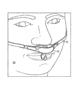
10093.1 FIG. 1 illustrates a respiration sensor 10 placed on a patient's
head 20,
according to some embodiments. The respiration sensor 10 is positioned on
patient's face between
the mouth and nose to measure nasal and oral breathing gas flow. The gas flow
measurement is
based on measuring temperature differences between inspiratory and expiratory
gas flows.
Patient's skin and ambient air temperatures can also be measured to verify
that the respiration
sensor 10 is placed appropriately against the patient. Some embodiments, later
described, include
other sensors, such as capacitive sensors or detectors and accelerometers to
ensure that respiration
sensor 10 has not fallen out of place, and that respiration sensor 10 is
making proper contact with
the patient's physiognomy. A securement string or strap 15 helps maintain the
position of
respiration sensor 10 relative to the patient's physiognomy.
100941 FIGS. 2A and 3 illustrate regions 200a, 200c for a gas flow
exiting from a
patient's nasal cavities, and FIG. 2B illustrates regions 200b for a gas flow
exiting from a patient's
oral cavity, according to some embodiments. Experiments show that breathing
gas flow exits nasal
and oral cavities in different regions between different subjects.
Accordingly, embodiments of a
respiration sensor as disclosed herein include a geometry that may separate
each of the different
flows through the regions 200a, 200b, 200c to provide a more accurate measure
of the respiratory
cycles of a patient. Accordingly, a precise determination of the positioning
of the respiration
sensor 100a, 100b relative to the patient's face is highly desirable.
100951 FIG. 4 illustrates a portion of nasal respiration flow 600C and
oral respiration
flow 600B for a patient 20, according to some embodiments. Sensor cavities of
the respiration
sensor capture nasal and oral breathing gas flow from the patient. The sensor
cavities are
positioned parallel to the average direction of that specific flow to maintain
flow as laminar as
possible inside the cavity. Thus, nasal sensor cavities are positioned
parallel to each other between
the nose and mouth, but also parallel to upper lip. More advantageously, nasal
sensor cavities
slightly diverge past the middle part of the mouth and upper lip into the
average direction of nasal
breathing gas flows. An oral sensor cavity is positioned transverse to the
nasal cavities, outwards
12
CA 03141875 2021-11-24
WO 2020/252070 PCT/US2020/037060
from the mouth. In some embodiments, the oral sensor cavity and the nasal
sensor cavities are
positioned relative to each other so that a direction of oral respiration flow
600B through the oral
cavity is transverse relative to a direction of nasal respiration flow 600C
through any of the nasal
sensor cavities. Sensor cavities are also smooth and straight, or more
advantageously slightly
tapered, to capture flow from a larger area, since any turn or sudden change
in the cross-section of
cavity along the flow path generate turbulences that mix inspiratory and
expiratory air flow phases
degrading the measurement speed, accuracy and response time.
[0096] FIG. 5 illustrates a respiration sensor 100a, for example,
including nasal flow
passages 301 and an oral flow passage 302. The nasal respiration flow exiting
a patient's nasal
cavity, e.g., gas flow regions 200a, 200c, can be captured and guided by a
nasal passage 301
parallel to average direction of nasal respiration flow 600C. Similarly, the
oral respiration flow
exiting a patient's mouth, e.g., gas flow region 200b, can be captured and
guided by the oral cavity
302 parallel to direction of the oral respiration flow 600B. By providing a
sensing element inside
of each of the different flow passages 301 and 302, the respiration sensor
100a may accurately
determine a respiration flow before the nasal flow and the oral flows are
mixed together adjacent
the patient's upper lip.
[0097] FIG. 6 illustrates the respiration sensor 100a, with portions
thereof shown in
detail views, including detail views of the nasal flow passages 301 and the
oral flow passage 302.
The respiration sensor 100a includes thermistors 400-1, 400-2, 400-3 for
sensing inhalation and
exhalation flows. A nasal respiratory flow of a patient can be captured by the
nasal passages 301
and measured with a first and second nasal thermistors 400-1, 400-2 therein.
An oral respiratory
flow of the patient can be captured by the oral cavity 302 and measured with
thermistor 400-3
therein. The resistance of each thermistor changes proportionally to flowing
gas heating or cooling
down the thermistor, e.g., during inspiration and expiration.
[0098] Moreover, the nasal flow passages 301 are separated from each
other such that
nasal thermistors 400-1 and 400-2 may separately identify and measure the
respiration flow
associated with each of the patient's nostrils. By separately identifying
respiration flow associated
with each of the patient's nostrils, potential respiratory conditions or
patient's positions can be
13
CA 03141875 2021-11-24
WO 2020/252070 PCT/US2020/037060
determined. For example, a blockage of a nasal passage or the respiration
device can be identified
and corrected.
[0099] In some embodiments, oral thermistor 400-3 is placed on a plane
that is
transverse or substantially perpendicular to nasal thennistors 400-1, 400-2.
This geometry also
enables an accurate and independent measurement between each of the themnstors
400-1, 400-2,
400-3, avoiding any mixing or turbulent area.
1001001 Referring to FIGS. 7 and 8, the thermistors 400-1,400-2, 400-3 can be
located
approximately in the middle of its corresponding sensor cavity to maximize
accuracy and
sensitivity to gas flows. To position the thermistor 400-1, 400-2, 400-3 in
the middle of a
corresponding sensor cavity, the thermistor 400-1, 400-2, 400-3 is coupled to
a tip portion of a
thin support structure 730. The support structure can have a proximal portion
coupled to an
electronics board and a distal portion transverse to a plane defined by the
top of the electronics
board, wherein the distal portion of the support structure extends into a
nasal flow passage. In
some embodiments, the respiration sensor 100a has a structure and geometry
that separates the
nasal flow from each nostril separately, to provide a more accurate and
detailed picture of the
patient's respiratory condition.
1001011 FIG. 7 illustrates a cross-section of a laminar respiration flow of a
patient
through a sensor cavity. Laminar flow speed distribution in a tube is
parabolic, thus the speed is
maximum at a point approximately in the middle of the tube. The respiration
flow is illustrated
relative to thermistor 400-1 of a respiration sensor 100a, however, the
present disclosure can apply
to any thermistor 400-1, 400-2, 400-3. By placing thermistor 400-1 as close as
possible to the
middle of the flow cavity in the respiration sensor 100a, for example, a more
accurate measurement
is expected, as the velocity of the gas flow is highest at the center of the
flow cavity. Accordingly,
it is expected that a temperature differential between inhalation and
exhalation be highest at the
middle point of the flow cavity. Moreover, the convection or radiated thermal
energy from
surrounding structures is minimized when a thermistor 400-1, 400-2, 400-3 is
located into the
middle of cavity by a support structure 730.
[00102] FIG. 8 illustrates the respiration sensor 100a, with a portion thereof
shown in a
cross-sectional detail view. The cross-sectional view illustrates a support
structure 730 extending
14
CA 03141875 2021-11-24
WO 2020/252070 PCT/US2020/037060
into the nasal flow passage 301, and the thermistor 400-1 positioned at a
distal end portion of the
support structure 730. It should be understood that the present disclosure,
including support
structures, can apply to any of the thermistors 400-1, 400-2, 400-3 and flow
passages 301, 302.
1001031 The support structure 730 extends from a portion of the respiration
sensor 100a
into the nasal flow passage 301. It should be understood that the support
structure 730 can extend
partially into a flow passage 301, 302. For example, the support structure 730
can extend into a
mid-portion of at least one of the two nasal flow passages 301. In some
embodiments of the present
disclosure, the support structure 730 extends beyond or across the respective
flow passage 301,
302. The support structure 730 can comprise a cantilevered structure that
extends into a respective
flow passage 301, 302. However, in some embodiments, the support structure 730
can comprise
an arch structure partially extending away from an inner surface of the flow
passage 301, 302
toward the thermistor 400-1, and partially extending from the thermistor 400-1
toward the inner
surface of the flow cavity. In some embodiments, the support structure 730 and
the thermistor
400-1 can extend across inner surfaces of the flow cavity.
[00104] The respiration sensor 100a includes walls having an inner surface
forming the
sensor cavities. The walls of the cavity extend around at least a portion of
the thermistors 400-1,
400-2, 400-3. The walls protect the sensitive thermistors 400-1, 400-2, 400-3
from various
disturbing ambient gas flows causing error to measured breathing gas flow
signal, for example, a
caregiver being able to touch or breathe into thermistors or air conditioning
in proximity to the
thermistor 400-1, 400-2, 400-3. In addition, the walls forming the cavities
also protect small,
mechanically sensitive thermistors from various mechanical forces, stresses,
and shocks, such as
touching etc.
[00105] FIGS. 9A and 9B illustrate the respiration sensor 100a, for example,
including
thermistors 500-1, 500-2 for sensing the positioning of the sensor relative to
a patient's
physiognomy, according to some embodiments. An ambient thermistor 500-1 can be
positioned
along a front side of the respiration sensor 100a, adjacent a portion of the
respiration sensor 100a
that faces away from the patient when the respiration sensor 100a is worn by a
patient. Similarly,
a skin thermistor 500-2 can be positioned along a back side of the respiration
sensor 100a, adjacent
a portion of the respiration sensor 100a that faces toward the patient when
the respiration sensor
CA 03141875 2021-11-24
WO 2020/252070 PCT/US2020/037060
100a is worn by a patient. In some embodiments, when the respiration sensor
100a is worn by a
patient, the thermistor 500-1 is distal to the patient's face, and the
thermistor 500-2 is proximal to
the patient's upper lip and engaged against the patient's skin.
1001061 The respiration sensor 100a can include a passage or cavity along any
of the
front side or the back side thereof. The thermistor 500-1 can be positioned in
a cavity along the
front side of the respiration sensor 100a to measure ambient air temperature.
The thermistor 500-
2 can be positioned in a cavity along the back side of the respiration sensor
100a to measure the
temperature of patient's skin.
[00107] In some instances, thermistor 500-2 can detect when the sensor 100a is
properly
positioned on the patient while thermistor 500-1 can detect the temperature of
ambient air.
Comparison of temperatures from 500-1 and 500-2 can be used to indicate a
patient condition or
proper positioning and function of the sensor 100a, for example. In some
embodiments, when
thermistors 500-1 and 500-2 detect the same temperature, it may be assumed
that respiration sensor
100a is likely not attached to the patient, or that the patient's temperature
is the same as the ambient
temperature, which may indicate a hazardous health condition.
1001081 FIG. 10 illustrates another embodiment of a respiration sensor 100b,
which is
substantially similar to respiration sensor 100a. Respiration sensor 100b is
also placed on a
patient's face between the mouth and nose to measure nasal and oral breathing
gas flows. Much
like the respiration sensor 100a, the measurement is based on measuring
temperature differences
between inspiratory and expiratory gas flows. The patient's skin temperature
and the ambient air
temperature can also be measured to verify or detect that the respiration
sensor 100b is placed
appropriately with respect to the patient's nasal and oral breathing gas flows
and to the patient's
upper lip.
1001091 Some embodiments described herein include other sensors, such as
capacitive
detectors or sensors to detect whether the respiration sensor 100b is making
proper contact with
the patient's physiognomy and accelerometers to detect movement and position
of the respiration
sensor 100b to ensure, for example, that the respiration sensor 100b has not
fallen out of place,
that the patient has not fallen down, or that the orientation of the patient's
head is not obstructing
the nasal and oral breathing gas flows (e.g., patient's face is downward
towards pillow or bed).
16
CA 03141875 2021-11-24
WO 2020/252070 PCT/US2020/037060
[001101 A string or strap 150b helps maintain the position of the
respiration sensor 100b
relative to the patient's physiognomy. According to some embodiments, the
respiration sensor
100b can include a nasal flow guide 160 to concentrate and provide laminar
inspiratory and
expiratory gas flows through the respiration sensor 100b.
[001111 FIGS. 11 and 12 illustrate the respiration sensor 100b having a
housing 2001, a
base 2010, and a shroud 2012. The shroud 2012 is positioned between the
housing 2001 and the
base 2010 to form at least a portion of a cavity. The respiration sensor 100b
includes nasal flow
passages 2018, which are similar to nasal flow passages 301, and an oral flow
passage 2016, which
is similar to oral flow passages 302 of respiration sensor 100a. The nasal
flow passages 2018
extend from a top portion to a bottom portion of the respiration sensor 100b.
In use, a nasal
respiration flow from a patient's nose can move between the nasal inlet 2024
and the nasal outlet
2026 of each of the nasal flow passages 2018. The nasal inlet 2024 of each of
the nasal flow
passages 2018 is where the breathing gas flows into the respiration sensor
100b during expiration.
The nasal outlet 2026 of each of the nasal flow passages 2018 is where the
ambient air flows into
the respiration sensor 100b during inspiration.
[00112] The shroud 2012 includes a battery frame 2014, which extends away from
a
front surface of the shroud 2012. The battery frame 2014 encloses a battery,
securing it to the base
2010 and divides the area between the shroud 2012 and the housing 2001 into
two distinct nasal
flow passages 2018, such that the nasal thermistor 400-1 is centrally disposed
in one of the nasal
flow passages 2018 and the nasal thermistor 400-2 is centrally disposed in the
other one of the
nasal flow passages 2018. The battery frame 2014 is disposed substantially
centrally on the
respiration sensor I 00b and is arranged to be positioned under the septum of
a patient's nose when
the respiration sensor 100b is placed on or attached adjacent to the upper lip
of the patient.
[00113] Housing 2001 can be made of a paper battery engineered to use a spacer
formed
largely of cellulose that makes paper batteries flexible and environmentally-
friendly. The
functioning is similar to conventional chemical batteries with the important
difference that they
are non-corrosive and do not require extensive housing, but can function as
housing.
[00114] An oral shroud 2017 extends from the shroud 2012, and includes a
passage
through a distal portion thereof. The passage forms an oral flow passage 2016
having a thermistor
17
CA 03141875 2021-11-24
WO 2020/252070 PCT/US2020/037060
400-3 positioned therein. The oral flow passage 2016 is arranged such that the
oral thermistor
400-3 is centrally disposed within the oral flow passage 2016. In use, an oral
respiration flow from
a patient's mouth can move between the oral inlet 3036 and the oral outlet
2038.
1001151 FIG. 13 illustrates an embodiment the respiration sensor 100b, showing
the base
2010 and the shroud 2012, with the housing 2001 omitted for clarity. The
shroud 2012 encloses
an electronics board, securing it to the base 2010. The thermistors 400-1,
extend from the
electronics board through the shroud 2012. The thermistors 400-1, 400-2 are
oriented such that a
distal portion of the thermistors 400-1, 400-2 extend into a space forming the
nasal cavities 1301
when the shroud 2012 and the housing 2001 are coupled together.
1001161 The respiration sensor 100b can include a light-emitting diode (LED)
2013,
which is visible through the shroud 2012. The LED 2013 can provide a
confirmation or an
indication of status. For example, the LED 2013 can indicate when the
respiration sensor 100b is
paired with another device. In some embodiments, the LED 2013 can indicate any
of a charged
or low battery, an indication that the respiration sensor 100b is functioning
as intended, or an
indication that there is a detected problem with the respiration sensor 100b.
The LED 2013 can
be used to indicate the location of the patient for example in hospital PACU
where there are many
patients, respiration sensors and monitoring devices in the same room. The LED
2013 can be
turned on or display a series of blinks from the monitoring device to indicate
the location of the
patient and the connected respiration sensor. This may be important to ensure
that a caregiver is
looking at the correct monitoring device connected to patient and the
respiration sensor. It should
be understood that any embodiment of the respiration sensor, such as
respiration sensor 100a,
100b, can include an LED 2013.
1001171 In some embodiments, a spacer 2019 can be positioned between the
battery and
a battery contact. The spacer 2019 can maintain the battery contact spaced
apart from the battery,
thereby preventing electrical conduction therebetween. The spacer 2019 can
prevent discharge of
the battery before the respiration sensor 100b is intended to be used. When
the respiration sensor
100b is intended to be used, the spacer 2019 can be removed or separated from
the respiration
sensor 100b. In some embodiments, the respiration sensor 100b can comprise an
opening or
passage 2015 that extends between the battery and an outer surface of the
housing 2001 or the
18
CA 03141875 2021-11-24
WO 2020/252070 PCT/US2020/037060
shroud 2012. The spacer 2019 can be moved through the passage 2015 to separate
the spacer 2019
from the respiration sensor 100b. In some embodiments, the spacer 2019 may
comprise a plastic
material in the form or a strip or tape.
1001181 FIG. 14 illustrates an embodiment the respiration sensor 100b, showing
the base
2010, the shroud 2012, and the flow guides 160, with a portion of the housing
2001 and other
features omitted for clarity. At least one nasal flow guide 160 is disposed in
each of the nasal flow
passages 2018 and extends between the shroud 2012 and the housing 2001, as
shown in at least
FIGS. 11 and 12.
[001191 In some embodiments, at least one nasal flow guide 160 is disposed
proximate
a nasal inlet 2024 of each of the nasal flow passages 2018 and at least one
nasal flow guide 160 is
disposed within each of the nasal flow passages 2018. A nasal flow guide 160
can be positioned
in a nasal flow passage, proximate any of the nasal inlet 2024 and the nasal
outlet 2026. The nasal
flow guide 160 is aligned relative to the nasal thermistor 400-1 or 400-2 to
direct a flow of gas
toward relative to the nasal thermistor 400-1 or 400-2.
[001201 FIGS. 14 and 15 illustrates flow of gases relative to the respiration
sensor 100b,
a patient's nares, and the ambient environment. Arrows 2028 illustrate a
portion of nasal
respiration flow from a patient's nares toward the nasal thermistor 400-1, 400-
2 during expiration,
and arrows 2029 illustrate a portion of ambient gas directed from the ambient
environment toward
the nasal thermistor 400-1,400-2 during inspiration.
1001211 In more detail, during expiration, the at least one nasal flow guide
160, disposed
proximate the nasal inlet 2024 guides the breathing gas flow through the nasal
flow passages 2018
of the respiration sensor 100b and concentrates the breathing gas flow toward
each of the nasal
thermistors 400-1,400-2 while maintaining the breathing gas flow laminar as it
passes each of the
nasal thermistors 400-1, 400-2 to minimize turbulent noise. Similarly, during
inspiration, the at
least one nasal flow guide 160 disposed proximate the nasal outlet 2026 guides
the ambient air
flow through the nasal flow passages 2018 of the respiration sensor 100b and
concentrates the
ambient air flow toward each of the nasal thermistors 400-1,400-2 while
maintaining the ambient
air flow laminar as it passes each of the nasal thermistors 400-1,400-2 to
minimize turbulent noise.
19
CA 03141875 2021-11-24
WO 2020/252070 PCT/US2020/037060
[00122] The at least one nasal flow guide 160 can prevent undesired objects
from
entering the nasal flow passages 2018 and disturbing or breaking the nasal
thermistors 400-1,400-
2 and/or their associated support structures. The at least one nasal flow
guide 160 can also form
an air gap around the nasal thermistors 400-1, 400-2 with respect to the
housing 2001 and the at
least one nasal flow guide 160, which prevents electro static discharge (ESD)
from entering the
electronics board 300 via the nasal thermistors 400-1,400-2 and their
associated support structures.
[00123] In some embodiments, the at least one nasal flow guide 160 includes a
thickness
that is less than 1 mm and a height that is more than 2 mm. In some
embodiments, two or four
nasal flow guides 160 are disposed within each of the nasal flow passages 2018
proximate the
nasal inlet 2024 and/or two or four nasal flow guides are disposed within each
of the nasal flow
passages 2018 proximate the nasal outlet 2026. In some embodiments, the number
of nasal flow
guides 160 does not exceed five to allow for proper gas flow through the nasal
flow passages 2018.
[00124] FIG. 16 illustrates a schematic view of a nasal respiration flow guide
grid 2030.
The flow guide grid 2030 can function similarly to flow guide 160, wherein a
flow of gas through
a cavity of the respiration sensor 100b is directed by the flow guide grid
2030. The flow guide
grid 2030 can have walls which intersect and are transverse relative to each
other. In some
embodiments, a flow guide grid 2030 is disposed proximate the nasal inlet 2024
and a flow guide
grid 2030 is disposed proximate the nasal outlet 2026 of each nasal cavity of
the nasal flow
passages 2018.
[00125] Additional sensors of a respiration sensor 100b are illustrated in the
back,
perspective view of the respiration sensor 100b in FIG. 17. The respiration
sensor 100b includes
a thermistor 500-2 and a sensor 1401 located on the back portion of the
respiration sensor 100b.
The thermistor 500-2 can provide temperature information regarding the patient
or an ambient
environment adjacent to the back portion of the respiration sensor 100b. The
sensor 1401 is a
capacitive plate, which can engage against the patient. The sensor 1401 can
engage against a
region between a patient's upper lip and nose, e.g., an area including the
philtrum, and provide
information to determine a location of the respiration sensor 100b relative to
the patient's face.
CA 03141875 2021-11-24
WO 2020/252070 PCT/US2020/037060
Gas Flow Through Respiration Sensors
1001261 Referring to FIGS. 17 and 18, an oral shroud 2017 of the respiration
sensor
100b can have a cross-sectional area that tapers along a portion thereof or
relative to an oral inlet
2036 and an oral outlet 2038. The oral inlet 2036 is where the breathing gas
flows into the oral
flow passage 2016 of the respiration sensor during expiration, and the oral
outlet 2038 is where
the ambient air flows into the oral flow passage 2016 of the respiration
sensor during inspiration.
1001271 In some embodiments, as illustrated in FIG. 17, a cross-sectional area
of the
oral shroud 2017 forms an hourglass shape. For example, a cross-sectional area
of the oral shroud
2017 can taper from the oral inlet 2036 toward the oral thermistor 400-3,
positioned between the
oral inlet 2036 and the oral outlet 2038, and can taper away from the oral
thermistor 400-3 toward
the oral outlet 2038. In some embodiments, as illustrated in FIG. 18, the
cross-sectional area of
the oral shroud 2017 can taper from the oral inlet 2036 toward the oral outlet
2038. The cross-
sectional area of the oral shroud 2017 can also taper from the oral outlet
2038 toward the oral inlet
2036.
[001281 In some aspects, the oral shroud 2017 can have a cross-sectional
profile
transverse to a flow through the oral shroud 2017. The cross-sectional profile
of oral shroud 2017
can be any regular or irregular shape, such as an oval, circle, square, or
rectangle.
1001291 FIG. 18 illustrates a detail schematic view of the oral shroud 2017,
including an
oral flow guide 2034. The oral flow passage 2016 of the oral shroud 2017
collects the breathing
gas flow ejected from a patient's mouth. The cross-sectional area of the oral
shroud 2017 tapers
from the oral inlet 2036 toward the oral thermistor 400-3, and from the oral
thermistor 400-3
toward the oral outlet 2038. Alternatively, in some embodiments, the cross-
sectional area of the
oral shroud 2017 can taper from the oral outlet 2038 to the oral inlet 2036.
1001301 The oral flow guide 2034 can direct at least portion of oral
respiration flow 2032
moving through the oral flow passage 2016 of the oral shroud 2017. An oral
flow guide 2034 is
disposed proximate an oral inlet 2036 of the oral shroud 2017 and an oral flow
guide 2034 is
disposed proximate an oral outlet 2038 of the oral shroud 2017.
21
CA 03141875 2021-11-24
WO 2020/252070 PCT/US2020/037060
[00131] During expiration, the oral flow guide 2034 disposed proximate the
oral inlet
2036 guides the breathing gas flow through the oral flow passage 2016 and
concentrates the
breathing gas flow toward the oral thermistor 400-3 while maintaining the
breathing gas flow
laminar as it passes the oral thermistors 400-3 to minimize turbulent noise.
Similarly, during
inspiration, the oral flow guide 2034 disposed proximate the oral outlet 2038
guides the ambient
air flow through the oral flow passage 2016 of the respiration sensor and
concentrates the ambient
air flow toward the oral thermistor 400-3 while maintaining the ambient air
flow laminar as it
passes the oral thermistor 400-3 to minimize turbulent noise.
[00132] The oral flow guide 2034 extends from the oral shroud 2017 within the
oral
flow passage 2016. The oral flow guide 2034 can extend radially inward from an
inner surface of
the oral shroud 2017. The oral flow guide 2034 can extend across a portion of
the oral flow passage
2016, or across the oral flow passage 2016 to engage against an opposite inner
surface of the oral
shroud 2017. In some embodiments, the oral flow guide 2034 can extend between
the oral inlet
2036 and the oral outlet 2038. The oral flow guide 2034 can comprise a surface
that is any of a
planar, a convex, and a concave surface. In some embodiments, the oral flow
guide 2034 is
arranged horizontally. In some embodiments, an oral flow guide 2034 is
arranged horizontally
and another oral flow guide is arranged vertically.
[00133] FIG. 19 illustrates a schematic view of the oral flow passage 2016
including an
entry angle s that can create gas flow turbulence 2040. If the entry angle a
is too high, the oral
shroud 2017 will create the turbulence 2040 in both directions during
inspiration and expiration.
FIG. 20 illustrates turbulent noise flow turbulence 2040 during expiration,
which is represented by
expiration curve 2042 of the measured electrical signal from a thermistor,
such as thermistors 400-
1, 400-2, 400-3, 500-1, 500-2.
[00134] In some embodiments, a cross-sectional area of the oral inlet 2036
mimics a
dimension of a patient's open mouth during sleep, but is much less than a
fully open mouth and
less than a diameter of a patient's forefinger. In some embodiments, the oral
inlet 2036 is elliptical
in the vertical direction. In such embodiments, the height of the elliptical
oral inlet 2036 is
approximately 9mm and the width is less than the height, such as approximately
5nim. In some
embodiments, the oral outlet 2038 is elliptical. In some embodiments, the oral
outlet 2038 is
22
CA 03141875 2021-11-24
WO 2020/252070 PCT/US2020/037060
circular. In some embodiments where the both the oral inlet 2036 and the oral
outlet 2038 are
elliptical, the entry angle a is relatively small, such that less turbulence
is generated, but the gas
flow is less concentrated towards the oral thermistor 400-3. In some
embodiments, the oral outlet
2038 is approximately 5 mm.
[00135] Analysis of entry angle and turbulence generation in the flow cavity,
can also
be used with reference to the nasal flow passages 301, 2018. FIGS. 21A and 21B
illustrate
schematic views of possible nasal expiration flow angles, which can be used to
determine the
potential for turbulence 2040. For example, in a flow path to the side of the
nose, an angle a
determines a flow width W of the flow path and an angle 13 determines the
direction of the flow
path nose. The flow width W is the distance between flow paths from both
nostrils. A gas flow
column (referred to herein as GFC) is gas flow directed away from the face and
the nose. For
example, in a flow path directed away from the face and the nose, an angle y
determines a width
of the flow path and an angle 5 determines the direction of the flow path away
from the face and
the nose. An area CA defines the cross-sectional surface area of a nostril,
which affects the average
width of a GFC. In general, a smaller cross-sectional surface area CA of the
nostril generates a
narrower average width of the GFC. Moreover, turbulence 2040 may be created
around the
thermistors 400-1, 400-2 by narrow (e.g., low angle a and low cross-sectional
surface area CA)
breathing GFC that is far to the side of the nose (e.g., high angle (3).
111. Resniration Sensor Size and Adiustabilitv
1001361 FIGS. 22A, 22B, 23, and 24, illustrate potential distances or
dimensions of a
patient's facial features or structures, determination of potential dimensions
of the respiration
sensor using the measured and average patient facial features, and average
measurement results
for various patient's facial features or structures.
1001371 FIGS. 22A and 22B illustrate potential distances or dimensions of a
patient's
facial features relative to an oral cavity 2016 having a thermistor 400-3 when
the respiration sensor
is placed on or attached to the patient. More particularly, the identified
dimensions include the
patient's nose width Al, isthmus width Bl, a distance Cl between the bottom of
the nose and the
upper lip, a distance DI between the bottom of the nose and the oral passage
(e.g. mouth), a
23
CA 03141875 2021-11-24
WO 2020/252070 PCT/US2020/037060
distance El between the front edge of the nasal passage and the upper lip, and
a lip thickness F1,
e.g., the distance the lip protrudes outwardly relative to the philtrum.
[00138] FIG. 23 illustrates a respiration sensor, such as, for example, the
respiration
sensor 100a, 100b, depicting dimensions of the respiration sensor, which can
correspond to
analysis of the measured features of the patient as shown in FIGS. 22A and
228. Accordingly, the
measured facial features identified in FIGS. 22A and 22B help facilitate the
design dimensions of
the respiration sensor 100a, 100b. A2 should be at least Al, but preferably A2
is more than Al to
ensure capturing flow through the patient's nostrils. Similarly, B2 should be
no more than Bl, but
preferably B2 is less than B1 to ensure that B2 does not prevent or disturb
flow through the
patient's nostrils.
[00139] The measured facial features shown in FIGS. 22A and 228 can be used to
select
design dimensions of the respiration sensor shown in FIG. 23. In some
embodiments,
measurements of particular patient can be used to select design dimensions for
the respiration
sensor. In some examples, measurements of a group of patients, such as adults
or children, can be
used to select design dimensions for an adult respiration sensor or a child
respiration sensor.
1001401 A measured facial feature can correspond to a design dimension of the
respiration sensor. For example: a patient nose width Al can be used to select
the width A2 of the
respiration sensor; a patient isthmus width B1 can be used to select the
battery frame 2014 width
B2; the distance Cl between the bottom of the nose and the upper lip can be
used to select a height
C2 of the respiration sensor housing 2001; the distance D1 between the bottom
of the nose and the
oral passage can be used to select a distance D2 between the top of the
respiration sensor 100a,
100h, adjacent the nasal inlet 2024 and the oral flow passage 2016, 302; the
distance El between
the front edge of the nasal passage and the upper lip can be used to select a
depth of the respiration
sensor 100a, 100b; and the lip thickness Fl can be used to select a depth F2
of the oral flow passage
302, 2016.
1001411 In some embodiments, the distance C2 of the respiration sensor 100a,
100b is
less than 20 mm, but preferably less than 15 mm. In some embodiments, the
distance C2 of the
respiration sensor 100a, 100b is approximately 10 mm to accommodate different
face structures.
In some embodiments, width A2 of the respiration sensor 100a, 100b is more
than 25 mm, but
24
CA 03141875 2021-11-24
WO 2020/252070 PCT/US2020/037060
preferably about 45 mm to adequately capture the gas flow of patients with
large width A2. In
some embodiments, the distance D2 of the respiration sensor 100a, 100b is more
than 5 mm, but
preferably more than 10min. In some embodiments, the distance D2 of the
respiration sensor 100a,
100b is more than 15 mm to capture gas flow coming out from the nostrils. In
some embodiments,
the cross-sectional area of the nasal flow passages 301, 2018 is greater than
the cross-sectional
area of the nostrils of a patient to capture breathing gas flow. In some
embodiments, the battery
frame 2014 includes a dimension B2 corresponding to the isthmus width B1 and
is preferably less
than 10 mm, but more preferably less than 5 mm. In some embodiments, the oral
flow passage
2016 is located parallel to the breathing gas flow directed from the mouth of
the patient.
[00142] FIG. 24 illustrates a graph 2044 of average measurement results for
various
facial features of a sample of patients including the patient's nose width Al,
the isthmus width BI,
the distance Cl between the bottom of the nose and the upper lip, the distance
D1 between the
bottom of the nose and the oral passage (e.g. mouth), the distance El between
the front edge of
the nasal passage and the upper lip, and the patient's lip height FL The graph
2044 illustrates the
measurement results of a group of 45 Caucasian people including women, men,
and children
between the ages of 0 to 70 years old. The measured values influence the
dimensional designs of
the respiration sensor 100a, 100b with respect to the nose and the mouth
including the size of nasal
passages and the location of the oral passage. It should be understood that
measurements for
patients may also be outside of the scope of the measured feature in this
graph.
[00143] FIG. 25A illustrates a respiration sensor, such as, for example,
respiration
sensor 100b that includes the distance D2 between the top of the respiration
sensor 100b, adjacent
the nasal inlet 2024, and the oral flow passage 2016, 302. The distance D2 can
be approximately
equal to 15 mm for patients with a smaller distance Dl. Such a respiration
sensor can
accommodate patients including a distance D1 in the range of approximately 10
mm to 25 mm. In
some embodiments, the distance D1 is between approximately 5 mm to 50 mm.
[00144] FIG. 25B illustrates a respiration sensor, such as, for example, the
respiration
sensor 100b that includes the distance D2 approximately equal to 33 mm for
patients with a larger
distance Dl. Such a respiration sensor can accommodate patients including a
distance D1 in the
CA 03141875 2021-11-24
WO 2020/252070 PCT/US2020/037060
range of approximately 24 mm to 40 mm. In some embodiments, the distance Dl is
between
approximately 5 mm to 50 mm.
1001451 FIGS. 26 and 27 illustrate embodiments of features to attach the
respiration
sensor 100a to a patient. The features to attach the respiration sensor 100a
can include any of a
string, strap, or band, which can maintain a position of respiration sensor
100a relative to the
patient's physiognomy. It should be understood that any of the features to
attach the respiration
sensors 100a or 100b can include the features to attach the respiration sensor
to a patient.
1001461 A strap 150a, shown in FIG. 26, can have ends that are attached to the
respiration sensor 100a to form a loop. The strap 150a can have a length such
that the respiration
sensor 100a is engaged against a patient's face when the device is worn by the
patient In some
embodiments, an additional strap 150b extends from any of the strap 150a or
the respiration sensor
100a. The additional strap 150b can provide additional support and tension to
secure the device
with the patient The strap 150a and additional strap 150b can be configured
such that a portion
of the strap 150a extends above a patient's ears, and a portion of the
additional strap 150b extends
below a patient's ears.
1001471 FIG. 27 illustrates a respiration sensor 100a having a placement band
150c. In
some embodiments, the placement band 150c comprises a semi-rigid framework
that is configured
to guide straps that overlay the placement band 150c and extend over preferred
placement portions
of a patient's face. In some embodiments, the placement band 1 50c comprises a
flexible plastic
material that is configured to substantially retain its shape during use. The
flexible placement band
150c can move, in a first plane, towards or away from a patient's face. The
placement band 150c
can be moved or biased in the first plane to engage against the patient's face
and adapt to the shape
of the patient's face. The placement band 150c is less flexible relative to a
second plane, transverse
to the first plane, thereby preventing or resisting movement of the placement
band 150c along the
patient's face or twisting of the band 150c.
1001481 The placement band 150a, 150c can have a width that is approximately
5mm,
but it can be wider or narrower. A wider band can reduce the surface pressure
on the face by the
band. At least a portion of a surface of the band can be covered with a
material that is soft and/or
26
CA 03141875 2021-11-24
WO 2020/252070 PCT/US2020/037060
breathable. For example, a surface of the band configured to engage against
the face or skin of the
patient can comprise a cotton or similar material.
[00149] The shape of the band 1 50c is configured to extend from the
respiration sensor
100a, below the cheek bones of the patient. The band 150c can curve from the
area below the
cheek bones of the patient toward the patient's ears, forming a shape of an S-
curve or similar.
[00150] The band 150c can be coupled with one or more additional band and/or
strap.
For example, the band 150c can be coupled to any of straps 150a and 150b. When
the straps 150a,
150b pull the band 150c and respiration sensor 100a towards the patient's
face, a force vector of
the respiration sensor 100a is approximately straight, towards the face or
upper lip of the patient.
Accordingly, the band 150c can decrease the surface pressure against the
patient's isthmus or
another portions of the patient's face or lip.
IV. Respiration Sensor Features for Monitoring and Analysis
[00151] FIG. 28 illustrates an exploded view of the respiration sensor 100a,
100b for
example, including a housing 2001, shroud 2012, and electronics board 300,
according to some
embodiments.
[00152] The electronics board 300 includes the electronic components used in
the
respiration sensor 100a, 100b. The electronics board 300 can include a battery
1110 and sensors,
such as a thermistor 400-1, 400-2, 400-3, and a capacitive plate. In some
embodiments, the
electronics board 300 is made of, for example, glass-reinforced epoxy laminate
material (e.g., FR4
substrate) containing automatic machine placed components, commonly used in
automated mass
series production to make the construction low cost. The electronics board 300
can be coupled to
a base plate or frame 320. In some embodiments, the frame 320 includes
plastics, which contains
electrically conductive areas or conductors.
[00153] In some embodiments of the present disclosure, the battery 1110 can be
a
disposable or rechargeable battery. In some embodiments, the respiration
sensor 100a, 100b is
configured to be powered by solar energy. For example, the respiration sensor
100a, 100b can
include a solar panel which can be coupled to a battery.
27
CA 03141875 2021-11-24
WO 2020/252070 PCT/US2020/037060
[00154] The shroud 2012 defines at least a portion of the nasal flow passages
and the
oral flow passage of the sensor. In some embodiments, the electronics board
300 is positioned
between the frame 320 and the shroud 2012. Any of the frame 320 and the shroud
2012 can include
a cavity to protect the electronics board 300 when the respiration sensor
100a, 100b is assembled.
The frame 320 and/or the shroud 2012 can be made of elastic silicone,
plastics, or similar material.
1001551 In some embodiments, an ambient air thermistor is positioned further
away
from the breathing gas flow otherwise interfering ambient air measurement. In
aspects of the
present disclosure, the shroud 2012 can include a perforation 501 that enables
ambient air to be in
touch with the ambient air thermistor through the shroud 2012 to get fast
response time, but also
to protect ambient air thermistor for example from touching with a finger or
any unwanted air
flow, such as air conditioning.
1001561 Referring to FIG. 29, a portion of the frame 320 can form a support
structure
for the thermistors 400-1, 400-2. The electronics board 300 may include two
perforations that
enable two poles of the frame 320 to pierce through the electronics board 300
to form the
thermistor support structure. The poles locate and keep the board in place
with a mechanical
locking mechanism. No screws or similar are needed. The poles also contain
electrical contacts
on the tip of the poles where thermistors 400-1, 400-2, which are sensitive to
nasal breathing gas
flow, are coupled. Electrically conductive connections 1012 on the side
surfaces of poles further
connect thermistors 400-1, 400-1 to the electronics board 300 via electrical
contacts on the top
surface of the electronics board 300 next to the poles. When the frame 320 is
placed under the
electronics board 300, electrical contacts on the top surface of the frame 320
connect with adjacent
electrical contacts on the bottom surface of electronics board 300.
Electrically conductive glue
can be used to ensure electrical contact. In some embodiments, a thermistor
400-3 sensitive to
breathing gas flow through the mouth is located to the tip of the electronics
board 300. A bottom
side of frame 320, adjacent to electronics board 300 contains an inset 303 to
enable thermistor
400-3 to locate into the middle of the flow cavity.
[00157] Electrical signals from thermistors 400-1, 400-2, 400-3 proportional
to
corresponding ambient, skin, nasal or oral temperature changes are conducted
through the
electrically conductive connections 1012 and conductors to central processing
unit on the
28
CA 03141875 2021-11-24
WO 2020/252070 PCT/US2020/037060
electronics board. The central processing unit can convert the analog data
into digital form, process
and transmit the data wirelessly, for example, via an RF transmitter, to a
host where the data can
be shown or displayed to a caregiver in a suitable form of numbers and/or
waveforms.
1001581 FIG. 30 illustrates a detailed view of an electronics assembly 1200,
for example,
any respiration sensor 100a, 100b which can include two nasal flow thermistors
400-1, 400-2 and
one oral flow thermistor 400-3, according to some embodiments. Thermistors 400-
1 and 400-2
are configured to measure breathing from nostrils. Thermistor 400-3 may be
configured to
measure breathing from the mouth. A thermistor 500-1 (see FIG. 32) may also be
included in
electronics assembly 1200 to measure ambient temperature.
1001591 Support structures 1230-1, 1230-2, 1230-3 contain electrical wires on
both sides
of a strip between electrical connections at both ends of the strips. The
support structures can
include first and second support structures 1230-1, 1230-2, which can support
the nasal flow
thermistors 400-1,400-2. Additionally, a third support structure 1230-3 can
support the oral flow
thermistor 400-3. In some embodiments, support structures 1230-1, 1230-2, 1230-
3 may include
an electrically and thermally insulating material (e.g., FR4 substrate).
Thermistors 400-1, 400-2,
400-3 can be soldered to electrical connections in the first end of the
strips. Second ends of strips
are placed into small holes in electronics board 300 and soldered to form
electrical connections on
the sides of the strip to corresponding electrical contacts on the board to
electrically connect
thermistors 400-1, 400-2, 400-3 to sensor electronics in the plane of the
electronics board.
1001601 The cross-sectional areas of copper or similar traces within support
structures
1230-1, 1230-2, 1230-3 are reduced to minimize thermal flow through the
electrical conductors
from the plane of board to thermistors 400-1, 400-2, 400-3. To minimize the
thermal mass of the
thermistors 400-1, 400-2, 400-3, the support structures 1230-1, 1230-2, 1230-3
can be formed from
a thermally non-conductive or insulating material. These optimizations make
thermistors 400-1,
400-2, 400-3 as sensitive as possible to thermal changes caused by the
breathing gas flowing past
the thermistor during expiration or ambient gas flowing past the thermistor
during inspiration.
1001611 FIG. 31 illustrates a partial view 1300 of an electronics board 300
in, for
example, any respiration sensor 100a, 100b including details of a nasal flow
thermistor 400-1,
according to some embodiments. Support structure 1230-1 may include an F'R4
substrate strip
29
CA 03141875 2021-11-24
WO 2020/252070 PCT/US2020/037060
with thermistor 400-1 placed on the tip of the strip. At the bottom of support
structure 1230-1, a
soldered contact provides electrical contacts to thermistor 400-1 on both
sides of support structure
1230-1 (e.g., +/- terminals).
1001621 In some embodiments, the support structures 1230-1, 1230-2, can have a
proximal portion coupled to the electronics board 300 and a distal portion
transverse to a plane
defined by the top of the electronics board 300. When the electronics board is
positioned within
the housing, the distal portion of the support structures 1230-1, 1230-2 can
extend into respective
nasal flow passages. In some embodiments, the support structure 1230-3 can
have a proximal
portion coupled to the electronics board 300 and a distal portion that is
normal with or substantially
parallel to a plane defined by the top of the electronics board 300.
[00163] FIG. 32 illustrates a detailed view of a bottom portion of an
electronics board
300 of a respiration sensor 100a, 100b including a thermistor 500-1 to measure
skin temperature,
and a capacitive plate or sensor 1401 to measure sensor location in the upper
lip, according to
some embodiments. A respiration sensor including electronics board may be
turned on/off based
on the signal. Additionally, in some embodiments, the electronics board 300
includes an
accelerometer 1150.
[00164] FIG. 33 illustrates a detailed view of an electronics board 300
coupled with a
frame 320 and a battery 1110, according to some embodiments. Support
structures 1130-1 and
1130-2 extend away from the electronics board, and include thermistors 400-1
and 400-2,
respectively. A thermistor 500-1 sensitive to skin temperature may be located
on the bottom side
of the board as close to skin as possible. In some embodiments, the thermistor
is placed close to
one of the two ridges above the upper lip to ensure closest distance to skin.
The frame 320 most
advantageously contains a perforation adjacent to thermistor that enables
better thermal contact to
upper lip skin. Perforation can also be filled with thermally conductive
material to increase
conductivity to skin.
1001651 The electronics board 300 includes a battery contact tab 1111 that
extends
toward the battery 1110. A portion of the spacer 2019 is positioned between
the battery contact
tab 1111 and the battery 1110 such that the contact tab 1111 is spaced apart
from the battery 1110.
CA 03141875 2021-11-24
WO 2020/252070 PCT/US2020/037060
When the spacer 2019 is coupled with the respiration sensor 100b, the battery
1110 the battery
does not provide power to the respiration sensor 100b.
[00166] In some embodiments, the board 300 includes an LED 2013, which can be
visible from an outer surface of the respiration sensor 100b when the
respiration sensor 100b is
assembled. In some embodiments, the board 300 includes a microphone 2020. The
microphone
2020 can detect ambient sounds or a patient speaking. The sound detected by
the microphone
2020 can be used to during processing of signals. For example, the sound
detected by the
microphone 2020 can filtered out to reduce or remove noise in the signals from
the other sensors.
[00167] In some embodiments, any respiration sensor 100a, 100b is an
affordable,
disposable, wireless sensor configured to detect breath flow in real time.
Accordingly, the sensor
100a, 100b includes a battery 1110, which may provide several days (e.g., five
days, or more) of
continuous, real time, fast response operation with a high signal quality. In
some embodiments,
the respiration sensor 100a, 100b is configured to measure a respiration rate
(RR) and magnitude,
and to provide real time respiration waveforms, in digital and/or analog form.
Furthermore, a
processor circuit in the respiration sensor may be configured to determine
trends and projections
based on the real-time data (e.g., via moving averages, Kalman filtering, and
the like). The
respiration sensor 100a, 100b may also provide skin temperature, body
position, movement, fall
detection (e.g., through an accelerometer 1150), sensor placement, and the
like.
V. Processing of Readings for Indications
[00168] FIG. 34 illustrates a block diagram 1410 of components, which are
utilized on
the electronics board 300 of the respiration sensor 100a, 100b according to
some embodiments. In
such embodiments, the electronics board 300 includes a temperature-to-voltage
converter 1412,
an analog-to-digital (AD) converter 1414, a central processing unit (CPU)
1416, and a
communications module or radio transceiver 1418 for providing a two-way data
communication
coupling to a network link that is connected to a local network. Such
communication may occur,
for example, through a radio-frequency transceiver. In addition, short-range
communication may
occur, such as using a Bluetooth, Wi-Fi, or other such transceiver. In some
embodiments, the CPU
1416 includes the Bluetooth low energy processor 1160 (shown in FIG. 33).
31
CA 03141875 2021-11-24
WO 2020/252070 PCT/US2020/037060
[00169] In some embodiments, the temperature-to-voltage converter 1412
includes any
of the thermistor 500-1, the thermistor 500-2, the thermistor 400-1, the
thermistor 400-2, and the
thermistor 400-3. In some embodiments, any of the thermistors 400-1, 400-2,
400-3, 500-1, 500-
2 are negative temperature coefficient (NTC) type thermistors, such that the
thermistor's electrical
resistance decreases when the temperature increases. In some other
embodiments, any of the
thermistors are positive temperature coefficient (PTC) type thermistors, such
that the thermistor's
electrical resistance increases when the temperature increases. The
respiration sensor 100a, 100b
can include any combination of NTC type thermistors and PTC type thermistors.
The temperature-
to-voltage converter 1412 converts or transforms the temperature resistance
value detected at any
of the thermistors to a voltage at Vout 1420. The AD converter 1414 then
converts the Vout 1420
into digital form, which is received by the CPU 1416 for further processing
and calculations. In
some embodiments, the CPU 1416 can transmit the digital signal to the host
monitor or other client
device. The CPU 1416 can transmit the digital signal via the Bluetooth low
energy processor
1160. In some other embodiments, the CPU 1416 transmits the digital signal to
the
communications module or radio transceiver 1418 for wireless transmission to
the host monitor or
other client device.
[00170] In addition to the respiration sensor 100a, 100b measuring or
detecting
temperature differences between inspiratory and expiratory gas flows via the
thermistors 400-1,
400-2, 400-3, the respiration sensor 100a, 100b also measures or detects
ambient air temperature
via the thermistor 500-1 and conductive temperature from the patient's skin
via the thermistor 500-
2.
[00171] In some instances, the thermistor 500-1 and the thermistor 500-2
include a wide
operating temperature range and can be adjusted to include a lowest and a
highest temperature of
operating range. The respiration sensor 100a, 100b is configured to measure or
detect the electrical
signal voltages proportional to the ambient air temperature via the thermistor
500-1 and the skin
temperature via the thermistor 500-2 and compensate the signal offset, gain,
and the peak to peak
amplitude errors from the inspiratory and expiratory gas flow signal
amplitude.
[00172] In some embodiments, any of the thermistors 400-1,400-2, 400-3, 500-1,
500-
2 can measure any of an inspiratory gas flow, an expiratory gas flow, an
ambient air temperature,
32
CA 03141875 2021-11-24
WO 2020/252070 PCT/US2020/037060
and a conductive temperature. For example, when the respiration sensor 100a,
100b is turned on,
but is not yet placed on or attached to the patient's face, the thermistor 400-
1, 400-2, 400-3, 500-
1, 500-2 detect ambient air temperature. When the respiration sensor 100a,
100b is placed on or
attached to the patient's face, the thermistor 500-2 begins detecting the
temperature of skin on the
patient's upper lip. Meanwhile, the thermistor 500-1 remains detecting the
ambient air temperature
and the thermistors 400-1, 400-2, 400-3 begin detecting the temperature
differences between the
inspiratory and the expiratory gas flows (e.g., inspired ambient air and
expired warm gas coming
out from the lungs).
[00173] During normal, stable ambient conditions, after the respiration sensor
100a,
100b is placed on or attached to the patient's face, the electrical voltage
signals from the thermistor
500-2 (e.g., detecting ambient air temperature) are stable and change slowly,
whereas the electrical
voltage signal from at least one of the thermistors 400-1, 400-2, 400-3
changes its amplitude
relatively faster. In some embodiments where the thermistors are NTC type and
the temperature-
to-voltage converter 1412 includes negative feedback amplifiers, the
electrical voltage signal
changes between maximum voltage proportional to ambient air temperature and
minimum voltage
proportional to temperature of exhaled warm, moister gas coming out of the
patient's lungs.
1001741 In other embodiments where the thermistors 400-1, 400-2,400-3, 500-1,
500-2
are PTC type and the temperature-to-voltage converter 1412 include positive
feedback amplifiers,
the electrical voltage signal changes between maximum voltage proportional to
temperature of
exhaled warm, moister gas coming out of the patient's lungs and minimum
voltage proportional
to ambient air temperature. In both NTC type and PTC type scenarios, the
frequency of electrical
signal may vary between 0 to 3 Hz (0-180 RR/min) depending on how fast the
patient is inhaling
and exhaling. Smaller patients tend to breathe relatively faster than
relatively larger patients, such
as adults.
[001751 FIG. 35 illustrates a block diagram 1436 of components, which are
utilized on
the electronics board 300 of the respiration sensor 100a, 100b according to
some embodiments. In
such embodiments, the electronics board 300 includes a temperature-to-voltage
converter 1438, a
filter 1440, an analog-to-digital (AD) converter 1442, a central processing
unit (CPU) 1444, and a
communications module 1446 for providing a two-way data communication coupling
to a network
33
CA 03141875 2021-11-24
WO 2020/252070 PCT/US2020/037060
link that is connected to a local network. Such communication may occur, for
example, through a
radio-frequency transceiver. In addition, short-range communication may occur,
such as using a
Bluetooth, Wi-Fi, or other such transceiver. In some embodiments, the CPU 1444
includes the
Bluetooth low energy processor 1160 (shown in FIG. 33).
[001761 In some embodiments, the temperature-to-voltage converter 1438
includes any
of the thermistors 400-1, 400-2,400-3, 500-1, 500-2. In some embodiments, any
of the thermistors
are negative temperature coefficient (NTC) type thermistors, such that the
thermistor's electrical
resistance decreases when the temperature increases. In other embodiments, any
of the thermistors
are positive temperature coefficient (FTC) type thermistors, such that the
thermistor's electrical
resistance increases when the temperature increases. The respiration sensor
100a, 100b can
include any combination of NTC type thermistors and PTC type thermistors. The
temperature-to-
voltage converter 1438 converts or transforms the temperature resistance value
detected at one of
the thermistors 400-1, 400-2, 400-3, 500-1, 500-2 to a voltage at Vout 1448.
In some
embodiments, the temperature-to-voltage converter 1438 also includes an
amplifier 1451, which
increases the voltage at Vout 1448 for increased accuracy and resolution of
the breathing gas flow
signal.
[00177] The filter 1440 eliminates or subtracts any of the ambient air and the
conducting
skin temperature change from the breathing gas flow signal. The AD converter
1442 then converts
the signal from the filter 1440 into digital form, which is received by the
CPU 1444 for further
processing and calculations. In some embodiments, the CPU 1444 can transmit
the digital signal
to the host monitor or other client device via the Bluetooth low energy
processor 1160. In some
other embodiments, the CPU 1444 transmits the digital signal to the
communications module 1446
for wireless transmission to the host monitor or other client device. In some
embodiments, the
filter 1440 is configured to subtract the electrical signal detected by the
thermistor 500-2 from the
electrical signal detected by the thermistor 500-1. In some embodiments, the
filter 1440 is
configured to subtract the electrical signal detected by the thermistor 500-2
and the electrical signal
detected by any of the nasal thermistor 400-1, the nasal thermistor 400-2, and
the oral thermistor
400-3 from the electrical signal detected by the thermistor 500-1.
34
CA 03141875 2021-11-24
WO 2020/252070 PCT/US2020/037060
[00178] FIG. 36 illustrates a respiration sensor detection state table 1458
for
determining the respiration sensor placement and function. For example, an
operation logic is
derived from the electrical signals from the thermistor 500-1, the thermistor
500-2, and the
thermistor 400-1, 400-2, 400-3 to detect different states of the respiration
sensor 100a, 10013. The
different states of the respiration sensor 100a, 100b are utilized to identify
sensor placement with
respect to the patient and function of the sensor for monitoring and notifying
of these states. The
respiration sensor 100a, 100b is capable of identifying, for example, early
signs of respiratory
depression, spasms, obstructions, and other symptoms, and notifying of these
identifications. In
addition to notifying of such identifications, the respiration sensor 100a,
100b is also capable of
notifying when an improper placement of the sensor is identified or detected
to alert a caregiver to
check on the patient and make sure that the sensor is not obstructing the
patient's airways or
otherwise disturbing the patient.
[00179] The respiration sensor 100a, 100b includes various detection states
including,
but not limited to: a not-yet-placed state (Not Yet Placed state 1460); a
correctly-placed and
measuring state (Correctly Placed & Measuring state 1462); a correctly-placed,
but no breath state
(Correctly Placed, No Breath state 1464); a loose device state (Loose state
1466); a detached or no
breath state (Detached or No Breath state 1468), and an operating-temperature
exceeded state
(Operating Temperature Exceeded state 1470).
[00180] In the Not Yet Placed state 1460, the respiration sensor 100a, 100b is
not yet
placed on the patient. For example, when the respiration sensor 100a, 100b is
turned on, but not
yet placed on or attached to the upper lip of the patient, the thermistor 500-
2, the thermistor 500-
1, and the thermistor 400-3 all detect a similar signal corresponding to
temperature proportional to
ambient temperature and the breath indicator 1453a (shown in FIG. 35) detects
no breaths. Under
these detected conditions, the respiration sensor 100a, 100b determines that
it is in the Not Yet
Placed state 1460 and will not transmit an alert notification.
[00181] After the respiration sensor 100a, 100b is placed on or attached to
the upper lip
of the patient, the thermistor 500-2 detects and adapts to a temperature close
to skin temperature
of the upper lip while the thermistor 500-1 remains detecting the ambient air
temperature. In some
embodiments, the temperature offset error in the thermistor 500-2, which is
caused by, for
CA 03141875 2021-11-24
WO 2020/252070 PCT/US2020/037060
example, a mustache, may be ignored since the temperature detection is enough
to monitor the
temperature change during the time it takes to detect or determine whether the
sensor is in proper
placement or not (e.g., not the absolute value). When the location of the
sensor between the nasal
and the oral passages of the patient is proper and the patient is breathing
the thermistor 400-3 start
to adapt to and detect the temperature of the sequentially changing gas flow
(e.g., Breath). At this
point, the breath indicator 1453a determines that the thermistor 400-3
detected a breath. Under
these conditions, the respiration sensor 100a, 100b determines that it is in
the Correctly Placed &
Measuring state 1462 and will not transmit an alert notification.
[00182] The respiration sensor 100a, 100b determines that it is in the
Correctly Placed,
No Breath state 1464 when the thermistor 500-2 remains detecting and adapting
to the skin
temperature and the thermistor 500-1 remains detecting the ambient air
temperature, but the
thermistor 400-3 no longer sufficiently adapts or detects the gas flow
temperature (e.g., detects
ambient air temperature instead) even though the breath indicator 1453a
detects breaths. In the
Correctly Placed, No Breath state 1464, the location of the respiration sensor
100a, 100b between
the nasal and/or oral cavities may be unsatisfactory and the gas flow through
the sensor cavities
may be insufficient and the respiration sensor 100a, 100b will transmit an
alert notification
indicating that "No Breath" is detected. It is also possible that, in the
Correctly Placed, No Breath
state 1464, the patient is not breathing sufficiently enough and needs
immediate attention from
clinical personnel.
[00183] The respiration sensor 100a, 100b determines that it is in the Loose
state 1466
when the thermistor 500-2 does not detect the skin temperature and detects,
instead, a similar value
as the thermistor 500-1 (e.g., ambient air temperature) while the thermistor
500-1 remains
detecting the ambient air temperature, the thermistor 400-3 detects the gas
flow temperature, and
the breath indicator 1453a detects breaths. In the Loose state 1466, the
respiration sensor 100a,
100b may be positioned askew with respect to the upper lip of the patient,
such that breathing gas
flow is not properly detected or monitored, and the respiration sensor 100a,
100b will transmit an
alert notification indicating that a "Loose Sensor" is detected so that care
personnel may adjust the
respiration sensor 100a, l 00b with respect to the patient's upper lip.
36
CA 03141875 2021-11-24
WO 2020/252070 PCT/US2020/037060
[00184] The respiration sensor 100a, 100b determines that it is in the
Detached or No
Breath state 1468 when the thermistor 500-2, the thermistor 500-1, and the
thermistor 400-3 all
detect ambient air temperature and the breath indicator 1453a detects no
breaths. In the Detached
or No Breath state 1468, the respiration sensor 100a, 100b is detached from
the patient and it will
transmit an alert notification indicating "Sensor Detached." In some
embodiments, in the
Detached or No Breath state 1468, in addition to or alternatively, the
respiration sensor 100a, 100b
will transmit an alert notification indicating that "No Breath" is detected.
[00185] The respiration sensor 100a, 100b determines that it is in the
Operating
Temperature Exceeded state 1470 when temperature detected by the thermistor
500-1 equals or
exceeds the temperature detected by the thermistor 500-2. This means that the
ambient
temperature is too close to the breathing gas temperature to give sufficient
differential temperature
readings, which is proportional to the respiration signal amplitude. Such a
situation may occur
when the patient is lying face downward against a surface (e.g., bed or
pillow). In the Operating
Temperature Exceeded state 1470, the respiration sensor 100a, 100b will
transmit an alert
notification indicating an "Operating Error."
[00186] In some embodiments, signals from the nasal thermistors 400-1, 400-2
are
compared to determine a state of the patient or the respiration sensor 100a,
100b. The signal from
the nasal therrnistors 400-1, 400-2 can be compared relative to each other to
determine if the
respiration sensor 100a, 100b is correctly placed on the patient. For example,
a normal signal from
thermistor 400-1 or 400-2, and a low or non-existent signal from the other of
thermistor 400-1 or
400-2, can indicate that the respiration sensor 100a, 100b is not positioned
correctly relative to the
patient's nostrils.
[00187] The capacitive sensor 1401 can also be used to activate and/or turn on
the
respiration sensor 100b. In some embodiments, the processor can be set into a
low power or sleep
mode when the respiration sensor 100b is in storage or not in use. When in the
sleep mode, the
respiration sensor 100b can process a measured value from the capacitive
sensor 1401 and compare
the measured value to a previous value stored into the memory. The previous
value stored into the
memory can correspond to a respiration sensor 100b that is not engaged against
a patient's face.
When the respiration sensor 100b is placed on a patient's upper lip, the
capacitive value measured
37
CA 03141875 2021-11-24
WO 2020/252070 PCT/US2020/037060
by the capacitive sensor 1401 can change. The change of capacitive value can
be caused by the
capacitive sensor 1401 engaged against the patient's lip or tissue, which can
have a different
permeability relative to another material such as the capacitive sensor 1401
packaging or ambient
air.
[00188] When a change in measured value from the capacitive sensor 1401 is
detected,
the processor can change the sensor from the low power or sleep mode to a
normal operating
mode., In some embodiments, the processor can activate other electrical
circuits on the electronics
board when a change in measured value from the capacitive sensor 1401 is
detected. In some
embodiments, when the respiration sensor 100b is separated from the face of a
patient, and a
measured value from the capacitive sensor 1401 corresponds to a respiration
sensor that is not
engaged against a patient's face, the respiration sensor 100b can turn off. In
some embodiments,
the respiration sensor 100b can turn off when the capacitive sensor 1401
detects a change back to
the permeability of, for example, air and/or no breathes are detected. In some
embodiments, the
respiration sensor 100b can wait for a predetermined safety time limit, e.g.,
5 minutes, and then
turn off or enter a low power mode.
[00189] In some embodiments, the respiration sensor 100a, 100b can begin
measurement automatically when the processor counts one or more breaths from
any of the nasal
and oral thermistors. For example, measurement can start automatically when
the processor counts
three different successful breaths from the nasal and/or oral thermistors.
[00190] To determine the respiration sensor placement and function, the
electronics
board 300 can include, for example, any of a Bluetooth low energy processor
1160, the
temperature-to-voltage converter 1438, the filter 1440, the AD converter 1442,
the CPU 1444, the
communications module 1446, and the breath indicator 1453a stored in the
memory 1453b.
1001911 The amplitude of the alternating electrical voltage signal from the
thermistors
400-1,400-2, 400-3, 500-1, 500-2 can be converted proportional to a real
temperature, for example
into degrees of Celsius. In principle, when the patient breathes normally, the
minimum amplitude
of electrical signal from the NTC type thermistors is proportional to the
maximum temperature of
exhaled air and the maximum amplitude of electrical signal from the NTC type
thermistors is
proportional to the minimum temperature of inhaled ambient air. For the PTC
type thermistors,
38
CA 03141875 2021-11-24
WO 2020/252070 PCT/US2020/037060
the maximum amplitude of electrical signal is proportional to the maximum
temperature of exhaled
air and the minimum amplitude of electrical signal is proportional to the
minimum temperature of
inhaled ambient air. The conversion from electrical voltage signal to
temperature is negative with
NTC type thermistor, whereas the conversion from electrical voltage signal to
temperature it is
positive with PTC type thermistor. Accordingly, both NTC and PTC type
thermistors can provide
the same temperature value.
[00192] FIG. 37 illustrates the temperature of breathing (respiratory flows)
during
changes in ambient air temperature. The amplitude of the alternating breathing
signal 1422
indicates a temperature difference between inhaled ambient air and exhaled gas
from the lungs in
degrees of Celsius [C1, and can be proportional to a strength of breathing or
volume and flow of
breathing. The peak-to-peak amplitude of alternating breathing signal 1422,
presented in degrees
of Celsius [C1, depends mostly on the flow rate of gas and ambient air
temperature 1424. As can
been seen in FIG. 37 the peak-to-peak amplitude of the breathing signal 1422
decreases when the
ambient air temperature 1424 increases.
[00193] In some instances, if exhaled breathing gas flow and volume decreases,
the
measured signal amplitude decreases proportionally. Due to lower gas volume
there is less thermal
energy, and due to lower gas flow speed, exhaled gas has more time to release
thermal energy to
surrounding air and sensor housing (e.g, housing 2001) before reaching the
thermistor 400-1,400-
2, 400-3. Additionally, the exhaled gas can have less thermal energy to warm
up the thermistor
400-1, 400-2, 400-3.
[00194] In some instances, if ambient air temperature decreases, the exhaled
gas releases
even more energy due to higher energy difference between two gas mediums. On
the other hand,
the maximum breathing signal is proportional to ambient temperature, and is
sensitive to ambient
air temperature changes, thus the peak-to-peak signal amplitude proportional
to sequentially
changing inhaled and exhaled gas is also dependent on ambient air temperature.
In some instances,
if ambient air temperature increases, the breathing signal amplitude between
inspirations and
expirations decreases and, vice versa, the breathing signal amplitude between
inspirations and
expirations increase when ambient air temperature decreases.
39
CA 03141875 2021-11-24
WO 2020/252070 PCT/US2020/037060
[001951 In some embodiments, energy in the form of heat from a patient's upper
lip can
be conducted through the respiration sensor housing to thermistors. In some
instances, energy
directed from or toward a gas flowing through the respiration sensor 100a,
100b can cause a similar
effect as ambient air change. FIG. 38 illustrates breathing (respiratory
flows) during a change in
thermal energy conducting temperature, for example when the respiration sensor
100a, 100b is
coupled to a patient's face. The sensor housing (e.g., housing 2001) that
guides gas flow through
the respiration sensor 100a, 100b is preferably made of plastic, silicon, or
similar material with
low thermal coefficient to minimize its ability to absorb, store, and conduct
thermal energy.
However, the housing may conduct some thermal energy from patient's upper lip
and elevate the
sensor temperature, similar to ambient temperature change. The change in
temperate generates a
small offset, represented by offset curves 1426, to the temperature signal
1428 proportional to
inhaled ambient air temperature decreasing the breathing signal peak-to-peak
amplitude. The
thermistor 400-1, 400-2, 400-3 senses when thermal energy, stored during
expiration, is released
during inspiration, and senses when thermal energy is released during the
expiration phase, thus
decreasing the breathing signal peak-to-peak amplitude between inspired and
expired phases. This
offset, represented by the offset curves 1426, can dependent on any of the
ambient air temperature
and the thermal coefficient of the sensor housing's material, which is a
constant based on
laboratory measurement and can be taken into account. The thermal energy
conducting from a
patient's upper lip through the sensor housing is strongly dependent on the
thermal connection
between the respiration sensor 100a, 100b and the patient's face, which in
turn is proportional to
temperature and the electrical signal from the themiistor 500-2.
[001961 When the respiration sensor has been placed on patient's face, each of
the inlets
to the nasal flow passage cavities can be separated from the corresponding
nasal outlet of the
patient, and the inlet to the oral flow passage cavity can be separated from
the corresponding oral
outlet of the patient (i.e., mouth). When the patient breathes, warm and moist
breathing gas flows
through any of the nasal and oral flow passages. Warm and moister exhaled
breathing gas releases
thermal energy into the ambient air if the ambient air temperature is lower
than the exhaled air
temperature. The temperature of exhaled air decreases as shown in FIG. 38
represented by the
offset curves 1426, which decreases the breathing signal amplitude. To get
maximal breathing
signal amplitude during exhalation, the sensor housing or respiration sensor
cavities are positioned
as close as possible to patient's nasal and oral passage cavities.
CA 03141875 2021-11-24
WO 2020/252070 PCT/US2020/037060
[001971 It can be important to have accurate breathing signal peak to peak
amplitude
proportional to patient's actual inspired and expired breathing efforts at any
time and during any
condition to be able to detect situations, such as, for example, opiates
deteriorating patient's
breathing, obstructions, bronchospasms, etc. Changes in ambient air
temperature and in
conducting thermal energy may cause a decrease in the peak-to-peak signal
amplitude, which
resembles a similar decrease, for example, as when opiates deteriorate the
patient's breathing. In
order to correctly identify or detect the cause of the decrease and to avoid a
misidentification, such
error signals can be compensated and eliminated to prevent any false
notifications of these error
signals. Ambient air temperature changes that decrease the breathing gas
signal can be
compensated and eliminated based on the temperature signal proportional to the
thermistor 500-1
sensitive to the ambient temperature. Compensation to the breathing gas signal
is inversely
proportional to increases in the ambient air temperature, thus if the ambient
air temperature
increases, then the gain of the breathing gas signal is increased, and vice
versa. Similarly, changes
in skin temperature, which is proportional to the conducting temperature
through the sensor
housing 2001, also decrease the breathing gas signal and is compensated and
eliminated based on
the temperature signal proportional to the thermistor 500-2 sensitive to the
skin temperature.
Compensation to the breathing gas signal is inversely proportional to
increases in the skin
temperature, thus if the skin temperature increases, then the gain of the
breathing gas signal is
increased, and vice versa.
1001981 In some embodiments, thermal transients can be eliminated and signal
amplitude relative to ambient and thermal energy conducting temperatures can
be compensated to
produce a respiratory flow signal. For example, after removing the thermal
effects as discussed
above with reference to FIG. 35, the breathing gas fl ow signal may be
displayed at the host monitor
or other client device. The accuracy and resolution of the breathing gas flow
signal is enhanced
due to the elimination of the thermal transients and compensating the signal
amplitude relative to
the ambient and conducting temperatures.
[00199] In some embodiments, a temperature is detected via the thermistor 500-
2 when
the respiration sensor 100a, 100b is initially placed on a patient's upper
lip. Small sensors placed
on the patient's airways or close to airways may block the airways if the
sensor detaches or the
attachment is loose. Some conventional approaches to mitigate the possibility
of the sensor from
41
CA 03141875 2021-11-24
WO 2020/252070 PCT/US2020/037060
detaching or becoming loose are to increase the size of the sensor and
increase the adhesive area
stuck to the skin. Larger sized sensors, however, may be uncomfortable for a
patient and the
increase in adhesive may irritate the skin of the patient, such that the
patient may intentionally or
unintentionally remove or detach the sensor. Some other conventional
approaches may utilize a
notification system when the sensor becomes detached. In such approaches,
however, care
personnel may experience "alarm fatigue" caused by false alarms. The disclosed
respiration sensor
100a, 100b determines different suitable measurement parameters that are used
to specify different
situations to generate appropriate notifications.
[00200] For example, when the respiration sensor 100a, 100b is turned on, but
is not yet
placed on or attached to the patient's face, the thermistor 500-1, the
thermistor 500-2, and the
thermistor 400-3 detects ambient air temperature. Additionally, data for a
breath indicator 1453a
can be stored in a memory 1453b associated with the CPU 1444, and can indicate
that no breaths
have been detected yet by the thermistors 400. While the memory 1453b is
illustrated to be
included in the CPU 1444, it can be a separate element. When the respiration
sensor 100a, 100b
is placed on the patient's upper lip, the thermistor 500-2 comes into close
contact with or makes
contact with the skin of the upper lip and begins detecting the skin
temperature on the patient's
upper lip as represented by a skin temperature curve. For example, at zero
seconds, the thermistor
500-2 detects the ambient air of approximately 23 C and warms up after the
respiration sensor
100a, 100b is placed on the upper lip of the patient, at approximately 8
seconds, to detect the skin
temperature of approximately 35.5 C at 55 seconds. Accordingly, when the
respiration sensor
100a, 100b is removed or loosened from the skin on the patient's upper lip,
the thermistor 500-2
adapts and begins detecting the ambient temperature.
[00201] In some embodiments, a temperature offset error may be induced to the
thermistor 500-2 to compensate for any space between the thermistor 500-2 and
the patient's upper
lip, such as a mustache or similar medium. As a result, the temperature
detected by the thermistor
500-2 may differ from the actual skin temperature. However, this compensation
or adjustment
may be tolerated as it may be important only to detect the temperature change.
For example, when
the respiration sensor 100a, 100b is placed on the patient's upper lip the
thermistor 500-2 monitors
or detects the temperature trend over time until detection of removal of the
respiration sensor 100a,
100b rather than measuring or detecting the absolute skin temperature value.
42
CA 03141875 2021-11-24
WO 2020/252070 PCT/US2020/037060
[00202] In some embodiments, a temperature is detected via the thermistor 500-
1 during
ambient temperature change. The thermistor 500-1 monitors or detects the
ambient temperature.
For example, on an ambient temperature curve, the thermistor 500-1 detects an
initial ambient
temperature of approximately 23 C at zero seconds and detects a new ambient
temperature of
approximately 25 C at 55 seconds when, for example, the patient is transferred
from an ambulance
to a hospital environment.
[00203] In some embodiments, a temperature is detected via the thermistor 400-
1, 400-
2, 400-3 during respiratory flows. The thermistor 400-1, 400-2, 400-3
sequentially detects the
temperature change of the breathing gas flow between exhaled breathing gas and
inspired ambient
air at a constant ambient temperature of 25 'V, as represented by a
respiration temperature curve.
During expiration, exhaled humid and warm air flows out from the nasal and/or
the oral passages
of the patient into the cavity, such as, for example, the nasal flow passages
301 and the oral flow
passage 302, inside the sensor housing (e.g., housing 2001) causing
temperature of the thermistor
400-1, 400-2, 400-3 located inside the cavity to adapt to the exhaled gas
flowing past the thermistor
400-1, 400-2, 400-3. During inspiration, the patient inhales causing the
ambient air to flow through
the cavity, such as, for example, the nasal flow passages 301 and the oral
flow passage 302, inside
the sensor housing (e.g., housing 2001) towards the oral and/or nasal passages
of the patient, at
which point, the thermistor 400-1, 400-2, 400-3 adapts back to the temperature
of inhaled ambient
air flowing past the thermistor 400. Thus, during expiration, the air flowing
out from the lungs
warms up the thermistor 400-1, 400-2, 400-3 and, during inspiration, the
ambient air cools down
the thermistor 400-1, 400-2, 400-3. The temperature difference between the
inhaled ambient air
and the exhaled breathing gas decreases and approaches zero when the
temperature of the ambient
air approaches the temperature of the exhaled breathing gas. When the
temperature of the inhaled
ambient air exceeds the temperature of the exhaled breathing gas the
temperature difference
exceeds zero again, but changes its sign.
[00204] In some embodiments, continuous, real time measurements of respiratory
flows
is determined. Accordingly, a curve indicates a respiration real time
waveform. Accordingly, the
curve is a waveform including more than two breathing cycles, each cycle
including an expiration
phase (positive amplitude) and an inspiration phase (negative amplitude). A
respiration rate (RR)
curve is a curve indicating a value of breaths per minute [bpm]. It can be
calculated from
43
CA 03141875 2021-11-24
WO 2020/252070 PCT/US2020/037060
respiration waveform curve according to equation RR = 60 seconds/breathing
cycle time
[seconds]. Each respiration cycle has respiration magnitude that may be
calculated from a
difference between maximum amplitude of expiration and minimum amplitude of
inspiration
(which is negative). In some embodiments, respiration magnitude is
proportional to a breathing
flow rate. When a patient exhales, the warm, moist breathing gas from the
lungs warm up
thermistors 400-1, 400-2, 400-3 causing respiration waveform signal curve to
rise. During
inspiration, ambient air cools down thermistors 400-1, 400-2, 400-3 to a
temperature close to the
ambient air temperature. Thus, the breathing cycle amplitude is proportional
to breathing gas flow
rate or respiration magnitude, which is proportional to a temperature change
of thermistors caused
by the cooling/warming effects of inspiratory and expiratory air flowing past
the thermistors. In
the particular case of curve, respiration magnitude is a value in percentages
indicating the breathing
flow magnitude or rate, relative to a maximum breathing flow magnitude or a
maximum rate for a
particular patient
[002051 In some embodiments, a respiration rate over an extended period of
time may
be monitored and fit to a curve. The curve may indicate a respiration
magnitude, corresponding
to the depth of breath, over an extended period of time. In some embodiments,
curves may reflect
both respiration rates and magnitude values calculated on a breath to breath
basis. In some
embodiments, the curves may include average values to reduce large
fluctuations in signals
received from sensors. In some embodiments, respiration rate and variance may
be desirable
parameters for detecting an upcoming heart stroke. In some embodiments, a
breathing signal
variance may anticipate a stroke event approximately 6-8 hours before the
actual stroke. Similarly,
overdose of opioids, or pain (e.g., too little opioids) may cause changes in
respiration variance that
are detectable in a respiration sensor, leading to quicker response and
treatment to mitigate or
prevent the impending risk.
VI. Accelerometer Functions
[00206] Referring to FIG. 39, the respiration sensor 100a, 100b can provide,
for
example, body position, movement, and fall detection via the accelerometer
1150 (shown in FIG.
33). The accelerometer 1150 measures or detects acceleration, position,
angular rotation and other
parameters derived from electrical signals proportional to at least x-, y-,
and z-axes directions of
44
CA 03141875 2021-11-24
WO 2020/252070 PCT/US2020/037060
accelerometer 1150 and can detect the patient's position and movement, the
patient's head position
and movement, acceleration caused by movement of the respiration sensor 100,
100b, 100c, and
movement of patient's upper lip while talking or movement of the patient's
heart. For example,
the electrical signals from the accelerometer 1150 can be sent or transmitted
to a monitoring
device, such as a host monitor or similar client device, via Bluetooth or
other communication
method to monitor mobile patients. In some embodiments, the accelerometer 1150
of the
respiration sensor 100a, 100b is a three-dimensional accelerometer that
measures acceleration and
position of at least x-, y-, and z-axes directions of the accelerometer 1150
as well as rotation around
at least these three axes.
1002071 As discussed above, the respiration sensor 100a, 100b detects movement
and
position to monitor, for example, that the respiration sensor 100a, 100b has
not fallen out of place
with respect to the patient, that the patient has not fallen, or that the
orientation of the patient's
head is not obstructing the nasal and oral breathing gas flows (e.g.,
patient's face is downward
towards pillow or bed). For example, it is desirable to obtain information
about how a patient's
head is positioned when the patient is lying in bed for determining the
measurement of respiratory
cycles from patients. When the patient is lying down on his/her back with
his/her face upwards
the patient can, for example, turn his/her head from left to right. In such a
position, the patient can
breathe in a manner that allows gas to flow freely through the nasal and/or
oral cavities of the
respiration sensor 100a, 100b. When the patient is lying sideways, his/her
head can turn upward
or downward. In this sideways position, it possible for the patient's head to
face sideways or
upward, such that the patient can breathe in a manner that allows gas to flow
freely through the
nasal and/or oral cavities of the respiration sensor 100a, 100b. It is also
possible, however, in this
sideways position, for the patient to turn his/her head downwardly toward the
bed or a pillow, such
that the gas does not flow freely or is obstructed through the nasal and/or
oral cavities of the
respiration sensor 100a, 100b. This uneven gas flow or obstruction of gas flow
can disturb the
measurement signal proportional to breathing or the patient's breathing may be
prevented or
deteriorated. A similar result may occur when the patient is laying on his/her
stomach with his/her
face downward into the bed or the pillow.
1002081 In such scenarios, the respiration sensor 100a, 100b may detect the
direction in
which the patient's face is pointing via the accelerometer 1150, which can
also measure or detect
CA 03141875 2021-11-24
WO 2020/252070 PCT/US2020/037060
the axial and/or angular position. The position of the patient's head is
determined or calculated
from the electrical signals in the x-, y-, and z-directions detected via the
accelerometer 1150. In
some embodiments, the respiration sensor 100a, 100b determines, via the
signals proportional to
the patient's position that are monitored by the accelerometer 1150, the
position of the patient's
head relative to the respiration sensor 100a, 100b. As a result, the
respiration sensor 100a, 100b,
responsive to determining that the patient's head is in a position that
inhibits or obstructs gas flow
therethrough and/or causes the respiration sensor 100a, 100b, to function
improperly, can transmit
a notification to inform of such positioning to the host monitor or other
client device via Bluetooth
or other communication method.
1002091 FIG. 39 illustrates the respiration sensor 100a, 100b in use on a
patient to
identify or detect any of a seated position 1166, a moving position 1168, and
a fallen position
1170. In some embodiments, the respiration sensor 100a, 100b can identify or
detect transitioning
of a patient between any of a seated position 1166, a moving position 1168,
and a fallen position
1170. In some scenarios, the patient may be mobile (e.g., getting up from the
bed to use the
restzoom) and it may be desirable to monitor the patient's movement and
position. For example,
the patient may be recovering from a health issue and feel dizzy when getting
up from a stationary
position, such that the patient may pass out, fall down, or hurt
himself/herself and require acute
medical attention and care. In some embodiments, the respiration sensor 100a,
100b detects, via
the signals proportional to the patient's position that are monitored by the
accelerometer 1150,
such situations and indicates or transmits a notification to inform or alert
the host monitor or other
client device via Bluetooth or other communication method.
1002101 As an example, the patient may be in a seated position 1166 and stand
up to an
upright position 1168, such that the accelerometer 1150 detects movement of
the patient's head
via the electrical signals in the x-, y-, and z-directions. Further, as the
patient moves or walks in
the upright position 1168, the accelerometer 1150 detects each step or
movement the patient may
make, such as when the patient gets out of bed to go to the restroom. Each
step generates
acceleration pulses that are detected by the accelerometer 1150 via the
electrical signals
proportional to acceleration in the y-, z-directions. If the patient
happens to fall down to the
fallen position 1170, the accelerometer 1150 detects a high acceleration value
proportional to a
falling down magnitude. With the patient in the fallen position 1170 (e.g.,
lying on the floor) from
46
CA 03141875 2021-11-24
WO 2020/252070 PCT/US2020/037060
the upright position 1168, the respiration sensor 100a, 100b determines that
the patient has fallen
down due to the accelerometer 1150 detecting a high acceleration value and
determining the
difference in the patient's head position in the upright position 1168 and the
fallen position 1170.
Responsive to the determination that the patient has fallen down, the
respiration sensor 100a, 100b,
transmits a notification to inform or alert the host monitor or other client
device, via Bluetooth or
other communication method, that the patient has fallen down and may require
immediate medical
care.
[00211] Additional measurements can be made based on movement of a patient's
upper
lip when patient talks. Talking is vibration of air coming from vocal cords
and it may disturb the
breathing gas flow measurement and the calculation of respiration rate (RR).
The movement of
the upper lip may be detected and indicate that the patient is talking. In
some embodiments,
movement of a patient's upper lip is detected by the accelerometer 1150.
[00212] Additional measurement can be made based on movement of a patient's
heart.
The measurements can be used to determine a heart rate of the patient. FIG. 40
illustrates blood
circulation through a heart 1172 as the heart 1172 pumps blood through the
body 1174, shown in
FIG. 41. Blood from the systemic circulation enters the right atrium from the
superior and inferior
vena cava and passes to the right ventricle. From the right ventricle, blood
is pumped into the
pulmonary circulation, through the lungs. Blood then returns to the left
atrium, passes through the
left ventricle and is pumped out through the aorta back to the systemic
circulation. Normally, with
each heartbeat, the right ventricle pumps the same amount of blood into the
lungs as the left
ventricle pumps to the body. Arteries transport blood away from the heart. The
heart 1172
contracts at a resting rate close to 72 beats per minute.
[00213] Due to a specific orientation of the myocardial fibers, in a heartbeat
cycle, the
heart 1172 makes a wringing or twisting motion along its long-axis. On the
other hand, the heart's
sequential contraction, which allows superior and inferior blood to enter the
right atrium and
ventricle as well as allows expansion to pump blood from the left ventricle
and the atrium back to
the systemic and pulmonary blood circulation, generate micro movement along
heart's long-axis.
This back and forth movement is slightly leaned to the right regarding the
body's longitudinal axis
1176, as illustrated in FIG. 41.
47
CA 03141875 2021-11-24
WO 2020/252070 PCT/US2020/037060
[00214] The heart's movement moves the whole body 1174 back and forth
cyclically at
the phase of a heartbeat close to the direction along body's longitudinal axis
1176. This micro
movement can be detected by the accelerometer 1150 of the respiration sensor
100a, 100b. The
most sensitive direction for the accelerometer 1150 to detect would be the z-
axis. In some
embodiments, the accelerometer 1150 contains an angular motion sensor or
sensing elements, in
addition or alternatively, such that it can be used to detect the heart's
rotation along its long-axis,
which also generates rotational force around body's longitudinal axis 1176 at
a phase of the
heartbeat. Either or both the body's longitudinal movement or rotational
movement around the
body's longitudinal axis 1176 can be transformed to a heartbeat or heartbeats
per minute value
from the electrical signals of accelerometer. This heart rate (HR) information
can be used together
with the respiration rate (RR) and flow information, by the respiration sensor
110a, 100b, in early
detection and prevention of respiratory depression and other symptoms.
[00215] In some embodiments, the accelerometer 1150 can also detect rise and
fall of a
patient's chest or other thoracic movement. This information can be coupled
with at least one of
HR. RR, and other breath indicators to aid in early detection and prevention
of respiratory distress
and other illnesses.
EtCO2 Surfaces
[00216] In some embodiments of the present disclosure, the respiration sensor,
such as,
for example, the respiration sensor 100a, 100b, can include end-tidal CO2
(EtCO2) sensing
features. The E1CO2 sensing features can include one or more EtCO2 sensitive
surface. The one
or more EtCO2 sensitive surface can be positioned on an outer surface of the
shroud 2012 and on
a surface of the oral shroud 2017. FIG. 42A shows a first EtCO2 sensitive
surface 402-1 positioned
on an outer surface of the shroud 2012 and adjacent to the thermistor 400-1, a
second EtCO2
sensitive surface 402-2 positioned on an outer surface of the shroud 2012 and
adjacent to the
thermistor 400-2, and a third EtCO2 sensitive surface 402-3 positioned on an
inner surface of the
oral shroud 2017 and adjacent to the thermistor 400-3.
[00217] The EtCO2 sensitive surface can change color as a result of nasal
and/or oral
breath detection of CO2. For example, the EtCO2 sensitive surface can change
color to indicate
the presence of CO2. In some embodiments, the one or more EtCO2 sensitive
surface is coupled
48
CA 03141875 2021-11-24
WO 2020/252070 PCT/US2020/037060
with an electrode. As nasal and/or or oral breath moves over the EtCO2
sensitive surface, a change
in resistance can occur. The change in resistance is used to determine the
presence of CO2 or other
breathing related conditions. In some implementations, one or more EtCO2
sensitive surfaces may
be included in an electronics board of a sensor, such as the electronics board
300, as shown in FIG.
42B.
1002181 Turning now to FIG. 42B, there is shown EtCO2 sensitive surfaces 404-
1, 404-
2,404-3 electrically coupled to the electronics board 300 via one or more
electrical contacts of the
electronics board 300. In some implementations, one or more EtCO2 sensitive
surfaces 404-1,
404-2,404-3, collectively referred to as EtCO2 sensitive surfaces 404, may be
sprayed on a surface
of one or more components of the electronics board 300. For example, EtCO2
sensitive surface
404-1 may be sprayed on support structure 1230-1, EtCO2 sensitive surface 404-
2 may be sprayed
on support structure 1230-2, and EtCO2 sensitive surface 404-3 may be sprayed
on support
structure 1230-3.
1002191 When sprayed on a surface of one or more components of the electronics
board
300, the EtCO2 sensitive surface may be sprayed on to overlap a cathode
electrical contact and an
anode electrical contact of the component. For example, when EtCO2 sensitive
surface 404.1 is
sprayed on support structure 1230-1, the EtCO2 sensitive surface 404-1 may be
sprayed on to
overlap a cathode electrical contact and an anode electrical contact of the
support structure 1230-
1. Similarly, when the EtCO2 sensitive surfaces 404-2, 404-3 are sprayed on
support structures
1230-2 and 1230-3, respectively, the EtCO2 sensitive surfaces 404-2 and 404-3
overlap cathode
electrical contact and an anode electrical contact of the support structures
1230-2 and 1230-3
respectively. In some implementations, the EtCO2 sensitive surfaces 404, may
be coupled to one
or more electrodes of the electronics board 300. For example, EtCO2 sensitive
surfaces 404-1
may be coupled to one or more electrodes of the support structure 1230-1,
EtCO2 sensitive surfaces
404-2 may be coupled to one or more electrodes of the support structure 1230-
2, and EtCO2
sensitive surfaces 404-3 may be coupled to one or more electrodes of the
support structure 1230-
3.
1002201 Each EtCO2 sensitive surface 404 forms an electrochemical cell. As
described
above, as nasal and/or or oral breath moves over an EtCO2 sensitive surface
404, a change in
49
CA 03141875 2021-11-24
WO 2020/252070 PCT/US2020/037060
resistance can occur. The EtCO2 sensitive surfaces 404 may be configured such
that a change in
resistance may be proportional to the content of CO2 molecules in the nasal
and/or oral breath that
moved over the EtCO2 sensitive surfaces 404. The EtCO2 sensitive surfaces 404
may be coupled
to an electrical circuit and the change in resistance can be transformed to a
corresponding voltage
via the electrical circuit. The voltage value may be transmitted to the
central processing unit on
the electronics board 300. In some implementations, the central processing
unit on the electronics
board 300 may be configured to determine the presence of CO2 and/or other
breathing related
conditions based on the change in resistance and/or corresponding voltage. In
some
implementations, the change in resistance and/or corresponding voltage may be
transmitted to one
or more electronic devices coupled to the sensors 100a, 100b, 100c, such as
the monitoring devices
described herein (e.g., hub 4, monitor 6, and the like).
VIII. Interconnectivitv
[00221] Referring to FIG. 43, a respiration monitoring system 1 is illustrated
including
a sensor 2, a hub 4, and a monitor 6. The sensor 2 may be the previously
described sensor 10 or
similarly configured as the previously described sensor 10. The sensor 2 may
include one or more
of the sensors described herein including, for example, sensor 100a and/or
sensor 100b. The sensor
2, hub 4, and monitor 6 can be in communication with each other with wires or
wirelessly. In
some embodiments, any of a sensor 2, a hub 4, and a monitor 6 can be in
communication with each
other and with a network 50. The network can include, for example, any of a
local area network
(LAN), a wide area network (WAN), the Internet, a remote or cloud server, and
the like. Further,
network 50 can include, but is not limited to a network topologies, including
any of a bus network,
a star network, a ring network, a mesh network, a star-bus network, tree or
hierarchical network,
and the like. Although one sensor 2, hub 4, and monitor 6 are shown, it should
be understood that
the respiration monitoring system can include multiple sensors 2, hubs 4, and
monitors 6.
[00222] Some embodiments of the respiration monitoring system can include a
patient
inside a hospital, a patient at home (e.g., homecare), and other original
equipment manufacturer
(OEM) applications. Accordingly, in some embodiments OEM parameters can be
added to
monitoring system (i.e., Sp02, Temp, NiBP, ECG etc.)
CA 03141875 2021-11-24
WO 2020/252070 PCT/US2020/037060
[00223] Communication between the sensor 2 and any of a hub 4 and a monitor 6
can
be established using low energy communication 8, such as Bluetooth. A hub near
the respiration
sensor, for example, attached to or near a patient, can enable longer
respiration sensor operation
time by using low energy communication 8. The low energy communication 8 can
include any of
a wireless personal area network technology or Bluetooth. The hub can also
provide respiration
sensor pairing with patient, which can help secure patient identification
information. Further, the
use of a hub 4 with the sensor 2 can permit patient mobility and continuous
monitoring throughout
the hospital.
[00224] A long distance communication 10 protocol (e.g., Wi-Fl, cellular or
other
communication) may provide data transfer between the hub 4 and a monitor 6. In
some
embodiments, data can transfer between the hub 4 and a monitor 6 through the
network 50. In
some embodiments, the sensor 2 communicates with a hub in the form of a
smartphone. The
smartphone communicates to interne through VVi-Fi or cellular systems. Data
can be transferred
and saved into a cloud in real time. Patient data can be viewed in a different
physical location in
real time with a smartphone, a tablet, a laptop or desktop computer, a smart
TV, and the like.
100225] FIG. 44 illustrates a sensor 2, such as respiration sensor 110a, 100b,
coupled to
a patient's 20 head, and a hub 4 positioned adjacent to the patient. The hub 4
provides a user
interface to the clinician for bedside monitoring. The hub 4 can also provide
connectivity and
communication between the patient 20 and a network 50 of the hospital.
1002261 FIG. 45 illustrates a sensor 2, such as respiration sensor 110a, 100b,
coupled to
a patient's 20 head, and a hub 4, in the form of a smartphone 14 connected via
a band to the
patient's 20 arm. The hub 4 provides a user interface to the clinician for
bedside monitoring. The
hub 4 can also provide connectivity and communication between the patient 20
and a network 50
of the hospital. In some embodiments of the present disclosure, the smartphone
14 can be placed
on a holder adjacent to the patient. The holder can couple with the smartphone
14 to provide any
of a communication interface of charging of the smartphone 14.
[00227] The smartphone 14 may include a camera, which can be used for pairing
with
the sensor 2; Bluetooth to communicate with a low power consumption sensor 2;
Wi-Fi to
communicate with cloud & hospital network; a user interface enabled for a
patient and/or a
51
CA 03141875 2021-11-24
WO 2020/252070 PCT/US2020/037060
caregiver; 4G. WCDMA, and GPS. In some embodiments, the smartphone 14
communication is
disabled for in-hospital use, and enabled for out-of-hospital use. For
example, in out-of-hospital
use, when patient and user authentication may be less readily available, the
smartphone 14 may
perform a face recognition algorithm or other personal/visual/audible
recognition algorithms to
pair the patient 20 and the respiration sensor 2, and authenticate that the
pairing is correct and
accurate. When any information is not authenticated, smartphone 14 may issue
an alert, sound an
alarm, or communicate a warning to a nurse in the centralized system. In some
embodiments, the
smartphone 14 is configured to integrate with hospital system to provide
authentication of patient
and/or user during in-hospital use.
1002281 FIG. 46 illustrates an interaction between, for example, the sensor 2
and the
smartphone 14 in a respiration monitoring system, according to some
embodiments. As will be
described further with reference to FIGS. 51 ¨ 55, the interaction can be used
to pair the sensor 2
and the hub 4, and can be used to identify the patient with the sensor 2.
1002291 In a first step 1800A, a nurse or authorized healthcare personnel may
read data
from the sensor 2 in a proximity mode (e.g., a sensor identification value,
such as a barcode and
the like). In a second step 1800B, the healthcare personnel may further read
the patient's wristband
12 to log in the respiratory data in the appropriate patient record. In a
third step 1800C, the
healthcare personnel may securely place the smartphone 4 in an arm belt 14 on
the patient 20.
After connection of the hub 4 with a network 50 or a centralized server, for
example, the sensor 2
can send and/or receive, in real-time, continuous respiratory data and other
information to the
network 50 or centralized server.
1002301 FIG. 47 illustrates a sensor 2, such as respiration sensor 100a, 100b,
and a
headdress 16 coupled to the head of a patient 20. The headdress 16 provides an
easy to wear,
wireless monitoring structure for a mobile patient. The headdress includes a
hub 4, in the form of
a pod 18 that can be coupled to the headdress 16 at a position adjacent the
top of the patient's head.
1002311 The headdress 16 can contain sensors attached to, integrated into or
in
connection with headdress fabric. A sensor 2 (e.g., respiration sensor 100a,
100b) can measure
respiration rate and flow. The pod 18 can include a pod sensor 22 to measure
any of skin
temperature, ambient temperature, or position, motion and acceleration of the
patient.
52
CA 03141875 2021-11-24
WO 2020/252070 PCT/US2020/037060
[00232] A head sensor 24 can be configured to engage against the patient's
head when
the headdress is worn by the patient. In some embodiments, the head sensor 24
is positioned
adjacent to the temple of the patient's head when the headdress is worn by the
patient In some
embodiments, the head sensor 24 can extend across the patient's forehead. The
head sensor can
measure any of temperature, frontal EEG, frontal oxygen saturation, or
movement of the patient.
In some examples, the head sensor 24 includes electrodes positioned at
different positions on the
patient's head to measure full EEG.
1002331 An ear sensor 26 can be configured to engage against an ear lobe of
the patient
when the headdress is worn by the patient The ear sensor 26 can measure oxygen
saturation. The
sensor 2 and headdress sensors 22, 24, 26 can transform physiological signals
into electrical signals
for measuring physiological parameters. For example, respiration sensor 100a,
100b, and/or other
headdress sensors 22, 24, 26, can measure any of respiration rate (RR),
breathing gas flow, nasal-
Sp02, ear-Sp02, frontal-Sp02, pulse rate (PR), heart rate (RR), skin
temperature, ambient
temperature, core temperature, body position or movement, chest or thoracic
motion, EtCO2, full-
EEG, frontal EEG, or similar parameters. The sensors are located at suitable
locations around the
headdress, depending on the measured physical parameter, to enable optimized
measurement of
that parameter.
1002341 Each sensor may contain a battery to electrically power up the sensor
and each
sensor may also contain a transceiver to communicate with a host (e.g.,
network 50) or monitor
further away. Preferably sensors are electrically powered through wires 28
integrated into
headdress 16, which connect the sensors with a battery located into one
location on the headdress
16. The sensors also communicate with the host through one transceiver located
in the pod 18. The
data communication between the sensors and the transceiver can be via the
wires 28 integrated
into headdress. This simplifies the electronics and power management
infrastructure, decreases
radio frequency pollution, which improves communication quality, lowers the
cost, weight and
size, decreases the power consumption and improves usability and patient
comfort
[00235] The sensors attached to headdress 16 only contain a minimum amount of
mechanics and electronics to simplify and minimize the sensors infrastructure.
For example, to
enable the measurement of a physiological signal, only the parameter specific
electronics to enable
53
CA 03141875 2021-11-24
WO 2020/252070 PCT/US2020/037060
to transform the physiological signal of that specific parameter into an
electrical signal are located
into each sensor. All the electronics that have commonalities between the
sensors can be combined
in the pod 18, which can also include the battery, processing unit,
transceiver and similar. This
centralizing reduces complexity, makes size and weight smaller, increase
patient comfort and
usability and also reduces the cost of the sensors.
1002361 Sensors located on fixed or certain places on the headdress 16 also
increase the
usability and the quality of measurement as sensors locate and place optimally
on patient's head
regardless of patient's appearance or differences between users. Simpler, easy
to dress wearable
system also increases the adoption of a complex multi-parameter system.
1002371 The pod 18 can be removed for reuse, and the headdress 16 and sensors
therein
disposed. Disposability reduces cross contamination risk and decreases care
personnel's working
time needed for otherwise disinfecting products.
[002381 The pod 18 can include most of the electronics, radio transceiver,
electrical
power source such as a battery, processor etc. and software. The system
hardware and mechanics
are simplified by centralizing complex functions into a reusable pod, which
also makes the system
more efficient, easy to clean to prevent cross contamination between patients
and low cost.
Further, the top of the head is also one of the most comfortable places for
the pod 18 when patient
is lying, sitting or moving, but it also ensures easy device access and alarm
visibility to care
personnel.
1002391 Electrical signals from any of the headdress sensors 22, 24, 26 and
respiration
sensor 2 can be transmitted from through the electrical wires 28 to the pod 18
where they are
processed into suitable form to be transmitted wirelessly to the monitor. In
some embodiments,
the pod 18 can communicate with any of the sensors 2, 22, 24, 26, headdress
16, and the monitor
via Low energy Bluetooth or similar communication method. Preferably the
communication with
the monitor is via WiFi, 3G, 4G communication or similar. This ensures that
data from a mobile
patient can be transferred to a monitor device and hospital from any place
inside or outside
hospital.
54
CA 03141875 2021-11-24
WO 2020/252070 PCT/US2020/037060
[00240] To ensure data is not lost during communication interruption the pod
18 can
contain internal First in first out (FiFo) memory to record data for a time of
interruption. The
monitor shows the processed data in a suitable form, for example on the host's
display in digits
and waveforms and alarms the care personnel when needed.
[00241] The pod 18 can have electrical contacts on a surface, which are
configured to
engage against reciprocal electrical contacts 30 on a surface of a pod frame
32 coupled to the
headdress 16. In some embodiments, the electrical contacts 30 in connection
with the headdress
16 are planar. When pod 18 is attached to pod frame 32 these electrical
contacts 30 connect
electrical power and electrical data lines to enable power and data transfer
between the sensors 2,
22, 24, 26 and the pod 18 through the electrical wires integrated into to
headdress 16. The
attachment between the pod 18 and pod frame 32 may be mechanical sliding or
pressing into rails
or it may be magnetic or similar.
[00242] A battery inside the pod 18 can be rechargeable. When charging is
needed, the
pod 18 can be separated from the pod frame 32 and coupled to a source of
electricity. In some
embodiments, the pod 18 can be placed on a wireless charging table or a
docking station based on
for example inductive charging.
[00243] The outer surfaces of pod 18 can be smooth to prevent injury, prevent
catching
on fabric, and permit easy cleaning and disinfecting. Power on/off and similar
functions are
implemented with for example capacitive buttons rather than mechanical buttons
so that the user
only touches the marked areas on the surfaces of pod 18. The pod 18 can have
any of an alarm
light and an audible alarm. The alarm light or audible alarm can be integrated
inside the pod 18.
The alarm light can become visible through a partially transparent housing
made of material such
as plastic.
1002441 The headdress can include straps 34a, 34b that extend around at least
a portion
of the patient's 20 head, as illustrated in FIG. 47. The headdress 16 can be
configured so that,
when the headdress 16 is worn by a patient 20, a first strap 34a can extend
over the top of the
patient's head, and a second strap 34b can extend across the forehead of the
patient. The headdress
16 can include a fastener to permit attachment of the straps 34a, 34b to each
other and to adjust
the headdress 16 to conform to a particular patient's head. The fastener can
include any of a hook
CA 03141875 2021-11-24
WO 2020/252070 PCT/US2020/037060
and loop fastener, button, snap, or adhesive. In some embodiments, the at
least a portion of the
straps 34a, 34b or headdress 16 is formed of an elastic material.
[00245] FIG. 48 illustrates an embodiment of a headdress 40, which extends
along a
greater portion of the patient's 20 head relative to the headdress 16
illustrated in FIG. 47. When
worn by a patient 20, the headdress 40 can extend along any of the patient's
head top, forehead,
crown, and nape, as well as the upper lip. The additional area covered by the
headdress 40
distributes pressure against the patient 20 over a greater area, thereby
reducing discomfort.
Further, the additional area covered by the headdress 40 can resist movement
of the headdress 40
relative to the patient's head. The headdress 40 can be used for adults and/or
children as well as
infants.
[002461 Referring to FIGS. 49 and 50, examples of monitors are
illustrated. The
monitor can be any device or system where data is received from a hub 4 or
respiration sensor 2.
FIG. 49 illustrates a monitor in the form of a smartphone 14. The smartphone
14 can be a patient's
phone, a caregiver's phone, or the phone of another person monitoring the
patient FIG. 50
illustrates a monitor in the form of a central station 42. The central station
42 can be a television,
computer station, display board, or another display that can be observed by a
person monitoring
the patient.
[002471 The monitor can graphically display information regarding the patient
and/or
data received from any of the sensor 2 and hub 4. The displayed information
can include a
temperature value from at least one of two nasal flow passages, a temperature
value from an oral
flow passage, a temperature value of a patient's skin surface, and a
temperature value of a patient's
environment. In some embodiments, the displayed information includes an
identification of the
patient and/or their location (e.g., 1-1, 1-2, 2-1), Sp02 measurement, heart
rate, and respiration
rate. Additionally, displayed information can include an indication of a
patient's orientation or
position. The patient's orientation or position can be shown in text or as a
symbol. For example,
the text or symbol may represent whether the patient is lying on the bed, is
standing upright, is
sitting up, or is in some other position.
IX. Pairing Process
56
CA 03141875 2021-11-24
WO 2020/252070 PCT/US2020/037060
[00248] As described above, the sensor device 2 may include one or more of the
sensors
described herein (including, for example, sensor 100a and/or sensor 100b), and
at least a portion
of sensor 2 may be positioned on a patient, such as on an upper lip of the
patient. The sensor 2
may include or work in connection with one or more processors, such as a CPU
unit 1416, and
together may be configured to initiate a pairing process with a monitoring
device, such as the hub
4, based on physiological parameters of the patient. Additional details of the
pairing process is
described herein with reference to FIG. 51.
[00249] Turning now to FIG. 51, there is shown a flowchart illustrating a
pairing process
of a sensor device and a monitoring device. For the purpose of illustrating a
clear example,
components of the monitoring system 1, and components of the respiration
sensors 100a, 100b,
previously described herein, may be used to describe the pairing process of a
sensor device and a
monitoring device.
[00250] The method 5100 includes, by a sensor device, (such as the sensor
device 2),
measuring a physiological parameter of a patient (block 5101). The sensor 2
may initially be in a
deep sleep mode or a low-power mode prior to measuring the physiological
parameter of the
patient; and, in some implementations, the sensor 2 may be in the deep sleep
mode or the low-
power mode while measuring the physiological parameter of the patient. In a
deep-sleep or low-
power mode, the sensor 2 may be configured to operate using fewer associated
processors (e.g.,
use only one of processors) than when the sensor 2 operates in a normal power
mode or a high
power mode. For example, in the deep-sleep or low-power mode, the sensor 2 may
be configured
to use only one of the associated processors, and when the sensor 2 is in a
normal mode or a high
power mode, the sensor 2 may be configured to use all of the associated
processors. In the deep-
sleep or low-power mode, the sensor 2 may be configured to only perform
certain predetermined
functions, such as measuring a physiological parameter of the patient and/or
detecting whether the
sensor 2 or portion thereof is in contact with a portion of the patient's
body, such as the upper lip.
By operating using fewer associated processors and/or performing only
predetermined critical
functions, the sensor 2 decreases power consumption and increases its battery
life.
[00251] The sensor 2 may remain in the deep sleep mode or low-power mode until
a
threshold condition related to the physiological parameter is satisfied. For
example, the sensor 2
57
CA 03141875 2021-11-24
WO 2020/252070 PCT/US2020/037060
may be configured to measure the breath of the patient (e.g., by use of any
sensor and/or method
described above) and convert the breath to a digital signal and/or a numerical
value. The one or
more processors associated with the sensor 2 may be configured to store this
physiological
parameter data in a storage unit of the sensor 2 or communicatively coupled to
the sensor device
2. For example, the CPU unit 1416 may be configured to determine a respiration
rate value and/or
flow rate value based on the measured breath and store the respiration rate
and/or the flow rate of
the patient in the storage unit The breath may be measured at one or more
nostrils or at the mouth
of the patient as described by any of the mechanisms above.
1002521 The one or more processors associated with the sensor device 2, may
determine
if the measured physiological parameter satisfies a threshold physiological
parameter value (block
5102). in some implementations, the threshold physiological parameter value
may be a certain
value of the respiration rate and/or a flow rate. For example, if the
physiological parameter is a
breath of the patient, then the threshold physiological parameter value may be
a certain level and/or
a value of a respiration rate or a flow rate. In some implementations, the one
or more processors
associated with the sensor device 2 may be configured to determine whether the
threshold
physiological parameter value is satisfied based on whether a consecutive
number of
measurements of the physiological parameter satisfy a certain value.
[00253] In some implementations, the one or more processors associated with
the sensor
device 2 may be configured to determine if each of the consecutive number of
measurements of
the physiological parameter is at least a certain value. For example, if the
measured physiological
parameter is a breath of the patient, then the threshold physiological
parameter may be a respiration
rate and it may be specified that each of the consecutive number of
measurements of the respiration
rate be at least a certain level. The one or more processors associated with
the sensor device 2 may
be configured to track a consecutive number of measurements of the
physiological parameter that
satisfy a threshold value of the physiological parameter via a counter. The
one or more processors
associated with the sensor device 2 may be configured to reset the counter if
one of the
measurements of the physiological parameter does not satisfy the threshold
value of the
physiological parameter.
58
CA 03141875 2021-11-24
WO 2020/252070 PCT/US2020/037060
[00254] If the one or more processors associated with the sensor device 2
determines
that the measured physiological parameter value does not satisfy the threshold
physiological
parameter value ('NO' at block 5102), then the method 5100 continues to block
5101. If the one
or more processors and/or the sensor device 2 determine that the measured
physiological parameter
value satisfies the threshold physiological parameter value (`YES' at block
5103), then the method
continues to block 5103.
[00255] The one or more processors associated with the sensor device 2 may be
configured to cause the sensor device 2 to enter an active mode (block 5103).
In the active mode,
at least a majority of the modules of the sensor device 2 are powered-on, and
the sensor device 2
may be configured to operate at a higher performance level than when the
sensor device 2 is
operating in low-power mode or deep sleep mode. The sensor device 2 may
operate at a higher
power level in the active mode than when the sensor device 2 is in a deep
sleep or low-power
mode, and the sensor device 2 may be configured to operate using all or most
of the processors
associated with the sensor device 2 while the sensor device 2 is in active
mode. The one or more
processors associated with the sensor device 2 may automatically cause the
sensor device 2 to
enter the active mode in response to the measured physiological parameter
value satisfying the
threshold physiological parameter value. The one or more processors associated
with the sensor
device 2 may be configured to automatically broadcast a wireless advertisement
signal (block
5104). In response to the sensor device 2 entering the active mode, the one or
more processors
associated with the sensor device 2 may be configured to automatically
broadcast the wireless
advertisement signal. In some implementations, the wireless advertisement
signal may be a
BI uetooth signal.
[00256] The one or more processors associated with the sensor device 2 may be
configured to receive a wireless request to perform a pairing process between
with a monitoring
device (block 5105), such as hub 4. The sensor device 2 may receive the
wireless request from the
monitoring device in response to automatic broadcast of the wireless
advertisement signal. The
one or more processors associated with the sensor device 2 may be configured
to automatically
complete the pairing process with the monitoring device (block 5106). After
the pairing process is
automatically completed, the one or more processors associated with the sensor
device 2 may be
configured to communicate with the monitoring device. In some implementations,
the one or more
59
CA 03141875 2021-11-24
WO 2020/252070 PCT/US2020/037060
processors associated with the sensor device 2 receives a patient identifier
of a patient during the
pairing process. For example, the monitor device, hub 4, may transmit the
patient identifier of the
patient to the sensor device 2 after sending the wireless request to perform
the pairing process with
the hub 4. In some implementations, the one or more processors associated with
the sensor device
2 stores the patient identifier of the patient in a storage unit associated
with and/or operably coupled
to the sensor device 2. In some implementations, prior to the initiation of
the pairing process, the
monitoring device, such as hub 4, may be configured to capture the patient
identifier of the patient.
Additional details of the monitoring device capturing the patient identifier
are described below
with reference to FIGS. 54 and 55.
[00257] In some implementations, the one or more processors associated with
the sensor
device 2 may be configured to determine and associate a color with the sensor
device 2. The sensor
device 2 may be configured to determine the color after the sensor device 2
enters the active mode,
and display the color via an electronic component of the sensor device 2 that
is configured to emit
or display color or colored light. Examples of such electronic components
include, but are not
limited to, light emitting diodes (LED), and the like. In some
implementations, the LED may be a
multicolor LED connected to a circuit that selects one of multiple
predetermined colors to be
illuminated by the LED (e.g., by providing a predetermined voltage to a pin of
the LED
corresponding to a particular color). Additional details of the sensor device
2 determining the color
is described herein with reference to FIG. 52.
[00258] In some implementations, the sensor device 2 may continue to remain in
deep
sleep mode until it detects skin and/or tissue of a patient. In some
implementations, one or more
processors of the sensor device 2 are activated when skin and/or tissue of the
patient is detected.
In some implementations, the sensor device 2 may be configured to determine
temperature of skin
of the patient, temperature of breathe, and the like, when skin and/or tissue
of the patient is
detected. In some implementations, when skin and/or tissue of the patient is
detected, the sensor
device 2 may determine if the temperature of the skin is near 37 degrees
Celsius, and in response,
initiates detection of the threshold number of breaths from the patient
[00259] Turning now to FIG. 52, there is shown a process to determine a color
associated with a sensor device, such as the sensor device 2. The method 5200
includes, at a sensor
CA 03141875 2021-11-24
WO 2020/252070 PCT/US2020/037060
device, such as the sensor device 2, determining a unique identifier
associated with the sensor
device by the one or more processors associated with the sensor device (block
5201). Each sensor
device may be associated with a unique identifier and the unique identifier
may be stored in a
storage unit of the sensor device 2. The one or more processors associated
with the sensor device
2, such as the CPU unit 1416, may be configured to determine the unique
identifier by retrieving
the unique identifier stored in the storage unit of the sensor device 2.
[00260] In some implementations, the color associated with the unique
identifier may
be determined based on a character of the unique identifier. In some
implementations, the color
associated with the unique identifier may be determined based on one or more
characters present
at one or more positions of the unique identifier. For example, the color may
be determined based
on a character present in the last position of the unique identifier.
Similarly, the color may be
determined based on characters present in the second and third positions of
the unique identifier.
The one or more processors and/or sensor device 2 may be configured with a set
of rules that
specify different colors for different characters that may be present in the
desired positions of the
unique identifier. For example, the set of rules may specify that if the
character in the last position
of the unique identifier is an "A," then the color is green. Similarly, the
set of rules may specify
that if the characters in the second and third positions of the unique
identifier is a "1" and a "b"
then the color is blue.
[00261] In some implementations, the set of rules may specify a color for each
possible
character that may be present in the desired positions of the unique
identifier. The one or more
processors associated with the sensor device 2 may be configured to determine
or retrieve the
unique identifier of the sensor device 2, determine the character present in
the desired position
(e.g., last position) of the unique identifier, and based on the set of rules
and the character in the
desired position, determine a color. For example, if the unique identifier of
a sensor device is
"4cx1oD" and the desired positions for determining a color is the last
position, then the one or
more processors associated with the sensor device 2, using the set of rules,
may determine a color
mapped to and/or associated with the character "D." In some implementations,
the one or more
processors associated with the sensor device 2 may be configured to associate
the determined color
with the sensor device 2 and store the association in a storage unit of the
sensor device 2 and/or a
storage unit operably and communicatively coupled to the sensor device 2.
61
CA 03141875 2021-11-24
WO 2020/252070 PCT/US2020/037060
[00262] The sensor device 2 may be configured to physically display the color
associated with the unique identifier by illumination of the LED (block 5203).
The one or more
processors associated with the sensor device 2 may be configured to cause the
color associated
with the unique identifier to be displayed on or via an electronic component
of the sensor device
2 (not shown). The electronic component may be a multicolored LED configured
to emit or display
color or colored light In some implementations, the multicolor LED may
comprise a
microcontroller and multiple light emitting diodes that are configured to emit
colored lights, such
as red, green, blue lights. The one or more processors associated with the
sensor device 2 may be
configured to cause a certain color to be displayed via the multicolor LED
based on a combination
of the different colored lights of the multicolor LED.
[00263] The one or more processors associated with the sensor device 2 may be
configured to determine whether an identifier of the patient is received from
a monitoring device
(block 5204). As described above, the monitoring device, such as the hub 4,
may be configured to
transmit an identifier of the patient together or concurrently with, or after,
sending the wireless
request to perform the pairing process. If the one or more processors
associated with the sensor
device 2 determines that the identifier of the patient is not received ('NO'
at block 5204), then the
method proceeds back to block 5204. In some implementations, the one or more
processors
associated with the sensor device 2 may be configured to wait a predetermined
amount of time
prior to proceeding back to the block 5204. For example, the one or more
processors associated
with the sensor device 2 may be configured to wait 10 seconds prior to
proceeding to block 5204.
[00264] If the one or more processors associated with the sensor device 2
determines
that the identifier of the patient is received (`YES' at block 5204), then the
method proceeds to
block 5205. The one or more processors may be configured to associate the
color with the identifier
of the patient (block 5205). The one or more processors associated with the
sensor device 2 may
be configured to store the association of the color with the received
identifier of the patient in a
storage unit associated with the sensor device 2 and/or a storage unit
operably coupled to the sensor
device 2. In doing so, the color is associated with the patient. The one or
more processors
associated with the sensor device 2 may be configured to transmit information
of the color to the
paired monitoring device (block 5206). In some implementations, the one or
more processors
associated with the sensor device 2 may transmit a message to the paired
monitoring device
62
CA 03141875 2021-11-24
WO 2020/252070 PCT/US2020/037060
indicating the color. In some implementations, the one or more processors
associated with the
sensor device 2 may include the information indicating that the color is
associated with the patient.
[00265] The one or more processors associated with the sensor device 2 may be
configured to cause the color to be displayed on one or more user interface
(UI) components on
the paired monitoring device (block 5207). Examples of UI components may be
graphical user
interface (GUI) components displayed on a display device of the monitoring
device. Examples of
the UI components may include, but are not limited to, one or more GUI icons,
boxes, labels,
frames, background and the like. In some implementations, a portion of the UI
components may
be displayed in the color associated with the identifier of the patient. For
example, one or more
edges of a graphical icon, box, label, frame and/or background may be
displayed in the color
associated with the identifier of the patient. The one or more processors
associated with the sensor
device 2 may be configured to transmit a message or a command to the paired
monitoring device,
such as hub 4, to instruct the hub 4 to display the one or more UI components
displayed on a
display device of the hub 4 or associated with the hub 4 in a color associated
with the patient.
[00266] In some scenarios, the wireless connection between the sensor device
and the
paired monitoring device may be lost. For example, the monitor device may be
lose power, may
be damaged, and/or experience other technical issues that may cause the
wireless connection to
the sensor device to be dropped or lost. The sensor device 2 may be configured
to detect the loss
of the wireless connection with the monitoring device, and the sensor device
2, in response to
pairing with a new monitoring device, the sensor device 2 may be configured to
associate the new
monitoring device with the patient via the association of patient and the
sensor device 2. Additional
details of a sensor device, such as sensor device 2, associating a new
monitoring device with a
patient associated with a previously paired monitoring device is described
with reference to FIG.
53.
[00267] Turning now to FIG. 53, there is shown a process to associate the new
monitoring device with the patient. The method 5300 includes, by a sensor
device, such as the
sensor device 2, detecting loss of a wireless connection to the paired
monitoring device, such as
hub 4 (block 5301). The one or more processors associated with the sensor
device 2, via the
communication module of the sensor device 2, may be configured to determine
whether a wireless
63
CA 03141875 2021-11-24
WO 2020/252070 PCT/US2020/037060
connection with a paired device, such as the hub 4, is still connected. The
one or more processors
associated with the sensor device 2 may be configured to automatically
broadcast a wireless
advertisement signal (block 5302). The one or more processors associated with
the sensor device
2 may be configured to automatically broadcast a wireless advertisement signal
in response
detecting loss of a wireless connection to the previously paired monitoring
device, such as the hub
4.
[00268] The one or more processors associated with the sensor device 2 may be
configured to receive a wireless request to perform a pairing process with a
new monitoring device
(block 5303). In some implementations, the new monitoring device has not been
previously
associated with the patient that is associated with the sensor device, the
sensor device 2. The one
or more processors associated with the sensor device 2 may be configured to
automatically
complete the pairing process with the new monitoring device (block 5304). The
one or more
processors associated with the sensor device 2 may be configured to
automatically complete the
pairing process with the new monitoring device in response to the receiving
the wireless request
to perform the pairing process with the new monitoring device.
1002691 The one or more processors associated with the sensor device 2 may be
configured to transmit the patient identifier to the new monitoring device
(block 5305). As
described above, the one or more processors associated with the sensor device
2 may store the
received patient identifier from the previously paired monitoring device in a
storage unit of the
sensor device 2 or a storage unit operably coupled to the sensor device 2. The
one or more
processors associated with the sensor device 2 may be configured to retrieve
the patient identifier
from the storage unit and transmit the patient identifier to the new
monitoring device. In some
implementations, the one or more processors associated with the sensor device
2 may be
configured to determine a most recently associated patient identifier with the
sensor device 2 and
transmit that identifier to the new monitoring device. The one or more
processors associated with
the sensor device 2 may be configured to cause association of the new
monitoring device with the
patient identifier (block 5306). In some implementations, the one or more
processors associated
with the sensor device 2 may be configured to transmit a message or command to
instruct the
monitoring device to associate the patient identifier transmitted to the new
monitoring device with
the new monitoring device. In some implementations, the monitoring device may
be placed in a
64
CA 03141875 2021-11-24
WO 2020/252070 PCT/US2020/037060
charging station, and connection with the charging station can be detected by
the monitoring device
(e.g., from a charging current received from the charging station, and/or an
accelerometer in the
monitoring device. In some implementations, the monitoring device may generate
alarms if not
placed within a threshold amount of time. In some implementations, the
monitoring device may
display a discharge button on a GUI and selection of the button sends a
message to paired sensor
device 2 to turn itself off.
[00270] Turning now to FIG. 54, there is shown a pairing process with a sensor
device
at a monitoring device. The method 5400, at a monitoring device, includes
receiving a patient
identifier of a patient (block 5401). As described above, in some
implementations, a patient
identifier of the patient may be received by the monitoring device by scanning
the patient identifier.
For example, the monitoring device, such as the hub 4, may be configured with
a scanning
apparatus, such as a one-dimensional (1-D) scanner, two-dimensional (2-D)
scanners, and the like.
The monitoring device, hub 4, may be configured with an image capturing
apparatus and may be
configured to receive a patient identifier of the patient via an image of the
patient identifier
captured by the image capturing apparatus of the monitoring device. The one or
more processors
of the monitoring device, such as the hub 4, may be configured to determine a
patient identifier
based on the image of the patient identifier. In some implementations, the one
or more processors
of the monitoring device may be configured to decode data encoded in barcodes,
such as linear
barcodes, matrix barcodes, and the like. In some implementations, the patient
identifier may be
encoded in barcodes, and the one or more processors of the monitoring device
may determine the
patient identifier based on an image of the barcode captured by the monitoring
device. An example
of a monitoring device, such as hub 4, receiving a patient identifier is shown
in FIG. 46.
[00271] The one or more processors of the monitoring device may be configured
to
automatically initiate a pairing process responsive to receiving the patient
identifier (block 5402).
As part of the pairing process, the one or more processors of the monitoring
device may initiate
searching of devices configured to communicate via wireless communication. For
example, the
one or more processors of the monitoring device may be configured to search
for devices
configured to communicate via Bluetooth technology. In some implementations,
the monitoring
device, such as the hub 4, may be configured to operate in a low-power mode
until the monitoring
CA 03141875 2021-11-24
WO 2020/252070 PCT/US2020/037060
device receives a patient identifier of a patient. In some implementations,
the monitoring device
may be configured to enter into an active mode after receiving or determining
the patient identifier.
[00272] The one or more processors of the monitoring device may be configured
to
detect wireless advertisement signal from a sensor device (block 5403), such
as the sensor device
2. In some implementations, the wireless advertisement signal may indicate
that the sensor device
has received a physiological parameter from the patient The wireless
advertisement signal may
indicate that the sensor device has received a threshold physiological
parameter data. The wireless
advertisement signal may indicate that the sensor device is ready to be paired
to the monitoring
device. The one or more processors of the monitoring device may be configured
to pair the
monitoring device with the sensor device (block 5404). The one or more
processors of the
monitoring device may be configured to pair the monitoring device with the
sensor device in
response to detecting the wireless advertisement signal from the sensor
device.
[00273] The one or more processors of the monitoring device may be configured
to
receive a physiological parameter detected by the sensor device (block 5405).
As described above,
examples of physiological parameter may include, but are not limited to,
respiration rate, flow rate,
and the like. The one or more processors of the monitoring device may be
configured to cause
displaying of the physiological parameter on a display device (block 5406).
The one or more
processors of the monitoring device may be configured to display the received
data of the
physiological parameter in a GUI displayed on a display device associated with
the monitoring
device. In some implementations, the one or more processors of the monitoring
device may be
configured to generate one or more visual representations of the data of the
received physiological
parameter. Examples of the visual representations of the data may include, but
is not limited to,
trendlines, and the like.
[00274] In some implementations, the monitoring device may be configured to
determine a color for one or more sensor devices paired with the monitoring
device and modify
one or more UI components displayed on the monitoring device based on the
determined color.
Additional details of the monitoring device determining a color are described
herein with reference
to FIG. 55. Turning now to FIG. 55, there is shown a process of determining a
color for the paired
sensor device by the monitoring device. The method 5500, at a monitoring
device, includes
66
CA 03141875 2021-11-24
WO 2020/252070 PCT/US2020/037060
determining a color for the paired sensor device (block 5501). In some
implementations, the one
or more processors of the monitoring device may be configured to determine a
list of colors that
are in use or associated with other sensor devices, and determines a color
that is available for the
paired sensor device based on the list of colors. In some implementations, the
monitoring device
may be configured with an exception list of colors that cannot be associated
with any sensor
devices. For example, the colors on the exception list of colors may be colors
that are used for
emergency scenarios in medical facility. The one or more processors may be
configured to
determine a color for the paired sensor device based on the exception list of
colors and the
determined list of colors that are in use.
1002751 The one or more processors of the monitoring device may be configured
to
associate the color with the paired sensor device (block 5502). The one or
more processors of the
monitoring device may be configured to store the association of the color with
the paired sensor
device in a storage unit of the monitoring device or a storage unit operably
coupled to the
monitoring device. The one or more processors of the monitoring device may be
configured to
associate the color with the patient (block 5503). In some implementations,
the one or more
processors of the monitoring device may be configured to store the association
of the color with
the patient in a storage unit of a centrally located server. In some
implementations, the one or more
processors of the monitoring device may be configured to store the association
of the color with
the patient in a storage unit of the monitoring device.
1002761 The one or more processors of the monitoring device may be configured
to
transmit data indicating the color to the paired sensor device (block 5504).
In some
implementations, the one or more processors of the monitoring device may be
configured to
transmit an instruction to the sensor device specifying that the sensor device
display the transmitted
color via an electronic component of the sensor device, such as an LED. The
one or more
processors of the monitoring device may be configured to automatically modify
one or more UI
components to reflect the color (block 5505). As described above, examples of
UI components
may be graphical user interface (GUI) components displayed on a display device
of the monitoring
device. The UI components may include, but are not limited to, one or more GUI
icons, boxes,
labels, frames, background and the like. In some implementations, the one or
more processors of
the monitoring device may be configured to modify color of portion of the UI
components in the
67
CA 03141875 2021-11-24
WO 2020/252070 PCT/US2020/037060
color transmitted to the sensor device. In some implementations, one or more
edges of a graphical
icon, box, label, frame and/or background may be displayed in the color
transmitted to the sensor
device.
X. Speech Detection
[00277] Detection of a patient's speech during a monitoring session of a
patient may
assist in identifying whether the patient is awake, lucid, and/or experiencing
any pain.
Furthermore, detection of a patient's speech while a sensor device comprising
breathing sensors is
actively measuring respiratory and/or flow rates of a patient may assist in
improving accuracy of
the measured breathing pattern data and provide a more accurate report to a
user, such as a nurse
or a doctor. The systems and methods described herein provide for detection of
a patient's speech
and improvement in the accuracy of the displayed breathing pattern data.
Additional details of
speech detection and improving accuracy of breathing pattern data are
described herein with
reference to FIG. 56.
[002781 Turning now to FIG. 56, there is shown a flow chart to detect speech
of a patient
and adjust breathing pattern data. For the purpose of illustrating a clear
example, components of
the monitoring system 1, and components of the respiration sensors 1 00a,
100b, previously
described herein, may be used to describe the process of determining whether a
patient is speaking
and adjusting breathing pattern data.
[00279] The method 5600 includes receiving, by one or more processors of a
monitoring
device, data indicating breathing patterns from breathing sensors associated
with left nostril, right
nostril, and/or mouth of patient (block 5601). As described above, a sensor
device, such as sensor
device 2, may include one or more breathing sensors, and in some
implementations, at least one
breathing sensor may be configured to be proximal to a left nostril, a right
nostril, and/or a mouth
of the patient when the sensor device is placed on the patient. As described
above, the sensor
device, such as the sensor device 2, may be configured to send the breathing
pattern data to the
monitoring device The breathing sensor proximal to the left nostril may be
associated with the left
nostril. Similarly, a breathing sensor proximal to the right nostril may be
associated with the right
nostril, and a breathing sensor proximal to the mouth may be associated with
the mouth. As
68
CA 03141875 2021-11-24
WO 2020/252070 PCT/US2020/037060
described herein, the term "data indicating breathing patterns" will be
referred to as breathing
pattern data.
[00280] The breathing pattern data from the breathing sensor associated with
the left
nostril, right nostril, and mouth may represent respiration rates, and/or a
flow rates from the left
nostril, the right nostril, and mouth, respectively. In some implementations,
this breathing pattern
data may include information that specifies that the data may be associated
with the left nostril,
right nostril, and/or mouth. The one or more processors of the monitoring
device may be
configured to store the received breathing pattern data of the left nostril,
the right nostril, and/or
mouth in a storage unit of and/or associated with the monitoring device.
[00281] The one or more processors of the monitoring device receive
accelerometer data
from the sensor device on the patient (block 5602). As described above, the
sensor device, such as
sensor device 2, may be configured to transmit accelerometer data from one or
more accelerometer
sensors and/or gyroscope sensors of the sensor device to the paired monitoring
device. The
accelerometer data may indicate movement of the patient and/or a portion of a
patient. For
example, the accelerometer data may indicate movement of a lip, such as an
upper lip, of the
patient. The sensor device may be configured to measure accelerometer data
while the breathing
pattern data is measured. For example, the accelerometer data may be measured
simultaneously
with the breathing pattern data. In some implementations, the one or more
processors of the
monitoring device may be configured to determine a current placement location
of the sensor
device on a patient based on the received breathing pattern data. For example,
the one or more
processors may be configured to determine a current placement location of the
sensor device on a
lip of the patient based on the received breathing pattern data.
[00282] The one or more processors of the monitoring device compare the
received
accelerometer data with one or more predetermined motion patterns (block
5603). The one or more
predetermined motion patterns may be motion patterns related to movement of a
mouth, a lip (e.g.
upper lip), and the like of a human. In some implementations, data related to
and/or models
associated with the one or more predetermined motion patterns may be stored in
a storage unit
associated with the monitoring device. As described above, the storage unit
associated with the
monitoring device may include, but are not limited to, one or more storage
units included in the
69
CA 03141875 2021-11-24
WO 2020/252070 PCT/US2020/037060
monitoring device, one or more storage units located remotely from the
monitoring device and
communicatively coupled with the monitoring device, and the like. In some
implementations, the
one or more processors of the monitoring device may be configured to determine
the placement
location of the sensor device on the patient. In some implementations, the one
or more processors
may be configured to initiate comparison of the received accelerometer data
(or motion data
determined based on accelerometer data) and the predetermined motion data
patterns in response
to determining the placement location of the sensor device. The one or more
processors may be
configured to determine whether the determined placement location is within a
predetermined
distance of a lip of the patient.
1002831 The one or more processors of the monitoring device may be configured
to
determine a motion pattern based on the accelerometer data. As described
above, in some
implementations, the one or more processors of the monitoring device may be
configured to
determine a frequency at which the motion pattern occurs. The one or
processors of the monitoring
device may be configured to determine a similarity level between the
determined motion pattern
and the one or more predetermined motion patterns based on the comparison. In
some
implementations, the similarity level may be represented by a value, such as a
numerical score,
alphanumerical score, a probability value, and the like. In some
implementations, the one or more
processors may store the determined similarity level in a storage unit
associated with the
monitoring device.
[00284] The one or more processors determine whether the patient is talking
based on
the comparison (block 5604). The one or more processors may be configured to
compare the
similarity level with a predetermined threshold similarity level to determine
a likelihood that the
patient is talking. The predetermined threshold similarity level may indicate
a minimum level at
which a motion pattern indicated by the accelerometer data and the one or more
predetermined
motion patterns should match. The predetermined threshold similarity level may
be represented in
the same format as the determined similarity level. For example, if the
similarity level is
represented by a numerical value, then the threshold similarity level may also
be represented by a
numerical value.
CA 03141875 2021-11-24
WO 2020/252070 PCT/US2020/037060
[00285] Based on the comparison, if the one or processors determine that the
similarity
level satisfies a predetermined threshold similarity level, then the one or
more processors
determine a high likelihood that the patient is talking. If the one or more
processors determine that
the similarity level does not satisfy a predetermined threshold similarity
level, then the one or more
processors determine a low likelihood that the patient is talking. In some
implementations, the one
or more processors of the monitoring device may receive audio data from a
microphone of the
sensor device. The received audio data may be for the same time period during
which the
accelerometer data was captured. For example, the sensor device may be
configured to capture the
audio data and the accelerometer data simultaneously. The one or more
processors may be
configured to determine a decibel level of the received audio data, and
compare the decibel level
with a threshold decibel level to determine whether the decibel level of the
audio data satisfies the
threshold decibel level. If the decibel level satisfies a threshold decibel
level, then the one or more
processors may increase the likelihood that a patient is talking. If the
decibel level does not satisfy
a threshold decibel level, then the one or more processors may decrease the
likelihood that a patient
is talking. In some implementations, the one or more processors may first
determining whether the
decibel level satisfies the threshold decibel level, and if the decibel level
satisfies the threshold
decibel level, then the one or more processors compare accelerometer data or
the determined
motion pattern with one or more predetermined motion patterns.
[00286] The one or more processors may be configured to compare the generated
likelihood value that a patient is talking with a predetermined threshold
likelihood value of a
patient talking. The one or more processors of the monitoring device may
determine that the patient
is talking if the generated likelihood value satisfies the predetermined
threshold likelihood value
of the patient talking. For example, if predetermined threshold likelihood of
a patient talking is set
at a 90% confidence level, then the one or more processors may determine that
the patient is talking
if the generated likelihood value also indicates a 90% confidence level that
the patient is talking.
[00287] If the one or more processors determine that the patient is not
talking ('NO' at
block 5604), then the method 5600 proceeds to block 5601. If the one or more
processors determine
that the patient is talking (`YES' at block 5604), then the method 5600
proceeds to block 5605.
The one or more processors adjust the received breathing pattern data (block
5605). Prior to
adjusting the breathing pattern data, the one or more processors may be
configured to determine
71
CA 03141875 2021-11-24
WO 2020/252070 PCT/US2020/037060
whether the received breathing pattern data differs from a baseline breathing
pattern data of the
patient by a threshold amount. The baseline breathing pattern data of the
patient may be determined
by the one or more processors of the monitoring device based on breathing
pattern data received
over multiple previous periods of time. If the one or more processors
determine that the received
breathing pattern data does not differ from the baseline breathing pattern
data by threshold amount,
then the one or more processors may not adjust the received breathing pattern
data in block 5605,
and the method proceeds to block 5606.
1002881 If the one or more processors determine that the received breathing
pattern data
differs from the baseline breathing pattern data by a threshold amount, then
the one or more
processors adjusts the received breathing pattern data. As described above,
the one or more
processors of the monitoring device may be configured determine a respiratory
and/or flow rate
for the left nostril, right nostril, and/or mouth based on the received
breathing pattern data. The
one or more processors may be configured to adjust the received breathing
pattern data by
adjusting the respiratory and/or flow rates of the left nostril, right
nostril, and/or mouth by a
predetermined amount The predetermined amount may be selected based on one or
more
machine-learned models that were trained to determine an effect on respiratory
and/or flow rates
of patients when talking. In some implementations, the one or more processors
may be configured
to adjust the breathing pattern data by decreasing the determined respiratory
and/or flow rates. For
example the breathing pattern data may be decreased to reduce the difference
between the
determined respiratory and/or flow rate data and threshold respiratory and/or
flow rate data. In
some implementations, if the patient is not talking and the breathing pattern
data cannot be
measured correctly, then the one or more processors may generate alarm for
sleep apnea condition.
1002891 The one or more processors display the adjusted breathing pattern data
(block
5606). The one or more processors may be configured to display the adjusted
breathing pattern
data by causing the adjusted breathing pattern data to be displayed on a
display device associated
with the monitoring device. A display device included with the monitoring
device and/or
communicatively coupled to the monitoring device may be referred to herein as
associated with
the monitoring device.
72
CA 03141875 2021-11-24
WO 2020/252070 PCT/US2020/037060
[00290] While the above describes one or more processors of the monitoring
device
performing the process of detecting speech and adjusting breathing pattern
data, one skilled in the
art should recognize that one or more processors of the sensor device 2 may be
configured to
perform the process of detecting speech and adjusting breathing pattern data
in accordance with
the process described in FIG. 56.
Xl. Monitoring Device
[00291] As described above, a monitoring device, such as a hub 4, may be
configured
to receive physiological parameter data from a sensor device and display
information and/or data
related to the received physiological parameter data on a display device of
and/or associated with
the monitoring device. Based on the received physiological parameter data, the
monitoring device
4 may be configured to determine whether the patient is at risk of
experiencing certain
physiological and/or medical conditions, such as sleep apnea, physical pain,
nasal cavity
conditions, and the like, generate alerts based on the determined
physiological conditions, and/or
provide alerts to one or more users, such as a nurse, a doctor, other
clinicians, friends, and/or family
members of a patient. The monitoring device may be configured to determine a
position of the
sensor device on a face of a patient based on the physiological parameter
data. The position may
be a location on the face relative to a facial feature such as the nose,
mouth/lips, or neck of the
patient. The monitoring device may be configured to determine whether a sensor
device is properly
positioned on the patient based on the position of the sensor device.
1002921 Furthermore, the monitoring device 4 may be configured to receive
location
and/or movement data of the patient, e.g., from the sensor device 2. The
location data may include,
but is not limited to, data from one or more wireless transmitter devices
configured to transmit
their identifiers, such as beacon devices, and the like. The monitoring device
may also be
configured to receive movement data of the patient, e.g., from the sensor
device 2. Examples of
movement data of a patient include, but are not limited to, data from one or
more accelerometer
sensors and/or gyroscope sensors on and/or associated with a patient (e.g.,
embedded in sensor
device 2). The monitoring device 4 may be configured to determine medical
conditions based on
the movement data of the patient. For example, the monitoring device 4 may
identify sleep apnea
based on the movement data of the patient The monitoring device 4 may be
configured to modify
73
CA 03141875 2021-11-24
WO 2020/252070 PCT/US2020/037060
and/or update user interfaces based on location of the monitoring device 4.
Additional details of
monitoring a patient are described herein with reference to FIGS. 57 ¨ 62.
[00293] Turning now to FIG. 57, there is shown a process to determine a
position of a
sensor device on a patient. For the purpose of illustrating an example,
components of the
monitoring system 1, and components of the respiration sensors 100a, 100b,
previously described
herein, may be used to describe the process of determining a position of a
sensor device on a
patient. In some implementations, a monitoring device 4 may include a memory
storage unit. In
some implementations, a monitoring device may be communicatively coupled with
a remotely
located storage unit (e.g., a cloud-based storage unit). One or more
processors of the monitoring
device may be configured to store data in any storage unit associated with the
monitoring device.
[00294] The method 5700 includes, by a monitoring device 4 receiving motion
data
(including, e.g., accelerometer data) from a sensor device on a patient (block
5701). As described
above, the sensor device, such as sensor device 2, may be configured to
transmit motion data
measured by one or more accelerometer sensors and/or gyroscope sensors of the
sensor device to
the paired monitoring device. The motion data may indicate movement of the
patient. For example,
the motion data may indicate the patient moving their head. The one or more
processors of the
monitoring device receive data indicating breathing patterns of a left nostril
of the patient from the
sensor device on the patient (block 5702). As described above, a sensor
device, such as sensor
device 2, may include one or more breathing sensors, and in some
implementations, at least one
breathing sensor may be configured to be positioned proximal to a left nostril
of the patient when
the sensor device is placed on the patient. The breathing sensor proximal to
the left nostril may be
programmatically associated with the left nostril (e.g., via coded
instructions). As described herein,
the term "data indicating breathing patterns" will be referred to herein as
breathing pattern data.
The breathing pattern data from the breathing sensor associated with the left
nostril may represent
a respiration rate, and/or a flow rate from the left nostril, and, in some
implementations, may
include information that specifies that this breathing pattern data is
associated with the left nostril
(e.g., an identification of the left nostril). The one or more processors of
the monitoring device
may be configured to store the received breathing pattern data of the left
nostril in a memory
storage unit associated with the monitoring device.
74
CA 03141875 2021-11-24
WO 2020/252070 PCT/US2020/037060
[00295] According to various aspects, one or more processors of the monitoring
device
receive data indicating breathing patterns of a right nostril of the patient
from the sensor device on
the patient (block 5703). Similar to the breathing sensor associated with the
left nostril, a breathing
sensor positioned proximal to the right nostril may be programmatically
associated with the right
nostril. The breathing pattern data from the breathing sensor associated with
the right nostril may
represent a respiration rate, and/or a flow rate from the right nostril. In
some implementations, this
breathing pattern data may include information that specifies that the data is
associated with the
right nostril (e.g., an identification of the right nostril). The one or more
processors of the
monitoring device may be configured to store the received breathing pattern
data of the right nostril
in a storage unit of and/or associated with the monitoring device.
[00296] The one or more processors of the monitoring device receive data
indicating
breathing patterns of a mouth of the patient from the sensor device on the
patient (block 5704).
Similar to the breathing sensors associated with the left and right nostrils,
the breathing pattern
data from the breathing sensor associated with the mouth may represent a
respiration rate, and/or
a flow rate from the mouth, and may include information that specifies that
the data is associated
with the mouth. The one or more processors of the monitoring device may be
configured to store
the received breathing pattern data of the mouth in a memory storage unit
associated with the
monitoring device.
[00297] The sensor device may transmit the breathing pattern data of the left
nostril,
right nostril, and/or the mouth to the monitoring device 4 at periodic
intervals. In some
implementations, the sensor device may transmit the breathing pattern data in
real time or near real
time such that the breathing pattern data received by the monitoring device
represents the current
respiration rate and/or flow rate of the patient in real time or near real
time. As data is received,
one or more processors of the monitoring device may be configured to timestamp
the breathing
pattern data, or otherwise associate the received breathing pattern data with
an instance of time or
a period of time. In some implementations, the associated instance or period
of time may represent
the time at which the breathing pattern data is captured by the sensor device.
In some
implementations, the associated instance or period of time may represent the
time at which the
breathing pattern data is received by the monitoring device. The one or more
processors may be
configured to store in the memory storage unit the received breathing pattern
data along with an
CA 03141875 2021-11-24
WO 2020/252070 PCT/US2020/037060
indication of time (e.g., a timestamp) that represents and/or specifies the
associated instance or
period of time.
[00298] The one or more processors of the monitoring device compare the
received data
indicating breathing patterns with threshold breathing pattern data (block
5705). The monitoring
device may be configured with a threshold breathing pattern data for each type
of breathing pattern
data. For example, a threshold breathing pattern data for a left nostril, a
threshold breathing pattern
data for a right nostril, and/or a threshold breathing pattern data for a
mouth may be stored in a
storage unit associated with the monitoring device. The one or more processors
of the monitoring
device may be configured to compare the received breathing pattern data of the
left nostril with
threshold breathing pattern data for the left nostril. Similarly, the one or
more processors may be
configured to compare the received breathing pattern data of the right nostril
and the mouth with
threshold breathing pattern data of the right nostril and the mouth,
respectively.
[00299] In some implementations, the threshold breathing pattern data may be
predetermined and provided as an input to the monitoring device. In some
implementations, the
monitoring device may determine the threshold breathing pattern data based on
a profile of the
patient, such as a biographical and/or physiological profile of the patient.
For example, the
monitoring device may be configured to determine an expected respiratory rate
and/or a flow rate
(e.g., for the left nostril, right nostril, and/or mouth) based on a patient's
age, weight, height, and
the like. In some implementations, the monitoring device may be configured
with one or more
machine learned modules that implement a machine-learned model trained to
determine an
expected respiratory rate and/or flow rate based on biological and/or
physiological factors related
to the patient, such as age, weight, height, and the like. Based on the
comparison, the one or more
processors of the monitoring device may be configured to calculate a
difference between the
received breathing pattern data and the threshold breathing pattern data and
store the difference in
the memory storage unit associated with the monitoring device. For example,
the one or more
processors may calculate a difference between the left nostril breathing
pattern data and the left
nostril threshold breathing pattern data, and store the difference in a
storage unit of the monitoring
device. Similarly, the one or more processors may calculate and store a
difference between the
right nostril breathing pattern data and the right nostril threshold breathing
pattern data, and the
mouth breathing pattern data and the mouth threshold breathing pattern data.
76
CA 03141875 2021-11-24
WO 2020/252070 PCT/US2020/037060
[00300] Based on the comparison, the one or more processors of the monitoring
device
determine whether the sensor device is placed correctly on the patient (block
5706). The one or
more processors of the monitoring device may determine whether the calculated
difference
between the received breathing pattern data and the threshold breathing
pattern data satisfies a
threshold difference. If the calculated difference satisfies the threshold
difference, then the one or
more processors may determine that the sensor device is placed correctly on
the patient. For
example, if a threshold difference is a three percent difference from the
threshold breathing pattern
data, then the one or more processors of the monitoring device determines that
the sensor device
is placed correctly if the calculated difference for the left nostril, right
nostril, and the mouth are
within three percent of corresponding threshold breathing pattern data. If the
calculated difference
does not satisfy the threshold difference, then the one or more processors may
determine that the
sensor device is placed incorrectly on the patient.
[00301] In some implementations, the one or more processors of the monitoring
device
may be configured to identify a portion of the sensor that is not positioned
properly if a calculated
difference is close to threshold difference but does not satisfy the threshold
difference. For
example, if the calculated difference for a left nostril is close to the
threshold difference but does
not satisfy the threshold difference, then the one or more processors may
generate an alert
indicating that the positioning of the sensor device on the patient needs
adjustment near the left
nostril. Similarly, if the one or more processors determine that the
calculated difference for the
right nostril is close to the threshold difference but do not satisfy their
respective threshold
differences, then the one or more processors may generate an alert indicating
that the position of
the sensor device on the patient needs adjustment near the right nostril or
the mouth.
[00302] If the one or more processors of the monitoring device determine that
the sensor
device is not placed correctly on the patient ('NO' at block 5706), then the
method 5700 proceeds
to block 5709. The one or more processors of the monitoring device generate
alert (block 5709).
The alert may specify that the sensor device is not positioned properly on the
patient In some
implementations, the one or more processors may specify instructions in the
generated alert to
reposition the sensor device to an appropriate position on the patient.
77
CA 03141875 2021-11-24
WO 2020/252070 PCT/US2020/037060
[00303] If the one or more processors of the monitoring device determine that
the sensor
device is places correctly on the patient (`YES' at block 5706), then the
method 5700 proceeds to
block 5707. The one or more processors of the monitoring device adjust the
threshold breathing
pattern data based on the received data indicating breathing patterns (block
5707). In some
implementations, the one or more processors may be configured to adjust the
threshold breathing
pattern data based on a statistical measurement (e.g., a weighted average) of
the threshold
breathing pattern data and the received breathing pattern data. In some
implementations, the one
or more processors may store the adjusted threshold breathing pattern data in
a storage unit of a
monitoring device.
1003041 The one or more processors of the monitoring device may be configured
to (e.g.,
using accelerometer data received from sensor device 2) identify a position in
a three-dimensional
coordinate space system as the current position of the sensor device on the
patient (block 5708).
The one or more processors of the monitoring device may be configured to
determine a set of
coordinates in a three-dimensional (3D) coordinate space system and identify
the set of coordinates
as the position of the sensor device in the 3D coordinate space system. For
example, the one or
more processors may identify a set of coordinates at a center of a coordinate
space system, such as
coordinates 0, 0, 0, and identify the set of coordinates 0, 0, 0 as the
position of the sensor device
in this 3D coordinate system. According to various implementations, the
coordinate space may be
mapped to the patient's face (or a default face for a patient). The default
coordinates (e.g., 0, 0, 0)
may be mapped to a facial feature, such as the nose, lips/mouth, chin, or neck
of the patient. The
one or more processors may store this set of coordinates in a storage unit
associated with the
monitoring device as a position or a first position of the sensor device.
Using the received motion
data, the one or more processors of the monitoring device may calculate an
offset from the first
position. Based on the offset, the one or more processors of the monitoring
device may determine
a second set of coordinates in the 3D coordinate space system and identify the
second set of
coordinates as a new or a second position of the sensor device in the 3D
coordinate space system,
The one or more processors may store the second set of coordinates in a
storage unit of the
monitoring device as an updated position or a second position of the sensor
device in the 3D
coordinate space system.
78
CA 03141875 2021-11-24
WO 2020/252070 PCT/US2020/037060
[00305] The one or more processors of the monitoring device display a position
of the
sensor device on the patient in a user interface (block 5709). The one or more
processors of the
monitoring device may be configured to present a graphical user interface
(GUI) on a display
device, and display the position of the sensor device in the GUI. The one or
processors of the
monitoring device display the sensor device in the GUI based on the stored
position of the sensor
device in the 3D coordinate space system described above. In some
implementations, the one or
more processors may be configured to generate a virtual representation of a
patient's face, head,
and/or body and display the sensor device at an appropriate position on the
patient's face, head,
and/or body. For example, the one or more processors display the sensor device
on an upper lip
and below the nostrils of the patient's face. In some implementations, the one
or more processors
may display the virtual representation of a patient's face, head, and/or body
with reference to the
3D coordinate space system described above, and display the position of the
sensor device based
on one or more of the stored positions described above. In some
implementations, the monitoring
device may receive data from a capacitive sensor and/or skin thermistor of
sensor device 2 and
based on that data and respiration and accelerometer data, the monitoring
device may detect
position of the sensor device 2 on the patient.
[00306] In some implementations, the one or more processors may be configured
to
detect a movement of sensor device based on received breathing data and
display the movement
in real-time in a user interface. Additional details of detecting the movement
and displaying the
movement in real-time are described below with reference to FIG. 58.
[003071 Turning now to FIG. 58, there is shown a process to detect a movement
of the
sensor device on a patient and display the movement in real-time in a user
interface. For the
purpose of illustrating a clear example, components of the monitoring system
1, and components
of the respiration sensors 100a, 100b, previously described herein, may be
used to describe the
process of detecting a movement of the sensor device based on breathing
pattern data and
displaying the movement.
[003081 The method 5800 includes determining, by a monitoring device, such as
the hub
4, a first position of the sensor device on a patient (block 5801). As
described above, the one or
more processors of the monitoring device may store a position of the sensor
device on the patient
79
CA 03141875 2021-11-24
WO 2020/252070 PCT/US2020/037060
in a storage unit associated with the monitoring device. The one or more
processors of the
monitoring device may determine the first position of the sensor device based
on the position data
stored in the storage unit associated with the monitoring device. For example,
the one or more
processors may retrieve the most recently stored position data and determine
the first position of
the sensor device as the most recently stored position. As described above,
the monitoring device
may receive data from the sensor device in real-time or near real-time,
therefore, the determined
first position may be the current position of the sensor device on the patient
[00309] The one or more processors of the monitoring device receive data
indicating
breathing patterns from breathing sensors associated with left nostril, right
nostril, and mouth of
the patient (block 5802). As described above, such data may be referred to
herein as breathing
pattern data. The one or more processors track changes in the respiratory
and/or flow rates of the
left nostril, right nostril, and/or mouth (block 5803). Based on the received
breathing pattern data,
the one or more processors may determine a respiratory rate and/or flow rate
of the left nostril,
right nostril, and the mouth of the patient for a current period of time. As
described above, the one
or more processors may be configured to store respiratory and/or flow rates
for each period of time
in a storage unit associated with the monitoring device. The one or more
processors may compare
each determined respiratory and/or flow rate with the respiratory and/or flow
rates at previous
periods of time. For example, the one or more processors may compare the
respiratory and/or flow
rate of the left nostril with the respiratory and/or flow rates of the left
nostril at previous periods
of time. Similarly, the one or more processors may compare the respiratory
and/or flow rate of the
right nostril and/or the mouth with previously received respiratory and/or
flow rates of the right
nostril and/or the mouth, respectively. Based on the comparison, the one or
more processors may
track changes in the respective respiratory and/or flow rates.
[00310] The one or more processors of the monitoring device determine a second
position of the sensor device on the patient based on the tracked changes and
the first position
(block 5804). Based on the tracked changes, the one or more processors may
determine whether
the respiratory and/or flow rates are increasing, decreasing, or are
unchanged. The one or more
processors may determine that the respiratory and/or flow rates are increasing
if the change in the
rates are above a threshold amount, decreasing if the change in the rates are
below a threshold
amount, and unchanged if the change is within a threshold amount. The one or
more processors
CA 03141875 2021-11-24
WO 2020/252070 PCT/US2020/037060
may determine whether the respiratory and/or flow rates are increasing,
decreasing, or are
unchanged over multiple time periods.
[00311] The one or more processors may be configured to compare the changes in
the
respiratory and/or flow rates for the left nostril, right nostril, and/or
mouth with each other to
determine a direction of movement of the sensor device on the patient. For
example, if the changes
for both nostrils (and, in some instances, the mouth) indicate a decrease in
the respective
respiratory and/or flow rates in a first time period, and data for a first
nostril (e.g., the right nostril)
indicates an increase in a second subsequent time period, then the one or more
processors may
determine that the sensor device moved towards the side of the patient
corresponding to the
opposing nostril (e.g., the left side of the patient, or beyond the left
nostril). Based on the direction
of movement, the one or more processors may be configured to determine the
second position. In
some implementations, the one or more processors may be configured to
determine a second
position by adjusting the first position in the 3D coordinated space system by
a predetermined
offset amount in the direction of movement.
[00312] The one or more processors may display a movement of the sensor device
on a
user interface based on the first and the second positions of the sensor
device (block 5805). The
one or more processors may be configured to generate a graphical movement of
the sensor device
from the first position to the second position, and display the movement on
the user interface (e.g.
a GUI). As described above, the user interface may display a face and the one
or more processors
may display the movement of the sensor device on the face.
[00313] Turning now to FIG. 59, there is shown a process to predict a
likelihood of the
patient experiencing sleep apnea. For the purpose of illustrating an example,
components of the
monitoring system 1, and components of the respiration sensors 100a, 100b,
previously described
herein, may be used to describe the process of predicting a likelihood of the
patient experiencing
sleep apnea.
[00314] The method 5900 includes receiving, by a monitoring device, motion
data from
a sensor device on a face of a patient (block 5901). One or more processors of
the monitoring
device receive data indicating breathing patterns from breathing sensors
associated with left
nostril, right nostril, and/or mouth of the patient (block 5902). The one or
more processors
81
CA 03141875 2021-11-24
WO 2020/252070 PCT/US2020/037060
determine a head motion of the patient based on the received motion data
(block 5903). Based on
this data, the one or more processors may determine a pattern (e.g., up and
down, side to side, and
the like) of the head motion. The one or more processors may also be
configured to determine a
frequency at which the head is moving in a certain pattern. For example, based
on the motion data,
if the determined pattern of the patient's head motion is up and down, the one
or more processors
may determine a number of times per ten seconds that a head moves up and down.
[003151 The motion data and the breathing data may be compared with one or
more
predetermined sleep patterns to determine whether the patient is in a normal
state of sleep or is
experiencing distress. According to various implementations, at least one of
the one or more
predetermined sleep patterns associated with an indication of sleep apnea. The
one or more
predetermined sleep patterns may include a predetermined movement pattern of a
patient's head
relative to a fixed position during a predetermined period of time associated
with a predetermined
breathing pattern for the predetermined period of time. In some
implementations, the movement
pattern may correspond to a lip or mouth movement (instead of, e.g., the
entire head). An apnea
score may be generated based on a strength of similarity between the
predetermined movement
pattern and a current movement pattern identified by the received motion data,
and the
predetermined breathing pattern and a current breathing pattern identified by
the received
breathing data, for a period of time equivalent to the predetermined period of
time.
1003161 In this regard, the one or more processors compare the determined head
motion
with the one or more predetermined motion patterns (block 5904). The one or
more predetermined
motion patterns may be patterns of a head movement associated with sleep
patterns. In some
implementations, the patterns of a head movement associated with sleep
patterns may be head
movement patterns associated with sleep apnea. In some implementations, the
one or more
predetermined motion patterns may be motion patterns identified using machine
learned models
that were trained using head motion data of patients that suffered sleep
apnea. The one or more
processors may be configured to generate a an indicator (e.g., a sleep apnea
score) that represents
how closely the determined head motion matches one or more predetermined head
motion patterns
based on the comparison of the determined head motion with the one or more
predetermined
motion patterns.
82
CA 03141875 2021-11-24
WO 2020/252070 PCT/US2020/037060
[003171 The one or more processors predict a likelihood of the patient
experiencing
sleep apnea based on the comparison and the received breathing pattern data
(block 5905). The
one or more processors may be configured to determine whether the generated
indicator satisfies
a threshold match level. If the generated indicator does not satisfy the
threshold level, then the one
or more processors may decrease a likelihood that the patient may experience
sleep apnea. If the
generated indicator satisfies the threshold level, then the one or more
processors may increase the
likelihood that the patient may experience sleep apnea. The one or more
processors may be
configured to further adjust the likelihood that the patient may experience
sleep apnea based on
the received breathing pattern data. In some implementations, the one or more
processors may be
configured to identify any irregularities in the patient's breathing, and
determine whether any
irregularities occur at or near the same time period as the patient's head
motion. If any irregularities
in the patient's breathing occur at the same time as the patient's head
motion, then the one or more
processors may increase the likelihood that the patient may experience sleep
apnea, and if the
irregularities do not occur at or near the same time period as the patient's
head motion, then the
one or more processors may decrease the likelihood that the patient may
experience sleep apnea.
[003181 The one or more processors of the monitoring device determine whether
the
predicted likelihood satisfies a threshold likelihood level (block 5906). In
some implementations,
the predicted likelihood may be a value that indicates a probability that the
patient may experience
sleep apnea. In some implementations, the predicted likelihood may be the
probability that the
patient may experience sleep apnea within a threshold amount of time from a
current instance or
period of time. If the one or more processors of the monitoring device
determine that the predicted
likelihood does not satisfy the threshold likelihood level ('NO' at block
5906), then the method
5900 proceeds to block 5901. If the one or more processors determine that the
predicted likelihood
satisfies the threshold likelihood level (`YES' at block 5906), then the
method proceeds to block
5907. The one or more processors may generate an alert (block 5907). The one
or more processors
may cause the alert and/or an indication of the alert to be displayed on a
display device associated
with the monitoring device. In some implementations, on a display device
associated with the
monitoring device, the one or more processors may cause the alert or the
indication of the alert to
be displayed together with a graphical representation of the sensor device. In
some
implementations, the one or more processors may cause the alert to be
transmitted to an identifier
(e.g., email address, phone number, and the like) of a user, such as a nurse,
a doctor, and the like.
83
CA 03141875 2021-11-24
WO 2020/252070 PCT/US2020/037060
[00319] In some implementations, the one or more processors may be configured
to
compare the determined head motion of the patient with one or more
predetermined head motion
patterns associated with pain. The one or more processors may be configured to
generate a pain
indicator (e.g., a pain score) that indicates a likelihood that the patient is
experiencing pain based
on the comparison. For example, the one or more processors may generate a
score based on how
closely the determined head motion matches one or more predetermined head
motion patterns
associated with pain. The one or more processors may be configured to display
a graphical element
indicating the likelihood that the patient is experiencing pain. For example,
the one or more
processors may cause the generated pain score to be displayed in a GUI on a
display device
associated with the monitoring device.
[00320] According to various implementations, the motion data and/or the
breathing
data may be compared with one or more predetermined distress patterns to
determine whether the
patient is in a normal state of sleep or is experiencing distress. In this
regard, at least one of the
one or more predetermined motion patterns associated with an indication of
distress. The one or
more predetermined distress patterns may include a predetermined movement
pattern of a patient's
head relative to a fixed position during a predetermined period of time
associated with a
predetermined breathing pattern for the predetermined period of time. In some
implementations,
the movement pattern may correspond to a lip or mouth movement (instead of,
e.g., the entire
head). A distress or pain score may be generated based on a strength of
similarity between the
predetermined movement pattern and a current movement pattern identified by
the received
motion data, and the predetermined breathing pattern and a current breathing
pattern identified by
the received breathing data, for a period of time equivalent to the
predetermined period of time.
[00321] Turning now to FIG. 60, there is shown a process to determine whether
a patient
is complying with an instruction from a clinician. For the purpose of
illustrating a clear example,
components of the monitoring system I, and components of the respiration
sensors 100a, 100b,
previously described herein, may be used to describe the process of
determining whether a patient
is complying with an instruction from a clinician.
[00322] The method 6000 includes receiving, by a monitoring device, data
related to an
instruction provided to a patient (block 6001). The data related to the
instruction provided to the
84
CA 03141875 2021-11-24
WO 2020/252070 PCT/US2020/037060
patient may be provided via an input device associated with the monitoring
device. For example,
if the instruction provided to the patient is not get out of the bed, then,
using a touchscreen display
of the monitoring device, the clinician may provide information about the
instruction provided to
the patient, and the one or more processors may receive the instruction. The
one or more processors
receive accelerometer data from a sensor device on the patient (block 6002).
The one or more
processors determine a movement of the patient based on the accelerometer data
(block 6003). The
one or more processors may be configured to determine a motion based on the
received
accelerometer data and compare the determined motion with predetermined motion
patterns that
indicate a movement of a patient Based on the comparison, the one or more
processors may
determine a movement (e.g., sitting up, standing up, walking, and the like) of
the patient.
[00323] The one or more processors determine whether the movement of the
patient
complies with the received instruction for the patient (block 6004). For
example, if the received
instruction is that the patient should lie down, and the determined movement
indicates that the
patient is sitting up, then the one or more processors determine that the
movement of the patient
does not comply with the received instructions. If the one or more processors
determine that the
movement of the patient complies with the received instruction (`YES' at block
6004), then the
method proceeds to block 6002. If the one or more processors determine that
the movement of the
patient does not comply with the received instruction (NO' at block 6004),
then the method
proceeds to block 6005. The one or more processors generate an alert that the
patient is not in
compliance with the instruction (block 6005). The one or more processors may
cause the alert to
be displayed on a display device associated with the monitoring device. In
some implementations,
the one or more processors may cause the alert to be transmitted to an
identifier (e.g., email
address, phone number, and the like) of a user, such as a nurse, a doctor, and
the like.
1003241 Turning now to FIG. 61, there is shown a process to detect nasal
cavity
conditions based on received breathing pattern data. For the purpose of
illustrating a clear example,
components of the monitoring system 1, and components of the respiration
sensors 100a, 100b,
previously described herein, may be used to describe the process of detecting
nasal cavity
conditions based on received breathing pattern data.
CA 03141875 2021-11-24
WO 2020/252070 PCT/US2020/037060
[003251 The method 6100 includes receiving, by one or more processors of the
monitoring device, data indicating breathing patterns from breathing sensors
associated with left
nostril, right nostril, and mouth of the patient (block 6101). The one or more
processors determine
respiratory rate and/or flow rate for left nostril, right nostril, and mouth
based on the received
breathing pattern data (block 6102). The one or more processors compare
respiratory and/or flow
rates of the left nostril and right nostril with the respiratory and/or flow
rates of mouth of the patient
(block 6103). The one or more processors may be configured to calculate a
difference based on
the comparison of the respiratory and/or flow rates. For example, the one or
more processors may
calculate a difference between respiratory and/or flow rates of left nostril
and mouth of the patient,
and calculate another difference between respiratory and/or flow rates of
right nostril and mouth
of the patient. The one or more processors may be configured to store the
calculated differences in
a storage unit associated with the monitoring device.
[00326] The one or more processors determine whether the patient has an
unhealthy
nasal cavity condition based on the comparison (block 6104). The one or more
processors
determine the patient has an unhealthy nasal cavity condition if the
respiratory and/or flow rate of
mouth of the patient is greater than the respiratory and/or flow rate left
nostril and/or right nostril.
In some implementations, the one or more processors may be configured to
compare the calculated
and/or stored differences with predetermined threshold difference values
associated with nasal
cavity conditions. If the one or more processors determine that a calculated
difference satisfies a
predetermined threshold difference value, then the one or more processors may
be configured to
determine that corresponding nasal cavity is not completely unblocked or
healthy.
[003271 For example, the one or more processors may compare the calculated
difference
between the left nostril and mouth with a threshold difference, and if the
calculated difference
satisfies the threshold difference, then the one or more processors may
determine that the nasal
cavity of the left nostril is not completely unblocked or healthy. Similarly,
if the one or more
processors compare the calculated difference between the right nostril and
mouth with a threshold
difference, and if the calculated difference satisfies the threshold
difference, then the one or more
processors may determine that the nasal cavity of the right nostril is not
completely unblocked or
healthy. The one or more processors of the monitoring device generates an
alert indicating one or
more nasal cavities are not completely unblocked (block 6105). In some
implementations, the one
86
CA 03141875 2021-11-24
WO 2020/252070 PCT/US2020/037060
or more processors may indicate each nasal cavity that is determined to not to
be completely
unblocked in the alert. For example, if the one or more processors determine
that the left nasal
cavity is not completely unblocked, then the one or more processors may
indicate in the alert that
the left nasal cavity is not completely unblocked. Similarly, if the one or
more processors
determine that the right nasal cavity not completely unblocked, then the one
or more processors
may indicate in the alert that the right nasal cavity is not completely
unblocked. The one or more
processors may cause the generated alert to be displayed on a display device
associated with
monitoring device. The one or processors may generate an alert indicating one
or more nasal
cavities are not unhealthy
1003281 Turning now to FIG. 62, there is shown a process to adjust or modify a
user
interface based on location of the monitoring device. For the purpose of
illustrating a clear
example, components of the monitoring system 1, and components of the
respiration sensors 100a,
1001), previously described herein, may be used to describe the process of
adjusting or modifying
a user interface based on a location of the monitoring device.
[00329] The method 6200 includes determining, by one or more processors of a
monitoring device, a location of the monitoring device (block 6201). In some
implementations,
the monitoring device may be configured with a geographical positioning
system, and the one or
more processors may be configured to determine a location of the monitoring
device based on data
from the geographical position system related to location information of the
monitoring device. In
some implementations, the monitoring device may be configured to receive
location information
from one or more beacon devices, and the one or more processors may be
configured to determine
location of the monitoring device based on the location information received
from one or more
beacon devices. The one or more processors may be configured to associate
certain area or units
within a medical facility with certain location information. The one or more
processors may
determine an area or unit with a medical facility based on the location
information and the stored
associations. For example, if a certain location information is associated
with a general ward and
certain other location information is associated with intensive care unit, and
if the received location
information matches the location information associated with the general ward,
then the one or
more processors may determine that the current monitoring device is in the
general ward.
87
CA 03141875 2021-11-24
WO 2020/252070 PCT/US2020/037060
[00330] The one or more processors determine a user of the monitoring device
(block
6202). The one or more processors may be configured to determine whether any
user is currently
logged into the monitoring device. For example, based on a stored login data,
the one or more
processors determine an identifier of a user that is currently logged in, and
the based on the
identifier, the one or more processors may determine a current user of the
monitoring device. The
one or more processors determine adjustments to the user interface based on
the user and the
location of the monitoring device (block 6203). The one or more processors may
be configured to
track and store information related to various user interface adjustments made
by a user over
certain period of time and associate such adjustments with the user and the
location of the
monitoring device. For example, a user, such as a nurse, may adjust a default
user interface by
adding certain graphical components and/or removing certain graphical
components, and the user
may make these adjustments over period of 15 days every time the user
interacts with a monitoring
device in a general ward area, then the one processors may track the addition
and deletion of the
graphical items and associate these additions and deletions with the nurse and
the general ward.
[00331] Based on the determined user of the monitoring device and the location
of the
monitoring device, the one or more processors may determine the adjustments
that may be made
to the user interface. The one or more processors modify the user interface
based on the determined
adjustments (block 6204). The one or more processors may modify the user
interface by adding
and/or deleting graphical components. Similarly, the one or more processors
may modify the user
interface by adjusting size of the graphical components, text displayed in the
user interface, the
manner in which data is displayed to a user and the like. Thus, once the user
is ready to interact
with the monitoring device at a particular location (e.g., an intensive care
unit), then the monitoring
device automatically modifies the user interface in order to display a user
interface that the user
desires.
XII. Chronic Obstructive Pulmonary Disease (COPD) Monitoring
[00332] Turning now to FIG. 63, there is shown a process to predict a
likelihood of a
patient experiencing a chronic obstructive pulmonary disease (COPD)
exacerbation. In some
implementations, an exacerbation of COPD may refer to worsening of COPD
symptoms. For the
purpose of illustrating an example, components of the monitoring system 1, and
components of
88
CA 03141875 2021-11-24
WO 2020/252070 PCT/US2020/037060
the sensor device 2 (including e.g., respiration sensors 100a, 100b),
previously described herein,
may be used to describe the process of predicting a likelihood of a patient
experiencing a COPD
exacerbation. As described above, in some implementations, a monitoring device
4 may include
a memory storage unit and/or may be communicatively coupled with a remotely
located storage
unit (e.g., a cloud-based storage unit). One or more processors of the
monitoring device may be
configured to store data in any storage unit associated with the monitoring
device.
[003331 The method 6300 includes receiving, by a monitoring device 4, motion
data
(e.g., movement data) from a sensor device associated with a patient (block
6301). As described
above, motion data may include data from an accelerometer (and/or gyroscope)
of a sensor device,
such as the sensor device 2. The motion data may indicate a movement of the
patient. For
example, the motion data may indicate whether a patient is moving or whether a
patient is at rest.
In some implementations, the monitoring device 4 may determine a heart rate of
a patient based
on the motion data.
1003341 The monitoring device 4 may determine a heart rate of the patient
based on the
detected movements of the body of the patient. As described above, an
accelerometer, such as the
accelerometer 1150 (shown in FIG. 41), of the sensor device, such as the
sensor device 2, may be
configured to detect back and forth cyclical movement of the body of a patient
at the phase of a
heartbeat of the patient. In some implementations, the accelerometer 1150 of
the sensor device 2
may be configured to detect the heart's rotation along its long-axis, which
also generates rotational
force around longitudinal axis of the patient's body at a phase of the
heartbeat. The monitoring
device 4 may detect the longitudinal movement or rotational movement around
the patient's body's
longitudinal axis and determine a heartbeat or heartbeats per minute value
from the data of the
accelerometer. In some implementations, the accelerometer 1150 can also detect
rise and fall of a
patient's chest or other thoracic movement and determine a heart rate based on
the detected rise
and fall of the patient's chest.
[00335] The monitoring device 4 receives physiological data of the patient
from the
sensor device (block 6302). The physiological data of the patient may include
data indicating
breathing patterns from breathing sensors of the sensor device 2. In some
implementations, the
monitoring device 4 may be coupled to one or more external medical devices,
such as a pulse
89
CA 03141875 2021-11-24
WO 2020/252070 PCT/US2020/037060
oximeter. The monitoring device 4 may receive physiological data that includes
amount of oxygen
in patient's blood from the one or more external medical devices, such as the
pulse oximeter. The
monitoring device 4 may determine a level of activity of the patient based on
the physiological
data. For example, the monitoring device 4 may determine a level of activity
of the patient based
on the heart rate and/or peripheral capillary oxygen saturation (Sp02) data of
the patient. A level
of activity may indicate a certain activity (e.g., walking, exercising,
running, and the like) and may
be associated with a predetermined activity category. Additional details of
determining a level of
activity are described herein with reference to FIG. 64.
[00336] As described above, predetermined activity category may be associated
with a
level of activity. The monitoring device 4 may be configured to determine a
predetermined activity
category based on a determined level of activity. As shown in FIG. 63, the
monitoring device 4
selects the predetermined activity category from a plurality of predetermined
activity categories
(block 6303). The monitoring device 4 may be configured to receive location
data related to the
patient from sensor device 2. The monitoring device 4 may determine a path
travelled by the
patient based on the location data and the motion data of the patient.
[00337] In some implementations, the monitoring device 4 may be configured to
determine a number of times a patient travelled a path, and the one or more
processors of the
monitoring device 4 may be configured to display the number of times the
patient travelled a path
in a GUI on a display device associated with the monitoring device 4. In some
implementations,
the monitoring device 4 may be configured to generate a graphical path line
for each path travelled
by the patient, and displays the generated graphical path lines on a GUI
displayed on a display
device associated with the monitoring device 4. The monitoring device 4 may be
configured to
indicate a number of times the path travelled by the patient by a size of a
graphical path generated.
For example, for each time the patient travelled a path, the monitoring device
4 may increase the
size of the graphical path. Similarly, in some implementations, the monitoring
device 4 may
decrease size of the graphical path if a patient does not travel on a path for
a threshold period of
time. In some implementations, the monitoring device 4 may be configured to
determine whether
a patient travelled vertically based on the motion data. As described above,
the motion data may
include accelerometer data, positional data, and/or orientation data, and the
monitoring device 4
may determine whether a patient travelled vertically (e.g., going up stairs,
going down stairs, and
CA 03141875 2021-11-24
WO 2020/252070 PCT/US2020/037060
the like). The monitoring device 4 may be configured to display a vertical
position of the patient
in a (JUL on a display device associated with the monitoring device 4.
[00338] In some implementations, the monitoring device 4 may be configured to
associate one or more baseline physiological values with a path. In some
implementations, the
monitoring device 4 may associate a baseline physiological value with a
determined activity
category of the patient. The monitoring device 4 may generate the baseline
physiological value
based on physiological data measured during a period of time the patient
traveled the associated
path and/or during which the motion data of the patient is collected and/or
measured.
[00339] The monitoring device 4 identifies a baseline physiological value from
a
plurality of baseline physiological values (block 6304). The monitoring device
4 may be
configured to identify the baseline physiological value based on the
determined activity category
and/or path travelled by the patient. The monitoring device determines a
difference between a
value in the physiological data and the identified baseline physiological
value (block 6305). For
example, the monitoring device 4 may calculate a difference between the
baseline respiration rate
and the respiration rate indicated by the received breathing pattern data.
Similarly, the monitoring
device 4 may calculate a difference between the baseline flow rate and the
flow rate indicated by
the received breathing pattern data. The monitoring device 4 predicts a
likelihood of the patient
experiencing a COPD exacerbation (block 6306). The monitoring device 4 may
predict the
likelihood based on the calculated difference between the baseline flow rate
and respiration rate
and the flow rate and respiration rate received from the data indicated by
breathing pattern data.
[00340] While the above describes one or more processors of the monitoring
device
performing the process to predict a likelihood of a patient experiencing a
COPD exacerbation, one
skilled in the art should recognize that one or more processors of the sensor
device may be
configured to perform the process to predict a likelihood of a patient
experiencing a COPD
exacerbation in accordance with the process described in FIG. 63.
[00341] Turning now to FIG. 64, there is shown a process to determine an
activity level
of a patient and associate the activity level with one or more baseline
physiological values. For
the purpose of illustrating an example, components of the monitoring system 1,
and components
of the respiration sensors 100a, 100b, previously described herein, may be
used to describe the
91
CA 03141875 2021-11-24
WO 2020/252070 PCT/US2020/037060
process of determining an activity level of a patient and associating the
activity level with one or
more baseline physiological values.
[00342] The method of 6400 includes receiving, by one or more processors of a
monitoring device 4, data indicating breathing patterns from breathing sensors
of a sensor device
of the patient (block 6401). As described above, the breathing pattern data
may indicate breathing
patterns from breathing sensors associated with left nostril, right nostril,
and/or mouth of the
patient. In some implementations, the one or more processors of the monitoring
device 4 may
determine a respiration rate and/or flow rate of the patient based on the
received breathing pattern
data. In some implementations, the received breathing pattern data may include
a respiration rate
and/or flow rate of the patient. The one or more processors of the monitoring
device 4 receive
motion data from a sensor device associated with the patient (block 6402). The
motion data may
include data from an accelerometer, such as the accelerometer 1170, of the
sensor device. The
received motion data may indicate how frequently a patient is moving. The
received motion data
may be captured and/or measured while the received breathing pattern data is
collected and/or
measured. The one or more processors of the monitoring device 4 may associate
the received
breathing pattern data with the received motion data, and may store the
received breathing pattern
data in association with the received motion data in a storage unit associated
with the monitoring
device 4.
[00343] The one or more processors of the monitoring device 4 determines a
heart rate
of the patient (block 6403). The received motion data may include data from an
accelerometer,
such as the accelerometer 1170, of the sensor device, such as the sensor
device 2. As described
above, the one or more processors of the monitoring device 4 may determine a
heart rate based on
the data from the accelerometer of the sensor device. The one or more
processors of the monitoring
device 4 determines a level of activity of the patient (block 6404). The one
or more processors of
the monitoring device 4 may determine a level of activity of the patient based
on a heart rate of
the patient. The monitoring device 4 may be configured with a set of rules
that specify different
levels of activity for different ranges of heart rates.
[00344] For example, the set of rules may specify that a patient is moving if
a heart rate
of the patient is between a first range of heart rates, such as a heart rate
between 80 and 100, is
92
CA 03141875 2021-11-24
WO 2020/252070 PCT/US2020/037060
resting or idle if the heart rate of the patient is between a second range of
heart rates, such as a
heart rate between 70 and 75, is sleeping if the heart rate is between a third
range of heart rates,
such as a heart rate between 55 and 60, and is engaged in a high effort
activity if the heart rate is
between a fourth range of heart rates, such as a heart rate between 110 and
175. Examples of a
high effort activity may include, but are not limited to, exercise, running,
walking up and/or down
a set of stairs, and the like. The one or more processors of the monitoring
device 4 may be
configured to determine a level of activity based on the set of rules and the
heart rate of the patient.
1003451 In some implementations, for each level of activity, the set of rules
may specify
a threshold amount of time during which a heart rate of patient should satisfy
the corresponding
range of heart rates. For example, the set of rules may specify that a heart
rate of a patient should
be between 80 and 100 for at least 5 seconds to determine that the patient is
walking. In some
implementations, the set of rules may specify different threshold amounts of
time for different
levels of activity. Continuing with the previous example, the set of rules may
specify that a heart
rate of a patient should be between 110 and 175 for at least 15 seconds to
determine that the patient
is engaged in a high effort activity. The one or more processors of the
monitoring device 4 may
be configured to determine a level of activity of the patient based on whether
a heart rate of a
patient is within a range of heart rates for a threshold amount of time.
1003461 As described above, the sensor device, such as the sensor device 2,
may be
configured to transmit data continuously in real-time or near real-time, and
the one or more
processors may be configured to store the determined heart rates of the
patient in association with
the time at which the heart rate is determined and/or the time at which
corresponding accelerometer
data is received and/or captured. The one or more processors of the monitoring
device 4 may be
configured to determine a level of activity of the patient by determining
whether the heart rate of
the patient is within a specified range of heart rates for the level of
activity for a corresponding
threshold duration of time based on the stored heart rates and their
associated times. For example,
the one or more processors of the monitoring device 4 may determine whether
the patient is
engaged in a high effort activity by determining whether the heart rate of a
patient is between 80
and 100 for at least 5 seconds based on the stored heart rates for the past 5
seconds.
93
CA 03141875 2021-11-24
WO 2020/252070 PCT/US2020/037060
[003471 The one or more processors of the monitoring device 4 may be
configured to
store the received breathing pattern data in association with the determined
level of activity in a
storage unit associate with the monitoring device. The one or more processors
of the monitoring
device 4 generates a baseline physiological value (block 6405). The one or
more processors of the
monitoring device may be configured to determine a baseline physiological
value based on
corresponding physiological values over a threshold period of time. As
described above, examples
of physiological values may include, but are not limited to, respiration rate
of a patient, a flow rate
of the patient, and the like.
1003481 As described above, in some implementations, the received breathing
pattern
data may include respiration rate of the patient and/or flow rate of the
patient, and the one or more
processors of the monitoring device 4 may be configured to store the
respiration rate and/or flow
rate in association with information related to a time at which the
respiration rate and/or flow rate
is captured and/or measured. For example, if the threshold period of time is
30 days, then the one
or more processors of the monitoring device 4 may determine a baseline
respiration rate based on
respiration rates of the patient over the last 30 days. Similarly, if the
threshold period of time is
30 days, then the one or more processors of the monitoring device may
determine a baseline flow
rate based on the flow rates of the patient over the last 30 days.
1003491 In some implementations, the one or more processors of the monitoring
device
4 may determine whether the determined level of activity is associated with a
baseline
physiological value. If the determined level of activity is associated with a
baseline physiological
value, then the one or more processors of the monitoring device may be
configured to generate a
new baseline physiological value by updating the associated baseline
physiological value based on
the received breathing pattern data. For example, if the determined level of
activity of the patient
is associated with a baseline respiration rate and a baseline flow rate, then
the one or more
processors of the monitoring device 4 may generate a new baseline respiration
rate and a new
baseline flow rate based on the received respiration rate and received flow
rate, respectively.
[00350] The one or more processors of the monitoring device 4 may associate
the
generated baseline physiological value with the determined level of activity
(block 6406). For
example, if the determined level of activity of the patient is moving, then
the one or more
94
CA 03141875 2021-11-24
WO 2020/252070 PCT/US2020/037060
processors may store the generated baseline respiration rate in association
with the determined
level of activity. Similarly, the one or more processors may store the
generated baseline flow rate
in association with the determined level of activity.
[00351] While the above describes one or more processors of the monitoring
device
performing the process to determine an activity level of a patient and
associate the activity level
with one or more baseline physiological values, one skilled in the art should
recognize that one or
more processors of the sensor device may be configured to determine an
activity level of a patient
and associate the activity level with one or more baseline physiological
values in accordance with
the process described in FIG. 64.
[00352] Turning now to FIG. 65, there is shown a process to determine a path
traveled
by the patient and associate the path with one or more baseline physiological
values. For the
purpose of illustrating an example, components of the monitoring system 1, and
components of
the respiration sensors 100a, 100b, previously described herein, may be used
to describe the
process of determining a path traveled by the patient and associate the path
with one or more
baseline physiological values.
1003531 The method 6500 includes determining, by one or more processors of a
monitoring device 4, location of a patient (block 6501). As described above,
in some
implementations, the monitoring device may be configured with a geographical
positioning
system, and the one or more processors may be configured to determine a
location of the
monitoring device based on data from the geographical position system related
to location
information of the monitoring device. In some implementations, the monitoring
device may be
configured to receive location information from one or more beacon devices,
and the one or more
processors may be configured to determine location of the monitoring device
based on the location
information received from one or more beacon devices.
[00354] The one or more processors may be configured to associate an area
(e.g.,
kitchen, living room, bedroom, and the like) of a patients home with location
information. The
one or more processors may determine an area of a patient's home based on the
location
information and the stored associations. For example, if location information
is associated with
the kitchen and other location information is associated with living room, and
if the received
CA 03141875 2021-11-24
WO 2020/252070 PCT/US2020/037060
location information matches the location information associated with the
kitchen, then the one or
more processors may determine that the current monitoring device is in the
kitchen.
[00355] The one or more processors of the monitoring device 4 receive motion
data
from a sensor device associated with a patient (block 6502). As described
above, the one or more
processors of the monitoring device 4, determine a level of activity of the
patient based on the
motion data. The one or processors of the monitoring device 4 determine a path
traveled by the
patient (block 6503). The one or more processors of the monitoring device 4
determine the path
based on the determined location of the patient and the received motion data.
For example, the
one or more processors may determine whether the patient is moving and
determine a second
location of the patient based on the movement of the patient. In some
implementations, the motion
data from the sensor device may include acceleration, position, angular
rotation, and/or orientation
data related to the patient, and the one or more processors of the monitoring
device may determine
whether the patient is travelling in a vertical direction based on the
acceleration, position, angular
rotation, and/or orientation data related to the patient
[00356] In some implementations, the monitoring device 4 may be configured
with one
or more machine learned models that are trained to detect paths travelled by a
patient. The one or
more machine learned models may be trained with inputs from a sensor device,
such as motion
data, breathing patterns of a person associated with the sensor device, and
the like. In some
implementations, the one or more machine learned models may be trained to
detect stairs or other
structures based on the movement data of the patient For example, the one or
more machine
learned models may be trained to detect stairs based on orientation data
received from the sensor
device, and the patient moving vertically. In some implementations, the one or
more machine
learned models may be trained to detect obstacles (e.g., couch, table, and the
like) in a path
travelled by the patient.
[00357] The one or more processors may be configured to generate a graphical
representation of a bounded area, such as a virtual map or a virtual floor
plan of a structure where
the patient resides. As described above, the one or more processors may be
configured to generate
graphical lines that indicate the paths travelled by the patient The one or
more processors may
include the generated graphical lines in the generated graphical
representation of the bounded area.
96
CA 03141875 2021-11-24
WO 2020/252070 PCT/US2020/037060
An example of a generated graphical representation of a bounded area including
the graphical
representation of paths travelled by a patient is shown in FIG. 66A. As shown
in FIG. 66A,
graphical representation of bounded area 6600 includes graphical generated
paths 6605, 6606. In
some implementations, a user and/or a patient may provide inputs to a
monitoring device that
indicate location information, such as kitchen 6601, living room 6602, bedroom
6604, and the like,
for certain portions of the graphical representation of the bounded area, and
the one or more
processors of the monitoring device may display the location information in
the generated
graphical representation of a bounded area, as shown in FIG. 66A.
[00358] The one or more processors of the monitoring device generate a
baseline
physiological value (block 6504). As descried above, the one or more
processors of the monitoring
device may determine a baseline physiological value, such as a baseline
respiration rate value
and/or baseline flow rate value, based on the respiration rate data, and flow
rate data, respectively.
The one or more processors of the monitoring device associate the generated
baseline
physiological value with the determined path (block 6505). The one or more
processors of the
monitoring device may store the generated physiological value in association
with the determined
path in a storage device associated with the monitoring device.
1003591 In some implementations, an existing baseline physiological value may
be
associated with the determined path, and an indicator may be stored in a
memory of the monitoring
device indicating that the determined path is associated with a baseline
physiological value. The
one or more processors of the monitoring device may identify existing baseline
physiological value
based on the determined path, and generate a new baseline physiological value
by updating the
existing baseline physiological value associated with the determined path
based on received
physiological data. For example, as described above, the one or more
processors of the monitoring
device may generate a new baseline respiration rate by updating an existing
baseline respiration
rate based on the respiration rate data. Similarly, the one or more processors
may generate a new
baseline flow rate by updating an existing baseline flow rate based on the
flow rate data. The one
or more processors of the monitoring device may associate the generated new
baseline
physiological value with the determined path.
97
CA 03141875 2021-11-24
WO 2020/252070 PCT/US2020/037060
[00360] While the above describes one or more processors of the monitoring
device
performing the process to determine a path traveled by the patient and
associate the path with one
or more baseline physiological values, one skilled in the art should recognize
that one or more
processors of the sensor device may be configured to determine a path traveled
by the patient and
associate the path with one or more baseline physiological values in
accordance with the process
described in FIG. 65.
[00361] FIG. 66B illustrates a first graphical display of real time
measurements for
indicating whether a patient is likely to experience a health event, according
to various aspects of
the subject technology. The patient's physiological parameters measured with
the sensor device
2. For example, respiration flow (Flow), respiration rate (RR), the sensor
location on face, as well
as position/movement (e.g., head posture/movement, walking, falling down), and
hear rate (MR)
may be determined as previously described above. The patient's location may
also be detected
and tracked based on various technologies (e.g., within the device or in
communication with
various aspects of the system). For example, location may be detected using
SiLabs I3TLE
tracking device, Wifi tracking, GPS, and the like. In this regard, monitoring
device 4 may
determine the patient's real time location, movement, path, and time used for
the movement
[00362] The device and system of the subject technology enable the patient's
location
and movement in a house can be detected with less than 25cm accuracy (e.g.,
using BTLE tracking,
which may include three dimensional vector with direction (a, 13) and length L
=> location/speed).
This location information can then be mapped with the layout of patient's
house, as described
above. Changes in patient's efforts can be approximated by combining patient's
location/movement & time with the parameter data from the sensors. Oxygenation
can also be
detected, which may affect to efforts of working approximations.
Approximations may then be
made and graphically displayed as in FIGS. 66A and 66B.
[00363] In the depicted example, the patient may be resting for a period of
time
("Resting"). A rest state measurement may include, for example, no movement by
the patient over
a period of time (a, 13, L constant). For example, the patient may be watching
television (e.g., on
a couch) in the living room. The monitoring device 4 may identify the location
as a couch and/or
living room based on previously inputted data for the patient and location
data received from
98
CA 03141875 2021-11-24
WO 2020/252070 PCT/US2020/037060
sensor device 2. In the depicted example, location and sensor information are
recorded for example
for 10 minutes to get rest state trend in the living room. Data received from
the sensor device 2
shows that patient is in the upright position (sitting), but no movement. RR
and flow are constant,
as well as HR. The monitoring device 4 determines, based on a combination of
these factors, that
the patient is resting, and the designation is visually indicated on the
graphical display of FIG.
66B.
[00364] The graphical display depicts an amount of flow or respiration over a
period of
time. IN the depicted example, the period of time is one hour. In some sub-
periods of time during
the example hour, no sensor activity may be detected. For example, the patient
may go in to the
kitchen to eat. In this regard, the patient location detection shows movement
from the couch in
the living room to the kitchen chair (a, L) and no movement after that (a, 13,
L constant). The
sensor first shows upright position and walking (steps). Then the sensor
device 2 detects that the
patient is in the upright position and location on the chair. From this data
the monitoring device 4
may indicate that the patient is sitting on the chair by the kitchen table.
[00365] During another period, the sensor location shows that sensor is not on
the face
(RR, Flow and HR = 0) and measurement is stopped. From this data, the
monitoring device may
indicate that the patient took the sensor away from the face for the time of
eating. The sensor
location then shows that sensor is on the face again (RR, Flow and HR 0) and
measurement is
started again. The patient placed the sensor on the face again. All of these
events may be presented
as graphical indicators in the graphical display.
[00366] In one period the patient is shown to be striving ("Striving"). In
this regard, the
measured data indicates that the patient climbs to upstairs into the bedroom
to sleep, and the
graphical display indicates measurements during high effort. In association
with this period, the
location data indicates that the patient is moving through the living room
towards the stairs (a, L).
The data further indicates that patient is in the upright position and walking
(steps). RR, flow and
HR is markedly increased due to the effort by the patient. The monitoring
device 4 determines,
based on a combination of these factors, that the patient is striving, and the
designation is visually
indicated on the graphical display.
99
CA 03141875 2021-11-24
WO 2020/252070 PCT/US2020/037060
[003671 The monitoring device 4 detects, based on the location data, that the
patient is
close to the first step of the (pre-programmed) stairs. Location and sensor
information recording
may be ongoing, or started (e.g., at a higher rate) at this time. The
monitoring device 4 detects
movement through the stairs and climbing (a, 13, L). RR, flow and BR increase,
and the sensor
data shows the patient in an upright position and walking (steps). The
monitoring device 4 then
detects and visually indicates that the that patient is close to the last step
of the stairs. At this point,
the location and sensor information recording may be reduced in frequency or
paused/stopped.
1003681 In one period the patient is shown to be sleeping ("Sleeping"). In
this regard,
the location data indicates that patient is moving towards the living room
upstairs (a, L) and finally
reaches the bed. The sensor shows that patient is in the upright position and
walking (steps) and
finally lays down. RR, flow and HR start to decrease. The monitoring device 4
determines, based
on a combination of these factors, that the patient is sleeping, and the
designation is visually
indicated on the graphical display. According to the depicted example,
location and sensor
information recording may be started when when patient's position stationary,
laying down and
breathing stable (for example RR and flow variance < 1 and BR variability <
10).
1003691 FIG. 66C illustrates a second graphical display of real time
measurements for
indicating whether a patient is likely to experience a health event, according
to various aspects of
the subject technology. The depicted example, graphical display visually
indicates the patient
baseline, and where a limit has been exceeded. Graphical display further
depicts when the
deviation from the baseline is representative of a patient exacerbation or
health event. On detecting
the patient exacerbation or health event, monitoring device 4 may provide a
visual indication or
alert on graphical display ("Exacerbation") that is distinguishable from other
indications on the
display. In some implementations, detection of the patient exacerbation or
health event may
further cause monitoring device 4 to provide a notification to a remote
device, such as a mobile
device of a caregiver associated with the patient.
XIII. Sensor Rise Times
100
CA 03141875 2021-11-24
WO 2020/252070 PCT/US2020/037060
[00370] Turning now to FIG. 67, there is shown a detailed view of an
electronics
assembly 6700 for a sensor device, such as sensor device 2. As described
above, sensor device 2
may be any of a respiration sensor 100a, 100b, 100c. The electronics assembly
6700 may include
support structures, such as support structure 6702. Examples of support
structure 6702 may
include support structures 1230-1, 1230-2, 1230-3, as shown in FIG. 30. The
support structure
6702 may be configured to support sensors, such as sensor 6704. Examples of
sensor 6704 may
include, but are not limited to, thermistors, such as thermistors 400-1, 400-
2, 400-3, as shown in
FIG. 30, thermocouples, and the like.
[00371] In some implementations, as described above, the support structure
6702 may
include electrically and/or thermally insulating material. In some
implementations, the support
structure 6702 may include, but are not limited to, fiberglass, epoxy resin,
flame retardant material,
and the like, and/or any combination thereof. In some implementations, thermal
conductivity of
the materials of the support structure 6702 may be less than 0.29
watti(meteekelvin) (W/(m*K)).
In some implementations, the thermal conductivity of the support structures
may be between 0.29
W/(m*K) and 0.343 W/(m*K).
1003721 As shown in FIG. 67, size of the support structure 6702 may be of a
length A',
a width B', and of a thickness or a height C'. In some implementations, a
length A' of the support
structure 6702 may be between 10 millimeters (mm) and 50rmn. For example, the
length A' of
the support structure 6702 may be 20mm. In some implementations, a width B' of
the support
structure 6702 may be between 0.1nun and 5mm. For example, the width B' of the
support
structure 6702 may be 1 inm. A thickness or height C' of the support structure
6702 may be
between 0.01nun and 0.5nun. For example, the thickness or height C' of the
support structure
6702 may be 0.1mm.
[00373] The support structure 6702 may be electrically connected to the
electronics
board 6701. For example, the support structure 6702 may be soldered to the
electronics board
6701, such as at position 6705, to form electrical connections between the
electrical connection of
electronics board 6701, such as electrical connections 6706, and the
electrical connections of the
support structure, such as electrical connections 6703. In some
implementations, width of the
electrical connection 6703 may be between 25pm and 200pm. For example, the
width of the
101
CA 03141875 2021-11-24
WO 2020/252070 PCT/US2020/037060
electrical connection 6703 may be I 50 m. In some implementations, thickness
of the electrical
connection 6703 may be between 51.tm and 50tim. For example, the thickness of
the electrical
connection 6703 may be 301.im. In some implementations, electrical connections
6703 and 6704
may comprise electrical wires. The material of electrical wires of the
electrical connections 6703
and 6704 may be copper.
1003741 The sensor 6704 may be electrically connected to the support structure
6702.
For example, the sensor 6704 may be soldered to the electrical connections
6703 of the support
structure 6702. In some implementations, a length of the sensor 6704 may be
between 0.1mm and
2mm. For example, the length of the sensor 6704 may be 1mm. In some
implementations, a width
of the sensor 6704 may be between 0.1mm and lmm. For example, the width of the
sensor 6704
may be 0.5mm. In some implementations, a thickness or height of the sensor
6704 may be between
0.1mm and lnun. For example, the thickness or height of the sensor 6704 may be
0.5tnrn.
[00375] Materials included in the components surrounding the sensor 6704,
and/or size
of the components surrounding the sensor 6704 may affect the thermal mass
surrounding the
sensor 6704. Thermal mass surrounding the sensor 6704 may affect a rise time
of the sensor 6704,
and/or a response time of the sensor 6704. For example, an increase in thermal
mass may result
in an increased rise time and/or a slower response time of the sensor 6704.
Similarly, a decrease
in thermal mass may result in a decreased rise time and/or faster response
time of the sensor 6704.
In some implementations, as referred to herein a rise time of a sensor may be
the time taken to
change from a first value to a second value. In some implementations, the
first value may be a
first percentage of a final output value of the sensor, and the second value
may be a second
percentage of the final output value. For example, the first value may be 10%
of the final output
value of the sensor 6704, and the second value may be 90% of the final output
value.
[00376] In some implementations, thermal mass surrounding the sensor 6704 may
be
reduced by using materials with very low thermal conductivity and/or very high
thermal insulating
properties in the components surrounding the sensor. For example, thermal mass
of support
structure 6702 may be reduced by using materials with a thermal conductivity
of less than 0.29
W/(m*K). In some implementations, the thermal mass surrounding a sensor may be
reduced by
reducing the size and/or dimensions of the components around the sensor 6704.
For example, as
102
CA 03141875 2021-11-24
WO 2020/252070 PCT/US2020/037060
shown in FIG. 68, the size of the support structure 6702 may be reduced by
reducing the width B'
and thickness C' of the support structure 6702. As an example, the width B' of
the support
structure 6702 may be reduced to 0.7 mm and the thickness C' of the support
structure may be
reduced to 0.08 mm.
[003771 In some implementations, the thermal mass surrounding the sensor 6704
may
be reduced by reducing size of the electrical connections 6703. For example, a
width of an electric
connection may be reduced to 50 m and a thickness of the electric connection
may be reduced to
14 m. In some implementations, the thermal mass surrounding the sensor 6704
may be reduced
by reducing size of the sensor 6704. For example, the size of the sensor 6704
may be reduced to
have a length of 0.5 mm, a width of 0.25 mm, and a thickness of 0.25 mm. In
some
implementations, the thermal mass surrounding the sensor 6704 may be reduced
based on a pattern
of the electrical connection 6703. For example, the electrical connection 6703
may be
implemented on the support structure 6702 in an undulated pattern, as shown in
FIG. 68.
1003781 Reducing the thermal mass surrounding the sensor 6704 may optimize the
sensitivity of the sensor 6704 to thermal changes. In some implementations,
the improvement to
sensitivity of the sensor 6704 may configure the sensor 6704 to detect thermal
changes across a
range of breath frequencies and/or respiration rates of the patient. For
example, if the sensor 6704
is a thermistor, such as the thermistor 401-1 (shown in FIG. 30), then the
sensor 6704 may detect
thermal changes at respiration rates of 120 breaths per minute (bpm) or
greater. Similarly, the
sensor 6704 may detect thermal changes at respiration rates of 10 bpm or less.
In some
implementations, the sensor 6704 may also detect thermal changes between
respiration rates of 10
bpm and 120 bpm.
[003791 In some implementations, a rise time of the sensor 6704 may be between
0.3
degrees Celsius per second (*Cis) and 2.6 C/s. For example, the rise time of
the sensor 6704 may
be 0.32 C/s. In some implementations, a rise time of the sensor 6704 may be
between 0.7 C/s and
1.6 C/s. In some implementations, it may be preferred to have a high response
time to detect
changes in temperatures more rapidly and the rise time of the sensor 6704 may
be preferred to be
between 0.7 C/s and 1.6 C/s. For example, the rise time of the sensor 6704 may
be 0.77 C/s. In
some implementations, it may be preferred to capture more detailed changes in
temperatures at
103
CA 03141875 2021-11-24
WO 2020/252070 PCT/US2020/037060
very high respiration rates (e.g., 120 breaths per minute) and the rise time
of the sensor 6704 may
be preferred to be greater than 1.29 C/s, such as between 1.3 C/s and 1.6 C/s.
[00380] In some implementations, a sensor device 2, (including e.g.,
respiration sensors
100a, 110b) may be configured with a sensor to measure Sp02 and/or oxygen
saturation of a
patient. An example of the sensor device 2 configure with a sensor to measure
Sp02 and oxygen
saturation of a patient is shown FIG. 69. In FIG. 69, sensor 6902 may be
coupled to a frame sensor
device, such as frame 320, as described above with reference to FIG 28. The
sensor 6902 may be
placed against a lip 6910 of a patient when the patient wears the sensor
device.
[00381] The sensor 6902 may be configured with at least one light emitting
diode
configured with emitting a red light 6904-1 and at least one light emitting
diode emitting an
infrared light 6904-2. The sensor 6902 may be configured to transmit the red
light 6904-1 and the
infrared light 6904-2. When transmitted from the sensor 6902, the red light
6904-1 and the infrared
light 6904-2 transmit through the lip 6910 of the patient and reflect off a
bone behind the lip 6910
of the patient, such as bone 6912 of the patient.
[00382] The sensor 6902 may be configured with a red light detector 6908-1 and
an
infrared light detector 6908-2, and the reflected red light 6906-1 may be
detected and/or measured
by the red light detector 6908-1 and the reflected infrared light 6906-2 may
be detected and/or
measured by the infrared light detector 6908-2. The sensor 6902 may be
configured to calculate
the levels of the measured red light and infrared light. The sensor 6902 may
be configured to
calculate oxygen saturation based on the calculated levels of the red light
and the infrared light.
Illustration of Subject Technology as Clauses
1003831 Various examples of aspects of the disclosure are described as
numbered
clauses (1, 2, 3, etc.) for convenience. These are provided as examples, and
do not limit the subject
technology. Identifications of the figures and reference numbers are provided
below merely as
examples and for illustrative purposes, and the clauses are not limited by
those identifications.
1003841 Clause 1. A respiration sensor comprising: a housing having a first
nasal flow
passage and a second nasal flow passage that extend therethrough, wherein the
first and second
nasal flow passages are disposed in parallel to one another with respect to a
nasal respiratory flow
104
CA 03141875 2021-11-24
WO 2020/252070 PCT/US2020/037060
direction; and an electronics board comprising a first nasal thermistor and a
second nasal
thermistor, the electronics board coupled to the housing such that the first
and second nasal
thermistors are positioned into each of the first and second nasal flow
passages, respectively.
1003851 Clause 2. The respiration sensor of Clause 1, wherein the electronics
board
comprises a support structure, the support structure having a proximal portion
coupled to the
electronics board and a distal portion transverse to a plane defined by a top
of the electronics board,
wherein, when the electronics board is positioned within the housing, the
distal portion of the
support structure extends into at least one of the first and second nasal flow
passages.
[00386] Clause 3. The respiration sensor of Clause 2, wherein any of the first
or second
nasal thermistors are coupled to the distal portion of the support structure.
[00387] Clause 4. The respiration sensor of any of Clauses 1 and 2, further
comprising
an oral flow passage and an oral thermistor, the oral flow passage disposed
transverse to the first
and second nasal flow passages, along an oral respiratory flow direction.
[00388] Clause 5. The respiration sensor of Clause 4, wherein the electronics
board
comprises a support structure having a proximal portion coupled to the
electronics board and a
distal portion extending along a plane defined by a top of the electronics
board wherein, when the
electronics board is positioned within the housing, the distal portion of the
support structure
extends into at least one of the first and second nasal flow passages.
[00389] Clause 6. The respiration sensor of Clause 5, wherein the oral
thermistor is
coupled to the distal portion of the support structure.
[00390] Clause 7. The respiration sensor of any of Clauses 4 to 6, further
comprising at
least one oral flow guide disposed in the oral flow passage.
[00391] Clause 8. The respiration sensor of Clause 7, wherein a first oral
flow guide of
the at least one oral flow guide is disposed proximate an oral inlet of the
oral flow passage and a
second oral flow guide of the at least one oral flow guide is disposed
proximate an oral outlet of
the oral flow passage.
105
CA 03141875 2021-11-24
WO 2020/252070 PCT/US2020/037060
[00392] Clause 9. The respiration sensor of Clause 8, wherein any of the oral
inlet and
the oral outlet is elliptical.
[00393] Clause 10. The respiration sensor of any of Clauses 8 and 9, wherein
the oral
flow passage tapers from the oral inlet toward the oral outlet.
[00394] Clause 11. The respiration sensor of any of Clause 1 to 10, further
comprising
a third thermistor and a fourth thermistor, wherein the third thermistor is an
ambient thermistor
and the fourth thermistor is a skin thermistor configured to determine whether
the respiration
sensor is properly positioned against a patient's physiognomy.
[00395] Clause 12. The respiration sensor of Clause 11, wherein the
electronics board
further comprises a filter configured to subtract a first electrical signal
detected by the skin
thermistor from a second electrical signal detected by the ambient thermistor.
[00396] Clause 13. The respiration sensor of any of Clauses 11 and 12, wherein
the
electronics board further comprises a filter configured to subtract a first
electrical signal detected
by the skin thermistor and a second electrical signal detected by any of the
first and second nasal
thennistors and an oral thermistor from a third electrical signal detected by
the ambient thermistor.
1003971 Clause 14. The respiration sensor of any of Clauses 1 to 13, further
comprising
a shroud configured to protect the electronics board and to form at a least a
portion of the first and
second nasal flow passages.
[00398] Clause 15. The respiration sensor of any of Clauses 1 to 14, wherein
the
electronics board further comprises an accelerometer configured to detect
movement of the
respiration sensor.
[00399] Clause 16. The respiration sensor of Clause 15, wherein the
accelerometer is
configured to determine whether the respiration sensor has fallen from a face
of a patient or the
patient has fallen.
[00400] Clause 17. The respiration sensor of any of Clauses 1 to 16, wherein
the
electronics board further comprises a radio transceiver configured to
communicate with an external
device that is coupled with a network.
106
CA 03141875 2021-11-24
WO 2020/252070 PCT/US2020/037060
[00401] Clause 18. The respiration sensor of any of Clauses 2 to 17, wherein
the
electronics board comprises a capacitive sensor configured to detect a contact
between the housing
and a patient's face, when the respiration sensor is in condition for use.
1004021 Clause 19. The respiration sensor of any of Clauses 1 to 18, further
comprising
at least one nasal flow guide disposed in each of the first and second nasal
flow passages.
1004031 Clause 20. The respiration sensor of Clause 19, wherein a first nasal
flow guide
of the at least one nasal flow guide is disposed proximate a nasal inlet of
one of the first and second
nasal flow passages, and a second nasal flow guide of the at least one nasal
flow guide is disposed
proximate a nasal outlet of the one of the first and second nasal flow
passages.
1004041 Clause 21. The respiration sensor of any of Clauses Ito 20, further
comprising
a battery.
[00405] Clause 22. A respiration sensor comprising: one or more thermistors
configured
to detect at least one of an inspiratory temperature, an expiratory
temperature, an ambient
temperature adjacent the respiratory sensor, or a temperature of a patient's
skin engaged against
the respiration sensor; an accelerometer configured to detect at least one of
a movement of the
patient, a position of the patient, a heart rate, or a respiration rate; and
an electronics board coupled
to the one or more thermistors and the one or more thermistors.
[00406] Clause 23. The respiration sensor of Clause 22, comprising a
thermistor
configured to detect a temperature of a patient's skin engaged against the
respiration sensor.
[00407] Clause 24. The respiration sensor of Clause 22, comprising a EtCO2
sensitive
surface configured to detect the presence of CO2.
[00408] Clause 25. A system, comprising: a server having a memory storing
commands,
and a processor configured to execute the commands to: receive, from a hub, a
data indicative of
a respiratory condition of a patient; transfer the data into a memory in a
remote server; provide the
data to a mobile computer device, upon request; and instruct the mobile
computer device to
graphically display the data, wherein the data comprises a temperature value
from at least one of
107
CA 03141875 2021-11-24
WO 2020/252070 PCT/US2020/037060
two nasal flow passages, a temperature value from an oral flow passage, a
temperature value of a
patient's skin surface, and a temperature value of a patient's environment.
[00409] Clause 26. The system of Clause 25, wherein the processor is
configured to
determine a respiration rate from the data indicative of the respiratory
condition of a patient.
[00410] Clause 27. The system of any of Clauses 25 to 26, wherein the
processor is
configured to determine a respiration magnitude from the data indicative of
the respiratory
condition of a patient.
[00411] Clause 28. The system of any of Clauses 25 to 27, wherein the
processor is
configured to determine a probability of a patient having a stroke or the
patient being under an
opioid based on a variance of the data indicative of the respiratory condition
of a patient.
[00412] Clause 29. A method, comprising: receiving, from a hub, a data
indicative of a
respiratory condition of a patient; transferring the data into a memory in a
remote server; providing
the data to a monitor, upon request; and instructing the monitor to
graphically display the data,
wherein the data comprises a temperature value from at least one of two nasal
flow passages, a
temperature value from an oral flow passage, a temperature value of a
patient's skin surface, and
a temperature value of a patient's environment.
[00413] Clause 30. The method of Clause 29, further comprising determining a
respiration rate from the data indicative of the respiratory condition of a
patient.
[00414] Clause 31. The method of any of Clauses 29 to 30, further comprising
determining a respiration magnitude from the data indicative of the
respiratory condition of a
patient.
[00415] Clause 32. The method of any of Clauses 29 to 31, further comprising
determining a probability of a patient having a stroke or the patient being
under an opioid based
on a variance of the data indicative of the respiratory condition of a patient
[00416] Clause 33. The method of any of Clauses 29 to 32, further comprising
associating, in the remote server, a patient record with the respiratory
condition of the patient.
108
CA 03141875 2021-11-24
WO 2020/252070 PCT/US2020/037060
[004171 Clause 34. The method of any of Clauses 29 to 33, wherein the patient
is one
of a hospital patient or a home-care patient, the method further comprising
alerting an emergency
care unit when the respiratory condition of the patient indicates a
catastrophic event.
1004181 Clause 35. A respiration sensor system comprising: a respiration
sensor
comprising a housing having a nasal flow passage that extends therethrough,
wherein the nasal
flow passage is aligned with a nasal respiratory flow direction, and an
electronics board comprising
a nasal thermistor, the electronics board coupled to the housing such that the
nasal thermistor is
positioned into the nasal flow passage; and a hub configured to move data
between the respiration
sensor and a network.
1004191 Clause 36. The respiration sensor system of Clause 35, wherein the hub
is a
smartphone.
1004201 Clause 37. The respiration sensor system of Clause 35, further
comprising a
monitor configured to receive data from an of the respiration sensor and the
hub.
[00421] Clause 38. A method, comprising: monitoring data using a respiration
sensor;
receiving, by a hub separate from the respiration sensor, data from a
respiration sensor;
transmitting, from the hub to a network, the data from the respiration sensor;
and transmitting,
from the network to a monitor, the data from the respiration sensor.
1004221 Clause 39. The method of Clause 38, wherein the data monitored by the
respiration sensor is monitored via at least one of a skin thermistor, an
ambient thermistor, at least
one nasal thermistor, an oral thermistor, an accelerometer, and a breath
indicator.
1004231 Clause 40. The method of Clause 39, further comprising receiving, by
the hub
from the respiration sensor, a notification indicating a correctly-placed-no-
breath state, wherein
receiving the notification is in response to the respiration sensor
determining that the skin
thermistor is detecting a skin temperature, the ambient thermistor is
detecting an ambient air
temperature, one of the at least one nasal thermistor and the oral thermistor
is detecting the ambient
air temperature, and the breath indicator is detecting breaths.
109
CA 03141875 2021-11-24
WO 2020/252070 PCT/US2020/037060
[00424] Clause 41. The method of any of Clauses 39 to 40, further comprising
receiving,
by the hub from the respiration sensor, a notification indicating a loose
state, wherein receiving
the notification is in response to the respiration sensor determining that the
skin thermistor is
detecting an ambient air temperature, the ambient thermistor is detecting the
ambient air
temperature, one of the at least one nasal thermistor and the oral thermistor
is detecting a gas flow
temperature, and the breath indicator is detecting breaths.
[00425] Clause 42. The method of any of Clauses 39 to 41, further comprising
receiving,
by the hub from the respiration sensor, a notification indicating any of a
detached or no breath
state, wherein receiving the notification is in response to the respiration
sensor determining that
the skin thermistor is detecting an ambient air temperature, the ambient
thermistor is detecting the
ambient air temperature, one of the at least one nasal thermistor and the oral
thermistor is detecting
the ambient air temperature, and the breath indicator is detecting no breaths.
[00426] Clause 43. The method of any of Clauses 39 to 42, further comprising
receiving,
by the remote hub from the respiration sensor, a notification indicating an
operating temperature
exceeded state, wherein receiving the notification is in response to the
respiration sensor
determining that the skin thermistor is detecting a skin temperature and the
ambient thermistor is
detecting a temperature equal to or greater than the skin temperature.
[004271 Clause 44. A method, comprising: measuring, by a first sensor device,
a
physiological parameter of a patient proximate to the first sensor device;
automatically
broadcasting, by the first sensor device, responsive to measuring the
physiological parameter, a
wireless advertisement signal configured to facilitate a pairing process
between the first sensor
device and a first monitoring device; receiving, by the first sensor device
after broadcasting the
wireless advertisement signal, a wireless request to perform the pairing
process between the first
sensor device and the first monitoring device; and automatically completing
the pairing process
responsive to receiving the wireless request.
[004281 Clause 45. The method of Clause 44, further comprising: receiving,
during the
pairing process, a patient identifier of a patient, wherein the patient
identifier is collected prior to
the pairing process being initiated; and completing the pairing process based
on receiving the
patient identifier.
110
CA 03141875 2021-11-24
WO 2020/252070 PCT/US2020/037060
[00429] Clause 46. The method of Clause 45, further comprising: automatically
associating, by the first sensor device, responsive to receiving the patient
identifier, the patient
identifier with an identifier associated with the first sensor device.
1004301 Clause 47. The method of any of Clauses 44 to 45, further comprising:
prior to
broadcasting the wireless advertisement signal, determining whether a value of
the measured
physiological parameter satisfies a threshold physiological parameter value;
and automatically
transmitting the wireless advertisement signal when the value of the measured
physiological
parameter satisfies the threshold physiological parameter value.
[00431] Clause 48. The method of Clause 47, wherein the value of the measured
physiological parameter satisfying the threshold physiological parameter value
requires a
predetermined number of measurements of the physiological parameter being at
or above a
predetermined value.
[004321 Clause 49. The method of any of Clauses 44 to 48, further comprising:
receiving, at the first sensor device, data related to a color associated with
the first sensor device
from the monitoring device, and displaying the color on an LED of the first
sensor device.
1004331 Clause 50. The method of clause 49, further comprising: providing, by
the
sensor device to the monitoring device, before receiving the data related to
the color, an identifier
associated with the first sensor device, wherein the color is based on the
identifier associated with
first sensor device.
[00434] Clause 51. The method of clause 49, wherein the color is determined
based on
colors associated with other sensor devices within a threshold distance of the
first sensor device.
1004351 Clause 52. The method of any of Clauses 44 to 51, further comprising:
detecting
a loss of a wireless connection to the first monitoring device; receiving,
responsive to broadcasting
a second wireless advertisement signal, a second wireless request to perform a
second pairing
process between the first sensor device and a second monitoring device;
transmitting, immediately
after completing the second pairing process, the patient identifier to the
second monitoring device;
and causing association of the second monitoring device with the patient.
111
CA 03141875 2021-11-24
WO 2020/252070 PCT/US2020/037060
[004361 Clause 53. A method comprising: receiving, by a monitoring device,
breathing
data indicating a breathing pattern of a patient and motion data indicating a
movement of the
patient while the breathing data is collected; comparing, by the monitoring
device, the motion data
and one or more predetermined motion patterns associated with a lip movement;
determining, by
the monitoring device, based on the comparison, that the motion data was
collected when the
patient is talking; and adjusting, by the monitoring device, based on the
determination, the
breathing data.
1004371 Clause 54. The method clause 53, further comprising: determining, by
the
monitoring device, based on the received breathing data, a current placement
location of the sensor
device on a face of the patient; and initiating the step of comparing the
motion data and the one or
more predetermined motion patterns associated with the lip movement, in
response to determining
the placement location of the sensor device.
[004381 Clause 55. The method of clause 54, wherein the placement location is
a
location within a predetermined distance of a lip of the patient.
[00439] Clause 56. The method of clause 53, further comprising: determining,
by the
monitoring device, a similarity level between the motion data and at least one
of the one or more
predetermined motion patterns; determining that the motion data was collected
when the patient is
talking based on the similarity level satisfying a threshold similarity level;
and adjusting, by the
monitoring device, the breathing data based on determining that the motion
data was collected
when the patient is talking.
[00440] Clause 57. The method of clause 53, further comprising: receiving, by
the
monitoring device, from the sensor device, audio data collected by a
microphone of the sensor
device while the breathing data is collected; determining, by the monitoring
device, based on the
audio data and the comparison, that the patient is talking; and; adjusting, by
the monitoring device,
the breathing data based on determining that the motion data was collected
when the patient is
talking.
[004411 Clause 58. The method of clause 57, further comprising: determining,
by the
monitoring device, whether a decibel level of the audio data satisfies a
threshold decibel level; and
112
CA 03141875 2021-11-24
WO 2020/252070 PCT/US2020/037060
initiating the step of comparing the motion data and the one or more
predetermined motion patterns
in response to determining the decibel level satisfies the threshold decibel
level.
[00442] Clause 59. The method of clause 53, further comprising: determining,
by the
monitoring device, a difference between the breathing data and baseline
breathing data indicating
baseline breathing patterns of the patient; determining, by the monitoring
device, whether the
difference satisfies a threshold difference; and; adjusting, by the monitoring
device, based on the
determination that the difference satisfies the threshold difference and the
determination that the
patient is talking, the breathing data.
[00443] Clause 60. The method of clause 59, wherein the breathing data is
adjusted to
reduce the difference and satisfy the threshold difference.
[00444] Clause 61. The method of clause 59, wherein the breathing data is
received for
a first period of time.
[00445] Clause 62. The method of clause 61, further comprising: determining,
by the
monitoring device, the baseline breathing data based on breathing data
indicating breathing
patterns of the patient for a second period of time, wherein the second period
of time occurs prior
to the first period of time.
[00446] Clause 63. A method comprising: receiving, by a monitoring device,
from a
sensor device, breathing data indicating a breathing pattern of a patient and
motion data indicating
a movement of the patient while the breathing data is collected; determining,
by the monitoring
device, based on the received motion data and the received breathing data, a
position of the sensor
device on a face of the patient in a three-dimensional space; and providing,
by the monitoring
device, for display on a display device, a graphical representation of the
position.
[00447] Clause 64. The method of clause 63, further comprising: comparing the
motion
data and the breathing data with one or more predetermined sleep patterns, at
least one of the one
or more predetermined sleep patterns associated with an indication of sleep
apnea; generating an
apnea score, by the monitoring device, based on the comparing, indicating a
likelihood that the
patient is experiencing sleep apnea; generating an alert when the generated
apnea score satisfies a
113
CA 03141875 2021-11-24
WO 2020/252070 PCT/US2020/037060
threshold likelihood level; and providing, by the monitoring device, for
display on the display
device, an indication of the alert together with the graphical representation
of the position.
[00448] Clause 65. The method of clause 63, further comprising: determining,
by the
monitoring device, based on the breathing data, a first breathing pattern
indicating a first
respiratory rate and flow rate of a first nostril of the patient and a second
breathing pattern
indicating a second respiratory rate and flow rate of a second nostril of the
patient; comparing the
first breathing pattern and the second breathing pattern with one or more
predetermined breathing
patterns; and determining, based on comparing the first breathing pattern and
the second breathing
pattern with the one or more predetermined breathing patterns, the position of
the sensor device
on the face of the patient.
[00449] Clause 66. The method of clause 65, further comprising: determining a
third
breathing pattern indicating a third respiratory rate or a flow rate of a
mouth of the patient;
determining that the third respiratory rate or flow rate is greater than at
least one of the first
respiratory rate or flow rate and the second respiratory rate or flow rate;
and generating, based on
determining that the third respiratory rate or flow rate is greater, an alert
indicating a nasal cavity
condition.
[004501 Clause 67. The method of clause 65, further comprising: detecting that
the
sensor device moved in a horizontal direction based on changes in the first
respiratory rate or flow
rate and the second respiratory rate or flow rate.
1004511 Clause 68. The method of clause 63, further comprising: determining,
based on
the motion data, a physical position of the patient in the three-dimensional
space, the physical
position being selected from a sitting position, standing position, and lying
position; determining
whether the physical position of the patient breaches a predetermined medical
instruction
associated with the patient; and generating an alert when the physical
position breaches the medical
instruction.
1004521 Clause 69. The method of clause 63, further comprising: determining,
by the
monitoring device, a geographical location of the monitoring device; and
automatically modifying,
by the monitoring device, based on the geographical location of the monitoring
device and a user
114
CA 03141875 2021-11-24
WO 2020/252070 PCT/US2020/037060
identifier granted access to the monitoring device, a GUI of the monitoring
device by adding or
removing one or more graphical components on the GUI.
[00453] Clause 70. The method of clause 63, further comprising: comparing the
motion
data with one or more predetermined motion patterns associated with an
indication of pain;
generating a pain score, by the monitoring device, indicating a likelihood
that the patient is
experiencing pain based on the comparing; and providing, by the monitoring
device, for display
on the display device, a graphical element indicating the likelihood that the
patient is experiencing
pain.
[00454] Clause 71. The method of clause 63, wherein the motion data includes
accelerometer data collected by an accelerometer embedded in the sensor
device.
[00455] Clause 72. The method of clause 64, wherein the one or more
predetermined
sleep patterns comprises a predetermined movement pattern of a patient's head
relative to a fixed
position during a predetermined period of time associated with a predetermined
breathing pattern
for the predetermined period of time, and wherein the apnea score is generated
based on a strength
of similarity between the predetermined movement pattern and a current
movement pattern
identified by the received motion data, and the predetermined breathing
pattern and a current
breathing pattern identified by the received breathing data for a period of
time equivalent to the
predetermined period of time.
[00456] Clause 73. A method comprising: receiving, by a monitoring device,
physiological data of a patient and physical motion data indicating movement
of the patient data
from a sensor device associated with a the patient, wherein the motion data is
measured while the
physiological data is collected and the physical movement data are measured
during a same period
of time; selecting, by the monitoring device, based on the physical movement
motion data, a
predetermined activity category from a plurality of predetermined activity
categories; identifying,
by the monitoring device, based on the selected predetermined activity
category, a baseline
physiological value from a plurality of baseline physiological values;
determining, by the
monitoring device, based on the identified baseline physiological value, a
difference between a
value in the physiological data and the identified baseline physiological
value; predicting, by the
monitoring device, based on the determined difference, a likelihood that the
patient will experience
115
CA 03141875 2021-11-24
WO 2020/252070 PCT/US2020/037060
a Chronic Obstructive Pulmonary Disease (COPD) exacerbation within a
predetermined period of
time from a current time.
[00457] Clause 74. The method of clause 73, further comprising: receiving, by
the
monitoring device, location data of the patient from the sensor device,
wherein the location data is
measured while the physiological data is collected during the same period of
time; determining,
by the monitoring device, based on the location data and the physical motion
data, a path traveled
by the patient; identifying, by the monitoring device, the baseline
physiological value based on the
path and the selected predetermined activity category.
[00458] Clause 75. The method of clause 74, further comprising: generating, by
the
monitoring device, a graphical user interface (GUI) indicating a plurality of
paths traveled by the
patient, wherein each path is indicated by a graphical path line and
associated with the path; and
providing, by the monitoring device, the GUI for display at a display device
associated with the
monitoring device.
[00459] Clause 76. The method of clause 75, further comprising: for each of
the plurality
of paths travelled by the patient: determining, by the monitoring device, a
number of times the
path is traveled by the patient in a recent period of time, wherein a size of
the graphical path line
is based on the number of times the patient traveled the path.
1004601 Clause 77. The method of clause 75, further comprising: generating, by
the
monitoring device, the plurality of baseline physiological values based on
physiological data
measured during different earlier periods of time; and associating, by the
monitoring device, each
of the plurality of baseline physiological values with a predetermined
activity category selected
from the plurality of predetermined activity categories, wherein the
predetermined activity
category is selected based on physical movement data measured during a
corresponding earlier
period of time.
[00461] Clause 78. The method of clause 77, wherein each path from the
plurality of
paths traveled by the patient is associated with a baseline physiological
value from the plurality of
baseline physiological values, and wherein each associated baseline
physiological value is
116
CA 03141875 2021-11-24
WO 2020/252070 PCT/US2020/037060
generated based on physiological data measured during a period of time the
patient traveled the
associated path.
[00462] Clause 79. The method of clause 73, further comprising: determining,
by the
monitoring device, based on the physical movement data, whether the patient
travelled vertically,
wherein the physical movement data comprises accelerometer data, positional
data, and orientation
data; and in response to determining that the patient travelled vertically,
visually indicating, by the
monitoring device, a vertical movement of the patient in three-dimensional
space.
[00463] Clause 80. A respiration sensor comprising: a housing having a first
nasal flow
passage and a second nasal flow passage that extend therethrough, wherein the
first and second
nasal flow passages are disposed in parallel to one another; and an
electronics board comprising a
first sensor and a second sensor, the electronics board coupled to the housing
such that the first
sensor is positioned into the first nasal flow passage and the second sensor
is positioned into the
second nasal flow passage.
[004641 Clause 81. The respiration sensor of Clause 80, wherein the
electronics board
comprises a support structure, wherein the support structure comprises
materials with a thermal
conductivity less than 0.29 watti(meter*kelvin) (W/(m*K).
[00465] Clause 82. The respiration sensor of Clause 81, wherein any of the
first or
second sensors are electrically coupled to a portion of the support structure
by one or more
electrical wires.
[00466] Clause 83. The respiration sensor of Clause 82, wherein the electrical
wires are
traced in an undulated pattern on the support structure.
1004671 Clause 84. The respiration sensor of Clause 81, wherein a width of the
support
structure is less than 0.71mm.
[00468] Clause 85. The respiration sensor of Clause 81, wherein a thickness of
the
support structure is less than 0.81mm.
[00469] Clause 86. The respiration sensor of Clause 82, wherein a width of the
one or
more electrical wires is less than 60 micrometers.
117
CA 03141875 2021-11-24
WO 2020/252070 PCT/US2020/037060
[00470] Clause 87. The respiration sensor of Clause 82, wherein a thickness of
the one
or more electrical wires is less than 20 micrometers.
[00471] Clause 88. The respiration sensor of Clause 82, wherein a rise time of
any of
the first and second sensors is 0.71 C/s.
[00472] Clause 89. The respiration sensor of Clause 82, wherein a rise time of
any of
the first and second sensors is 0.32 C/s.
[00473] Clause 90. A system comprising: a first monitoring device and a first
sensor
device, the first sensor device comprising a memory and one or more processors
configured to
execute instructions stored on the memory to cause the first sensor device to
perform the steps in
the method in clauses 44 to 52.
[00474] Clause 91. A system comprising: a sensor device and a monitoring
device, the
monitoring device comprising a memory and one or more processors configured to
execute
instructions stored on the memory to cause the monitoring device to perform
the steps in the
method in clauses 53 to 62
[00475] Clause 92. A system comprising: a sensor device and a monitoring
device, the
monitoring device comprising a memory and one or more processors configured to
execute
instructions stored on the memory to cause the monitoring device to perform
the steps in the
method in clauses 63 to 72.
[00476] Clause 93. A system comprising: a sensor device and a monitoring
device, the
monitoring device comprising a memory and one or more processors configured to
execute
instructions stored on the memory to cause the monitoring device to perform
the steps in the
method in clauses 73 to 79.
Further Consideration
[00477] In some embodiments, any of the clauses herein may depend from any one
of
the independent clauses or any one of the dependent clauses. In one aspect,
any of the clauses
(e.g., dependent or independent clauses) may be combined with any other one or
more clauses
(e.g., dependent or independent clauses). In one aspect, a claim may include
some or all of the
118
CA 03141875 2021-11-24
WO 2020/252070 PCT/US2020/037060
words (e.g., steps, operations, means or components) recited in a clause, a
sentence, a phrase or a
paragraph. In one aspect, a claim may include some or all of the words recited
in one or more
clauses, sentences, phrases or paragraphs. In one aspect, some of the words in
each of the clauses,
sentences, phrases or paragraphs may be removed. In one aspect, additional
words or elements
may be added to a clause, a sentence, a phrase or a paragraph. In one aspect,
the subject technology
may be implemented without utilizing some of the components, elements,
functions or operations
described herein. In one aspect, the subject technology may be implemented
utilizing additional
components, elements, functions or operations.
[00478] The foregoing description is provided to enable a person skilled in
the art to
practice the various configurations described herein. While the subject
technology has been
particularly described with reference to the various figures and
configurations, it should be
understood that these are for illustration purposes only and should not be
taken as limiting the
scope of the subject technology.
1004791 There may be many other ways to implement the subject technology.
Various
functions and elements described herein may be partitioned differently from
those shown without
departing from the scope of the subject technology. Various modifications to
these configurations
will be readily apparent to those skilled in the art, and generic principles
defined herein may be
applied to other configurations. Thus, many changes and modifications may be
made to the subject
technology, by one having ordinary skill in the art, without departing from
the scope of the subject
technology.
[004801 As used herein, the phrase "at least one of' preceding a series of
items, with the
term "and" or "or" to separate any of the items, modifies the list as a whole,
rather than each
member of the list (i.e., each item). The phrase "at least one of' does not
require selection of at
least one of each item listed; rather, the phrase allows a meaning that
includes at least one of any
one of the items, and/or at least one of any combination of the items, and/or
at least one of each of
the items. By way of example, the phrases "at least one of A, B, and C" or "at
least one of A, B,
or C" each refer to only A, only B, or only C; any combination of A, B, and C;
and/or at least one
of each of A, B, and C.
119
CA 03141875 2021-11-24
WO 2020/252070 PCT/US2020/037060
[00481] Furthermore, to the extent that the term "include," "have," or the
like is used in
the description or the claims, such term is intended to be inclusive in a
manner similar to the term
"comprise" as "comprise" is interpreted when employed as a transitional word
in a claim. The
word "exemplary" is used herein to mean "serving as an example, instance, or
illustration." Any
embodiments described herein as "exemplary" is not necessarily to be construed
as preferred or
advantageous over other embodiments.
[00482] In one or more aspects, the terms "about," "substantially," and
"approximately"
may provide an industry-accepted tolerance for their corresponding terms
and/or relativity between
items.
1004831 A reference to an element in the singular is not intended to mean "one
and only
one" unless specifically stated, but rather "one or more." The term "some"
refers to one or more.
All structural and functional equivalents to the elements of the various
configurations described
throughout this disclosure that are known or later come to be known to those
of ordinary skill in
the art are expressly incorporated herein by reference and intended to be
encompassed by the
subject technology. Moreover, nothing disclosed herein is intended to be
dedicated to the public
regardless of whether such disclosure is explicitly recited in the above
description.
[00484] While certain aspects and embodiments of the subject technology have
been
described, these have been presented by way of example only, and are not
intended to limit the
scope of the subject technology. Indeed, the novel methods and systems
described herein may be
embodied in a variety of other forms without departing from the spirit
thereof. The accompanying
claims and their equivalents are intended to cover such forms or modifications
as would fall within
the scope and spirit of the subject technology.
120