Note: Descriptions are shown in the official language in which they were submitted.
CA 03142108 2021-11-26
WO 2020/239866
PCT/EP2020/064755
1
Title
Method for generation of genetically modified T cells
Field of invention
The present invention relates to the field of the generation of genetically
engineered T cells, in
particular to the generation of genetically engineered T cells within a short
period of time and with
low concentration of contaminating substances and/or undesired cells in the
target population.
Background of the invention
.. The clinical manufacture of gene-modified T cells is a complex process.
Patient's peripheral blood
mononuclear cells (PBMCs) are often enriched for T cells and activated prior
to gene modification
with viral or nonviral vectors. The modified T cells are then expanded in
order to reach the cell
numbers required for treatment, after which the cells are finally formulated
and/or cryopreserved
prior to reinfusion. The cell product must be subjected to a number of quality
control assays and
has to meet all release criteria and Good Manufacturing Practices (GMP)
guidelines. Thus far,
adoptive cell transfer (ACT) using gene-modified T cells has often been
carried out by investigators
who have developed their manufacturing process for small scale clinical trials
by using the devices
and infrastructure at hand. Meanwhile automated processes in closed systems
are also available
(e.g. W02015162211A1, W02019046766A1). In W02019032929A1 a method for
genetically
engineering T cells is disclosed, wherein a sample comprising T cells is
incubated under
stimulating conditions and wherein a nucleic acid is introduced into the
stimulated T cells at least
during a portion of said incubating.
There is a need in the art for improved methods for generation of genetically
modified T cells,
preferentially automated processes, for example to reduce toxicity and/or
reduced processing time
of the generated T cells, to allow, for example, improved administration to
patients in need thereof.
CA 03142108 2021-11-26
WO 2020/239866
PCT/EP2020/064755
2
Summary of the invention
Remaining modulatory agents contaminating the drug product may be harmful upon
infusion as
they may lead to unwanted activation of T cells in vivo. This may lead to a
rapid release of
proinflammatory cytokines, causing severe cytokine release syndrome, fever,
hypotension, organ
failure and even deaths. In addition, remaining lentiviral vectors
contaminating the drug product in
soluble and/or cell bound form may be harmful upon infusion as they may
provoke an unwanted
immune response such as complement activation, antibody-dependent cell-
mediated cytotoxicity,
inducing an adoptive immune response against antigens delivered by the
lentiviral vector and/or
transduction of non-target cells in vivo. The transduction of non-target cells
and the subsequent
expression of the transgene may induce unwanted side-effects such as the
induction of unwanted
immune responses, oncogenicity, altered survival, proliferation, physiological
state and natural
function.
Surprisingly, it was found that the process of generating modified T cells as
disclosed herein can
be reduced to less than 144 hours, less than 120 hours, less than 96 hours,
less than 72 hours, less
than 48 hours, or even less than 24 hours from the beginning of the process,
when molecules,
reagents potentially hazardous to the patient are removed during and/or at the
end of the process as
cleanup and additional layer of safety i.e. the provision of a sample that
comprises T cells, to the
sample that comprises the genetically modified T cells that subsequent may be
ready to (re)-
infusion to a patient in need thereof. The genetically modified T cells may be
T cells that express
a chimeric antigen receptor and the application may be for treating cancer in
a patient.
It was surprising that there is no need to expand in-vitro the engineered T
cells to cell numbers that
have been known to be required for effective treatment in a patient as the
further expansion of these
genetically T cells to therapeutic effective amounts of cells will take place
in vivo. The expansion
of the number (amount) of genetically modified T cells in the generated sample
as disclosed herein
may be less than 10-fold, preferentially less than 5-fold compared to the
number (amount) of T
cells of the provided sample at the begin of the process. This is possible due
to the high quality of
composition/sample of genetically modified T cells generated by the method as
disclosed herein,
i.e. the low contamination with reagents, lentiviral vectors and non-
engineered T cell components.
The present invention successfully demonstrates that CAR T cells in-vitro
generated within few
days, e.g. in equal or less than 3 days (72 hours) using the method as
disclosed herein in the absence
of an explicit expansion step surprisingly promote robust antitumoral activity
in vitro and in vivo
CA 03142108 2021-11-26
WO 2020/239866
PCT/EP2020/064755
3
proving that in vivo expansion but not in vitro expansion is essential for the
generation of functional
CAR T cells (see Example 10).
The data have been shown for the method performed in 3 days but it is self-
explaining that the in-
vivo effect will be observed also with a generated sample of said method in
less than 72 hours (3
days), e.g. 48 hours or 24 hours, merely the duration of triggering the in-
vivo effect of killing the
cancerous cells by the generated cells will be delayed. FIG 9 provides data
indicating the
manufacturing time may be reduced even further with an only reduction in gene
transfer efficiency.
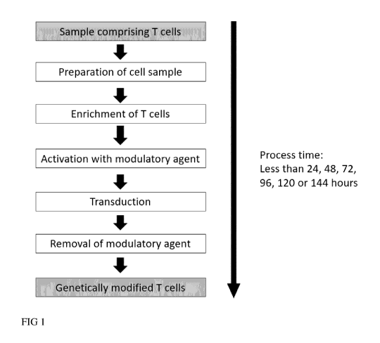
Brief description of the drawings
FIG I: Schematic representation for the generation of genetically modified T
cells in a short period
of time
A sample is provided containing T cells such as whole blood of a human,
leukapheresis, buffy coat,
PBMC, outgrown or isolated T cells. Optionally, the sample contains serum
containing substances
inhibiting the genetic modification by lentiviral vectors. To enable efficient
transduction serum is
removed by washing. In addition, T cells are polyclonally activated with a
modulatory agent
binding to CD3 and CD28 and subsequently genetically modified using lentiviral
vectors. As
cleanup, the modulatory agent is removed to obtain purified genetically
engineered T cells.
FIG 2: Schematic representation for the removal of the modulatory activating
agent
T cells are polyclonally activated with a modulatory reagent comprising an
antibody or antigen
binding fragment thereof specific for CD3 and an antibody or antigen binding
fragment thereof
specific for CD28. Both antibodies or fragments thereof are coupled directly
or indirectly to a
biodegradable linker. The modulatory activating reagent may be removed by
washing or by adding
enzymes specifically degrading the linker, thereby the antibodies or fragments
specific for CD3
and CD28 are released. In addition, the activating reagent may be removed by
chemical disruption
of said antibodies or antigen binding fragments thereof specific for CD3 and
CD28. Removal of
the modulatory agent or fragments thereof from the cells may be performed by
one or several
washing steps.
CA 03142108 2021-11-26
WO 2020/239866
PCT/EP2020/064755
4
FIG 3: Schematic representation for the removal of the magnetic enrichment
reagents
CD4+ and/or CD8+ T cells are separated by magnetic cell separation such as
MACS with magnetic
particles directly or indirectly contacting T cells with coupled antibodies or
antigen binding
fragments thereof specific for CD4 and/or CD8. The antibodies or antigen
binding fragments
thereof are coupled to the magnetic particles via a biodegradable linker. The
coupled magnetic
particles may be removed by washing or by adding enzymes specifically
degrading the linker,
thereby the antibodies or fragments specific for CD4 and/or CD8 are released
from the magnetic
particle. In addition, the magnetic particle may be removed by chemical
disruption. Removal of
the magnetic particle or fragments thereof may be performed by one or several
washing steps.
FIG 4: Schematic representation for the removal of reagents for the indirect
magnetic labelling of
T cells.
T cells may be indirectly labelled with a magnetic particle contacting T cells
with coupled
antibodies or antigen binding fragments thereof specific for CD4 and/or CD8
via a biodegradable
linker that is biotinylated and a magnetic particle that is coupled to an
antibody or antigen binding
fragment thereof specific for biotin. The magnetic particle may be released
from said T cell by
adding an enzyme that specifically digests the biodegradable linker and/or by
adding biotin (as a
competitor). In addition, the indirectly coupled magnetic particle may be
removed by washing
and/or chemical disruption. Removal of disrupted agents or the magnetic
particle may be performed
by one or several washing steps.
FIG 5: Removal of the activating reagent by washing
Enriched T cells were polyclonally stimulated with T Cell TransAct ' (Miltenyi
Biotec) - a
modulatory reagent comprising an antibody or antigen binding fragment thereof
specific for CD3
and an antibody or antigen binding fragment thereof specific for CD28 coupled
directly to a
biodegradable linker. 20h post stimulation T cells containing the modulatory
activating reagent
were washed and the presence of bound biodegradable linker was measured by
flow cytometry at
several timepoints post stimulation. Washing removes the stimulation reagent
efficiently as
detected by reduced levels of the biodegradable linker over time.
CA 03142108 2021-11-26
WO 2020/239866
PCT/EP2020/064755
FIG 6: Removal of the modulatory agent by washing and enzymatic activity
Enriched T cells were polyclonally stimulated with T Cell TransAct ' (Miltenyi
Biotec) - a
modulatory reagent comprising an antibody or antigen binding fragment thereof
specific for CD3
and an antibody or antigen binding fragment thereof specific for CD28 coupled
directly or
5 indirectly to a biodegradable linker. 20h post stimulation T cells with
bound modulatory activating
reagent were washed and the presence of the biodegradable linker was measured
by flow cytometry
after 24h. 26h post stimulation the enzyme specific for the biodegradable
linker was added and
presence of the biodegradable linker was measured over time at several
timepoints post stimulation.
Washing and the addition of the enzyme specific for the biodegradable linker
removes the
stimulation reagent efficiently.
FIG 7: The enzyme specific for the biodegradable linker is non-toxic
Enriched T cells were polyclonally stimulated with T Cell TransAct ' (Miltenyi
Biotec) - a
modulatory reagent comprising an antibody or antigen binding fragment thereof
specific for CD3
and an antibody or antigen binding fragment thereof specific for CD28 coupled
directly or
indirectly to a biodegradable linker. 26h post stimulation the enzyme was
added to the T cells and
24h later the viability was measured by PI staining by flow cytometry. The
enzyme specific for the
biodegradable linker does not harm the enriched and activated T cells as
comparable viabilities
were detectable with and without the enzyme specific for the biodegradable
linker.
FIG 8: Efficient removal of non-cellular components by cumulative washing
The efficiency of cumulative washing and removal of non-cellular components
was calculated
based on two different washing regimen: either 2.6 fold dilution per
individual washing step or 5
fold dilution per individual washing step. The calculated cumulative dilution
efficiency was
normalized to undiluted (i.e. 100%). For 2.6-fold step wise dilution the ratio
of non-cellular
components falls below 0.001 % after 11 consecutive washing steps. For 5-fold
step wise dilution
the ratio of non-cellular components falls below 0.001 % after 7 consecutive
washing steps.
FIG 9: Setting up the process for the genetic engineering of T cells in 3 days
T cells transduced on day 0 and incubated with dextranase on day 1- a enzyme
specific for the
biodegradable linker - showed the lowest transduction efficiency levels
indicating insufficient T
CA 03142108 2021-11-26
WO 2020/239866
PCT/EP2020/064755
6
cell stimulation. This was confirmed by analyzing T cells that were stimulated
longer by adding
later on day 2 or 3 and higher transduction efficiency levels were detectable
as compared to T cells
incubated with the enzyme on day 0. Better transduction efficiencies that were
close to the
conventional protocols were observed for stimulated T cells that were
transduced on day 1 and
incubated with dextranase on day 2 or 3.
FIG 10: Efficient removal of the modulatory agent in CliniMACS Prodigy system
for genetically
engineered T cells generated within 3 days
A leukapheresis sample of a healthy donor with up to 1e9 CD4/CD8 cells was
automatically
.. processed in the CliniMACS Prodigy system to generate CAR T cells within 3
days. 4e8 T cells
were polyclonally stimulated with the modulatory agent MACS GMP T Cell
TransAct '
(Miltenyi Biotec) and genetically modified with VSV-G pseudotyped lentiviral
vectors. On day 2,
10 ml of a solution containing dextranase was automatically added specifically
degrading the
biodegradable linker releasing the antibodies or fragments specific for CD3
and CD28 and
abolishing the activity of the modulatory agent. As control, a manufacturing
run in the
CliniMACS Prodigy system was performed under the same conditions and the same
donor
material but without the addition of the enzyme specific for the biodegradable
linker.
Figure 10A: The presence of the biodegradable linker was assessed for both T
cell engineering runs
in the CliniMACS Prodigy system by flow cytometry on the formulated cells by
staining with
antibodies specific for the biodegradable linker.
Figure 10B: The biodegradable linker was efficiently removed in the CliniMACS
Prodigy system
as only a minor fraction of linker positive cells was detectable when compared
to the CliniMACS
Prodigy run without added enzyme. In addition, the mean intensity levels (MFI)
for the
biodegradable linker for all viable cells was at background levels when the
enzyme was added.
FIG 11: T cell stimulation levels in the CliniMACS Prodigy system upon
enzymatic removal of
the activation reagent on day 2
The impact of removing the modulatory agent on the stimulation levels was
evaluated by flow
cytometry upon staining for CD25 and CD69 as both are described to be reliable
T cell activation
markers (CD25: REA570; CD69: REA824; Miltenyi Biotec). Non-stimulated T cells
obtained
CA 03142108 2021-11-26
WO 2020/239866
PCT/EP2020/064755
7
from the same donor from small scale cultures served as control and harvested
T cells from the
CliniMACS Prodigy system treated with or without enzyme were analyzed.
Compared to the non-stimulated control cells, highly elevated mean intensity
levels for both
activation markers were detected for T cell samples treated with or without
dextranase confirming
that the stimulation until day 2 was already sufficient to upregulation of
both activation markers.
This also indicates that the modulatory agent may be removed already at day 2
or even earlier
without affecting the stimulation.
FIG 12: Proliferation of stimulated T cells in the CliniMACS Prodigy system
for the genetic
engineering of T cells within 3 days.
T cell expansion was not detectable on day 3 for three independent
manufacturing runs suggesting
that the T cells were sufficiently activated but proliferation of T cells has
not started yet (see also
Fig. 12). In consequence, the manufacturing protocol for the genetic
modification of T cells within
3 days is too short to support T cell proliferation in vitro.
FIG 13: Evaluating the CAR expression kinetics in small scale
Figure 13A: After day 5 the transduction efficiency reached plateau levels at
18-22% confirming
stable transgene delivery and transgene expression.
Figure 13B: 2 days post transduction 16% of the T cells were already CAR
positive but a distinct
CAR positive population was not detectable yet. At later time points a
distinct CAR expressing
population was detected by flow cytometry.
FIG 14: Evaluating the CAR expression kinetics for the large scale manufacture
in the
CliniMACS Prodigy system
In contrast to the experiments in small scale ( see Fig 13), the plateau level
of CAR expression in
the CliniMACS Prodigy system were not reached at early time points. 2 days
post transduction
19 % of the T cells were CAR positive. Transduction efficiency increased to
75% at later time
points indicating that the CAR was not yet sufficiently expressed 2 days post
transduction.
CA 03142108 2021-11-26
WO 2020/239866
PCT/EP2020/064755
8
FIG 15: Optimizing CAR T cell manufacturing parameters in the CliniMACS
Prodigy system
Isolated and stimulated T cells were genetically modified with 2.5m1 of VSV-G
pseudotyped
CD20/CD19 tandem CAR encoding lentiviral vectors for 1e8 T cells (see Fig 15:
Condition I) and
in parallel with the same lentiviral vector volume for 4e8 T cells (see Fig
15: Condition II).
Condition II also supports cultivation at higher cell densities by increasing
the volume and by
implementing early shaking steps. On day 2, the same volume of dextranase was
applied to both T
cell manufacturing conditions. On day 3, the manufactured T cells were washed
multiple times,
harvested and the total T cell number was determined by cell counting. A
washed and harvested
cellular sample of both CAR T cell manufacturing conditions was cultivated for
another 8 days in
24 wells in the incubator to enable a reliable assessment of the transduction
efficiency when steady
state levels of the CAR expression are typically observed.
Figure 15A: The transduction efficiency was 32% for condition II, whereas the
transduction
efficiency for condition I was only 20%. Importantly, a higher LV dose per
cell (MOI) was applied
for condition I.
Figure 15B: For condition II not only a higher transduction efficiency was
determined but also 4
times more T cells (i.e. 4e8) were transduced. This increased the yield of CAR
transduced T cells
almost 7 fold for condition II when compared to condition I.
FIG 16: Cytokine expression levels of CAR T cells generated within 3 days
Stimulated, CD2O-CAR transduced and with dextranase treated T cells
manufactured within 3 days
in the CliniMACS Prodigy system were cocultivated at different effector to
target ratios (E:T)
with CD20, GFP expressing Raji cells and the presence of inflammatory
cytokines such as
Interferon-gamma (IFN-g), Granulocyte-macrophage colony-stimulating factor (GM-
CSF) and IL-
2 was evaluated 24 h later using the MACSPlex Cytokine Kit Assay (Miltenyi
Biotec). For CD20
CAR transduced T cells generated within 3 days, IFN-g, GM-CSF and IL-2 levels
were detectable
at high levels even beyond the level of quantification in an E:T dependent
manner. In contrast, no
cytokines were detectable for non-stimulated T cells and for stimulated T
cells that remained
untransduced. This confirms the tumor antigen specific response of CAR
transduced T cells that
were manufactured within 3 days.
FIG 17: Cytotoxic activity of CAR T cells generated within 3 days
CA 03142108 2021-11-26
WO 2020/239866
PCT/EP2020/064755
9
CAR T cells manufactured within 3 days and Raji-GFP cells were cocultered for
another 2 days
when 50% of the cells were analyzed by flow cytometry to quantify the number
of remaining tumor
cells and consequently the cytolytic potential of the CAR T cells (round 1;
left). Another 20,000
Raji-GFP tumor cells were added to the remaining 50% of the coculture to
evaluate the potency of
the CAR T cells in a second consecutive round of coculture when additional
tumor cells were added
and the cytotoxic activity was assessed under conditions meant to be
challenging for the CAR T
cells (round 2: right). After 72h flow cytometry was performed to quantify the
number of remaining
tumor cells of the second round of coculture. For high E:T ratios (i.e.
1.25:1) almost 100% of the
Raji cells were lysed in the first and also in the second round. In contrast
only 50% and 40%
remaining target cells were detectable for the untransduced control. For a E:T
ratio of 0.425:1 the
functionality was comparable as for 1.25:1 but at lower overall levels: 60% of
the tumor cells were
lysed in the presence of CAR transduced T cells in the first and second round
of coculture. In
contrast only 40% of the tumor cells were lysed in the presence of not
transduced CAR T cells in
the first round and no killing was detectable in the second round. No specific
killing was detectable
in the first round for E:T ratios of 0.15:1 when not transduced T cells are
compared to CAR
transduced T cells. In summary, the functionality of CAR transduced T cells
manufactured within
3 days was confirmed in vitro as less tumor cells were present after 2
consecutive rounds of
coculture were detectable when compared to the not- transduced control.
FIG 18: In vivo function of CAR T cells generated within 3 days
The in vivo functionality of CAR transduced T cells generated within 3 days
was confirmed in 6
to 8 week old NOD scid gamma (NSG) (NOD.Cg-PrkdcscKII12rgtmlwil/SzJ) mice. All
experiments
were performed in compliance with the "Directive 2010/63/EU of the European
Parliament and of
the Council of 22 September 2010 on the protection of animals used for
scientific purposes" and
in compliance with the regulations of the German animal protection law.
Briefly, a leukapheresis sample of a healthy donor was automatically processed
in the
CliniMACS Prodigy system to generate CAR T cells within 3 days (see Fig.18
top). On day 0, a
bag containing the leukapheresis sample was sterile connected to the CliniMACS
Prodigy Tubing
Set 520 by welding. The cells were automatically washed and labelled with CD4
and CD8
CliniMACS reagent to enrich T cells. 2e8 T cells were transferred in IL-7/IL-
15 containing medium
to the cultivation chamber and were polyclonally stimulated with MACS GMP T
Cell
CA 03142108 2021-11-26
WO 2020/239866
PCT/EP2020/064755
TransAct ' (Miltenyi Biotec) in a cultivation volume of 200 ml. On day 1, the
isolated and
stimulated T cells were genetically modified with VSV-G pseudotyped lentiviral
vectors to induce
the expression of CD22/CD19 Tandem-CAR. A bag containing 10 ml of lentiviral
vectors was
sterile connected to the tubing set and automatically transferred to the
chamber containing the T
5 cells. On day 2, 10 ml of a solution containing dextranase were sterile
connected to the tubing set
and automatically added to the chamber containing the T cells to specifically
degrade the linker,
thereby the antibodies or fragments specific for CD3 and CD28 are released and
the activity of the
modulatory agent is inhibited. After washing multiple times the cell product
was analyzed by flow
cytometry to determine the transduction efficiency, viability and cellular
composition at each step
10 (see Fig. 19). Per mouse 3e6 or 6e6 total T cells from CAR transduced
groups were injected at the
harvesting day (see Fig. 18 bottom). 4d days earlier tumors have been
established by intravenously
inoculation with 5e5 Firefly luciferase-expressing Raji cells (see Figure 17).
Per group 7 mice were
treated. Two additional groups were established as negative control: one group
received tumor cells
but no T cells (n=7; tumor only) and one group received tumor cells and 3e6
untransduced T cells
(n=7) from the same donor cultivated in parallel in small scale. Tumor growth
as well as anti-
tumoral response was monitored frequently using an In vivo Imaging System
(IVIS Lumina III).
For this purpose, 100 1 XenoLight Rediject D-Luciferin Ultra was injected
i.p. and subsequently
mice were anesthetized using the Isofluran XGI-8 Anesthesia System.
Measurement was
performed six min after substrate injection. At the end of the experiment
spleen, bone marrow and
blood was prepared and analyzed by flow cytometry to the determine the
frequency of tumor cells
and T cell subsets.
FIG 19: Cellular composition
The cellular composition was determined by flow cytometry by staining for
CD45h, CD3, CD4,
CD8, CD16/CD56, 7-AAD, CD19, CD14 on samples taken pre enrichment, post
enrichment and
after harvesting to determine the quality of the cell product. The cellular
composition after
formulation was 67% CD4 T cells, 18% CD8 T cells and 7% NKT cells. The
frequency of NK
cells, eosinophils, neutrophils, B cells or monocytes was at background levels
confirming the T
cell purity after enrichment.
CA 03142108 2021-11-26
WO 2020/239866
PCT/EP2020/064755
11
FIG 20: Representative In vivo imaging data for selected groups
The tumor burden as well as the antitumoral activity of the CAR T cells was
monitored frequently
by in vivo imaging. All mice are shown for the cohorts containing mice that
have received 3e6
viable T cells: Transduced and not transduced. 3 representative mice out of 7
are shown for the
tumor only group. The tumor burden increased rapidly for the mice in cohorts
that received
untransduced T cells or tumor cells only. Mice in both control groups had to
be sacrificed 14d post
T cell injection as critical levels of tumor burden were reached. In contrast,
mice that have received
CAR transduced T cells showed a decelerated increase at early time points in
an dose-dependent
manner 3 and 7 days post T cell injection when compared to the control groups.
The level of tumor
burden for the CAR transduced T cell groups peaked on day 7 post T cell
injection followed by a
steady reduction of the tumor burden down to levels measured at the beginning
of the experiment.
FIG 21: In vivo imaging data for all groups
The mean tumor burden +/-SEM measured as p/s over time is shown for all mice
for all groups.
The data for the 6E6 CAR transduced T cell group (n=7) is included. Mice
treated with 6E6 T cells
showed a quicker antitumoral response than the 3E6 group. On day 14 post T
cell injection the
tumor burden was substantially decreased to a comparable, low level for both T
cells doses. The
control groups (i.e. tumor only and untransduced T cells) were not able to
control the tumor growth
and mediate potent antitumoral activity.
FIG 22: Abundance of T cells in bone marrow
The abundance of human T cells in the bone marrow was quantified by flow
cytometry for 3
randomly selected mice upon staining for CD45h, CD4, CD8, CD20, CD22, 7-AAD,
CD19 CAR
Detection (all Miltenyi Biotec). The number of each mouse is shown. For the
control groups the
analysis was performed on bone marrow sampled on day 14. For the 3e6 CAR
transduced T cells
group, 3 randomly selected mice were analyzed on day 18. As expected no T
cells were found in
the Tumor only group. Up to 20% T cells were detectable for the non-transduced
cohort. In contrast,
the frequency of human T cells was highest with up to 75% in the cohort
containing mice that were
infused with CAR transduced T cells indicating homing of the CAR T cells to
this niche and in
vivo proliferation.
CA 03142108 2021-11-26
WO 2020/239866
PCT/EP2020/064755
12
FIG 23: Abundance of tumor and T cells in bone marrow
The human cellular compartment was investigated in more detail to determine
the frequency of the
human Raji tumor cells and the human T cells. Therefore, the frequency of all
human cells was set
to 100%. The number of each mouse is shown. As expected no human T cells but
only Raji cells
were found in the tumor only cohort. Bone marrow is the preferred niche of the
Raji tumor cells.
In contrast only a minor fraction of Raji cells was detectable in this organ
for the CAR transduced
T cell group. This is in line with about 50% human CD4 and ¨50% human CD8 T
cells present in
the organ of these representative mice. 20 ¨ 60% of the human cells were Raji
cells for the
untransduced T cell group with a CD4 to CD8 T cell ratio of 2:1 to 3:1.
FIG 24: Abundance of T cell subsets in spleen
The abundance of T cells in the spleen of 3 randomly selected mice was
quantified by flow
cytometry upon staining for CD45h, CD4, CD8, CD20, CD22, 7-AAD, CD19 CAR
Detection (all
Miltenyi Biotec). The number of each mouse is shown. For the control groups
the analysis was
performed on spleen sampled on day 14. For the 3e6 CAR transduced T cell
group, 3 randomly
selected mice were analyzed on day 18. As expected no T cells were found in
the tumor only group.
Up to 10% T cells were detectable for non-transduced cohort. In contrast, the
frequency of human
T cells was highest with up to 40% in the cohort containing mice that were
infused with CAR
transduced T cells.
FIG 25: Abundance of T cell subsets in blood
The abundance of T cells circulating in the blood of 3 randomly selected mice
was quantified by
flow cytometry upon staining for CD45h, CD4, CD8, CD20, CD22, 7-AAD, CD19 CAR
Detection
(all Miltenyi Biotec). The number of each mouse is shown. For the control
groups the analysis was
performed on spleen sampled on day 14. For the 3e6 CAR transduced T cell
group, 3 randomly
selected mice were analyzed on day 18. No T cells were found in the tumor only
group and only
minor fractions in the cohort containing mice with untransduced T cells. In
contrast, the frequency
of human T cells circulating in blood was highest with up to 25% in the cohort
containing mice
that were infused with CAR transduced T cells.
CA 03142108 2021-11-26
WO 2020/239866
PCT/EP2020/064755
13
Detailed description of the invention
It is an aspect of the present invention that it provides a method for the
generation of genetically
modified T cells comprising the steps
a) a sample provided, said sample comprising T cells
b) preparation of said sample by centrifugation
c) enrichment of the T cells of step b (enrichment of the T cells from the
prepared sample)
d) activation of the enriched T cells using modulatory agents
e) genetic modification of the activated T cells by transduction with
lentiviral vector particles
t) removal of said modulatory agents,
thereby generating a sample of genetically modified T cells,
wherein said method is performed in equal or less than 144 hours, less than
120 hours, less than 96
hours, less than 72 hours, less than 48 hours, or less than 24 hours.
To date, the most prevalent adverse effect following infusion of CAR T cells
is the onset of immune
activation, known as cytokine release syndrome (CRS). It is a systemic
inflammatory response
caused by cytokines released by infused CAR T cells shortly after infusion
recognizing a
potentially high load of tumor cells expressing the CAR antigen. CAR T cell
manufacturing within
a short period of time may at least partially reduce this toxicity because not
all CAR T cell express
the CAR at this early time point followed by a steady but slow increase of CAR
expression levels
(see Example 10).
The combination e.g. of removal of modulatory agents and/or magnetic particles
used for
enrichment of T cells as disclosed herein and the performance of the method as
disclosed herein in
equal or less than 144 hours, less than 120 hours, less than 96 hours, less
than 72 hours (3days),
less than 48 hours, or less than 24 hours allows successfully to apply to
treat i-vivo a patient
suffering from e.g. a cancer, wherein the number of T cells in said generated
sample of said method
is less than 10-fold or less that 5-fold higher compared to the number of T
cells in said provided
sample.
A sample provided (or providing a sample) comprising T cells may be provided
from a subject
such as a human (a sample comprising T cells provided by a subject). Said
provided sample may
CA 03142108 2021-11-26
WO 2020/239866
PCT/EP2020/064755
14
be whole blood of a human, a leukapheresis of a subject, buffy coat, PBMC,
outgrown or isolated
T cells.
Preparation of said sample may result in volume reduction, rebuffering,
removal of serum,
erythrocyte reduction, platelet removal, and/or washing.
Alternatively said method may start with step a: providing a sample comprising
T cell.
Alternatively said method may start with step b: preparation of a sample
comprising T cells by
centrifugation. This alternative step b may be followed by steps c to f.
Said method, wherein said sample of step a) comprises human serum and wherein
said serum is
removed by step b).
Said human serum may comprise components that reduce the transduction
efficiency of the
lentiviral vector particle into the cell. Said components of the human serum
that may reduce said
transduction efficiency may be components of the complement system of a
subject or may be
neutralizing antibodies (see e.g. DePolo et al, 2000, Molecular Therapy, 2:
218-222).
The removal of human serum may be performed by washing (a washing step)
achieved by said
.. centrifugation. The washing step may be performed by a series of
media/buffer exchanges (at least
twice exchanges) thereby removing the human serum and/or its components from
the T cells.
Said method, wherein said T cell are prepared and enriched in less than 2
hours, preferentially in
less than 1 hour.
Said method, wherein said T cells are activated (stimulated) using said
modulatory agents in less
than 72 hours, preferentially in less than 48 hours, more preferentially in
less than 24 hours, i.e. the
addition of said modulatory agents and the removal of said modulatory agents
occur within the
period of said hours.
Said method, wherein the transduction of said activated T cells starts 2 days
after said stimulation
of T cells using modulatory agents, preferentially 1 day after said
stimulation, more preferentially
at the same time as said stimulation.
Said method, wherein said modulatory activating agents may be removed (step t)
in less than 2
hours, preferentially in less than 1 hour, more preferentially in less than 30
minutes after the
addition of said modulatory agents to the T cells (step d).
Said method, wherein said genetic modification of said T cells by transduction
with lentiviral
vector particles (step e) may be performed in less than 2 days, preferentially
in less than 1 day,
more preferentially in less than 12 hours.
CA 03142108 2021-11-26
WO 2020/239866
PCT/EP2020/064755
Said method, wherein the T cells of the provided sample may be enriched prior
to said genetic
modification of the T cells for CD4 positive and/or CD8 positive T cells by
using CD4 and/or CD8
as positive selection marker, and/or wherein the T cells of the provide sample
may be depleted of
cancer cells that contaminate the sample comprising T cells by using a tumor
associated antigen
5 (TAA) as a negative selection marker. The TAA may be selected from one or
more markers of e.g.
CD19, CD20, CD22, CD30, CD33, CD70, IgK, IL-1Rap, Lewis-Y, NKG2D ligands,
ROR1, CAIX,
CD133, CEA, c-MET, EGFR, EGFRvIII, EpCam, EphA2, ErbB2/Her2, FAP, FR-a, GD2,
GPC3,
IL-13Ra2, Li-CAM, Mesothelin, MUC1, PD-L1, PSCA, PSMA, VEGFR-2, BCMA, CD123
and
CD16V.
10 Said enrichment of CD4+ and/or CD8+ T cells and/or depletion of cancer
cells from the provided
sample may be performed by a separation step. Said separation may be performed
by flow
cytometry methods (fluorescence activated cell sorting) such as FACSorting,
magnetic cell
separation such as MACS or by microchip based cell sorting such as MACSQuant
Tyto .
Preferred is the use of a magnetic cell separation step.
Said method, wherein said enrichment of CD4 and/or CD8 positive T cells is
performed by
magnetic cell separation steps comprising:
i) contacting the T cells with magnetic particles that are directly or
indirectly coupled to antibodies
or antigen binding fragments thereof specific for CD4 and/or CD8, wherein said
magnetic particles
.. and said antibodies or antigen binding fragments thereof coupled thereto
can be removed
ii) separating the CD4 and/or CD8 T cells in a magnetic field
iii) removal of said magnetic particles from the enriched T cells after the
separation.
Said method, wherein said enrichment of CD4 and/or CD8 positive T cells is
performed by
magnetic cell separation steps comprising:
i) contacting the T cells with magnetic particles that are directly or
indirectly coupled to antibodies
or antigen binding fragments thereof specific for CD4 and/or CD8, wherein said
magnetic particles
and said antibodies or antigen binding fragments thereof coupled thereto can
be removed by
washing
ii) separating the CD4 and/or CD8 T cells in a magnetic field
iii) removal of said magnetic particles from the enriched T cells after the
separation step by washing.
CA 03142108 2021-11-26
WO 2020/239866
PCT/EP2020/064755
16
Said method, wherein said enrichment of CD4 and/or CD8 positive T cells is
performed by
magnetic cell separation steps comprising:
i) contacting the T cells with magnetic particles that are directly or
indirectly coupled to antibodies
or antigen binding fragments thereof specific for CD4 and/or CD8, wherein said
magnetic particles
and said antibodies or antigen binding fragments thereof coupled thereto can
be disrupted
chemically and/or enzymatically
ii) separating the CD4 and/or CD8 T cells in a magnetic field
iii) removal of said magnetic particles from the enriched T cells after the
separation step by
chemical and/or enzymatical disruption of said magnetic particles and said
antibodies or antigen
binding fragments thereof coupled thereto.
Said removal of said magnetic particles from the enriched T cells after the
separation step by
chemical and/or enzymatical disruption may be performed within the magnetic
field or after
removal of the magnetic field.
Methods and systems for removal of magnetic particles from a cell that have
been directly or
indirectly bound to said cell are well-known in the art.
Exemplary, some methods and systems for reversible labelling of a cell with
magnetic particles
that lead to a disruption of magnetic particles from the cells are listed
here.
One strategy exploits the specific competition of a non-covalent binding
interaction.
U520080255004 discloses a method for reversible binding to a solid support,
e.g., magnetic
particle, using antibodies recognizing the target moiety which are conjugated
to modified biotin
like desthiobiotin, and modified streptavidin or avidin bound to the solid
support. The binding
interaction of the modified binding partners is weaker compared to the strong
and specific binding
between biotin and streptavidin therefore facilitating the dissociation in the
presence of these
competitors. EP2725359B1 describes a system for reversible magnetic cell
separation based on the
non-covalent interaction of a ligand-PEO-Biotin-conjugate recognizing the
target moiety and an
anti-Biotin-antibody compromising a magnetic particle that can be released by
adding the
competing molecule biotin, streptavidin or an auxiliary reagent.
Said method, wherein said enrichment of CD4 and/or CD8 positive T cells is
performed by
magnetic cell separation step comprising:
i) contacting the T cells with magnetic particles that are indirectly coupled
via a linker to antibodies
or antigen binding fragments thereof specific for CD4 and/or CD8, wherein said
magnetic particles
CA 03142108 2021-11-26
WO 2020/239866
PCT/EP2020/064755
17
and said antibodies or antigen binding fragments thereof coupled thereto can
be removed by adding
a competing agent that competes with the binding of said linker to said
antibodies or antigen
binding fragments thereof
ii) separating the CD4 and/or CD8 T cells in a magnetic field
iii) removal of said magnetic particles from the enriched T cells after the
separation step by adding
the competing agent.
Said method, wherein said competing agent is biotin, streptavidin or an
auxiliary reagent.
Beside these competitive release mechanisms, the removal of labelling is
mentioned by mechanical
agitation, chemically cleavable or enzymatically degradable linkers. WO
96/31776 describes a
method to release after separation magnetic particles from target cells by
enzymatically cleaving a
moiety of the particle coating, or a moiety present in the linkage group
between the coating and the
antigen recognizing moiety. An example is the application of magnetic
particles coated with
dextran and/or linked via dextran to the antigen recognizing moiety.
Subsequent cleavage of the
isolated target cells from the magnetic particle is initiated by the addition
of the dextran-degrading
enzyme dextranase. A related method in EP3037821 discloses the detection and
separation of a
target moiety according to, e.g. a fluorescence signal, with conjugates having
an enzymatically-
degradable spacer.
Recently, the interest grew in techniques utilizing antigen recognizing
moieties whose binding to
the target moiety is characterized by a low-affinity constant. To ensure a
specific and stable
labelling with those low-affinity antigen recognizing moieties the structure
of the labelling
conjugate has to comprise a multimerization of the antigen recognizing moiety
providing high
avidity. Upon disruption of the multimerization the low-affinity antigen
recognizing moiety can
dissociate from the target moiety therefore providing the opportunity to
release at its best the
detection moiety and the antigen recognizing moiety from the target moiety.
This reversible multimer staining was first described in U57776562
respectively U58298782
wherein the multimerization is build up by a non-covalent binding interaction.
Exemplary, low
affinity peptide/MHC-monomers having a StreptagII are multimerized with
streptactin and the
multimerization is reversible upon addition of the competing molecule biotin.
The method was revised in U59023604 regarding the characteristics of the
antigen recognizing
moiety respectively receptor binding reagent to enable reversible labelling.
Receptor binding
reagents characterized by a dissociation rate constant about 0,5x10⁻⁴ sec-1 or
greater with a binding
CA 03142108 2021-11-26
WO 2020/239866
PCT/EP2020/064755
18
partner C are multimerized by a multimerization reagent with at least two
binding sites Z
interacting reversibly, non-covalently with the binding partner C to provide
complexes with high
avidity for the target antigen. The detectable label is bound to the
multivalent binding complex.
Reversibility of multimerization is initiated upon disruption of the binding
between binding partner
C and the binding site Z of the multimerization reagent. For example, in
multimers of Fab-
StreptagII/Streptactin, multimerization can be reversed by the competitor
Biotin.
In EP0819250B1 a method is provided for releasing magnetic particles bound to
a cell surface
through an affinity reagent, e.g. an antibody or antigen binding fragment
thereof. The magnetic
particle is released through action of a glycosidase specific for a glycosidic
linkage present in at
least one of (a) the coating of the particle and (b) a linkage group between
the coating and the
affinity reagent.
In EP3336546A1 a method is disclosed for detecting a target moiety in a sample
of biological
specimens by:
a) providing at least one conjugate with the general formula (I)
A, ¨ P ¨ 6,, ¨ Cq¨X (I)
with A: antigen recognizing moiety;
P: enzymatically degradable spacer;
B: first binding moiety
second binding moiety
X: detection moiety;
n, m, q, o integers between 1 and 100,
wherein B and Care non-covalently bound to each other and A and B are
covalently bound
to P
b) labelling the target moiety recognized by the antigen recognizing moiety
A with at least
one conjugate
c) detecting the labelled target moiety via detecting moiety X
d) cleaving Cq-X0 by disrupting the non-covalent bond between Bm and Cq from
the
labelled target moiety
e) cleaving the binding moiety Bm from the labelled target moiety by
enzymatically
degrading spacer P.
CA 03142108 2021-11-26
WO 2020/239866
PCT/EP2020/064755
19
The method of EP3336546A1 may be utilized not only for detecting target
moieties i.e. target cells
expressing such target moieties, but also for isolating the target cells from
a sample of biological
specimens. The isolating procedures makes use of detecting the target
moieties. For example, the
detection of a target moiety by fluorescence may be used to trigger an
appropriate separation
process as performed on FACS or TYTO separation systems. In the method in
EP3336546A1, the
well-known magnetic cell separation process can also be used as detection and
separation process,
wherein the magnetic particles are detected by the magnetic field.
In a preferred embodiment of the invention, said magnetic particles that are
directly coupled to
antibodies or antigen binding fragments thereof specific for CD4 and/or CD8
are coupled via a
biodegradable linker, wherein said biodegradable linker is degraded by adding
an enzyme that
(specifically) digests the biodegradable linker. Said biodegradable linker may
be or may comprise
a polysaccharide and said enzyme that specifically digests the glycosidic
linkages is a hydrolase.
Said biodegradable linker may be or may comprise dextran and said enzyme that
(specifically)
digests dextran may be dextranase.
In another preferred embodiment of the invention, said magnetic particles that
are indirectly
coupled to antibodies or antigen binding fragments thereof, such as Fabs,
specific for CD4 and/or
CD8 are coupled via two components
i) a linker, such as dextran, that is coupled to a tag such as PEO-Biotin or
said Fabs specific for
CD4 and/or CD8 that are coupled to a tag such as PEO-biotin,
ii) a magnetic particle that is coupled to an antibody or antigen binding
fragment thereof specific
for said tag, e.g. biotin, wherein after combining component i and ii and
after contacting the T cells
with said indirectly coupled magnetic particle, the magnetic particle may be
disrupted (removed)
by adding a competing agent that competes with said tag, e.g. biotin (as a
competitor).
In another preferred embodiment of the invention, said magnetic particles that
are indirectly
coupled to antibodies or antigen binding fragments thereof, such as Fabs,
specific for CD4 and/or
CD8 are coupled via two components
i) a biodegradable linker, such as dextran, that is coupled to a tag such as
PEO-Biotin,
ii) a magnetic particle that is coupled to an antibody or antigen binding
fragment thereof specific
for said tag, e.g. biotin, wherein after combining component i and ii and
after contacting the T cells
with said indirectly coupled magnetic particle, the magnetic particle may be
disrupted from said
CA 03142108 2021-11-26
WO 2020/239866
PCT/EP2020/064755
T cell by adding an enzyme that specifically digests the biodegradable linker
such as dextranase
and/or by adding a competing agent that competes with said tag, e.g. biotin
(as a competitor).
Said method, wherein said competing agent is biotin, streptavidin or an
auxiliary reagent.
The principle of this embodiment of the invention is illustrated with regard
to the release/disruption
5 principle in the FIG 4.
Said method, wherein said modulatory agents comprise an antibody or antigen
binding fragment
thereof specific for CD3 and/or an antibody or antigen binding fragment
thereof specific for CD28
coupled directly or indirectly via a linker, wherein said antibodies or
antigen binding fragments
10 thereof specific for CD3 and CD28 can be removed.
Said removal of the modulatory agents from the cells may be further performed
by one or more
washing steps.
Said method, wherein said modulatory agents comprise an antibody or antigen
binding fragment
thereof specific for CD3 and/or an antibody or antigen binding fragment
thereof specific for CD28
15 coupled directly or indirectly via a linker, wherein said antibodies or
antigen binding fragments
thereof specific for CD3 and CD28 can be disrupted chemically and/or
enzymatically, and wherein
said modulatory agents are removed by chemical and/or enzymatical disruption
of said antibodies
or antigen binding fragments thereof specific for CD3 and CD28. Removal of the
disrupted
modulatory agents from the cells may be further performed by one or more
washing steps.
20 The methods and systems described above for removal of magnetic
particles from a cell that have
been directly or indirectly bound to said cell may also be suitable, may be
transferred to and/or
may be applied for the removal of said modulatory agents that comprise an
antibody or antigen
binding fragment thereof specific for CD3 and an antibody or antigen binding
fragment thereof
specific for CD28 coupled directly or indirectly via a linker.
Said method, wherein said removal of said modulatory agents of said antibodies
or antigen binding
fragments thereof specific for CD3 and/or CD28 is performed by
a) a competitive reaction comprising the step of adding a competing agent that
competes with a tag,
e.g. biotin (as a competitor), if said modulatory agents comprise indirectly
coupled antibodies or
antigen binding fragments thereof, such as Fabs, specific for CD3 and/or CD28
via two components,
wherein said two components may be
i) antibodies or antigen binding fragments thereof, such as Fabs, specific for
CD3 and/or CD28
are coupled to said tag such as PEO-Biotin, or antibodies or antigen binding
fragments thereof,
CA 03142108 2021-11-26
WO 2020/239866
PCT/EP2020/064755
21
such as Fabs, specific for CD3 and/or CD28 that are coupled via a linker such
as dextran that is
coupled to said tag such as PEO-biotin, and
ii) antibodies or antigen binding fragments thereof, such as Fabs, specific
for the tag, e.g. biotin,
and wherein said to components i) and ii) have been combined and contacted
with said cells, and/or
b) an enzymatic disruption comprising the step of adding an enzyme that
biodegrades said linker,
if the linker is a biodegradable linker (i.e. an indirect or direct linkage of
the two antibodies or
antigen binding fragments thereof via the linker).
In another preferred embodiment of the invention, said modulatory agents
comprise indirectly
coupled antibodies or antigen binding fragments thereof, such as Fabs,
specific for CD3 and/or
CD28 via two components:
i) antibodies or antigen binding fragments thereof, such as Fabs, specific for
CD3 and/or CD28
are coupled to a tag such as PEO-Biotin, or antibodies or antigen binding
fragments thereof, such
as Fabs, specific for CD3 and/or CD28 that are coupled via a linker such as
dextran that is coupled
to a tag such as PEO-biotin
ii) antibodies or antigen binding fragments thereof, such as Fabs, specific
for the tag, e.g. biotin,
wherein after combining component I and ii and after contacting the T cells
with said combined
components, said combined components may be disrupted (removed) by adding a
competing agent
that competes with said tag, e.g. biotin (as a competitor).
Said biodegradable linker may be or may comprise a polysaccharide and said
enzyme that
specifically digests the glycosidic linkages may be a hydrolase.
Said biodegradable linker may be or may comprise dextran and said enzyme that
specifically
digests dextran may be dextranase.
In a preferred embodiment of the invention, said modulatory agents comprise an
antibody or
antigen binding fragment thereof specific for CD3 and/or an antibody or
antigen binding fragment
thereof specific for CD28 that are directly coupled via a biodegradable
linker, wherein said
biodegradable linker is degraded by adding an enzyme that specifically digests
the biodegradable
linker. Said biodegradable linker may be or may comprise a polysaccharide and
said enzyme that
specifically digests the glycosidic linkages is a Hydrolase.
CA 03142108 2021-11-26
WO 2020/239866
PCT/EP2020/064755
22
Said biodegradable linker may be or may comprise dextran and said enzyme that
specifically
digests dextran may be dextranase.
Said method, wherein after the genetic modification of the T cells by
transduction with lentiviral
vector particles residual lentiviral vector particles are removed.
Said removal of residual lentiviral vector particles may be performed before,
subsequent or after
the removal of said modulatory agents and/or said removal of said magnetic
particles.
Said removal of residual lentiviral vector particles may be performed by
washing, wherein the
washing results in an at least 10-fold, preferably 100-fold reduction of
residual vector particles in
the sample that comprises the genetically modified T cells.
The washing step may be performed by a series of media/buffer exchanges (at
least twice
exchanges) thereby removing said residual lentiviral vector particles from
said sample comprising
said genetically modified T cells. The exchanges may be performed by
separation of cells and
media/buffer by centrifugation, sedimentation, adherence or filtration and
subsequent exchange of
media/ buffer.
The at least 10-fold, preferably 100-fold reduction of residual vector
particles in the sample that
comprises the genetically modified T cells by washing can be achieved for
example by
i) Separating cells and media/buffer
ii) Removal of 90%, preferably 99% of the volume of media/buffer
iii) Adding new media/buffer to the original volume.
iv) Resuspension of cells in media/buffer
Washing steps may be performed in a consecutive manner that may result in a
cumulative reduction
of lentiviral vectors (i.e. two washing steps with a 10-fold reduction per
step result in cumulative
reduction of 100-fold).
Said removal of residual lentiviral vector particles may be performed by
incubation with substances
that inactivate lentiviral vector particles and/or reduce their stability.
Substances that inactivate
lentiviral vector particles and/or reduce their stability may be washed away
after said incubation,
wherein said incubation occurs for no longer than 3 hours, preferentially no
longer than 1 hour.
CA 03142108 2021-11-26
WO 2020/239866
PCT/EP2020/064755
23
Such substances that inactivate lentiviral vector particles and/or reduce
their stability may be e.g.
Heparin, antiretrovirals, complement factors of a human blood, neutralizing
antibodies that a
contained in human blood or a mild basic buffer.
Said antiretrovirals may be e.g. inhibitors of viral enzymes such as Zidovudin
(zidothymidin, AZT)
or Raltegravir.
Complement factors and/or neutralizing antibodies that are contained in blood,
e.g. human blood,
may be isolated by methods well-known in the art.
The mild basic buffer may have a pH value of about 7 to 9, being sufficiently
mild to not harm the
T cells of the sample. Such a buffer is described e.g in Holic et al. (Hum
Gene Ther Clin Dev. 2014
Sep;25(3):178-85)
Said method, wherein the removed human serum as disclosed herein or isolated
substances
therefrom such as complement factors and/or neutralizing antibodies that
inhibit productive
transduction of lentiviral vector particles to T cells may be added to the
genetic modified T cells,
thereby removing and/or neutralizing residual lentiviral vector particles.
The method as disclosed herein, wherein said method is an automated method,
preferentially
performed in a closed system.
The method as disclosed herein can be fully implemented as an automated
process, preferentially
in a closed system under GMP conditions.
Such a closed system allows to operate under GMP or GMP-like conditions
("sterile") resulting in
cell compositions which are clinically applicable. Herein exemplarily the
CliniMACS Prodigy
(Miltenyi Biotec GmbH, Germany) is used as a closed system. This system is
disclosed in
W02009/072003. But it is not intended to limit the use of the method of the
present invention to
the CliniMACS Prodigy.
The CliniMACS Prodigy System is designed to automate and standardize complete
cellular
product manufacturing processes. It combines CliniMACS Separation Technology
(Miltenyi
Biotec GmbH, Germany) with a wide range of sensor-controlled, cell processing
capabilities.
Prominent features of the device are:
= disposable CentriCult ' Chamber enabling standardized cell processing and
cultivation
= Cell enrichment and depletion capabilities, alone or combined with CliniMACS
Reagents
(Miltenyi Biotec GmbH)
CA 03142108 2021-11-26
WO 2020/239866
PCT/EP2020/064755
24
= Cell cultivation and cell expansion capabilities thanks to temperature
and controlled CO2 gas
exchange.
= Final product formulation in pre-defined medium and volume
= the possibility to program the device using Flexible Programming Suite
(FPS) and GAMP5
compatible programming language for customization of cell processing
= Tailor-made tubing sets for a variety of applications
The centrifugation chamber and the cultivation chamber may be identical. The
centrifugation
chamber and the cultivation chamber can be used in various conditions: for
example, for separation
or transduction, high rotational speed (i.e. high g-forces) can be applied,
whereas for example,
culturing steps may be performed with slow rotation or even at idle state. In
another variant of the
invention, the chamber changes direction of rotation in an oscillating manner
that results in a
shaking of the chamber and maintenance of the cell in suspension. Accordingly,
in the process of
the invention, T cell stimulation, gene modifying and/or cultivation steps can
be performed under
steady or shaking conditions of the centrifugation or the cultivation chamber.
Said method, wherein the number of T cells in the generated sample may be less
than 10-fold,
preferentially less than 5-fold higher compared to the number of T cells in
said provided sample.
Said method, wherein the generated T cells underwent less than 4,
preferentially less than 3 cell
divisions.
There is no need to expand in-vitro the engineered T cells to cell numbers
that have been known
to be required for effective treatment in a patient as the further expansion
of these genetically T
cells to therapeutic effective amounts of cells will take place in vivo (see
e.g. Ghassemi et al, 2018,
Cancer Immunol Res 6:1100-1109). This is possible due to the high quality of
composition/sample
of genetically modified T cells generated by the method as disclosed herein,
i.e. the low
contamination with non-engineered T cell components and toxic substances.
The omission of in-vitro expanding/proliferation of the genetically modified T
cells to larger cell
numbers allows for a reduction of time needed to prepare a clinical applicable
composition
comprising modified T cells.
CA 03142108 2021-11-26
WO 2020/239866
PCT/EP2020/064755
Said genetically modified T cells may be genetically modified to express a
chimeric antigen
receptor (CAR), a T cell receptor (TCR), or any accessory molecule, on their
cell surface.
For final formulation, the genetically modified T cells may be washed by
centrifugation and
replacement of culture medium with a buffer appropriate for subsequent
applications such as
5 infusion of the generated cell composition into a patient.
When required, genetically-modified T cells can be separated from non-modified
T cells e.g. using
again the magnetic separation technology.
In one aspect the present invention provides a cell composition obtained by
the methods as
10 disclosed herein.
In one embodiment of the invention said cell composition is a pharmaceutical
cell composition
optionally comprising a pharmaceutical carrier.
The method of the present invention may comprise any embodiment of the
invention and/or step
15 as described herein in any order and/or combination resulting in a
functional method for the
generation of genetically modified T cells as disclosed herein.
In addition to above described applications and embodiments of the invention
further embodiments
of the invention are described in the following without intention to be
limited to these embodiments.
Embodiments
In a preferred embodiment of the invention, T cells are genetically modified
in a closed system in
an automated process, e.g. by using the CliniMACS Prodigy (Miltenyi Biotec
GmbH) to express
a chimeric antigen receptor.
A sample comprising T cells may be provided that originate from a human e.g.
suffering from
cancer. The human serum of the provided sample comprising T cells may be
washed away by a
centrifugation step.
CD4+ and/or CD8+ T cells may be enriched by a magnetic separation step using
anti-CD4 and/or
anti-CD8 antibodies or antigen binding fragments thereof coupled via dextran
to a magnetic
particle. After separation of CD4+ and/or CD8+ T cells in a magnetic field
from the sample
comprising T cells the magnetic particle is removed from the enriched cells by
adding dextranase
CA 03142108 2021-11-26
WO 2020/239866
PCT/EP2020/064755
26
that disrupt the binding of the antibodies or fragments thereof to the
magnetic particle by cleavage
of the dextran chains.
The enriched CD4+ and/or CD8+ T cells may be activated for 24 hours using an
antibody or antigen
binding fragment thereof specific for CD3 and an antibody or antigen binding
fragment thereof
specific for CD28 coupled via a linker that comprises dextran as a modulatory
agent.
Lentiviral vector particles that comprise nucleic acid that encodes for a CAR
may be added the
sample comprising activated CD4+ and/or CD8+ T cells. Transduction may be
performed during
the stimulation or after the stimulation for 24 hours.
After transduction of the lentiviral particles into the CD4+ and/or CD8+ T
cells the modulatory
agent is washed away or removed by adding dextranase that disrupt the binding
of the antibodies
or fragments thereof to each other by cleavage of the dextran chains. Residual
lentiviral vector
particles are reduced in the sample comprising genetically modified T cells at
least 10-fold,
preferentially at least 100-fold by repeated washing. As a result a pure
sample comprising
genetically modified T cells is achieved in equal or less than 144 hours, less
than 120 hours, less
than 96 hours, less than 72 hours, less than 48 hours, or less than 24 hours,
and the expansion of
the genetically modified T cells in the generated sample is less than 10-fold,
preferentially less than
5-fold compared to the amount of T cells of the originally provided sample
comprising T cells. The
sample or composition comprising the genetically modified T cells may be
applied to said human
and said genetically modified T cells may express a CAR that recognizes an TAA
in said human.
Definitions
Unless defined otherwise, technical and scientific terms used herein have the
same meaning as
commonly understood by one of ordinary skill in the art to which this
invention belongs.
As used herein the term "comprising" or "comprises" is used in reference to
compositions, methods,
and respective component(s) thereof, that are essential to the method or
composition, yet open to
the inclusion of unspecified elements, whether essential or not.
The terms "modulatory agents", "activating agents" and "stimulating agents" as
used herein may
be used interchangeably.
The modulatory agents may be selected from the group consisting of agonistic
antibodies or antigen
binding fragment thereof, cytokines, recombinant costimulatory molecules and
small drug
inhibitors. Said modulatory agents are anti-CD3 and anti-CD28 antibodies or
fragments thereof
coupled to beads or nanostructures. The modulatory agents may be a nanomatrix,
the nanomatrix
CA 03142108 2021-11-26
WO 2020/239866
PCT/EP2020/064755
27
comprising a) a matrix of mobile polymer chains, and b) attached to said
matrix of mobile polymer
chains anti-CD3 and anti-CD28 antibodies or fragments thereof, wherein the
nanomatrix is 1 to
500 nm in size. The anti-CD3 and anti-CD28 antibodies or fragments thereof may
be attached to
the same or to separate matrices of mobile polymer chains. If the anti-CD3 and
anti-CD28
antibodies or fragments thereof are attached to separate matrices of mobile
polymer chains, fine-
tuning of nanomatrices for the stimulation of the T cells is possible. The
nanomatrix may be
biodegradable. The nanomatrix may be of collagen, purified proteins, purified
peptides,
polysaccharides, glycosaminoglycans, or extracellular matrix compositions. A
polysaccharide may
include for example, cellulose ethers, starch, gum arabic, agarose, dextran,
chitosan, hyaluronic
acid, pectins, xanthan, guar gum or alginate. The choice of degrading enzyme
agent will be
determined by the glycosidic linkage. Where the macromolecular coating is a
polysaccharide, the
polysaccharide will be chosen to have glycosidic linkages not normally found
in mammalian cells.
Hydrolases that recognize specific glycosidic structures may be used as an
enzyme e.g. dextran and
dextranase, which cleaves at the a(1¨>6) linkage; cellulose and cellulase,
which cleaves at the p(1
¨>4) linkage; amylose and amylase; pectin and pectinase; chitin and chitinase,
etc.
In addition sterile filtration of said small nanomatrices as disclosed e.g. in
W02014/048920A1 is
possible which is an important feature for T cell activation under conditions
which are compliant
with rigorous GMP standards, i.e. in a closed system.
The term "depletion" as used herein refers to a process of a negative
selection that separates the
desired cells from the undesired cells, herein normally the cancer cells,
which are labelled by an
antibody or antigen-binding fragment thereof coupled to a solid phase such as
a particle,
fluorophore or hapten.
The term "particle" as used herein refers to a solid phase such as colloidal
particles, microspheres,
nanoparticles, or beads. Methods for generation of such particles are well
known in the field of the
art. The particles may be magnetic particles. The particles may be in a
solution or suspension or
they may be in a lyophilised state prior to use in the present invention. The
lyophilized particle is
then reconstituted in convenient buffer before contacting the sample to be
processed regarding the
present invention.
The term "magnetic" in "magnetic particle" as used herein refers to all
subtypes of magnetic
particles which can be prepared with methods well known to the skilled person
in the art, especially
ferromagnetic particles, superparamagnetic particles and paramagnetic
particles.
CA 03142108 2021-11-26
WO 2020/239866
PCT/EP2020/064755
28
"Ferromagnetic" materials are strongly susceptible to magnetic fields and are
capable of retaining
magnetic properties when the field is removed. "Paramagnetic" materials have
only a weak
magnetic susceptibility and when the field is removed quickly lose their weak
magnetism.
"Superparamagnetic" materials are highly magnetically susceptible, i.e. they
become strongly
magnetic when placed in a magnetic field, but, like paramagnetic materials,
rapidly lose their
magnetism.
The linkage between antibody (or an antigen binding fragment thereof) and
particle can be covalent
or non-covalent. A covalent linkage can be, e.g. the linkage to carboxyl-
groups on polystyrene
beads, or to NH2 or SH2 groups on modified beads. A non-covalent linkage is
e.g. via biotin-avidin
or a fluorophore-coupled-particle linked to anti-fluorophore antibody. Methods
for coupling
antibodies to particles, fluorophores, haptens like biotin or larger surfaces
such as culture dishes
are well known to the skilled person in the art.
For enrichment, isolation or selection in principle any sorting technology can
be used. This includes
for example affinity chromatography or any other antibody-dependent separation
technique known
in the art. Any ligand-dependent separation technique known in the art may be
used in conjunction
with both positive and negative separation techniques that rely on the
physical properties of the
cells. An especially potent sorting technology is magnetic cell sorting.
Methods to separate cells
magnetically are commercially available e.g. from Invitrogen, Stem cell
Technologies, in Cellpro,
Seattle or Advanced Magnetics, Boston. For example, monoclonal antibodies can
be directly
coupled to magnetic polystyrene particles like Dynal M 450 or similar magnetic
particles and used
e.g. for cell separation. The Dynabeads technology is not column based,
instead these magnetic
beads with attached cells enjoy liquid phase kinetics in a sample tube, and
the cells are isolated by
placing the tube on a magnetic rack. However, in a preferred embodiment for
enriching CD4+
and/or CD8+ T cells from a sample comprising T cells according the present
invention monoclonal
antibodies or antigen binding fragments thereof are used in conjunction with
colloidal
superparamagnetic microparticles having an organic coating by e.g.
polysaccharides (Magnetic-
activated cell sorting (MACS) technology (Miltenyi Biotec, Bergisch Gladbach,
Germany)). These
particles (nanobeads or MicroBeads) can be either directly conjugated to
monoclonal antibodies or
used in combination with anti-immunoglobulin, avidin or anti-hapten-specific
MicroBeads.
The MACS technology allows cells to be separated by incubating them with
magnetic
nanoparticles coated with antibodies directed against a particular surface
antigen. This causes the
CA 03142108 2021-11-26
WO 2020/239866
PCT/EP2020/064755
29
cells expressing this antigen to attach to the magnetic nanoparticles.
Afterwards the cell solution is
transferred on a column placed in a strong magnetic field. In this step, the
cells attach to the
nanoparticles (expressing the antigen) and stay on the column, while other
cells (not expressing the
antigen) flow through. With this method, the cells can be separated positively
or negatively with
respect to the particular antigen(s)/marker(s).
In case of a positive selection the cells expressing the antigen(s) of
interest, which attached to the
magnetic column, are washed out to a separate vessel, after removing the
column from the magnetic
field.
In case of a negative selection the antibody used is directed against surface
antigen(s) which are
known to be present on cells that are not of interest. After application of
the cells/magnetic
nanoparticles solution onto the column the cells expressing these antigens
bind to the column and
the fraction that goes through is collected, as it contains the cells of
interest. As these cells are non-
labelled by an antibody coupled to nanoparticels, they are "untouched".
The procedure can be performed using direct magnetic labelling or indirect
magnetic labelling. For
direct labelling the specific antibody is directly coupled to the magnetic
particle. Indirect labelling
is a convenient alternative when direct magnetic labelling is not possible or
not desired. A primary
antibody, a specific monoclonal or polyclonal antibody, a combination of
primary antibodies,
directed against any cell surface marker can be used for this labelling
strategy. The primary
antibody can either be unconjugated, biotinylated, or fluorophore-conjugated.
The magnetic
labelling is then achieved with anti-immunoglobulin MicroBeads, anti-biotin
MicroBeads, or anti-
fluorophore MicroBeads.
The term "disruption" as used herein in the context of disruption of a
magnetic particle or
modulatory agent for activation may refer to the removal
by washing alone and/or
by adding a competing agent and subsequent washing and/or
by chemical disruption, i.e. by adding a substance (non-proteinous, chemical
compound) that
breaks covalent bonds and/or
by enzymatic disruption and subsequent washing, and/or
by input of energy (physical disruption) that breaks covalent bonds.
The term "competitive reaction" in the context of disruption as used herein
refers to a magnetic
particle or modulatory agent for activation that comprise 2 components, that
are not covalently
linked wherein one component binds to said cell via antibodies or antigen
binding fragments
CA 03142108 2021-11-26
WO 2020/239866
PCT/EP2020/064755
specific for CD3, CD28, CD4 and/or CD28 and contains a tag and a second
component that binds
to said tag and wherein said binding to so said tag may be dissolved by the
addition of a competitor.
The competitor may compete and/or replace one component, said magnetic
particle, said
modulatory agent or said antibodies or antigen binding fragments thereof for
example due to higher
5 affinity to the respective component or due to higher concentration of
the competitor molecule
compared to concentration of the magnetic particle or modulatory agent that is
indirectly coupled
to antibodies or antigen binding fragments thereof specific for CD3, CD28, CD4
and/or CD8.
The term "enzymatical disruption" as used herein in the context of disruption
of magnetic particles
or modulatory agents refers to antibodies or antigen binding fragments thereof
specific for CD3,
10 CD28, CD4 or CD8 that are directly or indirectly linked via a
biodegradable linker and wherein
said biodegradable linker may be specifically biodegraded, digested or cut by
the activity of said
enzyme and thereby split said magnetic particle or modulatory agent in at
least two separate
molecules. Released single antibody or antigen binding fragment thereof such
as a Fab specific for
CD3 or CD28 then has no further effect on activation of the T cell to which it
is bound. In addition,
15 if said antibody or antigen binding fragment thereof such as said Fab
has low affinity and/or a high
k(off) rate said antibody or antigen binding fragment thereof such as said Fab
will be removed from
the cell to that is has bound.
The term "chemical disruption" as used herein in the context of disruption of
magnetic particles or
modulatory agents refers to antibodies or antigen binding fragments thereof
specific for CD3,
20 CD28, CD4 or CD8 that are directly or indirectly linked via a chemically
degradable linker and
wherein said chemically degradable linker may be specifically degraded or
cleaved by the addition
of a non-proteinous, chemical substance that breaks covalent bonds under
physiological conditions
and thereby split said magnetic particle or modulatory agent in at least two
separate molecules.
Examples for suitable reactions for chemical disruption under physiological
conditions may be
25 reductions, such as the reduction of disulfide bonds by a reducing agent
or the reduction of diazo
bonds with dithionite, or oxidations, such as the cleavage of glycol residues
by periodate.
Released single antibody or antigen binding fragment thereof such as a Fab
specific for CD3 or
CD28 then has no further effect on activation of the T cell to which it is
bound. In addition, if said
antibody or antigen binding fragment thereof such as said Fab has low affinity
and/or a high k(off)
30 rate said antibody or antigen binding fragment thereof such as said Fab
will be removed from the
cell to that is has bound.
CA 03142108 2021-11-26
WO 2020/239866
PCT/EP2020/064755
31
The term "physical disruption" as used herein in the context of disruption of
magnetic particles or
modulatory agents refers to antibodies or antigen binding fragments thereof
specific for CD3,
CD28, CD4 or CD8 that are directly or indirectly linked via a physically
disruptable linker and
wherein said physically disruptable linker may be specifically degraded or
cleaved by energy input
that breaks covalent bonds under physiological conditions and thereby split
said magnetic particle
or modulatory agent in at least two separate molecules. Examples for suitable
reactions for physical
disruption under physiological conditions may be photo-reactions, such as the
photocleavage of
light sensitive linkers by UV or visible light as exemplified by the cleavage
of ortho-nitrobenzyl
derivatives by near-UV light (300 ¨ 365 nm). Released single antibody or
antigen binding fragment
thereof such as a Fab specific for CD3 or CD28 then has no further effect on
activation of the T
cell to which it is bound. In addition, if said antibody or antigen binding
fragment thereof such as
said Fab has low affinity and/or a high k(off) rate said antibody or antigen
binding fragment thereof
such as said Fab will be removed from the cell to that is has bound.
The term "marker" as used herein refers to a cell antigen that is specifically
expressed by a certain
cell type. Preferentially, the marker is a cell surface marker so that
enrichment, isolation and/or
detection of living cells can be performed. The markers may be positive
selection markers such as
CD4, CD8 and/or CD62L or may be negative selection markers (e.g. depletion of
cells expressing
CD14, CD16, CD19, CD25, CD56).
The term "expression" as used herein is defined as the transcription and/or
translation of a particular
nucleotide sequence driven by its promoter in a cell.
The term "antigen-binding molecule" as used herein refers to any molecule that
binds preferably
to or is specific for the desired target molecule of the cell, i.e. the
antigen. The term "antigen-
binding molecule" comprises e.g. an antibody or antigen binding fragment
thereof. The term
"antibody" as used herein refers to polyclonal or monoclonal antibodies, which
can be generated
by methods well known to the person skilled in the art. The antibody may be of
any species, e.g.
murine, rat, sheep, human. For therapeutic purposes, if non-human antigen
binding fragments are
to be used, these can be humanized by any method known in the art. The
antibodies may also be
modified antibodies (e.g. oligomers, reduced, oxidized and labeled
antibodies).
The term "antibody" comprises both intact molecules and antigen binding
fragments, such as Fab,
Fab', F(ab')2, Fv and single-chain antibodies. Additionally, the term "antigen-
binding fragment"
includes any molecule other than antibodies or antibody fragments that binds
preferentially to the
desired target molecule of the cell. Suitable molecules include, without
limitation, oligonucleotides
CA 03142108 2021-11-26
WO 2020/239866
PCT/EP2020/064755
32
known as aptamers that bind to desired target molecules, carbohydrates,
lectins or any other antigen
binding protein (e.g. receptor-ligand interaction). The linkage (coupling)
between antibody and
particle or nanostructure can be covalent or non-covalent. A covalent linkage
can be, e.g. the
linkage to carboxyl-groups on polystyrene beads, or to NH2 or SH2 groups on
modified beads. A
non-covalent linkage is e.g. via biotin-avidin or a fluorophore- coupled-
particle linked to anti-
fluorophore antibody.
The terms "specifically binds to" or "specific for" with respect to an antigen-
binding molecule, e.g.
an antibody or fragment thereof, refer to an antigen-binding molecule (in case
of an antibody or
fragment thereof to an antigen-binding domain) which recognizes and binds to a
specific antigen
in a sample, e.g. CD4, but does not substantially recognize or bind other
antigens in said sample.
An antigen-binding domain of an antibody or fragment thereof that binds
specifically to an antigen
from one species may bind also to that antigen from another species. This
cross-species reactivity
is not contrary to the definition of "specific for" as used herein. An antigen-
binding domain of an
antibody or fragment thereof that specifically binds to an antigen, e.g. the
CD4 antigen, may also
bind substantially to different variants of said antigen (allelic variants,
splice variants, isoforms
etc.). This cross reactivity is not contrary to the definition of that antigen-
binding domain as
specific for the antigen, e.g. for CD4.
The terms "genetically modified T cell" or "engineered T cell" may be used
interchangeably and
mean containing and/or expressing a foreign gene or nucleic acid sequence
which in turn modifies
the genotype or phenotype of the cell or its progeny. Especially, the terms
refer to the fact that cells
can be manipulated by recombinant methods well known in the art to express
stably or transiently
peptides or proteins, e.g. CARs which are not expressed in these cells in the
natural state. Genetic
modification of cells may include but is not restricted to transfection,
electroporation, nucleofection,
transduction using retroviral vectors, lentiviral vectors, non-integrating
retro- or lentiviral vectors,
transposons, designer nucleases including zinc finger nucleases, TALENs or
CRISPR/Cas.
The genetically modified T cells obtainable by the methods as disclosed herein
may be used for
subsequent steps such as research, diagnostics, pharmacological or clinical
applications known to
the person skilled in the art.
The genetically modified T cells may also be used as a pharmaceutical
composition in the therapy,
e.g. cellular therapy, or prevention of diseases. The pharmaceutical
composition may be
transplanted into an animal or human, preferentially a human patient. The
pharmaceutical
CA 03142108 2021-11-26
WO 2020/239866
PCT/EP2020/064755
33
composition can be used for the treatment and/or prevention of diseases in
mammals, especially
humans, possibly including administration of a pharmaceutically effective
amount of the
pharmaceutical composition to the mammal. Pharmaceutical compositions of the
present disclosure
may be administered in a manner appropriate to the disease to be treated (or
prevented). The
quantity and frequency of administration will be determined by such factors as
the condition of the
patient, and the type and severity of the patient's disease, although
appropriate dosages may be
determined by clinical trials.
The term "therapeutic effective amount" means an amount which provides a
therapeutic benefit for
the patient.
The composition of genetically modified T cells obtained by the method of the
present invention
may be administered either alone, or as a pharmaceutical composition in
combination with diluents
and/or with other components such as cytokines or cell populations. Briefly,
pharmaceutical
compositions of the present invention may comprise the genetically modified T
cells of the present
disclosure, in combination with one or more pharmaceutically or
physiologically acceptable
carriers, diluents or excipients. Such compositions may comprise buffers such
as neutral buffered
saline, phosphate buffered saline and the like; carbohydrates such as glucose,
mannose, sucrose or
dextrans, mannitol; proteins; polypeptides or amino acids such as glycine;
antioxidants; chelating
agents such as EDTA or glutathione; adjuvants (e.g., aluminum hydroxide); and
preservatives.
The term "activation" as used herein refers to inducing physiological changes
with a cell that
increase target cell function, proliferation and/or differentiation.
The term "transduction" means the transfer of genetic material from a viral
agent such as a lentiviral
vector particle into a eukaryotic cell such as a T cell.
The tumor associated antigen (TAA) as used herein refers to an antigenic
substance produced in
tumor cells. Tumor associated antigens are useful tumor or cancer markers in
identifying
tumor/cancer cells with diagnostic tests and are potential candidates for use
in cancer therapy.
Preferentially, the TAA may be expressed on the cell surface of the
tumor/cancer cell.
The term "removal of modulatory agents" as used herein refers to the physical
removal of the
modulatory agents from the T cells and/or to the inactivation of the
modulatory agent to that effect
that it has no effect anymore on the activity of T cells.
Lentivirus is a genus of Retroviridae that cause chronic and deadly diseases
characterized by long
incubation periods, in the human and other mammalian species. The best-known
lentivirus is the
CA 03142108 2021-11-26
WO 2020/239866
PCT/EP2020/064755
34
Human Immunodeficiency Virus HIV which can efficiently infect nondividing
cells, so lentiviral
derived retroviral vectors are one of the most efficient methods of gene
delivery.
To generate retroviral vectors such as lentiviral vectors the gag/pol and env
proteins needed to
assemble the vector particle are provided in trans by means of a packaging
cell line, for example,
HEK 293T. This is usually accomplished by transfection of the packaging cell
line with one or
more plasmids containing the gag/pol and env genes.
The term "removal of residual lentiviral vector particle" as used herein
refers to the physical
removal of the residual lentiviral vector particles from the T cells and/or to
the inactivation of the
residual lentiviral vector particles to that effect that they do not
genetically modify T cells anymore
The term "residual lentiviral vector particles" as used herein refer to the
portion of lentiviral vector
particles that have not transduced T cells in the sample comprising T cells.
The term "the method is performed in equal or less than 144 hours, less than
120 hours, less than
96 hours, less than 72 hours, less than 48 hours, or less than 24 hours" means
that the duration of
the process as disclosed herein does not take longer than the respective
timeframe from the
beginning of the process, i.e. the provision of a sample that comprises T
cells, to the sample that
comprises the genetically modified T cells that subsequent may be ready to
(re)-infusion to a patient
in need thereof.
In blood, the serum is the component that is neither a blood cell (serum does
not contain white
blood cells- leukocytes, or red blood cells- erythrocytes), nor a clotting
factor; it is the blood plasma
not including the fibrinogens. Serum includes all proteins not used in blood
clotting and all the
electrolytes, antibodies, antigens, hormones, and any exogenous substances.
Human serum is the
serum from a human.
As used herein, the term "subject" refer to an animal. Preferentially, the
subject is a mammal such
as mouse, rat, cow, pig, goat, chicken dog, monkey or human. More
preferentially, the individual
is a human. The subject may be a subject suffering from a disease such as
cancer (a patient), but
the subject may be also a healthy subject.
The term "closed system" as used herein refers to any closed system which
reduces the risk of cell
culture contamination while performing culturing processes such as the
introduction of new
material, e.g. by transduction, and performing cell culturing steps such as
proliferation,
differentiation, activation, and/or separation of cells. Such a system allows
to operate under GMP
CA 03142108 2021-11-26
WO 2020/239866
PCT/EP2020/064755
or GMP-like conditions ("sterile") resulting in cell compositions which are
clinically applicable.
Herein exemplarily the CliniMACS Prodigy (Miltenyi Biotec GmbH, Germany) is
used as a
closed system. This system is disclosed in W02009/072003. But it is not
intended to restrict the
use of the method of the present invention to the CliniMACS Prodigy .
5 The process of the invention may be performed in a closed system (a
closed cell sample processing
system), comprising a centrifugation chamber comprising a base plate and cover
plate connected
by a cylinder, pumps, valves, a magnetic cell separation column and a tubing
set. The blood
samples or other sources comprising T cells may be transferred to and from the
tubing set by sterile
docking or sterile welding. A suitable system is disclosed in W02009/072003.
10 The closed system may comprise a plurality of tubing sets (TS) where
cells are transferred between
TS by sterile docking or sterile welding.
Different modules of the process may be performed in different functionally
closed TS with transfer
of the product (cells) of one module generated in the one tubing set to
another tubing set by sterile
means. For example, T cells can be magnetically enriched in a first tubing set
(TS) TS100 by
15 Miltenyi Biotec GmbH and the positive fraction containing enriched T
cells is welded off the
TS 100 and welded onto a second tubing set TS730 by Miltenyi Biotec GmbH for
further activation,
modification, cultivation and washing.
The terms "automated method" or "automated process" as used herein refer to
any process being
automated through the use of devices and/or computers and computer software.
Methods
20 (processes) that have been automated require less human intervention and
less human time. In some
instances the method of the present invention is automated if at least one
step of the present method
is performed without any human support or intervention. Preferentially the
method of the present
invention is automated if all steps of the method as disclosed herein are
performed without human
support or intervention other than connecting fresh reagents to the system.
Preferentially the
25 automated process is implemented on a closed system such as CliniMACS
Prodigy as disclosed
herein.
The closed system may comprise a) a sample processing unit comprising an input
port and an
output port coupled to a rotating container (or centrifugation chamber) having
at least one sample
chamber, wherein the sample processing unit is configured to provide a first
processing step to a
30 sample or to rotate the container so as to apply a centrifugal force to
a sample deposited in the
chamber and separate at least a first component and a second component of the
deposited sample;
and b) a sample separation unit coupled to the output port of the sample
processing unit, the sample
CA 03142108 2021-11-26
WO 2020/239866
PCT/EP2020/064755
36
separation unit comprising a separation column holder, a pump, and a plurality
of valves configured
to at least partially control fluid flow through a fluid circuitry and a
separation column positioned
in the holder, wherein the separation column is configured to separate labeled
and unlabeled
components of sample flown through the column.
Said rotating container may also be used as a temperature controlled cell
incubation and cultivation
chamber (CentriCult Unit = CCU). This chamber may be flooded with defined gas
mixes, provided
by an attached gas mix unit (e.g. use of pressurized air/ N2 / CO2 or
N2/CO2/02).
All agents may be connected to the closed system before process initiation.
This comprises all
buffers, solutions, cultivation media and supplements, MicroBeads, used for
washing, transferring,
suspending, cultivating, harvesting cells or immunomagnetic cell sorting
within the closed system.
Alternatively, such agents might by welded or connected by sterile means at
any time during the
process.
The cell sample comprising T cells may be provided in transfer bags or other
suited containers
which can be connected to the closed system by sterile means.
The term "providing a (cell) sample comprising T cells" means the provision of
a cell sample,
preferentially of a human cell sample of hematologic origin. Normally, the
cell sample may be
composed of hematologic cells from a donor or a patient. Such blood product
can be in the form
of whole blood, buffy coat, leukapheresis, PBMCs or any clinical sampling of
blood product. It
may be from fresh or frozen origin.
The term "washing" means for example the replacement of the medium or buffer
in which the cells
are kept. The replacement of the supernatant can be in part (example 50% of
the medium is
removed and 50% fresh medium is added) this often is applied for dilution or
feeding purposes, or
entirely. Several washing steps may be combined in order to obtain a more
profound replacement
of the original medium in which the cells are kept. A washing step often may
involve pelleting the
cells by centrifugation forces and removing the supernatant. In the method of
the present invention,
cells may be pelleted by rotation of the chamber at e.g. 300xg and the
supernatant may be removed
during rotation of the chamber. Medium may be added during rotation or at
steady state.
Generally, the washing or washing step may be performed once or by a series of
media/buffer
exchanges (at least twice exchanges, e.g. 2, 3, 4, 5, 6, 7, 8, 9 or 10
exchanges) thereby removing
the substances intended to be removed from the T cells such as human serum
and/or its components,
the magnetic particles or the residual lentiviral vector particles. The
exchanges may be performed
CA 03142108 2021-11-26
WO 2020/239866
PCT/EP2020/064755
37
by separation of cells and media/buffer by centrifugation, sedimentation,
adherence or filtration
and subsequent exchange of media/ buffer.
In general, a CAR may comprise an extracellular domain (extracellular part)
comprising the antigen
binding domain, a transmembrane domain and a cytoplasmic signaling domain
(intracellular
signaling domain). The extracellular domain may be linked to the transmembrane
domain by a
linker or spacer. The extracellular domain may also comprise a signal peptide.
In some
embodiments of the invention the antigen binding domain of a CAR binds a tag
or hapten that is
coupled to a polypeptide ("haptenylated" or "tagged" polypeptide), wherein the
polypeptide may
bind to a disease-associated antigen such as a tumor associated antigen (TAA)
that may be
expressed on the surface of a cancer cell.
Such a CAR may be also named "anti-tag" CAR or "adapterCAR" or "univerdal CAR"
as disclosed
e.g. in U59233125B2.
The haptens or tags may be coupled directly or indirectly to a polypeptide
(the tagged polypeptide),
wherein the polypeptide may bind to said disease associated antigen expressed
on the (cell) surface
of a target.
A "signal peptide" refers to a peptide sequence that directs the transport and
localization of the
protein within a cell, e.g. to a certain cell organelle (such as the
endoplasmic reticulum) and/or the
cell surface.
Generally, an "antigen binding domain" refers to the region of the CAR that
specifically binds to
an antigen, e.g. to a tumor associated antigen (TAA) or tumor specific antigen
(TSA). The CARs
of the invention may comprise one or more antigen binding domains (e.g. a
tandem CAR).
Generally, the targeting regions on the CAR are extracellular. The antigen
binding domain may
comprise an antibody or an antigen binding fragment thereof. The antigen
binding domain may
comprise, for example, full length heavy chain, Fab fragments, single chain Fv
(scFv) fragments,
divalent single chain antibodies or diabodies. Any molecule that binds
specifically to a given
antigen such as affibodies or ligand binding domains from naturally occurring
receptors may be
used as an antigen binding domain. Often the antigen binding domain is a scFv.
Normally, in a
scFv the variable regions of an immunoglobulin heavy chain and light chain are
fused by a flexible
linker to form a scFv. Such a linker may be for example the "(G4/S)3-linker".
In some instances, it is beneficial for the antigen binding domain to be
derived from the same
species in which the CAR will be used in. For example, when it is planned to
use it therapeutically
in humans, it may be beneficial for the antigen binding domain of the CAR to
comprise a human
CA 03142108 2021-11-26
WO 2020/239866
PCT/EP2020/064755
38
or humanized antibody or antigen binding fragment thereof. Human or humanized
antibodies or
antigen binding fragments thereof can be made by a variety of methods well
known in the art.
"Spacer" or "hinge" as used herein refers to the hydrophilic region which is
between the antigen
binding domain and the transmembrane domain. The CARs of the invention may
comprise an
extracellular spacer domain but is it also possible to leave out such a
spacer. The spacer may include
e.g. Fc fragments of antibodies or fragments thereof, hinge regions of
antibodies or fragments
thereof, CH2 or CH3 regions of antibodies, accessory proteins, artificial
spacer sequences or
combinations thereof. A prominent example of a spacer is the CD8alpha hinge.
The transmembrane domain of the CAR may be derived from any desired natural or
synthetic
source for such domain. When the source is natural the domain may be derived
from any
membrane-bound or transmembrane protein. The transmembrane domain may be
derived for
example from CD8alpha or CD28. When the key signaling and antigen recognition
modules
(domains) are on two (or even more) polypeptides then the CAR may have two (or
more)
transmembrane domains. The splitting key signaling and antigen recognition
modules enable for a
small molecule-dependent, titratable and reversible control over CAR cell
expression (e.g.
W02014127261A1) due to small molecule-dependent heterodimerizing domains in
each
polypeptide of the CAR.
The cytoplasmic signaling domain (or the intracellular signaling domain) of
the CAR is responsible
for activation of at least one of the normal effector functions of the immune
cell in which the CAR
is expressed. "Effector function" means a specialized function of a cell, e.g.
in a T cell an effector
function may be cytolytic activity or helper activity including the secretion
of cytokines. The
intracellular signaling domain refers to the part of a protein which
transduces the effector function
signal and directs the cell expressing the CAR to perform a specialized
function. The intracellular
signaling domain may include any complete, mutated or truncated part of the
intracellular signaling
domain of a given protein sufficient to transduce a signal which initiates or
blocks immune cell
effector functions.
Prominent examples of intracellular signaling domains for use in the CARs
include the cytoplasmic
signaling sequences of the T cell receptor (TCR) and co-receptors that
initiate signal transduction
following antigen receptor engagement.
Generally, T cell activation can be mediated by two distinct classes of
cytoplasmic signaling
sequences, firstly those that initiate antigen-dependent primary activation
through the TCR
(primary cytoplasmic signaling sequences, primary cytoplasmic signaling
domain) and secondly
CA 03142108 2021-11-26
WO 2020/239866
PCT/EP2020/064755
39
those that act in an antigen-independent manner to provide a secondary or co-
stimulatory signal
(secondary cytoplasmic signaling sequences, co-stimulatory signaling domain).
Therefore, an
intracellular signaling domain of a CAR may comprise one or more primary
cytoplasmic signaling
domains and/or one or more secondary cytoplasmic signaling domains.
Primary cytoplasmic signaling domains that act in a stimulatory manner may
contain ITAMs
(immunoreceptor tyrosine-based activation motifs).
Examples of ITAM containing primary cytoplasmic signaling domains often used
in CARs are that
those derived from TCRC (CD3C), FcRgamma, FcRbeta, CD3gamma, CD3delta,
CD3epsilon,
CD5, CD22, CD79a, CD79b, and CD66d. Most prominent is sequence derived from
CD3C.
The cytoplasmic domain of the CAR may be designed to comprise the CD3 C
signaling domain by
itself or combined with any other desired cytoplasmic domain(s). The
cytoplasmic domain of the
CAR can comprise a CD3C chain portion and a co-stimulatory signaling region
(domain). The co-
stimulatory signaling region refers to a part of the CAR comprising the
intracellular domain of a
co-stimulatory molecule. A co-stimulatory molecule is a cell surface molecule
other than an antigen
receptor or their ligands that is required for an efficient response of
lymphocytes to an antigen.
Examples for a co-stimulatory molecule are CD27, CD28, 4-1BB (CD137), 0X40,
CD30, CD40,
PD-1, ICOS, lymphocyte function-associated antigen- 1 (LFA-1), CD2, CD7,
LIGHT, NKG2C,
B7-H3.
The cytoplasmic signaling sequences within the cytoplasmic signaling part of
the CAR may be
linked to each other with or without a linker in a random or specified order.
A short oligo- or
polypeptide linker, which is preferably between 2 and 10 amino acids in
length, may form the
linkage. A prominent linker is the glycine-serine doublet.
As an example, the cytoplasmic domain may comprise the signaling domain of
CD3C and the
signaling domain of CD28. In another example the cytoplasmic domain may
comprise the signaling
domain of CD3C and the signaling domain of CD137. In a further example, the
cytoplasmic domain
may comprise the signaling domain of CD3C, the signaling domain of CD28, and
the signaling
domain of CD137.
As aforementioned either the extracellular part or the transmembrane domain or
the cytoplasmic
domain of a CAR may also comprise a heterodimerizing domain for the aim of
splitting key
signaling and antigen recognition modules of the CAR.
CA 03142108 2021-11-26
WO 2020/239866
PCT/EP2020/064755
The CAR may be further modified to include on the level of the nucleic acid
encoding the CAR
one or more operative elements to eliminate CAR expressing immune cells by
virtue of a suicide
switch. The suicide switch can include, for example, an apoptosis inducing
signaling cascade or a
drug that induces cell death. In one embodiment, the nucleic acid expressing
and encoding the CAR
5 can be further modified to express an enzyme such thymidine kinase (TK)
or cytosine deaminase
(CD).
In some embodiments, the endodomain may contain a primary cytoplasmic
signaling domains or
a co-stimulatory region, but not both. In these embodiments, an immune
effector cell containing
the disclosed CAR is only activated if another CAR containing the missing
domain also binds its
10 respective antigen.
In some embodiment of the invention the CAR may be a "SUPRA" (split,
universal, and
programmable) CAR, where a "zipCAR" domain may link an intra-cellular
costimulatory domain
and an extracellular leucine zipper (W02017/091546). This zipper may be
targeted with a
complementary zipper fused e.g. to an scFv region to render the SUPRA CAR T
cell tumor specific.
15 This approach would be particularly useful for generating universal CAR
T cells for various tumors;
adaptor molecules could be designed for tumor specificity and would provide
options for altering
specificity post-adoptive transfer, key for situations of selection pressure
and antigen escape.
The CARs that may be expressed in the genetically modified T cells obtained by
the method as
disclosed herein may be designed to comprise any portion or part of the above-
mentioned domains
20 as described herein in any order and/or combination resulting in a
functional CAR, i.e. a CAR that
mediated an immune effector response of the immune effector cell that
expresses the CAR as
disclosed herein.
Examples
25 Example 1: Manual generation of genetically engineered T cells in a
short period of time.
A sample containing T cells was provided from buffy coat and PBMC were
isolated. The blood
products were diluted in CliniMACS buffer in a ratio of 1:2 or 1:3 and 30 mL
were layered onto
a 15 mL cushion of Pancoll human. The tubes were centrifuged for 30 mm at room
temperature
and 450xg with moderate brakes. After centrifugation, the cells at the
interface were carefully
30 .. sucked off, and washed three times with 50 mL CliniMACS buffer in order
to remove platelets
and residual Pancoll. T cells were isolated using CD4 and CD8 specific
MicroBeads (Miltenyi
Biotec) according to the manufacturer's instructions. T cells were seeded into
24-well plates with
CA 03142108 2021-11-26
WO 2020/239866
PCT/EP2020/064755
41
2 mL T cell suspension per well at a concentration of 1 x10⁶ cells/mL in
TexMACS medium
containing human AB serum (10% (v/v) GemCell), IL-7 (10 ng/mL) and IL-15 (5
ng/mL). To
activate the T cells T Cell TransAct ' is added to a final dilution of 1:100.
After 24h of cultivation
in an incubator at 37 C, 5-10% CO2, lentiviral vectors encoding therapeutic
CARs are added at a
MOI of 2. 24h post transduction an enzyme dextranase was added 1:100 for lh at
37 C that
specifically degrades the biodegradable linker present in T Cell TransAct '
and Microbeads and
both reagents are released from the T cells. Non-cellular components such as
remaining lentiviral
vectors, degraded components of the T cell TransAct ' and Microbeads are
separated from the
transduced T cells by centrifugation at 450xg for 10 min. The supernatant is
removed and fresh
media is added to the same volume. The washing procedure is repeated 3 times
to decrease the
impurities. The transduced T cells are analysed by flow cytometry to determine
the transduction
efficiency and perform functional assays such as killing assays in coculture
with tumor target cells
expressing the CAR antigen.
Example 2: Automated generation of genetically modified T cells within a short
period of time
A sample of T cells is provided in bag derived from a leukapheresis from a
donor. The bag is
connected by sterile welding to a tubing set installed on the CliniMACS
Prodigy device.
CliniMACS buffer, CliniMACS CD4 and CD8 reagents (Miltenyi Biotec GmbH) as
well as
activating reagent are also connected to the same Tubing set. Within the fully
automated process,
the enrichment step is launched that takes in total 30min to 2h. In detail,
the tubing set is
automatically primed with buffer, then the leukapheresis product is
transferred to the chamber of
the tubing set where it is washed 3 times with CliniMACS buffer in order to
remove serum and
platelets. The cells are magnetically labelled with CliniMACS CD4 and CD8
reagents and trapped
onto a column placed in a magnetic field. The labeled cells trapped onto the
column are rinsed
several times and eluted into the target cell fraction bag. Part of the
enriched cells are transferred
to the CentriCult ' Chamber via sterile welding connection and formulated in
MACS GMP
TexMACS medium supplemented with IL-7/IL-15 (all Miltenyi Biotec GmbH). Within
the
automated process the activation step is started and the activation reagent
MACS GMP TransAct
is automatically added to the culture. After enrichment and up to 24 hours, a
bag containing
lentiviral vector is sterile welded onto the tubing set and the lentiviral
vector suspension is
transferred into the CentriCult ' Chamber containing the activated T cells. 24
hours to 48 hours
the activation reagent and magnetic particles are degraded by adding an
dextranase, specific for the
CA 03142108 2021-11-26
WO 2020/239866
PCT/EP2020/064755
42
biodegradable linker present in the activation reagent and magnetic particles.
After 1 h, non-cellular
components such as residual lentiviral vectors, degrading enzymes and degraded
components of
the activation reagent and CliniMACS CD4 and CD8 reagents are removed by
washing. The
genetically modified T cells are automatically formulated in a solution
suitable for human infusion.
Example 3: Administration of CAR T cells with additional cleanup steps.
CAR T cell therapy is provided e.g. to treat pediatric and adult patients with
relapsed or refractory
CD19 positive B cell malignancies. The clinical method of preparing the
genetically engineered T
cells is based on example 2, whereby patient cells (derived from BM, blood or
leukapheresis) are
connected to the CliniMACS Prodigy device and processed rapidly (i.e.
preferably less than 24h)
and reinfused into the patient. The duration of the process can be modulated
to match timing for
required patient preparative regimen (e.g. chemotherapeutic treatment to
lymphodeplete), meet
medical needs and clinical applicability (e.g. clinical protocol, patient
health status, reactivity of
the doctors, hospital stay). Advantages of the described invention are to
enable rapid treatment and
patient care as well as to enable "bed side" preparation of drug products. The
invention describes
a solution for such rapid cell preparation where by potentially harmful
substances such as viral
vectors and activation reagents are removed prior to infusion. For example,
remaining activation
reagents contaminating the drug product may be harmful upon infusion as they
may lead to
activation of T cells in vivo. This may lead to a rapid release of
proinflammatory cytokines, causing
severe cytokine release syndrome, fever, hypotension, organ failure and even
deaths.
In addition, remaining lentiviral vectors contaminating the drug product in
soluble and/or cell
bound form may be harmful upon infusion as they may provoke an unwanted immune
response
such as complement activation, antibody-dependent cell-mediated cytotoxicity,
inducing an
adoptive immune response against antigens delivered by the lentiviral vector
and/or transduction
of non-target cells in vivo. The transduction of non-target cells and the
subsequent expression of
the transgene may induce unwanted side-effects such as the induction of
unwanted immune
responses, oncogenicity, altered survival, proliferation, physiological state
and natural function.
Example 4: Setting up the process for the genetic engineering of T cells in 3
days
T cells from 2 healthy donors were enriched untouched with the Pan T cell
isolation kit, human
(Miltenyi Biotec) and polyclonally stimulated on day 0 with T Cell TransAct '
(Miltenyi Biotec)-
CA 03142108 2021-11-26
WO 2020/239866
PCT/EP2020/064755
43
a modulatory reagent comprising an antibody or antigen binding fragment
thereof specific for CD3
and an antibody or antigen binding fragment thereof specific for CD28 coupled
directly or
indirectly to a biodegradable linker. The stimulated T cells were transduced
on the same day or day
1 with VSV-G pseudotyped GFP encoding LV at a MOI of 2 in 24 wells at 1e6
cells /ml. in IL-
7/IL-15 containing TexMACS media. On day 1, 2 or 3, dextranase specifically
digesting the
biodegradable linker was added and the transduction efficiency was evaluated
on day 10 by flow
cytometry. T cells stimulated on day 0 and transduced on day 1 without adding
dextranase served
as control for the conventional protocol for the genetic engineering of T
cells. As depicted in Fig.
9, T cells transduced on day 0 and incubated with the enzyme specific for the
biodegradable linker
showed the lowest transduction efficiency levels indicating insufficient T
cell stimulation. This
was confirmed by analyzing T cells that were stimulated longer by adding later
on day 2 or 3 and
higher transduction efficiency levels were detectable as compared to T cells
incubated with the
enzyme on day 0. Higher transduction efficiencies that were close to the
conventional protocols
were observed for stimulated T cell that were transduced on day 1 and
incubated with dextranase
on day 2 or 3.
Example 5: Removal of the modulatory agent in the CliniMACS Prodigy system
for the genetic
T cell engineering within 3 days and analysis of the T cell activation levels
A leukapheresis sample of a healthy donor with up to 1e9 CD4/CD8 cells was
automatically
processed in the CliniMACS Prodigy system to generate CAR T cells within 3
days. On day 0, a
bag containing the leukapheresis sample was sterile connected to the CliniMACS
Prodigy Tubing
Set 520 by welding. The cells were automatically washed and labelled with CD4
and CD8
CliniMACS reagent to enrich T cells. 4e8 T cells were transferred in IL-7/IL-
15 containing medium
to the centrifugation and cultivation chamber and were polyclonally stimulated
with the modulatory
agent MACS GMP T Cell TransAct ' (Miltenyi Biotec) in a cultivation volume of
200m1. On
day 1, the isolated and activated T cells were genetically modified with VSV-G
pseudotyped
lentiviral vectors with a MOI of 3 to induce the expression of CD20/CD19
specific tandem CAR.
A bag containing 10 ml of lentiviral vectors was sterile connected to the
tubing set and
automatically transferred to the chamber containing the T cells. On day 2, 10
ml of a solution
containing the enzyme specific for the biodegradable linker was sterile
connected to the tubing set
and automatically added to the chamber containing the T cells to specifically
degrade the linker,
CA 03142108 2021-11-26
WO 2020/239866
PCT/EP2020/064755
44
thereby the antibodies or fragments specific for CD3 and CD28 are released and
the activity of the
modulatory agent is inhibited. As control, a CliniMACS Prodigy system run was
performed
under the same conditions and the same donor material but without the addition
of the enzyme
specific for the biodegradable linker. After washing multiple times a cell
product was obtained that
is suitable for therapeutic application. The presence of the biodegradable
linker was assessed for
both T cell engineering runs in the CliniMACS Prodigy system by flow
cytometry on the
formulated cells by staining with antibodies specific for the biodegradable
linker. As depicted in
Fig. 10A and 10B, the biodegradable linker was efficiently removed in the
CliniMACS Prodigy
system as only a minor fraction of linker positive cells was detectable when
compared to the
CliniMACS Prodigy run without added enzyme. In addition, the mean intensity
levels (MFI) for
the biodegradable linker for all viable cells was at background levels when
the enzyme was added
(see Fig. 10B). In contrast, high mean intensity levels (MFI) were present for
the CliniMACS
Prodigy run without added enzyme.
The impact of removing the modulatory agent on the stimulation was evaluated
by flow cytometry
upon staining for CD25 and CD69 as both are described to be reliable T cell
activation markers
(CD25: clone REA570 and CD69: REA824 (both Miltenyi Biotec): CD69 an earlier
activation
marker than CD25. Non-stimulated T cells obtained from the same donor from
small scale cultures
served as control and harvested T cells from the CliniMACS Prodigy system
treated with or
without enzyme were analyzed. Compared to the non-stimulated control cells,
highly elevated
mean intensity levels for both activation markers were detected for both T
cell engineering
conditions (see Fig. 11) . This confirmed that the stimulation until day 2 was
already sufficient to
induce upregulation of both activation markers. This also indicates that the
modulatory agent may
be removed already at day 2 without affecting the stimulation.
Example 6: Assessing the expansion potential of stimulated T cells in the
CliniMACS Prodigy
system for the genetic engineering of T cells within 3 days.
Multiple manufacturing runs with stimulated T cells were performed as
described in Example 5 in
the presence of dextranase added on day 2 but with varying starting T cell
numbers ranging from
1e8 to 4e8. The input T cell number on day 0 was compared to the output T cell
number obtained
on day 3. T cell expansion was not detectable on day 3 suggesting that the T
cells were sufficiently
stimulated but proliferation has not started yet (see also Fig. 12). In
consequence, the
CA 03142108 2021-11-26
WO 2020/239866
PCT/EP2020/064755
manufacturing protocol for the genetic modification of T cells within 3 days
is too short to support
T cell proliferation in vitro. The data also suggests that the yield of
harvestable CAR T cells is
efficiently increased by increasing the starting cell number.
5 Example 7: Evaluating the CAR expression kinetics in small scale and in
the CliniMACS@ Prodigy
system
CAR expression kinetics are especially crucial for the success of CAR T cell
therapy when short
manufacturing processes are applied. Infused CAR T cells that express the
therapeutic CAR
molecule not sufficiently remain non-functional because the tumor antigen
cannot be recognized.
10 During this time the tumor progression may continue within the patient
making it more challenging
for the CAR T cells to scope with the higher tumor burden. To date, the most
prevalent adverse
effect following infusion of CAR T cells is the onset of immune activation,
known as cytokine
release syndrome (CRS). It is a systemic inflammatory response caused by
cytokines released by
infused CAR T cells shortly after infusion recognizing a potentially high load
of tumor cells
15 expressing the CAR antigen. CAR T cell manufacturing within a short
period of time may at least
partially reduce this toxicity because not all CAR T cell express the CAR at
this early time point
and at high CAR expression levels.
For small scale studies, CD4/CD8 enriched T cells from 2 healthy donors were
polyclonally
stimulated with T Cell TransAct ' (Miltenyi Biotec). On day 1 the stimulated T
cells were
20 transduced with CAR encoding LV at a MOI of 9 in 24 wells and 1e6 cells
/ml and the kinetic of
CAR expression was determined by flow cytometry until day 13 as ratio of
transduced cells (i.e.
transduction efficiency) with CAR detection reagents comprising the CAR
antigen peptide directly
or indirectly coupled to PE (e.g. CD19 CAR Antibody, anti-human, 130-115-965,
Miltenyi Biotec).
As depicted in Fig. 13, 2 days post transduction 16% of the T cells were CAR
positive but a distinct
25 population expressing the CAR was not detectable yet. Upon day 5 the
transduction efficiency
levels reached plateau levels at 18-22% with a distinct CAR expressing
population.
For the studies in large scale in the CliniMACS@ Prodigy system 2e8 CD4, CD8
enriched T cells
were polyclonally stimulatd on day 0 with MACS GMP T Cell TransAct '
(Miltenyi Biotec) in
100 ml of IL-7/IL-15 containing medium in the cultivation chamber. On day 1,
the isolated and
30 stimulated T cells were genetically modified with VSV-G pseudotyped CD19
CAR encoding
lentiviral vectors with a MOI of 62.5 by sterile connecting a LV containing
bag to the tubing set.
CA 03142108 2021-11-26
WO 2020/239866
PCT/EP2020/064755
46
On day 2, 10 ml of a solution containing dextranase specific for the
biodegradable linker of the
modulatory agent was sterile connected and automatically added to the chamber
containing the T
cells. On day 3 a sample of the cell suspension was analyzed by flow cytometry
after staining with
CAR detection reagents comprising the CAR antigen peptide directly or
indirectly coupled to PE
(e.g. CD19 CAR Antibody, anti-human, 130-115-965, Miltenyi Biotec). As
depicted in Figure 14,
2 days post transduction 19 % of the T cells were CAR positive. The
cultivation process within
CliniMACS@ Prodigy was prolonged to enable analysis at later time points. The
transduction
efficiency increased to 75% on day 10 indicating that the CAR is not yet
sufficiently expressed 2
days after transduction.
Example 8: Optimizing CAR T cell manufacturing parameters in the CliniMACS@
Prodigy system
The manufacturing process of CAR T cells is a complex process dependent on
multiple parameters
and with a high degree of donor variation. Optimizing the gene transfer
efficiency and T cell
cultivation offers the possibility to reduce the amount of lentiviral vector
needed and to obtain a
higher number of (CAR) T cells. In two separate T cell engineering runs 1e8 or
4e8 CD4, CD8
enriched T cells were polyclonally stimulated on day 0 with MACS GMP T Cell
TransAct '
(Miltenyi Biotec) in IL-2 containing medium in the CliniMACS@ Prodigy system.
On day 1, the
isolated and stimulated T cells were genetically modified with 2.5m1 of VSV-G
pseudotyped
CD20/CD19 tandem CAR encoding lentiviral vectors for 1e8 T cells (see Figure
15: Condition I)
and in parallel with the same volume for 4e8 T cells. For the 4E8 CAR T cell
manufacturing run
the process activity matrix was additionally modified to enable cultivation at
higher cell densities
by increasing the volume and by implementing early shaking steps directly
after adding the
lentiviral vector volume (see Figure 15: Condition II). On day 2, the same
volume of dextranase
was applied to both T cell manufacturing runs by sterile connection and
automatic addition to the
cultivation chamber. On day 3, the manufactured T cells were washed multiple
times, harvested
and the total T cell number was determined by cell counting. A washed and
harvested cellular
sample of both CAR T cell manufacturing runs was cultivated for another 8 days
in 24 wells in the
incubator to enable reliable assessment of the transduction efficiency at
later time points with CAR
detection reagents comprising the CAR antigen peptide directly or indirectly
coupled to PE (e.g.
CD19 CAR Antibody, anti-human, 130-115-965, Miltenyi Biotec). As depicted in
Figure 15A, the
transduction efficiency was 32% for condition II, whereas the transduction
efficiency for condition
CA 03142108 2021-11-26
WO 2020/239866
PCT/EP2020/064755
47
I was only 20% - albeit a higher LV dose per cell (MOI) was applied for
condition I. This indicates
that the parameters of condition II favor higher frequencies of CAR T cells
underlining the potential
of optimization the CAR T cell manufacturing protocol. For condition II not
only a higher
transduction efficiency was determined but also 4e8 T cells were transduced.
This increased the
total number of CAR transduced T cells almost 7 fold for condition II when
compared to condition
I.
Example 9: The in vitro function of CAR T cells generated within 3 days
The function of CAR T cells is typically evaluated in vitro upon coculturing
with tumor cells
expressing the CAR antigen. Within a short period of time and upon CAR antigen
contact, CAR T
cells release inflammatory cytokines such as Interferon-gamma (IFN-g),
Granulocyte-macrophage
colony-stimulating factor (GM-CSF) and IL-2. In addition granzyme B and
perforM B is released
and the number of viable tumor cells is reduced. These functional assays were
performed to
characterize the functionality of CAR T cells manufactured within 3 days. T
cells from 2 healthy
donors were enriched untouched with the Pan T cell isolation kit, human
(Miltenyi Biotec) and
polyclonally stimulated on day 0 with T Cell TransAct ' (Miltenyi Biotec)- a
modulatory agent
comprising an antibody or antigen binding fragment thereof specific for CD3
and an antibody or
antigen binding fragment thereof specific for CD28 coupled directly or
indirectly to a
biodegradable linker. The stimulated T cells were transduced on day 1 with VSV-
G pseudotyped
CD20 CAR encoding LV at a MOI of 10 in 24 wells at 1e6 cells /ml in IL-7/IL-15
containing
TexMACS media. On day 2 dextranase was added and the transduced T cells were
washed and
harvested on day 3 to setup the coculture at different effector to target
ratios (E:T). The transduction
efficiency was 70% at day 3 as measured by flow cytometry using a CAR specific
detection reagent.
50000, 17000, 6000 or 2000 total T cells were added to 40000 CD20 expressing
Raji cells in
triplicate in 96 Well flat bottom plates in RPMI/10% FCS/ L-Glutamin media.
Transgenic Raji
cells expressing GFP (Raji-GFP) were used to enable identification and
quantification of the tumor
cells in the coculture by flow cytometry to determine the cytotoxic activity
of the manufactured
CAR T cells. As control, cocultures with Raji-GFP cells were established in
triplicate in parallel
with non-stimulated, non-transduced T cells. In addition, tumor cells were
cocultivated with
stimulated but non-transduced T cells. This way potentially unspecific,
cytotoxic activity is easily
detected. 24h post coculture setup, 100 1 of supernatant were taken from each
well to evaluate the
CA 03142108 2021-11-26
WO 2020/239866
PCT/EP2020/064755
48
cytokine expression levels by flow cytometry using the MACSPlex Cytokine Kit
Assay (Miltenyi
Biotec). For CD20 CAR transduced T cells generated within 3 days, IFN-g, GM-
CSF and IL-2
levels were detectable at high levels even beyond the level of quantification
in an E:T dependent
manner (see Fig. 16). In contrast, no cytokines were detectable for non-
stimulated T cells and for
stimulated T cells that remained untransduced. This confirms the specific
antitumoral response of
CAR transduced T cells that were manufactured within 3 days.
Cocultured T and Raji-GFP cells were cultivated for another 2 days when 50% of
the cells were
analyzed by flow cytometry to quantify the number of remaining tumor cells and
consequently the
CAR T cell potency (round 1; left). Another 20,000 Raji-GFP tumor cells were
added to the
remaining 50% of the coculture to evaluate the potency of the CAR T cells when
more tumor cells
are present resembling conditions to be challenging for CAR T cells. After
another 72h flow
cytometry was performed to quantify the number of remaining tumor cells of the
second round of
coculture (round 2: right). For a high E:T ratio of 1.25:1 almost 100% of the
Raji cells were killed
in the first and the second round of coculture (see Fig. 17). In contrast only
50% and 40% of the
target cells were detectable for the untransduced control in the first and
second coculture. For a E:T
ratio of 0.425:1 a comparable functionality pattern was detectable as for
1.25:1 but at lower overall
levels. 60% of the tumor cells were lysed in the presence of CAR transduced T
cells in the first and
second round of coculture. In contrast for the untransduced controls 40% of
the tumor cells were
lysed in the first round and no killing was measured in the second round. For
E:T ratios of 0.15:1
the frequency of T cells was too low to induce the cytotoxic activity against
the Raji-GFP tumor
cells. In summary, the functionality of CAR T cells generated within 3 days
was confirmed by in
vitro assays showing cytokine release and specific killing for CAR transduced
T cells.
Example 10: In vivo function of CAR T cells generated within 3 days
The in vivo functionality of CAR transduced T cells generated within 3 days
was confirmed in 6
to 8 week old NOD scid gamma (NSG) (NOD.Cg-Prkdcsc'dIl2rgtmlwil/SzJ) mice. All
experiments
were performed in compliance with the "Directive 2010/63/EU of the European
Parliament and of
the Council of 22 September 2010 on the protection of animals used for
scientific purposes" and
in compliance with the regulations of the German animal protection law.
Briefly, a leukapheresis sample of a healthy donor was automatically processed
in the
CliniMACS Prodigy system to generate CAR T cells within 3 days (see Fig.18
top). On day 0, a
CA 03142108 2021-11-26
WO 2020/239866
PCT/EP2020/064755
49
bag containing the leukapheresis sample was sterile connected to the CliniMACS
Prodigy Tubing
Set 520 by welding. The cells were automatically washed and labelled with CD4
and CD8
CliniMACS reagent to enrich T cells. 2e8 T cells were transferred in IL-7/IL-
15 containing medium
to the cultivation chamber and were polyclonally stimulated with MACS GMP T
Cell
TransAct ' (Miltenyi Biotec) in a cultivation volume of 200 ml. On day 1, the
isolated and
activated T cells were genetically modified with VSV-G pseudotyped lentiviral
vectors to induce
the expression of CD22/CD19 Tandem-CAR. A bag containing 10 ml of lentiviral
vectors was
sterile connected to the tubing set and automatically transferred to the
chamber containing the T
cells. On day 2, 10 ml of a solution containing dextranase were sterile
connected to the tubing set
and automatically added to the chamber containing the T cells to specifically
degrade the linker,
thereby the antibodies or fragments specific for CD3 and CD28 are released and
the activity of the
modulatory agent is inhibited. After washing multiple times the cell product
was analyzed by flow
cytometry to determine the transduction efficiency, viability and cellular
composition at each step
(see Fig. 19). The cellular composition was determined upon staining for
CD45h, CD3, CD4, CD8,
.. CD16/CD56, 7-AAD, CD19, CD14. After formulation 67% CD4 T cells, 18% CD8 T
cells and 7%
NKT cells were detected. The frequency of NK cells, eosinophils, neutrophils,
B cells or
monocytes was at minimum level of detection. The transduction efficiency was
determined by flow
cytometry with CAR detection reagents comprising the CAR antigen peptide
directly or indirectly
coupled to PE (e.g. CD19 CAR Antibody, anti-human, 130-115-965, Miltenyi
Biotec). At the day
of harvest the transduction efficiency was 21%. This increased to 73% when
analyzed after an
extended cultivation in small scale for another 8 days when stable CAR
expression levels were
reached. Raji tumors have been established by intravenous inoculation with 5e5
Firefly luciferase-
expressing Raji cells 4d days before harvesting the genetically engineered T
cells (see Fig. 17). 3e6
or 6e6 total T cells from the CAR transduced groups were injected per mouse at
the harvesting day
(see Fig. 18 bottom) with 7 mice per group. Two additional groups were
established as negative
control: one group received 5e5 tumor cells but no T cells (n=7; tumor only)
and one group received
5e5 tumor cells and 3e6 not transduced T cells (n=7) from the same donor
cultivated in parallel in
small scale. Tumor growth as well as anti-tumor response was monitored
frequently using an In
vivo Imaging System (IVIS Lumina III). For this purpose, 100 1 XenoLight
Rediject D-Luciferin
Ultra was injected i.p. and subsequently mice were anesthetized using the
Isofluran XGI-8
Anesthesia System. Measurement was performed six mm after substrate injection.
CA 03142108 2021-11-26
WO 2020/239866
PCT/EP2020/064755
All mice are shown for the group that received 3e6 untransduced and 3e6
transduced T cells (see
Fig. 20). 3 representative mice out of 7 mice are shown for the group that
received no T cells (i.e.
tumor only). Tumor burden increased rapidly for all mice that received
untransduced T cells or no
T cells. The increase in tumor burden over time is comparable for both control
groups. Mice in
5 both control groups had to be sacrificed 14d post T cell injection before
reaching critical tumor
burden levels. In contrast, mice in groups that have received CAR transduced T
cells manufactured
within 3 days showed a decelerated increase at early time points in an dose-
dependent manner 3
and 7 days post T cell injection when compared to the control groups. The
level of tumor burden
for the CAR transduced T cell groups peaked on day 7 post T cell injection.
The tumor progression
10 was completely reversed as detected by a steady and uniform reduction of
the tumor burden for all
mice to levels that were initially measured at the start of the experiment.
Representative in vivo
imaging data is shown for all mice in the groups that received 3e6 CAR
transduced T cells and the
same dose of untransduced T cells. Representative mice are shown for the tumor
only group.
The tumor burden is depicted as mean and SEM for all groups in Fig. 21
including also the group
15 that has received the highest dose with 6E6 CAR transduced T cells per
mouse group (n=7). As
expected the 6e6 group the quickest antitumoral response but the tumor burden
at the end of the
experiment was comparable to 3e6 CAR T cell group. This data confirms that CAR
T cells
generated within 3 days and without any expansion are mediating potent
antitumoral responses.
This result was confirmed by flow cytometry data to quantify human tumor cells
and human T cell
20 subsets in spleen, bone marrow and blood. 3 randomly selected mice from
the "Tumor only" and
"3E6 untransduced T cells" control groups were sacrificed on day 14 and the
abundance of human
cells, Raji cells and T cells subset in these organs was quantified by
staining for CD45h, CD4, CD8,
CD20, CD22, 7-AAD, CD19 CAR Detection (all Miltenyi Biotec). The mice in the
CAR
transduced T cells groups were analyzed analogously when 3 out 7 mice were
randomly selected
25 and sacrificed on day 18. As expected no T cells were found in the Tumor
only group (see Figure
22). Only up to 20% T cells were detectable for non-transduced cohort. In
contrast, the frequency
of human T cells was highest with up to 75% in the cohort containing mice that
were infused with
CAR transduced T cells. Thus T cells are more abundant in the cohort
containing the transduced T
cells than in the cohort with the untransduced T cells. This indicates the CAR
T cells were capable
30 of expanding and persist. This is in line data shown in Figure 21,
showing that CAR transduced T
cells are capable of controlling the tumor, whereas the abundance of non-
transduced T cells was
relatively low and not able to control tumor outgrowth.
CA 03142108 2021-11-26
WO 2020/239866
PCT/EP2020/064755
51
Fig 23 further demonstrated the effect of non-transduced on Tumor still being
present in contrast
to transduced group where tumor cells are gone and shows also that CD8
expanded more
The cellular composition of the human subset was investigated in more detail
by determining the
frequency of cell subsets for human cells only (see Fig. 23). Again, no human
T cells were found
in the tumor only groups. 20 ¨ 60% of the human cells were remaining Raji
cells for the
untransduced T cell group with a CD4 to CD8 ratio of 2:1 to 3:1. For the CAR
transduced T cells
groups about 50% human CD4 and 50% human CD8 T cells were found and values
close to
background level were detectable for the remaining Raji cells in the CAR
transduced group. This
data suggest specific expansion of the CD8 T cell subset in vivo for to the
cohort containing mice
with CAR transduced T cells.
When analyzing the spleen, no human T cells were found in this lymphocytic
organ for the tumor
only cohort (see Fig. 24). T cell frequencies up to 10% were determined for
the cohort containing
untransduced T cells. In contrast the frequency of human T cells was much
higher for the CAR
transduced T cell group with up to 40% human T cells confirming T cell
expansion and antitumoral
activity as measured by the in vivo imaging data.
In summary the in vivo data confirms the in vitro functionality data and shows
that the
untransduced T cells were not able to control the outgrowth of Raji cells. In
contrast, the highest
fraction of human T cells was found in bone marrow (which is the preferred
niche of the Raji
engraftment and expansion), showing that the 3 day expanded CAR T cells are
also capable to
home to such niches and promote antitumoral activity.
Thus, CAR T cells generated within 3 days even in the absence of an explicit
expansion step
surprisingly promote robust antitumoral activity in vitro and in vivo proving
that in vivo expansion
but not in vitro expansion is essential for the generation of functional CAR T
cells.