Note: Descriptions are shown in the official language in which they were submitted.
CA 03142188 2021-11-29
WO 2020/239981 PCT/EP2020/064984
- 1 -
HYPERSPECTRAL QUANTITATIVE IMAGING CYTOMETRY SYSTEM
DESCRIPTION
TECHNICAL FIELD OF THE INVENTION
The present invention relates to hyperspectral detection of luminescence and,
in
particular, to the detection of luminescence from solid phase samples which
are
stimulated with radiation sources.
BACKGROUND OF THE INVENTION
The function of a biological tissue is the result of the coordinated action of
its cellular
components. Each of those cells present a specific phenotype resulting from
its
interaction with the histological environment and any deregulation of these
mechanisms
may result in diseases like cancer. Therefore, being able to analyze single-
cell
characteristics within a spatial context is essential to understand how
tissues work under
normal and disease situations and to help the development of effective
treatments.
The result of a disease affecting a specific tissue may not always be
noticeable by
classical histological and morphological evaluation. Some structures and
components of
the tissues may appear morphologically similar but present important
differences in
terms of their molecular constituents as a result of the disease associated
deregulation.
In such a case, multiple and more specific staining, like those provided by
immunological
methods, need to be employed to identify those differences. An example of such
is the
identification of immune cells within a tissue under study. Their presence may
result from
a disease condition affecting the tissue and inducing the recruitment of those
cells to the
lesioned zone or, on the other hand, their presence may be the primary cause
of the
disease affecting the tissue.
In both cases a correct identification of the lineage and functional status of
those cells is
mandatory and, particularly in the case of small lymphocytes, even the
morphological
evaluation from an expert is not enough to unravel the nature, origin and
heterogeneity
of those cells. Only a multiparametric immunophenotyping approach allows for
the
correct characterization and heterogeneity evaluation of the cell infiltrates.
And, as
important as being able to obtain multiparametric information of the tissue
constituents,
CA 03142188 2021-11-29
WO 2020/239981 PCT/EP2020/064984
- 2 -
is the association of the different phenotypically identified characteristics
with the
possible anatomical changes observed in the tissue. Those changes may be
identified
by the direct observation of an expert in the field.
The multiparametric analysis of single cells using flow cytometry has proven
fundamental
to unravel the heterogeneity of cellular phenotypes under normal and disease
situations
when applied to cell suspensions. Modern flow cytometers can analyze dozens of
simultaneous parameters and the development of multispectral systems and the
onset
of mass cytometry promise to push those numbers up in the near future.
However, these
technologies cannot work with tissue specimens without disturbing their native
architecture and are not capable of studying constituents of the extracellular
environment
of the tissue. On the other hand, despite being the standard choice for
cellular
morphology visualization and spatial localization, microscopy instruments fail
to allow a
quantitative and objective analysis of cellular components on statistically
significant
number of cells and lack the standardization capabilities existent in other
methodologies
like flow cytometry.
Others have attempted to solve some of these issues by adapting the
configuration of a
flow cytometer to scan samples immobilized on a microscope slide using lasers
to excite
fluorescent molecules on the sample and to build a representation of the
molecules
present on the tissue pixel by pixel (Laser Scanning Cytometry). This idea was
later
adapted to use mass spectroscopy instead of fluorescence detection to increase
the
number of simultaneous molecules to be analyzed (Imaging Mass Cytometry).
Nevertheless, both approaches are considerably slow due to the need for
studying the
biological tissue one pixel at a time.
Accordingly, there is a need for a system that provides quantitative data on
the size and
expression of markers from cells and/or tissues immobilized in a solid phase
sample
support.
The most common approach currently available is to perform multiple single-
parametric
studies using conventional microscopy. This approach maintains the
architectural
structure of the samples (usually biological solid tissues) but is not
quantitative and lacks
the multiparametric dimension needed for complex studies.
Alternatively, multiparametric flow cytometry may be employed to obtain
multiparametric
CA 03142188 2021-11-29
WO 2020/239981 PCT/EP2020/064984
- 3 -
information on the biological tissues but at the cost of losing spatial
information due to
the tissue disaggregation needed to obtain single cell suspensions.
Laser Scanning Cytometry (LSC) has been developed by adapting the
configuration of
a flow cytometer to scan samples immobilized on a microscope slide. It uses
lasers to
excite fluorescent molecules on the sample and to build a representation of
the
molecules present on the tissue pixel by pixel. For this reason, and despite
being a snap-
shot system, where all the "colors" are sampled simultaneously, it is a very
slow
methodology. Moreover, this technology remained limited to a very limited
potential in
terms of multiplexing, only allowing the study of 3-4 simultaneous parameters.
In a similar way, others have adapted mass cytometry to perform studies on
solid tissues
(Imaging Mass Cytometry-IMC). Unlike LSC, IMC uses metal-conjugates instead of
fluorescent or chromogenic conjugates to reveal tissue components and, yet,
have a
high multiplex potential. Nevertheless, since, like in LSC, the sample is
"imaged" in a
single pixel basis, it suffers from the same drawbacks being a very slow
technology.
Alternatively, multispectral and hyperspectral capabilities have been applied
to
microscopy-based systems in order to increase the number of simultaneous
markers
that can be analyzed. Using two-dimensional sensors for sampling data, these
systems
can sample multiple spatial locations simultaneously; nevertheless, these
systems are
meant to provide visual information instead of reproducible and quantifiable
data and are
designed to provide mostly high-resolution information on small amounts of a
biological
material than to analyze large areas of tissues.
SUMMARY OF THE INVENTION
The present invention provides a solution for the aforementioned problems, by
a
hyperspectral quantitative imaging cytometry system according to claim 1 and a
method
according to claim 15. In dependent claims, preferred embodiments of the
invention are
defined.
The present invention provides a system and a method to obtain quantitative
data on the
size and expression of markers from cells and tissues of biological samples
immobilized
on a solid phase sample support, in a rapid way, by focusing on the overall
tissue
structure with cellular resolution, rather than on the subcellular level of
resolution.
CA 03142188 2021-11-29
WO 2020/239981 PCT/EP2020/064984
- 4 -
In a first inventive aspect, the invention provides a hyperspectral
quantitative imaging
cytometry system comprising:
an observation region, comprising a sample holder configured to hold one or
more
solid-phase samples,
at least one radiation source configured to irradiate the observation region,
a collection element configured to collect the radiation emitted through or
reflected
by the sample upon irradiation by the at least one radiation source,
a multichannel filtration element configured to selectively filter the
wavelength of
the radiation collected by the collection element, and
an image sensor configured to receive the filtered radiation and to generate
an
image that is a two-dimensional map of the sample, the image sensor comprising
a two-
dimensional array of radiation detecting elements.
The solid-phase samples are generally provided on solid phase sample supports.
In an
embodiment the sample holder is configured to retain at least one solid phase
sample
support, each support adapted to contain an immobilized sample, preferably a
biological
sample. The supports may be of different materials, preferably crystalline and
transparent to light (e.g. glass or plastic), and of different shapes and
sizes, preferably
with rectangular shape (e.g. a microscope slide).
The term "component" will be used to mean any molecule naturally present in a
cell or
tissue. The terms "tag" and "molecular tag" will be used to mean any substance
added
to the sample in order to reveal the presence of specific components naturally
present
in the sample. The term "marker" will be used to define any component on the
sample
that emits radiation either naturally or due to the presence of molecular tags
added to
the sample; a marker is used to define the nature of a cell or tissue. The
term "spectral
signature" is used to mean the unique emission spectrum of a structure or a
pixel and
resulting from the unique combination of markers present in that structure or
pixel. The
term "list mode file" is used to mean a data file structure where the
information on
different elements of interest, like biological structures, are stored and
each of those
elements is represented by a row on a multi-row list.
The at least one radiation source is arranged to irradiate the observation
region. Thus,
when a sample is present in the observation region, the radiation interacts
with the
components of the sample and, if present, with the molecular tags used in
combination
CA 03142188 2021-11-29
WO 2020/239981 PCT/EP2020/064984
- 5 -
with the sample. From this interaction, radiation may be emitted by the sample
as a result
of any process such as scattering, fluorescence, phosphorescence,
chemiluminescence
or selective absorption/transmittance. The radiation emitted from the sample
is collected
using the collection element, passes through the multichannel filtration
element and
reaches the image sensor. The multichannel filtration element should be
understood as
a filter whose spectral properties vary along the filter, thus providing
position-dependent
filtration of incoming radiation.
The image sensor comprises a two-dimensional array of radiation detecting
elements.
The radiation detecting elements receive radiation and provide an output
related to the
radiation received at each radiation detecting element. As a result, an image
is generated
that is a two-dimensional map of the sample. In an embodiment the image sensor
is a
charge-coupled device (CCD), a complementary metal oxide semiconductor (CMOS)
or
an electron multiplier charge-coupled device (EMCCD).
Preferably, the multichannel filtration element is arranged between the
collection element
and the image sensor, spaced from them. In a specific embodiment, the system
further
comprises at least one lens configured to project the image captured or
collected by the
collection element on a plane; the multichannel filtration element is
positioned at such
plane where the image is projected such that the image is projected on the
filtration
element; and the system comprises at least one additional lens configured to
capture
said intermediate image filtered by the multichannel filtration element and to
project it to
the image sensor. In other words, the filtration element is placed on the
exact plane (or
slightly offset from it) where the first lens projects an intermediate image
formed between
the collection element and the image sensor. Therefore, two lenses or,
similarly, two sets
of lenses may be used to form and collect this intermediate image.
In an embodiment the at least one radiation source is configured to emit
radiation with a
wavelength within the ultraviolet (UV), visible (VIS) or near infrared (NIR)
range,
preferably within the range from 200 nm to 1200 nm, more preferably within the
range
from 350 nm to 950 nm. The system may include one or several radiation
sources. In an
embodiment the system includes a plurality of radiation sources, each
radiation source
being configured to emit radiation in a different wavelength interval, for
example the
wavelengths comprised in the range from 525 nm to 625 nm.
In an embodiment the filtration element is arranged to be movable between at
least two
CA 03142188 2021-11-29
WO 2020/239981 PCT/EP2020/064984
- 6 -
positions, wherein each position of the filtration element selectively filters
the wavelength
of the radiation that reaches each radiation detecting element of the image
sensor. In a
preferred embodiment the filtration element is arranged to be movable to a
plurality of
positions. Preferably, the movement of the filtration element is parallel to
one of the
spatial dimensions of the field of view (FOV) of the collection element.
In an embodiment the filtration element is a continuous or a semi-continuous
linear
variable filter. Preferably, the filtration element is configured to filter
radiation
wavelengths between 200 nm and 1200 nm, more preferably between 350 nm and 950
nm.
In an embodiment the observation region is interposed between at least one
radiation
source and the collection element, such that the radiation of said radiation
source passes
through the observation region before being collected by the collection
element, i.e.
.. according to a trans-illumination configuration.
In an embodiment at least one radiation source is arranged on the same side of
the
observation region as the collection element, such that the observation region
reflects
the radiation of the radiation source before being collected by the collection
element, i.e.
according to an epi-illumination configuration.
In an embodiment at least one radiation source and the collection element are
arranged
so the beams of radiation from the radiation source are directed at a non-zero
angle with
respect to the optical axis of the collection element, i.e. according to a
dark field
configuration.
In an embodiment at least one radiation source and the collection element
(105) are
arranged so the beams of radiation from the radiation source are directed
along the
optical axis of the collection element, i.e. according to a bright field
configuration.
In a preferred embodiment, the system comprises a plurality of radiation
sources, each
radiation source providing radiation of a given wavelength (A) and being
arranged
according to a different irradiation mode (a) selected from: bright field epi-
illumination,
dark field epi-illumination, bright field trans-illumination and dark field
trans-illumination.
In an embodiment the system comprises a processor. In a particular embodiment,
the
CA 03142188 2021-11-29
WO 2020/239981 PCT/EP2020/064984
- 7 -
processor is part of a computer, for example a personal computer.
In an embodiment the processor is configured to perform the following steps:
- receiving a plurality of wavelength-coded two-dimensional maps of the
sample,
the plurality of wavelength-coded two-dimensional maps being associated to a
plurality
of positions of the filtration element, wherein a wavelength-coded two-
dimensional map
is an image generated by the image sensor based on the radiation it receives
for a
position of the filtration element;
- generating a plurality of monochromatic two-dimensional maps of the
sample
by combining parts of the wavelength-coded two-dimensional maps of the sample
which
correspond to a specific wavelength;
- building a spectral cube comprising the plurality of monochromatic two-
dimensional maps;
- identifying sample structures on the spectral cube and obtain their
spectral
signature;
- comparing the spectral signatures obtained with a database of spectral
signatures of known structures, and/or decomposing the spectral signatures and
obtaining an estimation of the abundance of each marker in each of the
identified sample
structures, a marker being any component on the sample that emits radiation
either
naturally or due to the presence of molecular tags added to the sample so that
a marker
is used to define the nature of a cell or tissue.
In an alternative embodiment wherein the system comprises a processor, the
processor
is configured to:
- receive a plurality of wavelength-coded two-dimensional maps of the sample,
the plurality of wavelength-coded two-dimensional maps being associated to a
plurality
of positions of the filtration element, wherein a wavelength-coded two-
dimensional map
is an image generated by the image sensor based on the radiation it receives
for a
position of the filtration element;
- generate a plurality of monochromatic two-dimensional maps of the sample
which correspond to a specific wavelength (A) by estimating, for each pixel of
a
monochromatic two-dimensional map, the measured signal at such specific
wavelength (A) from all the wavelength-coded two-dimensional maps by a
multivariate
interpolation process;
- build a spectral cube comprising the plurality of monochromatic two-
dimensional
maps;
CA 03142188 2021-11-29
WO 2020/239981 PCT/EP2020/064984
-8-
- identify sample structures on the spectral cube and obtain their spectral
signature;
- compare the obtained spectral signatures with a database of spectral
signatures
of known structures and/or decompose the spectral signatures and obtain an
estimation
of the abundance of each marker in each of the identified sample structures, a
marker
being any component on the sample that emits radiation either naturally or due
to the
presence of molecular tags added to the sample so that a marker is used to
define the
nature of a cell or tissue.
In an embodiment, the processor is configured to obtain the size and/or shape
of the
sample structures.
In an embodiment the processor is configured to perform any of the previous
steps for a
plurality of irradiation wavelengths (A) and/or for a plurality of irradiation
modes (a),
wherein each monochromatic two-dimensional map corresponds to radiation
emitted at
a specific wavelength (A) when the sample is irradiated with a given
irradiation
wavelength (A) and in a given irradiation mode (a), and wherein the step of
building a
spectral cube is performed by combining the plurality of monochromatic two-
dimensional
maps.
In an embodiment the processor is configured to control the sequential
recording of the
plurality of wavelength-coded two-dimensional maps of the sample coordinated
with the
sequential displacement of the filtration element. In another embodiment the
system
comprises a second processor configured to control the sequential recording of
the
plurality of wavelength-coded two-dimensional maps of the sample coordinated
with the
sequential displacement of the filtration element. Preferably, the filtration
element is a
continuous or a semi-continuous linear variable filter, more preferably a
continuous linear
variable filter.
In an embodiment the system comprises a memory for data storage. In a
particular
embodiment, the memory for data storage is a non-volatile computer memory,
such as
a hard disk drive, an EEPROM memory, or an optical disk.
In an embodiment at least one radiation source is a laser, a light emitting
diode or a lamp.
The radiation sources may be configured to provide monochromatic or broadband
radiation.
CA 03142188 2021-11-29
WO 2020/239981 PCT/EP2020/064984
- 9 -
In an embodiment the system comprises a band-pass filter interposed between at
least
one radiation source and the observation region. Advantageously, the band-pass
filter
allows selecting specific radiation wavelengths emitted by a broadband
radiation source.
In an embodiment the collection element comprises a lens, or a combination of
lenses,
configured to capture radiation from multiple spatial locations of the
observation region
simultaneously.
In an embodiment the collection element has a magnification factor value lower
than 20,
preferably lower than 10, more preferably lower than 2.
In an embodiment the collection element has a numerical aperture value higher
than
0.25, preferably equal to or higher than 0.5.
In a second inventive aspect the invention provides a method for obtaining
data from a
solid-phase sample using a hyperspectral quantitative imaging cytometry system
according to any of the embodiments of the first inventive aspect, the method
comprising
the following steps:
a) providing a sample;
b) irradiating the sample with radiation that interacts with the sample, such
that the
sample emits radiation;
c) capturing the emitted radiation with the collection element;
d) filtering the emitted radiation using the multichannel filtration element;
e) sequentially recording a plurality of wavelength-coded two-dimensional maps
of
the sample coordinating with the sequential displacement of the filtration
element,
wherein each position of the filtration element selectively filters the
wavelength of the
radiation that reaches each radiation detecting element of the image sensor
and wherein
a wavelength-coded two-dimensional map is generated by the image sensor based
on
the radiation it receives for each position of the filtration element;
f) generating a plurality of monochromatic two-dimensional maps of the sample
by
combining parts of the wavelength-coded two-dimensional maps of the sample
which
correspond to a specific wavelength;
g) building a spectral cube comprising the plurality of monochromatic two-
dimensional maps;
h) identifying sample structures on the spectral cube and obtaining their
spectral
CA 03142188 2021-11-29
WO 2020/239981 PCT/EP2020/064984
- 10 -
signature;
i) comparing the spectral signatures obtained with a database of spectral
signatures of known structures, and/or decomposing the spectral signatures and
obtaining an estimation of the abundance of each marker of the sample in each
of the
identified sample structures.
According to the method of the invention, a sample placed in the observation
region is
irradiated with radiation emitted by one or several radiation sources. The
components of
the sample may selectively absorb radiation of certain wavelengths and emit
radiation
usually at a different wavelength. Often, the internal components of a sample
may lack
enough contrast to be directly studied and, in such case, class-specific tags
may be
added to the sample in order to provide contrast and allow the detection of
specific
components. These components may be DNA, proteins, lipids, carbohydrates, or
others,
and multiple tags may be used to study multiple components simultaneously.
Some of
the tags may selectively absorb radiation of certain wavelengths and, when
irradiated
with a broadband radiation source, emit radiation of wavelength complementary
to the
radiation absorbed (chromogenic tags). Other tags, when irradiated with high
energy
radiation of specific wavelengths may emit radiation in a spectrum of
wavelengths higher
than those absorbed (fluorescent tags). Fluorescent or chromogenic tags may be
fluorochromes or chromogens with natural affinity for specific molecules or
may be a
combination of affinity molecules (e.g. antibodies, DNA reporters, or other
affinity
molecules known in the literature) and reporter molecules (e.g. chromogens or
fluorochromes).
The radiation emitted by the sample is collected by the collection element.
After being
collected by the collection element and before reaching the image sensor, the
emitted
radiation is directed through the multichannel filtration element. Displacing
the filtration
element, a plurality of wavelength-coded two-dimensional maps of the sample is
sequentially recorded, wherein each position of the filtration element
selectively filters
the wavelength of the radiation that reaches each radiation detecting element
(or group
of radiation elements) of the image sensor. Thus, for each position of the
filtration
element a wavelength-coded two-dimensional map is generated by the image
sensor
based on the radiation it receives. In an embodiment, the filtration element
is moved
parallel to one of the spatial dimensions of the FOV of the collection
element, the number
of steps needed for the complete displacement of the filtration element
through the full
FOV defining the number of wavelength-coded two-dimensional maps taken.
CA 03142188 2021-11-29
WO 2020/239981 PCT/EP2020/064984
- 1 1 -
From the plurality of wavelength-coded two-dimensional maps, a plurality of
monochromatic two-dimensional maps of the sample is built. In an embodiment,
the
monochromatic two-dimensional maps of the sample is built by combining parts
of the
wavelength-coded two-dimensional maps which correspond to a specific
wavelength.
Thus, a plurality of monochromatic two-dimensional maps of the sample is
obtained,
wherein each one of these monochromatic two-dimensional maps has information
of
radiation emitted at a given wavelength when the sample is irradiated with a
given
radiation source. The set of different monochromatic two-dimensional maps
obtained
compose a spectral cube.
The images obtained with the image sensor are formed by a plurality of pixels,
each pixel
corresponding to the output of a radiation detecting element. For each pixel
the method
provides a discontinuous emission spectrum, wherein the point of the spectrum
corresponding to a specific wavelength is found in the monochromatic two-
dimensional
map associated to said wavelength.
Each discontinuous emission spectrum is usually formed by several overlapping
pure
emission spectra, wherein each pure emission spectrum corresponds to a
specific
marker. The amount of overlap or mixing of the pure emission spectra depends
on the
spatial distribution of the markers in the sample.
After the spectral cube has been obtained, the sample structures present in
the sample
are identified based on the spectral and spatial information obtained and a
specific
spectral signature is obtained for each identified structure. As a result, a
"list mode" file
is obtained where the information on the spectral signature and spatial
localization is
stored for every structure identified.
Finally, the spectral signature of each structure is compared to a set of
known spectral
signatures and/or is decomposed (unmixed) and an estimation of the abundance
of each
marker is obtained for that structure.
That is, in this embodiment the monochromatic two-dimensional maps are
generated by
combining parts of the wavelength-coded two-dimensional maps which correspond
to a
specific wavelength. In an alternative embodiment of step f) of the method,
the
monochromatic two-dimensional maps are generated by employing a multivariate
CA 03142188 2021-11-29
WO 2020/239981 PCT/EP2020/064984
- 12 -
interpolation process to obtain for each pixel an estimation of the radiation
received at
such specific wavelength (A) based on the recordings from the wavelength-coded
two-
dimensional maps.
This multivariate interpolation calculation may be performed by several
methods well
known in the literature such as polynomial interpolation, nearest-neighbor
interpolation,
kriging, inverse distance weighting, natural neighbor interpolation, radial
basis function
interpolation, trilinear interpolation, tricubic interpolation, spline
interpolation, among
others.
In a preferred embodiment, the multivariate interpolation process is a
tricubic spline
interpolation method.
In a particular embodiment, step h) identifies structures in the spectral cube
using spatial
segmentation, also denoted spatial clustering, and obtains their specific
spectral
signature by:
determining the number of pixels corresponding to the sample structure,
identifying an area considered to be representative of background,
determining the area in the background, and
determining a background corrected signal for each wavelength of the spectrum
as:
Nst ENbck
S'st = Ssti ¨ Nst ___ -bcki
N
i= bck
i
where Nst and Nbck is the number of pixels in the selected sample structure
and
background, respectively; Ssti is the signal measured at pixel i of the sample
structure;
Stwkj is the signal measured at pixel j of the background; and S'st is the
background
corrected signal of the sample structure.
Advantageously, background correction allows revealing subtle changes in
signal
intensity.
In an embodiment, step h) comprises obtaining the size and/or shape of the
sample
CA 03142188 2021-11-29
WO 2020/239981 PCT/EP2020/064984
- 13 -
structures.
In a particular embodiment, step h) identifies structures in the spectral cube
using spatial
segmentation, also denoted spatial clustering, and obtains their size by:
determining the number of pixels corresponding to the sample structure, and
determining the size of the structure as:
Nst
S = P
where Nst is the number of pixels in the selected sample structure; P is the
size of the
pixel and M is the total optical magnification of the system.
The purpose of step i) is to obtain information on the biological nature of
the structures
identified in a spectral cube. This may be achieved by comparing the spectral
signature
of each structure to a set of spectral signatures of known biological
structures.
Additionally or alternatively, the spectral signature of a structure may be
decomposed
and the relative contribution of each of the markers under study obtained; the
relative
contribution of multiple markers may help to identify the nature of said
structures based
on the knowledge of an expert in the field or based on a reference database of
.. proportions previously built by experts.
The determination of the relative contribution from each marker of the sample
to each
structure of the spectral cube in step i) is performed using any of the
spectral unmixing
methods known in the literature. In a preferred embodiment, a Linear Mixing
Model for a
mixture of emissions is assumed and different methods, like ordinary least-
square (OLS),
weighted least squares (WLS), generalized linear model (GLM), non-negative
least-
squares (NNLS), among other, may be employed to solve the LMM problem. As a
result
of this unmixing process, a plurality of images is obtained for each marker of
the sample.
These monochromatic images will be denoted "marker images" herein and
represent the
abundance of each marker in the sample.
In an embodiment, steps b) to f) are performed for a plurality of irradiation
wavelengths
(A) and/or a plurality of irradiation modes (a). In this embodiment, in step
f) each
monochromatic two-dimensional map corresponds to radiation emitted at a
specific
wavelength (A) when the sample is irradiated with a given irradiation
wavelength (A)
CA 03142188 2021-11-29
WO 2020/239981 PCT/EP2020/064984
- 14 -
using a given irradiation mode (a), and step g) is performed by combining the
plurality of
monochromatic two-dimensional maps. The plurality of irradiation wavelengths
and/or
irradiation modes may be provided using one radiation source with tunable
wavelength
and/or selectable position or with a plurality of radiation sources.
In addition, in a particular embodiment, the spectral cube is built sorting
the plurality of
monochromatic two-dimensional maps according to both the specific wavelength
(A) and
given irradiation wavelength (A).
In an embodiment the solid-phase sample is a biological sample, such as a
sample of
human, animal, fungal or botanical origin.
In an embodiment the solid-phase sample is a biopsy of human tissue.
In an embodiment the method comprises the step of staining the sample with at
least
one molecular tag.
In an embodiment, during the staining step, at least one molecular tag is
chromogenic
or fluorescent.
In an embodiment during the staining step, at least one tag is a combination
of an affinity
molecule, which presents a natural affinity for at least one component of the
sample, and
a reporter molecule, which is a chromogen or a fluorochrome.
In an embodiment at least one of the affinity molecules combined with a
reporter
molecule is an antibody.
In an embodiment during the staining step, at least one tag is a single
molecule which
has a natural affinity for at least one component of the sample and is,
simultaneously, a
chromogen or a fluorochrome.
All the features described in this specification (including the claims,
description and
drawings) and/or all the steps of the described method can be combined in any
combination, with the exception of combinations of such mutually exclusive
features
and/or steps.
CA 03142188 2021-11-29
WO 2020/239981 PCT/EP2020/064984
- 15 -
DESCRIPTION OF THE DRAWINGS
These and other characteristics and advantages of the invention will become
clearly
understood in view of the detailed description of the invention which becomes
apparent
from a preferred embodiment of the invention, given just as an example and not
being
limited thereto, with reference to the drawings.
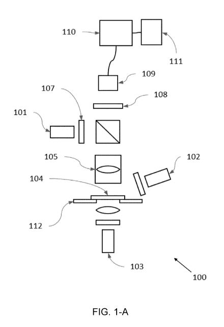
Figures 1-A to 1-E schematically show five embodiments of the hyperspectral
quantitative imaging cytometry system according to the invention.
Figure 2 shows a flow chart of a method according to an embodiment of the
invention.
Figure 3 schematically shows the process of obtaining wavelength-coded spatial
maps
of the sample.
DETAILED DESCRIPTION OF THE INVENTION
A hyperspectral quantitative imaging cytometry system (100) according to the
invention
is schematically shown in figures 1-A to 1-E.
The system (100) comprises an observation region (104), comprising a sample
holder
(112) configured to hold one or more solid-phase samples. In an embodiment the
sample
holder is configured to retain at least one solid phase sample support, each
support being
adapted to contain an immobilized sample, preferably a biological sample.
These solid
phase sample supports may be of different materials, preferably crystalline
and
transparent to light (e.g. glass or plastic), and may have different shapes
and sizes, such
as rectangular shape (e.g. a microscope slide).
The biological samples may be of human, animal, fungal or plant origin and may
be used
alone or in combination with a molecular tag.
Preferably, a molecular tag is used to reveal the components of the biological
sample.
The reporter part of the molecular tag may luminesce or selectively absorb
radiation
when irradiated. The molecular tag may exhibit a characteristic radiation
spectrum
because of its physical structure or when combined with a biological sample.
CA 03142188 2021-11-29
WO 2020/239981 PCT/EP2020/064984
- 16 -
The system (100) comprises one or several radiation sources (101, 102, 103) to
irradiate
and stimulate the markers of a sample placed in the observation region (104).
From this
interaction, radiation may be emitted by the sample as a result of any process
such as
scattering, fluorescence, phosphorescence, chemiluminescence or selective
absorption/transmittance.
In an embodiment, the biological sample to be placed in the observation region
(104) is
a biological tissue, i.e. a collection of interconnected cells and their
extracellular matrix
that perform a similar function within an organism. The components of that
biological
tissue may naturally absorb light of certain wavelengths and emit radiation
usually at a
different wavelength. If the components of a biological tissue lack enough
contrast to be
directly studied, class-specific tags may be added to the sample in order to
provide
contrast and allow the detection of specific components. These components may
be
DNA, proteins, lipids, carbohydrates, or others, and multiple tags may be used
to study
multiple components simultaneously.
In response to the radiation, the components of the sample and/or the
molecular tags
used in combination with the sample emit a spectrum of radiation which is
captured by a
collection element (105). The collected radiation is directed to a
multichannel filtration
element (108) and redirected to an image sensor (109).
The embodiment shown in figure 1-A includes three radiation sources (101, 102,
103)
which emit light in the visible spectrum. However, a different number of
radiation sources
may be used. Also, any known radiation source suitable for excitation of the
target
sample material may be used. For example, the radiation sources (101, 102,
103) may
be lasers, light emitting diodes ("LEDs") and/or lamps. The lasers or LEDs may
be
configured to emit a multiple number of excitation wavelengths or a single
wavelength.
If the radiation source (101) produces more than one wavelength of radiation,
a band-
pass filter (107) may be placed in front of the radiation source (101) in
order to filter out
any unwanted wavelength before the radiation reaches the sample in the
observation
region (104).
In the embodiment of figure 1-A each radiation source (101, 102, 103)
irradiates the
totality of the observation region (104) at once (i.e. wide field
irradiation).
In the embodiment shown in figure 1-B, there is only one radiation source
(101) which
CA 03142188 2021-11-29
WO 2020/239981 PCT/EP2020/064984
- 17 -
irradiates the observation region (104) with beams of light (106) coming from
the same
side where the collection element (105) is placed and directed along the
optical axis of
the collection element (105) (bright field epi-illumination). In another
embodiment shown
in figure 1-C, a radiation source (102) irradiates the observation region
(104) with beams
of light (106) coming from the same side where the collection element (105) is
placed
and directed at a non-zero angle with respect to the optical axis of the
collection element
(105) (dark field epi-illumination). In another embodiment shown in figure 1-
D, a radiation
source (103) irradiates the observation region (104) with beams of light (106)
coming
from the side of the observation region opposite to the side where the
collection element
(105) is placed and directed along the optical axis of the collection element
(105) (bright
field trans-illumination). In another embodiment shown in figure 1-E, a
radiation source
(103) irradiates the observation region (104) with beams of light (106) coming
from the
side of the observation region opposite to the side where the collection
element (105) is
placed and directed at a non-zero angle with respect to the optical axis of
the collection
element (105) (dark field trans-illumination).
Thus, in the embodiments of figures 1-B and 1-C the radiation source (101,
102) is
arranged on the same side of the observation region (104) as the collection
element
(105), such that the sample in the observation region (104) reflects the
radiation of the
radiation source (101, 102) before being collected by the collection element
(105). In the
embodiments of figures 1-D and 1-E the observation region (104) is interposed
between
the radiation source (103) and the collection element (105), such that the
radiation from
the radiation source (103) passes through the sample in the observation region
(104)
before being collected by the collection element (105).
In a preferred embodiment as the one shown in figure 1-A, two or more of the
above
imaging geometries shown in figures 1-B to 1-E may co-exist in the system and
may be
used sequentially to obtain complementary data.
The radiation emitted by the irradiated sample is collected by the collection
element
(105). In a preferred embodiment the collection element is configured to
capture radiation
from multiple spatial locations of the observation region (104)
simultaneously. In an
embodiment the collection element is a lens or a combination of lenses.
In a preferred embodiment the collection element has a low magnification
factor (M) in
order to achieve a large field of view (FOV) and obtain information on a
larger two-
CA 03142188 2021-11-29
WO 2020/239981 PCT/EP2020/064984
- 18 -
dimensional area of the observation region (104) and, consequently, of the
sample.
Preferably, the magnification factor value (M) is lower than 20, more
preferably lower
than 10, most preferably lower than 2. The total magnification factor of the
collection
element (105) combined with any other element of the system with a
magnification factor
may be selected to guarantee a correct sampling frequency of the FOV by the
image
sensor (109). The sampling frequency of the FOV may be selected to distinguish
individual cells in a biological tissue sample, but not to distinguish small
subcellular
details. The image sensor (109) comprises a two-dimensional array of radiation
detecting
elements. The final magnification factor of the system may depend on the
characteristics
of the image sensor, such as the size of the radiation detecting elements.
After being collected by the collection element (105) and before reaching the
image
sensor (109), the radiation is directed through a multichannel filtration
element (108).
The multichannel filtration element (108) selectively filters the radiation
that reaches each
radiation detecting element, or a group of radiation detecting elements, of
the image
sensor (109). In an embodiment the multichannel filtration element (108) is a
continuous
or semi-continuous variable bandpass filter.
In this embodiment the multichannel filtration element is arranged to be
displaceable in
one or two spatial dimensions (x, y), thus allowing the image sensor (109) to
generate a
plurality of two-dimensional outputs, each output representing a different
wavelength-
coded (A=A(y)) two-dimensional (x, y) map of the sample. Each wavelength-coded
two-
dimensional map of the sample generated by the image sensor (109) may be sent
to a
processor (110) and/or stored in a memory (111) for further processing.
The image sensor (109) is a two-dimensional array sensor, where each radiation
detecting element in the array receives radiation coming from a different two-
dimensional
spatial location in the sample, generating an image that is a spatial (x, y)
map of the
sample under study. The image sensor (109) may be a two-dimensional
photodetector
array sensor, such as a charge-coupled device (CCD), a complementary metal
oxide
semiconductor (CMOS), an electron multiplier CCD (EMCCD) or any other similar
system to obtain two-dimensional spatial data. The two-dimensional image
sensor may
be triggered to sample data cumulatively during a specified amount of time.
Figure 2 shows a flow chart of a method according to an embodiment of the
invention. A
sample, which may have been stained with a tag, is placed (201) in the
observation
CA 03142188 2021-11-29
WO 2020/239981 PCT/EP2020/064984
- 19 -
region (104). A first radiation source (101, 102, 103) is activated (202), the
filtration
element (108) is located (203) in its initial position and the image sensor
(109) records
(204) a first image representing the first wavelength-coded two-dimensional
map of the
biological sample. The filtration element (108) is then moved (203) to a
second position
and a second wavelength-coded two-dimensional map is obtained. This process is
continued until the last position of the filtration element (108) is reached.
If more than
one irradiation modes (a) or irradiation wavelengths (A) are used, a second
radiation
source (101, 102, 103) is activated (202), the filtration element (108) is
moved (203) to
its initial position and a new image is taken, corresponding to a new
wavelength-coded
two-dimensional map. This process is continued until the last position of the
filtration
element (108) is reached and repeated for all the radiation sources.
A processor (110) takes the plurality of wavelength-coded two-dimensional maps
(301)
of the biological sample and builds (205) a plurality of monochromatic two-
dimensional
maps (304) of the sample by combining parts of the wavelength-coded two-
dimensional
maps (301) which correspond to a specific wavelength. Each one of these
monochromatic two-dimensional maps has spatially related (x, y) information on
the
radiation emitted at a given wavelength (A) when the sample is irradiated with
a given
radiation wavelength (A) using a given irradiation mode (a). The combination
of these
plurality of monochromatic two-dimensional maps (304) constitutes a five-
dimensional
(x, y, A, A, a) dataset (305) which may be simplified into a spectral cube
(306) with two
spatial dimensions (x, y) and one spectral dimension (a, A, A).
Although in this exemplary embodiment, the monochromatic two-dimensional maps
are
generated by combining parts of the wavelength-coded two-dimensional maps
which
correspond to a specific wavelength, in an alternative embodiment, the
monochromatic
two-dimensional maps are generated by employing a multivariate interpolation
process
to obtain for each pixel an estimation of the radiation received at such
specific
wavelength (A) based on the recordings from the wavelength-coded two-
dimensional
maps.
The processor (110) takes a spectral cube (306) generated from a biological
sample and
performs a spatial segmentation (206) of data locations in order to identify
meaningful
biological sample structures (e.g. cells) and obtain the spectral signature
(207) of each
of those structures. Then, the processor (110) compares (208) the spectral
signature of
each structure to a database of spectra of known structures stored in a memory
(111).
CA 03142188 2021-11-29
WO 2020/239981 PCT/EP2020/064984
- 20 -
Either alternatively or concurrently with step 208, the processor (110)
performs an
estimation (209) of the abundance of each marker (i.e. spectral unmixing) in
each of
those identified sample structures by determining the relative contribution
from each
marker to the spectral signature of each structure identified in the spectral
cube. The
information obtained on the nature of the structures identified and/or on the
abundance
of each marker, may be correlated (210) by an expert in the field with other
relevant
information on the sample.
A generally accepted model for a mixture of emissions, needed to perform
spectral
unmixing, is a Linear Mixing Model (LMM) which assumes a linear combination of
the
abundance of the emissions. The plurality of monochromatic two-dimensional
maps
provides a discontinuous emission spectrum for each pixel, wherein the point
of the
spectrum corresponding to a specific wavelength is found in the monochromatic
two-
dimensional map corresponding to said wavelength.
According to LMM, it is assumed that the discontinuous emission spectrum is a
linear
combination of the spectra of individual markers. The emissions of M excited
markers
coming from N pixels or structures are taken at L excitation wavelengths (A)
and
generate L individual signals (channels). Each pixel or structure is therefore
represented
by a vector of L channels that contains the sum of contributions of M markers
per
channel. The LMM can be written in its matrix form as:
Y = AH
where Y is the LxN matrix of detected intensities, A is the LxM matrix of
mixing that
contains the expected emission of each of the M markers in each of the L
spectral
channels and H is the MxN matrix of real markers concentrations for each
structure.
The uncertainty in the measurement may also be considered by including noise
into the
model. Usually, two noise models are adopted: the first model is an additive
gaussian
noise (white noise) model, in which the above equation is modified into:
Y = AH + R
where R is a matrix formed by independently identically distributed gaussian
variables
with zero mean. The second model uses a Poisson process to model the photon
CA 03142188 2021-11-29
WO 2020/239981 PCT/EP2020/064984
-21 -
emission.
Different methods, like ordinary least-square (OLS), weighted least squares
(WLS),
generalized linear model (GLM), non-negative least-squares (NNLS), among
other, may
be employed to solve the LMM problem and obtain spatial maps with the
abundance of
each marker, which may be sent to a processor and/or stored in a memory for
further
processing.
Figure 3 represents an embodiment of the steps of obtaining wavelength-coded
two-
dimensional maps of the biological sample (A) and their transformation into
monochromatic two-dimensional maps of the biological sample (B & C) which,
when
combined, constitute a spectral cube of the sample (D). The process uses all
the
consecutive images taken with the different positions of the filtration
element (108) for a
given irradiation wavelength (A) and a given irradiation mode (a) and, then,
works in a
(x, y, A) dimensional space. If more than one irradiation wavelength (A)
and/or more than
one irradiation mode (a) has been used, the process is done for each set of
images
independently, wherein each set of images correspond to a given irradiation
wavelength
(A) and a given irradiation mode (a).
Each wavelength-coded two-dimensional map (301) is obtained by moving the
filtration
element (108) parallel to one of the spatial dimensions (y) of the FOV of the
collection
element in several consecutive discrete steps (A). The number of steps needed
for the
complete displacement of the filtration element through the full FOV (303)
defines the
number of images taken and thus the number of wavelength-coded two-dimensional
maps.
Each wavelength-coded two-dimensional map (301) is divided (sliced) into "n"
bands
along the "y" spatial dimension (B). The number of bands "n" corresponds to
the number
of steps needed for the complete displacement of one band of the filter
through the full
FOV. Each one of the bands is combined with the bands from the other
wavelength-
coded two-dimensional maps (301) that correspond to the same wavelengths to
recreate
monochromatic two-dimensional maps (304) of the biological sample, each one
corresponding to a specific wavelength. The complete set of monochromatic two-
dimensional maps (304) constitute a spectral cube (306). In figure 3 the bands
corresponding to the same wavelength are represented with the same pattern.
CA 03142188 2021-11-29
WO 2020/239981 PCT/EP2020/064984
- 22 -
In an alternative embodiment, the monochromatic two-dimensional maps are
generated
from the wavelength-coded two-dimensional maps by employing a multivariate
interpolation process to obtain for each pixel an estimation of the radiation
received at
specific wavelengths (A) based on the recordings from the wavelength-coded two-
dimensional maps.
The following clauses are herein provided according to the invention:
Clause 1. A hyperspectral quantitative imaging cytometry system (100),
comprising:
an observation region (104), comprising a sample holder configured to hold one
or
more solid-phase samples,
at least one radiation source (101, 102, 103) configured to irradiate the
observation
region (104),
a collection element (105) configured to collect the radiation emitted through
or
reflected by the sample upon irradiation by the at least one radiation source
(101, 102,
103),
a multichannel filtration element (108) configured to selectively filter the
wavelength of the radiation collected by the collection element (105), and
an image sensor (109) configured to receive the filtered radiation and to
generate
an image that is a two-dimensional map of the sample, the image sensor (109)
comprising a two-dimensional array of radiation detecting elements.
Clause 2. Hyperspectral quantitative imaging cytometry system (100) according
to the
previous clause, wherein the filtration element (108) is arranged to be
movable between
at least two positions, wherein each position of the filtration element (108)
selectively
filters the wavelength of the radiation that reaches each radiation detecting
element of
the image sensor (109).
Clause 3. Hyperspectral quantitative imaging cytometry system (100) according
to
clause 2, wherein the system further comprises a processor (110), the
processor (110)
being configured to:
- receive a plurality of wavelength-coded two-dimensional maps (301) of the
sample, the plurality of wavelength-coded two-dimensional maps (301) being
associated
to a plurality of positions of the filtration element (108), wherein a
wavelength-coded two-
dimensional map is an image generated by the image sensor (109) based on the
radiation it receives for a position of the filtration element (108);
CA 03142188 2021-11-29
WO 2020/239981 PCT/EP2020/064984
- 23 -
- generate (205) a plurality of monochromatic two-dimensional maps (304) of
the
sample by combining parts of the wavelength-coded two-dimensional maps (301)
of the
sample which correspond to a specific wavelength (A);
- build a spectral cube (306) comprising the plurality of monochromatic two-
dimensional maps (304);
- identify sample structures (206) on the spectral cube (306) and obtain
their
spectral signature (207);
- compare (208) the obtained spectral signatures with a database of
spectral
signatures of known structures and/or decompose the spectral signatures and
obtain
(209) an estimation of the abundance of each marker in each of the identified
sample
structures.
Clause 4. Hyperspectral quantitative imaging cytometry system (100) according
to any
of the previous clauses, wherein the observation region (104) is interposed
between at
least one radiation source (103) and the collection element (105), such that
the radiation
of said radiation source (103) passes through the observation region (104)
before being
collected by the collection element (105), according to a trans-illumination
configuration.
Clause 5. Hyperspectral quantitative imaging cytometry system (100) according
to any
of the previous clauses, wherein at least one radiation source (102), the
observation
region (104) and the collection element (105) are arranged according to a dark
field
configuration.
Clause 6. Hyperspectral quantitative imaging cytometry system (100) according
to any
of the previous clauses, wherein at least one radiation source (101, 103), the
observation
region (104) and the collection element (105) are arranged according to a
bright field
configuration.
Clause 7. Hyperspectral quantitative imaging cytometry system (100) according
to any
of the previous clauses, wherein at least one radiation source (101, 102) is
oriented
towards the observation region (104), such that the observation region (104)
reflects the
radiation of said radiation source (101, 102) before being collected by the
collection
element (105), according to an epi-illumination configuration.
Clause 8. Hyperspectral quantitative imaging cytometry system (100) according
to any
of the previous clauses, wherein the system (100) further comprises a memory
(111).
CA 03142188 2021-11-29
WO 2020/239981 PCT/EP2020/064984
- 24 -
Clause 9. Hyperspectral quantitative imaging cytometry system (100) according
to any
of the previous clauses, wherein the system (100) further comprises at least
one band-
pass filter (107) interposed between at least one radiation source (101, 102,
103) and
the observation region (104).
Clause 10. Hyperspectral quantitative imaging cytometry system (100) according
to any
of the previous clauses, wherein at least one radiation source (101, 102, 103)
is a laser,
a light emitting diode or a lamp.
Clause 11. Hyperspectral quantitative imaging cytometry system (100) according
to any
of the previous clauses, wherein the collection element (105) comprises a
lens, or a
combination of lenses, configured to capture radiation from multiple spatial
locations of
the observation region simultaneously.
Clause 12. Hyperspectral quantitative imaging cytometry system (100) according
to any
of the previous clauses,
wherein the collection element (105) has a magnification factor value (M)
lower
than 20, preferably lower than 10, most preferably lower than 2; and/or
wherein the collection element (105) has a numerical aperture value higher
than
0.25, preferably equal to or higher than 0.5.
Clause 13. Hyperspectral quantitative imaging cytometry system (100) according
to any
of the previous clauses, wherein the filtration element (108) is a continuous
or a semi-
continuous linear variable filter between 200 nm and 1200 nm, preferably
between 350
nm and 950 nm.
Clause 14. Method for obtaining data from a solid-phase sample using a
hyperspectral
quantitative imaging cytometry system (100) according to any of the previous
claims,
comprising the following steps:
a) providing a sample (201);
b) irradiating the sample (202) with radiation that interacts with the sample,
such
that the sample emits radiation;
c) capturing the emitted radiation with the collection element (105);
d) filtering the emitted radiation using the multichannel filtration element
(108);
e) sequentially recording (204) a plurality of wavelength-coded two-
dimensional
CA 03142188 2021-11-29
WO 2020/239981 PCT/EP2020/064984
- 25 -
maps (301) of the sample coordinating with the sequential displacement (203)
of the
filtration element (108), wherein each position of the filtration element
(108) selectively
filters the wavelength (A) of the radiation that reaches each radiation
detecting element
of the image sensor (109) and wherein a wavelength-coded two-dimensional map
is
generated by the image sensor (109) based on the radiation it receives for
each position
of the filtration element (108);
f) generating (205) a plurality of monochromatic two-dimensional maps (304) of
the
sample by combining parts of the wavelength-coded two-dimensional maps (301)
of the
sample which correspond to a specific wavelength (A) of emitted radiation;
g) building a spectral cube (306) comprising the plurality of monochromatic
two-
dimensional maps (304);
h) identifying sample structures (206) on the spectral cube (306) and
obtaining
their spectral signature (207);
i) comparing (208) the spectral signatures obtained with a database of
spectral
signatures of known structures and/or decomposing the spectral signatures and
obtaining (209) an estimation of the abundance of each marker in each of the
identified
sample structures.
Clause 15. Method according to clause 14, wherein steps b) to f) are performed
for a
plurality of irradiation wavelengths (A) and/or a plurality of irradiation
modes (a), wherein
in step f) each monochromatic two-dimensional map (304) corresponds to
radiation
emitted at a specific wavelength (A) when the sample is irradiated with a
given irradiation
wavelength (A) and in a given irradiation mode (a), and wherein step g) is
performed by
combining the plurality of monochromatic two-dimensional maps (304).