Note: Descriptions are shown in the official language in which they were submitted.
CA 03143225 2021-12-10
WO 2021/038559
PCT/IL2020/050925
ANCHOR CHANNEL TIP
CROSS-REFERENCE TO RELATED APPLICATIONS
[0001] This application claims the benefit of U.S. Provisional Patent
Application No.
62/894,517, filed August 30, 2019, which is incorporated by reference herein
for all purposes.
BACKGROUND
[0002] Annuloplasty structures comprising a flexible material through which
anchors are
delivered tend to twist and warp as a result of passage of the anchor through
the material of the
annuloplasty structure. It is therefore often advantageous to provide devices
and techniques to
facilitate deployment of the tissue anchor through the flexible material of
the annuloplasty
structure while minimizing or eliminating twisting or warping of the flexible
material during
deployment of the anchor.
SUMMARY OF THE INVENTION
[0003] This summary is meant to provide some examples and is not intended to
be limiting of
the scope of the invention in any way. For example, any feature included in an
example of this
summary is not required by the claims, unless the claims explicitly recite the
features. Also, the
features described can be combined in a variety of ways. Various features and
steps as described
elsewhere in this disclosure can be included in the examples summarized here.
[0004] A tubular structure is used to advance toward a tissue site of a
subject an anchor driver
used to drive a tissue anchor into tissue of a subject. The tubular structure
has a distal end portion
comprising an implant-gripping element that is configured to temporarily grip
material (e.g.,
flexible material) of an annuloplasty structure, in accordance with some
applications of the
present invention.
[0005] For some applications of the present invention, the implant-gripping
element comprises
a plurality of teeth which reversibly grip the material of the annuloplasty
structure during
deploying, or driving, a tissue anchor through material of the annuloplasty
structure so as to
anchor the annuloplasty structure to tissue of the subject.
[0006] For some applications of the present invention, the implant-gripping
element comprises
a deformable element which changes its structural configuration as a tissue
anchor is passed with
respect to and engages the deformable element. This is advantageous because
the tubular
structure is able to move freely within a lumen of the annuloplasty structure
and only engage
1
CA 03143225 2021-12-10
WO 2021/038559
PCT/IL2020/050925
and grip the annuloplasty structure once the desired location of tissue has
been reached and it
has been determined that in this location, a tissue anchor be driven into
tissue.
[0007] There is therefore provided, in accordance with an application of the
present invention,
a system and/or apparatus, for use with a tissue anchor, the system/apparatus
including an
implant, dimensioned to be advanced into a body of a subject and an anchor-
delivery tool. The
anchor delivery tool can include an anchor-delivery channel, shaped to define
a lumen
therethrough, the lumen having a diameter, and the channel being dimensioned
to be moveable
within a lumen of the implant. The anchor-delivery tool can also include an
implant-gripping
element disposed at a distal end portion of the anchor-delivery channel. The
implant-gripping
element can be configured to reversibly grip an inner wall of the implant
during implantation of
the tissue anchor via the anchor-delivery channel.
[0008] In an application, the implant-gripping element includes a radiopaque
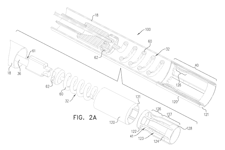
material.
[0009] In an application, the implant includes a flexible material, and the
flexible material of
the implant encases a distal portion of the channel.
[0010] In an application, the implant-gripping element includes a plurality of
teeth which
increase friction between the implant and the anchor-delivery channel.
[0011] In an application, the plurality of teeth are cut from a distal portion
of a cylinder coupled
to the distal end portion of the anchor-delivery channel.
[0012] In an application:
each one of the plurality of teeth includes a respective elongate element that
is aligned
with a longitudinal axis of the distal end portion of the anchor-delivery
channel,
a portion of the implant surrounds the plurality of elongate elements, and
the elongate elements are spaced apart from one another such that the
plurality of
elongate elements are configured to grip the portion of the implant.
[0013] In an application, a respective distal portion of each of the plurality
of teeth are
configured to grip the implant.
[0014] In an application, the implant includes a braided fabric, and the
distal portions of the
plurality of teeth are configured to reversibly ensnare the braided fabric.
2
CA 03143225 2021-12-10
WO 2021/038559
PCT/IL2020/050925
[0015] In an application, the system/apparatus further includes the tissue
anchor, and the tissue
anchor includes:
an anchor head; and
a tissue-engaging member, coupled to the anchor head, extending distally away
from
the anchor head until a distal tip of the tissue-engaging member, and
configured to anchor the
anchor to the tissue.
[0016] In an application, the tissue-engaging member includes a helical tissue-
engaging
member, and the implant-gripping element is configured to reversibly grip the
implant and
prevent twisting of the implant during corkscrewing of the helical tissue-
engaging member with
1 0 respect to the implant.
[0017] In an application, the system/apparatus further includes an anchor
driver slidable
through the lumen of the anchor-delivery channel, the anchor driver including:
a longitudinal shaft, having a flexible distal portion and a distal end; and
a deployment element coupled to the distal end of the shaft, and reversibly
couplable to
the anchor head.
[0018] In an application, the implant-gripping element includes at least one
deformable element
configured to change shape from a resting state to a gripping state in
response to passage of the
tissue anchor alongside the deformable element.
[0019] In an application, the implant-gripping element includes a plurality of
deformable
2 0 elements disposed circumferentially with respect to the distal end
portion of the anchor-delivery
channel.
[0020] In an application, the deformable element is shaped so as to define an
elongate tine
having a straight portion and a curved portion in the resting state of the
deformable element, and
in the gripping state of the deformable element, the anchor is configured to
radially push against
the curved portion so as to straighten the curved portion and responsively,
longitudinally
lengthen the deformable element.
[0021] In an application, in the gripping state, a distal end of the
deformable element extends
beyond a distal end of the anchor-delivery channel.
[0022] In an application, the at least one deformable element includes a
plurality of elongate
tines, and the anchor is configured to radially push against the respective
curved portions of the
plurality of elongate tines.
3
CA 03143225 2021-12-10
WO 2021/038559
PCT/IL2020/050925
[0023] In an application, the distal ends of the plurality of elongate tines
are configured to
increase surface area contact with the inner wall of the implant in the
gripping state of the
deformable element.
[0024] In an application:
the deformable element is shaped so as to define a laterally-moveable lateral
projection,
in the resting state of the deformable element, a lateral-most portion of the
projection is
aligned with a lateral surface of the anchor-delivery channel, and
in the gripping state, the anchor is configured to radially push against the
lateral
projection so as to extend the lateral-most portion of the projection beyond
the lateral surface
of the anchor-delivery channel.
[0025] In an application, the at least one deformable element includes a
plurality of lateral
projections, and the anchor is configured to radially push against the
plurality of lateral
projections.
[0026] In an application, the plurality of lateral projections are configured
to increase surface
area contact with the inner wall of the implant in the gripping state of the
deformable element.
[0027] There is further provided, in accordance with an application of the
present invention, a
method including positioning an implant along an annulus of a heart valve of a
subject. The
implant is optimally dimensioned to be advanced into a body of the subject.
The method can
further include advancing an anchor-delivery tool with respect to the implant.
[0028] In some applications, the anchor-delivering tool includes an anchor-
delivery channel,
shaped to define a lumen therethrough, the lumen having a diameter. The
channel can be
dimensioned to be moveable within a lumen of the implant.
[0029] In some applications, the anchor-delivering tool includes an implant-
gripping element
disposed at a distal end portion of the anchor-delivery channel, the implant-
gripping element
being configured to reversibly grip a portion or wall of the implant (e.g., an
inner wall of the
implant, etc.) during implantation of the tissue anchor via the anchor-
delivery channel.
[0030] The method can further include gripping a first portion of the implant
using the implant-
gripping element, and during the gripping of the first portion, anchoring the
first portion of the
implant to the annulus using a tissue anchor deliverable through the anchor-
delivery channel.
[0031] In an application, the method further includes:
4
CA 03143225 2021-12-10
WO 2021/038559
PCT/IL2020/050925
decoupling the implant-gripping element from the first portion of the implant
subsequently to the anchoring of the first portion of the implant to the
annulus;
moving the anchor-delivery channel to a second portion of the implant;
gripping the second portion of the implant using the implant-gripping element;
and
during the gripping of the second portion, anchoring the second portion of the
implant
to the annulus using a second tissue anchor deliverable through the anchor-
delivery channel.
[0032] In an application, the implant-gripping element includes a radiopaque
material.
[0033] In an application, the implant includes a flexible material, and the
flexible material of
the implant encases a distal portion of the channel.
[0034] In an application, the implant-gripping element includes a plurality of
teeth, and
gripping the first portion of the implant includes increasing friction between
the first portion of
the implant and the anchor-delivery channel.
[0035] In an application, the plurality of teeth are cut from a distal portion
of a cylinder coupled
to the distal end portion of the anchor-delivery channel, and gripping the
first portion of the
implant includes sandwiching the first portion of the implant between
respective distal ends of
the plurality of teeth and the annulus.
[0036] In an application:
each one of the plurality of teeth includes a respective elongate element that
is aligned
with a longitudinal axis of the distal end portion of the anchor-delivery
channel,
a lateral portion of the implant surrounds the plurality of elongate elements,
the elongate elements are spaced apart from one another, and
gripping the first portion of the implant includes gripping the lateral
portion of the
implant by the elongate elements.
[0037] In an application, a respective distal portion of each of the plurality
of teeth are
configured to grip the implant, and gripping the first portion of the implant
includes sandwiching
the first portion of the implant between respective distal ends of the
plurality of teeth and the
annulus.
[0038] In an application, the implant includes a braided fabric, and gripping
the first portion of
the implant includes reversibly ensnaring the braided fabric by the plurality
of teeth.
[0039] In an application:
5
CA 03143225 2021-12-10
WO 2021/038559
PCT/IL2020/050925
the tissue anchor includes:
an anchor head; and
a tissue-engaging member, coupled to the anchor head, extending distally away
from the anchor head until a distal tip of the tissue-engaging member, and
configured to
anchor the anchor to the tissue, and
anchoring the first portion of the implant to the annulus includes anchoring
the first
portion using the tissue anchor including the anchor head and the tissue-
engaging member.
[0040] In an application:
the tissue-engaging member includes a helical tissue-engaging member,
1 0 anchoring the first portion of the implant includes corkscrewing the
helical tissue-
engaging member with respect to the first portion of the implant and into the
annulus, and
gripping the first portion of the implant includes using the implant-gripping
element to
reversibly grip the first portion of the implant and prevent twisting of the
implant during the
corkscrewing of the helical tissue-engaging member with respect to the first
portion of the
.. implant.
[0041] In an application, the method further includes sliding through the
lumen of the anchor-
delivery channel an anchor driver including:
a longitudinal shaft, having a flexible distal portion and a distal end; and
a deployment element coupled to the distal end of the shaft, and reversibly
couplable to
the anchor head.
[0042] In an application, the implant-gripping element includes at least one
deformable element
configured to change shape from a resting state to a gripping state in
response to passage of the
tissue anchor alongside the deformable element, and the method further
includes changing the
shape of the deformable element by passing the tissue anchor alongside the
deformable element.
[0043] In an application, the implant-gripping element includes a plurality of
deformable
elements disposed circumferentially with respect to the distal end portion of
the anchor-delivery
channel.
[0044] In an application, the deformable element is shaped so as to define an
elongate tine
having a straight portion and a curved portion in the resting state of the
deformable element, and
passing the tissue anchor alongside the deformable element includes radially
pushing the anchor
against the curved portion, and by the pushing, straightening the curved
portion and
6
CA 03143225 2021-12-10
WO 2021/038559
PCT/IL2020/050925
responsively, longitudinally lengthening the deformable element such that the
deformable
element assumes the gripping state.
[0045] In an application, in the gripping state, longitudinally lengthening
the deformable
element includes extending a distal end of the deformable element beyond a
distal end of the
anchor-delivery channel.
[0046] In an application, the at least one deformable element includes a
plurality of elongate
tines, and radially pushing the anchor includes radially pushing the anchor
against the respective
curved portions of the plurality of elongate tines.
[0047] In an application, gripping the first portion of the implant includes
increasing surface
area contact with the inner wall of the implant in the gripping state of the
deformable element
using the distal ends of the plurality of elongate tines.
[0048] In an application:
the deformable element is shaped so as to define a laterally-moveable lateral
projection,
in the resting state of the deformable element, a lateral-most portion of the
projection is
aligned with a lateral surface of the anchor-delivery channel,
in the gripping state, the anchor is configured to radially push against the
lateral
projection so as to extend the lateral-most portion of the projection beyond
the lateral surface
of the anchor-delivery channel,
passing the tissue anchor alongside the deformable element includes radially
pushing
2 0 the anchor against the lateral projection, and by the pushing,
extending the lateral-most portion
of the projection beyond the lateral surface of the anchor-delivery channel.
[0049] In an application, the at least one deformable element includes a
plurality of lateral
projections, and pushing the anchor against the lateral projection includes
radially pushing the
anchor against the plurality of lateral projections.
[0050] In an application, radially pushing the anchor against the plurality of
lateral projections
includes increasing surface area contact with the inner wall of the implant in
the gripping state
of the deformable element.
[0051] The foregoing method(s) and other methods herein can be performed on a
living animal
or on a simulation, such as on a cadaver, cadaver heart, simulator (e.g. with
the body parts, tissue,
etc. being simulated), etc.
7
CA 03143225 2021-12-10
WO 2021/038559
PCT/IL2020/050925
[0052] The present invention will be more fully understood from the following
detailed
description of applications thereof, taken together with the drawings, in
which:
BRIEF DESCRIPTION OF THE DRAWINGS
[0053] Fig. 1 is a schematic illustration of an example of an implant-gripping
element
comprising a plurality of teeth;
[0054] Figs. 2A-C are schematic illustrations of an example of an implant-
gripping element
comprising a deformable element; and
[0055] Figs. 3A-C are schematic illustrations of an example of an implant-
gripping element
comprising another deformable element.
DETAILED DESCRIPTION OF EMBODIMENTS
[0056] Reference is now made to Fig. 1, which is a schematic illustration of a
system 10
providing one or more rotationally-controlled steering catheters configured
for delivering an
implant to a heart of a subject, in accordance with some applications of the
present invention.
Fig. 1 shows a distal portion of an implant that comprises an annuloplasty
ring structure 222 (i.e.,
an implant, e.g., an annuloplasty band) comprising a flexible sleeve 26. The
implant is
dimensioned to be advanced into a body of a subject. System 10 comprises an
anchor-delivery
tool comprising an implant-decoupling channel 18. As described hereinbelow,
channel 18 is used
to facilitate delivery of tissue anchors through channel 18 and into a lumen
of sleeve 26. Thus,
channel 18 functions as an anchor-delivery channel. Channel 18 is shaped so as
to define a lumen
having a diameter. Sleeve 26 comprises a flexible material which encases a
distal portion of
channel 18. Channel 18 is dimensioned to be moveable within a lumen of the
implant. An
implant-gripping element 40 is disposed at a distal end portion of channel 18.
Implant-gripping
element 40 is configured to reversibly grip an inner wall 50 of the implant
during implantation
of tissue anchor 32 via channel 18.
[0057] Sleeve 26 typically comprises a braided fabric mesh, e.g., comprising
polyethylene
terephthalate (such as Dacron (TM)). Sleeve 26 can be configured to be placed
only partially
around a cardiac valve annulus (i.e., to assume a C-shape), and, once anchored
in place, to be
contracted so as to circumferentially tighten the valve annulus. Though
optionally, the ring
structure can also be configured to be placed entirely around the valve
annulus.
[0058] Sleeve 26 has a tubular lateral wall 253 that (i) circumscribes a
central longitudinal axis
of the sleeve, and (ii) defines the lumen of the sleeve.
8
CA 03143225 2021-12-10
WO 2021/038559
PCT/IL2020/050925
[0059] In order to tighten the annulus, annuloplasty ring structure 222
comprises a flexible
elongated contraction member 226 that extends along sleeve 26. Elongated
contraction member
226 comprises a wire, a ribbon, a rope, or a band, which typically comprises a
flexible and/or
superelastic material, e.g., nitinol, polyester, stainless steel, or cobalt
chrome. For some
applications, the wire comprises a radiopaque material. For some applications,
contraction
member 226 comprises a braided polyester suture (e.g., Ticron). For some
applications,
contraction member 226 is coated with polytetrafluoroethylene (PTFE). For some
applications,
contraction member 226 comprises a plurality of wires that are intertwined to
form a rope
structure.
[0060] For some applications, annuloplasty ring structure 222 comprises an
adjustment
mechanism as described with reference to PCT application PCT/IL2016/050433 to
Iflah, et al.,
which published as WO 16/174669, and which is incorporated herein by
reference. The
adjustment mechanism facilitates contracting and expanding of annuloplasty
ring structure 222
so as to facilitate adjusting of a perimeter of the annulus and leaflets of
the cardiac valve. The
adjustment mechanism can comprise a rotatable structure (e.g., a spool).
[0061] System 10 can comprise a concentric arrangement of tubes defining an
implant-delivery
tool. System 10 can comprise a first, outer catheter 12 comprising a sheath
configured for
transluminal advancement through vasculature of a subject. For some
applications of the present
invention, outer catheter 12 comprises a sheath configured for advancement
through a femoral
artery toward an interatrial septum of a heart of a subject. A distal end
portion 112 of outer
catheter 12 is configured to pass through the transatrial septum of the
subject, and to be oriented
in a desired spatial orientation within the left atrium. System 10 comprises a
second catheter, or
guide catheter 14, comprising a distal end portion 114 that is configured to
pass through catheter
12 (i.e., a primary lumen thereof), to become disposed outside of a distal end
of the outer catheter,
and to be oriented in a desired spatial orientation within the left atrium.
[0062] Distal end portion 112 of outer catheter 12 is steerable. That is,
distal end portion 112 is
deflectable with respect to an immediately more proximal portion of catheter
12 (e.g., by using
extracorporeal elements of system 10). Distal end portion 114 of inner
catheter 14 is steerable.
That is, distal end portion 114 is deflectable with respect to an immediately
more proximal
portion of catheter 14 (e.g., by using extracorporeal elements of system 10.
[0063] Guide catheter 14 is steerable to a desired spatial orientation in
order to facilitate
advancing and implantation of an implant in a body cavity of the subject.
9
CA 03143225 2021-12-10
WO 2021/038559
PCT/IL2020/050925
[0064] For applications in which system 10 is used to deliver an implant to
the mitral valve of
the subject, often, outer catheter 12 is configured for initial advancement
through vasculature of
the subject until a distal end of catheter 12 is positioned in the left
atrium. The distal steerable
end portion of catheter 12 is then steered such that distal end of catheter 12
is positioned in a
desired spatial orientation within the left atrium. The steering procedure can
be performed with
the aid of imaging, such as fluoroscopy, transesophageal echo, and/or
echocardiography.
Following the steering of the distal end portion of catheter 12, guide
catheter 14 (which houses
annuloplasty ring structure 222) is advanced through catheter 12 in order to
facilitate delivery
and implantation of structure 222 along the annulus of the mitral valve.
During the delivery, at
least a portion of steerable distal end portion 114 is exposed from the distal
end of catheter 12
and is thus free for steering toward the annulus of the mitral valve, as is
described hereinbelow.
[0065] During delivery of sleeve 26 to the annulus of the cardiac valve,
sleeve 26 is disposed
within a lumen of catheter 14 and can be aligned longitudinally with a
longitudinal axis of
catheter 14.
[0066] In addition, in some applications, system 10 comprises a plurality of
anchors 32,
typically between about 5 and about 20 anchors, such as about 10 or about 16
anchors. Each
anchor 32 comprises a tissue-coupling element 60 (e.g., a helical tissue-
coupling element), and
a tool-engaging head 62 (e.g., a non-helically-shaped portion), or an anchor
head, fixed to one
end of the tissue-coupling element. Each tissue-coupling element 60 defines a
respective tissue-
engaging member. Each anchor 32 is deliverable to the target tissue site by a
deployment element
of an anchor driver 36 of an anchor deployment manipulator 61. Driver 36
comprises (1) a
longitudinal shaft having a flexible distal portion and a distal end, and (2)
a deployment element
coupled to the distal end of the shaft. The deployment element of driver 36 is
reversibly
couplable to tool-engaging head 62 of anchor 32. When sleeve 26 is disposed
along the annulus
of the cardiac valve, deployment manipulator 61 is configured to advance
within a lumen of
sleeve 26 and deploy each anchor 32 from within sleeve 26 through a wall of
sleeve 26 and into
cardiac tissue, thereby anchoring sleeve 26 around a portion of the valve
annulus.
[0067] Typically, but not necessarily, anchors 32 comprise a biocompatible
material such as
stainless steel 316 LVM. For some applications, anchors 32 comprise nitinol.
For some
applications, anchors 32 are coated fully or partially with a non-conductive
material.
[0068] Deployment manipulator 61 comprises anchor driver 36 and the deployment
element.
For some applications, deployment manipulator 61 comprises an implant-
decoupling channel
CA 03143225 2021-12-10
WO 2021/038559
PCT/IL2020/050925
18. As described hereinbelow, channel 18 is used to facilitate delivery of
tissue anchors through
channel 18 and into a lumen of sleeve 26. Thus, channel 18 functions as an
anchor-delivery
channel.
[0069] Sleeve 26 is disposed within a lumen of guide catheter 14. Implant-
decoupling channel
18 is advanceable within a lumen of sleeve 26. A distal end 17 of implant-
decoupling channel
18 is placeable in contact with an inner wall of sleeve 26, e.g., at a distal
end thereof.
[0070] For some applications, channel 18 is steerable.
[0071] For some applications, manipulator 61 advances within channel 18. For
some
applications, system 10 comprises a plurality of anchor drivers of manipulator
61, each driver
36 being coupled to a respective anchor 32. Each driver 36 is advanced within
channel 18 in
order to advance and implant anchor 32 in tissue. Following implantation of
anchor 32, anchor
32 is decoupled from driver 36, as described herein, and driver 36 is removed
from within
channel 18. A subsequent anchor 32 is then advanced within channel 18 while
coupled to a driver
36 (e.g., a new driver).
[0072] As will be described hereinbelow, a first one of anchors 32 is
configured to be deployed
through an end wall, or an end, of sleeve 26 into cardiac tissue, when sleeve
26 is positioned
along the annulus of the valve. Following the deployment of the first tissue
anchor, a distal
portion of sleeve 26 is slid distally off a portion of implant-decoupling
channel 18. In order to
decouple sleeve 26 distally from a portion of outer surface of channel 18, (1)
a proximal force is
applied to channel 18, while (2) a reference-force tube (disposed proximally
to sleeve 26) is
maintained in place in a manner in which a distal end of the reference-force
tube provides a
reference force to sleeve 26, thereby facilitating freeing of a successive
portion of sleeve 26 from
around channel 18. Channel 18 is then positioned at a successive location
within the lumen of
sleeve 26 while the reference-force tube and/or catheter 14 is steered toward
a successive
location along the annulus of the valve (as will be described hereinbelow).
Consequently, the
successive portion of sleeve 26 provides a free lumen for advancement of a
successive anchor
32 and deployment of the anchor through the wall of the sleeve at the
successive portion thereof.
Such freeing of the successive portion of sleeve 26 creates a distance between
successive anchors
deployed from within the lumen of sleeve 26.
[0073] For some applications, sleeve 26 comprises a plurality of radiopaque
markers, which
are positioned along the sleeve at respective longitudinal sites. The markers
can provide an
indication in a radiographic image (such as a fluoroscopy image) of how much
of the sleeve has
11
CA 03143225 2021-12-10
WO 2021/038559
PCT/IL2020/050925
been deployed at any given point during an implantation procedure, in order to
enable setting a
desired distance between anchors 32 along the sleeve. For some applications,
the markers
comprise a radiopaque ink, but other configurations are also possible.
[0074] As described hereinabove, implant-gripping element 40 is disposed at a
distal end
portion of channel 18. For some applications, as shown, element 40 comprises a
plurality of teeth
44 which extend beyond the distal end portion of channel 18 and beyond a
distal end of the
lumen defined by channel 18. The plurality of teeth 44 are circumferentially
disposed around a
circumference of the distal end portion of channel 18. For some applications
of the present
invention, each one of teeth 44 is jagged. The plurality of teeth 44 are
configured to increase
friction between channel 18 and the implant. Collectively, the plurality of
teeth 44 form a series
of peaks and valleys which increase surface area contact between channel 18
and inner wall 50
of sleeve 26. For some applications of the present invention, teeth 44 are
slanted. For some
applications of the present invention, teeth 44 are rectangular. In either
application, teeth 44 are
configured to create increased surface area between the distal end of channel
18 and sleeve 26.
Additionally, teeth 44 are configured to reversibly grip sleeve 26 by pressing
against sleeve 26.
[0075] As shown, each one of teeth 44 is at a distal end of a respective
elongate element 42 that
is aligned with a longitudinal axis of the distal end portion of anchor-
delivery channel 18.
Elongate elements 42 are spaced apart from one another such that the plurality
of elongate
elements 42 are configured to grip the portion of the implant. Collectively,
the plurality of
elongate elements 42 form a series of peaks and valleys which increase surface
area contact
between channel 18 and inner wall 50 of sleeve 26. Elongate elements 42
increase surface area
between the lateral surface of channel 18 and inner wall 50 of sleeve 26 while
teeth 44 increase
surface area between the distal opening of channel 18 and inner wall 50 of
sleeve 26. For some
applications of the present invention, a respective distal portion of each of
the plurality of teeth
44 are configured to grip the implant. That is, the implant comprises a
braided fabric, and the
distal portions of the plurality of teeth 44 are configured to reversibly
ensnare the braided fabric.
[0076] For some applications of the present invention, teeth 44 and/or
elongate elements 42
comprise radiopaque material.
[0077] The portion of sleeve 26 reversibly engaged and gripped by teeth 44 is
the portion of
sleeve 26 that is sandwiched between the distal end channel 18 (i.e., the
distal ends of teeth 44)
and tissue. For some applications of the present invention, elongate elements
42 reversibly grip
and engage lateral portions of sleeve 26 proximal to the portion of sleeve 26
that is sandwiched
between the distal end channel 18 (i.e., the distal ends of teeth 44) and
tissue. That is, elongate
12
CA 03143225 2021-12-10
WO 2021/038559
PCT/IL2020/050925
elements 42 are spaced apart from each other creating a series of peaks and
valleys which
increase surface area so as to increase friction between elongate elements 42
and sleeve 26.
[0078] For some applications of the present invention, the plurality of teeth
44 are cut from a
distal portion of a cylinder coupled to the distal end portion of anchor-
delivery channel 18. For
some applications of the present invention, the plurality of teeth 44 are cut
from a distal portion
of anchor-delivery channel 18.
[0079] Prior to delivery of tissue anchor 32 into tissue of the subject, a
portion of sleeve 26 is
sandwiched between the distal end of channel 18 (i.e., the distal ends of
teeth 44) and the tissue.
This is because a distal end of channel 18 contacts inner wall 50 of sleeve
26. An anchor 32 is
passed through a lumen of channel 18 and toward the target tissue site by a
deployment element
of anchor driver 36 of an anchor deployment manipulator 61. During the driving
of the tissue
anchor through material of sleeve 26 and subsequently into the target tissue,
implant-gripping
element 40 grips the material of sleeve 26 to prevent or minimize distortion,
movement,
deformation, twisting, torsion, bunching, and any other relative movement of
sleeve 26 with
respect to tissue. For applications in which tissue-coupling element 60 of
anchor 32 comprises a
helical tissue coupling-element, implant-gripping element 40 prevents or
minimizes twisting or
torsion of sleeve 26 during the driving of anchor 32 through the material of
sleeve 26.
[0080] Once anchor 32 is delivered through sleeve 26, teeth 44 and elongate
elements 42 are
decoupled from sleeve 26 (and thereby the grip on sleeve 26 by gripping
element 40 is removed),
by simply applying a pulling force to channel 18. Since sleeve 26 is firmly
anchored to tissue of
the annulus by anchor 32, a slight upward pulling force to channel 18
overcomes the reversible
grip teeth 44 and elongate elements 42 temporarily have on sleeve 26.
[0081] It is to be noted that the gripping and ungripping of gripping element
40 can occur
repeatedly throughout the process of anchoring sleeve 26 to tissue of the
annulus. For each
anchor delivery, gripping element 40 grips sleeve 26 as each anchor 32 is
deployed to anchor a
given portion of the implant to the annulus, and once anchor 32 has been
deployed, gripping
element 40 is pulled proximally in order to reverse the gripping of sleeve 26
by gripping element
40. Channel 18 is then moved to a different portion of the implant, and the
gripping of sleeve 26
by gripping element 40 occurs once more as another anchor is deployed to
anchor the different
portion of the implant to the annulus.
[0082] Reference is now made to Figs. 2A-C, which are schematic illustrations
of a system 100
comprising one or more rotationally-controlled steering catheters configured
for delivering an
13
CA 03143225 2021-12-10
WO 2021/038559
PCT/IL2020/050925
implant to a heart of a subject, in accordance with some applications of the
present invention.
System 100 is similar to system 10 described hereinabove with reference to
Fig. 1, with the
exception that implant-gripping element 40 comprises a deformable element 41
disposed within
a housing 120. For some applications of the present invention, housing 120 is
tubular and is
shaped so as to define a lumen therethrough. Housing 120 is coupled to a
distal end portion of a
tube of channel 18. For some applications of the present invention, housing
120 defines the distal
end portion of channel 18. For some applications of the present invention, a
distal end 121 of
housing 120 defines the distal end of channel 18. Deformable element 41
comprises a plurality
of tines 123 disposed circumferentially with respect to an inner surface of
housing 120, i.e., with
respect to a distal end portion of channel 18. A proximal end of each tine 123
is coupled to a ring
in order to couple together tines 123 and orient tines 123 circumferentially
with respect to the
distal end portion of channel 18. For some applications of the present
invention, tines 123
comprise radiopaque material.
[0083] Deformable element 41 has a resting state (as shown in Fig. 2A) and a
gripping state (as
shown in Fig. 2B). Each tine 123 comprises a curved portion 126 and a straight
portion 127 and
a gripper 128 (e.g., a tooth) at a distal end of the straight portion. In the
resting state of deformable
element 41, curved portion 126 curves convexly toward and into the lumen of
housing 120 such
that the overall length of tine 123 is shortened. In the resting state of
deformable element 41,
gripper 128 is disposed within housing 120 and does not extend beyond a distal
end 121 of
housing 120 (i.e., gripper 128 does not extend beyond a distal end of channel
18). In the resting
state, anchor 32 is disposed proximally to curved portions 126 of deformable
element 41.
[0084] Fig. 2B shows deformable element 41 in its gripping state. In the
gripping state, anchor
32 is disposed within the lumen of housing 120 and radially, or laterally,
pushes against curved
portions 126 of tines 123 so as to change a structural configuration of
deformable element 41 by
.. straightening curved portions 126 and responsively, longitudinally
lengthening the overall length
of each tine 123 and thereby longitudinally lengthening deformable element 41.
Anchor 32 is
disposed within the lumen of housing 120 and radially, or laterally, pushes
against curved
portions 126 in order to transition deformable element 41 from its resting
state to its gripping
state. In the gripping state, gripper 128 of each tine 123 is disposed
distally to distal end 121 of
housing 120, and thereby distally to a distal end of channel 18. In this
state, gripper 128 is
exposed from within housing 120 so that it is able to grip, press against,
ensnare, or otherwise
reversibly couple gripping element 40 to sleeve 26. The plurality of elongate
tines 123 are
configured to increase surface area contact with inner wall 50 of the implant
in the gripping state
of deformable element 41.
14
CA 03143225 2021-12-10
WO 2021/038559
PCT/IL2020/050925
[0085] In the resting state of deformable element 41, as shown in Fig. 2A,
grippers are disposed
within housing 120 such that they do not ensnare sleeve 26 during advancement
of channel 18
with respect to sleeve 26. Only once a tissue anchor 32 is passed through the
distal end portion
of channel 18, and through housing 120, as shown in Fig. 2B, deformable
element 41 is engaged
and grippers 128 are exposed.
[0086] Fig. 2C shows the steps involved in implanting two anchors 32 through
material of
sleeve 26. In the first step, a first anchor 32 is passed through housing 120
in a manner in which
anchor 32 pushes radially against curved portions 126 of tines 123 such that
portion 126 are
straightened and the overall length of tines 123 increases, as shown in Fig,
2B. In this step, the
distal grippers 128 engage sleeve 26 by pushing sleeve 26 slightly distally
enough to engage
sleeve 26 but not penetrate sleeve 26. This distal pushing increases friction
between channel 18
and the implant. The portion of sleeve 26 engaged and gripped by grippers 128
is the portion of
sleeve 26 that is sandwiched between distal end 121 of housing 120 (i.e., the
distal end of channel
18) and tissue. Surface area between grippers 128 and sleeve 26 increases. As
shown in the first
step, housing 120 defines a plurality of inner grooves 129 which house a
respective tine 123. As
anchor 32 is being driven through fabric of sleeve 26 from within the lumen of
sleeve 26, and
into tissue of the subject, grippers 128 of deformable element 41 of anchor-
gripping element 40
reversibly grip and hold in place sleeve 26 in order to prevent or minimize
distortion, movement,
deformation, twisting, torsion, bunching, and any other relative movement of
sleeve 26 with
respect to tissue. For applications in which tissue-coupling element 60 of
anchor 32 comprises a
helical tissue coupling-element, implant-gripping element 40 prevents or
minimizes twisting or
torsion of sleeve 26 during the driving of anchor 32 through the material of
sleeve 26.
[0087] In the second step of Fig. 2C, anchor 32 has been driven fully into
tissue. Once anchor
32 is driven into tissue, the radial force against curved portions 126 is
absent, and curved portions
126 each return to their resting state of a curved shape, as shown in Fig. 2A,
and the overall
length of tine 123 decreases. Decreasing the length of tine 123 retracts
grippers 128 into housing
120 such that they no longer contact sleeve 26. Since sleeve 26 is firmly
anchored to tissue of
the annulus, this slight upward movement of tines 123 overcomes the reversible
grip grippers
128 temporarily have on sleeve 26.
[0088] It is to be noted that the radial force on curved portions 126 may be
provided by tissue-
coupling element 60 of anchor 32 and/or by tool-engaging head 62 of anchor,
and/or by any part
of anchor driver 36. For such applications, radial force against curved
portions 126 may be
CA 03143225 2021-12-10
WO 2021/038559
PCT/IL2020/050925
maintained only until anchor driver 36 and/or anchor 32 has been removed from
within housing
120 (i.e., in a state in which housing 120 is empty, as shown in the third
step of Fig. 2C).
[0089] In the third step of Fig. 2C, deformable element 41 is in its resting
state awaiting the
advancement through housing 120 of an additional anchor. In the resting state,
grippers 128 are
disposed within housing 120 and do not extend beyond distal end 121 of housing
120, and
thereby of channel 18. Since grippers 128 do not extend beyond distal end 121,
deformable
element 41 is in its resting state as shown in Fig. 2A, and implant-gripping
element 40 does not
engage sleeve 26. This stage in which implant-gripping element 40 does not
engage sleeve 26
enables channel 18 to move unobstructedly through the lumen of sleeve 26
without ensnaring or
inadvertently gripping or engaging sleeve 26 from within the lumen of sleeve
26. Thus, housing
120 and the overall structural configuration of deformable element 41 in its
resting state enables
such free movement of channel 18 within the lumen of sleeve 26. This is
advantageous because
channel 18 is able to move freely within a lumen of sleeve 26 and only engage
and grip sleeve
26 once the desired location of tissue has been reached and it has been
determined that in this
location, a tissue anchor 32 be driven into tissue.
[0090] It is to be noted that the gripping and ungripping of gripping element
40 occurs
repeatedly throughout the process of anchoring sleeve 26 to tissue of the
annulus. For each
anchor delivery, gripping element 40 grips sleeve 26 as each anchor 32 is
deployed to anchor a
given portion of the implant to the annulus, and once anchor 32 has been
deployed, gripping
element 40 is pulled proximally in order to reverse the gripping of sleeve 26
by gripping element
40. Channel 18 is then moved to a different portion of the implant, and the
gripping of sleeve 26
by gripping element 40 occurs once more as another anchor is deployed to
anchor the different
portion of the implant to the annulus.
[0091] Reference is now made to Figs. 3A-C, which are schematic illustrations
of a system 200
comprising one or more rotationally-controlled steering catheters configured
for delivering an
implant to a heart of a subject, in accordance with some applications of the
present invention.
System 200 is similar to system 10 described hereinabove with reference to
Fig. 1, with the
exception that implant-gripping element 40 comprises a deformable element 41
of a housing
220. System 200 is similar to system 100 described hereinabove with reference
to Figs. 2A-C,
with the exception that implant-gripping element 40 comprises a deformable
element 41
comprising laterally-moveable lateral projections 230. For some applications
of the present
invention, housing 220 is tubular and is shaped so as to define a lumen
therethrough. Housing
220 is coupled to a distal end portion of a tube of channel 18. For some
applications of the present
16
CA 03143225 2021-12-10
WO 2021/038559
PCT/IL2020/050925
invention, housing 220 defines the distal end portion of channel 18. For some
applications of the
present invention, a distal end 221 of housing 220 defines the distal end of
channel 18.
Deformable element 41 comprises a plurality of laterally-moveable lateral
projections 230
disposed circumferentially with respect to housing 220, i.e., slightly
proximally with respect to
a distal end portion of channel 18. For some applications of the present
invention, projections
are disposed at a middle section of housing 220, by way of illustration and
not limitation. In such
a manner, projections 230 grip the lateral portions of sleeve 26 as sleeve 26
hugs channel 18
and/or housing 220. The plurality of projections 230 are configured to
increase surface area
contact with inner wall 50 of the implant in the gripping state of deformable
element 41.
[0092] For some applications of the present invention, projections 230
comprise radiopaque
material.
[0093] Deformable element 41 has a resting state (as shown in Fig. 3A) and a
gripping state (as
shown in Fig. 3B). Each laterally-moveable lateral projections 230 comprises a
lateral-most
portion 43. In the resting state of deformable element 41, lateral-most
portion 43 is aligned with
a lateral surface of housing 220, i.e., with a lateral surface of channel 18.
In the resting state of
deformable element 41, portion 43 is disposed aligned with housing 220 and
does not extend
laterally beyond an external surface of housing 220. For some applications of
the present
invention, an inwardly-facing portion of projection 230 is disposed within the
lumen of housing
220. In the resting state, anchor 32 is disposed proximally to projections 230
of deformable
element 41.
[0094] Fig. 3B shows deformable element 41 in its gripping state. In the
gripping state, anchor
32 is disposed within the lumen of housing 220 and radially, or laterally,
pushes against laterally-
moveable lateral projections 230 so as to change a structural configuration of
deformable
element 41 by extending lateral-most portions 43 of projections 230 beyond the
lateral surface
of anchor-delivery channel 18. As described hereinabove, an inwardly-facing
portion of
projection 230 is disposed within the lumen of housing 220 in a manner in
which anchor 32
pushes against this inwardly-facing portion of projection 230 in order to
outwardly push against
projection 230 in order to transition deformable element 41 from its resting
state to its gripping
state. In the gripping state, lateral-most portions 43 of each projection 230
is disposed laterally
with respect to housing 220. In this state, projection 230 projects away from
housing 220 so that
it is able to grip, press against, ensnare, or otherwise reversibly couple
gripping element 40 to
sleeve 26.
17
CA 03143225 2021-12-10
WO 2021/038559
PCT/IL2020/050925
[0095] In the resting state of deformable element 41, as shown in Fig. 3A,
portions 43 are
aligned with the surface of housing 220 such that they do not ensnare sleeve
26 during
advancement of channel 18 with respect to sleeve 26. Only once a tissue anchor
32 is passed
through the distal end portion of channel 18, and through housing 220, as
shown in Fig. 3B,
deformable element 41 is engaged and projections 230 project beyond a lateral
surface of
housing 220.
[0096] Fig. 3C shows the steps involved in implanting two anchors 32 through
material of
sleeve 26. In the first step, a first anchor 32 is passed through housing 220
in a manner in which
anchor 32 pushes radially against projections 230 of such that distal-most
portions 43 project
away from the external surface of housing 220, as shown in Fig, 3B. In this
step, the projections
230 engage sleeve 26 by pushing sleeve 26 slightly laterally enough to engage
sleeve 26 but not
penetrate sleeve 26. This lateral pushing increases friction between channel
18 and the implant.
Surface area between projections 230 and sleeve 26 increases. As anchor 32 is
being driven
through fabric of sleeve 26 from within the lumen of sleeve 26, and into
tissue of the subject,
projections 230 of deformable element 41 of anchor-gripping element 40
reversibly grip and
hold in place sleeve 26 in order to prevent or minimize distortion, movement,
deformation,
twisting, torsion, bunching, and any other relative movement of sleeve 26 with
respect to tissue.
For applications in which tissue-coupling element 60 of anchor 32 comprises a
helical tissue
coupling-element, implant-gripping element 40 prevents or minimizes twisting
or torsion of
sleeve 26 during the driving of anchor 32 through the material of sleeve 26.
[0097] In the second step of Fig. 3C, anchor 32 has been driven fully into
tissue. Once anchor
32 is driven into tissue, the radial force against projections 230 is absent,
and projections 230
each return to their resting state by retracting laterally, as shown in Fig.
3A, and proximal-most
portions 43 align with the external surface of housing 220. Retracting
projections 230 laterally
moves lateral-most portions 43 inwardly radially such that they no longer
contact sleeve 26.
Since sleeve 26 is firmly anchored to tissue of the annulus, this slight
inward radial movement
of projections 230 overcomes the reversible grip projections 230 temporarily
have on sleeve 26.
[0098] It is to be noted that the radial force on projections 230 can be
provided by tissue-
coupling element 60 of anchor 32 and/or by tool-engaging head 62 of anchor,
and/or by any part
of anchor driver 36. For such applications, radial force against projections
230 may be
maintained only until anchor driver 36 and/or anchor 32 has been removed from
within housing
220 (i.e., in a state in which housing 220 is empty, as shown in the third
step of Fig. 3C).
18
CA 03143225 2021-12-10
WO 2021/038559
PCT/IL2020/050925
[0099] In the third step of Fig. 3C, deformable element 41 is in its resting
state awaiting the
advancement through housing 220 of an additional anchor. In the resting state,
proximal-most
portions 43 of projection 230 align with the external surface of housing 220
and do not extend
beyond the lateral surface of housing 220, and thereby of channel 18. Portions
43 of projections
230 do not extend beyond the lateral surface of housing 220, deformable
element 41 is in its
resting state as shown in Fig. 3A, and implant-gripping element 40 does not
engage sleeve 26.
This stage in which implant-gripping element 40 does not engage sleeve 26
enables channel 18
to move unobstructedly through the lumen of sleeve 26 without ensnaring or
inadvertently
gripping or engaging sleeve 26 from within the lumen of sleeve 26. Thus,
housing 220 and the
overall structural configuration of deformable element 41 in its resting state
enables such free
movement of channel 18 within the lumen of sleeve 26. This is advantageous
because channel
18 is able to move freely within a lumen of sleeve 26 and only engage and grip
sleeve 26 once
the desired location of tissue has been reached and it has been determined
that in this location, a
tissue anchor 32 be driven into tissue.
[0100] It is to be noted that the gripping and ungripping of gripping element
40 occurs
repeatedly throughout the process of anchoring sleeve 26 to tissue of the
annulus. For each
anchor delivery, gripping element 40 grips sleeve 26 as each anchor 32 is
deployed to anchor a
given portion of the implant to the annulus, and once anchor 32 has been
deployed, gripping
element 40 is pulled proximally in order to reverse the gripping of sleeve 26
by gripping element
40. Channel 18 is then moved to a different portion of the implant, and the
gripping of sleeve 26
by gripping element 40 occurs once more as another anchor is deployed to
anchor the different
portion of the implant to the annulus.
[0101] Reference is now made to Figs. 1-3C. For some applications, systems 10,
100, and 200
are used in combination with one or more techniques and or devices, systems,
etc. described in
one or more of the following references, which are all incorporated herein by
reference:
= US patent application 12/437,103 to Zipory et al., filed May 7, 2009,
which
published as US 2010/0286767. For example, (1) systems 10, 100, and 200 of
the present application may be used to facilitate the techniques described
with
reference to Figs. 2-3 and/or 6A-12 of US 2010/0286767 to Zipory et al.,
mutatis
mutandis; (2) anchor driver 36 of the present application may comprise or
correspond to anchor driver 68 and/or anchor deployment manipulator 24 of US
2010/0286767 to Zipory et al., mutatis mutandis; (3) tissue anchor 32 of the
present application may comprise or correspond to anchor 38 of US
19
CA 03143225 2021-12-10
WO 2021/038559
PCT/IL2020/050925
2010/0286767 to Zipory et al., mutatis mutandis; and/or (4) the implant of the
present application may comprise or correspond to annuloplasty ring 22 of US
2010/0286767 to Zipory et al., mutatis mutandis.
= US patent application 12/689,635 to Zipory et al., filed January 19,
2010, which
published as US 2010/0280604. For example, (1) systems 10, 100, and 200 of
the present application may be used to facilitate the techniques described
with
reference to Figs. 2-3 and/or 11A-17 of US 2010/0280604 to Zipory et al.,
mutatis mutandis; (2) anchor driver 36 of the present application may comprise
or correspond to anchor driver 68 and/or anchor deployment manipulator 24 of
US 2010/0280604 to Zipory et al., mutatis mutandis; (3) tissue anchor 32 of
the
present application may comprise or correspond to anchor 38 of US
2010/0280604 to Zipory et al., mutatis mutandis; and/or (4) the implant of the
present application may comprise or correspond to annuloplasty ring 22 of US
2010/0280604 to Zipory et al., mutatis mutandis.
= PCT patent application IL2012/050451 to Sheps et al., filed November 8,
2013,
which published as WO 2013/069019. For example, (1) systems 10, 100, and
200 of the present application may be used to facilitate the techniques
described
with reference to Figs. 14A-I of WO 2013/069019 to Sheps et al., mutatis
mutandis; (2) systems 10, 100, and 200 of the present application may comprise
or correspond to system 10 of WO 2013/069019 to Sheps et al., mutatis
mutandis; (3) anchor driver 36 of the present application may comprise or
correspond to anchor deployment manipulator 61 and/or anchor driver 36 of WO
2013/069019 to Sheps et al., mutatis mutandis; and/or (4) the implant of the
present application may comprise or correspond to annuloplasty structure 222
and/or sleeve 26 of WO 2013/069019 to Sheps et al., mutatis mutandis.
= PCT patent application IL2013/050860 to Sheps et al., titled "Controlled
steering
functionality for implant-delivery tool", filed on October 23, 2013, which
published as WO 2014/064694. For example, (1) systems 10, 100, and 200 of
the present application may be used to facilitate techniques described with
reference to Figs. 10A-I, 12A-14B, 18A-C, 21-28, 34, and 36 of this PCT
application titled "Controlled steering functionality for implant-delivery
tool",
mutatis mutandis; (2) systems 10, 100, and 200 of the present application may
comprise or correspond to system 10 of this PCT application titled "Controlled
steering functionality for implant-delivery tool", mutatis mutandis; anchor
driver
CA 03143225 2021-12-10
WO 2021/038559
PCT/IL2020/050925
36 of the present application may comprise or correspond to anchor deployment
manipulator 61, anchor driver 36 and/or deployment element 2338 of this PCT
application titled "Controlled steering functionality for implant-delivery
tool",
mutatis mutandis; and/or (4) the implant of the present application may
comprise
or correspond to annuloplasty structure 222 and/or sleeve 26 of this PCT
application titled "Controlled steering functionality for implant-delivery
tool",
mutatis mutandis.
= PCT patent application IL2013/050861 to Herman et al., titled
"Percutaneous
tissue anchor techniques", filed on October 23, 2013, which published as WO
2014/064695. For example, (1) systems 10, 100, and 200 of the present
application may be used to facilitate the techniques described with reference
to
Figs. 9A-C and/or 13A-D of this PCT application titled "Percutaneous tissue
anchor techniques", mutatis mutandis; (2) tissue anchor 32 of the present
application may comprise or correspond to tissue anchor 40 of this PCT
application titled "Percutaneous tissue anchor techniques", mutatis mutandis;
and/or (3) anchor driver 36 of the present application may comprise or
correspond to anchor driver 500, anchor driver 236, deployment manipulator
261, or tool 80 of this PCT application titled "Percutaneous tissue anchor
techniques", mutatis mutandis.
= PCT patent application IL2019/050777 to Brauon et al., titled " Annuloplasty
Systems and Locking Tools Therefor", filed on July 11, 2019, which published
as WO 2020/012481.
[0102] It will be appreciated by persons skilled in the art that the present
invention is not limited
to what has been particularly shown and described hereinabove. Rather, the
scope of the present
invention includes both combinations and subcombinations of the various
features described
hereinabove, as well as variations and modifications thereof that are not in
the prior art, which
would occur to persons skilled in the art upon reading the foregoing
description. Further,
techniques, methods, operations, steps, etc. described or suggested herein can
be performed on
a living animal or on a non-living simulation, such as on a cadaver, cadaver
heart, simulator (e.g.
with the body parts, tissue, etc. being simulated), etc.
21