Note: Descriptions are shown in the official language in which they were submitted.
CA 03143300 2021-12-10
WO 2021/119391 - / ¨ PCT/US2020/064443
HYBRID ANNULOPLASTY RING
CROSS REFERENCE TO RELATED APPLICATION
[0001] This application claims the benefit of U.S. Patent Application No.
62/947454,
filed December 12, 2019, the entire disclosure of which is incorporated by
reference for
all purposes.
TECHNICAL FIELD
[0002] This disclosure relates generally to methods and apparatuses for
heart valve
repair and, more particularly, to annuloplasty rings and sewing rings
comprising
bioprosthetic tissue.
BACKGROUND
[0003] The human heart generally includes four valves. Of these valves, the
mitral
valve is located in the left atrioventricular opening and the tricuspid valve
is located in
the right atrioventricular opening. Both of these valves are intended to
prevent
regurgitation of blood from the ventricle into the atrium when the ventricle
contracts. In
preventing blood regurgitation, both valves must be able to withstand
considerable back
pressure as the ventricle contracts. The valve cusps are anchored to the
muscular wall of
the heart by delicate but strong fibrous cords in order to support the cusps
during
ventricular contraction. Furthermore, the geometry of the heart valves ensure
that the
cusps overlay each other to assist in controlling the regurgitation of the
blood during
ventricular contraction.
[0004] Diseases and certain natural defects to heart valves can impair the
functioning of the cusps in preventing regurgitation. For example, certain
diseases cause
the dilation of the heart valve annulus. Dilation may also cause deformation
of the valve
geometry or shape displacing one or more of the valve cusps from the center of
the valve.
Other diseases or natural heart valve defects result in deformation of the
valve annulus
with little or no dilation.
[0005] Dilation and/or deformation result in the displacement of the cusps
away
from the center of the valve. This results in an ineffective closure of the
valve during
ventricular contraction, which results in the regurgitation or leakage of
blood during
ventricle contraction. For example, diseases such as rheumatic fever or
bacterial
inflammations of the heart tissue can cause distortion or dilation of the
valvular
annulus. Other diseases or malformations result in the distortion of the
cusps, which
will also lead to ineffective closure of the valve.
CA 03143300 2021-12-10
WO 2021/119391 - 2 ¨ PCT/US2020/064443
[0006] Various surgical procedures have been developed to correct the
deformation of
the valve annulus and retain the intact natural heart. These surgical
techniques involve
repairing the shape of the dilated or elongated valve. Such techniques,
generally known
as annuloplasty, require surgically restricting the valve annulus to minimize
dilation.
Typically, a prosthesis is sutured about the base of the valve leaflets to
reshape the
valve annulus and restrict the movement of the valve annulus during the
opening and
closing of the valve.
[0007] A suitable prosthesis should allow the surgeon to properly
reconstruct the
heart valve annulus and minimize dilation, while allowing natural movement of
the
valve annulus during the opening and closing of the valve. The ability of the
prosthesis
to allow for a natural opening and closing of the valve is particularly
important since
such prostheses are not normally removed from the heart valve, even if the
valve
annulus heals to a normal geometry.
[0008] Many different types of prostheses have been developed for use in
annuloplasty surgery. In general prostheses are annular or partially annular
shaped
members which fit about the base of the valve annulus. Initially the
prostheses were
designed as rigid frame members, to correct the dilation and reshape the valve
annulus
to the natural state. These annular prostheses were formed from a metallic or
other
rigid material, which flexes little, if at all, during the normal opening and
closing of the
valve.
[0009] Current annuloplasty rings are typically comprised of a silicone and
metal
base that is wrapped with a sewing ring made of cloth. Repair of the valve
annulus
using current annuloplasty rings can, however, fail due to ring dehiscence at
the
implanting suture line and/or as a result of fibrous tissue overgrowth or
pannus that can
be triggered by the host response to the annuloplasty ring.
[0010] What is therefore desired are devices and methods for repairing a
valve
annulus which reduce the likelihood of dehiscence and which promote sufficient
host
tissue ingrowth to stabilize device once implanted in the host.
SUMMARY
[0011] The present disclosure includes an annuloplasty ring prosthesis
comprising a
frame and a cover surrounding an outer surface of the frame. The cover
comprises a
bioprosthetic tissue.
CA 03143300 2021-12-10
WO 2021/119391 - 3 ¨
PCT/US2020/064443
[0012] In one example, the cover can comprise a sheet of bioprosthetic
tissue, the
sheet having a first edge and a second edge. The sheet can cover the outer
surface of the
frame, and the first edge and the second edge can be joined together to form a
seam. In
another example, the cover can comprise a plurality of sheets that abut each
other at
abutment seams. In a further example, the sheet can be dimensioned to permit
the first
edge and the second edge of the sheet to fold or roll upon each other to form
a lip. In an
additional example, the lip can protrude away from the outer surface of the
frame.
[0013] In one example, the bioprosthetic tissue can be fixed and non-
regenerative. In
another example, the fixed, non-regenerative bioprosthetic tissue can be
selected from
the group consisting of pericardium, blood vessels, skin, dura mater, small
intestinal
submucosa, ligaments, tendons, muscle, ureter, urinary bladder, liver, and
heart. In a
further example, the fixed, non-regenerative bioprosthetic tissue can be a
pericardium.
In an additional example, the fixed, non-regenerative bioprosthetic tissue can
be fixed
with an aldehyde. In yet another example, the aldehyde can be a
glutaraldehyde.
[0014] In one example, the free aldehyde groups in the fixed, non-
regenerative
bioprosthetic tissue can be subjected to a capping treatment comprising a
capping agent.
In one example, the capping agent can comprise an amine. In another example,
the
capping treatment can further comprise a reducing agent. In an additional
example, the
reducing agent can be a borohydride. In yet another example, the fixed, non-
regenerative bioprosthetic tissue can be plasticized. In one example, the
fixed, non-
regenerative bioprosthetic tissue can be plasticized with a polyol. In another
example,
the polyol can be a glycerol.
[0015] In one example, the bioprosthetic tissue can be regenerative. In
another
example, the regenerative bioprosthetic tissue can be a decellularized
biological tissue.
In a further example, the decellularized tissue can be selected from the group
consisting
of: pericardium, blood vessels, skin, dura mater, small intestinal submucosa,
ligaments,
tendons, muscle, ureter, urinary bladder, liver, and heart. In an additional
example, the
regenerative bioprosthetic tissue can be an artificial scaffold. In yet
another example,
the artificial scaffold can be a biodegradable polymer scaffold. In one
example, the
biodegradable polymer scaffold can comprise a polyglycolic acid. In another
example, the
artificial scaffold can further comprise an extracellular matrix protein. In
another
example, the extracellular matrix protein can be one or more proteins selected
from the
group consisting of: hydroxyproline, vitronectin, fibronectin, collagen I,
collagen III,
CA 03143300 2021-12-10
WO 2021/119391 - 4 ¨
PCT/US2020/064443
collagen IV, collagen VI, collagen XI, collagen XII, fibrillin I, tenascin,
decorin, byglycan,
versican, asporin, agrin, and combinations thereof.
[0016] In one example, the frame can comprise one or both of a non-
degradable
polymer and a non-degradable metal or metal alloy. In another example, the
frame can
comprise a non-degradable metal or metal alloy selected from the group
consisting of:
stainless steel, a nickel-based alloy, a cobalt-chromium alloy, a nickel-
cobalt-chromium
alloy, nitinol, and combinations thereof.
[0017] In one example, the frame can be bioabsorb able. In another example,
the
bioabsorbable frame can comprise a metal or a metal alloy. In another example,
the
metal or the metal alloy can comprise one or a combination selected from the
group
consisting of magnesium, aluminum, iron, and zinc. In a further example, the
metal or
the metal alloy can have an ultimate tensile strength of about 30 MPa to about
400 MPa. In an additional example, the metal or the metal alloy can have an
elongation
of about 0.3 percent to about 170 percent. In yet another example, the
bioabsorb able
frame can be a bioabsorbable material. In one example, the bioabsorbable
material can
be one or a combination of polymers selected from the group consisting of:
poly(b-
lactide), poly(D-lactide), polyglycolide, poly(b-lactide-co-glycolide),
polyhydroxyalkonate,
polysaccharides, polyesters, polyhydroxyalkanoates, polyalkelene esters,
polyamides,
polycaprolactone, polylactide-co-polycaprolactone, polyvinyl esters, polyamide
esters,
polyvinyl alcohols, modified derivatives of caprolactone polymers,
polytrimethylene
carbonate, polyacrylates, polyethylene glycol, terminal dials, poly(b-lactide-
co-
trimethylene carbonate), polyhydroxybutyrate, polyhydroxyvalerate, poly-
orthoesters,
poly-anhydrides, polyiminocarbonate, and copolymers. In another example, the
bioabsorbable frame can be reinforced with a reinforcing composition. In a
further
example, the reinforcing composition can comprise magnesium or a magnesium
alloy.
[0018] Each feature or concept outlined above is independent, and can be
combined
with other features or concepts outlined above or with any other feature or
concept
disclosed in this application. For example, a skilled person should recognized
without
any doubt from the application that the aspects relating to the regenerative
bioprosthetic tissue or the fixed, non-regenerative bioprosthetic tissue can
be combined
with aspects relating to the bioabsorb able frame or the frame comprising one
or both of
a non-degradable polymer and a non-degradable metal or metal alloy.
[0019] The present disclosure further includes a sewing ring for a
prosthetic heart
valve. The sewing ring comprises a suture-permeable annular member and a cover
CA 03143300 2021-12-10
WO 2021/119391 - 5 ¨
PCT/US2020/064443
surrounding an outer surface of the suture-permeable annular member. The cover
comprises a bioprosthetic tissue.
[0020] In one example, the cover can comprise a sheet of bioprosthetic
tissue, the
sheet having a first edge and a second edge. The sheet can cover the outer
surface of the
suture-permeable annular member, and the first edge and the second edge can be
joined
together to form a seam. In another example, the cover can comprise a
plurality of
sheets that abut each other at abutment seams. In a further example, the sheet
can be
dimensioned to permit the first edge and the second edge of the sheet to fold
or roll upon
each other to form a lip. In an additional example, the lip can protrude away
from the
outer surface of the suture-permeable annular member.
[0021] In one example, the suture-permeable annular member can be molded
from a
suture-permeable, biocompatible polymer. In another example, the biocompatible
polymer can be silicone. In a further example, the annular member can comprise
a
molded polymer. In an additional example, the molded polymer can be selected
from the
group consisting of: silicone, polyurethane, and combinations thereof.
[0022] In one example, the bioprosthetic tissue can be fixed and non-
regenerative. In
another example, the fixed, non-regenerative bioprosthetic tissue can be
selected from
the group consisting of pericardium, blood vessels, skin, dura mater, small
intestinal
submucosa, ligaments, tendons, muscle, ureter, urinary bladder, liver, and
heart. In a
further example, the fixed, non-regenerative bioprosthetic tissue can be a
pericardium.
In an additional example, the fixed, non-regenerative bioprosthetic tissue can
be fixed
with an aldehyde. In yet another example, the aldehyde can be a
glutaraldehyde.
[0023] In one example, the free aldehyde groups in the fixed, non-
regenerative
bioprosthetic tissue can be subjected to a capping treatment comprising a
capping agent.
In one example, the capping agent can comprise an amine. In another example,
the
capping treatment can further comprise a reducing agent. In an additional
example, the
reducing agent can be a borohydride. In yet another example, the fixed, non-
regenerative bioprosthetic tissue can be plasticized. In one example, the
fixed, non-
regenerative bioprosthetic tissue can be plasticized with a polyol. In another
example,
the polyol can be a glycerol.
[0024] In one example, the bioprosthetic tissue can be regenerative. In
another
example, the regenerative bioprosthetic tissue can be a decellularized
biological tissue.
In a further example, the decellularized tissue can be selected from the group
consisting
CA 03143300 2021-12-10
WO 2021/119391 - 6 ¨ PCT/US2020/064443
of: pericardium, blood vessels, skin, dura mater, small intestinal submucosa,
ligaments,
tendons, muscle, ureter, urinary bladder, liver, and heart. In an additional
example, the
regenerative bioprosthetic tissue can be an artificial scaffold. In yet
another example,
the artificial scaffold can be a biodegradable polymer scaffold. In one
example, the
biodegradable polymer scaffold can comprise a polyglycolic acid. In another
example, the
artificial scaffold can further comprise an extracellular matrix protein. In
another
example, the extracellular matrix protein can be one or more proteins selected
from the
group consisting of: hydroxyproline, vitronectin, fibronectin, collagen I,
collagen III,
collagen IV, collagen VI, collagen XI, collagen XII, fibrillin I, tenascin,
decorin, byglycan,
versican, asporin, agrin, and combinations thereof.
[0025] The present disclosure further includes a ring prosthesis. The ring
prosthesis
can comprise an elongated rod member that can be formed into a substantially
ring
shape. The elongated rod member can have a rod body, first and second ends and
a free
edge between the first and second ends. The free edge of the rod member can be
secured
to the rod body. The elongated rod member can be formed from a substantially
flat
bioprosthetic tissue having a length, a width, a first surface and a second
surface
opposing the first surface.
[0026] In one example, the second surface of the bioprosthetic tissue can
have a
texture that is smoother than the first surface of the bioprosthetic tissue.
In another
example, the first surface of the bioprosthetic tissue can have a texture that
is rougher
than the second surface of the bioprosthetic tissue. In one example, the first
surface of
the bioprosthetic tissue can form an external surface of the rod body. In a
further
example, the second surface of the bioprosthetic tissue can form an external
surface of
the rod body.
[0027] In an additional example, the free edge can be secured to the rod
body with
sutures. In accordance with this example, the ring prosthesis can consist of
the
substantially flat bioprosthetic tissue and sutures. In a further example, the
free edge
can be secured to the rod body with an adhesive. In accordance with this
further
example, the ring prosthesis can consist of the substantially flat
bioprosthetic tissue and
the adhesive.
[0028] In a further example, the ring prosthesis may not have a frame or a
support.
In another example, the ring prosthesis can consist essentially of the
bioprosthetic
tissue. In a further example, the rod member consists essentially of the
bioprosthetic
tissue.
CA 03143300 2021-12-10
WO 2021/119391 - 7 ¨
PCT/US2020/064443
[0029] In yet another example, the first and second ends of the elongated
rod
member can be spaced apart such that the ring prosthesis is an open ring. In a
further
embodiment, the first and second ends of the elongated rod members can be
joined
together such that the ring prosthesis is a closed ring.
[0030] In yet a further example, the elongated rod member can have a
substantially
cylindrical shape and the substantially flat bioprosthetic tissue can be
rolled upon itself
to form the substantially cylindrical shape. In yet a further embodiment, the
elongated
rod member can have a substantially triangular shape and the substantially
flat
bioprosthetic tissue can be folded upon itself to form the substantially
triangular shape.
In yet a further embodiment, the elongated rod member can have a substantially
rectilinear shape and the substantially flat bioprosthetic tissue can be
folded upon itself
to form the substantially rectilinear shape. In yet a further embodiment, the
tissue can
be folded upon itself in an alternating sequence to form the substantially
rectilinear
shape.
[0031] In yet another example, the bioprosthetic tissue can be selected
from the
group consisting of: pericardium, blood vessels, skin, dura mater, small
intestinal
submucosa, ligaments, tendons, muscle, ureter, urinary bladder, liver, and
heart. In
another example, the bioprosthetic tissue can be a pericardium.
[0032] In yet another example, the bioprosthetic tissue can be fixed with
an
aldehyde. In another example, the aldehyde can be a glutaraldehyde. In yet
another
example, free aldehyde groups in the fixed bioprosthetic tissue can be
subjected to a
capping treatment with a capping agent. In yet another example, the capping
agent can
comprise an amine. In yet a further example, the capping treatment can further
comprise a reducing agent. In yet a further example, the reducing agent can be
a
borohydride.
[0033] In yet a further example, the bioprosthetic tissue can be
plasticized. In a
further example, the bioprosthetic tissue can be plasticized with a polyol. In
a further
example, the polyol can be a glycerol.
[0034] The present disclosure also further includes a method for
manufacturing a
ring prosthesis. The method can comprise shaping a substantially flat
bioprosthetic
tissue to form an elongated rod. The bioprosthetic tissue can have a length, a
width, a
first surface and a second surface opposing the first surface. The rod can
comprise a
body having first and second ends and a free edge between the first and second
ends.
CA 03143300 2021-12-10
WO 2021/119391 - 8 ¨ PCT/US2020/064443
The method can further comprise securing the free edge of the rod onto the rod
body.
The method can further comprise arranging the first and second ends of the rod
body in
proximity to one another such that the rod is substantially shaped as a ring.
[0035] In one example, the shaping can further comprise folding the
substantially
flat bioprosthetic tissue. In another example, the shaping can comprise
rolling the
substantially flat bioprosthetic tissue.
[0036] In another example, the first surface can be rough or fibrous and
the second
surface can be smoother than the first surface. In another example, the second
surface
can be rough or fibrous and the first surface can be smoother than the second
surface. In
a further embodiment, the first surface forms an exposed surface of the
elongated rod
and the second surface form an internal surface of the elongated rod. In
another
embodiment, the second surface can form an exposed surface of the elongated
rod and
the first surface can form an internal surface of the elongated rod.
[0037] In a further example, the step of securing can comprise suturing the
free edge
of the rod onto the rod body. In yet another example, the step of securing can
comprise
gluing the free edge of the rod onto the rod body.
[0038] In yet another example, the rod can be substantially shaped as an
open ring.
In yet a further example, the first and second ends of the rod body can be
joined together
to form a closed ring. In one example, the first and second ends can be joined
with
sutures. In another example, the first and second ends can be joined with an
adhesive.
[0039] In yet a further example, a cross-section of the rod body can be
substantially
circular. In yet another example, a cross-section of the rod body can be
substantially
triangular. In yet a further example, a cross-section of the rod body can be
substantially
rectangular.
[0040] In yet a further example, the method can further comprise
decellularizing the
bioprosthetic tissue.
[0041] In yet a further example, the bioprosthetic tissue can be selected
from the
group consisting of: pericardium, blood vessels, skin, dura mater, small
intestinal
submucosa, ligaments, tendons, muscle, ureter, urinary bladder, liver, and
heart. In a
further example, the bioprosthetic tissue can be a pericardium.
[0042] In yet another example, the bioprosthetic tissue can be fixed with
an
aldehyde. In another example, the aldehyde is a glutaraldehyde. In another
example,
free aldehyde groups in the fixed bioprosthetic tissue can be subjected to a
capping
CA 03143300 2021-12-10
WO 2021/119391 - 9 ¨
PCT/US2020/064443
treatment with a capping agent. In another example, the capping agent can
comprise an
amine. In another example, the capping treatment can further comprise a
reducing
agent. In another example, the reducing agent can be a borohydride.
[0043] In yet a further example, the bioprosthetic tissue can be
plasticized. In a
further example, the bioprosthetic tissue can be plasticized with a polyol. In
a further
example, the polyol can be a glycerol.
[0044] All methods disclosed herein also encompass simulations of the
methods, for
example, for training; testing; demonstration; or device or procedure
development.
Methods for treating a patient can include simulating treatment on a simulated
human
or non-human patient, for example, an anthropomorphic ghost. Examples of
suitable
simulated patients can include both an entire body, any portion of a body, or
at least a
portion of an organ, for example, a heart. The simulations can be physical,
virtual, or
any combination thereof. Examples of physical simulations can include any
combination
of natural or manufactured whole human or animal cadavers, portions thereof,
or
cadaver organs. Virtual simulations can include any combination of virtual
reality,
projections onto a screen or on at least a portion of a physical simulation,
or other in
silico elements. Some simulations can include non-visual elements, for
example,
auditory, tactile, or olfactory stimuli.
[0045] Each feature or concept outlined above is independent, and can be
combined
with other features or concepts outlined above or with any other feature or
concept
disclosed in this application. Other features and advantages disclosed herein
should
become apparent from the following description of the preferred examples,
taken in
conjunction with the accompanying drawings, which illustrate, by way of
example, the
disclosed principles.
BRIEF DESCRIPTION OF THE DRAWINGS
[0046] Figure 1A is a top plan view of a mitral annuloplasty ring in
accordance with
one example.
[0047] Figure 1B is a side view of the annuloplasty ring shown in Figure
1A.
[0048] Figure 1C is a cross-sectional view of the annuloplasty ring shown
in
Figure 1A, taken along the lines 1C-1C of Figure 1A.
[0049] Figure 1D is a perspective cross-sectional view of an alternative
example of
the mitral annuloplasty ring shown in Figure 1A.
CA 03143300 2021-12-10
WO 2021/119391 - /0 ¨
PCT/US2020/064443
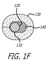
[0050] Figures 1E and 1F are cross-sectional views of alternative examples
of the
mitral annuloplasty ring shown in Figure 1A.
[0051] Figure 2 is a top plan view of a sheet of bioprosthetic tissue in
accordance
with another example.
[0052] Figure 3A is a top plan view of a tricuspid annuloplasty ring in
accordance
with another example.
[0053] Figure 3B is a side view of the annuloplasty ring shown in Figure
3A.
[0054] Figure 3C is a cross-sectional view of the annuloplasty ring shown
in
Figure 3A, taken along the lines 3C-3C of Figure 3A.
[0055] Figure 3D is a cross-sectional view of the annuloplasty ring shown
in
Figure 3A, taken along the lines 3D-3D of Figure 3A.
[0056] Figures 3E and 3F are cross-sectional views of alternative examples
of the
tricuspid annuloplasty ring shown in Figure 3A.
[0057] Figure 4A is a partially-exploded perspective view of a sewing ring
and a
prosthetic heart valve in accordance with another example.
[0058] Figure 4B is a top perspective view of the prosthetic heart valve
and sewing
ring shown in Figure 4A.
[0059] Figure 5A is a perspective view a substantially flat biological
tissue having
opposing surfaces.
[0060] Figure 5B is a perspective view of the rolling of the substantially
flat
biological tissue of Figure 5A.
[0061] Figure 5C is a perspective view the rolled, substantially flat
biological tissue
of Figure 5A.
[0062] Figure 5D is a perspective view of the rolled, substantially flat
biological
tissue of Figure 5C having its free edge sutured.
[0063] Figure 5E is a perspective view of the rolled, substantially flat
biological
tissue of Figure 5D with its free edge sutured and its two ends cut off.
[0064] Figure 6 is a perspective view of an annuloplasty ring having a
discontinuous
or C-shaped periphery in accordance with another example.
CA 03143300 2021-12-10
WO 2021/119391 - // ¨ PCT/US2020/064443
[0065] Figure 7 is a perspective view of an annuloplasty ring having a
continuous
periphery in accordance with another example.
[0066] Figures 8A is a cross-sectional view of the annuloplasty ring shown
in Figures
6 or 7, taken along the lines 8A-8A of Figure 7.
[0067] Figures 8B-8C are alternative cross-sectional views of embodiments
of the
annuloplasty ring shown in Figures 6 or 7.
DETAILED DESCRIPTION OF SOME EXAMPLES
[0068] With reference to Figures 1A-1F and 3A-3F of the illustrative
drawings,
there are shown examples of annuloplasty rings 100. The annuloplasty ring 100
can be
suitable for annulus of a valve, such as the mitral annulus (Figures 1A-1F) or
the
tricuspid annulus (Figures 3A-3F).
[0069] Figures 1A-1F illustrate examples of a mitral annuloplasty ring 100
having a
continuous or D-shaped periphery. The mitral annuloplasty ring 100 can be
shaped to
closely mimic the geometry of a healthy mitral annulus, and can be configured
to
minimize the likelihood of dehiscence while maintaining the shape of a healthy
valve
annulus.
[0070] Figures 3A-3F illustrate examples of a tricuspid annuloplasty ring
100, with
a discontinuous or C-shaped periphery, including two free ends 133 that define
a gap
therebetween. The tricuspid annuloplasty ring 100 can be shaped to closely
mimic the
geometry of a healthy tricuspid annulus, and can be configured to minimize the
likelihood of dehiscence while maintaining the shape of a healthy valve
annulus. The
tricuspid annuloplasty ring 100 is not complete in about ten percent of the
circumference around the anteroseptal commissure of the tricuspid annulus.
This is to
prevent suture injury to the conduction system. With particular reference to
Figure 3B,
the tricuspid annuloplasty ring 100 can have a somewhat spiral shape that
mimics the
shape of a healthy tricuspid annulus.
[0071] In one example, the two free ends 133 are separated at a distance to
define a
gap therebetween. The distance between the two free ends 133 can be about 1%,
about
2%, about 3%, about 4%, about 5%, about 6%, about 7%, about 8%, about 9%,
about 10%,
about 15%, about 20%, about 25%, about 30%, about 35%, about 40%, about 45%,
about
50% of a length of the tricuspid annuloplasty ring 100, or in a range that
includes and is
between any two of the foregoing values.
CA 03143300 2021-12-10
WO 2021/119391 - 12 - PCT/US2020/064443
[0072] In one example, the two free ends 133 can be coplanar with the frame
110. In
another example, one of the two free ends 133 can be offset from the other one
of the free
ends 133. The two free ends 133 can be vertically offset from one another at a
distance
that is about 1%, about 2%, about 3%, about 4%, about 5%, about 6%, about 7%,
about
8%, about 9%, about 10%, about 11%, about 12%, about 13%, about 14%, about
15%,
about 16%, about 17%, about 18%, about 19%, about 20%, about 21%, about 22%,
about
23%, about 24%, about 25%, about 26%, about 27%, about 28%, about 29%, about
30%,
about 35%, about 40%, about 45%, about 50% of a length of the tricuspid
annuloplasty
ring 100, or in a range that includes and is between any two of the foregoing
values.
[0073] The annuloplasty ring 100 can comprise a frame 110 having an outer
surface 120, and a cover 130 surrounding the frame 110. The cover 130
comprises a
bioprosthetic tissue. In some examples, the bioprosthetic tissue can be fixed
and non-
regenerative. In other examples, the bioprosthetic tissue can be regenerative.
[0074] The term "regenerative" as it relates to bioprosthetic tissue is
understood to
mean tissue that permits or even stimulates ingrowth of host cells and tissue
into the
bioprosthetic tissue after implantation. Thus, "regenerative tissue" can
include three-
dimensional scaffolds that support the ingrowth of host cells and tissue. In
one example,
the regenerative tissue can remain after in-growth of host cells and tissue.
In another
example, the regenerative tissue can partially or completely biodegrade after
in-growth
of host cells and tissue.
[0075] For the fixed and non-regenerative examples, the bioprosthetic
tissue can be
selected from the group consisting of: pericardium, blood vessels, skin, dura
mater,
small intestinal submucosa, ligaments, tendons, muscle, ureter, urinary
bladder, liver,
and heart. For example, the fixed, non-regenerative bioprosthetic tissue can
be a
pericardium. In one example, the fixed, non-regenerative bioprosthetic tissue
can be
fixed with an aldehyde such as a glutaraldehyde. In another example, the free
aldehyde
groups in the fixed, non-regenerative bioprosthetic tissue can be subjected to
a capping
treatment comprising a capping agent. In a one example, the capping treatment
can
comprise an amine. In an additional example, the capping treatment can further
comprise a reducing agent such as a borohydride. In one example, the fixed,
non-
regenerative bioprosthetic tissue can be plasticized. In another example, the
fixed, non-
regenerative bioprosthetic tissue can be plasticized with a polyol such as a
glycerol.
[0076] In one example, the bioprosthetic tissue can be subjected to a
fixation or
cross-linking treatment, as a result of which the bioprosthetic tissue is
rendered less
CA 03143300 2021-12-10
WO 2021/119391 - 13 ¨
PCT/US2020/064443
antigenic and is at least partially or completely cross-linked. The fixation
process can
also render the tissue non-regenerative. The fixation process is understood to
include
any chemical, heat or other processes, as a result of which the bioprosthetic
tissue is
preserved and rendered mechanically and dimensionally stable.
[0077] The fixation process can include contacting the tissue with one or
more
fixatives. Known fixatives include aldehydes, polyaldehydes, diisocyanates,
carbodiimides, photo-oxidation agents, and polyepoxide compounds. In a
preferred
example, the fixative used is glutaraldehyde. Glutaraldehyde-fixed tissue,
however, is
particularly vulnerable to calcification since glutaraldehyde fixation results
in the
generation of residual aldehyde groups and labile Schiff bases. The residual
aldehydes
and Schiff bases can be potential binding sites for calcium. The aldehyde
groups can
oxidize to carboxylic acid groups, which are known to attract and bind
calcium.
[0078] Various techniques have therefore been developed to reduce the
aldehyde and
acid levels of glutaraldehyde-fixed tissues, and thus reduce its propensity to
calcify after
implantation in the patient.
[0079] The fixation process can include adjusting the pH of the
glutaraldehyde
fixative in solution to reduce the generation of calcium binding sites, as
disclosed in U.S.
Patent No. 6,878,168 to Edwards Lifesciences, the entire contents of which are
incorporated into this description by reference. In a preferred example, the
pH of the
glutaraldehyde fixative in solution is about or provided in a range including
and
between any two of the following pH values: 4.5, 5, 5.5, 6, 6.5, 7, 7.5, 8,
8.5, and 9.
[0080] The fixation process can also further include the addition of a heat-
treating
step after contacting with the one or more fixatives. Glutaraldehyde-fixed
tissue have
demonstrated a reduced aldehyde and carboxylic acid content after heat
treatment, and
thus a marked reduction in calcification after implantation, as compared to
glutaraldehyde-fixed tissue without heat treatment. The glutaraldehyde
fixative in
solution can be heat treated before, during, or after the bioprosthetic tissue
is immersed
in the solution. The heat treatment can include heating the glutaraldehyde
fixative in
solution to a temperature provided in a range including and between any two of
the
following temperatures: 40 C, 41 C, 42 C, 43 C, 44 C, 45 C, 46 C, 47
C, 48 C, 49 C,
500 C, 51 C, 52 C, 53 C, 54 C, 55 C, 56 C, 57 C, 58 C, 59 C, 60 C,
61 C, 62 C,
63 C, 64 C, 65 C, 66 C, 67 C, 68 C, 69 C, 70 C, 71 C, 72 C, 73 C,
74 C, 75 C,
76 C, 77 C, 78 C, 79 C, and 80 C. Exemplary processes for heat treating
glutaraldehyde-fixed tissue are described in U.S. Patent No. 6,561,970, issued
May 13,
CA 03143300 2021-12-10
WO 2021/119391 ¨ 14 ¨ PCT/US2020/064443
2003 to Edwards Lifesciences, the entire contents of which are incorporated
into this
description by reference. The heat treatment of glutaraldehyde-fixed tissue is
also
commercially known as the Carpentier-Edwards ThermaFix0 (TFX) tissue treatment
process from Edwards Lifesciences.
[0081] Following or concurrently with the fixation process, the
bioprosthetic tissue
can be subjected to a capping treatment that comprises a capping agent, a
reducing
agent, or both. The bioprosthetic tissue can include functional groups that
exist either
inherently in the bioprosthetic tissue, as a result of being cross-linked or
fixed, or as a
result of being subjected to any number of chemical or physical processes,
including the
pre-conditioning, pre-stressing, or pre-damaging processes disclosed in this
description.
Exemplary processes for treatment with capping and reducing agents are
described in
U.S. Patent No. 7,972,376, the entire contents of which are incorporated into
this
disclosure by reference.
[0082] In one example, the bioprosthetic tissue can be subjected to a
capping
treatment without the step of fixing or crosslinking the bioprosthetic tissue.
In another
example, the bioprosthetic tissue can be subjected to the capping treatment
before,
during, or after the step of fixing or crosslinking the bioprosthetic tissue.
[0083] In one example, the capping agent can include any one or a
combination of
the following: an amine, such as an alkyl amine, amino alcohols and
ethanolamine; an
amino acid, such lysine and hydroxylysine; an amino sulfonate, such as
taurine, amino
sulfates, dextran sulfate, and chondroitin sulfate; hydrophilic
multifunctional polymers,
such as polyvinyl alcohols and polyethyleneimines; a hydrophobic
multifunctional
polymer; a-dicarbonyls, including methylglyoxal, 3-deoxyglucosone, and
glyoxal;
hydrazines, such as adipic hydrazide; disuccinimidyl N,N-carbonate;
carbodiimides, such
as 1-ethyl-3-[3-dimethylaminopropyl]carbodiimide hydrochloride (ED C), N-
cyclohexyl-
N'-(2-morpholinoethyl)carbodiimide (CMC), and 1,3-dicyclohexyl carbodiimide
(DCC);
and 2-chloro- 1-methylpyridinium iodide (CMPI).
[0084] In another example, the capping agent can be any agent that is
reactive with
a functional group, wherein the functional group is a free aldehyde or a free
carboxylic
acid. The capping agent can be an amine, such as an alkyl amine or an amino
alcohol.
The capping agent can be an ethanolamine.
[0085] In a further example, the capping agent can be any agent that is
reactive with
a functional group, wherein the functional group is an amine, a hydroxyl, or a
sulfhydryl
CA 03143300 2021-12-10
WO 2021/119391 ¨ 15 ¨ PCT/US2020/064443
group. In accordance with this example, the capping agent can comprise a
carbonyl
functional group. The carbonyl functional group can be an aldehyde or a
carboxylic acid
and can be selected from a monoaldehyde, a polyaldehyde, a monocarboxylic
acid, a
polycarboxylic acid, and the like.
[0086] Regardless, certain reactions of the capping agent and functional
groups can
produce labile Schiff bases and it can be desirable to reduce the Schiff bases
and replace
them with a more stable amine.
[0087] Accordingly, the capping treatment of the bioprosthetic tissue can
further
include treatment with a reducing agent. The reducing agent can be selected to
reduce
Schiff bases formed from the reaction of the crosslinking agent and the
bioprosthetic
tissue, the capping agent and the bioprosthetic tissue, and the capping agent
and the
crosslinking agent. In one example, the bioprosthetic tissue can be treated
with the
reducing agent, with or without the fixing or crosslinking the bioprosthetic
tissue. In
another example, the bioprosthetic tissue can be treated with the reducing
agent, with
or without the capping agent. In a further example, the bioprosthetic tissue
can be
treated with the reducing agent, with or without both the fixing or
crosslinking and
capping the bioprosthetic tissue.
[0088] The reducing agent can be any one or a combination of agents that
comprise a
borohydride. In one example, the reducing agent can be one or a combination
selected
from the group consisting of sodium borohydride, sodium cyanoborohydride,
sodium
triacetoxyborohydride, sodium bisulfate in acetylacetone, formic acid in
formaldehyde,
alkyl borohydride, amino borohydride, lithium aminoborohydrides, and an
organoborate
hydride salt having the formula XBR3H, where R is an alkyl group and X is
lithium,
sodium, or potassium. The lithium aminoborohydride can be a lithium
dimethylaminoborohydride, a lithium morpholinoborohydride, and a lithium
pyrrolidinoborohydride, to name a few. The organoborate hydride salt reducing
agent
can be a lithium tri-sec-butyl(hydrido)borate, a sodium tri-sec-
butyl(hydrido)borate, a
potassium tri-sec-butyl(hydrido)borate, or a lithium aluminum hydride.
[0089] The bioprosthetic tissue can be subjected to a capping treatment in
which it is
treated with a capping agent and a reducing agent in a solution. In one
example, the
capping agent is selected to react with one or more functional groups
associated with the
bioprosthetic tissue and the reducing agent is selected to reduce Schiff
bases. The Schiff
bases can be formed from any one or more of the reaction of the crosslinking
agent and
the bioprosthetic tissue, the reaction of the capping agent and the
bioprosthetic tissue,
CA 03143300 2021-12-10
WO 2021/119391 - 16 ¨ PCT/US2020/064443
and the reaction of the capping agent and the crosslinking agent. The capping
agent can
be an amine or an amino alcohol, such as an ethanolamine; the functional
groups can be
an aldehyde or a carboxylic acid; the reducing agent can be a borohydride,
such as a
sodium borohydride; and the crosslinking agent can be an aldehyde-containing
agent,
such as a glutaraldehyde. The capping treating can be performed sequentially
with first
the capping agent and then the reducing agent in solution or simultaneously
with both
the capping and reducing agents present in the solution. In one example, the
capping
treating can be performed with the capping agent and reducing agent in a
solution on an
orbital shaker operating at about 80 to about 100 rpm for about 4 hours.
[0090] Exemplary methods for treating bioprosthetic tissue with capping and
reducing agents are described in U.S. Patent No. 7,972,376, issued July 5,
2011 to
Edwards Lifesciences Corp., the entire contents of which are incorporated into
this
disclosure by reference for all purposes.
[0091] The capping treatment can comprise a capping agent, a reducing
agent, or
both. The capping treatment can be performed after the fixed bioprosthetic
tissue has
been subjected to a process of pre-conditioning, pre-stressing, or pre-
damaging to
generate additional acid binding sites, which can subsequently be capped, as
described
in U.S. Patent Application Publication No. 2008/0302372, published December
11, 2008,
entitled "Methods for Pre-Stressing and Capping Bioprosthetic Tissue" to
Edwards
Lifesciences, the entire contents of which are incorporated into this
disclosure by
reference for all purposes. In one example, the bioprosthetic tissue can be
subjected to a
rapid pulsed fluid flow (in the range of about 4 Hz to about 1,500 Hz),
repeated flexion of
the bioprosthetic tissue valve, elevated temperature (in the range of about 26
C to about
65 C), an acidic solution (pH in the range of about 4 to about 7), alkaline
solution (pH in
the range of about 8 to about 10), or any combination of the foregoing for the
purpose of
generating additional acid binding sites, which can be capped, reduced, or
both, in a
separate treatment process.
[0092] The bioprosthetic tissue can further undergo treatment with
anhydrous, non-
aqueous, or aqueous solutions to substantially, if not completely, dehydrate
the
bioprosthetic tissue for dry storage. The bioprosthetic tissue following
glycerol treatment
can contain residual water or moisture within the tissue interstices but can
be packaged
for dry storage.
[0093] In one example, the bioprosthetic tissue can be treated with an
anhydrous,
non-aqueous, or aqueous solution that comprises glycerol. In one example, the
CA 03143300 2021-12-10
WO 2021/119391 - 17 ¨ PCT/US2020/064443
anhydrous, non-aqueous, or aqueous solution can comprise about 25% by volume,
30%
by volume, 35% by volume, 40% by volume, 45% by volume, 50% by volume, 55% by
volume, 60% by volume, 65% by volume, 70% by volume, 75% by volume, 80% by
volume, 85% by volume, 90% by volume, or 95% by volume glycerol. In another
example,
the anhydrous, non-aqueous, or aqueous solution comprises an amount of
glycerol
within and including any two of the foregoing values.
[0094] In another example, the anhydrous, non-aqueous, or aqueous glycerol
solution
can comprise alcohol. In one example, the anhydrous, non-aqueous, or aqueous
solution
can comprise about 5% by volume, about 10% by volume, about 15% by volume,
about
20% by volume, about 25% by volume, about 30% by volume, about 35% by volume,
about 40% by volume, about 45% by volume, about 50% by volume, about 55% by
volume, about 60% by volume, about 65% by volume, about 70% by volume, or
about
75% by volume alcohol. In another example, the anhydrous, non-aqueous, or
aqueous
solution comprises an amount of alcohol within and including any two of the
foregoing
values. The alcohol can be any one or a combination of Ci, C2, C3, C4, and C5
alcohols,
such as ethanol, prop anol, and butanol.
[0095] In one example, the solution is a non-aqueous solution of about 75%
by
volume glycerol and 25% by volume ethanol. The bioprosthetic tissue is
immersed in the
solution for a period of time sufficient to permit the solution to permeate
the
bioprosthetic tissue. The bioprosthetic tissue is then removed from the
solution to allow
removal of excess solution. Suitable treatment for the bioprosthetic tissues
is described
in U.S. Patent No. 8,007,992, issued August 30, 2011, to Edwards Lifesciences
Corp., the
entire contents of which are incorporated into this disclosure by reference
for all
purposes.
[0096] In another preferred example, an aqueous glycerol solution can be
used to at
least partially dehydrate the tissue, as described in U.S. Patent No.
6,534,004, issued
March 18, 2003, issued to The Cleveland Clinic Foundation, the entire contents
of which
are incorporated into this disclosure by reference for all purposes.
[0097] The bioprosthetic tissue can also be treated by means other than the
glycerol
treatment process described above to dry or dehydrate the bioprosthetic
tissue. The
terms "dry" or "dehydrate," as used in this disclosure with reference to the
bioprosthetic
tissue or the implantable bioprosthetic device, is understood to include
residual water or
moisture that can be present in the bioprosthetic tissue following glycerol or
other
treatment to reduce the water content of the bioprosthetic tissue. In one
example, the
CA 03143300 2021-12-10
WO 2021/119391 - 18 ¨
PCT/US2020/064443
water content of the dried or dehydrated bioprosthetic tissue following
glycerol or other
treatment is about 25% by weight or less, about 20% by weight or less, about
15% by
weight or less, about 10% by weight or less, about 9% by weight or less, about
8% by
weight or less, about 7% by weight or less, about 6% by weight or less, about
5% by
weight or less, about 4% by weight or less, about 3% by weight or less, about
2% by
weight or less, or about 1% by weight or less. These percentages are
understood to be
based on the combined weight of the bioprosthetic tissue and water content.
[0098] For the regenerative examples, the bioprosthetic tissue can be an
artificial or
biological scaffold or a decellularized biological tissue. For example, the
bioprosthetic
tissue can be selected from the group consisting of: pericardium, blood
vessels, skin,
dura mater, small intestinal submucosa, ligaments, tendons, muscle, ureter,
urinary
bladder, liver, and heart. It is understood that the tissue selected is
decellularized using
any suitable method.
[0099] In one example, the regenerative bioprosthetic tissue can be an
artificial
scaffold. In another example, the artificial scaffold can be a biodegradable
polymer
scaffold. A biodegradable polymer can include a polymer in which the bonds of
the
polymer-chain cleave, primarily by aqueous hydrolysis as a result of contact
with blood
and other bodily fluids at physiological pH (e.g., around 7 to 7.5). This
process results in
the fragmentation and eventual decomposition of the polymer in vivo. The
fragmentation and decomposition process can be catalyzed by enzymes or other
endogenous biological compounds. In a further example, the biodegradable
polymer
scaffold can comprise a polyglycolic acid. In an additional example, the
artificial scaffold
can further comprise one or more extracellular matrix proteins. For example,
the
extracellular matrix protein can be one or more proteins selected from the
group
consisting of: hydroxyproline, vitronectin, fibronectin, collagen I, collagen
III, collagen
IV, collagen VI, collagen XI, collagen XII, fibrillin I, tenascin, decorin,
byglycan,
versican, asporin, agrin, and combinations thereof.
[0100] In some examples, the cover 130 can be formed from a sheet 134 of
bioprosthetic tissue (Figure 2). The sheet 134 can have a first edge 131 and a
second
edge 132. With reference to Figures 1C and 3C, the sheet 134 can cover the
outer
surface 120 of the frame 110, and the first edge 131 and the second edge 132
can be
sewn, glued, or otherwise joined together to form a seam 136. In some examples
of a
tricuspid annuloplasty ring 100 (Figures 3A-3D), the sheet 134 can extend
beyond the
frame 110 to form an enlarged region of covering 130 at the free ends (Figure
3D).
CA 03143300 2021-12-10
WO 2021/119391 - 19 - PCT/US2020/064443
[0101] The glue used to form and shape the cover 130, such as joining the
first and
second edges 131, 132, is preferably one that is biocompatible and strongly
bonds tissue
in a wet environment (e.g., flowing blood). In one example, the glue is a
hydrophobic
light-activated adhesive (HLAA). The HLAA can be formed by combining a
poly(glycerol
sebacate acrylate) (PGSA) with a photo-initiator, such as 2-hydroxy-1-[4-(2-
hydroxyethoxy)pheny1]-2-methyl-l-propanone) (IRGACURE 2959, Sigma-Aldrich) to
create the HLAA. The HLAA can be a thick gel that can be applied onto the
cover and
then cross-linked by ultraviolet light. The resulting bond is preferably water-
tight yet
flexible and stays intact in the face of high pressure and flowing blood.
[0102] With reference to Figures 1A, 1B, 3A, and 3B, the cover 130 can
comprise a
plurality of sheets 134 that abut each other at part-interface seams 137. In
some
examples, the part-interface seams 137 can define markings to aid a surgeon
with
correct positioning of the ring 100 on the valve anulus. With reference to
Figure 1D, in
some examples, the sheet 134 can be dimensioned to permit the first edge 131
and the
second edge 132 of the sheet to fold or roll upon each other to form a lip
138. In further
examples, the lip 138 can protrude away from the outer surface 120 of the
frame 110.
[0103] The outer cover 130 comprising a bioprosthetic tissue can encourage
native
tissue growth on the annuloplasty ring 100, which can help to maintain the
ring 100 in
place on the valve annulus. In addition, bioprosthetic-tissue cover 130 can be
used in
patients with endocarditis or in patients who are otherwise intolerant of
cloth-covered
implants.
[0104] As discussed above, the cover 130 is wrapped around a frame 110. In
some
examples, the frame 110 can be bioabsorbable. In other examples, the frame 110
can be
non-degradable.
[0105] For the non-degradable examples, the frame 110 can comprise one or
both of
a non-degradable polymer and a non-degradable metal or metal alloy. For
example, in
one example, the frame 110 can comprise a non-degradable metal or metal alloy
selected
from the group consisting of: stainless steel, a nickel-based alloy, a cobalt-
chromium
alloy, a nickel-cobalt-chromium alloy, nitinol, and combinations thereof.
[0106] For the bioabsorbable examples, the bioabsorbable frame 110 can
comprise a
degradable metal or a metal alloy. For example, the degradable metal or metal
alloy can
comprise one or a combination selected from the group consisting of:
magnesium,
aluminum, iron, and zinc. The metal or metal alloy can have an ultimate
tensile
CA 03143300 2021-12-10
WO 2021/119391 - 20 ¨ PCT/US2020/064443
strength of about 30 MPa to about 400 MPa and an elongation of about 0.3
percent to
about 170 percent.
[0107] In one example, the bioabsorbable frame 110 can be a bioabsorbable
material.
For example, the bioabsorbable material can be one or a combination of
polymers
selected from the group consisting of: poly(L-lactide), poly(D-lactide),
polyglycolide,
poly(L-lactide-co-glycolide), polyhydroxyalkonate, polysaccharides,
polyesters,
polyhydroxyalkanoates, polyalkelene esters, polyamides, polycaprolactone,
polylactide-
co-polycaprolactone, polyvinyl esters, polyamide esters, polyvinyl alcohols,
modified
derivatives of caprolactone polymers, polytrimethylene carbonate,
polyacrylates,
polyethylene glycol, terminal dials, poly(L-lactide-co-trimethylene
carbonate),
polyhydroxybutyrate, polyhydroxyvalerate, poly-orthoesters, poly-anhydrides,
polyiminocarbonate, and copolymers.
[0108] In another example, the bioabsorbable frame 110 can be reinforced
with a
reinforcing composition. For example, in one example, the reinforcing
composition can
comprise magnesium or a magnesium alloy.
[0109] While the frame 110 is shown to have a circular cross-section in
Figures 1C,
1D, and 3C, it should be understood that the frame 110 can have other cross-
sectional
shapes and thicknesses. For example, in some examples, the frame 110 can have
a
rounded semi-circular or even polygonal cross-section. In other examples, the
shape or
thickness of the frame 110 can be uniform across the periphery of the ring 100
or it can
vary across the periphery of the ring 100.
[0110] In some examples, the ring 100 can further include a suture-
permeable
interface 140 having one or more layers between the frame 110 and the cover
130. For
instance, the suture-permeable interface can comprise an elastomeric sleeve
(Figures 1F
and 3F) such as a silicone rubber molded around the frame 110. The elastomeric
sleeve
can provide bulk to the ring for ease of handling and implant, and permit
passage of
sutures though not significantly adding to the anchoring function of the outer
cover 130.
In one example, the suture-permeable interface 140 can be provided around at
least a
portion of the periphery of the ring 100 (Figures 1E and 1F). In another
example, the
suture-permeable interface 140 can be provided around the entire periphery of
the
ring 100.
[0111] With reference now to Figures 4A and 4B of the illustrative
drawings, there is
shown a sewing ring 200 for a prosthetic heart valve 300. The prosthetic heart
valve 300
CA 03143300 2021-12-10
WO 2021/119391 ¨ 21 ¨ PCT/US2020/064443
can comprise a support frame 310 defining an orifice 320 about an axis A along
an
inflow-outflow direction. A plurality of leaflets 330 can be mounted for
movement on the
support frame 310 to provide a one-way valve 340 in the orifice 320. Each of
the
plurality of leaflets 330 can comprise the bioprosthetic tissue described
above.
[0112] The sewing ring 200 can be connected to and positioned around the
support
frame 310 for attaching the heart valve 300 to a valve annulus (not shown).
The sewing
ring 200 can include a suture-permeable annular member 210 comprising an outer
surface 220, and a cover 230 surrounding the annular member 210.
[0113] The cover 230 comprises the bioprosthetic tissue described above.
For
example, as previously outlined, the cover 230 can comprise bioprosthetic
tissue that is
fixed and non-regenerative, or bioprosthetic tissue that is regenerative. In
either case,
the cover 230 is wrapped around the annular member 210, which can be molded
from a
suture-permeable, biocompatible polymer such as silicone. In one example, the
annular
member 210 can comprise a molded polymer selected from the group consisting
of:
silicone, polyurethane, and combinations thereof. As with the cover 130 for
the
annuloplasty ring 100, the cover 230 for the sewing ring 200 can be formed
from a
sheet 134 of bioprosthetic tissue (Figure 2), as discussed above.
[0114] In one example, the cover 230 and the plurality of leaflets 330 are
made from
the same bioprosthetic tissue.
[0115] With reference to Figures 5A-5E of the illustrative drawings, there
is shown
a method of manufacturing various embodiments of ring protheses which includes
annuloplasty rings and sewing rings, including those depicted in Figures 6 and
7. As
with the annuloplasty rings described herein, ring protheses fabricated in
accordance
with the depicted method can be suitable for the annulus of a valve, such as
the
tricuspid annulus (Figure 6) or the mitral annulus (Figure 7). Additionally,
ring
protheses fabricated in accordance with the depicted manner can be used in
fabricating
a prosthetic heart valve, as shown in Figures 4A-4B.
[0116] Figure 5A depicts an example of a bioprosthetic tissue 500 having a
length L
and a width W. The bioprosthetic tissue 500 can comprise two opposing
surfaces: a first
surface 504 and a second surface 502. In one embodiment, the first surface 504
can have
a rough texture or can be fibrous and the second surface 502 can be smoother
than the
first surface 504. Certain biological tissues can be characterized as having
such surfaces,
such as pericardial tissue and dura mater. These tissues are covered by a cell
surface
CA 03143300 2021-12-10
WO 2021/119391 ¨ 22 ¨
PCT/US2020/064443
layer only on one side and thus have a smooth and even surface on this side
while the
opposing side is rough and fibrous. For purposes of cellular integration and
infiltration
by the host, the rough side can provide a suitable surface.
[0117] While Figure 5A depicts a perfectly rectilinear shape, it is
understood that
the biological tissue 500 can be of any shape of a defined length and width so
long as it
can provide the desired diameter of the resulting ring prosthesis. Figures 5B-
5C depict
one example of how the biological tissue 500 can be rolled upon itself to
produce an
elongated rod member 510 having a free edge 506 between the first and second
secured
ends 501A and 501B. While Figures 5A and 5B depict the elongated rod member
510 as
has having a substantially cylindrical cross-sectional shape (Figure 8A), it
is understood
that the biological tissue 500 can be rolled or folded in a manner to produce
other cross-
sectional shapes, such as a triangular cross-sectional shape (Figure 8C).
Alternatively,
the elongated rod member 510 can be formed into a rectilinear cross-sectional
shape by
alternating folds in an accordion-style (Figure 8B).
[0118] The elongated rod body 510 can be secured at its first and second
secured
ends 501A, 501B by wrapping with a string or suture 50 so that it may retain
its
substantially cylindrical shape. Once the first and second ends 501A, 501B are
secured,
the free edge 506 can be secured to the rod body 510. While Figure 5D depicts
the free
edge 506 being secured to the rod body 510 with sutures 60, it is understood
that the
free edge 506 can be secured with any biocompatible substance, such as the
adhesives
described herein.
[0119] As shown in Figures 5E and 6, the free edge 506 may be sutured in
such a
manner to impart a curvature to the shape of the rod body 510. The two free
ends 500A
and 500B can be separated at a distance d to define a gap therebetween to
produce an
open ring 540 as depicted in FIG. 6. The distance D between the two free ends
500A and
500B can be about 1%, about 2%, about 3%, about 4%, about 5%, about 6%, about
7%,
about 8%, about 9%, about 10%, about 15%, about 20%, about 25%, about 30%,
about
35%, about 40%, about 45%, about 50% of a length of the rod body 510, or in a
range
that includes and is between any two of the foregoing values. Once the free
edge 506 is
sutured, the first and second secured ends 501A, 501B can be cut off to
produce first and
second free ends 500A and 500B. The resulting rod body 510 can consist only of
the
biological tissue 500 and the sutures 60 which can be made of a dissolvable or
bioabsorbable material.
CA 03143300 2021-12-10
WO 2021/119391 - 23 - PCT/US2020/064443
[0120] In one example, the two free ends 500A and 500B can be coplanar with
the
rod body 510. In another example, one of the two free ends 500A, 500B can be
offset
from the other one of the free ends. The two free ends 500A, 500B can be
vertically offset
from one another at a distance that is about 1%, about 2%, about 3%, about 4%,
about
5%, about 6%, about 7%, about 8%, about 9%, about 10%, about 11%, about 12%,
about
13%, about 14%, about 15%, about 16%, about 17%, about 18%, about 19%, about
20%,
about 21%, about 22%, about 23%, about 24%, about 25%, about 26%, about 27%,
about
28%, about 29%, about 30%, about 35%, about 40%, about 45%, about 50% of a
length of
the ring prosthesis, or in a range that includes and is between any two of the
foregoing
values.
[0121] In another example, the two free ends 500A, 500B can be joined
together to
produce an enclosed ring 550 as depicted in FIG. 7. In one example, the two
free ends
500A, 500B can be sutured together to produce a seam 512 therebetween.
[0122] It is understood that the bioprosthetic tissue can be treated in any
manner as
described above either before or after it is formed into a ring prosthesis.
[0123] It should be appreciated from the foregoing description that the
present
disclosure provides improved ring prostheses, including annuloplasty rings and
sewing
rings that can encourage native tissue growth around the implant, to help
maintain the
implants in place, and that can be used in patients with endocarditis or in
patients who
or otherwise intolerant of cloth-covered implants.
[0124] Specific methods, devices, and materials are described, although any
methods
and materials similar or equivalent to those described can be used in the
practice or
testing of the present example. Unless defined otherwise, all technical and
scientific
terms used herein have the same meanings as commonly understood by one of
ordinary
skill in the art to which this example belongs. The terms "a," "an," and "at
least one"
encompass one or more of the specified element. That is, if two of a
particular element
are present, one of these elements is also present and thus "an" element is
present. The
terms "a plurality of' and "plural" mean two or more of the specified element.
The term
"or" used between the last two of a list of elements means any one or more of
the listed
elements. For example, the phrase "A, B, or C" means "A, B, and/or C," which
means
"A," "B," "C," "A and B," "A and C," "B and C," or "A, B, and C." The term
"coupled"
generally means physically coupled or linked and does not exclude the presence
of
intermediate elements between the coupled items absent specific contrary
language.
CA 03143300 2021-12-10
WO 2021/119391 - 24 ¨ PCT/US2020/064443
[0125] Without further elaboration, it is believed that one skilled in the
art, using
the proceeding description, can make and use the disclosed subject matter to
the fullest
extent. The subject matter has been described in detail with reference only to
the
presently preferred examples. Persons skilled in the art will appreciate that
various
modifications can be made without departing therefrom. Accordingly, the scope
is
defined only by the following claims.