Note: Descriptions are shown in the official language in which they were submitted.
CA 03143314 2021-12-13
WO 2021/001822
PCT/IL2020/050728
1
STERILE BARRIERS AND SENSOR SETS
FOR A MEDICAL DEVICE
RELATED APPLICATION/S
This application claims the benefit of priority of U.S. Provisional Patent
Application
No. 62/868,940 filed on June 30, 2019, the contents of which are incorporated
herein by
reference in their entirety.
FIELD AND BACKGROUND OF THE INVENTION
The present invention relates to medical devices having sterile barriers and
sensor sets and
related algorithms for controlling and tracking movements of device components
and user.
Medical devices, especially surgical devices, must remain sterile during use
in order to
minimize the risk of infection or other contamination to the patient.
Medical devices having internal parts and mechanisms are difficult to clean
and sterilize
and can pose a health risk especially if the device or its internal components
(e.g. sensors, motor
packs) are used in more than one procedure. Without disassembling, cleaning
and sterilizing the
exterior parts of the device, and then re-assembling the device, it is
difficult to maintain sterility
of such devices. Furthermore, internal components such as sensor and motor
packs are sensitive
and oftentimes cannot be sterilized or repeatedly sterilized.
Barriers, such as tubular sheaths, that can prevent contact between the non-
sterile parts of
a medical device and the patient are known in the art. However, such barriers
do not adequately
shield internal components and moving parts that are capable of transmitting
infective particles to
the patient.
There is thus a need for medical devices having sterile barriers that protect
internal
components and moving parts and eliminate the need for re-sterilization of an
internal component
or an entire device.
SUMMARY OF THE INVENTION
According to one aspect of the present invention there is provided a medical
device
having compartments that enable loading and securing motor packs, internal
parts, sensors,
electrical circuits and/or control interface sensors.
According to another aspect of the present invention there is provided a
sterile barrier
between the contained parts and the sterile end effector, were the sterile
barrier reduces the
CA 03143314 2021-12-13
WO 2021/001822
PCT/IL2020/050728
2
possibility of contamination of the sterilized end effector while allowing
transfer of forces and
moments from the internal parts to the end effector.
According to another aspect of the present invention there is provided a
medical device
having a sensors pack that can measure the movement of the control interface
operated by the
surgeon while correlating between the sensor pack and portions of the device
and user.
Unless otherwise defined, all technical and scientific terms used herein have
the same
meaning as commonly understood by one of ordinary skill in the art to which
this invention
belongs. Although methods and materials similar or equivalent to those
described herein can be
used in the practice or testing of the present invention, suitable methods and
materials are
described below. In case of conflict, the patent specification, including
definitions, will control.
In addition, the materials, methods, and examples are illustrative only and
not intended to be
limiting.
BRIEF DESCRIPTION OF THE SEVERAL VIEWS OF THE DRAWINGS
The invention is herein described, by way of example only, with reference to
the
accompanying drawings. With specific reference now to the drawings in detail,
it is stressed that
the particulars shown are by way of example and for purposes of illustrative
discussion of the
preferred embodiments of the present invention only, and are presented in the
cause of providing
what is believed to be the most useful and readily understood description of
the principles and
conceptual aspects of the invention. In this regard, no attempt is made to
show structural details
of the invention in more detail than is necessary for a fundamental
understanding of the
invention, the description taken with the drawings making apparent to those
skilled in the art how
the several forms of the invention may be embodied in practice.
In the drawings:
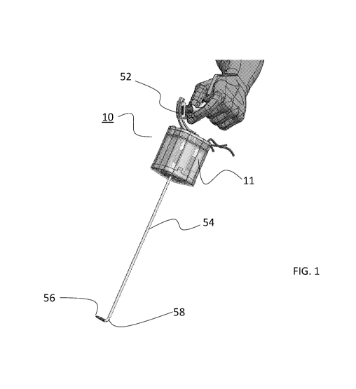
FIG. 1 illustrates an embodiment of a motor unit connected to an interface and
tool shaft.
FIG. 2 illustrates the components of the motor unit of Figure 1.
FIGs. 3A and 3B illustrate an instrument adaptor and gearbox connectable to
the motor
unit of the present invention.
FIGs. 4A and 4B illustrates a motor pack component of the present motor unit.
FIGs. 5A and 5B illustrate the sterile shell component of the present motor
unit.
FIGs. 6A, 6B and 6C illustrate assembly of the present motor unit.
FIGs. 7A, 7B, 7C and 7D illustrate the motors and gearbox interfaces of the
shell of the
present motor unit.
CA 03143314 2021-12-13
WO 2021/001822
PCT/IL2020/050728
3
FIGs. 8A, 8B and 8C illustrate the drivetrain interconnecting the motors heads
to the
gearbox of the instrument adaptor and gearbox component.
FIGs. 9A and 9B illustrate a user interface attached to the shell of a medical
device
(Figure 9A) showing the sensors pack positioned in a housing of the user
interface (Figure 9B).
FIGs. 10A and 10B illustrate one embodiment of the present sensor pack.
FIGs. 11A, 11B, 11C and 11D illustrate loading of the sensor pack into the
housing of the
user interface.
FIGs. 12A, 12B and 12C illustrate the finger interface mechanism of the user
interface.
FIGs. 13A, 13B, 13C and 13D illustrate a sensors pack-carrying wrist bracelet.
FIG. 14 illustrates possible sensor positions in and on the device and user.
FIG. 15 illustrates the main components of the control interface of the
device, the end
effector and their related angles.
FIGs. 16, 17, 18, 19 and 20 are flowchart diagrams illustrating several
calibration and set
up functions for device control and tracking.
DESCRIPTION OF SPECIFIC EMBODIMENTS OF THE INVENTION
The present invention is of devices having sterile barriers that isolate
internal components
from the patient and environment and as such, allow reuse of such internal
components without
sterilization.
The principles and operation of the present invention may be better understood
with
reference to the drawings and accompanying descriptions.
Before explaining at least one embodiment of the invention in detail, it is to
be understood
that the invention is not limited in its application to the details set forth
in the following
description or exemplified by the Examples. The invention is capable of other
embodiments or of
being practiced or carried out in various ways. Also, it is to be understood
that the phraseology
and terminology employed herein is for the purpose of description and should
not be regarded as
limiting.
Sterile barriers for medical devices are well known in the art and typically
take the form
of sheaths/covers that cover an entire device or components that come in
contact with the patient.
While such sheaths are somewhat effective in preventing patient contamination,
they are
oftentimes ineffective in preventing contamination of internal components that
are either reused
or are a part of a reusable device. Since internal components such as sensors,
electric components
and motor packs are sensitive and can be damaged by some forms of
sterilization, sterilization of
these reusable components is typically carried out via manual cleaning with
antiseptic fluids, a
CA 03143314 2021-12-13
WO 2021/001822
PCT/IL2020/050728
4
time consuming and laborious task that can be unsuccessful in completely
eradicating pathogens
and contaminants.
Several paths of infection exists in medical devices:
(i) The motor pack (or another internal component) can transfer
contaminants to the
end effector when the pressure in the body cavity is less than the pressure in
the motor pack.
(ii) The end effector can transfer contaminants such as blood to the motor
pack (or
another internal component) when the pressure in the body cavity is greater
than the pressure in
the motor pack.
(iii) Cycle of (i) and (ii) when pressure differences between body cavity and
outer
atmospheric alternate.
A sterile barrier can eliminate the need for sterilizing internal components
or entire
devices. Embodiments of the present invention relate to surgical devices
having sensors and
motors packs that are isolated from device components that come in contact
with the patient (end
effector) and as such, do not need to be sterilized while being incapable of
transmitting pathogens
and contaminations to the patient.
While reducing the present invention to practice, the present inventors have
devised
several sterile barrier configurations that can be used in a medical device to
isolate internal
components that are not easily serializable from the patient and from
components of the device
that come in contact with the patient.
As is describe hereinunder, these barriers can be used to isolate motor packs
and batteries
as well as sensors packs from the environment and from potential contamination
by pathogens
and contaminants. As such, these barriers enable reuse of internal components
without a need for
sterilization between uses.
Several barrier configurations are contemplated herein. Such configurations
can be used
in any medical device having internal components such as motor and sensor
packs and batteries.
Depending on use and device type, a medical device can incorporate one or more
of these
barriers.
The following describes the sterile barriers of the present invention in
context with a
surgical device (laparoscope) having a user interface connected to a steerable
shaft having an end
effector. It will be understood that the sterile barriers of the present
invention can also be used
with medical devices such as endoscopes, laparoscopes or catheters.
CA 03143314 2021-12-13
WO 2021/001822
PCT/IL2020/050728
Motor Pack
While experimenting with several prototypes, the present inventors discovered
that a
motor pack that is engaged in serial manner greatly increased the length of
the device body. In
addition, serial engagement between the electronic pack and motor also
increased the length of
5
the device body. To solve these problems, the present inventors positioned the
motor pack of the
present invention such that it surrounds the instrument gear box with the
motors, the electronic
boards and the batteries positioned around the gear box (the gear box is
positioned within the
motor pack) to thereby substantially decrease the overall length of the device
body and device.
Figures 1-9B illustrate the motor pack and associated components, collectively
referred to
herein as motor unit 10. Motor unit 10 can be integrated into a device 50
(laparoscope 50 shown)
that includes a user interface 52 and a shaft 54 having an end effector 56
(grasper 56 shown)
positioned at a distal end 58 of shaft 54. Shaft 54 can be rigid or steerable.
Examples of steerable
shafts are described in U520150366572 which is fully incorporated herein by
reference.
Motor unit 10 includes a removable shell 12 that is externally sterile (and
may be re-
sterilized) and is dimensioned for encasing a motor pack 11. Shell 12 includes
a shell body 14
and a front cover 16. Shell 12 is fabricated from PPSU or PEEK or PSU
Silicone (for reusable)
and is typically 80-140 mm in length, 50-100 mm in width and 50-100 mm in
height. Shell 12
isolates motor pack 11 from the environment and thus prevents any migration of
contaminants or
pathogens beyond the walls of shell 12.
Figure 2 illustrates the arrangement of motor pack 11 and shell 12. Motor pack
11 is
positioned inside shell body 14, front cover 16 is attached to shell body 14
with cylindrical
component 18 positioned through motor pack 11. Figures 6A-C illustrate
assembly of motor unit
10.
An instrument adaptor and gearbox 20 (attached to shaft 54) is attachable
within
cylindrical component 18 of front cover 16 and interfaces with motor pack 11
through adapters
provided in shell 12 (described hereinunder). Instrument adaptor and gearbox
20 is unique to the
tool shaft used and varies between different types of tools but is connectable
to any motor unit
10.
Figures 3A-B illustrate instrument adaptor and gearbox 20 in more detail.
Figure 3A
illustrates the shell-interfacing end of instrument adaptor and gearbox 20
showing sterile adapters
22 and optional end effector energy connector 24 (mono-polar type connector
shown). Insert
guides 25 are provided to align instrument adaptor and gearbox 20 with motor
pack 11. Figure 3B
illustrates the shaft side of instrument adaptor and gearbox 20 showing finger
holds 26 that can
CA 03143314 2021-12-13
WO 2021/001822
PCT/IL2020/050728
6
be grasped by the user when connecting instrument adaptor and gearbox 20 to
shell 12 and an
optional end effector energy connector 28.
Figures 4A-B illustrate motor pack 11 including cover 30 and internal
components.
Motor pack 11 includes one or more of motor 32 and associated gear 34 (four
shown). Gear 34
terminates in a protruding motor head 36 that interfaces with adapters within
shell 12 (further
described below). Motor heads 36 (best shown in Figure 4A) are pushed into
adapters for
coupling.
Motor pack 11 includes an opening 38 for accepting (cylindrical) component 18
of front
cover 16. Slots 40 are provided for guiding the instrument into the shell and
lock it. At least IMU
chip 33 is installed on electrical circuits boards 31.
Figures 5A-B illustrate shell 12 in greater detail. The backside (facing the
user) of shell
12 includes an interface rail 42 that allows the surgeon to move user
interface handle 52, to the
best ergonomic angle, and several mechanical push buttons and channels 71 that
contain
mechanical push rod 75 that transmit the push forces through the shell to
sensors located at the
motor pack. (The sensors may be capacitive, optical or mechanical). By pushing
head button 70
(shown also in Figure 7B), of push rod 75, the surgeon may select modes such
as jaws speed of
rotation, jaws angle of rotation, control mode etc. The ports 73 may be used
for connecting
external different types of cords, such as motor unit power cord, or energy
cords (monopolar,
Bipolar), to motor pack 11, through shell 12. Shell 12 includes a shell wall
13 and an opening 15
for accepting motor pack 11.
Figures 7A-D illustrate the interfaces for motor pack 11 and instrument
adapters of
gearbox 20 within shell 12. Push buttons 70 activate sensors 83 located in
motor unit 11 by
pushing rods 75. Internal openings 77 of ports 73 contains a seal 79, (e.g., 0-
ring), that enables
connecting of external cords to motor unit 11 while keeping the motor unit
insulated from the
sterile environment. For example, external power cord will be connected to
power connector 81
located in motor unit 11 through opening 73, while seals 79 ensures that the
other external parts
of the power cord will not be contaminated by the power cord distal plug.
Motor heads adapters
76 transfer rotation of motor heads 36 to drive train transmitting motor
moment to gear train
distal heads 72. Distal heads 72 engages with adapters 22 of gear box 20 of
the instrument,
enabling the control of the instrument end effector jaws and the articulation.
When the shell and the motor pack are fully engaged the heads of the
mechanical mode
switches are positioned near sensors 83 which they activate. When the surgeon
presses on one of
the heads 70 of mode buttons, the distal head of the push rod 75 moves toward
the motor pack
and activates the designated mode sensor, and the desired mode is selected.
CA 03143314 2021-12-13
WO 2021/001822
PCT/IL2020/050728
7
Figures 8A-C illustrate a drivetrain 74 that includes a plurality of gears for
interconnecting between motor head adapters 76 and instrument head adapters
72. The drive train
may be an integral part of the shell or a separate module connected to the
shell. The drive train
transfers rotation of the motors from the motor pack to the surgical
instrument. The gear drive
train allows the manufacturer to adapt the device to various of present or
future instruments, just
by changing the gear drive train, without the need to change the motor pack.
For example, for
power tools such as staplers, clip appliers or vessel sealers, the gears
diameter may be changed in
order to increase the moments transferred to the power instrument adapters.
For other instruments
such as needle holder, hook or grasper, where fast movements are required the
manufacturer may
choose gear train that transfers faster rotation to the instrument. Some power
tools with less
degrees of freedom may need less motorized inputs, in this case, a gear train
design, which
combine 2 or more motors to a single output may be used. The gear train
geometrical
configuration may be also be changed in order to adapt to different geometries
of instrument gear
box.
Figures 8A-B show the shell of the motor pack and its inner side components.
Four input
adapters 76 that transmit the power from the motors into the gear trains in
the shell are located at
the corners of the shell. Gear trains transfer the motors movement to the
output heads, arranged in
a T formation, 3 instruments heads 72 in horizontal line and one instrument
head under the
central motor head.
Figure 8B is an upper view of the shell and the gear trains. Each gear train
is labeled as
follows:
J gear train transfers the power from the motor pack to the jaws mechanism to
enable open and
close movement of the jaws.
R gear train transfers the power from the motor pack to the jaws mechanism to
enable roll
movement of the jaws.
Al gear train transfers the power from the motor pack to the articulation, to
enable up/down
articulation of the shaft.
A2 gear train transfers the power from the motor pack to the articulation to
effect right/left
articulation of the shaft.
Sensor pack
In order to control the instrument functions the present invention describe a
control
interface shaped to fit the hand of the surgeon allowing the surgeon to
simultaneously position
the end effector in the patient body, orient the control interface in order to
control the bending of
CA 03143314 2021-12-13
WO 2021/001822
PCT/IL2020/050728
8
the articulation and operate the jaws. The control interface has 3 main
components: the control
interface body including fingers interface, the dorsum interface 59, and the
handle which serves
as a container to the sensors capsule.
This interface design enables re-sterilization of the control interface body,
while
eliminating the need to sterilize delicate electric components contained in
the sensors capsule.
The design also enables future upgrading of the electric circuits and sensors,
contained in the
sensors capsule without the need to make any change in the control interface
body. In addition,
the handle may be changed without the need to change the sensors capsule.
In order to ensure complete insulation between the electric circuits in the
sensors capsule
and the control interface body, the sensors capsule is sealed, and the sensors
are insulated from
their measurement reference.
For example, a Hall Effect sensor (such as Melexis) with a magnet which serves
as the
rotation measurement reference is embedded in the control interface body, and
the Hall Effect
sensors 120,130 (shown in Figure 10B), are located in the sealed sensors
capsule. Although there
is no direct contact between the magnet and the sensor, the Hall Effect sensor
is able to measure
accurately the angle position between the sensor and magnet. The insulation
concept is also valid
for rotation potentiometer, where a stationary reference base may be coupled
to the potentiometer
rotor without exposing the sensor electric circuits to the control interface
body.
Figure 9A-11D illustrate one embodiment of the sensor pack of the present
invention
which is referred to herein as sensor pack 100.
Sensor pack 100 is position within a housing 53 of a user interface 52 (also
referred to
herein as controller or control interface) of device 50. As is shown in
Figures 11A-D, sensor pack
100 is loaded into housing 53 by opening a hinged cover 55 and sliding sensor
pack 100 into a
recess 57 within housing 53. The sensor pack includes sensors that may sense
continuously the
orientation of the control interface with respect to the orientation of the
motor pack, measured by
similar sensors located in the motor pack.
As is described above, sensor pack 100 may also include sensors 120, 130 that
may sense
movement of fingers. The fingers interface transfers finger motion to a magnet
that serves as
sensor references located near the sensors 120, 130 at the sensors pack. The
sensors located in the
sensors pack, measure the sensor reference rotations or translations as is
shown in Figures 12A-
C.
Sensor pack 100 may include independent energy source and wired or wireless
connectivity (e.g., Bluetooth), in order to transmit data obtained by the
sensors to the motor pack
CA 03143314 2021-12-13
WO 2021/001822
PCT/IL2020/050728
9
in order to control the instrument end effector. Sensor pack 100 may also
include memory
circuits.
Sensor pack 100 is shown in Figures 1A-B. Sensor pack 100 is sealed within a
capsule
110 made of materials such as Polycarbonate, ABS etc. The sensor pack may
include 2 Melexis
sensors. The 1st Melexis sensor 120, measures the angle between the fingers
pads levers. This
measurement controls the angle between the jaws of the end effector. The
second Melexis sensor
130, measures the rotation of the fingers pads levers. This measurement
controls the rotation of
the end effector jaws. Sensors pack 100 can include at least one IMU (Inertial
Measurement Unit)
sensor 140. The Examples section below describes sensor function in greater
detail.
Once positioned within recess 57 and cover 55 is closed, sensor pack 100 is
sealed within
housing and is isolated from the environment and patient.
Once sensor pack 100 is functionally coupled to device 50, the surgeon "wakes"
the
sensors capsule from sleep mode by pressing on the dialog button. Sensors pack
100 transmits a
signal to the motor pack and "awakes" the motor pack from the sleep mode and
the device is
ready for use.
In order to use the device, the surgeon inserts the instrument into the
patient body through
a trocar, positions the instrument and activates the jaws and the articulation
according to his
needs. As is described herein, the fingers interface controls the roll and the
jaws open/close
action, while the control interface movements control the articulation
deflection and orientation.
The measurement of the signals from the sensors located in the sensor pack and
in the
motor pack are sampled by control processor that may be programed to different
modes of
control. The mode of control is selected by the surgeon by sequence of
pressing on the dialog
button 56. The selected mode reflects the changing needs of the surgeon, in
different phases of
the procedure.
For example, when suturing the surgeon may prefer to deflect the articulation
to any
direction in order to preform knots, while in another surgical phase the
surgeon might prefer to
fix the articulation in a certain orientation with respect to the shaft, or to
keep the articulation
with fixed orientation in space in order preform a running suture.
If the surgeon is ergonomically uncomfortable, articulation can be frozen in a
desired
orientation enabling the surgeon to orient the control interface to a more
preferred position.
Articulation can then be un-frozen to reenable control of articulation.
The Examples section below describes the operation of the interface and
associated
sensors.
CA 03143314 2021-12-13
WO 2021/001822
PCT/IL2020/050728
While the surgeon holds the control interface body and orients it, the fingers
are in contact
with finger pads 52 located at the distal end of finger interface 90. In order
to measure the
movements of the surgeon's fingers, finger interface 90 includes 2 mechanisms
that may be
operated simultaneously: a finger roll mechanism and a finger open/close
mechanism.
5
Fingers roll and open/close interface mechanisms are located in the control
interface body
shown in Figure 12A.
Figure 12B shows in detail the Fingers roll mechanism. When the surgeon
rotates finger
pads 52, flexible shaft 202 rotates therewith. Gear 204 located in the handle
is attached to the end
of shaft 202. Gear 204 rotates gear 206 and gear 208 which are connected to
the two ends of shaft
10
207. Gear 208 rotates gear 210 which rotates shaft 212. Magnet 216 is embedded
in the end of
shaft 212 and positioned in front of Hall Effect sensor 130, located in the
sealed sensors capsule.
When the surgeon rotates his/her fingers, the rotation movement is transferred
by the gear train
described above and sampled by the sensor located in sensor pack 100.
Figure 12C shows in detail the fingers open/close interface mechanism. The
surgeon
controls the jaws open/close action and angle by controlling the angle between
finger pads 52.
When the surgeon presses on pads 52, the end of the finger's open/close shaft
220, located in
flexible rotation shaft 202, moves linearly, when the surgeon closes pads 52
shaft 220 moves
forward and when the fingers are released shaft 220 move backwards. Links
train 222, 224, 226
and 228 converts shafts 220 linear motion to rotation of magnet house 230. The
magnet is located
at a sensing distance from another Hall Effect sensor 120, installed in sensor
pack 100. The Hall
Effect sensor 120, samples the rotation of the magnet, and the sensor readings
serve as input for
the device controller.
Figures 13A-D show in detail the IMU bracelet device. Figure 13D shows the IMU
bracelet device worn on the surgeon wrist. The IMU bracelet device may serve
as reference
measurement used for controlling and orienting the device end effector
articulation as is
described in detail below.
The IMU bracelet device 300, includes a strip 310 fabricated from rubber or
any other
flexible polymer. The strip is connected to the IMU device housing 320 as
shown in Figure 13A.
The IMU device housing 320 includes the IMU device capsule 330 as shown in
Figure 13B.
Figure 13C shows in detail the structure of the IMU device housing 330. The
IMU device
includes a PCB 334 with an IMU chip 332 and a wireless communication chip 338.
An On/Off
push button 336 is used to switch on the device and initiate the communication
between the
device and the control circuits in the device, and to start measuring the
orientation of the IMU
chip. The IMU 332 measurements, may be used to control the orientation of the
end effector
CA 03143314 2021-12-13
WO 2021/001822
PCT/IL2020/050728
11
articulation as is described below. The IMU capsule device 330 includes
rechargeable batteries
339 that are packed along with circuitry in a sealed capsule.
Figure 14 illustrates possible locations for various IMU devices a wrist IMU
332, a handle
IMU 140 (located in sensors pack 100) and a device IMU 33 located in a portion
of the surgical
device (e.g., motor pack electric boards).
The signals from the IMU devices can be collected simultaneously by the main
control
circuits of the surgical device. The main control circuit may use a single IMU
device or
combination of IMU devices in order to calculate control commands for the
motors that drive the
articulation.
As used herein the term "about" refers to 10 %.
Additional objects, advantages, and novel features of the present invention
will become
apparent to one ordinarily skilled in the art upon examination of the
following examples, which
are not intended to be limiting.
EXAMPLES
Reference is now made to the following example, which together with the above
descriptions, illustrate the invention in a non-limiting fashion.
Interface and sensors
The following describes sensors and related algorithms that gets as an input,
the
movements of the device portions (interface, device body, shaft, tip, end
effector) and the user
hand, and, calculating as an output, control commands for the articulation
member. The sensor
set can include three IMU sensors positioned in the handle, and/or a wrist
bracelet and/or device
body (e.g. motor unit housing and shaft) and two pairs of relative sensors
(potentiometers or the
like) that may be positioned in order to measure the angles of the handle with
respect to the
device body and / or in order to measure the orientation of the handle with
respect to the wrist of
the user.
The above described sensor set can be reduced in number and yet still provide
similar
functionality. For example, the sensor set can be reduced to 3x IMU sensors in
handle, wrist
bracelet and device - no relative sensors, 2x IMU sensors in handle and device
- no relative
sensors, 2x IMU sensors in handle and wrist bracelet - no relative sensors, lx
IMU sensor in the
handle and a relative sensor between the handle and device body or lx IMU
sensor in the device
body and a relative sensor between the handle and device body.
CA 03143314 2021-12-13
WO 2021/001822
PCT/IL2020/050728
12
Sensor positions and measurements
Figure 14 schematically illustrates possible sensor positions. Figure 15
details the parts
and angles that are referenced herein.
The following measurements can be made by the sensor set:
(i) Relative measurement between handle and device can be achieved using
the
relative sensors or by calculating the difference between the handle and
device's IMU sensors
33.
(ii) Relative measurement between handle and the user arm (wrist angle) can
be
achieved using the relative sensors or by calculating the difference between
the handle and wrist
wearable IMU 332 device sensors.
(iii) Relative measurement between device and the user arm can be achieved
using the
relative sensors in chain or by calculating the difference between the device
IMU 33 and wrist
wearable IMU 332 device sensors.
(iv) Absolute measurement handle, device or arm orientation can be achieved
using
IMU sensors 33, 140, 332.
(v) Combination of some or all IMU devices sensors.
Handle-articulation ergonomics settings mode
Handle-articulation settings mode may be used by the surgeon in order to
achieve better
ergonomics while using the device. When using trocars in laparoscopic
procedures the position of
the trocar may impose non ergonomic positions between the hand of the surgeon
and the surgical
device and shaft. The IMU devices allow the surgeon to re-position the handle
with respect to the
device body, in order to achieve an optimal ergonomic working environment.
When a surgeon wishes to re-position the control interface handle in order to
achieve a
better ergonomic position, the surgeon presses dialog button 56 (shown in
Figure 9B), and the
device control circuits lock the articulation in its current bending position.
If the user keeps
pressing the dialog button, the user may move the handle to a desired
ergonomic position, while
the articulation bending position does not change. When the surgeon releases
the dialog button,
the handle orientation becomes the new control position for the current
bending position of the
articulation, and the new zero position and the orientation of the control
interface coordinate
system is re-calculated. Essentially, the surgeon can repeat this sequence any
time during the
procedure and configure the handle's coordinate system to his ergonomic needs.
An algorithm embedded in the control circuits transforms the sensors' inputs
to the desired
articulation bending.
CA 03143314 2021-12-13
WO 2021/001822
PCT/IL2020/050728
13
The setting described above, can be implemented at the sensor level as
follows:
let the relative yaw, pitch and roll angles between the handle and the device
be fy,p,r). A user
sets a new coordinate system at relative angle iyo,po,r0) by positioning the
shaft at a desired
handle-device orientation. A transformation matrix is then set as follows:
C(j)C(r0) S(y)S(p0)C(r) + CCyo)S(ro) ¨qya.).5(po)C(ro) + .5(y0).c(ro)
T = ¨C(p)S(r0) ¨5(y0)StpoOr0) C(y3)C(1o) C(ye)S(p0),S(r0) + S(y0)C(r)
S(Po) ¨S(y0)C(p) C(y0)C(t1/4)
-
iy,i -yi 731
The relative angle between the handle and device will be shifted: Pi = p ¨ Po
ri -r _ro
I ill .. Yi
The transformed output to the articulation bending is calculated: Pil = T Pi .
ri _11
Figure 16 is a flowchart diagram describing this process.
Articulation stabilization mode
Referring now to a control mode where the articulation bending is calculated
by the
difference between the spatial angle of the control interface and the spatial
angle of the device:
econtroi = ( )device -13 ci
econtroi includes an unknown Oparasitic resulting from changes in the
orientation and position of the
device while the surgeon moves the device. The stabilization function measures
the parasitic
angle (Oparasitic) and cancels this parasitic motion by subtracting Oparasitic
from the econtroi.
Such a setting can be implemented at the sensor level as follows:
When a surgeon initially starts working with the device, the handle's absolute
yaw, pitch and roll
[yip, r) are initialized and set to correspond to a straight
articulationfyo,pD,r0).
ry
i -y YD
The articulation bending is controlled by the handle's shifted orientation: Pi
= pi¨ Po
ri -r ro
User can initialize iyo,piprel at any point.
Figure 17 is a flowchart diagram describing this process.
SUBSTITUTE SHEET (RULE 26)
CA 03143314 2021-12-13
WO 2021/001822
PCT/IL2020/050728
14
Alternatively in an embodiment using a single IMU sensor, when user initially
starts
working with the device, the device's absolute yaw, pitch and roll [yd,pd,rd)
are initialized
tYe,,,P di) , rifõ J=
Let the relative yaw, pitch and roll angles between the handle and the device
be tytt, pt,rhl. The articulation bending is controlled by the handle's
shifted orientation:
1 -Y1 /Y Yd
ri)
Pi = Ph ¨ , Pd ¨ Pd,,,
. ri ."1-1., ,L rd rd.
User can initialize 1:374,p4, rd.} at any point.
Figure 18 is a flowchart diagram describing this process.
Implementation of a lock orientation mode
Lock orientation mode allows the user to keep the tip absolute orientation
(with respect to
the inertial coordinate system). The ability to keep the tip absolute
orientation when changing the
device's orientation is useful when for example, the surgeon preforms number
of sutures along a
suture line.
Such a setting can be implemented at the sensor level as follows:
When a user enters lock orientation mode, the device's absolute yaw, pitch and
roll Evd,p41,r4) are
initialized {y4,pd.,rd.). Also, the tip's relative angle to device fy,,P,70 is
initialized be,..põ..r,.).
The articulation bending movement compensates for the device movement and
keeps the tip in
r -Yr, ' -Yid -314,
E
T
the same absolute orientation: Pi = Pr, ¨Pd ¨ Pa, ). r
E_rd _rd. i
During lock orientation mode, handle orientation does not control the bending
of the
articulation while keeping the ability to control the jaws. When user exits
the mode, a clutch
function, similar to the "handle-articulation ergonomics settings mode"
described above, can
correlate between current articulation and device handle and arm orientation
to continue working
from that point (depending on chosen control function).
Figure 19 is a flowchart description of this function.
Implementation of wrist control mode
The "wrist control mode" aims to avoid parasitic motion caused by the relative
movement between the handle and device, by measuring the relative angle
between a user's arm
SUBSTITUTE SHEET (RULE 26)
CA 03143314 2021-12-13
WO 2021/001822
PCT/IL2020/050728
and the control interface handle. This control mode allows the user to control
the tip orientation
more instinctively by envisioning the wrist angles as directly controlling the
tip.
Such a setting can be implemented at the sensor level as follows. When a user
initially
starts working with the device in wrist control mode, the relative yaw and
pitch (y ,p.} of the
5 handle and arm are initialized and set to correspond to a straight
articulation orientation lyo,poi.
Articulation bending movement is controlled by a shifted orientation of the
handle:
pi= _ p}. The user can initialize {yo,p0} at any point.
Figure 20 is a flowchart description of this function.
It is appreciated that certain features of the invention, which are, for
clarity, described in
10 the context of separate embodiments, may also be provided in combination in
a single
embodiment. Conversely, various features of the invention, which are, for
brevity, described in
the context of a single embodiment, may also be provided separately or in any
suitable
subcombination.
Although the invention has been described in conjunction with specific
embodiments
15 thereof, it is evident that many alternatives, modifications and
variations will be apparent to those
skilled in the art. Accordingly, it is intended to embrace all such
alternatives, modifications and
variations that fall within the spirit and broad scope of the appended claims.
All publications,
patents and patent applications mentioned in this specification are herein
incorporated in their
entirety by reference into the specification, to the same extent as if each
individual publication,
patent or patent application was specifically and individually indicated to be
incorporated herein
by reference. In addition, citation or identification of any reference in this
application shall not be
construed as an admission that such reference is available as prior art to the
present invention. In
addition, any priority document(s) of this application is/are hereby
incorporated by reference in
its/their entirety.
SUBSTITUTE SHEET (RULE 26)