Note: Descriptions are shown in the official language in which they were submitted.
IN VITRO PRODUCTION OF MEDIAL GANGLIONIC EMINENCE PRECURSOR CELLS
CROSS REFERENCE
[0001] This application claims the benefit of U.S. Patent Application No.
61/783,594
filed March 14, 2013.
STATEMENT REGARDING FEDERALLY SPONSORED RESEARCH
[0002] This invention was made with government support under Grant No.
MH081880
awarded by the US National Institutes of Health. The government has certain
rights in the
invention. This invention was made with support under Grant Nos. RC1-00346 and
RB2-
01602 awarded by California Institute for Regenerative Medicine.
INTRODUCTION
100031 Inhibitory intemeurons account for about 20% of neurons in the
cerebral cortex.
Deficiencies of interneurons are implicated in several neurological disorders.
Most
cortical interneurons originate in the medial ganglionic eminence (MGE) of the
developing ventral telencephalon region of the brain.
[0004] Mouse MGE transplants were shown to ameliorate multiple rodent
models of
neurological disorders, suggesting human MGE cells may represent a unique
therapeutic
candidate.
[0005] However, in vitro methods for efficient generation of cells having
characteristics
of cells of the MGE are not available.
[0006] As such, there is a need for method for efficiently generating MGE
precursor cells
in vitro and for cell populations enriched in MGE precursor cells.
SUMMARY
[0007] Methods and systems for generating MGE precursor cells in vitro as
well as
compositions of enriched MGE precursor cells are provided. The methods and
systems
provide efficient production of functional MGE precursors, which differentiate
into
functional GABAergic interneurons.
1
WO 2014/153230 PCT/US2014/029734
100081 A method of producing medial ganglionic eminence (MGE) precursor
cells from
primate pluripotent stem (pPS) cells is provided.
[00091 In certain embodiments, the method includes culturing the pPS cells
in a serum
free medium containing an activator of sonic hedgehog pathway and a neural
inducing
supplement to generate the MGE precursor cells. The pPS cell may be cultured
in an
adherent culture or in a suspension culture.
[00101 In certain embodiments, the method includes culturing the pPS cells
in a serum
free medium containing an activator of sonic hedgehog pathway and a neural
inducing
supplement to generate embryoid bodies (EBs), wherein the EBs comprise the MGE
precursor cells.
[00111 In certain cases, the neural inducing supplement may be B27. in
certain cases,
the neural inducing supplement may be NS21.
100121 In certain embodiments, the pPS cells may be human pluripotent stem
(hPS)
cells. The hPS cells may be human embryonic stem (hES) cells or induced
pluripotent
stem (iPS)
100131 In certain embodiments, the pPS cells may be induced to
differentiate prior to
culturing the pPS cells in the serum free medium comprising the activator of
sonic
hedgehog pathway and the neural inducing supplement. For example, the pPS
cells may
be induced to differentiate by overgrowth of the pPS cell culture, or by
culturing pPS
cells in suspension in culture vessels having a substrate with low adhesion,
culturing pPS
in absence of feeder layer, or adding a differentiation factor such as FGF
before
culturing the pPS cells in the serum free medium comprising the activator of
sonic
hedgehog pathway and the neural inducing supplement.
[00141 In certain embodiments, the method may include isolating the EBs;
plating the
isolated EBs on an adherent substrate to provide adherent EBs; and culturing
the
adherent EBs.
100151 In certain embodiments, the method may include isolating the EBs;
dissociating
the EBs mechanically or enzymatically to produce single cells or clusters of
cells;
plating the dissociated cells on an adherent substrate to provide an adherent
monolaycr;
and culturing the adherent monolayer.
100161 In certain embodiments, the method may include isolating the EBs;
dissociating
the EBs mechanically or enzymatically to produce single cells or clusters of
cells;
2
WO 2014/153230 PCT/US2014/029734
plating the dissociated cells on a cellular feeder layer to provide an
adherent co-culture;
and culturing the adherent co-culture.
[0017] In certain embodiments, the method may include isolating the EBs,
adherent
EBs, monolayer, or co-cultures; dissociating the EBs, adherent EBs, monolayer,
or co-
cultures mechanically or enzymatically to produce single cells; incubating the
single
cells with an antibody to a cell surface marker for MGE precursor cells; and
isolating the
precursor cells.
100181 In certain embodiments, the method may include isolating the EBs,
adherent
EBs, monolayer, co-cultures, dissociated cultures, or isolated precursor
cells; and adding
a cryoprotectant, such as, antifreeze compounds, e.g., glycols (glycerol,
ethylene glycol,
propylene glycol), dimethyl sulfoxide (DMSO), or sucrose.
[0019] In certain cases, a method of producing medial ganglionic eminence
(MGE)
precursor cells from primate pluripotent stem (pPS) cells may include
culturing the pPS
cells in a serum free medium to generate embryoid bodies (EBs), wherein the
EBs
include the MGE precursor cells, wherein the serum free medium includes an
activator
of sonic hedgehog pathway, an inhibitor of Rho-associated kinase (ROCK), an
inhibitor
of SMAD, an inhibitor of Writ and B27.
100201 The pPS cells are human pluripotent stem (hPS) cells may be human
embryonic
stem (hES) cells or induced pluripotent stem (iPS) cells.
[00211 In certain cases, the method may further include isolating the EBs;
plating the
isolated EBs on an adherent substrate to provide adherent EBs; and culturing
the
adherent EBs.
100221 In certain embodiments, the adherent EBs are cultured in a serum
free medium
comprising an activator of sonic hedgehog pathway, an inhibitor of SMAD, an
inhibitor
of Wnt, and B27.
[0023] In certain embodiments, the adherent EBs are cultured in a serum
free medium
that does not contain an inhibitor of ROCK.
100241 A method for producing inhibitory intemeurons is provided, the
method may
include isolating the EBs, adherent EBs, monolayer, co-cultures, dissociated
cultures, or
sorted cells produced as described above; producing a cell suspension of the
isolated
cells and transplanting cell suspensions into the primate nervous system.
3
[0024A] Aspects of the disclosure relate to a method of producing a cell
culture enriched
for medial ganglionic eminence (MGE) precursor cells, the method comprising:
culturing
primate pluripotent stem cells in a serum-free culture medium; and introducing
to the
culture medium factors comprising: a) an activator of the sonic hedgehog (shh)
pathway;
b) a neural inducing supplement; c) one or more SMAD inhibitors; and d) a wnt
pathway
inhibitor; wherein the introduction of a) through d) results in a cell culture
enriched in
MGE precursor cells compared to a cell culture untreated by this combination
of factors.
[0024B] Various embodiments of the claimed invention also relate to a
method of
providing a cell culture enriched for GABAergic neuronal precursors, the
method
comprising: a) providing primate pluripotent stem (pPS) cells in a culture
medium; b)
introducing to the culture medium, factors comprising: i) an activator of the
sonic
hedgehog (shh) pathway, ii) a SMAD inhibitor, and iii) a wnt pathway
inhibitor, to
produce a cell culture enriched in MGE precursor cells; and c) introducing a
Notch
pathway inhibitor to the cell culture enriched in MGE precursor cells; wherein
steps a)
through c) result in a cell culture enriched in GABAergic neuronal precursors.
3a
WO 2014/153230 PCT/US2014/029734
BRIEF DESCRIPTION OF TILE DRAWINGS
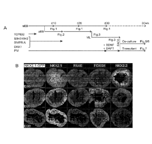
100251 Figures 1 (Panels A-B) illustrate the generation of MGE-like
Precursor Cells.
100261 Figures 2 (Panels A-F) illustrate hESC-MGE-like progenitors exhibit
VZ and
SVZ Radial Glial Stem Cell-like Divisions.
100271 Figures 3 (Panels A-E) illustrate hESC-MGE-like progenitors
differentiate into
neurons with properties of telencephalic GABAergic interneurons.
[0028] Figures 4 (Panels A-H) depict mieroarray gene expression profiling
of hESC-
MGE-like NKX2.1-GFP+ Cell Populations.
[0029] Figures 5 (Panels A-.1) illustrate hESC-MGE-like cell-derived
GABAergic
interneuron maturation and firing properties.
/00301 Figures 6 (Panels A-J) illustrate GABAergic Synaptic Properties of b
ESC-
derived Intemeurons.
[0031] Figures 7 (Panels A-H) show bESC-derived MGE-like interneuror
precursor cell
maturation and functional integration in the mouse brain.
100321 Figures 8 (Panels A-F) provide a schematic of differentiation
protocols and
FACS analysis of differentiated hESCs.
[0033] Figure 9 illustrates bESC-derived cells have telencephalic MGE-like
identity and
GABAergic neuronal fate.
[0034] Figures 10 (Panels A-F) depict transcript expression profiling of
hESC-derived
NICX2.1-GFP+ cells.
100351 Figure 11 depicts maturation of hESC-derived MGE-like cells into
GABAergic
intemeuron subtypes.
[0036] Figures 12 (Panels A-C) show development of intemeuron subtypes in
human
fetal cortex and MGE, and in cultures derived from human fetal MGE.
100371 Figures 13 (Panels A-F) show maturation of bESC-derived intemeumn
firing
properties.
100381 Figures 14 (Panels A-G) depict maturation of hESC-derived MGE-like
intemeurons and subtype firing properties in the mouse brain.
[0039] Figure 15 provides a summary of marker expression during
differentiation from
hESCs.
100401 Figure 16 provides a summary of hESC differentiation protocol
optimization,
animal transplantation, and tumor incidence.
4
WO 2014/153230 PCT11JS2014/029734
100411 Figure 17 depicts MCiE precursor cells differentiated in vitro from
hESC line
ESI17.
100421 Figure 18 depicts MGE precursor cells differentiated in vitro from
hESC line
ES135.
100431 Figure 19 depicts MGE precursor cells differentiated in vitro from
hESC line
EY:5 I.
100441 Figure 20 depicts MGE precursor cells differentiated in vitro from
hESC line :H9.
100451 Figure 21 illustrates generation of NIGH precursor cells by
differentiation of
naïve human pluripotent stem cells.
100461 Figures 22 (Panels A-N) illustrates utilization of an MGE-enriched
enhancer
sequence for the selection and purification of interneurons derived from .MGE
precursor
cells generated. by differentiation of hPSC.
100471 Figures 23 (Rows A.-D) depict generation of mori derived
.interneurons using
iong-tenn suspension culture.
100481 Figure 24 (Panels A-E) illustrates that numerous small molecule
inhibitors of
BMP and WNT signaling pathways are effective in inducing differentiation of
hESCs
into MGE precursor cells,
DEFINITIONS
100491 As used herein, "embryoid body", "embryoid bodies", "IEBs" or "EB
cells"
typically refers to a morphological, three-dimensional, or organoid-type
structure
comprised of a population of undifferentiated and differentiated cells which
are derived
from pluripotent stern cells (e.g., primate pluripotent stem cells (pPS),
embryonic stem
(ES) cells, induced pluripotent stem (IPS) cells) that have undergone
differentiation.,
Under culture conditions suitable for EB formation, ES cells proliferate and
form small
mass of cells that begin to differentiate. In the first phase of
differentiation, usually
corresponding, to about days 1-4 of differentiation for human cells, the small
mass of
cells forms a layer of endodermal cells on the outer layer, and is considered
a "simple
embryoid body." In. the second phase, usually corresponding to about days 3-20
post-
differentiation for human cells, "complex embryoid bodies" are formed, which
arc
characterized by extensive differentiation of ectodermal and mesodermal cells
and
derivative tissues. As used herein, the term "embryoid bodies" or "EB"
encompasses
WO 2014/153230 PCT/US2014/029734
both simple and complex embryoid bodies unless otherwise required by context.
The
determination of when embryoid bodies have formed in a culture of ES/iPS cells
is
routinely made by persons of skill in the art by, for example, visual
inspection of the
morphology, detection of cell markers. Floating masses of about 20 cells or
more (e.g.,
ES/iPS cells) are considered to be suspension embryoid bodies (sEB). (see.
e.g., Schmitt,
R., et al. (1991) Genes Dev. 5:728-740; Doetschman, T. C., et al. (1985) J.
Embryo!.
Exp. Morph. 87:27-45). Suspension EBs can be plated onto an adherent substrate
to
generate adherent EBs (aEB).
f00501 As used herein, "medial ganglionic eminence (MGE) precursor cell(s)"
or "MGE
neural precursor cells," refer to a population of mitotic and post-mitotic
cells that
express the markers expressed by cells in the MGE region of the developing
brain. In
general MGE precursor cells express markers such as, horneobox gene Alicr2. 1,
LIM-
homeobox genes thx6, Lhx7, or Lhx8. MGE precursor cells are capable of
differentiating into intemeurons under suitable differentiation conditions.
[0051] By "pluripotent stem cell" or "pluripotent cell" it is meant a cell
that has the
ability under appropriate conditions of producing progeny of several different
cell types
that are derivatives of all of the three germinal layers (endoderm, mesoderm,
and
ectoderm). Pluripotent stem cells are capable of forming teratomas. Examples
of
pluripotent stem cells are embryonic stem (ES) cells, embryonic germ stem (EG)
cells,
embryonal carcinoma stem (EC) cells, and induced pluripotent stem (iPS) cells.
PS cells
may be from any organism of interest, including, e.g., human; primate; non-
human
primate; canine; feline; murine; equine; porcine; avian; camel; bovine; ovine,
and so on.
100521 By "embryonic stem cell" or "ES cell" it is meant a cell that a) can
self-renew, b)
can differentiate to produce all types of cells in an organism, and c) is
derived from a
developing organism or is an established ES cell line which was derived from a
developing organism. ES cell may be derived from the inner cell mass of the
blastula, or
from the epiblast, of a developing organism. ES cell may be derived from a
blastomere
generated by single blastomere biopsy (SBB) involving removal of a single
blastomere
from the developing organism. In general, 51313 provides a non-destructive
alternative to
inner cell mass isolation. SBB and generation of hES cells from the biopsied
blastomere
is described in Cell Stem Cell, 2008 Feb 7; 2(2):113-7. ES cells can be
cultured over a
long period of time while maintaining the ability to differentiate into all
types of cells in
6
an organism. In culture, ES cells typically grow as flat colonies with large
nucleo-
cytoplasmic ratios, defined borders and prominent nucleoli. In addition, hES
cells
express SSEA-3, SSEA-4, TRA-1-60, TRA-1-81, and Alkaline Phosphatase, but not
SSEA-1. Examples of methods of generating and characterizing ES cells may be
found
in, for example, US Patent No. 7,029,913, US Patent No. 5,843,780, and US
Patent No.
6,200,806. Examples of ES cells include naive ES cells.
[0053] By "embryonic germ stem cell", embryonic germ cell" or "EG cell" it
is meant a
cell that a) can self-renew, b) can differentiate to produce all types of
cells in an
organism, and c) is derived from germ cells and germ cell progenitors, e.g.
primordial
germ cells, i.e. those that would become sperm and eggs. Embryonic germ cells
(EG
cells) are thought to have properties similar to embryonic stem cells as
described above.
Examples of methods of generating and characterizing EG cells may be found in,
for
example, US Patent No. 7,153,684; Matsui, Y., et al., (1992) Cell 70:841;
Shamblott, M.,
et al. (2001) Proc. Natl. Acad. Sci. USA 98: 113; Shamblott, M., et al. (1998)
Proc. Natl.
Acad. Sci. USA, 95:13726; and Koshimizu, U., etal. (1996) Development,
122:1235.
[0054] By "induced pluripotent stem cell" or "iPS cell" it is meant a cell
that a) can self-
renew, b) can differentiate to produce all types of cells in an organism, and
c) is derived
from a somatic cell. iPS cells have an ES cell-like morphology, growing as
flat colonies
with large nucleo-cytoplasmic ratios, defined borders and prominent nucleoli.
In
addition, iPS cells express one or more key pluripotency markers known by one
of
ordinary skill in the art, including but not limited to Alkaline Phosphatase,
SSEA3,
SSEA4, Sox2, 0ct3/4, Nanog, TRA160, TRA181, TDGF 1, Dnmt3b, FoxD3, GDF3,
Cyp26a1, TERT, and zfp42. iPS cells may be generated by providing the cell
with
"reprogramming factors", i.e., one or more, e.g., a cocktail, of biologically
active factors
that act on a cell to alter transcription, thereby reprogramming a cell to
pluripotency.
Examples of methods of generating and characterizing iPS cells may be found
in, for
example, Application Nos. U520090047263, US20090068742, US20090191159,
US20090227032, US20090246875, and US20090304646.
7
WO 2014/153230 PCT/US2014/029734
100551 By "somatic cell" it is meant any cell in an organism that, in the
absence of
experimental manipulation, does not ordinarily give rise to all types of cells
in an
organism. In other words, somatic cells are cells that have differentiated
sufficiently that
they will not naturally generate cells of all three germ layers of the body,
i.e., ectoderm,
mesoderm and endoderm. For example, somatic cells would include both neurons
and
neural progenitors, the latter of which may be able to self-renew and
naturally give rise
to all or some cell types of the central nervous system but cannot give rise
to cells of the
mesoderm or endoderm lineages.
100561 The term "cell line" refers to a population of largely or
substantially identical
cells that has typically been derived from a single ancestor cell or from a
defined and/or
substantially identical population of ancestor cells. The cell line may have
been or may
be capable of being maintained in culture for an extended period (e.g.,
months, years, for
an unlimited period of time).
[00571 By "endoderm" it is meant the germ layer formed during animal
embryogenesis
that gives rise to the gastrointestinal tract, respiratory tract, endocrine
glands and organs,
certain structures of the auditory system, and certain structures of the
urinary system.
[0058] By "mesoderm" it is meant the germ layer formed during animal
embryogenesis
that gives rise to muscles, cartilage, bones, dermis, the reproductive system,
adipose
tissue, connective tissues of the gut, peritoneum, certain structures of tbe
urinary system,
rnesothelium, notochord, and spleen.
[00591 By "ectoderm" it is meant the germ layer formed during animal
embryogenesis
that gives rise to the nervous system, tooth enamel, epidermis, hair, nails,
and linings of
mucosal tissues.
[00601 By "bone morphogenic proteins" or "BMPs" it is meant the family of
growth
factors that is a subfamily of the transforming growth factor l (TGF
superfamily.
BMPs (e.g. BMPI , BMI)2, BMP3, BMP4, BMP5, BMP6, BMP7, BMP8a, BMP8b,
BMP9/GDF, BMPIO, BMP11/GDF 1 1, BMP12/GDF7, BMP13/GDF6, BMP14/GDF5,
BMPI5/GDF9B) were first discovered by their ability to induce the formation of
bone
and cartilage. BMPs interact with specific receptors on the cell surface,
referred to as
bone morphogenetic protein receptors (BMPRs). Signal transduction through
I3MPIts
results in mobilization of members of the SMAD family of proteins, which in
turn
modulate transcription of target genes. Inhibitors of BMP signaling, can
readily be
8
WO 2014/153230 PCT/US2014/029734
identified by one of ordinary skill in the art by any of a number of methods,
for example
competitive binding assays for binding to BMP or BMI' receptors, functional
assays,
e.g., measuring enhancement of activity of downstream signaling proteins such
as
relocalization of SMADs, such as, BR-Smad to the nucleus and transcriptional
activation
of downstream gene targets as known in the art.
100611 By "transforming growth factor betas", "lfGF-fis", and "IGFI3s" it
is meant the
TGFB secreted proteins belonging to the subfamily of the transforming growth
factor 13
(IGO) superf.amily. IGFBs (TGFB I, TGFB2, TGFI33) are multifunctional peptides
that regulate proliferation, differentiation, adhesion, and migration and in
many cell
types. The mature peptides may be found as homodimers or as heterodimers with
other
TGFB family members. TGFBs interact with transforming growth factor beta
receptors
(TGF-f3Rs, or TGFBRs) on the cell surface, which binding activates MAP kinase-
, Aid-,
Rho- and Rac/cdc42-directed signal transduction pathways, the reorganization
of the
cellular architecture and nuclear localization of SMAD proteins, and the
modulation of
target gene transcription. Inhibitors of TGFB signaling, can be readily be
identified by
one of ordinary skill in the art by any of a number of methods, for example
competitive
binding assays for binding to TGFB or TGFB receptors, or functional assays,
e.g.
measuring suppression of activity of downstream signaling proteins such as
MAPK, Akt,
Rho, Rac, and SMADs, e.g., AR-Smad, etc., as well known in the art.
[0062] By "Writs" it is meant the family of highly conserved secreted
signaling
molecules which play key roles in both embryogenesis and mature tissues. The
human
Wnt gene family has at least 19 members (Wnt-1, Wnt-2, Wnt-2B/Wnt-13, Wnt-3,
Wnt3a, Wnt-4, Wnt-SA, Writ-5B, Wnt-6, Wnt-7A, Wnt-7B, Wnt-8A, Wnt-8B, Wnt-
9A/Wnt-14, Wnt-9B/Wnt-15, Wnt-10A, Wnt-10B, Wnt-11, Wnt-16). Wnt proteins
modulate cell activity by binding to Wnt receptor complexes that include a
polypeptide
from the Frizzled (Fz) family of proteins and a polypeptide of the low-density
lipoprotein receptor (LDLR)-related protein (LRP) family of proteins. Once
activated
by Wnt binding, the Wnt receptor complex will activate one or more
intracellular
signaling cascades. These include the canonical Wnt signaling pathway; the
Wnt/planar
cell polarity (WntfPCP) pathway; and the Wnt-calcium (Wnt/Ca2+) pathway.
100631 By culturing under "non-adherent conditions" it is meant culturing
under
conditions that suppress the adhesion of cells to the vessel in which they are
cultured,
9
WO 2014/153230 PCT/US2014/029734
e.g., the bottom of a tissue culture plate or flask. In some instances, the
cells are
naturally non-adherent, i.e., they will not adhere to a surface unless the
surface is coated
with a matrix composition, e.g., fibronectin, kuninin, poly-
lysine, collagen
IV, matrigel, and polycarbonate membranes. In some instances, cells may be
maintained
in a non-adherent state by agitating the culture.
[00641 By culturing under "adherent conditions" it is meant culturing under
conditions
that promote the adhesion of cells to the container in which they arc
cultured, e.g. the
bottom of a tissue culture plate or flask. In some instances, cells may be
induced to
adhere to the container simply by keeping the culture stationary. In some
instances, the
wall of the container to which it is desirable to promote adhesion may be
coated with a
composition to which the cells may adhere, e.g., fibronectin, laminin, poly-
ornithin,
poly-lysine, collagen TV, matrigel, and polycarbonate membranes.
1006511 The terms "treatment", "treating" and the like are used herein to
generally mean
obtaining a desired phammcologic and/or physiologic effect. The effect may be
prophylactic in terms of completely or partially preventing a disease or
symptom thereof
and/or may be therapeutic in terms of a partial or complete cure for a disease
and/or
adverse effect attributable to the disease. "Treatment" as used herein covers
any
treatment of a disease in a mammal, and includes: (a) preventing the disease
from
occuffing in a subject which may be predisposed to the disease but has not yet
been
diagnosed as having it; (b) inhibiting the disease, i.e., arresting its
development; or (c)
relieving the disease, i.e., causing regression of the disease. The
therapeutic agent may
be administered before, during or after the onset of disease or injury. The
treatment of
ongoing disease, where the treatment stabilizes or reduces the undesirable
clinical
symptoms of the patient, is of particular interest. Such treatment is
desirably performed
prior to complete loss of function in the affected tissues. The subject th
tapy will
desirably be administered during the symptomatic stage of the disease, and in
some
cases after the symptomatic stage of the disease.
100661 The terms 'individual", "subject", "host", and "patient" are used
interchangeably
herein and refer to any mammalian subject for whom diagnosis, treatment, or
therapy is
desired, particularly humans.
100671 The term "medium" in context of cell culture or the phrase "cell
culture medium"
or "cell medium" refer to a cellular growth medium suitable for culturing of a
cell
WO 2014/153230 PCT/US2014/029734
population of interest.. Examples of cell culture medium include Minimum
Essential
Medium (MEM), Eagle's Medium, Dulbecco's Modified Eagle Medium (DMEM),
Dulbecco's Modified Eagle Medium: Nutrient Mixture F-12 (DMEM/F12), F10
Nutrient
Mixture, Ham's F10 Nutrient Mix, Ham's F12 Nutrient Mixture, Medium 199, RPM1,
RPM 1640, reduced serum medium, basal medium (BME), DMEM/F12 (1:1),
Neurobasal medium, and the like, and combinations thereof. The medium or cell
culture
medium may be modified by adding one or more factors, such as, supplements,
differentiation factors, anti-apoptotic agents.
100681 The term "isolated" in context of cells or cell population refers to
cells that are in
an environment other than their native environment, such as, apart from tissue
of an
organism.
[0069] The phrase "differentiation factor(s)" as used herein refers to the
agent(s) that are
included in the medium for culturing cells of the present disclosure, which
agent(s)
promote the differentiation of the cells from a first cell type to a second
cell type, where
the second cell type is differentiated compared to the first cell type.
100701 In the context of cell ontogeny, the adjective "differentiated" is a
relative term.
A "differentiated cell" is a cell that has progressed further down the
developmental
pathway than the cell it is being compared with. Thus, pluripotent embryonic
stem cells
can differentiate to lineage-restricted precursor cells. These in turn can be
differentiated
further to cells further down the pathway, or to an end-stage differentiated
cell, such as
GABAergic intemeuron.
10071] "Feeder cells" or "feeders" are terms used to describe cells of one
type that are
co-cultured with cells of another type, to provide an environment in which the
cells of
the second type can grow. pPS cell populations are said to be "essentially
free" of feeder
cells if the cells have been grown through at least one round after splitting
in which fresh
feeder cells are not added to support the growth of pPS cells.
100721 As used herein, "expression" and grammatical equivalents thereof, in
the context
of a marker, refers to production of the marker as well as level or amount of
the marker.
For example, expression of a marker or presence of a marker in a cell or a
cell is positive
for a marker, refers to expression of the marker at a level that is similar to
a positive
control level. The positive control level may be determined by the level of
the marker
expressed by a cell known to have the cell fate associated with the marker.
Similarly,
11
WO 2014/153230 PCT/US2014/029734
absence of expression, of a marker or a cell is negative for a marker, refers
to expression
of the marker at a level that is similar to a negative control level. The
negative control
level may be determined by the level of the marker expressed by a cell known
to not
have the cell fate associated with the marker. As such, absence of a marker
does not
simply imply an undetectable level of expression of the marker, in certain
cases, a cell
may express the marker but the expression may be low compared to a positive
control or
may be at a level similar to that of a negative control
[0073] As used herein, "marker" refers to any molecule that can be measured
or
detected. For example, a marker can include, without limitations, a nucleic
acid, such as,
a transcript of a gene, a polypeptide product of a gene, a glycoprotein, a
carbohydrate, a
glycolipid, a lipid, a lipoprotein, a carbohydrate, or a small molecule (for
example, a
molecule having a molecular weight of less than 10,000 amu).
100741 A "variant" polypeptide means a biologically active polypeptide as
defined
below having at least 70%, 75%, 80%, 85%, 90%, 95%, 98%, or 99% sequence
identity
with a native sequence polypeptide. Such variants include polypeptides wherein
one or
more amino acid residues are added at the N- or C-terminus of, or within, the
native
sequence; from about one to forty amino acid residues are deleted, and
optionally
substituted by one or more amino acid residues; and derivatives of the above
polypeptides, wherein an amino acid residue has been covalently modified so
that the
resulting product has a non-naturally occurring amino acid. Ordinarily, a
biologically
active variant will have an amino acid sequence having at least about 90%
amino acid
sequence identity with a native sequence polypeptide, at least about 95%, or
at least
about 99%. The variant polypeptides can be naturally or non-naturally
glycosylated, i.e.,
the polypeptide has a glycosylation pattern that differs from the
glycosylation pattern
found in the corresponding naturally occurring protein. The variant
polypeptides can
have post-translational modifications not found on the natural polypeptide.
100751 The terms 'enriching" or "enriched" are used interchangeably herein
and mean
that the yield (fraction) of cells of one type is increased by at least 10%
over the fraction
of cells of that type in the starting culture or preparation.
[0076] A "growth environment" is an environment in which cells of interest
will
proliferate, differentiate, or mature in vitro. Features of the environment
include the
medium in which the cells are cultured, any growth factors or differentiation-
inducing
12
factors that may be present, and a supporting structure (such as a substrate
on a solid
surface) if present.
DETAILED DESCRIPTION
[0077] As noted above, methods and systems for generating MGE precursor
cells in vitro
as well as compositions of enriched MGE precursor cells are provided. The
methods and
systems provide efficient production of functional MGE precursors, which
differentiate
into functional GABAergic interneurons.
[0078] Before the present invention is further described, it is to be
understood that this
invention is not limited to particular embodiments described, as such may, of
course,
vary. It is also to be understood that the terminology used herein is for the
purpose of
describing particular embodiments only, and is not intended to be limiting,
since the
scope of the present invention will be limited only by the appended claims.
[0079] Where a range of values is provided, it is understood that each
intervening value,
to the tenth of the unit of the lower limit unless the context clearly
dictates otherwise,
between the upper and lower limit of that range and any other stated or
intervening value
in that stated range, is encompassed within the invention. The upper and lower
limits of
these smaller ranges may independently be included in the smaller ranges, and
are also
encompassed within the invention, subject to any specifically excluded limit
in the stated
range. Where the stated range includes one or both of the limits, ranges
excluding either
or both of those included limits are also included in the invention.
[0080] Unless defined otherwise, all technical and scientific terms used
herein have the
same meaning as commonly understood by one of ordinary skill in the art to
which this
invention belongs. Although any methods and materials similar or equivalent to
those
described herein can also be used in the practice or testing of the present
invention, the
preferred methods and materials are now described.
[0081] It must be noted that as used herein and in the appended claims,
the singular
forms "a," "an," and "the" include plural referents unless the context clearly
dictates
otherwise. Thus, for example, reference to "a SMAD inhibitor" includes a
plurality of
such inhibitors and reference to "the ROCK inhibitor" includes reference to
one or more
13
WO 2014/153230 PCT/US2014/029734
ROCK inhibitor and equivalents thereof known. to those skilled in the art, and
so forth.
It is further noted that the claims may be drafted to exclude any optional
element. As
such, this statement is intended to serve as antecedent basis for use of such
exclusive
terminology as "solely," "only" and the like in connection with the recitation
of claim
elements, or use of a "negative" limitation.
[0082] The publications discussed herein are provided solely for their
disclosure prior to
the filing date of the present application. Nothing herein is to be construed
as an
admission that the present invention is not entitled to antedate such
publication by virtue
of prior invention. Further, the dates of publication provided may be
different from the
actual publication dates which may need to be independently confirmed.
METHOD FOR GENERATING MGE PRECURSOR CELLS
100831 In certain embodiments, a method of producing medial ganglionic
eminence
(MGE) precursor cells from primate pluripotent stem (pPS) cells is provided.
[0084] In general, the pPS are maintained in an undifferentiated state till
the method for
production of MGE precursor cells is commenced.
[0085] The method may include culturing the pPS cells in a serum free
medium
comprising an activator of sonic hedgehog pathway and a neural inducing
supplement to
generate the MGE precursor cells. The pPS cells may be cultured as an adherent
culture
or a suspension culture.
[0086] In certain embodiments, at the start of the method for production of
MGE
precursor cells, pPS are plated cells into a cell culture container with an
adherent
substrate that facilitate the attachment of the pPS cells and the cells are
contacted with
serum free medium comprising an activator of sonic hedgehog pathway and a
neural
inducing supplement to generate the MGE precursor cells.
[0087] In certain embodiments, the method may include culturing the pPS
cells in a
scum free medium comprising an activator of sonic hedgehog pathway and a
neural
inducing supplement to generate embryoid bodies (EBs), wherein the EBs
comprise the
MGE precursor cells.
100881 In certain embodiments, at the start of the method for production of
MGE
precursor cells, pPS may be plated cells in suspension in culture containers
having a
substrate with low adhesion properties that allows suspension embryoid bodies
to form
14
WO 2014/153230 PCT/US2014/029734
In an exemplary method, confluent monolayer cultures of pPS cells are
harvested and
then plated in non-adherent cell culture plates, keeping the cells in
suspension.
[00891 In certain cases, CollagenaseIV/Dispase may be used for preferential
selection
for pPS colonies. The colonies may be trypsinized to single cells and plated
into low-
attachment round-bottom plates to form suspension EB.
[00901 In certain cases, the process of differentiation can be induced by
causing the pPS
cells to differentiate, e.g., to form embryoid bodies or aggregates: for
example, by
overgrowth of a donor pPS cell culture, or by culturing pPS cells in
suspension in culture
vessels having a substrate with low adhesion properties that allows embryoid
bodies to
form, or culturing pPS in absence of feeder layer. In an exemplary method,
confluent
monolayer cultures of pPS cells are harvested and then plated in non-adherent
cell
culture plates, keeping the cells in suspension, and providing regular feeding
with
nutrient medium.
[0091] Alternatively or in addition, the differentiation process can be
initiated by
culturing with certain factors that prevent the cells from maintaining the
undifferentiated
phenotype. The initial differentiation factors need not limit differentiation
into the MGE
precursor cell lineage, but should be inclusive of MGE precursor cell or their
precursors
within the range of cell types in the differentiated population.
[00921 At some stage, the culture can be directed more specifically into
the MGE
precursor cell lineage. This can be done by including in the culture medium a
factor that
more specifically promotes the generation and proliferation of MGE precursor
cell.
Exemplary factors that promote the formation and/or growth of MGE precursor
cells
include neural inducing supplements as provided herein, activators of shh
signaling,
inhibitors of I3MP-signaling, inhibitors of TGF-I3 signaling, Wnt inhibitors,
and anti-
apoptotic agents, and in some cases can include activator(s) of FGF signaling.
100931 Exemplary methods for generating MGE precursor cells are described
below.
[00941 In certain cases, the method may include a step of generation of sEB
following
by a step of generation of aEB. In other cases, the step of generation of sEB
may be
replaced by an adherent culture.
WO 2014/153230 PCT11JS2014/029734
Generation of Suspension Embrvoid Bodies (sEB
100951 In an exemplary method, culturing pPS cells in suspension in culture
vessels
having a substrate with low adhesion properties that allows suspension
embryoid bodies
to form may be carried out in the presence of an activator of shh and a neural
inducing
supplement, such as B27 or NS21. The pPS cells may be cultured in suspension
in
absence of a feeder layer for 0 day-9 days before an activator of shh and/or
neural
inducing supplement is added to the culture medium, for example, the pPS cells
may be
cultured in suspension for at least 0 hr, I hr, 3 hrs, 6 hrsõ 12 hrs, 18 hrs,
24 hrs, 36 hrsõ 48
hrs, 2 days, 3 days, 4 days, 5 days, 6 days, 7 days, 8 days, or 9 days before
an activator
of shh and/or neural inducing supplement is added to the culture medium,
Accordingly,
the pPS are induced to form sEBs in the presence of a neural inducing
supplement as
described herein and an activator of shh signaling.
100961 The pPS cell may be cultured in suspension to form sEB for a period
of at least 1
day, e.g., 1400 days, 1-60 days, 1-50 days, 2-100 days, 2-50 days, 3400 days,
4-100
days, 5-10 days, or 7-10 days, or 25-100 days in the presence of a neural
inducing
supplement as described herein and an activator of still signaling. In cases,
where the
pPS cell may be cultured in suspension to form sEB for a period of less than 9
days, an
activator of shh and/or neural inducing supplement may be added to the culture
medium
within 0-8 days from the start of the culture of pPS cells to form sEB,
100971 In certain embodiments, the pPS are plated in suspension, in culture
containers
having a substrate with low adhesion properties, in a cell culture medium that
includes a
neural inducing supplement as provided herein and an activator of shh
signaling.
100981 In addition to a neural inducing supplement as provided herein and
an activator
of shh signaling, the culture medium for culturing pPS cells in suspension to
form sEBs
may contain one or more of an anti-apoptotic agent, SMAD inhibitor (eõg., TGE-
13
inhibitors, BMP inhibitors, Activin inhibitor, Nodal inhibitor, or growth
differentiation
factor ((IMF) signaling pathway inhibitor), and Wnt inhibitor.
100991 in certain cases, the method for producing MOE precursor cells from
pPS cells
may include culturing the pPS cells in a medium that includes an ant-apoptotic
agent,
e.g., a ROCK inhibitor, for about I hr-35 days, e.g., at least I hr, at least
3 hrs, at least 10
hrs, at least 24 hrs, at least 36 hrs, at least 48 hrs, at least 2 days, at
least 3 days, such as,
4 days, 5 days, 6 days, 7 days, 8 days, 9 days, 15 days, 20 days, 25 days, or
35 days. An
16
WO 2014/153230 PCT/US2014/029734
exemplary method may include plating the pPS cells in suspension in a medium
containing an anti-apoptotic agent, culturing the pPS cells for a period of
lhr-35 days in
the presence of the anti-apoptotic agent. In certain cases, the anti-apoptotic
agent may be
present from the start of culturing of pPS cells in suspension and may be
removed after 1
hr-35 days, such as 1 day to 7 days, e.g. I days, 2 days, 3 days, 4 days, 5
days, 6 days, or
7 days.
1001001 In certain cases, the anti-apoptotic agent may be present
transiently during
differentiation of the pPS into MGE cells, e.g., the anti-apoptotic agent may
be present
in the culture medium on day 1 when the pPS cells are exposed to the neural
inducing
supplement as provided herein and an activator of shh signaling. The
differentiation of
the pPS cell may be carried out in the presence of neural inducing supplement
as
provided herein, an activator of shh signaling, and an anti-apoptotic agent
for 1 hr to 35
days are noted above, after which the culturing may be continued in the
absence of the
anti-apoptotic agent.
[00101] In certain cases, the method for producing MGE precursor cells from
pPS cells
may include culturing the pPS cells in a medium that includes one or more
inhibitors of
wnt. Although the Wnt signal inhibitor may be added to the medium already at
the start
of cultivation of pPS cells, it may be added to the medium after several days
of
cultivation (for example, at a time within 10 days of cultivation). In certain
cases, the
Wnt signal inhibitor is added to the medium at a time within 5 days of start
of culturing
of pPS cells in suspension, such as, within 0 days, I day, or 3 days. The writ
inhibitor
may be present throughout the step of generation of sEII or may be present for
a period
of 5 days-10 days, e.g., 5 days, 6 days, 7 days, 8 days, 9 days, or 10 days,
after which the
culture may be continued in absence of the wnt inhibitor(s).
1001021 In certain cases, the method for producing MGE precursor cells from
pPS cells
may include culturing the pPS cells in a medium that includes one or more
inhibitors of
SIVLAD. Although the SM.AD signal inhibitor may be added to the medium already
at the
start of cultivation of pPS cells, it may be added to the medium after several
days of
cultivation (for example, at a time within 10 days of cultivation). In certain
eases, the
one or more SMAD signal inhibitors are added to the medium at a time within 5
days of
start of culturing of OS cells in suspension, such as, within 0 days, 1 day,
or 3 days. The
17
WO 2014/153230 PCT/US2014/029734
wnt inhibitor may be present throughout the step of generation of sEB or may
be present
for a period of 5 days-10 days.
[001031 In certain cases, the pPS are differentiated in the presence of shh
activator, neural
inducing supplement as provided herein, and SMAD inhibitor(s) for a period of
5 to 15
days (e.g., 5-10 days, such as, 5 days, 6 days, 7 days, 8 days, 9 days, 10
days, 12 days)
after which the differentiation may be continued in absence of the SMAD
inhibitor(s).
[001041 In certain embodiments, the method of producing medial ganglionic
eminence
(MGE) precursor cells from primate pluripotent stem (pPS) cells may include
culturing
the pPS cells in a serum free medium to generate sEBs, wherein the sEBs
include the
MGE precursor cells, wherein the serum free medium includes an activator of
sonic
hedgehog pathway, an anti-apoptotic agent, an inhibitor of SMAD, an inhibitor
of Wnt
and B27. The sEB produced by the methods described herein include a population
of
MGE precursor cells.
[00105] In certain embodiments, the method of producing medial ganglionic
eminence
(MGE) precursor cells from primate pluripotent stem (pPS) cells may include
culturing
the pPS cells in a serum free medium as suspension culture to generate the MGE
precursor cells, wherein the serum free medium includes an activator of sonic
hedgehog
pathway, an anti-apoptotic agent, an inhibitor of SMAD, an inhibitor of Wnt
and B27. In
certain, cases, the sEB may be dissociated and plated as a monolayer to
generate a
monolayer that includes MGE precursor cells.
[001061 In certain cases, the sEBs may be dissociated and plated as a
monolayer after
about 5 days from the beginning of the differentiation of pPS cells. For
example, the
sEBs may dissociated and plated as a monolayer within 1-100 days after
formation of
the sEB, e.g., 1-75 days, 1-50 days, 1-30 days, 1-10 days. In exemplary cases,
sEB may
be dissociated and plated as a monolayer after about 10 days of formation of
the sEB,
e.g., 10-50 days, 10-40 days, 10-30 days, 10-20 days, 10 days, 12 days, etc.
The
differentiation factors as well as additives, supplements, or factors, used as
well as the
timing of addition/removal of the same may be as disclosed above.
1001071 In certain embodiments, the method of producing medial ganglionic
eminence
(MGE) precursor cells from primate pluripotent stem (pPS) cells may include
culturing
the pPS cells in a serum free medium as an adherent culture to generate the
MGE
18
WO 2014/153230 PCT/US2014/029734
precursor cells, wherein the serum five medium includes an activator of sonic
hedgehog
pathway, an anti-apoptotic agent, an inhibitor of SMAD, an inhibitor of Wnt
and B27.
1001081 In general, the MGE precursor cells produced by the method
described herein
express a marker of MGE precursor cells, such as, NKX2.1.
[00109] In certain cases, the PS cells at the start of the culturing to
generate MGE
precursor cells are present at a cell density of i 3 to 107 cells/ml.
1001101 The medium used in the suspension culture can be prepared using any
basal
medium. The medium may be BM.E medium, Balt) medium, CMRI, 1066 medium,
Glasgow MEM medium, Improved MEM Zinc Option medium, IMDM medium,
Medium 199 medium, Eagle's MEM medium, DMEM medium, Ham's medium, RPM!
1640 medium, Fischer's medium, Neurobasal medium, and a mixed medium thereof
and
the like. The medium may be modified by addition of additives, supplements, or
factors,
as disclosed herein.
[00111] A cell culture container with an adherent substrate may be used in
methods of
culturing the pPS as an adherent culture. The differentiation factors as well
as additives,
supplements, or factors, used as well as the timing of addition/removal of the
same may
be as disclosed above.
Generation of Adherent Entbryoid Bodies (aEll)
[001121 In certain cases, the sEBs generated by the above described methods
may be
plated into a cell culture container with an adherent substrate that
facilitate the
attachment of the sIHI3 to form adherent EI3s. In general, the sE13 may be
plated onto a
cell culture container with an adherent substrate in a culture medium
containing a neural
inducing supplement as provided herein and an activator of shh signaling.
[001131 In embodiments where the pPS cells are cultured as an adhesion
culture, as noted
above, the method may further culturing the pPS cells in the serum free medium
comprising the activator of sonic hedgehog pathway and the neural inducing
supplement
to generate aEBs, which aEBs include MGE precursor cells.
1001141 The sEI3 replated and cultured in adhesion culture to form aBB may
be cultured
for a period of 1- 100 days. In exemplary methods, the replating of sEB may
involve the
steps of dissociating the F.13s mechanically or enzymatically to produce
single cells or
clusters of cells, plating the dissociated cells on an adherent substrate to
provide an
19
WO 2014/153230 PCT/US2014/029734
adherent monolayer; and culturing the adherent monolayer to generate aEBs. In
certain
methods, the sEBs are not dissociated before further culturing in adherent
conditions.
[001151 In certain embodiments, the method for generating MGE precursor
cells from
pPS cells may include culturing the pPS cells in a serum free medium
comprising an
activator of sonic hedgehog pathway and a neural inducing supplement to
generate sEBs
and plating of the .sEBs on a cell culture container with an adherent
substrate and
culturing the plated sEBs on the adherent substrate in the scrum free medium
comprising
the activator of sonic hedgehog pathway and the neural inducing supplement to
generate
aEBs, which aEBs include MGE precursor cells.
[001161 In certain cases, the aEBs may be dissociated and replated as a
monolayer, which
monolayer may be cultured in a serum free medium that includes the activator
of sonic
hedgehog pathway and the neural inducing supplement to generate MGE precursor
cells.
100117) The cell culture medium for culturing of the adhesion culture to
generate aEB
from the sEB may also include one or more of factors such as, an anti-
apoptotic agent,
an inhibitor of SMAD, and an. inhibitor of Wnt. The factors may be present at
the start
of the adhesion culture or may be added within 5 days of initiation of the
adhesion
culture, such as, 0 lit, 1 hr, 3 hr, 10 hr, I day, 2 days, or 3 days from the
initiation of the
adhesion culture. The factors may be removed from the adhesion culture after l
day to
20 days of culturing.
1001181 In certain embodiments, the method for generating MGE precursor
cells from
pPS cells may include culturing the cells of the sEBs obtained by the methods
described
herein in an adhesion culture in a medium that includes activator of sonic
hedgehog
pathway, a neural inducing supplement, Wnt and SMAD inhibitors, for a period
of 4-20
days, followed by culturing the adhesion culture for 8-20 days in a medium
that includes
activator of sonic hedgehog pathway and a neural inducing supplement but does
not
include Wnt and SMAD inhibitors.
1001191 The aEBs generated by the methods described herein include a
population of
MGE precursor cells. In general, the MGE precursor cells present in the aEBs
produced
by the method described herein express NKX2.1 and FOXGI. In certain cases, the
MGE
precursor cells produced by the methods disclosed herein may express one or
more
markers of MGE precursor cells, such as, NKX2.1., LfIX6, LHX718, FOX0I,
()LIG2,
DLXI/2, and ASCU.
WO 2014/153230 PCT/US2014/029734
[001201 In certain embodiments, the MGE precursor cells produced by the
methods
described herein may also include a population of cells differentiated from
the MGE.
precursor cells, such as, intemeurons, e.g., GABAergic intemeurons.
1001211 En general, the methods described herein result in generation of
MGE precursor
cells at a high efficiency, resulting in cell cultures where at least 50%
(e.g. 65%, 70%,
75%, 80%, 85%, 90%, or 95%) of the cells in the cell culture are MGE precursor
cells.
[001221 As such, the method may include culturing pPS cells in a scrum free
culture
medium comprising activator of sonic hedgehog pathway and a neural inducing
supplement for a period of 10-100 days (e.g. 5-50 days) to generate MGE
precursor
cells, wherein the pPS cells are cultured in adherent or suspension culture,
wherein the
pPS cells are induced to differentiate prior to culturing in the presence of
activator of
sonic hedgehog pathway and a neural inducing supplement. As such, the pPS
cells may
include differentiated cells, such as El3s, prior to culturing pPS cells in a
serum free
culture medium comprising activator of sonic hedgehog pathway and a neural
inducing
supplement. In certain cases, the serum free culture medium may additionally
include an
anti-apoptotic agent, an inhibitor of SMAD, and an inhibitor of Wnt.
1001231 En certain cases, the aEBs obtained from the sEB may be replated in
a suspension
culture to form sEBs or dissociated and replated as a monolayer in adherent
culture.
1001241 Culturing of pPS cells as an adherent culture in a method for
generating MGE
precursor cells is further described below.
Adherent Culture for Generation of MGE Precursor Cells
[001251 As noted above, in certain embodiments, at the start of the method
for production
of MGE precursor cells, pPS are plated cells into a cell culture container
with an
adherent substrate that facilitate the attachment of the pPS cells and the
cells are
contacted with serum free medium comprising an activator of sonic hedgehog
pathway
and a neural inducing supplement to generate the MGE precursor cells.
1001261 In certain cases, the MGE precursor cells generated in the adherent
culture may
be present in the aEBs.
1001271 In some cases, the aEB produced by the subject culture method may
be
dissociated and replated as a monolayer and cultured in a serum free medium
comprising
an activator of sonic hedgehog pathway and a neural inducing supplement to
generate
21
WO 2014/153230 PCT/US2014/029734
the MGE precursor cells. The aEB may be maintained in the of supplements and
factors
as described herein for a period of time of 1-100 days before being replated
in a
suspension culture and cultured further as sEB or before being dissociated and
replated
as a monolayer in an adherent culture. In certain cases, the period of time
may be 1-75
days, 1-50 days, 1-30 days, 1-10 days, e.g., 10-50 days, 10-40 days, 10-30
days, 10-20
days, 5 days, 10 days, 20 days, or 30 days.
1001281 Adherent substrates known in the art as well as those described
herein may be
used for culturing the pPS as an adherent culture in a method for generating
MGE
precursor cells.
[001291 The pPS cells may be grown as an adherent culture for a period of
time before
contacting with serum free medium comprising an activator of sonic hedgehog
pathway
and a neural inducing supplement. In certain cases the pPS cells may be
induced to
differentiate by overgrowth of a donor pPS cell culture, or culturing pPS in
absence of
feeder layer, or culturing pPS cells in presence of FGF, or the like.
Alternatively or in
addition, the differentiation process can be initiated by culturing with
certain factors that
prevent the cells from maintaining the undifferentiated phenotype. The initial
differentiation factors need not limit differentiation into the MGE precursor
cell lineage,
but should be inclusive of MGE precursor cell or their precursors within the
range of cell
types in the differentiated population.
1001301 At some stage, the culture can be directed more specifically into
the MGE
precursor cell lineage. This can be done by including in the culture medium a
factor that
more specifically promotes the generation and proliferation of MGE precursor
cell.
Exemplary factors that promote the formation and/or growth of MGE precursor
cells
include neural inducing supplements as provided herein, activators of shh
signaling,
inhibitors of BMP-signaling, inhibitors of TGF-13 signaling, Wnt inhibitors,
and anti-
apoptotic agents, and in some cases can include activator(s) of FGF signaling.
[001311 Exemplary methods for generating MGE precursor cells are described
below.
[001321 The pPS cells may be cultured in adherent conditions for 0 day-9
days before an
activator of shh and/or neural inducing supplement is added to the culture
medium, for
example, the pPS cells may be cultured in adherent conditions for at least 0
hr, 1 hr, 3
hrs, 6 firs, 12 hrs, 18 hrs, 24 firs, 36 hrs, 48 hrs, 2 days, 3 days, 4 days,
5 days, 6 days, 7
days, 8 days, or 9 days before an activator of shh and/or neural inducing
supplement is
22
WO 2014/153230 PCTT1JS2014/029734
added to the culture medium, In certain embodiments, the OS are differentiated
in
absence of a feeder cell layer.
[00133] In cases, where the pPS cell may be cultured in adherent conditions
in a culture
medium containing an activator of shh and/or neural inducing supplement for a
period of
1-100 days from the start of the culture of pPS cells to form MGE precursor
cells.
[00134] In addition to a neural inducing supplement as provided herein and
an activator
of shh signaling, the culture medium for culturing pPS cells in adherent
conditions may
contain one or more of an anti-apoptotic agent, SMAD inhibitor (e.g., Tcf-i3
inhibitors,
BMP inhibitors, Activin inhibitor, Nodal inhibitor, or GDF signaling pathway
inhibitor),
and Vint inhibitor.
[00135] The timing of addition and removal of differentiation factors may
be as descrbied
for the aEB formation above.
1001361 In certain cases, the method for producing MGE precursor cells from
pPS cells
may include culturing the pPS cells in a medium that includes an anti-
apoptotic agent,
e.g., a ROCK inhibitor, for about 1 hr-35 days, e.g., at least 1 hr, at least
3 hrs, at least 10
hrs, at least 24 .hrs, at least 36 hrs, at least 48 hrs, at least 2 days, at
least 3 days, such as,
4 days, 5 days, 6 days, 7 days, 8 days, 9 days, 15 days, 20 days, 25 days, or
35 days. An
exemplary method may include plating the pPS cells in adherent culture in a
medium
containing an anti-apoptotic agent, culturing the pPS cells for a period of
lhr-35 days in
the presence of the anti-apoptotic agent. In certain cases, the anti-apoptotic
agent may be
present from the start of culturing of pPS cells for generation of MGE
precursor cells
and may be removed after I hr-35 days, such as 1 day to 7 days.
1001371 In certain cases, the method for producing MGE precursor cells from
pPS cells
may include culturing the pPS cells in a medium that includes one or more
inhibitors of
wnt. Although the Writ signal inhibitor may be added to the medium already at
the start
of culturing of pPS cells, it may be added to the medium after several days of
cultivation
(for example, at a time within 10 days of culturing). In certain, cases, the
Writ signal
inhibitor is added to the medium at a time within 5 days of start of culturing
of pPS cells
in adherent condition, such as, within 0 days, 1 day, or 3 days. The wnt
inhibitor may be
present throughout the culturing or may he present for a period of 5 days-1.0
days.
1001381 In certain cases, the method for producing MGE precursor cells from
pPS cells
may include culturing the pPS cells in a medium that includes one or more
inhibitors of
23
WO 2014/153230 PCT/US2014/029734
SMAD. Although the SMAD signal inhibitor may be added to the medium already at
the
start of culturing of pPS cells, it may be added to the medium after several
days of
culturing for generation of MGE precursor cells (for example, at a time within
10 days
of culturing). In certain cases, the one or more SMAD signal inhibitors are
added to the
medium at a time within 5 days of start of culturing of pPS cells in adherent
condition,
such as, within 0 days, I day, or 3 days. The wnt inhibitor may be present
throughout the
culturing to generate MGE precursor cells or may be present for a period of 5
days-10
days.
1001391 In certain embodiments, the method of producing medial ganglionic
eminence
(MGE) precursor cells from primate pluripotent stem (pPS) cells may include
culturing
the pPS cells in adherent condition to generate MGE precursor cells, wherein
the culture
medium includes an activator of sonic hedgehog pathway, an anti-apoptotic
agent, an
inhibitor of SMAD, an inhibitor of Wnt and 1327.
[00140] In certain cases, the PS cells at the start of the culturing to
generate MGE
precursor cells are present at a cell density of 103 to 107 cells/ml.
[001411 As noted above, a serum free medium may be used in the method of
generating
MGE precursor cells from pPS cells. A serum-free medium means a medium not
containing an unadjusted or unpurified serum, such as, fetal bovine serum,
fetal calf
serum. The serum-five medium may include a serum replacement, such as, those
described herein, e.g., 1327 or NS21.
Culture of MGE Precursor Cells
[00142] The sEBs and aEBs generated by the methods described herein may be
dissociated, enzymatically or mechanically, and cultured as a monolayer on a
cell culture
vessel with adherent substrate. Accordingly, the MGE precursor cells present
in the sEB
and aEBs may be cultured as a monolayer.
100143] In certain cases, culturing of MGE precursor cells in a monolayer
may be carried
out for a period of 1-100 days, such as 10 days-15 days.
[001441 The culturing of :MGE precursor cells in a monolayer may be carried
out in a
culture medium that contains a neural inducing supplement as provided herein
and an
activator of shh signaling.
24
WO 2014/153230 PCT11JS2014/029734
1001451 In certain cases, the MGE precursor cells generated by the method
described
herein may be cultured in a culture medium that promote generation of neurons,
such as,
inhibitory interneurons, e.g., GABAergic inteineurons. Accordingly, sEB, aEB,
and
monolayer produced from dissociation of sIEBs and aEBs generated by the
methods
described herein and containing MGE precursor cells may be contacted with a
culture
medium that promotes differentiation of the MGE precursor cells into post
mitotic
neurons. In certain cases, this culture medium may not include SMAD
inhibitors. In
addition, in certain cases, this culture medium may include SMAD activators.
.As such,
the culture medium may include SMAD activators in order to increase the
population of
interneuron.s present in the .MGE precursor cells generated by the protocols
described
herein. Exemplary SMAD activators include TGFs (e.g., TG.F133), BMPs (e.g.,
BIMP2,
BMP4, BMP8), Activin, Nodal, GDF, and IDEL
1001461 In certain cases, the culture medium to promote differentiation of
the MGE
precursor cells to intemeurons may include a NOTCH inhibitor, 13DNF, GDNF,
NT3,
NT4, camp, vitamin c, serum, inatrigel, insulin, IGF, SDF la, Neuregutini,
TGFp.
[001471 The culturing of MGE precursor cells in a monolayer may lead to
proliferation of
MGE precursor cells and/or differentiation of MGE, precursor cells into cells
having a
neuronal cell fate. In certain cases, the MGE precursor cells that
differentiate into cells
having a neuronal cell fate express DI,X112, TUJ, M.AP2, GAD1/2, and GABA, and
may express one or more of NKX2.1, ASCI,1, LHX6, LHX7/8, DCX, MAN, and
VGAT, and may express subtype markers calbindin, calretinin, somatostatin, and
parvalbum in.
100148] In certain cases, the MGE precursor cells generated by the method
described
herein may be co-cultured with a support cell population to induce
differentiation of the
MGE precursor cells into intemeurons, such as, GABAergic interneurons.
Propagation of OS Cells in an Undifferentiated State
1001491 pPS cells can be propagated continuously in culture, .using culture
conditions that
promote proliferation without promoting differentiation. Exemplary ES medium
is made
with 80% DMEM (such as Knockout DMEM "KO IDMEM"), 20% of either defined
fetal bovine serum (FBS, flyelone) or serum replacement (e.g., knockout serum
replacement (KSR)), 1% non-essential amino acids (NEAA), 1% pen-strep-
glutamine (1
mM L-glutamine), 0.0008% 13-mercaptoethanol, and lOng/m1 FGF-basic (bFGF).
[00150] The pPS cells can be expanded in the undifferentiated state by
culturing in an
environment that inhibits differentiation. Traditionally, pPS cells are
cultured on a layer
of feeder cells derived from embryonic or fetal tissue of the mouse. Culture
plates are
plated with 375,000 irradiated mouse embryonic fibroblasts (mEFs) per well
(irradiated
to inhibit proliferation but permit synthesis of factors that support pPS
cells), and used 5
h to 10 days after plating. In certain embodiments, human feeder cells may
also be used.
[00151] pPS cells can be maintained in an undifferentiated state even
without feeder cells.
The environment for feeder-free cultures includes a suitable culture
substrate, particularly
an extracellular matrix such as Matrigel0 or laminin. The pPS cells are plated
at
>15,000 cells cm-2 (optimally 90,000 cm-2 to 170,000 cm-2). Feeder-free
cultures are
supported by a nutrient medium containing factors that support proliferation
of the cells
without differentiation. Such factors may be introduced into the medium by
culturing the
medium with cells secreting such factors, such as irradiated (-4,000 rad)
primary mouse
embryonic fibroblasts, telomerized mouse fibroblasts, or human feeder cells
derived from
pPS cells. Medium can be conditioned by plating the feeders at a density of ¨5-
6x10⁴
cm-2 in a serum free medium such as KO DMEM supplemented with 20% serum
replacement and 4 to 8 ng/mL bFGF. Medium that has been conditioned for 1-2
days is
supplemented with further bFGF, and used to support pPS cell culture for 1-2
days.
Features of the feeder-free culture method are further discussed in
International Patent
Publications W099/20741 & W001/51616; and Xu et al., Nat. Biotechnol. 19:971,
2001.
Factors
[00152] The methods and compositions of the present disclosure involve the
use of
various factors, such as, neural inducing supplements, anti-apoptotic agents,
differentiation factors, and the like. Examples of neural inducing
supplements, anti-
apoptotic agents, differentiation factors used in the methods and compositions
of the
present disclosure are described below.
26
WO 2014/153230 PCT/US2014/029734
Neural .inducing simplement
1001531 Exemplary neural inducing supplements include B27, NS21, or an
equivalent
supplement.
1001541 In certain embodiments, the neural inducing supplement may be 1327.
B-270
Serum-Free Supplement is available from Life Technologies. B27 supplement
contains
bovine serum albumin, traxisferrin, insulin, progesterone, corticosterone,
triiodo-l-
thyronine, retinal acetate, DL tocophcrol, DL tocopherol acetate, Biotin,
Linoleic acid,
Linolenic acid, ethanolamine, Na Selenite, L-camitine, glutathione reduced,
catalase,
superoxide dismutase, D-galactose and putrescine. In certain cases, B27-
vitamin A may
be used.
[001551 In certain cases, the neural inducing supplement may be NS21. NS21
is
described in Y. Chen et al., J. Neurosci. Methods., 171:239,2008. Y. Chen et
al. showed
that NS21 is equivalent to B27 supplement in a neuronal culture. The
formulation of
NS21 is described in Y. Chen et al. and is reproduced in Table 1 below.
Table 1. NS I Formulation
Final Concentration
pg/m.1 pM Stock (mg/ml) For 400m1.
NS21 (20L
final medium)
Albumin, bovine 2500 37 Add as.powder 50g
Catalase 2.5 0.010 Add as powder 50mg
Giutathione (reduced) 1.0 3.2 Add as powder 20mg
Insulin 4.0 0.6 10 8m1
Superoxidase dismutase 7.5 0.077 Add as powder 50rng
floto transfer-in 5.0 0.062 Add as powder 100mg
T3 (triiodol-l-thyronin) 0.002 0.0026 2.0 20p.I
L-Carnitine 2.0 12 Add as powder 40mg
Ethanolamine 1.0 16 Liquid .(1 20.0
D(+)-galactose 15 83 Add as..powder 300m.g.
Putrescine 16.1 183 Add as powder 322mg
Sodium Selenite 0.01435 0.083 1.0 28011
Ethanolic Stocks
Corticosterone 0.02 0.058 2.0 0.2m1
Linoleic acid 1.0 3.5 100.0 0.2m1
Linolenic acid 1.0 3.5 1.00.0 0.2m1
Lipoic acid (thioctic acid) 0.047 0.2 4.7 0.2m1
Progesterone 0.0063 0.020 3.2 0.04m1
Retinol acetate 0.1 0.2 20.0 0.1m1
Retinol all trans (vit. A) 0.1 0.3 10.0 0.2m1
27
WO 2014/153230 PCT/US2014/029734
D, L-alpha-Tocopherol (vit. 1.0 23 100.0 0.2m1
1,-alpha-Tocopherol 1.0 2.1 100.0 0.2m1
acetate
100156] In certain cases, the neural inducing supplement may be present in
the serum free
medium for culturing pPS cells at a concentration ranging from 0.5% to 10%,
for
example, 0.5 %-5%, e.g., 0.5%, 1%, 2%, or 3%.
[001571 In certain embodiments, the serum free medium comprising a shh
activator and a
neural inducing supplement for culturing of pPS to generate EBs does not
include KSR
or N2 supplement. In certain embodiments, the method of generating MGE
precursor
cells does not include culturing the pPS cells in a serum free medium
comprising bFGF
or FGF-2. In certain cases, the pPS cells are cultured in a serum free medium
comprising
a shh activator and a neural inducing supplement and not containing KSR or N2
supplement or bFGF or FGF-2 for a period of time sufficient to generate sEI3
or aEB.
1001581 In certain cases, the sEBs may be further cultured in a serum free
medium
comprising a shh activator and a neural inducing supplement and further
containing one
or more of KSR supplement, N2 supplement, bFGF, and FGF-2 for a period of
sufficient
to generate aEB.
1001591 In certain cases, the pPS cells may be cultured in a serum free
medium
comprising a shh activator and a neural inducing supplement and further
containing one
or more of KSR supplement, N2 supplement, bFGF, and FGF-2 for a period of
sufficient
to generate sEB. The additional supplements may be added at the same time as a
shh
activator and the neural inducing supplement as described herein or at a later
time point,
such as, after 5 days ¨2 weeks, such as, after 1 weeks- 2weeks after exposing
the pPS
cells to shh activator and the neural inducing supplement as described herein.
In certain
cases, the KSR supplement and/or N2 supplement may be present added at day 0
of
differentiation, or later such as day 5, day 7, day 10, day 14, day 21, after
contacting the
pPS cells shh activator and the neural inducing supplement as described
herein.
[001601 In certain embodiments, the cell culture medium used in the methods
disclosed
herein does not include serum replacements, such as, :KSR or N2.
28
WO 2014/153230 PCT11JS2014/029734
APti.-0,Qpto0c. Age!lt5
100161j in certain embodiments of the methods and compositions described
herein, an
anti-apoptotic agent may be included in the medium for PS culturing cells.
100162] In certain cases, the anti-apoptotic agent may be an inhibitor of
Rho-associated
protein kinase (ROCK). In certain cases, the ROCK inhibitor may be Y27632, HA-
100,
H-1152, ( )-trans-4-(1-aminoethyl)-1-(pyridin-4-ylaminocarbonyl) cyclo hexane
dihydro-chtoridc .monohydrate (described in W000078351, W000057913),
imidazopyridine derivatives (described in U.S. Pat. No. 7348,339), substituted
pyrimidinc and pyridine, derivatives (described in U.S. Pat. No. 6,943,172)
and
substituted isoquinolin.e-sulfonyl compounds (described in EP00187371), or
GSK429286A, ROCKII inhibitor, or Thiazovivin, or an analog or derivative
thereof.,
1001631 The anti-apoptotic agent may be present at a concentration of 0.1
M, 0.3 NI,
0.5 i_tiN4, 1 laM, at least about 1,3 iaM, at least about 1.5 4M, at least
about .2 }tM, at least
about 2.3 !AM, at least about 2.5 p.M, at least about 2.8 uM, at least about 3
uMõ at least
about 3.5 JIM, at least about 4 iM., at least about 4.5 RIVI, at least about 5
04, at least
about 10 0.4, at least about 20 uM, at least about 30 M, at least about 40
tiM or at least
about 50 uM., such as, 0.5 p.1\41 -50 gM, 1 !AM -25 ,M, or 2.5 p.M -20
Inhibitors of SMAD
1001641 In certain embodiments of the methods and compositions described
herein, an
inhibitor of WAD may be present in the medium for culturing cells, in some
embodiments, an inhibitor of SMAD can be present in the medium, used for
culturing
cells, at a concentration of 10 nglml, 200 ng/ml, 300 ngiml, 400 ngiml, 500
ng/ml, 1
pg/ml, 1.5 jig/nil, 2 lag/ml, 2.5 jig/ml, or 5 jig/ml for example, at a
concentration of 500
ng/rn1-3 jig/nil, e.g.. I gem1-3
1001651 The inhibitor of SMAD may be present at a concentration of at least
about 0.01
4M, at least about 0.03 at least about OA iuM, at least about 0.2 RM, at
least about
0.25 jaM, at least about 0.3 laM, at least about 1 laM, at least about 1.3
ti:M, at least about
1 uMI, at least about 2 p1M, at least about 2.3 p.M. at least about 2.5
uM, at least about
2.8 laM, at least about 3 0M., at least about 3.5 .thM, at least about 4 p.M,
at least about
4.5 ILMI, at least about 5 pM, at least about 10 litM, at least about 20 FM,
at least about
29
WO 2014/153230 PCT/US2014/029734
30 M, at least about 40 M or at least about 50 M, such as, 0.5 pAil -50 pM,
1 p.M -25
p.M, or 5 M -20 pM.
(001661 In certain embodiments, the inhibitor of SMAD may be an inhibitor
of IGF-0
signaling. For example, the SMAD inhibitor may be an ALK inhibitor, or
antibody or a
fragment thereof that binds to TGF-131, TGF-02, TGF-133, TGF-0 receptor I
and/or ii. In
certain embodiments, the inhibitor of TGF-fi signaling may be a small molecule
inhibitor. In certain cases, the inhibitor of TGF-13 signaling may be LY364947
(5D208),
SM16, SB-505124, ALK5 Inhibitor II, SB-431542, LY2157299 , LDN-193189, A83-01,
(+)-ITD-1 , ITD-1 (ethyl 4-([1,11-bipheny1]-4-y1)-2,7,7-trimethyl-5-oxo-
1,4,5,6,7,8-
hexahydroquinoline-3-carboxylate), or ITDts.
[001671 In certain embodiments, the SMAD inhibitor may be BMPRIA-Fc,
Noggin, or
derivatives thereof.
[001681 In certain embodiments, the SMAD inhibitor may be a BMP pathway
inhibitor,
such as, dorsomorphin.
[00169] In certain embodiments, the SMAD inhibitor may be an Activin
inhibitor, Nodal
inhibitor, or GDF signaling pathway inhibitor. Exemplary activin inhibitors
include
SB431542, Follistatin, A8301, DMH1, Dorsomorphin, K02288, and SB505124. In
certain cases, inhibitors of Nodal, such as, SI3431542, Lefty, or Cerebrus may
be used.
In certain cases, SB431542, D4476, GW788388, LY364947, RepSox, SB525334,
SD208 may be used to inhibit GDF signaling pathway.
1001701 In certain embodiments, two or more SMAD inhibitors may be included
in the
cell culture medium used in the methods described herein.
Activators of Sonic Hedgehog Signaling
[00171] In certain embodiments of the methods and compositions described
herein, an
activator of sonic hedgehog signaling may be present in the medium for
culturing cells.
The activator of sonic hedgehog signaling may be present at a concentration of
at least
about 0.01 p.M, at least about 0.03 plq, at least about 0.1 NI, at least
about 0.2 M, at
least about 0.25 M, at least about 0.3 AM, at least about 1 IN, at least
about 1.3 !AM, at
least about 1.5 gM, at least about 2 M, at least about 2.3 M, at least about
2.5 M, at
least about 2.8 pM, at least about 3 OA, at least about 3.5 I.LM, at least
about 4 p.M, at
WO 2014/153230 PCT/US2014/029734
least about 4.5 gM, at least about 5 pM, at least about 10 M, at least about
20 ,uM, at
least about 30 i_tM, at least about 40 p.M or at least about 50 AM, such as,
0.05 AM -5
p.M, 0.01 !AM -2.5 p.M, 0.05 pM -2 JAM, or 0.1p.M
[00172] In certain cases, the activator of sonic hedgehog signaling may be
shh or a
derivative thereof. In certain cases, the activator of sonic hedgehog
signaling may be a
small molecule, such as, pumiorphamine, SAG smoothened agonist, Iih-Ag1.5, or
derivatives and analogs thereof.
Wnt Inhibitor
1001731 In certain embodiments of the methods and compositions described
herein, an
inhibitor of Wnt signaling may be present in the medium for culturing cells.
[00174] Wnt inhibitors are agents that downregulate expression or activity
of wnt.
Agents of interest may interact directly with writ, e.g. drugs, i.e., small
molecules,
blocking antibodies, etc., or may interact with wilt associated proteins, e.g.
Wnt co-
receptors 1_,RP5/6 and the transmembrane protein Kremen. A number of wnt
inhibitors
have been described and are known in the art.
[00175] Wnt inhibitors of interest interfere with the interaction between
soluble,
extracellular Wnt proteins, and the frizzled receptors that are present on the
surface of
normal cells. Such agents include, without limitation, soluble frizzled
polypeptides
comprising the wnt binding domains; soluble frizzled related polypeptides;
writ specific
antibodies; frizzled specific antibodies; and other molecules capable of
blocking
extracellular writ signaling.
100176] Among the known wnt inhibitors are members of the DicIdcopf (Dkk)
gene
family (see Krupnik et al. (1999) Gene 238(2):301-13). Members of the human
Dkk
gene family include Dkk-1, Mk-2, I)kk-3, and Dkk-4, and the Dkk-3 related
protein
Soggy (Sgy).
1001771 Other inhibitors of wnt include Wise (Itasaki et al. (2003)
Development
13008):4295-30), which is a secreted protein. The Wise protein physically
interacts
with the Wnt co-receptor, lipoprotein receptor-related protein 6 (LR.P6), and
is able to
compete with Wnt8 for binding to LRP6.
[00178] Inhibitors may also include derivatives, variants, and biologically
active
fragments of native inhibitors.
31
WO 2014/153230 PCT/US2014/029734
1001791 In certain cases, the Writ inhibitor may be a small molecule such
as, CKI-7, IWP
analogs, IWR analogs, XAV939, 53AH , Wnt-059.
1001801 In certain cases, the Wnt inhibitor may be present in the culture
medium at a
concentration of 10 ng/ml, 200 ng/ml, 300 ng/ml, 400 ng/ml, 500 ng/ml, I
ng/ml, 1.5
pg/ml, 2 jag/ml, 2.5 gghnl, or 5 pg/m1 for example, at a concentration of 500
ng/ml-
3 ng/ml, e.g., 1 pig/m1-3 jag/ml.
Assessing Generation of Cell Populations
1001811 In certain cases, the cell populations cultured according to the
methods disclosed
herein may be monitored to assess changes in the cells imparted by culturing
(e.g.,
during one or more time points in the culture method disclosed herein) so as
to
characterize the cell population produced. In certain embodiments, the
production of
MGE precursor cells (mitotic MGE precursor cells and/or post-mitotic
interneurons)
may be assessed by determining the expression of markers characteristic of
these cell
populations.
[001821 In certain cases, the expression of certain markers is determined
by detecting the
presence or absence of the marker transcript or protein expression.
Alternatively, the
expression of certain markers can be determined by measuring the level at
which the
marker is present in the cells of the cell culture or cell population. In such
processes, the
measurement of marker expression can be qualitative or quantitative. One
method of
quantitating the expression of markers that are produced by marker genes is
through the
use of quantitative PCR. (Q-PCR). Methods of performing Q-PCR are well known
in the
art. Other methods which are known in the art can also be used to quantitate
marker gene
expression. For example, the expression of a marker gene product can be
detected by
using antibodies specific for the marker gene product of interest. In certain
processes, the
expression of marker genes characteristic of the cell population of interest
as well as the
lack of significant expression of marker genes characteristic of PS cells and
other cell
types may be determined.
[00183] Monitoring of generation of MGE precursor cells may be by
determining
expression of IVKX2.1 gene. As such, the MGE precursor cells produced by the
processes described herein express the NKX2.1 marker gene, thereby producing
the
NKX2.1 gene product. The MGE precursor cells produced by the methods described
32
WO 2014/153230 PCT/US2014/029734
herein also express the FOXG1, and may express LI-IX6,11.1X7/8, OLIG2, ASCL1,
and
DLX2. Furthermore, the MGE precursor cells produced by the methods described
herein
do not express PAX6.
1001841 In some embodiments described herein, the expression of the NKX2.1
marker
and/or the FOXG1 marker in MGE precursor cells is at least about 2-fold higher
to at
least about 10,000-fold higher than the expression of the NKX2.1 marker and/or
the
FOXG1 marker in non- MGE precursor cells, for example pluripotent stem cells.
In
other embodiments, the expression of the NKX2.I marker and/or the FOXG1 marker
in
MGE precursor cells is at least about 4-fold higher, at least about 6-fold
higher, at least
about 8-fold higher, at least about 10-fold higher, at least about 15-fold
higher, at least
about 20-fold higher, at least about 40-fold higher, at least about 80-fold
higher, at least
about 100-fold higher, at least about 150-fold higher, at least about 200-fold
higher, at
least about 500-fold higher, at least about 750-fold higher, at least about
1000-fold
higher, at least about 2500-fold higher, at least about 5000-fold higher, at
least about
7500-fold higher or at least about 10,000-fold higher than the expression of
the NKX2.2
marker and/or the FOXG1 marker in non- MGE precursor cells, for example
pluripotent
stem cells.
(00185] In certain cases, the monitoring of generation of MGE precursor
cells (mitotic
MGE precursor cells and/or post-mitotic interneurons) may be carried out by
performing
functional analysis of the cells of interest. For example, MGE precursor cells
generated
by the methods described herein may be may generate intemeurons in vivo or in
vitro. In
certain cases, MGE precursor cells produced by the methods disclosed herein
may
generate intemeurons that differentiate into inhibitory GABAergic intemeurons
that can
migrate and functionally integrate with neuron in vivo.
[001861 In certain cases, the method does not include monitoring of
generation of MGE
precursor cells.
Enrich ntent, Isolation and/or Purification of Cell Populations
[001871 Cell populations of interest, such as, MGE precursor cells (mitotic
MGE
precursor cells and/or post-mitotic intemeurons) produced by any of the above-
described
processes can be enriched, isolated and/or purified by using an affinity tag
that is
specific for such cells. Examples of affinity tags specific for a cell or cell
population of
33
WO 2014/153230 PCT/US2014/029734
interest include antibodies, ligands or other binding agents that are specific
to a marker
molecule, such as a polypeptide, that is present on the cell surface of the
cells of interest
but which is not substantially present on other cell types that may be found
in a cell
culture produced by the methods described herein.
[00188] Methods for making antibodies and using them for cell isolation are
known in the
art and such methods can be implemented for use with the antibodies and cells
described
herein. In one process, an antibody which binds to a marker expressed by cell
population
of interest is attached to a magnetic bead and then allowed to bind to the
cells of interest
in a cell culture which has been enzymatically treated to reduce intercellular
and
substrate adhesion. The cell/antibody/bead complexes are then exposed to a
magnetic
field which is used to separate bead-bound definitive endoderm cells from
unbound
cells. Once the cells of interest are physically separated from other cells in
culture, the
antibody binding is disrupted and the cells are replated in appropriate tissue
culture
medium.
1001891 Additional methods for obtaining enriched, isolated, or purified
cell populations
of interest can also be used. For example, in some embodiments, an antibody
for a
marker expressed by the cells of interest is incubated cell culture containing
the cells of
interest that has been treated to reduce intercellular and substrate adhesion.
The cells are
then washed, centrifuged and resuspended. The cell suspension is then
incubated with a
secondary antibody, such as an FffC-conjugated antibody that is capable of
binding to
the primaty antibody. The cells are then washed, centrifuged and resuspended
in buffer.
The cell suspension is then analyzed and sorted using a fluorescence activated
cell sorter
(PACS). Antibody-bound cells are collected separately from cells not bound to
the
marker specific antibody, thereby resulting in the isolation of cells of
interest If desired,
the isolated cell compositions can be further purified by using an alternate
affinity-based
method or by additional rounds of sorting using the same or different markers
that are
specific for the cells of interest. In certain cases, the MGE precursor cells
may be
enriched by sorting the cells based on size.
[00190.1 In certain cases, cells of interest, such as, MGE precursor cells
are enriched,
isolated and/or purified from other types of cells after the PS cell cultures
are induced to
differentiate towards the MGE precursor cell lineage. It will be appreciated
that the
34
WO 2014/153230 PCT11JS2014/029734
above-described enrichment, isolation and purification procedures can be used
with such
cultures at any stage of differentiation.
[001911 In addition to the above-described procedures, cells of interest,
such as, MGE
precursor cells may also be isolated by other techniques for cell isolation.
Additionally,
cells of interest, such as MGE precursor cells, may also be enriched or
isolated by
methods of serial subculture in growth conditions which promote the selective
survival
or selective expansion of the cells of interest,
[00192] Using the methods described herein, cell populations or cell
cultures enriched in
cells of interest, such as, M.GE precursor cells, by at least about 2- to
about 1.000-fold as
compared to un-enriched cell populations are produced. In some embodiments,
.MGE
precursor cells can be enriched by at least about 5- to about 500-fold as
compared to
untreated cell populations or cell cultures. In other embodiments, MGE
precursor cells
can be enriched from at least about 1_0- to about 200-fold, at least about 20-
to about
100-fold, at least about 40- to about 80-fold, or at least about 2- to about
20-fold as
compared. to undifferentiated cell populations or cell cultures.
Genutvpie Features of Cell Populations of the Present Disclosure
1001931 When derived from an isolated PS cell, or an established line of PS
cells, the cell
populations of this disclosure can. be characterized as being the progeny of
the
originating cell or cell line. Accordingly, the cell populations will have the
same genorne
as the cells from which they are derived. This means that over and above any
karyotype
changes, the chromosomal. DNA will be over 90% identical between the PS cells
and the
cell populations generated therefrom. Cell populations of the present
disclosure that have
been treated by recombinant methods to introduce a transgene or knock out an.
endogenous gene are still considered to have the same genome as the line from
which
they are derived, since all non-manipulated genetic elements are preserved.
Cell
populations of the present disclosure and PS cells can be identified as having
the same
genome by standard genetic techniques. Possession of the same genome can also
be
inferred if the cell populations are obtained from the undifferentiated line
through the
course of normal mitotic division.
1001941 In certain industrial applications, this characteristic is a
valuable feature of the
cell populations of the present disclosure. In particular, the availability of
the originating
WO 2014/153230 PCT/US2014/029734
PS cells provides a further supply of genetically matched differentiated cell
populations,
since the PS cells can be caused to proliferate and differentiated into more
cell
populations of the present disclosure as required. Furthermore, the PS cells
can be
differentiated into other therapeutically important lineages.
[00195] The techniques described in this application allow for the
production of large cell
populations that share the same genome, by expanding the cells before or after
differentiation. Populations of 10g, 1010, or 1012 cells arc theoretically
possible. Such
large populations are usually divided into separate containers suitable for
further culture,
drug screening, or therapeutic administration.
[00196] Certain embodiments of the disclosure include originating cells
(such as an
undifferentiated PS cell line, or an intermediate population, in combination
with one or
more populations of differentiated cells bearing characteristics of MGE
precursor cells.
The populations may either be in the same container, in separate containers in
the same
facility, or in two different locations. The undifferentiated and
differentiated cells may
be present simultaneously or at a different time, such as when a culture of
undifferentiated cells is caused to differentiate into MGE precursor cells, as
described
herein.
Compositions Comprising Cell Populations of the Present Disclosure
1001971 Cell compositions produced by the above-described methods include
cell cultures
that contain isolated MGE precursor cells and cell populations enriched in
isolated MGE
precursor cells.
[00198] In some embodiments, cell compositions which include cells of the
present
disclosure, wherein at least about 50%-80% of the cells in culture are the
cells of
interest, can be produced. The differentiation methods described herein can
result in
about 5%, about 10%, about 15%, about 20%, about 25%, about 30%, about 35%,
about
40%, about 45%, about 50%, about 55%, about 60%, about 65%, about 70%, about
75%, about 80%, about 85%, about 90%, about 95%, or greater than about 95%
conversion of pluripotent cells to cells of interest.
[001991 In embodiments, in which isolation of cells of interest is
employed, for example,
by using an affinity reagent that binds to the cells of interest, a
substantially pure cell
population of interest can be recovered.
36
WO 2014/153230 PCT/US2014/029734
[00200] Some embodiments described herein relate to cell compositions
comprising from
at least about 5% cells of interest to at least about 95% cells of interest.
In some
embodiments, the cell cultures or cell populations comprise mammalian cells.
In
preferred embodiments, the cell cultures or cell populations comprise human
cells. For
example, certain specific embodiments relate to cell compositions comprising
human
cells, wherein from at least about 5% to at least about 95% of the human cells
are MOB
precursor cells. Other embodiments relate to cell compositions comprising
human cells,
wherein at least about 5%, at least about 10%, at least about 15%, at least
about 20%, at
least about 25%, at least about 30%, at least about 35%, at least about 40%,
at least
about 45%, at least about 50%, at least about 55%, at least about 60%, at
least about
65%, at least about 70%, at least about 75%, at least about 80%, at least
about 85%, at
least about 90% or greater than 90% of the human cells are MGE precursor
cells.
100201] En a specific embodiment, a composition comprising human cells,
where at least
70% of the human cells are human MGE precursor cells is provided.
1002021 In another embodiment, a composition comprising human cells, where
at least
80% of the human cells are human MGE precursor cells is provided.
[00203] En certain embodiments, the composition may include pluripotent
stem cells
and/or inhibitory intemeurons.
1002041 Cell populations of the present disclosure, such as, sEB comprising
MGE
precursor cells, aEB comprising MGE precursor cells, MGE precursor cells, or
neurons
genemted from MGE precursor cells may be present in a composition comprising
one or
more of these cell populations.
1002051 The cells populations of the present disclosure may be used fresh
or stored in art
accepted methods, such as, cryopreserved for a period ofl day to I 0 years
before being
thawed and used.
[00206] Cell compositions produced by the above-described methods and
compositions
thereof may be assessed by using the markers and methods described herein as
well as
those known in the art.
1002071 Cell compositions produced by the above-described methods and
compositions
thereof may be enriched, isolated or purified using methods described herein
as well as
those known in the art.
37
WO 2014/153230 PCT/US2014/029734
USES OF CH POPU l'IONS OF THE PRESENT DISCLOSURE
Cell Populations for Screening
1002081 The cells of the present disclosure can be used to screen for
agents (such as,
small molecules, peptides, polynucleotides) or environmental conditions (such
as,
culture conditions or manipulation) that affect the characteristics of MGE
precursor cells
and/or cells generated therefrom, such as, intemeurons.
1002091 In one example, MGE precursor cells arc used to screen factors that
promote
maturation into intemeurons, or promote proliferation and maintenance of MGE
precursor cells in long-term culture. For example, candidate differentiation
factors or
growth factors are tested by adding them to cells in different wells, and then
determining
any phenotypic change that results, according to desirable criteria for
further cult= and
use of the cells. This can lead to improved derivation and culture methods for
generating
and/or maintaining MGE precursor cells and/or cells generated therefrom.
[00210] Other screening methods of the present disclosure relate to the
testing of
pharmaceutical compounds for a potential adverse effect on MGE precursor cells
and/or
cells generated therefrom. This type of screening is appropriate not only when
the
compound is designed to have a pharmacological effect on MGE precursor cells
themselves, but also to test for MGE precursor cells-related side-effects of
compounds
designed for a primary pharmacological effect elsewhere.
[00211] Other screening methods relate to the use of MGE precursor cells to
measure the
effect of small molecule drugs that have the potential to affect MGE precursor
cells. To
this end, the cells can be combined with test compounds in vitro, and the
effect of the
compound on MGE precursor cells is determined.
[00212] General principles of drug screening are described in U.S. Pat No.
5,030,015,
and in the textbook In vitro Methods in Pharmaceutical Research, Academic
Press 1997.
Assessment of the activity of candidate pharmaceutical compounds generally
involves
combining the differentiated cells of this invention with the candidate
compound, either
alone or in combination with other drugs. The investigator determines any
change in the
morphology, marker, or functional activity of the cells that is attributable
to the
compound (compared with untreated cells or cells treated with a negative
control
compound), and then correlates the effect of the compound with the observed
change.
38
WO 2014/153230 PCT/US2014/029734
MGE precursor cells in Clinical Therapy
[002131 Cell populations comprising MGE precursor cells, such as, cell
populations
enriched in MGE precursor cells, as well as, purified MGE precursor cells
produced by
the methods described herein may be used in a number of clinical applications.
[00214] In certain embodiments, the MGE precursor cells produced using the
methods
provided herein may be used for treating a subject in need for treatment with
MGE
precursor cells.
[00215] In certain cases, a subject in need for treatment with MGE
precursor cells may be
a patient having or at risk of developing a neurological disorder
characterized by
decreased inhibitory intemeuron activity. In certain cases, the patient may
have reduced
inhibitory neuron function and/or elevated excitatory neuron function.
[002161 In certain cases, the MGE precursor cells may be transplanted into
a target site in
the subject that provides appropriate differentiation conditions for the MGE
precursor
cells to differentiate into intemeurons, such as, GABAergic inhibitory
intemeurons.
Cells may be transplanted by any of a number of standard methods in the art
for
delivering cells to tissue, e.g., injecting them as a suspension in a carrier,
such as, a
suitable solution or a solid or semi-solid support. Suitable solutions include
saline, PBS,
L15, DMEM, Iscove's media, etc. Suitable solid supports include beads, a
filter such as
a mesh filter, a membrane, etc.
[00217] In certain cases, the MGE precursor cells may be administered to
the nervous
system of the subject. In certain cases, the administering may be performed by
transplanting the :MGE precursor cells into one or more locations in the
nervous system
of the subject.
[00218] In certain embodiments, the MG:E precursor cells may be
administered into one
or more locations in the nervous system of the subject, such as, central
nervous system,
such as, brain, e.g.. cerebellum, cerebral cortex, hippocampus, striatum (
e.g., basal
ganglia), thalamus, hypothalamus, subthalamic nucleus; and spinal cord.
1002191 In certain embodiments, the administering of MGE precursor cells
may result in
the inhibitory neuron function being restored. In certain cases, the
administering may
include transplanting the MGE precursor cells in a first portion of the brain
of the subject
and restoring inhibitory neuron function in a second portion of the brain,
distal from the
first.
39
WO 2014/153230 PCT/US2014/029734
100220j In certain embodiments, the MGE precursor cells may be administered
to a
subject having or at risk of developing a neurological disorder, such as,
seizure disorder,
e.g., epilepsy, Huntington's disease, Parkinson's disease, ALS, schizophrenia,
Alzheimer's disease, autism, dyskinesia, chronic pain, spasticity, neuropathic
pain,
multiple sclerosis, traumatic brain injury, diseases of dis-myelination, bi-
polar disorder,
depression, and cancer.
EXAMPLES
1002211 The following examples are put forth so as to provide those of
ordinary skill in
the art with a complete disclosure and description of how to make and use the
present
invention, and are not intended to limit the scope of what the inventors
regard as their
invention nor are they intended to represent that the experiments below are
all or the
only experiments performed. Efforts have been made to ensure accuracy with
respect to
numbers used (e.g. amounts, temperature, etc.) but some experimental errors
and
deviations should be accounted for. Unless indicated otherwise, parts are
parts by
weight, molecular weight is weight average molecular weight, temperature is in
degrees
Celsius, and pressure is at or near atmospheric. Standard abbreviations may be
used,
e.g., bp, base pair(s); kb, kilobase(s); pl, picoliter(s); s or sec,
second(s); min, minute(s);
h or hr, hour(s); aa, amino acid(s); kb, kilobase(s); bp, base pair(s); nt,
nucleotide(s);
i.m., intramuscular(ly); i.p., intraperitoneak(y); s.c., subcutaneous(ly); and
the like.
[002221 While the present invention has been described with reference to
the specific
embodiments thereof, it should be understood by those skilled in the art that
various
changes may be made and equivalents may be substituted without departing from
the
true spirit and scope of the invention. In addition, many modifications may be
made to
adapt a particular situation, material, composition of matter, process,
process step or
steps. to the objective, spirit and scope of the present invention. All such
modifications
are intended to be within the scope of the claims appended hereto.
Materials and Methods:
Cell Culture and FACS-sorting
1002231 HES-3 hESCs were maintained on irradiated mouse embryonic
fibroblasts
(Millipore) in knockout DMEM with 20% knockout serum replacement, 1%
WO 2014/153230 PCT/US2014/029734
nonessential amino acids (NEAA), 1% pen-strep-glutamine, 0.0008% 2-
mercaptoethanol, and 10 ng/mL FGF-basic (Invitrogen). Differentiation was
initiated by
CollagenaselV/Dispase (1mg/mL each; Invitrogen) preferential selection for
hESC
colonies. Colonies were trypsinized to single cells, and, as described
(Eiraku, M., et al.
(2008). Cell Stem Cell 3, 519-532), -40,000 cells/well were plated into low-
attachment
round-bottom 96-well plates (Coming or NOF) to form one sEB/well in optimized
B27+5F differentiation medium #1, consisting of Neurobasal-A, 2% B27-vitamin A
(Invitrogen), and the same supplements in hESC media but without FGF. Also,
Y27632
(10 M), SB431542 (101Ø4), Purmorphamine (1-2 11M) (Stemgent), BMPRIA-Fc
(1.511g/mt), and DKK I (1 tag/mL) (nvitrogen) were added.
[00224] On --,c17, Y27632 was removed, and sEBs were plated adherent en-
bloc onto
matrigel (BD Biosciences) coated plates in medium #1. On -d14, factors were
removed
from medium #1 except for Purmorphamine. On -4:125, aEl3s were hypsinized and
replated as dissociated monolayer onto matrigel or polyomithine/laminin coated
plates.
DAFT (10M) (Tocris) was added from -d27- d30. Alter FACS sorting on -day 35,
GFP+ cells could be replated (10-25,000 cellakm2) onto cortical ghat cells in
medium
#1. Glial cells were prepared from newborn mouse cortex, passaged at least 3X
with
serum to remove mouse neurons, and pre-treated at conflueney with Ara-C (5 M)
(Sigma). Every 3-4 days, half of media was replaced with differentiation
medium #2:
Neurobasal-A, 2% B27+vitaminA, Onvitrogen) and same supplements in medium #1,
without factors, except NEAA and Purmorphamine were removed, and BDNF
(25ng/mL) was added (R&D Systems). Media was replaced every 3-4 days.
[002251 FACS analysis and sorting was performed on FACS Aria II (BD
Biosciences).
Cells were gated for live cell DAPI exclusion, small scatter size, single
cells, NKX2.1-
GFP signal, and PSA-NCAM-APC or --PE (Miltenyi Biotech) signal intensity. GFP-
negative cells and isotype antibody (Santa Cruz) controls were used. Data was
analyzed
with Flovvjo (rreestar) software. For live cell imaging, differentiation was
similar to
above, but 5,000 cells/well were plated into Aggrewell-800 plates (Stem Cell
Technologies), and sEI3s were plated en-bloc on day 4-7. Imaging was performed
by
time-lapse confocal microscopy with temperature (37 C) and gas (5% 02,5% CO2,
90% N2) controls (Leica SP5).
41
WO 2014/153230 PCT/US2014/029734
1002261 Second trimester human fetal cortex or MGE tissue was dissected,
dissociated to
single cells with Papain (Worthington Biochemical), plated onto matrigel
coated plates
(-400,000 cells/cm2) in differentiation medium #2, and treated with Ara-C at
confluence.
MGE cells cultured for one week, or hESCs sorted on day 35, were replated onto
cortical
cultures (-10-20,000 cells/cm2).
[002271 All experiments conducted on hESCs and human fetal tissue adhered
to approved
UCSF Stem Cell Research Oversight committee and Committee on Human Research
protocols.
Transplantation and Graft Proliferation
[002281 Excess medium was removed from sorted cell pellets to create
concentrated cell
suspensions of 1000cells/n1. Cells were loaded into a beveled glass
micropipefte
(Wiretrol 5u1, Drummond Scientific Company) mounted on a stereotactic
hydraulic
injector. P2 CB.17-SCID pups (Charles River) were anesthetized through
hypothermia
and positioned in a clay head mold on the injector platform. 10-100,000ce11s
per
injection site were delivered transcranially into the right cerebral cortex
using the
following coordinates: 0.9mm from the midline (sagittal sinus), and 2.6mm from
lambda. The depth of injection was 0.45mm from the skin surface. All
transplantation
experiments adhered to approved UCSF Institutional Animal Care and Use
Committee
protocols.
[002291 Initially, day 35 monolayer cultures were sorted for NKX2.1-GFP+,
without
PSANCAM selection, and injected into newborn SCID mouse cortex. However,
within 4
months, injected mouse brains (12/12 mice) contained tumor-like overgrowths at
the
injection site and/or at the pial surface near the injection tract (data not
shown). These
were not pluripotent cell-derived teratomas; OCT4+ cells or polarized
neuroepithelial
rosettes were not found. The growths primarily contained NKX2.1-GFP+ neural
cells.
Tumors were defined as a core of human-specific nuclear antigen (HNA) positive
and
KI67+ human cells persisting for more than 4 months post-injection (MPI).
Initially,
tumor incidence was a surprise because day 35 cultures contained seemingly few
neural
progenitor cells expressing 1I67, GFAP, or OLIG2 (Figure 3E). But, focal
growths (-1
focus/2500 plated cells, n=3) were occasionally detected in extended co-
cultures,
consistent with tumor formation in vivo. Therefore, we performed protocol
optimization
42
WO 2014/153230 PCT11JS2014/029734
to impede these growths. Low-density monolayer culture promoted neuronal
differentiation and lowered tumor incidence to 50% (6/12 mice). Tumor
incidence was
further reduced to 33% (3/9 mice) by brief addition of DAPT, a gamma secretase
inhibitor of Notch signaling, to induce neuronal differentiation prior to
injection. In vitro
focal growths (n=3) and in vivo tumor incidence were eliminated (0/6 mice)
when day
35 cultures were pretreated with DAPT and FACS-sorted for both Ni00.1-GF PH-
and
PSA-NCAM cells prior to co-cuiturc or transplantation, whereas GET+ and PSA-
NCAM-negative cells continued to foito foci and tumors (3/4 mice). A summary
of
injected animals is provided (Figure 16).
1m ro nu stain I n g
[OVA Cultured cells were fixed in 4% paraformaldehyde for 10-20min. Mouse
brains
were fixed overnight-4 C after trans-cardial perfusion and sectioned (50pm) by
vibratome or sliding microtome. EBs and human tissue were sectioned (25 p.m)
by
cryostat. Cells and sections were stained: 5-10min antigen retrieval with
boiling 0.01M
citrate buffer pl-1-6, lbr block with 5% serum and 0.1% triton-3i00 in PBS,
overnight-
4 C primary antibody in block buffer (except triton-free buffer used for GABA
antibody), wash 3x in Pi3S+tritonX100, 2hr secondary antibody (Invitrogen),
wash 4x in
PBS+tritoriX100. 'Primary antibodies are listed in Table 2.
43
WO 2014/153230 PCT113S2014/029734
Table 2. Antibodies
,
Antibody Company Catalog#
Dilution
ASCU Cosmo Bio SK-T01-003 1-500 _
CALB Swant CB 38 1-2000
. .
CALR Swant 7699 1-2000
CHAT Millipore AB144P 1-300
COUPTF1T Perseus Proteomics PP-117147-00 , 1-1000
DARPP32 Santa Cruz se-11365 1.-1000
DCX Cell Signaling 4604S 1-500
DLX2 . Gift from KNoshikawa 1-1000
ER81 Covance PRB-362C 1-1000
. .
FOXG1 Gift from .Y.Sasal 1-500
_ _
GABA Sigma A2052 1-2000
GFAP Millipore MAB3402 1-500
GET Ayes Labs GFP-1020 1-1000
Ii_N-.A Millipore MA31281 1-500
ISLET1 DSRB 39.41)5 1-200
K167 Abeam ab15580 1-500
NCadherin BD Biosciences 561554 1-50
NEUN Millipore MA377 1-150
.NEUN Millipore MAB377B 1-150
NIKX2.1 Novacastra TIT-1-L-CE 1-150
NKX2.1 Santa Cniz se-13040 1-250
,
NKX2.2 DSHB 74.5A5 1-100
OLIG2 Millipore AB9610 1-500
¨ _
PAX6 Millipore AB5409 1-250
-
PV Sigma P3088 1-4000
PV Swam PV-25 1-2000
RAX . Gift from Y. Sasai 1-500
RFP Clontech 632496 1-500
,
RFP Chromotek 5f8 1-1000
¨ _
SST Bachem T-4103.0050 1-500
TBRI. Abeam ab31.940 1-500
TB Pelfreeze P401.01-0 1.-1000
".17U;l1 Covance MMS-435P 1-1000
VGAT Synaptic Systems 131 003 1-500
,
Transcript Expression
[002311 RNA was prepared from cell pellets with RNEasy kit (Qiagen). CDNA
was
prepared with Superscript 111-first strand kit (Invitrogen). Quantitative
RTPCR was
performed with SYBR green master mix on a real-time PCR system (Applied
Biosystems), Reverse-transcriptase negative controls were used. Amplicon
specificity
44
WO 2014/153230 PCT/US2014/029734
was determined by gel-electrophoresis and melt-curve analysis. Primer
sequences are
listed in Table 3.
Table 3. Primer Sequences
Gene Strand Primer Sequence
ASCIA F
GICCTGTCGCCCACCATCTC
ASCL1 R. CCCICCCAACGCCACMAC
CALB2 F
TCAGAGATGTCCCGAC17CCTG
CALB2 R
GCCGCrTCTATCCTIGICGTAA
DLX2 F
GCCICAACAACGICCCTIACT
DLX2 R TCACTATCCGAATITCAGGCTCA
FOXG1 F _AGAAGAACGGCAAGTACGAGA_
TaTit-Ad6TATA-6TATICiTGT
GADI F CGAGGACTCTGGACAGTAGAGG
GAD! R
GATCTTGA.GCCCCAGT.TITCTG
GAPDH F
GGTGGTCTCCTCTGACTIVA AC
GAPDH R TICGTIGTCATACCAGGAAAIG
LEIX6 F ICTGCAAGATGGACTACITCAGC
LHX6 R
CTIGGGTFGACIGTCCTGTTC
NKX2.1 P
AGACTCGCTCGCICATITGE.
....... NK.X2.1 R
CTCCATGCCCACTTTCTTGT
NPY F
CGCTGCGACACTACATCAAC
NPY R
CAGGGTCTTCAAGCCGAGTT
OLIG2 F
AGCTCCTCAAATCGCATCC
OLIG2 R
ATAGTCGTCGCAGCTTTCG
'IOUS Fl F
GCAAAACCUIGAGGAGGAGIC
POU5F I R j
CCACATCGGCCTGTG17A'FATC
PVA LB F AAAGAGIGaiGATGAIGTGAAG
PVALB R
ACCCCAXITTI'GCCCTIVCC
SST F
GCTGCTGTCTGAACCCAAC
........ SST ........ R CGTTCTCGGGGTGCCATAG
TUBB3 F GCAACTACGTGGGCGACT
L TUBB3 R .................... I CGAGGCACGTACTTGTGAGA
[00232] For microarray analysis, RNA was subtnitted to the Southern
California
Genotyping Consortium for hybridization to Illumina Human HT-12 v4.0
expression
bead-chip. hESC = one sample and three technical replicates. D20 = three
independent
samples. D35 = one sample and two technical replicates. D55 = one sample and
one
technical replicate. Data was analyzed with GenomeStudio (Illumina) software.
Probes
without signal were validated by confirming hybridization to control human
brain
reference RNA and/or to samples archived in ArrayExpress.
WO 2014/153230 PCT/US2014/029734
Electrophysiology and Optical Methods
[002331 The patch electrodes were made from borosilicate glass capillaries
(B-120-69-15,
Sutter Instruments) with a resistance in the range of 5-7 MCI. The pipettes
were tip-filled
with internal solution containing (in mM): 125 K-giuconate, 15 KCI, 10 HEPES,
4
MgCl2, 4 Na2ATP, 0.3 Na3GTP, 10 Tris-phosphocreatine, 0.2 EGTA.
[002341 For cultured neurons, the bath was constantly perfused with fresh
recording
medium containing (in mM): 145 NaCI, 3 Ka, 3 CaCl2, 2 MgCl2, 10 HEPES, 8
glucose. Transverse slices (300 tim) were cut on a tissue chopper (1,eica
VT1200S) and
maintained in an incubation chamber with aC8F containing (in mM): 110 Choline
Cl,
2.5 KC1, 0.5 CaC12, 7 .MgCl2, 13 NaH2PO4, 25 NatIC03, 10 glucose. Slice
recording
medium contained (in mM): 125 NaC1, 2.5 KC1, 2 CaCl2, 1.3 MgCl2, 1.3 Nall2PO4,
25
NaHCO3, 10 glucose. Recordings were made with an Axon 700B patch-clamp
amplifier
and 1320A interface (Axon Instruments). Signals were filtered at 2 kHz using
amplifier
circuitry, sampled at 10 kHz, and analyzed using Clampex 10.2 (Axon
Instruments).
[00235] Photostimulation was delivered by mercury lamp (75 mW) with a GFP
excitation
bandpass filter and light pulses were generated by Maste--8 (A.M.P.I.) through
a high-
speed shutter (UNIBLITZ), the power density of the blue light (Boyden, E*S.,
et al.
(2005). Nat Neurosci 8, 1263-1268) (Nagel, G., et al. (2003). Proc Nati Acad
Sci USA
100, 13940-13945) was 8-12 mW=mm-2, measured with a power meter (Coherent
instruments).
Statistical Analyses
1002361 Data are presented as meanz'a s.e.m. Figure 3E data are represented
as mean % of
co-expressing/GFP+ cells. Figure 4C-H data as mean bead signal intensity.
Figure 5C
data as mean % of UhC-RFP+, ChR2-YFP+, or 111J+ neurons. Figure 7D-E data as
mean % of IINA+ or UbC-RFP+ human cells. Statistical comparisons used one-way
ANOVA with post hoc Bonferroni test for electrophysiology data, and used a
twotailed,
two-sample unequal variance Student's t-Test for immunostaining data. Cell
counts were
calculated with Imaris (Bitplane) software using MATLAI3 plugin. A summary of
sample sizes and cell counts for each experiment and marker are listed in
Table 4.
46
WO 2014/153230 PCT/US2014/029734
Table 4. Summary of sample sizes and cell counts for each figure, marker, and
stage of differentiation.
......................... T .........
Number of Total
Figur M ar k er Stag Differentiation Positiv Cells
e e Experiments/Animal e Cells C,'ounte
s d
3E NKX2.1 5wk 6 1103 1219
3E FOXG1 5wk 6 946 1320
.......
3E NKX2.2 5wk 5 , 138 1213
808
3E PAX6 .L. 5wk ______ 2 0
_
3E ASCL1 5wk 2 ........ 476 622
3E COUPTFI 5wk 7 848
1 t 2 .
3E 0L1G2 5wk 2 ........ 21 331
3E GFAP 5wk 3 58 786
. 3E K167 5wk 3 33 1044
3E -rw 5wk 9 1777 2204
3E GABA 5wk 7 955 1292
3E DLX2 5wk 2 ________ 391 472
3E DARPP32 5wk 2 0 848
3E ER.81 5wk ...... 2 4 43-4--
3E ISLET 1. 5wk 4 139 1467
3E CHAT 5wk 3 2 2082
3E Tli 5wk 3 64 2082
3E TBR1 5wk 3 5 1691
5C GFP 5wk 5 314 342
5C GFP lOw 3 333 376
k
5C GFP 20w 3 416 517
k
5C GFP 30w 3 132 211
k
5C GA13A 5wk Same as 3E --- 5wk
5C GABA lOw 4 801 1113
k
5C GABA 20w . 1 38 56
k
5C GABA 30w 2 140 162
k ,
5C VGAT 5wk¨+ 6 553 1424
5C VGAT lOw 2 162 211
______________________ k ______________
5C VGAT 20w 3 127 169
k
5C VGAT 30w 1 16 21
1 k
,
IL 5C CALB 5wk 5 488 1639
.._ ....... ........ ....
47
WO 2014/153230 PCT/US2014/029734
Development of the 827+5F Method
[002371 We compared three published protocols (methods #1, #2, and #3) for
their ability
to induce NKX2.1+ MGE precursor cells from hESCs. The first method (#1)
reported
hESC-derived NKX2.1+ and FOXG1+ MGE precursor cells at ¨13% efficiency
(Watanabe, K., et al. (2007). Nat Biotechnol 25, 681-686). This 35-day
protocol utilized
scrum-free (knockout serum replacement (KSR)-carly/B27-latc) supplemented
medias
and dual-SMAD inhibition of BMP (via BMPRIA-Fc) and activin/nodal (via
SB431542)
signaling pathways to direct neural ectoderm-like differentiation, along with
WNT
pathway inhibition (via DKK1) to specify anterior forebrain-like identity of
embryoid
bodies (E13s). From day 24-35, inhibitors were removed, and sonic hedgehog
(SIM) was
added to specify ventroanterior forebrain-like cells. The second method (#2)
reported
NKX2.1+ and FOXG1+ MGE precursor cells at ¨84% efficiency after 28 days in
serum-
free (KSRN2) supplemented medias (Li, X.J., et al. (2009). Development
136,4055-
4063). Dual-SMAD inhibition was not used, but SHIT treatment, with or without
simultaneous WNT inhibition, was initiated earlier during differentiation (day
10-28).
Although these protocols were reported to generate FOXG1+ and NKX2.1+ cells,
ventral telencephalic MGE-like versus POA/septurn-like identity was not
investigated.
[00238] In our hands, method #1 (Figure 8B), method #2 (not shown), and an
optimized
version of method #1 (Figure 9A) were unable to generate .NKX2.1-GFP+ MGE
precursor-like cells. The same was true when SHH was used instead of
purmorphamine.
However, we found that a hybrid method could generate .NKX2.1-GFP+ MGE
precursor-
like cells at 12% efficiency as quantified by fluorescence activated cell
sorting (FACS)
(Figure 8C). This 25-day hybrid method involved dual-SMAD (via SB431542 and
BMPRIA-Fc) and WNT (via DKK.1) inhibition throughout the protocol and
simultaneous Smoothened agonist, purmorphamine, treatment from day 10-25 in
serum-
free (KSRIB27) supplemented medias. A similar efficiency was achieved in
B27/B27
supplemented media (not shown). We assumed that GFP+ cell induction resulted
from
early (di 0) SHE] pathway activation, in contrast to late (d24) SIM from
method #1, and
we hypothesized that even earlier addition of purmorphamine (from day 0-25)
would
increase the percentage of NKX2.1+ MGE precursor cells. However, this
modification
resulted in decreased (1.9%) efficiency in KSR/B27 media (Figure 8D).
48
WO 2014/153230 PCT/US2014/029734
1002391 However, replacing KSR with B27 supplemented media throughout the
25-day
protocol, along with early addition of five factors [Rho-associated protein
kinase
(ROCK) inhibitor (Y27632), dual-SMAD inhibitors (SB431542 and BMPRIA-Fc),
WNT inhibitor (DKK1), and Smoothened agonist (purmorphamine), surprisingly
resulted in most cells (70.2%) becoming NKX2.1-GFP+ MGE precursor cells
(Figure
8E). Furthermore, when the inhibitors were removed after two weeks of
differentiation,
and the protocol extended to day 35, we achieved NKX2.1-GFP+ differentiation
efficiencies up to 90.8% by FACS analysis (Figure 8F). Thus, early activation
of the
SHH pathway in combination with B27, or lack of KSR, media (B27+5F method)
directed efficient ventral forebrain-like differentiation from hESCs.
1002401 During this study, a third method (method #3) reported to direct
hypothalamic
forebrain-like differentiation from HES-3 NKX2.1GFPlw bESCs at 12- 14%
efficiency
in the presence of FGF2 and retinoic acid (Goulburn, A.L., et a/. (2011). Stem
Cells 29,
462-473). SHH was not used, but the media appeared to induce SHH, and WNT
inhibitor, expression in their cultures and promoted a ventro-anterior neural
fute despite
use of retinoic acid, which can act as a caudalizing agent. After 50 days,
some cells
expressed FOXG1, but the efficiency and fates of these cells were not
determined. In our
hands, method #3 generated NIcx2.1-GFP+ cells at ¨7% efficiency. In line with
hypothalamic-like identity, method #3 generated less NKX2.1+ expression and
more
NICX2.2+ cells on day 24 compared to the B27+5F method (data not shown).
[002411 SHE pathway activation was required for NICX2.1-GFP+ cell
derivation. In this
study, we used 1-21.1M of purmorphamine or 500ngimL of SHE Although 2-61IM of
purmorphamine slightly increased GFP+ differentiation efficiency in EB
cultures, these
higher concentrations decreased the viability of later-stage monolayer
cultures (not
shown). We also investigated whether wmr. inhibition was dispensable for hESC-
teleneephalic-like identity, as suggested previously (Li, XI, etal. (2009).
Development
136,4055-4063). While GFP+ cell differentiation efficiency was similar, DIUC1
WNT
inhibitor absence resulted in a decrease in the number of cells expressing
telencephalic
FOXG1 from 70% to 45% (not shown). In addition to WNTs, FGFs act as important
rosto-caudal patterning factors during neural development (Borello, U., et al.
(2008).
Neural Dev 3, 17) (Mason, I. (2007). Nat Rev Neurosci 8, 583-596) (Ye, W., et
al.
(1998). Cell 93, 755-766), and FGF8 has been implicated in MGE telencephalic-
like
49
WO 2014/153230 PCT/US2014/029734
development from mouse ESCs (Danjo, T., etal. (2011).3 Neurosci 31, 1919-
1933). We
did not add FGF8 to our cultures, but we did detect FGF8 transcript
expression, in
agreement with its role downstream of SHE! (Gutin, G., etal. (2006).
Development 133,
2937-2946) (Ohkubo, Y., et (2002). Neuroscience 111, 1-17). Addition of an FGF
inhibitor (PD173074) from the onset of differentiation (d0-25) caused a
decrease in cells
expressing FOXG1 (55%), but addition of the same inhibitor on day 14 (d14-25)
had no
effect on FOXG1 levels (not shown). Therefore, similar to fetal forebrain
development,
early inhibition of WNT and activation of FGF signaling pathways, along with
SHH
activation, play roles in patterning the ventral-telencephalic-like identity
of hESC-
derived cultures.
[002421 Cost-effective cell production methods will be preferred over
lengthy protocols
and expensive recombinant protein-based reagents, and cryopreservation of
cells will
facilitate future work. Here, we used 3 small molecules (Y27632, S13431542,
and
purmorphamine) and 2 recombinant proteins (BMPRIA-Fc and DICK1). Several small
molecule substitutes now exist for inhibition of BMP and WNT pathways. We
found
that dorsomorphin (BMP pathway inhibitor) and CK1-7 (WNT pathway inhibitor)
could
replace the proteins in the B27+5F method (Kim, D.S., et a/. (2010). Stem Cell
Rev 6,
270-2810) (Osakada, F., et a/. (2009). J Cell Sci 122, 3169-3179). Although
cell
viability was reduced by ¨50%, NKX2.1-GFP+ efficiency was comparable (not
shown).
in addition, ¨25% of hESC-derived MGE-like cells were viable after
cryopreservation
and thawing, and thawed cells matured into neurons with functional properties
as
confirmed by electrophysiology.
[00243] For clinical use, GMP-grade hPSC lines may be required.
Accordingly, we
examined cGMP-matched hESC lines (ESI17, ESI35, ESI51, and ES153 (Biotime))
for
their ability to generate NKX2.1+ MGE-like cells. All of the lines were
similar to HES-3
in NKX2.1+ differentiation efficiency using the B27+5F method (see Example 8).
Since
the clinical grade lines do not have the NKX2.1 knock-in reporter, future work
may be
needed to determine acceptable impurity thresholds. Perhaps 75% purity of hESC-
derived NKX2.1+ cells will be sufficient, particularly if the remaining
impurities
represent non-MGE-type GABAergic intemetwons. Suggesting this possibility, we
observed that most HES3 NICK21-GFP-negative neurons expressed GABA, and some
expressed TEL, but none expressed TBR1 or CHAT excitatory neuronal markers. To
WO 2014/153230 PCT11JS2014/029734
obtain a farther purified composition of the NKX2,1+ MGE-like cells antibodies
to
MGE--specific cell markers, DNA plasmids, RNA, or virus to deliver transgenes
(selectable marker, fluorescence, antibiotic resistance, gene
overexpressioniinhibition)
that select for MGE cells or interneurons (Potter, G.13., et al. (2009). Mol
Cell Neurosci
40, 167-186), or FACS-/magnetic MACS-based purification, or anti-mitotic
compounds
(such as AraC or MitoC) may be used.
Leativiros Preparation
1002441 Self-inactivating lentiviral plasmids, .FUGW-UbC-REP (RH) dimer2)
or pLenti-
Synapsin-hChR2(11.134R)-EYFP-WPRE (kind gift from Karl Deisseroth), were co-
transfected with de1ta8.9 and VSVCi plasmids into 293T cells (ATCC).
Lentiviral
particle supernatants were collected, concentrated by ultracentrithgation, and
used to
transduce cells overnight with 8 g/mL polybrene (Millipore).
Example 1: hESC-derived Telencephalic MGE-like Identity.
[002451 To facilitate the identification of .hPSC-derived NKX2.1+ MGE
precursor cells,
we used the HES-3 hESC line (HES-3 NKX2./GFP/w) with a GFP knock-in construct
inserted into the second exon of NKX2.1 (Goulburn, Al., etal. (2011). Stem
Cells 29,
462-473). -NIOC2.1 expression marks ventral forebrain-specific identity in the
developing
nervous system, including telencephalic MGE, pre-optic area (POA), septum, and
diencephalic hypothalamus. In combination with dual -SMAD (ST3431542 and
BMPRIA-
Fe) and WNT inhibition (DKK1), we found that early SIM pathway activation
(purmorphamine) and 13-27 supplementation enabled highly efficient and
reproducible
ventral forebrain-like differentiation from hESCs. The average NKX2.1-GFP+
efficiency
on day 20-30 post-differentiation was 74.9 2.1% (n=25 independent
differentiation
experiments). We also found that additional hPSC lines, research grade eCiMP-
matched
ESI-17, 35, 51, and 53 (Biotime), and I-19 (WiCell), hESC lines, and a human
adult
melanocyte-derived hiPSC line, differentiated into N10(2.1+ MGE precursor
cells at a
similar efficiency. The optimized B27 + five factor (B27+5F) method is
outlined in
Figure 1.A, Figure 8. MGE precursor cells are also referred to as MGE-like
progenitors.
1002461 To determine the regional identity of the NKX2.1-GFP+ cells, we
performed a
50-day time course of suspension embryoid body (sE13) differentiation and
51
WO 2014/153230 PCT/US2014/029734
inummostaining analysis for markers of forebrain development (Figures 1B and
9). We
detected robust NKX2.1-GFP expression by day 10 of differentiation that
colocalized
with NK.X2.1 protein and was expressed throughout the time course. In
contrast, PAX6,
a marker of dorsal telencephalic neural progenitors, was not detected.
Telencephalon
marker, FOXG1, was found in most cells by day 20 and remained highly
expressed. In
opposition to this trend, NICX2.2 expression, a marker of hypothalamus and
more
ventrocaudal regions, was primarily only expressed between days 10-20.
Additional
ventral telencephalic progenitor markers, 0L102 and ASCU (MASH!), were induced
by day 20-30 (Figure 9). ISLET.' is strongly expressed in lateral GE (LGE) and
POA,
and is expressed in scattered cells within the MGE; it was induced at later
(d30-40) time
points. By 30 days, the cells expressed the migratory neuronal marker,
doublecortin
(DCX), and the neurotransmitter, GABA. We did not detect the hypothalamic
marker
RAX or cholinergic neuron marker CHAT during the time course. Therefore, the
hESCderived NKX2.1-GFP+ cells appeared to represent a telencephalic MGE-like
progenitor and GABAergie neuronal lineage. A summary of marker expression
during
sEB culture is provided (Figure 15).
[00247] Figure 1. hESC-derived Telencephalic MGE-like Interneuron Precursor
Cells
(also called MGE precursor cells). (A) Outline of B27+5F method used to
generate
MGE precursor cells and GABAergic intemeurons derived therefrom, and
corresponding figures. Abbreviations: sEI3= suspension embryoid body; aEB=
adherent
embryoid body; ML= monolayer; FACS= fluorescence activated cell sorting;
Y27632=
Rho-associated ldnase (ROCK) inhibitor; SB431542= inhibitor of the ToFol
activin
receptor-like ldnases; BMPRIA= Bone Morphogenetic Protein Receptor la Fc
chimera;
DKK1.= Dickkopf homolog 1; PM= Purmorphamine; BDNF= Brain-derived
Neurotrophic Factor; DAFT= inhibitor of 7-secretase. See also Figure 8. (B)
Sections of
sEBs and representative immunofluorescence analysis showing NKX2.1-GFP, PAX6,
FOXG1, and NKX2.2 expression. Blue: DAP1. Scale Bar: 100 gm. See also Figure 9
and Figure 15.
1002481 Figure 8. FACS analysis of differentiated hESCs showing
modifications to
previously published protocols and the induction of NICX-2.1-GFP+ cells by
early SHH
pathway activation combined with B27 media supplementation, related to Figure
1.
52
WO 2014/153230 PCT/US2014/029734
(A) Method #1a: KSR-N2-B27 supplemented media along with late (d25) PM. (B)
Method #1b: KSR-B27 media with late (d25) PM treatment. (C) Method #1/2
hybrid:
KSR-B27 media with mid (d10) PM treatment. (D) Similar to (C) but with early
(d0)
PM treatment. (E) Similar to (1)) but with B27-B27 media to induce 70% GFP+
efficiency. (F) Similar to (E): B27-B27 media and early (d0) PM addition, but
with
inhibitor removal on d14 and PM treatment to d35 to induce up to 90% GFP+
efficiency.
Tinted histograms = differentiated cultures. Empty histograms =
undifferentiated culture
controls.
002491 Figure 9. hESC-derived telencephalic MGE-like identity and
GAJ3Aergic
neuronal fate, related to Figure 1 and Figure 15. Additional immunostaining
analysis of
sEEJ sections showed induction of the ventral telencephalic markers 0LIG2 and
ASCL1
(early), ISLETI (late), and mitotic marker IC167 (throughout). The migratory
neuronal
marker, DCX, and GABA were induced over time. In contrast, the hypothalamic
marker,
RAX, and the cholinergic neuronal marker, CHAT, were not detected after 50
days of
differentiation. Blue¨DAN.
[002501 Figure 15. A summary of marker expression during suspension
embryoid body
differentiation from hESCs, related to Figures 1 and 9.
Example 2: hESC-derived MGE Precursor Cells Exhibited VZ and SVZ Radial Glial-
like Stem Cell Behaviors
[002511 A defining feature of embryonic neural development is the
acquisition of apico-
basal polarity and development of radial glial neural stem cells (Kriegstein
and Gotz,
2003). There is evidence that neuroepithelial progenitors in hESC-derived
neural rosettes
represent apico-basal-like polarity (Elkabetz, Y., et at (2008). Genes Dev 22,
152-165).
When sEBs were plated en-bloc on day 4-7, the adherent EBs (aEBs) flattened
and
revealed the organization of NKX2.I -GFP+ cells in rosette structures (Figures
2A and
2B). N-cadherin expression was restricted to the rosette luminal surface,
confirming
polarity, and was consistent with localization to radial gfial end feet on the
apical
ventricular surface in the embryonic brain. K167 was expressed in many cells,
particularly in those near the rosette lumen. In contrast, neuronal markers,
ASCL1 and
I)CX, were detected away from the lumen (Figure 213).
53
WO 2014/153230 PCT/US2014/029734
100252( Rosettes were labeled with RFP [UbiquitinC promoter-RFP (UbC-RFP)
virus]
and were monitored by live time-lapse imaging. We detected NK12.1-GFP+ and UbC-
RFP+ cells in rosette structures displaying ventricular zone (VZ) radial glia
(vRG)-like
interkinetic nuclear migration (IN:M) behavior prior to division (Figures 2C
and 2E).
VRG-like cell bodies translocated toward the rosette lumen and divided with a
vertical
cleavage plane (parallel to the fiber). Interestingly, daughter cells appeared
to extend
radial fibers, resembling the symmetrical divisions attributed to embryonic
vRGs that
divide with a vertical cleavage plane.
(002531 We next investigated whether recently described (Fietz, S.A., etal.
(2010). Nat
Neurosci 13, 690-699) (Hansen, D.V., etal. (2010). Nature 464, 554-561) human-
enriched outer sub-VZ (SVZ) radial glial (oRG)-like cells were present in our
cultures.
We focused on NICK2.1-GFP+ cells with unipolar fibers located away from the
rosette
clusters (Figures 2D and 2F). We discovered GFP+ oRCi-like cells displaying
mitotic
somal translocation (MST) prior to division. These cell bodies translocated
toward the
unipolar fiber and divided with a horizontal cleavage plane (perpendicular to
the fiber).
In summary, hESC-derived MGE-like progenitors (also called MGE precursor
cells)
could recapitulate VZ and human-enriched SVZ radial glial-like stern cell
behaviors.
(00254] Figure 2. hESC-MGE-like Progenitors (also referred to as MGE
precursor cells)
Exhibited VZ and SVZ Radial Ghat Stem Cell-like Divisions. (A) sEBs plated en-
bloc
on day 4-7, and aEBs fixed on day 14 for analysis; or aEBs were infected with
UbC-RFP
virus and live cultures time-lapse imaged. (B) Day 14 NKX2.1-GFP expression
and a
panel of markers in red shown merged and separate: N-Cadherin, K167, ASCL1,
and
DCX. Blue: DAPI. Scale Bar: 50 gm. (C) A cluster of rosettes with RFP
fluorescence
alone or merged with NK.X2,1-GFP. (D) NKX2.1-GFP expressing cells outside of
the
clusters. C, D Scale Bar: 100 gm. (E) Time-lapse imaging series of boxed
region (C)
showing three RFP+ cells (blue, orange, and peen arrowheads) that displayed
vR.G-like
INM behavior: translocation toward the rosette lumen and division (star) with
a vertical
cleavage plane. Time: hours. Scale Bar: 20 gm. (F) Time-lapse series of boxed
region
(D). A GFP-I- cell with characteristic unipolar morphology (white arrowhead
and smaller
arrowheads to mark fiber) exhibited oRG-like MST behavior: translocation
toward the
fiber (46 gm) and division (star) with a horizontal cleavage plane. Time:
hours. Scale
Bar: 50 pim.
54
WO 2014/153230 PCT/US2014/029734
Example 3: hESC-derived MGE-like Progenitors Generated GABAergic Intemeurons
[002551 We further explored the identity and fate of the hESC-derived
cultures.
Immunostaining analysis was conducted on d25 aEI3s (Figures 3A and 3B). Day 25
aEBs were also dissociated, cultured as a monolayer (ML), and inununostaining
analysis
was performed and quantified on day 35 (Figures 3C-3E). At both day 25 and day
35
stages, NKX2.1-GFP expression was specific to cells expressing endogenous
NKX2.1
protein (91.3 1.7%) (Figure 3E). On day 25, FOXGI and OLIG were expressed in
most
of the NKX2.1-GFP+ cells, providing further evidence for ventral telencephalic-
like
identity (Figure 3B). By day 35, most GFP+ cells continued to express FOXG I
(81.5
3.6%) but had downregulated OLIG2 (6.8 3%). The majority of GFP+ cells also
expressed ASCL1 (79.9 6.5%) and DLX2 (79.8 - 3.7%) by day 35. Since 0LI02
marks
GE progenitors while 1)LX2 marks GABAergic neuronal lineages, these results
suggested that day 25 hESC-derived MGE-like progenitors (also referred to
herein as
MG:E precursor cells) began to differentiate into GABAergic neurons by day 35.
Indeed,
neuronal identity was confirmed by 'FUJI (92 2.4%) staining (Figures 3D and
3E).
Most of the GFP+/TUJ1+ cells co-expressed GABA (75.8 2.3%). The intemeuron
subtype marker Calbindin (CALB1 or CALB) was also expressed by day 35 (31.1
5.4%), but other intemeuron subtype markers Calretinin (CALB2 or CALR),
Somatostatin (SST), and Parvalbumin (PVALB or PV) were not detected at this
stage,
except in rare instances for SST or CALR.
1002561 A minority of GFP+ cells expressed the diencephalic/oligodendrocyte
marker
NKX2.2 (13.6 4.7%), neural progenitor/glial cell markers GFAP (3.9 3.9%) and
K167
(2.8 1.5%), LGE/POA-enriched marker ISLET I (7.6 3.3%), or dopaminergic
neuron
marker TH (4.4 1.3%) (Figure 3E). Virtually none of the GFP+ cells expressed
the
neocottical marker PAX6, caudal GE (CGE)/dorsal MGE marker COUPTFIL striatal
medium spiny neuron marker DARPP32, globus pallidus projection neuron marker
ER8I/ETV I, cholinergic neuron marker CHAT, or glutamatergic neocortical
projection
neuron subtype marker TBR1 (Figure 3E). Based on these results, hESC-derived
MGE-
like progenitors appeared to have differentiated into predominantly post-
mitotic
GABAergic intemeurons.
WO 2014/153230 PCT/US2014/029734
1002571 Figure 3. hESC-MGE-like Progenitors Differentiated into Neurons
with
Properties of Telencephalic GABAergic Interneurons. (A) aEBs fixed for
immtmofiuorescence staining on day 25. (B) Day 25 MGE-like progenitor cells
expressed NKX2.1-GFP, NKX2.1, FOXG1, OLIG2, ASCL1, and DLX2. Blue: DAPI.
Scale Bar: 50 pm. (C) aEBs dissociated, replated as a ML, and fixed for day 35
immunofluoreseence. (D) Day 35 dissociated cells expressed NKX2./-GFP, TUJ1,
ASCL1, GABA, and CALB. Blue: DAN. Scale Bar: 50 p.m. (E) Quantification of day
35 imnaunostaining. The majority of NKX2.1-GFP+ cells expressed NKX2.1, FOXGI,
ASCL1, TUJ, GABA, and DLX2. Data represented as mean SEM.
Example 4: hESC-derived NKX2.1-GFP+ Microarray Profiling
[002581 We performed microarray profiling to obtain a global transcriptome
comparison
of undifferentiated hESCs and FACS-sorted NKX2.1-GFP+ populations over a 55-
day
time course of differentiation: from 20-day aEBs (blue bars) or 35-day
adherent
monolayers (orange bars), or from 55-day cultures (green bars) that had been
previously
labeled with UbC-RFP virus, sorted for GFP on day 35, and co-cultured with
glial cells
(Figure 4A). The percentage of NKX2. I-GFP+ cells remained at a high level in
dissociated monolayer culture (-81% on d35) or in co-culture (-94% of RFT+
cells on
d55) (Figure 4B). Dendrogram clustering analysis showed the differentiated
GFP+
populations to be more closely related to each other than to undifferentiated
hESCs
(Figure 10A). To inspect the identities of the hESC-derived GFP+ populations
in greater
detail, we selected panels of lineage-specific markers and assessed transcript
hybridization intensities over the time course (Figure 4C-4H and 10C-10F). For
a subset
of markers, quantitative RTPCR. was performed and confirmed the array data
(Figure
10B). Markers of pluripotency were only detected in undifferentiated hESCs,
whereas
markers of a neural lineage (HES5, DCX, SYP, SYN1) were induced in
differentiating
GFP-I- cells. Markers of glial cells, neural crest, or microglia were not
detected. In
contrast, GFP+ cells expressed neuronal markers (TUBB3, DCX, SIT, SYN1), and
transcript levels increased over time. We then examined anterior-posterior
central
nervous system (CNS) patterning and detected expression of anterior CNS
markers
(FOXG1, 5113, 0112). In agreement with prior results, diencephalic (and more
caudal)
NKX2.2 was transiently expressed and then downregulated. Unexpectedly, FOXA2,
a
56
WO 2014/153230 PCT/US2014/029734
marker of CNS floor plate, was expressed in day 20 progenitors but was not
detected at
later time points.
[002591 We next investigated markers that identify sub-regions of the
forebrain. Aside
from temporary expression of NKX2.2, markers of dieneephalic hypothalamus were
not
robustly expressed (Figure 10C). Also, dorsal excitatory neuronal lineage
markers were
not detected (Figure 10D). Instead, ventral telencepbalic GABAergic neuronal
lineage
markers (ASCU, DLX1, DLX5) were expressed along with MGE markers (NKX2.1,
LI1X6, LHX8, CXCR7), and their expression intensities generally increased over
time
(Figures 4F and 4H). Markers of non-MGE ventral telencephalon were not
detected, nor
were markers of NKX2.1+ P0A/septun-i or globus pallidus (Figure 10E).
GABAergic
markers (GAD1, SLC32A1, SLC6A1) were found, but glutamatergic or cholinergic
neuronal markers were not expressed (Figure 40). We detected dopaminergic
neuronal
transcript (TR) but did not identify many neurons expressing TEl protein
(Figure 3E).
These results suggested that GFP+ cells were of a principally MGE-like
GABAergic
neuronal lineage.
1002601 in addition to cortical GABAergic interneurons, the MGE generates
striatal
GABAergic intemeurons as well as GABAergic projection neurons of the globus
pallidus. Since GFP+ cells did not express globus pallidus marker transcript
or protein,
we further assessed markers of cortical/striatal intemeuron lineages.
1002611 Striatal interneurons maintain NKX2.1 and LHX8 expression, whereas
migratory
cortical intemeurons extinguish these markers and express ZEB2, MAF, ARX,
CXCR7,
and eXCI1.4. In support of a cortical-like intemeuron lineage, robust
C'XCR7expression
was detected in GFP+ cells. Although increased ZEB2, ARX, and CXCR4 transcript
signals were found at later stages, overall levels were modest, and NKX23 and
LHX8
continued to be expressed, suggesting a striatal intemeuron-like lineage
and/or a
cortical-like lineage at an immature stage (Figures 4F and 411). Lastly, of
the
neuropeptide and calcium binding proteins that mark subtypes of interneurons,
only SST
transcript was robustly detected by day 55 (Figure 4H). In summary, the hESC-
derived
NKX2.1-GFP+ populations represented MGE-like neural progenitor cells and
GABAergic cortical- and/or striatial-like intemeurons.
1002621 Figure 4. Microamty Gene Expression Profiling of hESC-MGE-like
NKX2.1-
GER+ Cell Populations. (A) Schematic and legend for microarray data.
Undifferentiated
57
WO 2014/153230 PCT/US2014/029734
hESCs (black); and FACS-sorted GFP+ cells from day 20 aEBs (blue), day 35 ML
cultures (orange), and GFP+ cells from d35 co-cultured to day 55 (green). (B)
Representative FACS histogram analysis of each differentiation stage and
undifferentiated hESC controls (black). (C-H) Average transcript hybridization
signal
intensities for marker panels. IN=intemeuron, DA=doparninergic,
ACh=cholinergic,
Glu=glutamatergic. Data represented as mean SEM. See also Figure 10.
1002631 Figure 10. Transcript expression profiling of hESC-dcrived NKX2.1-
GFP+ cells,
related to Figure 4.
(A) Dendmgram clustering analysis of rnicroarray data identified the d20, d35,
and d55
GFP+ populations to be more closely related to each other than to the
undifferentiated
hESCs.
(B) Quantitative RTPCR analysis of undifferentiated hESCs (black bars) and 3-
week
GFP+ cells (blue bars) relative to GAPI)Il expression. Data represented as
mean SD.
(C-F) Additional markers from microarray analysis. Legend: undifferentiated
hESC
(black), d20 GFP+ (blue), d35 GFP+ (orange), and d55 GFP+ (green) samples.
Panels
show: hypothalamic (C), cortical excitatory neuronal lineage (D), ventral
telencephalic
(B), and general fetal developmental markers (F). GP=globus pallidus, POA=pre-
optic
area, Sep=septum, PN=projection neuron. Hypothalamic and cortical excitatory
neuronal
markers were not detected. The POAJSep, GP, and MGE-derived PN markers, ETV1
and GBX2, were also not detected. The dorsal MGE and CGE marker, NR2F2
(COUPTFM, was identified at a low level, consistent with rare GFP+ cells
expressing
courrm protein (Figure 3E), and NKX6-2 was also weakly detected at d20. The
early
embryonic markers, DPPA4, LI428, and LIN28B, have been used to estimate the
developmental stage of neural cells derived from human pluripotent stem cells
(Patterson, M., etal. (2012). Cell Res 22, 178-193). In human fetal spinal
cord, LIN28
expression is downregulated by 7gw, whereas DPPA4 and LIN28B are not reduced
until
13gw. In our cultures, DPPA4 and LIN28 were not detected by d35, and LIN28B
expression persisted to d55. These results suggest d35-55 GFP+ cells may be
similar to a
7-13gw fetal developmental stage. Data represented as mean SEM.
58
WO 2014/153230 PCT/US2014/029734
Example 5: Protracted Maturation of hE,SC-derived MGE-like Cells into.SuktTes
of
GABAergic Interneurons
100264] We next sought to more convincingly determine both the neuronal
subtypes
generated by the NKX2.1-GFP+ MGE-precursor like cells and the developmental
timeline of subtype maturation. To study their maturation, day 35 FACS-sorted
GFP+
cells were co-cultured with cortical glial cells (Figure 5A), and some cells
were labeled
with UbC-RFP virus prior to co-culture. Cultures were fixed for analysis after
five, 10,
20, and 30 weeks post-differentiation (WPD), and neuronal subtype marker
expression
was quantified (Figure 5C). Following 30 WPD, RFP+ hESC-derived neurons were
notably larger in somal size and expressed the GABAergic neuron-specific
marker,
VGAT, and intemetwon subtype markers SST, CALB, and CALR (Figure 5B). Images
from I 0-20-WPD cultures are shown in Figure 11. Virtually all neurons at five
WPD
expressed NKX2. i-GFP, but, similar to cortical interneurons, the percentage
of
NKX2.1+ neurons significantly declined by 30 WPD (66.7 6.1%; p= 0.03). Most
of the
neurons expressed GABA (75-86%) and VGAT (53-78%) from 5-30 WPD. Aside from
rare cells (11 of 3,110 neurons), the excitatory neuronal marker TBR I was not
expressed. CALB was expressed in neurons throughout the time course (24-36%).
In
contrast, the percentage of SST+ and CALR+ neurons increased over time and
were
significantly induced from 10 to 20 WPD (SST: 2.8 1% to 12.8 9%;p= 0.03, and
CALR: 8.8 -4.9% to 52.6 6.2%; p= 0.004). By 30 WPD, the percentage of SST+
neurons increased to 40.6 8.6%, and CALR+ neurons increased to 77.7 14.9%.
Conversely, PV+ neurons were not detected by 30 WPD (0 of 1,146 neurons), and
NPY+ neurons were rare (6 of 819 neurons, not shown). Thus, NKX2.1-GFP+ MGE-
like
cells matured into NKX2.1+ and NKX2.1-negative GABAergic interneurons
expressing
CALB, CALR., and/or SST subtype markers, and pronounced SST and CALR subtype
maturation occurred between 10 and 20 WPD.
1002651 It was surprising that our hESC-derived GABAergic intemeurons
required 20-30
weeks to show substantial expression of SST and CALR. However, this protracted
timeline of differentiation is similar to human fetal and infant intemeuron
subtype
development (Fertazinhos, S., et al. (2009). Cereb Cortex 19, 2196-2207). We
confirmed these findings with our own histological analysis of developing
human cortex
and MGE (Figures 12A and 12B). To further investigate human fetal MGE-derived
fates, we dissected, labeled with UbC-RFP virus, and co-cultured 18
gestational-week
59
WO 2014/153230 PCT/US2014/029734
(gw) human fetal MGE cells. By 12 weeks in culture, RFP+ human fetal MGE cells
had
matured into CALB+, CALR+, SST+, and GABA+ neurons, and they did not express
TBRI (Figure 12C). Therefore, hESC-derived MGE-like cell maturation paralleled
both
endogenous and cultured human fetal MGE development: comparable intemeuron
subtypes were generated in a similar sequence and time frame.
1002661 Figure 5. hESC-MGE-precursor like Cell-derived GABAergic Intemeuron
Maturation and Firing Properties.
(A) Dissociated Mt cultures infected with UbC-RFP lentivirus, FACS-sorted on
day 35
for GFP+ cells, and co-cultured.
(B) lmmunostaining of 30-week cultures showing highly branched RFP+ human
neurons
that expressed VGAT, SST, CALB, and CALR. Scale Bar: 50 fl.M.
(C) Quantification of immunostaining analyses over 5, 10, 20, and 30 weeks.
Data
represented as mean SEM. See also Figures 11 and 12.
(D) DIC image of hESC-derived neurons at 12 and 30 WPD, insets show RFP
expression of recorded neurons. Scale bar: 20 um.
(E) Statistical results showing membrane resistance (Rm), resting membrane
potential
(RMP), membrane capacitance (Cm), and action potential (AP) 1/2-width.
(F.) Representative AP firing patterns at each stage upon near threshold
(upper) and
superthreshold (lower) current injection. Scale bars: 50 mV and 100 ms. See
also Figure
13.
(G) Average first AP traces upon threshold current injection. Scale bars: 25
mV and 25
ms.
(H) Statistical results showing AliPs at each stage (dashed line=baseline).
(I-3) I-V curve of Na + (1) and K+ currents (.1) at each stage, measured under
stepped
voltages (500 ms duration). E. H, I, J: Data represented as mean SEM. ***
represents
p < 0.001.
[00267] Figure 11. Maturation of hESC-derived MGE precursor-like cells into
GABAergic intemeuron subtypes, related to Figure 5.
Immunostaining analysis of NKX2.I-GFP+ cells pre-labeled with UbC-RFP virus,
FACS-sorted for GFP on d35, and co-cultured for 10 and 20 weeks
postdifferentiation
(WPD). By 10 WPD, hESC-derived neurons expressed VGAT and Calbindin (CALB1),
WO 2014/153230 PCT/US2014/029734
and rare cells expressed Calretinin (CALB2) or SST. By 20 WPD, human neurons
expressed VGAT, CALB1, CALB2, and SST. Parvalbumin (PVALB) was not detected
at either time point Blue=DAPI.
1002681 Figure 12. Development of intemeuron subtypes in human fetal cortex
and MGE,
and in cultures derived from human fetal MGE, related to Figure 5.
(A) Intemeuron subtype marker expression in human fetal cortical sections from
14
15gw, 24gw, and 8mo post-natal (pn). CALB and CALR were expressed in all
samples,
but SST and PV were expressed in 24gw and 8mo, and 8mo only samples,
respectively.
Blue=DAPI.
(8) Subtype marker expression in 15gw human fetal MGE sections. Both CALB and
CALR were expressed and co-localized with NKX2.1. Blue=DAPI.
(C) Human fetal MGE was dissociated, labeled with UbC-RFP, FACS-sorted for
RFP+
cells, and co-cultured with dissociated human fetal cortical cells. RFP+ cells
expressed
CALB, CALR, SST, and GABA, but did not express TBR1. Blue=DAPI.
Example 6: Functional Maturation of hESC-derived MGE-like Cells: Interneuron-
like
Firing Properties, Synapse Formation, and CiAllAergic Output
[002691 To test whether the hESC-derived cells were functional neurons, we
performed
whole-cell patch recordings to examine their electrophysiological properties
at different
WPD (8 weeks, n = 21; 12 weeks, n = 35; 15 weeks, n = 31; 30 weeks, n = 18).
We
found that action potential (AP) firing patterns of hESC-derived neurons were
quite
immature at eight WPD, judged by the broad AP 1/2-width of the first AP, small
after-
hypeipolarization (AIIP), and inability to fire repetitively upon high current
injection
(Figures 5E-5H). The peak voltage-gated Na+ and K+ channel currents increased
significantly from eight to 12 WPD (Figures 51 and 5J), concomitant with a
significant
decrease in membrane resistance (Rm) (Figure 5E). Many neurons showed more
mature
repetitive AP firing upon near threshold current injection at 12 and 15 WPD
(Figures SF,
138 and 13C). By 30 WPD, hESCderived neurons exhibited high-frequency
repetitive
AP firing upon superthreshold current injection (Figures 5F and 13D), along
with a
corresponding increase in membrane capacitance (Cm) and more hyperpolarized
resting
membrane potential (RMP) (Figure 5E). In addition, 30-WPD neurons exhibited
smaller
AP 1/2-width (Figure 5E) and larger AllPs (Figure 50 and 511). Consistent with
these
more mature biophysical properties, we also noted more mature morphologies of
the
61
WO 2014/153230 PCT/US2014/029734
hESC-derived neurons with multiple long processes at 30 WPD compared to the
earlier
12 WPD stage (Figure 5D).
[002701 Next, we investigated whether the MGE-like cells were GABAergic
neurons by
studying their synaptic properties in co-culture with mouse glial cells. bESC-
derived
NKX2.1-GFP+ neuronal processes co-localized with punctate pre-synapatic VGAT
expression, suggesting the formation of GABAergic synapses (Figure 6A).
Spontaneous
post-synaptic currents (sPSC) were detected by eight weeks postdiffcrentiation
and were
fully blocked by the GABAA receptor inhibitor bicuculline methiodide (BMI, 20
jiM),
indicating functional GABAergic-specific synapse formation (Figure 6B). The
percentage of neurons receiving sPSCs increased from 33.3% at eight WPD (11=
12) to
82.1% at 12 WPD (n = 28, Figure 6C). To confirm that CIABAergic neurons were
able to
send outputs to neighboring neurons, we transfected half of the neurons with
Synapsin
promoter .... Channelrhodopsin2-EYFP (ChR2-EYFP) by lentiviral infection. Blue
light
stimulation reliably induced action potential firing in EYFP-positive neurons
(Figure
13F) (Weick, LP., et al. (2011). Proc Natl Acad Sci USA 1.08, 20189-20194),
and
evoked robust post-synaptic currents (PSCs) in neighboring neurons (Figures 6D
and
6E). In addition, the PSCs showed a long decay time (31.4 1.9 ms, n = 26),
characteristic of GABAergic PSCs. This was further verified by reversible
blockade of
light-evoked PSCs by BMI (Figures 6D and 6E). The reversal potential of light-
evoked
PSCs was -32.7 mV (Figures 6F and 6G), close to the expected Cl- reversal
potential
under our recording conditions [-37.3 mV = -53.4 mV (by Nernst equation) +
16.1 mV
(junction potential)]. These results suggested that hESC-derived MGE-like
intemeurons
produced exclusively GABAergic synaptic output.
[00271] To examine whether bESC-derived intemeurons could form synapses
onto
primary human neurons, MGE-like cells were labeled at four WPD with ChR2-YFP
and
UbC-RFP virus, and RFP+ FACS-sorted cells were co-cultured for seven weeks
with
dissociated human fetal cortical cells from 20gw. Whole-cell recordings were
obtained
from RFP-negative primary cortical neurons after co-culture (Figure 6H). Blue
light
stimulation of hESC-derived neurons induced GABAergic-specific PSCs in
recorded
primary neurons that were completely blocked by BMI (Figures 61 and 6.1).
Furthermore,
we found polysynaptic responses upon light stimulation (Figure 61), which were
also
blocked by 13MI, indicating robust synaptic integration of hESCderived neurons
into
62
WO 2014/153230 PCT/US2014/029734
cultured human fetal neuronal circuits. Thus, hESC-derived neurons
demonstrated
functional neuronal properties, GABAergic-exclusive synaptic output, and slow
30-week
maturation of intemeuron firing properties, consistent with the slow pace of
subtype
marker expression.
[002721 Figure 6. GABAergic Synaptic Properties of hESC-derived
Intemeurons.
(A) Images showing VGAT expression in hESC-derived NKK2. 1-GFP+ neurons at 12
WPD. Right: zoom of dashed rectangle. Scale bar: left, 50 um; right, 10 um.
(B) Traces showing spontaneous post-synaptic currents (PSCs) in hESC-derived
neurons, bottom: PSCs were fully blocked by BMI. Scale bar: 100 pA, 5 sand
0.25 s
(dashed line) for middle trace.
(C) Percentage of neurons showing spontaneous PSCs at different stages.
(D) hESC-derived neurons were transfected with ChR2-EYFP. Traces show pulses
of
blue light (blue bar) evoked PSCs in neighboring cells that were reversibly
blocked by
BMI. Scale bar: 50 pA and 50 ms. See also Figure 13.
(E) Average amplitudes of light--evoked GABAergic PSCs and application of BMI.
(F-G) Traces showing light-evoked (blue bar) PSCs at different holding
potentials.
Summarized results (nr--7) showing I-V curve of light-evoked GABAergic PSCs
(G).
(H) Merged image showing DIC of human fetal cortical cells co-cultured with
sorted
UbC-RFP+ and ChR2 transfected hESC-derived neurons. Scale bar: 20 urn.
(I) Traces showing blue light (blue bar) stimulation of hESC-derived neuron-
evoked
PSCs in RFP-negative recorded human fetal cortical neurons. Upper panel shows
PSC
mono-synaptic response, lower panel shows PSC with poly-synaptic responses--
both
fully blocked by BMI. Scale bar: 50pA and 50 ms.
(J) Averaged amplitudes of light-evoked PSCs and application of BMI. E, J:
Data
represented as mean SEM.
100273] Figure 13. Maturation of hESC-derived intemeuron firing properties,
related to
Figures 5 and 6.
(A-D) Example traces of action potentials (APs) at different stages post-
differentiation.
Stepped currents were injected into recorded neurons at \Thom of -60 mV to -70
mV at
different stages: (A) 8 WPD (B) 12 WPD (C) 15 WPD (D) 30 WPD. Red traces
indicated AI's upon threshold current injection. Black traces indicated Al's
upon two or
three-fold times threshold current injection. Scale bars: 50mV and 200ms.
63
WO 2014/153230 PCT/US2014/029734
CE-F) Blue light induced APs in ChR2-EYFP positive hESC-derived neurons.
Merged
image of EYFP fluorescence and DIC at 10 WPD (E). Scale bar: 20 pm. Pulses of
blue
light (blue trace) reliably induced APs (black trace) in ChR2-expressing
neurons (F).
Example 7: hESC-derived MGE-like Intemeuron Maturation and Functional
Integration
in the Mouse Brain
(002741 To rigorously evaluate cell fate and function, hESC-derived MGE-
like cells were
transplanted into the mouse brain. We modified our protocol to avoid injection
of
undifferentiated NKX2.1+ neural stem cells (Figure 16). Treatment with DAPT, a
gamma secret= inhibitor of the Notch signaling pathway, was used to induce
neuronal
differentiation combined PSA-NCAM purification of neuronal precursors
(Schmandt, T.,
et al. (2005). Stem Cells Dev 14, 55-64). An average of 75.7 5.2% (n=12) of
NKX2. I-
GFP+ cells were positive for high PSA-NCAM expression by FACS (Figure 14A)
NKX2.1-GFP+ and PSA-NCAM+ cells from day 35, enriched for GABAergic neuronal
precursors (Figure 3E), were injected into severe combined immuno-deficient
(SCID)
newborn mouse cortex (Figure 7A). The human-specific nuclear antigen (11NA)
positive
human cells survived for seven months post-injection (MPI) (the longest time
point), and
some human cells migrated more than 1mm from the injection site (Figures 7B,
14B and
14C). Human cell survival rates (% of injected cells) after two, four, and
seven MPI
were 5.6 2.6%, 3.1 1.5%, and 8.6 3.1%, respectively. After two MPI, human
cells
expressing I-INA and NKX2.1-GFP (67.8 1.6%), KI67 (25.5 1.7%), or DCX (79.8
3.8%) were mostly still located at the injection site (Figures 7B and 14B).
But by four
MPI, KI67 expression was significantly reduced (1.7 0.27%; p=0.04), and DCX
expression was similarly reduced over time (5.9 4.9% by 7 MPI; p=0.008).
Also,
NKX23-6 FP was detected in only 35.6 14% of human cells after seven MPI¨ a
lower
percentage than was found in 30 WPD co-cultures. A reverse trend was found for
the
post-mitotic neuronal marker, NEUN, which increased to 68.4 8.3% of human
cells by
seven MK In contrast, glial cell markers, GFAP and OLIG2, were expressed by a
lower
percentage of human cells at seven MPI (11.2 4.3% and 10.7 4.4%,
respectively).
Some hESC-derived cultures were labeled with UbC-RFP virus pre-injection.
Following
seven MPI in the mouse brain, RF'P+ human cells with neuronal morphologies
were
found to express GABA, SST, CALB, and CALR (Figures 7C and 7E). PV+ human
cells were not detected, except for rare cells with weak signal (4 of 1,829
cells). In
64
WO 2014/153230 PCT/US2014/029734
summary, MGE-like GABAergic neuronal precursors injected into the mouse cortex
primarily matured into neurons that expressed SST, CALR, and CALB intemeuron
subtype markers.
1002751 To examine whether hESC-derived MGE-like cells could develop into
functional
interneurons that synaptically integrate in vivo, we performed whole-cell
recordings of
REP+ human cells in mouse brain slices seven MR Intracellular filling with
neurobiotin
and post-staining revealed the extensive process branching of recorded RFF+
neurons
(Figure 7F). Among 17 total human cells patched from three animals, 16 neurons
exhibited the ability to fire action potentials with an average IMP of -64.8
4.0 mV. In
addition, two groups of interneurons were identified, type 1 and type II, with
different
membrane properties and firing patterns. Type I interneurons had an average
RM1) of
-67.3 2.9 mV, Rm of 257 78 Mf2, and Cm of 69.4 0.6 pF. The firing pattern of
type I
interneurons displayed a significant delay to spike at threshold, and little
adaptation upon
superthreshold, current injection (Figures 7G and 14D). Type II intemeurons
had more
hypeipolarized RMP (-80.1 mV), smaller Rrn (91 28 MO), and smaller Cm
(27.75
4.6 pF). The firing pattern of type II interneurons showed rapid adaptation of
initial
spikes upon superthreshold current injection (Figures 70 and 14E).
Furthermore, the
transplanted hESC-derived interneurons received synaptic inputs (16 of 16)
containing
both BMI sensitive GABAergic and 6-Cyano-2, 3-dihydroxy-7- nitro-quinoxaline
(CNQX) sensitive glutamatergic components (Figure 711), suggesting functional
integration into the host cortex.
[00276] Figure 7. hESC-derived MGE-like Intemeuron Precursor Cell
Maturation and
Functional Integration in the Mouse Brain.
(A) Day 35 ML cultures FACS-sorted for NICX2.1-OFF and PSA-NCAM, and injected
into newborn mouse cortex. See also :Figure 16.
(B) Mouse brain tissue sections at 2 and 7 MN stained for human-specific hNA..
OFF,
and KI67. Blue: DAPI. Scale Bar: 200 gm. See also Figure 14.
(C) Histological analysis of human cells labeled with UbC-RFP that co-
expressed
(arrow) NEUN, GABA, SST, CALB, and CALR at 7 MPI. Blue: DAP1. Scale Bar: 50
gm.
WO 2014/153230
PCT/US2014/029734
(D-E) Quantification of histology at 2 (black), 4 (orange), and 7 (blue) MPI,
and of SST,
CALB, and CALR (E). Data represented as mean SEM.
(F) hESC-derived neuron labeled by intracellular filling of neurobiotin (NB,
green).
Inset: RFP fluorescence of filled neuron 7 MPI. Scale bar: 20 gm; inset 5 AM.
(G) Traces of AP firing patterns of type I (left) and type 11 (right) hESC-
derived neurons
upon near threshold (top) and superthreshold (bottom) current injection at 7
MPI. Scale
bars: 50 my and 100 ms.
(H) Left panel: traces of spontaneous PSCs recorded from hESC-derived neurons
post-
injection; upper right: BMI blocked PSCs with slow decay-time (arrow), and the
remaining PSCs with fast decay-time (arrow head) were blocked by subsequent
application of CNQX (lower right panel). Scale bars: 50 pA, 2.5 s and 0.2 s
(dashed line)
for zoomed traces. See also Figure 14.
100277] Figure 14.
Maturation of hESC-derived MGE-like interneurons and subtype
firing properties in the mouse brain, related to Figure 7 and Figure 16.
(A) FACS analysis histogram on d35 showing high expression of PSA-11/44CAM by
most
GFP+ cells (red) compared to isotype antibody control (grey).
(B) Immunostaining analysis of migration and maturation of NKX2.1-GFP+ and
I'SANCAM+ hESC-derived MGE-like cells 2,4, and 7 months post-injection (MPI).
By
7 MP1, human-specific nuclear antigen (IINA)+ human cells could migrate,
downregulate GFP and DCX, and upregulate NE UN, a marker of neuronal
maturation.
Blue=DAPI.
(C) hESC-derived MGE-like cell migration in 6 mice. Human cells were counted
in
rostral and caudal cortical sections flanking a single injection site at 2,4,
and 7 MPI.
Some migration was detected by 7 MN. Plotted as the percentage of injected
cells.
(D-G) Firing properties of type I and type II hESC-derived intemeurons at 7
MPI. AP
firing patterns upon near threshold (top) and superthreshold (400-500 pA,
bottom)
current injection of type 1(D) and type [1(E) neurons. Each column (top trace
and
bottom trace) represents AP firing patterns of one neuron. Top panels: red
trace
represents threshold Al' firing pattern; black trace is 2-fold threshold Al'
firing pattern.
Scale bars: 50 mV and 200 ms. (F) Statistical results showing the differences
in AP
characteristics between type 1 and type 11 neurons. Data represented as mean
:.-LSEM, and
students' t-test was used for statistical comparisons. * represents p <0.05.
(0) Analysis
66
WO 2014/153230 PCT/US2014/029734
of AP firing frequency upon superthreshold current injection. 'The type 11
neurons
exhibited rapid adaptation firing properties.
[002781 Figure 16. A summary of hESC differentiation protocol optimization,
animal
transplantation, and tumor incidence, related to Figures 7 and 14.
Example 8: hESC-derived MGE-precursor like cells
[00279j Clinical wade GMP-matched hESC lines ESI17 (Fig. 17), E5I35 (Fig.
18),
ESI51 (Fig. 19), and H9 (Fig. 20) were differentiated into MGE precursor like
cells (top
row) and further into interneurons (bottom row).
[00280] Figs. 17-20, Top Row: hESC lines were differentiated with the
B27+5F method
as suspension embryoid bodies (sEB's) to (lay 7 followed by adherent EB (aEB)
culture
to day 28. Cultures were fixed for immunofluorescence staining. Most of the
cells in
the adherent EBs exhibited high expression of markers of MGE (NKX2.1),
telencephalon (FOXG I), and neuronal specification (ASCU.). The minority of
cells
expressed markers of ventral hypothalamus (NKX2.2) and oligodendrocyte
progenitor
cells (OLIG2).
[002811 Figs. 17-20, Bottom Row: Day 28 aliB cultures were dissociated to
single cells,
replated as a monolayer, cultured for an additional 2 weeks in neurobasal
media with
B27 supplement with or without BDNF, DAFT, S1-111 and fixed for similar
analysis.
These day 42 monolayer cultures expressed NK.X2.1, neuronal marker (UM), and
began
to express inhibitory neuron marker (GABA). OLIG2 expression. was not
detected, and
NICX2.2 was only found in rare cells.
Example 9: Derivation of MG.E precursor cells from naive human pluripotent
stem cells
1002821 Typical primed 1{ES3 (NKX2.1-GFP) hESCs express homogeneous OCT4
but
do not express naive stem cell marker IFE3 homogeneously in the nucleus (Fig.
21, top
row). Primed HES3 (NKX2.1-GFP) hESCs were converted into naive HES3 stem cells
using published methods (Gafni and Hanna et al, Nature 2013). Naïve hESCs
expressed
TFE3 in the nucleus of virtually every cell (Fig. 21, middle row).
Differentiation of
naive stem cells using the B27+5F method resulted in differentiation of the
HES3 hESCs
into cells that expressed MGI:7. markers NKX2.1-GFP, NKX2.1, and LFIX6 by 2 to
6
weeks of adherent EB culture (Fig. 21, bottom row).
67
WO 2014/153230 PCT/US2014/029734
[002831 Naive ES cells are more undifferentiated than traditional human
ES/iPS cells
grown in typical media with bFGF only. Traditional hPSCs are more equivalent
to the
later-stage post-implantation embryo epiblast than mouse niPSCs, which are
more
similar to the earlier-stage pre-implantation inner cell mass. Naïve human ES
and iPS
cells are therefore more similar to mouse ES cells. Their properties (gene
expression
and epigenetics) are equivalent to the pre-implantation embryo. They can be
identified
by the expression and nuclear localization of the transcription factor TFE3,
and by
colony morphology. These properties distinguish naive cells from traditional
primed
hPSCs.
Example 10: Utilization of an MG E-enriched enhancer sequence for the
selection and
purification of intemeurons derived from MGE precursor cells differentiated in
vitro
from hPSC
[00284] Transgenic mice that contain various enhancer-reporter transgenes
are available.
In these mice, the reporter gene is expressed with different patterns and
lineage
specificities in the forebrain depending on the DNA sequence of the enhancer
(Fig. 22
A-D),
(00285] Based on their expression pattern in the forebrain of the
transgenic mice, MGE-
enriched enhancer sequences were cloned into viral vectors to drive the
expression of
fluorescent reporter genes and/or antibiotic resistance genes in an MGE-
selective
manner. The constructs also contained a Rex 1-antibiotic resistance cassette
to enable the
selection and expansion of stable transgenic hPSCs. (Fig. 22, E).
[00286] The intergenic DLX1/2 i12b (422) enhancer driving the mCherry RFP
reporter
gene (112b-RFP) was delivered into the HES3 NKX2.1-GFP hESC line using
lentivirus,
and two stable cell lines were generated (#5 and #10) as confirmed by genomic
DNA
PCR for mCbeffy. (Fig. 22, F).
100287j The modified lines were differentiated using the B27+5F method, and
i12b-RFP
expression was detected after three weeks of differentiation, along with
NICX2.1. (Fig.
22, G). i12b-RFP+ cells in E13s co-expressed GABAergic neuron marker DLX2 and
neuronal marker TUJ1. (Fig. 22, H and 1).
[00288] Flow cytometry analysis confirmed that many cells derived with the
B27+5F
method co-expressed both NKX2.1-CiFP and il2b-RFP (Fig. 22, j). Treatment of
these
68
WO 2014/153230 PCT/US2014/029734
cultures with NOTCH pathway inhibitor, DAPT, resulted in a marked increase in
this
double positive population of MGE derived intemeurons (Fig. 22, K).
[00289] Double positive (NKX2.1-GFP+ and i12b-RFP+) MGE precursor cells
were
purified by FACS and transplanted into the SCII) mouse cortex. Several months
post-
injection, human cells expressing both NKX2.1-GFP and i12b-RFP were found to
disperse from the injection site and to integrate into the surrounding rodent
grey matter,
consistent with hallmark properties of differentiation into MGE derived
intemeurons.
(Fig. 22, L).
[002901 Cultured hPSC-derived MGE derived intemeurons expressing i12b-RFP
(Fig. 22,
M) were also analyzed using electrophysiology. Recorded RFP+ intemeurons fired
repetitive trains of action potentials, confirming their neuronal fate (Fig.
22, N).
Example 11: Generation of MGE derived intemeurons using long-term suspension
culture
[00291] HES3 hESCs differentiated using B27+5F conditions as sF,B's in
normoxic gas
(20% oxygen tension) produced MGE intemeurons expressing NICX2.1-GFP and LHX6.
The cells were analyzed on day 35 of culture (Fig. 23, row A)
(00292] HES3 hESCs differentiated using B27+5F conditions as sEBs in
normoxie gas
produced MGE intemeurons expressing NKX2.1.-GFP and 1,1IX6. In this
differentiation
protocol, SHH agonist (pumiorphamine) was removed on day 21 (in contrast to
Fig. 23,
row A, above, where SHH agonist (purmorphamine) was present throughout the
culture
period). The cells were analyzed on day 35 of culture (Fig. 23, row B).
[00293] HES3 hESCs were cultured in GMEM and DMEM/F12 media with KSR, N2,
and B27 supplements (added sequentially) and ROCK, WNT, and SMAD inhibitors
and
SHH agonist in hyperoxic gas (40% oxygen tension). The cells were analyzed on
day 35
of culture. (Fig. 23, row C).
[00294] HES3 hESCs were cultured in GMEM and DMEM/F12 media with KSR, N2,
and B27 supplements (added sequentially) and ROCK, WNT, and SMAD inhibitors
and
SHE agonist with matrigel added to the media (1-2%). sEl3s were maintained for
more
than 50 days in culture in hyperoxic gas. The cells were analyzed on day 60 of
culture.
(Fig. 23, row D).
69
WO 2014/153230 PCT/US2014/029734
Example 12: Generation of MGE precursor cells usirwinallynolecule inItibitors
of
BMP and WNT signaling pathways
[002951 The following are further examples of small molecule inhibitors of
BMP and
WNT signaling pathways that are useful for generation of MGE precursor cells.
The
differentiation protocol was as described in Example 1.
[002961 Fig. 24, A: B27+5F differentiation protocol with BMPR IA and 1)KK1
as
inhibitors of BMP and WNT signaling pathways, respectively, induced
differentiation of
hESCs into MGE precursor cells that co-express NKX2.1GFP (top) and FOXG I
(bottom). (see also, Example 1 for details).
1002971 Fig. 24, B: 827+5F differentiation protocol with LDNI93189 (0.1 AM,
Catalog#
04-0019 (Stemgent)) and XAV939 (2 i.tM, Catalog# 3748 (Tocris)) as inhibitors
of BMP
and WNT signaling pathways, respectively, induced differentiation of hESCs
into MGE
precursor cells that co-express NKX2.1GFP (top) and FOXG1 (bottom).
[002981 Fig. 24, C: I327+5F differentiation protocol with LDN193189 (0.1
WM, Catalog#
04-0019 (Stemgent)) and i.WR1e (3 pM, Cayman Chemical Catalog# 13659) as
inhibitors of BMP and WNT signaling pathways, respectively, induced
differentiation of
hESCs into MGE precursor cells that co-express NKX2.1GFP (top) and FOXGI
(bottom).
[002991 Fig. 24, D: B27+5F differentiation protocol with LDN193189
(0.11.tM, Catalog#
04-0019 (Stemgent)) and IWP2 (5 4M, Stemgent Catalog# 04-0034) as inhibitors
of
BMP and WNT signaling pathways, respectively, induced differentiation of hESCs
into
MGE precursor cells that co-express NKX2.1GFP (top) and LHX6 (bottom).
[003001 Fig. 24, E: B27+5F differentiation protocol with Dorsamorphin (1
jiM, Sigma
Catalog# P5499) and (CKI)-7 (1 JAM, Sigma Catalog# C0742) as inhibitors of BMP
and
WNT signaling pathways, respectively, induced differentiation of hESCs into
MGE
precursor cells that co-express NKX2.1GFP and NKX2.1.
[00301] The timing of addition of the inhibitors of BMP and WNT signaling
pathways
was as depicted in Fig. 1A.
1003021 Fig. 24, Bottom Panel: Flow cytometry was used to determine the
efficiency of
generation of NKX2.1GFP+ MGE precursor cells using the small molecule
inhibitors.
The efficiency of generation of NKX2.1GFP+ MGE precursor cells was determined
as what
percent of cells of the cells analyzed were NKX2.1GFP+ MGE precursor cells.
[00303] For the B27+5F differentiation protocol with BMPR1A and DKKI as
inhibitors of
BMP and WNT signaling pathways, respectively, efficiency of generation of was
81.6%. When
LDN193189 and XAV939 were substituted for BMPR1A and DKK1, the efficiency of
generation of NKX2.1GFP+ MGE was 86.9%. Substitution of BMPR1A and DKK1 with
LDN193189 and IWR resulted in generation of NKX2.1GFP+ MGE at an efficiency of
89.3%.
(Fig. 24, bottom panel). 30,000 to 100,000 cells were analyzed.
SEQUENCE LISTING
[00304] This description contains a sequence listing in electronic form in
ASCII text format. A
copy of the sequence listing is available from the Canadian Intellectual
Property Office.
71