Note: Descriptions are shown in the official language in which they were submitted.
CA 03144419 2021-12-20
WO 2020/257626 PCT/US2020/038698
METHODS OF PROMOTING VASCULOGENESIS
CROSS-REFERENCE
[0001] This application claims the benefit of U.S. Provisional Application No.
62/864,379,
filed June 20, 2019, which application is incorporated herein by reference.
STATEMENT REGARDING FEDEARLLY SPONSORED RESEARCH
[0002] This invention was made with the support of the United States
government under
Contract number RO1 EY06819 by National Institutes of Health.
SUMMARY OF THE DISCLOSURE
[0003] Disclosed herein, in certain embodiments, are methods of promoting
vasculogenesis in
an individual in need thereof, comprising contacting a tissue comprising
endothelial cells and
pericytes with a fetal support tissue product. In some embodiments, the fetal
support tissue
product is from placenta, placental amniotic membrane, umbilical cord,
umbilical cord amniotic
membrane, chorion, amnion-chorion, amniotic stroma, amniotic jelly, amniotic
fluid or a
combination thereof In some embodiments, the fetal support tissue product is
isolated from a
fetal support tissue that is frozen or previously frozen. In some embodiments,
the fetal support
tissue product is ground, pulverized, morselized, a graft, a sheet,
micronized, a powder, a
homogenate, or an extract. In some embodiments, the fetal support tissue
product comprises
umbilical cord amniotic membrane (UCAM). In some embodiments, the UCAM further
comprises Wharton's jelly. In some embodiments, the fetal support tissue
product comprises
umbilical cord that is substantially free of a vein or artery. In some
embodiments, the fetal
support tissue product comprises umbilical cord comprising a vein or artery.
In some
embodiments, the fetal support tissue product comprises native HC-HA/PTX3
complex,
reconstituted HC-HA/PTX3 (rcHC-HA/PTX3) complex, or a combination thereof In
some
embodiments, the rcHC-HA/PTX3 complex comprises high molecular weight
hyaluronic acid
(HMW HA), heavy chain 1 (HC1) and heavy chain 2 (HC2) of inter-a-inhibitor
(lad) protein, and
pentraxin 3 protein (PTX3). In some embodiments, the rcHC-HA/PTX3 complex
consists of
HMW HA, HC1, HC2, and PTX3. In some embodiments, the rcHC-HA/PTX3 complex
consists
of HMW HA, HC1, HC2, PTX3, and TSG-6. In some embodiments, the native HC-
HA/PTX3
complex is from a fetal support tissue. In some embodiments, the fetal support
tissue product
comprises a pharmaceutically acceptable excipient, carrier, or combination
thereof In some
embodiments, the fetal support tissue product is formulated as a non-solid
dosage form. In some
embodiments, the fetal support tissue product is formulated as a solid dosage
form. In some
embodiments, the fetal support tissue product is formulated as a solution,
suspension, paste,
-1-
CA 03144419 2021-12-20
WO 2020/257626 PCT/US2020/038698
ointment, oil emulsion, cream, lotion, gel, a patch, sticks, film, paint, or a
combination thereof In
some embodiments, the fetal support tissue product is formulated for local
administration,
administration by injection, topical administration, or inhalation. In some
embodiments, the fetal
support tissue product formulated for topical administration further comprises
a penetration
enhancer, a gelling agent, an adhesive, an emollient, or combination thereof
In some
embodiments, the fetal support tissue product is formulated for controlled
release. In some
embodiments, the fetal support tissue product is formulated into controlled
release particles, lipid
complexes, liposomes, nanoparticles, microspheres, microparticles, or
nanocapsules. In some
embodiments, the tissue comprises ischemic tissue. In some embodiments, the
tissue comprises
an ulcer, wound, perforation, burn, surgery, injury, or fistula. In some
embodiments, the method
prevents necrosis of the tissue. In some embodiments, the method further
comprises selecting an
individual having a tissue comprising endothelial cells and pericytes, prior
to the contacting step.
In some embodiments, the selecting comprises detecting a pericyte marker in
the tissue. In some
embodiments, the pericyte marker is FLK-1, CD34, CD31, a-SMA, PDGFRO, NG2, or
a
combination thereof
[0004] Disclosed herein, in certain embodiments, are methods of treating an
ischemic condition
in an individual in need thereof, comprising contacting an ischemic tissue
with a fetal support
tissue product. In some embodiments, the fetal support tissue product is from
placenta, placental
amniotic membrane, umbilical cord, umbilical cord amniotic membrane, chorion,
amnion-
chorion, amniotic stroma, amniotic jelly, amniotic fluid or a combination
thereof In some
embodiments, the fetal support tissue product is isolated from a fetal support
tissue that is frozen
or previously frozen. In some embodiments, the fetal support tissue product is
ground,
pulverized, morselized, a graft, a sheet, micronized, a powder, a homogenate,
or an extract. In
some embodiments, the fetal support tissue product comprises UCAM. In some
embodiments,
the UCAM further comprises Wharton's jelly. In some embodiments, the fetal
support tissue
product comprises umbilical cord that is substantially free of a vein or
artery. In some
embodiments, the fetal support tissue product comprises umbilical cord
comprising a vein or
artery. In some embodiments, the fetal support tissue product comprises native
HC-HA/PTX3
complex, rcHC-HA/PTX3 complex, or a combination thereof In some embodiments,
the rcHC-
HA/PTX3 complex comprises high molecular weight hyaluronic acid (HMW HA),
heavy chain 1
(HC1) and heavy chain 2 (HC2) of inter-a-inhibitor (lad) protein, and
pentraxin 3 protein
(PTX3). In some embodiments, the rcHC-HA/PTX3 complex consists of HMW HA, HC1,
HC2,
and PTX3. In some embodiments, the rcHC-HA/PTX3 complex consists of HMW HA,
HC1,
HC2, PTX3, and TSG-6. In some embodiments the native HC-HA/PTX3 complex is
from a fetal
support tissue. In some embodiments, the fetal support tissue product
comprises a
-2-
CA 03144419 2021-12-20
WO 2020/257626 PCT/US2020/038698
pharmaceutically acceptable excipient, carrier, or combination thereof In some
embodiments, the
fetal support tissue product is formulated as a non-solid dosage form. In some
embodiments, the
fetal support tissue product is formulated as a solid dosage form. In some
embodiments, the fetal
support tissue product is formulated as a solution, suspension, paste,
ointment, oil emulsion,
cream, lotion, gel, a patch, sticks, film, paint, or a combination thereof In
some embodiments,
the fetal support tissue product is formulated for local administration,
administration by injection,
or topical administration. In some embodiments, the fetal support tissue
product is formulated for
topical administration further comprises a penetration enhancer, a gelling
agent, an adhesive, an
emollient, or combination thereof In some embodiments, the fetal support
tissue product is
formulated for controlled release. In some embodiments, the fetal support
tissue product is
formulated into controlled release particles, lipid complexes, liposomes,
nanoparticles,
microspheres, microparticles, or nanocapsules. In some embodiments, the
ischemic condition
comprises cardiac ischemia, ischemic colitis, mesenteric ischemia, brain
ischemia, acute limb
ischemia, cyanosis, and gangrene.
[0005] Described herein, in certain embodiments, are methods of promoting
neurovasculogenesis in an individual in need thereof, comprising contacting a
tissue comprising
neural crest progenitor cells with a fetal support tissue product. In some
embodiments, the fetal
support tissue product is from placenta, placental amniotic membrane,
umbilical cord, umbilical
cord amniotic membrane, chorion, amnion-chorion, amniotic stroma, amniotic
jelly, amniotic
fluid or a combination thereof In some embodiments, the fetal support tissue
product is isolated
from a fetal support tissue that is frozen or previously frozen. In some
embodiments, the fetal
support tissue product is ground, pulverized, morselized, a graft, a sheet,
micronized, a powder, a
homogenate, or an extract. In some embodiments, the fetal support tissue
product comprises
umbilical cord amniotic membrane (UCAM). In some embodiments, the UCAM further
comprises Wharton's jelly. In some embodiments, the fetal support tissue
product comprises
umbilical cord that is substantially free of a vein or artery. In some
embodiments, the fetal
support tissue product comprises umbilical cord comprising a vein or artery.
In some
embodiments, the fetal support tissue product comprises native HC-HA/PTX3
complex,
reconstituted HC-HA/PTX3 (rcHC-HA/PTX3) complex, or a combination thereof In
some
embodiments, the rcHC-HA/PTX3 complex comprises high molecular weight
hyaluronic acid
(HMW HA), heavy chain 1 (HC1) and heavy chain 2 (HC2) of inter-a-inhibitor
(lad) protein, and
pentraxin 3 protein (PTX3). In some embodiments, the rcHC-HA/PTX3 complex
consists of
HMW HA, HC1, HC2, and PTX3. In some embodiments, the rcHC-HA/PTX3 complex
consists
of HMW HA, HC1, HC2, PTX3, and TSG-6. In some embodiments, the native HC-
HA/PTX3
complex is from a fetal support tissue. In some embodiments, the fetal support
tissue product
-3-
CA 03144419 2021-12-20
WO 2020/257626 PCT/US2020/038698
comprises a pharmaceutically acceptable excipient, carrier, or combination
thereof In some
embodiments, the fetal support tissue product is formulated as a non-solid
dosage form. In some
embodiments, the fetal support tissue product is formulated as a solid dosage
form. In some
embodiments, the fetal support tissue product is formulated as a solution,
suspension, paste,
ointment, oil emulsion, cream, lotion, gel, a patch, sticks, film, paint, or a
combination thereof In
some embodiments, the fetal support tissue product is formulated for local
administration,
administration by injection, topical administration, or inhalation. In some
embodiments, the fetal
support tissue product is formulated for topical administration further
comprises a penetration
enhancer, a gelling agent, an adhesive, an emollient, or combination thereof
In some
embodiments, the fetal support tissue product is formulated for controlled
release. In some
embodiments, the fetal support tissue product is formulated into controlled
release particles, lipid
complexes, liposomes, nanoparticles, microspheres, microparticles, or
nanocapsules. In some
embodiments, the tissue comprises ischemic tissue. In some embodiments, the
tissue comprises
an ulcer, wound, perforation, burn, surgery, injury, or fistula. In some
embodiments, the method
prevents necrosis of the tissue.
[0006] Described herein, in certain embodiments, are methods of promoting
vasculogenesis of
a tissue comprising endothelial cells and pericytes in an individual in need
thereof, comprising
reprogramming the pericytes to a first progenitor phenotype by contacting the
tissue with a fetal
support tissue product and reprogramming the endothelial cells to a second
progenitor phenotype
by contacting the tissue with the fetal support tissue product. In some
embodiments, the pericytes
are selectively contacted with the fetal support tissue product. In some
embodiments, the
endothelial cells are selectively contacted with the fetal support tissue
product. In some
embodiments, the fetal support tissue product comprises native HC-HA/PTX3
complex,
reconstituted HC-HA/PTX3 (rcHC-HA/PTX3) complex, or a combination thereof In
some
embodiments, the rcHC-HA/PTX3 complex comprises high molecular weight
hyaluronic acid
(HMW HA), heavy chain 1 (HC1) and heavy chain 2 (HC2) of inter-a-inhibitor
(lad) protein, and
pentraxin 3 protein (PTX3). In some embodiments, the rcHC-HA/PTX3 complex
consists of
HMW HA, HC1, HC2, and PTX3. In some embodiments, the rcHC-HA/PTX3 complex
consists
of HMW HA, HC1, HC2, PTX3, and TSG-6. In some embodiments, the native HC-
HA/PTX3
complex is from a fetal support tissue. In some embodiments, the tissue
further comprises neural
crest progenitor cells. In some embodiments, the method further comprises
contacting the neural
crest progenitor cells with the fetal support tissue product. In some
embodiments, the fetal
support tissue product is from placenta, placental amniotic membrane,
umbilical cord, umbilical
cord amniotic membrane, chorion, amnion-chorion, amniotic stroma, amniotic
jelly, amniotic
fluid or a combination thereof In some embodiments, the fetal support tissue
product is isolated
-4-
CA 03144419 2021-12-20
WO 2020/257626 PCT/US2020/038698
from a fetal support tissue that is frozen or previously frozen. In some
embodiments, the fetal
support tissue product is ground, pulverized, morselized, a graft, a sheet,
micronized, a powder, a
homogenate, or an extract. In some embodiments, the fetal support tissue
product comprises
umbilical cord amniotic membrane (UCAM). In some embodiments, the UCAM further
comprises Wharton's jelly. In some embodiments, the fetal support tissue
product comprises
umbilical cord that is substantially free of a vein or artery. In some
embodiments, the fetal
support tissue product comprises umbilical cord comprising a vein or artery.
In some
embodiments, the fetal support tissue product comprises a pharmaceutically
acceptable excipient,
carrier, or combination thereof In some embodiments, the fetal support tissue
product is
formulated as a non-solid dosage form. In some embodiments, the fetal support
tissue product is
formulated as a solid dosage form. In some embodiments, the fetal support
tissue product is
formulated as a solution, suspension, paste, ointment, oil emulsion, cream,
lotion, gel, a patch,
sticks, film, paint, or a combination thereof In some embodiments, the fetal
support tissue
product is formulated for local administration, administration by injection,
topical administration,
or inhalation. In some embodiments, the fetal support tissue product is
formulated for topical
administration further comprises a penetration enhancer, a gelling agent, an
adhesive, an
emollient, or combination thereof In some embodiments, the fetal support
tissue product is
formulated for controlled release. In some embodiments, the fetal support
tissue product is
formulated into controlled release particles, lipid complexes, liposomes,
nanoparticles,
microspheres, microparticles, or nanocapsules. In some embodiments, the tissue
comprises
ischemic tissue. In some embodiments, the tissue comprises an ulcer, wound,
perforation, burn,
surgery, injury, or fistula. In some embodiments, the method prevents necrosis
of the tissue. In
some embodiments, the method further comprises selecting an individual having
a tissue
comprising endothelial cells and pericytes, prior to the contacting step. In
some embodiments,
the selecting comprises detecting a pericyte marker in the tissue. In some
embodiments, the
pericyte marker is FLK-1, CD34, CD31, a-SMA, PDGFRP, NG2, or a combination
thereof
[0007] Described herein, in certain embodiments, are methods of treating an
ischemic tissue
comprising endothelial cells and pericytes in an individual in need thereof,
comprising
reprogramming the pericytes to a first progenitor phenotype by contacting the
tissue with a fetal
support tissue product and reprogramming the endothelial cells to a second
progenitor phenotype
by contacting the tissue with the fetal support tissue product. In some
embodiments, the
pericytes are selectively contacted with the fetal support tissue product. In
some embodiments,
the endothelial cells are selectively contacted with the fetal support tissue
product. In some
embodiments, the fetal support tissue product comprises native HC-HA/PTX3
complex, rcHC-
HA/PTX3 complex, or a combination thereof In some embodiments, the rcHC-
HA/PTX3
-5-
CA 03144419 2021-12-20
WO 2020/257626 PCT/US2020/038698
complex comprises high molecular weight hyaluronic acid (HMW HA), heavy chain
1 (HC1)
and heavy chain 2 (HC2) of inter-a-inhibitor (lad) protein, and pentraxin 3
protein (PTX3). In
some embodiments, the rcHC-HA/PTX3 complex consists of HMW HA, HC1, HC2, and
PTX3.
In some embodiments, the rcHC-HA/PTX3 complex consists of HMW HA, HC1, HC2,
PTX3,
and TSG-6. In some embodiments, the native HC-HA/PTX3 complex is from a fetal
support
tissue. In some embodiments, the tissue further comprises neural crest
progenitor cells. In some
embodiments, the method further comprises contacting the neural crest
progenitor cells with the
fetal support tissue product. In some embodiments, the fetal support tissue
product is from
placenta, placental amniotic membrane, umbilical cord, umbilical cord amniotic
membrane,
chorion, amnion-chorion, amniotic stroma, amniotic jelly, amniotic fluid or a
combination
thereof In some embodiments, the fetal support tissue product is isolated from
a fetal support
tissue that is frozen or previously frozen. In some embodiments, the fetal
support tissue product
is ground, pulverized, morselized, a graft, a sheet, micronized, a powder, a
homogenate, or an
extract. In some embodiments, the fetal support tissue product comprises UCAM.
In some
embodiments, the UCAM further comprises Wharton's jelly. In some embodiments,
the fetal
support tissue product comprises umbilical cord that is substantially free of
a vein or artery. In
some embodiments, the fetal support tissue product comprises umbilical cord
comprising a vein
or artery. In some embodiments, the fetal support tissue product comprises a
pharmaceutically
acceptable excipient, carrier, or combination thereof In some embodiments, the
fetal support
tissue product is formulated as a non-solid dosage form. In some embodiments,
the fetal support
tissue product is formulated as a solid dosage form. In some embodiments, the
fetal support
tissue product is formulated as a solution, suspension, paste, ointment, oil
emulsion, cream,
lotion, gel, a patch, sticks, film, paint, or a combination thereof In some
embodiments, the fetal
support tissue product is formulated for local administration, administration
by injection, or
topical administration. In some embodiments, the fetal support tissue product
is formulated for
topical administration further comprises a penetration enhancer, a gelling
agent, an adhesive, an
emollient, or combination thereof In some embodiments, the fetal support
tissue product is
formulated for controlled release. In some embodiments, the fetal support
tissue product is
formulated into controlled release particles, lipid complexes, liposomes,
nanoparticles,
microspheres, microparticles, or nanocapsules. In some embodiments, the
ischemic condition
comprises cardiac ischemia, ischemic colitis, mesenteric ischemia, brain
ischemia, acute limb
ischemia, cyanosis, and gangrene.
-6-
CA 03144419 2021-12-20
WO 2020/257626 PCT/US2020/038698
BRIEF DESCRIPTION OF THE DRAWINGS
[0008] The novel features of the disclosure set forth with particularity in
the appended claims.
A better understanding of the features and advantages of the disclosure will
be obtained by
reference to the following detailed description that sets forth illustrative
embodiments, in which
the principles of the disclosure are utilized, and the accompanying drawings
of which:
[0009] FIG. 1 illustrates apoptotic and necrotic effect of immobilized HC-
HA/PTX3 on
HUVEC with or without LNCs.
[0010] FIGS. 2A-2B illustrate apoptosis effect of soluble HC-HA/PTX3/4P on GFP
HUVEC
with or without P4 LNCs on plastic. FIG. 2A illustrates immunofluorescence
staining to detect
apoptosis and necrosis. FIG. 2B illustrates percentage of apoptosis and
necrosis.
[0011] FIGS. 3A-3B illustrate immunofluorescence staining to detect apoptosis.
FIG. 3A
illustrates immunofluorescence staining to detect apoptosis in HUVEC,
pericyte, and LNC. FIG.
3B illustrates immunofluorescence staining following simultaneous or
sequential addition of HC-
HA/PTX3 to detect apoptosis in HUVEC + pericyte and HUVEC + LNC.
[0012] FIG. 4 illustrates apoptosis effect of soluble HC-HA/PTX3/4P on GFP-
HUVEC with or
without LNC on MatrigelTM.
[0013] FIG. 5 illustrates soluble HC-HA/PTX3 promotes quiescence of LNC when
co-cultured
with GFP-HUVEC on coated MatrigelTM.
[0014] FIG. 6 illustrates the reunion of GFP-HUVEC and LNC resulting in growth
of sprout-
like LNC at a low dosage of HC-HA/PTX3 (2ug/m1) but inhibited growth at a
higher dosage (100
ug/ml).
[0015] FIG. 7 illustrates HC-HA/PTX3 promotes the early sphere formation at 60
min in P10
LNC.
[0016] FIGS. 8A-8D illustrate time course mRNA expression on HC-HA/PTX3, HA,
or 3D
MatrigelTM. FIG. 8A illustrates time course mRNA expression of CXCR4. FIG. 8B
illustrate
time course mRNA expression of SDF-1. FIG. 8C illustrate time course mRNA
expression of
NGF. FIG. 8D illustrate time course mRNA expression of VEGF.
[0017] FIGS. 9A-9D illustrate immunofluorescence staining confirming
cytoplasmic/nucleus
expression of CXCR4 and SDF-1. FIG. 9A illustrates immunofluorescence staining
of CXCR4
following exposure to HC-HA/PTX3. FIG. 9B illustrates immunofluorescence
staining of
CXCR4 following exposure to HA. FIG. 9C illustrates immunofluorescence
staining of CXCR4
on 3D MatrigelTM (3D MG). FIG. 9D illustrates immunofluorescence staining of
SDF-1
following exposure to HC-HA/PTX3.
[0018] FIGS. 10A-10E illustrates time course mRNA expression pattern of HIF
signaling.
FIG. 10A illustrates a time course mRNA expression pattern of HIFI f3. FIG.
10B illustrates a
-7-
CA 03144419 2021-12-20
WO 2020/257626 PCT/US2020/038698
time course mRNA expression pattern of HIFla. FIG. 10C illustrates a time
course mRNA
expression pattern of HIF2a. FIG. 10D illustrates a time course mRNA
expression pattern of
HIFla. FIG. 10E illustrates a time course mRNA expression pattern of HIF1f3 .
[0019] FIGS. 11A-11C illustrate immunofluorescence (IF) staining of HIF1f3.
FIG. 11A
illustrates IF staining of HIF1f3 in the presence of HC-HA/PTX3. FIG. 11B
illustrates IF staining
of HIF1f3 in the presence of HA. FIG. 11C illustrates IF staining of HIF1f3 on
3D MatrigelTM (3D
MG).
[0020] FIGS. 12A-12E illustrate immunofluorescence (IF) staining. FIG. 12A
illustrates IF
staining of HIFla in the presence of HC-HA/PTX3. FIG. 12B illustrates IF
staining of HIFla in
the presence of HA. FIG. 12C illustrates IF staining of HIFla on 3D MatrigelTM
(3D MG). FIG.
12C illustrates IF staining of HIFla. FIG. 12D illustrates IF staining of
HIF1f3.
[0021] FIGS. 13A-13F illustrate immunofluorescence (IF) staining of
phosphorylated PHD2
(p-PHD2, Ser125). FIG. 13A illustrates immunofluorescence staining of p-PHD2
in the presence
of HC-HA/PTX3. FIG. 13B illustrates IF staining of phosphor-PHD2 (p-PHD2) in
the presence
of HA. FIG. 13C illustrates IF staining of p-PHD2 on 3D MatrigelTM (3D MG).
FIG. 13D
illustrates immunofluorescence staining of PHD2 in the presence of HC-HA/PTX3.
FIG. 13E
illustrates IF staining of PHD2 in the presence of HA. FIG. 13F illustrates IF
staining of PHD2
on 3D MatrigelTM (3D MG).
[0022] FIGS. 14A-14C illustrate immunofluorescence (IF) staining of PP2A C
subunit. FIG.
14A illustrates immunofluorescence staining of PP2A C subunit in the presence
of HC-
HA/PTX3. FIG. 14B illustrates IF staining of PP2A C subunit in the presence of
HA. FIG. 13C
illustrates IF staining of PP2A C subunit on 3D MatrigelTM (3D MG).
[0023] FIGS. 15A-15C illustrate immunofluorescence (IF) staining of PP2A B55a.
FIG. 15A
illustrates immunofluorescence staining of PP2A B55a in the presence of HC-
HA/PTX3. FIG.
15B illustrates IF staining of PP2A B55a in the presence of HA. FIG. 15C
illustrates IF staining
of PP2A B55a on 3D MatrigelTM (3D MG).
[0024] FIG. 16 illustrates IF staining of HIF2a in the presence of HC-HA/PTX3.
[0025] FIG. 17 illustrates IF staining of aryl hydrocarbon receptor (AHR) in
the presence of
HC-HA/PTX3.
[0026] FIGS. 18A-18C illustrates time course mRNA expression pattern of Hes-1,
Notch3,
and Jagl. FIG. 18A illustrates a time course mRNA expression pattern of Hes-1.
FIG. 18B
illustrates a time course mRNA expression pattern of Notch3. FIG. 18C
illustrates a time course
mRNA expression pattern of Jag 1.
-8-
CA 03144419 2021-12-20
WO 2020/257626 PCT/US2020/038698
[0027] FIGS. 19A-19B illustrate immunofluorescence (IF) staining of Hesl. FIG.
19A
illustrates IF staining of Hesl in the presence of HC-HA/PTX3. FIG. 19B
illustrates IF staining
of Hesl in the presence of HA.
[0028] FIGS. 20A-20C illustrate immunofluorescence (IF) staining of Notch 1 or
Notch3.
FIG. 20A illustrates IF staining of Notchl in the presence of HC-HA/PTX3. FIG.
20B illustrates
IF staining of Notch3 in the presence of 3D Matrigel (MG). FIG. 20C
illustrates IF staining of
Notch3 in the presence of HC-HA/PTX3.
[0029] FIGS. 21A-21F illustrates time course mRNA expression pattern of VEGF,
PDGFa,
CD31, IGF-1, NGF, and p75NTR. FIG. 21A illustrates a time course mRNA
expression pattern of
VEGF. FIG. 21B illustrates a time course mRNA expression pattern of PDGFa.
FIG. 21C
illustrates a time course mRNA expression pattern of CD31. FIG. 21D
illustrates a time course
mRNA expression pattern of IGF-1. FIG. 21E illustrates a time course mRNA
expression pattern
of NGF. FIG. 21F illustrates a time course mRNA expression pattern of p75NTR.
FIG. 21G
illustrates a time course mRNA expression pattern of Sox2. FIG. 2111
illustrates a time course
mRNA expression pattern of Musashi-1. FIG. 211 illustrates a time course mRNA
expression
pattern of PDGFRP.
[0030] FIGS. 22A-22D illustrate immunofluorescence (IF) staining of HIF1a,
HIF1f3, CXCR4,
HIF2a, Hesl, AHR, NICD, and SDFlin P4 LNC in the presence of HC-HA/PTX3. FIG.
22A
illustrates immunofluorescence (IF) staining of HIFla and HIF1f3. FIG. 22B
illustrates
immunofluorescence (IF) staining of CXCR4 and HIF2a. FIG. 22C illustrates
immunofluorescence (IF) staining of Hesl and AHR. FIG. 22D illustrates
immunofluorescence
(IF) staining of NICD and SDF1.
[0031] FIGS. 23A-23D illustrate immunofluorescence (IF) staining of HIF1a,
HIF1f3, CXCR4,
and Hesl in the presence of HA. FIG. 23A illustrates immunofluorescence (IF)
staining of
HIFla. FIG. 23B illustrates immunofluorescence (IF) staining of HIFI f3. FIG.
23C illustrates
immunofluorescence (IF) staining of CXCR4. FIG. 23D illustrates
immunofluorescence (IF)
staining of Hesl.
[0032] FIGS. 24A-24E illustrate immobilized HC-HA/PTX3, but not on 3D
MatrigelTM,
promotes neural crest progenitors with neuroglial potential in P10 LNC.
1x10⁵/m1 P10 LNC were
seeded on 5% coated MG, 3D MG or immobilized HC-HA/PTX3 in Covalink-NH 96
plate for
48 h in Modified Embryonic Stem Cell Medium (MESCM). FIG. 24A shows results
sphere
formation at 24 h and 48 h determined from phase contrast microscopy. White
scale bar = 50 p.m.
FIG. 24B illustrates quantitative RT-PCR analysis was used to compare the mRNA
levels of
neural crest markers for pax6, p75NTR, Musashi-1, Nestin, Msx-1, FoxD3 of P10
LNC on HC-
HA/PTX3 when compare to respective gene expressions on coated MG (## p<0.05,
n=3) or 3D
-9-
CA 03144419 2021-12-20
WO 2020/257626 PCT/US2020/038698
MG (** p<0.05, n=3). FIG. 24C illustrates immunofluorescence staining showed
the
cytolocalization of neural crest progenitor markers for pax6, Sox2, p75 NTR
and Musashi-1.
Nuclear counterstaining by Hoechst 33342. White scale bars = 25 um. The
differentiation
potential for cells derived from cell aggregates were assessed after being
cultured in the
respective induction media by phase microscopy and immunofluorescence staining
of
neurofilament M (NFM), 04, and glial fibrillary acidic protein (GFAP),
respectively (FIG. 24D).
Nuclear counterstaining by Hoechst 33342. Scale bars = 50 um. FIG. 24E
illustrates
immunofluorescence staining to pax6, 5ox2, p75NTR, Musashi-1, and Nestin.
[0033] FIGS. 25A-25E illustrate soluble HC-HA/PTX3 promoted early cell
aggregation and
Pax6+ neural crest progenitors in P10 LNC. 1x10⁵/m1 of P10 limbal niche cells
were seeded on
soluble HC-HA/PTX3, 3D MG or coated MG in MESCM. FIG. 25A illustrates phase
contrast
microscopy images of cell morphology and aggregation (marked by a white
arrow). White scale
bar = 100 um. Quantitative RT-PCR analysis at different time course on 3D MG
and HC-
HA/PTX3 were used to compare to the mRNA of p75NTR (FIG. 25B), NGF (FIG. 25C),
and
Musashi-1 (FIG. 25D) in P10 LNC. (## p <0.01, n=3). FIG. 25E illustrates
immunofluorescence staining confirmed the expression of Pax6, p75NTR and 5ox2
on coated MG,
immobilized HC-HA/PTX3 or soluble HC-HA/PTX3 at 48 h. Bar scale: 50 um.
Nuclear
counterstaining by Hoechst 33342.
[0034] FIGS. 26A-26F illustrate cell aggregation and nuclear Pax6 expression
promoted by
soluble HC-HA/PTX3 is mediated by CXCR4/SDF-1 signalingP10 LNC were seeded in
3D MG
or on coated MG with or without soluble HC-HA/PTX3 and pretreated with or
without
AMD3100 in MESCM for 5, 15, 30, 60 min or 48 h. Cell aggregation was assessed
by phase
contrast microscopy (FIG. 26A, bar = 100 m). CXCR4/SDF-1 signaling was
determined by
qRT-PCR to compare the mRNA transcript levels of SDF-1 (FIG. 26C) and CXCR4
(FIG. 26B)
using the expression level in 3D Matrigel at time 0 set as 1 (** p<0.01 or "
p<0.01, n=3).
Phenotypic characterization was performed by qRT-PCR for the mRNA transcript
levels of Pax6,
p75NTR,
Musashi-1, Msx-1, and FoxD3 using the expression level of coated MG set as 1
(FIG. 26E, ** p<0.01) and by immunofluorescence staining of CXCR4, SDF-1, and
Pax6 (FIG.
26D, nuclear counterstaining by Hoechst 33342, Bar = 50 um). Protein
expression of
cytoplasmic or nuclear extract fraction of Pax6 and CXCR4 were confirmed by
western blot using 13-
actin or Histone H3 as the loading control. (FIG. 26F).
[0035] FIGS. 27A-27G illustrate HC-HA/PTX3 promotes cell aggregation and BMP
Signaling
in P10 LNC; however, BMP ligands alone on Plastic does not promote BMP
signaling with
reduced cell aggregation. Early (P4) of limbal niche cells were expanded on
the plastic with or
without addition of BMP ligands or HC-HA/PTX3 in Modified Embryonic Stem Cell
Medium
-10-
CA 03144419 2021-12-20
WO 2020/257626 PCT/US2020/038698
(MESCM) for 24 h. Late (P10) passaged of limbal niche cells were seeded on 3D
MG or
immobilized HC-HA/PTX3 in MESCM for 5, 15, 30, 60 and 120 minutes. Cell
aggregates in
HC-HA/PTX3 or plastic treating with BMP ligands were compared P4 LNC on at 24
h and
immunofluorescence staining of nuclear pSmad1/5/8 were compared. Phase white
scale bars =
100 um. FIG. 27A illustrates transcript expression of BMP ligands and
receptors, BMP2, BMP4,
BMP6, BMPR1A, BMPR2 and ACVR1 on coated MG or HC-HA/PTX3 by RT-qPCR were used
to compare in P4 and P10 LNC. FIG. 27B illustrates immunofluorescence staining
of nuclear
pSmad1/5/8 in P4 and P10 LNC on coated MatrigelTM, HC-HA/PTX3 or soluble HC-
HA/PTX3
were compared. IF white scale bars = 25 [tm. Quantitative RT-PCR analysis at
different time
course on 3D MG and HC-HA/PTX3 were used to compare the mRNA expression of
BMP2
(FIG. 27C), BMP4 (FIG. 27D), and BMP6 (FIG. 27E) in P10 LNC. (** p <0.01, n=3;
## P <
0.01, n=3). FIG. 27F illustrates immunofluorescence staining of nuclear
pSmad1/5/8. FIG. 27G
illustrates protein expression of nuclear and cytoplasmic extract fractions of
pSmad1/5 as confirmed
by western blot using 13-actin and Histone H3 as the loading control.
[0036] FIGS. 28A-28G illustrate immobilized HC-HA/PTX3 Promotes BMP Signaling,
required for Cell Aggregation and the Initiation of PCP Signaling in P4 LNC.
1x10⁵/m1 of P4
LNC were pre-treated with LDN-193189 for 1 h or transfection reagent
containing 50 ul of
DMEM mixed with HiPerfect siRNA transfection reagent and scrambled RNA,
siBMPR1A,
siBMPR2 or siBMPR1A/siBMPR2 for 72h before seeding in immobilized HC-HA/PTX3
on
Covalink-NH 96 plate for 48 h in Modified Embryonic Stem Cell Medium. FIG. 28A
illustrates
the resulting cell aggregates imaged by phase contrast microscopy at 24 h.
FIG. 28B illustrates
qRT-PCR of the transcript expression of Wnt5a. FIG. 28C illustrates qRT-PCR of
the transcript
expression of Wnt5b. FIG. 28D illustrates qRT-PCR of the transcript expression
of Wntl 1.
FIG. 28E illustrates immunostaining of pc-Jun, and Pax6 in P10 LNC seeded on
immobilized
HC-HA/PTX3, coated MatrigelTM, or 3D MatrigelTM. FIG. 28F illustrates qRT-PCR
of the
transcript expression of BMP ligands and receptors (and PCP ligands and
receptors). FIG. 28G
illustrates immunostaining of pSmad1/5/8, (p-c-Jun and NKD1) were performed to
confirm the
status of canonical BMP signaling (and PCP signaling). Nuclear counterstaining
by Hoechst
33342. Scale bars = 25 um.
[0037] FIGS. 29A-29E illustrate unique nuclear 46 kDa Pax6 in limbal niche
cells (LNC).
FIG. 29A illustrates freshly isolated PCK (-) LNC (arrows) and PCK (+) limbal
epithelial cells
from the limbal tissue exhibited positive nuclear staining of Pax6 while
freshly isolated PCK (-)
CSC from epithelially denuded corneal stroma exhibited cytoplasmic staining of
Pax6. LNC and
CSC were expanded in the same manner on coated MatrigelTM in MESCM up to
passage 4 (P4)
while CSC were also cultured on plastic in neural stem cell medium (NSCM) or
DMEM/10%
-11-
CA 03144419 2021-12-20
WO 2020/257626 PCT/US2020/038698
FBS. FIG. 29B illustrates a comparison made on day 6 of cell morphology by
phase microscopy.
FIG. 29C illustrates transcript expression by RT-qPCR of neural crest markers
(Pax6, p75NTR,
Musashi-1, Sox2, Nestin, Msx2, and FoxD3) in P4 LNC was compared to that of P4
CSC under
the identical culture conditions (p<0.05, n-3). Bars from left to right: P4
CSC/DMEM; P4
CSC/NSCM; P4 CSC/MESCM; P4 LNC/ MESCM. FIG. 29D illustrates immunofluorescence
staining showing the cytolocalization of vimentin, Pax6, p75NTR, Musashi-1,
Sox2, and Nestin in
P4 LNC and P4 CSC on coated MatrigelTM in MESCM (nuclear counterstaining by
Hoeschst
33342) Scale bars = 100 p.m. FIG. 29E illustrates protein expression of Pax6
from P4 CSC, P4
LNC, and P10 LNC were confirmed by western blot using Histone 3 as a loading
control.
[0038] FIGS. 30A-301I illustrate loss of nuclear Pax6 staining in LNC after
serial passages.
LNC and CSC were isolated from four quadrants (labeled as A-D) and central
cornea (labeled as
E) of the same donor, as illustrated in FIG. 30A. These LNC and CSC were
serially passaged to
measure cumulative doubling time on coated MatrigelTM in MESCM, as illustrated
in FIG. 30C.
FIG. 30B illustrates a comparison of cell morphology as determined by phase
microscopy on day
6. FIG. 30D illustrates transcript expression of angiogenic markers (a-SMA,
PDGFRP, FLK-1,
CD31), mesenchymal stem cell markers (CD73 and CD105) determined by RT-qPCR
using the
transcript expression level of each marker in P2 set at 1 (** p<0.01, n=3).
Bars from left to right:
P2, P4, P6, P8, P13. FIG. 30E illustrates transcript expression of neural
crest markers (Pax6,
p75NTR, Musashi-1, 5ox2, Nestin, FoxD3, and Msxl) determined by RT-qPCR using
the
transcript expression level of each marker in P2 set at 1 (** p<0.01, n=3).
Bars from left to right:
P2, P4, P6, P8, P13. FIG. 30F illustrates immunofluorescence staining showed
the
cytolocalization of Pax6, p75 NTR, Musashi-1, 5ox2, and Nestin. Scale bars =
100 i.tm. FIG. 30G
illustrates the percentage of cells with nuclear Pax6 staining in total LNC
from region A declined
during the serial passages. FIG. 3011 illustrates transcript expression of
various markers
determined by RT-qPCR using the transcript expression level of each marker in
P2 set at 1.
[0039] FIGS 31A-31F illustrates neural potential of LNC and CSC declines after
serial
passages. For each passage, 5 x 10³/cm2 LNC cells were seeded on a 12 well
plate coated with
poly-HEMA in NSCM neurosphere medium to generate neurospheres for 6 days (FIG.
31A;
scale bar = 50 i.tm). FIG. 31B illustrates a live and dead assay showed the
sphere formed by P4
LNC was alive on day 6 without dead cells. Scale bar =200 i.tm. The
neurosphere-forming
efficiency (%) was measured from LNC expanded from four different limbal
regions and was
compared with that of CSC region at each passage (FIG. 31C; " p<0.001 (LNC A);
** p<0.001
(LNC B)). The transcript level of neural crest markers such as Pax6, p75 NTR,
Musashi-1, 5ox2,
Nestin, Msxl, and FoxD3 in neurospheres formed by P4 CSC was compared with
those by P4
LNC or P4 CSC seeded on coated MatrigelTM in MESCM which the transcript
expression was set
-12-
CA 03144419 2021-12-20
WO 2020/257626 PCT/US2020/038698
as 1 (FIG. 31D, ** p=0.0001; # p=0.001, n=3, respectively). Bars from left to
right: P4 CSC
MESCM, P4 CSC Neurosphere, P4 LNC Neurosphere. FIG. 31E illustrates
immunofluorescence staining showing cytolocalization of Pax6, Musashi-1, and
Nestin in
neurospheres derived from P4 CSC and P4 LNC. Scale bar =100 p.m. FIG. 31F
illustrates P4 or
P10 LNC were assessed for their potential of differentiation into neurons,
oligodendrocytes, and
astrocytes by immunofluorescence staining of neurofilament M (NFM) and (3-III
tubulin, 04, and
Glial fibrillay acidic protein (GFAP), respectively. Scale bar = 50 p.m.
Nuclear counterstaining
by Hoeschst 33342.
[0040] FIGS. 32A-32E illustrates forced expression of Pax6 upregulates
expression of neural
crest markers in PlOLNC. FIG. 32A illustrates an Ad-GFP (GFP) plasmid or an Ad-
GFP-Pax6
(GFP-Pax6) plasmid. Plasmids were transfected in P10 LNC cultured on coated
MatrigelTM in
MESCM after their respective multiplicity of infection (MOI) was pre-
determined during a
period of 5 days (FIG. 32B, * p<0.1, **p<0.05, n=3). Following the respective
transfection, RT-
PCR analysis was used to compare the transcript levels of ESC markers (0ct4,
5ox2, and Nanog)
and neural crest markers (P75NTR, Musashi-1, Nestin, Msxl, and FoxD3) (FIG.
32C, **p<0.05,
n=3). FIG. 32D illustrates a Western blot analysis was used to compare the
protein expression of
46 kDa Pax6, 0ct4, p75NTR, and Musashi-1 using 13-actin as the loading
control. Cytolocalization
of Pax6 and 0ct4, Pax6 and 5ox2, as well as p75NTR and Musashi-1 were
determined by either
double or single immunofluorescence staining (FIG. 32E). Nuclear
counterstaining by Hoechst
33342. Scale bar = 100 p.m.
[0041] FIGS. 33A-33C illustrate forced expression of Pax6 upregulates
expression of neural
crest markers in PlOLNC. P10 LNC on coated MatrigelTM in MESCM was transfected
with Ad-
GFP (GFP) or Ad-GFP-Pax6 (GFP-Pax6) plasmid at MOI 100 for 4 days, then the
medium was
switched to NSCM neurosphere medium for 7 days. Neurospheres were imaged by
confocal
microscopy with or without fluorescence for GFP (FIG. 33A). The total number
of neurospheres
with a size greater than 50 p.m in diameter were compared (FIG. 33B, *p=0.001,
n=3). The
differentiation potential for cells derived from neurospheres was assessed
after cells were
cultured in different induction media and observed by phase microscopy and
immunofluorescence staining of neurofilament M (NFM), 04, and glial fibrillary
acidic protein
(GFAP) (FIG. 33C, nuclear counterstaining by Hoechst 33342, scale bars = 50
p.m).
[0042] FIGS. 34A-34F illustrate P10 LNC with forced expression of Pax6
promoted self-
renewal of LEPC. In vitro reunion assay was performed between P10 LNC
transfected with Ad-
GFP or Ad-GFP-Pax6 plasmid at MOI 100 and LEPC in comparison with the positive
control of
P4 LNC and the negative control of P4 CSC. Sphere morphology was imaged by
phase and GFP
fluorescence under confocal microscopy at Day 1 and Day 6 (FIG. 34A; scale bar
= 50 p.m). The
-13-
CA 03144419 2021-12-20
WO 2020/257626 PCT/US2020/038698
resultant reunion spheres were analyzed by qRT-PCR for transcript expression
of Bmi-1 (**p =
0.003, n=3), ANp63a (**p=0.06, n=3), and cytokeratin 12 (CK12) (**p=0.000004,
n=3) when
compared with P4 CSC as the control (FIG. 34B). Double immunostaining was
performed for
Bmi-l/PCK, GFP/p63a, and GFP/CK12 for PCK (+) cells (FIG. 34C, white arrows
indicate
PCK (-) cells; scale bar = 50 p.m.). In vitro clonal assay for LEPC with or
without reunion with
P10 LNC transfected with Ad-GFP or Ad-GFP-Pax6, P4 LNC or P4 CSC was performed
on 3T3
fibroblast feeder layers. The clonal growth was assessed by rhodamine B
staining (FIG. 34D;
scale bar = 0.5mm.) while the colony-forming efficiency (%) for total,
holoclone, meroclone, and
paraclone was compared (FIG. 34E, *p<0.05; **<0.01). The epithelial morphology
of holoclone
was further characterized by phase image and immunostaining of p63a, Pax6, and
CD12 (FIG.
34F; scale bar = 50 p.m.). Nuclear counterstaining by Hoechst 33342.
[0043] FIGS. 35A-35B illustrate progressive loss of nuclear Pax6 neural crest
progenitor status
in LNC after serial passage. P10 LNC were on 5% coated MG in MESCM and
serially passaged.
The phenotype of P10 LNC was determined by quantitative RT-PCR for mRNA levels
of neural
crest markers such as Pax6, Sox2, p75NTR, Musashi-1, and Nestin using the
expression level at
passage 2 (P2) set as 1 (FIG. 35A, ## p<0.01, n=3) and immunofluorescence
staining of Pax6,
Sox2, p75NTR, Musashi-1, and Nestin between P4 and P10 LNC (FIG. 35B, Bar =
100 p.m).
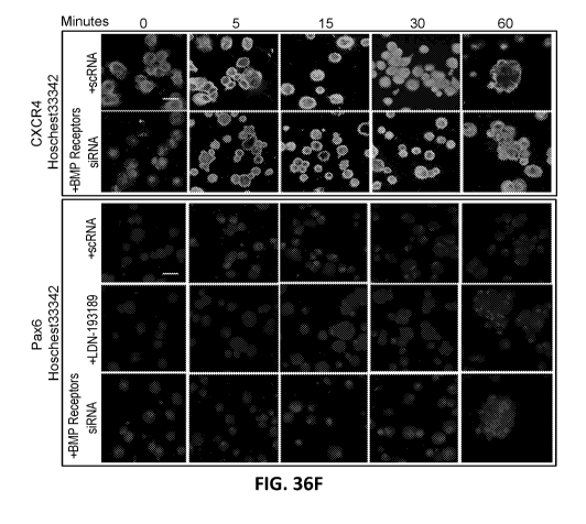
[0044] FIGS. 36A-36F illustrate cell aggregation and CXCR4/SDF-1 signaling
promoted by
HC-HA/PTX3 is not affected by BMP signaling. P10 LNC on coated MG in MESCM
were pre-
treated with or without transfection with siRNAs for BMPR1A, BMPR1B, BMPR2 and
ACVR1
before being seeded on coated MG with or without soluble HC-HA/PTX3 in MESCM.
The
transfection efficiency was verified by qRT-PCR when compared to scrambled RNA
(scRNA) as
the control (FIG. 36A, ** p <0.01, n=3). BMP signaling was measured by
immunofluorescence
staining to pSmad1/5/8 (FIG. 36B) and cell aggregation was detected by phase
contrast
microscopy (FIG. 36C, bar = 100 p.m). CXCR4/SDF-1 signaling was assessed by
qRT-PCR for
the expression of CXCR4 (FIG. 36D) and SDF-1 (FIG. 36E) transcripts using the
expression
level by cells with HC-HA/PTX3 + scRNA at time 0 set as 1. (* p > 0.05, n = 3;
+ scRNA
represented by darker line) and by immunofluorescence staining to CXCR4 and
Pax6 (FIG. 36F,
nuclear counterstaining by Hoechst 33342, bar = 25 p.m).
[0045] FIGS. 37A-37C illustrate cytoskeletal change by HA and HC-HA/PTX3 in
LNCs
correlates with Rho GTPase RhoA, Racl and Cdc42 effectors within 60 minutes.
FIG. 37A
illustrates phase images of LNC treated with HC-HA/PTX. FIG. 37B illustrates
graphs of
RhoA, Racl, and Cdc42 activities after treatment with HA and HC-HA/PTX3. FIG.
37C
illustrates double immunostaining of DNase I/Phalloidin (G-actin/F-actin).
-14-
CA 03144419 2021-12-20
WO 2020/257626 PCT/US2020/038698
[0046] FIGS. 38A-38B illustrate expression of Notch ligands and receptors in
human cornea,
limbus, and conjunctiva. FIG. 38A illustrates in vivo signaling of notch
receptors (Notch 1,
Notchl intracellular domain (NICD), Notch2, and Notch 3) and Notch ligands
(Jagged 1, Delta).
FIG. 38B illustrates in vivo notch signaling in freshly collagenase isolated
clusters.
[0047] FIGS. 39A-39E illustrate expression of Notch signal on plastic, 3D
Matrigel, and HC-
HA/PTX3.
[0048] FIGS. 40A-40B illustrate expression of canonical Notch signaling in
LEPC and LNC
on immobilized HC-HA/PTX3 at 48 hours.
[0049] FIGS. 41A-41C illustrate blocking Notch signaling inhibits BNIP and non-
canonical
Wnt in LEPC and LNC on immobilized HC-HA/PTX3 at 48 hours. FIG. 41A
illustrates a graph
of mRNA levels of various genes following treatment of HC-HA/PTX3 and HC-
HA/PTX3/DAPT in LNCS renunioned with LEPC. FIG. 41B illustrates immunostaining
with
various markers.
[0050] FIG. 42 illustrates Notch signaling in LNC on plastic, 3D Matrigel or
immobilized HC-
HA/PTX3 at 48 hours.
[0051] FIG. 43 illustrate immunofluorescence (IF) staining of Hesl, Notch3,
and Notchl in the
presence of HC-HA/PTX3 or 3D Matrigel (MG).
[0052] FIG. 44 illustrates phase contrast microcopy image showing cell
aggregation was
promoted by soluble HC-HA/PTX3 as early as 60 minutes but not in HA or coated
Matrigel
(MG).
[0053] FIGS. 45A-45C illustrate soluble HC-HA/PTX3, but not HA or 3D MG alone,
promotes angiogenesis sprouting. FIG. 45A illustrates phase contrast
microscopy images
showing cell morphology reunion aggregates at 4h. FIGS. 45B-45C illustrates a
graph of
diameter of sprouting outgrowth measured from the two sides of invading edges
on D13.
[0054] FIG. 46 illustrates a graph of HIFla mRNA expression in human corneal
fibroblasts
(HCF) that were seeded on plastic with or without immobilized HA, HC-HA/PTX3
complex and
then treated with or without TGF131.
DETAILED DESCRIPTION OF THE DISCLOSURE
[0055] Blood vessels comprise endothelial cells, which form the inner lining
of the vessel wall,
and pericytes, which are found on the surface of the vessel. Blood vessels are
generated by two
different processes, angiogenesis which involves the formation of new vessels
from existing
vessels, and vasculogenesis, which involves the de novo formation of vessels.
Normal
angiogenesis is a complex, multi-step process including the creation the
gradient formation of
matrix-bound growth factor (GF) (e.g. VEGF-A, bFGF, PDGF-BB), migration and
proliferation
-15-
CA 03144419 2021-12-20
WO 2020/257626 PCT/US2020/038698
of endothelial cells (EC), dissolution of the extracellular matrix, and
recruitment of mural cells
(e.g., pericytes) to stabilize capillary development. Abnormal angiogenesis
associated with
tumors is characterized by vessel leakiness and hemorrhage, and is often
associated with the lack
of pericytes and/or accompanied by inability to bind VEGF-A associated matrix
heparan.
[0056] Pericytes in the brain are derived from neural crest cells, and promote
both
neurogenesis and vasculogenesis, a process referred to herein as
neurovasculogenesis. Pericytes
have diverse support functions to regulate blood-brain barrier (BBB)
integrity, angiogenesis,
influence neuroinflammatory response, and have multipotent stem cell activity.
Pericyte
deficiency has been noted as an early hallmark in diabetes-associated
microvascular diseases,
such as retinopathy and nephropathy, and may contribute to abnormal
angiogenesis, resulting in
vessel leakiness and hemorrhage, increased metastases in mouse tumor models,
cerebrovascular
dysfunction in complex neurological disease such as Alzheimer's disease, and
amyotrophic
lateral sclerosis.
[0057] Provided herein, in some embodiments, are methods of promoting
vasculogenesis or
normal angiogenesis in an individual in need thereof, comprising contacting a
tissue comprising
endothelial cells and pericytes or neural crest progenitor cells with a fetal
support tissue product.
In some embodiments, the vasculogenesis occurs as part of neurovasculogenesis.
In some
embodiments, neurovasculogenesis further comprises neurogenesis. Further
provided herein, in
some embodiments, are methods of treating an ischemic condition in an
individual in need
thereof, comprising contacting an ischemic tissue with a fetal support tissue
product. Further
provided herein, in some embodiments, are methods of treating a neuropathic
condition in an
individual in need thereof, comprising contacting an ischemic tissue with a
fetal support tissue
product.
[0058] Further provided herein, in some embodiments are methods of inhibiting
abnormal
angiogenesis in an in an individual in need thereof, comprising contacting a
tissue comprising
endothelial cells with a fetal support tissue product. In some embodiments,
the tissue lacks
pericytes. In some embodiments, the method further comprises selecting the
individual by
detecting an absence of pericyte markers.
Certain Definitions
[0059] Unless defined otherwise, all technical and scientific terms used
herein have the same
meaning as is commonly understood by one of skill in the art to which the
claimed subject matter
belongs.
[0060] As used herein, in some embodiments, ranges and amounts are expressed
as "about" a
particular value or range. About also includes the exact amount. Hence "about
5 i.tg" means
-16-
CA 03144419 2021-12-20
WO 2020/257626 PCT/US2020/038698
"about 5 i.tg" and also "5 pg." Generally, the term "about" includes an amount
that would be
expected to be within experimental error.
[0061] As used herein, "fetal support tissue product" means any isolated
product derived from
tissue used to support the development of a fetus. Examples of fetal support
tissue product
includes, but are not limited to, (i) placental amniotic membrane (PAM), or
substantially isolated
PAM, (ii) umbilical cord amniotic membrane (UCAM) or substantially isolated
UCAM, (iii)
chorion or substantially isolated chorion, (iv) amnion-chorion or
substantially isolated amnion-
chorion, (v) placenta or substantially isolated placenta, (vi) umbilical cord
or substantially
isolated umbilical cord, or (vii) any combinations thereof In some
embodiments, the fetal
support tissue is selected from the group consisting of placental amniotic
membrane (PAM),
umbilical cord amniotic membrane (UCAM), chorion, amnion-chorion, placenta,
umbilical cord,
and any combinations thereof In some embodiments, the fetal support tissue
comprises
umbilical cord. Fetal support tissue product includes any form of the fetal
support tissue,
including cryopreserved, terminally-sterilized, lyophilized fetal support
tissue or powders
resulting from grinding fetal support tissue. In some embodiments, the fetal
support tissue
product is ground, pulverized, morselized, a graft, a sheet, a powder, a gel,
a homogenate, an
extract, or a terminally-sterilized product.
[0062] As used herein, "placenta" refers to the organ that connects a
developing fetus to the
maternal uterine wall to allow nutrient uptake, waste elimination, and gas
exchange via the
maternal blood supply. The placenta is composed of three layers. The innermost
placental layer
surrounding the fetus is called amnion. The allantois is the middle layer of
the placenta (derived
from the embryonic hindgut); blood vessels originating from the umbilicus
traverse this
membrane. The outermost layer of the placenta, the chorion, comes into contact
with the
endometrium. The chorion and allantois fuse to form the chorioallantoic
membrane.
[0063] As used herein, "chorion" refers to the membrane formed by
extraembryonic mesoderm
and the two layers of trophoblast. The chorion consists of two layers: an
outer formed by the
trophoblast, and an inner formed by the somatic mesoderm; the amnion is in
contact with the
latter. The trophoblast is made up of an internal layer of cubical or
prismatic cells, the
cytotrophoblast or layer of Langhans, and an external layer of richly
nucleated protoplasm devoid
of cell boundaries, the syncytiotrophoblast. The avascular amnion is adherent
to the inner layer of
the chorion.
[0064] As used herein, "amnion-chorion" refers to a product comprising amnion
and chorion.
In some embodiments, the amnion and the chorion are not separated (i.e., the
amnion is naturally
adherent to the inner layer of the chorion). In some embodiments, the amnion
is initially
separated from the chorion and later combined with the chorion during
processing.
-17-
CA 03144419 2021-12-20
WO 2020/257626 PCT/US2020/038698
[0065] As used herein, "umbilical cord" refers to the organ that connects a
developing fetus to
the placenta. The umbilical cord is composed of Wharton's jelly, a gelatinous
substance made
largely from mucopolysaccharides. It contains one vein, which carries
oxygenated, nutrient-rich
blood to the fetus, and two arteries that carry deoxygenated, nutrient-
depleted blood away.
[0066] As used herein, "placental amniotic membrane" (PAM) refers to amniotic
membrane
derived from the placenta. In some embodiments, the PAM is substantially
isolated.
[0067] As used herein, "umbilical cord amniotic membrane" (UCAM) means
amniotic
membrane derived from the umbilical cord. UCAM is a translucent membrane. The
UCAM has
multiple layers an epithelial layer, a basement membrane; a compact layer; a
fibroblast layer; and
a spongy layer. It lacks blood vessels or a direct blood supply. In some
embodiments, the UCAM
comprises Wharton's Jelly. In some embodiments, the UCAM comprises blood
vessels and/or
arteries. In some embodiments, the UCAM comprises Wharton's Jelly and blood
vessels and/or
arteries.
[0068] As used herein, "human tissue" means any tissue derived from a human
body. In some
embodiments, the human tissue is a fetal support tissue selected from the
group consisting of
placental amniotic membrane, umbilical cord, umbilical cord amniotic membrane,
chorion,
amnion-chorion, placenta, or any combination thereof
[0069] As used herein, "minimal manipulation" means (1) for structural tissue,
processing that
does not alter the original relevant characteristics of the tissue relating to
the tissue's utility for
reconstruction, repair, or replacement; and (2) for cells or nonstructural
tissues, processing that
does not alter the relevant biological characteristics of cells or tissues.
[0070] As used herein, "graft" means a matrix of proteins (e.g., collagen and
elastin) and
glycans (e.g., dermatan, hyaluronan, and chondroitin) that is used to replace
damaged,
compromised, or missing tissue. In certain instances, the matrix is laid down
and host cells
gradually integrate into the matrix.
[0071] As used herein, "sheet" means any continuous expanse or surface. In
some
embodiments, a sheet of a fetal support tissue product is substantially
flattened. In some
embodiments, a sheet of a fetal support tissue product is flat. In some
embodiments, a sheet of
fetal support tissue product is tubular. In some embodiments, the sheet is any
shape or size
suitable for the wound to be treated. In some embodiments, the sheet is a
square, circle, triangle,
or rectangle.
[0072] The term "fresh fetal support tissue" refers to fetal support tissue
that is less than 10
days old following birth, and which is in substantially the same form as it
was following birth. In
some embodiments, the fresh fetal support tissue comprises fetal support
tissue cells. In some
embodiments, the fetal support tissue cells comprise pericytes. In some
embodiments, at least
-18-
CA 03144419 2021-12-20
WO 2020/257626 PCT/US2020/038698
50%, at least 60%, at least 70%, at least 80%, at least 90%, or at least 95%
of the biological
activity of the cell support tissue cells is maintained.
[0073] "Substantially isolated" or "isolated" when used in the context of a
fetal support tissue
product means that the fetal support tissue product is separated from most
other non-fetal support
tissue materials (e.g., other tissues, red blood cells, veins, arteries)
derived from the original
source organism.
[0074] As used herein, the phrase "wherein the biological and structural
integrity of the
isolated fetal support tissue product is substantially preserved" means that
when compared to the
biological activity and structural integrity of fresh fetal support tissue,
the biological activity and
structural integrity of the isolated fetal support tissue has only decreased
by about 5%, about
10%, about 15%, about 20%, about 25%, about 30%, about 35%, about 40%, about
50%, or
about 60%.
[0075] As used herein, "processing" means any activity performed on a fetal
support tissue or a
preparation comprising HC-HA/PTX3, other than recovery, donor screening, donor
testing,
storage, labeling, packaging, or distribution, such as testing for
microorganisms, preparation,
sterilization, steps to inactivate or remove adventitious agents, preservation
for storage, and
removal from storage.
[0076] As used herein, the terms "purified" and "isolated" mean a material
(e.g., HC-
HA/PTX3 complex) substantially or essentially free from components that
normally accompany
it in its native state. In some embodiments, "purified" or "isolated" mean a
material (e.g., HC-
HA/PTX3 complex) is about 50% or more free from components that normally
accompany it in
its native state, for example, about 50%, about 55%, about 60%, about 65%,
about 70%, about
75%, about 80%, about 85%, about 90%, about 91%, about 92%, about 93%, about
94%, about
95%, about 96%, about 97%, about 98%, or about 99% free from components that
normally
accompany it in its native state.
[0077] As used herein, "biological activity" means the activity of
polypeptides and
polysaccharides of the fetal support tissue product comprising HC-HA/PTX3. In
some
embodiments, the biological activity of polypeptides and polysaccharides found
in the fetal tissue
support product is anti-inflammatory, anti-scarring, anti-angiogenic, or anti-
adhesion. In some
embodiments, the biological activity refers to the in vivo activities of the
HC-HA/PTX3 complex
in the fetal tissue support product or physiological responses that result
upon in vivo
administration of the fetal support tissue product. In some embodiments, the
biological activity
of HC-HA/PTX3 complex in the fetal support tissue product is substantially
preserved. In some
embodiments, the activity of polypeptides and polysaccharides found in the
fetal tissue support
product is promoting wound healing. In some embodiments, the activity of
polypeptides and
-19-
CA 03144419 2021-12-20
WO 2020/257626 PCT/US2020/038698
polysaccharides found in the fetal support tissue product is preventing
scarring. In some
embodiments, the activity of polypeptides and polysaccharides found in the
fetal support tissue
product is reducing inflammation. Biological activity, thus, encompasses
therapeutic effects and
pharmaceutical activity of the HC-HA/PTX3 complex in the fetal support tissue
product.
[0078] As used herein, "structural integrity" means the integrity of stroma
and basement
membrane that make up the fetal support tissue product. In some embodiments,
the structural
integrity of the fetal support tissue product results in suture pull out
strength.
[0079] As used herein, a reconstituted HC-HA/PTX3 (rcHC-HA/PTX3) complex is an
HC-
HA/PTX3 complex that is formed by assembly of the component molecules of the
complex in
vitro. The process of assembling the rcHC-HA/PTX3 includes reconstitution with
purified native
proteins or molecules from biological source, recombinant proteins generated
by recombinant
methods, or synthesis of molecules by in vitro synthesis. In some instances,
the purified native
proteins used for assembly of the rcHC-HA/PTX3 are proteins in a complex with
other proteins
(i.e. a multimer, a multichain protein or other complex). In some instances,
PTX3 is purified as a
multimer (e.g. a homomultimer) from a cell and employed for assembly of the
rcHC-HA/PTX3
complex.
[0080] As used herein, a purified native HC-HA/PTX3 (nHC-HA/PTX3) complex
refers to an
HC-HA/PTX3 complex that is purified from a biological source such as a cell, a
tissue or a
biological fluid. In some embodiments, the nHC-HA/PTX3 is purified from a
fetal support tissue.
In some embodiments the nHC-HA/PTX3 is purified from amniotic membrane. In
some
embodiments the nHC-HA/PTX3 is purified from umbilical cord. Such complexes
are generally
assembled in vivo in a subject or ex vivo in cells, tissues, or biological
fluids from a subject,
including a human or other animal.
[0081] As used herein, a PTX3/HA complex refers to an intermediate complex
that is formed
by contacting PTX3 with immobilized HA. In the methods provided herein, the
PTX3/HA
complex is the generated prior to the addition of HC1 to HA.
[0082] As used herein, "hyaluronan," "hyaluronic acid," or "hyaluronate" (HA)
are used
interchangeably to refer to a substantially non-sulfated linear
glycosaminoglycan (GAG) with
repeating disaccharide units of D-glucuronic acid and N-acetylglucosamine (D-
glucuronosyl-N-
acetylglucosamine).
[0083] As used herein, the term "tissue having unwanted changes" refers to
tissue that is
degenerated due to, for example, a degenerative disease (for example,
arthritis, multiple sclerosis,
Parkinson's disease, muscular dystrophy, and Huntington's disease) or aging;
scar tissue; or
damaged due to an insult, such as a burn, wound, laceration, injury, ulcer,
surgery, or due to
ischemia.
-20-
CA 03144419 2021-12-20
WO 2020/257626 PCT/US2020/038698
[0084] As used herein, the term "mesenchymal cell characteristic of the
tissue" refers to
specialized cells characteristic of the tissue, such as, for example,
cardiomyocytes, osteoblasts
(bone cells), chondrocytes (cartilage cells), myocytes (muscle cells), and
adipocytes (fat cells).
[0085] As used herein, the term "high molecular weight" or "HMW," as in high
molecular
weight hyaluronan (HMW HA), is meant to refer to HA that has a weight average
molecular
weight that is greater than about 500 kilodaltons (kDa), such as, for example,
between about 500
kDa and about 10,000 kDa, between about 800 kDa and about 8,500 kDa, between
about 1100
kDa and about 5,000 kDa, or between about 1400 kDa and about 3,500 kDa. In
some
embodiments, the HMW HA has a weight average molecular weight of 3000 kDa or
greater. In
some embodiments, the HMW HA has a weight average molecular weight of 3000
kDa. In some
embodiments, the HMW HA is Healong with a weight average molecular weight of
about 3000
kDa. In some embodiments, HMW HA has a molecular weight of between about 500
kDa and
about 10,000 kDa. In some embodiments, BMW HA has a molecular weight of
between about
800 kDa and about 8,500 kDa. In some embodiments, BMW HA has a molecular
weight of
about 3,000 kDa.
[0086] As used herein, the term "low molecular weight" or "LMW," as in low
molecular
weight hyaluronan (LMW HA), is meant to refer to HA that has a weight average
molecular
weight that is less than 500 kDa, such as for example, less than about 400
kDa, less than about
300 kDa, less than about 200 kDa, less than about 100 kDa, less than about 50
kDa, less than
about 40 kDa, less than about 30 kDa, less than about 20 kDa, about 200-300
kDa, about 1-300
kDa, about 15 to about 40 kDa, or about 8-10kDa.
[0087] As used herein, pentraxin 3, or PTX3, protein or polypeptide refers to
any PTX3
protein, including but not limited to, a recombinantly produced protein, a
synthetically produced
protein, a native PTX3 protein, and a PTX3 protein extracted from cells or
tissues. PTX3 include
multimeric forms (e.g. homomultimer) of PTX3, including, but not limited to,
dimeric, trimeric,
tetrameric, pentameric, hexameric, tetrameric, octameric, and other multimeric
forms naturally or
artificially produced.
[0088] As used herein, tumor necrosis factor stimulated gene-6 (TSG-6) refers
to any TSG-6
protein or polypeptide, including but not limited to, a recombinantly produced
protein, a
synthetically produced protein, a native TSG-6 protein, and a TSG-6 protein
extracted from cells
or tissues.
[0089] As used herein, inter-a-inhibitor (IaI) refers to the IaI protein
comprised of light chain
(i.e., bikunin) and one or both heavy chains of type HC1 or HC2 covalently
connected by a
chondroitin sulfate chain. In some embodiments, the source of IaI is from
serum or from cells
producing IaI e.g., hepatic cells or amniotic epithelial or stromal cells or
umbilical epithelial or
-21-
CA 03144419 2021-12-20
WO 2020/257626 PCT/US2020/038698
stromal cells under a constitutive mode stimulation by proinflammatory
cytokines such as IL-I or
TNF-a.
[0090] As used herein, a "hyaluronan binding protein," "HA binding protein,"
or "HABP"
refers to any protein that specifically binds to HA.
[0091] The terms "effective amount" or "therapeutically effective amount," as
used herein,
refer to a sufficient amount of an agent or a compound being administered
which will relieve to
some extent one or more of the symptoms of the disease or condition being
treated. In some
embodiments, the result is a reduction and/or alleviation of the signs,
symptoms, or causes of a
disease, or any other desired alteration of a biological system. For example,
an "effective
amount" for therapeutic uses is the amount of the composition including a
compound as
disclosed herein required to provide a clinically significant decrease in
disease symptoms without
undue adverse side effects. In some embodiments, an appropriate "effective
amount" in any
individual case is determined using techniques, such as a dose escalation
study. The term
"therapeutically effective amount" includes, for example, a prophylactically
effective amount.
An "effective amount" of a compound disclosed herein, is an amount effective
to achieve a
desired effect or therapeutic improvement without undue adverse side effects.
It is understood
that, in some cases, "an effective amount" or "a therapeutically effective
amount" varies from
subject to subject, due to variation in metabolism of the composition, age,
weight, general
condition of the subject, the condition being treated, the severity of the
condition being treated,
and the judgment of the prescribing physician. In some embodiments, an
effective amount is an
amount of a product or compound sufficient to promote vasculogenesis or normal
angiogenesis
in a tissue.
[0092] As used herein, the terms "subject," "individual" and "patient" are
used
interchangeably. None of the terms are to be interpreted as requiring the
supervision of a medical
professional (e.g., a doctor, nurse, physician's assistant, orderly, hospice
worker). As used herein,
the subject is any animal, including mammals (e.g., a human or non-human
animal) and non-
mammals. In one embodiment of the methods and compositions provided herein,
the mammal is
a human.
[0093] As used herein, the terms "treat," "treating" or "treatment," and other
grammatical
equivalents, include alleviating, abating or ameliorating one or more symptoms
of a disease or
condition, ameliorating, preventing or reducing the appearance, severity or
frequency of one or
more additional symptoms of a disease or condition, ameliorating or preventing
the underlying
metabolic causes of one or more symptoms of a disease or condition, inhibiting
the disease or
condition, such as, for example, arresting the development of the disease or
condition, relieving
the disease or condition, causing regression of the disease or condition,
relieving a condition
-22-
CA 03144419 2021-12-20
WO 2020/257626 PCT/US2020/038698
caused by the disease or condition, or inhibiting the symptoms of the disease
or condition either
prophylactically and/or therapeutically. In a non-limiting example, for
prophylactic benefit, an
rcHC-HA/PTX3 complex or composition disclosed herein is administered to an
individual at risk
of developing a particular disorder, predisposed to developing a particular
disorder, or to an
individual reporting one or more of the physiological symptoms of a disorder.
Methods of Use
[0094] Provided herein, in some embodiments, are methods of promoting
vasculogenesis in an
individual in need thereof Provided herein, in some embodiments, are methods
of promoting
neurovasculogenesis in an individual in need thereof In some embodiments,
promoting
vasculogenesis or neurovasculogenesis in an individual in need thereof
comprises contacting a
tissue with a fetal support tissue product described herein. In some
embodiments, the tissue
comprises endothelial cells and pericytes. In some embodiments, the tissue
comprises neural
crest progenitor cells. In some embodiments, the tissue comprises endothelial
cells and the
method comprises further recruiting pericytes to the tissue. In some
embodiments, the tissue
comprises endothelial cells and the method comprises further recruiting neural
crest progenitor
cells to the tissue. In some embodiments, the tissue is an ischemic tissue. In
some embodiments,
the methods described herein prevent necrosis of the tissue. In some
embodiments, the fetal
support tissue product recruits pericytes, neural crest progenitors, or a
combination thereof to a
site of administration. In some embodiments, the site of administration is a
tissue. In some
embodiments, the fetal support tissue product reprograms a progenitor cell
into a cell that
promotes vasculogenesis or neurovasculogenesis. In some embodiments, the
progenitor cell is a
neural crest progenitor cell. In some embodiments, the neural crest progenitor
cell is
reprogrammed into a pericyte.
[0095] Further provided herein, in some embodiments, are methods of treating
an ischemic
condition in an individual in need thereof In some embodiments, treating an
ischemic condition
in an individual comprises contacting an ischemic tissue with a fetal support
tissue product
described herein. In some embodiments, the ischemic tissue comprises
endothelial cells and
pericytes. In some embodiments, the ischemic tissue comprises endothelial
cells and the method
comprises further recruiting pericytes to the ischemic tissue. In some
embodiments, the methods
described herein prevent necrosis of the ischemic tissue. In some embodiments,
the ischemic
condition comprises cardiac ischemia, ischemic colitis, mesenteric ischemia,
brain ischemia,
acute limb ischemia, cyanosis, and gangrene. Further provided herein, in some
embodiments, are
methods of treatment microvascular disease. In some embodiments, the
microvascular disease is
a diabetes-associated microvascular disease. In some embodiments, the diabetes-
associated
-23-
CA 03144419 2021-12-20
WO 2020/257626 PCT/US2020/038698
microvascular disease is retinopathy or nephropathy. In some embodiments, the
ischemic
condition is a neurotrophic or neuropathic condition. In some embodiments, the
neuropathic
condition diminishes the function of one nerve or more than one nerve. In some
embodiments,
the neuropathic condition is a hereditary neuropathy or an acquired
neuropathy. In some
embodiments, the acquired neuropathy is neuropathy caused by a trauma, an
infection, a disease,
a medication, a vascular disorder, a vitamin imbalance, or alcoholism. In some
embodiments, the
disease is diabetes.
[0096] In some instances, the tissue is an ocular tissue, a brain tissue, a
cardiac tissue, a skin
tissue, a joint, a spine, a soft tissue, a muscle tissue, a cartilage, a bone,
a tendon, a ligament, a
nerve, or an intervertebral disc. In some instances, the tissue is an ocular
tissue. In some
instances, the tissue is a cardiac tissue. In some instances, the tissue is a
skin tissue. In some
instances, the tissue having unwanted changes is a joint tissue. In some
instances, the tissue is
from a spine. In some instances, the tissue is an intervertebral disc. In some
instances, the tissue
is a soft tissue. In some instances, the tissue is a muscle tissue. In some
instances, the tissue is a
cartilage. In some instances, the tissue is a bone. In some instances, the
tissue is a tendon. In
some instances, the tissue is a ligament. In some instances, the tissue is a
nerve.
[0097] In some instances, the tissue comprises degenerated tissue, a burn, a
laceration,
ischemic tissue, a wound, an injury, an ulcer, or a surgical incision. In some
embodiments, the
tissue comprises an ulcer, wound, perforation, burn, surgery, injury, or
fistula. In some instances,
the tissue comprises a degenerated tissue. In some instances, the tissue
comprises a burn. In some
instances, the tissue comprises a laceration. In some instances, the tissue
comprises an ischemic
tissue. In some instances, the tissue comprises a wound. In some instances,
the tissue comprises
an injury. In some instances, the injury is a myocardial infarction. In some
instances, the tissue
comprises an ulcer. In some embodiments, the ulcer is a diabetic ulcer. In
some instances, the
tissue comprises a surgical incision.
[0098] In some embodiments, the contacting occurs for a time sufficient
vasculogenesis or
neurovasculogenesis to occur. In some embodiments, the period of time at least
1 day, 2 days, 3
days, 4 days, 5 days, 6 days, 1 week, or 2 weeks.
[0099] In some embodiments, the contacting occurs for a time sufficient for
the fetal support
tissue product to reprogram a progenitor cell into a cell that promotes
vasculogenesis or
neurovasculogenesis. In some embodiments, the progenitor cell is a neural
crest progenitor cell.
In some embodiments, the neural crest progenitor cell is reprogrammed into a
pericyte. In some
embodiments, the period of time at least 1 day, 2 days, 3 days, 4 days, 5
days, 6 days, 1 week, or
2 weeks.
-24-
CA 03144419 2021-12-20
WO 2020/257626 PCT/US2020/038698
[0100] In some embodiments, the contacting occurs for a time sufficient to
induce gene
expression. In some embodiments, the contacting to induce gene expression
comprises 1 day, 2
days, 3 days, 4 days, 5 days, 6 days, 1 week, or 2 weeks.
[0101] In some embodiments, the contacting occurs for a time sufficient to
induce nuclear
translocation of a transcription factor. In some embodiments, the contacting
to induce nuclear
translocation of a transcription factor comprises at least about 5 minutes, 10
minutes, 15 minutes,
20 minutes, 30 minutes, 1 hour, 4 hours, 8 hours, 12 hours, 16 hours, 1 day, 2
days, 3 days, 4
days, or more than 4 days. at least 1 day, 2 days, 3 days, 4 days, 5 days, 6
days, 1 week, or 2
weeks.
[0102] In some embodiments, recruiting a neural crest progenitor cell to the
tissue comprises
contacting the tissue with a fetal support tissue product described herein. In
some embodiments,
recruiting pericytes to the tissue comprises administering to the tissue a
fetal support tissue
product described herein. In some embodiments, the fetal support tissue
product attracts
pericytes, neural crest progenitor cells, or a combination thereof to a site
of the administration. In
some embodiments, the pericytes are cells expressing a pericyte phenotype. In
some
embodiments, the cells expressing a pericyte phenotype are limbal niche cells
(LNCs).
[0103] In some embodiments, the ratio of endothelial cells to pericytes in the
tissue is 1:1, 2:1,
3:1, 4:1, 5:1, 6:1, 7:1, 8:1, 9:1, or 10:1. In some embodiments, the tissue is
contacted with
pericytes to reach a ratio of endothelial cells to pericytes is 1:1, 2:1, 3:1,
4:1, 5:1, 6:1, 7:1, 8:1,
9:1, or 10:1. In some embodiments, pericytes are recruited to the tissue to
reach a ratio of
endothelial cells to pericytes of 1:1, 2:1, 3:1, 4:1, 5:1, 6:1, 7:1, 8:1, 9:1,
or 10:1
[0104] In some embodiments, the fetal support tissue product comprises an
extract of fetal
support tissue, a fetal support tissue homogenate, a fetal support tissue
powder, morselized fetal
support tissue, pulverized fetal support tissue, ground fetal support tissue,
a fetal support tissue
graft, purified HC-HA/PTX3, reconstituted HC-HA/PTX3 or a combination thereof
[0105] Provided herein, in certain embodiments, are methods of promoting
vasculogenesis or
neurovasculogenesis in an individual in need thereof, wherein contacting a
tissue with a fetal
support tissue product modulates gene expression. In some embodiments, the
fetal support tissue
product comprises native HC-HA/PTX3 complex, reconstituted HC-HA/PTX3 (rcHC-
HA/PTX3)
complex, or a combination thereof In some embodiments, the HC-HA/PTX3 results
in an
increase in an expression of angiogenic genes, neurogenic genes, or a
combination thereof In
some embodiments, the HC-HA/PTX3 results in an increase in an expression of
angiogenic
genes, neurogenic genes, or a combination thereof by at least about 0.5X,
1.0X, 1.5X, 2.0X,
3.0X, 4.0X, or more than 4.0X. In some embodiments, the angiogenic genes,
neurogenic genes,
-25-
CA 03144419 2021-12-20
WO 2020/257626 PCT/US2020/038698
or a combination thereof comprises VEGF, PDGFa, PDGF A CD31, IGF-1, NGF,
p75NTR, sox-2,
Musashi-1, PDGFRa, PDGFRA VEGFR1, or VEGFR2.
[0106] In some embodiments, a tissue is contacted with a fetal support tissue
product
comprising native HC-HA/PTX3 complex, reconstituted HC-HA/PTX3 (rcHC-HA/PTX3)
complex, or a combination thereof for a sufficient amount of time to modulate
gene expression.
In some embodiments, the tissue is contacted with a fetal support tissue
product comprising
native HC-HA/PTX3 complex for at least about 5 minutes, 10 minutes, 15
minutes, 20 minutes,
30 minutes, 1 hour, 4 hours, 8 hours, 12 hours, 16 hours, 1 day, 2 days, 3
days, 4 days, or more
than 4 days. at least 1 day, 2 days, 3 days, 4 days, 5 days, 6 days, 1 week,
or 2 weeks.
[0107] In some embodiments, HC-HA/PTX3 modulates cellular function. In some
embodiments, HC-HA/PTX3 promotes apoptosis, necrosis, or a combination thereof
In some
embodiments, HC-HA/PTX3 promotes apoptosis, necrosis, or a combination thereof
by at least
about 0.5X, 1.0X, 1.5X, 2.0X, 3.0X, 4.0X, or more than 4.0X. In some
embodiments, HC-
HA/PTX3 inhibits cell proliferation. In some embodiments, HC-HA/PTX3 inhibits
cell
proliferation by at least about 0.5X, 1.0X, 1.5X, 2.0X, 3.0X, 4.0X, or more
than 4.0X.
[0108] In some embodiments, HC-HA/PTX3 modulates cell signaling. In some
embodiments,
HC-HA/PTX3 modulates cell signaling by increasing gene expression, protein
expression,
protein activity, or combinations thereof In some embodiments, HC-HA/PTX3
modulates cell
signaling by decreasing gene expression, protein expression, protein activity,
or combinations
thereof In some embodiments, HC-HA/PTX3 modulates SDF-1/CXCR signaling. In
some
embodiments, HC-HA/PTX3 modulates HIFI signaling. In some embodiments, HIFI
comprises
H1Fla. In some embodiments, HIFI comprises HIF1f3. In some embodiments, HC-
HA/PTX3
modulates TGFP signaling. In some embodiments, HC-HA/PTX3 modulates non-
canonical
TGFP signaling. In some embodiments, HC-HA/PTX3 modulates CD44ICD signaling.
In some
embodiments, HC-HA/PTX3 modulates Hes signaling. In some embodiments, HC-
HA/PTX3
modulates Pax6 signaling. In some embodiments, HC-HA/PTX3 modulates Notch
signaling. In
some embodiments, HC-HA/PTX3 modulates Notch signaling by modulating
expression of
Notch ligands, Notch receptors, or a combination thereof In some embodiments,
the Notch
ligands comprises Notch 1, Notch 2, Notch 3, Notch 4, Jagged 1, Jagged 2,
Jagged 3, DLL1,
DLL2, DLL3, or DLL4. In some embodiments, the Notch ligands comprises Notch 2,
Notch 3,
Jagged 1 or DLL2.
[0109] In some embodiments, HC-HA/PTX3 modulates multiple signaling pathways.
In some
embodiments, HC-HA/PTX3 modulates SDF-1/CXCR, HIFI, TGFP, CD44ICD, Hes, Pax6,
Notch signaling, or combinations thereof
-26-
CA 03144419 2021-12-20
WO 2020/257626 PCT/US2020/038698
[0110] Provided herein, in certain embodiments, are methods of promoting
vasculogenesis or
neurovasculogenesis in an individual in need thereof, wherein contacting a
tissue with a fetal
support tissue product results in cell reprograming. In some embodiments, HC-
HA/PTX3
modulates cell reprograming. In some embodiments, HC-HA/PTX3 modulates cell
aggregation,
cell shape, or an expression of a cell-specific marker.
[0111] In some embodiments, HC-HA/PTX3 reprograms LNCs to a progenitor
phenotype. In
some embodiments, HC-HA/PTX3 reprograms LNCs to a vascular progenitor
phenotype. In
some embodiments, HC-HA/PTX3 modulates expression of FLK-1, CD34, CD31, a-SMA,
PDGFRO, NG2, Pax6, p75NTR, Musashi-1, Sox2, Nestin, Msxl, FoxD3, FLK-1,
PDGFRO, CD31,
or combinations thereof In some embodiments, HC-HA/PTX3 modulates expression
Pax6. In
some embodiments, a time sufficient to reprogram LNCs is at least 1 hour, 2
hours, 3 hours, 4
hours, 5 hours, 6 hours, 7 hours, 8 hours, 9 hours, 10 hours, 11 hours, 12
hours, 24 hours, 36
hours, 2 days, 3 days, 4 days, 5 days, 6 days, 7 days, 2 weeks, 3 weeks, or 4
weeks.
[0112] In some embodiments the methods further comprise contacting a tissue
with TGF131. In
some embodiments, additional administration of TGFP1is required to perform the
methods
described herein. In some embodiments, additional administration of TGFP1is
not required to
perform the methods described herein. In some embodiments, the cell is
contacted
simultaneously with a fetal support tissue product comprising HC-HA/PTX3 and
TGF131. In
some embodiments, the tissue is contacted sequentially with the fetal support
tissue product
comprising HC-HA/PTX3 first and then the TGF131. In some embodiments, the
tissue is
contacted sequentially with the TGF131 first and then the fetal support tissue
product comprising
HC-HA/PTX3. In some embodiments, the TGF131 is administered in a
therapeutically effective
amount. In some embodiments, a therapeutically effective amount of TGF131 is
an amount of
TGF131 sufficient to enable the fetal support tissue product comprising HC-
HA/PTX3 to perform
the methods described herein.
Fetal Support Tissue Products
[0113] In some embodiments, a fetal support tissue product is ground fetal
support tissue,
pulverized fetal support tissue, powdered fetal support tissue, micronized
fetal support tissue,
morselized fetal support tissue, a fetal support tissue graft, a fetal support
tissue sheetõ a fetal
support tissue homogenate, a fetal support tissue extract, or any combinations
thereof In some
embodiments, the fetal support tissue product is terminally-sterilized. In
some embodiments, the
fetal support tissue product is a purified native HC-HA/PTX3 complex, a
reconstituted HC-
HA/PTX3, or a combination thereof In some embodiments, the fetal support
tissue product is
pulverized, powdered, or micronized fetal support tissue. In some embodiments,
the fetal
-27-
CA 03144419 2021-12-20
WO 2020/257626 PCT/US2020/038698
support tissue product is morselized fetal support tissue. In some
embodiments, the fetal support
tissue product is an extract of a fetal support tissue. In some embodiments,
the fetal support tissue
is a placental amniotic membrane, umbilical cord, umbilical cord amniotic
membrane, chorion,
amnion-chorion, placenta, amniotic stroma, amniotic jelly, or any combination
thereof
[0114] In some embodiments, the fetal support tissue product is an umbilical
cord product, an
amniotic membrane product, or umbilical cord amniotic membrane product. In
some
embodiments, the umbilical cord product comprises umbilical cord amniotic
membrane and at
least some Wharton's jelly. In some embodiments, the umbilical cord product
lacks umbilical
cord vein and arteries.
[0115] In some embodiments, the fetal support tissue product is an extract of
a fetal support
tissue. In some embodiments, the fetal support tissue product is purified
native HC-HA/PTX3
complex (nHC-HA/PTX3) from a fetal support tissue. In some embodiments, the
fetal support
tissue product is a reconstituted HC-HA/PTX3 complex (rHC-HA/PTX3). In some
embodiments,
the fetal support tissue product consists essentially of nHC-HA/PTX3. In some
embodiments, the
fetal support tissue product consists essentially of rcHC-HA/PTX3. In some
embodiments, the
fetal support tissue product a combination of nHC-HA/PTX3 and rcHC-HA/PTX3. In
some
embodiments, the nHC-HA/PTX3 or the rcHC-HA/PTX3 further comprises a small
leucine rich
proteoglycan (SLRP). In some embodiments, the SLRP is a class I, class II or
class II SLRP. In
some embodiments, the SLRP is selected from among class I SLRPs, such as
decorin and
biglycan. In some embodiments, the SLRP is selected from among class II SLRPs,
such as
fibromodulin, lumican, PRELP (proline arginine rich end leucine-rich protein),
keratocan, and
osteoadherin. In some embodiments, the SLRP is selected from among class III
SLRPs, such as
epipycan and osteoglycin. In some embodiments, the SLRP is selected from among
bikunin,
decorin, biglycan, and osteoadherin. In some embodiments, the SLRP comprises a
glycosaminoglycan. In some embodiments, the SLRP comprises keratan sulfate.
[0116] Generation of fetal support tissue products
[0117] In some embodiments, the fetal support tissue product is derived from
an umbilical cord
(UC) tissue. In some embodiments, the fetal support tissue product is derived
from an amniotic
membrane (AM) tissue. In some embodiments, the fetal support tissue product is
derived from
an umbilical cord amniotic membrane tissue. In some embodiments, the fetal
support tissue
product comprises: isolated fetal support tissue that does not comprise a vein
or an artery. In
some embodiments, the fetal support tissue product comprises: isolated fetal
support tissue that
does not comprise a vein or an artery, a cell with metabolic activity, active
HIV-1, active HIV-2,
active HTLV-1, active hepatitis B, active hepatitis C, active West Nile Virus,
active
cytomegalovirus, active human transmissible spongiform encephalopathy, or
active Treponema
-28-
CA 03144419 2021-12-20
WO 2020/257626 PCT/US2020/038698
pallidum, wherein the natural structural integrity of the fetal support tissue
product is
substantially preserved for at least 15 days after initial procurement. In
some embodiments, the
fetal support tissue product comprises umbilical cord amniotic membrane and
Wharton's Jelly.
In some embodiments, the biological activity of HC-HA/PTX3 complex in the
fetal support
tissue product is substantially preserved. In some embodiments, the biological
activity of HC-
HA/PTX3 complex in the fetal support tissue product is substantially preserved
for at least 15
days. In some embodiments, the biological and structural integrity of the
fetal support tissue
product is substantially preserved for at least 20 days after initial
procurement. In some
embodiments, the biological and structural integrity of the fetal support
tissue product is
substantially preserved for at least 25 days after initial procurement. In
some embodiments, the
biological and structural integrity of the fetal support tissue product is
substantially preserved for
at least 30 days after initial procurement. In some embodiments, the
biological and structural
integrity of the fetal support tissue product is substantially preserved for
at least 35 days after
initial procurement. In some embodiments, the biological and structural
integrity of the fetal
support tissue product is substantially preserved for at least 40 days after
initial procurement. In
some embodiments, the biological and structural integrity of the fetal support
tissue product is
substantially preserved for at least 45 days after initial procurement. In
some embodiments, the
biological and structural integrity of the fetal support tissue product is
substantially preserved for
at least 50 days after initial procurement. In some embodiments, the
biological and structural
integrity of the fetal support tissue product is substantially preserved for
at least 55 days after
initial procurement. In some embodiments, the biological and structural
integrity of the fetal
support tissue product is substantially preserved for at least 60 days after
initial procurement. In
some embodiments, the biological and structural integrity of the fetal support
tissue product is
substantially preserved for at least 90 days after initial procurement. In
some embodiments, the
biological and structural integrity of the fetal support tissue product is
substantially preserved for
at least 180 days after initial procurement. In some embodiments, the
biological and structural
integrity of the fetal support tissue product is substantially preserved for
at least 1 year after
initial procurement. In some embodiments, the biological and structural
integrity of the fetal
support tissue product is substantially preserved for at least 2 years after
initial procurement. In
some embodiments, the biological and structural integrity of the fetal support
tissue product is
substantially preserved for at least 3 years after initial procurement. In
some embodiments, the
biological and structural integrity of the fetal support tissue product is
substantially preserved for
at least 4 years after initial procurement. In some embodiments, the
biological and structural
integrity of the fetal support tissue product is substantially preserved for
at least 5 years after
-29-
CA 03144419 2021-12-20
WO 2020/257626 PCT/US2020/038698
initial procurement. In some embodiments, the fetal support tissue is obtained
from a human, a
non-human primate, a cow or a pig.
[0118] In some embodiments, the fetal support tissue product is kept below 0 C
until donor
and specimen eligibility has been determined. In some embodiments, the fetal
support tissue
product is kept from between 0 C to -80 C until donor and specimen eligibility
has been
determined. In some embodiments, storing the fetal support tissue product at -
80 C kills
substantially all cells found in the fetal support tissue. In some
embodiments, storing the fetal
support tissue product at -80 C kills substantially all cells found in the
fetal support tissue
product while maintaining or increasing the biological activity of the fetal
support tissue product
(e.g., its anti-inflammatory, anti-scarring, anti-antigenic, and anti-adhesion
properties) relative to
fresh (i.e., non-frozen) fetal support tissue. In some embodiments, storing
the fetal support tissue
product at -80 C results in the loss of metabolic activity in substantially
all cells found in the
fetal support tissue. In some embodiments, the fetal support tissue is dried.
In some
embodiments, the fetal support tissue is not dehydrated.
[0119] Processing of fetal support tissue
[0120] In some embodiments, processing is done following Good Tissue Practices
(GTP) to
ensure that no contaminants are introduced into the fetal support tissue
product.
[0121] In some embodiments, the fetal support tissue is tested for HIV-1, HIV-
2, HTLV-1,
hepatitis B and C, West Nile virus, cytomegalovirus, human transmissible
spongiform
encephalopathy (e.g., Creutzfeldt-Jakob disease) and Treponema pallidum using
FDA licensed
screening test. In some embodiments, any indication that the tissue is
contaminated with HIV-1,
HIV-2, HTLV-1, hepatitis B and C, West Nile virus, or cytomegalovirus results
in the immediate
quarantine and subsequent destruction of the tissue specimen. In some
embodiments, the donor's
medical records are examined for risk factors for and clinical evidence of
hepatitis B, hepatitis C,
or HIV infection.
[0122] In some embodiments, the fetal support tissue is frozen. In some
embodiments, the fetal
support tissue is not frozen. If the fetal support tissue is not frozen, it is
processed as described
below immediately.
[0123] In some embodiments, substantially all of the blood is removed from the
fetal support
tissue (e.g., from any arteries and veins found in the fetal support tissue,
and blood that has
infiltrated into the tissue). In some embodiments, substantially all of the
blood is removed before
the fetal support tissue is frozen. In some embodiments, blood is not removed
from the fetal
support tissue. In some embodiments, blood is not removed from the fetal
support tissue before
the fetal support tissue is frozen. In some embodiments, the blood is
substantially removed after
the fetal support tissue has been frozen.
-30-
CA 03144419 2021-12-20
WO 2020/257626 PCT/US2020/038698
[0124] In some embodiments, the fetal support tissue is washed with buffer
with agitation to
remove excess blood and tissue. In some embodiments, the fetal support tissue
is soaked with
buffer with agitation to remove excess blood and tissue.
[0125] In some embodiments, the fetal support tissue product is a fetal
support tissue graft. In
some embodiments, isolated fetal support tissue is used to generate a fetal
support tissue graft. In
some embodiments, the fetal support tissue is cut into multiple sections
(e.g., using a scalpel).
The size of the sections depends on the desired use of the fetal support
tissue graft derived from
the fetal support tissue. In some embodiments, the cut fetal support tissue is
optionally washed
again with buffer to further remove excess blood and tissue.
[0126] The umbilical cord comprises two arteries (the umbilical arteries) and
one vein (the
umbilical vein). In some embodiments, the vein and arteries are removed from
the UC. In some
embodiments, the vein and the arteries are not removed from the UC. In certain
instances, the
vein and arteries are surrounded (or suspended or buried) within the Wharton's
Jelly. In some
embodiments, the vein and arteries are removed concurrently with the removal
of the Wharton's
Jelly.
[0127] The desired thickness of the fetal support tissue product determines
how the fetal
support tissue product is processed. In some embodiments, the desired
thickness of the fetal
support tissue product determines how much of the Wharton's Jelly is removed.
In some
embodiments, the fetal support tissue product is contacted with a buffer to
facilitate separation of
the Wharton's Jelly and the UCAM. In some embodiments, the Wharton's jelly is
removed using
peeling, a rotoblator (i.e., a catheter attached to a drill with a diamond
coated burr), a
liposuction, a liquid under high pressure, a brush (e.g., a mechanized brush
rotating under high
speed), or a surgical dermatome. In some embodiments, Wharton's Jelly is not
removed. In some
embodiments, Wharton's Jelly and the umbilical vein and arteries are not
removed. In some
embodiments, Wharton's Jelly is not removed, and the umbilical vein and
arteries are removed.
[0128] In some embodiments, the fetal support tissue product comprises
isolated umbilical
cord amniotic membrane (UCAM). In certain instances, the UCAM comprises
proteins, glycans,
protein-glycan complexes (e.g., a complex of hyaluronic acid and a heavy chain
of IaI and
PTX3) and enzymes that promote tissue repair. For example, the stroma of UCAM
contains
growth factors, anti-angiogenic and anti-inflammatory proteins, as well as
natural inhibitors to
various proteases. In some embodiments, proteins and enzymes found in the UCAM
diffuse out
of the UC and into the surrounding tissue. In some embodiments, the UCAM is
isolated by
removing all of the Wharton's Jelly and umbilical vessels from the UC, leaving
the UCAM.
After substantially pure UCAM has been obtained, the UCAM is optionally washed
with buffer
to remove excess blood and tissue. In some embodiments, the UCAM comprises
Wharton's Jelly.
-31-
CA 03144419 2021-12-20
WO 2020/257626 PCT/US2020/038698
In some embodiments, the UCAM comprises Wharton's Jelly and the umbilical vein
and arteries.
In some embodiments, the UCAM comprises Wharton's Jelly and not the umbilical
vein and
arteries.
[0129] In some embodiments, the fetal support tissue product is in any
suitable shape (e.g., a
square, a circle, a triangle, a rectangle). In some embodiments, the fetal
support tissue product is
generated from a sheet of fetal support tissue. In some embodiments, the sheet
is flat. In some
embodiments, the sheet is tubular.
[0130] In some embodiments, the fetal support tissue product is cut into
multiple sections (e.g.,
using a scalpel). In some embodiments, the fetal support tissue product is
divided into sections
that are about 1.0 cm x about 0.25 cm, 0.5 cm, 0.75 cm, 1.0 cm, 2.0 cm, 3.0
cm, 4.0 cm, 5.0 cm,
or 6 cm. In some embodiments, the fetal support tissue product is divided into
sections that are
about 2 cm x about 2 cm, 3 cm, 4 cm, 5 cm, or 6 cm. In some embodiments, the
fetal support
tissue product is divided into sections that are about 3 cm x about 3 cm, 4
cm, 5 cm, or 6 cm. In
some embodiments, the fetal support tissue product is divided into sections
that are about 4 cm x
about 4 cm, 5 cm, or 6 cm. In some embodiments, the fetal support tissue
product is divided into
sections that are about 5 cm x about 5 cm or 6 cm. In some embodiments, the
fetal support tissue
product is divided into sections that are about 6 cm x about 6 cm. In some
embodiments, the
fetal support tissue product is divided into sections that are about 8 cm x
about 1 cm, 2 cm, 3 cm,
4 cm, 5 cm, 6 cm, 7 cm, or 8 cm. In some embodiments, the fetal support tissue
product is
divided into sections that are about 10 cm x about 10 cm. In some embodiments,
the fetal support
tissue product is divided into sections that are about 12 cm x about 10 cm. In
some embodiments,
the fetal support tissue product is divided into sections that are about 15 cm
x about 10 cm. In
some embodiments, the fetal support tissue product is divided into sections
that are about 20 cm
x about 10 cm. In some embodiments, the fetal support tissue product is
divided into sections
that are about 25 cm x about 10 cm. In some embodiments, the fetal support
tissue product is
divided into sections that are about 30 cm x about 10 cm.
[0131] In some embodiments, the fetal support tissue product is contacted
with buffer under
agitation to remove substantially all remaining red blood cells. In some
embodiments, the fetal
support tissue product is contacted with a buffer for 10 minutes, 15 minutes,
20 minutes, 25
minutes, 30 minutes, 40 minutes, 1 hour, 2 hours, 3 hours, 4 hours, 5 hours, 6
hours, 12 hours, 18
hours, 24 hours, or more than 24 hours. In some embodiments, the UC product is
contacted with
a buffer for 2 days, 3 days, 4 days, 5 days, 6 days, 7 days, 1 week, 2 weeks,
3 weeks, 4 weeks or
more than 4 weeks.
[0132] In some embodiments, the fetal support tissue product comprises a
pharmaceutically
acceptable excipient, carrier, or combination thereof In some embodiments, the
fetal support
-32-
CA 03144419 2021-12-20
WO 2020/257626 PCT/US2020/038698
tissue product is formulated as a non-solid dosage form. In some embodiments,
the fetal support
tissue product is formulated as a solid dosage form.
[0133] Processing a fetal support tissue product
[0134] In some embodiments, isolated fetal support tissue is used to generate
a morselized fetal
support tissue product. As used herein, "morsel" refers to particles of tissue
ranging in size from
about 0.1 mm to about 1.0 cm in length, width, or thickness that have been
obtained from a larger
piece of tissue. A "morsel" as described herein, retains the characteristics
of the tissue from
which it was obtained and upon inspection is identifiable as said tissue. As
used herein, the terms
"morselized," "morselizing," and "morselization" refer to actions involving
the "morsels" of the
present application. In some embodiments, the morselized fetal support tissue
product is further
processed into a solution, suspension or emulsion by mixing the morselized
fetal support tissue
with a carrier. In some embodiments, the morselized fetal support tissue
product is formulated
into a solution, suspension, paste, ointment, oil emulsion, cream, lotion,
gel, a patch, sticks, film,
paint, or a combination thereof In some embodiments, the morselized fetal
support tissue
product is contacted with a patch or wound dressing. In some embodiments, the
morselized fetal
support tissue product is formulated for parenteral injection, is administered
as a sterile solution,
suspension, or emulsion, or is formulated for inhalation.
[0135] In some embodiments, a mixture of amniotic membrane tissue and
umbilical cord tissue
in any ratio from 0.001:99.999 w/w % to 99.999:0.001 w/w % is morselized from
either fresh or
frozen tissue through the use of any morselizing tool known to one of skill in
the art such as, for
example, tissue grinder, sonicator, bread beater, freezer/mill, blender,
mortar/pestle, Roto-stator,
kitchen chopper, grater, ruler and scalpel to yield morsels ranging in size
from about 0.1 mm to
about 1.0 cm in length, width, or thickness. In some embodiments, the
resulting morsels are
homogenized to yield consistently sized morsels. In some embodiments, the
resulting morsels are
used wet, partially dehydrated or essentially dehydrated by any means known to
one of skill in
the art such as, for example, centrifuge or lyophilization. In some
embodiments, the resulting
fetal support tissue product is used immediately or stored for later use in
any type of contained
known to one of skill in the art such as, for example, pouch, jar, bottle,
tube, ampule and pre-
filled syringe. In some embodiments, the morselized fetal support tissue
product is sterilized by
any method known to one of skill in the art such as, for example, y radiation.
[0136] In some embodiments, isolated fetal support tissue is used to generate
a pulverized fetal
support tissue product. As used herein, "pulverized fetal support tissue
product" means a fetal
support tissue product comprising tissue that has been broken up (or,
disassociated). In some
embodiments, the pulverized fetal support tissue product is a dry powder. In
some embodiments,
the pulverized fetal support tissue product is further processed into a
solution, suspension or
-33-
CA 03144419 2021-12-20
WO 2020/257626 PCT/US2020/038698
emulsion by mixing the fetal support tissue powder with a carrier. In some
embodiments, the
pulverized fetal support tissue product is formulated into a solution,
suspension, paste, ointment,
oil emulsion, cream, lotion, gel, a patch, sticks, film, paint, or a
combination thereof. In some
embodiments, the pulverized fetal support tissue product is contacted with a
patch or wound
dressing. In some embodiments, the pulverized fetal support tissue product is
formulated for
parenteral injection, is administered as a sterile solution, suspension, or
emulsion, or is
formulated for inhalation.
[0137] In some embodiments, the isolated fetal support tissue is pulverized by
any suitable
method. In some embodiments, the isolated fetal support tissue is pulverized
by use of a
pulverizer (e.g., a Bessman Tissue Pulverizer, a Biospec biopulverizer, or a
Covaris CryoPrep).
In some embodiments, the isolated fetal support tissue is pulverized by use of
a tissue grinder
(e.g., a Potter-Elvehjem grinder or a Wheaton Overhead Stirrer). In some
embodiments, the
isolated fetal support tissue is pulverized by use of a sonicator. In some
embodiments, the
isolated fetal support tissue is pulverized by use of a bead beater. In some
embodiments, the
isolated fetal support tissue is pulverized by use of a freezer/mill (e.g., a
SPEX SamplePrep
Freezer/Mill or a Retsch Ball Mill). In some embodiments, the isolated fetal
support tissue is
pulverized by use of a pestle and mortar. In some embodiments, the isolated
fetal support tissue
is pulverized by manual use of a pestle and mortar.
[0138] In some embodiments, the fetal support tissue product is an extract
from a fetal support
tissue. In some embodiments, the fetal support tissue product is an HC-HA/PTX3
complex. In
some embodiments, the HC-HA/PTX3 complex is an nHC-HA/PTX3, an rcHC-HA/PTX3,
or the
combination thereof In some embodiments, the HC-HA/PTX3 complex is purified by
any
suitable method.
[0139] In some embodiments, the HC-HA/PTX3 complex is purified by
centrifugation (e.g.,
ultracentrifugation, gradient centrifugation), chromatography (e.g., ion
exchange, affinity, size
exclusion, and hydroxyapatite chromatography), gel filtration, or differential
solubility, ethanol
precipitation or by any other available technique for the purification of
proteins (See, e.g.,
Scopes, Protein Purification Principles and Practice 2nd Edition, Springer-
Verlag, New York,
1987; Higgins, S. J. and Hames, B. D. (eds.), Protein Expression: A Practical
Approach, Oxford
Univ Press, 1999; and Deutscher, M. P., Simon, M. I., Abelson, J. N. (eds.),
Guide to Protein
Purification: Methods in Enzymology (Methods in Enzymology Series, Vol 182),
Academic
Press, 1997, all incorporated herein by reference).
[0140] In some embodiments, an nHC-HA/PTX3 is isolated from an extract. In
some
embodiments, the extract is prepared from an amniotic membrane extract. In
some embodiments,
the extract is prepared from an umbilical cord extract. In some embodiments,
the umbilical cord
-34-
CA 03144419 2021-12-20
WO 2020/257626 PCT/US2020/038698
extract comprises umbilical cord stroma and/or Wharton's jelly. In some
embodiments, the nHC-
HA/PTX3 complex is contained in an extract that is prepared by
ultracentrifugation. In some
embodiments, the nHC-HA/PTX3 complex is contained in an extract that is
prepared by
ultracentrifugation using a CsC1/4-6M guanidine HC1 gradient. In some
embodiments, the extract
is prepared by at least 2 rounds of ultracentrifugation. In some embodiments,
the extract is
prepared by more than 2 rounds of ultracentrifugation (i.e. nHC-HA/PTX3 2nd).
In some
embodiments, the extract is prepared by at least 4 rounds of
ultracentrifugation (i.e. nHC-
HA/PTX3 4th). In some embodiments, the nHC-HA/PTX3 complex comprises a small
leucine-
rich proteoglycan. In some embodiments, the nHC-HA/PTX3 complex comprises HC1,
HA,
PTX3 and/or a small leucine-rich proteoglycan.
[0141] Cry opreservation
[0142] In some embodiments, the fetal support tissue product is frozen for
cryopreservation. In
some embodiments, cryopreserving the fetal support tissue product does not
destroy the integrity
of the fetal support tissue extracellular matrix. In some embodiments, the
fetal support tissue
product is exposed to a liquid gas (e.g., liquid nitrogen or liquid hydrogen).
In some
embodiments, the fetal support tissue product is exposed to liquid nitrogen.
In some
embodiments, the fetal support tissue product does not contact the liquid gas.
In some
embodiments, the fetal support tissue product is placed in a container and the
container is
contacted with liquid gas. In some embodiments, the fetal support tissue
product is exposed to
the liquid gas until the fetal support tissue product is frozen.
[0143] Lyophilization
[0144] In some embodiments, the fetal support tissue product is lyophilized.
In some
embodiments, the fetal support tissue product is lyophilized before being
morselized, pulverized,
cryopreserved, sterilized, or purified. In some embodiments, the fetal support
tissue product is
lyophilized after being morselized, pulverized, cryopreserved, sterilized, or
purified. In some
embodiments, the fetal support tissue product is lyophilized following
freezing. In some
embodiments, the fetal support tissue product is lyophilized following
freezing by any suitable
method (e.g., exposure to a liquid gas, placement in a freezer). In some
embodiments, the fetal
support tissue product is frozen by exposure to a temperature below about 0 C.
In some
embodiments, the fetal support tissue product is frozen by exposure to a
temperature below about
-20 C. In some embodiments, the fetal support tissue product is frozen by
exposure to a
temperature below about -40 C. In some embodiments, the fetal support tissue
product is frozen
by exposure to a temperature below about -50 C. In some embodiments, the fetal
support tissue
product is frozen by exposure to a temperature below about -60 C. In some
embodiments, the
fetal support tissue product is frozen by exposure to a temperature below
about -70 C. In some
-35-
CA 03144419 2021-12-20
WO 2020/257626 PCT/US2020/038698
embodiments, the fetal support tissue product is frozen by exposure to a
temperature below about
-75 C. In some embodiments, the fetal support tissue product is frozen by
exposure to a
temperature below about -80 C. In some embodiments, the fetal support tissue
product is frozen
by exposure to a temperature below about -90 C. In some embodiments, the fetal
support tissue
product is frozen by exposure to a temperature below about -100 C. In some
embodiments, the
fetal support tissue product is frozen by exposure to a liquid gas. In some
embodiments, the fetal
support tissue product is placed in a vacuum chamber of a lyophilization
device until all or
substantially all fluid (e.g., water) has been removed. In some embodiments, a
cryopreserved
fetal support tissue product is lyophilized.
[0145] Sterilization
[0146] In some embodiments, the fetal support tissue product is subject to
terminal sterilization
by any suitable (e.g., medically acceptable) method. In some embodiments, the
fetal support
tissue product is a lyophilized fetal support tissue product. In some
embodiments, the fetal
support tissue product is exposed to gamma radiation for a period of time
sufficient to sterilize
the fetal support tissue product. In some embodiments, the fetal support
tissue product is exposed
to gamma radiation at 25 kGy for a period of time sufficient to sterilize the
fetal support tissue
product. In some embodiments, the fetal support tissue product is exposed to
an electron beam
for a period of time sufficient to sterilize the fetal support tissue product.
In some embodiments,
the fetal support tissue product is exposed to X-ray radiation for a period of
time sufficient to
sterilize the fetal support tissue product. In some embodiments, the fetal
support tissue product is
exposed to UV radiation for a period of time sufficient to sterilize the fetal
support tissue
product.
[0147] Rehydration
[0148] In some embodiments, the fetal support tissue product is partially or
fully rehydrated. In
some embodiments, the fetal support tissue product is rehydrated by contacting
the fetal support
tissue product with a buffer or with water. In some embodiments, the fetal
support tissue product
is contacted with an isotonic buffer. In some embodiments, the fetal support
tissue is contacted
with saline. In some embodiments, the fetal support tissue product is
contacted with PBS. In
some embodiments, the fetal support tissue product is contacted with Ringer's
solution. In some
embodiments, the Ringer's solution is Lactate Ringer's Saline. In some
embodiments, the fetal
support tissue product is contacted with Hartmann's solution. In some
embodiments, the fetal
support tissue product is contacted with a TRIS-buffered saline. In some
embodiments, the fetal
support tissue product is contacted with a HEPES-buffered saline; 50% DMEM +
50% Glycerol;
10%, 20%, 30%, 40%, 50%, 60%, 70%, 80%, 90% or 100% glycerol; and/or 10%, 20%,
30%,
40%, 50%, 60%, 70%, 80%, 90% or 100% propylene glycol.
-36-
CA 03144419 2021-12-20
WO 2020/257626 PCT/US2020/038698
[0149] In some embodiments, the fetal support tissue product is contacted with
a buffer for 10
minutes, 15 minutes, 20 minutes, 25 minutes, 30 minutes, 40 minutes, 1 hour, 2
hours, 3 hours, 4
hours, 5 hours, 6 hours, 12 hours, 18 hours, 24 hours, or more than 24 hours.
In some
embodiments, the UC product is contacted with a buffer for 2 days, 3 days, 4
days, 5 days, 6
days, 7 days, 1 week, 2 weeks, 3 weeks, 4 weeks or more than 4 weeks.
[0150] In some embodiments, the fetal support tissue product is stored for
later use. In some
embodiments, storing the fetal support tissue product does not destroy the
integrity of the fetal
support tissue extracellular matrix. In some embodiments, the fetal support
tissue product is
lyophilized. In some embodiments, the fetal support tissue product is stored
in any suitable
storage medium.
[0151] In some embodiments, the fetal support tissue product is optionally
contacted with a
substrate (i.e., a supportive backing). In some embodiments, the fetal support
tissue product is
not contacted with a substrate. In some embodiments, the fetal support tissue
product is
orientated such that the fetal support tissue product is in contact with the
substrate. In some
embodiments, the fetal support tissue product is orientated such that the
stroma is in contact with
the substrate. In some embodiments the fetal support tissue product is
orientated such that the
epithelial side is in contact with the substrate.
[0152] In some embodiments, the fetal support tissue product is attached to
the substrate. In
some embodiments, the substrate is nitrocellulose paper (NC). In some
embodiments, the
substrate is nylon membrane (NM). In some embodiments, the substrate is
polyethersulfone
membrane (PES).
HC-HA/PTX3 Compositions
[0153] In some embodiments, the fetal support tissue product is a native HC-
HA/PTX3 (nHC-
HA/PTX3) complex, a reconstituted HC-HA/PTX3 (rcHC-HA/PTX3) complex, or a
combination
thereof In some embodiments, the fetal support tissue product comprises HC-
HA/PTX3 and
pharmaceutical excipient. In some embodiments, the fetal support tissue
product consists
essentially of an nHC-HA/PTX3 complex or a rcHC-HA/PTX3 complex. In some
embodiments,
the fetal support tissue product comprises a pharmaceutically acceptable
diluent, excipient,
vehicle, or carrier. In some embodiments, proper formulation is dependent upon
the route of
administration selected.
[0154] In some embodiments, the nHC-HA/PTX3 is isolated from an extract. In
some
embodiments, the extract is prepared from an amniotic membrane extract. In
some embodiments,
the extract is prepared from an umbilical cord extract. In some embodiments,
the umbilical cord
extract comprises umbilical cord stroma and/or Wharton's jelly. In some
embodiments, the nHC-
-37-
CA 03144419 2021-12-20
WO 2020/257626 PCT/US2020/038698
HA/PTX3 complex is contained in an extract that is prepared by
ultracentrifugation. In some
embodiments, the nHC-HA/PTX3 complex comprises a small leucine-rich
proteoglycan. In some
embodiments, the nHC-HA/PTX3 complex comprises HC1, HA, PTX3 and/or a small
leucine-
rich proteoglycan (SLRP).
[0155] In some embodiments, ultracentrifugation is performed on a tissue
extract. In some
embodiments, ultracentrifugation is used to purify a nHC-HA/PTX3 soluble
complex. In some
embodiments, the nHC-HA soluble complex comprises a small leucine-rich
proteoglycan. In
some embodiments, the nHC-HA/PTX3 soluble complex comprises HC1, HA, PTX3
and/or a
small leucine-rich proteoglycan.
[0156] In some embodiments, the nHC-HA/PTX3 complex is purified by
immunoaffinity
chromatography, affinity chromatography, or a combination thereof. In some
embodiments, anti
HC1 antibodies, anti-HC2 antibodies, or both are generated and affixed to a
stationary support. In
some embodiments, the HC-HA complex binds to the antibodies (e.g., via
interaction of (a) an
anti-HC1 antibody and HC1, (b) an anti-HC2 antibody and HC2, (c) an anti-PTX
antibody and
PTX3, (d) an anti-SLRP antibody and the SLRP, or (e) any combination thereof).
In some
embodiments, HABP is generated and affixed to a stationary support.
[0157] In some embodiments, the nHC-HA/PTX3 complex is purified from the
insoluble
fraction as described herein using one or more antibodies. In some
embodiments, the nHC-
HA/PTX3 complex is purified from the insoluble fraction as described herein
using anti-SLRP
antibodies.
[0158] In some embodiments, the nHC-HA/PTX3 complex is purified from the
soluble fraction
as described herein. In some embodiments, the nHC-HA/PTX3 complex is purified
from the
soluble fraction as described herein using anti-PTX3 antibodies.
[0159] In some embodiments, the nHC-HA/PTX3 complex comprises a small leucine
rich
proteoglycan (SLRP). In some embodiments, the nHC-HA/PTX3 complex comprises a
class I,
class II or class II SLRP. In some embodiments, the small leucine-rich
proteoglycan is selected
from among class I SLRPs, such as decorin and biglycan. In some embodiments,
the small
leucine-rich proteoglycan is selected from among class II SLRPs, such as
fibromodulin, lumican,
PRELP (proline arginine rich end leucine-rich protein), keratocan, and
osteoadherin. In some
embodiments, the small leucine-rich proteoglycan is selected from among class
III SLRPs, such
as epipycan and osteoglycin. In some embodiments, the small leucine-rich
proteoglycan is
selected from among bikunin, decorin, biglycan, and osteoadherin. In some
embodiments, the
small leucine-rich protein comprises a glycosaminoglycan. In some embodiments,
the small
leucine-rich proteoglycan comprises keratan sulfate.
-38-
CA 03144419 2021-12-20
WO 2020/257626 PCT/US2020/038698
[0160] In some embodiments, a method for generating reconstituted HC-HA/PTX3
complexes
comprises (a) contacting high molecular weight hyaluronan (HMW HA) with IaI
and TSG-6 to
HA to form an HC-HA complex pre-bound to TSG-6 and (b) contacting the HC-HA
complex
with pentraxin 3 (PTX3) under suitable conditions to form an rcHC-HA/PTX3
complex.
Provided herein are rcHC-HA/PTX3 complexes produced by such method. In some
embodiments, HClof IaI forms a covalent linkage with HA. In some embodiments,
the steps (a)
and (b) of the method are performed sequentially in order. In some
embodiments, the method
comprises contacting an HC-HA complex pre-bound to TSG-6 with PTX3. In some
embodiments, the purified, rcHC-HA/PTX3 complex is produced in vitro by a
method
comprising (a) contacting high molecular weight hyaluronan (HMW HA) with (i)
pentraxin 3
(PTX3) protein, (ii) inter-a-inhibitor (IaI) protein comprising heavy chain 1
(HC1) and heavy
chain 2 (HC2) and (iii) tumor necrosis factor a-stimulated gene 6 (TSG-6) to
form an rcHC-
HA/PTX3 complex comprising HMW HA, HC1, HC2, and PTX3; and (b) purifying the
rcHC-
HA/PTX3 complex from unwanted components. In some embodiments, the purified
nHC-
HA/PTX3 does not comprise an inter-a-inhibitor (IaI) protein heavy chain 2
(HC2). In some
embodiments, the purified rcHC-HA/PTX3 comprises an inter-a-inhibitor (IaI)
protein
comprising heavy chain 2 (HC2). In some embodiments, the rcHC-HA/PTX3
comprises HA,
HC1, HC2, and PTX3. In some embodiments, the rcHC-HA/PTX3 comprises HA, HC1,
HC2,
PTX3, and TSG-6.
[0161] In some embodiments, the method comprises first contacting high
molecular weight
hyaluronan (HMW HA) with pentraxin 3 (PTX3) under suitable conditions to form
a PTX3/HA
complex, then contacting the PTX3/HA complex with IaI and TSG-6.
[0162] In some embodiments, the IaI protein and TSG-6 protein are contacted to
the complex at
a molar ratio of about 1:1, 2:1, 3:1, 4:1, 5:1, 6:1, 7:1, 8:1, 9:1, 10:1,
15:1, or 20:1 (IaI:TSG-6). In
some embodiments the ratio of IaI:TSG-6 ranges from about 1:1 to about 20:1,
such as about 1:1
to about 10:1, such as about 1:1 to 5 about:1, such as about 1:1 to about 3:1.
In some
embodiments, the ratio of IaI:TSG-6 is 3:1 or higher. In some embodiments, the
ratio of
IaI:TSG-6 is 3:1.
[0163] In some embodiments, the steps (a) and (b) of the method are performed
sequentially in
order. In some embodiments, the method comprises contacting a PTX3/HA complex
with IaI and
TSG-6.
[0164] In some embodiments, the pharmaceutical composition further comprises
at least one
pharmaceutically acceptable carrier. In some embodiments, the pharmaceutical
composition
further comprises an adjuvant, excipient, preservative, agent for delaying
absorption, filler,
binder, adsorbent, buffer, and/or solubilizing agent. Exemplary pharmaceutical
compositions that
-39-
CA 03144419 2021-12-20
WO 2020/257626 PCT/US2020/038698
are formulated to comprise an HC-HA/PTX3 complex provided herein include, but
are not
limited to, a gel, solution, suspension, emulsion, syrup, granule, powder,
homogenate, ointment,
tablet, capsule, pill, paste, cream, lotion, a patch, sticks, film, paint, an
aerosol, or a combination
thereof In some embodiments, the fetal support tissue product comprising HC-
HA/PTX3 is a
graft or a sheet.
[0165] Dosage Forms
[0166] Provided below are dosage forms of fetal support tissue product. In
some
embodiments, the fetal support tissue product comprises an HC-HA/PTX3 complex.
In some
embodiments, the HC-HA/PTX3 complex is native complex purified from a fetal
support tissue,
or a reconstituted HC-HA/PTX3 complex or a combination thereof
[0167] In some embodiments, a fetal support tissue product is administered as
an aqueous
suspension. In some embodiments, an aqueous suspension comprises water,
Ringer's solution
and/or isotonic sodium chloride solution. In some embodiments, the Ringer's
solution is Lactate
Ringer's Saline. In some embodiments, an aqueous suspension comprises a
sweetening or
flavoring agent, coloring matters or dyes and, if desired, emulsifying agents
or suspending
agents, together with diluents water, ethanol, propylene glycol, glycerin, or
combinations thereof
In some embodiments, an aqueous suspension comprises a suspending agent. In
some
embodiments, an aqueous suspension comprises sodium carboxymethylcellulose,
methylcellulose, hydroxypropylmethyl-cellulose, sodium alginate, polyvinyl-
pyrrolidone, gum
tragacanth and/or gum acacia. In some embodiments, an aqueous suspension
comprises a
dispersing or wetting agent. In some embodiments, an aqueous suspension
comprises a naturally-
occurring phosphatide, for example lecithin, or condensation products of an
alkylene oxide with
fatty acids, for example polyoxyethylene stearate, or condensation products of
ethylene oxide
with long chain aliphatic alcohols, for example heptadecaethylene-oxycetanol,
or condensation
products of ethylene oxide with partial esters derived from fatty acids and a
hexitol such as
polyoxyethylene sorbitol monooleate, or condensation products of ethylene
oxide with partial
esters derived from fatty acids and hexitol anhydrides, for example
polyethylene sorbitan
monooleate. In some embodiments, an aqueous suspension comprises a
preservative. In some
embodiments, an aqueous suspension comprises ethyl, or n-propyl p-
hydroxybenzoate. In some
embodiments, an aqueous suspension comprises a sweetening agent. In some
embodiments, an
aqueous suspension comprises sucrose, saccharin or aspartame.
[0168] In some embodiments, a fetal support tissue product is administered as
an oily
suspension. In some embodiments, an oily suspension is formulated by
suspending the active
ingredient in a vegetable oil (e.g., arachis oil, olive oil, sesame oil or
coconut oil), or in mineral
oil (e.g., liquid paraffin). In some embodiments, an oily suspension comprises
a thickening agent
-40-
CA 03144419 2021-12-20
WO 2020/257626 PCT/US2020/038698
(e.g., beeswax, hard paraffin or cetyl alcohol). In some embodiments, an oily
suspension
comprises sweetening agents (e.g., those set forth above). In some
embodiments, an oily
suspension comprises an anti-oxidant (e.g., butylated hydroxyanisol or alpha-
tocopherol).
[0169] In some embodiments, a fetal support tissue product is formulated for
parenteral
injection (e.g., via injection or infusion, including intraarterial,
intracardiac, intradermal,
intraduodenal, intramedullary, intramuscular, intraosseous, intraperitoneal,
intrathecal,
intravascular, intravenous, intravitreal, epidural, and/or subcutaneous). In
some embodiments, the
fetal support tissue product is administered as a sterile solution, suspension
or emulsion. In some
embodiments, the fetal support tissue product is formulated for inhalation.
[0170] In some embodiments, a formulation for injection is presented in unit
dosage form, e.g.,
in ampoules or in multi-dose containers, with an added preservative.
[0171] In some embodiments, a fetal support tissue product comprising an HC-
HA/PTX3
complex is formulated for topical administration. Topical formulations
include, but are not
limited to, ointments, creams, lotions, solutions, pastes, gels, films,
sticks, liposomes,
nanoparticles. In some embodiments, a topical formulation is administered by
use of a patch,
bandage or wound dressing.
[0172] In some embodiments, a fetal support tissue product comprising an HC-
HA/PTX3
complex is formulated as composition is in the form of a solid, a cross-linked
gel, or a liposome.
In some embodiments, the fetal tissue support product comprising an HC-HA/PTX3
complex is
formulated as an insoluble cross-linked hydrogel. In some embodiments, the
fetal support tissue
product is formulated as a gel.
[0173] In some embodiments, a topical formulation comprises a gelling (or
thickening) agent.
Suitable gelling agents include, but are not limited to, celluloses, cellulose
derivatives, cellulose
ethers (e.g., carboxymethylcellulose, ethylcellulo se, hydroxyethylcellulose,
hydroxymethylcellulose, hydroxypropylmethylcellulose, hydroxypropylcellulose,
methylcellulose), guar gum, xanthan gum, locust bean gum, alginates (e.g.,
alginic acid),
silicates, starch, tragacanth, carboxyvinyl polymers, carrageenan, paraffin,
petrolatum, acacia
(gum arabic), agar, aluminum magnesium silicate, sodium alginate, sodium
stearate,
bladderwrack, bentonite, carbomer, carrageenan, carbopol, xanthan, cellulose,
microcrystalline
cellulose (MCC), ceratonia, chondrus, dextrose, furcellaran, gelatin, ghatti
gum, guar gum,
hectorite, lactose, sucrose, maltodextrin, mannitol, sorbitol, honey, maize
starch, wheat starch,
rice starch, potato starch, gelatin, sterculia gum, polyethylene glycol (e.g.
PEG 200-4500), gum
tragacanth, ethyl cellulose, ethylhydroxyethyl cellulose, ethylmethyl
cellulose, methyl cellulose,
hydroxyethyl cellulose, hydroxyethylmethyl cellulose, hydroxypropyl cellulose,
poly(hydroxyethyl methacrylate), oxypolygelatin, pectin, polygeline, povidone,
propylene
-41-
CA 03144419 2021-12-20
WO 2020/257626 PCT/US2020/038698
carbonate, methyl vinyl ether/maleic anhydride copolymer (PVM/MA),
poly(methoxyethyl
methacrylate), poly(methoxyethoxyethyl methacrylate), hydroxypropyl cellulose,
hydroxypropylmethyl-cellulose (HPMC), sodium carboxymethyl-cellulose (CMC),
silicon
dioxide, polyvinylpyrrolidone (PVP: povidone), or combinations thereof
[0174] In some embodiments, a topical formulation disclosed herein comprises
an emollient.
Emollients include, but are not limited to, castor oil esters, cocoa butter
esters, safflower oil
esters, cottonseed oil esters, corn oil esters, olive oil esters, cod liver
oil esters, almond oil esters,
avocado oil esters, palm oil esters, sesame oil esters, squalene esters, kikui
oil esters, soybean oil
esters, acetylated monoglycerides, ethoxylated glyceryl monostearate, hexyl
laurate, isohexyl
laurate, isohexyl palmitate, isopropyl palmitate, methyl palmitate,
decyloleate, isodecyl oleate,
hexadecyl stearate decyl stearate, isopropyl isostearate, methyl isostearate,
diisopropyl adipate,
diisohexyl adipate, dihexyldecyl adipate, diisopropyl sebacate, lauryl
lactate, myristyl lactate,
and cetyl lactate, oleyl myristate, oleyl stearate, and oleyl oleate,
pelargonic acid, lauric acid,
myristic acid, palmitic acid, stearic acid, isostearic acid, hydroxystearic
acid, oleic acid, linoleic
acid, ricinoleic acid, arachidic acid, behenic acid, erucic acid, lauryl
alcohol, myristyl alcohol,
cetyl alcohol, hexadecyl alcohol, stearyl alcohol, isostearyl alcohol,
hydroxystearyl alcohol, oleyl
alcohol, ricinoleyl alcohol, behenyl alcohol, erucyl alcohol, 2-octyl
dodecanyl alcohol, lanolin
and lanolin derivatives, beeswax, spermaceti, myristyl myristate, stearyl
stearate, carnauba wax,
candelilla wax, lecithin, and cholesterol.
[0175] In some embodiments, a fetal tissue support product comprising an HC-
HA/PTX3
complex is formulated with one or more natural polymers. In some embodiments,
a fetal tissue
support product n comprising an HC-HA/PTX3 complex is formulated with a
natural polymer
that is fibronectin, collagen, laminin, keratin, fibrin, fibrinogen,
hyaluronic acid, heparan sulfate,
chondroitin sulfate. In some embodiments, a fetal tissue support product
comprising an HC-
HA/PTX3 complex is formulated with a polymer gel formulated from a natural
polymer. In some
embodiments, a fetal tissue support product comprising an HC-HA/PTX3 complex
is formulated
with a polymer gel formulated from a natural polymer, such as, but not limited
to, fibronectin,
collagen, laminin, keratin, fibrin, fibrinogen, hyaluronic acid, heparan
sulfate, chondroitin
sulfate, and combinations thereof
[0176] In some embodiments, a fetal tissue support product comprising an HC-
HA/PTX3
complex is formulated for administration to an eye or a tissue related
thereto. Formulations
suitable for administration to an eye include, but are not limited to,
solutions, suspensions (e.g.,
an aqueous suspension), ointments, gels, creams, liposomes, niosomes,
pharmacosomes,
nanoparticles, or combinations thereof In some embodiments, a fetal tissue
support product
comprising an HC-HA/PTX3 complex for topical administration to an eye is
administered
-42-
CA 03144419 2021-12-20
WO 2020/257626 PCT/US2020/038698
spraying, washing, or combinations thereof In some embodiments, a fetal tissue
support product
comprising an HC-HA/PTX3 complex is administered to an eye via an injectable
depot
preparation.
[0177] As used herein, a "depot preparation" is a controlled-release
formulation that is
implanted in an eye or a tissue related thereto (e.g., the sclera) (for
example subcutaneously,
intramuscularly, intravitreally, or within the subconjunctiva). In some
embodiments, a depot
preparation is formulated by forming microencapsulated matrices (also known as
microencapsulated matrices) of a fetal tissue support product comprising an HC-
HA/PTX3
complex in biodegradable polymers. In some embodiments, a depot preparation is
formulated by
entrapping a fetal tissue support product comprising an HC-HA/PTX3 complex in
liposomes or
microemulsions.
[0178] A formulation for administration to an eye has an ophthalmologically
acceptable
tonicity. In certain instances, lacrimal fluid has an isotonicity value
equivalent to that of a 0.9%
sodium chloride solution. In some embodiments, an isotonicity value from about
0.6% to
about1.8% sodium chloride equivalency is suitable for topical administration
to an eye. In some
embodiments, a formulation for administration to an eye disclosed herein has
an osmolarity from
about 200 to about 600 mOsm/L. In some embodiments, a formulation for
administration to an
eye disclosed herein is hypotonic and thus requires the addition of any
suitable to attain the
proper tonicity range. Ophthalmically acceptable substances that modulate
tonicity include, but
are not limited to, sodium chloride, potassium chloride, sodium thiosulfate,
sodium bisulfite and
ammonium sulfate.
[0179] A formulation for administration to an eye has an ophthalmologically
acceptable clarity.
Examples of ophthalmologically-acceptable clarifying agents include, but are
not limited to,
polysorbate 20, polysorbate 80, or combinations thereof
[0180] In some embodiments, a formulation for administration to an eye
comprises an
ophthalmologically acceptable viscosity enhancer. In some embodiments, a
viscosity enhancer
increases the time a formulation disclosed herein remains in an eye. In some
embodiments,
increasing the time a formulation disclosed herein remains in the eye allows
for greater drug
absorption and effect. Non-limiting examples of mucoadhesive polymers include
carboxymethylcellulose, carbomer (acrylic acid polymer),
poly(methylmethacrylate),
polyacrylamide, polycarbophil, acrylic acid/butyl acrylate copolymer, sodium
alginate and
dextran.
[0181] In some embodiments, a formulation for administration to an eye is
administered or
delivered to the posterior segments of an eye (e.g., to the retina, choroid,
vitreous and optic
nerve). In some embodiments, a topical formulation for administration to an
eye disclosed herein
-43-
CA 03144419 2021-12-20
WO 2020/257626 PCT/US2020/038698
for delivery to the posterior of the eye comprises a solubilizing agent, for
example, a glucan
sulfate and/or a cyclodextrin. Glucan sulfates which are used in some
embodiments include, but
are not limited to, dextran sulfate, cyclodextrin sulfate and 13-1,3-glucan
sulfate, both natural and
derivatives thereof, or any compound which temporarily binds to and be
retained at tissues which
contain fibroblast growth factor (FGF), which improves the stability and/or
solubility of a drug,
and/or which improves penetration and ophthalmic absorption of a topical
formulation for
administration to an eye disclosed herein. Cyclodextrin derivatives which are
used in some
embodiments as a solubilizing agent include, but are not limited to, a-
cyclodextrin, f3-
cyclodextrin, y-cyclodextrin, hydroxyethyl I -cyclodextrin, hydroxypropyl y -
cyclodextrin,
hydroxypropyl 3-cyclodextrin, sulfated a -cyclodextrin, sulfated 0 -
cyclodextrin, sulfobutyl ether
f3 -cyclodextrin.
[0182] Dosages
[0183] The amount of pharmaceutical compositions administered is dependent in
part on the
individual being treated. In instances where pharmaceutical compositions are
administered to a
human subject, the daily dosage will normally be determined by the prescribing
physician with
the dosage generally varying according to the age, sex, diet, weight, general
health and response
of the individual, the severity of the individual's symptoms, the precise
disease or condition
being treated, the severity of the disease or condition being treated, time of
administration, route
of administration, the disposition of the composition, rate of excretion, drug
combination, and the
discretion of the prescribing physician.
[0184] In some embodiments, the dosage of the fetal support tissue product
comprising an HC-
HA/PTX3 complex is between about 0.001 to about 1000 mg/kg body weight/day. In
some
embodiments, the amount of the fetal support tissue product comprising an HC-
HA/PTX3
complex is in the range of about 0.5 to about 50 mg/kg/day. In some
embodiments, the amount of
nHC-HA/PTX3 or rcHC-HA/PTX3 complex disclosed herein is about 0.001 to about 7
g/day. In
some embodiments, the amount of the fetal support tissue product comprising an
HC-HA/PTX3
complex is about 0.01 to about 7 g/day. In some embodiments, the amount of the
fetal support
tissue product comprising an HC-HA/PTX3 complex disclosed herein is about 0.02
to about 5
g/day. In some embodiments, the amount of the fetal support tissue product
comprising an HC-
HA/PTX3 complex is about 0.05 to about 2.5 g/day. In some embodiments, the
amount of the
fetal support tissue product comprising an HC-HA/PTX3 complex is about 0.1 to
about 1 g/day.
[0185] In some embodiments, the fetal support tissue product comprising an HC-
HA/PTX3
complex is administered, before, during or after the occurrence of unwanted
changes in a tissue.
In some embodiments, the fetal support tissue product comprising an HC-HA/PTX3
complex is
administered with a combination therapy before, during or after the occurrence
of a disease or
-44-
CA 03144419 2021-12-20
WO 2020/257626 PCT/US2020/038698
condition. In some embodiments, the timing of administering the composition
containing an
nHC-HA/PTX3 or rcHC-HA/PTX3 disclosed herein varies. Thus, in some examples,
the fetal
support tissue product comprising an HC-HA/PTX3 complex is used as a
prophylactic and is
administered continuously to subjects with a propensity to develop unwanted
changes in a tissue
in order to prevent the occurrence of unwanted changes in the tissue. In some
embodiments, the
fetal support tissue product comprising an HC-HA/PTX3 complex is administered
to a subject
during or as soon as possible after the onset of the unwanted changes. In some
embodiments, the
administration of the fetal support tissue product comprising an HC-HA/PTX3
complex is
initiated within the first 48 hours of the onset of the unwanted changes,
preferably within the first
48 hours of the onset of the symptoms, more preferably within the first 6
hours of the onset of the
symptoms, and most preferably within 3 hours of the onset of the symptoms. In
some
embodiments, the initial administration is via any route practical, such as,
for example, an
intravenous injection, a bolus injection, infusion over 5 minutes to about 5
hours, a pill, a
capsule, transdermal patch, buccal delivery, or combination thereof The fetal
support tissue
product comprising an HC-HA/PTX3 complex is preferably administered as soon as
is
practicable after the onset of unwanted changes is detected or suspected, and
for a length of time
necessary for the treatment, such as, for example, from about 1 month to about
3 months. In
some embodiments, the length of treatment varies for each subject, and the
length is determined
using the known criteria. In some embodiments, the fetal support tissue
product comprising an
HC-HA/PTX3 complex or a formulation containing a complex is administered for
at least 2
weeks, preferably about 1 month to about 5 years, and more preferably from
about 1 month to
about 3 years.
[0186] In some embodiments, the fetal support tissue product comprising an HC-
HA/PTX3
complex is administered in a single dose, once daily. In some embodiments, the
fetal support
tissue product comprising an HC-HA/PTX3 complex is administered in multiple
doses, more
than once per day. In some embodiments, the fetal support tissue product
comprising an HC-
HA/PTX3 complex is administered twice daily. In some embodiments, the fetal
support tissue
product comprising an HC-HA/PTX3 complex is administered three times per day.
In some
embodiments, an nHC-HA/PTX3 or rcHC-HA/PTX3 complex is administered four times
per
day. In some embodiments, the fetal support tissue product comprising an HC-
HA/PTX3
complex is administered more than four times per day.
[0187] In the case wherein the individual's condition does not improve, upon
the doctor's
discretion the fetal support tissue product comprising an HC-HA/PTX3 complex
is administered
chronically, that is, for an extended period of time, including throughout the
duration of the
-45-
CA 03144419 2021-12-20
WO 2020/257626 PCT/US2020/038698
individual's life in order to ameliorate or otherwise control or limit the
symptoms of the
individual's disease or condition.
[0188] In some embodiments, the fetal support tissue product comprising an HC-
HA/PTX3
complex is packaged as articles of manufacture containing packaging material,
a pharmaceutical
composition which is effective for prophylaxis and/or treating a disease or
condition, and a label
that indicates that the pharmaceutical composition is to be used for
reprogramming a fibroblastic
cell in a tissue having unwanted changes due to a disease or condition. In
some embodiments, the
pharmaceutical compositions are packaged in unit dosage forms contain an
amount of the
pharmaceutical composition for a single dose or multiple doses. In some
embodiments, the
packaged compositions contain a lyophilized powder of the pharmaceutical
compositions, which
is reconstituted (e.g., with water or saline) prior to administration.
[0189] Medical Device and Biomaterials Compositions
[0190] In some embodiments, the fetal support tissue product comprising an HC-
HA/PTX3
complex is assembled directly on a surface of or formulated as a coating for
an implantable
medical device. In some embodiments, an nHC-HA/PTX3 or rcHC-HA/PTX3 complex is
assembled directly on a surface of an implantable medical device or a portion
thereof
[0191] Exemplary implantable medical devices include, but are not limited to
an artificial joint,
orthopedic device, bone implant, contact lenses, suture, surgical staple,
surgical clip, catheter,
angioplasty balloon, sensor, surgical instrument, electrode, needle, syringe,
wound drain, shunt,
urethral insert, metal or plastic implant, heart valve, artificial organ, lap
band, annuloplasty ring,
guide wire, K-wire or Denham pin, stent, stent graft, vascular graft,
pacemaker, pellets, wafers,
medical tubing, infusion sleeve, implantable defibrillator, neurostimulator,
glucose sensor,
cerebrospinal fluid shunt, implantable drug pump, spinal cage, artificial
disc, ocular implant,
cochlear implant, breast implant, replacement device for nucleus pulposus, ear
tube, intraocular
lens, drug delivery system, microparticle, nanoparticle, and microcapsule.
[0192] In some embodiments, a fetal tissue support product comprising an HC-
HA/PTX3
complex is assembled directly on a scaffold, a microparticle, a microcapsule
or microcarrier
employed for the delivery of a biomaterial, such as a stem cell or an insulin
producing cell. In
some embodiments, a fetal tissue support product comprising an HC-HA/PTX3
complex is
attached to the microcapsule or assembled directly on a microcapsule.
EXAMPLES
Example 1: A co-culture of Limbal Niche Cells (LNC) with Human Umbilical Vein
Endothelial Cells (HUVEC) on HC-HA/PTX3 prevented apoptosis in HUVEC
-46-
CA 03144419 2021-12-20
WO 2020/257626 PCT/US2020/038698
[0193] Previously it was demonstrated that soluble HC-HA/PTX3 can suppress
HUVEC
viability independently by blocking CD44 Ab for 24 h, inhibit cell
proliferation and reduce cell
death in HUVEC. It has also been reported that collagenase-isolated clusters
containing
mesenchymal vimentin+ cells from cornea limbus heterogeneously express ESC and
Nestin.
Such LNCs can be further expanded on coated MatrigelTM (MG) more than 12
passages in
MESCM. P4 LNCs maintain the phenotype expressing vascular pericyte markers
(pericyte-EC)
markers (e.g. FLK-1, CD34, CD31, a-SMA, PDGFItf3 and NG2) with MSC tri-lineage
differentiation. When HC-HA/PTX3 was added to limbal epithelial cells (LEPCs)
in the
presence of LNC, cell proliferation was reduced with quiescence markers of
nuclear Bmi-1. It
remains unclear whether the addition of LNCs, which possess pericyte phenotype
may prevent
HUVEC from cell death in the presence of HC-HA/PTX3.
[0194] Materials and methods
[0195] Cells: GFP HUVEC (P3) were purchased from Neuromics (Cat#GF01). These
cells
were isolated from normal human umbilical vein and transfected with GFP-
lentiviral particle at
passage 1. Puromycin resistant GFP HUVEC were maintained on fibronectin coated
solution in
Endo-growth medium containing 5% FBS and growth supplement until passage 3.
Cells were
split 1:3 every three days when ¨70%-90% confluence is reached. Cells of
passage 3-8 were used
for all experiments.
[0196] LNC (P2-P5) was expanded in MESCM containing 4 ng/ml bFGF and 10 ng/ml
LIF on
6-well plastic coated with 5% MatrigelTM.
[0197] Co-Culture: Total 2x10⁴/per 96 well of GFP-HUVEC, LNC or GFP-
HUVECs/LNCs
(1:1)1 were resuspended in EGM medium with 10 ng/ml of VEGF on coated
fibronectin coating
mix (Athena, 0407). The Endothelial Basal Medium-2 (EBM-2) contained 2% FBS,
basic
fibroblast growth factor (bFGF), EGF, insulin-like growth factor-1 (IGF-1),
vascular endothelial
growth factor (VEGF), hydrocortisone, ascorbic acid, heparin, gentamicin, and
amphotericin-B
(Lonza). 2-2.5ug/per 96 well (25ug/m1} of soluble or immobilized HC-HA/PTX3
was added
during or prior the seeding and cultured at 37 C for 48 h. For vascular tube
formation, cells were
seeded at the density of 105 cells per cm2 on the surface of MatrigelTM, which
was prepared by
adding 50 Ill of 100% MatrigelTM into 24 well plates for 30 minutes (min)
before use, and
cultured in EGM2 to elicit vascular tube-like network as reported. P4/3D cells
or HUVEC alone
were also seeded at the same density as the controls. Experiments were
performed in triplicate.
[0198] Cell Death Assay (GFP-certified Apoptosis/Necrosis Detection Kit): A
cell death
assay kit was used for live cell imaging and to determine the suitable
termination time. A positive
control (apoptotic inducer and necrosis agent) was added at least 4 hours (h)
before cell death
assay and cell cultivation was terminated followed by manufacture suggested
protocol. A pilot
-47-
CA 03144419 2021-12-20
WO 2020/257626 PCT/US2020/038698
study was tested on the image visualization of positive and negative control
in LNC or GFP
HUVEC on a fibronectin 96 coated plate for 24 h prior to the actual experiment
(Table 1; FIG.
1). An apoptosis inducer (Staurosporine) was used with a final concentration
at 2 M (1:500).
The negative control was treated with DMSO. Five represented field of images
were
documented. GFP-HUVEC or non-GLP LNC were counted and compared for positive
apoptosis
Annexin V shown in Cyanine 3 (yellow) and positive for necrosis (red-7-AAD).
The percentage
of positive of yellow or red in total GFP-HUVEC, LNCs or total cells was
calculated and
compared.
Table 1. Experimental layout of pilot study.
1 2 3 4 5
Co-Culture GFP- GFP- GFP- GFP- GFP-
1.5-2x10^ 4/ HUVEC HUVEC HUVE HUVE HUVEC
per 96 well (+ ctrl) (+Neg) C C +LNC
Treatment Fibronectin PL Immobilized
(25ug/m1) Apoptosis inducer HCHA
(Staurosporine)
Medium MESCM on immobilized 96
wells
Readout Cell counts positive
of yellow or red in GFP
HUVEC and/or LNCs.
[0199] Results and Conclusion
[0200] On plastic (PL) at 24h, apoptosis and necrosis were absent in HUVEC
alone or co-
cultured with LNC. When treated with HC-HA/PTX3 for 24h, HUVEC alone showed a
significantly higher percentage (>7.1%, white arrows; FIGS. 2A-2B) necrosis
than HUVEC co-
cultured with LNC, suggesting that HUVEC co-cultured with LNC can prevent
HUVEC from
necrosis.
[0201] On PL, cell apoptosis was promoted in GFP-HUVEC alone on HC-HA/PTX3 but
not in
PL or HA (FIG. 3A, n=3). HC-HA/PTX3 promoted cell apoptosis in GFP-HUVEC but
not in
pericytes or LNC. However, simultaneously or sequentially added HC-HA/PTX3 at
6h post
seeding promoted cell apoptosis in GFP-HUVEC when co-cultured with Pericytes
or LNC alone
(FIG. 3B, n=3). On MatrigelTM at 24h, GFP-HUVEC alone promoted classical tube
formation
(FIG. 4). When HC-HA/PTX3 was simultaneously added to GFP-HUVEC, the HUVEC
failed to
form tube formation (FIG. 4). When HC-HA/PTX3 was simultaneously added to LNC
co-
cultured with GFP-HUVEC, GFP-HUVEC tube formation was observed as early as 4h
and GFP-
HUVEC quickly formed aggregates and wrapped around by LNC at 24h. This result
suggested
that the reunion of LNC to GFP-HUVEC is critical to prevent GFP-HUVEC cell
death that is
otherwise induced by HC-HA/PTX3 (FIG. 4).
-48-
CA 03144419 2021-12-20
WO 2020/257626 PCT/US2020/038698
[0202] In summary, GFP-HUVEC formed classical vascular tube formation on
MatrigelTM.
HC-HA/PTX3 encouraged anti-angiogenesis effect by facilitating the apoptosis
and necrosis in
GFP-HUVEC alone. Reunion of GFP-HUVEC to LNC prevented GFP-HUVEC from
apoptosis
and necrosis induced by the HC-HA/PTX3.
Example 2: A co-culture of LNC with HUVEC on HC-HA/PTX3 inhibited cell
proliferation
and promoted tube formation in HUVEC
[0203] In vitro vascular tube formation is the most direct functional evidence
indicating the
ability of endothelial cell to form vascular tubes and lumens. It remains
unclear whether the
addition of LNC can promote cell proliferation and angiogenesis in the
presence of HC-
HA/PTX3.
[0204] Materials and methods
[0205] Cells: GFP HUVEC (P3) were purchased from Neuromics (Cat#GF01). These
cells
were isolated from normal human umbilical vein and transfected with GFP-
lentiviral particle at
passage 1. Puromycin resistant GFP HUVEC were maintained on fibronectin coated
solution in
Endo-growth medium containing 5% FBS and growth supplement until passage 3.
Cells were
split 1:3 every three days when ¨70%-90% confluence is reached. Cells of
passage 3-8 were used
for all experiments.
[0206] LNC (P2-P5) was expanded in MESCM containing 4 ng/ml bFGF and 10 ng/ml
LIF on
6-well plastic coated with 5% MatrigelTM.
[0207] Results and Conclusion
[0208] Addition of soluble HA in HUVEC or pericytes (LNC) alone (FIGS. 2A-2B)
promoted
cell proliferation as suggested by Edu nuclear staining. In contrast, addition
of soluble HC-
HA/PTX3 inhibited cell proliferation of both cell types (FIGS. 3A-3B). When
HUVEC seeded
together with pericytes or LNCs simultaneously with treatment, HC-HA/PTX3
promoted cell
death and inhibited proliferation in contrast to HA treatment at 24h (FIG. 5).
The reunioned
GFP-HUVEC and LNC and grew into sprout-like LNC at low dosage of HC-HA/PTX3
(25ug/m1) but inhibited the growth into sprout at high dosage (10Oug/m1).
(FIG. 6) The reunion
GFP-HUVEC and LNC aggregates promoted angiogenesis sprouting on MatrigelTM.
Example 3: Immobilized HC-HA/PTX3 promoted signaling that can be correlated
with cell
aggregation in P10 LNC
[0209] It remains unclear whether HC-HA/PTX3 uniquely promotes early signaling
(CXCR4/SDF-1, HIF or other) before cell aggregation in P10 LNC.
[0210] Materials and methods
-49-
CA 03144419 2021-12-20
WO 2020/257626 PCT/US2020/038698
[0211] Cell culture: For time course study, 1x10⁵/m1 of P10 LNC was seeded on
three
substrates: 3D MatrigelTM, HA, or HC-HA/PTX3 in MESCM at 5, 15, 30, 60 mins,
24 h and 48 h
(Table 2).
Table 2. Experimental setup.
Experimental Group Length of Treatment
(min)
1. P10 LNC HC-HA/PTX3 0
2. P10 LNC HC-HA/PTX3 15
3. P10 LNC HC-HA/PTX3 30
4. P10 LNC HC-HA/PTX3 60
5. P10 LNC HC-HA/PTX3 120
6. P10 LNC HC-HA/PTX3 240
7. P10 LNC HC-HA/PTX3 24h
8. P10 LNC HC-HA/PTX3 48h
9. P10 LNC 3D MG 0
10. P10 LNC 3D MG 15
11. P10 LNC 3D MG 30
12. P10 LNC 3D MG 60
13. P10 LNC 3D MG 120
14. P10 LNC 3D MG 240
15. P10 LNC 3D MG 24h
16. P10 LNC 3D MG 48h
17. P10 LNC HA 0
18. P10 LNC HA 15
19. P10 LNC HA 30
20. P10 LNC HA 60
21. P10 LNC HA 120
22. P10 LNC HA 240
23. P10 LNC HA 24h
24. P10 LNC HA 48h
[0212] qPCR: Comparisons were made of mRNA expression of HIF1a, HIF1f3, HIF2a,
SDF-
1, CXCR4, Hesl at 5, 15, 30, 60, 120, 240 min or 24 and 48 h.
[0213] Immunostaining: LNC were then subjected to cytospin to determine
nuclear
translocation of HIF1a, HIF1f3, HIF2a, SDF-1, CXCR4, and Hesl at 5, 15, 30 and
60 mins.
-50-
CA 03144419 2021-12-20
WO 2020/257626 PCT/US2020/038698
[0214] Results and Discussion
[0215] Time course of phase contrast of 10 LNC HC-HA/PTX3 were observed
promoting
sphere formation as early as 60 min and 120 min in 3D MatrigelTM (FIG. 7). The
time course
mRNA expression of CXCR4 (FIG. 8A) and SDF-1 (FIG. 8B) revealed HC-HA/PTX3
promoted the upregulation of transcript levels of CXCR4 as early as 15 min and
reached at peak
at 60 min. The upregulation of transcript levels of SDF-1 was promoted later
after 240 min.
[0216] Immunofluorescence staining confirmed cytoplasmic/membrane CXCR4/SDF-1
were
present in control (FIGS. 9A-9D). HC-HA/PTX3 promotes CXCR4 translocated to
nucleus as
early as 15 min and was prominently expressed in most cells at 30 min (FIG.
9A). At 60 min,
CXCR4 was no longer expressed in nucleus (FIG. 9A). In contrast, HA and 3D MG
promoted
membrane translocation of CXCR4 (FIG. 9B). These data suggested HC-HA/PTX3
uniquely
promotes transient nuclear CXCR4 prior to the sphere formation in P10 LNC at
60 min. (3D
hanging drops derived aggregates have been shown to promote expression of
CXCR4 in human
MSC so as to promote the adhesion of HUVEC.)
[0217] It is unclear the role of the nuclear translocation of CXCR4. CXCR4
nuclear
localization can promote nuclear HIFI a, and nuclear HIFI a promotes CXCR4
transcription,
which promotes nuclear CXCR4 expression as a feed-forward loop in carcinomas
metastasis.
CXCR4 has also reported to translocate to nucleus upon the binding of SDF-1 or
non-muscle
myosin heavy chain IIA protein in renal carcinoma cells. Nuclear translation
of CXCR4 has also
been associated with HIFla in rat neural crest stem cells during hypoxia to
inhibit proliferation.
HIFla binds directly as the upstream to the hypoxia response element on the
CXCR4 promoter
and thereby up-regulates CXCR4 expression in endothelial cells and various
carcinoma cells.
[0218] The time course mRNA expression pattern of the HIF signaling revealed
that HC-
HA/PTX3 uniquely upregulated expression of HIF1f3 (FIG. 10A) at 15 min while
it did not cause
any significant differences in HIFla (FIG. 10B) or HIF2a (FIG. 10C).
Immunofluorescence (IF)
staining confirmed that HC-HA/PTX3 promotes nuclear staining of HIFla (FIG.
12A) and
HIF1f3 (FIG. 11A) at 5 min and sustained nuclear staining of HIFla till 15 min
and 30 min
(significantly reduced in cell aggregates at 60 min). However, HA promoted
both cytoplasmic
and nuclear HIFla (FIG. 12B) and HIF1f3 (FIG. 11B). IF staining showed nuclear
phosphorylated PHD2 (Phospho-PHD2 (p-PHD2) serine 125) in the control. HC-
HA/PTX3
uniquely inhibited nuclear p-PHD2 at 5 min and 15 min, during which time
nuclear HIFI a was
most notable (FIG. 13A). In contrast, HA and 3D MG maintained nuclear Phospho-
PHD2
throughout 60 min (FIG. 13B). These data shows there was no significant
differences between
HC-HA/PTX3, HA, or 3D MG in the protein expression of the non-phosphorylated
form of
PHD2 (FIGS. 13D-13F) and PP2A/B55a (FIGS. 15A-15C). The IF data demonstrated
HC-
-51-
CA 03144419 2021-12-20
WO 2020/257626 PCT/US2020/038698
HC/PTX3 promoted high activities of nuclear PP2A C subunit at 5 and 30 min
(FIG. 14A). In
contrast, HA (FIG. 14B) or 3D MG (FIG. 14C) showed significantly less
expression of nuclear
PP2A C subunit throughout the entire 60 min.
[0219] Interestingly, addition of AMD3100 upregulated two peaks of HIF-la and
HIF-10
transcripts, suggesting that with or without SDF1-CXCR4 signaling acts
differently in transcript
regulation of HIF-la and HIF-10 (FIGS. 10D and 10E)
[0220] IF staining showed that there were no differences in HIF2a (FIG. 15)
and Aryl
hydrocarbon receptor (AHR) (FIG. 17) during the entire time course study. The
above data
suggested HC-HA/PTX3 uniquely promotes transient nuclear expression of HIFla
and HIF1f3
(as early as 5 min), which was correlated with transient downregulation of
phosphor-PHD2,
which is known to promote the proteasomal degradation of HIF-la. In contrast,
HA promotes the
continue expression of cytoplasmic and nuclear HIFla and HIF1f3 and the
maintenance of p-
PHD2 (serine 125) in nucleus.
[0221] HIFla is a master regulator of cellular processes including regulation
of oxygen
concentrations, aerobic glycolysis, cell migration, and inflammation.
Interestingly, HIF-la has
been reported to be associated in mammalian tissue regeneration. HIF complex
binds to DNA at
specific promoter or enhancer sites [e.g. hypoxia response elements (HREs)],
resulting in
transcriptional regulation of more than 100 gene products. These include
molecules of interest in
regenerative processes such as angiogenesis, which is induced by vascular
endothelial growth
factor (VEGF), VEGF receptor-1 (VEGFR-1), platelet-derived growth factor
(PDGF), and
erythropoietin (EPO). Other processes involved in regeneration include tissue
remodeling, which
is induced by urokinase-type plasminogen activator receptor (uPAR), matrix
metalloproteinase 2
(MMP2), MMP9, and tissue inhibitors of metalloproteinase (TIMPs), and
glycolytic metabolism
induced by lactate dehydrogenase (LDH), which converts pyruvate into lactate
and pyruvate
dehydrogenase kinase (PDK), which blocks the entry of pyruvate into the
tricarboxylic acid
(TCA) cycle. Inhibition HIFla delay the spontaneous regeneration ear closer in
adult MRL
mouse suggesting HIFla play central node for regeneration.
[0222] HIF1f3 is required for the ligand-binding subunit to translocate from
the cytosol to the
nucleus after ligand binding, enhances HIF target gene activation. HIF-1 binds
to HIRE sequence
promoters in BMP4 and CXCR4.
[0223] PHD-2 is mainly in the cytoplasm, shuttles between the cytoplasm and
the nucleus and
can be in the nucleus in cancer cells. PHD2 mediated hydroxylation of HIF-la
predominantly
occurs in the nucleus.
[0224] PHD2 is phosphorylated on serine 125 by m-TOR mediated P70S6-kinase
(p70S6K) to
increases its ability to degrade HIF1a, but dephosphorylated by PP2A/B55a. It
remained unclear
-52-
CA 03144419 2021-12-20
WO 2020/257626 PCT/US2020/038698
whether HC-HA/PTX3 promotes PP2A to dephosphorylate the PHD2 in the nucleus.
Protein
phosphatase 2A holoenzyme is a heterotrimeric protein composed of a structural
subunit A, a
catalytic subunit C, and a regulatory subunit B. Phosphorylation of PP2A at
Tyr307 by Src
occurs in response to EGF or insulin and results in a substantial reduction of
PP2A activity.
Reversible methylation on the carboxyl group of Leu309 of PP2A has been
observed.
Methylation alters the confirmation of PP2A, as well as its localization and
association with B
regulatory subunits.
[0225] HC-HA/PTX3 also uniquely induced unclear CXCR4 at 15 min (Example 3).
Nuclear
HIFla accumulation requires nuclear translocation CXCR4 and nuclear HIFla
promoted
CXCR4 transcription in a feed-forward loop to promote carcinoma metastasis. It
has been
reported the initiation HIFla is required to upregulate SDF-1/CXCR signaling
to promote
positive feedback between glial-neuronal interaction in mouse central post-
stroke pain suggesting
strong relationship of feedback loop between HIF and CXCR4. It remains unclear
about the
relationship between nuclear CXCR4, HIFI a, and p-PHD2.
[0226] mRNA expression of MMP2 and MMP9 has been shown to be downregulated in
P4
LNC on 3D MatrigelTM but not by HC-HA/PTX3. It is unclear the protein level.
It has also been
shown that PTX3 and TSG-6 downregulate the activation of MMP1 and MMP-3 in
mRNA and
protein of conjunctivochalasis fibroblast. HC-HA/PTX3 inhibits MT1-MMP in HCF
with/out
TGF-01. (P-272, unpublished protein data) It was possible the regenerative
process may involve
rapid turnover of MMPs by increase of TIMPs. Since MMPs and TIMPs are involved
during
tissue regeneration, it is tempting to speculate that HC-HA/PTX3 may be
involved increase of
TIMPs for quickly turnover MMPs.
[0227] Time course revealed HC-HA/PTX3 promoted mRNA expression of Hesl as
early as
15 min and at peak by thousand-fold at 120 min in P10 LNC when compared to the
transcript
level on HA or 3D MatrigelTM (**<0.05, n=3) (FIG. 18A). Expression levels of
Notch3 (FIG.
18B) and Jagl (FIG. 18C) were significantly upregulated when compare to 3D
MatrigelTM.
[0228] Immunofluorescence staining confirmed the HC-HA/PTX3 promotes nucleus
Hesl as
early as 5 min (FIG. 19A) where HA promotes nucleus Hesl at as early as 15 min
(FIG. 19B).
Expression of Notchl was verified and absent in the nucleus within 60 min
suggesting the
activation of Hesl may be notch independent (FIG. 20A).
[0229] Hesl has been known to regulate the undifferentiated status/maintenance
of neural stem
cell progenitors to promote proper neuronal differentiation and cell-cell
interactive lateral
inhibition. Expression of Hesl often in an oscillatory manner every 2 hours as
demonstrated in
fibroblast and neural progenitors. Without Hes gene, progenitor cells
prematurely differentiate
into certain types of neurons only and are depleted before they have
proliferated sufficiently for
-53-
CA 03144419 2021-12-20
WO 2020/257626 PCT/US2020/038698
other neuronal and glial cell types. These data showed transient nuclear
translocation of Hesi
within 5 min when treated by HC-HA/PTX3. The sustained expression of Hesi
enhanced
repression the pro-neural gene and maintained the low proliferative or
quiescence mode of cells.
Notch-Hesi mediation is responsible for activation of HIFla signaling for
phosphorylation
STAT3 at Tyr 416. It remains unclear the mechanistic event responsible for
nuclear translation of
protein Hesi but expressed from post-transcriptional event.
[0230] Expression of Hesi has been demonstrated to be mediated through Notch
dependent
and -independent pathways to promote angiogenesis and neurogenesis.
Oscillation of Hesi has
demonstrated notch independence and mediation through BMP and LIF signaling in
ES cells,
FGF2-JNK axis in ES derived neural progenitors, NGF-NF-KB with sustained
expression of
Hesi to maintain the dendriotogenesis, VEGF-FLK-1-ERK for retinal progenitor
proliferation
and retinal ganglion cell fate specification and acetylation of Pax3 binding
the promoter of Hesi
to enhance neural SC maintenance.
Example 4: HC-HA/PTX3 promoted increased expression of angiogenic and
neurogenic
genes
[0231] It remained unclear whether the phenotypic changes in LNC by HC-HA/PTX3
were
different from HA or 3D MatrigelTM. The early changes of mRNA levels were
screened for
angiogenic genes VEGF, CD31, VEGFRB and IGF-1 and neurogenic genes NGF and
p75NTR.
[0232] Materials
[0233] Cell culture: 1x10⁵/m1 of P10 LNC was seeded on three substrates: 3D
MatrigelTM,
HA, or HC-HA/PTX3 in MESCM for 48 h. For time course study on HC-HA/PTX3, P10
LNC
will be treated HC-HA/PTX3 for 5, 15, 30, 60 mins, 24 h and 48 h.
[0234] qPCR: Comparison mRNA expression VEGF, PDGFa, CD31 and IGF-1 for
angiogenesis and NGF and p75NTR for neurogenesis at 5, 15, 30, 60, 120, 240
min 24 or 48 h on
3D MG, immobilized HA and immobilized HC-HA/PTX3.
[0235] Immunostaining: LNC were then subjected to cytospin to determine
nuclear
translocation of HIF1a, HIF1f3, HIF2a, SDF-1, CXCR4, and Hesi at 5, 15, 30 and
60 mins.
[0236] Results and Conclusions
[0237] Time course on mRNA expression showed that HC-HA/PTX3 significantly
upregulated
transcript levels of VEGF (FIG. 21A) and PDGFa (FIG. 21B) in P10 LNC in a
cyclic pattern as
early as 15 min and peaked at 240 min when compare to 3D MatrigelTM or HA. HC-
HA/PTX3 is
also significantly upregulated the transcript levels of CD31 (FIG. 21C) and
IGF-1 (FIG. 21D) at
240 min after the cell aggregation. Interestingly, HC-HA/PTX3 also
significantly upregulated the
transcript levels of NGF (FIG. 21E) and p75NTR (FIG. 21F) within 24 h when
compare to HA or
-54-
CA 03144419 2021-12-20
WO 2020/257626 PCT/US2020/038698
3D MG. The above data collectively suggests the HC-HA/PTX3 uniquely
upregulated the
angiogenic and neurogenic genes different from basement membrane on 3D
MatrigelTM and HA
within 24 h.
Example 5: Early signaling in promoting angiogenesis
[0238] Previously it was found cell-cell aggregation between LNC and Sc is
mediated by
CXCR4/SDF-1 axis, in which CXCR4 is strongly expressed by limbal stromal NCs
and SDF-1 is
expressed by S. Inhibition of CXCR4 by AMD3100 or a blocking antibody to CXCR4
at the
time of seeding disrupted their reunion and yielded separate aggregates with a
reduced size,
while resultant epithelial spheres exhibited more corneal differentiation and
a notable loss of
holoclones. It remained unclear whether HC-HA/PTX3 uniquely promoted early
signaling
(CXCR4/SDF-1, HIF or other) before cell aggregation in P10 LNC.
[0239] Cell culture: For time course study, 1x10⁵/m1 of P4 LNC were seeded on
substrates HA
or HC-HA/PTX3 in MESCM at 5, 15, 30, 60 min, 24 h and 48 h (Table 3).
Table 3
Experimental Group Length of Treatment
(min)
1. P4 LNC HC-HA/PTX3 0
2. P4 LNC HC-HA/PTX3 15
3. P4 LNC HC-HA/PTX3 30
4. P4 LNC HC-HA/PTX3 60
5. P4 LNC HC-HA/PTX3 120
6. P4 LNC HC-HA/PTX3 240
7. P4 LNC HC-HA/PTX3 24h
8. P4 LNC HC-HA/PTX3 48h
9. P4 LNC HA 0
10. P4 LNC HA 15
I I. P4 LNC HA 30
12. P4 LNC HA 60
13. P4 LNC HA 120
14. P4 LNC HA 240
15. P4 LNC HA 24h
16. P4 LNC HA 48h
-55-
CA 03144419 2021-12-20
WO 2020/257626 PCT/US2020/038698
[0240] qPCR: Comparison mRNA expression of HIF1a, HIF1f3, HIF2a, SDF-1, CXCR4,
Hesl for at 5, 15, 30, 60, 120, 240 min or 24 and 48 h.
[0241] Immunostaining: LNC then were subjected to cytospin to determine
nuclear
translocation of HIF1a, HIF1f3, HIF2a, SDF-1, CXCR4, and Hesl at 5, 15, 30 and
60 min.
[0242] Results and Conclusions
[0243] CXCR4: Immunofluorescence staining confirmed cytoplasmic/membrane CXCR4
were present in the control. HC-HA/PTX3 promoted CXCR4 translocated to the
nucleus 5, 30
min, transient nuclear translocation (FIG 9A). In contrast, HA does not (FIG.
9B). The above
data suggested HC-HA/PTX3 uniquely promoted transient nuclear CXCR4 prior to
the sphere
formation and neurogenesis in P4 LNC. It has been shown that CXCR4 nuclear
localization
promoted nuclear HIF-la and nuclear HIF-la promoted CXCR4 transcription as a
feed-forward
loop in carcinomas metastasis. CXCR4 has also been reported translocate to
nucleus upon the
binding of SDF-1 or non-muscle myosin heavy chain IIA protein in renal
carcinoma cells.
Nuclear translation of CXCR4 has also been associated with HIF-la in rat
neural crest stem cells
during hypoxia to inhibit proliferation. HIFla binds directly as the upstream
to the hypoxia
response element on the CXCR4 promoter and thereby up-regulates CXCR4
expression in
endothelial cells and various carcinoma cells.
[0244] HIF: Immunofluorescence staining suggested that HC-HA/PTX3 promoted
extended
HIFla nuclear translocation (5-60 min) (FIG. 12A). Similarly, HA did the same
(FIG. 12B),
suggesting that this promotion was caused by HA molecules. The results
suggested that HC-
HA/PTX3 and HA played a role in angiogenesis. HC-HA/PTX3 promoted transit
HIF1f3 nuclear
translocation (15 min) (FIG. 11A). In contrast, HA promoted extended HIF1f3
nuclear
translocation (5-30 min) (FIG. 11B). It is unclear what caused this
discrepancy. No changes for
HIF2a were observed in LNC treated with either HC-HA/PTX3 or HA, suggesting
that HIF2a is
not involved in actions mentioned above. HC-HA/PTX3 also promoted nuclear
translocation of
AHR (15 min) (FIG. 17), suggesting that AHR may play a role in angiogenesis.
[0245] HIF1f3 is required for the ligand-binding subunit to translocate from
the cytosol to the
nucleus after ligand binding, enhances HIF target gene activation. HIF-1 binds
to HIRE sequence
promoters in BMP4 and CXCR4. HIF-la is a master regulator of cellular
processes including
regulation of oxygen concentrations, aerobic glycolysis, cell migration, and
inflammation.
Interestingly, HIF-la has reported associate in mammalian tissue regeneration.
HIF complex
binds to DNA at specific promoter or enhancer sites [hypoxia response elements
(HREs)],
resulting in transcriptional regulation of more than 100 gene products). These
include molecules
of interest in regenerative processes such as angiogenesis, which is induced
by vascular
endothelial growth factor (VEGF), VEGF receptor-1 (VEGFR-1), platelet-derived
growth factor
-56-
CA 03144419 2021-12-20
WO 2020/257626 PCT/US2020/038698
(PDGF), and erythropoietin (EPO). Other processes involved in regeneration
include tissue
remodeling, which is induced by urokinase-type plasminogen activator receptor
(uPAR), matrix
metalloproteinase 2 (MMP2), MMP9, and tissue inhibitors of metalloproteinase
(TIMPs), and
glycolytic metabolism induced by lactate dehydrogenase (LDH), which converts
pyruvate into
lactate and pyruvate dehydrogenase kinase (PDK), which blocks the entry of
pyruvate into the
tricarboxylic acid (TCA) cycle. Inhibition HIFI a delay the spontaneous
regeneration ear closer
in adult MRL mouse suggesting HIFla play central node for regeneration.
[0246] Hes: Hes1 has been known to regulate the undifferentiated
status/maintenance of neural
stem cell progenitors to promote proper neuronal differentiation and cell-cell
interactive lateral
inhibition. Expression of Hesl often in oscillatory manner of every 2 hours
demonstrated in
fibroblast and neural progenitors. Without Hes gene, progenitor cells
prematurely differentiate
into certain types of neurons only and are depleted before they have
proliferated sufficiently for
other neuronal and glial cell types. These data showed that HC-HA/PTX3
promoted transient
nuclear translocation of Hesl in 15 min when treated by HC-HA/PTX3 (FIG. 19A).
Similarly,
HA promotes Hesl nuclear translocation 5-15 min (FIG. 19B). The results
suggest that Hesl
nuclear translocation is caused by HA.
[0247] NICD and SDF1: No changed were observed in LNC treated with HC-HA/PTX3
(FIG. 22D) or HA.
Example 6: HC-HA/PTX3 purified from human amniotic membrane reverts late
passaged
limbal niche cells to nuclear Pax6+ neural crest progenitors by promoting cell
aggregation
via CXCR4/SDF-1 signaling
[0248] HC-HA/PTX3 was purified from water-soluble AM extract as a unique
matrix
consisting of high molecular weight hyaluronic acid (HA) covalently linked
with heavy chain 1
(HC1) from inter-a-trypsin inhibitor ("-" is used to denote the covalent
linkage) and further
complexed with pentraxin 3 (PTX3) ("/" is used to denote the non-covalent
linkage). HC-
HA/PTX3 has been shown to exert an anti-inflammatory action that extends from
innate immune
responses by facilitating apoptosis of stimulated neutrophils and polarizing
M2 macrophages to
adaptive immune responses by suppressing activation of Thl and Th17
lymphocytes to
downregulate alloreactive immune responses. In addition, HC-HA/PTX3 also
suppresses the
TGF-01 promoter activity in human corneal fibroblasts. Herein, it was
discovered that HC-
HA/PTX3 differs from 3D MG in reverting late passaged LNC to regain the
nuclear Pax6+ NC
progenitor status by promoting early cell aggregation through CXCR4/SDF-1
signaling but not
BNIP signaling.
[0249] Results
-57-
CA 03144419 2021-12-20
WO 2020/257626 PCT/US2020/038698
[0250] Progressive Loss of Nuclear Pax6+ NC Phenotype by Serial Passage of LNC
[0251] The serial passage of LNC to P10 results in the loss of the NC
progenitor status that is
characterized by nuclear Pax6 staining, expression of ESC markers and NC
progenitor markers
such as Sox2, p75NTR, Musashi-1, Nestin, Msxl, and FoxD3, and neuroglial
differentiation.
Because there are regional difference of expression of nuclear Pax6, LNC were
serially passaged
on coated MG in MESCM to P10 and characterized their phenotype by transcript
expression and
immunoassaying to establish the baseline. The results confirmed that the
transcript expression
level of Pax6, Sox2, p75NTR, Musashi-1, and Nestin by P10 LNC was indeed
significantly
reduced when compared to that of P2 LNC (FIG. 35A, ## p < 0.01, n=3).
Immunofluorescence
staining further confirmed the loss of nuclear staining of Pax6 in P10 LNC and
notable reduction
of staining to such NC markers as p75NTR and Musashi-1 when compared to P4 LNC
(FIG.
35B).
[0252] Immobilized HC-HA/PTX3 promotes cell aggregation and reverts P10 LNC to
nuclear
Pax6+ neural crest progenitors
[0253] P4 LNC expanded on coated MG in MESCM form cell aggregation when
reseeded on
3D MG or immobilized HC-HA/PTX3, of which the latter also helps regain
expression of ESC
markers. It was wondered whether P10 LNC could behave the same to regain the
nuclear Pax6+
NC progenitor status by reseeding on immobilized HC-HA/PTX3. P10 LNC expanded
on coated
MG in MESCM was therefore reseeded on coated MG, 3D MG or immobilized HC-
HA/PTX3 in
MESCM for 48 h. Phase contrast microscopy showed that P10 LNC formed cell
aggregation in
3D MG and immobilized HC-HA/PTX3 at 24 h and 48 h (FIG. 24A). Quantitative RT-
PCR
showed that transcript levels of Pax6, p75NTR, Musashi-1, Nestin, Msx-1, and
FoxD3 were
significantly upregulated in P10 LNC on immobilized HC-HA/PTX3 when compare to
on coated
MG (FIG. 24B, ** p < 0.01, n=3) or 3D MG (FIG. 24B, " p <0.01, n=3). The
immunofluorescence staining confirmed the reappearance of nuclear Pax6 with
other neural crest
markers, Sox2, p75NTR and Musashi-1 but no difference in Nestin (FIG. 24E).
The
differentiation potential into neurons, oligodendrocytes, and astrocytes by
P10 LNC after being
re-seeded on 3D MG or immobilized HC-HA/PTX3 was examined. Phase contrast
microscopy
showed that P10 LNC exhibited a reduced size and adopted expanded
differentiation potential
into neurons, astrocytes and oligodendrocytes in when re-seeded on immobilized
HC-HA/PTX3
when compared to their counterpart re-seeded in 3D MG (FIG. 24D).
Collectively, these results
suggested that immobilized HC-HA/PTX3, but not 3D MG, uniquely reverted P10
LNC to
nuclear Pax6+ NC progenitors with higher neuroglial differentiation potential.
[0254] Soluble HC-HA/PTX3 promotes early cell aggregation and reverts to Pax6+
NC
progenitors
-58-
CA 03144419 2021-12-20
WO 2020/257626 PCT/US2020/038698
[0255] It was then tested whether soluble HC-HA/PTX3 added directly into MESCM
in P10
LNC seed on coated MG might also achieve the same outcome. Phase contrast
microscopy
showed that cell aggregation was also promoted by soluble HC-HA/PTX3 as early
as 60 min
(marked by a white arrow) but aggregated cells spread to single spindle cells
on coated MG by
24 h while cell aggregation became more prominent in 3D MG (FIG. 25A) similar
to what is
shown in Figure 2. Quantitative RT-PCR revealed significant upregulation of
p75NTR, NGF and
Musashi-1 transcripts by soluble HC-HA/PTX3 at 24 and 48 h when compared to 3D
MG
(FIGS. 25B-25D, " p<0.01, n=3). Immunofluorescence staining also confirmed
nuclear staining
of Pax6 and Sox2 and cytoplasmic staining of p75NTR achieved by soluble HC-
HA/PTX3 when
compared to cells cultured on 3D MG at 48 h (FIG. 25A). Such a staining
pattern resembled
what was noted on immobilized HC-HA/PTX3 (FIG. 24E).
[0256] Cell aggregation promoted by soluble HC-HA/PTX3 is mediated by
CXCR4/SDF-1
signaling and leads to nuclear Pax6+ NC progenitors
[0257] Previously it had been reported the reunion between P4 LNC and LEPC in
3D MG is
mediated by CXCR4/SDF-1 signaling with the receptor CXCR4 strongly expressed
by LNC and
SDF-1 ligand expressed by LEPC and such reunion is pivotal to maintain self-
renewal of LEPC.
Therefore, it was wondered whether cell aggregation promoted by soluble HC-
HA/PTX3 might
also be mediated by CXCR4/SDF-1 signaling in P10 LNC. To test this hypothesis,
CXCR4/SDF-
1 signaling was perturbed by addition of AMD3100, which is a small-molecule
CXCR4
inhibitor. Phase contrast microscopy confirmed that cell aggregation was
indeed promoted by
soluble HC-HA/PTX3 at 60 min in P10 LNC, similar to what was noted above, and
that such
aggregation was completely aborted by AMD3100 (FIG. 26A). The time course
study of the
transcript expression by qRT-PCR showed that CXCR4 transcript was marked
upregulated by
four-fold as early as 15 min and reached a high peak by nearly 500-fold at 60
min when soluble
HC-HA/PTX3 was added to P10 LNC on coated MG in comparison to their
counterpart in 3D
MG (FIG. 26B, ** p <0.01 and ** p <0.01, n=3). Addition of AMD310
significantly
downregulated such upregulation of CXCR4 transcript at 24 h and completely
aborted at 48 h
(FIG. 26B). In contrast, the SDF-1 transcript was not upregulated during the
first 60 min in all
cultures but was significantly upregulated by 40-fold at 24 h by 3D MG and 10-
fold by soluble
HC-HA/PTX3, of which the latter was also completely abolished by AMD3100 (FIG.
26B, ##
p<0.01, n=3). Immunofluorescence staining of CXCR4 showed membrane/cytoplasmic
staining
throughout the 60 min period in 3D MG. In contrast, CXCR4 staining was
membrane/cytoplasmic at 0 and 5 min but nuclear at 15 and 30 min and reverted
to predominant
membranous in cell aggregation at 60 min in soluble HC-HA/PTX3 (FIG. 26D). The
latter
staining pattern was reverted to that of 3D MG when AMD3100 was added (FIG.
26D). In
-59-
CA 03144419 2021-12-20
WO 2020/257626 PCT/US2020/038698
contrast, the immunofluorescence of SDF-1 was strongly membranous/cytoplasmic
throughout
60 min in cells seeded in 3D MG or soluble HC-HA/PTX3 and became negative
after addition of
AMD3100 (FIG. 26D). Blockade of CXCR4/SDF-1 signaling by AMD3100 not only
prevented
cell aggregation promoted by soluble HC-HA/PTX3 but also led to significant
downregulation of
Pax6, p75NTR, NGF, Musashi-1, Msx-1 and FoxD3 transcripts (FIG. 26E, **
p<0.01, n=3).
Furthermore, nuclear Pax6 staining promoted by soluble HC-HA/PTX3 was aborted
by
AMD3100 in P10 LNC (FIG. 26D). To confirm the abovementioned finding,
quantitative
comparison of subcellular cytoplasmic and nuclear fractions of CXCR4 and Pax6
were
conducted. Consistent to what was observed, Western blot analysis showed both
CXCR4 and
Pax6 protein decrease in cytoplasmic fractions with increase of nuclear
translocation at 15 and 30
min in soluble HC-HA/PTX3. (FIG. 26F) Blockade of CXCR4/SDF-1 signaling by
AMD3100,
both CXCR4 and Pax6 remain in cytoplasmic fraction at all time point. (FIG.
26F) These data
collectively indicated that cell aggregation promoted by soluble HC-HA/PTX3
was mediated by
CXCR4/SDF-1 signaling, which was causatively linked to the regain of the
nuclear Pax6+ NC
progenitor phenotype in P10 LNC.
[0258] CXCR4/SDF-1 is required for activation of BMP signaling by HC-HA/PTX3
[0259] It has been reported that immobilized HC-HA/PTX3, but not 3D MG,
upregulates BNIP
signaling in P4 LNC, which is responsible for the maintenance of limbal SC
quiescence. Thus, it
was questioned whether BNIP signaling might also be promoted by soluble HC-
HA/PTX3 in P10
LNC and if so whether it might be affected by CXCR4/SDF1 signaling activated
by HC-
HA/PTX3. qRT-PCR showed that transcript expression of BNIP ligands and BNIP
receptors by
P10 LNC was significantly downregulated when compared to P4 LNC expanded on
coated MG
(FIG. 27A, ** p<0.01, n=3) Immunofluorescence staining confirmed that nuclear
localization of
pSmad1/5/8 was weakly expressed in P4 LNC and nil in P10 LNC (FIG. 27B). In
contrast, qRT-
PCR revealed that the expression levels of BMP2, BMP4, and BMP6 transcripts
were
significantly upregulated by soluble HC-HA/PTX3 when compared to 3D MG. (FIGS.
27C-27E,
## p < 0.01, n=3) Interestingly, the upregulation of BMP4 and BMP6 was as
early as 15 min and
cyclic to a higher level toward 48 h while that of BMP2 was only noted after
24 h (FIGS. 27C-
27E). Addition of AMD3100 aborted the transcript levels of BMP2, BMP4, and
BMP6
throughout 48 h (FIGS. 27C-27E, ** p<0.01, n=3). Immunofluorescence staining
further
confirmed strong nuclear staining of pSmad1/5 indicating that canonical BNIP
signaling was
promoted by soluble HC-HA/PTX3 in P10 LNC but absent nuclear staining after
being treated
with AMD3100 (FIG. 27F). Western blot confirmed soluble HC-HA/PTX3 promotes
nuclear
pSmad1/5 as early as 5, 15, 30 min; Blockade of CXCR4/SDF-1 signaling by
AMD3100, nuclear
pSmad1/5 was not promoted. (FIG. 27G) These findings strongly suggested that
CXCR4/SDF-1
-60-
CA 03144419 2021-12-20
WO 2020/257626 PCT/US2020/038698
signaling promoted by HC-HA/PTX3 was also causally linked to activation of
canonical BMP
signaling in P10 LNC.
[0260] Suppression of BMP signaling does not affect nuclear Pax6 staining and
cell
aggregation mediated by CXCR4/SDF-1 signaling promoted by HC-HA/PTX3
[0261] BMP signaling promoted by soluble HC-HA/PTX3 was perturbed to determine
whether
BMP signaling was required for cell aggregation mediated by CXCR4/SDF-1
signaling. To do
so, P10 LNC was pre-treated with or without SDN-193189, a small molecule BMP
inhibitor
(data not shown) or short interfering RNAs (siRNA) to BMP receptors, i.e.,
BMPR1A,
BMPR1B, BMPR2, and Activin A receptor, type I (ACVR1) seeded on coated MG
before
adding soluble HC-HA/PTX3 in MESCM for another 48 h. Quantitative RT-PCR and
immunofluorescence staining confirmed the efficiency of siRNAs to BMP
receptors in reducing
the transcript expressions of BMP receptors (FIG. 36A, ** p<0.01, n=3) and
preventing nuclear
staining of pSmad1/5/8 (FIG. 36B). However, phase contrast microscopy revealed
that cell
aggregation of P10 LNC by soluble HC-HA/PTX3 was not affected by either LDN-
193189 or
siRNAs to BMP receptors when compared to the control pre-treated with
scrambled RNA
(scRNA) (FIG. 36C). Quantitative RT-PCR further revealed that there was no
significant
difference in the expression level of CXCR4 and SDF-1 throughout 48 h when P10
LNC were
pre-treated siRNAs to BMP receptors (FIGS. 36D-36E, P>0.1 n=3). Furthermore,
immunofluorescence staining also showed that the transient nuclear
translocation of CXCR4 and
nuclear Pax6 staining were not affected (FIG. 36F). Collectively, these data
indicated that cell
aggregation, nuclear Pax6 staining, and activation of CXCR4/SDF-1 signaling by
HC-HA/PTX3
were not affected when canonical BMP signaling was inhibited.
[0262] Discussion
[0263] Early passaged P4 LNC regain the expression of ESC markers lost during
serial passage
in coated MG when reseeded on immobilized HC-HA/PTX3. Herein, it was shown
that late
passaged P10 LNC also regained the nuclear Pax6+ NC multipotent NC progenitor
phenotype
lost during serial passage when reseeded on immobilized HC-HA/PTX3 (FIGS. 24A-
24E).
Although both immobilized HC-HA/PTX3 and 3D MG promoted cell aggregation
(FIGS. 24A-
24E), such phenotypic reversal was unique to HC-HA/PTX3 because cell
aggregation occurred
as early as 60 min when soluble HC-HA/PTX3 was added in MESCM even when P10
LNC were
still cultured on coated MG, but not in their counterparts without HC-HA/PTX3
or reseeded on
3D Matrigel (FIGS. 25A-25E). The notion that cell aggregation induced by HC-
HA/PTX3 was
different from that by 3D MG was further supported by activation of CXCR4/SDF-
1 signaling by
the former but not the latter. This was illustrated by notable upregulation of
CXCR4 transcript
and nuclear translocation of CXCR4 prior to cell aggregation facilitated by HC-
HA/PTX3
-61-
CA 03144419 2021-12-20
WO 2020/257626 PCT/US2020/038698
(FIGS. 26A-26F). Suppression of CXCR4 by AMD3100 not only abolished
upregulation of
CXCR4 transcript and nuclear translocation of CXCR4 but also eliminated
membranous and
cytoplasmic staining of SDF-1 to interrupt CXCR4/SDF-1 signaling. Because it
also abolished
cell aggregation at 60 min, we concluded that early cell aggregation
facilitated by HC-HA/PTX3
was mediated by CXCR4/SDF-1 signaling and pivotal to the phenotypic reversal
to nuclear
Pax6+ NC progenitor status as illustrated by the finding after addition of
AMD3100 (FIGS. 26A-
26F). Because phenotypic reversal occurred only by HC-HA/PTX3 but not
Matrigel, of which
both caused cell aggregation, it was speculated that cell aggregation
triggered by homotypic
CXCR4/SDF-1 signaling is unique. Future studies are needed to see if such a
mechanism can be
expanded to understand mesenchymal cell aggregation/condensation that is
linked to promote
organogenesis in tooth, bone, hair, skin and muscle.
[0264] CXCR4 is highly expressed in LNC subjacent to limbal basal epithelial
stem/progenitors, but its expression also declined with serial passage on
coated Matrigel (data not
shown). Herein, it was noted nuclear translocation of CXCR4 soon after
addition of HC-
HA/PTX3 (FIGS. 26A-26F). Furthermore, addition of AMD3100 prevented such
transient
nuclear translocation of CXCR4 and abolished cell aggregation and ensuing
phenotypic reversal
(FIGS. 26A-26F). Therefore, it is tempting to speculate that HC-HA/PTX3
activates
CXCR4/SDF-1 signaling by nuclear translocation of CXCR4. As yet nuclear
location of CXCR4
has been regarded as a strong indicator for high malignancy in several cancer
cells and associated
with HIFla as a feed- forward loop to promote tumor growth and cancer
metastasis. Because
nuclear translocation of CXCR4 in LNC occurred in normal cells and much
faster, i.e., 15 and 30
min after addition of HC-HA/PTX3, than what has been noted by sustained SDF-1
stimulation in
cancer cells, future studies are needed to determine whether nuclear
translocation of CXCR4 in
LNC is promoted by HC-HA/PTX3 through a similar mechanism.
[0265] Immobilized HC-HA/PTX3, but not 3D MG, has been shown to activate BNIP
signaling
in P4 LNC, which is required to maintain limbal epithelial SC quiescence.
Herein, it was learned
that BNIP signaling evidenced by nuclear translocation of pSmad1/5/8 and
upregulation of BNIP
ligands and receptors was also lost during serial passage (FIGS. 27A-27B)
along with the loss of
nuclear Pax6 staining (FIGS. 35A-35B). However, both immobilized (not shown)
and soluble
HC-HA/PTX3 uniquely activated BNIP signaling in P10 LNC as evidenced by
nuclear staining
of pSmad1/5/8 at 30 min and upregulation of BMP4 and BMP6 transcript in a
cyclic wave
pattern before cell aggregation (FIGS. 27A-27G). BNIP signaling is involved
during the early
stage of somatic cell reprogramming, which is also highlighted by cell
aggregation and
mesenchymal epithelial transition from adult skin fibroblasts to Induced
Pluripotent Stem cells
(iPSCs). These data revealed that disruption of CXCR4/SDF-1 signaling by
AMD3100 abolished
-62-
CA 03144419 2021-12-20
WO 2020/257626 PCT/US2020/038698
the aforementioned BMP signaling promoted by HC-HA/PTX3 (FIGS. 27A-27G). In
contrast,
disruption of BMP signaling by siRNAs to BMP receptors neither affected cell
aggregation
mediated by CXCR4/SDF-1 signaling based on CXCR4 transcript expression and
nuclear
CXCR4 staining nor abolished nuclear Pax6 staining (FIGS. 36A-36F).
Collectively, these
results suggest that HC-HA/PTX3 promotes early cell aggregation by activating
CXCR4/SDF-1
signaling, which is also required to activate BMP signaling in P10 LNC and
that CXCR4/SDF-1
signaling is, but BMP signaling is not pivotal in the phenotypic reversal of
P10 LNC.
[0266] HC-HA/PTX3 purified from human AM exerts a broad anti-inflammatory and
anti-
scarring actions and supports LNC to ensure limbal epithelial SC quiescence.
These actions
collectively explain the molecular mechanism explaining why cryopreserved
amniotic membrane
may promote regenerative healing. Herein, for the first time, evidence has
been provided to
suggest that HC-HA/PTX3 may also facilitate the reversal of aged LNC to regain
their Pax6+ NC
progenitor status, a finding that helps explain why transplantation of AM
augments the success of
in vivo and ex vivo expansion of limbal SCs to treat corneal blindness caused
by limbal SC
deficiency. Because Pax6+ NC progenitors have wide differentiation potential
into neurovascular
cells, HC-HA/PTX3 might also support SC in many other neurovascular niches of
the body.
[0267] Material and Methods
[0268] Cell Isolation and Expansion
[0269] Human corneolimbal rim and central cornea button stored at 4 C in
Optisol (Chiron
Vision, Irvine, CA) for less than 7 days were obtained from donors (Florida
Lions Eye Bank,
Miami, FL). Tissue were rinsed three times with PBS pH 7.4 containing 50
pg/mlgentamicin
and 1.25 pg/m1 amphotericin B, the excess sclera, conjunctiva, iris, corneal
endothelium and
trabecular meshwork were removed up to the Schwalbe's line for the
corneoscleral rim before
being cut into superior, nasal, inferior, and temporal quadrants at 1 mm
within and beyond the
anatomic limbus. An intact epithelial sheet, including basal epithelial cells,
was obtained by
subjecting each limbal quadrant to digestion with 10 mg/ml dispase in modified
embryonic stem
cell medium (MESCM), which was made of Dulbecco's Modified Eagle's Medium
(DMEM)/F-
12 nutrient mixture (F-12) (1:1) supplemented with 10% knockout serum, 10
ng/ml LIF, 4 ng/ml
bFGF, 5 mg/ml insulin, 5 mg/ml transferrin, 5 ng/ml sodium selenite supplement
(ITS), 50 pg/m1
gentamicin and 1.25 pg/m1 amphotericin B in plastic dishes containing at 4 C
for 16 h under
humidified 5% CO2 incubator. Remaining stroma were subjected to 2 mg/mL
collagenase A at
37 C for 16 h to generate floating clusters.
[0270] For expansion, single cells derived from limbal clusters or CSC after
digestion with
0.25% trypsin and 1mM EDTA (TIE) were seeded at 1x10⁴/cm2 in the 6-well plate
pre-coated
with 5% MatrigelTM in MESCM and cultured in humidified 5% CO2 with media
change every 3-
-63-
CA 03144419 2021-12-20
WO 2020/257626 PCT/US2020/038698
4 days for total 6-7 days. For cells culture in three-dimensional (3D)
Matrigel, Matrigel was
prepared by adding 50% Matrigel diluted in MESCM per chamber of an 8-well
chamber slide
following incubation at 37 C for 60 min. LNC cells were seeded in 3D Matrigel
and cultured for
24 h or 48 h in MESCM.
[0271] Upon 80% confluence, P10 LNC cultured on coated MG were pre-treated
with 0.1%
DMSO with or without 20 pg/mL AMD3100 or 100 nM LDN-193189 for 30 min before
being
trypsinized and seeded at 2x10⁵/mL on coated MG in MESCM containing 20 pg/mL
of
AMD3100 or 100 nM LDN-193189 with 20 pg/mL soluble HC-HA/PTX3 for another 48
h. For
the siRNA knockdown, 80% confluent P10 LNC on 6-well coated MG were subjected
to
transfection by mixing 2001.iL of serum-free, antibiotic-free MESCM with 41.iL
of HiPerFect
siRNA transfection reagent (Final dilution, 1:300) and 61.iL of 201.tM of
scRNA or siRNAs for
BMPR1A, BMPR1B, BMPR2, and ACVR1 at the final concentration of 100 nM, drop-
wise,
followed by culturing in 1 mL of fresh MESCM at 37 C for 24 h before soluble
HC-HA/PTX3
was added at a final concentration of 20 pg/mL in MESCM.
[0272] Purification, characterization and immobilization of HC-HA/PTX3
[0273] HC-HA/PTX3 was purified from cryopreserved human placentas provided by
Bio-
Tissue, Inc. (Miami, FL), with modification. In brief, AM retrieved from
placenta was
cryopulverilzed by FreezeMill (FreezerMill 6870, SPEX SamplePrep, Metuchen,
NJ),
extracted by PBS (pH 7.4) at 4 C for 1 h, and the centrifuged at 48,000 x g
at 4 C for 30 min to
generate the supernatant which was designated as AM extract. This extract was
then fractionated
by ultracentrifugation in a CsC1 gradient at an initial density of 1.35 g/ml
in 4 M GnHC1 at
125,000 g at 15 C for 48 h (OptimaTM L-80 X, 5W41 rotor, Beckman Coulter,
Indianapolis, IN).
A total of 12 fractions (1 mL/fraction) were collected from each
ultracentrifuge tube. The weight
of each fraction was measured to calculate the density, while HA content and
protein content in
each fraction were measured by the enzyme-linked immunosorbent HA Quantitative
Test Kit
(Corgenix, Broomfield CO) and the BCA Protein Assay Kit (Life Technologies,
Grand Island,
NY), respectively. The fractions of 2 - 12 which contained most of HC-HA/PTX3
were pooled
and were further subjected to three consecutive runs of ultracentrifugation at
125,000 g in CsC1/4
M guanidine HC1 at a density of 1.40 g/mL for the 2' run and 1.42 g/mL for 3rd
and 4th run, each
run at 15 C for 48 h. The fractions 3-9 after the 4th run were pooled and
dialyzed against
distilled water at 4 C for 48 h with a total of 5 times of water change,
lyophilized, stored at -80
C, and designated as HC-HA/PTX3. Before use, HC-HA/PTX3 was qualified by
verifying its
biochemical composition containing high molecular weight HA based on agarose
gel
electrophoresis and validate the presence of HC1 (ab70048, Abcam, Cambridge,
MA) and PTX3
(ALX-804-464-C100, Enzo Life Sciences, Farmingdale, NY) in purified HC-HA/PTX3
by
-64-
CA 03144419 2021-12-20
WO 2020/257626 PCT/US2020/038698
Western blot with or without HAase digestion (1 U/I.tg HA) in the presence of
protease inhibitors
(Sigma-Aldrich, St. Louis, MO). Because the negligible amount of protein
therein, the amount of
HC-HA/PTX3 used in the experiment was expressed based on the HA amount.
[0274] 100 !IL of 20 pg/mL HC-HA/PTX3 was immobilized on Covalink-NH 96 wells
(Pierce) by first sterilizing the Covalink-NH 96 wells in 70% alcohol for 30
min and then the
wells were washed with distilled water two times. HC-HA/PTX3 with the
crosslinking reagents
of Sulfo-NHS at 9.2 mg/mL (Pierce) and 1-ethyl-3(3-dimethylaminopropyl)
carbodiimide
(Pierce) at 6.15 mg/mL were added to each well and incubated at 4 C
overnight. After that, the
un-crosslinked HC-HA/PTX3 and crosslinking reagents were removed and the wells
were
washed twice with 2 M NaCl/50 mM MgSO4/PBS, followed by two washes of PBS.
[0275] Neuroglial Differentiation
[0276] A total of 1x10⁴/m1 of P10 LNC were seeded on 50 tg /ml poly-L-
ornithine and 20
pg/m1 laminin-coated or Collagen Type IV coated cover glass in 48-well plate
in NSCM
supplement with 0.5% N2 and 1% B27 for 2 days. For neuronal differentiation,
medium was
then replaced to neuronal induction base medium containing DMEM/F12 (1:3) with
0.5% N2 and
1% B27 in additional to 10 ng/ml FGF2 and 20 ng/ml of BDNF (medium A) for 3
days and
replaced with base medium in addition to 6.7 ng/ml FGF2 and 30 ng/ml of BDNF
for another 3
days. Cell then replaced to base medium in addition to 2.5 ng/ml FGF2, 30
ng/ml BDNF, and
200 mM ascorbic acid for another 8 days. For oligodendrocyte differentiation,
medium then
replaced with base medium containing DMEM/F12 (1:1) with 1% N2 in addition to
10 ng/ml
FGF2, 10 ng/ml PDGF, and 1011M forskolin for 4 days and then medium was
replaced by the
base medium in addition to 10 ng/ml FGF2, 30 ng/ml 3,3,5-triiodothyronine, and
200 11M
ascorbic acid for another 7 days. For astrocyte differentiation (Thermo
Scientific, Santa Clara,
CA), medium was replaced by DMEM containing 1% FBS, 1% N2, and 2mM GlutaMax
for 10
days. Induction media were changed every 3-4 days.
[0277] Subcellular fractionation and western blotting
[0278] Nuclear and cytoplasmic fractions were prepared using the NE-PER
Nuclear and
Cytoplasmic Extraction Reagents Kit (Pierce, Rockford, IL, USA) as per
manufacturer's
instruction. Briefly, the treated P10 LNC were washed once on cold PBS and
centrifuged at 500 g
for 5 min.
[0279] The cell pellet was suspended in 100 tL of cytoplasmic extraction
reagent I containing
protease inhibitor by vortexing. The suspension was incubated on ice for 10
min followed by the
addition of 6 !IL of a second cytoplasmic extraction reagent II, vortexed for
5s, incubated on ice
for 1 min and centrifuged for 5 min at 16 000 g. The supernatant fraction
(cytoplasmic extract)
was transferred to a pre-chilled tube. The insoluble pellet fraction, which
contains crude nuclei,
-65-
CA 03144419 2021-12-20
WO 2020/257626 PCT/US2020/038698
was resuspended in 50 tL of nuclear extraction reagent by vortexing during 15s
three times and
incubated on ice for 10 min each, then centrifuged for 10 min at 16 000 g. The
resulting
supernatant, constituting the nuclear extract, was used for the subsequent
experiments. Protein
concentration was quantitated using the BCA protein assay kit (Pierce). Equal
amounts of protein
were loaded in each lane and separated on 4-15% gradient acrylamide gels under
denaturing and
reducing conditions for Western blotting. The protein extracts were
transferred to the
nitrocellulose membrane, which was then blocked with 5% (w/v) fat-free milk in
TBST. [50 mM
Tris-HC1, pH 7.5, 150 mM NaCl, 0.05% (v/v) Tween-20], followed by sequential
incubation
with the specific primary antibody against either Pax6, CXCR4, phospho-
Smad1/5/8 and its
respective horseradish peroxidase (HRP)-conjugated secondary antibody using 13-
actin and
Histone H3 for their respective cytoplasmic or nucleus fraction of loading
control.
Immunoreactive proteins were detected with Western Lighting Chemiluminescence
(PerkinElmer, Waltham, MA) and images captured by GE ImageQuant LAS 4000 (GE
Healthcare Biosciences, Pittsburgh, PA).
[0280] Quantitative Real-Time PCR
[0281] Total RNAs were extracted from expanded LNC by RNeasy Mini Kit (Qiagen,
Valencia, CA) according to manufacturer's guideline and 1-2 ug of
RNA extract was reverse transcribed to cDNA with reverse-transcribed using
Applied
BiosystemTM High Capacity Reverse Transcription Kit (Thermo Fisher, Santa
Clara, CA) using
primers. The resultant cDNAs were amplified by specific TaqMan gene expression
assay mix
and universal PCR master mix in QuantStudioTM 5 Real Time PCR System (Thermo
Fisher,
Santa Clara, CA) with real-time RT-PCR profile consisting of 10 min of initial
activation at
95 C, followed by 40 cycles of 15 sec denaturation at 95 C, and 1 min
annealing and extension
at 60 C. The threshold was set at 10 times the standard deviation above the
mean baseline
emission value for the first 15 cycles. Threshold cycle number (CO was
calculated with
QuantStudio Design and Analysis v.1.4.3 (Thermo Fisher, Santa Clara, CA). The
relative gene
expression data were analyzed by the comparative CT method (AACT). All assays
were
performed in triplicate. The results were normalized by glyceraldehyde 3-
phosphate
dehydrogenase (GAPDH) as an internal control. All assays were performed in
triplicate.
[0282] Immunofluorescence Staining
[0283] Single cells of LNC or CSC at different passages were harvested with
0.05% trypsin
and 1mM EDTA at 37 C for 10 min and prepared for cytospin using Cytofuge
(StatSpin Inc.,
Norwood, MA) at 1000 g for 8 min. Cells were fixed with 4% formaldehyde, pH
7.0, for 15 min
at room temperature, permeabilized with 0.2% Triton X-100 in PBS for 15 min
and blocked with
2% bovine serum albumin (BSA) for 1 h before incubated with primary antibodies
for 16 h at
-66-
CA 03144419 2021-12-20
WO 2020/257626 PCT/US2020/038698
4 C. After 3 washes with PBS, the corresponding Alexa Fluor-conjugated
secondary IgG (all
1:100 dilution) were incubated for 60 min and 3 washing with PBS. After 3
washes with PBS,
the second primary antibodies were incubated for 60 min and followed with the
corresponding
Alex Fluor-conjugated secondary IgG. The nucleus was counterstained with
Hoechst 33342
before being analyzed with Zeiss LSM 700 confocal microscope (Carl Zeiss,
Thornwood, NY).
Corresponding mouse and rabbit sera were used as negative controls for primary
monoclonal and
polyclonal antibodies, respectively.
[0284] Statistical analysis
[0285] All summary data were reported as mean SD. Significance was
calculated for each
group and compared with two-tailed Student's t-test by Microsoft Excel
(Microsoft, Redmond,
WA). Test results were reported as p values, where p <0.05 were considered
statistically
significant.
Example 7: Pax6 controls neural crest potential of limbal niche cells to
support self-renewal
of limbal epithelial stem cells
[0286] On the ocular surface, corneal epithelial stem cells (SCs) reside in
limbus bordered
between cornea and conjunctiva. From the limbal stroma subjacent to limbal
epithelial SC, a
subpopulation of limbal niche cells (LNC) that express SC markers such as
0ct4, 5ox2, Nanog,
Rexl, Nestin, N-cadherin, and SSEA4 and exhibit differentiation potential into
vascular
endothelial cells, pericytes, osteoblasts, chondrocytes, and adipocytes. From
the entire human
limbal stroma, others have also isolated progenitors that can differentiate
into neurons and retinal
sensory cilia. It has been reported that limbal niche cells (LNC) in the
stroma support limbal
epithelial stem (progenitor) cells better by promoting holoclones and
preventing corneal
epithelial differentiation than that in central corneal stromal cells.
Interestingly, a subpopulation
of corneal stromal cells (CSC) can also be isolated to exhibit sphere
formation and differentiation
potential into adipocytes, neurons, and chrondrocytes besides keratocan-
expressing keratocytes.
The aforementioned limbal and corneal stromal progenitors expressed
developmental neural crest
genes, such as ATP binding cassette (ABCG2), Nestin, Musashi-1, 5ox2, 5ix2/3,
and 5ox9.
These results indicated both limbal and corneal stroma may contain multi-
potent progenitors. It is
plausible that these stromal progenitors are derived from migrating per-ocular
mesenchyme of
the cranial neural crest during development.
[0287] Paired box homeotic gene 6 (Pax6) is an evolutionally conserved
transcription factor
essential for proper development of eye, central nerve system, craniofacial
skeletal, olfactory
epithelium, and pancreas. In the eye, the primarily function of Pax6 is
mediated the commitment
of head ectoderm of optic vesicle into the lens ectoderm and promote the
formation of lens
-67-
CA 03144419 2021-12-20
WO 2020/257626 PCT/US2020/038698
vesicle. Homozygous Pax6-deficient mouse embryo exhibits lack of eyes and nose
and dies soon
at birth. Expression of Pax6 is dosage dependent as a mutation or missing
allele leads to aniridia
in humans and the small eye (sey, Pax6) in mouse animal model. Patients with
aniridia-related
keratopathy (ARK) observed as typical ocular surface disease with limbal stem
cells deficiency
(LSCD). However, the pathophysiology of underlying mechanism that leads to
LSCD remains to
be elucidated. Post-natal expression of Pax6 is restricted to corneal and
limbal epithelial cells.
Studies reported inadequate levels of Pax6 in corneal epidermis leads to
abnormal differentiation
in human and mouse. Interestingly, Pax6 +/- in heterozygous adult mice has
profound severe
defect in cornea stroma and endothelium but less of impact in epithelial cells
with delay wound
healing. Because transient expression of Pax6 is noted in the corneal stroma
during development
and in aforementioned limbal and corneal stromal progenitors, it was
hypothesized that
expression of Pax6 in the limbal stroma might have a unique developmental role
in maintaining
corneal epithelial homeostasis. Herein, the expression and nuclear
localization of Pax6 was found
to differentiate LNC from CSC and causally correlated with the neural crest
progenitor status
regarding marker expression, neurosphere formation, and neuroglial
differentiation. Furthermore,
such a phenotype is crucial to endow LNC with the capability of supporting
self-renewal of
limbal epithelial SCs by suppressing corneal epithelial differentiation and
maintaining holoclone
formation.
[0288] Results
[0289] Unique Nuclear Expression of 46 kDa Pax6 in Limbal Niche Cells
[0290] To determine whether there was any difference between LNC and CSC in
the
expression of Pax6 immediately after isolation, LNC and CSC were isolated from
epithelium-
containing limbal stroma and epithelially denuded corneal stroma from the same
donor using
collagenase digestion. Double immunostaining of Pax6 and pan-cytokeratin (PCK)
showed
positive nuclear staining of Pax6 in PCK (+) epithelial cells as expected but
also in freshly
isolated PCK (-) LNC (FIG. 29A, arrows). In contrast, weak cytoplasmic
staining of Pax6 was
noted in PCK (-) CSC (FIG. 29A). LNC and CSC were then expanded on coated
MatrigelTM in a
modified serum-free ESC medium (MESCM) and compared to CSC expanded on plastic
in
DMEM/10%FBS or in neural stem cell expansion medium (NSCM). Phase images
showed that
cells in these cultures at the same passage 4 (P4) all exhibited similar
spindle cells (FIG. 29B).
Compared to P4 CSC cultured on coated MatrigelTM in MESCM, P4 LNC had
significant higher
transcript expression of Pax6 as well as other neural crest markers such as
p75 NTR, Musashi-1,
5ox2, Nestin, Msxl, and FoxD3 (FIG. 29C, ## p<0.05). Compared to P4 CSC
expanded on
coated MatrigelTM in MESCM, expression of Pax6, Musashi-1, 5ox2 and Msxl was
higher in P4
CSC cultured in NSCM (FIG. 29C, * p<0.1, ** p<0.05), but the expression of
p75NTR, Nestin,
-68-
CA 03144419 2021-12-20
WO 2020/257626 PCT/US2020/038698
Msxl, and FoxD3 transcripts was downregulated when P4 CSC were cultured in
DMEM/10%FBS (FIG. 29C, * p<0.1, ** p< 0.05).
[0291] Immunofluorescence staining confirmed the universal expression of
vimentin by these
mesenchymal cells. However, nuclear staining of Pax6 was noted in P4 LNC while
cytoplasmic
staining of Pax6 was predominantly noted in P4 CSC when both cultured on
coated MatrigelTM
in MESCM (FIG. 29D). In addition, P4 LNC expressed nuclear expression of
p75NTR, Musashi-
1, Sox2, and Sox10 and strong cytoplasmic expression of Nestin. In contrast,
the CSC
counterpart expressed weak or absent with the nuclear staining of Pax6, p75
NTR, Musashi-1, and
Sox2 and exhibited weak cytoplasmic staining of Nestin (FIG. 29D). After
confirming the
specificity of the antibody to recognize 46 kDa Pax6 protein in the positive
control of ARPE-19
cell lysate as previously reported, it was then demonstrated by western blot
analysis that 46 kDa
Pax6 protein was prominently expressed by P4 LNC more so than P4 CSC (FIG.
29E). These
results collectively suggested that 46 kDa Pax6 contributed to the nuclear
Pax6 staining of P4
LNC and correlated with high expressions of other neural crest markers.
[0292] Nuclear Pax6 in LNC was Lost After Serial Passage
[0293] It has previously been reported that P4 LNC exhibit vascular angiogenic
potential to
differentiate into vascular endothelial cells or pericytes capable of
stabilizing vascular tube
formation and more potent potential than human bone marrow-derived mesenchymal
stem cells
to differentiate into osteoblasts, chondrocytes, and adipocytes. To know
whether serial passages
might affect the aforementioned nuclear localization of Pax6 and expression of
the
aforementioned neural crest markers in LNC, LNC was isolated from four
different limbal
quadrants (labeled as A ¨ D) and CSC from the central cornea (labeled as E) of
the same donor
tissue (FIG. 30A) and serially expanded on coated MatrigelTM in MESCM. Both
LNC and CSC
exhibited similar spindle cells at P4 and gradual cell enlargement at P10
(FIG. 30B). LNC from
Region A (i.e., the superior limbus) reached P13 with 20.2 cumulative cell
doublings, LNC from
Regions B - D reached P8 - P9 with an average of 10.9 1.9 cumulative cell
doublings, while
CSC reached P8 with 9.6 cell doublings (FIG. 30C). LNC expanded after P2 did
not express
transcripts of such epithelial markers as cytokeratin 12 (CK12) and
cytokeratin 15 (CK15).
Transcript expression of pericyte markers such as a-SMA, PDGFRP, and
mesenchymal stem cell
markers such as CD105 was higher at P4 (FIG. 30D). It was further noted the
continuous
expression of FLK-1 (VEGFR2), CD31, and CD73 by serial passage (FIG. 30D, **
p<0.01,
n=3). Compared to the expression level at P4, serial passages reduced
expression of Pax6,
p75NTR, Musashi-1, Sox2, Nestin, FoxD3 and Msxl in LNC isolated from Region A
(FIG. 30E,
** p<0.01, n=3) and Region B.
-69-
CA 03144419 2021-12-20
WO 2020/257626 PCT/US2020/038698
[0294] Immunofluorescence staining further showed that nuclear staining of
Pax6 by P4 LNC
and became nearly nil staining by P10 LNC; nuclear staining of p75NTR and Sox2
at P4 was lost
in P10 LNC (FIG. 30F). Cytoplasmic and nuclear staining of Musashi-1 and
Nestin at P4 was
reduced at P10 when cell enlargement was noted (FIG. 30F). Western Blot
analysis confirmed
that 46 kDa Pax6 protein was prominently expressed by P4 LNC and nearly nil in
P10 LNC
(FIG. 29E). The percentage of nuclear Pax6 (+) LNC in Region A showed a
progressive decline
by serial passages (FIG. 30G). These data collectively indicated that serial
passage of LNC on
coated MatrigelTM in MESCM resulted in the progressive loss of nuclear Pax6,
which was
accompanied by decreased expression of neural crest markers and increased
expression of
angiogenesis and MSC markers.
[0295] Neural Potential in LNC Declined by Serial Passage
[0296] In vitro neurosphere growth assay is gold standard for neural stem
cells. Because serial
passage of LNC led to reduced expression and loss of nuclear Pax6 staining and
other neural
crest markers, it was wondered whether such a change was correlated with the
loss of the neural
progenitor status defined by neurosphere formation and neuroglial
differentiation potential. LNC
from 4 regions and CSC were serially passaged and seeded at the same density
of 5x10³/cm2 in
poly-HEMA coated 12-well in the neurosphere medium containing 1.6% of
methylcellulose for 7
days. Spheres emerged with an increasing size (FIG. 31A, representative P4 and
P10 LNC from
Region A). Live and dead assay showed these spheres from P4 LNC on day 6 were
alive as
shown by positive calcein-AM staining and negative ethidium homodimer staining
(FIG. 31B).
The counting of spheres with a size of greater than 50 p.m in diameter at day
6 showed that CSC
yielded a very low sphere-forming efficiency, i.e., 0.3 0.1%, between P2 to
P8 (Fig. 3C). In
contrast, P2 LNC from all 4 regions had a significant higher efficiency of 2.9
0.5%
("p=0.0006, n=3) with Region A being significantly higher than other 3 regions
(FIG. 31C, **
p=0.003, n=3). For all limbal regions, the sphere-forming efficiency declined
after serial passage
and reached 0.8 0.4% by P10 (FIG. 31C). P4 LNC neurospheres expressed a
significantly
higher transcript level of p75NTR and Musashi-1 than P4 CSC neurospheres (FIG.
31D, **
p=0.001, n=3). P4 CSC neurospheres expressed significantly lower levels of
p75NTR and
Musashi-1 (Fig. 3D, # p=0.001, n=3) but higher levels of Nestin and Msxl (FIG.
31D, # p=0.001,
n=3) than P4 CSC cultured on coated MatrigelTM as the control.
Immunofluorescence staining
confirmed the positive nuclear Pax6 staining and cytoplasmic and nuclear
staining of Musashi-1
in P4 LNC neurospheres but weak cytoplasmic staining of Pax6 and negative
expression of
Musashi-1 in P4 CSC neurospheres, and no difference in the staining pattern of
Nestin (FIG.
31E). P4 LNC cultured on coated MatrigelTM could be differentiated into
neurons with
expression of neurofilament M (NFM, red) and f3-III tubulin (green),
oligodendrocytes with
-70-
CA 03144419 2021-12-20
WO 2020/257626 PCT/US2020/038698
expression of 04, and astrocytes with expression of glial fibrillary acidic
protein (GFAP) (FIG.
31F). As a comparison, P10 LNC could not differentiate into astrocytes
although they were still
able to adopt differentiation into neurons and oligodendrocytes with larger
cells (FIG. 31F).
These results collectively supported the notion that serial passage of LNC
resulted in the loss of
the neural crest progenitor status as evidenced by reduced neurosphere
formation and neuroglial
differentiation potential.
[0297] Forced Expression of Pax6 Restored Neural Crest Progenitor Status in
P10 LNC
[0298] Forced expression of Pax6 was carried out in P10 LNC, which exhibited a
gradual loss
of transcript expression of 0ct4, Sox2, Nanog, and Rex14 and the loss of
nuclear Pax6 staining
as well as expression of neural crest markers. The optimal transfection
efficiency of the
adenoviral plasmid construct with CMV promoter and enhanced green fluorescent
protein (GFP)
with or without Pax6, i.e., Ad-GFP-Pax6 (experimental) and Ad-GFP (control)
(FIG. 32A) was
confirmed to be at the multiplicity of infection (MOI) of 100 (FIG. 32B, *
p<0.1 and ** p<0.05,
n=3). P10 LNC transfected by GFP-Pax6 upregulated transcript expression of ESC
markers
(0ct4, Sox2, Nanog) and neural crest markers (p75NTR, Musashi-1, and FoxD3)
when compared
to cells transfected by GFP (FIG. 32C, ** p<0.05, n=3). Western blot analysis
showed
overexpression in P10 LNC enhanced the intensity of 46 kDa Pax6 band (FIG.
32D). Following
the overexpression of 46 kDa Pax6, there was upregulation of 0ct4 (39 kDa),
p75NTR (30 kDa),
and Musashi-1 (39 kDa) proteins (FIG. 32D). Immunofluorescence staining
confirmed nuclear
Pax6 staining in P10 LNC transfected by GFP-Pax6 but not GFP (FIG. 32E).
Nuclear Pax6
staining was co-localized with enhanced nuclear staining of 0ct4 and Sox2
(FIG. 32E). In
addition, forced expression of Pax6 also resulted in enhanced nuclear and
cytoplasmic expression
of p75NTR and nuclear expression of Musashi-1 (FIG. 32E).
[0299] Neurosphere formation (FIG. 33A) and neurosphere-forming efficiency
(FIG. 33B,
*p=0.001, n=3) were also significantly promoted in P10 LNC transfected by GFP-
Pax6 when
compared to cells transfected by GFP. Furthermore, cell morphology was reduced
in size in P10
LNC transfected by GFP-Pax6 during their respective differentiation into
neuronal, astrocytes
and oligodendrocytes (FIG. 33C). The loss of differentiation potential into
astrocyte by P10
LNC (FIG. 31F) was restored after transfection with GFP-Pax6, which also
promoted the
potential to differentiate into neurons with strong expression of NFM and
oligodendrocytes with
expression of 04 (FIG. 33C). These data collectively indicated a strong causal
relationship
between the nuclear localization of Pax6 and the restoration of the neural
crest progenitor status.
[0300] P10 LNC with Forced Expression of Pax6 Supported Self-Renewal of LEPC
[0301] Reunion of single LEPC with single P4 LNC or P4 LNC aggregates in 3D
MatrigelTM
prevented corneal fate decision/differentiation of limbal epithelial
progenitor cells (LEPC).
-71-
CA 03144419 2021-12-20
WO 2020/257626 PCT/US2020/038698
Furthermore, corneal fate decision is prevented more by reunion between LEPC
and P4 LNC
than that between LEPC and P4 CSC4. The same experiment was repeated and noted
that
reunion between LEPC and P4 LNC generated similar cell aggregates (FIG. 34A)
but with
higher expression of ANp63a and reduced expression of CK12 when compared to
that between
LEPC and P4 CSC (FIGS. 34B-34C). Under the same condition, reunion of LEPC
with P10
LNC did not alter the transcript expression but promoted expression of CK12
protein when
compared to that with P4 LNC (FIGS. 34B-34C), suggesting that loss of nuclear
Pax6 staining in
P10 LNC was associated with the outcome favorable of driving LEPC toward more
corneal fate
decision. In contrast, compared to that with P10 LNC, reunion with P10 LNC
with forced
expression of Pax6 significantly higher transcript expression of Bmil but
downregulated CK12
transcript and protein (FIGS. 34B-34C), suggesting that gain of Pax6
expression in LNC was
linked to suppression of corneal fate decision in LEPC.
[0302] In an in vitro colony forming assay on mitomycin-treated 3T3 fibroblast
feeder layers,
reunion between LEPC and P4 LNC on 3D Matrige!TM yielded greater clonal growth
of
holoclone (FIG. 34D). Herein, it was noted that the colony-forming efficiency
(CFE) of
holoclone was significantly promoted when reunion of LEPC was made with P4 LNC
when
compared to LEPC alone or with P4 CSC (FIG. 34E, *p = 0.02) when the same
number of
PCK+ cells were seeded. Compared to reunion between LEPC and P4 LNC, the CFE
of
holoclone was significantly reduced in reunion between LEPC and P10 LNC-GFP
(FIG. 34E, *p
= 0.02), suggesting that late passage LNC, which lost nuclear Pax6 staining,
did not support
clonal growth of LEPC as potent as P4 LNC, which maintained nuclear Pax6
staining. In
contrast, the holoclone CFE was significantly promoted in reunion between LEPC
and P10 LNC
with forced expression of Pax6 when compared to that between LEPC and P10 LNC
GFP (FIG.
34E, ** p = 0.0001), suggesting that nuclear Pax6 staining endowed P10 LNC
with a capacity of
supporting clonal growth by LEPC. Further characterization of the resultant
holoclone by
immunofluorescence staining revealed nuclear p63a+ holoclone in LEPC no matter
if they were
reunioned with P4 CSC, P4 LNC, or P10 LNC with or without forced expression of
Pax6 (FIG.
34F). However, nuclear Pax6+ LEPCs were noted in holoclone formed following
reunion with
P4 CSC, both nuclear Pax6+ and Pax6- LEPCs were noted in holoclone formed
following
reunion with P4 LNC and P10 LNC GFP, while nuclear Pax6- LEPCs were noted in
holoclone
formed following reunion with P10 LNC GFP-Pax6 (FIG. 34F). CK12+ basal and
suprabasal
LEPCs were noted in holoclone generated following reunion with P4 CSC and P10
LNC GFP,
CK12+ basal LEPCs were noted in holoclone generated following reunion with P4
LNC, while
CK12- basal LEPCs were noted in holoclone generated following reunion with P10
LNC GFP-
Pax6 (FIG. 34F). Collectively, these findings strongly suggested that
overexpression of Pax6 in
-72-
CA 03144419 2021-12-20
WO 2020/257626 PCT/US2020/038698
P10 LNC prevented corneal fate decision and promoted holoclone formation by
LEPC in 3D
Matrige!TM.
[0303] Discussion
[0304] During eye morphogenesis, Pax6-expressing cranial neural crest cells
are involved in
the formation of lens placodes, retina, and anterior segment. During eye
development, nuclear
Pax6+ staining is observed in corneal stroma, ciliary body, endothelial and
trabecular meshwork.
Herein, it was found nuclear Pax6+ staining in freshly isolated (FIG. 29A) and
early passaged
(P4) of LNC (FIG. 30F), but not in their corneal counterpart, i.e., P4 CSC,
which exhibited weak
cytoplasmic Pax6 staining (FIG. 29D). Western blot analysis confirmed that it
was 46 kDa Pax6
responsible for the nuclear Pax6 staining of P4 LNC (FIG. 29E). Because such a
phenotype was
correlated with higher expression of ESC markers such as 0ct4, Sox2 and many
other neural crest
markers such as p75NTR, Musashi-1, Sox2, Nestin, Msxl and FoxD3 (FIG. 30E),
neurosphere
formation (FIGS. 31A-31E) and differentiation potential into neuroglial
lineages (FIG. 31F),
nuclear staining with 46 kDa Pax6 in LNC may be used as a hallmark to signify
the neural crest
progenitor status. The role of Pax6 in neuronal differentiation has also been
reported by others.
The strong nuclear Pax6+ staining has also been noted in radial glia cells of
the ventricular
(germinal) zone housing neural stem/progenitor cells. Pax6-haploinsufficiency
leads to reduced
production of neural stem/progenitors in adult hippocampus rat. Non-viral
plasmid transfection of
Pax6 and Sox2 in adult human fibroblast direct reprogram cells to a neural
precursor cell-like
state. The Pax6 -Brgl/BAF complex is essential and sufficient to convert glia
into neuron in the
adult mouse olfactory bulb. Hence, a gradual loss of nuclear Pax6 staining in
LNC during serial
passage might contribute to the gradual loss of the expression of neural crest
markers and
reduction of neurosphere formation and neuroglial differentiation potential
(FIGS. 31A-31F).
Interestingly, such gradual loss of neural crest potential during serial
passage was correlated with
an increase expression of angiogenesis and MSC markers, suggesting that LNC
have the plasticity
of undergoing both neuronal and vascular differentiation potentials, a notion
that has also been
noted in adult mammalian neural crest derived carotid body. Future studies are
needed to see if
LNC can be ascribed an important role in partaking in regenerative wound
healing, which requires
restoration of both neural and vascular tissue components.
[0305] The critical role of Pax6 in governing the neural crest progenitor
status was further
substantiated by forced expression of 46 kDa Pax6 in late passaged LNC. Gain
of function by
forced expression with adenoviral vector GFP-Pax6 resulted in the reappearance
of nuclear 46
kDa Pax6 staining in P10 LNC and re-expression of neural crest markers (FIGS.
32C-32E) and
increased neurosphere formation and neuroglial differentiation potential
(FIGS. 33A-33C).
Expression of ESC markers such as 0ct4, Sox2, Nanog and Rexl, which are noted
in freshly
-73-
CA 03144419 2021-12-20
WO 2020/257626 PCT/US2020/038698
isolated LNC, is also gradually lost during serial passage. Herein, it was
noted that forced
expression of Pax6 in P10 LNC helped regain expression of 0ct4 and Sox2 and
neural crest
markers (FIGS. 32C-32E). Chromatin immunoprecipitation chip sequencing study
reveals that
Pax6 targets to several gene promotors in neural progenitor cells. Pax6 binds
directly to
pluripotent genes, 0ct4 and Nanog to repress their expression and to promote
neural
neuroectoderm genes in human ES cells, and cooperates with 5ox2 to ensure the
unidirectional
lineage commitment toward neuronal differentiation in radial glial cells.
Therefore, it is plausible
that nuclear localization of Pax6 might help to reinforce the nuclear 0ct4,
5ox2, and Nanog to
ensure the neural crest progenitor status in LNC.
[0306] For the post-natal corneal and limbal epithelia, Pax6 together with p63
specifies limbal
epithelial SCs from the surface ectoderm and with Wnt7A controls corneal fate
decision by
promoting CK12 expression by limbal and corneal epithelial cells. To
demonstrate the important
role of Pax6 in LNC to modulate self-renewal of limbal epithelial SCs, an in
vitro colony
forming assay was utilized, which is frequently used to measure the self-
renewal property of a
single SC. For epithelial stem (progenitor) cells, the standard of proof
relies on categorizing
resultant clones based on morphology and phenotypic characterization as
holoclone, meroclone,
and paraclone. Only holoclones are capable of performing extensive
proliferation and self-
renewal, whilst meroclones have a limited proliferative capacity and cannot
self-renew and
paraclones are incapable of further proliferation. Previously, the
aforementioned practice was
followed, adopted the same criteria, and reported that the reunion of P4 LNC
with limbal
epithelial progenitor cells (LEPC) supports self-renewal of the latter in 3D
MatrigelTM by
demonstrating the greater yield of holoclones with nil expression of corneal
epithelial
differentiation marker, cytokeratin 12, when compare to LEPC alone. Herein, by
taking advantage
of the success in establishing the in vitro reunion assay between LNC and
LEPC, which contain
limbal epithelial SCs34, P10 LNC, which lost nuclear Pax6 staining (FIG. 30F),
were shown to
yield fewer holoclones than P4 LNC (FIG. 34E). In contrast, reunion between
LEPC and P10
LNC with forced expression of Pax6 yielded significantly more holoclones than
LEPC alone or
reunion between P10 LNC GFP and LEPC (FIG. 34E). The reunion between LEPC and
P4 LNC
prevented corneal fate decision as evidenced by suppression of CK12 expression
and promoted
holoclone formation in LEPC when compared to LEPC alone or LEPC with P4 CSC
(FIGS. 34B-
34C). Although transcript expression of epithelial progenitor markers such as
Bmi-1 and ANp63a
and corneal fate maker such as CK12 did not change in LEPC when reunion with
P4 LNC or P10
LNC, forced expression of 46 kDa Pax6 in P10 LNC upregulated Bmi-1 transcript
and
downregulated CK12 transcript and protein (FIGS. 34B-34C), indicating that
Pax6 plays an
important role in LNC in preventing LEPC from taking corneal fate decision.
This finding was
-74-
CA 03144419 2021-12-20
WO 2020/257626 PCT/US2020/038698
accompanied by an increase of CFE of holoclone (FIG. 34E), in which the basal
epithelial
monolayer uniquely exhibited small uniform nuclear p63 a + staining, Pax6-
nuclear staining, and
negative CK12 (FIG. 34F).
[0307] Based on the studies, Pax6 plays an important role in LNC to support
self-renewal of
limbal epithelial SCs. The finding that LNC from the superior limbus, i.e.,
Region A (FIG. 30A),
maintained the longest passage number with the highest nuclear Pax6+ staining
and exhibited
greatest neurosphere formation also supports the general belief that superior
limbus contains the
most prominent limbal palisade of Vogt, which specifies the limbal SC niche.
[0308] Materials and Methods
[0309] Cell Isolation and Expansion
[0310] Human corneolimbal rim and central cornea button stored at 4 C in
Optisol (Chiron
Vision, Irvine, CA) for less than 7 days were obtained from different donors
(Florida Lions Eye
Bank, Miami, FL). After rinsing three times with PBS pH 7.4 containing 50
pg/mlgentamicin
and 1.25 pg/m1 amphotericin B, the excess sclera, conjunctiva, iris, corneal
endothelium and
trabecular meshwork were removed up to the Schwalbe's line for the
corneoscleral rim before
being cut into superior, nasal, inferior, and temporal quadrants (FIG. 30A,
denoted as region A to
D) at 1 mm within and beyond the anatomic limbus. An intact epithelial sheet
including basal
epithelial cells was obtained by subjecting each limbal quadrant to digestion
with 10 mg/ml
dispase in modified embryonic stem cell medium (MESCM), which was made of
Dulbecco's
Modified Eagle's Medium (DMEM)/F-12 nutrient mixture (F-12) (1:1) supplemented
with 10%
knockout serum, 10 ng/ml LIF, 4 ng/ml bFGF, 5 mg/ml insulin, 5 mg/ml
transferrin, 5 ng/ml
sodium selenite supplement (ITS), 50 pg/m1 gentamicin and 1.25 pg/m1
amphotericin B in plastic
dishes containing at 4 C for 16 h under humidified 5% CO2 incubator. LNC were
isolated by
digestion with 2 mg/ml collagenase A at 37 C for 16 h to generate floating
clusters. CSC were
isolated in the same manner except that the overlying epithelium from the
central cornea (FIG.
30A, denoted as region E) was digested with 10 mg/ml of dispase II at 37 C for
2 h in MESCM
to remove epithelial sheets first.
[0311] For expansion, single cells derived from limbal clusters or CSC after
digestion with
0.25% trypsin and 1mM EDTA (TIE) were seeded at 1x10⁴/cm2 in the 6-well plate
pre-coated
with 5% MatrigelTM in MESCM and cultured in humidified 5% CO2 with media
change every 3-
4 days for total 6-7 days. In some instance, CSC were expanded in Neural Stem
Cells Serum-
Free Expansion Medium (NSCM) consist of DMEM/F-12 (1:1) supplemented, 2%
Neural
Supplement (consist of B-27 and N-2), 20 ng/ml human FGF-basic recombinant, 20
ng/ml
human EGF recombinant. CSC were also expanded on plastic in DMEM with 10% FBS,
50
pg/mlgentamicin and 1.25 pg/m1 amphotericin B. When cells reach at 80-90%
confluence and
-75-
CA 03144419 2021-12-20
WO 2020/257626 PCT/US2020/038698
were serially expanded at the seeding density of 5x10³ per cm2 for up to 13
passages. The extent
of total expansion was measured by the number of cell doubling (NCD) calculate
from formulate:
NCD= log10(y/x)/log102, where "y" is the final density of the cells and "x" is
the initial seeding
density of the cells.
[0312] In vitro reunion assay
[0313] An in vitro reunion assay was performed. In brief, P4 LNC, P4 CSC, and
P10 LNC
transfected with Ad-GFP or Ad-GFP-Pax6 that were expanded on coated MatrigelTM
were seeded
in 3D MatrigelTM at the density of 5 x 10⁴ cells/cm2 to generate aggregates in
MESCM for 24 h.
Single LEPC obtained from dispase-isolated limbal epithelial sheet were seeded
at the density of
x 104 cells/cm2 in 3D MatrigelTM with or without the aforementioned LNC or CSC
aggregates
for 6 days. The resultant spheres were harvested by digesting MatrigelTM with
10 mg/ml dispase
II at 37 C for 2 h, of which some were rendered into single cells by TIE
before being prepared for
cytospin.
[0314] In vitro colony forming assay
[0315] An in vitro epithelial colony forming assay was performed on mitomycin-
treated 3T3
fibroblast feeder layers in supplemental hormonal epithelial medium (SHEM),
which was made
of an equal volume of HEPES-buffered DMEM and Ham's F-12 containing
bicarbonate, 0.5%
dimethyl sulfoxide, 2 ng/ml mouse-derived epidermal growth factor, 5 mg/ml
insulin, 5 mg/ml
transferrin, 5 ng/ml sodium selenite, 0.5 mg/ml hydrocortisone, 30 ng/ml
cholera toxin A subunit,
5% fetal bovine serum (FBS), 50 mg/ml gentamicin, and 1.25 mg/ml amphotericin
B. In brief, a
total 2,000 single cells obtained from LEPC with or without reunion with P4
LNC, P4 CSC, and
P10 LNC transfected with GFP or GFP-Pax6 were seeded on MMC-treated 3T3
fibroblast feeder
layers for 10 days. The resultant clonal growth was fixed in 4%
paraformaldehyde and assessed
by 1% rhodamine B staining solution for marking clones for the measurement of
colony-forming
efficiency by calculating the percentage of the clone number divided by the
total number of
PCK+ cells seeded that was determined by double immunostaining of PCK/Vim.
Clone
morphology was subdivided into holoclone, meroclone, and paraclone based on
the criteria
established for skin keratinocytes49.
[0316] Forced expression of GFP-Pax6
[0317] The forced expression experiment was performed in P10 LNC on coated
MatrigelTM in
MESCM by adding Ad-GFP-Pax6, which is pre-packaged human adenovirus Type-5
vector
(dEl/E3) expressing human enhanced GFP-Pax6 construct gene (NCBI reference
sequence of
Pax6 is BC011953) under the control of the cytomegalovirus (CMV) promoter or
Ad-GFP,
which is the empty vector with GFP promoter (both purchased from Vector
Biolabs, Malvern,
PA), at the MOI of 0, 4, 20, 100, 500 and 2500 for 1 to 5 days.
-76-
CA 03144419 2021-12-20
WO 2020/257626 PCT/US2020/038698
[0318] Neurosp here formation
[0319] Single cells of both LNC or CSC expanded at different passages were
plated at cell
density of 5000/cm2 on anti-adhesive poly-HEMA in 12 well-plate for 6 days in
neural stem cell
medium (NSCM) consisting of 20 ng/ml EGF, 20 ng/ml FGF2, 2% NSCM supplement,
and
1.6% methylcellulose. Sphere formation was monitored by phase microscope and
spheres with
the size of greater than 50 p.m in diameter were counted throughout the entire
12-well on day 6
by Zeiss Axio-Observer Z1 Motorized Inverted Microscope (Carl Zeiss,
Thornwood, NY). The
neurosphere-forming efficiency was calculated by subdividing the total number
of spheres by the
total number of seeded cells x 100%.
[0320] Neuroglial differentiation
[0321] 1x10⁴/m1 of P4 or P10 LNC were seeded on 50 tg /ml poly-L-ornithine and
20 pg/m1
laminin-coated or Collagen Type IV coated cover glass in 48-well plate in NSCM
supplement
with 0.5% N2 and 1% B27 for 2 days. For neuronal differentiation, medium was
then replaced to
neuronal induction base medium containing DMEM/F12 (1:3) with 0.5% N2 and 1%
B27 in
additional to 10 ng/ml FGF2 and 20 ng/ml of BDNF (medium A) for 3 days and
replaced with
base medium in addition to 6.7 ng/ml FGF2 and 30 ng/ml of BDNF for another 3
days. Cell then
replaced to base medium in addition to 2.5 ng/ml FGF2, 30 ng/ml BDNF, and 200
mM ascorbic
acid for another 8 days. For oligodendrocyte differentiation, medium then
replaced with base
medium containing DMEM/F12 (1:1) with 1% N2 in addition to 10 ng/ml FGF2, 10
ng/ml
PDGF, and 101..LM forskolin for 4 days and then medium was replaced by the
base medium in
addition to 10 ng/ml FGF2, 30 ng/ml 3,3,5-triiodothyronine, and 2001..LM
ascorbic acid for
another 7 days. For astrocyte differentiation (Thermo Scientific, Santa Clara,
CA), medium was
replaced by DMEM containing 1% FBS, 1% N2, and 2mM GlutaMax for 10 days.
Induction
media were changed every 3-4 days.
[0322] RNA extraction, reverse transcription, and quantitative real-time PCR
[0323] Total RNAs were extracted from expanded LNC, CSC, or neurospheres on
day 6 by
RNeasy Mini Kit (Qiagen, Valencia, CA) according to manufacturer's guideline
and 1-2 of
RNA extract was reverse transcribed to cDNA with reverse-transcribed using
High Capacity
Reverse Transcription Kit (Applied Biosystems, Foster City, CA) using primers.
The resultant
cDNAs were amplified by specific TaqMan gene expression assay mix and
universal PCR master
mix in 7300 Real Time PCR System (Applied Biosystems, Foster City, CA) with
real-time RT-
PCR profile consisting of 10 min of initial activation at 95 C, followed by 40
cycles of 15 sec
denaturation at 95 C, and 1 min annealing and extension at 60 C. The relative
gene expression
data were analyzed by the comparative CT method (AACT). All assays were
performed in
-77-
CA 03144419 2021-12-20
WO 2020/257626 PCT/US2020/038698
triplicate. The results were normalized by glyceraldehyde 3-phosphate
dehydrogenase (GAPDH)
as an internal control.
[0324] Immunofluorescence staining
[0325] Single cells of LNC or CSC at different passages and their neurospheres
with or without
knockdown by forced expression of Pax6 were harvested with 0.05% trypsin and
1mM EDTA at
37 C for 10 min and prepared for cytospin using Cytofuge (StatSpin Inc.,
Norwood, MA) at
1000 rpm for 8 min. Cells were fixed with 4% formaldehyde, pH 7.0, for 15 min
at room
temperature, permeabilized with 0.2% Triton X-100 in PBS for 15 min and
blocked with 2%
bovine serum albumin (BSA) for 1 h before incubated with primary antibodies
for 16 h at 4 C.
After 3 washes with PBS, the corresponding Alexa Fluor-conjugated secondary
IgG (all 1:100
dilution) were incubated for 60 min and 3 washing with PBS. The method to
calculate the %
nuclear Pax6 positive cells was based on counting of nuclear Pax6 positive
cells using
AxioVision software (Carl Zeiss, Thornwood, NY) of immunofluorescence staining
images with
Pax6 staining and Hoechst 33342 counter nuclear staining taken by confocal
microscopy.
Corresponding mouse and rabbit sera were used as negative controls for the
primary monoclonal
and polyclonal antibodies, respectively. Neurospheres were also incubated in
NSCM containing
4 i.tM of EthD-1 and 2 i.tM of Calcein AM at 37 C for 30 min for fluorescence
detected at
494/517 nm for viable and 528/617 nm for dead cells, respectively under the
confocal
microscope.
[0326] Western blot
[0327] Cell lysates were extracted from P10 LNC transfected with Ad-Pax6 GFP
or Ad-GFP
on day 4 by cold lysis buffer containing radioimmunoprecipitation assay
buffer, protease
inhibitor cocktail (100x) and 1mM phenylmethylsulfonyl fluoride.(Sigma-
Aldrich, St. Louis,
MO) Total protein of the cell lysate was measured and normalized by the BCA
assay (Pierce,
Rockford, IL) and 5 tg of protein lysate was loaded on a 4-15% (w/v) gradient
sodium dodecyl
sulfate-polyacrylamide gel and transferred to nitrocellulose membrane using
mini Trans-blot
electrophoretic transfer apparatus (Bio-Rad, Hercules, CA). Each membrane was
blocked with
5% (W/V) fat-free milk in 50 mM Tris-HC1, pH 7.5, containing 150 mM NaCl, and
0.05%
Tween-20 for 1 h before incubation with specific primary antibodies in 5%
(W/V) fat-free milk
overnight at 4 C follow by their respective horseradish peroxidase-conjugated
secondary
antibodies using antibody against Histone 3 and 13-actin as the loading
control. The
immunoreactive bands were detected by Western Lightning Chemiluminescence
(PerkinElmer,
Waltham, MA) using an ImageQuant LAS 4000 digital imaging system (GE
Healthcare
Piscataway, NJ).
[0328] Statistical analysis
-78-
CA 03144419 2021-12-20
WO 2020/257626 PCT/US2020/038698
[0329] All summary data were reported as mean SD. Significance was
calculated for each
group and compared with two-tailed Student's t-test and ANOVA by Microsoft
Excel
(Microsoft, Redmond, WA). Test results were reported as p values, where p <
0.05 were
considered statistically significant.
Example 8: Nuclear translocation of CXCR4 in cells
[0330] Endogenous CXCR4 found in cytoplasmic and nucleus of young fetal blood
and bone
marrow mesenchymal stem cells (MSC) was compared to plasma membrane expressing
CXCR4
in adult MSC. Internalization of CXCR4 has been noted to interact with other
proteins, such as
ferritin, heat shock cognate protein (Hsc73), plectin, and Myosin IIA after
SDF-1 treatment.
Interestingly, the internalization of endogenous CXCR4 has reported
specifically regulated by
Racl via extracellular domain 2 (ECL2) that control conformational
heterogeneity of CXCR4.
Inhibition of Racl by inhibitors N5C23766 or EHT1864 leads the reduced cell
surface CXCR4.
Different CXCR4 antibodies against this domain can differentiate conformation
changes thus
affecting coreceptor efficiency on the cell surface. These data use an
antibody against CXCR4
(Clone 44716.111), which is known to specifically recognize this ECL2 domain
and was found
translocated to nucleus at 15 min. A previous observation showed that
transient activation of
Racl at 5 and 15 min but reduced at 30 min by soluble HC-HA/PTX3, in contrast
to a gradual
decline of Racl GTPase activities by HA (FIGS. 37A-37C). This may suggest that
internalization of CXCR4 to nucleus is correlated to the reduction of RAC1 at
30 min
Example 9: Determination of whether reversal of Pax P10 LNC neural crest
progenitors
promoted by HC-HA/PTX3 LNC can maintain self-renewal of limbal epithelial
progenitor/stem cells on 3D Matrigel (MG)
[0331] Previously it has been shown that in an in vitro reunion assay between
limbal epithelial
progenitor cells (LEPC) and P4 LNC maintains the self-renewal status and
prevent corneal SC
epithelial from differentiation in 3D Matrigel and promoted their clonogenic
potential on
mitomycin C-arrested 3T3 fibroblast feeder layers. Both immobilized and
soluble HC-HA/PTX3
have been demonstrated to reverse P10 LNC with neural crest phenotype at 48 h
and CXCR4
mediated signaling is necessary to promote Pax6 P10 LNC. In this example, it
was asked whether
the reversed Pax6 P10 LNC can support self-renewal of LEPC on 3D MG.
[0332] Experimental Design
[0333] The epithelial progenitor status of the sphere growth was determined by
a clonal assay
on 3T3 fibroblast feeder layers in supplemental hormonal epithelial medium,
which was made of
an equal volume of HEPES-buffered DMEM and Ham's F-12 containing bicarbonate,
0.5%
-79-
CA 03144419 2021-12-20
WO 2020/257626 PCT/US2020/038698
dimethyl sulfoxide, 2 ng/ml mouse-derived epidermal growth factor, 5 mg/ml
insulin, 5 mg/ml
transferrin, 5 ng/ml sodium selenite, 0.5 mg/ml hydrocortisone, 30 ng/ml
cholera toxin A subunit,
5% fetal bovine serum (FBS), 50 mg/ml gentamicin, and 1.25 mg/ml amphotericin
B. The feeder
layer was prepared by treating 80% sub confluent 3T3 fibroblasts with 4 mg/ml
mitomycinC
(MMC) at 37C for 2 hours in DMEM containing 10% newborn calf serum before
seeding at the
density of 2 x 10⁴ cells per square centimeter.
[0334] P10 and P4 LNC were pre-treated with or without immobilized HC-HA/PTX3
or
soluble HC-HA/PTX3 for 48h. 5x10⁴ /cm2 treated LNC were reunion with 5x10⁴
/cm2 LEPC on
3D MG, sphere growth was harvested on day 6 by 10 mg/ml of dispase 37C for 2h.
Harvested
spheres were subjected for qPCR and colony forming assay.
[0335] For colony forming assay, 500 LEPC or reunion 1,000 single cells sphere
growth were
seeded on MMC-treated 3T3 fibroblast feeder layers for another 8-10 days.
Resultant clonal
growth was assessed by 1% rhodamine B staining, which allowed measurement of
the colony-
forming efficiency by calculating the percentage of the clone number divided
by the total number
of PCK cells initial seeding with double immunostaining with PCK and Vimentin.
Clone
morphology was subdivided into holoclone, meroclone, and paraclone based on
the criteria
established for skin keratinocytes.
[0336] Results
[0337] Findings in cross-sectioned human corneoscleral rims demonstrated that
strong
membrane Notch 1 and Notch 2 receptors were predominantly expressed in corneal
and
conjunctiva epithelia but absent in limbal basal epithelium. The data further
suggested antibody
against NICD staining was predominantly found in nuclei of suprabasal corneal
and conjunctival
epithelium but weakly expressed in nuclei of limbal suprabasal epithelium and
absent in the
limbal basal epithelium further suggest that NICD-Notch signaling was
inhibited in limbal basal
epithelium (FIG. 38A). Furthermore, it was further found Notch3, Jagged1, and
Hesl were
strongly expressed in limbal basal epithelium and its subjacent mesenchymal
cells. (FIGS. 38A-
38B). Consistently, collagenase isolated limbal cluster revealed weak nuclear
NICD expressed
PCK+ cells. Interestingly, PCK-negative population (non-epithelial) contained
mixture of nuclear
NICD(+) cells (non-circled arrows) and NICD(-) cells (FIG. 38B, circled
arrows). These data
collectively suggests that Notch3/Jaggedl may play differential role from
Notchl in limbal
epithelium and mesenchymal cells.
[0338] Recently, it has been reported that P10 LNC on HC-HA/PTX3 promotes cell
aggregation
and nuclear Pax6 with neuro crest phenotypes and neural crest potential. These
preliminary data
demonstrated that (5) P4 LNC on immobilized HC-HA/PTX3, but not 3D Matrigel,
upregulated
transcript expression of Notch2/3, Notch ligand, Jaggedl, Dll, and Hesl
signaling (FIG. 39A). It
-80-
CA 03144419 2021-12-20
WO 2020/257626 PCT/US2020/038698
is unclear the unique upregulation of Notch3 promotes by HC-HA/PTX3 promotes
LNC into
lineage negative neuroepithelium that has not yet committed to epithelium (p63-
).
[0339] Blocking notch signaling by DAPT did not prevent cell aggregation (FIG.
39B) but
further promoted Notch1/2/3/Jagged 1/Hes1 signaling with MET epithelial
phenotype (p63a,
Pax6, Sox9) (FIG. 39C), suggesting inhibition of a-secretase that blocks the
canonical notch
signaling in LNC on HC-HA/PTX3 actually promotes the aforementioned gene
expression. If
such upregulation is correlates with notch signaling, i.e., nuclear Hesl,
notch signaling can be
promoted by non-canonical Notch signaling (FIGS. 39A-39E). It was demonstrated
in Western
blot that HCF on HC-HA/PTX3/4P or 4G promotes E-cadherin (FIG. 39E),
suggesting that HC-
HA/PTX3 may be also promote MET in LNC. If so, it is plausible that such MET
is mediated by
Jaggl-notch3 signaling, which may not be suppressed by canonical notch
signaling. It remains
unclear whether the Notch 3/Jagged 1 is required to maintain the
abovementioned signaling.
[0340] When two cell types, LEPC and LNC, were compared on immobilized HC-
HA/PTX3,
LEPC alone expressed Notchl, DLL1, Jag2, LFNG and MFNG whereas LNC alone
expressed
Notch2, Notch3 and Jagged 1 (FIG. 40A). This data is consistent to the notch
ligands and
receptors expression in FIG. 39A. When LEPC was co-cultured with LNC on
immobilized HC-
HA/PTX3, the transcript of Notch2/3 were further promoted suggesting co-
culture of
LEPC+LNC were reinforced expression of Notch2/3.
[0341] Previous it was shown that co-cultured of LNC+LEPC on HC-HA/PTX3
promotes
BMP and PCP signaling and quiescence markers, Bmi-1 of LEPC. The mechanism of
how BMP
and PCP signaling were activated remains unclear. These preliminary data
demonstrated when
blocking Notch signaling by DAPT in LEPC+LNC significantly downregulated the
quiescence
epithelium markers (FIG. 41A) and led to absence of nuclear psmad/1/5/8 and c-
Jun. Because
DAPT also inhibit other Notch receptors, Notch3 specific inhibitors is
required to verify such
finding warrant that BMP and c-Jun requires Notch3/Jaggedl specific signaling
for SC
quiescence.
Example 10: In vivo expression of Notch signaling in human cornea, limbus, and
conjunctiva
[0342] In the cornea, Notch signaling has been reported in regulating the
maintenance of the
corneal transient amplified corneal epithelium (TAC) in fate decision,
differentiation and wound
healing. Notch 1-/-mouse leads cornea epithelial into hyperproliferative skin-
like epidermis.
Overexpressed in cornea epithelium-specific K14 NICD transgenic mice promoted
corneal
epithelial wound healing. Although Notch1/2 receptors have been reported to
predominantly
expressed at human corneal suprabasal epithelium and absent at limbal basal
epithelium, other
-81-
CA 03144419 2021-12-20
WO 2020/257626 PCT/US2020/038698
groups have reported the opposite finding that membrane Notchl at limbal basal
and subjacent
suprabasal epithelium. Notch ligands, Delta I, Jagged 1 and Jagged 2 have
characterized
expressed throughout the entire corneal epithelium. HEY and HES proteins
cooperate with each
other in suppressing bHLH activator-driven neuronal differentiation and in
maintaining the
neural stem cell fate. The objective of this example is to confirm and
identify whether they are
more than one Notch signaling occur between corneal epithelium and subjacent
stroma in cornea,
limbus and conjunctiva.
[0343] Experimental Design
[0344] Human corneoscleral rims for less than 5 days were obtained from the
Florida Lions
Eye Bank and handled according to the declaration of Helsinki. Briefly, after
the rims were
rinsed three times PBS with 50 pg/mlgentamicin and 1.25 1g/m1 amphotericin B;
the iris,
trabecular meshwork, and endothelium were removed.
[0345] Results
[0346] The results in cross-sectioned human corneoscleral rims demonstrated
that strong
membrane Notch 1 and Notch 2 receptors are predominantly expressed in corneal
and
conjunctiva epithelia but absent in limbal basal epithelium. These data
further suggests antibody
against NICD staining is predominantly found in nuclei of suprabasal corneal
and conjunctival
epithelium but weakly expressed in nuclei of limbal suprabasal epithelium and
absent in the
limbal basal epithelium further suggest that NICD-Notch signaling is inhibited
in limbal basal
epithelium (FIG. 38A). Furthermore, it was found Notch3, Jagged1, and Hesl
were strongly
expressed in limbal basal epithelium and its subjacent mesenchymal cells
(FIGS. 38A-38B).
Example 11: Notch3/Jaggedl/Hes1 expression in basal epithelium and PCK-
Nimentin+/1Pax6+ LNC from freshly isolated limbal clusters
[0347] Previously it had been demonstrated collagenase A isolated clusters
contain limbal
epithelial with its subjacent mesenchymal niche. Those niche cells uniquely
express neural crest
progenitor defined PCK-/Vim+/Pax6+ mesenchymal expressed 5ox2, p75', Musashi-1
and
Msxl. It was questioned whether expression Notch3/Jaggedl/Hes1 are indeed
highly expressed
in limbus basal epithelial with subjacent stroma when compared to cornea
corneal stromal and
epithelial cells.
[0348] Experimental Design
[0349] Human tissue was handled according to the Declaration of Helsinki. In
this study,
human corneoscleral rim from donors aged 61years were provided by the Florida
Lions Eye
Bank. Immediately after the central corneal button had been used for corneal
transplantation,
they were transferred in Optisol-GS (Bausch & Lomb; www.bausch.com) and
transported at 4 C
-82-
CA 03144419 2021-12-20
WO 2020/257626 PCT/US2020/038698
to the laboratory. The rim was then rinsed three times with PBSx1 pH7.4
containing 50 mg/mL
gentamicin and 1.25 mg/mL amphotericin B. All materials used for cell
culturing. After removal
of excessive sclera, conjunctiva, iris, and corneal endothelium, the tissue
was cut into 12 one-
clock-hour segments, from which a limbal segment was obtained by incisions
made at lmm
within and beyond the anatomic limbus. An intact epithelial sheet including
basal epithelial cells
could be obtained by subjecting each limbal segment to digestion with MESCM.
Alternatively,
central cornea contains intact epithelial sheet consisted of predominant
suprabasal epithelial cells
was obtained by dispase digestion at 37 C for 2 h and the remaining stroma was
then digested
with lmg/mL collagenase A in MESCM at 37 C for 16 h from the stroma. In
parallel, each
limbal segment, without any further trimming off any stromal tissue, was
directly digested with
lmg/mL collagenase A in SHEM at 37C for 16 h under humidified 5% CO2 to
generate a cell
aggregate termed "cluster."
[0350] Results
[0351] Results are illustrated in FIG. 38B. PCK-negative population (non-
epithelial) contained
mixture of nuclear NICD(+) cells (white arrows) and NICD(-) cells (FIG. 38B,
circled arrows).
Example 12: HC-HA/PTX3, but not basement membrane 3D Matrigel, uniquely
activated
Notch3 in LNC
[0352] Consistently, collagenase isolated limbal cluster revealed weak nuclear
NICD expressed
PCK+ cells. The preliminary data as seen in Example 11 collectively suggested
that
Notch3/Jaggedl may play differential role from Notchl in limbal epithelium and
mesenchymal
cells.
[0353] P4 LNC on HC-HA/PTX3, but not on plastic or 3D Matrigel, uniquely
promotes Notch
signaling by upregulation of notch ligands notch2, notch3, DLL2 and receptors
Jagged 1 and
DLL2. In contrast, 3D Matrigel uniquely promotes Beta-1,3-N-
acetylglucosaminyltransferance
manic fringe (MFNG) (FIG. 42). Addition of LEPC to LNC on HC-HA/PTX3, Notch2
and
Notch3 were unique expressed in LNC where the upregulation of notchl, DLL1,
Jagged 1,
Jagged 2, Lunatic fringe (LFNG) and MFNG are LEPC dependent. Nuclear Bmi-1 in
LEPC is
expressed in limbus but not cornea or conjunctiva. It remains unclear whether
the collagenase
isolated cluster express in similar fashion.
[0354] Experimental Design
[0355] 1x10⁵/m1 of P10 LNC were seeded on three substrates, coated Matrigel,
HA or HC-
HA/PTX3 in MESCM 48 h. For time course study on soluble HC-HA/PTX3, P10 LNC
were
treated HC-HA/PTX3 for 5, 15, 30, 60 min, 24 h and 48 h.
[0356] Results
-83-
CA 03144419 2021-12-20
WO 2020/257626 PCT/US2020/038698
[0357] Time course revealed HC-HA/PTX3 promoted mRNA expression of Notch3/Jagl
and
Hesl as early as 15 minutes and at peak by thousand-fold at 120 min in P10 LNC
when compared
to the transcript level on 3D Matrigel (FIGS. 18A-18C,**<0.05, n=3)
Immunofluorescence
staining confirmed the HC-HA/PTX3 promotes nuclear Hes-1 as early as 5 min but
weakly
expressed in 3D Matrigel. Expression of Notchl and notch3 absent in the
nucleus within 60 min
when antibodies were used that recognized nuclear NICD domain, suggesting the
nuclear Hes-1
may be notch mediated through non-canonical Notch signaling.
[0358] Discussion
[0359] Activation of Notch signaling has been reported necessary to convert
cranial neural
crest derived mesenchyme to perivascular cells. Constitutive activation of
notch pathway through
expression of NICD, in mouse embryonic fibroblast cell line or cranial neural
crest mesenchyme
were sufficient to promote cells into perivascular cell fate. Activation of
ligand binds to Notch
triggers shedding of its extracellular domain by a metalloprotease.
[0360] Expression of Hesl has been demonstrated to be mediated through Notch
dependent
and -independent pathways to promote angiogenesis and neurogenesis.
Oscillation of Hesl has
been demonstrated notch independent and mediated through BMP and LIF signaling
in ES cells,
FGF2-JNK axis in ES derived neural progenitors, NGF-NF-KB with sustained
expression of
Hesl to maintain the dendriotogensis, VEGF-FLK-1-ERK for retinal progenitor
proliferation and
retinal ganglion cell fate specification and acetylation of Pax3 binding the
promoter of Hesl to
enhance neural SC maintenance.
[0361] Hesl has been known to regulate the undifferentiated status/maintenance
of neural stem
cell progenitors to promote proper neuronal differentiation and cell-cell
interactive lateral
inhibition. Expression of Hesl often in an oscillatory manner of every 2 hours
has been
demonstrated in fibroblast and neural progenitors. Without Hes gene,
progenitor cells
prematurely differentiate into certain types of neurons only and are depleted
before they have
proliferated sufficiently for other neuronal and glial cell types. These data
showed that transient
nuclear translocation of Hesl within 5 minutes when treated by HC-HA/PTX3. The
sustained
expression of Hesl enhances repression the pro-neuronal gene and maintain the
low proliferative
or quiescence mode of cells. Notch-Hesl mediated is responsible for activation
of HIFla
signaling for phosphorylation STAT3 at Tyr 416. It remains unclear mechanism
event
responsible for nuclear translation of protein Hesl but expressed from post-
transcriptional event.
Example 13: HC-HA/PTX3, but not HA and 3D MG, reverted P10 LNC to Pax6
(nuclear
positive) neural crest progenitors with angiogenic phenotype
-84-
CA 03144419 2021-12-20
WO 2020/257626 PCT/US2020/038698
[0362] The native limbal niche cells isolated from the limbus has been shown
to possess with
neural crest and angiogenic potentials. Recently, it has been reported that
serially passage of
LNC at P10 results in the loss of neural crest progenitor status, which was
characterized by
downregulation of neural crest progenitor markers such as p75NTR, Musashi-1,
Sox2, Nestin,
Msxl, and FoxD3, and neuroglial differentiation. Similarly, cells also lose
the angiogenic
progenitor status characterized by downregulation of FLK-1, PDGFRP and CD31.
It has been
demonstrated that the reversal of aged P10 LNC with neural crest potential can
be achieved by
seeding in soluble HC-HA/PTX3, but not in 3D basement membrane Matrigel.
Because HC-
HA/PTX3 complex purified from AM consists of HMW HA (>3000 kDa) covalently
linked with
HC1 and tightly bound PTX3, it was speculated whether HC-HA/PTX3, but not HA,
can
uniquely reverse the aged LNC to their native neural crest progenitor, p75NTR,
Musashi-1, 5ox2,
Nestin, Msxl, and FoxD3 and vascular progenitor phenotype, FLK-1, PDGFRP and
CD31.
[0363] Experimental Design
[0364] Single cells derived from limbal clusters after digestion with 0.25%
trypsin and 1mM
EDTA (T/E) were seeded at 1x10⁴/cm2 in the 6-well plate pre-coated with 5%
MatrigelTM in
MESCM and cultured in humidified 5% CO2 with media change every 3-4 days for
total 6-7
days.
[0365] Cells treated by HC-HA/PTX3, HA or coated MG were lysed and harvested
for RT-
PCR. The comparison the mRNA expression of CXCR4, SDF-1 on soluble HC-HA/PTX3
(25
g/ml) for 15, 30, 60, 120, 240 min, 24h and 48h for neural crest (p75NTR, NGF,
5ox2, Musashi-
1) and angiogenic markers (PDGFRP, VEGFR, and CD31).
[0366] For cytospin, P10 LNC were harvested at 48h and subjected for IF for
p75NTR, 5ox2,
PDGFRP, CD31.
[0367] Supernatant at Oh, lh, 2h,4h, 24h, 48h after treating with HC-HA/PTX3
were collected
for measurement of VEGF, PDGFRP and NGF measured in samples of culture medium
using a
specific ELISA (Quantikine Human VEGF Immunoassay; R&D Systems, Minneapolis,
MN).
This assay recognized VEGF165, as well as VEGF121. An enzyme immunoassay multi-
well
reader to read at an emission of 450 nm was used to quantify the results. The
inter-assay
coefficient of variation was 8.5%, and the sensitivity of the assay was 5
pg/ml.
[0368] Results
[0369] Phase contrast microscopy showed that cell aggregation was promoted by
soluble HC-
HA/PTX3 as early as 60 min but not in HA or coated MG (FIG. 44). Quantitative
RT-PCR
revealed significant upregulation of neural crest progenitor markers, p75 NTR,
NGF, 5ox2 and
Musashi-1 transcripts and angiogenic progenitor markers receptors PDGFRa/f3,
VEGFR1/2 and
ligands, VEGF, PDGFB, NG2, IGF-1 and CD31 by soluble HC-HA/PTX3 (FIGS. 21A and
21C-
-85-
CA 03144419 2021-12-20
WO 2020/257626 PCT/US2020/038698
211, ** # p <0.01) or soluble HA when compared to 3D MG (FIGS. 21A and 21C-
21I, ##
p<0.01, n=3).
Example 14: HC-HA/PTX3, but not HA and 3D Matrigel, promoted anti-angiogenesis
in
HUVEC can be averted by the reversal of P10 LNC
[0370] Previously, it has been shown that early passage P4 LNC express
neurovascular
phenotypes such as vascular pericyte markers (pericyte-EC) (e.g. FLK-1, CD34,
CD31, a-SMA,
PDGFItf3 and NG2) with MSC tri-lineage differentiation and neuro crest marker
(Pax6, p75NTR,
Musashi-1, Sox2, Msx-1, FoxD3). It has been demonstrated that soluble amniotic
extract or HC-
HA can suppress endothelial (HUVEC) viability that is CD44 independently and
inhibit cell
proliferation and suppress HUVEC tube formation (data not shown). Pericytes
have been known
to stabilize vessels and survival of endothelial cells. Co-culture of
mesenchymal stem cells
(MSC) with developing vascular endothelial cells reduce the rate of
proliferation and apoptosis in
endothelial cells. It remains whether the anti-angiogenic of the apoptosis of
HUVEC by HC-
HA/PTX3 can be averted by the reversal of late passage LNC.
[0371] Experimental Design
[0372] 5 x 10⁵ /ml HUVEC and P10 LNC (2:1) were seeded in ECGM supplemented
with 2%
FBS on Matrigel and treated with PBS or 25 g/m1 of HA or HC=HA/PTX3 for 16 h
or longer.
Fewer tube formations were found in HC=HA-treated cultures based on
representative phase
contrast micrographs. Total length of tube formations per field in 5 random
100x fields were
recorded and compared to control PBS. It was anticipated that HC-HA/PTX3
inhibits tube
formation of HUVEC but not HA or non-treated cells on 3D Matrigel at 16 hrs or
longer.
[0373] 5 x 10⁵ /ml HUVEC and/or P10 LNC (2:1) were seeded in ECGM supplemented
with
2% FBS on Matrigel and treated with PBS or 25 g/m1 of HA or HC=HA/PTX3 for
Oh, 30 min,
lh, 4h, 24 h and 48h. Caspase-9 was found in cytoplasmic and is an initiator
caspase that is part of
intrinsic apoptosis pathway. Upon activation, it translocates to the
mitochondria. Following
mitochondrial disruption, Cytochrome C is released from mitochondria and
interact with APAF-1
resulting in Pro-Caspase dimerization. The act of dimerization activates Pro-
Caspase-9 leading to
activation of Caspase-3. Thus the anti-angiogenesis effect of HC-HA/PTX3 in
HUVEC or/and
LNC through Caspase 9 apoptosis assay by a Caspase Colorimetric assay 9
(Abcam, ab65608)
was examined. To perform the assay, lysis buffer was added to the samples.
After incubation,
assay was based on detection of chromophore p-nitroanilide (p-NA) after
cleavage from the
labeled substrate LEHD-P-NA. The p-NA light emission was quantified with a
microplate reader
at 400 nm. This assay allowed the earliest time of activation of caspase-9 in
a time course study.
-86-
CA 03144419 2021-12-20
WO 2020/257626 PCT/US2020/038698
[0374] Because Annexin V is expressed in early stage of apoptotic cells on
cell membrane
(earlier than caspase-9), GFP-CERTIFIED Apoptosis/Necrosis detection kit
using fluorescent
probes were utilized to determine earliest time of expression of Annexin V
(should be earlier
than Caspase 9) by HC-HA/PTX3. In the presence of LNC, apoptosis in GFP-HUVEC
was
particularly in the aggregates cells.
[0375] Results
[0376] HC-HA/PTX3, but not HA or 3D Matrigel, induced anti-angiogenesis in
HUVEC
apoptosis in the absence of LNC but increased HUVEC cell survival in the
presence of LNC
(data not shown).
Example 15: HC-HA/PTX3, but not HA and 3D Matrigel, promoted quiescence
vasculogenic niche in P10 LNC
[0377] Because the main driver of sprouting angiogenesis is the arrangement of
endothelial
cells in tip and stalk cells, it remains unclear whether P10 LNC alone that
expressed
aforementioned angiogenic progenitor markers, PDGFRP, VEGR2, IGF-1 and CD31 by
soluble
HC-HA/PTX3, promotes angiogenesis sprouting on 3D Matrigel or require addition
of vascular
endothelial cells. It was anticipated the HC-HA/PTX3 but not HA or 3D MG would
promote
quiescence vasculogenic niche.
[0378] Experimental Design
[0379] Single 5 x 10⁵ /ml P10 LNC, GFP-HUVEC or P10 LNC+GFP-HUVEC (1:2) were
seeded on 8-wells chambers containing Endothelial Cell Growth Medium 2 (EGM2)
supplemented with 10 ng/mL VEGF and 2% FBS with or without soluble HC-HA/PTX3
or
soluble HA for 4h, 4, 13 and 30 days. Sprouting diameter on D13 was measured
from the both
invading edges. Measurements of mean values recorded.
[0380] The migration assay was performed in 24-well transwell plate (8 p.m
pore size, Costar,
Kennebunk, ME) by adding Endothelial Cell Growth Medium 2 (EGM2) supplemented
with 10
ng/mL VEGF and 2% FBS in the lower compartment while adding 0.1 ml of P10 LNC
and GFP-
HUVEC in the same media with PBS (vehicle control), HA (25 pg/mL), or HC-
HA/PTX3 (25 tg
/mL) to the upper compartment that coated with Matrigel. After incubation at
37 C for 24 h,
cells not migrating through the pores were removed by a cotton swab, while
cells on the filter
facing the lower compartment were fixed with 5% glutaraldehyde, stained with
1% crystal violet,
and counted from six random microscopic fields for each control or treatment
group. It was
anticipated HA or non-treated cells would promote cell invasion but not in HC-
HA/PTX3 on 3D
Matrigel.
-87-
CA 03144419 2021-12-20
WO 2020/257626 PCT/US2020/038698
[0381] The Click-iT EdU Assay is an alternative to the BrdU assay. EdU (5-
ethyny1-2"-
deoxyuridine), is a nucleoside analog of thymidine and is incorporated into
DNA during active
DNA synthesis.1 Detection is based on a click reaction,2-5 a copper-catalyzed
covalent reaction
between an azide and an alkyne.
[0382] EdU staining was conducted using Click-iTTM EdU imaging kit
(Invitrogen, Carlsbad,
CA) according to the manufacturer's protocol. Cell will be cytospin onto slide
and fixed with 4%
paraformaldehyde in phosphate buffer saline (PBS) for 15 min. After washing
twice with 3%
bovine serum albumin (BSA) in PBS the sections permeabilize with 0.5% Triton X-
100 in PBS
for 20 min. The sections were again washed twice with 3% BSA in PBS and then
incubated with
a Click-iTTM reaction cocktail containing Click-iTTM reaction buffer, CuSO4,
Alexa Fluor
594 Azide, and reaction buffer additive for 30 min while protected from light.
The sections were
washed once more with 3% BSA in PBS. For subsequent DNA staining, sections
were washed
once with PBS and then incubated with 51.tg/mL Hoechst 33342 for 30 min. The
slides were then
washed twice with PBS and coverslip with Vectashield mounting media (Vector
Laboratories
Inc, Burlingame, CA). All steps were carried out at room temperature. Cell
proliferation was
anticipated to take place in both sprouting LNC and GFP-HUVEC cells on D10 in
HC-HA/PTX3
treated group but not in HA or non-treated groups
[0383] Results
[0384] P10 LNC, GFP-HUVEC or P10 LNC + GFP-HUVEC were seeded on 3D MG in EGM
medium with or without soluble HA or soluble HC-HA/PTX3. Phase contrast
microscopy
showed represented cell morphology reunion aggregates at 4h, D4 and D13. (FIG.
45A, bar =
100 rim) The diameter of sprouting outgrowth was measured from the two sides
of invading
edges on D13. Normal distribution mean value of sprouting outgrowth diameter
at 75% (dark
grey column) and 50% (light grey column) compare to control LNC without
treatment.
(**P<0.01, n=20) (FIG. 45B) Normal distribution mean value of GFP-HUVEC
diameter
sprouting outgrowth at 75% (dark grey column,) and 50% (light grey column
n=20) compare to
control LNC without treatment. (**P<0.01, n=20) (FIG. 45C).
Example 16: Activation of CD44ICD and non-canonical TGFI3RI synergistically
promotes
HIFla signaling by HC-HA/PTX3 and TGFI31
[0385] HIF-la is a master regulator of cellular processes including regulation
of oxygen
concentrations, aerobic glycolysis, cell migration, and inflammation. The
effects of HC-
HA/PTX3 and TGF431 on HIFla signaling was determined.
[0386] Briefly, P3 human corneal fibroblasts (HCF) were seeded on plastic with
or without
immobilized HA, HC-HA/PTX3 complex in DMEM+10%FBS for 72 h, and then in
-88-
CA 03144419 2021-12-20
WO 2020/257626 PCT/US2020/038698
DMEM+ITX for 24 h, and then treated with or without TGF131 for 24 h before
being harvested
for mRNA quantitation of HIFla.
[0387] As seen in FIG. 46, HC-HA/PTX3 upregulates HIFla mRNA by 3-fold (third
bar from
left) and 5-fold when TGF431 (10 ng/ml) was also added for 24 hours (fourth
bar from the left).
**P<0.01 and ***P<0.001. N=3. The data suggests a synergistic increase of HIFI
a mRNA in
HCF when treated with HC-HA/PTX3 and TGF131. Further, the data suggests HIFla
signaling
is involved in CD44ICD signaling and non-canonical TGFPRI signaling.
[0388] While preferred embodiments of the disclosure have been shown and
described herein,
it will be obvious to those skilled in the art that such embodiments are
provided by way of
example only. Numerous variations, changes, and substitutions will now occur
to those skilled in
the art without departing from the disclosure. It should be understood that
various alternatives to
elements of the embodiments of the disclosure described herein may be employed
in practicing
the disclosure. It is intended that the following claims define the scope of
the disclosure and that
methods and structures within the scope of these claims and their equivalents
be covered thereby.
-89-