Note: Descriptions are shown in the official language in which they were submitted.
CA 03145037 2021-12-22
WO 2020/264039 PCT/US2020/039445
ARTIFICIAL ANTIGEN-SPECIFIC IMMUNOREGULATORY T (AIRT) CELLS
CROSS-REFERENCE TO RELATED APPLICATIONS
[0001] This application claims priority to U.S. Prov. App. No.
62/987,810 filed
March 10, 2020 entitled "ARTIFICIAL ANTIGEN-SPECIFIC IMMUNOREGULATORY T
(AIRT) CELLS", and U.S. Prov. App. No. 62/867670 filed June 27,2019 entitled
"ANTIGEN-
SPECIFIC TREG THERAPY FOR AUTOIMMUNE DISEASE" which are each expressly
incorporated by reference in its entirety.
STATEMENT REGARDING FEDERALLY SPONSORED R&D
[0002] This invention was made with government support under contract
numbers
U01AI101981 awarded by the National Institutes of Health and W81XWH-15-1-0003
awarded by the Department of Defense. The government has certain rights in the
invention.
REFERENCE TO SEQUENCE LISTING
[0003] The present application is being filed along with a Sequence
Listing in
electronic format. The Sequence Listing is provided as a file entitled
SCRI252WOSEQLIST,
created June 23, 2020, which is approximately 550 Kb in size. The information
in the
electronic format of the Sequence Listing is incorporated herein by reference
in its entirety.
FIELD OF THE INVENTION
[0004] Some embodiments provided herein include artificial antigen-
specific
immunoregulatory T (airT) cells. AirT cells include artificially engineered
immune system T
lymphocytes stably reprogrammed by gene editing to exhibit certain regulatory
T cell (Treg)
properties and are also artificially engineered by gene editing, viral vector
transduction,
transfection or other genetic engineering methodologies to express desired
functional T cell
antigen receptors (TCR) or other antigen receptors such as chimeric antigen
receptors (CAR).
In some embodiments, the airT cells are capable of immunosuppressive activity
in response to
specific antigen recognition by TCR
-1-
CA 03145037 2021-12-22
WO 2020/26-1039 PCT/US2020/039445
BACKGROUND OF THE INVENTION
100051 Autoimmune diseases, such as type 1 diabetes mellitus, multiple
sclerosis,
myocarditis, rheumatoid arthritis (RA), and systemic lupus erythematosus (SLE,
or "lupus"),
are chronic, often life-threatening conditions that result from alterations in
immunological self-
tolerance, leading to aberrant immune activity and end-organ pathology.
Inappropriate and
deleterious dysregulation of immune tolerance can also contribute undesirably
to pathologies
associated with allergy, asthma, transplant rejection, and/or graft-versus-
host disease (GVHD).
The role of specialized antigen-recognizing thymic-derived T lymphocytes known
as
regulatory T cells (Treg, also referred to as suppressor T cells) in the
maintenance of immune
tolerance and prevention of autoimmunity is well established, and multiple
autoimmune
conditions are characterized by dysfunctional or dysregulated Treg
compartments.
[00061 As a potential therapy for autoimmune disease, adoptive transfer
to an
afflicted subject of functional Treg selected for their immunosuppressive
ability has been
explored in mouse models and early phase clinical trials. However, a lack of
autoantigen
specificity of such Treg cells, and uncontrolled cell plasticity (e.g.,
conversion from
immunosuppressive negative regulator of immunity to pro-inflammatory effector-
like
phenotype) resulting in the loss of immunosuppressive Treg activity, comprise
two major
limitations for the effective and sustained therapeutic benefit of such Treg
adoptive transfer.
It is believed that the use of immunosuppressive Treg selected to respond
antigen-specifically
to disease-associated autoantigens would lead to a safer, more effective
adoptive transfer
strategy than simple transfer of polyspecific Treg. In this respect, following
infusion into a
subject, the autoantigen-specific Treg cells would be expected to home
specifically to tissue
sites where autoimmune activity is manifest and, importantly, would mediate
immune
suppression specifically in response to the autoantigens that drive autoimmune
disease
pathogenesis. In support of this concept, studies in mice have shown that
antigen-specific
Tregs are more efficacious than polyclonal Tregs in murine models of
autoimmune disease.
(Duggleby etal., 2018 Front Immunol. 9:252; Tang et al 2004 J Exp Med.
I99(11):1455-
1465; Tarbell eta! 2004 J Exp Med 199:1467-1477.)
100071 Therapeutic applications of adoptively transferred antigen-
specific Treg or
even of polyclonal Treg to treat autoimmune disease have, however, been
limited, inter alia,
by difficulties encountered in the course of isolating sufficient quantities
of rare, antigen-
-2-
CA 03145037 2021-12-22
WO 2020/26-1039 PCT/US2020/039445
specific Treg cells from a natural source, such as blood, and lymph, and by
the overall scarcity
of natural Treg in the peripheral blood, for example, approximately 1-4% of
peripheral blood
mononuclear cells include natural Treg. Development of Treg adoptive transfer
therapies has
also been hindered by challenges associated with expanding Treg populations to
therapeutic
numbers ex vivo while maintaining their immunosuppressive function, with the
poor ability of
adoptively transferred Treg cells to persist in an adoptive host and to
proliferate after re-
infusion. Also
problematic has been Treg plasticity, such as conversion from
immunosuppressive negative regulator of immunity to pro-inflammatory effector-
like
phenotype, in inflammatory settings in vivo. (Singer et al., 2014 Front.
Immunol. 5:Art. 46;
Trzonkowski et al., 2015 Sci. Translat. Med. 7(304):ps18 Romano et al., 2016
Transplant.
Internatl. 30:745, McGovern etal., 2017 Front. Immunol. 8: Art. 1517).
[0008] No
prior approaches provide bulk populations of stable Treg cells with
suppressive activity having a desired antigen specificity, such as specificity
for an antigen
involved in the pathogenesis of a condition where antigen-specific
irrununosuppression would
be beneficial, for instance, autoimmune disease, allergy, and/or other
inflammatory conditions.
[0009] Accordingly, there remains a need for stable, antigen-specific
immunoregulatory cells that maintain antigen-specific immunosuppressive
capability in vitro
and in vivo without exhibiting plasticity, as may be usefully administered to
subjects in need
of antigen-specific immunosuppression by adoptive transfer immunotherapy.
Embodiments
provided herein address this need, and offers other related advantages.
SUMMARY OF THE INVENTION
[0010] Some
embodiments of the methods and compositions provided herein
include an artificial CD4+CD25+ antigen-specific immunoregulatory T (airT)
cell,
comprising: (a) an artificial modification of a forkhead box protein 3/winged
helix
transcription factor (FOXP3) gene, wherein the modified gene constitutively
expresses a
FOXP3 gene product at a FOXP3 expression level that is equal to or greater
than the FOXP3
expression level of a naturally occurring regulatory T (Treg) cell; and (b) at
least one
transduced polynucleotide encoding an antigen-specific T cell receptor (TCR)
polypeptide.
[0011] In
some embodiments there is provided an artificial CD4+CD25+ antigen-
specific immunoregulatory T (airT) cell obtained by artificial modification of
a forkhead box
-3-
CA 03145037 2021-12-22
WO 2020/26-1039 PCT/US2020/039445
protein 3/winged helix transcription factor (FOXP3) gene in a CD4+CD25¨ T
cell, wherein
the artificial modification causes the airT cell to constitutively express a
FOXP3 gene product
at a FOXP3 expression level that is equal to or greater than the FOXP3
expression level of a
naturally occurring regulatory T (Treg) cell; and at least one transduced
polynucleotide
encoding an antigen-specific T cell receptor (TCR) polypeptide.
100121 In some embodiments the FOXP3 gene is present in a FOXP3 gene
locus
comprising an intronic regulatory T cell (Treg)-specific demethylation region
(TSDR) having
a plurality of cytosine-guanine (CG) dinucleotides, wherein each CG
dinucleotide comprises
a methylated cytosine (C) nucleotide at a nucleotide position that comprises a
demethylated C
nucleotide in a naturally occurring Treg cell. In some embodiments at least
80%, 85%, 90%,
95%, 96%, 97%, 98%, or 99% of the TSDR C nucleotides at nucleotide positions
that comprise
a demethylated C nucleotide in a naturally occurring Treg cell are methylated.
In some
embodiments the FOXP3 gene product is expressed at a level sufficient for the
airT cell to
maintain a CD4+CD25+ phenotype for at least 21 days in vitro. In some
embodiments the
FOXP3 gene product is expressed at a level sufficient for the airT cell to
maintain a
CD4+CD25+ phenotype for at least 60 days in vivo following adoptive transfer
to an
immunocompatible mammalian host in need of antigen-specific immunosuppression.
In some
embodiments the cell comprises a phenotype selected from one or more of: (i)
HeliosLo, (ii)
CD152+, (iii) CD127¨, or (iv) ICOS+. In some embodiments the artificial
modification
comprises a knockout of a native FOXP3 gene locus in the cell.
[0013] In some embodiments the artificial modification comprises an
inserted
nucleic acid molecule comprising a constitutively active promoter at a native
FOXP3 gene
locus of the cell, wherein the promoter is positioned in the FOXP3 gene so as
to be capable of
promoting transcription of an endogenous FOXP3-encoding nucleotide sequence of
the
FOXP3 gene locus. In some embodiments the inserted nucleic acid molecule
further comprises
a nucleic acid sequence encoding a first chemically inducible signaling
complex (CISC)
component capable of specifically binding to a CISC inducer molecule. In some
embodiments
the transduced polynucleotide encoding the TCR polypeptide further comprises a
nucleic acid
sequence encoding a second chemically inducible signaling complex (CISC)
component that
is different from the first CISC component and is capable of specifically
binding to the CISC
inducer molecule. In some embodiments the nucleic acid sequence encoding the
first CISC
-4-
CA 03145037 2021-12-22
WO 2020/264039 PCT/US2020/039445
component and the nucleic acid sequence encoding the second CISC component
further
comprises a nucleic acid sequence encoding a third CISC component that is
different from the
first and second CISC components and is capable of specifically binding to the
CISC inducer
molecule. In some embodiments the nucleic acid molecule comprising the
constitutively active
promoter is inserted downstream of an intronic regulatory T cell (Treg)-
specific demethylation
region (TSDR) in the native FOXP3 gene locus. In some embodiments the
constitutively
active promoter is an MND promoter. In some embodiments the artificial
modification
comprises an inserted nucleic acid molecule comprising an exogenous FOXP3-
encoding
polynucleotide operably linked to a constitutively active promoter at a native
FOXP3 gene
locus of the cell. In some embodiments the inserted nucleic acid molecule
comprising the
exogenous FOXP3-encoding polynucleotide operably linked to the constitutively
active
promoter further comprises a nucleic acid sequence encoding a first chemically
inducible
signaling complex (CISC) component capable of specifically binding to a CISC
inducer
molecule. In some embodiments the transduced polynucleotide encoding the TCR
polypeptide
further comprises a nucleic acid sequence encoding a second chemically
inducible signaling
complex (CISC) component that is different from the first CISC component and
is capable of
specifically binding to the CISC inducer molecule. In some embodiments at
least one of the
nucleic acid sequence encoding the first CISC component and the nucleic acid
sequence
encoding the second CISC component further comprises a nucleic acid sequence
encoding a
third CISC component that is different from the first and second CISC
components and is
capable of specifically binding to the CISC inducer molecule.
[0014] In some embodiments the nucleic acid molecule comprising the
exogenous
FOXP3-encoding polynucleotide operably linked to the constitutively active
promoter is
inserted downstream of an intronic regulatory T cell (Treg)-specific
demethylation region
(TSDR) in the native FOXP3 gene locus. In some embodiments the constitutively
active
promoter is an MND promoter.
100151 In some embodiments the artificial modification comprises an
insertion of
a nucleic acid molecule comprising an exogenous FOXP3-encoding polynucleotide
operably
linked to a constitutively active promoter at a chromosomal site other than a
native FOXP3
gene locus of the cell. In some embodiments at least one native T cell
receptor (TCR) gene
locus of the airT cell is knocked out or inactivated and replaced with the at
least one transduced
-5-
CA 03145037 2021-12-22
WO 2020/264039 PCT/US2020/039445
polynucleotide encoding an antigen-specific T cell receptor (TCR) polypeptide.
In some
embodiments the at least one native TCR gene locus that is knocked out or
inactivated is a
native TCR alpha chain (TRAC) locus. In some embodiments the inserted nucleic
acid
molecule comprising the exogenous FOXP3-encoding polynucleotide operably
linked to the
constitutively active promoter further comprises a nucleic acid sequence
encoding a first
chemically inducible signaling complex (CISC) component capable of
specifically binding to
a CISC inducer molecule. In some embodiments the transduced polynucleotide
encoding the
TCR polypeptide further comprises a nucleic acid sequence encoding a second
chemically
inducible signaling complex (CISC) component that is different from the first
CISC component
and is capable of specifically binding to the CISC inducer molecule. In some
embodiments at
least one of the nucleic acid sequence encoding the first CISC component and
the nucleic acid
sequence encoding the second CISC component further comprises a nucleic acid
sequence
encoding a third CISC component that is different from the first and second
CISC components
and is capable of specifically binding to the CISC inducer molecule. In some
embodiments
the constitutively active promoter is an MIND promoter. In some embodiments
the
chromosomal site that is other than a native FOXP3 gene locus, and at which is
inserted the
nucleic acid molecule comprising the exogenous FOXP3-encoding polynucleotide
operably
linked to the constitutively active promoter, is within a T cell receptor
alpha chain (TRAC)
locus of the cell.
[0016] In some embodiments in the airT cell at least one native T cell
receptor
(TCR) gene locus is knocked out or inactivated and replaced with the at least
one transduced
polynucleotide encoding an antigen-specific T cell receptor (TCR) polypeptide.
In some
embodiments the at least one native TCR gene locus that is knocked out is a
native TCR alpha
chain ('TRAC) locus.
100171 In some embodiments there is provided an artificial CD4+CD25+
antigen-
specific immunoregulatory T (airT) cell, comprising: (a) a transduced nucleic
acid sequence
encoding an exogenous forkhead box protein 3/winged helix transcription factor
(FOXP3)
gene product, wherein the cell constitutively expresses the FOXP3 gene product
at a FOXP3
expression level that is equal to or greater than the FOXP3 expression level
of a naturally
occurring regulatory T (Treg) cell; and (b) at least one transduced
polynucleotide encoding an
exogenous antigen-specific T cell receptor (TCR) polypeptide; wherein the
transduced nucleic
-6-
CA 03145037 2021-12-22
WO 2020/26-1039 PCT/US2020/039445
acid sequence encoding the exogenous FOXP3 gene product further comprises a
nucleic acid
sequence encoding a first chemically inducible signaling complex (CISC)
component capable
of specifically binding to a CISC inducer molecule; and wherein the transduced
nucleic acid
sequence encoding the exogenous TCR gene product further comprises a nucleic
acid sequence
encoding a second chemically inducible signaling complex (CISC) component that
is different
from the first CISC component and is capable of specifically binding to the
CISC inducer
molecule.
100181 In some embodiments there is provided an artificial
CD4+CD25+antigen-
specific immunoregulatory T (airT) cell, comprising: (a) a native FOXP3 gene
locus that has
been knocked out or inactivated, and into which FOXP3 locus has been inserted,
by homology-
directed repair, either: (i) a nucleic acid molecule comprising a
constitutively active promoter
that is capable of promoting transcription of an endogenous FOXP3-encoding
nucleotide
sequence of the FOXP3 gene, or (ii) a nucleic acid molecule comprising a
constitutively active
promoter operably linked to a nucleotide sequence encoding an exogenous FOXP3
protein or
a functional derivative thereof, and which constitutively expresses the FOXP3
gene product at
a FOXP3 expression level that is equal to or greater than the FOXP3 expression
level of a
naturally occurring regulatory T (Treg) cell, wherein the inserted nucleic
acid molecule
encoding the constitutively active promoter or encoding the constitutively
active promoter
operably linked to the nucleotide sequence encoding exogenous FoxP3 protein or
functional
derivative thereof further comprises a nucleic acid sequence encoding a first
chemically
inducible signaling complex (CISC) component capable of specifically binding
to a CISC
inducer molecule; and (b) a native T-cell receptor alpha (TRAC) locus that has
been knocked
out and into which TRAC locus has been inserted, by homology-directed repair,
at least one
transduced polynucleotide encoding an exogenous antigen-specific T cell
receptor (TCR)
polypeptide, wherein the transduced nucleic acid sequence encoding the
exogenous TCR
polypeptide further comprises a nucleic acid sequence encoding a second
chemically inducible
signaling complex (CISC) component that is different from the first CISC
component and is
capable of specifically binding to the CISC inducer molecule.
100191 In some embodiments at least one of the nucleic acid sequence
encoding the
first CISC component and the nucleic acid sequence encoding the second CISC
component
further comprises a nucleic acid sequence encoding a third CISC component that
is different
-7-
CA 03145037 2021-12-22
WO 2020/26-1039 PCT/US2020/039445
from the first and second CISC components and is capable of specifically
binding to the CISC
inducer molecule. In some embodiments the airT cell comprises at least a first
and a second
transduced polynucleotide each encoding an antigen-specific TCR polypeptide,
wherein said
first transduced polynucleotide encodes a TCR V-alpha polypeptide and said
second
transduced polynucleotide encodes a TCR V-beta polypeptide, wherein said V-
alpha
polypeptide and said V-beta polypeptide comprise a functional TCR capable of
specific
antigen recognition. In some embodiments the airT cell expresses an antigen-
specific T cell
receptor (TCR) comprising the antigen-specific TCR polypeptide encoded by the
at least one
transduced polynucleotide encoding said TCR polypeptide and which is capable
of antigen-
specifically induced immunosuppression in response to HLA-restricted
stimulation by an
antigen that is specifically recognized by said TCR polypeptide. In some
embodiments the
antigen-specifically induced immunosuppression comprises one or more of: (i)
inhibition of
either or both of activation and proliferation of effector T cells that
recognize the antigen that
is specifically recognized by the airT TCR comprising the TCR polypeptide that
is encoded by
the at least one transduced polynucleotide, (ii) inhibition of expression of
inflammatory
cytokines or inflammatory mediators by effector T cells that recognize the
antigen that is
specifically recognized by the airT TCR comprising the TCR polypeptide that is
encoded by
the at least one transduced polynucleotide (iii) elaboration of one or more
immunosuppressive
cytokines, perform& granzyme, or anti-inflammatory products by the airT cell
or induction in
the airT cell of at least one of indoleamine 2,3-dioxygenase (TDO),
competition for TL2 or
adenosine, catabolism of tryptophan, and expression of inhibitory receptors,
and (iv) inhibition
of either or both of activation and proliferation of effector T cells that do
not recognize the
antigen that is specifically recognized by the airT TCR comprising the TCR
polypeptide that
is encoded by the at least one transduced polynucleotide.
100201 In some embodiments the TCR specifically recognizes an antigen
associated with pathogenesis of an autoimmune condition, an allergic
condition, or an
inflammatory condition. In some embodiments (i) the autoimmune condition is
selected from
type 1 diabetes mellitus, multiple sclerosis, systemic lupus erythematosus,
myasthenia gravis,
rheumatoid arthritis, Crohn's disease, bullous pemphigoid, pemphigus vulgaris,
autoimmune
hepatitis, psoriasis, Sjogren's syndrome, or celiac disease; (ii) the allergic
condition is selected
from allergic asthma, pollen allergy, food allergy, drug hypersensitivity, or
contact dermatitis;
-8-
CA 03145037 2021-12-22
WO 2020/264039 PCT/US2020/039445
and (iii) the inflammatory condition is selected from pancreatic islet cell
transplantation,
asthma, hepatitis, inflammatory bowel disease (1BD), ulcerative colitis, graft-
versus-host
disease (GV'HD), tolerance induction for transplantation, transplant
rejection, or sepsis. In
some embodiments (i) the antigen associated with pathogenesis of the
autoimmune condition
is selected from an autoantigen set forth in any one or more of FIG.s 141-144,
(ii) the antigen
associated with pathogenesis of the allergic condition is selected from an
allergenic antigen set
forth in any one or more of FIG.s 141-144, and (iii) the antigen associated
with pathogenesis
of the inflammatory condition is selected from an inflammation-associated
antigen set forth in
any one or more of FIG.s 141-144.
100211 In some embodiments the airT cell comprises at least one
transduced
polynucleotide sequence encoding a TCR polypeptide that specifically binds in
a human HLA-
restricted manner to an antigenic polypeptide epitope of no more than 35, 34,
33, 32, 31, 30,
29, 28, 27, 26, 25, 24, 23, 22, 21, 20, 19, 18, 17, 16, 15, 14, 13, 12, 11,
10, 9, 8, or 7 consecutive
amino acids of an amino acid sequence selected from any one of the antigenic
polypeptide
sequences set forth in any one or more of FIG.s 141-144, or that is encoded by
a nucleotide
sequence set forth in any one or more of FIG.s 139-140. In some embodiments
the airT cell
comprises at least a first and a second transduced polynucleotide sequence
encoding,
respectively, a TCR V-alpha polypeptide and a TCR V-beta polypeptide of a TCR
that
specifically binds in a human HLA-restricted manner to an antigenic
polypeptide epitope of
no more than 35, 34, 33, 32, 31, 30, 29, 28, 27, 26, 25, 24, 23, 22, 21, 20,
19, 18, 17, 16, 15,
14, 13, 12, 11, 10, 9, 8, or 7 consecutive amino acids of an amino acid
sequence selected from
any one of the antigenic polypeptide sequences set forth in any one or more of
FIG.s 141-144,
or that comprises any one TCR-alpha polypeptide sequence set forth in any one
or more of
FIG.s 136-140 or encoded by a nucleotide sequence set forth in any one or more
of FIG.s 139-
140. In some embodiments the airT cell comprises at least a first and a second
transduced
polynucleotide sequence encoding, respectively, a TCR V-alpha polypeptide and
a TCR V-
beta polypeptide of a TCR that specifically binds in a human HLA-restricted
manner to an
antigenic polypeptide, wherein the TCR V-alpha and \"-beta polypeptides
comprise paired
sequences selected from any one paired TCR V-alpha and V-beta polypeptide
sequences set
forth in FIG. 143.
-9-
CA 03145037 2021-12-22
WO 2020/26-1039 PCT/US2020/039445
[0022] In some embodiments the cell exhibits an induced level of Treg
biological
activity that is increased in response to MHC-restricted stimulation of the
airT cell by an
antigen recognized by the TCR polypeptide encoded by the at least one
transduced
polynucleotide, relative to a control level of Treg biological activity that
is exhibited by the
airT cell without WIC-restricted stimulation by the antigen, wherein the Treg
biological
activity comprises one or more of: (i) inhibition of either or both of
activation and proliferation
of effector T cells that recognize the antigen that is specifically recognized
by the airT TCR
comprising the TCR polypeptide that is encoded by the at least one transduced
polynucleotide,
(ii) inhibition of expression of inflammatory cytokines or inflammatory
mediators by effector
T cells that recognize the antigen that is specifically recognized by the airT
TCR comprising
the TCR polypeptide that is encoded by the at least one transduced
polynucleotide (iii)
elaboration of one or more immunosuppressive cytokines, perforini' granzyme,
or anti-
inflammatory products by the airT cell or induction in the airT cell of at
least one of
indoleamine 2,3-dioxygenase (MO), competition for IL2 or adenosine, catabolism
of
tryptophan, expression of inhibitory receptors, or (iv) inhibition of either
or both of activation
and proliferation of effector T cells that do not recognize the antigen that
is specifically
recognized by the airT TCR comprising the TCR polypeptide that is encoded by
the at least
one transduced polynucleotide.
[0023] In some embodiments, (1) the antigen associated with
pathogenesis of an
autoimmune condition is TGRP(241-270) peptide, wherein the autoimmune
condition is type
1 diabetes, and wherein the TCR is TCR Ti D4 recognizing the IGRP(241-270)
peptide in an
HLA DRB1*0404-restricted manner, or (2) the antigen associated with
pathogenesis of an
autoimmune condition is IGRP(305-324) peptide, wherein the autoimmune
condition is type
1 diabetes, and wherein the TCR is TCR T1D5 recognizing the IGRP(305-324)
peptide in an
HLA DRB1*0404-restricted manner.
[0024] Some embodiments of the methods and compositions provided herein
include any one of the foregoing airT cells for use in the treatment,
inhibition, or amelioration
of an autoimmune condition, such as one selected from type 1 diabetes
mellitus, multiple
sclerosis, systemic lupus erythematosus, myasthenia gravis, rheumatoid
arthritis, Crohn's
disease, bullous pemphigoid, pemphigus vulgaris, or autoimmune hepatitis, an
allergic
condition, such as one selected from allergic asthma, pollen allergy, food
allergy, drug
-10-
CA 03145037 2021-12-22
WO 2020/26-1039 PCT/US2020/039445
hypersensitivity, or contact dermatitis, or an inflammatory condition, such as
one selected from
pancreatic islet cell transplantation, asthma, hepatitis, inflammatory bowel
disease (IBD),
ulcerative colitis, graft-versus-host disease (G'VHD), tolerance induction for
transplantation,
transplant rejection, or sepsis.
[0025] In some embodiments, the TCR polypeptide binds to an antigen
associated
with a disorder selected from type 1 diabetes mellitus, multiple sclerosis,
myocarditis,
rheumatoid arthritis (RA), and systemic lupus erythematosus (SLE).
[0026] In some embodiments, the antigen is selected from the group
consisting of
vimentin, aggrecan, cartilage intermediate layer protein (CILP),
preproinsulin, islet-specific
glucose-6-phosphatase catalytic subunit-related protein (IGRP), and enolase.
[0027] In some embodiments, the antigen comprises an epitope selected
from the
group consisting of Eno1326, CILP297-1, Vim418, Agg520, and SLE3.
[0028] In some embodiments, the antigen comprises an epitope having the
amino
acid sequence of any one of SEQ ID NOs 1363-1376 and 1408-1415.
[0029] In some embodiments, the TCR polypeptide comprises: a CD3 alpha
polypeptide having the amino acid sequence of any one of SEQ ID NOs 1377-1390;
and/or a
CD3 beta polypeptide having the amino acid sequence of any one of SEQ ID NOs
1377-1390.
[0030] Some embodiments of the methods and compositions provided herein
include a pharmaceutical composition comprising any one of the foregoing airT
cells and a
pharmaceutically acceptable excipient.
[0031] Some embodiments of the methods and compositions provided herein
include use of any one of the foregoing airT cells as a medicament.
[0032] In some embodiments there is provided a method of producing an
artificial
antigen-specific immunoregulatory T (airT) cell, comprising: (a) introducing
into a CD4+ T
cell (1) a FOXP3 guide RNA (gRNA) comprising a spacer sequence complementary
to a
sequence within a native forkhead box protein 3/winged helix transcription
factor (FOXP3)
gene in the cell, or a nucleic acid encoding the FOXP3 gRNA; (2) a DNA
endonuclease capable
of forming a complex with the FOXP3 gRNA of (1), or a nucleic acid encoding
the DNA
endonuclease; and (3) a FOXP3 locus donor template selected from (i) a nucleic
acid molecule
comprising a constitutively active promoter capable of promoting transcription
of an
endogenous FOXP3-encoding nucleotide sequence of the FOXP3 gene; and (ii) a
nucleic acid
-11-
CA 03145037 2021-12-22
WO 2020/26-1039 PCT/US2020/039445
molecule comprising a constitutively active promoter operably linked to a
nucleotide sequence
encoding a FOXP3 protein or a functional derivative thereof, under conditions
and for a time
sufficient for knock-out or inactivation of the native FOXP3 gene locus in the
cell and insertion
of all or a portion of the FOXP3 locus donor template nucleic acid; and (b)
simultaneously or
sequentially and in any order with (a), transducing the CD4+ T cell with at
least one
polynucleotide encoding an antigen-specific T cell receptor (TCR) polypeptide.
In some
embodiments step (b) is selected from: (i) transducing the CD4+ T cell with at
least one
retroviral vector comprising the polynucleotide encoding the antigen-specific
T cell receptor
(TCR) polypeptide, and (ii) introducing into the CD4+ T cell (1) a T cell
receptor alpha
(TRAC) guide RNA (gRNA) comprising a spacer sequence complementary to a
sequence
within a native TRAC gene locus in the cell, or a nucleic acid encoding the
TRAC gRNA; (2)
a DNA endonuclease capable of forming a complex with the TRAC gRNA of (1), or
a nucleic
acid encoding the DNA endonuclease; and (3) a TRAC locus donor template
comprising the
at least one polynucleotide encoding the antigen-specific T cell receptor
(TCR) polypeptide,
under conditions and for a time sufficient for knock-out or inactivation of
the native TRAC
gene locus in the cell and insertion of all or a portion of the TRAC locus
donor template nucleic
acid.
[00331 In some embodiments there is provided a method of producing an
artificial
antigen-specific immunoregulatory T (airT) cell, comprising: (a) introducing
into a CD4+ T
cell (1) a first T cell receptor alpha (TRAC) guide RNA (gRNA) comprising a
first spacer
sequence complementary to a first sequence within a native TRAC gene locus in
the cell, or a
nucleic acid encoding the first TRAC gRNA; (2) a first DNA endonuclease
capable of forming
a complex with the first TRAC gRNA of (1), or a nucleic acid encoding the
first DNA
endonuclease; and (3) a first T'RAC locus donor template selected from (i) a
nucleic acid
molecule comprising a nucleotide sequence encoding a FOXP3 protein or a
functional
derivative thereof, and (ii) a nucleic acid molecule comprising a
constitutively active promoter
operably linked to a nucleotide sequence encoding a FOXP3 protein or a
functional derivative
thereof, under conditions and for a time sufficient for knock-out of the
native TRAC gene locus
in the cell and insertion of all or a portion of the first TRAC locus donor
template nucleic acid;
and (b) simultaneously or sequentially and in any order with (a), introducing
into the CD4+ T
cell (1) a second T cell receptor alpha (TRAC) guide RNA (gRNA) comprising a
second spacer
-12-
CA 03145037 2021-12-22
WO 2020/26-1039 PCT/US2020/039445
sequence complementary to a second sequence within a TRAC gene, or a nucleic
acid encoding
the second TRAC gRNA, wherein the second spacer sequence is not identical to
the first spacer
sequence; (2) a second DNA endonuclease capable of forming a complex with the
second
TRAC gRNA of (1), or a nucleic acid encoding the second DNA endonuclease,
wherein the
second DNA endonuclease is selected from a DNA endonuclease that is identical
to the first
DNA endonuclease and a DNA endonuclease that is not identical to the first DNA
endonuclease; and (3) a second TRAC locus donor template comprising the at
least one
polynucleotide encoding the antigen-specific T cell receptor (TCR)
polypeptide, under
conditions and for a time sufficient for knock-out or inactivation of the
native TRAC gene
locus in the cell and insertion of all or a portion of the second TRAC locus
donor template
nucleic acid.
[00341 In some embodiments there is provided a method of producing an
artificial
antigen-specific immunoregulatory T (airT) cell, comprising: introducing into
a CD4+ T cell
(1) a T cell receptor alpha (TRAC) guide RNA (gRNA) comprising a spacer
sequence that is
complementary to a sequence within a native MAC gene locus in the cell, or a
nucleic acid
encoding the TRAC gRNA; (2) a DNA endonuclease that is capable of forming a
complex
with the TRAC gRNA of (1), or a nucleic acid encoding the DNA endonuclease;
and (3) a
TRAC locus donor template which comprises the at least one polynucleotide that
encodes the
antigen-specific T cell receptor (TCR) polypeptide, under conditions and for a
time sufficient
for knock-out or inactivation of the native 'TRAC gene locus in the cell and
insertion of all or
a portion of the TRAC locus donor template by homology-directed repair. In
some
embodiments a first one of said insertion donor templates further comprises a
nucleic acid
sequence encoding a first chemically inducible signaling complex (CISC)
component that is
capable of specifically binding to a CISC inducer molecule, and a second one
of said insertion
donor templates further comprises a nucleic acid sequence encoding a second
chemically
inducible signaling complex (CISC) component that is different from the first
CISC component
and is capable of specifically binding to the CISC inducer molecule. In some
embodiments at
least one of the first and second insertion donor templates further comprises
a nucleic acid
sequence encoding a third CISC component that is different from the first and
second CISC
components and is capable of specifically binding to the CISC inducer
molecule. In some
embodiments wherein one or more of: (a) the DNA endonuclease is selected from
a
-13-
CA 03145037 2021-12-22
WO 2020/26-1039 PCT/US2020/039445
CRISPR/Cas, a TALEN, a meganuclease, megaTAL, or a zinc finger nuclease, (b)
the
constitutively active promoter is MND, insertion is by a mechanism selected
from homology-
directed repair or non-homologous end joining, (d) the first and second CISC
components are
selected in a mutually exclusive manner from IL2RB or IL2RG, (e) the third
CISC component
is FKBP, and (f) the CISC inducer molecule is rapamycin or an analog thereof.
100351 Some embodiments of the methods and compositions provided herein
include a method of producing any one of the foregoing artificial antigen-
specific
immunoregulatory T (airT) cells, comprising performing any one of the
foregoing methods of
producing an artificial antigen-specific immunoregulatory T (airT) cell.
100361 In some embodiments there is provided a method for treating,
inhibiting, or
ameliorating a subject having a condition in need of antigen-specific
immunosuppression,
comprising administering to the subject a therapeutically effective amount of
a plurality of the
artificial immunoregulatory T (airT) cells, wherein said airT cells express at
least one T cell
receptor (TCR) that specifically recognizes the antigen for which antigen-
specific
immunosuppression is needed. In some embodiments the condition in need of
antigen-specific
immunosuppression is an autoimmune condition, an allergic condition, or an
inflammatory
condition. In some embodiments (i) the autoimmune condition is selected from
type 1 diabetes
mellitus, multiple sclerosis, systemic lupus erythematosus, myasthenia gravis,
rheumatoid
arthritis, Crohn's disease, bullous pemphigoid, pemphigus vulgar's, or
autoimmune hepatitis;
(ii) the allergic condition is selected from allergic asthma, pollen allergy,
food allergy, drug
hypersensitivity, or contact dermatitis; and (iii) the inflammatory condition
is selected from
pancreatic islet cell transplantation, asthma, hepatitis, inflammatory bowel
disease (TBD),
ulcerative colitis, graft-versus-host disease (GV11113), tolerance induction
for transplantation,
transplant rejection, or sepsis. In some embodiments (i) the antigen
associated with
pathogenesis of the autoimmune condition is selected from an autoantigen set
forth in any one
or more of FIG. s 141-144, (ii) the antigen associated with pathogenesis of
the allergic condition
is selected from an allergenic antigen set forth in any one or more of FIG.s
141-144, and (iii)
the antigen associated with pathogenesis of the inflammatory condition is
selected from an
inflammation-associated antigen set forth in any one or more of FIG.s 141-144.
-14-
CA 03145037 2021-12-22
WO 2020/264039 PCT/US2020/039445
[0037] Some embodiments of the methods and compositions provided herein
include a method for treating or ameliorating a subject having a disorder
comprising
administering to the subject any one of the foregoing artificial
immunoregulatory T (airT) cells.
[0038] In some embodiments, the disorder is selected from the group
consisting of
type 1 diabetes mellitus, multiple sclerosis, systemic lupus erythematosus
(SLE), myasthenia
gravis, rheumatoid arthritis (RA), Crohn's disease, bullous pemphigoid,
pemphigus vulgaris,
or autoimmune hepatitis, an allergic condition, such as one selected from
allergic asthma,
pollen allergy, food allergy, drug hypersensitivity, or contact dermatitis, or
an inflammatory
condition, such as one selected from pancreatic islet cell transplantation,
asthma, hepatitis,
inflammatory bowel disease (IBD), ulcerative colitis, graft-versus-host
disease (GVHD),
tolerance induction for transplantation, transplant rejection, and sepsis. In
some embodiments,
the TCR polypeptide binds to an antigen associated with a disorder selected
from type 1
diabetes mellitus, multiple sclerosis, myocarditis, rheumatoid arthritis (RA),
and systemic
lupus ery. thematosus (SLE).
[0039] In some embodiments, the TCR polypeptide binds to an antigen
selected
from the group consisiting of vimentin, aggrecan, cartilage intermediate layer
protein (OLP),
preproinsulin, islet-specific glucose-6-phosphatase catalytic subunit-related
protein (IGRP),
and enolase. In some embodiments, the TCR polypeptide binds to an antigen
comprising an
epitope having the amino acid sequence of any one of SEQ TD NOs 1363-1376 and
1408-1415.
[0040] In some embodiments, the TCR polypeptide comprises: a CD3 alpha
polypeptide having the amino acid sequence of any one of SEQ ID NOs 1377-1390;
and/or a
CD3 beta polypeptide having the amino acid sequence of any one of SEQ ID NOs
1377-1390.
BRIEF DESCRIPTION OF THE DRAWINGS
[0041] FIG.s 1A-11 relate to the engineering of human CD4+ T cells into
airT cells
using gene editing.
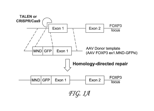
[0042] FIG 1A, FIG. 1B and FIG. 1C depict exemplary schema for
converting
CD4+ T cells into airT cells of the present disclosure. FIG. 1 A is a
schematic diagram of
FOXP3 locus before (top) and after (bottom) gene editing using FOXP3 TALEN or
CRISPR/Cas9 with FOXP3 guide RNA. TALEN or CRISPR/Cas9 cleaves FoxP3 locus at
exon 1, initiating site-specific double stranded DNA break. AAV provides donor
template
-15-
CA 03145037 2021-12-22
WO 2020/264039 PCT/US2020/039445
containing MND and GFP (to allow analysis of editing efficiency), which is
inserted into exon
1 at the DNA break. After the homology-directed repair, the MND promoter
drives expression
of FoxP3 and GFP reporter. FIG. 1B depicts a timeline of steps of gene editing
and cell
analysis and efficacy of airT generation from input Tconv cells. FIG. 1C
depicts representative
flow plots showing correlation between Foxp3 and GFP on day 4 after editing.
The three panels
on the right-hand side of the figure show CD25, CD127, Helios, CD45RO, ICOS,
and C'TLA-
4 expression in Foxp3+ GFP+ gated cells, respectively.
[0043] FIG. 2 depicts flow plots (bottom) showing GFP and Foxp3
expression on
day 4 and day 11 after editing according to the timeline shown at top. These
data show that
Foxp3 editing in CD4+ T cells is efficient and results in high, stable
expression of Foxp3.
[0044] FIG. 3A, FIG. 3B, FIG. 3C and FIG. 3D depict data comparing airT
cells
and activated natural T regulatory (nTreg) cells. FIG. 3A depicts a timeline
of steps to generate
edTreg and activated nTreg for comparison. CD4+ cells were isolated from PBMC
using
MACS CD4+ isolation kit and Tconv (CD25- CD127+) and Treg (CD25 high CD127-)
cells
were further sorted by flow. Sorted Tconv and Treg cells were activated with
CD3/CD28
activator beads and beads were removed after 48 hr activation. Only Tconv
cells were Foxp3-
edited using Cas9/Foxp3 gRNA and AAV-MND-LNGFR-Foxp3 ki to generate
edTreg/airT.
nTreg cells were treated in the same manner without Foxp3 editing. LNGFR+
cells from
Foxp3-edited Tconv cells were enriched using MACS LNGFR beads on day 10.
LNGFR+
edTreg and nTreg cells were used for suppression assay. FIG. 3B depicts a
comparison of
efficacy in generation of edTreg and nTreg from 1x10⁷ PBMC. At day 0, 1x10⁷
PBMC. Tconv
and nTreg cells activated on day 0 were expanded 10-30 times and 1-2 times,
respectively,
from day 0 to day 10. For edTreg, Treg yield on day 10 was calculated based on
editing rate
(10-30%). FIG. 3C depicts representative flow plots showing Treg phenotype in
Foxp3-edited
Tconv and nTreg cells on day 10. Top panels show (left-most panel) LNGFR
expression in
edited Tconv and (right) Foxp3, Helios, CD25, CD127, 1COS, and CTLA-4
expression in
edited Treg (LNGFR+ gate, top panels) and nTreg (bottom panels). FIG. 3D
(upper panels)
depicts comparison of Foxp3, CTLA-4, and 1COS expression in edTreg/airT (blue)
and nTreg
(red). FIG. 3D (bottom table) shows the MFI.
[0045] FIG. 4A and FIG. 4B show that airT cells have superior in vitro
suppressive
activity to nTreg. FIG. 4A depicts data from an in vitro suppression assay
comparing
-16-
CA 03145037 2021-12-22
WO 2020/264039 PCT/US2020/039445
suppressive activities of edTreg/airT and nTreg on CD4+ Teff cells at the
indicated Treg:Teff
ratios. airT or nTreg cells were labeled with EF670, and CD4+ Teff cells were
labeled with Cell
Trace Violet (CTV). Teff cells were co-cultured with airT or nTreg at
different ratios, 0:1 (Ten
only), 1:1, 1:2, 1:4, 1:8, 1:16, and 1:32 (Treg:Teff). CD3/CD28 activator
beads were added at
1:25 (bead to Ten' ratio) and cells were analyzed by flow after 4d incubation.
Dilution of CTV
in Ten' cells was measured as proliferation. FIG. 4B depicts percent
suppression calculated as
(% proliferation in Ten' only+beads - % proliferation in Ten' cells cultured
with Treg) / (%
proliferation in Teff only+beads) x 100.
[0046] FIG. 5 depicts exemplary lentiviral islet-specific TCR
constructs expressing
rare islet-specific TCRs derived from Type 1 diabetes (T1D) subjects. Panel A
depicts a table
of lentiviral vectors encoding GAD65 or IGRP specific TCRs (4.13, T1D2, T1D4,
T1D5-1, or
T1D5-2), their epitope specificity, and TCR alpha or beta chain usage. Panel B
depicts
structure of lentiviral islet-specific TCR. TCR constructs include human TCR
variable regions
from the islet-specific TCRs and mouse TCR constant regions that allow to
improve pairing
between the transduced human TCR chains.
[0047] FIG. 6 depicts validation of islet Ag-specific TCR expression:
murine
TC1113 expression and proliferation of islet antigen-specific T cells. Panel A
depicts flow plots
for CD4+ T cells isolated, activated with CD3/CD28 beads, and transduced with
LV islet-
TCRs. Flow plots show mTCRI3 expression gated on CD3/CD28-activated CD4+ cells
day 9
post-transduction with lentivirus (LV) encoding islet-specific TCR. Panel B
depicts flow plots
for CD4+ T cells transduced with LV islet-TCRs labeled with CTV and co-
cultured with APC
(irradiated PBMC) and their cognate peptide or irrelevant peptide for 5 days.
Flow plots
showing cell proliferation of LV-transduced CD4+ T cells labeled with CTV
following 5-day
co-culture with antigen-presenting cells (APC; irradiated PBMC) and cognate or
irrelevant
peptide. Proliferation is shown as CTV dilution.
[0048] FIG. 7 shows generation of Foxp3-edited T cells with islet-
specific TCR.
Panel A depicts a timeline of generating edTreg cells with islet-specific
TCRs. Panel B depicts
representative flow plots showing mTCRI3 expression and LNGFR/Foxp3 expression
on CD4+
cells on day 7 after transduction with T1D4 or T1D5-1 TCR and Foxp3 editing.
Right panels
show expression of CD25, CD127, CTLA-4, and 1COS gated on LNGFR+ cells.
-17-
CA 03145037 2021-12-22
WO 2020/26-1039 PCT/US2020/039445
[0049] FIG. 8 relates to exemplary antigen-specific suppression assays
of the
present disclosure. Panel A depicts a timeline for generation of edTreg cells
expressing islet-
specific TCRs. edTreg cells with islet-specific TCRs (no LV TCR, T1D4, or T1D5-
1 TCR)
were enriched by LNGFR expression using MACS LNGFR beads. LNGFR+ cells were
aliquoted and frozen down for further experiments. Panel B depicts a summary
of method
used to assess antigen-specific suppression assays. CD4+ T cells transduced
with islet-specific
TCRs (T1D4 or T1D5-1 TCR) were used as Ten' cells. Ten' cells and Treg cells
were labeled
with different reagents, for example CTV or EF670, and co-cultured with or
without edTreg
cells with 1:1 or 1:2 ratio in the presence of APC (autologous irradiated
PBMC) and various
peptides. Cells were stained and analyzed by flow after 1 d or 4 d incubation
for measuring
cytokine generation and proliferation of Teff cells, respectively.
[0050] FIG. 9 and FIG. 10 depict suppressive activity of edTreglairT on
Teff
proliferation in the presence of APC and the indicated peptide(s). Ten' and
Treg cells were
labeled with CTV and EF670, respectively. CD4+ T cells transduced with 11D4-
TCR (T1D4
Ten') were co-cultured with or without edTreg expressing T1D4-TCR (T1D4
edTreg) or T1D5-
1-TCR (T1D5-1 edTreg) in the presence of APC and various peptides (DMSO, IGRP
241,
IGRP 305, or IGRP241+IGRP 305). 4 days after the co-culture, cells were
stained and analyzed
for Ten' proliferation as dilution of CTV. Flow plots show Teff proliferation
gated on CD3+
CD4+ CTV+ EF670- LNGFR-.
[0051] FIG. 11 depicts suppression of cytokine generation in Ten' by
edTreg/airT.
Ten' and Treg cells were labeled with CTV and EF670, respectively. T1D4 Ten'
cells were
cocultured with or without untransduced edTreg or T1D4 edTreglairT cells in
the presence of
APC and peptides (DMSO or IGRP 241). 1 day after the co-culture, cells were
contacted with
BFA for 4h, stained, and analyzed for cytokine generation from Ten' cells.
Flow plots show
TNF, IFNg, or IL-17 generation from T1D4 Ten' cells gated on CD4+ CTV+ EF670-.
[0052] FIG.s 12-17 relate to the development and characterization of
antigen-
specific human Foxp3-edited human CD4+ T cells.
[0053] FIG. 12 depicts (top) an exemplary scheme for generating human
antigen-
specific edTreg/airT from peripheral blood cells and (bottom) phenotype of
FOXP3-edited
human antigen-specific CD4+ T cells. In the bottom panels, representative flow
plots (left)
-18-
CA 03145037 2021-12-22
WO 2020/264039 PCT/US2020/039445
and percentage (right) of GFP expression in tetramer positive (Tr+; a mixture
of MHC class II
tetramers with flu or tetanus peptides) human CD4+ T cells at 4 days post-gene
editing (n=5).
[0054] FIG. 13 depicts a characterization of FOXP3-edited human antigen-
specific
CD4+ T cells. Panel A depicts phenotype of FOXP3 edited human antigen-specific
CD4+ T
cells. Bar chart summarizes flow cytometry data (n=5); chart shows expression
of Treg
markers and intracellular IL- 2 production in Tmr+edTreg , Tmr+ Mock-edited
cells, as well
as in thymus-generated Treg (tTreg) obtained from an unrelated donor. Data
shown are
representative of 5 independent experiments. P values of statistically
significant differences
are indicated above bars. Panel B depicts human antigen-specific edTregiairT
suppresses
proliferation of Teff in vitro. Suppression assays conducted using
Tmr+edTreglairT or mock-
edited Tmr+ cells co-cultured with Teff from healthy controls, APCs, and
soluble anti-CD3 and
anti-CD28. Ratio of antigen presenting cells (irradiated CD4- PBMC):
Tmr+edTreg or mock-
edited Tmr+ cells: Teff was 2:1:1. 1 Ki 3H was added 18 hours prior to the end
of the 4 day
assay and proliferation was measured by a scintillation counter. Bar graph
indicated averaged
results from three experiments with three donors.
[0055] FIG. 14 depicts successful generation of antigen-specific
edTreg/airT by
peptide stimulation followed by Foxp3 editing. Panel A depicts a timeline of
steps of antigen-
specific T cell expansion and gene editing. After 9 days of peptide
stimulation to expand T
cells specific for MP, HA, or Tetanus, cells were activated with CD3/CD28
activator beads for
gene editing. Beads were added to the sorted cells to enhance expansion of
antigen-specific
Tregs. Panel B depicts flow plots show GFP and Foxp3 expression on day 15
after editing.
GFP+ Foxp3+ cells were CD25+ CD127- and about 60% of cells were MP, HA, or TT
specific
by tetramers.
[0056] FIG. 15 depicts antigen-specific suppression by Foxp3-edited
Tregs/airT.
Panel A a timeline of steps of generating antigen-specific edTreg/airT cells
for suppression
assay. GFP+ cells were sorted and expanded with CD3/CD28 beads on day 15 after
editing.
Beads were removed after 7d incubation and edTregiairT cells were harvested
and used for
suppression assay after 11 days of expansion. Panel B depicts a summary of
suppression assay
design. CD4+CD25+ cells were isolated from autologous PBMC, labeled with
EF670, and
used as Teff cells. CD4-CD25+ cells were irradiated and used as APC, and
edTreglairT cells
were labeled with Cell Trace Violet (CTV). Teff cells and APC were co-cultured
with or
-19-
CA 03145037 2021-12-22
WO 2020/264039 PCT/US2020/039445
without edTreg/airT cells in the presence of DMSO or peptide pool (MP+HA+TT).
Panel C
depicts after 7 days of co-culture, cells were stained and analyzed by flow.
CD3+ CD4+
EF670+ CTV- cells were gated as Teff cells. Panel D depict a dilution of EF670
in Teff cells
was measured as proliferation and 15% of EF670- cells from co-culture of Ten'
cells with APC
and the peptide pool was normalized as 100% proliferation. % suppression was
calculated as
(100-% Proliferation).
[0057] FIG. 16 depicts an expansion of islet-specific T cells of
multiple
specificities by peptide stimulation. Panel A depicts an exemplary timeline
for generating islet-
antigen specific edTreg/airT cells. Freshly isolated CD4+CD25- cells were
stimulated by a
pool of islet-specific peptides and APC (irradiated autologous CD4- CD25+
cells) for 14 days
and expansion of islet-specific T cells was analyzed on day 13 by tetramer
staining. Panel B
depicts flow plots showing islet-specific T cells stained by individual
tetramers or tetramer
pool, gated on CD4+ cells.
[0058] FIG. 17 depicts generation of islet-specific Tregs of multiple
specificities.
Panel A depicts islet-specific T cells were stained by tetramers and sorted on
day 14. Sorted
tetramer+ cells were activated with CD3/CD28 beads for 72h for Foxp3 editing.
3 days after
editing, cells were stained and analyzed. Flow plots show Foxp3 and LNGFR
expression in
mock or edited cells (left) and CD25, CD127, and CD45R0 expression in LNGFR+
gated cells
(right). Panel B depicts cells were stained by individual tetramers or
tetramer pool and flow
plots show tetramer+ cells in LNGFR+ Foxp3+ edited cells.
[0059] FIG.s 18-33 relate to the generation of dual-edited human CD4+ T
cells
using bi-allelic targeting to engineer artificial Treg cells expressing Foxp3
and antigen-specific
TCR, with endogenous TCR inactivation.
[0060] FIG. 18 depicts a schematic of an exemplary CD4+ T cell edited
to possess
Treg phenotype and to express exogenous Ag-specific TCR, but not endogenous
TCR. In this
scheme, the conversion of a conventional CD4+ T-cell into an antigen-specific
Treg comprises
three genetic alterations: 1) stable expression of the transcription factor
FOXP3 to drive cells
toward a Treg phenotype; 2) stable expression of a defined, antigen-specific
rearranged T-Cell
receptor (Ag-specific TCR) to direct Treg immunosuppressive activity; and 3)
genetic deletion
of the endogenous T-Cell Receptor (TCR) to ensure that immunosuppressive
function is
directed solely toward the desired antigen.
-20-
CA 03145037 2021-12-22
WO 2020/26-1039 PCT/US2020/039445
[0061] FIG. 19 depicts exemplary AAV constructs for CRISPR gene editing
at the
human and mouse TRAC loci. The list includes adeno-associated virus plasmid
constructs
generated for CRISPR-based homology directed repair, organized based on the
relevant
gRNA, and includes number designation.
[0062] FIG. 20 depicts an exemplary CRISPR-based approach for targeting
of the
human TRAC locus for knockout/knock-in. In particular, the image shows a
schematic
representation of the human TRAC locus showing the relative position of the
four gRNA
sequences tested (PC_TRAC_E1_gRNA1 to PC TRAC El_gRNA4). The TRAC exon 1 is
indicated by the lowermost bar from about position 1160 continuing past 1400.
Common SNPs
are indicated by about positions 1160 and 1400. The position of a previously
published positive
control gRNA sequence (TCRa G4old) is indicated at about position 1320.
[0063] FIG. 21 relates to guide RNA (gRNA) qualification of non-
homologous end
joining (NHEJ) for knockout of CD3 in human CD4+ primary T cells. Data are
from FACS
analysis. Panel A depicts flow plots show expression of CD3 2 days post-
editing in mock-
edited and TCR-edited CD4+ T cells using four different guide RNAs.
TCRa_G4o1d,
previously demonstrated to knockout CD3 expression, was used as a control.
Panel B depicts
histograms showing percent CD3 knockout.
[0064] FIG. 22 depicts results from Inference of CRISPR Edits (ICE)
analysis of
indel frequency. On-target site-specific activity was measured by ICE
(Inference of CRISPR
Edits) and confirmed specific indel induction for gRNA_l and gRNA _4 in TRAC
relative to
predicted off-target sites.
[0065] FIG. 23 depicts results from ICE analysis of predicted off
target sites for
TRAC gRNAs. The top 3 predicted off target sites for TRAC gRNA 1 and TRAC gRNA
2
(based on frequency and position of mismatches) were tested for indel
induction frequency by
ICE sequence deconvolution analysis.
[0066] FIG. 24 depicts an exemplary experimental outline for performing
dual
AAV editing for assessment of bi-allelic knock-in. A. Diagram of AAV
constructs used in this
experiment; after editing, MND promoter drives expression of GFP/BFP. B.
Timeline of
experimental procedures. CD4+ T cells were bead-stimulated (CD3/CD28) for 3
days prior to
editing. Three and six days post-editing, cells were evaluated for GFP and BFP
expression by
flow cytometry.
-21-
CA 03145037 2021-12-22
WO 2020/264039 PCT/US2020/039445
[0067] FIG. 25 depicts dual editing of the TRAC locus in human CD4+
cells leads
to a double-positive population of cells. Panel A depicts flow plots show GFP
and BFP
expression in mock-edited, and mixed MND.GFP- and MND.BFP-edited cells (10%
#3207
virus + 10% #3208 AAV) two days post-editing. Viral titers were 3.3x10'12 and
2.53x10'12
for #3207 and #3208, respectively. Panel B depicts histograms showing percent
double-
negative, GFP single-positive, mCherry single-positive and GFP/mCherry double-
positive
cells within the dual-edited cells.
100681 FIG. 26 depicts schematic diagrams showing exemplary Split IL-2
CISC
HDR knock-in constructs for selection of dual-edited cells. In the depicted
constructs, CISC
(chemically induced signaling complex) is split onto 2 different constructs
and each CISC
component is co-expressed with a different reporter, in this case either GFP
or mCherry. Each
construct contains half of a rapamycin-binding complex (either FKBP or FRB
domain, with
the chimeric endoplasmic reticulum targeting domain fused to one half of an IL-
2R signaling
complex (IL-2RB or IL-2RG) transmembrane and intracellular domains. Delivery
of cDNA
encoding each CISC component co-expressed with the GFP / mCherry tag to
primary human
CD4+ T cells allows selective expansion of cells that contain both CISC
components and thus
are also dual edited for GFP and BFP.
[0069] FIG. 27 depicts an exemplary timeline of steps for dual AAV
editing of
CD4+ T cells, expansion with rapalog, and analysis of enriched cells. Cells
were bead
stimulated (CD3/CD28) for 3 days prior to editing. Two days post-editing,
cells were analyzed
by flow for GFP and mCherry expression, and then expanded in media containing
5Onglml
human IL-2 or 100nM rapalog. Flow cytometry to assess enrichment of GFP,
mCherry double-
positive cells was carried out on days 6, 8, and 10 post-editing.
[0070] FIG. 28 depicts FACS analysis of initial dual editing rate.
Panel A depicts
flow plots show GFP and mCherry expression in mock-edited, MND.GFP.FRBIL-2RB-
edited
(20% #3207 AAV), MND.mCherry.FKBP.IL-2RB (20% #3208 AAV)-edited and mix-edited
(10% #3207 + 10% #3208) cells. Viral titers were 3.3x10'2 and 2.53x10¹² for
#3207 and #3208,
respectively. Panel B depicts histograms show percent of double-negative, GFP-
positive,
mCherry-positive and GFP/mCherry double-positive cells within the dual-edited
cells.
[0071] FIG. 29 depicts exemplary data showing rapalog enrichment of
dual-edited
cells. Panel A depicts flow plots show GFP and BFP expression in mock-edited,
and mixed
-22-
CA 03145037 2021-12-22
WO 2020/26-1039 PCT/US2020/039445
MND.GFP- and MND.BFP- edited cells (10% #3207 virus + 10% #3208 AAV) two days
post-
editing. Viral titers were 3.3x10'2 and 2.53x10'2 for #3207 and #3208,
respectively. Panel B
histograms showing percent double-negative, GFP single-positive, mCherry-
single positive
and GFP/mCherry double-positive cells within the dual-edited cells.
[0072] FIG. 30 depicts histograms showing percent double-negative, GFP
single-
positive, and mCherry single-positive cells after contact with IL-2 and
rapalog. These data
show that single-positive and unedited populations do not significantly change
with rapalog
treatment
[0073] FIG. 31 depicts data from FACS analysis of initial dual editing
rates using
two different donors. Panel A depicts a timeline of editing and analysis
steps. Panel B depicts
histograms showing percent double-negative, GFP-positive, mCherry-positive and
GFP/mCherry double-positive cells within the dual-edited cells for each donor.
Donor
R003657 is male, Caucasian and 28 y.o. Donor R003471 is male, Caucasian and 29
years old.
[0074] FIG. 32 depicts data from FACS analysis of rapalog enrichment of
Bi-
Allelic R003471 cells. Panel A depicts flow plots showing expression of GFP
and mCherry
following 5 days enrichment in rapalog. Panel B depicts histograms showing
percent
GFP/mCherry double-positive cells after expansion in IL-2 or rapalog.
[0075] FIG. 33 depicts schematic diagrams showing exemplary split-CISC
constructs for insertion of TCR and Foxp3 and enrichment of dualedited cells.
CISC is split
onto two different constructs and each CISC component is co-expressed with
either an Ag-
specific TCR (in the diagram, exemplary Ti D4 TCR) or Foxp3. Each construct
contains half
of a rapamycin-binding complex (either FKBP or FRB domain, with the chimeric
endoplasmic
reticulum targeting domain fused to one half of an IL-2R signaling complex (IL-
2RB or IL-
2RG) transmembrane and intracellular domains. Delivery of cDNA encoding each
CISC
component co-expressed with the T1D4 TCR / Foxp3 to primary human CD4+ T cells
allows
selective expansion of cells that contain both CISC components and thus are
also dual edited
for T1D4 TCR and Foxp3.
[0076] FIG.s 34-37 relate to the generation of reagents for assessing
antigen-
specific airT cell function in in vivo models of autoimmunity.
[0077] FIG. 34 depicts a schematic representation of the murine TRAC
locus
showing the relative position of the three novel gRNA sequences tested
-23-
CA 03145037 2021-12-22
WO 2020/264039 PCT/US2020/039445
(PC_mmTrac_E1_gRNA1 to PC_mmTrac_E1_gRNA3). The TRAC exon 1 is indicated in
blue.
[0078] FIG. 35 depicts data from FACS analysis of CD3 knockout in
murine CD4+
T cells. Panel A depicts flow plots show expression of murine CD3 two days
post-editing in
mock-edited and TCR-edited CD4+ T cells using three different guides. Panel B
depicts
histograms showing percent mCD3 knockout for each guide RNA.
[0079] FIG. 36 depicts an exemplary experimental outline for dual AAV
editing
for assessment of bi-allelic knock-in. Panel A depicts a diagram of AAV
constructs used in
this experiment; after editing, MND promoter drives expression of GFP/BFP. B.
Timeline of
experimental procedures. Murine CD4+ T cells were bead stimulated (CD3/CD28)
for 3 days
prior to editing. Three and five days post-editing, cells were evaluated for
GFP and BFP
expression by flow cytometry.
[0080] FIG. 37 depicts data from FACS analysis of single- and dual-
editing rates
in the murine TCRa locus. Flow plots show GFP and BFP expression 3 days post-
editing in
mock, MND.GFP (10% #3211), MND.BFP (10% #3212), and mix-edited cells (5% #3207
+
5% #3208). Mixed edited cells had a total of 1.97% GFP/BFP double-positive
cells.
[0081] FIGs 38-43 relate to airT cell function in an antigen-specific
in vivo setting.
[0082] FIG. 38 depicts a schematic diagram of an experimental design to
test the
ability of MOG-specific edTreg/airT (shown in white) to suppress T effectors
(Teff) in a mouse
model of multiple sclerosis, Experimental Autoimmune Encephalomyelitis.
[0083] FIG. 39 relates experiments showing that mouse FOXP3 TALENs
catalyze
efficient FOXP3 disruption and initiate non-disruptive recombination of donor
template. Panel
A depicts binding sites for the FOXP3 TALEN pair in the human FOXP3 gene.
Panel B depicts
target binding sites for the mouse FOXP3 TALEN pair in the murine FOXP3 gene.
Panel C
depicts indel frequency at FOXP3 TALEN cut site in human (left) and mouse
(right) CD4+ T
cells 5-7 days after transfection with mRNA encoding either control mRNA
(encoding blue
fluorescent protein), or TALENs specific for human FOXP3 or mouse FoxP3,
respectively.
Graph shows average frequency of indels after colony sequencing PCR amplicons
surrounding
gDNA target site; 20-40 colonies were sequenced per experiment.
[0084] FIG. 40 relates to generation of edTregiairT from antigen-
specific murine
CD4+ T cells. Panel A depicts a schematic diagram of FOXP3 locus after
successful gene
-24-
CA 03145037 2021-12-22
WO 2020/264039 PCT/US2020/039445
editing using mouse FOXP3 TALENs and the mouse AAV FOXP3 MND-GFP knock-in (ki)
donor template. After editing, the MND promoter drives expression of chimeric
GFP-FoxP3
protein. Panel B depicts flow plots showing GFP expression in antigen-specific
mouse CD4+
T cells at Day 2 post-editing. Panel C depicts average percent of GFP+ cells
across multiple
experiments (n = 10). D. Flow plot of murine edTreg/airT showing expression of
relevant Treg
markers.
[0085] FIG. 41 shows functional assessment of antigen specific vs.
polyclonal
edTregiairT in a mouse model of Multiple sclerosis. Panel A depicts flow plots
showing GFP
expression in MOG-specific and polyclonal mouse CD4+ T cells at Day 2 post-
editing after
FACS sorting. Panel B depicts schematic diagram of murine EAE in vivo
experimental design
and timeline. 2D2 (MOG-specific) Teff (30K) were delivered with or without co-
transferred
edTreg/airT (30K) generated from either 2D2 or C57Bl/6 mice into RAG1-/-
recipient mice;
all strains were on C57B1/6 background. Analysis was performed at Day 7.
[0086] FIG. 42 depicts data showing that antigen-specific edTreg/airT
delay
expansion, activation and cytokine production of Teff. Immunophenotype of T
cells obtained
from inguinal and axillary lymph nodes in recipient mice at day 7 post-cell
transfer was
assessed by flow cytometry. CD45+ = panCD45 (recognizing all CD45 isoforms and
both
CD45.1 and CD45.2 alloantigens). Shown are total number of total CD45+ CD4+
cells (A)
and other indicated T cell subsets (B) and (C), expansion of GFP+ cells. Data
is representative
of results from 3 independent experiments; bar graphs show mean SD; p-values
of
statistically significant differences are indicated above bars.
[0087] FIG. 43 provides data showing that antigen-specific edTreg/airT
cells
suppress Ten' proliferation in vivo. Panel A depicts flow plots: to label
actively dividing cells,
the thymidine analog 5-Ethyny1-2'-deoxyuridine (EdU) was administered 2 hours
prior to
sacrifice in selected animals. EdU incorporation in T cells was determined by
intracellular
labeling with an anti-EdU antibody and flow cytometry. Flow plots are from T
cells isolated
from LNs 7 days post-cell transfer. Panel B depicts bar graphs summarize mean
% of cells
incorporating EdU in different cell subsets and (C) the % GFP + lymphocytes.
Flow plots are
representative of results from at least 3 independent experiments; bar graphs
show mean SD;
p-values of statistically significant differences are indicated above bars.
-25-
CA 03145037 2021-12-22
WO 2020/264039 PCT/US2020/039445
[0088] FIG.s 44-47 relate to experiments investigating antigen specific
T cell
function in a NSG adoptive transfer model of Type 1 diabetes. Engineered
antigen-specific
(BDC) or polyclonal (NOD) edTregs/airTs, or antigen-specific nTregs were
infused into the
mice followed by infusion of antigen-specific Teff cells. Mice were monitored
for diabetes up
to 90 days following infusion. Graph shows the percent of diabetic mice that
received effector
cells plus the designated mock edited, Foxp3-edited, or nTreg cells from NOD
and BDC2.5
mice.
100891 FIG. 44 relates to Foxp3 editing in CD4+ T cells of antigen-
specific NOD
mice. Panel A depicts CAS9/CRISPR RNP cutting efficiency in BDC2.5 NOD mice
using
different guide RNAs. Panel B depicts AAV5-delivered repair template. After
editing, the
MND promoter will drive expression of chimeric GFP-FoxP3 protein. Panel C
depicts flow
plots showing GFP expression in mock-edited and GFP-Foxp3-edited antigen-
specific mouse
CD4+ T cells at day 2 post-editing.
[0090] FIG. 45 relates to phenotype of FOXP3-edited antigen-specific
NOD CD4+
T cells. Left. Flow cytometry plots showing GFP and Foxp3 expression in edited
cells. Middle.
Flow cytometry plots showing IL-2, IFN-g and IL-4 expression in GFP-Foxp3-
edited (upper
plots) and mock-edited (lower plots) murine antigen-specific NOD CD4+ T cells.
Right.
Histograms showing % of cells positive for IL-2, IFN-y and IL-4 four days post-
editing.
[0091] FIG. 46 relates to an experiment investigating phenotype of
input cells for
NSG adoptive transfer model. Panel A depicts an experimental design showing
amount and
type of cells administered for each group of animals. Panel B depicts flow
cytometry plots
showing the phenotype of Teff, edTreg/airT and nTreg cells injected into NSG
mice.
[0092] FIG. 47 relates to antigen-specific T cell function in NSG
adoptive transfer
model. Panel A depicts an experimental design; engineered antigen-specific
(BDC) or
polyclonal (NOD) edTregs/airTs, or antigen-specific nTregs were infused into
the mice,
followed by infusion of antigen-specific Teff cells. Mice were monitored for
diabetes up to 90
days following infusion. Panel B depicts a graph shows the percent of diabetic
mice that
received effector cells plus the designated mock-edited, Foxp3-edited, or
nTreg cells from
NOD and BDC2.5 mice. Antigen-specific edTreglairT exhibited significantly
greater level of
protection from T1D compared with mock-edited T cells, polyclonal
edTregs/airTs or
polyconal nTregs.
-26-
CA 03145037 2021-12-22
WO 2020/26-1039 PCT/US2020/039445
[0093] FIG.s 48-51 relate to engineering a mouse AAV donor template
design to
generate airT cell product with a selectable marker (LNGFR).
[0094] FIG. 48 depicts exemplary repair templates used in murine Foxp3
editing.
AAV.Promoter-LNGF.P2A knock-in constructs were tested in murine T cells for
stable
expression of Foxp3.
[0095] FIG. 49 depicts phenotype of murine edTregiairT using
alternative
homology donor cassettes. Flow cytometry plots show LNGFR, FOXP3, CD25, and
CTLA-4
in mock-edited cells and cells edited with MND.LNGFRP2A KI (3189) or
PGK.LNGFR.P2A
KI (3227).
[0096] FIG. 50 depicts data showing editing rate and expression of
LNGFR in
murine edited Treg/airT cells. Flow cytometry plots show LNGFR and GFP
expression in
mock, MND-GFPki (#1331) MND.LNGFRP2A.K1 (#3189) edited cells.
[0097] FIG. 51 depicts data showing enrichment of LNGFR+ edited T cells
from
B6 mice using an anti-LNGFR column. Flow cytometry plots show LNGFR expression
of cells
prior to purification on a Miltenyi anti-LNGFR column, cells in the flow
through and cells
eluted from the column.
[0098] FIG. 52 depicts a comparison of FOXP3-edited vs. FOXP3
lentiviral (LV)
transduced human CD4 T cells. Panel A depicts a diagram of LV construct: MND
promoter
drives expression of a transcript encoding identical GFP-FOXP3 fusion protein
as that of airT;
transcript contains WPRE and poly(A) signals for efficient nuclear export and
mRNA stability.
Below are representative flow plots showing FOXP3 and GFP expression in mock-
edited T
cells or sorted tTreg (CD4+CD25++CD127-), airT and LV Treg (CD4+GFP+), all
post >14-
day expansion in vitro with CD3/CD28 beads. Panel B depicts mean viral copy
number ( SD)
of LV-transduced sorted cells (left; n = 6). Scatter plots (right) show the
MFI of the GFP+
population for each sample (n =5; P value from two-tailed Student's T-test).
Panel C depicts
bar graphs showing mean % of cells (top), and MF1 (bottom) by flow cytometry
staining for
the proteins indicated. Viable singlets were further gated on: CD4+ GFP+ (LV
Treg and
edTreg), CD4+FOXP3+ (tTreg), or CD4+ (mock). For markers with distinct bimodal
distributions, MF1 was calculated for the positive population only. Error bars
show SD. An
ordinary two-way ANOVA was performed, and P values adjusted with Tukey's
multiple
comparisons test. P values in black indicate comparison with mock-edited
cells; those in red
-27-
CA 03145037 2021-12-22
WO 2020/264039 PCT/US2020/039445
were comparison of groups indicated by dashed lines. Panel D depicts percent
suppression as
a function of Treg or mock dilution (top). Histograms of proliferation dye at
different ratios
of Treg or mock to Teff (bottom). % suppression = [(% divided with no Treg - %
divided with
Treg)/ % divided with no Treg] x 100. Panel E depicts a plot showing data
points and simple
linear regression of % GFP+ cells over time in culture after FACS
purification; airT (n = 4)
and LV Treg (n = 6); data from 6 experiments. Dashed lines indicate 95%
confidence intervals;
P value was obtained using an F Test.
[0099] FIG.s 53-73 provide additional schematics and data related to
exemplary
dual-editing strategies of the present disclosure for generation of antigen-
specific, drug-
selectable airT cells with knock-out of endogenous TCR.
[0100] FIG. 53 depicts schematics showing dual-editing strategies
designed to: a)
eliminate the endogenous TCR expression and b) generate selectable antigen-
specific airTs.
Delivery of expression cassettes for FOXP3 and a candidate islet antigen-
specific TCR (11D4)
paired to the two halves of the IL-2 CISC/DISC (FKBP-IL2RG and FRB-IL2RB), can
be
directed to the same locus (Strategy 1) or two separate loci (Strategy 2).
Targeting of the
TRAC locus in CD4+ T cells allows for deletion of the endogenous TCR Strategy
2 may
result in higher initial dual editing rates but requires two nuclease target
sites, leading to two
double stranded breaks (DSBs) in the host cell genome that mediate HDR.
Strategy 1 utilizes
a single nuclease target site leading to a single DSB.
[0101] FIG. 54 depicts a schematic of AAV HDR donor constructs used in
human
T cell dual-editing. The first 7 constructs are IL-2 split-CISC repair
templates with either GFP,
mCherry, HA-tagged FOXP3 or Ti D4 driven by the MND promoter. Each component
of the
split CISC includes a heterodimeric rapamycin binding complex (either FKBP and
FRB
domains), along with the chimeric endoplasmic reticulum targeting domain fused
to one half
of the IL2R signaling complex (either IL2RB or 1L2RG) trans-membrane and
intracellular
domains. Each repair template is flanked by 300 bp homology arms matched to a
gRNA
targeting either the TRAC locus (gRNA_4) or FOXP3 locus (gRNA_T9) (#3207,
3208, 3240,
3243, 3251, 3252, 3273). The next four constructs (#3253, 3258, 3292, 0001)
are used for in-
frame knock-in of a promoter-less TCR cassette including components of the
CISC, targeting
the first exon of TRAC locus (gRNA_1). The final two constructs (#3280 and
#3262) are split-
DISC repair templates that include the CISC elements as well as cDNA encoding
a free FRB
-28-
CA 03145037 2021-12-22
WO 2020/264039 PCT/US2020/039445
domain that functions in cytoplasmic Rapamycin sequestration (which eliminates
or reduces
any negative impact of rapamycin on gene edited cells). These latter
constructs also contain
either mCherry or FOXP3 driven off the MND promoter.
101021 FIG. 55 depicts dual editing rates within the human TRAC locus
in the
presence or absence of rapalog-based selection of CISC edited CD4+ T cells
from donor
R003657. Panel A depicts a timeline of editing (using RNP and AAV co-
delivery), enrichment
and analysis steps with donor R003657 CD4+ T cells using AAV #3207 and #3208.
Panel B
depicts flow plots show initial percent GFP/mCherry double positive cells in
the mock vs. dual-
edited samples, and percent GFP/mCherry double positive following 7 days
enrichment in the
presence of IL-2 or Rapalog (AP21967). Panel C depicts histograms show percent
double-
negative, GFP-positive, mCherry-positive and GFP/mCherry double-positive cells
within the
dual-edited cells following enrichment in IL-2 vs. Rapalog.
[0103] FIG. 56 depicts dual editing rates in the TRAC locus and rapalog-
based
selection of CISC-edited CD4+ T cells from donor R003471. Panel A depicts a
timeline of
editing, enrichment and analysis steps with donor R003471 CD4+ T cells using
AAV #3207
and #3208. Panel B depicts flow plots show initial percent GFP/mCherry double-
positive cells
in the mock- vs. dual-edited samples, and percent GFP/mCherry double-positive
following 7
days enrichment in the presence of IL-2 or Rapalog (AP21967). Panel C depicts
histograms
show percent double-negative, GFP-positive, mCherry-positive and GFP/mCherry
double-
positive cells within the dual-edited cells following enrichment in IL-2 vs.
Rapalog.
[01041 FIG. 57 shows dual-editing of the TRAC locus in human CD4+ T
cells
generates rapalog-selectable, antigen-specific airTs. Panel A depicts a
schematic showing
AAV HDR donors construct used to introduce of the "split" IL-2 CISC elements
for selection
of dual-edited cells. CISC components (IL2RG vs. IL2RB) are split between 2
constructs and
co-expressed with either HA-FoxP3 cDNA or the islet-specific TCR, T1D4 (AAVs
#3240 and
#3243 respectively). Each repair template is flanked by identical homology
arms that cannot
be cleaved by the gRNA targeting the TRAC locus. Only edited CD4+ T cells
incorporating
one copy of each construct are predicted to selectively expand under Rapalog
treatment. Panel
B depicts a timeline of key steps for dual AAV/RNP-based editing of CD4+ T
cells, expansion
with Rapalog and analysis of enriched cells. Cells were bead stimulated
(CD3/CD28) for 3
days prior to editing. Two days post-editing, cells were analyzed by flow for
HA-FoxP3 and
-29-
CA 03145037 2021-12-22
WO 2020/264039 PCT/US2020/039445
TCR expression, and then expanded in media containing 50ngiml human IL-2 or
100nM
Rapalog. Flow cytometry to assess enrichment of HA-FoxP3, TCR double-positive
cells was
carried out on days 5 and 8 post-editing. Panel C depicts rapalog enrichment
of dual-edited
cells. Left panel: Flow plots for HA-FoxP3 and TCR in dual-edited cells
following 8 days
expansion in IL-2 or Rapalog; Right panel: quantitation of percent HA-FoxP3 /
TCR double-
positive cells following expansion in IL-2 or Rapalog for 5 and 8 days.
[0105] FIG. 58 provides data showing that decreasing serum
concentration
increases total- and dual-editing rates within the TRAC locus. Panel A depict
a timeline
showing steps for dual AAV editing of CD4+ T cells and expansion with Rapalog.
Human
CD4+ T cells were edited using TRAC gRNA 4 and #3243 and #3240 AAV constructs
(Single-locus dual editing). Immediately following electroporation to deliver
the RNP, the
cells were placed in either 20%, 2.5%, 1% or 0% FBS containing media (recovery
media) and
infected with AAV. After ¨16 hours, the media was replaced with 20% FBS
containing media
and FACS analysis done on day 3 to determine editing rate. Cells recovered in
2.5% FBS
containing media were expanded in the presence of either IL-2 or Rapalog for
an additional 7
days. Panel B depicts flow plots show 11D4 and FOXP3 expression in mock-
edited, single-
edited and mixed edited cells (10% #3243 and 10% #3240 AAV) three days post
editing. Viral
titers were 4.2E" and 1.3E12 for #3243 pAAV.MND.T1D4.FRBIL2RB and #3240
pAAV.MND.FOXP3-HA.FKBP.IL2RG respectively. Panel C depicts histograms show
percent double-negative, FOXP3-HA-positive, Ti D4-positive and FOXP3/TID4
double-
positive cells within the dual-edited cells.
[0106] FIG. 59 shows IL-2 vs. Rapalog enrichment of dual-edited cell
populations.
TRAC locus dual-editing was performed as shown in FIG. 5. Panel A depicts flow
plots show
TI D4 and FOXP3 expression in mock-edited vs. FOXP3/T1D4 (#324013243) dual-
edited cells
treated with either 50ngimL IL-2 or 100nM Rapalog (AP21967) for 7 days. Data
are shown
only for the 2.5% FBS recovery media condition. Panel B depicts histograms
show percent
double-negative, FOXP3-HA-positive, TID4-positive and FOXP3,71D4 double-
positive cells
within the dual-edited cells following enrichment
[0107] FIG. 60 relates to a strategy for testing two-loci dual-editing
of human
CD4+ T cells. Panel A depicts a diagram of AAV HDR-donor constructs designed
to introduce
split IL-2 constructs for selection of dual-edited cells using a two loci dual-
editing approach.
-30-
CA 03145037 2021-12-22
WO 2020/26-1039 PCT/US2020/039445
CISC components are split between 2 constructs and co-expressed with either
mCherry or GFP
(#3207 and #3251 respectively). Repair templates are flanked by homology arms
matched to
gRNAs targeting either the TRAC or FOXP3 locus, respectively. Only edited CD4+
T cells
that incorporate both expression cassettes (into the appropriate locus) are
predicted to
selectively expand under Rapalog treatment. Panel B depicts a timeline showing
steps for dual
AAV editing of CD4+ T cells and expansion with Rapalog. Human CD4+ T cells
were edited
using human TRAC gRNA 4, human FOXP3 gRNA_T9 and #3251
(MND.mCherry.FKBP.IL2RG) and #3207 (MND.GFP.FRBIL2RB) AAV constructs (two-
loci dual editing). Immediately following electroporation, the cells were
placed in either 20%
or 2.5% FBS containing media (recovery media). After ¨16h, the media was
replaced with
20% FBS containing media and FACS analysis done on day 3 to determine editing
rate. Cells
recovered in 2.5% FBS containing medium were further grown in the presence of
either IL-2
or Rapalog for an additional 7 days to monitor enrichment.
[01081 FIG. 61 shows that recovery in 2.5% FBS containing medium
improves
dual-editing rates measured at Day 3 post-editing. Two-loci dual editing was
performed as
shown in FIG. 59. Panel A depicts flow plots show GFP and mCherry expression
in mock-
edited and dual-edited cells in 20% FBS vs. 2.5% FBS recovery media at 3 days
post-editing.
Viral titers were 6.55E^10 and 2.50E^12 for #3251 pAAV.MND.mCherry.FKBP.IL2RG
and
#3207 pAAV.MND.GFP.FRB.IL2RB respectively, and 10% culture volume of each
virus was
used for the editing reactions. Panel B depicts histograms show percent double-
negative, GFP-
positive, mCherry-positive and GFP/mCherry double-positive populations within
the dual-
edited cells.
[0109] FIG. 62 shows robust enrichment of two-loci dual-edited cells
treated with
rapalog selection. Two-loci dual-editing was performed as shown in FIG. 59.
Panel A depicts
flow plots show GFP and mCherry expression in mock-edited and GFP/mCherry
(#3207/3251)
edited cells (edited in 2.5% serum) treated with either 50ng/mL IL-2 vs. 100nM
Rapalog
(AP21967) for 10 days. Panel B depicts histograms show percent double-
negative, GFP-
positive, mCherry-positive, and GFP/mCherry double-positive cells within the
edited
population following treatment in IL-2 vs. Rapalog for 10 days.
[0110] FIG. 63 relates to engineering of two-loci dual-editing of human
CD4+ T
cells. Editing conditions and timeline for dual AAV editing of CD4+ T cells
and expansion
-31-
CA 03145037 2021-12-22
WO 2020/264039 PCT/US2020/039445
with Rapalog. Human CD4+ T cells were edited using human TRAC gRNA 4, human
FOXP3
gRNA_T9 and #3251 (MND.mCherry.FKBP.IL2RG) and #3207 (MND.GFP.FRB.IL2RB)
AAV (two-loci dual editing). Editing conditions were varied according to the
table with
different % of viral stock and either in the presence of the HDR enhancer or
DMSO.
Immediately following electroporation, the cells were placed in 2.5% FBS
containing media
(recovery media). After ¨16h, the media was replaced with 20% FBS containing
media and
FACS analysis done on day 3 to determine editing rate. Cells recovered in 2.5%
FBS
containing medium were further grown in the presence of either IL-2 or Rapalog
for an
additional 10 days to monitor enrichment.
101111 FIG. 64 depicts a graph showing that matched 10% volume of AAV
HDR
donors leads to improved dual editing. Editing was performed as outlined in
FIG. 62. Graphs
show percent double-negative, GFP-positive, mCherry-positive and GFP/mCherry
double-
positive populations within the dual-edited cells 3 days post-editing with
varying amounts of
#3207 and #3251 AAV in the presence of 30uM HDR enhancer or DMSO.
[0112] FIG. 65 provides data showing robust enrichment of two-loci dual-
edited
CD4+ T cells with rapalog selection; with optimal results using 2.5% FBS media
and matched
10% volume of AAV donor. Editing was performed as outlined in FIG. 62. Graphs
show the
cells from FIG. 10, (edited in 2.5% serum, matched 10% virus +/- HDR enhancer)
as percent
double negative, GFP positive, mCherry positive, and GFP/mCherry double
positive cells
within the editing population following contact with IL-2 or Rapalog for 10
days.
[01131 FIG. 66 provides a diagram of exemplary split-CISC constructs
for insertion
of islet-specific TCR and FOXP3 and enrichment of dual-edited cells using a
two-loci dual-
editing strategy. The IL-2 CISC (chemically induced signaling complex) is
split onto 2
different constructs and co-expressed with either T1D4 TCR or FOXP3 (#3243 and
#3252
respectively). Each construct contains half of a heterodimeric rapamycin
binding complex
(FKBP and FRB domains), along with the chimeric endoplasmic reticulum
targeting domain
fused to one half of the IL-2R signaling complex (IL-2RB or IL-2RG) trans-
membrane and
intracellular domains. Delivery of cDNA encoding each CISC component co-
expressed with
the T1D4 TCR / FOXP3 to primary human CD4+ T cells allows us to only expand
cells that
contain both CISC components and thus are also dual edited for T1D4 TCR and
FOXP3
expression.
-32-
CA 03145037 2021-12-22
WO 2020/264039 PCT/US2020/039445
[0114] FIG. 67 shows an exemplary strategy for single locus dual-
editing with
capture of 'TRAC promoter. Schematic of AAV HDR-editing constructs designed
for dual-
editing within the TRAC locus to introduce: (top) (#3240 FOXP3 expression and
split CISC
and (bottom) (#3258) in-frame knock-in to TRAC exon 1 to drive expression of
TI D4 TCR
and split CISC using the TRAC endogenous promoter.
[0115] FIG. 68 shows that TRAC locus HDR editing disrupts TCR
expression and
mediates robust transgene expression via the endogenous TRAC enhancer-
promoter. Panel A
depicts an editing strategy for in-frame integration of a mCherry-Split-CISC
cassette at the
endogenous TRAC locus. By using a gRNA targeting the TRAC exon 1, in-frame
integration
of a marker fluorophore (mCherry) followed by the FRB-IL2RB CISC separated by
a P2A
element (construct #3253) allows for expression driven by the endogenous TRAC
promoter,
while disrupting expression of the endogenous TCR. Panel B depicts a timeline
showing steps
for AAV #3253 editing of CD4+ T cells. Cells were bead-stimulated (CD3/CD28)
for 3 days
prior to editing. Panel C depicts an analysis: seven days post-editing, cells
were analyzed by
flow for CD3 and mCherry expression. Flow cytometry plots show significant
expression of
mCherry with concomitant loss of CD3 in edited cells compared to mock-edited
and AAV-
only controls.
[0116] FIG. 69 shows comparison of mCherry expression mediated via the
TRAC
endogenous promoter vs. MND promoter. Gene editing was performed as shown in
FIG. 67
using alternative HDR donors (#3253 vs. #3208) to assess the relative
expression activity from
the TRAC endogenous promoter vs. MND promoters, respectively. Flow cytometry
plots
shows that the level of mCherry expression when driven off the endogenous
promoter
(P2A.mCherry.FRB.IL2RB (#3253)) is lower than compared to when driven by the
MND
promoter (MND.mCherry.FICBP.IL2RG (#3208). The bottom row of panels shows data
from
a repeat experiment performed using the #3253 donor.
[0117] FIG. 70 show exemplary alternative dual-editing strategies for
targeting the
TRAC and/or FOXP3 loci and that utilize in-frame knock-in constructs to
capture the TRAC
endogenous promoter. Schematic of exemplary AAV donor constructs for testing
single-locus
and two-loci dual-editing strategies and to generate antigen-specific airT
with IL-2 CISC
selection capacity. T1D4 TCR is shown as a representative TCR that can be
replaced by
-33-
CA 03145037 2021-12-22
WO 2020/264039 PCT/US2020/039445
alternative TCRs based upon disease target and other relevant features for
therapeutic
application. IL-2 DISC constructs are similarly applied.
[0118] FIG. 71 relates to dual-editing of human CD4+ T Cells using
decoy-CISC
(split-DISC) constructs. Panel A depicts a diagram of Split IL-2 DISC HDR
knock-in construct
(#3280) for selection of dual-edited cells in Rapamycin. To generate the split
decoy-CISC
(split-DISC), the free FRB domain for cytoplasmic Rapamycin sequestration was
added to the
MND.mCherry.FKBP.IL2RG construct to generate (M1'lD.mCherry.FKBP.IL2RG.FRB
(#3280)). Each repair template (#3280 and #3207, not shown) is flanked by
identical
homology arms matched to a gRNA targeting the TRAC locus. Edited CD4+ T cells
incorporating one copy of each construct are predicted to selectively expand
under Rapalog or
Rapamycin treatment. Panel B depicts a timeline showing steps for dual AAV
editing of CD4+
T cell using AAV #3280 and #3207), expansion with Rapalog/Rapamycin and
analysis of
enriched cells. Cells were bead-stimulated (CD3/CD28) for 3 days prior to
editing. Two days
post-editing, cells were analyzed by flow for GFP and mCherry expression, and
then expanded
in media containing 50ng/m1 human IL-2, 100 nM Rapalog or 1 OnM Rapamycin.
Panel C
depicts flow plots show the percentage of GFP/mCherry double-positive cells on
day 3 post-
editing.
[0119] FIG. 72 shows that dual editing of human CD4+ T cells with split-
DISC
constructs generates Rapamycin-selectable cells. Dual-editing was performed as
described in
FIG. 70. Panel A depicts flow plots show percent double-positive GFP/mCherry
cells
following 8 days in the presence of 50ng/mL human 1L-2, 100nM Rapalog
(AP21967), lOnM
Rapamycin, or no treatment. Panel B depicts histograms quantitate percent
double-negative,
GFP-positive, mCherry-positive and GFP/mCherry double-positive cells within
the dual-
edited cells following enrichment in IL-2, Rapalog (AP21967), Rapamycin or no
treatment.
[0120] FIG. 73 show exemplary constructs for in vivo testing of dual-
edited Tregs
(split-DISC). Diagram of FOXP3 split IL-2 DISC HDR knock-in construct (#3262)
to be
paired with a T1D4 CISC construct (#3243) for Rapamycin selection of dual-
edited cells. CISC
components are split between 2 constructs and co-expressed with either HA-
FoxP3 or T1D4
TCR The FOXP3 CISC construct also contains the FRB domain and is predicted to
protect
mTOR signaling in the presence of Rapamycin (FOXP3 DISC construct). Each
repair template
is flanked by identical homology arms matched to a gRNA targeting the TRAC
locus. Edited
-34-
CA 03145037 2021-12-22
WO 2020/264039 PCT/US2020/039445
CD4+ T cells incorporating one copy of each construct may selectively expand
under both
Rapalog and Rapamycin treatment.
[0121] FIG.s 74-94 provide additional schematics and data related to
generation
and characterization of murine airT cells.
[0122] FIG. 74 depicts repair templates used in murine Foxp3 editing.
Diagram of
alternative AAV.GFP.KI and AAV.LNGFR.P2A constructs that were developed and
tested in
murine T cells for editing efficiency, FOXP3 expression and suppressive
function.
10123] FIG. 75 depicts a schematic showing methods used for generation
of murine
airT using the MND.GFP.KI (or alternative) HDR donor construct.
10124] FIG. 76 relates to generation and enrichment of murine airT
cells utilizing
alternative promoters to express endogenous Foxp3. Flow cytometry plots
showing LNGFR
and GFP expression prior to and post LNGFR enrichment via FACS sorting in
mock,
MND.GFP.KI (#1331), MND. LNGFR.P2A (#3189 and 3261), PGK. LNGFR. P2A (#3227)
and EF-la-LNGFR.P2A (#3229) edited cells. Upper plots show initial editing
rates and lower
plots show enrichment post FACS sorting. Data indicate that airT can be
generated with each
of the candidate donor constructs.
[0125] FIG. 77 depicts expression levels of Foxp3 in murine airT using
alternative
homology donor cassettes. Panel A depicts flow cytometry plots showing LNGFR
and GFP
expression in HDR-edited splenic T cells. Panels show un-manipulated C57BL/6
control cells;
mock-edited, MND.GFP.KI (#1331), MND.GFP.KI with UCOE (#3213), and PGK.GFP.KT
(#3209)- edited C57 BL/6 murine T cells, respectively. Panel B depicts flow
histograms
showing FOXP3 expression from the data in panel A. Panel C depicts a bar chart
showing
FOXP3 MFI in nTreg and edTreglairT generated with the indicated alternative
HDR-donor
constructs. MND promoter containing donors mediate the highest levels of FOXP3
expression.
[0126) FIG. 78 depicts design of, and results from, an in vitro
suppression assay
using murine tTreg or airT. A. airT cells used for in vitro suppression assay
were enriched by
FACS sorting at day 2 post editing and resuspended into RPMI media containing
10% FBS.
nTregs (CD4+CD25+), Teff (CD4+CD25-) and antigen presenting cells (CD4-CD25-)
were
isolated from the combined spleen and lymph node cells of 8 to 10 weeks-old
C56BL/6 mice
by column enrichment Enriched 5x10⁶ Teff were resuspended in 2 ml of PBS and
labeled with
cell trace violet (CTV) for 15 minutes at 37 C, then washed and resuspended in
media before
-35-
CA 03145037 2021-12-22
WO 2020/264039 PCT/US2020/039445
their addition in the suppression assay. To set up the assay, 1.25 x 10⁵
irradiated APCs (2500
rad) were co-cultured with 0.25 x 10⁵ Teff and a titrated number of nTregs and
airT in the
presence of 1 mg/ml anti-CD3 in a U bottom 96 well tissue culture plate with
total volume of
300 ul media and incubated at 37 C CO2 incubator for four days. At day 4,
cells were washed
twice with PBS and stained with live/dead indicator, anti-CD4, anti-CD45 and
anti-CD25, and
analyzed by FACS (LSRII) for the suppression of Teff proliferation by airT. B)
Representative
flow data showing a reduction of Teff proliferation in the presence of airT
cells.
101271 FIG. 79 depicts results from testing murine airT suppressive
function in
vitro. Flow cytometry plots showing cell trace violet labeled CD4+ T cells in
the presence and
absence of mock-, MND.GFP.KI- (#1331), or MND.LNGFR.P2A- (#3261-edited T
cells, or
nTregs from C57 BL/6 mice. These data demonstrate that murine airT (generated
with the
MND.GFP.KI or MND.LNGFR.P2A HDR donors) and nTregs exhibit comparable, robust
in
vitro suppressive function.
[01281 FIG. 80 depicts in vitro suppressive function of murine airT
with alternative
promoters. Flow cytometry plots showing cell trace violet-labeled CD4+ T cells
in the presence
and absence of mock-edited, MND.GFP.KI- (#1331), MND.LNGFR.P2A- (#3261),
PGK.LNGFR.P2A- (#3227), and EF-la.LNGFR.P2A (#3229)-edited T cells, or nTregs
from
C57 BL/6 mice. Murine airT with MIND promoter exhibit suppressive function
that is
comparable to nTreg. In contrast, airT using the PGK or EF-la promoters
exhibit only limited
or no suppression.
[0129] FIG. 81 depicts the design of an experiment to compare sorted
vs. column-
purified enriched LNGFR+ edited cells in an NSG adoptive transfer model. The
table lists the
number of recipient NSG host animals, and source and number of adoptively
transferred
control, airT or nTreg cells in each of the 5 experimental cohorts
101301 FIG. 82 depicts flow analysis of LNGFR.P2A-edited NOD BDC2.5+
murine cells prior to and post-column purification. Panel A depicts flow
cytometry plots
showing LNGFR expression in mock-, and MND.LNGFR.P2A- (#3189)-edited cells.
Panel B
depicts flow cytometry plots showing LNGFR expression in MND.LNGFR.P2A (#3189)-
edited cells post enrichment via sorting. FACS sorting consistently enriched
to edTreg
products of >90% purity for use in in vitro and in vivo studies.
-36-
CA 03145037 2021-12-22
WO 2020/26-1039 PCT/US2020/039445
[0131] FIG. 83 depicts a flow analysis of edited murine cells before
and after
column enrichment Flow cytometry plots showing MND-LNGFR.P2A (#3189) edited
cells
prior to- and post-column enrichment. In this example of enrichment, 72 X 106
cells with
initial editing rate of ¨7% were added to an anti-LNGFR column yielding 2 X
106 edTreg with
>84% purity.
[0132] FIG. 84 depicts an experimental design and results assessing
islet antigen-
specific airT function in the NSG adoptive transfer model: comparison of FACS-
sorted and
column-enriched airT. Islet antigen-specific (BDC) airT (generated using the
HDR donors,
3261 or 3389), or antigen-specific nTregs were adoptively transferred by retro-
orbital (RØ)
delivery into adult, 8-10 wk old recipient NSG mice, followed by infusion of
antigen-specific
Teff cells. Mice were monitored for development of diabetes for up to 60 days.
Graph shows
the percent of diabetic mice after receiving effector cells plus the
designated mock-edited,
MND.LNGFR.P2A-edited (FACS sorted or column enriched), or nTreg cells from NOD
BDC2.5 mice. Column-enriched Ag-specific MND.LNGFR.P2A airT reduced diabetes
incidence in NSG mice and shows comparable function to FACS-sorted airT.
Higher doses of
column-enriched MND.LNGFRP2A airT or nTreg fully protected recipient animals
from
development of diabetes.
[0133] FIG. 85 depicts a comparison of in vivo function of airT
generated using
alternative promoters in the NSG adoptive transfer model. Engineered antigen-
specific (BDC)
airT (generated using either the MND or PGK promoter; donor constructs 1331 or
3209,
respectively), or antigen-specific nTregs were adoptively transferred into NSG
recipient mice
followed by infusion of antigen-specific Ten' cells. Mice were monitored for
development of
diabetes for up to 60 days. Graph shows the percent of diabetic mice after
receiving effector
cells (5 x 10⁴) plus the designated mock edited, MND.GFP.KI (#1331),
PGK.GFP.KI (#3209)
airT or nTreg cells (5 X 10⁴) from NOD BDC2.5 mice. Antigen-specific airT with
the MND
promoter prevented diabetes development in all recipient mice. nTreg prevented
disease in 4/5
recipient mice. In contrast, antigen-specific airT that incorporated the PGK
promoter had little
or no protective effect. These data directly demonstrate that protection from
T1D is specific
to airT generated using the MND promoter to drive Foxp3 expression supporting
the use of
this architecture in human trials for T1D or other immune diseases.
-37-
CA 03145037 2021-12-22
WO 2020/26-1039 PCT/US2020/039445
[0134] FIG. 86 shows that islet Ag-specific MND.GFP.KI airT persist in
vivo in
the target organ (pancreas) for at least 60 days and exhibit a stable
phenotype. Flow cytometry
plots showing FOXP3 and GFP expression in MND.GFP.K1 (#1331) NOD BDC2.5 airT
recovered in the pancreas at day 60 following adoptive transfer in NSG mice.
Data shows
results from two mice. Recipient mice also exhibit expansion of endogenous
Treg or iTreg
(FOXP3+. GFP- CD4 T cells) derived from the input Tee' population (likely
secondary to
additional beneficial bystander impacts of airT delivery).
[0135] FIG. 87 depicts design and results of CRISPR-based targeting of
the murine
Rosa26 Locus for Knock-in/knock-out. Panel A depicts the Rosa26 locus was
selected as a
model of a safe-harbor HDR integration site for murine T cells. Position of
the two novel
gRNAs (gRNA_1 and gRNA_2) within the murine Rosa26 locus. gRNAs from Pesch et.
al.
and Wu et. al. comprise previously published gRNAs within this locus region.
Panel B depicts
on-target site-specific activity as measured by ICE (Inference of CRISPR
Edits) demonstrates
specific indel induction using R26 gRNA_1 in Rosa26 after Cas9-RNP delivery to
primary
mouse CD4+ T-Cells.
[0136] FIG. 88 depicts an experimental outline for HDR editing at the
mouse
Rosa26 locus. Panel A depicts a diagram of AAV construct #3245 used in this
experiment.
After HDR-based editing in mouse T cells, the MND promoter drives expression
of GFP. Panel
B depicts a timeline of experimental procedures. Murine C57BL/6J CD4+ T cells
were
isolated and bead stimulated (CD3/CD28) for 3 days prior to editing. Cells
were evaluated for
GFP by flow cytometry at days 3 and 8 post editing.
[0137] FIG. 89 depicts data demonstrating HDR-based editing within the
Rosa26
locus in murine CD4+ T cells. CD4+ T cells were edited as outlined in FIG. 88
and assessed
at Day 3 by flow cytometry. Panel A depicts flow plots showing GFP expression
in mock-
edited, AAV #3245 alone and AAV #3245/RNP-edited cells at 3 days post editing.
Panel B
depicts histograms show percent viability, % GFP positive cells and high GFP+
cells within
the edited population.
[0138] FIG. 90 shows that murine T cells maintain stable expression of
GFP
following HDR editing of the Rosa26 locus. CD4+ T cells were edited as
outlined in FIG. 88
and assessed at Day 8 by flow cytometry. Panel A depicts flow plots show GFP
expression in
-38-
CA 03145037 2021-12-22
WO 2020/26-1039 PCT/US2020/039445
mock edited, AAV #3245 alone and AAV #3245/RNP edited cells 8 days post
editing. Panel
B depicts histogram shows % GFP positive cells within the edited population.
[0139] FIG. 91 depicts schematics of AAV HDR donor constructs for
expression
of murine Foxp3- and P2A- linked LNGFR within the Rosa26 locus in murine T
cells. Repair
templates are flanked by 300bp homology arms matched to R26. JRNA_1 cleavage
site and
contain alternative promoters (MIND or PGK) driving expression of mFOXP3 and
LNGFR. In
addition, a cassette containing a Foxp3 4X CDK phosphorylation site mutant is
included as
this construct is predicted increase the stability of Foxp3.
[0140] FIG. 92 relates to lentiviral CISC constructs used to transduce
murine CD4+
T cells and test selective expansion with Rapalog. Panel A depicts a diagram
of lentiviral
construct #1272. This construct was developed to assess proof-of-concept for
enrichment of
murine T cells using human CISC components in the presence of Rapalog. After
transduction
of mouse T cells, the MIND promoter drives expression of mCherry linked to IL-
2 CISC
components (FKBP-IL2RG and FRB-IL2RB). Panel B depicts a timeline of
experimental
procedures. Murine C57BL/6J CD4+ T cells were bead stimulated (CD3/CD28) for 3
days
prior to transduction. Cells were evaluated for mCherry by flow cytometry at
days 2 and 5
post-transduction.
[0141] FIG. 93 shows that murine CD4+ T cells transduced with
lentiviral CISC
show robust enrichment in Raplog. Panel A depicts flow plots show mCherry
expression 2
days following mock or lentiviral transduction (#1272) of murine CD4+ T cells.
Panel B
depicts flow plots show mCherry expression in mock murine cells treated with
IL-2, IL-7 and
IL-15, or lentiviral (#1272) transduced murine cells that are treated with
either IL-2, IL-7 and
IL-15, Rapalog alone, or Rapalog + bead stim.
[0142] FIG. 94 shows that airT cells suppress proliferation of CD8+ T
cells, as well
as, CD4+ T cells.
[0143] FIG. 95 depicts a schematic of a process for generating antigen-
specific airT
cells by stimulation with a model antigen peptide (MP) and editing for FoxP3
expression.
[0144] FIG. 96 depicts antigen-specific suppression by MP peptide-
specific airT
cells. Briefly: Teff: day 23 T cells stimulated by MP peptide (right) or HA
peptide (left); Treg:
day 23 edited cells specific for MP peptide (right) or HA peptide (left),
edited by CRISPR/Cas9
-39-
CA 03145037 2021-12-22
WO 2020/26-1039 PCT/US2020/039445
and AAV Foxp3-MND-LNGFRki; APC: irradiated autologous CD4-CD25+ cells DMSO or
HA peptide 5ug1m1; 6 day incubation.
[0145] FIG. 97 shows that airT cells show suppressive activity on Teff
proliferation.
Briefly: 3 day incubation; for bead suppression: Teff Treg (untd edTreg, T1D5-
1 airT, or
T1D5-1 mock); For Ag-specific suppression: T1D5-1 Teff + Treg; Teff gate:
CD4+CD1 1 c-
CTV+EF670-mTCRb+ gate.
[0146] FIG. 98 depicts suppression of cytokine production in Teff by
airT. Briefly:
T1D4 Teff; Treg (di 0): T1D4 mock or T1D4 airT; and Peptide lug/m1; for a 3
day incubation.
[0147] FIG. 99 depicts antigen-specific and bystander suppression on
Teff by airT.
Briefly: Teff 1.25x10⁴; Treg 2.5x10⁴; APC 1x10⁵; and Peptide 5ug/ml.
[0148] FIG. 100 depicts antigen-specific and bystander suppression on
Teff by airT.
Briefly: Teff 1.25x10⁴; Treg 2.5x10⁴; APC 1x10⁵; and Peptide 5uglml.
[0149] FIG. 101 shows bystander suppression of Teff cytokine
production. Briefly:
T1D5-2 Teff; Treg (d10): T1D4 mock or T1D4 edTreg; and Peptide lug/ml; and 3
day
incubation.
[01501 FIG. 102 shows dose response of TCR: proliferation assay.
Briefly: mTCR
expression data: day 8 post-transduction; Proliferation assay: day 11 cells,
and 4 day
incubation.
[0151] FIG. 103 shows validation of islet Ag-specific TCR expression:
mTCRb
expression & proliferation assay. Briefly: T cells: day 9 post transduction,
labeled with Cell
Trace Violet; APC: irradiated CD4-CD25+ cells; and 5 day incubation.
[0152] FIG. 104 shows that antigen-specific GFP+ airT can be detected
in the
pancreas. See also FIG. 107 and FIG. 116.
[0153] FIG. 105 relates to generation and enrichment of murine LNGFR+
airT cells
for in-vivo suppression studies. See also FIG. 114.
[0154] FIG. 106 shows that Ag-specific MND.LNGFR.P2A-airT completely
prevented diabetes in NSG mice. See also FIGs 115, 134 and 135.
[0155] FIG. 107 shows that antigen-specific GFP+ airT can be detected
in the
pancreas.
[0156] FIG. 108 shows schematics and data related to an exemplary IL-2
C1SC of
the present disclosure.
-40-
CA 03145037 2021-12-22
WO 2020/264039 PCT/US2020/039445
[0157] FIG. 109 shows that in vivo rapamycin contact promotes CISC cell
persistence.
[0158] FIG. 110 shows a schematic of an exemplary edited cell of the
present
disclosure.
[0159] FIG. 111 relates to gRNA selection for TRAC locus targeting.
[0160] FIG. 112 relates to a dual editing strategy with IL-2 Split-CISC
components
targeted to the TRAC locus.
[0161] FIG. 113 shows that CISC-engagement selects for dual-edited
cells in-vitro.
[0162] FIG. 114 depicts a flow analysis of LNGFR.P2A-edited NOD BDC2.5+
murine cells prior to and post column purification. Panel A depicts a flow
cytometry plots
showing LNGFR expression specifically in MND.LNGFR.P2A (#3261)-edited cells
but not in
mock cells. Panel B depicts a flow cytometry plots showing LNGFR expression in
MND.LNGFR.P2A (#3261)-edited cells in the flow through (FT.) and eluted sample
post
enrichment via column purification. Column enrichment led to an airT product
of 74.5% purity
for use in in vivo studies.
[0163] FIG. 115 depicts an assessment of islet antigen-specific airT
function in the
NSG adoptive transfer model: Islet antigen-specific (BDC) airT (generated
using the HDR
donor 3261), or antigen-specific nTregs (50K), were adoptively transferred by
retro-orbital
(R0.) delivery into adult, 8-10 wk old recipient NSG mice followed by infusion
of 50K
antigen-specific Teff cells. Panel A depicts a flow cytometry plots showing
the CD4 and CD25
profile of nTreg and LNGFR+ expression in MND.LNGFR.P2A (#3261)-edited cells.
Panel
B depicts a graph: mice were monitored for development of diabetes for up to
49 days. Graph
shows the percent of diabetic mice after receiving effector cells plus the
designated mock-
edited, MND.LNGFR.P2A-edited (column enriched), or nTreg cells from NOD BDC2.5
mice.
Column-enriched Ag-specific MND.LNGFRP2A-airT completely prevented diabetes in
NSG
mice.
[0164] FIG. 116 shows that islet Ag-specific MND.GFP.M airT persist in
vivo in
the target organ (pancreas) for at least 49 days and exhibit a stable
phenotype. Flow cytometry
plots showing LNGFR and FOXP3 expression in NOD BDC2.5 airT recovered in the
pancreas
at day 49 following adoptive transfer in NSG mice.
-41-
CA 03145037 2021-12-22
WO 2020/264039 PCT/US2020/039445
[0165] FIG. 117A depicts flow plots of mTCRb expression gated on CD4+
cells
day 9 post-transduction.
[0166] FIG. 117B depicts flow plots of CD4+ T cells transduced with RA
Ag-
specific TCRs labeled with CTV and co-cultured with APC (irradiated PBMC) and
their
cognate peptide or DMSO for 3 days.
[0167] FIG. 118B depicts a polyclonal suppression assay and an antigen-
specific
suppression assay using enolase-specific edTreg.
[0168] FIG. 118C depicts a graph of percentage suppression of Teff
proliferation
by no Treg, untd edTreg, Enol edTreg, or mock in the presence of a-CD3/CD28
(black) or
APC and enolase peptide (grey) calculated from percentage proliferation in
FIG. 118B.
[0169] FIG. 119A depicts flow plots of mTCRb expression in untransduced
edTreg
and CILP297-1 edTreg gated on LNGFR+ Foxp3+ in edited cells transduced with no
LV and
LV CILP297-1-TCR, respectively.
[0170] FIG. 119B depicts a polyclonal suppression assay and an antigen-
specific
suppression assay using CILP-specific edTreg.
[0171] FIG. 119C depicts a graph of percentage suppression of CILP Teff
proliferation by no Treg, untd edTreg, CILP edTreg, or mock in the presence of
a -CD3/CD28
(black) or APC and CILP peptide (grey) calculated from percentage
proliferation in FIG. 119B.
[0172] FIG. 120A depicts flow plots of mTCRb expression in untransduced
edTreg
and Vim418 edTreg gated on LNGFR+ Foxp3+ in edited cells transduced with no LV
and LV
Vim418-TCR, respectively.
[0173] FIG. 120B depicts a polyclonal suppression assay and an antigen-
specific
suppression assay using vimentin-specific edTreg.
[0174] FIG. 120C depicts a graph of percentage suppression of Vim Teff
proliferation by no Treg, untd edTreg, Vim edTreg, or mock in the presence of
a-CD3/CD28
(black) or APC and Vimentin peptide (grey) calculated from percentage
proliferation in FIG.
120B.
[0175] FIG. 121A depicts flow plots show mTCRb expression in
untransduced,
Agg520, and Vim418 edTreg gated on LNGFR+ Foxp3+ in edited cells transduced
with no
LV, LV Agg520-TCR, and LV Vim418-TCR, respectively.
-42-
CA 03145037 2021-12-22
WO 2020/264039 PCT/US2020/039445
[0176] FIG. 121B depicts a polyclonal suppression assay using Agg520
Teff and
edTreg or mock specific to Agg520 or V im418.
[0177] FIG. 121C depicts a graph of percentage suppression of Agg520
Teff
proliferation by no Treg, untd edTreg, Agg edTreg/mock, or Vim edTreg/mock
calculated from
percentage proliferation in FIG. 121B.
10178] FIG. 121D depicts an antigen-specific and a bystander
suppression assay
using Agg520 Teff and edTreg or mock specific to Agg520 or Vim418.
10179] FIG. 121E depicts a graph of percentage suppression of Agg520
Teff
proliferation by no Treg, edTreg or mock calculated from percentage
proliferation in FIG.
121D.
[0180] FIG. 122A depicts flow plots of mTCRb expression in
untransduced,
CILP297-1, and Vim418 edTreg gated on LNGFR+ Foxp3+ in edited cells transduced
with no
LV, LV CILP297-1-TCR, and LV Vim418-TCR, respectively.
[0181] FIG. 122B depicts a polyclonal suppression assay using CILP297-1
Teff
and edTreg or mock specific to CILP297 or Vim418.
[0182] FIG. 122C depicts a graph of percentage suppression of CILP Teff
proliferation by no Treg, untd edTreg, CILP edTreg or mock, or Vim edTreg or
mock
calculated from percentage proliferation in FIG. 122B.
[0183] FIG. 122D depicts an antigen-specific and bystander suppression
assay
using CILP297-1 Teff and edTreg specific to CILP297 and Vim418.
[0184] FIG. 122E depicts a graph of percentage suppression of CILP Teff
proliferation by no Treg, edTreg or mock calculated from percentage
proliferation in FIG.
122D.
[0185] FIG. 123A depicts flow plots of mTCRb expression and LNGFR/Foxp3
expression in edited cells expressing SLE3-TCR on day 7.
[0186] FIG. 123B depicts a polyclonal suppression assay and an antigen-
specific
suppressing assay using SLE-specific edTreg.
[0187] FIG. 124A depicts a schematic diagram of AAV HDR-donor
constructs
designed to introduce split-CISC elements into the TRAC locus using a single
locus dual
editing approach.
-43-
CA 03145037 2021-12-22
WO 2020/264039 PCT/US2020/039445
101881 FIG. 124B depicts a timeline for key steps for dual AAV editing
of CD4+
T cells and expansion with Rapalog.
[0189] FIG. 125A depicts flow plots of T1D4 and FOXP3 expression in
mock
edited, single edited and dual-edited cells (using 10% volume of both #3243
and #3240 AAV)
at Day 3 post editing.
10190] FIG. 125B depicts flow plots of T1D4 and CD4 expression in mock
edited,
and mixed edited cells.
10191] FIG. 125C depicts histograms of percent double negative, FOXP3-
HA
positive, T1D4 positive and FOXP3/T1D4 double positive cells within the dual
edited cells.
10192] FIG. 125D depicts histograms of percent CD3 knockout in
FOXP3/T1D4
dual edited cells vs. mock edited cells.
[0193] FIG. 126A depicts flow plots of viability and T1D4 and FOXP3
expression
in dual-edited cells treated with either 50ng/mL IL-2 (upper panels) or 100nM
Rapalog
(AP21967; lower panels) for 7 days.
[0194] FIG. 126B depicts flow plots of CTLA4 expression of T1D4/FOXP3
double
positive vs. double negative cell populations treated with either 50ng/mL IL-2
(upper panels)
or 100nM Rapalog (AP21967; lower panels) for 7 days.
[0195] FIG. 127A depicts flow plots of viability (right plots) and T1D4
and FOXP3
expression (left plots) in dual-edited cells following treatment with 50ng/mL
IL-2 (upper plots)
vs. 100nM AP21967 (lower plots) after recovery in IL-2 medium.
[0196] FIG. 127B depicts a graph of fold enrichment of double positive
T1D4/FOXP3 cells treated with either 50ngimL IL-2 or 100nM Rapalog (AP21967)
over a 10
day period with the last 3 days being in recovery media containing IL-2.
[0197] FIG. 128A depicts a diagram of Split IL-2 DISC HDR knock-in
construct
(#3280), for selection of dual-edited cells in either Rapamycin or Rapalog.
[0198] FIG. 128B depicts a timeline of key steps for dual AAV editing
of CD4+ T
cell using AAV #3280 and #3207, expansion with RapalogfRapamycin and analysis
of
enriched cells.
[0199] FIG. 129A depicts flow plot of mCherry and GFP expression in
dual edited
cells (10% culture volume of #3280 and #3207 AAV donors, respectively) four
days post
editing. Viral titers were 3.30E+12 and 3.1E+10 for #3280 and #3207
respectively. Dual-edit
-44-
CA 03145037 2021-12-22
WO 2020/264039 PCT/US2020/039445
4 million total cells initial dual positive rate: 4.47%. gRex vessel was
seeded with 7.6 million
total cells, 340,000 double-positive.
[0200] FIG. 129B depicts flow plots of viability (upper panel) and GFP
and
mCherry expression (lower panel) following the seeding of 7.6 million edited
cells in gREX
and 7 day expansion in the presence of AP21967 leading to 32-fold expansion of
double-
positive cells. Total double positive cells in gRex: 11.1 million.
[0201] FIG. 130A depicts a timeline of steps for dual AAV editing of
CD4+ T cell
using AAV #3280 and #3207, expansion with Rapalog and analysis of enriched
cells.
[0202] FIG. 130B depicts flow plots of mCherry and GFP expression in
dual edited
cells (10% #3280 and 10% #3207 AAV). Viral titers were 3.30E+12 and 3.1E+10
for #3280
MND.mCherry.FKBP.IL2RG.FRB and #3207 pAAV.MND.GFP.FRB.1L2RB respectively.
Edit 10 million total cells, initial dual positive rate: 2.37%. Seeded gRex
with 9.1 million total
cells 216,000 double-positive.
[0203] FIG. 131 depicts flow plots of viability and GFP and mCherry
expression
following the seeding of edited cells in gREX and 7 day expansion in the
presence of AP21967.
The results after 7 day expansion included Total double positive cells in
gRex: 9.7 million;
about 45-fold expansion from original 216,000 double positive cells.
[0204] FIG. 132A depicts a design for in vitro suppression assay using
mouse
edTreg or nTreg.
[0205] FIG. 132B depicts representative flow date showing a reduction
of
BDC2.5+ Teff proliferation in the presence of BDC2.5+ edTreg cells.
[0206] FIG. 133 depicts flow cytometry plots showing cell trace violet
labeled
CD4+ T cells in the presence and absence of mock, MND.LNGFR.p2A (#3261) edited
Treg
or nTregs from NOD BDC2.5+ mice. Murine Islet TCR+ edTreg (generated with the
MND.LNGFR p2A (#3261) HDR donors) and tTregs exhibit antigen-specific in vitro
suppressive function. 50 K Teff + anti-CD3 (1 ug/m1) + 200 K irradiated APCs
(2500 rad).
Analysis 0 Day 4. CTV = cell trace violet. Data shown: 1:1 (Teff to Treg
ratio).
[0207] FIG. 134 depicts a graph of the percent of diabetic mice after
receiving
effector cells plus the designated mock edited, MNDINGFR.P2A edited or nTreg
cells from
NOD BDC2.5 mice. Similar to nTregs, MND.LNGFR p2A edTregs completely prevented
the
onset of diabetes while mock edited control cells did not show any impact on
disease onset.
-45-
CA 03145037 2021-12-22
WO 2020/264039 PCT/US2020/039445
[0208] FIG. 135 depicts a graph of the percent of diabetic mice after
receiving
effector cells plus the designated mock edited, MNDINGFR.P2A edited or nTreg
cells from
NOD BDC2.5 mice in a repeat experiment. Column enriched Ag-specific LNGFR p2A
edTregs completely prevented (Day 33) diabetes in NSG mice
[0209] FIG. 136A, FIG. 136B and 136 C each show a TABLE listing amino
acid
sequences of TCR alpha and beta CDR3 and J regions of TCR that specifically
recognize
antigens associated with pathogenesis of autoimmune, allergic, or inflammatory
conditions.
102101 FIG. 137 shows a TABLE listing amino acid sequences of TCR alpha
and
beta CDR3 and J regions of TCR that specifically recognize antigens associated
with
pathogenesis of autoimmune, allergic, or inflammatory conditions.
[02111 FIG. 138 shows a TABLE listing amino acid sequences ofJ regions
of TCR
that specifically recognize antigens associated with pathogenesis of
autoimmune, allergic, or
inflammatory conditions.
[0212] FIG. 139A shows a TABLE listing nucleotide sequences encoding
TCR
alpha and beta chain V regions of TCR that specifically recognize antigens
associated with
pathogenesis of autoimmune, allergic, or inflammatory conditions;
[0213] FIG. 139B shows a TABLE listing amino acid sequences of J
regions of
TCR that specifically recognize antigens associated with pathogenesis of
autoimmune,
allergic, or inflammatory conditions.
[0214] FIG. 140A shows a TABLE listing nucleotide sequences encoding
TCR
alpha and beta chain V regions of TCR that specifically recognize antigens
associated with
pathogenesis of autoimmune, allergic, or inflammatory conditions;
[0215] FIG. 140B shows a TABLE listing amino acid sequences of TCR
alpha and
beta J regions of TCR that specifically recognize antigens associated with
pathogenesis of
autoimmune, allergic, or inflammatory conditions.
[0216] FIG. 141 shows a TABLE listing amino acid sequences of antigenic
epitopes recognized by specific TCR for a CYP2D6 antigen associated with
autoimmune
hepatitis type 2.
102171 FIG.142 shows a TABLE listing amino acid sequences of antigenic
epi topes
recognized by specific TCR for a BP230 or a BPI 80 antigen associated with
bulbous
pemphigoid.
-46-
CA 03145037 2021-12-22
WO 2020/264039 PCT/US2020/039445
[0218] FIG.143A shows a TABLE listing amino acid sequences of
polypeptide
antigens associated with pathogenesis of autoimmune, allergic, and/or
inflammatory
conditions, containing antigenic epitopes recognized by specific TCR and amino
acid
sequences of TCR alpha and beta CDR3 regions of TCR that specifically
recognize the
antigens.
[0219] FIG. 143B shows a TABLE listing amino acid sequences of
polypeptide
antigens associated with pathogenesis of autoimmune, allergic, and/or
inflammatory
conditions, containing antigenic epitopes recognized by specific TCR and amino
acid
sequences of TCR alpha and beta CDR3 regions of TCR that specifically
recognize the
antigens.
[0220] FIG. 144 shows a TABLE listing amino acid sequences of
polypeptide
antigens associated with pathogenesis of autoimmune, allergic, or inflammatory
conditions,
containing antigenic epitopes recognized by specific TCR and amino acid
sequences of TCR
alpha and beta CDR3 regions of TCR that specifically recognize the antigens.
[0221] FIG. 145 shows a TABLE listing certain nucleic acid sequences
useful with
embodiments provided herein including guide RNA (gRNA), and an AAV vector
containing
TOXP3 editing sequences.
DETAILED DESCRIPTION
[0222] Some embodiments of the methods and compositions provided herein
relate
to artificial antigen-specific immunoregulatory T (airT) cells. AirT cells may
also be referred
to as "edTreg" or "Edited Treg" cells. Some embodiments include an
artificially engineered
T cell (e.g., a T lymphocyte) comprising a CD4+CD25+ T cell having an
artificial modification
of a forkhead box protein 3/winged helix transcription factor (FOXP3) gene,
and that
constitutively expresses a FOXP3 gene product at a FOXP3 expression level that
is equal to or
greater than the FOXP3 expression level of a naturally occurring regulatory T
(Treg) cell; and
at least one transduced polynucleotide encoding an antigen-specific T cell
receptor (TCR)
polypeptide.
[0223] In some embodiments, the airT cells are capable of mediating
antigen-
specific immunosuppression when induced by a specific antigen that is
recognized by the TCR,
such as an autoantigen, an allergen, or another antigen associated with the
pathogenesis of an
-47-
CA 03145037 2021-12-22
WO 2020/264039 PCT/US2020/039445
inflammatory condition characterized by an excessive immune response.
Significantly,
production of the present airT cells does not require the time, costs, and
inefficiencies
associated with isolation of relatively rare (1-4% of human PBMC) natural Treg
cells as a
starting material for gene editing, thus affording certain advantages for the
generation of
therapeutically effective amounts of desired cells for adoptive immunotherapy.
[0224] In some embodiments, the airT cell expresses a functional TCR
that
specifically recognizes an antigen associated with pathogenesis of an
autoimmune condition,
an allergic condition, or an inflammatory condition, such as a TCR comprising
any of the TCR
polypeptide sequences disclosed herein or any of the TCR polypeptides encoded
by the TCR-
encoding polynucleotide sequences disclosed herein, including those set forth
in the Drawings.
[0225] In some embodiments, the airT cell expresses a functional TCR
that
specifically recognizes an antigen associated with pathogenesis of an
autoimmune condition,
an allergic condition, or an inflammatory condition, such as any of the
polypeptide
autoantigens, allergens, and/or inflammation-associated antigens comprising
the polypeptide
antigen amino acid sequences disclosed herein, or any polypeptide antigens
that are
immunologically cross-reactive with the polypeptide autoantigens, allergens,
and/or
inflammation-associated antigens comprising the polypeptide antigen amino acid
sequences
disclosed herein, including those set forth in the Drawings.
[0226] Certain of the herein disclosed embodiments relate to gene
editing strategies
for the generation of the airT cells that include surprisingly advantageous
functional linkage
of (i) stable FoxP3 expression that results from targeted FoxP3 gene editing,
including the
introduction of a constitutive promoter to drive FoxP3 expression in cells
that did not
previously express FoxP3, wherein the FoxP3 expression is at a level equal to
or greater than
the FoxP3 expression level of a naturally occurring regulatory T (Treg) cell,
to maintain a
stable FoxP3-controlled immunoregulatory (immunosuppressive) program of the
airT cell, and
(ii) stable expression of an exogenously sourced TCR in the same cells by gene
editing to
introduce into the airT cell the particular presently disclosed nucleotide
sequences encoding
TCR that recognize antigens associated with pathogenesis of an autoimmune
condition, an
allergic condition, or an inflammatory condition, to permit selection and
expansion of
engineered T cells characterized by stable immunosuppressive potential that co-
segregates
with desired TCR expression. Without wishing to be bound by theory, it is
believed that by
-48-
CA 03145037 2021-12-22
WO 2020/26-1039 PCT/US2020/039445
artificially engineered stable FoxP3 expression, some embodiments of the
presently described
airT cells include safe and effective adoptive transfer immunotherapy cells
for indications
where antigen-specific immunosuppression is desirable, such as autoimmune,
allergic, or other
inflammatory conditions, without the risks associated with natural Treg
plasticity (e.g.,
reversion to T effector behavior).
102271 Production of the presently disclosed airT cell advantageously
and in some
embodiments does not include first isolating natural Treg cells, which as
noted above, are
naturally present in peripheral blood at a low frequency, representing only
about 1-4% of
human peripheral blood mononuclear cells. Instead, as described herein,
generation of airT
cells can be achieved by isolating CD4+ T cells, which although heterogeneous
with respect
to other cell surface markers may comprise approximately 25-60% of human PBMC
and thus
represent a relatively abundant starting material for gene editing according
to the various
strategies provided herein.
[02281 The present antigen-specific immunoregulatory T (airT) cell
compositions
and methods will, in certain embodiments, find uses in the treatment and/or
amelioration of
certain autoimmune conditions, allergic conditions, and/or inflammatory
conditions, including
in adoptively transferable immunotherapy, where stable airT cell viability and
maintenance of
antigen-specific immunoregulatory function provide unprecedented advantages.
[0229] In certain embodiments the airT cell described herein is
unexpectedly
capable of inducing an antigen-specific immunosuppressive response when
stimulated by an
antigen associated with pathogenesis of an autoimmune condition, an allergic
condition, or an
inflammatory condition such as one of the antigens disclosed herein. Such
antigen-specifically
induced immunosuppression may comprise one or more of: (i) inhibition of
either or both of
activation and proliferation of effector T cells that recognize the antigen
that is specifically
recognized by the airT TCR comprising the TCR polypeptide that is encoded by
the at least
one transduced polynucleotide, (ii) inhibition of expression of inflammatory
cytokines or
inflammatory mediators by effector T cells that recognize the antigen that is
specifically
recognized by the airT TCR comprising the TCR polypeptide that is encoded by
the at least
one transduced polynucleotide (iii) elaboration of one or more
immunosuppressive cytokines
or anti-inflammatory products, for example, elaboration of one or more
inhibitory mechanisms
including release of immunosuppressive cytokines or perforin/granzyme,
induction of
-49-
CA 03145037 2021-12-22
WO 2020/26-1039 PCT/US2020/039445
indoleamine 2,3-dioxygenase (ID0), competition for IL2 or adenosine,
catabolism of
tryptophan, and expression of inhibitory receptors by the airT cell, by the
airT cell, and (iv)
inhibition of either or both of activation and proliferation of effector T
cells that do not
recognize the antigen that is specifically recognized by the airT TCR
comprising the TCR
polypeptide that is encoded by the at least one transduced polynucleotide. In
some
embodiments such antigenic stimulation of the airT cell is HLA-restricted.
[0230] In some embodiments, the generation of the present airT cells,
which stably
express FoxP3 as described herein, overcomes certain disadvantages associated
with prior
methodologies in which FOXP3 transgene expression was achieved by retroviral
or lentiviral
gene transfer. The resulting virally FoxP3-transduced cell populations were
genetically
heterogeneous by virtue of having randomly integrated FOXP3 transgenes of
varying stability
and varying expression levels at various genomic sites. Despite at least
transiently exhibiting
Treg characteristics such as phenotypic markers and cytokine expression
profile, such
transduced populations were also potentially compromised by carrying a
concomitant risk of
genotoxicity, as well as vulnerability to silencing by local regulatory
elements at sites of viral
integration.
[0231] To avoid these risks, some embodiments provided herein include
the use of
specifically targeted gene editing for artificial modification of the FOXP3
gene instead of
relying on viral FOXP3 gene transfer and, optionally specifically targeted TCR
gene editing.
Certain embodiments described herein utilize lentiviral gene delivery to
introduce candidate
autoimmune-related TCRs into CD4 T cells, followed by FOXP3 gene editing of
the cells to
force stable FoxP3 expression. In some related embodiments, this approach is
combined with
gene editing methods to simultaneously delete the endogenous TCR gene (e.g.,
via
inactivation, also referred to herein as "knockout").
[0232] As an alternative strategy distinct from lentiviral TCR
delivery, some
embodiments described herein relate to simultaneous gene editing at different
alleles of the
same gene locus e.g., single-locus bi-allelic dual editing in which dual-
editing is achieved at a
single locus (e.g., with a single guide RNA and AAV donor homology
constructs).
[0233] As another alternative, some embodiments described herein relate
to
simultaneous gene editing at two different gene loci, e.g., two-loci dual
editing in which a
-50-
CA 03145037 2021-12-22
WO 2020/264039 PCT/US2020/039445
distinct gene editing event takes place at each of two loci (e.g., with two
different guide R.NAs
and locus-specific AAV donor homology cassettes).
[0234] By these approaches, engineered FOXP3 and TCR genes may be
delivered
to a single specific gene locus or to two different specific loci. As also
described herein, in
some embodiments this strategy may further include incorporating split
chemical-induced
signaling complex (split CISC) components that permit selective expansion of
only those T
cells that express both the inserted TCR and the Foxp3 genes in the same cell,
thereby enriching
for airT cells.
Chemical-induced signaling complex (CISC)
[0235] As described herein, some embodiments exploit a split chemical-
induced
signaling complex (split CISC) strategy by which gene-edited airT cells may be
generated and
selectively expanded on the basis of successful expression in the same cells
of both (i) a
constitutively expressed FoxP3 gene-edited gene product, the expression of
which is associated
with cell surface expression of a first CISC component that specifically binds
to a CISC inducer
molecule, the first CISC component being present as a transmembrane fusion
protein having a
first extracellular CISC inducer molecule binding domain, a transmembrane
domain, and a first
intracellular activation signal transduction domain; and (ii) a transduced
heterologous TCR
gene-edited gene product, the expression of which is associated with cell
surface expression of
a second CISC component that is different than the first CISC component and
specifically
binds to the CISC inducer molecule, the second CISC component being present as
a
transmembrane fusion protein having a second extracellular CISC inducer
molecule binding
domain, a transmembrane domain, and a second intracellular activation signal
transduction
domain that is different than the first intracellular activation signal
transduction domain.
[0236] In certain embodiments, CD4+ T cells are enriched from a
biological
sample such as peripheral blood mononuclear cells (PBMC) prior to gene editing
(e.g., dual
editing) as described herein. In certain embodiments enriched CD4+ T cells are
non-
specifically activated (e.g., with solid-phase immobilized anti-CD3 and anti-
CD28 antibodies)
prior to gene editing (e.g., dual editing) as described herein.
[0237] In some embodiments, exposure of dual-edited T cells as
described herein
to the CISC inducer molecule results in binding of the inducer molecule to the
extracellular
-51-
CA 03145037 2021-12-22
WO 2020/26-1039 PCT/US2020/039445
domains of both the first and second CISC components and heterodimer formation
by the first
and second CISC components to activate a functional signal transduction
complex that is
formed by the first and second intracellular activation signal transduction
domains. As a
consequence, the population of airT cells in which are expressed both the
first and second
CISC components, and hence both FoxP3 and the heterologous TCR, is selectively
expanded.
102381 In some embodiments, the two gene editing events that give rise
to
expression in the present airT cells, of the first CISC component concomitant
with the FoxP3
gene product and of the second CISC component concomitant with the TCR gene
product, may
be designed to take place in different alleles of the same gene locus (e.g.,
bi-allelic dual
editing), or at two different gene loci (e.g., two-loci dual editing). In some
embodiments, a
third CISC component that specifically binds to the CISC inducer molecule may
also be co-
expressed with either the FoxP3 gene product or the TCR gene product. The
third CISC
component remains at an intracellular locale when expressed and acts as a
decoy to bind and
thereby avoid toxicities associated with certain CISC inducer molecules that
may reach the cell
interior.
[02391 Details of CISC systems, including structures of first, second
and third
CISC components and of CISC inducer molecules are described elsewhere herein
and in
WO/2018/111834 and WO/2019/210078, which are both expressly incorporated by
reference
in their entireties. Briefly, WO/2018/111834 describes compositions and
methods for
genetically editing host cells by knock-in (insertion) of genetic constructs
encoding a ligand-
dimerizable fusion protein chemical-induced signaling complex (CISC). Cellular
expression
of both fusion protein subunits followed by exposure of the host cells to the
chemical ligand
permits ligand-induced dimerization of the CISC to transduce a cellular
activation signal. The
CISC system thus provides selection and expansion (e.g., activation-induced
proliferation) of
cells that have undergone gene modification to incorporate both of the CISC
components, to
select cells in which gene editing has occurred. WO/2019/210078 describes gene
editing
compositions and methods in which nucleic acid sequences encoding first and
second CISC
subunit components are introduced to host cells as part of gene editing at a
single targeted
FOXP3, TRAC, or AAVS1 gene locus. Chemical ligand-induced dimerization of the
CISC
can induce a biological signal transduction event for selection and expansion
of edited cells.
Optionally and in some related embodiments a nucleic acid encoding a third
CISC subunit
-52-
CA 03145037 2021-12-22
WO 2020/26-1039 PCT/US2020/039445
component is also expressed in the host cells; the third CISC component
remains intracellularly
expressed as a decoy to decrease potential harmful effects on the cell of
internalized CISC
ligand.
102401 Exemplary first and second CISC subunit components may comprise
functional intracellular signal transduction domains of IL2-receptor beta and
gamma subunits
(IL2RB, IL2RG). An exemplary third CISC component may comprise a functional
rapamycin-
binding domain of FK506-binding protein (FKBP).
FOXP3/ airT phenotypic markers and suppressor function
102411 FOXP3 gene editing may include artificial modification of a
native FOXP3
gene locus and/or may also include artificial modification of a chromosomal
site other than a
native FOXP3 gene locus. For example, gene editing may include knock-in (e.g.,
insertion) of
a nucleic acid molecule comprising an exogenous FOXP3-encoding polynucleotide
operably
linked to a constitutive promoter at a chromosomal site other than a native
FOXP3 gene locus,
such as a T cell receptor alpha chain (TRAC) gene locus, a T cell receptor
beta chain (TCRB)
locus or an adeno-associated virus integration site 1 (AAVS1) or another gene
locus. Certain
embodiments thus surprisingly provide the herein described airT cells, which
are capable of
mediating antigen-specific immunosuppression, when artificial FoxP3 gene
sequences are
introduced to a genomic site other than the native FOXP3 gene locus (e.g., in
the 'TRAC locus)
and are able constitutively to express a FOXP3 gene product at a level that is
equal to or greater
than the FOXP3 expression level of a naturally occurring Treg cell.
[0242] The two gene editing events that give rise in certain
embodiments to
expression in the present airT cells, of the FoxP3 gene product concomitant
with the first CISC
component and of the TCR gene product concomitant with the second CISC
component
concomitant, may be designed to take place in different alleles of the same
gene locus (e.g.,
bi-allelic dual editing), or at two different gene loci (e.g., two-loci dual
editing). In some
embodiments the presently disclosed airT cell is surprisingly capable of
expressing the FOXP3
gene product at an expression level sufficient for the airT cell to maintain a
CD4+CD25+
phenotype for at least 21 days in vitro, or for at least 60 days in vivo
following adoptive transfer
to an immunocompatible mammalian host in need of antigen-specific
immunosuppression,
while functionally expressing a herein-disclosed TCR that specifically
recognizes an antigen
-53-
CA 03145037 2021-12-22
WO 2020/264039 PCT/US2020/039445
associated with pathogenesis of an autoimmune condition, an allergic
condition, or an
inflammatory condition, or a TCR that specifically recognizes a herein-
disclosed antigen
associated with pathogenesis of an autoimmune condition, an allergic
condition, or an
inflammatory condition.
[0243] The CD4+CD25+ airT cell disclosed herein thus in certain
embodiments
relates to a genetically engineered cell obtained by artificial modification
of a FOXP3 gene in
a CD4+CD25¨ T cell. in some embodiments, the artificial modification causes
the airT cell to
constitutively express a FOXP3 gene product at a FOXP3 expression level that
is equal to or
greater than the FOXP3 expression level of a naturally occurring regulatory T
(Treg) cell. In
some embodiments, the airT cell may also express the CD25, CD152, and/or ICOS
cell surface
markers at levels which are characteristic of immunoregulatory cells such as
natural Treg.
Unlike natural Treg, however, in some embodiments the present airT cells may
exhibit a
HeliosLo cell surface phenotype, e.g., an expression level of the Helios cell
surface marker
that is decreased, in a statistically significant manner, relative to the
Helios expression level in
naturally occurring Treg cells.
[0244] Exemplary details of gene editing strategies to induce FoxP3
expression in
T cells are described herein and in WO/2018/080541 and in WO/2019/210078,
which are
expressly incorporated by reference in their entireties. Exemplary details of
forced FOXP3
expression by gene editing including knock-in (insertion) of a full length,
codon-optimized
FoxP3 cDNA into the FOXP3 or AAVS1 locus may be found in WO/2019/210042, which
is
expressly incorporated by reference in its entirety.
[0245] Briefly, WO/2018/080541 describes CD4+ T cells in which stable
expression of endogenous FoxP3 is engineered by gene editing using Cas9, ZFN,
or TALEN
to knock-in (e.g., by insertion) a constitutive promoter that is an EF 1 a,
PGK, or MND
promoter. FoxP3 expression may be achieved by targeted knock-in (insertion),
at the FOXP3
gene locus, of a polynucleotide comprising a regulatory sequence operably
linked to a coding
sequence for the first expressed FOXP3 exon. The regulatory sequence may
comprise a
promoter which in some embodiments may be the MND, PGK, or EFla promoter, or
another
inducible, weak, or constitutive promoter. Exemplary edited FOXP3+ cells may
comprise a
fully methylated FOXP3 gene intronic regulatory T cell-specific demethylation
region (TSDR)
upstream of the knocked-in promoter integration site.
-54-
CA 03145037 2021-12-22
WO 2020/26-1039 PCT/US2020/039445
[0246] WO/2019/210078 describes forced expression of FoxP3 in CD4+ T
cells to
achieve cells having a Treg-like phenotype, methods of selecting for such
cells to obtain a
Treg-enriched preparation, and methods for expanding populations of such cells
in vitro.
WO/2019/210078 also describes compositions and methods for targeted gene
editing at the
FOXP3, AAVS1, and/or TCRalpha (TRAC) loci, including guide RNA (gRNA)
sequences
specific for each of these loci and donor templates for gene editing by HDR.
WO/2019/210078
also describes a CISC system in which a chemical-ligand dimerization of first
and second CISC
components results in an activation signal that effects T cell proliferation
and hence selective
expansion of edited T cells. WO/2019/210078 describes first and second CISC
components
in which the CISC inducer molecule is rapamycin or any of a large number of
disclosed
rapamycin analogues, derivatives, and mimetics, and in which the activation
signal
transduction domains of the CISC components comprise functional portions of
the cytoplasmic
domains of the IL-2 receptor beta (IL2Rb, also referred to as IL2RP) and IL-2
receptor gamma
(IL2Rg, also referred to as IL2RD) subunits of the IL-2 receptor (IL2R).
[0247] Certain methods for phenotypic and functional characterization
of Treg
cells including cells in which FoxP3 overexpression has been induced are known
in the art
(e.g., WO/2018/080541, WO/2019/210078, McMurchy et al., 2013 Meths. Mol. Biol.
946:
115-132; Thornton et aL, 2019 Eur. J. Immunol. 49:398-412; Aarts-Riemens et
aL, 2008 Eur.
J. Immunol. 38: 1381-1390; McGovern et aL, 2017 Front. Immunol. 8: Art. 1517;
which are
each expressly incorporated by reference in its entirety) and are described
herein. These and
related methodologies are applicable to characterization of the present airT
cells as described
herein.
[0248] Unlike natural Treg cells, in the presently disclosed airT cells
the intronic
Treg-specific demethylated region (TSDR) in the FoxP3 gene locus comprises
cytosine-
guanine (CG) dinucleotides having cytosine (C) nucleotides at certain
positions that are
predominantly methylated. For example, in the present airT cells at least 80%,
85%, 90%,
95%, 96%, 97%, 98%, or 99% of the TSDR C nucleotides at nucleotide positions
that comprise
a demethylated C nucleotide in a naturally occurring Treg cell are methylated.
Methylation
analysis of the FoxP3 TSDR is known to be routine in the art by any of several
different
methodologies (e.g., Salazar et aL, 2017 Front. Immunol. 8:219; Ngalamika et
al., 2014
Immunol. Invest. 44(2): 126-136 which is expressly incorporated by reference
in its entirety).
-55-
CA 03145037 2021-12-22
WO 2020/26-1039 PCT/US2020/039445
[0249] Despite this difference in TSDR epigenetic modification between
airT cells
and natural Treg cells, the present airT cells are capable of mounting an
immunosuppressive
response to TCR stimulation by a specifically recognized antigen. The antigen-
specific
immunosuppressive properties of the presently disclosed airT cells were
therefore unexpected
in view of the report by Wright et al. (2009 Proc. Nat. Acad. Sci. USA 106:
19078) that co-
transfection of CD4+ cells with FoxP3 and TCR constructs in viral vectors did
not produce
Treg-like cells that were functionally capable of exhibiting antigen-specific
suppression.
Without wishing to be bound by theory, it is believed that the presently
disclosed airT cells
thus provide unforeseen advantages that may derive at least in part from the
manner in which
they are prepared, including by artificial gene editing as described herein.
T cell receptor (TCR)
[0250] A transduced polynucleotide encoding an exogenously sourced TCR
to be
expressed in an airT cell may involve artificial modification of a native TCR
gene locus (e.g..
TRAC) and/or may also involve artificial modification of a chromosomal site
other than a
native TCR gene locus, for example, gene editing by knock-in (insertion) of a
nucleic acid
molecule comprising an exogenous TCR-encoding polynucleotide at a chromosomal
site other
than a native TCR gene locus, such as the FOXP3 gene locus or AAVS1 or another
gene locus.
[0251] The two gene editing events that give rise in certain
embodiments to
expression in the present airT cells, of the TCR gene product concomitant with
the first CISC
component and of the FoxP3 gene product concomitant with the second CISC
component
concomitant, may be designed to take place in different alleles of the same
gene locus (e.g.,
bi-allelic dual editing), or at two different gene loci (e.g., two-loci dual
editing). Exemplary
TCR amino acid and encoding nucleotide sequences are disclosed herein (e.g.,
FIG.s. 136-144)
for TCR that specifically recognize antigens associated with the pathogenesis
of autoimmune,
allergic, and/or inflammatory conditions.
Gene editing
102521 As used herein, the term "chromosomal gene knockout" refers to a
genetic
alteration, inactivation, or introduced inhibitory agent in a host cell that
prevents (e.g., reduces,
delays, suppresses, or abrogates) production, by the host cell, of a
functionally active
-56-
CA 03145037 2021-12-22
WO 2020/26-1039 PCT/US2020/039445
endogenous polypeptide product. Alterations resulting in a chromosomal gene
knockout or
inactivation can include, for example, introduced nonsense mutations
(including the formation
of premature stop codons), missense mutations, gene deletion, or strand
breaks, as well as the
heterologous expression of inhibitory nucleic acid molecules that inhibit
endogenous gene
expression in the host cell.
102531 In certain embodiments, a chromosomal gene knock-out or gene
knock-in
(e.g., insertion) is made by chromosomal editing of a host cell. Chromosomal
editing can be
performed using, for example, endonucleases. As used herein "endonuclease"
refers to an
enzyme capable of catalyzing cleavage of a phosphodiester bond within a
polynucleotide
chain. In certain embodiments, an endonuclease is capable of cleaving a
targeted gene thereby
inactivating or "knocking out" the targeted gene. An endonuclease may be a
naturally
occurring, recombinant, genetically modified, or fusion endonuclease. Examples
of
endonucleases for use in gene editing include zinc finger nucleases (ZFN),
TALE-nucleases
(TALEN), CRISPR-Cas nucleases, meganucleases, or megaTALs.
[0254] The nucleic acid strand breaks caused by the endonuclease are
typically
double-strand breaks (DSB) that may be commonly repaired through the distinct
mechanisms
of homology directed repair (HDR) by homologous recombination, or by non-
homologous end
joining (NHEJ). (NHEJ: Ghezraoui et aL, 2014 Mol Cell 55(6):829-842; HDR:
Jasin and
Rothstein, 2013 Cold Spring Harb Perspect Biol 5(11):a012740, PMID 24097900)
During
HDR/ homologous recombination, a donor nucleic acid molecule may be used for a
donor gene
"knock-in", for target gene "knock-out", and optionally to inactivate a target
gene through a
donor gene knock in or target gene knock out event. NHEJ is an error-prone
repair process
that often results in changes to the DNA sequence at the site of the cleavage,
e.g., a substitution,
deletion, or addition of at least one nucleotide. NHEJ may be used to "knock-
out" a target
gene. HDR is favored by the presence of a donor template at the time of DSB
formation and
is a preferred gene editing mechanism according to certain herein described
embodiments.
102551 As used herein, a "zinc finger nuclease" (ZFN) refers to a
fusion protein
comprising a zinc finger DNA-binding domain fused to a non-specific DNA
cleavage domain,
such as a Fokl endonuclease. Each zinc finger motif of about 30 amino acids
binds to about 3
base pairs of DNA, and amino acids at certain residues can be changed to alter
triplet sequence
specificity (see, e.g., Desjarlais etal., Proc. Natl. Acad. Sci. 90:2256-2260,
1993; Wolfe etal.,
-57-
CA 03145037 2021-12-22
WO 2020/26-1039 PCT/US2020/039445
J. Mol. Biol. 285:1917-1934, 1999). Multiple zinc finger motifs can be linked
in tandem to
create binding specificity to desired DNA sequences, such as regions having a
length ranging
from about 9 to about 18 base pairs. By way of background, ZFNs mediate genome
editing by
catalyzing the formation of a site-specific DNA double strand break (DSB) in
the genome, and
targeted integration of a transgene comprising flanking sequences homologous
to the genome
at the site of DSB is facilitated by homology directed repair (HDR).
Alternatively, a DSB
generated by a ZFN can result in knock out of target gene via repair by non-
homologous end
joining (NHEJ), which is an error-prone cellular repair pathway that results
in the insertion or
deletion of nucleotides at the cleavage site. In certain embodiments, a gene
knockout or
inactivation comprises an insertion, a deletion, a mutation or a combination
thereof, made
using a ZFN molecule.
[02561 As used herein, a "transcription activator-like effector
nuclease" (TALEN)
refers to a fusion protein comprising a TALE DNA-binding domain and a DNA
cleavage
domain, such as a Fold endonuclease. A "TALE DNA binding domain" or "TALE" is
composed of one or more TALE repeat domains/units, each generally having a
highly
conserved 33-35 amino acid sequence with divergent 12th and 13th amino acids.
The TALE
repeat domains are involved in binding of the TALE to a target DNA sequence.
The divergent
amino acid residues, referred to as the Repeat Variable Diresidue (RVD),
correlate with
specific nucleotide recognition. The natural (canonical) code for DNA
recognition of these
TALEs has been determined such that an HD (histine-aspartic acid) sequence at
positions 12
and 13 of the TALE leads to the TALE binding to cytosine (C), NG (asparagine-
glycine) binds
to a T nucleotide, NI (asparagine-isoleucine) to A, NN (asparagine-asparagine)
binds to a G or
A nucleotide, and NG (asparagine-glycine) binds to a T nucleotide. Non-
canonical (atypical)
RVDs are also known (see, e.g., U.S. Patent Publication No. US 2011/0301073,
which atypical
R'VDs are incorporated by reference herein in their entirety). TALENs can be
used to direct
site-specific double-strand breaks (DSB) in the genome of T cells. Non-
homologous end
joining (NIIEJ) ligates DNA from both sides of a double-strand break in which
there is little
or no sequence overlap for annealing, thereby introducing errors that knock
out gene
expression. Alternatively, homology directed repair (HDR) can introduce a
transgene at the
site of DSB providing homologous flanking sequences are present in the donor
template
-58-
CA 03145037 2021-12-22
WO 2020/26-1039 PCT/US2020/039445
containing the transgene. In certain embodiments, a gene knockout comprises an
insertion, a
deletion, a mutation or a combination thereof, and made using a TALEN
molecule.
[0257] As used herein, a "clustered regularly interspaced short
palindromic
repeats/Cas" (CRISPR/Cas) nuclease system refers to a system that employs a
CRISPR RNA
(crRNA)-guided Cas nuclease to recognize target sites within a genome (known
as
protospacers) via base-pairing complementarity and then to cleave the DNA if a
short,
conserved protospacer associated motif (PAM) immediately follows 3' of the
complementary
target sequence. CRISPR/Cas systems are classified into three types (i.e.,
type I, type II, and
type III) based on the sequence and structure of the Cas nucleases. The crRNA-
guided
surveillance complexes in types I and III need multiple Cas subunits. Type 11
system, the most
studied, comprises at least three components: an RNA-guided Cas9 nuclease, a
crRNA, and a
trans-acting crRNA (tracrRNA). The tracrRNA comprises a duplex forming region.
A crRNA
and a tracrRNA form a duplex that is capable of interacting with a Cas9
nuclease and guiding
the Cas9/crRNA:tracrRNA complex to a specific site on the target DNA via
Watson-Crick
base-pairing between the spacer on the crRNA and the protospacer on the target
DNA upstream
from a PAM. Cas9 nuclease cleaves a double-stranded break within a region
defined by the
crRNA spacer. Repair by NHEJ results in insertions and/or deletions which
disrupt expression
of the targeted locus. Alternatively, a donor template transgene with
homologous flanking
sequences can be introduced at the site of DSB via homology directed repair
(HDR). The
crRNA and tracrRNA can be engineered into a single guide RNA (sgRNA or gRNA)
(see, e.g.,
Jinek etal., Science 337:816-21, 2012). Further, the region of the guide RNA
complementary
to the target site can be altered or programed to target a desired sequence
(Xie et al., PLOS
One 9:e100448, 2014; U.S. Pat. Appl. Pub. No. US 2014/0068797, U.S. Pat. Appl.
Pub. No.
US 2014/0186843; U.S. Pat. No. 8,697,359, and PCT Publication No. WO
2015/071474; each
of which is incorporated by reference).
[0258] In certain embodiments, a gene knockout or inactivation
comprises an
insertion, a deletion, a mutation or a combination thereof, and made using a
CRISPR/Cas
nuclease system. US/2016/033377 which is expressly incorporated by reference
in its entirety,
teaches methods for enhancing endonuclease based gene editing, including AAV-
expressed
guide RNAs for use in CRISPR/Cas (e.g., Cas9) gene editing systems. Exemplary
gRNA
sequences and methods of using the same to knock out endogenous genes that
encode immune
-59-
CA 03145037 2021-12-22
WO 2020/26-1039 PCT/US2020/039445
cell proteins include those described in Ren etal., Clin. Cancer Res.
23(9):2255-2266 (2017),
the gRNAs, CAS9 DNAs, vectors, and gene knockout techniques of which are
hereby
expressly incorporated by reference in their entirety.
102591 As
used herein, a "meganuclease," also referred to as a "homing
endonuclease," refers to an endodeoxyribonuclease characterized by a large
recognition site
(double stranded DNA sequences of about 12 to about 40 base pairs).
Meganucleases can be
divided into five families based on sequence and structure motifs: LAGLIDADG,
GIY-YIG,
HNH, His-Cys box and PD-(D/EPCK. Exemplary meganucleases include I-SceI, I-
CeuI, PI-
PspI, PI-Sce, I-SceIV, I-CsmI, I-Paul, I-SceII, I-PpoI, I-
CreI, I-TevI, I-TevIE and I-
TevIII, whose recognition sequences are known (see, e.g., U.S. Patent Nos.
5,420,032 and
6,833,252; Belfort etal., Nucleic Acids Res. 25:3379-3388, 1997; Dujon etal.,
Gene 82:115-
118, 1989; Perler etal., Nucleic Acids Res. 22:1125-1127, 1994; Jasin, Trends
Genet. 12:224-
228, 1996; Gimble etal., J. Mol. Biol. 263:163-180, 1996; Argast etal., J.
Mol. Biol. 280:345-
353, 1998).
[0260] In
certain embodiments, naturally occurring meganucleases may be used to
promote site-specific genome modification of a target selected from PD-1,
LAG3, 111143,
CTLA4, TIGIT, FasL, an HLA-encoding gene, or a TCR component-encoding gene. In
other
embodiments, an engineered meganuclease having a novel binding specificity for
a target gene
is used for site-specific genome modification (see, e.g., Porteus etal., Nat.
Biotechnol. 23:967-
73, 2005; Sussman et al., J. Mol. Biol. 342:31-41, 2004; Epinat et al.,
Nucleic Acids Res.
31:2952-62, 2003; Chevalier et al., Molec. Cell 10:895-905, 2002; Ashworth ei
al., Nature
441:656-659, 2006; Paques et al., Cliff. Gene Ther. 7:49-66, 2007; U.S. Patent
Publication
Nos. US 2007/0117128; US 2006/0206949; US 2006/0153826; US 2006/0078552; and
US
2004/0002092). In further embodiments, a chromosomal gene knockout is
generated using a
horning endonuclease that has been modified with modular DNA binding domains
of TALENs
to make a fusion protein known as a megaTAL. MegaTALs can be utilized to not
only knock-
out or inactivate one or more target genes, but to also introduce (knock in)
heterologous or
exogenous polynucleotides when used in combination with an exogenous donor
template
encoding a polypeptide of interest.
[0261] A
chromosomal gene knockout can be confirmed directly by DNA
sequencing of the host immune cell following use of the knockout procedure or
agent.
-60-
CA 03145037 2021-12-22
WO 2020/264039 PCT/US2020/039445
Chromosomal gene knockouts can also be inferred from the absence of gene
expression (e.g.,
the absence of an mRNA or polypeptide product encoded by the gene) following
the knockout.
[0262] In certain embodiments, a chromosomal gene knockout or
inactivation
comprises a knockout or inactivation of a TCR component gene selected from a
TCR a variable
region gene, a TCR 13 variable region gene, a TCR constant region gene, or a
combination
thereof.
T cells and TCR
[0263] The present CD4+CD25+ airT cell comprises an artificial
modification of a
FOXP3 gene as described herein and further comprises at least one transduced
polynucleotide
encoding an antigen-specific T cell receptor (TCR) polypeptide. Preferably and
in certain
embodiments, the native TCR gene has been knocked out, for example by a
targeted gene
editing knock out in the TCR alpha (TRAC) gene locus. As used herein "knocked
out" can
refer to the inactivation of a gene and/or the gene product, for example, such
as by deletion of
the gene or a portion of the gene, by insertion of nucleic acids into the gene
to interrupt
transcription and/or translation of the gene and/or its product. Also
preferably and in certain
embodiments, the transduced polynucleotide encoding the TCR has been knocked
in by gene
editing to a specific gene locus, such as the TRAC gene locus or another
targeted locus.
[0264] The present airT cells thus comprise artificial immunoregulatory
T cells that
in preferred embodiments are produced by selective editing of one or more
specific gene loci
in T lymphocytes as described herein. Preferred T cells are of mammalian
origin, for example,
T cells obtained from humans, non-human primates (e.g., chimpanzees, macaques,
gorillas,
etc.), rodents (e.g., mice, rats, etc.), lagomorphs (e.g., rabbits, hares,
pikas, etc.), ungulates
(e.g., cattle, horses, pigs, sheep, etc.), or other mammals. In certain
preferred embodiments
the T cells are human T cells.
[0265] A T cell or T lymphocyte is an immune system cell that matures
in the
thymus and produces a T cell receptor (TCR), e.g., an antigen-specific
heterodimeric cell
surface receptor typically comprised of an alpha-beta heterodimer or a gamma-
delta
heterodimer. T cells of a given clonality typically express only a single TCR
clonotype that
recognizes a specific antigenic epitope presented by a syngeneic antigen-
presenting cell in the
context of a major histocompatibility complex-encoded determinant. T cells can
be naive
-61-
CA 03145037 2021-12-22
WO 2020/26-1039 PCT/US2020/039445
("TN"; not exposed to antigen; increased expression of CD62L, CCR7, CD28, CD3,
CD127,
and CD45RA, and decreased or no expression of CD45RO as compared to TCM
(described
herein)), memory T cells (TM) (antigen experienced and long-lived), including
stem cell
memory T cells, and effector cells (antigen-experienced, cytotoxic). TM can be
further divided
into subsets of central memory T cells (TCM, expresses CD62L, CCR7, CD28,
CD95,
CD45RO, and CD127) and effector memory T cells (TEM, express CD45RO, decreased
expression of CD62L, CCR7, CD28, and CD45RA). Effector T cells (TE) refers to
antigen-
experienced CD8+ cytotoxic T lymphocytes that express CD45RA, have decreased
expression
of CD62L, CCR7, and CD28 as compared to TCM, and are positive for granzyme and
perforin.
Helper T cells (TH) are CD4+ cells that influence the activity of other immune
cells by
releasing cytokines. CD4+ T cells can activate and suppress an adaptive immune
response,
and which of those two functions is induced will depend on the presence of
other cells and
signals. T cells can be collected using known techniques, and the various
subpopulations or
combinations thereof can be enriched or depleted by known techniques, for
example, using
antibodies that specifically recognize one or more T cell surface phenotypic
markers, by
affinity binding to antibodies, flow cytometry, fluorescence activated cell
sorting (FACS), or
immunomagnetic bead selection. Other exemplary T cells include regulatory T
cells (Treg,
also known as suppressor T cells), such as CD4+ CD25+ (Foxp3+) regulatory T
cells and
Treg17 cells, as well as Trl, Th3, CD8+CD28-, and Qa-1 restricted T cells.
[0266] As used herein, "T cell receptor" (TCR) refers to an
immunoglobulin
superfamily member having a variable binding domain, a constant domain, a
transmembrane
region, and a short cytoplasmic tail; see, e. g., Janeway et al.,
Immunobiology: The Immune
System in Health and Disease, 3rd Ed., Current Biology Publications, p. 433,
1997. The TCR
is capable of specifically binding to an antigen peptide bound to a major
histocompatibility
complex encoded (MHC) receptor. A TCR can be found on the surface of a T cell
or may be
released into the extracellular milieu in soluble form, and generally is
comprised of a
heterodimer having a and 13 chains (also known as TCR a and TCR13,
respectively), or y and 5
chains (also known as TCRy and TCR8, respectively), each having chain-
characteristic
constant (C) regions and highly polymorphic variable (V) regions in which
reside
complementarity determining regions (CDR) that are largely responsible for
specific antigen
recognition and binding by the TCR In certain embodiments, a polynucleotide
encoding a
-62-
CA 03145037 2021-12-22
WO 2020/26-1039 PCMS2020/039445
TCR can be codon optimized to enhance expression in a particular host cell,
such as, for
example, a cell of the immune system, a hematopoietic stem cell, a T cell, a
primary T cell, a
T cell line, a NK cell, or a natural killer T cell (Scholten et al., Clin.
Immunol. 119:135, 2006).
102671 Exemplary T cells that can express TCRs encoded by heterologous
genetic
material introduced in the cells according to certain embodiments of this
disclosure include
CD4+ T cells, CD8+ T cells, and related subpopulations thereof (e.g., naïve,
central memory,
stem cell memory, effector memory). In preferred embodiments the exemplary T
cells are
CD4+ T cells, the TCR-encoding genetic material is introduced by gene editing
(e.g.,
homology directed repair following a specifically targeted double-strand break
in genomic
DNA), and the TCR specifically recognizes an antigen associated with
pathogenesis of an
autoimmune condition, an allergic condition, or an inflammatory condition,
such as the specific
TCRs that are structurally defined herein or TCRs that specifically recognize
the particular
autoantigen, allergen, or inflammatory disease antigen T cell epitopes that
are disclosed herein.
[0268] Like other antigen-binding members of the immunoglobulin
superfamily
(e.g., the immunoglobulins, also referred to as antibodies), the extracellular
portion of TCR
chains (e.g., a-chain, (3-chain) contain two immunoglobulin domains, a
variable domain (e.g.,
a-chain variable domain or Va, (3-chain variable domain or V13; typically
amino acids 1 to 116
based on Kabat numbering (Kabat et al.," Sequences of Proteins of
Immunological Interest,
US Dept. Health and Human Services, Public Health Service National Institutes
of Health,
1991, 5th ed.)) at the N-terminus, and one constant domain (e.g., a-chain
constant domain or
Ca, typically 5 amino acids 117 to 259 based on Kabat, (3-chain constant
domain or C13,
typically amino acids 117 to 295 based on Kabat) adjacent the cell membrane.
Also, like
immunoglobulins, the variable domains contain complementary determining
regions (CDRs)
separated by framework regions (FRs) (see, e.g., Jores et al., Proc. Nat'l
Acad. Sci. USA
87:9138, 1990; Chothia et al., EMBO J. 7:3745, 1988; see also Lefranc et al.,
Dev. Comp.
Immunol. 27:55, 2003). The source of a TCR as used in the present disclosure
may be from
various animal species, such as a human, non-human primate, mouse, rat,
rabbit, or other
mammal.
102691 The term "variable region" or "variable domain" refers to the
structural
domain of an immunoglobulin superfamily binding protein (e.g., a TCR a-chain
or (3-chain (or
7 chain and 5 chain for 75 TCRs)) that is involved in specific binding of the
immunoglobulin
-63-
CA 03145037 2021-12-22
WO 2020/264039 PCT/US2020/039445
superfamily binding protein (e.g., TCR) to antigen. The variable domains of
the a chain and
13 chain (Va and V13, respectively) of a native TCR generally have similar
structures, with each
domain comprising four generally conserved framework regions (FRs) and three
CDRs. The
Va domain is encoded by two separate DNA segments, the variable gene segment
and the
joining gene segment (V-J); the VI3 domain is encoded by three separate DNA
segments, the
variable gene segment, the diversity gene segment, and the joining gene
segment (V-D-J). A
single Va or V13 domain may be sufficient to confer antigen-binding
specificity. Furthermore,
TCRs that bind a particular antigen may be isolated using a Va or V13 domain
from a TCR that
binds the antigen to screen a library of complementary Va or V13 domains,
respectively.
102701 The terms "complementarity determining region," and "CDR," are
synonymous with "hypervariable region" or "HVR," and are known in the art to
refer to
sequences of amino acids within immunoglobulin (e.g., TCR) variable regions,
which confer
antigen specificity and/or binding affinity and are separated from one another
in primary amino
acid sequence by framework regions. In general, there are three CDRs in each
TCR a-chain
variable region (aCDR1, aCDR2, aCDR3) and three CDRs in each TCR 13-chain
variable
region (f3CDR1, 13CDR2, 13CDR3). In TCRs, CDR3 is thought to be the main CDR
responsible
for recognizing processed antigen. In general, CDR1 and CDR2 interact mainly
or exclusively
with the MHC.
(02711 CDR1 and CDR2 are encoded within the variable gene segment of a
TCR
variable region-coding sequence, whereas CDR3 is encoded by the region
spanning the
variable and joining segments for Va, or the region spanning variable,
diversity, and joining
segments for VI3. Thus, if the identity of the variable gene segment of a Va
or Vf3 is known,
the sequences of their corresponding CDR1 and CDR2 can be deduced; e.g.,
according to a
numbering scheme as described herein. Compared with CDR1 and CDR2, CDR3 is
typically
significantly more diverse due to the addition and loss of nucleotides during
the recombination
process.
102721 TCR variable domain sequences can be aligned to a numbering
scheme
(e.g., Kabat, Chothia, EU, IMGT, Enhanced Chothia, and Aho), allowing
equivalent residue
positions to be annotated and for different molecules to be compared using,
for example,
ANARCI software tool (2016, Bioinformatics 15:298-300). A numbering scheme
provides a
standardized delineation of framework regions and CDRs in the TCR variable
domains. In
-64-
CA 03145037 2021-12-22
WO 2020/26-1039 PCT/US2020/039445
certain embodiments, a CDR of the present disclosure is identified according
to the IMGT
numbering scheme (Lefranc et al., Dev. Comp. Immunol. 27:55, 2003;
imgt. org/IMGTindexN-QUEST.php).
102731 As used herein, "CD4" is an immunoglobulin co-receptor
glycoprotein that
assists the TCR in communicating with antigen-presenting cells (see, Campbell
& Reece,
Biology 909 (Benjamin Cummings, Sixth Ed., 2002)). CD4 is found on the surface
of immune
cells such as T helper cells, monocytes, macrophages, and dendritic cells, and
includes four
immunoglobulin domains (D1 to D4) that are expressed at the cell surface.
During antigen
presentation, CD4 is recruited, along with the TCR complex, to bind to
different regions of the
MHC class II molecule (CD4 binds MHCII 132, while the TCR complex binds
MR01111431).
Without wishing to be bound by theory, it is believed that close proximity to
the TCR complex
allows CD4-associated kinase molecules to phosphorylate the immunoreceptor
tyrosine
activation motifs (ITAMs) present on the cytoplasmic domains of CD3. This
activity is
thought to amplify the signal generated by the activated TCR in order to
produce or recruit
various types of immune system cells, including T helper cells, and to promote
immune
responses.
[0274] In certain embodiments, a TCR is found on the surface of T cells
(or T
lymphocytes) and associates with a CD3 complex. "CD3"is a multi-protein
complex of six
chains (see, Abbas and Lichtman, 2003; Janeway etal., p. 172 and 178, 1999)
that is associated
with antigen signaling in T cells. In mammals, the complex comprises a CD3y
chain, a CD38
chain, two CD3s chains, and a homodimer of CD3t, chains. The CD37, CD3I3, and
CD3s
chains are related cell surface proteins of the immunoglobulin superfamily
containing a single
immunoglobulin domain. The transmembrane regions of the CD3y, CD313, and CD3s
chains
are negatively charged, which is believed to allow these chains to associate
with positively
charged regions of T cell receptor chains. The intracellular tails of the
CD3?, CD313, and CD3E
chains each contain a single conserved motif known as an immunoreceptor
tyrosine-based
activation motif or ITAM, whereas each CD3 chain has three such motifs.
Without wishing
to be bound by theory, it is believed that the ITAMs are important for the
signaling capacity
of a TCR complex. CD3 as used in the present disclosure may be from various
animal species,
including human, non-human primate, mouse, rat, or other mammals.
-65-
CA 03145037 2021-12-22
WO 2020/264039 PCT/US2020/039445
[0275] As used herein, "TCR complex" refers to a complex formed by the
association of CD3 with TCR. For example, a TCR complex can be composed of a
CD31
chain, a CD3I3 chain, two CD3e chains, a homodimer of CD3 C chains, a TCRa
chain, and a
TCR13 chain. Alternatively, a TCR complex can be composed of a CD3y chain, a
CD313 chain,
two CD3e chains, a homodimer of CD3 C chains, a TCRy chain, and a TCR I3
chain.
10276] A "component of a TCR complex", as used herein, refers to a TCR
chain
(i.e., TCRa, TCR13, TCRy or TCR8), a CD3 chain (i.e., CD3y, CD38, CD3e or
CD3), or a
complex formed by two or more TCR chains or CD3 chains (e.g., a complex of
TCRa and
TCRI3, a complex of TCRy and TCR8, a complex of CD3e and CD38, a complex of
CD3y and
CD3e, or a sub-TCR complex of TCRa, TCR(, CD3y, CD38, and two CD3e chains).
[0277] As used herein, "Chimeric antigen receptor" (CAR) refers to a
fusion
protein that is engineered to contain two or more naturally occurring amino
acid sequences,
domains, or motifs, linked together in a way that does not occur naturally or
does not occur
naturally in a host cell, which fusion protein can function as a receptor when
present on a
surface of a cell such as a T cell. CARs can include an extracellular portion
comprising an
antigen-binding domain (e.g., obtained or derived from an immunoglobulin or
immurioglobulin-like molecule, such as a TCR antigen binding domain derived or
obtained
from a TCR specific for an autoantigen, an allergen, or an inflammatory
disease-associated
antigen, a scFv derived or obtained from an antibody, or an antigen-binding
domain derived or
obtained from a killer immunoreceptor from an NK cell) linked to a
transmembrane domain
and one or more intracellular signaling domains (optionally containing co-
stimulatory
domain(s)) (see, e.g., Sadelain et cd., Cancer Discov., 3(4):388 (2013); see
also Harris and
Kranz, Trends Pharmacol. Sci., 37(3):220 (2016), Stone etal., Cancer Immunol.
Immunother.,
63(11):1163 (2014), and Walseng et cd., Scientific Reports 7:10713 (2017),
which CAR
constructs and methods of making the same are incorporated by reference
herein).
[0278] Many polypeptides may, as encoded by a polynucleotide sequence,
comprise a "signal peptide" (also known as a leader sequence, leader peptide,
or transit
peptide). Signal peptides target newly synthesized polypeptides to their
appropriate location
inside or outside the cell. A signal peptide may be removed from the
polypeptide during
biosynthesis or after subcellular localization or extracellular secretion of
the polypeptide is
completed. Polypeptides that have a signal peptide are referred to herein as a
"pre-protein"
-66-
CA 03145037 2021-12-22
WO 2020/26-1039 PCT/US2020/039445
and polypeptides having their signal peptide removed are referred to herein as
"mature"
proteins or polypeptides.
[0279] A "linker" refers to an amino acid sequence that connects two
proteins,
polypeptides, peptides, domains, regions, or motifs and may provide a spacer
function
compatible with interaction of the two sub-binding domains so that the
resulting polypeptide
retains a specific binding affinity (e.g., seTCR) to a target molecule or
retains signaling activity
(e.g., TCR complex). In certain embodiments, a linker is comprised of about
two to about 35
amino acids or 2-35 amino acids, for instance, about four to about 20 amino
acids or 4-20
amino acids, about eight to about 15 amino acids or 8-15 amino acids, about 15
to about 25
amino acids or 15-25 amino acids. Exemplary linkers include glycine-serine
linkers as are
known in the art.
[0280] Exemplary TCR V region sequences and polynucleotide sequences
coding
therefor are disclosed herein, including in the Examples and Drawings, for TCR
that
specifically recognize antigens associated with autoimmune, allergic, and
inflammatory
conditions as provided herein. Also disclosed herein, including in the
Examples and Drawings,
are polypeptide sequences containing TCR-recognized antigenic epitopes of
antigens
associated with autoimmune, allergic, and inflammatory conditions as provided
herein.
[0281] An "antigen" typically refers to an immunogenic molecule that
provokes an
immune response. This immune response may involve production of antibodies
that
specifically bind to the antigen, activation of specific immunologically
competent cells (e.g.,
T cells such as 1-helper, T-effector, Treg, etc.), or both. Although an
antigen may frequently
be thought of as a "non-self' structure to which a host immune system responds
by recognizing
the antigen as foreign, in the present disclosure "antigen" is not intended to
be so limited and
may in certain embodiments also include any autoantigen, which refers to a
"self' molecular,
cellular, organ, or tissue structure to which a host immune system may react
inappropriately in
the context of autoimmune disease. An antigen (immunogenic molecule) may be,
for example,
a peptide, glycopeptide, polypeptide, glycopolypeptide, polynucleotide,
polysaccharide, lipid
or the like. It is readily apparent that an antigen can be synthesized
artificially, produced
recombinantly, or derived from a biological sample. Exemplary biological
samples that can
contain one or more antigens include tissue samples, cells, biological fluids,
biopsies, primary
cultures, or combinations thereof. Antigens can be produced by cells that have
been modified
-67-
CA 03145037 2021-12-22
WO 2020/264039 PCT/US2020/039445
or genetically engineered to express an antigen, or that endogenously (e.g.,
without
modification or genetic engineering by human intervention) express a mutation
or
polymorphism that is immunogenic.
[0282] In certain embodiments a T cell as provided herein may be used
as a host
cell that may be modified to include one or more heterologous polynucleotides
comprising
regulatory sequences (e.g., promoters, enhancers, etc.) and/or nucleic acid
sequences encoding
a desired TCR and/or nucleic acid sequences encoding all or a portion of a
FoxP3 transcription
factor as described herein. Methods for transfecting/transducing T cells with
desired nucleic
acids have been described (e.g., U.S. Patent Application Pub. No. US
2004/0087025) as have
adoptive transfer procedures using T cells of desired target-specificity
(e.g., Schmitt et al.,
Hum. Gen. 20:1240, 2009; Dossett etal., Mol. Ther. 17:742,2009; Till etal.,
Blood 112:2261,
2008; Wang etal., Hum. Gene Ther. 18:712, 2007; Kuball etal., Blood 109:2331,
2007; US
2011/0243972; US 2011/0189141; Leen etal., Ann. Rev. Immunol. 25:243, 2007),
such that
adaptation of these methodologies to the presently disclosed embodiments is
contemplated,
based on the teachings herein. Particularly preferred embodiments relate to
artificial
modification of a T cell genome by targeted gene editing as described herein.
[0283] Any appropriate method can be used to transfect or transduce the
cells, for
example, the T cells, or to administer the polynucleotides or compositions of
the present
methods. Known methods for delivering polynucleotides to host cells include,
for example,
use of cationic polymers, lipid-like molecules, and certain commercial
products such as, for
example, IN-VIVO-JET PEI. Other methods include ex viva transduction,
injection,
electroporation, DEAE-dextran, sonication loading, liposome-mediated
transfection, receptor-
mediated transduction, microprojectile bombardment, transposon-mediated
transfer, and the
like. Still further methods of transfecting or transducing host cells employ
vectors, as also
described herein and known to the art.
[0284] A nucleic acid may comprise DNA or RNA and may be wholly or
partially
synthetic. Reference to a nucleotide sequence as set out herein encompasses a
DNA molecule
with the specified sequence, and encompasses a RNA molecule with the specified
sequence in
which U is substituted for T, unless context requires otherwise. The term
"isolated
polynucleotide" as used herein shall mean a polynucleotide of genomic, cDNA,
or synthetic
origin or some combination thereof, wherein by virtue of its origin the
isolated polynucleotide
-68-
CA 03145037 2021-12-22
WO 2020/26-1039 PCT/US2020/039445
(1) is not associated with all or a portion of a polynucleotide in which the
isolated
polynucleotide is found in nature, (2) is linked to a polynucleotide to which
it is not linked in
nature, or (3) does not occur in nature as part of a larger sequence.
[0285] The term "operably linked" means that the components to which
the term is
applied are in a relationship that allows them to carry out their inherent
functions under suitable
conditions. For example, a transcription control sequence "operably linked" to
a protein coding
sequence is ligated thereto so that expression of the protein coding sequence
is achieved under
conditions compatible with the transcriptional activity of the control
sequences.
[0286] The term "control sequence" as used herein refers to
polynucleotide
sequences that can affect expression, processing or intracellular localization
of coding
sequences to which they are ligated or operably linked. The nature of such
control sequences
may depend upon the host organism. In particular embodiments, transcription
control
sequences for prokaryotes may include a promoter, ribosomal binding site, and
transcription
termination sequence. In other particular embodiments, transcription control
sequences for
eukaryotes may include promoters comprising one or a plurality of recognition
sites for
transcription factors, transcription enhancer sequences, transcription
termination sequences
and polyadenylation sequences. In certain embodiments, "control sequences" can
include
leader sequences and/or fusion partner sequences.
[0287] In certain embodiments the present airT cell may be gene edited
so as to
express a FoxP3 gene product that is encoded by a FoxP3-encoding nucleotide
sequence that
is operably linked to a constitutive promoter, wherein constitutive expression
of the FoxP3
gene product refers to a FOXP3 expression level that is equal to or greater
than the FOXP3
expression level of a naturally occurring regulatory T (Treg) cell. In certain
preferred
embodiments the constitutive promoter is the MND promoter, and in certain
preferred
embodiments the MND promoter has been knocked-in to the native FOXP3 gene
locus by
HDR gene editing. In certain embodiments the constitutively active promoter is
knocked-in
downstream of an intronic regulatory T cell (Treg)-specific demethylation
region (TSDR) in
the native FOXP3 gene locus. In certain embodiments, a nucleic acid molecule
comprising an
exogenous FOXP3-encoding polynucleotide operably linked to the constitutive
promoter is
knocked-in by HDR gene editing to the native FOXP3 gene locus. In certain
embodiments, a
nucleic acid molecule comprising an exogenous FOXP3-encoding polynucleotide
operably
-69-
CA 03145037 2021-12-22
WO 2020/26-1039 PCT/US2020/039445
linked to the constitutive promoter is knocked-in by HDR gene editing at a
chromosomal site
other than the native FOXP3 gene locus, such as a TRAC gene locus or an AAVS1
locus or
another gene locus. Accordingly, in these and related embodiments, the present
disclosure for
the first time teaches certain unexpected advantages that are associated with
artificial gene
editing by which FOXP3 gene expression is regulated by the constitutively
active promoter,
and in particularly preferred embodiment by the constitutively active MIND
promoter, for the
production of the presently described engineered artificial immunoregulatory T
(airT) cells.
102881 The term "polynucleotide" as referred to herein means single-
stranded or
double-stranded nucleic acid polymers. In certain embodiments, the nucleotides
comprising
the polynucleotide can be ribonucleotides or deoxyribonucleotides or a
modified form of either
type of nucleotide. Such modifications may include base modifications such as
bromouridine,
ribose modifications such as arabinoside and 2',3'-dideoxyribose and
internucleotide linkage
modifications such as phosphorothioate, phosphorodithioate,
phosphoroselenoate,
phosphorodiselenoate, phosphoroanilothioate, phoshoraniladate or
phosphoroamidate. The
term "polynucleotide" specifically includes single and double stranded forms
of DNA.
[02891 The term "naturally occurring nucleotides" includes
deoxyribonucleotides
and ribonucleotides. The term "modified nucleotides" includes nucleotides with
modified or
substituted sugar groups and the like. The term "oligonucleotide linkages"
includes
oligonucleotide linkages such as phosphorothioate, phosphorodithioate,
phosphoroselenoate,
phosphorodiselenoate, phosphoroanilothioate, phoshoraniladate, or
phosphoroamidate, and
the like. See, e.g., LaPlanche et al., 1986, Nucl. Acids Res., 14:9081; Stec
etal., 1984, J. Am.
Chem. Soc., 106:6077; Stein etal., 1988, Nucl. Acids Res., 16:3209; Zon etal.,
1991, Anti-
Cancer Drug Design, 6:539; Zon etal., 1991, OLIGONUCLEO'TIDES AND ANALOGUES:
A PRACTICAL APPROACH, pp. 87-108 (F. Eckstein, Ed.), Oxford University Press,
Oxford
England; Stec et al.,U.S. Pat No. 5,151,510; Uhlmann and Peyman, 1990,
Chemical Reviews,
90:543, the disclosures of which are hereby expressly incorporated by
reference in their
entireties. An oligonucleotide can include a detectable label to enable
detection of the
oligonucleotide or hybridization thereof.
10290j The term "vector" is used to refer to any molecule (e.g.,
nucleic acid,
plasmid, or virus) used to transfer coding information to a host cell. The
term "expression
vector" refers to a vector that is suitable for transformation of a host cell
and contains nucleic
-70-
CA 03145037 2021-12-22
WO 2020/26-1039 PCT/US2020/039445
acid sequences that direct and/or control expression of inserted heterologous
nucleic acid
sequences. Expression includes, but is not limited to, processes such as
transcription,
translation, and RNA splicing, if introns are present
[0291] As will be understood by those skilled in the art,
polynucleotides may
include genomic sequences, extra-genomic and plasmid-encoded sequences and
smaller
engineered gene segments that express, or may be adapted to express, proteins,
polypeptides,
peptides and the like. Such segments may be naturally isolated or modified
synthetically by
the skilled person.
[0292] As will be also recognized by the skilled artisan,
polynucleotides may be
single-stranded (coding or antisense) or double-stranded, and may be DNA
(genomic, cDNA
or synthetic) or RNA molecules. RNA molecules may include HnRNA molecules,
which
contain introns and correspond to a DNA molecule in a one-to-one manner, and
mRNA
molecules, which do not contain introns. Additional coding or non-coding
sequences may, but
need not, be present within a polynucleotide according to the present
disclosure, and a
polynucleotide may, but need not, be linked to other molecules and/or support
materials.
Polynucleotides may comprise a native sequence or may comprise a sequence
encoding a
variant or derivative of such a sequence.
[0293] In other related embodiments, polynucleotide variants may have
substantial
identity to a polynucleotide sequence encoding an immunomodulatory polypeptide
described
herein. For example, a polynucleotide may be a polynucleotide comprising at
least 70%
sequence identity, preferably at least 75%, 80%, 85%, 90%, 95%, 96%, 97%, 98%,
or 99% or
higher, sequence identity or a sequence identity that is within a range
defined by any two of
the aforementioned percentages as compared to a reference polynucleotide
sequence such as a
sequence encoding an antibody described herein, using the methods described
herein, (e.g.,
BLAST analysis using standard parameters, as described below). One skilled in
this art will
recognize that these values can be appropriately adjusted to determine
corresponding identity
of proteins encoded by two nucleotide sequences by taking into account codon
degeneracy,
amino acid similarity, reading frame positioning and the like.
[0294] Typically, polynucleotide variants will contain one or more
substitutions,
additions, deletions and/or insertions, preferably such that the binding
affinity of a polypeptide
variant of a given polypeptide which is capable of a specific binding
interaction with another
-71-
CA 03145037 2021-12-22
WO 2020/26-1039 PCT/US2020/039445
molecule and is encoded by the variant polynucleotide is not substantially
diminished relative
to a polypeptide encoded by a polynucleotide sequence specifically set forth
herein.
[0295] In certain other related embodiments, polynucleotide fragments
may
comprise or consist essentially of various lengths of contiguous stretches of
sequence identical
to or complementary to a sequence encoding a polypeptide as described herein.
For example,
polynucleotides are provided that comprise or consist essentially of at least
or at least about 5,
6, 7, 8, 9, 10, 11, 12, 13, 14, 15, 16, 17, 18, 19, 20, 21, 22, 23, 24, 25,
26, 27, 28, 29, 30, 31,
32, 33, 34, 35, 36, 37, 38, 39, 40, 45, 50, 55, 60, 65, 70, 75, 80, 85, 90,
95, 100, 110, 120, 130,
140, 150, 200, 300, 400, 500 or 1000 or more contiguous nucleotides of a
sequences the
encodes a polypeptide, or variant thereof, disclosed herein, as well as, all
intermediate lengths
there between. It will be readily understood that "intermediate lengths", in
this context, means
any length between the quoted values, such as 50, 51, 52, 53, etc.; 100, 101,
102, 103, etc.;
150, 151, 152, 153, etc.; including all integers through 200-500; 500-1,000,
and the like. A
polynucleotide sequence as described here may be extended at one or both ends
by additional
nucleotides not found in the native sequence. This additional sequence may
consist of 1, 2, 3,
4, 5, 6, 7, 8, 9, 10, 11, 12, 13, 14, 15, 16, 17, 18, 19, or 20 nucleotides at
either end of a
polynucleotide encoding a polypeptide described herein or at both ends of a
polynucleotide
encoding a polypeptide described herein.
[0296] In another embodiment, polynucleotides are provided that are
capable of
hybridizing under moderate to high stringency conditions to a polynucleotide
sequence
encoding a polypeptide, or variant thereof, provided herein, or a fragment
thereof, or a
complementary sequence thereof. Hybridization techniques are well known in the
art of
molecular biology. For purposes of illustration, suitable moderately stringent
conditions for
testing the hybridization of a polynucleotide as provided herein with other
polynucleotides
include prewashing in a solution of 5 X SSC, 0.5% SDS, 1.0 mM EDTA (pH 8.0);
hybridizing
at 50 C-60 C, 5 X SSC, overnight; followed by washing twice at 65 C for 20
minutes with
each of 2X, 0.5X and 0.2X SSC containing 0.1% SDS. One skilled in the art will
understand
that the stringency of hybridization can be readily manipulated, such as by
altering the salt
content of the hybridization solution and/or the temperature at which the
hybridization is
performed. For example, in another embodiment, suitable highly stringent
hybridization
-72-
CA 03145037 2021-12-22
WO 2020/264039 PCT/U82020/039445
conditions include those described above, with the exception that the
temperature of
hybridization is increased, e.g., to 60 C-65 C or 65 C 70 C.
[0297] The polynucleotides described herein, or fragments thereof,
regardless of
the length of the coding sequence itself, may be combined with other DNA
sequences, such as
promoters, polyadenylation signals, additional restriction enzyme sites,
multiple cloning sites,
other coding segments, and the like, such that their overall length may vary
considerably. It is
therefore contemplated that a nucleic acid fragment of almost any length may
be employed,
with the total length preferably being limited by the ease of preparation and
use in the intended
recombinant DNA protocol. For example, illustrative polynucleotide segments
with total
lengths of or about of 10,000, 5000, 3000, 2,000, 1,000, 500, 200,100, or 50
base pairs in
length, and the like, (including all intermediate lengths) are contemplated to
be useful.
[0298] When comparing polynucleotide sequences, two sequences are said
to be
"identical" if the sequence of nucleotides in the two sequences is the same
when aligned for
maximum correspondence, as described below. Comparisons between two sequences
are
typically performed by comparing the sequences over a comparison window to
identify and
compare local regions of sequence similarity. A "comparison window" as used
herein, refers
to a segment of at least or at least about 20 contiguous positions, usually 30
to 75, or 40 to 50,
in which a sequence may be compared to a reference sequence of the same number
of
contiguous positions after the two sequences are optimally aligned.
[0299] Optimal alignment of sequences for comparison may be conducted
using
the Megalign program in the Lasergene suite of bioinformatics software
(DNASTAR, Inc.,
Madison, WI), using default parameters. This program embodies several
alignment schemes
described in the following references: Dayhoff, M.O. (1978) A model of
evolutionary change
in proteins ¨ Matrices for detecting distant relationships. In Dayhoff, M.O.
(ed.) Atlas of
Protein Sequence and Structure, National Biomedical Research Foundation,
Washington DC
Vol. 5, Suppl. 3, pp. 345-358; Hein J., Unified Approach to Alignment and
Phylogenes, pp.
626-645 (1990); Methods in Enzymology vol. 183, Academic Press, Inc., San
Diego, CA;
Higgins, D.G. and Sharp, P.M., CABIOS 5:151-153 (1989); Myers, E.W. and Muller
W.,
CABIOS 4:11-17 (1988); Robinson, E.D., Comb. Theor 11:105 (1971); Santou, N.
Nes, M.,
Mol. Biol. Evol. 4:406-425 (1987); Sneath, P.H.A. and Sokal, R.R., Numerical
Taxonomy ¨
-73-
CA 03145037 2021-12-22
WO 2020/264039 PCT/US2020/039445
the Principles and Practice of Numerical Taxonomy, Freeman Press, San
Francisco, CA
(1973); Wilbur, W.J. and Lipman, D.J., Proc. Natl. Acad., Sci. USA 80:726-730
(1983).
103001 Alternatively, optimal alignment of sequences for comparison may
be
conducted by the local identity algorithm of Smith and Waterman, Add. APL.
Math 2:482
(1981), by the identity alignment algorithm of Needleman and Wunsch, J. Mol.
Biol. 48:443
(1970), by the search for similarity methods of Pearson and Lipman, Proc.
Natl. Acad. Sci.
USA 85: 2444 (1988), by computerized implementations of these algorithms (GAP,
BESTFIT,
BLAST, FASTA, and TFASTA in the Wisconsin Genetics Software Package, Genetics
Computer Group (GCG), 575 Science Dr., Madison, WI), or by inspection.
[0301] One preferred example of algorithms that are suitable for
determining
percent sequence identity and sequence similarity are the BLAST and BLAST 2.0
algorithms,
which are described in Altschul etal., Nucl. Acids Res. 25:3389-3402 (1977),
and Altschul et
al., J. Mol. Biol. 215:403-410 (1990), respectively. BLAST and BLAST 2.0 can
be used, for
example with the parameters described herein, to determine percent sequence
identity among
two or more the polynucleotides. Software for performing BLAST analyses is
publicly
available through the National Center for Biotechnology Information. In one
illustrative
example, cumulative scores can be calculated using, for nucleotide sequences,
the parameters
M (reward score for a pair of matching residues; always >0) and N (penalty
score for
mismatching residues; always <0). Extension of the word hits in each direction
are halted
when: the cumulative alignment score falls off by the quantity X from its
maximum achieved
value; the cumulative score goes to zero or below, due to the accumulation of
one or more
negative-scoring residue alignments; or the end of either sequence is reached.
The BLAST
algorithm parameters W, T and X determine the sensitivity and speed of the
alignment. The
BLASTN program (for nucleotide sequences) uses as defaults a wordlength (W) of
11, and
expectation (E) of 10, and the BLOSUM62 scoring matrix (see Henikoff and
Henikoff, Proc.
Natl. Acad. Sci. USA 89:10915 (1989)) alignments, (B) of 50, expectation (E)
of 10, M=5,
N=-4 and a comparison of both strands.
[0302] In certain embodiments, the "percentage of sequence identity" is
determined by comparing two optimally aligned sequences over a window of
comparison of
at least 20 positions, wherein the portion of the polynucleotide sequence in
the comparison
window may comprise additions or deletions (i.e., gaps) of 20 percent or less,
usually 5 to 15
-74-
CA 03145037 2021-12-22
WO 2020/26-1039 PCT/US2020/039445
percent, or 10 to 12 percent, as compared to the reference sequences (which
does not comprise
additions or deletions) for optimal alignment of the two sequences. The
percentage is
calculated by determining the number of positions at which the identical
nucleic acid bases
occurs in both sequences to yield the number of matched positions, dividing
the number of
matched positions by the total number of positions in the reference sequence
(i.e., the window
size) and multiplying the results by 100 to yield the percentage of sequence
identity.
[0303] It will be appreciated by those of ordinary skill in the art
that, as a result of
the degeneracy of the genetic code, there are many nucleotide sequences that
encode a FoxP3,
TCR, or antigenic peptide as described herein, or an antibody that
specifically binds to such a
peptide, as described herein. Some of these polynucleotides bear minimal
sequence identity
to the nucleotide sequence of the native or original polynucleotide sequence
that encode
FoxP3, TCR, or antigenic polypeptides described herein. Nonetheless,
polynucleotides that
vary due to differences in codon usage are expressly contemplated by the
present disclosure.
In certain embodiments, sequences that have been codon-optimized for mammalian
expression
are specifically contemplated.
[0304] Therefore, in another embodiment of the invention, a mutagenesis
approach, such as site-specific mutagenesis, may be employed for the
preparation of variants
and/or derivatives of the FoxP3, TCR, or antigenic polypeptides described
herein. By this
approach, specific modifications in a polypeptide sequence can be made through
mutagenesis
of the underlying polynucleotides that encode them. These techniques provide a
straightforward approach to prepare and test sequence variants, for example,
incorporating one
or more of the foregoing considerations, by introducing one or more nucleotide
sequence
changes into the polynucleotide.
[0305] Site-specific mutagenesis allows the production of mutants
through the use
of specific oligonucleotide sequences which encode the DNA sequence of the
desired
mutation, as well as a sufficient number of adjacent nucleotides, to provide a
primer sequence
of sufficient size and sequence complexity to form a stable duplex on both
sides of the deletion
junction being traversed. Mutations may be employed in a selected
polynucleotide sequence
to improve, alter, decrease, modify, or otherwise change the properties of the
polynucleotide
itself, and/or alter the properties, activity, composition, stability, or
primary sequence of the
encoded polypeptide.
-75-
CA 03145037 2021-12-22
WO 2020/264039 PCT/US2020/039445
[0306] In certain embodiments, the inventors contemplate the
mutagenesis of the
polynucleotide sequences that encode a FoxP3, TCR, or antigenic polypeptide
disclosed
herein, or a variant thereof, to alter one or more properties of the encoded
polypeptide, such as
(e.g., for TCR or antigenic peptides) the binding affinity of the peptide or
the variant thereof
for a cognate ligand, or (e.g., for FoxP3) the inununosuppressive effects. The
techniques of
site-specific mutagenesis are well-known in the art and are widely used to
create variants of
both polypeptides and polynucleotides. For example, site-specific mutagenesis
is often used
to alter a specific portion of a DNA molecule. In such embodiments, a primer
comprising
typically 14 to 25 nucleotides or about 14 to about 25 nucleotides or so in
length is employed,
with about 5 to about 10 residues or 5 to 10 residues on both sides of the
junction of the
sequence being altered.
[0307] As will be appreciated by those of skill in the art, site-
specific mutagenesis
techniques have often employed a phage vector that exists in both a single
stranded and double
stranded form. Typical vectors useful in site-directed mutagenesis include
vectors such as the
M13 phage. These phage are readily commercially-available and their use is
generally well-
known to those skilled in the art. Double-stranded plasmids are also routinely
employed in
site directed mutagenesis that eliminates the step of transferring the gene of
interest from a
plasmid to a phage.
[0308] In general, site-directed mutagenesis in accordance herewith is
performed
by first obtaining a single-stranded vector or melting apart of two strands of
a double-stranded
vector that includes within its sequence a DNA sequence encoding the desired
peptide. An
oligonucleotide primer bearing the desired mutated sequence is prepared,
generally
synthetically. This primer is then annealed with the single-stranded vector
and subjected to
DNA polymerizing enzymes such as E. coli polymerase I Klenow fragment, in
order to
complete the synthesis of the mutation-bearing strand. Thus, a heteroduplex is
formed wherein
one strand encodes the original non-mutated sequence and the second strand
bears the desired
mutation. This heteroduplex vector is then used to transform appropriate
cells, such as E. coli
cells, and clones are selected which include recombinant vectors bearing the
mutated sequence
arrangement.
[0309] The preparation of sequence variants of the selected peptide-
encoding DNA
segments using site-directed mutagenesis provides a means of producing
potentially useful
-76-
CA 03145037 2021-12-22
WO 2020/26-1039 PCT/US2020/039445
species and is not meant to be limiting as there are other ways in which
sequence variants of
peptides and the DNA sequences encoding them may be obtained. For example,
recombinant
vectors encoding the desired peptide sequence may be treated or contacted with
mutagenic
agents, such as hydrox-ylamine, to obtain sequence variants. Specific details
regarding these
methods and protocols are found in the teachings of Maloy et aL, 1994; Segal,
1976; Prokop
and Bajpai, 1991; Kuby, 1994; and Maniatis et aL, 1982, each incorporated
herein by
reference, for that purpose.
[0310] As used herein, the term "oligonucleotide directed mutagenesis
procedure"
refers to template-dependent processes and vector-mediated propagation which
result in an
increase in the concentration of a specific nucleic acid molecule relative to
its initial
concentration, or in an increase in the concentration of a detectable signal,
such as
amplification. As used herein, the term "oligonucleotide directed mutagenesis
procedure" is
intended to refer to a process that involves the template-dependent extension
of a primer
molecule. The term template dependent process refers to nucleic acid synthesis
of an RNA or
a DNA molecule wherein the sequence of the newly synthesized strand of nucleic
acid is
dictated by the well-known rules of complementary base pairing (see, for
example, Watson,
1987). Typically, vector mediated methodologies involve the introduction of
the nucleic acid
fragment into a DNA or RNA vector, the clonal amplification of the vector, and
the recovery
of the amplified nucleic acid fragment Examples of such methodologies are
provided by U.
S. Patent No. 4,237,224, expressly incorporated herein by reference in its
entirety.
[0311] In another approach for the production of polypeptide variants,
recursive
sequence recombination, as described in U.S. Patent No. 5,837,458 which is
expressly
incorporated by reference in its entirety, may be employed. In this approach,
iterative cycles
of recombination and screening or selection are performed to "evolve"
individual
polynucleotide variants having, for example, increased binding affinity.
Certain embodiments
also provide constructs in the form of plasmids, vectors, transcription or
expression cassettes
which comprise at least one polynucleotide as described herein.
[0312] It will be appreciated that the practice of the several
embodiments of the
present invention will employ, unless indicated specifically to the contrary,
conventional
methods in virology, immunology, microbiology, molecular biology and
recombinant DNA
techniques that are within the skill of the art, and many of which are
described below for the
-77-
CA 03145037 2021-12-22
WO 2020/264039 PCT/US2020/039445
purpose of illustration. Such techniques are explained fully in the
literature. See, e.g., Current
Protocols in Molecular Biology or Current Protocols in Immunology, John Wiley
& Sons, New
York, N.Y.(2009); Ausubel et al., Short Protocols in Molecular Biology, 3rd
ed., Wiley &
Sons, 1995; Sambrook and Russell, Molecular Cloning: A Laboratory Manual (3rd
Edition,
2001); Maniatis et al. Molecular Cloning: A Laboratory Manual (1982); DNA
Cloning: A
Practical Approach, vol. I & 11 (D. Glover, ed.); Oligonucleotide Synthesis
(N. Gait, ed., 1984);
Nucleic Acid Hybridization (B. Hames & S. Higgins, eds., 1985); Transcription
and
Translation (B. Hames & S. Higgins, eds., 1984); Animal Cell Culture (R.
Freshney, ed.,
1986); Perbal, A Practical Guide to Molecular Cloning (1984) which are each
incorporated by
reference in its entirety.
[0313] Standard techniques may be used for recombinant DNA,
oligonucleotide
synthesis, and tissue culture and transformation (e.g., electroporation,
lipofection). Enzymatic
reactions and purification techniques may be performed according to
manufacturer's
specifications or as commonly accomplished in the art or as described herein.
These and
related techniques and procedures may be generally performed according to
conventional
methods well known in the art and as described in various general and more
specific references
that are cited and discussed throughout the present specification. Unless
specific definitions
are provided, the nomenclature utilized in connection with, and the laboratory
procedures and
techniques of, molecular biology, analytical chemistry, synthetic organic
chemistry, and
medicinal and pharmaceutical chemistry described herein are those well-known
and commonly
used in the art. Standard techniques may be used for recombinant technology,
molecular
biological, microbiological, chemical syntheses, chemical analyses,
pharmaceutical
preparation, formulation, and delivery, and treatment of patients.
Autoimmune, allergic, and inflammatory conditions and associated antigens
[0314] As noted above, the present airT cells may find uses in the
treatment and/or
amelioration of certain autoimmune, allergic, and inflammatory conditions.
Clinical signs and
symptoms of, and diagnostic criteria for, such conditions are known in the
art. Non-limiting
examples of such conditions for which the present airT cells may be
beneficially administered
to a human patient or other mammalian host in need of antigen-specific
immunosuppression,
which may refer to an individual in whom there may be present a clinically
inappropriate array
-78-
CA 03145037 2021-12-22
WO 2020/264039 PCT/US2020/039445
of pro-inflammatory mediators (e.g., cytokines, lymphokines, hormones, and the
like) and/or
locally or systemically elevated levels of inflammatory cells, include: type 1
diabetes mellitus,
multiple sclerosis, systemic lupus erythematosus, myasthenia gravis,
rheumatoid arthritis,
Crohn's disease, inflammatory bowel disease, bullous pemphigoid, pemphigus
vulgaris,
autoimmune hepatitis, asthma, allergy (e.g., specific hypersensitivity to
food, plant, animal,
environmental, drug, chemical, or other allergens), tolerance induction for
transplantation (e.g.,
pancreatic islet cell transplantation), graft-versus-host disease (GVHD)
following stem cell
(e.g., hematopoietic SC) transplantation, and the like.
[0315] Antigens associated with these conditions, and in particular,
portions of
such antigens in which epitopes recognized by TCR reside, are known and are
set forth in the
Drawings. Also set forth in the Drawings are TCR V-region sequences of TCR
that have been
described on the basis of their ability to recognize the herein disclosed
antigens associated with
autoimmune, allergic, and inflammatory conditions.
[0316] Methods for the identification and characterization of antigens
associated
with pathogenesis of an autoimmune condition, an allergic condition, or an
inflammatory
condition, including determination of autoreactive T cell epitopes, are known
in the art. For
instance, exemplary methods for identifying pancreatic islet autoantigenic
polypeptides,
including peptide fragments thereof that are recognized by T cells from type 1
diabetes (TI D)
subjects, are described in Cerosaletti et al. (2017 J. Immunol. 199:323, which
is expressly
incorporated by reference in its entirety). Other polypeptide antigens
associated with
pathogenesis of an autoimmune condition, an allergic condition, or an
inflammatory condition,
including determination of autoreactive T cell epitopes, are disclosed herein
including in the
Drawings.
[0317] Cerosaletti et al. (2017) also describe exemplary and non-
limiting
methodologies for determining the structures of T cell receptors (TCR) that
recognize antigens
associated with pathogenesis of an autoimmune condition, an allergic
condition, or an
inflammatory condition. TCR structural features, including partial or complete
TCR alpha
chain variable (V-alpha) and/or beta chain variable (V-beta) region amino acid
sequences and
encoding polynucleotide sequences therefor are disclosed herein including in
the Drawings for
a variety of TCRs specific for different antigens associated with pathogenesis
of an
autoimmune condition, an allergic condition, or an inflammatory condition.
-79-
CA 03145037 2021-12-22
WO 2020/26-1039 PCT/US2020/039445
Compositions and methods of use
103181 Accordingly, certain presently disclosed embodiments contemplate
administration of the herein described airT cells as adoptively transferred
immunotherapeutic
cells to provide antigen-specific immunosuppression for such a condition in
which excessive
and/or clinically deleterious antigen-specific immune activity is present. For
example, by way
of illustration and not limitation, according to certain embodiments there are
contemplated
immunotherapeutic protocols involving the adoptive transfer to a subject
(e.g., a patient having
an autoimmune, allergic, or other inflammatory condition) of the presently
disclosed airT cells.
Adoptive transfer protocols using unselected or selected T cells are known in
the art (e.g.,
Schmitt et aL, 2009 Hum. Gen. 20:1240; Dossett etal., 2009 Mol. Ther. 17:742;
Till et al.,
2008 Blood 112:2261; Wang etal., 2007 Hum. Gene Ther. 18:712; Kuball etal.,
2007 Blood
109:2331; US2011/0243972; US2011/0189141; Leen etal., 2007 Ann. Rev. Immunol.
25:243;
US2011/0052530, US2010/0310534; Ho et al., 2006 J. Imm. Meth. 310:40; Ho
etal., 2003
Canc. Cell 3:431) and may be modified according to the teachings herein for
use with transfer
cell populations containing desired airT cells generated as described herein.
[0319] Administration of the airT cells can be carried out via any of
the accepted
modes of administration of agents for serving similar utilities. The airT
cells can be prepared
in a pharmaceutical composition by combining with an appropriate
physiologically acceptable
carrier, diluent or excipient, such as an aqueous liquid optionally containing
suitable salts,
buffers and/or stabilizers. Administration of airT cells may be achieved by a
variety of
different routes such as intravenous, intrahepatic, intraperitoneal,
intragastric, intraarticular,
intrathecal, or other routes, and in preferred embodiments by intravenous
infusion.
[0320] Preferred modes of administration depend upon the nature of the
condition
to be treated or prevented, which in certain embodiments will refer to a
deleterious or clinically
undesirable condition the extent, severity, likelihood of occurrence and/or
duration of which
may be decreased (e.g., reduced in a statistically significant manner relative
to an appropriate
control situation such as an untreated control) according to certain methods
provided herein.
An amount that, following administration, detectably reduces, inhibits, or
delays such a
condition, for instance, the local or global level autoimmune, allergic, or
other harmful
inflammatory activity, is considered effective. Persons skilled in the
relevant arts will be
-80-
CA 03145037 2021-12-22
WO 2020/26-1039 PCT/US2020/039445
familiar with any number of diagnostic, surgical and other clinical criteria
to which can be
adapted to evaluation of the effects of administration by adoptive transfer of
the
immunoregulatory airT cell compositions described herein. See, e.g., Humar et
aL, Atlas of
Organ Transplantation, 2006, Springer; Kuo et aL, Comprehensive Atlas of
Transplantation,
2004 Lippincott, Williams & Wilkins; Gruessner et aL, Living Donor Organ
Transplantation,
2007 McGraw-Hill Professional; Antin et al., Manual of Stem Cell and Bone
Marrow
Transplantation, 2009 Cambridge University Press; Wingard etal. (Ed.),
Hematopoietic Stem
Cell Transplantation: A Handbook for Clinicians, 2009 American Association of
Blood Banks.
[0321] Accordingly, in some embodiments the airT cell may express an
antigen-
specific T cell receptor (TCR) that comprises the antigen-specific TCR
polypeptide encoded
by the at least one transduced polynucleotide that encodes said TCR
polypeptide and is capable
of antigen-specifically induced immunosuppression in response to HLA-
restricted stimulation
by an antigen that is specifically recognized by the TCR polypeptide.
Determination of the
presence of immunosuppression may be accomplished by any of a wide variety of
criteria with
which those skilled in the art will be familiar. See, e.g., Sakaguchi et al.,
2020 Ann. Rev.
Immunol. 38:541 which is expressly incorporated by reference in its entirety.
[0322] For example, by way of illustration and not limitation, multiple
mechanisms
contributing to suppressive phenotype of Treg cells have been described, such
as CTLA-4
immune checkpoint, expression of immunosuppressive cytokines such as IL-10 and
TGF-13,
cytotoxicity of target cells through the perforinlgranzyme pathway, induction
of indoleamine
2,3-dioxygenase (TIDO) and the catabolism of tryptophan in target cells, as
well as consumption
of adenosine by expression of CD73, and competition with effector T (Teff)
cells for IL-2 since
Treg cells constitutively express CD25 (the a subunit of the high affinity
receptor for IL-2).
See, e.g., Verbsky, J.W., and Chatila, T.A. (2014). Chapter 23 - Immune
Dysregulation
Leading to Chronic Autoimmunity. In Stiehm's Immune Deficiencies, K.E.
Sullivan, and E.R.
Stiehm, eds., (Amsterdam: Academic Press), pp. 497-516; Campbell etal. 2020
Cell Metab.
31(1):18-25; Dominguez-Villar etal., 2018 Nat. Immunol. 19:665-673; Sakaguchi
et al., 2008
Cell 133(5):775-787.
103231 In some embodiments, antigen-specifically induced
immunosuppression
thus may comprise one or more of: (i) inhibition of either or both of
activation and proliferation
of effector T cells that recognize the antigen that is specifically recognized
by the airT TCR
-81-
CA 03145037 2021-12-22
WO 2020/26-1039 PCT/US2020/039445
comprising the TCR polypeptide that is encoded by the at least one transduced
polynucleotide,
(ii) inhibition of expression of inflammatory cytokines or inflammatory
mediators by effector
T cells that recognize the antigen that is specifically recognized by the airT
TCR comprising
the TCR polypeptide that is encoded by the at least one transduced
polynucleotide (iii)
elaboration of one or more immunosuppressive cytokines or anti-inflammatory
products, for
example, elaboration of one or more inhibitory mechanisms including release of
immunosuppressive cytokines or perforin1granzyme, induction of indoleamine 2,3-
dioxygenase (IDO), competition for 112 or adenosine, catabolism of tryptophan,
and
expression of inhibitory receptors by the airT cell, by the airT cell, and
(iv) inhibition of either
or both of activation and proliferation of effector T cells that do not
recognize the antigen that
is specifically recognized by the airT TCR comprising the TCR polypeptide that
is encoded by
the at least one transduced polynucleotide.
[0324] In certain instances, adoptive transfer airT cell immunotherapy
doses (and
optionally, at least one other therapeutic agent dose) may be provided between
1 day and 14
days over a 30 day period. In certain instances, doses (and optionally, at
least one other
therapeutic agent dose) may be provided 1, 2, 3, 4, 5, 6, 7, 8, 9, 10, 11, 12,
13, or 14 days over
a 60 day period. Alternate protocols may be appropriate for individual
subjects. A suitable
dose is an amount of a compound that, when administered as described above, is
capable of
detectably altering or ameliorating symptoms, or decreases at least one
indicator of
autoimmune, allergic or other inflammatory immune activity in a statistically
significant
manner by at least 10-50% relative to the basal (e.g., untreated) level, which
can be monitored
by measuring specific levels of blood components, for example, detectable
levels of circulating
immunocytes and/or other inflammatory cells and/or soluble inflammatory
mediators
including proinflammatory cytokines.
103251 In general, an appropriate dosage and treatment regimen provides
the airT
cells in an amount sufficient to provide therapeutic and/or prophylactic
benefit. Such a
response can be monitored by establishing an improved clinical outcome (e.g.,
more frequent
remissions, complete or partial, or longer disease-free survival) in treated
subjects as compared
to non-treated subjects. Decreases (e.g., reductions having statistical
significance when
compared to a relevant control) in preexisting immune responses to an antigen
associated with
an autoimmune, allergic, or other inflammatory condition as provided herein
generally
-82-
CA 03145037 2021-12-22
WO 2020/264039 PCT/US2020/039445
correlate with an improved clinical outcome. Such immune responses may
generally be
evaluated using standard leukocyte and/or lymphocyte cell surface marker or
cytokine
expression, proliferation, cytotoxicity or released cytokine assays, which are
routine in the art
and may be performed using samples obtained from a subject before and after
therapy.
[0326] For example, an amount of airT cells that is administered is
sufficient to
result in clinically relevant reduction (e.g., a decrease that is clinically
remarkable, preferably
as may be detectable in a statistically significant manner relative to an
appropriate control
condition) in symptoms of autoimmune diseases, including but not limited to
type 1 diabetes,
rheumatoid arthritis (RA), systemic lupus erythematosus (SLE), multiple
sclerosis,
inflammatory bowel disease (BBD), psoriatic arthritis, Crohn's disease,
ulcerative colitis,
seronegative spondyloarthropathies, Behcet's disease, vasculitis, or other
autoimmune
diseases.
[0327] Accordingly, in some embodiments a reduction in one or more
relevant
clinical criteria as known in the art for assessing type 1 diabetes (T1D) may
be identified
following adoptive transfer, to a T1D patient, of airT cells expressing a TCR
that specifically
recognizes an epitope of an antigen having relevance to a T1D-associated
autoantigen.
Exemplary T1D-associated antigens and TCR structures that specifically
recognize such
antigens, which are typically autoantigens, are described herein.
[0328] Common defining criteria for stage two T1D may include detection
of two
or more pancreatic islet-specific autoantibodies in the patient and evidence
of dysglycemia
during an oral glucose-tolerance test. Dysglycemia may in some embodiments be
defined as
a fasting glucose level of 110 to 125 mg per deciliter (6.1 to 6.9 mmol per
liter), a two-hour
postprandial plasma glucose level of at least 140 mg per deciliter (7.8 mmol
per liter) and less
than 200 mg per deciliter (11.1 mmol per liter), or an intervening
postprandial glucose level at
30, 60, or 90 minutes of greater than 200 mg per deciliter. In some
embodiments clinical T1D
may be defined as the presence of symptoms of diabetes (e.g., increased
thirst, increased
urination, and/or unexplained weight loss, compared to normal subjects known
to be free of
any risk for having or presence of TI D) and a blood sugar level equal to or
greater than 200
milligrams per deciliter (mg/c1L), a fasting blood sugar level that is equal
to or greater than 126
mg/dL, or a two-hour oral glucose tolerance test (OGTT) result that is equal
to or greater than
-83-
CA 03145037 2021-12-22
WO 2020/264039 PCT/US2020/039445
200 mg/dL or a hemoglobin Al c level that is 6.5% or higher (e.g., Khokhar
etal., 2017 Clin.
Diabetes 35(3):133.)
[0329] Reduction in RA symptoms may be evidenced, for example by way of
illustration and not limitation, as reduction of any one or more of fatigue,
loss of appetite, low
fever, swollen glands, weakness, swollen joints, joint pain, morning
stiffness, warm, tender,
or stiff joints when not used for as little as an hour, bilateral joint pain
(fingers (but not the
fingertips), wrists, elbows, shoulders, hips, knees, ankles, toes, jaw, and
neck may be affected);
loss of range of motion of affected joints, pleurisy, eye burning, eye
itching, eye discharge,
nodules under the skin, numbness, tingling, or burning in the hands and feet.
Criteria for
diagnosis and clinical monitoring of RA patients are well known to those
skilled in the relevant
art. See, e.g,. Hochberg et cd., Rheumatology, 2010 Mosby; Firestein et cd.,
Textbook of
Rheumatology, 2008 Saunders. Criteria for diagnosis and clinical monitoring of
patients
having RA and/or other autoinunune diseases are also well known to those
skilled in the
relevant art. See, e.g., Petrov, Autoimmune Disorders: Symptoms, Diagnosis and
Treatment,
2011 Nova Biomedical Books; Mackay et al. (Eds.), The Autoimmune Diseases-
Fourth
Edition, 2006 Academic Press; Brenner (Ed.), Autoimmune Diseases: Symptoms,
Diagnosis
and Treatment, 2011 Nova Science Pub. Inc.
[0330] Standard techniques may be used for recombinant DNA, peptide and
oligonucleotide synthesis, immunoassays and tissue culture and transformation
(e.g.,
electroporation, or lipofection). Enzymatic reactions and purification
techniques may be
performed according to manufacturer's specifications or as commonly
accomplished in the art
or as described herein. These and related techniques and procedures may be
generally
performed according to conventional methods well known in the art and as
described in various
general and more specific references in microbiology, molecular biology,
biochemistry,
molecular genetics, cell biology, virology and immunology techniques that are
cited and
discussed throughout the present specification. See, e.g., Sambrook, etal.,
Molecular Cloning:
A Laboratory Manual, 3d ed., Cold Spring Harbor Laboratory Press, Cold Spring
Harbor,
N.Y.; Current Protocols in Molecular Biology (John Wiley and Sons, updated
July 2008);
Short Protocols in Molecular Biology: A Compendium of Methods from Current
Protocols in
Molecular Biology, Greene Pub. Associates and Wiley-Interscience; Glover, DNA
Cloning: A
Practical Approach, vol. I & II (IRL Press, Oxford Univ. Press USA, 1985);
Current Protocols
-84-
CA 03145037 2021-12-22
WO 2020/264039 PCT/US2020/039445
in Immunology (Edited by: John E. Coligan, Ada M. Kruisbeek, David H.
Margulies, Ethan
M. Shevach, Warren Strober 2001 John Wiley & Sons, NY, NY); Real-Time PCR:
Current
Technology and Applications, Edited by Julie Logan, Kirstin Edwards and Nick
Saunders,
2009, Caister Academic Press, Norfolk, UK; Anand, Techniques for the Analysis
of Complex
Genomes, (Academic Press, New York, 1992); Guthrie and Fink, Guide to Yeast
Genetics and
Molecular Biology (Academic Press, New York, 1991); Oligonucleotide Synthesis
(N. Gait,
Ed., 1984); Nucleic Acid Hybridization (B. Hames & S. Higgins, Eds., 1985);
Transcription
and Translation (B. Hames & S. Higgins, Eds., 1984); Animal Cell Culture (R.
Freshney, Ed.,
1986); Perbal, A Practical Guide to Molecular Cloning (1984); Next-Generation
Genome
Sequencing (Janitz, 2008 Wiley-VCH); PCR Protocols (Methods in Molecular
Biology) (Park,
Ed., 3rd Edition, 2010 Humana Press); Immobilized Cells And Enzymes (IRL
Press, 1986);
the treatise, Methods In Enzymology (Academic Press, Inc., N.Y.); Gene
Transfer Vectors For
Mammalian Cells (J. H. Miller and M. P. Cabs eds., 1987, Cold Spring Harbor
Laboratory);
Harlow and Lane, Antibodies, (Cold Spring Harbor Laboratory Press, Cold Spring
Harbor,
N.Y., 1998); Immunochemical Methods In Cell And Molecular Biology (Mayer and
Walker,
eds., Academic Press, London, 1987); Handbook Of Experimental Immunology,
Volumes I-
IV (D. M. Weir andCC Blackwell, eds., 1986); Roitt, Essential Immunology, 6th
Edition,
(Blackwell Scientific Publications, Oxford, 1988); Embryonic Stem Cells:
Methods and
Protocols (Methods in Molecular Biology) (Kurstad Turksen, Ed., 2002);
Embryonic Stem
Cell Protocols: Volume T. Isolation and Characterization (Methods in Molecular
Biology)
(Kurstad Turksen, Ed., 2006); Embryonic Stem Cell Protocols: Volume II:
Differentiation
Models (Methods in Molecular Biology) (Kurstad Turksen, Ed., 2006); Human
Embryonic
Stem Cell Protocols (Methods in Molecular Biology) (Kursad Turksen Ed., 2006);
Mesenchymal Stem Cells: Methods and Protocols (Methods in Molecular Biology)
(Darwin J.
Prockop, Donald G. Phinney, and Bruce A. Bunnell Eds., 2008); Hematopoietic
Stem Cell
Protocols (Methods in Molecular Medicine) (Christopher A. Klug, and Craig T.
Jordan Eds.,
2001); Hematopoietic Stem Cell Protocols (Methods in Molecular Biology) (Kevin
D. Bunting
Ed., 2008) Neural Stem Cells: Methods and Protocols (Methods in Molecular
Biology) (Leslie
P. Weiner Ed., 2008).
[0331] Unless specific definitions are provided, the nomenclature
utilized in
connection with, and the laboratory procedures and techniques of, molecular
biology,
-85-
CA 03145037 2021-12-22
WO 2020/26-1039 PCT/US2020/039445
analytical chemistry, synthetic organic chemistry, and medicinal and
pharmaceutical chemistry
described herein are those well-known and commonly used in the art. Standard
techniques
may be used for recombinant technology, molecular biological, microbiological,
chemical
syntheses, chemical analyses, pharmaceutical preparation, formulation, and
delivery, and
treatment of patients.
103321 Each embodiment described in this specification is to be applied
mutatis
mutandis to every other embodiment unless expressly stated otherwise.
103331 As used in this specification and the appended claims, the
singular forms
"a," "an" and "the" include plural references unless the content clearly
dictates otherwise.
Throughout this specification, unless the context requires otherwise, the word
"comprise", or
variations such as "comprises" or "comprising", will be understood to imply
the inclusion of a
stated element or integer or group of elements or integers but not the
exclusion of any other
element or integer or group of elements or integers. Each embodiment in this
specification is
to be applied mutatis mutandis to every other embodiment unless expressly
stated otherwise.
EXAMPLES
Example 1¨Generation of airT cells
[0334] A platform was developed to generate stable engineered Treg
(edTregs;
airT) by converting conventional human CD4 T cells into Treg-like cells
through Foxp3 gene
editing (FIG. 1A, FIG. 1B, FIG. 1C, FIG. 2). This platform included the use of
lentiviral TCR
gene transfer to generate antigen-specific edTregs.
[0335] Antigen-specific T cells were identified by activating PBMC with
a peptide
pool, followed by assessment of CD154 expression. This method utilized single
cell RNA-seq
for identifying TCR clonotypes expanded in T1D subjects and was used to
generate full TCR
sequences (Cerosaletti etal. 2017 J. Immunol. PMID: 28566371). Based on islet-
specific TCR
sequences identified from this study, lentiviral TCR constructs for TCR gene
transfer were
generated. These TCR constructs express human TCR variable regions from islet-
specific
TCRs and mouse TCR constant regions allowing improved pairing between the
transduced
human TCR chains (FIG. 5). Islet-specific TCR expression was validated by T
cell
proliferation assays using the TCR cognate peptides (or irrelevant peptides)
with antigen
-86-
CA 03145037 2021-12-22
WO 2020/264039 PCT/US2020/039445
presenting cells (APCs). T cells transduced with islet-specific TCRs
proliferated only in
response to their cognate peptides and APC (FIG. 6).
[0336] To
generate antigen-specific airT cells, Foxp3 locus was edited in CD4 T
cells that had been transduced with islet-TCRs, which resulted in the
successful generation of
airT cells expressing islet-specific TCRs. airT expressing islet-specific TCR
exhibited a Treg
phenotype: CD25+, CD127-, CTLA4+, ICOS+ (FIG. 7). Notably, airT expressing
islet-
specific TCRs show antigen-specific and bystander suppressive function by in
vitro
suppression assays (FIG.s 7-11). AirT cells also inhibited production of
inflammatory
cytokines such as TNF, IFN-g, IL-17 or IL-2 by Ten' cells in an airT-antigen-
specific manner
(FIG. 11).
[0337] Treg
phenotype, generation efficacy, and suppressive capacity of airT was
investigated in comparison to expanded nTreg. airT cells could be generated up
to 3 times of
input number of PBMC, while the number of nTreg cells after 10 days of
expansion was only
1-4% of the input cells. airT cells also exhibited a phenotype similar to
nTreg, but showed
higher expression of Foxp3, CTLA-4 and ICOS as compared to nTreg (FIG. 3).
[0338]
Notably, airT had similar or superior in vitro suppressive activity on
effector
T cell proliferation to expanded nTreg (FIG. 4).
Example 2 --------------------------------------------------------------
Antigen-specific human T cells adopt a Treg phenotype after FOXP3 editing and
are immunosuppressive in vitro
[0339] As an
alternative approach to generate antigen-specific FOXP3-edited
CD4+ T cells, methods were developed to isolate, edit, and expand antigen-
specific effector T
cells from healthy subjects or individuals with autoimmune disease. To
investigate feasibility
of FOXP3 editing in association with expansion of antigen-specific human T
cells, CD4+ T
cells from HLA DRB1*0401 human donors were expanded in the presence of
influenza (flu)
and tetanus antigens prior to gene editing. Following the editing procedure,
the cells were
further expanded in the antigen cocktail for 4 to 7 days. At this time, the
average editing rate
(GFP+) was 28 2.1 % (FIG. 12). Antigen-specific cells were purified by FACS
after labelling
with a mixture of PE-conjugated flu and tetanus MIC-II tetramers. These
tetramer-positive
airT (Tmr+airT) recapitulated the immunophenotype of activated tTreg for
canonical markers
of regulatory T cells: upregulating expression of FOXP3, CD25, CTLA4, and
Helios; and
-87-
CA 03145037 2021-12-22
WO 2020/264039 PCT/US2020/039445
suppressing IL-2 production, unlike Tmr+ Mock cells analyzed in parallel (FIG.
13A).
Tmr+airT were able to suppress polyclonal activated autologous CD4+ Tell in
vitro, unlike the
Tmr+Mock cells, indicating immunosuppressive function (FIG. 13B). These
results
demonstrate that CD4+ T cells from human peripheral blood can be enriched for
target antigen
specificity by tetramer-based flow sorting and modified by gene-editing to
impart tTreg-like
phenotypic and suppressive properties that retain antigen specificity.
[0340] These methods were further developed to enrich for antigen-
specific T cells
by stimulating T cells with model antigens (MP, HA, and TT). After around 2
weeks of
expansion, cells were stained with tetramers to identify antigen-specific T
cells, and then the
FoxP3 locus was edited (FIG. 14). Using this method, antigen-specific Tregs
were generated,
and these airT cells exhibited in vitro suppressive activity in antigen-
specific manner (FIG.
15). In addition, islet-specific T cells were enriched by peptide stimulation
method using a
pool of islet-specific peptides and islet-specific T cells of multiple
specificities were isolated
by tetramer staining. Again, islet-specific airT cells were generated by Foxp3
gene editing in
these cells (FIG. 16, FIG. 17).
Example 3¨Bi-allelic HDR editing for the generation of dual-edited human CD4+
T cells
[0341] FIG. 18, FIG. 19 and FIG. 110 summarize experimental approaches
used to
demonstrate the ability to introduce two separate expression cassettes into
the human 'TRAC
locus (FIG. 18), as well as the constructs used in these studies (FIG. 19).
[0342] Using a CRISPR-based approach, the efficacy of four novel gRNAs
targeting the first exon of human 'TRAC locus was tested for induction of full
TCR knockout
(FIG. 20). CD3 expression was evaluated using flow cytometry 48 hr after RNP
delivery and
demonstrated 96.8% and 84.7% CD3 knockout using gRNA_T and gRNA_4,
respectively
(FIG. 21). On-target site-specific activity was measured by ICE (Inference of
CRISPR Edits)
and confirmed specific indel induction for gRNA_T and gRNA 4 in TRAC relative
to
predicted off-target sites (FIG. 22). Next, to test the specificity of the
novel guides, the top
three off-target sites for each gRNA (as predicted based on bioinformatics
looking at the most
similar sequences in the human genome) were assessed. These were then directly
analyzed in
human T cells via amplification of the off-target site from T cells
transfected with the nuclease.
The amplicons were sequenced and analyzed by ICE program. The level of
cleavage activity
-88-
CA 03145037 2021-12-22
WO 2020/264039 PCT/US2020/039445
observed for the candidate off-target sites was 0 % cleavage. In contrast, on-
target site activity
in the same assay was 78 % for gRNA_1 and 66 % for gRNA_4 at the target TRAC
site (FIG.
23). This illustrates that these novel donor templates are highly-specific for
the TRAC locus.
103431 Next, the ability to dual edit human CD4+ T cells was tested
using
constructs that allow easy tracking of successfully edited cells. MIND-GFP and
MND-BFP
cassettes were generated, flanked by identical 300 bp homology arms matched to
TRAC
gRNA_1 or gRNA 4 (FIG. 24A), and were used to test the ability to generate
biallelic TRAC
edited T cells with stable expression of both GFP and BFP. The timeline for
cell expansion,
editing and analysis is shown in FIG. 24B and the resulting FACS analysis
demonstrated
20.3% and 10.6% BFP/GFP double-positive cells using gRNA_1 and gRNA_4,
respectively,
confirming successful integration of both repair cassettes after induction of
a single double
strand break (FIG. 25).
[0344] In order to obtain sufficient numbers of cells for therapeutic
use, it may be
useful, in some contexts, to selectively expand engineered cells in vitro. To
do this in the
context of dual-edited cells, split IL-2 CISC HDR knock-in constructs were
generated for
enrichment and selection. The method of using IL-2 CISC components has been
described for
the enrichment of edited CD4+ T-cells in the presence of rapamycin or a
heterodimerizing
rapamycin homolog, AP21967 (rapalog). See e.g., FIG. 108. In such methods, FRB-
IL2RB
and FKBP-IL2RG components were contained in the same cassette to select for
single
integration events.
[0345] For these studies, FRB-IL2RB and FKBP-IL2RG components were
split
into two separate cassettes, one containing GFP and the other containing
mCherry, to allow for
selection of two independent integrations. Constructs are shown in FIG. 26 and
the timeline
and editing conditions for this experiment are shown in FIG. 27. Although the
initial dual
editing rate with these constructs was 1.44% double positive GFP/mCherry
cells, potentially
due to the increased HDR template size, FIG. 28, dual edited cells could be
significantly
enriched-for using rapalog. In the presence of 100 nM rapalog treatment,
GFP/mCherry double
positive cells increased from 1.4% to 9% over 8 days (FIG. 29). Importantly,
GFP single-
positive, mCherry single-positive and double-negative cells percentages
remained the same in
the presence of rapalog, suggesting that expansion only takes place when a
functional IL-2
-89-
CA 03145037 2021-12-22
WO 2020/264039 PCT/US2020/039445
CISC protein is present through dual expression of FRB-IL2RB and FKBP-IL2RG.
As
expected, no expansion was observed in the presence of IL-2 (FIG. 30).
[0346] The reproducibility between experiments and variance between
donors was
tested (FIG. 31). Cells from the same donor from the previous experiment
(shown in FIG. 29)
were edited and compared with cells from an additional male, Caucasian donor
of similar age.
The percent dual editing of R003657 donor was 1.1%, which is similar to what
was observed
previously (FIG. 29). Bi-allelic editing of the second donor, R003471, was
6.4%. Overall, the
editing rate changed between donors, but the ratio between GFP-positive,
mCherry-positive
and double-positive cells remained similar, suggesting variability may be
based on how well
the donor can be edited. Importantly, dual-edited cells from both donors were
successfully
enriched-for in the presence of Rapalog, yielding 13.8% and 28.5% GFP/mCherry
double-
positive cells for donors R003657 and R003471, respectively (FIG. 32).
[0347] The results of these studies suggested that incorporation of the
split IL-2
CISC in dual HDR editing provides a means of efficient selection and
enrichment of dual-
edited cells and could provide a method to obtain edited cell numbers
necessary for therapeutic
use.
[0348] In view of the successful bi-allelic editing using MND-eGFP-FRB-
IL2RB
and MIND-mCherry-FKBP-IL2RG cassettes, and enrichment of dual-edited cells
using
Rapalog, constructs were generated to introduce FoxP3 and a pancreatic islet
antigen-specific
TCR (Ti D4) in combination with the IL-2 CISC components to generate antigen-
specific
Foxp3+ airT cells (FIG. 33).
Example 4: Bi-allelic targeting for the generation of dual edited murine CD4+
T-cells
[0349] In order to perform studies to assess the efficacy of Ag-
specific FoxP3 airT
in animal models of diabetes or other autoimmune conditions, analogous tools
for editing into
the murine Trac locus were generated. Three novel gRNA target sequences within
the first
exon of murine Trac locus were selected and tested for CD3 knockout in mouse
(C57/B6)
CD4+ primary T cells (FIG. 34). FIG. 35 shows that mTrac_gRNA 2 resulted in
the best
knock-out of 87.8%, as measured by flow analysis of mCD3 expression following
2-days post
transfection. As with the human construct, MND-GFP and MND-BFP constructs were
generated to enable convenient tracking of edited cells. The construct and
timeline for this
-90-
CA 03145037 2021-12-22
WO 2020/264039 PCT/US2020/039445
experiment is shown in FIG. 36. As was the case with dual-editing human cells
at the TRAC
locus, the dual editing efficiency in murine cells was relatively low (1.97%)
(FIG. 37).
Example 5: airT function in an antigen-specific murine model of multiple
sclerosis
[0350] In order to investigate airT function in an antigen-specific in
vivo setting, T
cells for editing were selected from myelin oligodendrocyte glycoprotein
peptide fragment 35-
55 (MOG)-specific TCR-transgenic mice (C57B1/6-Tg(Tcra2D2,Tcrb2D2)1Kuch,
abbreviated 2D2. MOG challenge of 2D2 transgenic mice leads to experimental
autoimmune
encephalomyelitis (EAE), a mouse model of multiple sclerosis. EAE in 2D2 mice
is not
controlled by endogenous 2D2 tTreg present within the central nervous system
(CNS), possibly
due to high levels of inflammatory cytokines produced by pathogenic Teff.
Adoptive transfer
of antigen-specific 2D2 airT may suppress Teff expansion in the periphery
before these
activated effectors migrate to the CNS (FIG. 38). To test this hypothesis,
TALEN and AAV
donor template reagents were designed that would mimic closely the GFP knock-
in editing
strategy used to generate the GFP+ human airT. After improved procedures for
murine T cell
stimulation and mRNA electroporation were designed, murine T cells were
transfected with
mRNA encoding TALEN pairs specific for the first coding exon of mouse Foxp3.
Seven to 9
days post-transduction, approximately 80% of alleles contained indels based on
colony
sequencing of PCR-amplified gDNA (FIG. 39) indicating efficient target site
cleavage. An
AAV donor template was cloned that substituted mouse Foxp3 homology arms for
the human
sequences used in the previous HDR experiments; homology was proximal to, but
not
overlapping with, the mouse TALEN binding sites. With slight modifications of
the conditions
used for human CD4+ T cell editing, including using AAV5 capsid for donor
template
transduction, editing rates of approximately 25-30% (GFP+ cells) were
consistently achieved
using 2D2 mCD4+ T cells isolated from spleen and lymph nodes (LNs). GFP+ cells
were
phenotypically FOXP3+ CD25+ CTLA-4 (FIG. 40). CD4+ T cells were also isolated
from
2D2neg littermate (C57BL/6) and edited, resulting in airT with a polyclonal
pool of TCR
specificities for comparison with 2D2 airT. Next, 3.0 x10⁴ CD4+ Teff cells
from 2D2 mice,
along with 3.0 x10⁴ mock or airT, were adoptively transferred into lymphopenic
Ragl-/- mice.
Recipient mice were challenged with M0G35-55 peptide in adjuvant followed by
pertussis
toxin to disrupt the blood-brain barrier. FIG. 41 shows the experimental
timeline of cell
-91-
CA 03145037 2021-12-22
WO 2020/26-1039 PCT/US2020/039445
transfer, immunization, and cell analysis. In this model, recipient animals
develop symptoms
of EAE (drooping tail progressing to tail, and then hind limb, paralysis)
beginning at
approximately 7-10 days post cell transfer. To assess effector T cell priming,
recipient mice
were euthanized at day 7 post-transfer and inguinal and axillary LNs were
collected. The
percentage of LN CD45+ CD4+ T cells were 2-fold lower in recipient of antigen-
specific 2D2
airT compared to recipients of mock-edited cells, and 1.7-fold lower compared
with recipients
of polyclonal C57B1/6 airT. The absolute number of CD4+ CD45+ T cells was
markedly
reduced in both airT cohorts relative to the mock control, with 2D2 airT and
C57B1/6 airT
having 49-fold fewer and 18-fold fewer CD4+ CD45+ T cells, respectively. In
all cohorts, the
majority of CD45+ CD4+ cells were GFP-, and fewer GFP- T cells from mice
receiving 2D2
edTreg expressed inflammatory markers CD25 and 1FN-y (FIG. 42). To determine
if the
observed effect was due to reduced Teff proliferation, a subset of animals
were injected with
the thymidine analog 5-ethyny1-2'-deoxyuridine (EdU) 2 hours prior to
sacrifice and its
incorporation into gDNA was detected after a "click" reaction by flow
cytometry (FIG. 43).
2D2 airT reduced the overall percentage of GFP- cells that had incorporated
EdU by 22% and
18% relative to groups treated with mock or polyclonal airT, respectively.
Importantly, GFP+
cells from 2D2 airT (-25%) and, to a lesser extent, from C57B1/6 airT (-10%)
recipient cohorts
incorporated EdU, consistent with the ability of tTreg to proliferate in vivo
in response to self-
antigen-stimulation. These combined findings show that m urine airT function
in vivo to
restrain pathogenic Teff priming and that antigen-specific airT exhibit
greater activity and
expansion in comparison with polyclonal airT.
Example 6: airT function in an antigen-specific murine model of Type I
diabetes (T1D)
[0351] To investigate the efficacy of antigen-specific airT cells in an
in vivo model
of autoimmune T1D, a BDC2.5N0D-NSG adoptive transfer model was used as a tool
to
determine if Foxp3-edited antigen specific T cells could delay or reverse the
onset of disease.
NOD mice: (NOD/Shith strain) were used as a polygenic model for autoimmune
Type 1
Diabetes (Ti D). In this model, onset of diabetes is marked by moderate
glycosuria and by a
non-fasting plasma glucose higher than 250 mg/d1. Diabetic mice are
hypoinsulinemic and
hyperglucagonemic, indicating a selective destruction of pancreatic islet beta
cells. It is
currently the most widely used polyclonal autoimmune animal model to study
spontaneous
-92-
CA 03145037 2021-12-22
WO 2020/26-1039 PCT/US2020/039445
T1D. BDC2.5NOD mice: [NOD.Cg-Tg(TcraBDC2.5,TcrbBDC2.5)1Doi/DoiJ] carry both
rearranged TCR alpha and beta genes from the cytotoxic CD4+ T cell clone BDC-
2.5. Mature
T cells in these mice express only the BDC2.5 TCR. On the NOD background, mice
carrying
the transgenes have a reduced incidence of diabetes relative to NOD/ShiLtJ
controls. However,
following transfer of CD4+CD25- BDC-2.5 T cells into irnmunodeficient
recipient mice,
recipient mice will quickly develop overt diabetes. In previous published
studies, nTregs from
antigen-specific BDC2.5NOD mice effectively prevent and reverse autoimmune
diabetes in
NOD mice relative to the nTregs from polyclonal WT NOD mice. Thus, in these
experiments,
these animal models were utilized as a tool to determine if Foxp3-edited
antigen-specific T
cells could delay or reverse the onset of autoimmune T1D compared to nTregs
from WT NOD.
The ability to generate airT in NOD mice was tested. Previous studies
demonstrate the ability
to generate airT cells in murine CD4+ T cells from B6 mice using HDR editing
of the Foxp3
locus (WO 2018/080541 and US 2019/0247443 which are each incorporated by
reference in
its entirety). Here, these studies were expanded and show that the same AAV
donor templates
have similar NHEJ and HDR efficiency in NOD murine CD4+ T cells (FIG. 44).
Importantly,
the Foxp3-edited BDC2.5NOD CD4+ T cells have a Treg phenotype expressing
increased
Foxp3, and less inflammatory cytokines as compared to mock cells (FIG. 45).
[03521 Antigen-specific airT cell function in an NSG adoptive transfer
model may
be more efficacious than non-antigen specific airT cells at delaying or
reversing the onset of
autoimmune T1D. nTregs were included in this study as this population has
previously been
shown to reduce the onset of T1D. The experimental design and the phenotype of
the input
Teff, airT and nTreg cells are shown in FIG. 46. Like nTreg, airT lead to a
reduction in
percentage of diabetes compared to mock airT or animals receiving Teff only
(FIG. 47).
Importantly, administration of BDC airT leads to a statistically significant
decrease in
percentage diabetes compared to polyclonal NOD airT. This finding demonstrates
that Ag-
specific airT more effectively prevent diabetes development compared with
polyclonal airT.
Of note, previous studies have shown that the N-terminal GFP-FOXP3 fusion
protein functions
as a hypomorph and can actually accelerate autoimmune diabetes within the
immunocompetent
NOD background. Consistent with this idea, while antigen-specific nTregs
performed better
than antigen-specific airT in these studies, a significant suppressive effect
was still observed
with airT expressing GFP-FOXP3 fusion protein. Testing the airT expressing
FOXP3 without
-93-
CA 03145037 2021-12-22
WO 2020/26-1039 PCT/US2020/039445
the N-terminal GFP fusion (including airT cells expressing a clinically
relevant cis-linked
LNGFR selectable marker; see below), an improved protective effect would be
expected.
[0353] Taken together, these findings clearly showed that in the BDC2.5
NOD T
cell into NSG adoptive transfer model, Ag-specific airT function to prevent
diabetes
development as compared with polyclonal airT.
Example 7: Engineering AAV donor template design to generate airT product with
LNGFR
selectable marker
[0354] The ability to enrich murine cells following editing is
important for
generating sufficient numbers of edited cells to perform in vivo experiments
without sorting.
To address this, a cis-linked LNGFR selectable marker has been developed for
use in
purification of murine edTeg products. FIG. 48 shows the design of repair
templates used in
murine Foxp3 editing. Each template contains a LNGF.P2A knock-in (ki) but
varies in terms
of the promoter. In addition, the presence and absence of 07UCOE was tested
with MIND
promoter. Following transfection of RNP+AAV5 #1331 (MND-GFP), #3189 (MND-
LNGFR), and #3227 (PGK-LNGFR) in B6 CD4+ T cells, the editing efficiency of
GFP and
LNGFR KI was very similar (FIG. 49, FIG. 50). In addition, an 8.7-fold
enrichment of
LNGFR+ cells was demonstrated using an anti-LNGFR microbeads and magnetic
field
separation (FIG. 51). These data suggest LNGFR can successfully be used as a
selection and
enrichment method of murine CD4+ T cells.
Example 8¨Methods
Foxp3 editing
[0355] CD4+ T cells were isolated from PBMCs using MACS CD4+ T cell
isolation kit and activated with CD3/CD28 activator beads (1:1, bead to cell
ratio) and IL-2,
IL-7, and IL-15. Beads were removed after 48 hr activation and cells were
rested for another
16-24 hr. For Foxp3 editing using Cas9/CRISPR, cells were transfected by
electroporation
with RNP complex combined with Cas9 and guide RNA and then transduced with AAV
template (AAV FOXP3 exl.MND-LNGFRki). For Foxp3 editing using TALEN nuclease,
cells were transfected by electroporation with TALEN RNA targeting FOXP3,
followed by
-94-
CA 03145037 2021-12-22
WO 2020/26-1039 PCT/US2020/039445
transduction with AAV template (AAV FOXP3 exl .MND-GFPki). Cells were expanded
in
media with IL-2 after editing.
Generation of airT cells with islet-TCR
[0356] CD4+ T cells were isolated from PBMCs and activated with
CD3/CD28
activator beads and 11-2, 1L-7, and IL-15. Transduction with lentiviral
vectors encoding
GAD65 or IGRP specific TCRs (4.13, T1D2, T1D4, T1D5-1, or T1D5-2) was
performed at
MOI 10 with protamine sulfate after 24 hr activation. Beads were removed after
total 48 hr
incubation from the initial activation. Cells were rested for another 16-24 hr
and then edited.
For Foxp3 editing, cells were electroporated with RNP complex combined with
Cas9 and guide
RNA and then transduced with AAV FOXP3 exl .MND-LNGFRki template. Cells were
expanded in media with IL-2 after editing and editing rate and TCR-
transduction were
measured 3-4 days after editing. airT cells were enriched by LNGFR expression
using
MACSelect LNGFR beads, al iquoted, and frozen down for future experiments.
Generation of antigen-specific airT cells
[0357] For generating T cells specific for Flu or Tetanus, CD4+ CD25-
cells were
isolated from PBMCs and co-cultured with APC (irradiated autologous CD4-CD25+
cells) and
MP, HA, and lrf peptides in the presence of IL-2. CD4+ T cells were stimulated
twice with
peptides and APC for 9 days and then activated with CD3/CD28 beads for Foxp3
editing using
TALEN and AAV FOXP3 exl .MND-GFPki template. 3 days after editing, GFP+ cells
were
sorted by flow and expanded with CD3/CD28 beads. Beads were removed after 7
days
expansion and airT cells were harvested for suppression assay after another 4
days incubation.
[0358] For generating islet-specific T cells by peptide stimulation,
CD4+CD25-
cells were isolated from PBMCs and co-cultured with APC and a pool of islet-
specific antigens
(total 9 peptides from IGRP, GAD65, and PPD. After 2 weeks of expansion, cells
were
harvested and stained with tetramers specific for 9 islet peptides for
sorting. Sorted islet-
specific CD4+ T cells were activated with CD3/CD28 activator beads for Foxp3
editing using
Cas9/CRISPR and AAV FOXP3 exl .MND-LNGFRki template. Cells were stained by
tetramers and analyzed by flow 3 days after editing.
-95-
CA 03145037 2021-12-22
WO 2020/264039
PCT/US2020/039445
[0359] Certain nucleic acid sequences useful with embodiments provided
herein
are listed in the TABLE depicted in FIG. 145.
Example 9 ...........................................................
Comparison of airT with T cells expressing lentivi ral (LV)-delivered FOXP3
[0360] LV transfer of a FOXP3 cDNA expression cassette into
conventional T cells
has been shown to confer a Treg-like phenotype and suppressive characteristics
in vitro and in
vivo (Allan et al. Mol Ther 16:194-202 (2008); Passerini et al. Sci Transl Med
5:215ra174
(2013)). As a comparison for the presently disclosed editing strategy, a LV
construct was
generated to deliver a cDNA encoding the same GFP-FOXP3 fusion protein made by
the airT
cells (FIG. 52A). The gene editing and viral transduction procedures produced
similar
proportions of GFP+FOXP3+cells (FIG. 52A). LV-treated cells (LV Treg) had an
average of
3.0 lentiviral copies per GFP+ cell genome; airT have only one targeted
insertion per cell in
this experiment, edited T cells from male donors. Despite their copy number
differences, the
MFI of GFP+ cells were consistently lower in LV Treg than in airT (FIG. 52B),
consistent
with more efficient expression from a genomic vs. cDNA context, or LV
integration in
transcriptionally less permissive loci. Both methods of enforcing FOXP3
expression skewed
the Teff towards tTreg phenotypes, including upregulation of CD25, CTLA-4, and
Helios, and
down-regulation of IL-2, TNF-a, and IFN-i (FIG. 52C). Except for FOXP3, the
percentage of
cells expressing regulatory T cell markers, as well as the mean expression (as
assessed by MFI)
per cell, were similar between airT and LV Treg. airT and LV Treg exhibited a
similar ability
to suppress polyclonal Teff proliferation in vitro (FIG. 52D). Importantly,
however, FACs
purified LV Treg cells lost GFP expression over 5 weeks in culture compared to
airT (FIG.
52E; starting % GFP+ >99% for both). This latter finding demonstrates that HDR
editing more
effectively maintains high-level FOXP3 expression compared with LV delivery
using the
identical promoter construct These findings are unexpected based upon previous
reports using
LV delivery of FOXP3 and support the concept that HDR editing of the FOXP3
locus provide
a more stable platform for sustained expression of FOXP3 in CD4 T cells.
Example 10 .. Dual-editing strategies
[0361] FIG. 53 provides an overview of the HDR gene-editing strategies
developed
to generate antigen-specific airT via HDR-editing-only approaches. These novel
approaches
-96-
CA 03145037 2021-12-22
WO 2020/26-1039 PCT/US2020/039445
eliminated the requirement for use of LV for TCR delivery and are designed to
generate airT
that concurrently: a) lack endogenous TCR expression; b) express an islet-
specific T1D (or
another disease-relevant, antigen-specific TCR); and c) can be enriched in
vitro and in vivo
using the novel CISC/DISC IL-2 platform. As shown in FIG. 53, strategies were
developed to
achieve the above goals by either targeting a single locus (TRAC) or two-loci
(TRAC and
FOXP3). Successful application of both of these strategies is illustrated
below.
[0362] FIG. 54 provides a schematic overview and construct number
designation
for each of the AAV HDR donor constructs used in the studies described below
for either
single and two locus dual-editing approaches for generation of Ag-specific
edTreg; with or
without IL2-CISC/DISC selection capacity.
Dual editing of human CD4+ T cells ¨ examples of single locus approach
[0363] FIG. 55 and FIG. 56 relate to reproducibility between
experiments and
variance between donors. Two donors were edited with AAV #3207 (MND.GFP.FRB-
IL2RG) and #3208 (MND.mCherry.FKBP-IL2RG), used in previous experiments and
compared the repeat experiments to the original data. In these experiments,
both HDR repair
templates are targeted to a single sgRNA cut site within the first exon of the
TRAC locus. The
percent dual-editing (incorporation of both the GFP and mCherry split-CISC
cassettes in a
single cell) of donor R003657 was 2.75% (FIG. 55) which is similar to results
observed in two
prior data sets (1.44% and 1.1% for donor R003657). Percent dual-editing of
the second donor
(R003471) was 6.78% (FIG. 56), also similar to the 6.4% observed in the
original data set.
Both donors were male, Caucasian of similar age. Overall, the editing rate
varied between
donors, but each donor had similar editing rates between experiments,
suggesting that
variability is based on how well the donor can be edited and not the between
different
experiments of the same donor. Importantly, using edited T cells derived from
both human
donors, dual-edited cells were successfully enriched in the presence of a
heterodimer-inducing
rapamycin analog (Rapalog, AP21967) to a similar level to what was previously
observed.
This study yielded 44.7% and 46.1% GFP/mCherry double positive cells for
donors R003657
and R003471 respectively after 7 days of Rapalog enrichment (FIG. 55, FIG.
56). As expected,
there was no enrichment in the presence of IL-2.
-97-
CA 03145037 2021-12-22
WO 2020/26-1039 PCT/US2020/039445
[0364] The results of these studies demonstrate that incorporation of
the IL-2 split-
CISC in dual HDR editing strategies provides a means of efficient selection
and enrichment of
dual edited cells which is reproducible between donors and repeat experiments.
Following
successful dual editing using the MND.GFP.FRB.112RB (#3207) and
MND.mCherry.FKBP.IL2RG (#3208) cassettes, and enrichment of dual edited cells
using
Rapalog, constructs were generated that can be used in introducing HA tagged
FOXP3 and a
pancreatic islet antigen-specific TCR (T1D4) in combination with the IL-2 CISC
components
(#3240 and 3243 respectively) to generate antigen-specific FOXP3+ edTreg cells
(FIG. 57).
[0365] After replacing GFP with T1D4 and mCherry with HA-tagged FOXP3,
respectively, the MND.HA.F0X133.FKBP.IL2RG (#3240) and MND.T1D4.FRBIL2RB
(#3243) constructs were used to test the initial editing rates and expansion
of FOXP3
expressing T1D4 positive human edTregs. Constructs are shown in FIG. 57 (A)
and the
timeline and editing conditions for this experiment are shown in FIG. 57 (B).
Despite lower
initial editing rate compared with using the GFP/mCherry -CISC constructs
(FIG. 55, FIG. 56),
2.75% and 6.37% double-positive GFP/mCherry cells versus 0.65% double-positive
FOXP3/T1D4 cells, (FIG. 57), the double-positive FOXP3/T1D4 cells could be
significantly
enriched. In the presence of Rapalog, FOXP3/T1D4-positive cells enriched from
0.65% to
11% after 8 days of treatment compared to 1.1% with IL-2 treatment (FIG.
57(C)). The
reduction of initial editing rate with these constructs compared to the Split-
CISC constructs
containing GFP and mCherry (#3207 and #3208) could potentially be due to the
increased size
of the T1D4 HDR template. MND.11D4.FR13.1L2RB (#3243) is 4.3kb, which was
significantly larger than MND.mCherry.FKBP.IL2RB (#3208) at 2.7kb.
[0366] In order to obtain sufficient numbers of edited cells for
therapeutic use, it
may be important to increase the rate of editing and/or enrichment of
FOXP3/TCR dual-
positive cells. To improve upon initial editing rates, conditions were
modified by varying the
serum concentration during the editing phase. FIG. 58 and FIG. 59 show results
from a dual-
editing experiments using AAV constructs MND.HA.FOXP3.FKBP.IL2RG (#3240) and
MND.T1D4.FRB.IL2RB (#3243) comparing four different concentrations of serum
during the
editing phase of human CD4+ T cells. The resulting FACS analysis demonstrates
an
improvement of initial editing rate from 1.8% with 20% FBS to 3.96%-4.75% in
lower or no
serum (FIG. 58). Importantly, the resulting enrichment was nearly 10-fold in
cells which
-98-
CA 03145037 2021-12-22
WO 2020/264039 PCT/US2020/039445
recovered in 2.5% FBS, increasing from 1.8% to 17.6% double-positive cells in
7 days of
Rapalog treatment, compared to 1.97% with IL-2 treatment (FIG. 59).
[0367] In summary, FIG.s 55-59 demonstrate the capacity to achieve
efficient
levels of dual-HDR editing at the TRAC locus leading to generation of antigen-
specific airT
that exhibit enrichment using the IL-2 CISC platform.
Dual editing of human CD4+ T cells ¨ examples of two loci approach
103681 As an alternative dual-editing strategy for generating antigen-
specific airT
product containing IL-2 CISC components, constructs targeting to the TRAC and
FOXP3 loci
were developed for two-locus editing. As shown in schematic on FIG. 53,
instead of having
two constructs targeted to the TRAC locus, one construct is targeted to TRAC
locus and the
other is targeted to FOXP3 locus. This approach might lead to an improved dual-
editing rate
and also permit coordinated use of multiple existing strategies to mediate
sustained FOXP3
expression. To test this, constructs were developed that would allow easy
tracking of
successfully edited cells. MND.mCherry.FKBP.IL2RG (#3251) and
MND.GFP.FKB.IL2RB
(1#3207) cassettes with FOXP3 and TRAC homology arms, respectively, were used
to test the
ability to generate dual-edited cells with stable expression of both mCherry
and GFP linked to
the IL-2 CISC. The constructs used and time line for editing, cell expansion
and analysis are
shown in FIG. 60. For this experiment, high serum and low serum editing
conditions were
compared since the single-loci editing suggested that lower serum
concentrations resulted in
higher dual editing rates (FIG. 58). The resulting FACS analysis demonstrates
successful
editing using the two-loci strategy with both serum conditions (FIG. 61). As
with the single-
locus editing, 2.5% serum media during the editing phase significantly
improved dual editing
at day 3 (4.99% editing rate with 20% serum vs 11.0% with 2.5% serum). The
cells edited in
2.5% serum containing medium were then expanded with either IL2 or Rapalog
(AP21967)
for 10 days. FIG. 62 shows the robust enrichment in the presence of Rapalog
for a total of 59%
mCherry/GFP double-positive cells.
[0369] The reproducibility between experiments was tested and
alternative editing
conditions were explored in an attempt to further increase the initial editing
rates. FIG. 63
outlines editing conditions and the time line for editing, cell expansion and
analysis. For this
experiment, 2.5% serum containing medium was used in the editing phase for all
conditions.
-99-
CA 03145037 2021-12-22
WO 2020/26-1039 PCT/US2020/039445
The percentage virus by volume was varied in the reaction and editing was
compared using
AAV #3207 and #3251 in the presence and absence of an HDR enhancer (HDR-E).
FIG. 64
shows histograms from flow data in each condition 3 days post editing. The
editing rate with
2.5% serum containing medium was similar in this study compared to the prior
experiment
shown in FIG. 61(15.4% double-positive GFP/mCherry cells compared 11% double-
positive
cells respectively). In addition, Rapalog enrichment of the dual-edited
population resulted in
54% GFP/mCherry double-positive cells (FIG. 65), similar to previous data and
demonstrating
reproducibility between experiments. Although the presence of HDR-E did not
affect the initial
editing rate, the % virus in the reaction did impact editing outcomes (FIG.
64). The results
showed that 10% culture volume of each virus was optimal compared to 15% each,
or any of
the other combinations with a total of 30% virus.
[03701 The results of these studies demonstrate that presently
disclosed two-loci
dual-editing strategies can be used to introduce the IL-2 split-CISC cassette
and lead to
efficient enrichment of dual-edited cells using Rapalog. Having been
successful using this
approach for generation and enrichment of dual-edited cells, constructs were
designed and
cloned for expression of FOXP3 and a pancreatic islet antigen-specific TCR
(T1D4) in
combination with IL-2 CISC components by targeting the FOXP3 and TRAC loci,
respectively. These and a range of other HDR donors are used to generate
antigen-specific
FOXP3 airTcells (FIG. 66).
In-frame TRAC knock-in as a dual-editing strategy
[0371] As an additional modification/improvement in our dual-editing
strategies,
methods were established for in-frame knock-in of a promoter-less TCR cassette
including
components of the IL-2 CISC, by targeting the first exon of the TRAC locus
(FIG. 67). This
editing strategy drives expression of the antigen-specific TCR via the
promoter/enhancer
activity of the endogenous TRAC locus. Advantages of this approach include
elimination of
endogenous TCR expression (and the potential for improper pairing with
delivered TCR
components) and concomitant near-endogenous levels of autoantigen-specific TCR
expression. To establish this approach, gene editing and exogenous gene
expression were
tested with a proof-of-concept mCherry IL-2 C1SC-containing construct (#3253)
(FIG. 68).
63.8% of cells were mCherry-positive with concomitant loss of CD3 (down from
99.9% in
-100-
CA 03145037 2021-12-22
WO 2020/264039 PCT/US2020/039445
AAV-only to 3.01% in P2A.mCherry.FRBIL2RB (#3253) edited cells). Following
gene-
editing, mCherry expression was easily detected by flow cytometry. As
expected, the MFI of
mCherry expression in P2A.mCherry.FRBIL2RB (#3253) edited cells was lower
compared to
mCherry expression in T cells edited in the same locus using MND promoter
expression
cassette (MND.mCherry.FKBP.IL2RG (#3208) FIG. 69). Thus, this HDR approach
successfully permitted transgene expression via the promoter/enhancer activity
of the
endogenous TRAC locus.
[0372] In follow-up studies, two distinct HDR donor cassettes are used
to achieve
dual-editing of TRAC (and capture of the endogenous promoter) and/or editing
of both the
TRAC and FOXP3 loci. HDR donors introduce split components of the IL-2 CISC to
permit
enrichment of dual-edited cells in parallel with delivery of the Ag-specific
TCR (under TRAC
promoter) and FOXP3 expression via cDNA expression or via locking on
expression of
endogenous FOXP3. Two-loci dual editing is tested using mCherry Split CISC
with
endogenous TRAC promoter (P2A.mCherry.FR13.1L2RB (#3253)) paired with
MND.GFP.FKBP.IL2RG (#3273) for editing into the FOXP3 locus. Expression of the
two
components of the IL-2 split-CISC from two distinct promoters may effect
overall CISC
function, so single-locus dual editing is also tested using
P2A.mCherry.FRBIL2RB (#3253)
and P2A.GFP.FKBP.IL2RG (#3292) to drive both components of the IL-2 CISC off
the
endogenous 'TRAC promoter (FIG. 70).
[0373] Following successful bi-allelic editing (using one or both
promoter-less
mCherry/GFP constructs and enrichment of dual edited cells using Rapalog),
constructs are
generated that can be used to introduce FOXP3 and antigen-specific TCR (Ti D4)
in
combination with the IL-2 CISC components as an alternative approach to
generate antigen-
specific FOXP3+ airT cells. (See FIG. 70). airT generated using these
strategies are compared
to cells wherein the antigen-specific TCR is driven by the exogenous MND
promoter.
Dual editing using decoy-CISC (split-DISC) constructs
[0374] Although CISC-expressing cells expand efficiently in the
presence of a
heterodimerizing Rapalog (AP21967), the FDA-approved drug, Rapamycin, may be
preferred
for clinical application. Intracellular binding of Rapamycin to its target
mTOR has well-
documented inhibitory effects on T-cell proliferation. Constructs and methods
to address this
-101-
CA 03145037 2021-12-22
WO 2020/26-1039 PCT/US2020/039445
problem by utilizing a naked "decoy" FRB domain expressed intracellularly in
CISC-
expressing cells are disclosed in W02019210057 which is incorporated by
reference in its
entirety. Constructs that express the naked FRB domain along with the CISC are
designated
as "decoy-CISC," or "DISC."
[0375] To
determine if the DISC would work with dual editing approach, CISC
containing constructs were modified to include an additional naked FRB domain
located 3' of
the CISC receptor (FIG. 70, FIG. 71). Constructs with the additional FRB
domain along with
the CISC components are designated as "decoy-CISC or "DISC". When utilized in
a dual-
editing approach, constructs are designated as split-DISC. Using the split-
DISC, the naked
FRB domain competed with the endogenous FRB domain of mTOR for binding to
Rapamycin,
thereby resulting in Rapamycin-mediated signaling through the split-CISC
components
without the associated inhibition of cell growth.
[0376] As
shown in FIG. 71, T cells dual-edited with the mCherry CISC construct
containing the added FRB domain (mCherry DISC, #3280) and the GFP CISC
construct
(#3207), expressed 7.07% double-positive GFP/mCherry cells. As predicted with
the DISC
construct, the double-positive GFP/mCherry cells enriched to a similar level
following
treatment with either Rapamycin or Rapalog (AP21967) (79.5% and 86.4%
respectively) (FIG.
72).
[0377]
Experiments are performed to determine the in vivo enrichmentiengraftment
of GFP/mCherry -split-DISC edited cells in NSG mice treated with Rapamycin.
Dual-edited
GFP/mCherry cells exhibit increased engraftment/enrichment in vivo. These
studies are
expanded using FOXP3- and Ti D4-containing constructs to evaluate engraftment
and
expansion of islet-specific airT in vivo. The
DISC construct
(MND.FOXP3.FKBP.IL2RG.FRB, #3262) shown in FIG. 73 is paired with existing
#3243
MND.T1D4.FRBIL2RB for dual-editing into the TRAC locus for development of
islet-
specific airT that can be enriched in vivo using Rapamycin.
Example 11¨In vitro and in vivo functional activities of Ag-specific murine
airT
In vitro characterization of murine airT products:
[0378]
Studies designed to identify advantageous HDR donor template designs for
generating airT products from murine CD4+ T cells with a suppressive Treg-like
phenotype
-102-
CA 03145037 2021-12-22
WO 2020/264039 PCT/US2020/039445
were performed. Questions addressed by these studies included: a) which of the
tested
promoter/enhancer constructs provided the best airT performance in vitro and,
ultimately, in
vivo and b) whether use of the clinically relevant cis-linked selectable
marker LNGFR allowed
for enrichment of airT in a manner suitable to GMP manufacturing. FIG. 74 to
FIG. 80 show
results from experiments that (1) evaluated the use of the clinically relevant
cis-linked
selectable marker LNGFR as a method to enrich murine cells following editing;
(2) tested a
variety of candidate promoters (in addition to MIND promoter); (3) tested two
Foxp3 homology
arms of different size in the donor templates; and (4) compared donor
templates with and
without the UCOE element (as a potential means to limit silencing of an
introduced promoter
within the Foxp3 locus).
[0379] First, the effect of extending the homology arm of MND.LNGFR.P2A
donor template from 0.6 kb to 1.0 kb of the Foxp3 gene on editing efficiency
in C57BL/6
mouse CD4+ T cells was evaluated (FIG. 76). These studies indicate that
MND.LNGFR.P2A
113261, encompassing a 1.0 kb arm, has a slightly higher editing efficiency
compared to
MND.LNGFR.P2A 113189, which contains a 0.6 kb arm. The improvement is ¨10%,
and the
increase in editing efficiency was reproducible between experiments, which
enabled selection
of AAV #3261 as a desirable targeted donor template for MND.LNGFR.P2A airT
murine cells
in the remaining studies. Also tested was the editing efficiency and purity
following
enrichment of C57BL/6 edTreg using AAV donor templates with alternative
promoters (FIG.
76). This demonstrated that the overall editing and purity of LNGFR+ enriched
cells were
similar between AAV donor templates containing MND, PGK and EF- 1 a promoters.
In
addition, the purity following enrichment of LNGFR+ cells and GFP+ cells were
comparable,
further demonstrating that LNGFR can be used as a selection and enrichment
method of murine
CD4+ T cells.
[03801 Although the editing efficiency and purity of edited cells using
AAV donor
templates with different promoters was similar, importantly, the level of
FOXP3 expression
varied depending the promoter. FIG. 77 shows evaluation of GFP and FOXP3
expression in
Mock-edited, MND.GFP.KI- (#1331) and PGK.GFP.KI- (#3209) in C57BL/6-edited
CD4+ T
cells. The results showed that FOXP3 MFI was significantly higher in cells
edited using
MND.GFP.KI (#1331) compared to PGK.GFP.KI (#3209). The level of FOXP3
expression in
PGK.GFP.KI- (#3209) edited CD4+ cells was similar to FOXP3 expression observed
in
-103-
CA 03145037 2021-12-22
WO 2020/26-1039 PCT/US2020/039445
splenic nTregs. These studies demonstrated the ability to introduce
alternative promoters into
the endogenous Foxp3 locus to control the overall level of FOXP3 in airT
products. This may
provide flexibility in product generation and may lead to airT with different
functional
properties. The relationship between FOXP3 expression level and in vitro and
in vivo function
was further explored in studies described in FIG. 79 and FIG. 85 (see below).
The ability of
Ubiquitous Chromatin Opening Element (UCOE) to stabilize FOXP3 expression was
also
tested (FIG. 77). This element can function to reduce silencing and limit
potential negative
impacts of promoter elements. These studies showed that FOXP3 was stable with
or without
UCOE and that inclusion of the UCOE element did not negatively impact the
relative FOXP3
expression level (MND.GFP.KI #1331 compared with MND.GFP.KI with UCOE #3213),
suggesting that UCOE shielded donor works effectively and inclusion of this
element may be
useful in airT products as it might protect expression in vivo or over time,
providing improving
duration of functional activity.
[0381] Additional studies were performed to assess: (a) the functional
activity of
LNGFR- expressing airT cells in vitro; and (b) determine if the promoter
driving endogenous
FOXP3 expression manifested any impact on Treg functional activity. An in
vitro suppression
assay outlined in FIG. 78 was utilized to analyze Teff cell proliferation in
the presence and
absence of sorted MND.GFP.KI- and MND.LNGFR.P2A- airTs; these effects were
compared
to the activity of purified murine nTreg. The results shown in FIG. 79
demonstrated that
murine airT (generated with the MND.GFP.KI or MND.LNGFR.P2A HDR donors) and
nTregs exhibited comparable, robust in vitro suppressive function. These
findings also
demonstrated that airT cells expressing the LNGFR selectable marker
(MND.LNGFR.P2A
#3261) can be successfully used as a selection and enhancement method of
murine CD4+ cells
without a loss in functional activity and are useful for modeling in vitro and
in vivo functional
activity of human airT products that utilize the same clinically relevant
selection marker.
[0382] Using the same in vitro suppression assay, the functional
activity of
different strength promoters was explored to determine if FOXP3 expression
levels (evaluated
in FIG. 77) correlated with functional activity. FIG. 80 shows a comparison of
LNGFR
constructs utilizing the MND, PGK and EF-1 a promoters (MND.LNGFR.P2A,
PGK.LNGFRP2A and EF-la.LNGFR.P2A respectively) with MND.GFP.KI C57BL/6 edited
CD4+ T cells and nTreg. The results showed that murine airT with MN]) promoter
exhibited
-104-
CA 03145037 2021-12-22
WO 2020/264039 PCT/US2020/039445
suppressive function that was comparable to nTreg. In contrast to the MND
promoter
constructs, airT cells utilizing the PGK promoter exhibited only partial in
vitro suppressive
function and airT utilizing the EF-1 a promoter failed to suppress.
Interestingly, although the
PGK.GFP.KI-edited cells expressed FOXP3 levels similar to nTregs (F1G. 77),
the in vitro
suppressive activity of nTreg was significantly greater than PGK.LNGFR.P2A
edited cells.
These findings suggested the surprising result that a threshold level of FOXP3
expression in
edited CD4+ T cells may be necessary to provide proper reprogramming and
effective in vitro
functional activity. The results described below also clearly demonstrate that
the MND
promoter was effective for reducing diabetes in vivo.
In vivo functional characterization of edTreg products:
[03831 The experiments summarized above suggest that murine airT cells
containing a clinically relevant cis-linked LNGFR selectable marker retained
functional
activity in vitro and that the MND promotor had superior in vitro suppressive
activity
compared to alternative promoters. These studies were expanded to evaluate
islet-specific airT
product in an NSG adoptive transfer diabetes model where transfer of islet-
specific NOD
(murine) CD4+T cells into adult recipient NSG mice triggers rapid onset of
diabetes.
[03841 Using this model, islet-specific MND.LNGFR.P2A airT derived from
NOD
BDC2.5 mice were evaluated for the ability to delay or prevent diabetes
development.
Although the in vitro experiments described in FIG. s 76-80 utilize cells
enriched via cell
sorting, the sorting process is both time-consuming and costly. In addition,
sorting may well
impact the engraftment and/or survival of cells post-adoptive transfer in
vivo. To enable
efficient enrichment of murine gene-edited airT that can be used in vivo, the
purification and
functional activity of airT purified using alternative methods was compared:
(1) LNGFR+ cell
enrichment through cell sorting by flow cytometer and (2) LNGFR+ cell
enrichment using
LNGFR column separation. FIG.s 82-83 show the flow plots prior to and post-
purification
using sorting and column enrichment Although purification using the sorting
method yielded
a somewhat more pure population of LNGFR+ cells, the column enrichment
produced ¨84%
pure cell population with a significant savings in time and resources.
Importantly, both the
column-enriched LNGFR+ airT and sorted cells delayed or prevented the onset of
diabetes
(FIG. 84).
-105-
CA 03145037 2021-12-22
WO 2020/264039 PCT/US2020/039445
[0385] Together, the data in FIG. 82 to FIG. 84 demonstrated the
capacity to
generate highly purified islet-specific edited cells using LNGFR column
separation and by
FACS sorting. Both products expressed high levels of LNGFR/FOXP3 and showed
Treg-like
phenotypes by reducing or preventing diabetes in vivo.
[0386] Finally, FIG. 85 shows that functional activity of islet-
specific airT products
in the NSG adoptive transfer model varied depending on the promoter used to
drive
endogenous FOXP3. Cells edited with MND.GFP.KI and nTregs both delayed the
onset or
prevented diabetes, but PGK.GFP.KI airT did not. This result was consistent
with the in vitro
suppression data shown in FIG. 80 suggesting that selection of the promoter
played a role in
optimal function. Importantly, consistent with protection from diabetes, islet-
specific airT
cells homed to the pancreas and persisted in the NSG model with stable FOXP3
expression
(FIG. 86).
[0387] These data demonstrated the capacity to engineer mouse airT from
Teff cells
for in vitro and in vivo studies. Consistent with the findings in human T
cells, the MND
promoter effectively converted mouse Teff into airT cells with high levels of
FOXP3 expression
and robust in vitro suppressive activity comparable with nTreg. Importantly,
murine islet-
specific MND airT and nTreg: (1) exhibited comparable, robust in vitro
suppressive function;
(2) blocked diabetes triggered by islet-specific Teff in recipient mice.
Moreover, the data
showed that (3) airT cells expressing the LNGFR selectable marker can be
enriched in vitro
without a loss in functional activity and can function in vivo and (4) MND
airT outperformed
airT generated with alternative promoters, including PGK and EF I A,
demonstrating that
choice of the promoter played a role in improved function.
[0388] The NSG adoptive transfer diabetes model described permitted
rapid
assessment of key functional features of murine airT including: LN
trafficking, expansion,
activation status, and the capacity to limit initial Teff activation. These
approaches were used
to compare the functional activity of antigen-specific, LNGFR enriched airT in
an
immunocompetent NOD mouse model of T1D.
Editing at the IZosa26 locus for generating, murine T cells edited cells
[0389] To expand the tool set for assessing the efficacy of Ag-specific
FOXP3 airT
in animal models of diabetes or other autoimmune conditions, gRNA targeting
the murine
-106-
CA 03145037 2021-12-22
WO 2020/264039 PCT/US2020/039445
Rosa26 locus were designed and tested. This well-characterized safe harbor
locus has
historically been used for stable expression of integrated transgenes in mouse
models. Two
novel gRNA target sequences within an intronic region of Rosa26, proximal to
published
gRNA target sites, were selected and on-target site-specific activity measured
by ICE
(Inference of CRISPR Edits) after RNP delivery to primary mouse CD4+ T-cells.
ICE
confirmed specific indel induction for R26_gRNA _1 in Rosa26 (FIG. 87).
[0390] Next, the ability to edit murine T cells using constructs that
would allow
easy tracking of successfully edited cells was tested. A MND-GFP cassette was
generated
flanked by identical 300 base pair Rosa26 homology arms matched to R26_gRNA_1
(#3245)
and was used to generate Rosa26 edited T cells with stable expression of GFP.
The timeline
for cell expansion, editing and analysis is shown in FIG. 88. The resulting
FACS analysis
demonstrates 11.4% GFP high cells with AAV #3245 plus RNP compared to 0.02%
with AAV
#3245 alone 3 days post-edit, confirming successful integration of MND-GFP
repair cassette
into the Rosa26 locus (FIG. 89). FACS analysis carried at 8 days post-editing
showed a similar
percent of GFP+ cells (10.8%), indicating that the GFP expression is stable
(FIG. 90).
[0391] Having achieved editing murine T cells at the Rosa26 locus using
the MND-
GFP cassette, repair templates containing mFoxp3 CDS with LNGFR marker (for
purification)
and alternative candidate promoters (in addition to MND promoter) are
developed to generate
further constructs with stable expression of FOXP3 in this safe harbor locus
in mouse cells
(FIG. 91). These constructs are used to explore dual editing in mouse cells
for the generation
of murine antigen-specific FOXP3-expressing airT for use in mouse autoimmune
models.
Mutant FOXP3 variants are tested that are predicted to have increased
stability. This includes
the 4x CDK phosphorylation mutant, where a set of 4 target residues for cyclin-
dependent
kinase phosphorylation have been replaced with alanine, blocking
phosphorylation events that
have been linked to protein degradation.
[0392] These data demonstrate that the Rosa26 safe harbor locus can be
used for
HDR editing in mouse T cells. This advance permits dual-editing studies of
mouse T cells
paralleling work in human T cells, facilitating nonclinical animal modeling of
Ag-specific airT.
-107-
CA 03145037 2021-12-22
WO 2020/26-1039 PCT/US2020/039445
Developing tools for expansion of murine cells using CISC elements
103931 An important feature of a human antigen-specific airT platform
is the
potential to expand airT in vitro and in vivo using, as an example, the IL-2-
CISC system. To
assess the function of airT containing the IL-2-CISC cassette in immune
competent animal
disease models, experiments were performed to test whether the human IL-2R
sequence
containing CISC/DISC cassette can promote selective expansion of murine cells
in vitro and
in vivo with Rapalog/Rapamycin. These data demonstrated proof-of-concept using
a lentiviral
construct, #1272, that contained a MND promoter-driven mCherry reporter and
cis-linked
human IL-2 CISC elements. FIG. 92 shows the schematic of the lentiviral
cassette and the
timeline of T cell transduction, expansion and analysis. In this study,
transduced cells were
placed in either: (a) IL-2, IL-7 and IL-15; (b) Rapalog alone; or (c) Rapalog
plus an additional
CD3/CD28 bead stimulation 2 days after transduction. FIG. 93 demonstrates
mCherry
expression in 8.85% of the transduced cells and further enrichment after 3
days of Rapalog
treatment. Enrichment was greatest (46.1%) when transduced T cells that were
concurrently
treated with both Rapalog and an additional CD3/CD28 bead stimulation.
[0394] These data show that murine CD4+ T cells can be enriched using
the IL-2
CISC technology and that human CISC is functional in the mouse system. These
findings
demonstrate the feasibility of studies to examine enrichment and function of
murine Ag-
specific airT using the split-MC/Split-DISC approach in non-clinical animal
models.
Example 12 .. Generation and testing of antigen-specific edTreg
RA antigen-specific TCRs identified from RA patients
[0395] RA antigen-specific TCRs were identified from T cells clones
isolated from
RA patients. Based on these sequences, lentiviral TCR constructs for TCR gene
transfer were
generated. TABLE 1 lists generated lentiviral constructs encoding RA antigen-
specific TCRs,
their epitope specificity, and HLA-restriction. Target T cell epitope
sequences included
citrulline modifications. TCRs recognizing citrullinated -vimentin, -aggrecan,
-CILP, and
enolase were identified from T cell clones that were previously isolated from
RA patients
-108-
CA 03145037 2021-12-22
WO 2020/26-1039 PCT/US2020/039445
TABLE 1
TCR SEQ ID
II I, A-
Antigen Epitope Target sequence
name NO DR
SEQ ID
Vim418 vimentin 418-431 SSLNL(Cit)ETNLDSL *0404
NO:1408
SEQ ID
Agg153 aggrecan 153-168 IVFHY(Cit)AIST(Cit)YTLDF 1409
*0404
NO:
SEQ ID
Agg520 aggrecan 520-539 GYEQCDAGWL(Cit)DQTV(Cit)YPIV *0401
NO:1410
SEQ ID
Agg553 aggrecan 553-570 PGV(Cit)TYGV(Cit)PSTETYDVY *0401
NO:1411
SEQ ID
Agg621 aggrecan 621-637 KCYAGWLADGSL(Cit)YPIV *0401
NO:1412
CILP297- SEQ ID
CILP 297-311 ATIKAEFV(Cit)AETPYM 1413
*0401
1 NO:
CILP297- SEQ ID
CILP 297-311 ATIKAEFV(Cit)AETPYM *0401
2 NO:1414
SEQ ID
Eno1326 enolase 326-340 K(Cit)IAKAVNEKSCNCL *0401
NO-1415
[0396] CD4+ T cells were isolated, activated with CD3/CD28 beads, and
transduced with lentiviral RA Ag-specific TCRs. Flow plots show mTCRb
expression gated
on CD4+ cells day 9 post-transduction (FIG. 117A). CD4+ T cells transduced
with RA Ag-
specific TCRs were labeled with CTV and co-cultured with APC (irradiated PBMC)
and their
cognate peptide or DMSO for 3 days. Flow plots show cell proliferation as CTV
dilution (FIG.
117B). RA-specific TCR expression was validated by T cell proliferation assays
using peptides
cognate with the TCRs and antigen presenting cells (APCs). T cells transduced
with RA-
specific TCRs (vimentin, aggrecan, CILP and enolase) proliferated in response
to their cognate
peptides and APC.
Suppressive activity of enolase-specific edTreg
[0397] Antigen-specific Treg were generated by editing the Foxp3 locus
in CD4 T
cells that had been transduced with enolase TCRs. This resulted in the
successful generation
of enolase-specific edTreg. FIG. 118A depicts flow plots of mTCRb expression
and
-109-
CA 03145037 2021-12-22
WO 2020/264039 PCT/US2020/039445
LNGFR/Foxp3 expression in edited cells without LV transduction (Untd Edited)
and edited
cells expressing Eno1326-TCR (Eno1326 Edited) on day 7. edTreg cells were
enriched by
LNGFR expression on day 10 and LNGFR- cells were used as mock cells for
suppression
assays. The transduced Eno1326-TCR had a specificity for an epitope of Enolase
326-340.
[0398] FIG. 118B depicts a polyclonal suppression assay and an antigen-
specific
suppressing assay using enolase-specific edTreg. Enolase-specific Teff cells
were produced
from LV Eno1326-TCR transduction of CD4+ T cells and expanded for 15 days. For
the
polyclonal assay, Eno1326 Teff were incubated with anti-CD3/CD28 beads at 1:30
of bead to
cell ratio with no Treg, untd edTreg, Eno1326 edTreg, or mock cells. For the
antigen-specific
suppression assay, Eno1326 Teff cells were co-cultured with APCs and Eno1326
peptide in the
presence of no Treg, untransduced (untd) edTreg, Eno1326 edTreg, or mock
cells. For all the
suppression assay set up, Teff and edTreg or mock cells were labeled with CTV
and EF670,
respectively and co-cultured at 1:1 ratio. 4 days after the co-culture, cells
were stained and
analyzed for Teff proliferation as dilution of CTV. FIG. 118C depicts a graph
of percentage
suppression of Teff proliferation by no Treg, untd edTreg, Enol edTreg, or
mock in the
presence of a-CD3/CD28 (black) or APC and enolase peptide (grey) calculated
from
percentage proliferation in FIG. 118B. The enolase-specific edTregs showed
antigen-specific
and polyclonal suppressive function of antigen-specific T effector cells by in
vitro suppression
assays.
Suppressive activity of CILP-specific edTreg
[0399] Suppressive activity of CILP-specific edTreg was determined.
FIG. 119A
depicts flow plots of mTCRb expression in untransduced edTreg and CILP297-1
edTreg gated
on LNGFR+ Foxp3+ in edited cells transduced with no LV and LV CILP297-1-TCR,
respectively. The CILP297-1 TCR had a specificity to a CILP 297-311 epitope.
FIG. 119B
depicts a polyclonal suppression assay and an antigen-specific suppressing
assay using CILP-
specific edTreg. CILP-specific Teff cells were produced from LV CILP297-1-TCR
transduction of CD4+ T cells and expanded for 15 days. For the polyclonal
assay, CILP Teff
were incubated with anti-CD3/CD28 beads with no Treg, untd edTreg, CILP
edTreg, or mock
cells. For the antigen-specific suppression assay, CILP Teff cells were co-
cultured with APCs
and CILP297 peptide in the presence of no Treg, untd edTreg, CILP edTreg, or
mock cells.
-110-
CA 03145037 2021-12-22
WO 2020/26-1039 PCT/US2020/039445
FIG. 119C depicts a graph of percentage suppression of CILP Teff proliferation
by no Treg,
untd edTreg, CILP edTreg, or mock in the presence of a-CD3/CD28 (black) or APC
and CILP
peptide (grey) calculated from percentage proliferation in FIG. 119B. Similar
results were seen
using CILP-specific edTregs. The CILP-specific edTregs showed antigen-specific
and
polyclonal suppressive function of antigen-specific T effector cells by in
vitro suppression
assays.
Suppressive activity of vimentin-specific edTreg
[0400] Suppressive activity of vimentin-specific edTreg was determined.
FIG.
120A depicts flow plots of mTCRb expression in untransduced edTreg and Vim418
edTreg
gated on LNGFR+ Foxp3+ in edited cells transduced with no LV and LV Vim418-
TCR,
respectively. The Vim418 TCR had a specificity to the epitope vimentin 418-
431.
[0401] FIG. 120B depicts a polyclonal suppression assay and an antigen-
specific
suppressing assay using vimentin-specific edTreg. Vimentin-specific Teff cells
were produced
from LV Vim418 TCR transduction of CD4+ T cells and expanded for 15 days. For
the
polyclonal assay, Vim Teff were incubated with anti-CD3/CD28 beads with no
Treg, untd
edTreg, Vim edTreg, or mock cells. For the antigen-specific suppression assay,
Vim Teff cells
were co-cultured with APCs and Vim418 peptide in the presence of no Treg, untd
edTreg, Vim
edTreg, or mock cells. FIG. 120C depicts a graph of percentage suppression of
Vim Teff
proliferation by no Treg, untd edTreg, Vim edTreg, or mock in the presence of
a-CD3/CD28
(black) or APC and Vimentin peptide (grey) calculated from percentage
proliferation in FIG.
120B. The vimentin -specific edTregs showed antigen-specific and polyclonal
suppressive
function of antigen-specific T effector cells by in vitro suppression assays
Antigen-specific and bystander suppression of aggrecan-specific Teff
[0402] Antigen-specific suppression and bystander suppression of
aggrecan-
specific Teff was demonstrated with aggrecan-specific edTreg and vimentin-
specific edTreg,
respectively.
[0403] FIG. 121A depicts flow plots show mTCRb expression in
untransduced,
Agg520, and Vim418 edTreg gated on LNGFR+ Foxp3+ in edited cells transduced
with no
LV, LV Agg520-TCR, and LV Vim418-TCR, respectively. The Agg520 TCR has
specificity
-111-
CA 03145037 2021-12-22
WO 2020/264039 PCT/US2020/039445
to the epitope Aggrecan 520-539. FIG. 121B depicts a polyclonal suppression
assay using
Agg520 Teff and edTreg or mock specific to Agg520 or Vim418. Aggrecan-specific
Teff cells
were produced from LV Agg520-TCR transduction of CD4+ T cells and expanded for
15 days.
Agg520 Teff were incubated with anti-CD3/CD28 beads with no Treg, edTreg, or
mock. FIG.
121C depicts a graph of percentage suppression of Agg520 Teff proliferation by
no Treg, untd
edTreg, Agg edTreg/mock, or Vim edTreg/mock calculated from percentage
proliferation in
FIG. 121B. FIG. 121D depicts an antigen-specific and a bystander suppression
assay using
Agg520 Teff and edTreg or mock specific to Agg520 or Vim418. Agg520 Teff cells
were co-
cultured with no Treg, edTreg, or mock in the presence of APCs and Agg520
peptide or
Agg520+Vim418 peptide. FIG. 121E depicts a graph of percentage suppression of
Agg520
Teff proliferation by no Treg, edTreg or mock calculated from percentage
proliferation in FIG.
121D. Significantly, bystander suppression of aggrecan-specific Teff by
vimentin-specific
edTreg was demonstrated.
Antigen-specific and bystander suppression of CILP-specific Teff
[04041 Antigen-specific suppression and bystander suppression of CILP-
specific
Teff was demonstrated with CILP-specific edTreg and vimentin-specific edTreg,
respectively.
FIG. 122A depicts flow plots of mTCRb expression in untransduced, CILP297-1,
and Vim418
edTreg gated on LNGFR+ Foxp3+ in edited cells transduced with no LV, LV
CILP297-1-
TCR, and LV Vim418-TCR, respectively. FIG. 122B depicts a polyclonal
suppression assay
using CILP297-1 Teff and edTreg or mock specific to CILP297 or Vim418. CILP-
specific
Teff cells were produced from LV CILP297-1-TCR transduction of CD4+ T cells
and
expanded for 15 days. CILP297-1 Teff were incubated with anti-CD3/CD28 beads
with no
Treg, edTreg, or mock. FIG. 122C depicts a graph of percentage suppression of
CILP Teff
proliferation by no Treg, untd edTreg, CILP edTreg or mock, or Vim edTreg or
mock
calculated from percentage proliferation in FIG. 122B. FIG. 122D depicts an
antigen-specific
and bystander suppression assay using C1LP297-1 Teff and edTreg specific to
CILP297 and
Vim418. CILP297-1 Teff cells were co-cultured with no Treg, edTreg, or mock in
the presence
of APCs and C1LP297 peptide or CILP297+Vim418 peptide. FIG. 122E depicts a
graph of
percentage suppression of CILP Teff proliferation by no Treg, edTreg or mock
calculated from
-112-
CA 03145037 2021-12-22
WO 2020/264039 PCT/US2020/039445
percentage proliferation in FIG. 122D. Significantly, bystander suppression of
CILP297-1
specific Teff by Vim edTregs was demonstrated.
SLE-specific edTreg and their suppressive activity
[0405] SLE-specific edTregs were generated. CD4 T cells were transduced
with a
SLE3 TCR, previously identified from a lupus patient, and the Foxp3 locus was
edited.
[0406] FIG. 123A depicts flow plots of mTCRb expression and LNGFR/Foxp3
expression in edited cells expressing SLE3-TCR on day 7. SLE3-TCR was
previously
identified from lupus patient. edTreg cells were enriched by LNGFR expression
on day 10 and
LNGFR- cells were used as mock cells for suppression assays. The SLE3-TCR had
a
specificity the epitope SmD1 65-80. FIG. 123B depicts a polyclonal suppression
assay and an
antigen-specific suppressing assay using SLE-specific edTreg. SLE-specific
Teff cells were
produced from LV SLE3-TCR transduction of CD4+ T cells and expanded for 15
days. For
the polyclonal assay, SLE3 Teff were incubated with anti-CD3/CD28 beads with
no Treg,
SLE3 edTreg, or mock cells. For the antigen-specific suppression assay, SLE3
Teff were co-
cultured with APCs and SmD1 peptide in the presence of no Treg, SLE3 edTreg,
or mock cells.
Edited cells expressed SLE-TCR and have polyclonal and antigen-specific
suppressive
activity.
[0407] At least data in this Example demonstrated the ability to
generate TI D, RA
and SLE antigen-specific edTdreg, with suppressive activity suggesting the
potential use of
antigen-specific edTreg therapies across a broad spectrum of autoimmune
diseases.
Example 13 -- Dual-editing of human CD4+ T cells
[0408] Human CD4+ T cells were dual edited to generate edTreg to have
an
endogenous TCR knock-outed /inactivated, to be antigen-specific, and/or to be
drug-
selectable.
Single locus approach
[0409] An IL-2 split-CISC system was used in a dual HDR editing
strategy to
provide efficient selection and enrichment of dual edited cells with
endogenous TCR knockout.
A challenge of the dual-editing approach is the ability to obtain sufficient
numbers of edited
-113-
CA 03145037 2021-12-22
WO 2020/264039 PCT/US2020/039445
cells for therapeutic use. This study aimed to increase cell viability during
an expansion phase.
The TRAC targeting AAV HDR-donor constructs used are depicted in FIG. 124A.
AAV HDR-
donor constructs were designed to introduce split-C1SC elements into the MAC
locus using a
single locus dual editing approach. C1SC components were split between 2
constructs and co-
expressed with either HA-FOXP3 or the T1D4 TCR (#3240 and #3243 respectively).
Repair
templates were flanked by homology arms matched to gRNAs targeting the TRAC
locus. Only
edited CD4+ T cells that incorporated both expression cassettes were predicted
to selectively
expand under Rapalog exposure.
[0410] A timeline for key steps for dual AAV editing of CD4+ T cells
and
expansion with Rapalog is depicted in FIG. 124B. The expansion protocol was
adjusted from
a 10-day expansion in AP21967 (a rapamycin analog) to 7 day expansion in
AP21967 followed
by a 3-day recovery in IL-2 containing medium. Briefly, human CD4+ T cells
were edited
using human TRAC gRNA_4, and #3240 (MND.HA.FOXP3.FKBP.IL2RG) and #3243
(MND.T1D4.FRB.IL2RB) AAV constructs (single-locus dual editing). Immediately
following
electroporation, the cells were placed in 2.5% FBS containing media (recovery
media) for ¨24
hours and then maintained in 20% FBS containing media throughout the rest of
the experiment.
FACS analysis was done on day 3 to determine editing rate and edited
populations were
cultured in the presence of either IL-2 or Rapalog for an additional 7 days to
enrich dual
FOXP3/T1D4 positive cells. Cells were allowed to recover for 3 days in media
containing IL-
2 prior to FACS analysis on day 14.
[0411] Dual editing of human CD4+ T cells using FOXP3 and T1D4 split-
CISC
constructs within the human TRAC locus resulted in FOXP3/T1D4 double positive
cells and
disrupted TCR expression. FIG. 125A depicts flow plots which show T1D4 and
FOXP3
expression in mock edited, single edited and dual-edited cells (using 10%
volume of both
#3243 and #3240 AAV) at Day 3 post editing. Viral titers were 4.2E" and 1.3E"
for #3243
and #3240, respectively. FIG. 125B depicts flow plots which show T1D4 and CD4
expression
in mock edited, and mixed edited cells. FIG. 125C depicts histograms which
show percent
double negative, FOXP3-HA positive, T1D4 positive and FOXP3/T1D4 double
positive cells
within the dual edited cells. FIG. 125D depicts histograms which show percent
CD3 knockout
in FOXP3/T1D4 dual edited cells vs. mock edited cells. FACS analysis
demonstrated an initial
-114-
CA 03145037 2021-12-22
WO 2020/26-1039 PCT/US2020/039445
editing rate of 1.6% in T1D4/FOXP3 dual-edited cells compared to 0% in mock
edited cells
and CD3 knock-out (KO) of 70% in dual-edited cells.
[0412] A robust enrichment of dual edited FOXP3/T1D4 expressing cells
and
increased CTLA4 expression was observed with AP21967 treatment of dual edited
cells.
TRAC locus dual-editing was performed as shown in FIG. 124A and FIG. 124B.
FIG. 126A
depict flow plots showing viability and T1D4 and FOXP3 expression in dual-
edited cells
treated with either 50ng/mL IL-2 (upper panels) or 100 nM Rapalog (AP21967;
lower panels)
for 7 days. FIG. 126B depict flow plots showing CTLA4 expression of T1D4/FOXP3
double
positive vs. double negative cell populations treated with either 50 ng/mL IL-
2 (upper panels)
or 100 nM Rapalog (AP21967; lower panels) for 7 days. FACS analysis following
enrichment
at day 7 showed a steady increase in FOXP3/T1D4 dual positive cells over time
with 19.1%
double positive cells at day 7 in AP21967 compared to 1.47% in IL-2.
[0413] Cell viability in AP21967 declined in comparison to cells
treated with 50
ng/mL IL-2 (11% viability vs. 95% viability respectively) (FIG. 126A). To
improve cell
viability following AP21967 treatment, cells were cultured in 50 ng/mL IL-2
containing
medium following 7 days in AP21967. An improved viability and continued
enrichment of
dual edited FOXP3/T1D4 expressing cells was observed for cells treated with
AP21967
following recovery in IL-2. TRAC locus dual-editing was performed as shown in
FIG. 124A
and FIG. 124B. Cells were analyzed at Day 10 following a 3-day recovery in IL-
2 containing
medium. FIG. 127A depicts flow plots showing viability (right plots) and T1D4
and FOXP3
expression (left plots) in dual-edited cells following treatment with 50 ng/mL
IL-2 (upper
plots) vs. 100 nM AP21967 (lower plots) after recovery in IL-2 medium. FIG.
127B depicts a
graph showing fold enrichment of double positive T1D4/FOXP3 cells treated with
either 50
ng/mL 1L-2 or 100 nM Rapalog (AP21967) over a 10 day period with the last 3
days being in
recovery media containing IL-2. Following recovery in IL-2, overall viability
increased from
11% to 20.7% (FIG. 126A, FIG. 127A) and the percentage of double positive
FOXP3/T1D4
cells continued to increase to 24.9%. Overall, the double-positive antigen-
specific Treg
population enriched approximately 15-fold over the course of this study (FIG.
127B),
suggesting this may be an approach to improve viability and expansion.
[0414] To further characterize the T1D4/FOXP3 expressing cells,
expression of
C'TLA4, a marker of FOXP3 expressing natural T regulatory cells (nTreg) was
measured. FIG.
-115-
CA 03145037 2021-12-22
WO 2020/26-1039 PCT/US2020/039445
126B shows that double positive T1D4/FOXP3 expressing cells exhibited an
increased
expression of CTLA4 compared to the double-negative population consistent with
a Treg-like
phenotype.
[0415] This study further demonstrated that dual-editing can be used to
introduce
both a candidate TCR and the IL2 split-CISC cassette, and for enrichment using
a Rapalog and
generation of antigen-specific edTreg.
Dual editing using decoy-CISC (split-DISC) constructs
[0416] Rapamycin can be used in clinical studies using CISC-expressing
edTreg.
"Decoy-CISC" (DISC) constructs for efficient enrichment using either Rapamycin
or
AP21967 were tested. Split-DISC constructs were used to determine the
enrichment and
expansion of dual-edited T cells. The ability to scale up manufacturing to
obtain cell numbers
sufficient for animal studies by expanding edited CD4+ T cells in gREX flasks
was assessed.
In particular, dual-editing and enrichment of human CD4+ T Cells using split-
DISC constructs
was studied. Briefly, FIG. 128A depicts a split IL-2 DISC HDR knock-in
construct (#3280),
for selection of dual-edited cells in either Rapamycin or Rapalog. To generate
the split decoy-
CISC (split-DISC), the free FRB domain for cytoplasmic Rapamycin sequestration
was added
to the MND.mCherry.FKBP.IL2RG construct to generate
(MND.mCherry.FKBP.ILIRG.FRB
(#328)). Each repair template (#3280 and #3207) was flanked by identical
homology arms
matched to a gRNA targeting the TRAC locus. Edited CD4+ T cells incorporating
one copy
of each construct were predicted to selectively expand under Rapalog or
Rapamycin treatment.
FIG. 128B depicts a timeline of steps for dual AAV editing of CD4+ T cell
using AAV #3280
and #3207, expansion with Rapalog/Rapamycin and analysis of enriched cells.
Cells were
bead stimulated (CD3/CD28) for 3 days prior to editing. Immediately following
electroporation, the cells were placed in 2.5% FBS containing media (recovery
media) for ¨24
hours and then maintained in 20% FBS containing media throughout the rest of
the experiment.
Three days post editing, cells were analyzed by flow for GFP and mCherry
expression, and
then expanded in media containing 5Ong/m1 human IL-2 or 100 nM Rapalog.
104171 Dual editing of human CD4+ T cells using decoy-CISC (split-DISC)
constructs and enrichment with AP21967 resulted in robust expansion of double
positive cells.
FIG. 129A depicts flow plots showing mCherry and GFP expression in dual edited
cells (10%
-116-
CA 03145037 2021-12-22
WO 2020/264039 PCT/US2020/039445
culture volume of #3280 and #3207 AAV donors, respectively) four days post
editing. Viral
titers were 3.30E+12 and 3.1E+10 for #3280 and #3207 respectively. FIG. 129B
depicts flow
plots showing viability (upper panel) and GFP and mCherry expression (lower
panel)
following the seeding of 7.6 million edited cells in gREX and 7 day expansion
in the presence
of AP21967 leading to 32-fold expansion of double-positive cells. The FACS
analysis
confirmed an initial editing rate of 4.47% mCherry/GFP double positive cells
and enrichment
to 66% mCherry/GFP double positive cells after 7 day expansion in gREX in the
presence of
AP21967. The results demonstrated a 32-fold expansion of double positive cells
during the 7-
day treatment in AP21967 resulting in a total of 11.1 million double positive
cells from the
original 340,000 cells seeded into gREX.
[0418] As second study was performed with a substantially similar
protocol as
immediately above. Dual editing of human CD4+ T cells using decoy-CISC (split-
DISC)
constructs and enrichment with AP21967 resulted in robust expansion of double
positive cells.
FIG. 130A depicts flow plots showing mCherry and GFP expression in dual edited
cells (10%
culture volume of #3280 and #3207 AAV donors, respectively) four days post
editing. Viral
titers were 3.30E+12 and 3.1E+10 for #3280 and #3207 respectively. FIG. 130B
depicts flow
plots show viability (upper panel) and GFP and mCherry expression (lower
panel) following
the seeding of 7.6 million edited cells in gREX and 7 day expansion in the
presence of
AP21967 leading to 32-fold expansion of double-positive cells.
[0419] Robust expansion of dual edited human CD4+ T cells using decoy-
CISC
(split-DISC) constructs was reproducible. FIG. 130A depicts a timeline of key
steps for dual
AAV editing of CD4+ T cell using AAV #3280 and #3207, expansion with Rapalog
and
analysis of enriched cells. Cells were bead stimulated (CD3/CD28) for 3 days
prior to editing.
Three days post editing, cells were analyzed by flow for GFP and mCherry
expression, and
then expanded in media containing 100nM Rapalog for an additional 7 days. FIG.
130B depicts
a flow plot showing mCherry and GFP expression in dual edited cells (10% #3280
and 10%
#3207 AAV). Viral titers were 3.30E+12 and 3.1E+10 for #3280
MND.mCherry.FKBP.IL2RG.FRB and #3207 pAAV.MND.GFP.FRB.IL2RB respectively.
[0420] Expansion of dual edited human CD4+ T cells using decoy-CISC
(split-
DISC) constructs with AP21967 resulted in 45-fold increase in enriched cells.
Cells were dual-
edited as depicted in FIG. 130A. FIG. 131 depicts flow plots show viability
and GFP and
-117-
CA 03145037 2021-12-22
WO 2020/26-1039 PCT/US2020/039445
mCherry expression following the seeding of edited cells in gREX and 7 day
expansion in the
presence of AP21967. The total number of double positive cells in gREX at day
7 was 9.7
million, a ¨45-fold increase from the initial seeding of 216,000 double
positive cells.
[0421] Importantly, these studies demonstrated that dual-editing
strategy into the
TRAC locus using the split DISC constructs provided at least a ¨45-fold
expansion of dual
edited cells. This level of expansion was similar to that observed using an
all-in-one DISC
constructs in a single editing event. Thus, this approach provides an
efficient enrichment of
dual-edited cells for in-vivo transplantation studies and ultimately clinical
application.
Ag-specific Treg mouse studies
[0422] To assess functional activity of mouse Ag-specific edTreg cells,
an antigen-
specific in vitro suppression assay was established. The proliferation of
BCD2.5 (islet-antigen
specific) Teff was assessed. The BCD2.5 (islet-antigen specific) Teff were
activated by a BDC
peptide in the presence and absence of BCD2.5-expressing MND.LNGFRP2A edTregs
or
purified BCD2.5 TCR expressing nTreg. Briefly, FIG. 132A depicts an in vitro
suppression
assay using mouse edTreg or nTreg. MND.LNGFR.p2A (#3261) edited Treg were
enriched
by anti-LNGFR column at day 2 post editing and resuspended into RPME media
containing
10% FBS. nTreg (CD4+CD25+), Teff (CD4+CD25+) and antigen presenting cells
(CD4+CD25+) were isolated from the spleen and lymph nodes cells of 8 to 10
weeks old NOD
BDC2.5+ mice by column enrichment. Enriched 5x10⁶ Teff were resuspended in 2
ml of PBS
and labeled with cell trace violet for 15 minutes at 37 C and then washed and
resuspended in
media before their addition in suppression assay. To setup this assay, 2.0 x
105 irradiated APCs
(2500 rad) were loaded with 0.25 ggiml BDC peptide together with 0.5 x 10⁵
Teff and titrated
numbers of BDC2.5+ nTreg or edTreg in a U bottom 96 well tissue culture plate
with total
volume of 250 gl media. Cells were incubated at 37 C in CO2 incubator of four
days. At day
4 cells were washed twice with PBS and stained with live/dead, anti-CD4, anti-
CD45 and
CD25, and analyzed by FACS (LSRII) for the suppression of Teff proliferation
by Treg. FIG.
132B depicts representative flow data obtained showing a reduction of BDC2.5+
Teff
proliferation in the presence of BDC2.5+ edTreg cells. This demonstrated
suppression of
peptide-activated Teff cells in the presence of edTregs.
-118-
CA 03145037 2021-12-22
WO 2020/264039 PCT/US2020/039445
[0423] An in vitro suppressive function of murine BDC2.5+ nTreg and
edTreg was
observed. FIG. 133 depicts flow cytometry plots showing cell trace violet
labeled CD4+ T cells
in the presence and absence of mock, MNDINGFR.p2A (#3261) edited Treg or
nTregs from
NOD BDC2.5+ mice. Numbers in each flow plots indicated the proportion of
proliferating vs
non-proliferating cells, respectively. Murine edTreg and nTregs exhibited
robust in vitro
suppressive function. These data demonstrated that edTreg cells expressing the
LNGFR
selectable marker (MND.LNGFR.P2A #3261) exhibited antigen-specific suppressive
activity
in vitro.
[0424] In vivo activities of edTreg were examined with methods
substantially
similar to those in Examples 6 and 11. In particular, antigen specific T cell
function was
examined in an NSG adoptive transfer model in which nTregs and column enriched
edTregs
were compared. Engineered BDC2.5+ antigen-specific (BDC) edTregs, or antigen-
specific
nTregs were infused into the mice followed by infusion of antigen-specific
Teff cells. Mice
were monitored for diabetes up to 49 days. FIG. 134 depicts a graph showing
the percent of
diabetic mice after receiving effector cells plus the designated mock edited,
MND.LNGFR.P2A edited or nTreg cells from NOD BDC2.5 mice. Column enriched Ag-
specific MND.LNGFRP2A edTregs completely prevented diabetes in NSG mice and
exhibited comparable function to nTregs.
[0425] In another study, engineered BDC2.5+ antigen-specific (BDC)
edTregs, or
antigen-specific nTregs were infused into the mice followed by infusion of
antigen-specific
Teff cells. Mice were monitored for diabetes up to 33 days. FIG. 135 depicts a
graph showing
the percent of diabetic mice after receiving effector cells plus the
designated mock edited,
MND.LNGFR.P2A edited or nTreg cells from NOD BDC2.5 mice. Column enriched Ag-
specific MND.LNGFR.P2A edTregs completely prevented diabetes in NSG mice and
exhibited comparable function to nTregs. Strikingly, column-enriched LNGFR+
BDC2.5
edTregs completely prevented diabetes in NSG mice and exhibited comparable
function to
BDC2.5 nTreg in two separate experiments (FIG. 134 and FIG. 135).
[0426] The term "comprising" as used herein is synonymous with
"including,"
"containing," or "characterized by," and is inclusive or open-ended and does
not exclude
additional, unrecited elements or method steps.
-119-
CA 03145037 2021-12-22
WO 2020/264039 PCT/US2020/039445
[0427] The above description discloses several methods and materials of
the
present invention. This invention is susceptible to modifications in the
methods and materials,
as well as alterations in the fabrication methods and equipment Such
modifications will
become apparent to those skilled in the art from a consideration of this
disclosure or practice
of the invention disclosed herein. Consequently, it is not intended that this
invention be limited
to the specific embodiments disclosed herein, but that it cover all
modifications and
alternatives coming within the true scope and spirit of the invention.
[0428] All references cited herein, including but not limited to
published and
unpublished applications, patents, and literature references, are incorporated
herein by
reference in their entirety and are hereby made a part of this specification.
To the extent
publications and patents or patent applications incorporated by reference
contradict the
disclosure contained in the specification, the specification is intended to
supersede and/or take
precedence over any such contradictory material.
-120-