Note: Descriptions are shown in the official language in which they were submitted.
WO 2021/016374
PCT/US2020/043112
SYSTEM AND METHOD FOR DETECTING AND MONITORING PATHOGENS
CROSS REFERENCE TO RELATED APPLICATION
This disclosure claims the priority benefit of the earlier filing date of U.S.
Provisional
Application No. 62/877,783, filed on July 23, 2019, which is hereby
incorporated by reference in
its entirety.
FIELD
This disclosure relates to the field of pathogens and in particular, to
systems and methods
for detecting and monitoring pathogens, such as bacterial, fungal and viral
foodborne pathogens.
BACKGROUND
Bacterial and viral foodborne pathogens are responsible for a consistent level
of human
illness that poses a substantial public health and economic burden.
Approximately one in six
Americans contracts a foodborne illness each year with an estimated annual
economic impact of
approximately $16 billion. A primary obstacle to rapid food pathogen analysis
today is having a
method that is sensitive enough to capture and detect a pathogen. Therefore,
current
methodologies rely on enrichment for obtaining enough cell numbers for
detection of bacterial
fungal, and some viral pathogens. Depending on which pathogen is targeted for
detection and
the effectiveness of the enrichment media this step can take from 8 hours to
24 hours or longer
for incubation times. Further, since culturing is not yet feasible for all
foodbome pathogens,
standard culture-based methods cannot analyze samples for some enteric viral
pathogens.
Therefore, improved assays and methods for detecting and monitoring foodborne
pathogens are
needed.
SUMMARY
Some of the primary drivers for culturing in foodborne pathogen detection and
analysis
include the need for high copy count to meet the detection assay limit of
detection (LOD) and to
dilute assay inhibitors in the original sample. The "holy grail" of foodborne
pathogen detection is
a field-deployable or on-site instrument with disposable, single-use
cartridges for rapid, culture-
independent sample preparation, and bacterial, fungal and viral pathogen
analysis for use in
- 1 -
CA 03145498 2022-1-24
WO 2021/016374
PCT/US2020/043112
surveillance and attribution by regulatory and public health agencies, food
industry, and other
researchers. The system ideally isolates and analyzes live cells, detects 1
colony forming unit
(CFU) per 25mL, processes large and complex sample matrices, and provides semi-
quantitative,
sample-to-answer results in a short amount of time, such as 90 minutes or
less.
Disclosed herein are systems and methods which eliminate the need to culture
bacteria or
fungi and delivers all the industry required and delivered capabilities for
foodborne pathogen
detection. The disclosed assays and methods provide a culture-independent
analysis of
environmental, food and water samples to enable a rapid, portable, highly
sensitive and specific
detection of pathogens at the point of need. Eliminating the need for
culturing prior to analysis
greatly reduces total-time-to-results for pathogen testing, enables
quantitative analysis, and
enables effective intervention strategies to reduce and mitigate the presence
of pathogens in the
food supply. It also dramatically reduces the time needed to systematically
identify, isolate,
eradicate, and confirm remediation and resolution of pathogen contamination.
This can greatly
reduce the number of people sickened and the overall economic impact from
pathogens in the
food supply. The food industry typically holds finished product in storage for
three days or more
while waiting for pathogen test results. For food processors, the disclosed
assay and method will
reduce the operational costs associated with storing food and will facilitate
a much more flexible
and customer-driven food supply chain model, significantly reducing the time
required to react to
an order, the quantity of inventory needed to be carried, and the amount of
waste due to spoilage.
In some embodiments, a disclosed system and method uses aptamer-based pathogen
capture or antibody/antigen pathogen capture followed by releasing pathogens
from aptamers or
antibodies. For example, an assay uses DNA aptamers to specifically sequester
pathogens away
from the contaminating matrix, including other non-targeted organisms and
nucleic acid
amplification inhibitors, all of which can be removed through aggressive and
thorough washing
prior to DNA/RNA extraction, amplification, and analysis.
In some embodiments, a method for detecting, quantitating and/or monitoring
pathogens,
comprises:
capturing one or more live pathogens from a contaminating matrix within a
sample by
aptamer-based capture or antibody-based capture; and
- 2 -
CA 03145498 2022-1-24
WO 2021/016374
PCT/US2020/043112
releasing the one or more captured live pathogens from aptamers or antibodies,
thereby
allowing the one or more live pathogens to be detected, quantitated and/or
monitored without
requiring cell culture.
In some embodiments, the one or more live pathogens is one or more disease
producing
organism, such as bacteria, fungi, protozoa and/or worms.
In some embodiments, the one or more live pathogens is one or more pathogens
in a
sample from food, water, environment, soil, plant, animal, insect, or human.
In some embodiments, capturing one or more live pathogens comprises applying
the
sample to a pathogen-isolation column containing multiple beads of one or more
sizes.
In some embodiments, cross-sectional area of the pathogen capture column is
constant.
In some embodiments, cross-sectional area of the pathogen capture column
varies.
In some embodiments, cross-section of the pathogen capture column is of a
uniform
shape, such as of a circle, oval or polygon.
In some embodiments, cross-section of the pathogen capture is a non-uniform
shape.
In some embodiments, magnesium is present at pathogen capture at a
concentration
sufficient to increase aptamer melting temperatures to result in stable
secondary structure
formation while in the presence of less than 100 m1V1 sodium and less than 20
degrees Celsius.
In some embodiments, the magnesium concentration is more than 0.1 m114.
In some embodiments, bead surface of each of the multiple beads comprises of a
material
that has been modified for aptamer or antibody attachment.
In some embodiments, capturing one or more live pathogens further comprises
applying
the sample to a pre-filter container containing beads of same size or smaller
than pathogen-
isolation column prior to the pathogen-isolation column containing multiple
beads of one or
more sizes.
In some embodiments, the beads of the pre-filter container have non-fouling
surface
properties.
In some embodiments, capturing one or more live captured pathogens from
aptamers or
antibodies, comprises: optional conditioning of the pre-filter container and
capture column by
flowing liquid through the pre-filter container and capture column.
In some embodiments, the sample is a pathogen sample.
- 3 -
CA 03145498 2022-1-24
WO 2021/016374
PCT/US2020/043112
In some embodiments, the method further comprises washing the one or more live
captured pathogens prior to releasing the live captured one or more pathogens
to remove
substances not specifically bound.
In some embodiments, substances not specifically bound comprise non-targeted
organisms and nucleic acid amplification inhibitors.
In some embodiments, releasing the one or more live captured pathogens from
aptamers
or antibodies, comprises washing the captured one or more live pathogens with
a release buffer.
In some embodiments, releasing the one or more captured live pathogens from
aptamers
or antibodies is performed by using a flow rate equal to or higher than the
flow rate used to
capture the one or more pathogens.
In some embodiments, the flow rate for releasing the one or more captured live
pathogens
from aptamers or antibodies is at least 2X higher than the flow rate for
capturing the one or more
pathogens.
In some embodiments, releasing the one or more live captured pathogens from
aptamers
or antibodies, comprises an air gap prior to washing the captured one or more
live pathogens
with a release buffer.
In some embodiments, the released live pathogens are optionally collected in
liquid in a
vented bubble trap acting as a reservoir and acting to remove air prior to the
one or more live
pathogens in liquid are flowed through a filter to collect the released one or
more pathogens.
In some embodiments, the vented bubble trap comprises at least one inlet port,
at least
one outlet port, and a vent to air.
In some embodiments, the vent optionally comprises one or more sensors that
permit
feedback control of volume of liquid pumped into the vented bubble trap.
In some embodiments, the one or more sensors comprise one or more liquid level
sensors
capable of detecting liquid level and/or one or more bubble sensors capable of
detecting bubbles
in the liquid.
In some embodiments, the method is aptamer-based and aptamers are used to
specifically
sequester pathogens away from the contaminating matrix.
In some embodiments, the aptamer contains DNA, RNA, PNA, peptide or other
natural
or synthetic molecules
- 4 -
CA 03145498 2022-1-24
WO 2021/016374
PCT/US2020/043112
In some embodiments, the method further comprises detecting, quantitating
and/or
monitoring pathogens by performing nucleic acid detection following releasing
captured live
pathogens.
In some embodiments, nucleic acid detection comprises detecting DNA or RNA.
In some embodiments, nucleic acid detection comprises performing polymerase
chain
reaction, isothermal amplification, hybridization detection, and/or
sequencing.
In some embodiments, one or more steps of the disclosed method is performed by
automation.
In some embodiments, the method further comprises sanitizing after performing
the
method of detecting, detecting, quantitating and/or monitoring pathogens.
In some embodiments, sanitizing comprises employing a sanitation system
comprising a
cartridge and a sanitation solution for sanitation prior to or following use
of system for detecting,
monitoring or quantitating one or more pathogens.
In some embodiments, the method further comprises applying a sample
temperature by
one or more cooling or heating elements to transfer the temperature to the
sample prior to
pathogen capture.
In some embodiments, a system for detecting, quantitating and/or monitoring
pathogens,
comprises: a sample input source for holding the sample prior to processing; a
sample straw
coupled to the sample input source; a pump coupled to the sample straw for
providing the sample
for processing; and one or more sensors to detect when sufficient sample
volume is collected or
sample volume is no longer detectable.
In some embodiments, the one or more sensors comprises one or more liquid
level
sensors capable of detecting liquid level and/or one or more bubble sensors
capable of detecting
bubbles in the liquid.
In some embodiments, the system further comprises a process for notifying an
insufficient sample volume being acquired.
In some embodiments, the system further comprises a pathogen capture system
coupled
to the sample straw, wherein one or more pathogens are captured and a method
to analyze the
captured pathogen.
In some embodiments, the sample input source is a temporary or permanent bag
or
reservoir.
- 5 -
CA 03145498 2022-1-24
WO 2021/016374
PCT/US2020/043112
In some embodiments, the straw is temporary or permanent.
In some embodiments, the system further comprises a sanitation system for
sanitation
prior to or following use of the system.
In some embodiments, the sanitation system comprises a sanitation cartridge
and
sanitation solution for sanitation prior to or following use of system for
detecting, monitoring or
quantitating one or more pathogens.
In some embodiments, a method for detecting, quantitating and/or monitoring
pathogens,
comprises: capturing one or more foodborne pathogens from a contaminating
matrix within a
sample by aptamer-based pathogen capture or antibody/antigen pathogen capture;
and releasing
the one or more captured foodborne pathogens from aptamers or antibodies,
thereby allowing the
one or more foodborne pathogens to be detected, quantitated and/or monitored
without requiring
cell culture.
In some embodiments, a method for capturing live pathogens from a
contaminating
sample matrix by aptamer-based pathogen capture, comprises: reducing sodium
concentration to
less than 100 nilv1 in pathogen capturing conditions; reducing temperature to
less than 20 degrees
Celsius in pathogen capturing conditions; and providing magnesium at a
concentration greater
than 0.1mM and sufficient to increase aptamer melting temperatures to result
in a stable
secondary structure formation.
In some embodiments, the method further comprises applying a sample
temperature by
one or more cooling or heating elements to transfer the sample temperature to
the sample prior to
pathogen capture.
In some embodiments, a method for capturing live pathogens from a
contaminating
sample matrix by aptamer-based pathogen capture, comprises: reducing sodium
concentration to
less than 70 rnIVI in pathogen capturing conditions; reducing temperature to
less than 20 degrees
Celsius in pathogen capturing conditions; and providing magnesium at a
concentration greater
than 0.25 mIVI and sufficient to increase aptamer melting temperatures to
result in desired
secondary structure formation.
In some embodiments, the method further comprises applying a sample
temperature by
one or more cooling or heating elements to transfer the temperature to the
sample prior to
pathogen capture.
- 6 -
CA 03145498 2022-1-24
WO 2021/016374
PCT/US2020/043112
In some embodiments, a method for detecting, quantitating and/or monitoring
infectious
particles, comprises: capturing one or more infectious particles from a
contaminating matrix
within a sample by aptamer-based capture or antibody/antigen capture; and
releasing the one or
more captured infectious particles from aptamers or antibodies, thereby
allowing the one or more
infectious particles to be detected, quantitated and/or monitored without
requiring cell culture.
In some embodiments, the one or more infectious particles is one of bacteria,
viruses,
fungi, protozoa, worms, proteins and peptides.
In some embodiments, capturing one or more infectious particles comprises
applying the
sample to an isolation column containing multiple beads of one or more sizes.
In some embodiments, bead surface of each of the multiple beads comprises a
material
that has been modified for aptamer or antibody attachment.
In some embodiments, capturing one or more infectious particles further
comprises
applying the sample to a pre-filter container containing beads of same size or
smaller than
isolation column prior to the isolation column containing multiple beads of
one or more sizes.
In some embodiments, the beads of the pre-filter container have non-fouling
surface
properties.
In some embodiments, the method further comprises washing the one or more
captured
infectious particles prior to releasing the one or more captured infectious
particles.
In some embodiments, the one or more released captured infectious particles
from
aptamers or antibodies are concentrated prior to detection.
In some embodiments, the aptamer contains DNA, RNA, PNA, peptides or other
natural
or synthetic molecules.
In some embodiments, the method further comprises nucleic acid extraction
prior to
detection.
In some embodiments, detection contains nucleic acid detection comprising
detecting
DNA or RNA.
In some embodiments, nucleic acid detection comprises performing polymerase
chain
reaction, isothermal amplification, hybridization detection, and/or
sequencing.
In some embodiments, a system to automatically link a machine-readable code on
a
sample collection bag from a food industry sample to sample analysis results,
comprises: a
machine-readable code on the sample collection bag; a machine-readable code on
a bag used for
- 7 -
CA 03145498 2022-1-24
WO 2021/016374
PCT/US2020/043112
paddle blending a sample; a machine-readable code on a cartridge used for
sample analysis; a
method to determine sample analysis results; and a database to automatically
link the machine-
readable code on the sample collection bag to the sample analysis.
In some embodiments, the sample collection bag and the bag used for paddle
blending
the sample are the same bag.
In some embodiments, the machine-readable code on the cartridge optionally
links to a
type of analysis for that cartridge.
In some embodiments, the database to link the machine-readable code on the
sample
collection bag resides on a server accessible from a computer network.
In some embodiments, sample collection information is linked to the machine-
readable
code on a cartridge.
In some embodiments, sample collection information is linked to the machine-
readable
code on the cartridge to confirm the cartridge is capable of performing an
analysis, comprises:
determining type of pathogens or infectious agents the cartridge has been
configured to analyze;
and determining viability of the cartridge, such as the age and whether the
cartridge has been
previously used.
In some embodiments, the food industry sample is one or more of a food product
or a
food processing environmental sample.
In some embodiments, the food industry sample is paddle blending with more
than 25
milliliters of an aqueous solution prior to analysis.
In some embodiments, a method for detecting, quantitating and/or monitoring
pathogens,
comprises: applying a contaminating matrix to a removable cartridge containing
non-volatile
digital data; the capability to write digital data when the cartridge is
manufactured; and the
capability to read and write cartridge digital data prior to, during and
following sample analysis.
In some embodiments, the pathogen is one of bacteria, viruses, fungi,
protozoa, worms,
proteins and peptides from one or more of food, water, environment, soil,
plant, animal, insect,
or human.
In some embodiments, the digital data can be associated with one of cartridge
manufacturing information, cartridge storage information, cartridge viability,
cartridge lifetime,
one or more pathogens the cartridge is designed for, the type of sample to be
applied to the
cartridge, data related to cartridge prior uses, and cartridge use data.
- 8 -
CA 03145498 2022-1-24
WO 2021/016374
PCT/US2020/043112
In some embodiments, the cartridge digital data can be automatically written
one or more
of prior to analysis, during analysis or following analysis.
In some embodiments, a system for generating a graphical representation of
analysis data,
comprises a coordinate axis wherein a first and second axis of each data point
can be correlated
to sample collection location and number of pathogens at each location is
represented by one or
more of a third coordinate axis, values and one or more differences in color,
hue or intensity.
In some embodiments, sample collection location is one or more locations where
samples
are collected, such as where food is processed, prepared, distributed and/or
sold or a location
where people are present, such as changing rooms, or one or more locations of
a farm, field or
slaughterhouse.
The foregoing and other features of the disclosure will become more apparent
from the
following detailed description, which proceeds with reference to the
accompanying figures.
BRIEF DESCRIPTION OF THE DRAWINGS
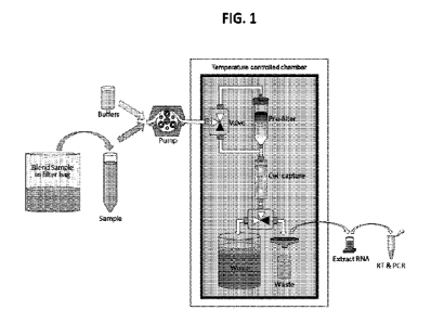
HG. 1 is a schematic diagram of sample processing steps in accordance with an
exemplary embodiment.
HG. 2 is a graph illustrating improved sensitivity of an exemplary embodiment
of the
disclosed probe-based assay when compared to commercial kit widely used by the
food industry.
The performance of the disclosed probe-based assay was compared with a
commercial kit,
MicrosEQTm Listeria tnonocytogenes Detection kit. A cell lysate oft.
tnonocytogenes and 10
fold dilution of the lysate was split and were tested with the probe-based
assay and the
MicroSEQTM assay. The disclosed probe-based assay was found to be 1000-times
more sensitive
than the commercial kit.
FIGS. 3 and 4 illustrate the specificity of an exemplary embodiment of a probe-
based
assay for target RNA detection in the presence of excess non-target RNA. The
specificity of the
probe-based assay for detecting 100 fg of Listeria RNA was tested using 20 ng
of non-target
RNA, which was equivalent to approximately 2 million non-target cells and 4
billion copies of
non-target RNA sequences. The non-target RNA was extracted from environmental
gram-
positive bacterial strains, Bacillus cereus and Bacillus subtilis subsp.
spizenzeii, which are
closely related to L. monocytogenes as well as gram-negative bacterial
strains, Citrobacter
- 9 -
CA 03145498 2022-1-24
WO 2021/016374
PCT/US2020/043112
freundii, Enterobacter cloacae, and Pseudomonas syringae. No amplification (no
Ct-value
recorded) was detected in all of the non-target strains tested when using the
probe-based assay.
FIG. 5 is a graph illustrating co-incubation of purified target nucleic acid
from L.
monocytogenes vs excess non-target nucleic acid from B. cereus. Assay
sensitivity was not
affected in the presence of excess non target. To further validate the
specificity of the probe-
based assay by co-incubating low amounts of RNA from Listeria monocytogenes in
the presence
of various amounts of Bacillus cereus strain ATCC 14579. Results indicated
that no significant
differences in the Ct-values were observed. To examine the effect of various
amounts of Bacillus
RNA on the efficiency of the amplification of the Listeria sequences, the
slope of the curve at the
pre-inflection point was examined. Analysis of the amplification curve
resulted in no significant
change in the slope of the curve under the various conditions tested,
demonstrating that the
efficiency of the amplification may not be adversely affected by addition of
the non-target
template.
HG. 6 is a graph illustrating sensitivity analysis with a disclosed platform.
Assay was
sensitive with close to 100% efficiency. Sensitivity was tested with reactions
containing over an
estimated 1,000,000 cells of L. grayi determined by measuring optical density
and 16-fold serial
dilutions of the samples to less than an estimated infectious dose of 1000
cells. The Ct-values of
the serial dilutions were about 4 cycles apart indicating close to 100%
efficiency.
HG. 7 is a graph illustrating swab sample enumeration results (blind study).
Ten
environmental samples selected to be as representative and challenging as
possible were obtained
collected from fresh produce facilities using stick-mounted sponges such as
the 3M Sponge-
Stick, typically pre-wetted with 10 mL of neutralizing buffer. After
collection, 90 mL of buffer is
added to a filter bag and the sponge is processed in a paddle blender, such as
the Seward
Stomacher, for 2 minutes to release the pathogen cells into the buffer_ Five
of the environmental
samples were spiked with Listeria grayi in a blinded fashion, and 1 rnL
aliquots were withdrawn
from each sample tube and plate enumerated. As shown by the enumeration
results in the figure
the Listeria grayi concentration were detected in the samples ranged from 3.5
CPU/mL and up.
The disclosed DNA-based platform demonstrated the ability to process "real-
world"
- 10 -
CA 03145498 2022-1-24
WO 2021/016374
PCT/US2020/043112
environmental sponge samples and to accurately identify those samples spiked
with Listeria and
those which were unspiked.
FIG. 8 is a block diagram of a system in accordance with an exemplary
embodiment of
the disclosure.
HG. 9 is a block diagram of a system in accordance with an exemplary
embodiment of
the disclosure.
FIG. 10 depicts an example cloud computing environment 1000 in which the
described
technologies can be implemented.
HG. 11 illustrates a generalized example of a suitable computing system 1100
in which
described examples, techniques, and technologies, including construction,
deployment,
operation, and maintenance of a disclosed system.
DETAILED DESCRIPTION OF SEVERAL EMBODIMENTS
L Terms
The following explanations of terms and methods are provided to better
describe the
present disclosure and to guide those of ordinary skill in the art in the
practice of the present
disclosure. Various operations may be described as multiple discrete
operations in turn, in a
manner that may be helpful in understanding embodiments; however, the order of
description
should not be construed to imply that these operations are order dependent.
The description may use perspective-based descriptions such as up/down,
back/front, and
top/bottom. Such descriptions are merely used to facilitate the discussion and
are not intended to
restrict the scope of the disclosure.
The terms "coupled" and "connected," along with their derivatives, may be
used. These
terms are not intended as synonyms for each other. Rather, aspects,
"connected" may be used to
indicate that two or more elements are in direct physical or electrical
contact with each other.
"Coupled" may mean that two or more elements are in direct physical or
electrical contact.
However, "coupled" may also mean that two or more elements are not in direct
contact with each
other, but still cooperate or interact with each other.
For the purposes of the description, a phrase in the form "A/B" or in the form
"A and/or
B" means (A), (B), or (A and B). For the purposes of the description, a phrase
in the form "at
least one of A, B, and C" means (A), (B), (C), (A and B), (A and C), (B and
C), or (A, B and C).
- 11 -
CA 03145498 2022-1-24
WO 2021/016374
PCT/US2020/043112
For the purposes of the description, a phrase in the form "(A)B" means (B) or
(AB) that is, A is
an optional element.
The singular terms "a," "an," and "the" include plural referents unless
context clearly
indicates otherwise. Similarly, the word "or" is intended to include "and"
unless the context
clearly indicates otherwise. The term "comprises" means "includes." Thus,
"comprising A or B,"
means "including A, B, or A and B," without excluding additional elements.
The description may use the terms "embodiment" or "embodiments," which may
each
refer to one or more of the same or different embodiments. Furthermore, the
terms "comprising,"
"including," "having," and the like, as used with respect to embodiments, are
synonymous, and
are generally intended as "open" terms (e.g., the term "including" should be
interpreted as
"including but not limited to," the term "having" should be interpreted as
"having at least," the
term "includes" should be interpreted as "includes but is not limited to,"
etc.).
With respect to the use of any plural and/or singular terms herein, those
having skill in
the art can translate from the plural to the singular and/or from the singular
to the plural as is
appropriate to the context and/or application. The various singular/plural
permutations may be
expressly set forth herein for sake of clarity.
Unless otherwise noted, technical terms are used according to conventional
usage.
Definitions of common terms in molecular biology can be found in Benjamin
Lewin, Genes IX,
published by Jones and Bartlet, 2008 (ISBN 0763752223); Kendrew et at (eds.),
The
Encyclopedia of Molecular Biology, published by Blackwell Science Ltd., 1994
(ISBN
0632021829); and Robert A. Meyers (ed.), Molecular Biology and Biotechnology:
a
Comprehensive Desk Reference, published by VCH Publishers, Inc., 1995 (ISBN
9780471185710); and other similar references.
Suitable methods and materials for the practice or testing of this disclosure
are described
below. Such methods and materials are illustrative only and are not intended
to be limiting.
Other methods and materials similar or equivalent to those described herein
can be used. In
addition, the materials, methods, and examples are illustrative only and not
intended to be
limiting.
All publications, patent applications, patents, and other references mentioned
herein are
incorporated by reference in their entirety. In case of conflict, the present
specification, including
- 12 -
CA 03145498 2022-1-24
WO 2021/016374
PCT/US2020/043112
explanations of terms, will control. In addition, the materials, methods, and
examples are
illustrative only and not intended to be limiting.
In order to facilitate review of the various embodiments of this disclosure,
the following
explanations of specific terms are provided:
3' end: The end of a nucleic acid molecule that does not have a nucleotide
bound to it 31
of the terminal residue.
5' end: The end of a nucleic acid sequence where the 5 position of the
terminal residue
is not bound by a nucleotide.
Agent: Any protein, nucleic acid molecule (including chemically modified
nucleic
acids), compound, antibody, small molecule, organic compound, inorganic
compound, or other
molecule of interest. Agent can include a therapeutic agent, a diagnostic
agent or a
pharmaceutical agent.
Amplification: A technique that increases the number of copies of a nucleic
acid
molecule (such as an RNA or DNA). An example of amplification is polymerase
chain reaction
(PCR), in which a sample is contacted with a pair of oligonucleotide primers
under conditions
that allow for the hybridization of the primers to a nucleic acid template in
the sample. The
primers are extended under suitable conditions (e.g., in the presence of a
polymerase enzyme and
dNTPs), dissociated from the template, re-annealed, extended, and dissociated
to amplify the
number of copies of the nucleic acid. The product of amplification can be
characterized or
quantified by electrophoresis, restriction endonuclease cleavage patterns,
oligonucleotide
hybridization or ligation, and/or nucleic acid sequencing using standard
techniques. For example,
the quantity and efficiency of the reaction can be determined using labels (as
defined below),
such as fluorescent reporters (including quenching reporters) in the
amplification process. It can
also be done without labels using methods such as UV spectroscopy. Various
commercial
products are also available, such as Qubit from ThermoFisher Scientific.
Other examples of amplification include quantitative real-time polymerase
chain reaction
(qPCR), strand displacement amplification, as disclosed in U.S. Patent No.
5,744,311;
transcription-free isothermal amplification, as disclosed in U.S. Patent No.
6,033,881; repair
chain reaction amplification, as disclosed in PCT publication WO 90/01069;
ligase chain
- 13 -
CA 03145498 2022-1-24
WO 2021/016374
PCT/US2020/043112
reaction amplification, as disclosed in European patent publication EP-A-
320,308; gap filling
ligase chain reaction amplification, as disclosed in U.S. Pat. No. 5,427,930;
and NASBA RNA
transcription-free amplification, as disclosed in U.S. Pat. No. 6,025,134.
Several embodiments
include multiplex qPCR assays, which are useful for amplifying and detecting
multiple nucleic
acid sequences in a single reaction.
Antibody: A polypeptide including at least a light chain or heavy chain
immunoglobulin
variable region which specifically recognizes and binds an epitope of an
antigen, such as a SCLC
associated molecule or a fragment thereof. Antibodies are composed of a heavy
and a light chain,
each of which has a variable region, termed the variable heavy (VLF) region
and the variable light
(VL) region. Together, the Vu region and the VL region are responsible for
binding the antigen
recognized by the antibody. Antibodies of the present disclosure include those
that are specific
for a disclosed SCLC-associated molecule.
The term antibody includes intact immunoglobulins, as well the variants and
portions
thereof, such as Fab' fragments, F(ab)12 fragments, single chain Fv proteins
("scFv"), and
disulfide stabilized Fv proteins ("dsFv"). A scFv protein is a fusion protein
in which a light chain
variable region of an immunoglobulin and a heavy chain variable region of an
immunoglobulin
are bound by a linker, while in dsFvs, the chains have been mutated to
introduce a disulfide bond
to stabilize the association of the chains. The term also includes genetically
engineered forms
such as chimeric antibodies (for example, humanized murine antibodies),
heteroconjugate
antibodies (such as, bispecific antibodies). See also, Pierce Catalog and
Handbook, 1994-1995
(Pierce Chemical Co., Rockford, IL); Kuby, J., Immunology, 3r1 Ed., W.H.
Freeman & Co., New
York, 1997.
Typically, a naturally occurring immunoglobulin has heavy (H) chains and light
(L)
chains interconnected by disulfide bonds. There are two types of light chain,
lambda (A) and
kappa (k). There are five main heavy chain classes (or isotypes) which
determine the functional
activity of an antibody molecule: IgM, IgD, IgG, 1/4A and 1/4E.
Each heavy and light chain contains a constant region and a variable region,
(the regions
are also known as "domains"). In combination, the heavy and the light chain
variable regions
specifically bind the antigen. Light and heavy chain variable regions contain
a "framework"
region interrupted by three hypervariable regions, also called
"complementarity-determining
regions" or "CDRs". The extent of the framework region and CDRs have been
defined (see,
- 14 -
CA 03145498 2022-1-24
WO 2021/016374
PCT/US2020/043112
Kabat et al., Sequences of Proteins of Immunological Interest, US. Department
of Health and
Human Services, 1991). The Kabat database is now maintained online. The
sequences of the
framework regions of different light or heavy chains are relatively conserved
within a species.
The framework region of an antibody, that is the combined framework regions of
the constituent
light and heavy chains, serves to position and align the CDRs in three-
dimensional space.
The CDRs are primarily responsible for binding to an epitope of an antigen.
The CDRs of
each chain are typically referred to as CDR1, CDR2, and CDR3, numbered
sequentially starting
from the N-terminus, and are also typically identified by the chain in which
the particular CDR is
located. Thus, a Vil CDR3 is located in the variable domain of the heavy chain
of the antibody in
which it is found, whereas a VL CDR1 is the CDR1 from the variable domain of
the light chain
of the antibody in which it is found. An antibody that binds RET will have a
specific VH region
and the VL region sequence, and thus specific CDR sequences. Antibodies with
different
specificities (such as different combining sites for different antigens) have
different CDRs.
Although it is the CDRs that vary from antibody to antibody, only a limited
number of amino
acid positions within the CDRs are directly involved in antigen binding. These
positions within
the CDRs are called specificity determining residues (SDRs).
References to "Vii" or "VH" refer to the variable region of an immunoglobulin
heavy
chain, including that of an Fv, scFv, dsFAT or Fab. References to "VC' or "VL"
refer to the
variable region of an immunoglobulin light chain, including that of an Fv,
scFv, dsFy or Fab.
A "monoclonal antibody" is an antibody produced by a single clone of B-
lymphocytes or
by a cell into which the light and heavy chain genes of a single antibody have
been transfected.
Monoclonal antibodies are produced by methods known to those of skill in the
art, for instance
by making hybrid antibody-forming cells from a fusion of myeloma cells with
immune spleen
cells. Monoclonal antibodies include humanized monoclonal antibodies.
A "polyclonal antibody" is an antibody that is derived from different B-cell
lines.
Polyclonal antibodies are a mixture of immunoglobulin molecules secreted
against a specific
antigen, each recognizing a different epitope. These antibodies are produced
by methods known
to those of skill in the art, for instance, by injection of an antigen into a
suitable mammal (such
as a mouse, rabbit or goat) that induces the B-lymphocytes to produce IgG
itnmunoglobulins
specific for the antigen, which are then purified from the mammal's serum.
- 15 -
CA 03145498 2022-1-24
WO 2021/016374
PCT/US2020/043112
A "chimeric antibody" has framework residues from one species, such as human,
and
CDRs (which generally confer antigen binding) from another species, such as a
murine antibody
that specifically binds a SCLC-associated molecule.
A "humanized" immunoglobulin is an immunoglobulin including a human framework
region and one or more CDRs from a non-human (for example a mouse, rat, or
synthetic)
immunoglobulin. The non-human immunoglobulin providing the CDRs is termed a
"donor," and
the human immunoglobulin providing the framework is termed an "acceptor." In
one example,
all the CDRs are from the donor immunoglobulin in a humanized immunoglobulin.
Constant
regions need not be present, but if they are, they are ly identical to human
immunoglobulin
constant regions, e.g., at least about 85-90%, such as about 95% or more
identical. Hence, all
parts of a humanized immunoglobulin, except possibly the CDRs, are
substantially identical to
corresponding parts of natural human immunoglobulin sequences. Humanized
immunoglobulins
can be constructed by means of genetic engineering (see for example, U.S.
Patent No.
5,585,089).
An "autoantibody" is an antibody produced by the immune system that is
directed against
one or more of the individual's own proteins.
Alteration or modulation in expression: An alteration in expression of a gene,
gene
product or modulator thereof. This phrase refers to a change or difference,
such as an increase or
decrease, in the level of the gene, gene product, or modulators thereof that
is detectable in a
biological sample relative to a control or a reference value known to be
indicative of the level of
the gene, gene product or modulator thereof in the absence of the pathogen. An
"alteration" in
expression includes an increase in expression (up-regulation) or a decrease in
expression (down-
regulation).
Aptamer: The term "aptamer", as referred to herein, should be understood to
include
synthetic antibodies, peptide aptamers, and nucleic acid aptamers, at least a
portion of which is
able to bind to another molecule. Nucleic acid aptamers are generally single-
stranded nucleic
acid molecules with complex secondary or tertiary structures (which as
discussed later may
include double-stranded portions or regions) that can specifically bind a
target molecule with
high affinity. The aptamers contemplated for use herein can be any suitable
nucleic acid or
equivalent thereof In this regard, the aptamers can include, for example, DNA,
RNA, a nucleic
acid analogue (XNA) such as Peptide Nucleic Acid (PNA) or Locked Nucleic Acid
(LNA),
- 16 -
CA 03145498 2022-1-24
WO 2021/016374
PCT/US2020/043112
glycol nucleic acid (GNA) or threose nucleic acid (TNA) or DNA or RNA
comprising one or
more modified nucleotides, and the like. Methods of developing and using
aptamers are known to
those of skill in the art, for example, see Reverdatto et al., Current Topics
in Medicinal
Chemistry, Vol. 15, Issue 12, 2015, which is hereby incorporated by reference
in its entirety.
"Modified" nucleotides include, for example, nucleotides having chemical
modifications to any
of the phosphate backbone, sugar moiety or base moiety of the nucleotide,
tritylated bases and
unusual bases such as inosine. The use of modified nucleotides can also affect
the binding
characteristics of the aptamer to the nuclease, for example as described in
Latham et al. (Nucl
Acids Res 22(14): 2817-2822, 1994 which is hereby incorporated by reference in
its entirety).
In some specific embodiments, RNA aptamers are used. Nucleic acid aptamers can
be
modified, for example to increase stability, in a number of ways including,
for example: (i)
Synthesis of aptamers using L-nucleotides (the mirror image of natural
nucleotides) so that they
cannot be degraded by naturally occurring nucleases; (ii) Incorporation of
locked nucleic acid
(LNA) and/or peptide nucleic acid (PNA) residues into the aptamer. LNAs and
PNAs also
increase stability of nucleic acid duplexes; (iii) Other chemical
modifications of ribonucleotides,
such as 2T-amino- and 2'-fluoro-pyrimidine nucleotides or 2T-0-methyl
nucleotides; and/or (iv)
Capping at the 3* end with a deoxythymidine to increase resistance to
exonuclease
degradation. Nucleic acid aptamers can be produced using methods disclosed
herein and known
in the art. For example, in-vitro selection methods (e.g., see Ellington and
Szostak, Nature
346(6287): 818-22, 1990, which is hereby incorporated by reference in its
entirety) and SELEX
methods (e.g., see Tuerk and Gold, Science 249(4968): 505-510, 1990 which is
hereby
incorporated by reference in its entirety) can be used. Further details
relating to the production
and selection of aptamers may also be found in the review of Osborne and
Ellington ((Them Rev
97(2): 349-370, 1997, which is hereby incorporated by reference in its
entirety). In some
embodiments, aptamers include a linker (see, for example,
idtdna.com/site/Catalog/Modifications/Category/2 as provided on July 23, 2019
which are hereby
incorporated by reference in their entireties) to facilitate attachment to a
solid surface and may
contain one or more spacers.
Bacterial pathogen: A bacteria that causes infection or disease (pathogenic
bacteria).
Examples of pathogenic bacteria include without limitation any one or more of
(or any
combination of) Acinetobacter baumanii, Actinobacillus sp., Actinomycetes,
Actinomyces
- 17 -
CA 03145498 2022-1-24
WO 2021/016374
PCT/US2020/043112
sp_ (such as Actinomyces israeliiand Actitzomyces naeslundii), Aerotnonas sp_
(such
as Aeromonas hylrophila, Aeromonas veronii biovar sobria (Aeromonas sobria),
and Aeromonas
caviae), Anaplasma phagocytophilum, Anaplasma tnarginal,e Alcaligenes
xylosoxidans,
Acinetobacter baumanii, Actinobacillus actinomycetemcomitans, Bacillus sp.
(such as Bacillus
anthracis, Bacillus cereus, Bacillus subtilis, Bacillus thuringiensis, and
Bacillus
stearothermophilus), Bacteroides sp.(such as Bacteroides fragilis), Bartonella
sp. (such
as Bartonella bacilliformis and Bartonella henselae, Bifidobactetiutn sp.,
Bordetella sp. (such
as Bordetella pertussis, Bordetella parapertussis, and Bordetella
bronchiseptica), Borrelia
sp. (such as Borrelia recurrentis, and Borrelia burgdorferi), Bruce/la sp.
(such as Brucella
abortus, Bruce/la canis, Bruce/la melintensis and Bruce//a suis), Burkholderia
sp. (such
as Burkholderia pseudotnallei and Burkholderia cepacia), Campylobacter sp.
(such
as Campylobacter jejuni, Campylobacter coli, Campylobacter lariand
Campylobacter
fetus), Capnocytophaga sp., Cardiobacteriutn hominis, Chlamydia trachotnatis,
Chlamydophila
pneumoniae, Chlamydophila psittaci, Citrobacter sp. Coxiella burnetii,
Corynebacterium
sp_ (such as, Cotynebacterium diphtheriae, Colynebacterium
jeikeum and Cotynebacterium), Clostridium sp.(such as Clostridium petfringens,
Clostridium
difficile, Clostridium botulinum and Clostridium tetani), Eikenella corrodens,
Enterobacter
sp. (such as Enterobacter aero genes, Enterobacter agglomerans, Enterobacter
cloacae and Escherichia coli, including opportunistic Escherichia coli, such
as enterotoxigenic
E. coli, enteroinvasive E coli, enteropathogenic E. coli, enterohemorrhagic E
coli,
enteroaggregative E cColi and uropathogenic E. coli) Enterococcus sp. (such as
Enterococcus
faecalis and Enterococcus faecium) Ehrlichia sp. (such as Ehrlichia
chafeensiaand Ehrlichia
canis), Erysipelothrix rhusiopathiae, Eubacterium sp., Francisella tularensis,
Fusobacterium
nucleation, Gardnerella vagina/is, Gemella morbillorum, Haemophilus sp_ (such
as Haemophitus
influenzae, Haemophilus ducreyi, Haemophilus aegyptius, Haemophilus
parainfluenzae,
Haemophilus haemolyticus and Haemophilus parahaemolyticus, Helicobacter sp.
(such
as Helicobacter pylori, Helicobacter cinaedi and Helicobacter fennelliae),
Kingella kingii,
Klebsiella sp. (such as Klebsiella pneumoniae, Klebsiella granulottuttis and
Klebsiella
oxytoca), Lactobacillus sp., Listeria monocytogenes, Leptospira interrogans,
Legionella
pneumophila, Leptospira inwrrogans, Peptostreptococcus sp., Mannheimia
hetnolytica,
Moraxella catarrhalis, Morganella sp., Mobiluncus sp., Micrococcus sp_,
Mycobacterium
- 18 -
CA 03145498 2022-1-24
WO 2021/016374
PCT/US2020/043112
sp_ (such as Mycobacterium leprae, Mycobacterium tuberculosis, Mycobacterium
paratuberculosis, Mycobacterium intracellulare, Mycobacterium avium,
Mycobacterium
bovis, and Mycobacterium marinum), Mycoplasm sp. (such as Mycoplasma
pneumoniae,
Mycoplasma hominis, and Mycoplasma genitalium), Nocardia sp. (such as Nocardia
astero ides,
Nocardia cyriacigeorgica and Nocardia brasiliensis), Neisseria sp. (such as
Neisseria
gonorrhoeae and Neisseria meningitidis), Pasteurella multocida, Plesiontonas
shigelloides.
Prevotella sp., Porphyromonas sp_, Prevotella tnelaninogenica, Proteus sp_
(such as Proteus
vulgaris and Proteus mirabilis), Providencia sp. (such as Providencia
alcalifaciens, Providencia
rettgeri and Providencia stuartii), Pseudomonas aeruginosa, Propionibacterium
acnes,
Rhodococcus equi, Rickettsia sp. (such as Rickettsia rickettsii, Rickettsia
akari and Rickettsia
prowazekii, Orientia tsutsugamitshi (formerly: Rickettsia tsutsugamushi) and
Rickettsia
typhi), Rhodococcus sp., Serratia marcescens, Stenotrophomonas maltophilia,
Salmonella
sp. (such as Salmonella enterica, Salmonella typhi, Salmonella paratyphi,
Salmonella enteritidis,
Salmonella cholerasuis and Salmonella typhimurium), Serratia sp. (such as
Serratia
marcesans and Serratia liquifaciens), Shigella sp. (such as Shigella
dysentetiae, Shigella
flexneri, Shigella boydii and Shigella sonnei), Staphylococcus sp. (such as
Staphylococcus
aureus, Staphylococcus epidertnidis, Staphylococcus hemolyticus,
Staphylococcus
saprophyticus), Streptococcus sp. (such as Streptococcus pneumoniae (for
example chloramphenicol-resistant serotype 4 Streptococcus pneumoniae,
spectinomycin-
resistant serotype 6B Streptococcus pneumoniae, streptomycin-resistant
serotype 9V
Streptococcus pneumoniae, erythromycin-resistant serotype 14 Streptococcus
pneumoniae,
optoch in-resistant serotype 14 Streptococcus pneumoniae, rifarnpicin-
resistant serotype 18C
Streptococcus pneumoniae, tetracycline-resistant serotype 19F Streptococcus
pneumoniae,
penicillin-resistant serotope 19F Streptococcus pneumoniae, and trimethoprim-
resistant serotype
23F Streptococcus pneumoniae, chloramphenicol-resistant serotype 4
Streptococcus
pneumoniae, spectinomycin-resistant serotype 6B Streptococcus pneumoniae,
streptomycin-
resistant serotype 9V Streptococcus pneumoniae, optochin-resistant serotype 14
Streptococcus
pneumoniae, rifampicin-resistant serotype 18C Streptococcus pneumoniae,
penicillin-resistant
serotype 19F Streptococcus pneumoniae, or trimethoprim-resistant serotype 23F
Streptococcus
pneumoniae), Streptococcus agalactiae, Streptococcus mutans, Streptococcus
pyogenes, Group A
streptococci, Streptococcus pyogenes, Group B streptococci, Streptococcus
agalactiae, Group C
- 19 -
CA 03145498 2022-1-24
WO 2021/016374
PCT/US2020/043112
streptococci, Streptococcus anginosus, Streptococcus equismilis, Group D
streptococci,
Streptococcus bovis, Group F streptococci, and Streptococcus anginosus Group G
streptococci), Spirillum minus, Streptobacillus monitjformi, Treponema sp.
(such as Treponema
carateum, Treponema petenue, Treponema pallidum and Treponema endemicum,
Tropherytna
whippelii, Ureaplastna urealyticum, Veillonella sp., Vibrio sp. (such as
Vibrio cholerae, Vibrio
parahemolyticus, Vibrio vulniflcus, Vibrio parahaemolyticus, Vibrio
vulniflcus, Vibrio
alginolyticus, Vibrio mimicus, Vibrio holiisae, Vibrio fluvialis, Vibrio
metchnikovii, Vibrio
damsela and Vibrio fitrnisii), Yersinia sp. (such asYersinia enterocolitica,
Yersinia
pestis, and Yersinia pseudotuberculosis) and Xanthomonas maltophilia among
others.
Bead: An insoluble structure having volume and one or more surfaces. Some bead
surfaces can be modified to include a reactive functional group, including but
not limited to
alcohol, halide, aldehyde, epoxide, amine, azide, alkyne, tetrazine,
maleimide, ester, thiol,
disulfide, sulfonyl halides or esters such that a covalent bond may be formed.
Other bead
surfaces have non-fouling properties and may not be easily modified. Beads may
have regular or
irregular shapes.
Binding or stable binding: An association between two substances or molecules,
such
as the hybridization of one nucleic acid molecule to another (or itself), the
association of an
antibody with a peptide, or the association of a protein with another protein
or nucleic acid
molecule. An oligonucleotide molecule binds or stably binds to a target
nucleic acid molecule if
a sufficient amount of the oligonucleotide molecule forms base pairs or is
hybridized to its target
nucleic acid molecule, to permit detection of that binding. "Preferentially
binds" indicates that
one molecule binds to another with high affinity, and binds to heterologous
molecules at a low
affinity.
Binding can be detected by any procedure known to one skilled in the art, such
as by
physical or functional properties of the target complex. For example, binding
can be detected
functionally by determining whether binding has an observable effect upon a
biosynthetic
process such as expression of a gene, DNA replication, transcription,
translation, and the like.
Methods of detecting binding of an antibody to a protein can include known
methods of protein
detection, such as Western blotting.
Cartridge: A removable component used for various aspects of sample
processing.
Cartridges can be used for sample analysis. Examples include a single assay
cartridge for
- 20 -
CA 03145498 2022-1-24
WO 2021/016374
PCT/US2020/043112
Listeria spp_ and a multiplexed cartridge for Salmonella and E_ colt
Cartridges can be used for
system functions, including but not limited to sanitation, priming,
transportation, storage and
calibration.
Contacting: Placement in direct physical association, including both a solid
and liquid
form.
Detecting: Identifying the presence, absence or relative or absolute amount of
the object
to be detected.
Epitope: An antigenic determinant. These are particular chemical groups or
peptide
sequences on a molecule that are antigenic, such that they elicit a specific
immune response. An
aptamer can bind to an epitope on a surface, such as a cell wall, of a
particular pathogen or an
antibody can bind to a particular antigenic epitope, such as an epitope of on
the surface of a
pathogen.
Expression: The process by which the coded information of a gene is converted
into an
operational, non-operational, or structural part of a cell, such as the
synthesis of a protein. Gene
expression can be influenced by external signals. For instance, exposure of a
cell to a hormone
may stimulate expression of a hormone-induced gene. Different types of cells
can respond
differently to an identical signal. Expression of a gene also can be regulated
anywhere in the
pathway from DNA to RNA to protein. Regulation can include controls on
transcription,
translation, RNA transport and processing, degradation of intermediary
molecules such as
mRNA, or through activation, inactivation, compartmentalization or degradation
of specific
protein molecules after they are produced.
The expression of a nucleic acid molecule can be altered relative to a normal
(wild type)
nucleic acid molecule. Alterations in gene expression, such as differential
expression, include but
are not limited to: (1) overexpression; (2) underexpression; or (3)
suppression of expression.
Alternations in the expression of a nucleic acid molecule can be associated
with, and in fact
cause, a change in expression of the corresponding protein.
Protein expression can also be altered in some manner to be different from the
expression
of the protein in a normal (wild type) situation. This includes but is not
necessarily limited to: (1)
a mutation in the protein such that one or more of the amino acid residues is
different; (2) a short
deletion or addition of one or a few (such as no more than 10-20) amino acid
residues to the
sequence of the protein; (3) a longer deletion or addition of amino acid
residues (such as at least
- 21 -
CA 03145498 2022-1-24
WO 2021/016374
PCT/US2020/043112
20 residues), such that an entire protein domain or sub-domain is removed or
added; (4)
expression of an increased amount of the protein compared to a control or
standard amount; (5)
expression of a decreased amount of the protein compared to a control or
standard amount; (6)
alteration of the subcellular localization or targeting of the protein; (7)
alteration of the
temporally regulated expression of the protein (such that the protein is
expressed when it
normally would not be, or alternatively is not expressed when it normally
would be); (8)
alteration in stability of a protein through increased longevity in the time
that the protein remains
localized in a cell; and (9) alteration of the localized expression of the
protein (such that the
protein is not expressed where it would normally be expressed or is expressed
where it normally
would not be expressed), each compared to a control or standard. Controls or
standards for
comparison to a sample, for the determination of differential expression,
include samples
believed to be normal as well as laboratory values (e.g., range of values),
even though possibly
arbitrarily set, keeping in mind that such values can vary from laboratory to
laboratory.
Laboratory standards and values can be set based on a known or determined
population
value and can be supplied in the format of a graph or table that permits
comparison of measured,
experimentally determined values.
Fungal pathogen: A fungus that causes infection or disease. Examples of fungal
pathogens include without limitation Trichophyton rubrunt, T. mentagrophytes,
Epidermophyton
fluccosum, Microsporum canis, Pityrosporum orbiculare (Malassezia furfur),
Candida sp. (such
as Candida albicans), Aspergillus sp. (such as Aspergillus ,fimigatus,
Aspergillus
flavus and Aspergillus clavatus), Cryptococcus sp. (such as Cryptococcus
neoformans,
Cryptococcus gattii, Cryptococcus laurentii and Cryptococcus albidus),
Histoplastna sp. (such
as Histoplasma capsulatum), Pneunzocystis sp. (such as Pneumocystis
firovecii),
and Stachybottys (such as Stachybottys chartarum) among others.
Hybridization: Oligonucleotides and their analogs hybridize by hydrogen
bonding,
which includes Watson-Crick, Hoogsteen or reversed Hoogsteen hydrogen bonding,
between
complementary bases. Generally, nucleic acid consists of nitrogenous bases
that are either
pyrimidines (cytosine (C), uracil (U), and thymine (T)) or purines (adenine
(A) and guanine (G)).
These nitrogenous bases form hydrogen bonds between a pyrimidine and a purine,
and the
bonding of the pyrimidine to the purine is referred to as base pairing. More
specifically, A will
hydrogen bond to T or U, and G will bond to C. In RNA molecules, G also will
bond to U.
- 22 -
CA 03145498 2022-1-24
WO 2021/016374
PCT/US2020/043112
Complementary refers to the base pairing that occurs between two distinct
nucleic acid
sequences or two distinct regions of the same nucleic acid sequence.
Hybridization conditions resulting in particular degrees of stringency will
vary depending
upon the nature of the hybridization method of choice and the composition and
length of the
hybridizing nucleic acid sequences. Generally, the temperature of
hybridization and the ionic
strength (especially the Nat concentration) of the hybridization buffer will
determine the
stringency of hybridization. Calculations regarding hybridization conditions
required for
attaining particular degrees of stringency are discussed by Sambrook et at
(ed.), Molecular
Cloning: A Laboratory Manual, 2nd ed., vol. 1-3, Cold Spring Harbor Laboratory
Press, Cold
Spring Harbor, NY, 1989, chapters 9 and 11, herein incorporated by reference.
Isolated: An "isolated" biological component (such as a nucleic acid molecule,
protein,
or cell) has been substantially separated or purified away from other
biological components in
the cell of the organism, or the organism itself, in which the component
naturally occurs, such as
other chromosomal and extra-chromosomal DNA and RNA, proteins and cells.
Nucleic acid
molecules and proteins that have been "isolated" include nucleic acid
molecules and proteins
purified by standard purification methods. The term also embraces nucleic acid
molecules and
proteins prepared by recombinant expression in a host cell as well as
chemically synthesized
nucleic acid molecules and proteins. The term "isolated" or "isolating" also
includes separation
or purification of one of more biological components from inorganic and
organic material. The
terms "isolating" and "capturing" are used interchangeable herein.
Label or Detectable Moiety: A composition detectable by spectroscopic,
photochemical,
biochemical, immunochemical, electromagnetic, or chemical means. For example,
useful labels
include radiolabels such as 32P, 35S, or '24; heavy isotopes such as 15N or
'3C or heavy atoms
such as selenium or metals; fluorescent dyes; chromophores, electron-dense
reagents; enzymes
that generate a detectable signal (e.g., alkaline phosphatase or peroxidase,
as commonly used in
an ELISA); or spin labels. The label or detectable moiety has or generates a
measurable signal,
such as a radioactive, chromogenic, or fluorescent signal, that can be used to
quantify the amount
of bound detectable moiety in a sample. The detectable moiety can be
incorporated in or attached
to a molecule (such as a protein, for example, an antibody) either covalently,
or through ionic,
van der Waals or hydrogen bonds, e.g., or by incorporation of labeled
precursors. The label or
detectable moiety may be directly or indirectly detectable. Indirect detection
can involve the
- 23 -
CA 03145498 2022-1-24
WO 2021/016374
PCT/US2020/043112
binding of a second directly or indirectly detectable moiety to the detectable
moiety. For
example, the detectable moiety can be the ligand of a binding partner, such as
biotin, which is a
binding partner for streptavidin, which can be linked to a directly detectable
label. The binding
partner may itself be directly detectable, for example, an antibody may be
itself labeled with a
fluorescent molecule. The binding partner also may be indirectly detectable,
for example, it may
be bound by another moiety that comprises a label. Quantitation of the signal
is achieved by any
appropriate means, e.g., fluorescence detection, spectrophotometric detection
(e.g., absorption at
a particular wavelength), scintillation counting, mass spectrometry,
densitometry, or flow
cytometry. Methods for labeling and guidance in the choice of labels
appropriate for various
purposes are discussed for example in Sambrook etal. (Molecular Cloning: A
Laboratory
Manual, Cold Spring Harbor, New York, 1989) and Ausubel et a/. (In Current
Protocols in
Molecular Biology, John Wiley & Sons, New York, 1998).
Measure: To detect, quantify or qualify the amount (including molar amount),
concentration or mass of a physical entity or chemical composition either in
absolute terms in the
case of quantifying, or in terms relative to a comparable physical entity or
chemical composition.
Pathogen: Anything that can produce disease. In some examples, a pathogen is
an
infectious molecule. In some examples, the pathogen is a live pathogen, such
as one or more
disease producing organism, such as bacteria, fungi, protozoa and/or worms. In
some examples,
a pathogen is one or more of a virus, bacteria, fungus, protozoa, worm,
protein and/or peptide
that is present in a sample obtained from food, water, environment, soil,
plant, animal, insect, or
human.
Purified: The term "purified" does not require absolute purity; rather, it is
intended as a
relative term. Thus, for example, a purified protein preparation is one in
which the protein
referred to is more pure than the protein in its natural environment within a
cell. For example, a
preparation of a protein is purified such that the protein represents at least
50% of the total
protein content of the preparation. Similarly, a purified mRNA preparation is
one in which the
mRNA is more pure than in an environment including a complex mixture of
nucleic acid
molecules.
Real-Time PCR (qPCR): A method for detecting and measuring products generated
during each cycle of a PCR, which are proportionate to the amount of template
nucleic acid prior
to the start of PCR. The information obtained, such as an amplification curve,
can be used to
- 24 -
CA 03145498 2022-1-24
WO 2021/016374
PCT/US2020/043112
determine the presence of a target nucleic acid and/or quantitate the initial
amounts of a target
nucleic acid sequence. Exemplary procedures for qPCR can be found in
"Quantitation of
DNA/RNA Using Real-Time PCR Detection" published by Perkin Elmer Applied
Biosystems
(1999); PCR Protocols (Academic Press, New York, 1989); A-Z of Quantitative
PCR, Bustin
(ed.), International University Line, La Jolla, CA, 2004; and Quantitative
Real-Time PCR in
Applied Microbiology, Filion (Ed), Caister Academic Press, 2012.
In some examples, the amount of amplified target nucleic acid is detected
using a labeled
probe, such as a probe labeled with a fluorophore, for example a TAQMANC)
probe. In other
examples, the amount of amplified target nucleic acid is detected using a DNA
intercalating dye.
The increase in fluorescence emission is measured in real-time, during the
course of the qPCR.
This increase in fluorescence emission is directly related to the increase in
target nucleic acid
amplification. In some examples, the change in fluorescence (Delta Rn; dRn;
ARn) is calculated
using the equation dRn = Rn+-Rn-, with Rn+ being the fluorescence emission of
the product at
each time point and Rn- being the fluorescence emission of the baseline. The
dRn values are
plotted against cycle number, resulting in amplification plots for each
sample. The threshold
cycle (Ct) is the PCR cycle number at which the fluorescence emission (dRn)
exceeds a chosen
threshold, which is typically 10 times the standard deviation of the baseline
(this threshold level
can, however, be changed if desired).
The threshold cycle is when the system begins to detect the increase in the
signal
associated with an exponential growth of PCR product during the log-linear
phase. This phase
provides information about the reaction. The slope of the log-linear phase is
a reflection of the
amplification efficiency. The efficiency of the reaction can be calculated by
the following
equation: E = 10 Pc), for example. The efficiency of the PCR should be 90-100%
meaning
doubling of the amplicon at each cycle. This corresponds to a slope of -3.1 to
-3.6 in the Ct vs.
log-template amount standard curve.
Sample: An aqueous solution or suspension used in analysis or testing. In some
examples, a sample may contain food, water or environmental material. In some
examples, a
sample may contain biological material, such as urine, saliva, sputum, feces,
semen, and other
bodily fluids and tissues_ In some examples, the material may be preprocessed
such as by adding
an aqueous solution and paddle blending or by isolating, concentrating or
removing material
such as through centrifugation or filtering. A sample may contain matter
including, but not
- 25 -
CA 03145498 2022-1-24
WO 2021/016374
PCT/US2020/043112
limited to microorganisms (e.g., bacteria, viruses and fungi), insect, plant,
animal, and/or fecal
material.
Sensitivity and specificity: Statistical measurements of the performance of a
binary
classification test. Sensitivity measures the proportion of actual positives
which are correctly
identified (e.g., the percentage of samples that are identified as including
nucleic acid from a
particular pathogen). Specificity measures the proportion of negatives which
are correctly
identified (e.g., the percentage of samples that are identified as not
including nucleic acid from a
particular pathogen).
Solution: A homogeneous mixture of one or more solutes dissolved in a solvent.
As used
herein, the terms buffer and solution are used interchangeably and the term
buffer does not
necessarily indicate that the solution has buffering capabilities.
Standard: A substance or solution of a substance of known amount, purity or
concentration. A standard can be compared (such as by spectrometric,
chromatographic, or
spectrophotometric analysis) to an unknown sample (of the same or similar
substance) to
determine the presence of the substance in the sample and/or determine the
amount, purity or
concentration of the unknown sample. In one embodiment, a standard is a
peptide standard. An
internal standard is a compound that is added in a known amount to a sample
prior to sample
preparation and/or analysis and serves as a reference for calculating the
concentrations of the
components of the sample. In one example, nucleic acid standards serve as
reference values for
expression levels of specific nucleic acids. In some examples, peptide
standards serve as
reference values for expression levels of specific peptides. Isotopically-
labeled peptides are
particularly useful as internal standards for peptide analysis since the
chemical properties of the
labeled peptide standards are almost identical to their non-labeled
counterparts. Thus, during
chemical sample preparation steps (such as chromatography, for example, HPLC)
any loss of the
non-labeled peptides is reflected in a similar loss of the labeled peptides.
Virus: A microscopic infectious organism that reproduces inside living cells.
A virus
consists essentially of a core of nucleic acid surrounded by a protein coat,
and has the ability to
replicate only inside a living cell. "Viral replication" is the production of
additional virus by the
occurrence of at least one viral life cycle. A virus may subvert the host
cells' normal functions,
causing the cell to behave in a manner determined by the virus. For example, a
viral infection
may result in a cell producing a cytokine, or responding to a cytolcine, when
the uninfected cell
- 26 -
CA 03145498 2022-1-24
WO 2021/016374
PCT/US2020/043112
does not normally do so. In some examples, a virus is a pathogen. Specific
examples of viral
pathogens include, without limitation; Arenaviruses (such as Guanarito virus,
Lassa virus, Junin
virus, Machupo virus and Sabia), Arteriviruses, Roniviruses, Astroviruses,
Bunyaviruses (such
as Crimean-Congo hemorrhagic fever virus and Hantavirus), Barnaviruses,
Birnaviruses,
Bornaviruses (such as Boma disease virus), Bromoviruses, Caliciviruses,
Chrysoviruses,
Coronaviruses (such as Coronavirus and SARS), Cystoviruses, Closteroviruses,
Comoviruses,
Dicistroviruses, Flaviruses (such as Yellow fever virus, West Nile virus,
Hepatitis C virus, and
Dengue fever virus), Filoviruses (such as Ebola virus and Marburg virus),
Flexiviruses,
Hepeviruses (such as Hepatitis E virus), human adenoviruses (such as human
adenovirus A-F),
human astroviruses, human BK polyomaviruses, human bocaviruses, human
coronavirus (such
as a human coronavirus HKU1, NL63, and 0C43), human enteroviruses (such as
human
enterovirus A-D), human erythrovirus V9, human foamy viruses, human
herpesviruses (such as
human herpesvirus 1 (herpes simplex virus type 1), human herpesvirus 2 (herpes
simplex virus
type 2), human herpesvirus 3 (Varicella zoster virus), human herpesvirus 4
type 1 (Epstein-Barr
virus type 1), human herpesvirus 4 type 2 (Epstein-Barr virus type 2), human
herpesvirus 5 strain
AD169, human herpesvirus 5 strain Merlin Strain, human herpesvirus 6A, human
herpesvirus
6B, human herpesvirus 7, human herpesvirus 8 type M, human herpesvirus 8 type
P and Human
Cyotmegalovirus), human immunodeficiency viruses (HIV) (such as HIV 1 and HIV
2), human
metapneumoviruses, human papillomaviruses (such as human papillomavirus-1,
human
papillomavirus-18, human papillomavirus-2, human papillomavirus-54, human
papillomavirus-
61, human papillomavirus-cand90, human papillomavirus RTRX7, human
papillomavirus type
10, human papillomavirus type 101, human papillomavirus type 103, human
papillomavirus type
107, human papillomavirus type 16, human papillomavirus type 24, human
papillomavirus type
26, human papillomavirus type 32, human papillomavirus type 34, human
papillomavirus type 4,
human papillomavirus type 41, human papillomavirus type 48, human
papillomavirus type 49,
human papillomavirus type 5, human papillomavirus type 50, human
papillomavirus type 53,
human papillomavirus type 60, human papillomavirus type 63, human
papillomavirus type 66,
human papillomavirus type 7, human papillomavirus type 71, human
papillomavirus type 9,
human papillomavirus type 92, and human papillomavirus type 96), human
parainfluenza viruses
(such as human parainfluenza virus 1-3), human parechoviruses, human
parvoviruses (such as
human parvovirus 4 and human parvovirus B19), human respiratory syncytial
viruses, human
- 27 -
CA 03145498 2022-1-24
WO 2021/016374
PCT/US2020/043112
rhinoviruses (such as human rhinovirus A and human rhinovirus B), human
spumaretroviruses,
human T-lymphotropic viruses (such as human T-lymphotropic virus 1 and human T-
lymphotropic virus 2), Human polyoma viruses, Hypoviruses, Leviviruses,
Luteoviruses,
Lymphocytic choriomeningitis viruses (LCM), Marnaviruses, Namaviruses,
Nidovirales,
Nodaviruses, Orthomyxoviruses (such as Influenza viruses), Partitiviruses,
Paramyxoviruses
(such as Measles virus and Mumps virus), Picomaviruses (such as Poliovims, the
common cold
virus, and Hepatitis A virus), Potyviruses, Poxviruses (such as Variola and
Cowpox),
Sequiviruses, Reoviruses (such as Rotavirus), Rhabdoviruses (such as Rabies
virus),
Rhabdoviruses (such as Vesicular stomatitis virus, Tetraviruses, Togaviruses
(such as Rubella
virus and Ross River virus), Tombusviruses, Totiviruses, Tymoviruses,
Noroviruses, bovine
herpesviruses including Bovine Herpesvirus (BHV) and malignant catarrhal fever
virus (MCFV),
among others.
II. Introduction
The current "gold" standard practice in the food industry employs the
culturing of food
and environmental samples using enrichment broth designed to specifically grow
the target
bacteria, followed by cell isolation with one or more buffer exchanges, often
accomplished with
centrifugation, cell lysis and DNA or RNA extraction, reverse transcription
from RNA to cDNA
if RNA is to be utilized, purification to remove substances that may inhibit
or otherwise
negatively impact amplification, and analysis using PCR or isothermal
amplification to amplify
and detect pathogen DNA or cDNA. In the absence of the culturing process, the
specificity and
reliability of commercial, off-the-shelf nucleic acid amplification kits is
insufficient. Reliable
amplification involves identifying small DNA sequences that are unique to the
target organism.
The varied and immense biodiversity in food or environmental samples precludes
reliable
specific amplification. Samples can contain microorganisms, such as bacteria,
viruses and fungi,
as well as insect, plant, animal, and fecal material. Each of these can have
DNA sequences with
millions of bases. Many organisms are unknown as are the sequences for every
possible
genotype or serotype of every species. It is difficult if not impossible to
define a DNA sequence
that would only bind to a specific sequence in a target pathogen with the
knowledge that it would
not bind to a DNA sequence that could be present in food or environmental
samples. In addition,
the presence of compounds such as chlorophyll and humic acids, components of
which can
inhibit nucleic acid amplification and confound interpretation of results (due
to baseline drift, for
- 28 -
CA 03145498 2022-1-24
WO 2021/016374
PCT/US2020/043112
example). Further, nucleic acid amplification works equally well on dead cells
as live cells as
long as the nucleic acid sequences are not degraded. Detection of only live
cells is required in the
food industry since "kill steps", such as sanitation, pasteurization, or
cooking can kill pathogens
without degrading the pathogen nucleic acid.
Culturing addresses the challenge of detecting relatively low pathogen cell
copy counts in
large biodiverse samples by increasing the population of the target pathogens
in the media.
Culturing significantly reduces the biodiversity in the sample since only
bacteria and fungi will
multiply in the process. Culturing also enables live cell analysis since only
live cells will
multiply during this process. It also effectively dilutes many nucleic acid
amplification inhibitors
however, other matrix-associated contaminants may remain, which are sources of
inhibitors for
downstream enzymatic applications. For example, humic acid, commonly found in
soils and
agricultural products, has proved challenging to eliminate, since it can
easily be co-extracted
along with the target DNA. Humic acid is implicated as a strong nucleic acid
amplification
inhibitor through sequence specific DNA binding.
Enrichment broth is designed to favor growth of target organisms over
competing non-
targeted organisms. Creating enrichment broths that significantly favor
specific target organisms
can be very challenging and costly. For example, enrichment broths designed
for Escherichia
coil 0157:H7 can range greatly in specificity and cost. The least specific
broths can lack the
specificity to prevent significant false positives while the most specific
broths can be deemed too
expensive for daily food analysis. In addition, it is often not possible or
practical to create a broth
that can enrich multiple different pathogens with sufficient specificity.
During the exponential
growth phase of the culturing process, the number of bacteria can double every
twenty minutes.
Depending on numerous factors, millions, billions or even trillions of cells
may be required for
reliable detection. As such, the culturing process can require up to a one-day
time period or
more, delaying analysis. Cells that have been stressed, such as by exposure to
chemicals, such as
chlorine, antimicrobials, or low temperature may take even longer to reach the
exponential
growth phase. It is generally not practical to increase the culture time for
all pathogen analysis
sufficiently to ensure reliable growth of all cells.
Food industry sample size and collection methods are the result of 40 years of
industry
experience and data analysis. It is highly desirable that pathogen analysis
utilize these established
sample volumes and collection methods. Environmental samples can be obtained
using stick-
- 29 -
CA 03145498 2022-1-24
WO 2021/016374
PCT/US2020/043112
mounted sponges such as the 3M Sponge-Stick, typically pre-wetted with 10 mL
of neutralizing
buffer. After collection, 90 rnL of buffer (typically, growth medium) is added
to a filter bag and
the sponge is processed in a paddle blender, such as the Seward Stomacher, for
2-3 minutes to
release the pathogen cells into the buffer. Compared to standard sample
collection and paddle
blending methods used by the food industry, the disclosed process utilizes a
different paddle
blending buffer composition. In comparison to the enrichment broth used as a
buffer for
traditional culture-based detection, the disclosed system utilizes buffered
saline which can be
less costly.
To reliably process environmental samples, the disclosed system ensures
sufficient filter
load capacity, consistently captures target cells, and removes substances that
can interfere or
inhibit nucleic acid amplification, such as humic acid and magnesium.
Bacteria cannot be effectively isolated from all food and environmental
samples using
filtration. These samples can contain a large number of organisms and
substances, such as clay
particles, that can be the same size as bacteria. The inventors have developed
a flow-through
assay that captures bacteria without the need for small particle filtering.
Capture time in an
aptamer-based or antibody-based cell capture is influenced by the distance
between the cells and
the capture molecule. Reducing the distance decreases the time for all cells
to be captured.
Capture time in a flow-through system is a function of the height of the flow
channel, the flow
rate, and the surface area of the capture surface along the flow path. The
disclosed surface
chemistry works on numerous surfaces including but not limited beads, slides
and other glass
surfaces, such as any surface containing soda-lime glass. Soda-lime glass is
one of the least
expensive and most commonly used glass types. Soda-lime glass is used for
commodity
beverage bottles, windowpanes and in street lane marker paint to make them
reflective. As such,
one can massively increase capture surface size without a material increase in
cost. The inventors
have demonstrated high binding efficiency in prototype flow channels flowing
at 10 milliliters or
15 milliliters per minute. They have shown the capability to increase the flow
rate without loss in
capture efficiency by simply increasing the diameter of the flow channel. By
extrapolating the
experimental results, it should be possible to efficiently capture cells with
flow rates in liters per
minute, tens of liters per minute or greater. This is of great benefit for
applications with large
sample volumes such as water, soil and food testing. The channel size can
alternatively be
reduced for applications where size and portability are desired, such as point-
of-cam diagnostics.
- 30 -
CA 03145498 2022-1-24
WO 2021/016374
PCT/US2020/043112
The disclosed system and method are believed to provide the ideal process for
rapid, low cost
capture of pathogen cells in food, environmental and other samples.
Culture-independent analysis of foodborne pathogens meeting all critical food
industry
requirements has never before been successfully accomplished prior to this
disclosure.
Foodbome pathogen analysis needs to detect as few as 1 pathogen cells per
milliliter of large,
biodiverse samples that can be challenging to process. Analysis should detect
only live cells in
order to verify the effectiveness of "kill steps" such as sanitation,
pasteurization, or cooking
(steps which typically do not materially alter cell RNA or DNA). It has been
assumed that
because bacterial transcripts are sensitive to degradation by intra- and extra-
cellular RNases,
mRNA levels should rapidly decline after death. Therefore, unlike DNA-based
detection, mRNA
would only be limited to the viable and active cells within the population. It
has been found that
RNA-based detection cannot be used to accurately enumerate living cells (see,
for example,
frontiersin.org/articles/10.3389/fmicb.2016.00223/full#B32, which is hereby
incorporated by
reference in its entirety). As such, a new method is needed to detect only
live cells.
The test process ideally is automated, usable by minimally trained personnel
and cost
effective. The disclosed system and method achieve all of these requirements
and was validated
in blind testing with 100% correct results (see Example Section).
The disclosed system and method also provide results that are quantitative;
key
information when identifying and isolating the source of a contamination. The
Food Safety
Modernization Act (FSMA) was signed into law in January 2011. Its aim is to
ensure the U.S.
food supply is safe by shifting focus from responding to foodborne
contamination to that of
preventing it. Rapid detection of pathogens can facilitate compliance with
FSMA by accelerating
identification and confirming the presence of pathogens. The disclosed system
and method
enable proactive prevention and process change, allowing for corrective action
prior to food
entering the supply chain.
It is believed that the disclosed system and method can significantly reduce
or eliminate
the food industry's test and hold cycles, enabling significant cost savings
through improved
operational efficiencies and reduced labor, transportation, and storage needs.
The public, through
retail and food service channels, can see safer product entering the supply
chain faster, with
greater shelf life, and the potential for better, more complete traceability.
The disclosed rapid
pathogen detection platform could help reduce the economic burden to food
processors and
-31 -
CA 03145498 2022-1-24
WO 2021/016374
PCT/US2020/043112
safeguard the public_ Reducing the reaction time to identify, qualify, and
eradicate foodborne
pathogen contaminations reduces the number of people impacted_ Thus, the
disclosed system and
method provide, for the first time, quantitative results to help food safety
professionals measure
the effectiveness of food safety processes. The technology also enables
testing in the field,
allowing for portable analysis in growing regions and for pathogen testing at
US ports and
borders.
III. System and Method for Detecting and Monitoring Pathogens
Disclosed herein is a system/platform, kit and method for rapid pathogen
detection,
quantitation and monitoring, including detecting, quantitating and monitoring
bacterial, fungal
and viral foiadbome pathogens. The disclosed system and method have the
ability to isolate only
live cells, not dead cells which is advantageous. In embodiments, the
disclosed system does not
isolate cells that have been killed from heat and exposure to certain
antimicrobial substances. For
example, in some embodiments, the disclosed system and method result in less
than 0.1%, such
less than 0.01%, less than 0.001% or less of cells that have been killed from
heat and exposure
being detected. In embodiments, the disclosed systems and methods capture,
release and process
sufficient live cells to enable detection from a typical sample size of 90
milliliters or larger
without requiring cell culture.
In some examples, the isolation conditions include reducing the concentration
of sodium
to preclude the capture of dead cells and adjusting the composition of the
solution and
conditions. In some examples, the isolation conditions include reducing the
temperature of
hybridization. In some examples, magnesium is introduced to stabilize the
aptamer formation
with reduced temperature. In some examples, the temperature is reduced to
stabilize the DNA
backbone of the aptamer structure_ In some examples, a temperature of less
than 20 degrees
Celsius is used as the lower temperatures is associated with decreased or no
dead cells binding.
In some examples, the magnesium is present at a concentration sufficient to
increase aptamer
melting temperatures to result in desired secondary structure formation while
in the presence of
less than 100 mIvI sodium and less than 20 degrees Celsius. In some examples,
the magnesium
concentration is more than 0.1 mM. In some examples, the isolation of live
cells is performed in
the presence of one or more of the following: (1) more than 0.1 mM of
magnesium, such as
between 0.1 mM and 100 m114, 0.1 m114 and 10 m114, 0.1 rnM and 5 mM, 0.2 mhil
and 1 m114,
including 0.1 mM, 0.2 mM, 0.3, mM, 0.4 mm, 0.5 mM, 0.6 mM, 0.7 mM, 0.8 mM, 0.9
mM, 1
- 32 -
CA 03145498 2022-1-24
WO 2021/016374
PCT/US2020/043112
tnM, 2 niNI, 3 nINI, 4 ntM, 5 mIVI, 6 tnNI, 7 ntiv1, 8 mtvl, 9 tnM or 10 InNI;
(2) in the presence of
less than 100 inN1 Na l" (sodium), such as between 100 mM and 1 mM, 100 EWA
and 10 mM,
including 100 mM, 90 mM, 80 mM, 70 mM, 60 mM, 50 mM, 40 m1v1, 30 mM, 20 mM, 10
mM,
mivi or 1 mM; and /or (3) at less than 20 degrees Celsius, such as between 20
and 0 degrees
5 Celsius, between 20 and 2 degrees Celsius, between 12 and 9 degrees,
between 11 and 9 degrees,
including 20 degrees Celsius, 19 degrees Celsius, 18 degrees Celsius, 17
degrees Celsius, 16
degrees Celsius, 15 degrees Celsius, 14 degrees Celsius, 13 degrees Celsius,
12 degrees Celsius,
11 degrees Celsius, 10 degrees Celsius, 9 degrees Celsius, 8 degrees Celsius,
7 degrees Celsius, 6
degrees Celsius, 5 degrees Celsius, 4 degrees Celsius, 3 degrees Celsius, 2
degrees Celsius, 1
degree Celsius. In some examples, the magnesium is present at a concentration
sufficient to
increase the aptamer melting temperatures to result in the desired secondary
structure formation
while in the presence of less than 100 mM sodium and less than 20 degrees
Celsius. In some
examples, the isolation conditions are performed in the presence of lx
phosphate buffer saline
(PBS) solution which contains approximately 150 naNI of sodium. In some
examples, the
isolation conditions are performed in the presence of 0.2x PBS solution which
contains
approximately 30 rn.N1 sodium. In some examples, the isolation conditions are
performed in the
presence of approximately 0.75x PBS solution which contains approximately 100
mM sodium.
In some examples, the disclosed method and system utilize a pathogen capture
column
containing of beads that are functionalized with aptamers with affinity to the
pathogen of interest
and through which sample is flowed to bind to the functionalized bead surface.
In some
examples, the beads are functionalized with more than one aptamer. In some
examples, the beads
are retained within the column by a filter, such as a stainless steel mesh,
having a pore size
smaller than the size of the beads. In some examples the container of beads
comprises a pipe of
uniform cross section. In some examples, cross-sectional area of the pathogen
capture column is
constant. In some examples, the cross-sectional area of the pathogen capture
column varies.
In some examples, capturing one or more live pathogens comprises applying the
sample to a
pathogen-isolation column containing multiple beads of one or more sizes. In
some examples,
the pathogen capture column is of a uniform shape, such as of a circle, oval
or polygon. In some
examples, the cross-section of the pathogen capture is a non-uniform shape.
The disclosed system and method are cell culture-independent and utilize a two-
stage
process which increases the specificity of detection. Specifically, the method
involves use of
- 33 -
CA 03145498 2022-1-24
WO 2021/016374
PCT/US2020/043112
pathogen isolation, such as pathogen cells, isolation and release as a first
step followed by
nucleic acid extraction and detection as a second step. In some examples, the
two-step process
includes pathogen isolation, such as pathogen cell isolation, accomplished via
aptamer-based or
antibody-antigen capture. In some examples, the two-step process includes cell
isolation
accomplished by antibody/antigen cell capture. In some examples, the two-step
process involves
the nucleic acid detection being performed by numerous methods including but
not limited to
polymerase chain reaction, isothermal amplification, hybridization detection,
and sequencing. In
some examples, the disclosed two-step process involves including sample, such
as cells, washing
in the pathogen isolation process to reduce/eliminate the need for
purification at the nucleic acid
level. In some examples, the two-step process includes multiple steps to
concentrate the
pathogen, such as pathogen cells and/or nucleic acids including pathogen
isolation on a surface,
such as on beads in a column, cell wash, cell release, cell capture on filter,
cell release. It is
contemplated that a surface can be on a bead, fiber, rods powders or other
material capable of
performing as a substrate. In some embodiments, the surfaces, such as bead
surfaces, are
primarily comprised of a material that has been modified for aptamer or
antibody attachment,
such as via reactive functional surface group or combination of functional
surface groups
(including, but not limited to alcohol, aldehyde, epoxide, amine,
functionalization via click
chemistry, cyclic diene, and/or covalent linkages). In some embodiments, non-
covalent
attachment is employed. It is contemplated that surface attachment of
functionalized molecules
can be established by methods known to one of skill in the art (see, for
example,
www.idtdna.com/pages/products/custom-dna-ma/oligo-modifications,
sfvideo.blob.core.windows.net/sitefinity/docs/default-source/technical-
report/attaching-oligos-to-
solid-supports.pdfisfvrsn=47483407_6 or www.gelest.com/wp-
content/uploads/Hydrophobicity-
Hydrophilicity_and_Silane_Surface_Modification.pdf, which are each
incorporated by reference
in each of the entirety as available on July 16, 2020). Insoluble substrates,
such as insoluble
beads, can be surface functionalized with chemical groups that react with cell
capture agents.
These functional groups include, but are not limited alcohol, halide, N-
hydroxy succinimide
(NHS) esters, isocyanates, isothiocyanates, benzoyl fluoride, iodoacetamide, 2-
thiopyridine,
diazonium salts, arylamine, phosphoesters, ketone, hydrazide, cycloalkyne,
trialkyl halosilane,
trimethoxy halosilanes, trialkoxy halosilanes aldehyde, epoxide, amine, azide,
alkyne, tetrazine,
maleirnide, ester, thiol, disulfide, sulfonyl halides or esters. In some
embodiments, aptamers,
- 34 -
CA 03145498 2022-1-24
WO 2021/016374
PCT/US2020/043112
antibodies or other cell capture agents are modified to include a reactive
functional group,
including but not limited to alcohol, halide, aldehyde, epoxide, amine, azide,
alkyne, tetrazine,
maleimide, ester, thiol, disulfide, sulfonyl halides or esters such that a
covalent bond is formed
that will not break during subsequent steps of the cell capture and
identification process.
Appropriate modification of the cell capture agent depends on the surface
modification, such as
bead surface modification. For example, an insoluble substrate, such as
insoluble bead,
functionalized with alcohol can react with cell capture agent modified to
include an ester or
epoxide. Similarly, an insoluble bead functionalized with an azide can react
with a cell capture
agent modified as an alkyne, or vice versa. In some embodiments, a
modification is made to add
or increase the hydroxyl groups in a polymer. In some examples, one or more
linkers, such as
thiol, biotin/streptavidin, or click chemistry is used.
In some examples, the disclosed two-step process includes a flow-through
process. In
some embodiments, releasing the one or more captured live pathogens from
aptamers or
antibodies is performed by using a flow rate equal to or higher than the flow
rate used to capture
the one or more pathogens. In some embodiments, the flow rate for releasing
the one or more
captured live pathogens is the same flow rate as for pathogen capture. In some
embodiments, the
flow rate for releasing the one or more captured live pathogens is at least
2X, such as about 2X,
3X, 4X, 5X, or 10X higher than the flow rate for capturing the one or more
pathogens. In some
embodiments, the flow rate for releasing the one or more captured live
pathogens is at least 10%
higher than the flow rate for capturing,
In some examples, the disclosed two-step process involves nucleic acid
detection,
wherein the nucleic acid detected is one or more of DNA or RNA. In some
examples, the
disclosed two-step process allows for one or more different cell types to be
isolated in which the
different cells can bind to an aptarner or antibody and where multiple
different aptamers or
antibodies are used to isolate different cells. In some examples, the
disclosed two-step process
can detect multiple nucleic acid sequences. For example, the nucleic acid
extraction can be
preceded by release of cells from aptamers or antibodies prior to nucleic acid
extraction. In some
examples, the disclosed system and method involve a culture-independent
process of isolate,
wash, release, extract, nucleic acid detection. It is contemplated that
conditions leading to release
of captured pathogen may depend on the pathogen (e.g., target) and the aptamer
or antibody
(e.g., probe). For example, conditions can be conducted at a relatively
neutral pH, such as
- 35 -
CA 03145498 2022-1-24
WO 2021/016374
PCT/US2020/043112
between 6.8 and 7.2 pH, resulting in reduced binding affinities conducive to
cell release at very
high or very low pHs. In embodiments, a release buffer includes a combination
of 1M NaCl,
100m1vI EDTA, 0.1% Tween 20,0.05% SDS, 30mM NaHCO3, pH 10.3-10.4. It is
contemplated
that known methods of aptamer or antibody release can be employed as well
(see, for example,
US Patent Publication US20120258870, Zhu et al., JET Nanobiotechnol. 2014 Mar;
8(1): 2-9;
Bellaperche and DeRosa, Pharmaceuticals 2018, 11, 80; doi:10.3390/ph11030080,
each of which
is hereby incorporated by reference in its entirety). For example, aptamer
release can be achieved
by manipulating various factors or combinations of factors including, but not
limited to
temperature (see Bellaperche and DeRosa, Pharmaceuticals 2018, 11, 80;
doi:10.3390/ph11030080 which is hereby incorporated by reference for such
teachings), solution
composition such as pH (see, Zhu et al., JET Nanobiotechnol. 2014 Mar; 8(1): 2-
9, which is
hereby incorporated by reference for such teachings), surfactants such as but
not limited to
Triton, polysorbate, and other substances such as, but not limited to
chelators such as but not
limited to EDTA. For example, aptamer design and selection (e.g., via Selex or
similar process)
is to be conducted at close to neutral pH, resulting in reduced binding
affinities conducive to cell
release at very high or very low pHs. In embodiments, cell release from
antibodies can be
accomplished through methods known to one of skill in the art as well as
through the use of
various agents and varying temperature, such as varying temperature (heat),
and/or solution
composition (e.g., EDTA; Asian J Transfus Sci. 2013 Jan-Jun; 7(1): 29-32.
doi: 10.4103/0973-6247.106727, which is hereby incorporated by reference).
Similarly, cell
release from various surfaces, including substrates disclosed herein can be
facilitated by the use
of surfactants and chelators, such as, but not limited to EDTA.
In some examples, the disclosed method and system utilize a pre-filter
comprised of a
column of beads to remove large particles prior to a cell isolation stage_ For
example, the beads
in the pre-filter contain similar or smaller size than the larger beads in the
cell isolation stage. In
some examples, the pit-filter and/or the cell isolation stage contains
multiple size beads. In some
examples, the beads in the pre-filter have non-fouling surface properties. In
some examples, the
pre-filter has a funnel-shaped component. In some examples, the pre-filter
also acts as a bubble
trap to prevent air from reaching the pathogen isolation container. In some
embodiments,
released cells may be collected in liquid in a vented bubble trap acting as a
reservoir and acting
to remove air before the cells in liquid are flowed through a filter to
collect cells. In some
- 36 -
CA 03145498 2022-1-24
WO 2021/016374
PCT/US2020/043112
examples, the filter permits concentration of cells and permits washing cells
with solution to
reduce or remove possible substances (from release solutions) that may or may
not interfere with
subsequent processes. The solution used to wash concentrated cells is
primarily water and may
or may not include surfactant, such as, but not limited to polysorbate 20
(e.g., 0.1% PS20).
In some embodiments, the bubble trap may include at least one inlet port, at
least one
outlet port, and a vent to air. In some embodiments, the vent is outside of
the inlet port. In some
embodiments, the bubble trap includes multiple ports, one of which is a vent
to air. The vent to
air is required and advantageous as it permits the bubble trap to eliminate
volumes of air in
excess of the volume capacity of the bubble trap from subsequent flow of cells
through a filter.
In embodiments, the vent may contain sensors (examples include liquid level
sensors and bubble
sensors) that permit feedback control of the volume of liquid pumped into the
bubble trap. Prior
systems with bubble traps (see, for example, US Patent No. 10,232, 369) are
inadequate for the
present system because of their limited capacity for air. These prior systems
had no means to
vent air directly from the bubble trap, therefore the functional air capacity
was limited by the
volume. The previous bubble trap also did not have any means to determine when
the bubble
trap was filled with liquid and/or nucleic acids including cell isolation on
beads in a column, cell
wash, cell release, cell capture on filter, cell release. The bubble trap in
the present system
overcomes these limitations of the previous systems by including a vent for
air and sensors
permitting feedback control.
Referring to FIG. 8, an exemplary system is shown which includes a first of a
sample
input chamber 8500, pre-filter 8402, cell capture filter 8403, lysis filter
8404, waste collection
8502 and PCR sample collection tube 8503. The system is inserted into an
instrument which
comprises a number of further fluidic components. Collectively, a fluidic
circuit is formed.
As illustrated, the disclosed system includes a selector valve 8202, such as a
3-way valve.
The sample input chamber 8500 is connected to pump 8102 on the instrument,
which is
connected to the common port of 8202 on the instrument. In operation, port
8202 is generally in
an open position and connected to the wash buffer reservoir 8300 on the
instrument and
sanitation buffer reservoir 8301 on the instrument and is used to select
between the sanitation
buffer reservoir 8301 and the wash buffer reservoir 8300.
In a disclosed system, an output of 8501 is connected to pump 8101 on the
instrument,
which is then connected to tube heat exchanger 8501, before entering pre-
filter 8402 on the
- 37 -
CA 03145498 2022-1-24
WO 2021/016374
PCT/US2020/043112
device. The output of the pre-filter is connected to common port of a pre-
filter bypass valve
8200. In operation, port 8202 is generally in a closed position and connected
to an inlet of 8401.
In operation, a port of pre-filter bypass valve 8200 is connected to the
normally open port of
valve 8201. The normally closed port of valve 8201 is connected to a
peristaltic pump 8103. The
inlet of 8103 is connected to the release buffer reservoir 8302. The common
port of valve 8201 is
connected to the cell capture filter 8403.
In an embodiment, the output of 8403 is connected to the common port of valve
8201
The normally open port of 8203 is connected waste reservoir 8502. The normally
closed port of
8203 is connected to the bottom of lysis filter 8404. The top of 8404 is
connected to the normally
open port of valve 8204. The normally closed port of 8204 is connected to
waste reservoir 8502.
The normally open port of 8204 is connected to syringe pump 104. The outlet of
8104 is also
connected to the common port of valve 8205. The normally open port of valve
8206 is connected
to lysis buffer reservoir 8303. The normally closed port of valve 8206 is open
to a filtered air
inlet 8504. The normally closed port of valve 8205 is connected to valve 8207.
The outlet of
8207 port is connected to the outlet of 8404. PCR sample collection tube 8503
is also connected
to the bottom of 8404.
In use, a disclosed system can be primed with buffers prior to use or as part
of the first
use. An exemplary method of detecting and monitoring includes pre-filter and
capture column
conditioning prior to loading a sample. Valves 8200, 8201, 8202 and 8203 are
unpowered. Pump
8102 is powered, drawing fluid from the wash buffer reservoir 8300 through
valve 8202 and into
the sample reservoir 8500. Pump 8102 continues to pump, drawing fluid from the
wash buffer
reservoir 8300 through valve 8202 and into the sample reservoir 8500. Pump
8101 continues to
pump, drawing fluid from the sample loading reservoir 8500, and into tubing
cooling heat
exchanger 8501, valve 8200, valve 8201, cell capture column 8403, valve 8203
and waste
reservoir 8502. Pump 8102 is stopped.
In an exemplary embodiment, the method includes loading the sample and
capturing the
cells. For example, a sample is loaded into sample loading chamber 8500.
Valves 8200, 8201,
8202 and 8203 are unpowered. Pump 8101 is powered, drawing fluid from the
sample loading
reservoir 8500, and into tubing cooling heat exchanger 8501, pre-filter 8402,
valve 8200, valve
8201, cell capture column 8403, valve 8203 and waste reservoir 8502. Large
particles are
trapped in the pre-filter. Target cells are captured in cell capture column
8403.
- 38 -
CA 03145498 2022-1-24
WO 2021/016374
PCT/US2020/043112
In an exemplary embodiment, the method includes flushing the remaining sample
from
the sample loading chamber. Valves 8200, 8201, 8202 and 8203 are unpowered.
Pump 8102 is
powered, drawing fluid from the wash buffer reservoir 8300 through valve 8202
and into the
sample reservoir 8500. Pump 8101 is powered, drawing fluid from the sample
loading reservoir
8500, and into tubing cooling heat exchanger 8501, pre-filter 8402, valve
8200, valve 8201, cell
capture column 8403, valve 8203 and waste reservoir 8502.
In an exemplary embodiment, the method includes washing captured cells to
remove
PCR inhibitors and air gap introduced to prevent buffers from mixing. In some
embodiments of
the method, valves 8201, 8202 and 8203 are unpowered. Valve 8200 is powered to
bypass the
pm-filter. Pump 8102 continues to pump, drawing fluid from the wash buffer
reservoir 8300
through valve 8202 and into the sample reservoir 8500. Pump 8101 continues to
pump, drawing
fluid from the sample loading reservoir 8500, and into tubing cooling heat
exchanger 8501, valve
8200, valve 8201, cell capture column 8403, valve 8203 and waste reservoir
8502. Pump 8102 is
stopped. Pump 8101 continues to pump, fluid in the sample loading reservoir
8500 is removed
and air is pulled through pump 8101, tube cooling heat exchanger 8501, valve
8200 and through
valve 8201 to create an air gap. Pump 8101 is stopped. The method includes
introducing an air
gap sufficient to (1) prevent or greatly reduce mixing of buffer components
(as release
performance deteriorates when buffers are mixed) and facilitate efficient
release from the capture
column. In some examples, an air gap less than 5 mL is created, such as
between 1 mL and 5
mL, between 2mL to 4 mL, 3 mL to 5 mL, lmL to 3 mL, including about 1 mL, 2
mL, 3 mL, 4
mL, and 5 mL. It is contemplated that the air gap can be scalable with the
dimensions (diameter
and/or length) of the capture column. For example, in some embodiments, tubing
diameter used
within the system and method is about 1/8 inch in diameter and the introduced
air gap is less than
5 mL, such as between 1 mL and 5 mL, between 2mL to 4 mL, 3 rnL to 5 mL, lmL
to 3 mL, 2
mL to 3 mL, including about 1 mL, 2 mL, 3 mL, 4 mL , and 5 mL. In some
embodiments, the air
gap is sufficient to yield an release efficiency of greater than 10%, greater
than 20%, greater than
30%, such as between 10% and 40%, 20% and 40%, 30% and 40%, 30% and 50% or
more.
In some examples, a single air gap is introduced to into the system. In some
examples,
more than one air gap is introduced into the system, such as several smaller
air gaps in
comparison to one larger air gap.
- 39 -
CA 03145498 2022-1-24
WO 2021/016374
PCT/US2020/043112
In an exemplary embodiment, the method includes capturing released cells and
trapping
in the lysis filter. Valves 8201, 8203 and 8204 are powered and pump 8103 is
started. Pump
8103 draws fluid from the release buffer reservoir 8302, through valve 8201,
capture column
8403, valve 8203, lysis filter 8404, valve 8204, and into the waste reservoir
8502. Pump 8103 is
stopped. Cells are trapped in lysis filter 8404.
In an exemplary embodiment, the method includes exchanging buffer with lysis
buffer
and then removing the buffer. Valve 8204 is powered and valves 8203 and 8205
are unpowered.
Syringe pump 8104 is withdrawn, pulling lysis buffer from the lysis buffer
reservoir 8303,
through valve 8206 and valve 8205 into syringe pump 8104. Valves 8204 and 8205
are powered,
valve 8207 is open, and valve 8203 is unpowered. Syringe pump 8104 is purged,
pushing lysis
buffer from the syringe pump 8104, through valves 8205 and 8207, through the
bottom of the
lysis filter, through valve 8204 and into waste reservoir 8502. Valve 8205 is
unpowered. Valves
8204 and 8206 are powered. Syringe pump 8104 is withdrawn, pulling air from
the air inlet
8504, through valve 8206 and valve 8205, and into the syringe pump 8104. Valve
8204, 8205 are
powered and valve 8207 is open. The syringe pump 8104 is purged, pushing air
from the syringe
pump 8104, through valves 8205 and 8207, through the bottom of the lysis
filer, through valve
8204 and into waste reservoir 8502.
In an exemplary embodiment, the method includes back flowing lysis buffer to
release
cells into removable PCR tube. Valve 8205 is unpowered. Valves 8204 and 8206
are powered.
Syringe pump 8104 is withdrawn, pulling air from the air inlet 8504, such as
approximately 950
AL of air from the air inlet 8504, through valve 8206 and valve 8205, and into
the syringe pump
8104. Valves 8204 is powered and valves 8203, 8205, 8206, and 8207 are
unpowered. Syringe
pump 8104 is withdrawn, pulling lysis buffer from the lysis buffer reservoir
8303, such as
pulling approximately 50 pL of lysis buffer from the lysis buffer reservoir
8303, through valve
8206 and 8205 into syringe pump 8104. Valves 8205 is powered and valves 8203,
8204 and
8207 are unpowered. The syringe pump 8104 is purged, pushing lysis buffer
followed by air
through valve 8204, through the top of the lysis filter 8404 and into the
removable PCR tube
8503.
In some embodiments, the method includes the optional sanitation process. For
the
optional sanitation process, a sanitation cartridge is inserted and valves
8200, 8202 are powered.
Pumps 8101 and 8102 are powered, drawing sanitation buffer from sanitation
reservoir 8301 into
- 40 -
CA 03145498 2022-1-24
WO 2021/016374
PCT/US2020/043112
and from the sample loading reservoir 8500, into tubing cooling heat exchanger
8501, pre-filter
8402, valve 8200, valve 8201, cell capture column 8403, valve 8203 and waste
reservoir 8502.
Referring to FIG. 9, an exemplary system is shown which includes a sample
input source,
which can be a temporary or permanent bag or reservoir, 9500, sample input bag
straw 9501,
pre-filter 9402, cell capture filter 9403, lysis filter 9404, waste collection
9503, bubble trap 9504
and PCR sample collection tube 9505. The system is inserted into an instrument
which can
include a number of further fluidic components. Collectively, a fluidic
circuit is formed.
As illustrated, the disclosed system includes a sample input bag straw 9501
inside sample
bag 9500, connected to pump 9102, which is connected to the input of the tube
heat exchanger
9502. Selector valve 9206, such as a 3-way valve, is used to select between
the wash solution
reservoir 9300 on the instrument and sanitation solution reservoir 9301 on the
instrument.
Selector valves that are in the open position when no power is applied are
generally in an open
position to conserve power and reduce heat. The common port of valve 9206 is
connected to
pump 9101 which is connected to the input of the tube heat exchanger 9502
along with pump
9102 after passing through liquid sensor 9702.
In a disclosed system, the output of tube heat exchanger 9502 is connected to
the input of
pm-filter 9402 and to one port of valve 9200. Valve 9200 is used to select
between the output of
tube heat exchanger 9502 and the output of pre-filter 9402 on the device.
Valve 9201 is used to
select between the common port of valve 9200 and the output of pump 9103. The
input of pump
9103 is connected to the common port of valve 9202. Valve 9202 selects between
filtered air
supply 9601 and the common port of valve 9204. Valve 9204 selects between cell
release
solution reservoir 9302 and wash solution reservoir 9303. The common port of
valve 9201 is
connected to the cell capture filter 9403.
In an embodiment, the output of 9403 is connected to the common port of valve
9201
The normally open port of 9203 is connected waste reservoir 9503. The
typically closed port of
9203 is connected to an input of bubble trap 9504. Lysis solution reservoir
9306 is connected to
pump 9105 and pump 9105 is connected to an input of bubble trap 9504. Filtered
air supply 9602
connects to an input of bubble trap 9504 after passing through liquid sensor
9701. The output of
bubble trap 9504 is connected to and regulated by valve 9208 which is
connected to the bottom
of lysis filter 9404. The top of lysis filter 9404 is connected to pump 9104.
Pump 9104 is
connected to the common port of valve 9205. Valve 9205 selects between lysis
solution reservoir
- 41 -
CA 03145498 2022-1-24
WO 2021/016374
PCT/US2020/043112
9304 and the common port of valve 9208. Valve 9208 selects between waste
reservoir 9305 and
filtered air supply 9603.
Prior to initial use, prior to system storage between uses, or after use, a
disclosed system
can be sanitized with solutions prior to subsequent use. An exemplary method
involves using a
sanitation cartridge, a sample bag and straw, and sanitation solution to allow
of sanitation of all
the connections among the cartridge components and sanitation of the bubble
trap without
adversely affecting an analysis cartridge. Valves 9200, 9201, 9203 and 9207
are unpowered.
Valve 9206 is powered. Pump 9101 and pump 9102 are powered, drawing fluid from
the
sanitation solution reservoir 9301 through valve 9206 and into the sample bag
or reservoir 9500
via straw 9501. Pump 9102 is stopped. Pump 9101 continues to pump, drawing
fluid from
sanitation solution reservoir 9301 through valve 9206, and into tubing cooling
heat exchanger
9502, into sanitation cartridge prefilter 9402, through valve 9200, valve
9201, cell capture
column 9403, valve 9203 and into waste reservoir 9503. Pump 9101 is stopped.
Valves 9200 and
9203 are powered (sanitizes prefilter bypass alternate path). Pump 9101
continues, moving
sanitation solution through sanitation cartridge alternate path through
normally closed valve
9200 port through normally closed Valve 9203 port to bubble trap 9207 until
sensor 9701 detects
that bubble trap is full. Pump 9101 is stopped. Valves 9203, 9206 and 9207 are
powered. Valve
9200 is unpowered.
Following prescribed contact times for sanitation solutions in the system, the
sanitation
solution is flushed to remove completely from the system using solutions that
do not possess
bactericidal or other lethal properties to cells and which do not pose
contamination risk to the
system. These solutions include wash buffer, lysis solution and water.
In an exemplary method, sanitation solution is removed from the bubble trap
using pump
9104. Wash buffer from reservoir 9300 is rinsed through the same flow paths to
remove
sanitizing solution, including twice completely filling the bubble trap 9504
and draining via
valve 9207 and sanitizing cartridge "lysis filter" path. An exemplary rinsing
method involves
using a sanitation cartridge, a sample bag and straw, and wash buffer to allow
rinsing of all the
connections among the cartridge components and rinsing of the bubble trap
without adversely
affecting an analysis cartridge. Valves 9200, 9201, 9203, 9206 and 9207 are
unpowered. Pump
9101 and pump 9102 are powered, drawing fluid from the wash buffer reservoir
9300 through
valve 9206 and into the sample bag or reservoir 9500 via straw 9501. Pump 9102
is stopped.
- 42 -
CA 03145498 2022-1-24
WO 2021/016374
PCT/US2020/043112
Pump 9101 continues to pump, drawing fluid from wash buffer reservoir 9300
through valve
9206, and into tubing cooling heat exchanger 9502, into sanitation cartridge
prefilter 9402,
through valve 9200, valve 9201, cell capture column 9403, valve 9203 and into
waste reservoir
9503. Pump 9101 is stopped. Valves 9200 and 9203 are powered (rinses prefilter
bypass
alternate path). Pump 9101 continues, moving wash buffer through sanitation
cartridge alternate
path through normally closed valve 9200 port through normally closed Valve
9203 port to
bubble trap 9207 until sensor 9701 detects that bubble trap is full. Pump 9101
is stopped. Valves
9203, 9206 and 9207 are powered. Valve 9200 is unpowered.
Waste reservoirs monitored to prompt emptying of the waste reservoir before it
exceeds
capacity. Following sanitation and post-sanitation rinses, the system can be
primed using a clean
cartridge that is not analysis cartridge.
In an exemplary embodiment, pump 9103 is capable of pumping at the cell
release flow
rate which is a higher flow rate than the cell capture flow rate as provided
by pump 9102.
Pinnung
In use, a disclosed system can be primed with solutions prior to use or as
part of the first
use. An exemplary method involves using a different cartridge and a different
sample bag and
straw for priming, such as a cartridge, sample bag and straw used for
sanitation, to allow priming
of cell release solution with adversely affecting the cell capture filter and
priming the
connections between the bubble trap and the lysis filter without adversely
affecting the lysis
filter.
With a suitable cartridge installed, priming the system may include flushing
cell release
buffer from the flow path between valve 9204 through valve 9202 and up to the
normally closed
port of valve 9201 using pump 9101 Pump 9103 draws fluid from wash solution
reservoir 9303
through valve 9204, valve 9202, valve 9201, valve 9203 and waste reservoir
9503. It is
advantageous to conduct this priming step before priming the rest of the lines
to allow
subsequent priming steps to flush cell release solution completely out of the
system and fill the
lines with cell isolation solution instead. Valves 9200, 9201, 9203 and 9206
are unpowered.
Pump 9101 and pump 9102 are powered, drawing fluid from the wash solution
reservoir 9300
through valve 9206 and into the sample reservoir 9500 via straw 9501. Pump
9102 is stopped.
Pump 9101 continues to pump, drawing fluid from wash solution reservoir 9300
through valve
9206, and into tubing cooling heat exchanger 9502, into prefilter 9402, valve
9200, valve 9201,
- 43 -
CA 03145498 2022-1-24
WO 2021/016374
PCT/US2020/043112
cell capture column 9403, valve 9203 and waste reservoir 9503. Pump 9101 is
stopped. Valves
9201, 9202, 9204 are powered. Valve 9203 is not powered. Pump 9103 draws fluid
from wash
solution reservoir 9303 through valve 9204, valve 9202, valve 9201, valve 9203
and waste
reservoir 9503. In an exemplary embodiment, pump 9103 is capable of pumping at
the cell
release flow rate which is a higher flow rate than the cell capture flow rate
as provided by pump
9102.
Analysis cartridge conditioning
After system priming, an exemplary method of detecting and monitoring includes
installation of an unused analysis cartridge and includes pm-filter and
capture column
conditioning prior to loading a sample.
Installation of analysis cartridge
Prior to removal of sanitation/priming cartridge for installation of analysis
cartridge,
Valve 9207 is powered on to maintain system priming between valve 9207 and
lysis filter 9404.
Correct analysis cartridge is removed from overwrap and installed into
correct, primed system
bay and prompt system to verify correct cartridge. Once correct cartridge is
installed, all valves
are unpowered. Sample bag is installed and sample straw is attached. Before
analysis, the system
verifies bay temperature and ready status of all reagent solution reservoirs.
If the system is not
ready, appropriate message is relayed
Conditioning
In embodiments, the first half at low flow rate using pump 9101 is to
condition prefilter
and cell isolation filter. Valves 9200,9201, 9203 and 9206 are unpowered. Pump
9101-is
powered to flow 15 m.L fluid at 10 mL/min from wash solution reservoir 9300
through pre-filter
9402 and cell isolation filter 9403 and flow-through is collected in waste
reservoir 9503. Pump
9101 is stopped. Second step uses pump 9103 at approximately double or thrice
flow rate
through cell isolation filter only (excluding prefilter). In an exemplary
embodiment, pump 9103
is capable of pumping at the cell release flow rate (which may be less or mom
than 30mL/min)
which is typically a higher flow rate than the cell capture flow rate as
provided by pump 9102. In
some embodiments, the second conditioning step of cell isolation filter 9403
flow rate is the
same as the flow rate to be used for intended cell release. In some
embodiments, the second
conditioning step of cell isolation filter 9403, the liquid flowed is the same
as is used for cell
capture. In embodiments, flow rate for conditioning cell isolation filter and
for cell release may
- 44 -
CA 03145498 2022-1-24
WO 2021/016374
PCT/US2020/043112
be higher (for example, flow 15mL fluid at 930 mL/min) than flow rate used for
cell isolation.
Valves 9201, 9202 and 9204 are powered. For conditioning the cell isolation
filter 9403, pump
9204 (which may be peristaltic, syringe, diaphragm, or other) moves cell
isolation solution from
reservoir 9303 through valve 9201 and cell isolation filter 9403 through valve
9203 to waste
reservoir 9503.
Sample loading:
In an exemplary embodiment, after analysis cartridge installation the method
includes
loading the sample and capturing only the live cells. For example, a sample is
loaded as a sample
bag into sample loading chamber 9500. Sample straw 9501 is installed in valves
9200, 9201 and
9203 are unpowered. Pump 9101 is powered. The sample size for applications
such as food
pathogen analysis may be too large to practicably fit in a disposable
cartridge. As such, an
instrument must be sanitized after analysis of sample loaded from external
sample reservoir.
In some examples, an air gap is created. In embodiments, an air gap is created
to reduce
and/or prevent mixing of buffer components because release performance
deteriorates when
buffers mix (e.g., release efficiency drops to less than 13% without a gap vs
greater than 40%
with approximately 3 mL air gap). Second, the air gap allows for efficient
release of cells from
the capture column as the air gap contributes "mechanically" to efficient
release from the capture
column. In embodiments, an air gap is created by a series of small gaps. In
embodiments, the air
gap is a contiguous air gap. In some examples, an air gap, such as a single
air gap, of less than 5
mL is created, such as between 1 mL and 5 mL, between 2mL to 4 mL, 3 mL to 5
mL, lmL to 3
mL, including about 1 mL, 2 mL, 3 mL, 4 mL , and 5 mL. It is contemplated that
the air gap can
be scalable with the dimensions (diameter and/or length) of the capture
column. For example, in
some embodiments, tubing diameter used within the system and method is about
1/8 inch in
diameter and the introduced air gap is less than 5 mL, such as between 1 mL
and 5 nth, between
2rnL to 4 mL, 3 mL to 5 mL, lmL to 3 mL, 2 rnL to 3 mL, including about 1 mL,
2 mL, 3 mL, 4
mL, and 5 mL. In some embodiments, the air gap is sufficient to yield an
release efficiency of
greater than 10%, greater than 20%, greater than 30%, such as between 10% and
40%, 20% and
40%, 30% and 40%, 30% and 50% or more.
In an exemplary embodiment, the method includes exchanging buffer with lysis
buffer
and then removing the buffer.
- 45 -
CA 03145498 2022-1-24
WO 2021/016374
PCT/US2020/043112
In an exemplary embodiment, the method includes back flowing lysis buffer to
release
cells into removable PCR tube. Pump 9104 pulling air from the bubble trap
9504. Syringe pump
9104 is withdrawn, pulling lysis buffer from the lysis buffer reservoir 9303,
such as pulling
approximately 50 pL of lysis buffer from the lysis buffer reservoir 9303,
through valve 9206 and
9205 into syringe pump 9104. Valves 9205 is powered and valves 9203, 9204 and
9207 are
unpowered. The syringe pump 9104 is purged, pushing lysis buffer followed by
air through
valve 9204, through the top of the lysis filter 9404 and into the removable
PCR tube 9501
In some embodiments, the method includes the optional sanitation process. For
the
optional sanitation process, a sanitation cartridge is inserted and valves
9200, 9202 are powered.
Pumps 9101 and 9102 are powered, drawing sanitation buffer from sanitation
reservoir 9301 into
and from the sample loading reservoir 9500, into tubing cooling heat exchanger
9501, pre-filter
9402, valve 9200, valve 9201, cell capture column 9403, valve 9203 and waste
reservoir 9502.
The secondary structure of the aptamer is involved in binding the target
because various
folds allow the aptamer to exploit various binding mechanisms such as
hydrophobicity,
molecular shape complementarity, electrostatic, charge-dipole, or hydrogen
bonding. The
melting temperature (Tm) is defined as the temperature at which 50% of the
complementary
bases are hybridized in a double-stranded state and 50% of the bases are
dissociated in single-
stranded state. In order for target cells to specifically bind to aptamers,
the temperature of the
assay is to be below the melting temperature to ensure properly formed, stable
aptamer
structures. Numerous factors affect the melting temperature of a nucleic acid
strand including the
length and GC content of the strand, monovalent ions (sodium, potassium,
Tris), magnesium ions
and commonly used denaturing agents such as formamide and DMSO.
Aptamer selection is typically accomplished using a technique called
Systematic
Evolution of Ligands by Exponential Enrichment (SELEX). Target-specific
sequences are
obtained by the cycles of selection and replication. The library containing
sequences with a
particular length is incubated with target molecules. This incubation leads to
binding of aptamers
with the highest affinity with the target molecule. During the process, only
nucleic acids bound
with target compound are selected and undergo replication, while the other
nucleotides are
removed from the sequence library. Specific aptamers can be generated for
molecules, bacteria,
viruses, and cells. The SELEX process is usually performed at room temperature
using a 1xPBS
(phosphate buffered saline) buffer such as one containing 137rnNI NaCl, 2.7mM
KC1, 8.1mM
- 46 -
CA 03145498 2022-1-24
WO 2021/016374
PCT/US2020/043112
Na2HPO4, 1.76mMKH2PO4. his contemplated that other buffers can be used, such
as Tris-
HC1, comprised of 50mM Tris, 150m1vI NaCl, 5mM MgCl2, linM EDTA. The buffer
composition is selected to ensure proper aptamer hybridization, which
typically results in the
equivalent of roughly 160rnM of monovalent cations. In some embodiments, the
buffer used to
bind cells is the same as that used in the SELEX process in order to maximize
specific binding.
For example, all aptamer processes are performed in a 1xPBS buffer or a Tris-
HC1 buffer as
detailed above.
The inventors have discovered that by changing the buffer solution, aptamer
binding can
be biased to favor live bacteria cells over dead ones. For example, in one
embodiment, the
method utilizes 0.2xPBS, then adds 2mM magnesium to the buffer, and reduces
the temperature
to 11 degrees Celsius. These conditions facilitate aptamers binding live
cells, but not dead cells.
Detection of dead cells would be considered a false positive. In some
embodiments, less than 1%
of cells detected are dead, such as between 1% and 0.5%, 0.5%, 0.1%, 0.01% and
0%, including
0.9%, 0.8%, 0.7%, 0.6%, 0.5%, 0.4%, 0.3%, 0.2%, 0.1% , 0.09%, 0.08%, 0.07%,
0.06%, 0.05%,
0.04%, 0.03%, 0.02%, 0.01% of the cells detected are dead.
In one specific embodiment, the system utilizes sample processing, targeted
aptamer cell
capture, rRNA extraction and RT-PCR or isothermal amplification to meet
industry needs
including sample size, sample collection methods, live cell analysis,
sensitivity, and specificity.
In other embodiments, cell isolation is accomplished by antibody/antigen cell
capture.
The disclosed system and method enable multiplexed analysis of bacterial,
viral, and
fungal cells in numerous media including, but not limited to, food industry
samples, drinking
water, waste material, industrial liquids, soil samples, aquatic biome samples
and clinical and
veterinarian samples which may include blood, sputum, stool, gut and organs.
Isolation of target
cells from complex matrices such as environmental samples from the food
industry can be
challenging. Filtering cannot effectively isolate small quantities of target
cells in many non-
clinical sample matrices. Particles that are essentially the same size as
target cells, such as clay,
along with numerous non-target cells can overwhelm a filter. In addition, size-
based isolation
lacks the specificity required to ensure a given nucleic acid amplification
region is unique to the
target organism. In many applications the sample volume can vary greatly. What
is desirable is a
new method to isolate target cells from samples of various volumes in the
presence of large
numbers of non-target cells and other substances of similar size. The
disclosed system is
- 47 -
CA 03145498 2022-1-24
WO 2021/016374
PCT/US2020/043112
designed to maximize the capture efficiency of target cells by flowing the
sample through a
tortuous path of target cell binding suttees. This allows small, undesired
particles in the sample
to pass through unimpeded and maximizes the opportunity for target cells to
bind to a surface. It
also allows rapid capture of small quantities of cells in large samples and
can be performed
inexpensively without human intervention. In embodiments, the disclosed system
uses a capture
column which is different than aptamer affinity purification used to isolate
target bacteria. The
application of aptamer affinity purification was first reported in 1999 for an
L-selectin-
inununoglobulin fusion protein (Romig et al., 1. Chromatogr. B.
1999;731(2):275-284). The
disclosed system uses a capture column containing glass beads roughly 100 pm
in diameter.
Some of the advantages of the disclosed approach include removal of inhibitors
to the
amplification process by washing the cells in the column, release of the
cells, and further cell
concentration using a filter to reduce the liquid volume to 20-50uL. In
addition, the disclosed
system and method in embodiments adds a filtration column, prior to the
capture column,
containing beads of similar size as the capture column to remove large
particles from the sample.
Although the concept of using small particles, such as SiO2, as a filter,
dates back to at least the
3rd or 4th century in India, when the Sushruta Samhita recommended filtering
water through
sand and coarse gravel, the disclosed system is dramatically different.
Previous systems were
used for water purification and therefore designed to block the passage of all
particles, including
bacteria cells. The disclosed system is designed to allow the passage of
specific substances, such
as bacteria. It uses the unique combination of beads in filter column followed
by beads capture
column where the beads in the filter column contain similar or smaller size
than the beads in the
capture column. It should be noted that both the filter column and the capture
column can
contain multiple size beads. In some embodiments, the smallest beads in the
filter column are the
same size or smaller than capture beads in the capture column. The filter
column beads have
non-fouling surface chemistry. In some embodiments, the disclosed system
utilizes similar or the
same surface chemistry on the filter beads as the capture column beads. In
some embodiments,
the disclosed system and method allows the filter column to be bypassed to
enable effective
washing and release of cells in capture column. In some embodiments, the
disclosed system
includes a filter column that is funnel shaped to maximize filtering load
capacity of very large
particles at the input and provide a long filter path to capture smaller
particles. In food safety
testing, a filter blender bag is often used, such as the Masco Whirl-Pak
B01385W having a large
- 48 -
CA 03145498 2022-1-24
WO 2021/016374
PCT/US2020/043112
membrane filter with pore size of 330 microns. The filter column is then used
to filter out
particles that are smaller than 330 pm but large enough so as not to pass in
the gaps between the
100 pm beads in the capture column. The beads in filter column must be no
larger than the beads
in the filter column to be assured that any substance that passes through the
filter column can
pass through the capture column.
The disclosed flow-through process captures foodborne pathogens with aptamers,
such as
DNA aptamers or antibodies_ For example, the system contains a highly
efficient process to
rapidly isolate, wash, release and concentrate cells, extract the ribosomal
RNA (rRNA) and
deliver an inhibitor-free sample volume of --40 pL for nucleic acid
amplification analysis
without the need to purify the RNA after extraction. Most nucleic acid
amplification processes
rely on purification after nucleic acid extraction to remove inhibitors.
Nucleic acid purification
results in loss of specificity due to dilution and/or losses in the
purification process. The
disclosed system is designed to detect a small number of cells and very few
nucleic acid copies.
The disclosed system purifies the assay at the cell level to eliminate the
need for nucleic acid
purification_
To minimize transfer losses, in some embodiments, the disclosed system mid
method
extract the nucleic acids from cells in the same container as used for nucleic
acid amplification.
The chamber is sealed prior to loading the cells so the amplification reagents
and lysis beads are
not lost during transport and the chamber is sealed again prior to nucleic
acid amplification to
minimize or avoid losses from spillage when the container is transferred to
the amplification
device and during nucleic acid amplification to minimize or avoid losses from
evaporation. In
some examples, in order to introduce the cells into the chamber, the disclosed
system uses a non-
coring needle in a septum with an optional second non-coring needle for air to
relieve pressure as
the liquid is introduced into the chamber.
In some embodiments, the disclosed system and method perform nucleic acid
extraction
with mechanical lysis performed using sonication using beads, preferably made
with yttrium-
stabilized zirconia. Yttrium-stabilized zirconia is very hard, has a high
density of roughly 6
g/cm3. Nucleic acid amplification is typically performed with liquid volumes
of 20uL to 40uL.
Larger volumes can be used but at an increased cost for the reagents. To
excite the beads, the
disclosed system and method uses an ultrasonic transducer with a mechanical
connection to the
chamber.
- 49 -
CA 03145498 2022-1-24
WO 2021/016374
PCT/US2020/043112
In some embodiments, the method and system allows for aptamer/epitope
combinations
associated with live cell binding versus dead cell binding to be determine. In
some examples, the
method and system accounts for live cell capture versus dead cell capture in
the aptamer
selection process. For example, the disclosed method and system utilize the
disclosed buffer
conditions in the SELEX process with both live cells and dead cells to find
the desired
aptamer/epitope combinations. In other examples, the disclosed method and
system use two or
more different buffer conditions in the SELEX process. It is contemplated that
other processes
can be used for aptarner selection.
In some embodiments, a system and method to automatically link a machine-
readable
code on a sample collection bag from a food industry sample to sample analysis
results is
provided. In some embodiments, a machine-readable code is an optical barcode.
In some
embodiments, the method includes a machine-readable code on a sample
collection bag; a
machine-readable code on a bag used for paddle blending a sample; a machine-
readable code on
a cartridge used for sample analysis; a method to determine sample analysis
results; and a
database to automatically link the machine-readable code on the sample
collection bag to the
sample analysis. In some embodiments, the sample collection bag and the bag
used for paddle
blending the sample are the same bag. In some embodiments, a machine-readable
code on a
sample collection bag can be associated to one or more of the sample
collection location, sample
collection time, sample type, and other sample specifics. In some embodiments,
the machine-
readable code on the cartridge can be associated with the type of analysis for
that cartridge and
one or more of the cartridge type, unique identifier, manufacturing data such
as the date and
location, and the expiration date. In some embodiments, specific cartridge
details, such as usage
data viability for analysis can be obtained from a database. In some
embodiments, this
information confirms whether the selected cartridge can process the desired
sample. In some
embodiments, the machine-readable code on the cartridge is electronic and can
be read and
written by multiple devices such as the diagnostic cartridge analysis system.
In some embodiments, the database to link the machine-readable code on the
sample
collection bag resides on a local computer or on a server accessible from a
computer network. In
some embodiments, sample collection information is linked to the machine-
readable code on a
cartridge. In some embodiments, sample collection information is linked to the
machine-readable
code on the cartridge to confirm the cartridge is capable of performing an
analysis comprising
- 50 -
CA 03145498 2022-1-24
WO 2021/016374
PCT/US2020/043112
determining type of pathogens or infectious agents the cartridge has been
configured to analyze;
and determining viability of the cartridge, such as the age and whether the
cartridge has been
previously used. In some embodiments, the food industry sample is one or more
of a food
product or a food processing environmental sample. In some embodiments, the
food industry
sample is paddle blending with more than 25 milliliters of an aqueous solution
prior to analysis.
In some embodiments, a straw is placed in the sample collection bag and the
sample is
removed from the sample collection bag with negative pressure, such as with a
peristaltic pump_
The pump withdraws the sample from the bag. In some embodiments, a liquid
level sensor
senses when the end of liquid is detected. In some embodiments, the sample
collection bag
contains a filter to remove large particulates. In some examples the volume of
liquid withdrawn
from the bag can be determined based on factors such as the flow rate and the
time. In some
embodiments, the amount of liquid withdrawn from the bag can be used to
determine whether a
sufficient volume of liquid was withdrawn from the bag.
In some embodiments, pathogen isolation occurs at a temperature below or above
the
sample temperature, such as between 9 C and 12 C in order to isolate only live
cells. In some
embodiments, the sample is applied to a tubing heat exchanger to cool or heat
the sample
temperature prior to pathogen isolation. In some embodiments, the heat
exchanger contains a one
or more flow paths having thermal conductive properties, such as stainless
steel, in which the
sample passing through the tubes is cooled or heated to the surrounding
temperature. In some
embodiments, the heat exchanger is heated or cooled by a thermally connected
device, such as a
thermoelectric device. In some embodiments, the heat exchanger is inside in a
thermally
insulated air or liquid container and the temperature inside the container is
heated or cooled by a
refrigeration or heating system. In some examples, the system includes one or
more thermostats
to achieve and maintain the desired operating temperature. In some examples,
the system is
operated in a temperature controlled environment. In some embodiments, the
heat exchanger has
sufficient thermal capacity to equilibrate the sample volume at the capture
flow rate to within the
specified temperature range to capture only live cells.
IV. Exemplary Computer Environments
L Example Cloud Computing Environment
FIG. 10 depicts an example cloud computing environment 1000 in which the
described
technologies can be implemented. The cloud computing environment 1000
comprises a
- 51 -
CA 03145498 2022-1-24
WO 2021/016374
PCT/US2020/043112
computing cloud 1090 containing resources and providing services. The
computing cloud 1090
can comprise various types of cloud computing resources, such as computer
servers, data storage
repositories, networking resources, and so forth. The computing cloud 1090 can
be centrally
located (e.g., provided by a data center of a business or organization) or
distributed (e.g.,
provided by various computing resources located at different locations, such
as different data
centers, laboratories, institutions and/or located in different cities or
countries).
The computing cloud 1090 can be operatively connected to various types of
computing
devices, such as computing devices 1012, 1014, and 1016, and can provide a
range of computing
services thereto. One or more of computing devices 1012, 1014, and 1016 can be
computers
(e.g., server, virtual machine, embedded systems, desktop, or laptop
computers), mobile devices
(e.g., tablet computers, smartphones, or wearable appliances), or other types
of computing
devices. Connections between computing cloud 1090 and computing devices 1012,
1014, and
1016 can be over wired, wireless, or optical links, or any combination
thereof, and can be short-
lived or long-lasting. These connections can be stationary or can move over
time, being
implemented over varying paths and having varying attachment points at each
end. Computing
devices 1012, 1014, and 1016 can also be connected to each other.
Computing devices 1012, 1014, and 1016 can utilize the computing cloud 1090 to
obtain
computing services and perform computing operations (e.g., data processing,
data storage, and
the like). Particularly, software 1080 for performing the described innovative
technologies can be
resident or executed in the computing cloud 1090, in computing devices 1012,
1014, and 1016,
or in a distributed combination of cloud and computing devices.
it Generalized Computer Environment
FIG. 11 illustrates a generalized example of a suitable computing system 1100
in which
described examples, techniques, and technologies, including construction,
deployment,
operation, and maintenance of a disclosed system can be implemented according
to disclosed
technologies. The computing system 1100 is not intended to suggest any
limitation as to scope of
use or functionality of the present disclosure, as the innovations can be
implemented in diverse
general-purpose or special-purpose computing systems.
With reference to FIG. 11, computing environment 1110 includes one or more
processing
units 1122 and memory 1124. In FIG. 11, this basic configuration 1120 is
included within a
dashed line. Processing unit 1122 executes computer-executable instructions,
such as for
- 52 -
CA 03145498 2022-1-24
WO 2021/016374
PCT/US2020/043112
implementing any of the methods or objects described herein for identifying,
monitoring and
quantitating pathogens described herein. Processing unit 1122 can be a general-
purpose central
processing unit (CPU), a processor in an application-specific integrated
circuit (ASIC), or any
other type of processor. In a multi-processing system, multiple processing
units execute
computer-executable instructions to increase processing power. Computing
environment 1110
can also include a graphics processing unit or co-processing unit 1130.
Tangible memory 1124
can be volatile memory (e.g., registers, cache, or RAM), non-volatile memory
(e.g., ROM,
EEPROM, or flash memory), or some combination thereof, accessible by
processing units 1122,
1130. The memory 1124 stores software 1180 implementing one or more
innovations described
herein, in the form of computer-executable instructions suitable for execution
by the processing
unit(s) 1122, 1130. The memory 1124 can also store configuration data, tree
structure
information, tables including structure tables, data tables, working tables,
change logs, output
structures, input vectors, output vectors, indices, or flags, as well as other
configuration and
operational data.
A computing system 1110 can have additional features, such as one or more of
storage
1140, input devices 1150, output devices 1160, or communication ports 1170. An
interconnection mechanism (not shown) such as a bus, controller, or network
interconnects the
components of the computing environment 1110. Typically, operating system
software (not
shown) provides an operating environment for other software executing in the
computing
environment 1110, and coordinates activities of the components of the
computing environment
1110.
The tangible storage 1140 can be removable or non-removable, and includes
magnetic
disks, magnetic tapes or cassettes, CD-ROMs, DVDs, or any other medium which
can be used to
store information in a non-transitory way and which can be accessed within the
computing
environment 1110. The storage 1140 stores instructions of the software 1180
(including
instructions and/or data) implementing one or more innovations described
herein.
The input device(s) 1150 can be a mechanical, touch-sensing, or proximity-
sensing input
device such as a keyboard, mouse, pen, touchscreen, trackball, a voice input
device, a scanning
device, or another device that provides input to the computing environment
1110. The output
device(s) 1160 can be a display, printer, speaker, optical disk writer, or
another device that
provides output from the computing environment 1110.
- 53 -
CA 03145498 2022-1-24
WO 2021/016374
PCT/US2020/043112
The communication port(s) 1170 enable communication over a communication
medium
to another computing device. The communication medium conveys information such
as
computer-executable instructions or other data in a modulated data signal. A
modulated data
signal is a signal that has one or more of its characteristics set or changed
in such a manner as to
encode information in the signal. By way of example, and not limitation,
communication media
can use an electrical, optical, RF, acoustic, or other carrier.
In some examples, computer system 1100 can also include a computing cloud 1190
in
which instructions implementing all or a portion of the disclosed technology
are executed. Any
combination of memory 1124, storage 1140, and computing cloud 1190 can be used
to store
software instructions and data of the disclosed technologies.
The present innovations can be described in the general context of computer-
executable
instructions, such as those included in program modules, being executed in a
computing system
on a target real or virtual processor. Generally, program modules or
components include
routines, programs, libraries, software objects, classes, components, data
structures, etc. that
perform tasks or implement particular abstract data types. The functionality
of the program
modules can be combined or split between program modules as desired in various
embodiments.
Computer-executable instructions for program modules can be executed within a
local or
distributed computing system.
The terms "system," "environment," and "device" are used interchangeably
herein.
Unless the context clearly indicates otherwise, none of these terms implies
any limitation on a
type of computing system, computing environment, or computing device. In
general, a
computing system, computing environment, or computing device can be local or
distributed, and
can include any combination of special-purpose hardware and/or general-purpose
hardware
and/or virtualized hardware, together with software implementing the
functionality described
herein. Virtual processors, virtual hardware, and virtualized devices are
ultimately embodied in a
hardware processor or another form of physical computer hardware.
In some specific embodiments, a smartphone, tablet, laptop computer or other
computing
devices have the capability to scan or enter barcode information linked to a
sample collection
bag. In some embodiments, the barcode can be scanned optically or
electronically. In some
embodiments, the computing device transmits the barcode data to a sample
collection database
- 54 -
CA 03145498 2022-1-24
WO 2021/016374
PCT/US2020/043112
including metadata related to sample and/or sample collection, such as the
sample collection
location, sample collection time, sample type, and environmental conditions.
In some embodiments, the sample is further processed after collection, such as
with a
paddle blender, and a computing device transmits the barcode data on a sample
collection bag to
the sample database including data related to sample processing.
In some embodiments, cartridge data related to the manufacture of a cartridge
having a
machine-readable code is provided to a cartridge database. In some
embodiments, the cartridge
data can be associated with which pathogens and the type of sample the
cartridge is capable of
analyzing. In some embodiments, the cartridge data can be associated with one
or more of the
cartridge manufacturing information, cartridge storage information, cartridge
viability, or
cartridge lifetime. In some embodiments, the cartridge data can be associated
with data related to
the number of type of prior cartridge uses.
In some embodiments, the sample collection database and the cartridge database
are
compared prior to analyzing a sample. In some embodiments, analysis parameters
may be
changed or analysis may be prevented after the sample collection database and
the cartridge
database are compared. In some embodiments, the sample is analyzed and
analysis data is
provided. In some embodiments, analysis data is uploaded to an analysis
database. In some
embodiments, the analysis database is linked and compared to the sample
collection database and
the cartridge database.
In some embodiments, the analysis data is represented graphically. In some
embodiments, the graphical representation of analysis data, contains a
coordinate axis wherein a
first and second axis of each data point can be correlated to a location where
samples are
collected, such as where food is processed, prepared, stored, distributed,
sold and/or consumed.
In some embodiments, a first and second axis of each data point can be
correlated to a location
where people are present, such as in locations where people congregate or
work. In some
embodiments, a first and second axis of each data point can be correlated to
one location of a
farm, field or where animals are present. In some embodiments, the number of
one or more
pathogens at each location is represented by a third axis such as the z axis
on an xyz coordinate
plot. In some embodiments, the number of one or more pathogens at each
location is represented
by one or more differences in color, hue or intensity, such as in a color or
grayscale heat map. In
some embodiments, the number of one or more pathogens at each location is
represented by a
- 55 -
CA 03145498 2022-1-24
WO 2021/016374
PCT/US2020/043112
continuous representation of data such as data that is interpolated, curve fit
and/or estimated. In
some embodiments, the number of one or more pathogens at each location is
represented by
values on the graphical representation of analysis data. In some embodiments,
the number of
one or more pathogens at each location includes historic data.
GENERAL CONSIDERATIONS
Although the operations of some of the disclosed methods are described in a
particular,
sequential order for convenient presentation, it should be understood that
this manner of
description encompasses rearrangement, unless a particular ordering is
required by specific
language set forth below. For example, operations described sequentially can
in some cases be
rearranged or performed concurrently. Moreover, for the sake of simplicity,
the attached figures
may not show the various ways in which the disclosed things and methods can be
used in
conjunction with other things and methods.
Theories of operation, scientific principles, or other theoretical
descriptions presented
herein in reference to the system or methods of this disclosure have been
provided for the
purposes of better understanding and are not intended to be limiting in scope.
The system and
methods in the appended claims are not limited to those apparatus and methods
that function in
the manner described by such theories of operation.
Any of the disclosed methods can be implemented as computer-executable
instructions or
a computer program product stored on one or more computer-readable storage
media, such as
tangible, non-transitory computer-readable storage media, and executed on a
computing device
(e.g., any available computing device, including tablets, smartphones, or
other mobile devices
that include computing hardware). Tangible computer-readable storage media are
any available
tangible media that can be accessed within a computing environment (e.g., one
or more optical
media discs such as DVD or CD, volatile memory components (such as DRAM or
SRAM), or
nonvolatile memory components (such as flash memory or hard drives)). By way
of example,
and with reference to FIG. 11, computer-readable storage media include memory
1124, and
storage 1140. The term computer-readable storage media does not include
signals and carrier
waves. In addition, the term computer-readable storage media does not include
communication
ports (e.g., 1170) or communication media.
- 56 -
CA 03145498 2022-1-24
WO 2021/016374
PCT/US2020/043112
Any of the computer-executable instructions for implementing the disclosed
techniques
as well as any data created and used during implementation of the disclosed
embodiments can be
stored on one or more computer-readable storage media. The computer-executable
instructions
can be part of, for example, a dedicated software application or a software
application that is
accessed or downloaded via a web browser or other software application (such
as a remote
computing application). Such software can be executed, for example, on a
single local computer
(e.g., any suitable commercially available computer) or in a network
environment (e.g., via the
Internet, a wide-area network, a local-area network, a client-server network,
a cloud computing
network, or other such network) using one or more network computers.
For clarity, only certain selected aspects of the software-based
implementations are
described. Other details that are well known in the art are omitted. For
example, it should be
understood that the disclosed technology is not limited to any specific
computer language or
program. For instance, the disclosed technology can be implemented by software
written in
ABAP, Adobe Flash, C, C++, C#, Curl, Dart, Fortran, Java, JavaScript, Julia,
Lisp, Matlab,
Octave, Perl, Python, R, Ruby, SAS, SPSS, SQL, WebAssembly, any derivatives
thereof, or any
other suitable programming language, or, in some examples, markup languages
such as HTML
or XML, or in any combination of suitable languages, libraries, and packages.
Likewise, the
disclosed technology is not limited to any particular computer or type of
hardware. Certain
details of suitable computers and hardware are well known and need not be set
forth in detail in
this disclosure.
Furthermore, any of the software-based embodiments (comprising, for example,
computer-executable instructions for causing a computer to perform any of the
disclosed
methods) can be uploaded, downloaded, or remotely accessed through a suitable
communication
means. Such suitable communication means include, for example, the Internet,
the World Wide
Web, an intranet, software applications, cable (including fiber optic cable),
magnetic
communications, electromagnetic communications (including RF, microwave,
infrared, and
optical communications), electronic communications, or other such
communication means.
The disclosed methods and systems should not be construed as limiting in any
way.
Instead, the present disclosure is directed toward all novel and nonobvious
features and aspects
of the various disclosed embodiments, alone and in various combinations and
sub-combinations
with one another. The disclosed methods and systems are not limited to any
specific aspect or
- 57 -
CA 03145498 2022-1-24
WO 2021/016374
PCT/US2020/043112
feature or combination thereof, nor do the disclosed embodiments require that
any one or more
specific advantages be present or problems be solved. The technologies from
any example can be
combined with the technologies described in any one or more of the other
examples.
The disclosure is further illustrated by the following non-limiting Examples.
EXAMPLES
Example 1
Development of an Integrated Detection Platform for the In-Process
Surveillance of
Listeria spp. in Environmental Monitoring Samples
This example demonstrates the ability to detect Listeria in food industry
environmental
samples taken from produce/leafy green processing facilities with the
disclosed system and
method.
Listeria tnonoeytogenes is a significant cause of foodbome illness. In the
general
population, most cases are expressed as a mild illness, but susceptible
populations of pregnant
women, neonates, elderly, or immune-compromised humans have a much higher
incidence of
systemic (invasive) listeriosis resulting in abortion, stillbirth, septicemia,
meningitis and
meningoencephalitis, with a mortality rate of about 30%. The annual economic
impact of
listeriosis in the United States is estimated at over $2.8 billion.
L. monoeytogenes is widespread in the environment, and control of Listeria in
food
production facilities requires constant focus by risk managers. Conventional
culture-based
methods rely on detecting a single bacterium through growth of a single cell
into a colony, a
process that can take several days. The time to culture can vary based on many
factors. It has
been found that some stressed cells can take many days to culture. It has also
been shown that
cells can be viable but nonculturable (see, for example,
nebienlm.nih.gov/pubme4129666286 on
the world wide web). Culturing also effectively dilutes many PCR inhibitors.
Eliminating the
need for culturing prior to analysis will greatly reduce total-time-to-results
and enable effective
intervention strategies to reduce and mitigate the presence of Listeria. Thus,
the objective of this
study was to develop and validate an integrated detection platform for the in-
process surveillance
of foodbome pathogens.
- 58 -
CA 03145498 2022-1-24
WO 2021/016374
PCT/US2020/043112
To achieve a high level of sensitivity in environmental samples, the method
targeted
conserved high copy sequences in the ribosomal RNA of Listeria spp. As
illustrated in FIG. 1,
Bacterial cells were subjected to a flow through aptamer-capture step and
rinsed, followed by
sample concentration and mechanical lysis. RNA copies were either purified or
recovered from
crude lysate and were further reverse transcribed. The amplification by
reverse transcriptase-
qPCR of the ribosomal RNA targeted region was achieved using modified
nucleotides to
stabilize DNA duplex and promote higher specificity. FIGS. 2-7 show the
results of the
aforementioned studies. In particular, validation experiments indicated that
the probe-based
assay had an RNA analytical sensitivity limit of less than 10 fg of Listeria
RNA or less than 5
CFU/ml by using crude lysate as template (Fisher's exact test, p<0.0001). No
positive signals
were detected when testing non-targeted environmental bacterial strains, such
as Bacillus spp.,
Citrobacter spp. Enterobacter spp., and Pseudomonas spp., and observations
indicated low
concentrations of Listeria were still detected in the presence of 1000 times
the amount of non-
target RNA. The feasibility of detecting Listeria spp. from sponge-swab
samples, collected at a
leafy greens processing facility, was evaluated. Results showed that Listeria
spp. were detected
at concentrations ranging from 3 CFU/ml to 32 CFU/ml (Fisher's exact test,
p<0.001), recovered
from spiked 100 ml-volume samples in the absence of an enrichment culturing
step. These data
have set the foundation for developing an integrated system to rapidly detect
Listeria at low cell
concentrations from environmental samples in large volume amounts without
enrichment steps.
REFERENCES (each of which is hereby incorporated by reference in its entirety)
Livezey. K et al., A new generation of food-borne pathogen detection based on
ribosomal RNA.
Annu. Rev. Food Sci. Technol. 2011 4:313-25.
Milner, M. G., et al. (2001). Relationship between nucleic acid ratios and
growth in Listeria
monocytogenes. Microbiology. 2001 147: 2689-2696.
Reddington K, et al. A current overview of commercially available nucleic acid
diagnostics
approaches to detect and identify human gastroenteritis pathogens. 2014.
Biomol Detect
Quantif 1:3-7.
Suh SH, Jaykus LA. Nucleic acid aptamers for capture and detection of Listeria
spp. J
Biotechnol. 2013. 167:454-461.
- 59 -
CA 03145498 2022-1-24
WO 2021/016374
PCT/US2020/043112
Teng J, et al. Aptamer-based technologies in foodborne pathogen detection.
2016. Front
Microbiol 7:1426.
Example 2
Materials and Methods
I. Equipment
= Optical microscope
= Neubauer Hemocytometer
= Hot water bath or hotplate
= Thermometer
= Magnetic stirrer hot plate, such as Thermolyne Nuova stir/heat plate, or
similar
= Environmental chamber, such as a Tenney Junior model TLJR temperature
control
chamber
= Peristaltic pump, such as a Masterflex model 7550-60
= Benchtop microcentrifuge such as Eppendorf model 5415C or 5415D or similar
= Vortex mixer, such as a benchtop VWR Mini Vortex, VWR Scientific Vortex-
Genie 2, or
similar, set to setting 7
IL Materials
Listeria culture, fresh, grown in appropriate growth media, such as prepared
by
inoculating a 10 mL tube of brain heart infusion (BHI) broth, such as Hardy
Diagnostics
BUT Broth part no. K25, and grown in a warm environment (-25-37 C) overnight
(>12
hours).
500mL of capture buffer (0.2X PBS, 2m1v1 MgCl2, 0.2% Triton X-100) such as
prepared by combining 470mL high purity water, 10mL of 10X PBS solution, pH
7.4
(such as Teknova 10X PBS solution, pH 7.4, CAT No. P0916), 10 mL of 100mM
MgC12
(such as prepared using Sigma magnesium chloride hexahydrate, Sigma Ultra Cat.
No.,
M-2670), 10 mL of 10% v/v Triton X-100 solution (such as prepared using EMD
Triton
X-100, Cat. No. TX1568-1, diluted by volume, and thoroughly dissolved in high
purity
water, such as with a glass beaker with stir magnet and stirred well using a
stir plate, such
as Thermolyne Nueva stir/heat plate, or similar, on gentle setting until well
mixed)100
mL of release buffer (1M NaCl, 100mN1 EDTA, 0.1% Tween 20, 0.05% SDS, 30nINI
- 60 -
CA 03145498 2022-1-24
WO 2021/016374
PCT/US2020/043112
NaHCO3, pH 10.3-10.4) such as prepared by mixing approximately 20 mL high
purity
water, 20 mL of 500mM EDTA pH 8 (such as Sigma-Aldrich Cat. No. 431788 or
similar,
dissolved in high purity water and titrated to pH 8 using 5M NaOH (such as a
Fisher
Scientific NaOH, Cat No S318, or similar dissolved in high purity water), 33mL
of 3M
NaCl (such as prepared using Sigma-Aldrich Cat No. 87653, or similar), 3 mL of
1M
NaHCO3pH 10.3 (such as prepared by dissolving NaHCO3, Sigma-Aldrich Cat. No.
5697, or similar, in high purity water and titrating to pH 10.3 using 5M NaOH,
such as a
Fisher Scientific NaOH, Cat No 8318, or similar, dissolved in high purity
water), 1 nth of
10% v/v Tween 20 (such as prepared by dissolving Tween 20, Sigma-Aldrich Cat
No.
P1379 or similar, in high purity water), 5 mL of 1% m/v SDS (such as prepared
using
Sigma Cat No. L3771, or similar, dissolved in high purity water), and pH
adjusted to 10.3
using 5M NaOH (such as a Fisher Scientific NaOH, Cat No S318, or similar
dissolved in
high purity water), and brought to final total volume of 100 mL with high
purity water.
100 mL of lysis buffer (0.1% Tween 20, such as prepared by diluting Tween 20,
Sigma-Aldrich Cat No. P1379 or similar, in high purity water).
Trypan Blue dye, such as Sigma T8154, or similar.
Capture beads: 100 pm glass beads (such as BioSpec soda lime beads Cat. No.
11079101 or similar) such as prepared with surface modification for silane
attachment,
such as described in gelest.com/wp-content/uploads/Goods-PDF-brochures-
couplingagents.pdf, which is hereby incorporated by reference as available on
July 23,
2019, and DNA aptamers attached using a process such as described in
sfvideo.blob.core.windows.net/sitefinity/docs/default-source/technical-
report/attaching-
oligos-to-solid-supports.pdrisfvrsn=47483407_6, which is hereby incorporated
by
reference as available on July 23, 2019, with the surface blocked using a
blocking agent
such as
arrayit.com/Products/Microarray_Buffers/Blocking_Solutions/BlockIt_Blocking_Buf
fer/
blockit_blocking_buffer.html, which is hereby incorporated by reference as
available on
July 23, 2019.
Prefilter beads: 100 pm glass beads (such as BioSpec soda lime beads Cat. No.
11079101 or similar) such as prepared with the surface blocked using a
blocking agent
such as
- 61 -
CA 03145498 2022-1-24
WO 2021/016374
PCT/US2020/043112
arrayit.com/Products/Microarray_Buffers/Blocking_Solutions/BlockIt_Blocking_Buf
fer/
blocicit_blocking_buffer.htmlõ which is hereby incorporated by reference as
available on
July 23, 2019, or similar.
Lysis filter, such as assembled using a supported filter holder such as
Millipore
Swinnex, Cat. No. SX0001300, or similar, and a wettable 0.2 pm pore size
filter
membrane such as a polycarbonate, cellulose, PES, or similar, such as
Sterlitech
Polycarbonate Membrane Filters, Cat. No. PCT0213100, or similar.
50 mL conical tubes (such as Corning, VWR, Becton Dickinson).
Microcentrifuge tubes, such as Eppendorf LoBind, Cat. No. 022431048 or
similar.
PCR tubes and caps, such as Molecular Bioproducts, Cat. No. 3418, or similar.
Lysis buffer with composition 0.1% v/v Tween 20, such as prepared by
dissolving
Tween 20, Sigma-Aldrich Cat No. P1379 or similar, in high purity water).
Live cell suspension sample in buffer. Samples for analysis were prepared
using
overnight cell suspension, the concentration of which was determined using
hemocytometer and optical counts of diluted cells prepared as follows. Cells
were
washed by transferring 1 - 2 mL aliquots of the overnight cell suspension to
2mL
microfuge tubes (Eppendorf LoBind, Cat. No. 022431048, or similar) and
centrifuging
(using a benchtop microcentrifuge such as Eppendorf model 5415C or 5415D or
similar)
set for 2-10 minutes at > 12000 rpm to pellet cells, discarding the
supernatant and
resuspending the cell pellet(s) to their original volume in capture buffer or
in -0.2X to IX
PBS (prepared by diluting 10X PBS (such as Teknova 10X PBS solution, pH 7.4,
Cat.
No. P0916, or similar) volumetrically in high purity water. Resuspension of
cells was
achieved by vortexing using a benchtop VWR Mini Vortexer, VWR Scientific
Vortex-
Genie 2, or similar, set to setting 7 (or a setting sufficient to completely
resuspend the
pellet after -30 seconds). Cell suspensions were washed a second time
following the
same procedure as above.
Environmental sample in sample buffer, such as a filtered stomached sample is
generated by stomaching an environmental sponge sample (such as generated
using a 3M
Sponge Stick, Cat. No. SSL1ONB, or similar) in 90 mL of sample buffer in a 300
pm
filtered stomaching bag (such as Whirl-Pak, B01547WA, or similar) for 3
minutes
- 62 -
CA 03145498 2022-1-24
WO 2021/016374
PCT/US2020/043112
(either manually or by using an automated Stomacher, such as Seward stomacher,
or
similar).
One or more capture columns containing capture beads, such as prepared using a
3/16 inch ID inert or 1/8 inch ID inert tubing, such as VersilonTM Inert
Tubing SE-200, of
sufficient length, such that the resulting bead bed is retained with one or
more
mechanisms to prevent bead loss, such as using stainless steel frits inserted
at one or
more ends of the capture column.
One or more prefilters, such as a leak-free container comprised of an inert
material such as polypropylene, with an inlet port connector to a funnel-
shaped opening
and an outlet port connected to a 10-40mm long, 1/8" ID tube, such container
containing
prefilter beads, with a mechanism to prevent bead loss such as using stainless
steel frits
inserted at the output port.
Zirconia beads, such as BioSpec Zirconia beads, Cat. No. 11079110zx, or
similar.
Biocompatible peristaltic pump tubing such as Saint-Gobain PharMede BPT
partit:AY242002.
Biocompatible system tubing, such as VersilonTM Inert Tubing SE-200, or
similar.
A tubing heat exchanger such as tubing submerged in a water bath.
Ultrasonic transducer such as Tide Wand, or similar.
PCR instrument, such as Applied Biosystems 7500 or similar.
Example 3
Live Cell Capture Analysis
L Condition the system
A capture column was placed in an environmental chamber at approximately 10 C
to
12 C. The system was conditioned by flowing approximately 30mL of capture
buffer through
the capture column at 15mUmin using peristaltic pump and discarding the flow-
through buffer.
././. Live cell capture analysis
A 10mL live cell reference buffer was created using cells diluted in capture
buffer, and a
5 EiL reference count aliquot was taken from the 10mL sample. A second 5 pL
aliquot was taken
- 63 -
CA 03145498 2022-1-24
WO 2021/016374
PCT/US2020/043112
for trypan blue testing to confirm all cells were live. An air gap was
introduced to the capture
column before the live cell reference buffer sample to prevent dilution from
the conditioning
buffer, and an air gap was introduced after the sample to ensure entire 10mL
live cell reference
was collected. The 10mL sample was flowed through capture column and the
flowthrough was
collected.
Percent of cells bound to beads in the capture column was calculated using the
calculation:
Reference count ¨ Flowthrough count
Percent bound = ____________________________________
Reference count
III. Dead cell capture analysis
A 10mL dead cell reference sample was created by heating 10mL of live cells in
capture
buffer in a tube to 72 C for 5 minutes, such as in a water bath on hotplate.
The live cell capture
analysis procedure was repeated using the 10mL dead cell reference sample.
The live cell analysis was compared to the dead cell analysis to confirm only
live cell were
captured.
IV. Confirmation
For optical cell counting using a Neubauer Hemocytometer and the 40X lens on
an
optical microscope, an aliquot of washed cells was diluted sufficiently (for
example, 15-fold) and
a 5uL aliquot placed under a coverslip on the Hemocytometer to result in
approximately 15 cells
on average counted per square. This count established a reference count for a
known (e.g., 15-
fold) dilution of prepared cells.
V. Run and data
All studies followed the standard process to measure cell capture efficiency
as follows:
1. Dilute Listeria cell culture to achieve a cell count of approximately 14-18
counts per
square using hemocytometer.
2. Take a 5 pit aliquot of a 10 mL reference sample and count cells.
3. Flow reference sample through capture column and collect entire flow
through.
4. Count cells in flow through.
- 64 -
CA 03145498 2022-1-24
WO 2021/016374
PCT/US2020/043112
5. To determine percentage of dead cells, combine an aliquot of reference
suspension and an
equal volume of trypan blue. Count number of all cells and number of blue
(dead) cells.
Capture buffer using diluted and reference counts averaged 14.8. No cells took
up trypan
blue which is consistent with all live cells from a fresh culture. The live
cell suspension was
flowed through the capture column and flow-through counts averaged 3.3 cells
per square,
equating to a capture efficiency of 78%.
The following day, Listeria cells from the same culture were heated to
approximately
72 C for at least 2 minutes. Cells were confirmed dead with trypan blue dye
(all cells were blue
and no individual counts were recorded). The dead cell suspension flowed
through the capture
column and flow-through counts averaged 17.2, demonstrating that no dead cells
bound.
3
4 - - S 6 7 31 9 10 II 12 13 14
IS 16 17 laveregeNbeuritWeed 1 - - Results
1 Live cell analysis Reference
16 12 15 16 16 14 14.8 78% bound 78%
Dec 24, 2018 Flow-thru 6 2
2 5 1 4 4 4 3 2 1 5 33 corm,iffx:
Dead cell analysis Reference 16 18 17 17 18 17
17.2 1% bound 1%
Dec 26,2018 Flow thru 16
18 17 17 18 16 17.0 (0(-ad
The data show that live listeria cells bound at high efficiency (78%), while
heat-treated (dead)
cells did not bind at all. This demonstrates the disclosed cell capture
system/process does not
bind dead Listeria cells.
Example 4
Cultured Listeria cells spiked into samples are processed by disclosed system
and analyzed
using RT-PCR
I. Setup
The device shown in FIG. 8 includes a first of a sample input chamber 8500,
heat
exchanger 8501, pre-filter 8402, cell capture column 8403, lysis filter 8404,
waste collection
8502 and PCR sample collection tube 8503. The device is inserted into the
instrument which
comprises a number of further fluidic components. Collectively, a fluidic
circuit is formed using
biocompatible system tubing.
- 65 -
CA 03145498 2022-1-24
WO 2021/016374
PCT/US2020/043112
The sample input chamber, heat exchanger, pre-filter, cell capture column,
lysis filter,
waste collection and PCR sample collection tube are all connected to
peristaltic pump using
biocompatible peristaltic pump tubing connected to biocompatible system
tubing, while the
output of the lysis filter is connected to a syringe pump using biocompatible
system tubing, and
the system is placed in an environmental chamber at approximately 10 C to 12
C.
IL Condition the system
The system is conditioned by flowing approximately 15mL of capture buffer
through the
prefilter and capture column at 10mUmin using peristaltic pump and discarding
the flow-through
buffer then by flowing approximately 15mL of capture buffer through the
capture column at
30mUmin.
III. Live cell capture and release
A live cell spiked sample of 10 to 100 mL is created using cells diluted in
capture buffer
or an environmental sample, and a 5 pL reference count aliquot is taken from
the sample to
determine approximate cell concentration. A second 5 pL aliquot is taken for
trypan blue testing
to confirm all cells are live. An air gap is introduced after the last sample
buffer wash to prevent
dilution and mixing of release buffer with capture buffer. The 10 to 100 mL
sample is flowed
through capture column at flow rate of approximately 15mL/min, and the flow-
through is
discarded. Immediately following the sample, approximately 5 mL of capture
buffer is flowed
through the prefilter and capture column and the flow-through is discarded. A
second volume of
approximately 5mL sample buffer is flowed through the capture column and flow
through is
discarded. Release buffer is flowed through the system, bypassing the
prefilter, and passing
through the capture column and lysis filter and flow-through is discarded.
Lysis filter is flowed
through the lysis filter and flow-through is discarded. Air is flowed through
the lysis filter to
clear excess liquid, which is discarded, from the system tubing.
A 50pL aliquot of lysis buffer is back-flowed through the lysis filter and
collected in a
PCR tube containing primers, probes, and enzymes such as used for PCR
amplification and
Listeria detection.
The back-flowed sample and PCR reagents undergo ultrasonic bead beating in the
PCR
tube to extract nucleic acids such as with using Zirconia beads and a sonic
transducer for two
- 66 -
CA 03145498 2022-1-24
WO 2021/016374
PCT/US2020/043112
minutes to generate a PCR. The PCR tube with extracted sample is placed in a
PCR instrument
for analysis.
In view of the many possible embodiments to which the principles of the
disclosed
invention may be applied, it should be recognized that the illustrated
embodiments are only
preferred examples of the invention and should not be taken as limiting the
scope of the
invention. Rather, the scope of the invention is defined by the following
claims. We therefore
claim as our invention all that comes within the scope and spirit of these
claims.
- 67 -
CA 03145498 2022-1-24