Note: Descriptions are shown in the official language in which they were submitted.
CA 03145576 2021-12-29
WO 2021/003496 PCT/US2020/070224
IMPROVED SURGICAL ELECTRODE AND LEAD FOR USE WITH IMPLANTED
PULSE GENERATOR AND METHOD OF USE
FIELD OF THE INVENTION
[0001] The present invention relates to an improved implantable pulse
generator (IPG)
and header combination for using optical reflectometry in spinal cord
stimulation (SCS).
BACKGROUND OF THE INVENTION
[0002] Chronic pain may arise from a variety of conditions, most
notably from nerve
injury as in the case of neuropathic pain, or from chronic stimulation of
mechanical nociceptors
such as with spinal pain. Functional ability may be severely impacted by pain,
which often is
refractory to pharmacological and surgical treatment. In such cases, spinal
cord stimulation
("SCS") can be an effective treatment for pain by modulating physiological
transmission of pain
signals from the periphery to the brain. This may be achieved by applying
electrical impulses to
the spinal cord via an electrode array implanted adjacent the spinal canal.
[0003] Spinal cord stimulator (SCS) system electrode leads may be
classified as
either "percutaneous leads" or "surgical leads". Percutaneous lead arrays
contain multiple
cylindrical electrode contacts which are arranged colinear along a thin
cylindrical cable which is
introduced into the body via a needle. In contradistinction, surgical leads
are generally
comprised of an array of electrode contacts which protrude on one side from a
thin lead body
composed of a flexible substrate which is directly placed in the dorsal
epidural space via a
surgical laminotomy.
[0004] In Figure 1, spinal column 1 is shown to have a number of
vertebrae,
categorized into four sections or types: lumbar vertebrae 2, thoracic
vertebrae 3, cervical
vertebrae 4 and sacral vertebrae 5. Cervical vertebrae 4 include the 1st
cervical vertebra (Cl)
1
CA 03145576 2021-12-29
WO 2021/003496 PCT/US2020/070224
through the 7th cervical vertebra (C7). Just below the 7th cervical vertebra
is the first of twelve
thoracic vertebrae 3 including the 1st thoracic vertebra (Ti) through the 12th
thoracic vertebra
(T12). Just below the 12th thoracic vertebrae 3, are five lumbar vertebrae 2
including the 1st
lumbar vertebra (L1) through the 5th lumbar vertebra (L5), the 5th lumbar
vertebra being
attached to sacral vertebrae 5 (Si to S5), sacral vertebrae 5 being naturally
fused together in the
adult.
[0005] In Figure 2, representative vertebra 10, a thoracic vertebra,
is shown to have a
number of notable features which are in general shared with lumbar vertebrae 2
and cervical
vertebrae 4. The thick oval segment of bone forming the anterior aspect of
vertebra 10 is
vertebral body 12. Vertebral body 12 is attached to bony vertebral arch 13
through which spinal
nerves 11 run. Vertebral arch 13, forming the posterior of vertebra 10, is
comprised of two
pedicles 14, which are short stout processes that extend from the sides of
vertebral body 12 and
bilateral laminae 15. The broad flat plates that project from pedicles 14 join
in a triangle to form
a hollow archway, spinal canal 16. Spinous process 17 protrudes from the
junction of bilateral
laminae 15. Transverse processes 18 project from the junction of pedicles 14
and bilateral
laminae 15. The structures of the vertebral arch protect spinal cord 20 and
spinal nerves 11 that
run through the spinal canal.
[0006] Surrounding spinal cord 20 is dura 21 that contains
cerebrospinal fluid (CSF)
22. Epidural space 24 is the space within the spinal canal lying outside the
dura.
[0007] Referring to Figures 1, 2 and 3, the placement of an electrode
array for spinal
cord stimulation according to the prior art is shown. Electrode array 30 is
positioned in epidural
space 24 between dura 21 and the walls of spinal canal 16 towards the dorsal
aspect of the spinal
canal nearest bilateral laminae 15 and spinous process 17.
2
CA 03145576 2021-12-29
WO 2021/003496 PCT/US2020/070224
[0008] Figure 4 shows a prior art surgical electrode array 30
including electrode
contacts 35 sealed into elastomeric housing 36. Electrode array 30 has
electrode leads 31 which
are connected to electrical pulse generator 32 and controller 33. Each
electrode contact has a
separate electrical conductor in electrode leads 31 so that the current to
each contact may be
independently controlled.
[0009] Spinal cord stimulators often include an implantable pulse
generator (IPG) 32
which monitors and delivers the electrical stimulation to the spinal cord
through the electrode
array 31. The IPG is typically contained in a titanium canister which is
implanted
subcutaneously near the upper buttocks or flank and draws power from a
battery. The electrode
array is connected to the IPG using subcutaneous leads.
[0010] The subcutaneous leads interface with electrode contacts
located in the header
of an IPG. Typically, the leads are secured in the IPG with an anchor screw.
[0011] The IPG delivers pulses of electrical current to the electrode
array, which
travel through the electrodes to targeted neurons within the ascending tracts
of the spinal cord.
The resulting electric field disrupts the perception of pain. Controlling the
amplitude of the
stimulating electrical field is paramount to success of spinal cord
stimulation. Applying
inadequate current will fail to depolarize the targeted neurons, rendering the
treatment
ineffective. Conversely, application of excess current will depolarize the
targeted neurons, but
also stimulate additional cell populations which renders the perception of a
noxious stimulation.
[0012] Establishing a consistent, therapeutic, and non-noxious level
of stimulation
is predicated upon establishing an ideal current density within the spinal
cord's targeted neurons.
Fundamentally, this should be a simple matter of establishing an optimal
electrode current given
the local bulk conductivity of the surrounding tissues. But in practice, the
optimal electrode
3
CA 03145576 2021-12-29
WO 2021/003496 PCT/US2020/070224
current changes as a function of patient position and activity due to motion
of the spinal cord as
the spinal cord floats in cerebrospinal fluid within the spinal canal.
Significant changes in
distance between the epidural electrode array and the targeted spinal cord
neurons have been
shown to occur. Consequently, optimal stimulation requires dynamic adjustment
of the electrode
stimulating current as a function of distance between the electrode array and
the spinal cord.
[0013] Dynamic modulation of spinal cord stimulator electrode current
as a
function of distance between the electrode array and the spinal cord thus has
several benefits.
Excess stimulation current can be avoided, thus reducing the prospects of
noxious stimulation
and potentially reducing device power consumption. Inadequate stimulation
current can also be
avoided, thus eliminating periods of compromised therapeutic efficacy.
[0014] Dynamic modulation of electrode current can be controlled
through the use
of optical reflectometry to determine the thickness of the dorsal
cerebrospinal fluid (dCSF)
column between the spinal cord and the electrode array. An optical signal is
transmitted into the
surrounding tissue and collected by a sensor to calculate the approximate
distance between the
electrode and the spinal cord. The stimulus magnitude is modified accordingly
to provide the
optimal current for pain relief. An example of this technology is shown in
U.S. Patent No.
10,035,019 to Wolf II, incorporated herein by reference.
[0015] One challenge to subcutaneous IPG implants is the long-term
survival of the
IPG in the harsh in vivo environment. Functional and mechanical degradation
may occur with
the ingress of body fluids. Proteins common in the blood and interstitial
fluid are known to bind
to metallic ions, leading to corrosion. Some materials can trigger an immune
response and
potentially a change in the local pH balance of the implantation site.
Specialized polymers and
epoxies can avoid some of these problems, but often exhibit unacceptably high
levels of
4
CA 03145576 2021-12-29
WO 2021/003496 PCT/US2020/070224
cytotoxicity. Consequently, it is imperative to maintain the IPG internal
components in a
hermetically sealed environment and that the external IPG components be
biocompatible.
[0016] Similarly, another challenge to subcutaneous IPG implants is
the tendency
for the surrounding tissue to degrade around the IPG due to increased pressure
the IPG edges
place on the tissue. Erosion of the device through the skin can occur,
typically at the corners of
the device where there is a focal concentration of pressure, and requires
revision surgery to
replace the device.
[0017] Another challenge to implementation of optical reflectometry
for adaptive
spinal cord stimulation is that leads coupled imprecisely to the IPG header
are susceptible to
movement which interferes with the stability of the optical signal. Unstable
optical signals result
in undesirable signal-to-noise ratio which results in errors in delivered
current and imprecise
stimulation.
[0018] Yet another challenge to subcutaneous IPG implants is the
extended
recharge times. IPGs including a rechargeable battery must be periodically
recharged.
Electromagnetic induction has evolved as the most widely used technology for
recharging IPG
batteries. However, during recharging, eddy currents are produced in the IPG
casing causing
temperature increase. To maintain an acceptable temperature, charging duty
cycles are typically
shorter than ideal, thereby increasing the time required for recharging.
[0019] The prior art has attempted to address these challenges in a
number of ways.
[0020] For example, U.S. Patent No. 6,011,993 to Tziviskos, et al.
describes a
method of making a strong ceramic case that can house electronics with a good
hermetic seal for
implantation into the body. However, Tziviskos does not describe how to
effectively connect or
secure electrical leads or optical fibers.
CA 03145576 2021-12-29
WO 2021/003496 PCT/US2020/070224
[0021] As another example, U.S. Patent No. 6,324,428 to Weinberg, et
at. describes
a medical implant that contains the internal electronics in a preferred
configuration that
minimizes the volume of the implant, making it easier to implant. However,
Weinberg does not
describe any design feature that reduces device erosion, nor does it disclose
how to couple
electrical leads or optical fibers to the implant.
[0022] Similarly, U.S. Patent No. 7,742,817 to Malinowski, et al.
describes an IPG
with connectors for electrical leads and an epoxy coating for
biocompatibility. However,
Malinowski does not disclose the use of optics in the design to achieve proper
pulse strength.
[0023] Deficiencies exist in the prior art related to the accuracy of
lead coupling
when using optical reflectometry for spinal cord stimulation. Thus, there is a
need in the art for
an improved IPG case, connectors, leads and electrodes which provide a stable
optical signal
while optimizing the longevity of the IPG.
6
CA 03145576 2021-12-29
WO 2021/003496 PCT/US2020/070224
BRIEF DESCRIPTION OF THE DRAWINGS
[0024] In the detailed description of the preferred embodiments
presented below,
reference is made to the accompanying drawings.
[0025] Figure 1 is a side view of the human spine showing the
approximate position
of an electrode array for spinal cord stimulation.
[0026] Figure 2 shows an axial view of a thoracic vertebra indicating
the position of
the spinal cord and an electrode array for spinal cord stimulation.
[0027] Figure 3 shows a sagittal cross-sectional view of the human
spine showing the
approximate position of an electrode array for spinal cord stimulation.
[0028] Figure 4 shows a prior art surgical electrode array and lead
connector for
spinal cord stimulation.
[0029] Figure 5 shows a schematic of an IPG charging and communication
system of
a preferred embodiment.
[0030] Figure 6A is an isometric view of a preferred IPG device.
[0031] Figure 6B is a cross-sectional top view of a preferred IPG
shape
demonstrating a super ellipse curve.
[0032] Figure 6C is a cross-sectional front view of a preferred IPG
shape
demonstrating a super ellipse curve.
[0033] Figure 6D is cross-sectional side view of a preferred IPG shape
demonstrating
a super ellipse curve.
[0034] Figure 6E is an isometric view of a preferred IPG shape
demonstrating a super
ellipse curve.
[0035] Figure 6F is an exploded isometric view of a preferred IPG
device.
7
CA 03145576 2021-12-29
WO 2021/003496 PCT/US2020/070224
[0036] Figure 7A is a side view of a header for a preferred IPG device.
[0037] Figure 7B is a cross-sectional top view of a header for a
preferred IPG device.
[0038] Figure 7C is a detail view a preferred header for an improved
IPG device.
[0039] Figure 7D is a top view of a preferred header bay for an
improved IPG device.
[0040] Figure 7E is a rear view of a header for an improved IPG device.
[0041] Figure 8 is a cross-sectional view of a preferred IPG body.
[0042] Figure 9A is a plan view of an optical window for an improved
IPG device.
[0043] Figure 9B is a cross-sectional side view of an optical window
for an improved
IPG device.
[0044] Figure 10A is a plan view of an optical window for an improved
IPG device.
[0045] Figure 10B is a cross-sectional side view of an optical window
for an
improved IPG device.
[0046] Figure 11A is a plan view of an optical window for an improved
IPG device.
[0047] Figure 11B is a cross-sectional side view of an optical window
for an
improved IPG device.
[0048] Figure 11C is an isometric view of an optical window for an
improved IPG
device.
[0049] Figure 11D is an isometric view of an optical window for an
improved IPG
device.
[0050] Figure 12A is a front view of a preferred daughterboard for an
improved IPG
device.
[0051] Figure 12B is a rear view of a preferred daughterboard for an
improved IPG
device.
8
CA 03145576 2021-12-29
WO 2021/003496 PCT/US2020/070224
[0052] Figure 12C is an isometric view of a preferred daughterboard for
an improved
IPG device.
[0053] Figure 12D is a schematic of an optical signal for an improved
IPG device.
[0054] Figure 12E is a graphical depiction of the advantages of a lead
configuration.
[0055] Figure 12F is a method diagram for calculating stimulation.
[0056] Figure 12G is a front view of a daughterboard for an improved
IPG device.
[0057] Figure 12H is a rear view of a daughterboard for an improved IPG
device.
[0058] Figure 121 is an isometric view of a daughterboard for an
improved IPG
device.
[0059] Figure 121 is a schematic of an optical signal for an improved
IPG device.
[0060] Figure 13A is a side view of a preferred embodiment of
subcutaneous leads.
[0061] Figure 13B is a cross-sectional view of a preferred embodiment
of
subcutaneous leads.
[0062] Figure 13C is a cross-sectional view of an alternative
embodiment of
subcutaneous leads.
[0063] Figure 13D is an exploded side view of an optical fiber and
ferrule
configuration.
[0064] Figure 13E is a side view of a preferred embodiment of an
optical fiber and
ferrule assembly.
[0065] Figure 13F is an exploded side view of an optical fiber and
collet assembly.
[0066] Figure 13G is a side view of a preferred embodiment of an
optical fiber and
collet assembly.
[0067] Figure 13H is an exploded side view of a lead assembly.
9
CA 03145576 2021-12-29
WO 2021/003496 PCT/US2020/070224
[0068] Figure 131 is a plan view of an optical fiber threading
assembly.
[0069] Figure 131 is a plan view of an optical fiber threading
assembly.
[0070] Figure 13K is an exploded perspective view of an optical fiber
threading
assembly.
[0071] Figure 14A is a plan view of a preferred surgical lead.
[0072] Figure 14B is a cross-sectional view of a preferred surgical
lead.
[0073] Figure 14C is a cross-sectional view of a preferred surgical
lead.
[0074] Figure 15A is a plan view of a preferred surgical lead.
[0075] Figure 15B is a cross-sectional view of a preferred surgical
lead.
[0076] Figure 16A is a plan view of a surgical lead.
[0077] Figure 16B is a cross-sectional view of a surgical lead.
[0078] Figure 16C is an isometric view of a parabolic reflector for a
surgical lead.
[0079] Figure 17A is a plan view of a surgical lead.
[0080] Figure 17B is a cross-sectional view of a surgical lead.
[0081] Figure 18 is flowchart of the steps of a preferred method of
placement of a
surgical lead.
[0082] Figure 19 is flowchart of the steps of a preferred method of
placement of a
percutaneous lead.
[0083] Figure 20 is flowchart of a method of the steps of a preferred
method of
securing an optical fiber in a stylet channel of a lead.
CA 03145576 2021-12-29
WO 2021/003496 PCT/US2020/070224
DETAILED DESCRIPTION OF THE INVENTION
[0084] In the description that follows, like parts are marked
throughout the
specification and figures with the same numerals, respectively. The figures
are not necessarily
drawn to scale and may be shown in exaggerated or generalized form in the
interest of clarity
and conciseness.
[0085] Referring then to Figure 5, IPG charging and communication
system 500
comprises an IPG device 510 implanted subcutaneously beneath skin surface 530.
[0086] IPG device 510 comprises an external non-metallic case 507
which facilitates
transmission of charging and communication signals, with external system
manager 516, as will
be further described.
[0087] IPG device 510 further comprises main processor 505,
operatively connected
to signal processor 509. Main processor 505 is further operatively connected
to secondary coil
511 and RF antenna 532, as will be further described.
[0088] Signal processor 509 is operatively connected to optoelectrical
devices 503, as
will be further described.
[0089] Optoelectrical devices 503 are positioned to send and receive
light into and
out of, respectively, leads 512 of surgical lead 514, as will be further
described.
[0090] Main processor 505 is further operatively connected to battery
533, secondary
coil 511 and RF antenna 532. In use, main processor 505 mitigates charging
battery 533 from
current induced in secondary coil 511, by primary coil 518, as will be further
described. Main
processor 505 further receives signals from RF antenna 532, for use in
communicating data
regarding operation of the IPG device, as will be further described.
[0091] The system further comprises external system manager 516.
External system
11
CA 03145576 2021-12-29
WO 2021/003496 PCT/US2020/070224
manager 516 includes external processor 520, operatively connected to primary
coil 518 and RF
antenna 534.
[0092] In use, external processor 520 includes a set of instructions
which control a
charging signal sent to primary coil 518. In use, primary coil 518 is placed
physically near
secondary coil 511 and activated. The activation of the primary coil induces a
current in the
secondary coil which is routed to the battery by the main processor for
charging the battery. The
activation of the primary coil and the inductive charging of the battery can
be continuous since
there are no eddy currents created in the non-metallic case. A continuous
charging duty cycle for
an IPG is a significant improvement over the prior art which reduces IPG
charging time.
[0093] RF antenna 534 is used to send and receive signals to RF
antenna 532 to
receive information and control operation of IPG device 510, as will be
further described.
[0094] Referring then to Figure 6A, IPG device 501 comprises IPG body
506 and
header 502. Leads 504 are removably secured in the header, as will be further
described.
[0095] Referring then to Figures 6B, 6C, 6D and 6E, the preferred
shape for IPG
device 501 will be described. In general, the preferred shape of the IPG case
is defined by two
(2) unique super ellipse equations, one for each of the side and top
perspectives. The case is
symmetrical about each principal axis. The external shape of the IPG case is
important because
a near Gaussian distribution of curvatures over the surface greatly reduces
the risk of erosion of
the case through the skin after implantation of the IPG device, thereby
increasing the
survivability of the surgical implant. The preferred super ellipse equations
which define the
shape of the case are preferably Lame curve equations.
[0096] The device three-dimensional shape is a volume of revolution
having principle
axes x, y and z. The volume of revolution is symmetrical about each principle
axis. Referring to
12
CA 03145576 2021-12-29
WO 2021/003496 PCT/US2020/070224
Figure 6C, from the front, in the x y plane, the volume of revolution is
preferably a circle,
defined by the equation:
a2 I b2
2
1-21 1-21 =r
where:
a = width along the x axis;
b = height along they axis;
r = radius.
Typical values for a and b are about 50 mm. A typical value for r is about 25
mm.
[0097] Referring to Figure 6D, from the side, in they z plane the
volume of
revolution is preferably a super-ellipse defined by the equation:
n , n
+ =1
Eq. 2
where:
b = height along they axis;
c = depth along the z axis;
n is between about 1.5 and about 5, and is preferably about 2.
A typical value for b is about 50 mm. A typical value for c is about 12 mm.
[0098] In one preferred embodiment, the super ellipse in they z plane
is rotated about
the z axis to obtain the volume of revolution.
[0099] Referring to Figure 6B, from a top, in the x z plane, the
volume of revolution
is preferably a super-ellipse defined by the equation.
n z n
+ = 1
a Eq. 3
13
CA 03145576 2021-12-29
WO 2021/003496 PCT/US2020/070224
where:
a = width along the x axis;
c = depth along the z axis;
A typical value for a, is about 50 mm. A typical value for c, is about 12 mm.
[0100] Referring then to Figure 6F, an exploded view of improved IPG
device 600
will be described.
[0101] IPG device 600 is comprised of header 602 and IPG body 606. IPG
body 606
is further comprised of IPG casing 622, optical window 618 and electrical
feedthrough plate 616.
IPG casing 622 is formed by two opposing shell halves, 622a and 622b,
hermetically sealed at
junction 620. In a preferred embodiment, IPG casing 802 is a ceramic material,
such as alumina,
sapphire or zirconia. In another embodiment, the IPG casing may be formed of a
molded
amorphous glass, such as Pyrex . In alternative embodiments, the IPG casing
may be comprised
of titanium or an alloy. In a preferred embodiment, ceramic brazing with
induced welding is
applied at the junction of the casing halves. Other processes may be used to
join the halves.
[0102] The header is fixed in header bay 619 by a suitable medical
grade permanent
adhesive, as will be further described.
[0103] Optical window 618 is preferably a crystal insert in a wall of
the header bay
that is hermetically sealed in the IPG casing, as will be further described.
Alternatively, in
embodiments where the IPG casing is formed of an optically transparent
material, optical
window 618 may take the form of a pair of polished surfaces integrally formed
in the header bay
wall, of the IPG casing, adjacent the header body.
[0104] Leads 604 are removably coupled with header 602 and secured in
place using
anchor screws 614 or 615, as will be further described.
14
CA 03145576 2021-12-29
WO 2021/003496 PCT/US2020/070224
[0105] Referring then to Figure 7A, header 700 is comprised of header
body 701.
The header body is preferably formed of a cast rigid non-metallic material of
sufficient strength
to support radial forces from the anchor screws, such as methyl PMMA or
polyester reinforced
with fiberglass or graphite fibers. The header body includes a plurality of
generally latitudinal
and parallel lead channels, such as lead channel 702. In a preferred
embodiment, the header
body includes four lead channels. Alternatively, it may have two lead
channels. Each lead
channel, such as lead channel 702 is generally cylindrical and includes a lead
channel axis, such
as axis 719, which forms an optical axis for the lead, as which will be
further described.
[0106] Each lead channel includes eight annular connector bays such as
connector
bay 703, formed inline on the interior of each channel. Connector bays 703 are
equally spaced
along the channel axis of each lead channel. Each connector bay houses a
canted coil connector
spring, such as canted coil spring 704. Each canted coil connector spring is a
helical metallic
coil which forms a toroid and which is spring loaded to exert an internally
directed radial bias
against a metallic lead connector, as will be further described. Preferably,
the canted coil springs
are platinum alloy to assure failsafe electrical and mechanical contact with
the lead contacts. In
a preferred embodiment, the canted coil springs are Bal Conn for
Neuromodulation available
from Bal-Seal Engineering of Foothill Ranch, California. Each of the canted
coil springs is
connected to one connector pin, such as connector pin 706, located at the base
of the header.
[0107] Header body 701 includes a set of horizontal threaded holes,
perpendicular to
the lead channels, such as threaded hole 732, adjacent the IPG casing,
extending from the
exterior of the header body to the lead channel. An anchor screw, such as
anchor screw 708, is
located in each threaded hole.
[0108] In a preferred embodiment, the threaded holes are tapped or
cast directly into
CA 03145576 2021-12-29
WO 2021/003496 PCT/US2020/070224
the header body or alternatively cast into the IPG casing. This configuration
is important
because it eliminates the need for a separate anchoring block in the header
and conserves space
by incorporating these components into the IPG casing. Furthermore, placement
of the
anchoring screw nearest the proximal end of the lead channel provides a secure
mechanical
connection of the lead closest to the optical components, promoting a stable
optical signal.
[0109] Optionally, the header body may further comprise an integrally
formed anchor
block 799. In this embodiment, the threaded holes and anchor screws are
resident in the anchor
block adjacent the optical window. The anchor block is preferably a medically
inert metal such
as titanium molded into the header body.
[0110] Referring then to Figure 7B, threaded hole 732 houses anchor
screw 708.
Diametrically opposed to threaded hole 732 is threaded hole 707. Threaded hole
707 houses
anchor screw 705.
[0111] Referring then to Figure 7C, frustoconical centering surface
724 is adjacent to
and coaxial with lead channel 702. Frustoconical centering surface 724 centers
the lead on the
optical axis of the lead channel as it is inserted into the lead channel. The
frustoconical centering
surface is adjacent anchor ring chamber 728. The anchor ring chamber is
bounded by cylindrical
alignment surface 726 and is coaxial with the frustoconical centering surface.
The anchor ring
chamber is also bounded by stop surface 730. Stop surface 730 is an annular
ring at the proximal
end of the anchor ring chamber. The stop surface is coaxial with the anchor
ring chamber. In
use, stop surface 730 abuts the proximal end of the lead body and prevents it
from being inserted
past the desired point in lead channel 702 during assembly. Each of these
surfaces is important
for accurate positioning of the lead and the optical fiber and promotes
efficient and accurate
optical signal transfer.
16
CA 03145576 2021-12-29
WO 2021/003496 PCT/US2020/070224
[0112] Anchor screw 708 engages the lead anchor ring in the anchor
ring chamber
when the IPG is assembled. In the case where the threaded holes are formed in
the header body,
the anchor screw is installed with a torque limited driver to prevent excess
force from being
placed on the header. In the case where the header body includes an anchor
block, the anchor
block allows sufficient axial force to be applied by the anchor screw to the
anchor ring to hold it
securely in place, without fracturing the header body.
[0113] The lead channel is further comprised of ferrule chamber 727
bounded by
alignment cylinder 712. The ferrule chamber is coaxial with the lead channel.
[0114] Ferrule centering surface 716 is adjacent to and coaxial with
alignment
cylinder 712 and is designed to hold the ferrule and the optical fiber in
optical alignment with the
optical axis of the lead channel. Alignment cylinder 712 forms chamfer angle
0, with ferrule
centering surface 716. In a preferred embodiment, chamfer angle 0 can range
from about 135 to
about 150 , 5 . Ferrule centering surface 716 centers and aligns the
proximal end of the lead
and optical ferrule with buffer gap 734, optical window 718 and composite
optoelectronic device
740.
[0115] Cylindrical buffer surface 714 is adjacent to and coaxial with
ferrule centering
surface 716. Cylindrical buffer surface 714 forms buffer gap 734 between the
proximal end of
the optical ferrule and optical window 718. The buffer gap prevents
application of pressure to
the optical window from fluid or tissue build up on the ferrule tip or from
irregularities of the
optical fiber polished surface at the ferrule tip.
[0116] Referring then to Figure 7D, electrical feedthrough plate 616
comprises a flat
insulator, preferably a ceramic material, and is fixed at the bottom of header
bay 619 by a
suitable adhesive, or by ceramic welding. Electrical feedthrough plate 616 is
comprised of a
17
CA 03145576 2021-12-29
WO 2021/003496 PCT/US2020/070224
plurality of receivers, such as receiver 746. The receivers are connected to
the main circuit
board, as will be further described. Connector pins 706 at the base of the
header body interface
with the receivers.
[0117] Referring then to Figure 7E, in a preferred embodiment, the
header is
comprised of four lead channels 702, 709, 713, and 717. Each lead channel
includes a
perpendicularly oriented threaded hole 732, 707, 733 and 739 and anchor screws
708, 705, 711
and 715, respectively.
[0118] In the prior art, there is typically an anchor ring, which is
engaged by a set-
screw to fix the lead contacts within the header. The anchor ring is typically
placed distal to the
contacts, requiring a separate anchoring block to engage the lead and set-
screw. One advantage
of this embodiment is that the anchor ring may be positioned proximal to the
lead contacts,
nearest the end of the lead. This positioning eliminates the need for a
separate anchoring block
and reduces the size of the IPG casing if threaded holes 732, 707, 733, and
739 are integrated
into the header body as may be achieved through injection molding of a ceramic
or glass.
Further, placement of the anchoring ring nearest the proximal tip of the lead
provides mechanical
fixation of the lead closest to the optical components, promoting a stable
optical signal.
[0119] Referring then to Figure 8, IPG body 800 is further comprised
of IPG casing
802, optical window 806, electrical feedthrough plate 804, composite
optoelectronic device 816,
connector card 812, main circuit board 818, battery 808, and capacitor 810.
[0120] The electrical components are secured in the casing with
appropriate insulated
plastic standoffs, such as standoffs 820 and 821.
[0121] Electrical feedthrough plate 804 is hermetically sealed to IPG
casing 802,
adjacent the header bay. The electrical feedthrough plate is mechanically
fixed to connector card
18
CA 03145576 2021-12-29
WO 2021/003496 PCT/US2020/070224
812 and is connected to main circuit board 818 by flexible ribbon cable 805.
[0122] Optical window 806 is hermetically sealed to the IPG casing in
a position
perpendicular to both the electrical feedthrough plate and the lead channels.
In a preferred
embodiment, optical window 806 is comprised of synthetic sapphire. Synthetic
sapphire
provides optimal optical properties for transmitting visible red or infrared
light between
composite optoelectronic device 816 and optical transmission fibers, as will
be further described.
[0123] Composite optoelectronic device 816 is positioned adjacent the
optical
window and held in position parallel to the optical window by the
daughterboard. The
optoelectronic device 816 is also perpendicular to the optical axis of the
lead channels.
Daughterboard 814 is further comprised of processor 803, as will be further
described.
Daughterboard 814 is held in position by the standoffs and is connected to
main circuit board
818 by ribbon cable 807. The ribbon cable supplies power to the daughterboard
and
communicates control signals as required.
[0124] Main circuit board 818 is positioned in the IPG casing by the
standoffs and is
operatively connected to the battery, the capacitor, the contacts of the leads
and the
daughterboard.
[0125] Main circuit board 818 receives data input from the
daughterboard and
generates stimulation pulses which vary in frequency, pulse-width, and
amplitude based on
signals from the daughterboard. The stimulation pulses are sent to the lead
contacts for
transmission to the electrodes. The daughterboard generates control signals
for the main circuit
board by sending light pulses from the light emitters and receiving and
interpreting signals from
light detectors, as will be further described. The main circuit board is also
operatively connected
to secondary induction coil 809 and RF antenna 811.
19
CA 03145576 2021-12-29
WO 2021/003496 PCT/US2020/070224
[0126] The main circuit board includes processors and radio signal
generators which
allow it to communicate signals to exterior receiving devices through RF
antenna 811. In a
preferred embodiment, the main processor and the RF antenna are used to
communicate a
warning signal from the daughterboard if an emitter current reaches a maximum
value, as will be
further described.
[0127] Capacitor 810 is connected to battery 808 and stores energy
from the battery
to produce the stimulation pulses. In a preferred embodiment, battery 808 is a
lithium-ion
rechargeable battery. Battery 808 is inductively charged through secondary
induction coil 809
positioned around the battery on one internal surface of the IPG casing. The
main circuit board
controls the recharging duty cycle.
[0128] Referring then to Figures 9A and 9B, in a preferred embodiment,
optical
window 900 is a polished rectangle single crystal alumina (sapphire) or
polycrystalline alumina
ceramic. It is joined to IPG case 901 in the header bay by ceramic brazing.
Niobium is used as a
metal to ceramic filler material. In a preferred embodiment the alumina is 94%
brazed to Fe-
29Ni-10Co internally at approximately 1000 C. Optical window 900 is brazed to
IPG case 901
along window braze junction 906 using hermetic braze fillet 904. In Figure 9B,
optical window
900 and IPG case 901 are shown as coplanar, but these may alternatively be
stacked or overlaid.
[0129] Referring then to Figures 10A and 10B, in another embodiment,
optical
window 1000 is overlaid on the outside of IPG case 1001, adjacent the header
bay. In this
embodiment, IPG case 1001 includes four (4) waveguides 1008. The waveguides
are holes in
the header bay wall that allow red or infrared light to be transmitted through
the optical window,
along the optical axis of each lead channel and into the interior of the IPG
casing. Optical
window 1000 is hermetically sealed to IPG case using brazing, soldering, epoxy
or other suitable
CA 03145576 2021-12-29
WO 2021/003496 PCT/US2020/070224
means along window junction 1006.
[0130] Referring then to Figures 11A, 11B, 11C and 11D, in another
preferred
embodiment, window plate 1104 comprises a flat sapphire rectangle about 1 mm
thick. Four
optical wave guides, 1106 are fused to the window plate using ceramic welding.
In another
embodiment, the window plate and optical waveguides are integrally formed from
the same
crystal structure. Each optical wave guide includes an internally reflective
iris 1108. The iris is
a cylindrical hole which is concentrically aligned with the optical axis of a
lead channel.
Window plate 1104 is laser welded to IPG casing 1101 along weld joint 1102.
[0131] When assembled, each of the optical wave guides passes through
holes 1110
and into the interior of the IPG casing. In a preferred embodiment, each
optical wave guide
abuts an optoelectronic device on the daughterboard secured in the IPG casing,
as previously
described. In practice, the iris is important because it prevents light loss
between the optical
fiber in the lead and the optoelectronic devices.
[0132] Referring then to Figures 12A, 12B, and 12C, daughterboard 814
is preferably
a 2-sided PC board supporting optoelectronic devices 1204, 1205, 1206, and
1207, connector
1210, and processor 1208. Processor 1208 draws power from the battery and is
supplied with an
onboard memory that contains instructions for its operation. The
optoelectrical devices are
positioned in quadrants adjacent the proximal surface of the optical window.
Each quadrant is
separated by an optical opaque light baffle 1212. In a preferred embodiment,
the baffle is a
"cross-shaped" PVC standoff, approximately 1-2 mm in height, coated with a
reflective layer,
such as TiO2, on its exterior surface and bonded to the daughterboard with a
suitable adhesive.
Each of the optoelectronic devices is positioned to be perpendicular to and
aligned with the
optical axis of one lead channel in order to maximize either transmission or
reception of light
21
CA 03145576 2021-12-29
WO 2021/003496 PCT/US2020/070224
from an optical fiber, positioned in the lead channel. In a preferred
embodiment, optoelectronic
devices 1204, 1205, 1206, and 1207 and light baffle 1212 may be integrated
into one or more
application specific integrated circuits (ASICs).
[0133] Connector 1210 links daughterboard 814 to the main circuit
board of the IPG
device. Processor 1208 is electrically connected to the optoelectronic devices
through the
daughterboard as required to communicate electrical signals to the processor.
[0134] In one embodiment, optoelectronic device 1204 is an optical
emitter and
optoelectronic devices 1205, 1206, and 1207 are optical detectors.
[0135] In another embodiment, optoelectronic devices 1204, and 1206
are optical
emitters and optoelectronic devices 1205, and 1207 are optical detectors.
[0136] The wavelengths of the emitters may range from visible red to
infrared, or
approximately 620-1700 nanometers. The emitter(s) may be either single
wavelength or
multiple wavelengths. For instance, the emitter could be a high-speed, single
wavelength
infrared emitting diode of 850 nm wavelength, such as part no. VSMY1850
available from
Vishay Intertechnology, Inc. of Malvern, Pennsylvania. Alternatively, the
emitter could be a
multi-chip emitter, such as product no. MTMD67885945MT6 available from
Marktech
Optoelectronics, Inc., of Latham, New York, which is capable of emitting
wavelengths 670 nm,
770 nm, 810 nm, 850 nm, and 950 nm. Alternatively, an emitter and detector may
be integrated
into a single ASIC such as with the ADPD144RI from Analog Devices, Inc. of
Norwood,
Massachusetts.
[0137] Referring to Figure 12D, a preferred embodiment of a coupling
arrangement
between optical leads and optical emitters in a surgical lead will be
described. Emitter 1292 is
optically coupled to central fiber 1215 of surgical lead 1211. Detector 1290
is optically coupled
22
CA 03145576 2021-12-29
WO 2021/003496 PCT/US2020/070224
to lead 1213 of surgical lead 1211. Detectors 1294 and 1296 are connected to
leads 1217 and
1219, respectively.
[0138] Referring to Figure 12E, a graph showing light output from a
side firing fiber
of a surgical lead and input current to a corresponding emitter over time,
will be described.
[0139] Light output over time is shown by the curve labeled "a". It
can be seen that
the light output of fiber 1215 degrades over time due to microfractures in the
fiber and other
degradation of optical components in the surgical lead. The decrease in
optical performance of
fiber 1215 is monitored over time by processor 1208 by reading the voltage
signal from detector
1296, which receives light from fiber 1215 reflected by the spinal cord.
Processor 1208 is
programmed to compensate for the degradation in light output by increasing the
current to
emitter 1204 according to curve "b". As can be seen, increasing the current to
emitter 1204
maintains the light output of fiber 1215 at a consistent level shown by curve
"c" as shown in the
drawing.
[0140] Referring to Figure 12F, a self-adjusting emitter current
program for adjusting
light output from an emitter fiber will be described. In a preferred
embodiment, the program is a
series of instructions that reside in the memory of processor 1208.
[0141] At step 1262, the program begins.
[0142] At step 1264, the processor sets the output current to emitter
1292. In a
preferred embodiment, the emitter current is set to the minimum requirement to
generate a
readable signal at detectors 1290 and 1296.
[0143] At step 1266, the processor reads the voltage at detector 1294.
At step 1268,
the voltage level is stored in memory. At step 1270, processor 1208 sends a
signal to main
circuit board 818 to initiate a stimulation program. The main circuit board
responds by sending
23
CA 03145576 2021-12-29
WO 2021/003496 PCT/US2020/070224
appropriate stimulation signals to the leads.
[0144] At step 1272, processor 1208 determines whether or not a self-
timer has
expired. If so, the program proceeds to step 1274. If not, the program returns
to step 1270.
[0145] At step 1274, processor 1208 reads the detector voltage at
detector 1294.
[0146] At step 1276, the processor compares the present value detector
voltage to the
stored detector voltage in memory. If the present value detector voltage is
less than the stored
detector voltage, then the process moves to step 1278. If not, the program
returns to step 1270.
[0147] At step 1278, processor 1208 increases the emitter current to
emitter 1204. In
a preferred embodiment, the emitter current is increased by 1/100 of the
maximum emitter
current permitted.
[0148] At step 1280, the processor determines whether or not the
emitter current is
set to the maximum allowed. If so, the program moves to step 1282. If not, the
program returns
to step 1274.
[0149] At step 1282, the processor sends a signal to the main circuit
board, which
communicates it through the RF antenna to an external receiver, indicating
that the maximum
emitter current has been reached. The program then returns to step 1270.
[0150] Referring then to Figures 12G, 12H, and 121 alternate
embodiment of
daughterboard 814 will be further described.
[0151] Daughterboard 1201 is a composite optoelectrical device
comprised of
optoelectronic devices 1250, and 1252, connector 1254, and signal processor
1209. The
optoelectrical devices are positioned adjacent and parallel to the optical
window. In a preferred
embodiment, each optoelectronic device is separated by an optical opaque light
baffle 1251. In a
preferred embodiment, baffle 1251 is a reflective or opaque rectangular PVC
standoff bonded to
24
CA 03145576 2021-12-29
WO 2021/003496 PCT/US2020/070224
the daughterboard, as previously described.
[0152] Connector 1254 links daughterboard 1201 to the main circuit
board of the IPG
device. Processor 1209 is electrically connected to the optoelectronic devices
through the
daughterboard as required and communicates external signals to the signal
processor. The
daughterboard communicates to the main circuit board through connector 1254.
[0153] Referring then to Figure 12J, a preferred embodiment of a
coupling
arrangement between optical leads and optical emitters in a surgical lead will
be described.
Emitter 1293 is optically coupled to central fiber 1225 of signal lead 1221.
Detector 1291 is
optically coupled to lead 1223 of signal lead 1221. Emitter 1295 is optically
coupled to central
fiber 1227 and detector 1297 is connected to lead 1229. In this configuration,
dual optical
reflectometry channels facilitate the stereoscopic detection of spinal cord
position in the sagittal
and coronal planes as previously described in U.S. Patent Nos. 8,239,038;
8,543,213; 9,132,273;
9,656,097 to Wolf II, incorporated herein by reference.
[0154] Referring to Figures 13A ¨ 13G, a preferred embodiment of
percutaneous lead
1400 is described.
[0155] Referring then to Figures 13A and 13B, in a preferred
embodiment of lead
body 1402 is comprised of a generally hollow tube terminated by transmission
window 1409. In
a preferred embodiment, the lead body is comprised of a flexible polymer such
as Pellethane 55-
D, or similar biocompatible polymer. The lead body is preferably a multi-lumen
extrusion
available from Zeus Industrial Products, Inc. of Orangeburg, South Carolina.
[0156] Transmission window 1409 is a hollow cylinder fused to the
terminus of the
flexible lead body enclosing diffuser cavity 1430. In a preferred embodiment,
the window is a
suitable optically transparent material such as thermoplastic polyurethane.
Transmission
CA 03145576 2021-12-29
WO 2021/003496 PCT/US2020/070224
window 1409 is terminated by cap 1425. Cap 1425 includes internally reflective
surface 1403
which faces into diffuser cavity 1430. In a preferred embodiment, the
internally reflective
surface is a titanium dioxide coating.
[0157] Stylet channel 1405 extends from the transmission window to the
proximal
end of the lead body. The stylet channel serves the dual purposes of housing a
guide stylet for
use during placement of the lead during surgery, and housing and optical fiber
after surgery, as
will be further described. In a preferred embodiment, stylet channel 1405 is
lined with
polytetrafluoroethylene (PTFE) lining 1407 which extends from the length of
the lead body up to
transmission window 1409. The extremely low surface friction afforded by the
carbon-fluorine
bonds of the PTFE facilitates manual insertion of the stylet and the optical
fiber. The lining does
not extend into the diffuser cavity, where the side-firing segment of the
optical transmission fiber
resides, to enhance optical transmission.
[0158] Metallic anchor ring 1410 is positioned at the proximal end of
the lead body.
The anchor ring is generally cylindrical and is permanently affixed to the
exterior of the lead
body proximal to the lead contacts. Eight cylindrical proximal metallic
contacts 1408a, 1408b,
1408c, 1408d, 1408e, 1408f, 1408g, 1408h are fixed to the exterior of the lead
body at even axial
distances along the lead body and positioned to electrically contact the coil
springs in the header.
[0159] In the same way, eight cylindrical distal metallic electrodes
1406a, 1406b,
1406c, 1406d, 1406e, 1406f, 1406g, 1406h are provided at the distal end of the
lead body. The
distal lead contacts are each and permanently fixed to the exterior surface of
the lead. The distal
lead contacts are evenly spaced along the lead body proximal to the optical
window.
[0160] The lead body further comprises eight radially oriented lumens
1431a, 1431b,
1431c, 1431d, 1431e, 1431f, 1431g and 1431h. Conductors 1420a, 1420b, 1420c,
1420d,
26
CA 03145576 2021-12-29
WO 2021/003496 PCT/US2020/070224
1420e, 1420f, 1420g, 1420h are located in the lumens and extend from
respective proximal
contacts to distal electrodes. In a preferred embodiment the conductors are
comprised of
MP35N, or another conductive material similarly resistant to corrosion. Each
of the conductors
connects exactly one proximal contact to a single paired distal electrode.
[0161] Referring then to Figure 13C, a cross-sectional view of an
alternate
embodiment of lead body 1450, is described.
[0162] Lead body 1450 comprises nine radially oriented lumens, 1449a,
1449b,
1449c, 1449d, 1449e, 1449f, 1449g, 1449h and 14491. Conductors 1451a, 1451b,
1451c, 1451d,
1451e, 1451f, 1451g and 1451h and ground line 14511 are located in the lumens.
Ground line
14511 extends from the proximal end of the lead body to the transmission
window. Ground line
14511 is electrically connected to anchor ring 1410. When the anchor screw
engages anchor ring
1410, the ground lead is connected directly to the IPG ground either through
the anchor block or
through a ground connection through the header. The ground line may be used to
supplement
electrical shielding of the electrode array contacts for better MRI
compatibility.
[0163] In another preferred embodiment, the lead body may incorporate
non-metallic
shielding layer 1496, connected to ground line 14511, to further enhance MRI
capability. In a
preferred embodiment, the shielding layer is formed by carbon fibers infused
into the surface of
the lead body. In another preferred embodiment, low friction layer 1493, such
as PTFE, is
included on the exterior of the lead body to aid in placement of the lead
during surgery.
[0164] Referring then to Figures 13D and 13E, optical fiber
subassembly 1419
includes ferrule 1412 and optical fiber 1418.
[0165] Ferrule 1412 is generally a ceramic cylinder. Ferrule 1412
includes integrally
formed alignment tip 1413. Alignment tip 1413 is a chamfer formed in the
ferrule with a
27
CA 03145576 2021-12-29
WO 2021/003496 PCT/US2020/070224
chamfer angle of 0, preferably is between about 1350 and 150 . In a preferred
embodiment,
chamfer angle 0 matches the chamfer angle 735 of centering surface 716, so
that when the ferrule
is mounted in the header there is an elastic compression of the ferrule by the
polymer lead body.
When mounted, the positive stop of the ferrule by ferrule centering surface
716 prevents pressure
being applied to sealed optical window 718 by the fiber or the ferrule. Hole
1415 is centered in
ferrule 1412 and extends through the length of the ferrule. In a preferred
embodiment, the
diameter of the hole closely matches the diameter of the optical fiber. In a
preferred
embodiment, ferrule 1412 is made of a polished ceramic, preferably zirconia,
or other MRI
compatible material.
[0166] Ferrule 1412 is positioned at the proximal end of optical fiber
1418. Optical
fiber 1418 includes end reflector 1414 and side-firing fiber segment 1416 at
its distal end and
polished optical tip 1411 at its proximal end. Optical fiber 1418 is
preferably comprised of a
polymethylmethacrylate core with a fluorocarbon cladding of about 250-400
micrometers in
diameter. In a preferred embodiment, the fiber also includes a low friction
layer 1447, preferably
comprised of PTFE. In use, the low friction layer aids in insertion of the
fiber in the stylet
lumen.
[0167] Optical fiber 1418 includes reflector 1414 at its distal end.
The reflector
prevents axial light emission from the fiber and improves radial dispersion of
light. The reflector
thereby improves optical signal strength and lowers power consumption.
Ideally, the reflector is
comprised of a titanium dioxide layer coated on the end of the fiber after it
has been thermally
polished.
[0168] Side-firing fiber segment 1416 is positioned in diffuser cavity
1430, adjacent
cap 1425, and is typically about 5 mm in length. Side-firing fiber segment
1416 is formed by
28
CA 03145576 2021-12-29
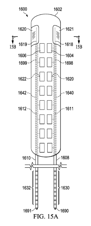
WO 2021/003496 PCT/US2020/070224
modification of the cladding of the optical fiber. The cladding may be
modified by using
femtosecond laser etching, mechanical abrasion, or an alternative method to
achieve radial
leakage of light.
[0169] Polished optical tip 1411 is positioned at the proximal end of
the optical fiber.
Polished optical tip 1411 is preferably a thermally polished surface
perpendicular to the optical
axis of the fiber. Optionally, a convex lens may be attached to the proximal
end of the fiber to
focus light into or out of the fiber, as will be further described.
[0170] Referring then to Figure 13E, optical fiber subassembly 1419 is
positioned in
stylet channel 1405. The outer diameter of ferrule 1412 is less than the outer
diameter of lead
body 1402 but greater than the diameter of stylet channel 1405, such that the
lead body acts as a
stop for the ferrule.
[0171] In one embodiment, optical fiber subassembly 1419 is placed in
the stylet
channel after surgical placement of the lead body in vivo, as will be further
described.
[0172] In another embodiment, the fiber subassembly is prefabricated
into the lead
body. In this embodiment, the proximal end of the optical fiber is secured to
the fiber by a
suitable adhesive. One such suitable medical grade adhesive is preferably an
optically
transparent biocompatible epoxy seal, such as EPO-TEK MED-353ND by Epoxy
Technology,
Inc. of Billerica, Massachusetts. In this case, polished optical tip 1411 is
polished flush with
alignment tip 1413.
[0173] Referring then to Figure 13F, an alternate embodiment of
optical fiber
subassembly 1419 will be described.
[0174] Lead body 1401 includes stylet channel 1405 having an optical
axis 1421.
The lead body incorporates proximal contacts, distal electrodes and electrical
conductors, as
29
CA 03145576 2021-12-29
WO 2021/003496 PCT/US2020/070224
previously described. Stylet channel 1405 is terminated with frustoconical
flare 1482.
Frustoconical flare 1482 is coaxial with optical axis 1421. The frustoconical
flare has inclination
angle (3, which can range from about 135 to about 150 .
[0175] In a preferred embodiment, the lead channel has a diameter of
about 15 ¨ 20%
greater than the optical fiber to allow the fiber to move within the channel.
Optical fiber 1444 is
preferably a plastic fiber, as previously described. Optical fiber 1444
includes end reflector 1446
and side-firing fiber segment 1445, as previously described.
[0176] Optical fiber 1444 is proximally terminated by convex lens
1452. Convex
lens 1452 consists of a polished ceramic material, such as sapphire, fixed to
the optical fiber
using a suitable optically transparent adhesive. In another embodiment, the
lens is formed
integrally with the transmission fiber. In another embodiment, the optical
fiber is polished flat
and no lens is incorporated.
[0177] Collet 1457 includes collet body 1453. Preferably, collet body
1453 is
comprised of a ceramic material, such as Zirconia, or another MRI compatible
material.
Alignment tip 1454 is a chamfer integrally formed in the distal end of the
collet body.
Alignment tip 1454 forms angle of inclination y of about 135 . In a preferred
embodiment, angle
y matches angle 0 of frustoconical flare 1482.
[0178] Collet body 1453 further includes cylindrical collet chamber
1456. Collet
chamber 1456 is coaxial with collet body 1453 and extends through alignment
tip 1454. In a
preferred embodiment, the collet is bonded to optical fiber 1444 at the collet
chamber.
[0179] Lens shield 1469 is integrally formed with the proximal end of
collet body
1453. It is designed to serve as a stop to prevent the optical fiber from
impinging on the optical
window of the IPG body. Lens shield 1469 further includes frustoconical lens
opening 1468
CA 03145576 2021-12-29
WO 2021/003496 PCT/US2020/070224
ductedly connected with collet chamber 1456. The frustoconical lens opening is
coaxial with the
collet chamber and has an angle of inclination 6 of about 1750 with the collet
chamber.
Frustoconical lens opening 1468 serves to focus light toward the optical
window.
[0180] Referring then to Figure 13G, the proximal end of the optical
fiber is shown
positioned in collet chamber 1456. Lens 1452 does not extend past lens shield
1469. The fiber
is fixed in the collet chamber by a suitable epoxy.
[0181] Optical fiber 1418 is positioned in stylet channel 1405 of lead
body 1401.
Frustoconical flare 1482 serves to guide insertion of the optical fiber into
the lead channel. The
interface of frustoconical flare 1482 and alignment tip 1454 also serves to
center optical fiber
1444 and lens 1452 on axis 1421. The outer diameter of collet 1457 is less
than the outer
diameter of lead body 1401 but greater than the diameter of stylet channel
1405, such that when
the lead is inserted in the header, the lead body acts as a stop for the
collet, such that the side
firing fiber segment of the fiber is adjacent the optical window.
[0182] Referring then to Figure 13H ¨ 13K, a preferred embodiment of
optical
threading assembly 1460 is described. Inserting the optical fiber into the
stylet channel of the
lead can be difficult due to the miniature size of the fiber and small
diameter of the stylet
channel. In the same way, during surgery, the replacement of the stylet in the
stylet channel can
be difficult. These difficulties are compounded by necessity for speed, manual
dexterity and
visual acuity. Improper insertion of the optical fiber can lead to damage to
the fiber causing
breakage or early degradation of the fiber. Likewise, improper stylet
insertion can compromise
the lead body. The optical fiber threading assembly solves these and other
problems.
[0183] Optical threading assembly 1460 includes guide body 1497. The
guide body
is a roughly 1 cm diameter cylinder and is comprised of thermoplastic. The
assembly is
31
CA 03145576 2021-12-29
WO 2021/003496 PCT/US2020/070224
preferably formed either using injection molding, or additive manufacturing.
Other methods of
manufacture will suffice. Guide body 1497 is generally cylindrical and is
comprised of two
opposing semicylinders, 1461 and 1462.
[0184] Optical threading assembly 1460 includes frustoconical lead
centering surface
1463 at its distal end. Frustoconical lead centering surface 1463 is coaxial
with axis 1421.
Frustoconical lead centering surface 1463 is adjacent cylindrical alignment
surface 1467.
Cylindrical alignment surface 1467 forms alignment cavity 1498. Frustoconical
lead centering
surface 1463 forms an angle of inclination T of about 1350 with cylindrical
alignment surface
1467. The alignment cavity has a diameter generally equal to that of lead body
1402. The
alignment cavity is terminated with stop surface 1466. Stop surface 1466 is a
generally annular
ring formed perpendicular to and coaxial with axis 1421.
[0185] Adjacent to and ductedly connected with alignment cavity 1498,
is generally
cylindrical fiber alignment duct 1499. Fiber alignment duct 1499 is coaxial
with axis 1421. The
diameter of the fiber alignment duct is generally the same as the diameter of
stylet channel 1405
of lead body 1402.
[0186] Fiber alignment duct 1499 is adjacent to and ductedly connected
with
frustoconical optical fiber centering surface 1465. Frustoconical optical
fiber centering surface
1465 forms an angle of inclination 11 of about 135 with fiber alignment duct
1499.
Frustoconical optical fiber centering surface 1465 is coaxial with axis 1421.
[0187] Semicylinder 1461 includes alignment pegs 1474, 1470, and 1472.
Semicylinder 1462 includes alignment recesses 1475, 1471, and 1473. Alignment
peg 1474 is
diametrically opposed to alignment recess 1475. Alignment peg 1470 is
diametrically opposed
to alignment recess 1471. Alignment peg 1472 is diametrically opposed to
alignment recess
32
CA 03145576 2021-12-29
WO 2021/003496 PCT/US2020/070224
1473. The diameter of each of alignment recesses 1475, 1471, and 1473 is such
that alignment
pegs 1474, 1470, and 1472 can be secured by a press fit. Using the alignment
pegs and recesses,
the semicylinders may be easily assembled for use and then disassembled after
use, as will be
further described.
[0188] In use, optical threading assembly 1460 aligns lead body 1402
and optical
fiber subassembly 1419 along axis 1421. Lead body 1402 is aligned using
frustoconical lead
centering surface 1463 and held in position in alignment cavity 1498 by
alignment surface 1467
and stop surface 1466. Optical fiber subassembly 1419 is aligned using
frustoconical centering
surface 1465 and moved through fiber alignment duct 1499 and into the stylet
channel of the lead
body.
[0189] In the same way, a stylet may be positioned in the stylet
channel in place of
the optical fiber.
[0190] Referring then to Figures 14A ¨ 15B, alternate embodiments for
surgical leads
are described.
[0191] Surgical leads may be configured with two or more multi-duct
leads
containing integrated optical fibers, depending upon the number of electrodes
in the array and
the desired number of optical reflectometry channels. The multi-duct leads may
be organized
into pairs of emitter leads and detector leads. Generally, a surgical lead
configured with two
multi-duct leads incorporate one optical reflectometry channel, while a
surgical lead with four
multi-duct leads incorporates two optical reflectometry channels. A surgical
lead with two
multi-duct leads with integrated optical fibers is capable of determining
sagittal spinal cord
position. Whereas a surgical lead with four multi-duct leads with integrated
optical fibers is
capable of determining sagittal and coronal spinal cord position.
33
CA 03145576 2021-12-29
WO 2021/003496 PCT/US2020/070224
[0192] Referring then to Figure 14A, a preferred embodiment of
surgical lead 1500 is
described.
[0193] Electrode array 1500 is comprised of integrated flexible panel
1502. Panel
1502 is preferably of a medical grade inert polymer material, such as
Pellethane 55-D. Flexible
panel 1502 houses multi-duct leads 1506, 1510, 1514, and 1518. In a preferred
embodiment, the
multi-duct leads are sealed within the body of the flexible panel. Each of
multi-duct leads 1506,
1510, 1514, and 1518 includes a central lumen 1595, 1596, 1597 and 1598,
respectively. Each
central lumen includes an optical transmission fiber 1541, 1543 1545 and 1547,
respectively.
Each optical transmission fiber terminates at a distal end in a side firing
optical fiber segment,
1535, 1536, 1537 and 1538, respectively. The side firing optical fiber
segments are constructed,
as previously described. Each side firing optical fiber segment is positioned
adjacent distally
positioned optical window 1533, 1532, 1531 and 1530, respectively. In a
preferred embodiment,
each of the optical windows is an optically transparent segment of the polymer
comprising
flexible panel 1502. By placing the optical windows and the side firing
segment at the most
distal portion of the panel, there are no metallic components such as
electrodes or connections to
interfere with radial dispersion of light, while keeping the optical sensing
region proximate the
electrode arrays. This improves optical signal strength and consequently
lowers power
consumption. In an alternate embodiment, the optical windows are placed
parallel to and
adjacent columns of the electrode arrays.
[0194] Panel 1502 further comprises electrode arrays 1504, 1508, 1512,
and 1516
positioned adjacent multi-duct leads 1506, 1510, 1514 and 1518, respectively.
In a preferred
embodiment, each of electrode arrays 1504, 1508, 1512, and 1516 include eight
electrodes
embedded in the panel and having an exposed face external to the panel. Each
of multi-duct
34
CA 03145576 2021-12-29
WO 2021/003496 PCT/US2020/070224
leads 1506, 1510, 1514 and 1518 incorporate eight electrical conductors that
extend the length of
the panel and the multi-duct leads, as previously described. Each electrode is
connected through
the conductors in the lead body to exactly one lead contact. Electrode array
1504 is connected to
lead contacts 1507. Electrode array 1508 is connected to lead contacts 1511.
Electrode array
1512 is connected to lead contacts 1515. Electrode array 1516 is connected to
lead contacts
1519. In a preferred embodiment, the electrodes are comprised of platinum-
iridium alloy
(nominally 90%/10% to 80%/20%).
[0195] Each of multi-duct leads 1506, 1510, 1514 and 1518 terminates
at ferrules
1505, 1509, 1513 and 1517, respectively. In a preferred embodiment, the
ferrules are bonded to
the fibers, as previously described.
[0196] Referring then to Figures 14B and 14C, each of electrode arrays
1504, 1508,
1512 and 1516 is connected through electrical connections 1519, 1521, 1523 and
1525 to one of
conductor bundles 1540, 1542, 1544 and 1546, respectively. The conductor
bundles contain the
individual conductors, radially separated, as previously described. The side
firing fiber segments
separate from the multi-duct leads and away from the conductor bundles in
manifolds 1580,
1581, 1582 and 1583, respectively.
[0197] Panel 1502 includes light reflectors 1550, 1551, 1552 and 1553,
adjacent side
firing optical fiber segments 1535, 1536, 1537, and 1538, respectively. The
light reflectors are
preferably semicylindrical or parabolic, and flexible. In a preferred
embodiment, light reflectors
1550, 1551, 1552 and 1553 are comprised of a non-conductive material polymer,
such as
Pellethane-55D, coated with a non-conductive reflective surface, such as
titanium dioxide. This
material operates at the desired wavelength from red to infrared and may be
applied as a paint or
film. The reflections improve optical efficiency by redirecting radially
produced light from the
CA 03145576 2021-12-29
WO 2021/003496 PCT/US2020/070224
emitter fiber segments toward the optical windows, or, alternatively,
reflecting incoming light
from the optical windows and into the detector fiber segments.
[0198] Panel 1502 is further comprised of lattice shield 1526. Lattice
shield 1526 is
comprised of a generally flat flexible film and interdigitates with the
polymeric material of the
lead body. In a preferred embodiment, lattice shield 1526 is coated with a
reflective material,
such as titanium dioxide (TiO2) adjacent the optical fibers. Lattice shield is
contained within the
panel adjacent each of conductor bundles 1540, 1542, 1544 and 1546, and
generally extends the
length of the panel.
[0199] In one embodiment, the lattice shield may be comprised of an
electrically
conductive material, such as carbon nanofibers, and operates as a heat-sink to
draw heat away
from the electrode contact arrays and disperse it dorsally. In another
embodiment, leads 1518,
1514, 1510, and 1506 each include a ground, as shown in Figure 13C. Ground
line 14511
connects lattice shield 1526 with the anchor ring located at the proximal end
of the lead. The
anchor ring is connected to the IPG ground. This configuration provides
optimal electrical
shielding for MM compatibility.
[0200] Referring then to Figure 15A, a preferred embodiment of surgical
lead 1600 is
described.
[0201] Surgical lead 1600 is comprised of integrated flexible panel
1602. In a
preferred embodiment, flexible panel 1602, is a medical grade inert flexible
polymeric material,
as previously described. Integrated within the flexible panel are multi-duct
leads 1608 and
1610. Each of multi-duct leads 1608 and 1610 includes central lumen 1698 and
1699,
respectively. The central lumens include optical fibers 1611 and 1612,
respectively. Each
optical fiber terminates in a side firing optical fiber segment 1618 and 1619,
respectively. The
36
CA 03145576 2021-12-29
WO 2021/003496 PCT/US2020/070224
side firing optical segments are constructed as previously described. Each
side firing optical
segment is positioned adjacent an optical window 1621 and 1620, respectively.
In a preferred
embodiment, each of the optical windows is an integrally formed optically
transparent region of
flexible panel 1602.
[0202] Panel 1602 further comprises electrode arrays 1604 and 1606.
Each of
electrode arrays 1604 and 1606 includes eight electrodes, embedded in the
surface of panel 1602,
having an exposed face exterior to the panel. In a preferred embodiment, the
electrodes are a
platinum-iridium alloy. Each of the electrodes is connected through the
conductors in the multi-
duct lead bodies to exactly one lead contact, as previously described.
Electrode array 1604 is
connected to lead contacts 1630. Electrode array 1606 is connected to lead
contacts 1632.
[0203] Each of multi-duct leads 1608 and 1610, terminates at ferrules
1690 and 1691,
respectively. The ferrules are attached and bonded to the optical fibers, as
previously described.
[0204] Referring then to Figure 15B, the conductors in the multi-duct
lead bodies
separate from side firing optical fiber segment 1618 and 1619 in optical
manifolds 1653 and
1652, respectively. Each of electrode arrays 1604 and 1606 is connected
through electrical
connections 1650 and 1651 to the conductors in one of conductor bundles 1640
and 1642,
respectively.
[0205] Panel 1602 is further comprised of lattice shield 1614. Lattice
shield 1614 is
comprised of a generally flat flexible film that is integrally formed with
both the flexible panel.
As previously described, the lattice shield may be coated with reflective
material adjacent the
optical fibers. The lattice shield may be connected to a ground contact for
further connection to
the IPG ground, as previously described.
[0206] Panel 1602 further comprise reflectors 1616 and 1625,
positioned adjacent
37
CA 03145576 2021-12-29
WO 2021/003496 PCT/US2020/070224
side firing optical fiber segments 1618 and 1619, respectively. In preferred
embodiments, the
reflectors are generally semicylindrical or are parabolic. In another
embodiment, the reflectors
may be flat flexible panels. The reflectors serve to aid in the reflection of
light emitted from a
side firing fiber segment out through an optical window or to focus incoming
light through
optical windows and back into the fiber for transmission to a detector within
the IPG.
[0207] Referring to the Figure 16A, 16B and 16C, an alternate
embodiment of
surgical lead 1700 will be described. Integrated panel 1702 is generally flat,
polymeric and
rectangular, as previously described. Integrated within integrated panel 1702
are multi-duct
leads 1718, 1720 and 1722 with optical fiber 1736, as previously described.
Each of the multi-
duct leads have proximal electrical contacts which are individually connected
to electrode arrays
1712, 1714 and 1716 through conductors, as previously described. Integrated
panels 1702
further comprises optical window 1709 integrated into the panel adjacent side
firing fiber
segments 1730, 1732 and 1734, as previously described.
[0208] Composite reflector 1710 is comprised a plurality of alternating
parabolic
surfaces, such as parabolic surfaces 1750, 1752 and 1754, and flat
interstitial surfaces such as
surfaces 1756 and 1758. The parabolic surfaces and the flat surfaces are
preferably comprised of
a flexible inert plastic with sufficient rigidity to sustain moderate bending.
In a preferred
environment, polyvinyl chloride is used. Internal surfaces 1751, 1753 and 1755
of the parabolic
surfaces and internal surfaces 1757 and 1759 of the flat surfaces all are
coated with a reflective
material such as titanium dioxide. Internal surfaces 1751, 1753 and 1755 are
positioned adjacent
side firing fiber segments 1730, 1732 and 1734 and serve to function as
previously described.
[0209] In another preferred embodiment, composite reflector 1710 is
grounded to the
IPG case, through a conductor in one of the multi-duct leads, as previously
described.
38
CA 03145576 2021-12-29
WO 2021/003496 PCT/US2020/070224
[0210] Referring then to Figures 17A and 17B an alternate embodiment
of surgical
lead 1800 will be further described.
[0211] Integrated panel 1802 generally comprises a flat, polymeric and
rectangular,
as previously described. Integrated panel 1802 include electrode arrays 1810
and 1812
connected to multi-duct leads 1814 and 1818, as previously described.
Integrated panel 1802
further comprise multi-duct leads 1814, 1816 and 1818 integrally formed, as
previously
described. Each of multi-duct leads 1814, 1816 and 1818 includes optical fiber
1850 with side
firing fiber segments 1820, 1822, and 1824. Each optical fiber 1850 is
positioned to terminate in
a right-angle prism, such as right-angle prism 1804, 1806, and 1808. The right-
angle prisms are
positioned to direct light to an optical fiber from optical windows 1811,
1813, and 1817, or from
the optical fiber to an optical window, as the case may be.
[0212] Referring then to Figure 18, method 1900 for the placement of a
surgical lead,
will be described.
[0213] At step 1902, a laminotomy is conducted at the segmental level
corresponding
to the somatotopic distribution of the patient's pain.
[0214] At step 1904, electrodes are placed in the spinal canal.
Typically, the
electrodes are placed in the dorsal epidural space by manually inserting the
electrode array in the
laminotomy cavity.
[0215] At step 1906, the electrodes are anchored to the fascia,
ligament or the
adjacent bone.
[0216] At step 1908, an incision is made for the IPG.
[0217] At step 1910 the leads are tunneled subcutaneously from the
electrode
insertion site to the IPG pocket.
39
CA 03145576 2021-12-29
WO 2021/003496 PCT/US2020/070224
[0218] At step 1914, the multi-duct leads are secured in the IPG
header. If the fibers
are not secured in the multi-duct leads, then they may be inserted and secured
at this step, as will
be further described. In practice, the lead body, including the fiber
subassembly, is threaded into
the appropriate lead channel bringing the proximal lead contacts into
electrical contact with the
canted coil springs, of the lead channel. The multi-duct lead is advanced in
the lead channel
until the multi-duct lead body encounters frustoconical centering surface 724,
which guides it
along cylindrical alignment surface 726, until it engages stop surface 730 in
anchor ring chamber
728. Simultaneously, the ferrule is advanced into alignment cylinder 712 until
it encounters
ferrule centering surface 716. Ferrule centering surface 716 aligns the
optical fiber in buffer gap
734 and with the optical window in the IPG casing, adjacent the composite
optoelectronic device
740. The multi-duct lead is secured in the lead channel by advancing anchor
screw 708, using a
torque limited ratchet, until it engages anchor ring 1410.
[0219] At step 1916 the IPG is placed in the pocket.
[0220] At step 1918, the procedure is ended.
[0221] Referring to Figure 19, preferred method 2000 of placement of a
percutaneous
lead will be described.
[0222] At step 2002, a Touhy needle and needle stylet are inserted
into spinal canal at
the appropriate segmental level.
[0223] At step 2004, the needle stylet is removed from the lumen of
the Touhy
needle.
[0224] At step 2006, the percutaneous lead with included stylet guide
wire is inserted
into the bore of the Touhy needle.
[0225] At step 2008, the percutaneous lead is guided to the proper
location in the
CA 03145576 2021-12-29
WO 2021/003496 PCT/US2020/070224
spinal canal using the stylet guide wire, under fluoroscopy.
[0226] At step 2010, the stylet guide wire is removed from the stylet
channel.
[0227] At step 2012, the optical fiber is inserted into the stylet
channel, as previously
described.
[0228] At step 2014, the Touhy needle is removed, while holding the
lead in place.
[0229] At step 2016, the proximal end of the percutaneous lead is
secured in the IPG
header, as previously described.
[0230] Referring then to Figure 20, step 2012 of securing the optical
fiber in a stylet
channel of a lead, will be further described.
[0231] At step 2102, semicylinders 1461 and 1462, are aligned with the
percutaneous
lead body. At step 2104, semicylinders 1461 and 1462 are assembled by press
fit. At step 2106,
the proximal end of the percutaneous lead body is inserted into the alignment
cavity of the
threading assembly, guided by frustoconical lead centering surface 1463.
Alignment surface
1467 aligns the multi-duct lead body with alignment cavity 1498.
[0232] At step 2108, optical fiber 1418 is inserted into fiber
alignment duct 1499,
guided by frustoconical optical fiber centering surface 1465. The optical
fiber is then inserted
into stylet channel 1405 of multi-duct lead body 1402, of the threading
assembly.
[0233] At step 2110, ferrule 1412 is threaded onto optical fiber 1418.
[0234] In an alternate embodiment, a stylet may be placed in the fiber
alignment duct
and the stylet channel at this step. If so, the method concludes here.
[0235] At step 2112, the threading assembly is disassembled.
[0236] At step 2114, the semicylinders are removed from the assembled
lead body
and optical fiber.
41