Note: Descriptions are shown in the official language in which they were submitted.
CA 03145655 2021-12-30
WO 2021/003281 PCT/US2020/040517
TISSUE REPAIR DEVICE
RELATED APPLICATION
[0001] This application claims priority to U.S. Provisional
Application Serial No.
62/869,806, filed on July 2, 2019, and entitles Tissue Repair Device, the
subject matter of which
is herein incorporated by reference.
BACKGROUND
[0002] This disclosure relates to a surgical device for repairing
damaged tissue and a
method for repairing the damaged tissue. Orthopedic procedures are often
performed to repair
musculoskeletal injuries, such as those sustained during sporting activities.
Tears in the meniscus
are known to be repaired by deploying implants on either side of the tear,
tensioning suture
between the implants to close the tear, and allowing it to heal.
SUMMARY
[0003] This disclosure relates to a tissue repair device that
comprises, inter al/a, a
fixed outer cannula that has a distal end and a proximal end opposite the
distal end and a fixed
length, the fixed length defining a depth stop; and a movable inserter
received inside of the fixed
outer cannula, the movable inserter may be slidable with respect to the fixed
outer cannula, and
the movable inserter may be configured to support a suture implant construct
that is positioned
for deployment.
[0004] In some embodiments, an actuator is received in the fixed outer
cannula for
sliding the movable inserter with respect to the fixed cannula; a depth limit
selection feature is
provided that corresponds to the fixed length of the fixed outer cannula; the
depth limit selection
feature is associated with a handle at the proximal end of the fixed outer
cannula; and/or the
depth limit selection feature is a rotating depth selector coupled to the
proximal end of the fixed
outer cannula.
1
CA 03145655 2021-12-30
WO 2021/003281 PCT/US2020/040517
[0005] In certain embodiments, the movable inserter includes a
longitudinal slot
providing access to an inside of the movable inserter; the width of the
longitudinal slot is sized to
accommodate a suture or sutures of the suture implant construct; and/or the
suture implant
construct includes at least first and second implants; and/ or the movable
inserter is a needle with
a piercing end.
[0006] A method includes, inter al/a, inserting a tissue repair device
into a tissue
cavity until an end face of a distal end of a fixed outer cannula of the
device abuts tissue at or
near damaged tissue; sliding a movable inserter of the tissue repair device,
that is received in the
fixed outer cannula, toward the distal end of the fixed outer cannula;
penetrating the tissue with a
piercing end of the inserter; and deploying a suture implant construct
residing in the movable
inserter.
[0007] In some embodiments, the method comprises the step of selecting
a depth
limit that corresponds to a fixed length of the fixed outer cannula; the step
of selecting a depth
limit includes rotating a depth selector coupled to a proximal end of the
fixed outer cannula to a
selected depth; the movable inserter remains inside of the fixed outer cannula
until the end face
of the fixed outer cannula abuts the tissue; the method comprises the step of
compressing the
tissue with the end face of the distal end of the fixed outer cannula; the
step of deploying a suture
implant construct includes deploying a first implant of a plurality of
implants supported by the
movable inserter; and/or the fixed outer cannula remains stationary and flush
against the tissue
while the inserter penetrates the tissue.
2
CA 03145655 2021-12-30
WO 2021/003281 PCT/US2020/040517
BRIEF DESCRIPTION OF THE DRAWINGS
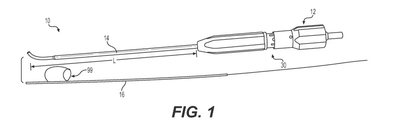
[0008] Figure 1 is a perspective view of a tissue repair device,
showing an inner
moveable inserter outside of the device for clarity.
[0009] Figure 2 is a perspective view of a fixed outer cannula and the
moveable
inserter of the tissue repair device illustrated in Figure 1.
[0010] Figure 3 is a partial perspective end view of the inserter
illustrated in Figure 2,
showing a suture implant construct associated with the inserter.
[0011] Figure 4 is a partial perspective end view of the inserter
illustrated in Figure 3,
the inserter being shown as transparent for clarity in order to illustrate the
suture implant
construct residing inside of the inserter.
[0012] Figure 5 is a partial perspective end view of the outer
cannula, showing the
outer cannula as transparent for clarity.
[0013] Figure 6 is a side elevational view of a handle and deployment
method for the
implants.
[0014] Figure 7 is an enlarged perspective view of one end of the
tissue repair device.
[0015] Figure 8 is a cross-sectional view of the one end of the tissue
repair device
illustrated in Figure 7.
[0016] Figures 9A and 9B illustrate a method of tissue repair.
3
CA 03145655 2021-12-30
WO 2021/003281 PCT/US2020/040517
DETAILED DESCRIPTION
[0017] This disclosure generally relates a tissue repair device 10
configured to deliver
a suture-implant construct or constructs and methods of repairing damaged
tissue. An exemplary
suture-implant construct includes one or more stands of suture and one or more
implants or
anchors. The implants may be any known type of implant or anchor, such as a
soft sheath
anchor, PEEK anchors, and the like. The tissue repair device 10 may be
designed for in-line
synchronization and simultaneous movement of a suture implant construct or
constructs within
an inserter for deploying the same. The tissue repair device 10 is configured
to reduce or
eliminate damage to tissue or cartilage when inserting the device into a
tissue cavity or joint.
The tissue repair device 10 is designed to be sturdy and more robust than
conventional device to
facilitate insertion of the tissue repair device 10 into the tissue cavity
during a repair.
[0018] Figure 1 illustrates an example surgical device 10 for tissue
repair. The
surgical device 10 generally includes a handle 12, a fixed outer cannula 14
projecting distally
from the handle 12 along a longitudinal axis, the fixed outer cannula 14 being
fixed to the handle
12, and a moveable inserter 16 received in the outer cannula 14 configured to
deploy a suture-
implant construct or constructs 99. The moveable inserter 16 is configured to
slide with respect
to the fixed outer cannula 14 along the longitudinal axis of the cannula 14.
An actuator 18
(Figure 4) may be provided for deploying the suture implant construct 99
through the inserter 16.
[0019] The handle 12 may include a trigger 13 (Figure 6) that is
moveable in the
distal and proximal directions to move the actuator 18, which itself is
moveable within the
inserter 16 in the distal and proximal directions. In one example, the handle
12 may include a
spring or other biasing element configured to bias the trigger 13 in the
proximal direction. In
order to move the trigger 13 in the distal direction, a user (i.e., a surgeon)
uses his or her thumb,
for example, to apply a force to the trigger 13 sufficient to overcome the
bias of the spring such
that the trigger 13 slides distally. When the user's thumb is released from
the trigger 13, the
trigger 13 moves proximally back to a resting position under the bias of the
spring or other
biasing element.
4
CA 03145655 2021-12-30
WO 2021/003281 PCT/US2020/040517
[0020] The fixed outer cannula 14 includes a proximal end 20
associated with the
handle 12 and an opposite distal end 22 configured to be inserted into a
tissue cavity within the
body. The fixed outer cannula 14 defines a fixed length L (Figure 1) which
corresponds to an
insertion depth limit of the device 10 when inserted into the tissue cavity.
An end face 24 of the
distal end 22 of the outer cannula 14 defines a depth stop and is configured
to abut the tissue 80
(Figures 9A and 9B) in the tissue cavity. A tip 26 (Figure 2) of the cannula's
distal end 22 may
be substantially flat, relatively sharp, curved, tapered, and/or pointed.
[0021] The tissue repair device 10 may be provided with a depth limit
selection
feature 30 that corresponds to the fixed length L of the outer cannula 14. The
depth limit
selection feature 30 may be associated with the handle 12 at the proximal end
20 of the outer
cannula 14. In an embodiment, the depth limit selection feature 30 comprises a
rotating depth
selector 32 (Figure 7) that indexes and acts as a positive stop to control the
distance the outer
cannula 14 can protrude from the handle 12, i.e. fixed length L. The rotating
depth selector 32
may involve rotation of a gripping section 34 of handle 12 over a housing
section 36, as seen in
Figure 8. A spring loaded retainer 38 is captured in the gripping section 34
such that when the
gripping section 34 is rotated over the housing section 36, the retainer 38
locks into one of the
depth selection notches 45, thereby setting the appropriate depth (and fixed
length L) determined
by the user.
[0022] The moveable inserter 16 is received within the fixed outer
cannula 14 and is
slidable therein. The inserter 16 is configured to be loaded with a suture-
implant construct or
constructs 99, which may comprise one or more strands of suture 40 and one or
more implants
42. The strand or strands of suture 40 may be attached to each implant 42 in
any known manner,
such as be splicing the strand 40 through splice points in the implant 42,
such as with a sheath
type implant. An end of the strand may be affixed back to the strand 40 by a
knot or other
fixation technique to create a loop 44 enclosing a portion of a first implant
42. The loop 44
retains the implant 42 relative to the strand of suture 40 and ensures that
the implant does not
slide distally off the strand of suture 40.
[0023] The suture-implant construct 99 may be referred to as a "soft"
construct
because it is formed of soft materials such as yarns, fibers, filaments,
strings, fibrils, strands,
CA 03145655 2021-12-30
WO 2021/003281 PCT/US2020/040517
sutures, etc., or any combination of such materials. The soft materials may be
synthetic or natural
materials, or combinations of synthetic and natural materials, and may be
biodegradable or non-
biodegradable within the scope of this disclosure. In an embodiment, the
suture-implant
construct 99 is made exclusively of soft, suture-based materials. The soft
materials confer the
ability to be inserted into or through tissue (e.g., bone, ligament, tendon,
cartilage, etc.) and then
bunch together, collapse, expand, and/or change shape to fixate the suture-
implant construct 99
relative to the tissue.
[0024] In an embodiment, the strand or strands of suture 40 may be any
flexible
strand suitable for surgical tissue repair. For example, the strand or strands
of suture 40 may be
one of the following examples: FiberWire , TigerWire , or FiberChain suture,
which are
each available from Arthrex, Inc. It should be understood, however, that any
type of suture may
be used, including cored or coreless sutures. In another embodiment, the
strand of suture 40 is
flat suture, such as FiberTape or SutureTape suture, which is also available
from Arthrex,
Inc. The strand or strands of suture 40 may also be a monofilament suture
having barbs.
Further, the strand or strands of suture 40 could include any soft, flexible
strand of material, and
is not limited to suture.
[0025] In an embodiment, the inserter 16 is a surgical needle with a
piercing end 46.
A longitudinal slot 48 may be provided in a wall of the inserter 16 that
extends generally parallel
to the longitudinal axis of the cannula 14 and provides access to an inside of
the inserter 16. The
width of the slot 48 may be sized to accommodate the strands or strands of
sutures 40 of the
construct 99. The slot 48 includes opposing side walls 50 and 52 which serve
to guide
movement of the actuator 18 in a direction parallel to the longitudinal axis.
The slot 48 may
extend along a portion of the length of the cannula 14 or alternatively may
extend along the
entire length of the cannula 14.
[0026] The actuator 18 may be a slider that is configured to push and
deploy the one
or more implants 42 in sequence. In one example, the actuator 18 includes a
pushrod or shaft
mechanically coupled to the trigger 13 of the handle 12. The actuator 18 is
configured to move
in the distal and proximal directions in response to corresponding movement of
the trigger 13.
The actuator 18 may include a relatively smooth superior surface and
relatively smooth side
6
CA 03145655 2021-12-30
WO 2021/003281 PCT/US2020/040517
surfaces configured to slide relative to the respective side walls 50 and 52
of the slot 48. The
actuator 18 can have a deployment mechanism, such as a shuttling rack or the
like, which is
configured to interact with implants 40 to move them distally within the
inserter 16.
[0027] A method of using the tissue repair device 10 will now be
described with
reference to Figures 9A and 9B. The surgeon initially determines the
appropriate insertion depth
limit of the device 10 into the cavity and then fixes the length L (Figure 1)
of the outer cannula
14 based on that determined depth limit. The length L of the outer cannula 14
may be fixed, for
example, by using the depth selection feature 30. The construct 99 is loaded
into the inserter 16
with a distal end 54 (Figure 4) of the actuator 18 positioned proximal to the
first implant 42, and
the loaded inserter 16 is positioned inside the fixed outer cannula 14.
[0028] The surgeon inserts the surgical device 10 within the tissue
cavity or joint
space, in order to deploy the one or more implants 42 in the area adjacent the
tear 82 in the tissue
80 in order to close the tear 82 and allow it to heal. The surgeon can insert
the device 10 until
the end face 24 of the outer cannula 14 abuts the tissue 80 in the cavity,
such as on the medial or
inside surface of a meniscus. The fixed length L of the cannula 14 ensures
that the device 10 is
inserted to the proper depth in the tissue cavity and not too deep, which
could result in damage to
the tissue or cartilage. This avoids the need to move the cannula 14, after
that device 10 is
already into the tissue cavity, to determine whether the inserter has been
inserted to the proper
depth, such as is the case in conventional tissue repair devices. In an
embodiment, the surgeon
can use the end face 24 of the outer cannula 14 to compress the tissue near
the tear 82 while
conducting the repair.
[0029] Once the device 10 is inserted to the proper depth in the
tissue cavity, the
inserter 16 is moved to slide with respect to the cannula 14 toward distal end
22 in order to
penetrate the tissue 80 in a first location where a first implant 42 of the
suture implant construct
99 is to be deployed. When in this first position, the fixed outer cannula 14
remains stationary
and flush against the tissue as the inserter 16 is penetrating the tissue. The
surgeon can penetrate
the inserter 16 into, for example, a medial surface of the tissue and out a
lateral surface of the
tissue. Once the inserter 16 slides and penetrates the tissue 80 at the first
location, the surgeon
operates the trigger 13 (Figure 6), which moves the actuator 18 (Figure 4)
distally to selectively
7
CA 03145655 2021-12-30
WO 2021/003281 PCT/US2020/040517
deploy the suture-implant construct 99 and its implants 42. The distal end of
the actuator 18
does not extend beyond the inserter 16 as the implants 42 are pushed out to
the piercing end 46
of the inserter 16.
[0030] After the first implant is deployed, the surgeon can remove his
or her thumb,
for example, from the trigger 13, thereby allowing the trigger 13 and the
actuator 18 to move
proximally. A second anchor is then moved from a standby position to a deploy
position. The
surgeon can position the cannula 14 flush against the tissue 80 in a second
location adjacent the
tear 82, and that is spaced-apart from the first location, and repeat the
above process to deploy
the second implant. In an embodiment, once the first and second implants are
deployed adjacent
the tear 82, the strand of suture 40 is tensioned to cinch or tension the
implants, thereby closing
the tear 82.
[0031] It should be understood that terms such as "lateral," "medial,"
"distal,"
"proximal," "superior," and "inferior" are used above consistent with the way
those terms are
used in the art. Further, these terms have been used herein for purposes of
explanation, and
should not be considered otherwise limiting. Terms such as "generally,"
"substantially," and
"about" are not intended to be boundaryless terms, and should be interpreted
consistent with the
way one skilled in the art would interpret those terms.
[0032] Although the different examples have the specific components
shown in the
illustrations, embodiments of this disclosure are not limited to those
particular combinations. It is
possible to use some of the components or features from one of the examples in
combination
with features or components from another one of the examples.
[0033] One of ordinary skill in this art would understand that the
above-described
embodiments are exemplary and non-limiting. That is, modifications of this
disclosure would
come within the scope of the claims. Accordingly, the following claims should
be studied to
determine their true scope and content.
8
