Note: Descriptions are shown in the official language in which they were submitted.
WO 2021/022360
PCT/CA2020/051020
1
SYSTEMS AND METHODS FOR OPTOGENETIC ACTIVATION AND MONITORING
RELATION PATENT APPLICATION
[0001] The present application claims priority to U.S. Provisional Patent
Application No. 62/884,344
filed on August 8, 2019, the disclosure of which is incorporated herein by
reference in its entirety.
TECHNICAL FIELD
[0002] The technical field generally relates to optogenetics and, more
particularly, to systems and
methods for optogenetic activation and monitoring.
BACKGROUND
[0003] Brain functions, such as cognition, learning, memory, behavior, and
physical action, are
controlled and regulated by cellular excitability. The understanding and
control of processes and
mechanisms involved in cellular excitability have been the subject of current
research in many fields
of medicine and biotechnology, for example, in the area of neurological
disorders and diseases.
Cellular excitability can be studied using a variety of techniques, among
which is optogenetics.
Optogenetics is a branch of biotechnology that combines optical methods with
genetic targeting tools
to achieve precise spatio-temporal control and monitoring of cell activity.
Optogenetics generally uses
two main classes of tools: actuators and reporters, which respectively enable
light-mediated control
and monitoring of cell activity.
[0004] Optogenetic actuators are typically genetically encoded light-sensitive
proteins that can
change their conformation upon exposure to light of specific wavelength. The
activation of
optogenetic actuators can cause ion channel gating or pump activation, cell
depolarization or
hyperpolarization, and ultimately cellular stimulation or inhibition in cells,
frequently neurons, in which
the actuators are expressed. Common optogenetic actuators are opsins, which
are naturally
occurring transmembrane proteins that can act as ion channels or pumps. Opsins
include both
stimulatory opsins, such as Channelrhodopsin-2 (ChR2), and inhibitory opsins,
such as
Natronomonas pharaonis Halorhodopsin (NpHR).
[0005] Optogenetic reporters, also referred to as optogenetic indicators, are
typically genetically
encoded fluorescent proteins whose emission characteristics vary in response
to physical and
biochemical changes within cells. Optogenetic reporters can be probed using
fluorescence
microscopy to enable sensing, monitoring, and/or imaging of biological
structures, parameters, and
processes. By way of example, fluorescence microscopy can be used to track the
spatial distribution
of optogenetic reporters within cells; sense biological parameters, such as
ion concentrations and
membrane potentials; monitor or detect phenomena, such as cell surface binding
or neurotransmitter
CA 03146259 2022-1-28
WO 2021/022360
PCT/CA2020/051020
2
release; and study cellular activity, notably cellular excitability, in
neurons and myocytes. In particular,
fluorescent reporters whose emission characteristics are modulated as a
function of changes in ionic
concentrations (e.g., calcium reporters, whose fluorescence varies in response
to changes in
intracellular calcium concentration) or as a function of changes in membrane
potential (e.g., voltage
reporters, whose fluorescence varies in response to transmembrane ion
exchanges between the
intra- and extra-cellular matrices) can allow for monitoring cellular
excitability.
[0006] While existing optogenetic techniques for controlling and monitoring
cellular excitability may
have certain advantages, they also have a number of drawbacks and limitations.
For example, since
membrane potential variations are relatively fast (e.g., of the order of 1
kilohertz), conventional pixel-
based cameras often struggle to measure the fluorescence signals from voltage
reporters. This may
be a reason why calcium reporters, whose response times are significantly
slower (e.g., of the order
of 30 hertz), have been favored up to now for use as optogenetic reporters. In
addition,
measurements of cell excitability can involve activating optogenetic actuators
present in one or more
regions of a specimen while simultaneously monitoring optogenetic reporters in
other regions of the
specimen. A number of microscopy modalities have been developed or adapted for
this purpose.
Non-limiting examples include random access microscopy based on acousto-optic
deflectors (A0Ds)
and laser scanning microscopy, such as confocal laser scanning microscopy
(CLSM) and
programmable array microscopy (PAM). However, these modalities still suffer
from a number of
drawbacks and limitations, such as high cost, single-wavelength operation, and
cameras with
relatively slow acquisition rates. Thus, challenges remain in the field of
optogenetic systems and
methods for controlling and monitoring cell activity.
SUMMARY
[0007] The present description generally relates to optogenetic systems and
methods for probing a
specimen using spatio-temporally modulated illumination. The disclosed systems
and methods may
provide high-throughput, space- and time-resolved, and/or cell-type-specific
control and monitoring
of cellular activity. The disclosed systems and methods may be implemented
with or in various types
of microscopy modalities including, but not limited to, widefield microscopy,
confocal microscopy, and
other types of fluorescence-based microscopy.
[0008] In accordance with an aspect, there is provided an optogenetic method
for probing a
specimen, including:
generating illumination light, the illumination light including a plurality of
illumination protocols
temporally sampled and interleaved with one another at a time-division-
multiplexed (TDM)
sampling rate, each illumination protocol being for illuminating a respective
region of interest
(ROI) of a plurality of ROls of the specimen; and
CA 03146259 2022-1-28
WO 2021/022360
PCT/CA2020/051020
3
applying a spatio-temporal modulation to the illumination light to produce
modulated illumination
light and directing the modulated illumination light onto the specimen, the
spatio-temporal
modulation including repeatedly imparting, at a pattern switching rate matched
and
synchronized with the TOM sampling rate, a sequence of a plurality of spatial
modulation
patterns to the plurality of temporally sampled and interleaved illumination
protocols, each
spatial modulation pattern mapping to a respective one of the ROls.
[0009] In some implementations, the plurality of illumination protocols is a
plurality of activation
protocols for activating optical actuators present in the plurality of ROls,
respectively.
[0010] In some implementations, the plurality of illumination protocols is a
plurality of excitation
protocols for exciting optical reporters present in the plurality of ROls,
respectively. In such
implementations, the method further includes detecting specimen light coming
from the optical
reporters present in the plurality of ROls in response to the plurality of
excitation protocols,
generating, from the detected specimen light, detection signal data conveying
information about the
specimen. The specimen light may include fluorescence light. In some
implementations, detecting
the specimen light includes detecting a plurality of time-interleaved
detection signals respectively
associated with the plurality of ROls, and generating the detection signal
data includes performing a
time-demultiplexing operation on the detected specimen light for
deinterleaving the plurality of time-
interleaved detection signals. In some implementations, the method may further
include repeatedly
imparting, at the pattern switching rate, the sequence of the plurality of
spatial modulation patterns
to the specimen light prior to detecting the specimen light.
[0011] In some implementations, generating the illumination light further
includes generating a
plurality of activation protocols temporally sampled and interleaved with one
another at the TDM
sampling rate, the plurality of activation protocols being for activating
optical actuators present in the
plurality of ROls, and applying the spatio-temporal modulation to the
illumination light further includes
repeatedly imparting, at the pattern switching rate, the sequence of the
plurality of spatial modulation
patterns to the plurality of temporally sampled and interleaved activation
protocols. In other
implementations, generating the illumination light further includes generating
a plurality of activation
protocols temporally sampled and interleaved with one another at the TOM
sampling rate, the plurality
of activation protocols being for activating optical actuators present in
another plurality of ROls of the
specimen, applying the spatio-temporal modulation to the illumination light
further includes
repeatedly imparting, at the pattern switching rate, a sequence of another
plurality of spatial
modulation patterns to the plurality of temporally sampled and interleaved
activation protocols, each
one of the other spatial modulation patterns mapping to a respective one of
the other ROI s.
CA 03146259 2022-1-28
WO 2021/022360
PCT/CA2020/051020
4
[0012] In some implementations, the spatio-temporal modulation is applied
using one or more digital
micromirror devices (DMDs). In some implementations, the TOM sampling rate and
the pattern
switching rate range from about 1 kHz to about 40 kHz, for example, from about
10 kHz to about
30 kHz.
[0013] In accordance with another aspect, there is provided an optogenetic
system for probing a
specimen, including:
an illumination unit configured to generate illumination light including a
plurality of illumination
protocols temporally sampled and interleaved with one another according to a
time-division-
multiplexed (TOM) scheme having a TOM sampling rate, each illumination
protocol being for
illuminating a respective region of interest (ROI) of a plurality of ROls of
the specimen;
a spatial light modulator (SLM) unit configured to apply a spatio-temporal
modulation to the
illumination light to produce modulated illumination light and to direct the
modulated
illumination light onto the specimen, the spatio-temporal modulation including
repeatedly
imparting, at a pattern switching rate, a sequence of a plurality of spatial
modulation patterns
to the plurality of temporally sampled and interleaved illumination protocols,
each spatial
modulation pattern mapping to a respective one of the ROls; and
a control and processing unit operatively coupled to the illumination unit and
the SLM unit, the
control and processing unit being configured to match and synchronize the TDM
sampling
rate of the TOM scheme applied by the illumination unit with the pattern
switching rate of the
spatio-temporal modulation applied by the SLM unit.
[0014] In some implementations, the illumination unit includes an activation
unit including at least
one activation light source configured to generate, as the plurality of
illumination protocols, a plurality
of activation protocols for activating optical actuators present in the
plurality of ROls, respectively.
[0015] In some implementations, the illumination unit includes an excitation
unit including at least
one excitation light source configured to generate, as the plurality of
illumination protocols, a plurality
of excitation protocols for exciting optical reporters present in the
plurality of ROls, respectively. In
such implementations, the optogenetic system further includes a detection unit
configured to detect
specimen light coming from the optical reporters present in the plurality of
ROls in response to the
plurality of excitation protocols. The detection unit may include a single-
element detector, also
referred to as a single-point detector, configured to detect the specimen
light in a time-resolved
manner, and the specimen light may include fluorescence light In some
implementations, the
detection unit is configured to detect the specimen light as a plurality of
time-interleaved detection
signals respectively associated with the plurality of ROls, and the control
and processing unit is
configured to perform a time-demultiplexing operation on the detected specimen
light for
CA 03146259 2022-1-28
WO 2021/022360
PCT/CA2020/051020
deinterleaving the plurality of time-interleaved detection signals. In some
implementations, the SLM
unit is disposed in a path of the specimen light and configured to repeatedly
impart, at the pattern
switching rate, the sequence of the plurality of spatial modulation patterns
to the specimen light prior
to the specimen light being detected by the detection unit.
5 [0016] In some implementations, the illumination unit further includes an
activation unit including at
least one activation light source configured to generate a plurality of
activation protocols temporally
sampled and interleaved with one another at the TDM sampling rate, the
plurality of activation
protocols being for activating optical actuators present in the plurality of
ROls. Furthermore, the SLM
unit is configured to repeatedly impart, at the pattern switching rate, the
sequence of the plurality of
spatial modulation patterns to the plurality of temporally sampled and
interleaved activation protocols.
In other implementations, the illumination unit further includes an activation
unit including at least one
activation light source configured to generate a plurality of activation
protocols temporally sampled
and interleaved with one another at the TOM sampling rate, the plurality of
activation protocols being
for activating optical actuators present in another plurality of ROls of the
specimen. Furthermore, the
SLM unit is configured to repeatedly impart, at the pattern switching rate, a
sequence of another
plurality of spatial modulation patterns to the plurality of temporally
sampled and interleaved
activation protocols, each one of the other spatial modulation patterns
mapping to a respective one
of the other ROls.
[0017] In some implementations, the SLM unit includes one or more digital
nnicronnirror devices.
[0018] In accordance with another aspect, there is provided a non-transitory
computer readable
storage medium having stored thereon computer executable instructions that,
when executed by a
processor, cause the processor to perform various steps of a method of
controlling an optogenetic
system such as described herein.
[0019] In accordance with another aspect, there is provided a computer device
for use with or in an
optogenetic system such as described herein, the computer device including a
processor and a non-
transitory computer readable storage medium operatively coupled to the
processor and having stored
thereon computer readable instructions that, when executed by a processor,
cause the processor to
perform various steps for controlling the optogenetic system.
[0020] In accordance with another aspect, there is provided a system for
optogenetic activation and
monitoring of a specimen. The optogenetic system may include an activation
unit including an
activation light source configured to generate activation light, and an
excitation unit including an
excitation light source configured to generate excitation light The activation
light and the excitation
CA 03146259 2022-1-28
WO 2021/022360
PCT/CA2020/051020
6
light may have illumination spectra that are different from each other. The
activation light may be
used to activate optogenetic actuators disposed in the specimen to cause
conformational changes
in the actuators, thereby stimulating or inhibiting cell activity in the
specimen. The excitation light may
be used to excite optogenetic reporters disposed in the specimen. The
optogenetic reporters may be
configured to emit fluorescence light when cell activity is stimulated or
inhibited through optical
activation of the optogenetic actuators by the activation light.
[0021] The optogenetic system may also include an SLM, for example, a DMD or
another suitable
type of SLM. The SLM may be configured to spatially modulate the activation
light and the excitation
light, and to direct the resulting spatially patterned activation light and
spatially patterned excitation
light onto the specimen. The SLM may also be configured to spatially modulate
specimen light, for
example, fluorescence light, coming from the specimen in response to the
excitation light and, in
some cases, in response also to the activation light
[0022] The optogenetic system may further include a detection unit including a
detector, for example,
a single-element detector, such as a photomultiplier tube (PMT) or an
avalanche photodiode (APD).
The detector may be configured to detect the spatially modulated specimen
light coming from the
SLM and generate, from the detected specimen light, a detection signal
conveying information about
the specimen. In other variants, however, the specimen light may not encounter
the SLM along its
path between the specimen and the detector. In such a case, the specimen light
is not spatially
modulated by the SLM prior to detection.
[0023] The optogenetic system may also include a control and processing unit
operatively coupled
to the activation light source, the excitation light source, the SLM, and the
detector to control, at least
partly, their operation.
[0024] In some implementations, the optogenetic system may include more than
one activation light
source and/or more than one excitation light source and/or more than one SLM
and/or more than
one detector. This may result in increased versatility and flexibility by
providing more degrees of
freedom for controlling and observing the spatial and/or temporal dynamics of
cell activity.
[0025] In some implementations, the optogenetic system may be configured to
implement a time-
division-multiplexed (TOM) scheme that involves subsampling and interleaving
in time a number of
activation and/or excitation protocols, where each protocol is to be applied
to a particular region of
interest (ROI) of the specimen. In such implementations, the SLM may be used
to spatio-temporally
modulate the activation light and/or the excitation light onto the specimen at
a modulation rate that is
matched to and synchronized with the sampling rate of the TDM scheme. Such a
TOM scheme may
CA 03146259 2022-1-28
WO 2021/022360
PCT/CA2020/051020
7
allow for activating and monitoring multiple ROls of the specimen in parallel
(i.e., quasi-
simultaneously) to increase throughput.
[0026] In accordance with another aspect, there is provided a method for
optogenetic activation and
monitoring of a specimen. The method may include a step of generating
activation light with an
activation light source and generating excitation light with an excitation
light source. Depending on
the application, the activation light and the excitation light may be
generated concurrently or not. The
activation light and the excitation light may be used respectively to activate
optogenetic actuators
and excite optogenetic reporters disposed in the specimen. In order to
mitigate or control crosstalk
between the activation of optogenetic actuators by the activation light and
the excitation of
optogenetic reporters by the excitation light, actuator-reporter pairs with
non-overlapping or negligibly
overlapping activation and excitation spectra may be used.
[0027] The method may also include a step of using an SLM, for example, a DMD,
to spatially
modulate the activation light and the excitation light to produce spatially
modulated activation and
spatially modulated excitation light, and direct (e.g., by deflection from the
SLM) the resulting spatially
patterned activation light and spatially patterned excitation light onto the
specimen. The SLM may
also be used to spatially modulate specimen light emanating from the specimen
in response to the
excitation light (and possibly the activation light). However, in some
implementations, the specimen
light may not be spatially modulated by the SLM. Depending on the application,
the spatial modulation
pattern applied by the SLM may be stationary or vary in time, for example,
depending on whether a
single ROI or several ROls are activated and/or observed.
[0028] The method may further include a step of detecting the spatially
modulated specimen light
and a step of generating, from the detected specimen light, a detection signal
conveying information
about the specimen. As noted above, in some embodiments, the specimen light
emanating from the
specimen may not be spatially modulated by the SLM.
[0029] In some implementations, the method may implement a time-division-
multiplexed (TDM)
scheme that allows for activating, exciting, and detecting multiple ROls of
the specimen in parallel.
In such implementations, the method may include steps of identifying a
plurality of ROls of the
specimen; determining a plurality of spatial light modulation patterns to be
applied by the SLM, where
each spatial light modulation pattern maps to a respective one of the
identified ROls; and determining
a plurality of illumination protocols for probing the plurality of ROls,
respectively. Each illumination
protocol may be defined by an activation time profile to be imparted to the
activation light by the
activation light source and/or an excitation time profile to be imparted to
the excitation light by the
excitation light source. Depending on the application, the activation and
excitation time profiles of
CA 03146259 2022-1-28
WO 2021/022360
PCT/CA2020/051020
8
each illumination protocol may be either time-varying or time-invariant. Also,
for each illumination
protocol, either the activation time profile or the excitation time profile
may be a constant zero-
intensity function, if the corresponding ROI is to be either activated or
excited, but not both.
[0030] In such implementations, the step of generating the activation light
and the excitation light
may include controlling the activation light source and the excitation light
source to generate the
activation light and the excitation light based on a TDM scheme by sampling
and interleaving the
plurality of illumination protocols at a TOM sampling rate. In some
implementations, the amplitude of
the activation time profile and/or the excitation time profile of each or any
illumination protocol may
be appropriately scaled (e.g., increased) to account for the fact that the
illumination duration of each
ROI is made shorter as a result of the sampling and interleaving operations.
The step of using the
SLM may include controlling the SLM to sequentially switch between the
plurality of spatial light
modulation patterns in accordance with the TOM scheme. This control may
involve matching and
synchronizing the SLM modulation rate with the TDM sampling rate. Furthermore,
the step of
detecting the specimen light (which may be spatially modulated or not,
depending on the application)
may include detecting the specimen light as a plurality of interleaved
responses, where each
interleaved response conveys information about a respective one of the ROls.
In such a case, a time-
demultiplexing operation may be performed to recover the time profile of the
response emanating
from each ROI.
[0031] It is to be noted that other method and process steps may be performed
prior to, during or
after the steps described herein. The order of one or more steps may also
differ, and some of the
steps may be omitted, repeated, and/or combined, as the case may be. It is
also to be noted that
some method steps may be performed using various image processing techniques,
which may be
implemented in hardware, software, firmware or any combination thereof.
[0032] Other features and advantages of the present description will become
more apparent upon
reading of the following non-restrictive description of specific embodiments
thereof, given by way of
example only with reference to the appended drawings. Although specific
features described in the
above summary and in the detailed description below may be described with
respect to specific
embodiments or aspects, it should be noted that these specific features can be
combined with one
another unless stated otherwise.
BRIEF DESCRIPTION OF THE DRAWINGS
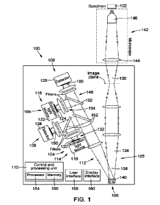
[0033] Fig. 1 is a schematic representation of an optogenetic system, in
accordance with an
embodiment.
CA 03146259 2022-1-28
WO 2021/022360
PCT/CA2020/051020
9
[0034] Fig. 2 is a schematic representation of aspects of a method of
operation of an optogenetic
system implementing a time-division-multiplexing (TDM) of illumination
protocols, in accordance with
another embodiment
[0035] Fig. 3 is a schematic representation of an optogenetic system, in
accordance with an
embodiment.
[0036] Fig. .4 is a schematic representation of an optogenetic system, in
accordance with an
embodiment.
100371 Fig. 5 is a schematic representation of an optogenetic system, in
accordance with another
possible embodiment.
[0038] Fig. 6 illustrates how the embodiment of Fig. 5 may be used to
distinguish out-of-focus
background components from in-focus components of the detected specimen light.
[0039] Fig. 7 is a schematic representation of an optogenetic system, in
accordance with another
possible embodiment.
[0040] Fig. 8 is a schematic representation of an optogenetic system, in
accordance with another
possible embodiment.
[0041] Figs. 9A to 9C depict different illumination and detection scenarios
that may be implemented
with the embodiment of Fig. 8.
DETAILED DESCRIPTION
[0042] In the present description, similar features in the drawings have been
given similar reference
numerals. To avoid cluttering certain figures, some elements may not be
indicated if they were
already identified in a preceding figure. It is appreciated that the elements
of the drawings are not
necessarily depicted to scale, since emphasis is placed on clearly
illustrating the elements and
structures of the present embodiments. Furthermore, positional descriptors
indicating the location
and/or orientation of one element with respect to another element are used
herein for ease and clarity
of description. Unless otherwise indicated, these positional descriptors
should be taken in the context
of the figures and should not be considered limiting. It is appreciated that
such spatially relative terms
are intended to encompass different orientations in the use or operation of
the present embodiments,
in addition to the orientations exemplified in the figures.
[0043] The terms "a", "an", and "one" are defined herein to mean "at least
one", that is, these terms
do not exclude a plural number of items, unless stated otherwise.
CA 03146259 2022-1-28
WO 2021/022360
PCT/CA2020/051020
[0044] Terms such as "substantially", "generally", and "about", that modify a
value, condition, or
characteristic of a feature of an exemplary embodiment, should be understood
to mean that the value,
condition, or characteristic is defined within tolerances that are acceptable
for the proper operation
of this exemplary embodiment for its intended application or that fall within
an acceptable range of
5 experimental error. In particular, the term "about" generally refers to a
range of numbers that one
skilled in the art would consider equivalent to the stated value (e.g., having
the same or equivalent
function or result). In some instances, the term "about" means a variation of
10 percent of the stated
value. It is noted that all numeric values used herein are assumed to be
modified by the term "about",
unless stated otherwise.
10 [0045] The terms ``connected" and "coupled", and derivatives and
variants thereof, are intended to
refer herein to any connection or coupling, either direct or indirect, between
two or more elements,
unless stated otherwise. For example, the connection or coupling between the
elements may be
mechanical, optical, electrical, magnetic, thermal, chemical, logical,
fluidic, operational, or any
combination thereof.
[0046] The terms "match", "matching", and "matched" are intended to refer
herein to a condition in
which two elements are either the same or within some predetermined tolerance
of each other. That
is, these terms are meant to encompass not only "exactly" or "identically"
matching the two elements
but also "substantially", "approximately", or "subjectively" matching the two
elements, as well as
providing a higher or best match among a plurality of matching possibilities.
[0047] The term "concurrently" refers herein to two processes that occur
during coincident or
overlapping time periods. The term "concurrently" does not necessarily imply
complete synchronicity
but encompasses various scenarios including: time-coincident or simultaneous
occurrence of two
processes; occurrence of a first process that both begins and ends during the
duration of a second
process; and occurrence of a first process that begins during the duration of
a second process, but
ends after the completion of the second process.
[0048] The terms "light' and "optical", and variants and derivatives thereof,
are intended to refer
herein to radiation in any appropriate region of the electromagnetic spectrum.
These terms are not
limited to visible light but can also include invisible regions of the
electromagnetic spectrum including,
without limitation, the terahertz (THz), infrared (IR), and ultraviolet (UV)
spectral bands. For example,
in non-limiting embodiments, the present techniques may be implemented with
light having a
wavelength band lying somewhere in the range from about 400 to about 780
nanometers (nm).
However, this range is provided for illustrative purposes only and the present
techniques may operate
outside this range.
CA 03146259 2022-1-28
WO 2021/022360
PCT/CA2020/051020
11
[0049] The terms "probe" and variants thereof are intended to refer herein to
any optical system
which can deliver optical energy to a region of interest and/or collect
optical energy from the region
of interest. In particular, the term "probe" and variants thereof are meant to
encompass optical
systems used solely for light delivery (e.g., activation and/or excitation),
solely for light collection (e.g.,
fluorescence detection), and for both light delivery and collection.
[0050] The present description generally relates to optogenetic systems and
methods that use
spatio-temporal light modulation to achieve all-optical manipulation and
observation of space- and
time-dependent processes occurring in a specimen.
[0051] The present techniques may be used with a variety of specimens, notably
biological
specimens, which may be studied in vivo, in vitro, or ex vivo. Non-limiting
examples of biological
specimens that may be studied using the present techniques include, to name a
few, cells, tissues,
organs, organisms, subcellular components, and other biological materials.
Notably, the present
techniques may be used to probe living cells expressing optogenetic proteins.
[0052] The present techniques may find use in a wide range of medical and
biological imaging
applications, notably in the study, diagnosis, treatment, and cure of various
diseases and disorders
that involve the excitability of cells, such as neurons and myocytes.
Furthermore, the present
techniques may be implemented with or in various types of microscopy
modalities including, but not
limited to, widefield microscopy, confocal microscopy, and other types of
fluorescence-based
microscopy. It is appreciated, however, that some implementations of the
present techniques may
be used in applications other than optogenetics, such as in thermal
stimulation applications. For
example, the present techniques may be used with non-biological specimens to
control and observe
certain events (e.g., chemical reactions) occurring in a specimen. In such
applications, activation light
may be used to initiate a change in a specimen and excitation light may be
used to excite the
specimen to emit light in response to the change. The characteristics of the
emitted light may be
detected and analyzed to convey information about the change.
[0053] As described in greater detail below, an optogenetic method for probing
a plurality of regions
of interest (ROls) of a specimen may include a step of generating illumination
light including a plurality
of illumination protocols. The illumination protocols are temporally sampled
and interleaved with one
another at a time-division-multiplexed (TOM) sampling rate. Each illumination
protocol is intended for
illuminating a respective one of the ROls. The method may also include a step
of applying a spatio-
temporal modulation to the illumination light to produce modulated
illumination light and directing the
modulated illumination light onto the specimen. The spatio-temporal modulation
may include
repeatedly imparting, at a pattern switching rate matched and synchronized
with the TDM sampling
CA 03146259 2022-1-28
WO 2021/022360
PCT/CA2020/051020
12
rate, a sequence of a plurality of spatial modulation patterns to the
plurality of temporally sampled
and interleaved illumination protocols, where each spatial modulation pattern
maps to the ROI
associated with its respective illumination protocol.
[0054] In some scenarios, the plurality of illumination protocols may be a
plurality of activation
protocols for activating optical actuators present in the ROls. In other
scenarios, the plurality of
illumination protocols may be a plurality of excitation protocols for exciting
optical reporters present
in the plurality of ROls. In such scenarios, the method may include a step of
detecting specimen light,
for example, fluorescence light, coming from the optical reporters in response
to the plurality of
excitation protocols, and a step of generating, from the detected specimen
light, detection signal data
conveying information about the specimen. Detecting the specimen light may
include detecting a
plurality of time-interleaved detection signals respectively associated with
the plurality of ROls, and
generating the detection signal data may include performing a time-
demuftiplexing operation on the
detected specimen light for deinterleaving the plurality of time-interleaved
detection signals.
Depending on the application, the spatio-temporal modulation may or may not be
applied to the
specimen light prior to its detection. In yet other scenarios, the
illumination light may include both a
plurality of activation protocols and a plurality of excitation protocols,
which may be used for
activating/exciting either a same set or different sets of ROls.
[0055] Various aspects and implementations of the present techniques are
described below with
reference to the figures.
[0056] Referring to Fig. 1, there is illustrated a possible embodiment of a
system 100 for probing a
specimen 102 by optogenetic activation and monitoring. The specimen 102 can
include cells that
have been genetically encoded to express (1) one or more optogenetic actuators
of electrical or
chemical activity and (2) one or more optogenetic reporters of electrical or
chemical activity.
[0057] Optogenelic actuators are typically genetically encoded proteins that
can change their
conformation upon exposure to light of specific wavelength, thereby initiating
an action potential in
the cells in which they are expressed. Common optogenetic actuators include
opsins, such as light-
gated ion channels or pumps, and optical switches. For example, the
optogenetic actuators may be
microbial opsins, such as channelrhodopsins, halorhodopsins, archaerhodopsins,
and leptosphaeria
rhodopsins_ Depending on the application, the optogenetic actuators may be
stimulatory (e.g.,
depolarizing) or inhibitory (e.g., hyperpolarizing). However, any other
suitable types of optogenetic
actuators may be used in other embodiments. It is appreciated that optogenetic
actuators and their
applications and principles of operation are generally known in the art and
need not be described in
greater detail herein.
CA 03146259 2022-1-28
WO 2021/022360
PCT/CA2020/051020
13
[0058] Optogenetic reporters are typically genetically encoded light-sensitive
fluorescent proteins,
dyes, or other compounds or biomolecules whose emission characteristics vary
in response to
physical and/or biochemical changes within cells in which they are expressed.
For example,
optogenetic reporters may emit fluorescence light in response to changes in
intracellular calcium
concentration (calcium reporters) or changes in membrane potential (voltage
reporters) initiated via
light-mediated activation of optogenetic actuators. Common optogenetic
reporters include Archon1,
Anine 6+, and VARNAM. However, any other suitable types of optogenetic
reporters may be used in
other embodiments. It is appreciated that, as for optogenetic actuators,
optogenetic reporters and
their applications and principles of operation are generally known in the art
and need not be described
in greater detail herein.
[0059] In the embodiment of Fig. 1, the optogenetic system 100 generally
includes an illumination
unit 104, a spatial light modulator (SLM) unit 106, a detection unit 108, and
a control and processing
unit 110.
[0060] The illumination unit 104 is configured to generate illumination light
112 for probing the
specimen 102. The illumination unit includes an activation unit 114 and an
excitation unit 116. The
activation unit 114 includes an activation light source 118 configured to
generate activation light 120.
The excitation unit 116 includes an excitation light source 122 configured to
generate excitation
light 124. The activation light 120 and the excitation light 124 together form
the illumination light 112.
The SLM unit 106 is configured to apply a spatio-temporal modulation to the
illumination light 112 to
produce modulated illumination light 126 and to direct the modulated
illumination light 126 onto the
specimen 102. The detection unit 108 includes a detector 128 configured to
detect specimen
light 130 emanating from the specimen 102. The control and processing unit 110
is operatively
coupled at least to the activation unit 114 and the excitation unit 116 of the
illumination unit 104, the
SLM unit 106, and the detection unit 108 to control, at least partly, their
operation. The structure and
operation of these and other possible components of the optogenetic system 100
are described in
greater detail below.
[0061] It is appreciated that Fig. us a simplified schematic representation
that illustrates a number
of basic components of the optogenetic system 100, such that additional
features and components
that may be useful or necessary for proper operation of the system 100 may not
be specifically
depicted. Non-limiting examples of such additional features and components may
include optical
components such as relay lenses, tube lenses, optical filters, mirrors, and
the like, configured to
condition and/or direct the activation light 120, the excitation light 124,
and the specimen light 130.
CA 03146259 2022-1-28
WO 2021/022360
PCT/CA2020/051020
14
[0062] In Fig. 1, the activation unit 114 includes a single activation light
source 118 to generate the
activation light 120 along an activation light path 132, and the excitation
unit 116 includes a single
excitation light source 122 to generate the excitation light 124 along an
excitation light path 134. It is
appreciated, however, that more than one activation light source 118 and/or
more than one excitation
light source 122 may be provided in other embodiments. The activation light
source 118 and the
excitation light source 122 may each be embodied by any appropriate device or
combination of
devices capable of generating activation light 120 and excitation light 124
having characteristics that
are suitable for optogenetic applications, respectively. Non-limiting examples
of possible light
sources include, to name a few, semiconductor light-emitting diodes (LEDs),
organic light-emitting
diodes (OLEDs), polymer light-emitting diodes (PLEDs), semiconductor laser
diodes, solid-state
lasers, gas lasers, and dye lasers. It is to be noted that using LED sources,
instead of laser sources
for generating the illumination light 112 may, in some instances, be
advantageous in terms of cost
and simplicity. Depending on the application, the activation light source 118
and the excitation light
source 122 may be operated in either a continuous or intermittent (e.g.,
pulsed) regime, and may or
may not be modulated. As can be appreciated, each of the activation light
source 118 and the
excitation light source 122 may be selected based on various factors
including, without limitation, its
operation wavelength; irradiance; spatial, temporal, and spectral profiles;
beam quality and
divergence; degree of coherence; compactness; reliability; and, for a pulsed
source, pulse
characteristics, such as its peak power, repetition rate, duration, temporal
shape, and center
wavelength.
[0063] The activation light source 118 may emit the activation light 120
according to an activation
protocol having an activation time profile. The activation time profile may
represent how the intensity
of the activation light 120 varies (or not) as a function of time during the
activation protocol. Likewise,
the excitation light source 122 may emit the excitation light 124 according to
an excitation protocol
having an excitation time profile. The excitation time profile may represent
how the intensity of the
excitation light 124 varies (or not) as a function of time during the
excitation protocol. Thus, in
operation of the optogenetic system 100, there may be times where both the
activation light
source 118 and the excitation light source 122 are illuminating the specimen
102, times where only
one of the activation light source 118 and the excitation light source 122 is
illuminating the
specimen 102, and times where neither the activation light source 118 nor the
excitation light
source 122 is illuminating the specimen 102.
[0064] The activation light 120 may be used to activate optogenetic actuators
disposed in the
specimen 102 to cause conformational changes in the optogenetic actuators and,
in turn, stimulate
or inhibit cell activity in the specimen 102. To this end, the activation
light 120 may have a wavelength
CA 03146259 2022-1-28
WO 2021/022360
PCT/CA2020/051020
suitable for activating the optogenetic actuators disposed in the specimen
102, such as between
about 420 nm and about 500 nm. For example, the wavelength of the activation
light 120 is equal to
460 nm in Fig. 1, corresponding to blue light However, as can be appreciated,
the activation light 120
may have any suitable activation wavelength, or range of activation
wavelengths, whether in the
5 visible range or in any other appropriate region of the electromagnetic
spectrum.
[0065] The excitation light 124 may be used to excite optogenetic reporters
disposed in the
specimen 102, where the optogenetic reporters may be configured to emit
radiation (e.g.,
fluorescence light) when cell activity is stimulated and/or inhibited
following activation of optogenetic
actuators. The excitation light 124 may have a wavelength suitable for
exciting the fluorescence of
10 the optogenetic reporters disposed in the specimen 102, such as between
about 600 nm and about
650 nm. For example, the wavelength of the excitation light 124 is equal to
620 nm in Fig. 1,
corresponding to red-orange light. However, as can be appreciated, the
excitation light 124 may have
any suitable excitation wavelength or range of excitation wavelengths, whether
in the visible range
or in any other appropriate region of the electromagnetic spectrum.
15 [0066] In some implementations, the activation light 120 and the
excitation light 124 may have
spectral profiles with no or little overlap, to avoid or reduce the risk of
unwanted crosstalk between
the activation of optogenetic actuators by the activation light 120 and the
excitation of the optogenetic
reporters by the excitation light 124. In particular, in some cases it may be
desirable or required that
the activation light 120 does not induce fluorescence from reporters and/or
that the excitation
light 124 does not activate actuators.
[0067] Referring still to Fig. 1, the SLM unit 106 is disposed along the
activation light path 132 and
the excitation light path 134, at a conjugate image plane of the optogenetic
system 100. The SLM
unit 106 is configured to spatially modulate the activation light 120 and the
excitation light 124 forming
the illumination light 112 according to a spatial modulation pattern to
produce spatially patterned or
modulated activation light 136 and spatially patterned or modulated excitation
light 138, respectively.
The modulated activation light 136 and the modulated excitation light 138
together form the
modulated illumination light 126. The SLM unit 106 is also configured to
direct the modulated
activation light 136 and the modulated excitation light 138 onto the specimen
102. The spatial
modulation pattern imparted by the SLM unit 106 to the activation light 120
and the excitation
light 124 maps to a corresponding ROI of the specimen 102, which is to be
illuminated by the
activation light 120 and the excitation light 124. Depending on the
application, the ROI corresponding
to a certain spatial modulation pattern defined by the SLM unit 106 may have
various sizes, shapes,
and configurations, and may consist of either a single area of the specimen
102 or a set of distinct
and unconnected areas of the specimen 102.
CA 03146259 2022-1-28
WO 2021/022360
PCT/CA2020/051020
16
[0068] In the embodiment of Fig. 1, the SLM unit 106 includes an SLM 140
embodied by a digital
micromirror device (DMD). The DMD functions as both an addressable binary-mask
spatial filter and
a high-speed light deflector interposed in the activation light path 132 and
the excitation light
path 134. The DMD includes a two-dimensional array of highly reflective,
micrometer-sized mirrors.
Each micromirror may be individually addressed and switched between two
resting states. Each
resting state may be defined by a respective discrete angular position or tilt
angle of the micromirror
relative to a flat state parallel to the plane of the micromirror array.
Depending on the application, any
of the number, size, shape, tilt angle, material, and switching rate of the
micromirrors of the DMD
may be varied. In one exemplary embodiment, the DMD may include an array of
1024x768 square
aluminum micromirrors, where each micromirror is about 10 pm in size, with a
tilt angle of 12 and
a switching rate of 32 kilohertz (kHz).
[0069] Each micromirror of the DM D acts as a dual reflector that deflects
light incident thereon along
either one of two distinct optical paths depending on its current resting
state. Each micromirror is said
to be in an "on" or "activated" state if light incident thereon (e.g., a
portion of the activation light 120
and/or a portion of the excitation light 124) is deflected onto the specimen
102. Conversely, each
micromirror is said to be in an "off' or "deactivated" state if light incident
thereon is deflected away
from the specimen 102, for example, into a beam dump (not shown). Thus, at any
given time, the
DMD may include an "activated portion", formed by all of the micromirrors that
are in their activated
state, and a "deactivated portion", formed by all of the micromirrors that are
in their deactivated state.
[0070] It is appreciated that the construction and operation of DM Ds are
generally known in the art
and need not be described in greater detail herein. DM Ds have become a
mature, reliable, and
relatively low-cost technology, which can provide high-speed and high-
resolution spatio-temporal
patterns for structured illumination and structured detection over large
fields of view. In particular,
DMDs offer various possibilities for controlling, both in space and over time,
the illumination pattern
of the activation light 120 and the excitation light 124 at the specimen 102.
In addition, by sequentially
activating groups of micromirrors, or single mirrors for higher resolution,
point-scanning imaging of
the specimen 102 can be achieved. It is also appreciated that while the SLM
unit 106 includes an
SLM 140 embodied by a DMD in the embodiment of Fig. 1, other embodiments may
use other types
of SLMs instead of, or in addition to, DMDs. Non-limiting types of SLMs
include electrically addressed
spatial light modulators (e.g., using ferroelectric liquid crystals or nematic
liquid crystals), optically
addressed spatial light modulators, and other suitable SLMs, which may or may
not be based on
dual-state SLM pixels.
[0071] By varying in time the spatial modulation pattern imparted by the SLM
unit 106 to the
activation light 120 and the excitation light 124, a variety of spatio-
temporal illumination patterns may
CA 03146259 2022-1-28
WO 2021/022360
PCT/CA2020/051020
17
be achieved for activating and/or exciting different ROls of the specimen 102
at different times over
a selected time period. In particular, by controlling the activation light
source 118 and the excitation
light source 122 to emit the activation light 120 and the excitation light 124
at different times, and by
coordinating the operation of the light sources 118, 122 with the operation of
the SLM unit 106, one
can devise optogenetic protocols in which ROls of the specimen 102 are
activated and/or excited
according to different spatio-temporal illumination patterns.
[0072] The optogenetic system 100 of Fig. 1 is configured to implement a time-
division-multiplexed
(TDM) scheme that involves temporally subsampling and interleaving, at a TDM
sampling rate, a
plurality of activation and/or excitation protocols, where each protocol
relates to a certain ROI of the
specimen 102. In such implementations, referred to as TDM implementations, the
SLM unit 106 is
used to spatio-temporally modulate the activation light 120 and the excitation
light 124 at a pattern
switching rate that is matched to and synchronized with the TDM sampling rate,
thus enabling a
spatio-temporally multiplexed activation and/or excitation of the specimen
102. The implementation
of such a TDM scheme may allow for several ROls to be activated ancUor excited
in parallel to
increase throughput.
[0073] It is to be appreciated that SLMs based on commercially available DMDs
can provide
structured illumination and deflection at high-speed modulation rates of up to
32 kHz, corresponding
to switching times of the order of 30 microseconds (ps). Such switching times
are significantly faster
than the response times associated with common optogenetic actuators, which
are of the order of
milliseconds for changes in membrane potential and of the order of tens of
milliseconds for changes
in calcium ion concentration. Thus, it can be envisioned to use the present
techniques to temporally
multiplex illumination/detection protocols associated with different ROls of a
specimen by sampling
and interleaving them, such that the samples of each illumination/detection
protocols occupy different
time positions and thus do not overlap, while maintaining a suitable temporal
resolution for activation,
excitation, and detection.
[0074] Referring to Fig. 2, various aspects of a method of operation of the
optogenetic system 100
of Fig. 1 related to TDM implementations will be described. The method may
include a step of
identifying a plurality of ROls of the specimen 102 to be probed in parallel
according to the TDM
scheme. In Fig. 2, three ROls of the specimen have been identified: R01-1, R01-
2, and R01-3.
However, depending on the application, the number of ROls that may be probed
using the TDM
scheme disclosed herein can range from two to up to a thousand or more.
[0075] Various methods may be used for identifying the ROls to be probed. For
example, the ROls
may be identified by analyzing an initial or previously obtained image of the
specimen 102. The initial
CA 03146259 2022-1-28
WO 2021/022360
PCT/CA2020/051020
18
image of the specimen 102 may be obtained with the system used to perform the
optogenetic method
or with another suitable imaging system. The initial image may have a
relatively coarse resolution
and may have been acquired in a relatively short acquisition time. In some
embodiments, the system
used to perform the optogenetic method (e.g., the optogenetic system 100 of
Fig. 1) may be used to
acquire such a relatively coarse image. For example, acquiring the relatively
coarse image may
involve implementing a point-by-point scanning of the specimen via a
sequential activation of groups
of micromirrors of the DMD, rather than single mirrors, for a lower spatial
resolution by a faster
acquisition time. Depending on the application, the analysis of the initial
image of the specimen in
order to identify ROls therein may be performed by a human and/or a computer.
As can be
appreciated, various computer-implemented and software-based techniques may be
employed for
this purpose. Such tools and techniques may use matching algorithms based on
feature extraction
and pattern recognition, and may rely on machine learning and/or artificial
intelligence. In some
implementations, a composite image of the specimen 102 may be obtained by
combining the non-
ROI portions of the initial image used to identify the ROls and the images of
the ROls obtained using
the TDM scheme described below.
[0076] Referring still to Fig. 2, the method of operation may include
generating the illumination
light 112 to include a plurality of illumination protocols for probing the
plurality of ROls. Each
illumination protocol is intended for illuminating a respective one of the
ROls. In some embodiments,
the plurality of illumination protocols is a plurality of activation protocols
for activating optical actuators
present in the ROls, while in other embodiments, the plurality of illumination
protocols is a plurality of
excitation protocols for exciting optical reporters present in the plurality
of ROls. In yet other
embodiments, the illumination light 112 includes both a plurality of
activation protocols, forming the
activation light 120, and a plurality of excitation protocols, forming the
excitation light 124.
[0077] In the embodiment of Fig. 2, the illumination light 112 is made up of
both activation light 120
and excitation light 124. The activation light 120 includes three activation
protocols, one for each of
the ROls. Likewise, the excitation light 124 includes three excitation
protocols, one for each of the
ROls. In Fig. 2, the activation protocols and the excitation protocols are
associated with the same set
of ROls. However, in other embodiments, the ROls associated with the
activation protocols may not
be all identical to the ROls associated with the excitation protocols.
Depending on the application,
the activation/excitation time profile associated with each
activation/excitation protocol may be time-
varying or time-invariant. Furthermore, the activation/excitation time profile
associated with each
activation/excitation protocol may be a constant zero-intensity function, if
the corresponding ROI is
to be either activated or excited, but not both.
CA 03146259 2022-1-28
WO 2021/022360
PCT/CA2020/051020
19
[0078] For simplicity, in Fig. 2 the activation and excitation protocols all
have the same onset time,
but this is not a requirement_ The activation protocols associated with R01-1
and R01-2 both include
two equal-duration activation periods separated by a non-activation period,
while the activation
protocol associated with R01-3 includes a single activation period that lasts
until the end of the non-
activation period for R01-1 and R01-2. Meanwhile, the excitation protocols
associated with R01-1,
R01-2, and R01-3 are all time-constant functions, with the excitation
intensity for R01-1 being less
than that for R01-2 and equal to that for R01-3. Of course, the activation and
excitation time profiles
of the activation and excitation protocols depicted in Fig. 2 are provided for
illustrative purposes only,
and any suitable activation and excitation time profiles may be used in other
embodiments.
[0079] In TDM implementations, the step of generating the activation light and
the excitation light
may include a step of controlling, for example, with a control and processing
unit such as described
herein, the activation light source and the excitation light source to
generate the activation light and
the excitation light based on a TOM scheme. The TDM scheme may include
temporally sampling and
interleaving the plurality of activation protocols at a TDM sampling rate, and
likewise for the plurality
of excitation protocols. The steps of sampling and interleaving the activation
time profiles for R01-1,
R01-2, and R01-3 are depicted schematically in Fig. 2, and likewise for the
steps of sampling and
interleaving the three excitation time profiles.
[0080] In some implementations, the amplitude of the activation/excitation
time profile of each
activation/excitation protocol may be appropriately scaled (e.g., increased)
to compensate for any
possible reduced activation and excitation durations resulting from the
sampling and interleaving
operations. It is appreciated that the TDM scheme depicted in Fig. 2 uses time
slots of equal duration
for each ROI, such that the duration of one TDM frame is equal to N times the
duration of one time
slot, where N is the number of ROls to be probed in parallel. However, this
need not be the case in
other variants, where different and/or more complex TDM schemes may be
employed without
departing from the scope of the present description. It is noted that the TDM
sampling rate is defined
as the rate of change between time slots. Thus, if the time slots associated
with the different ROls
are not all of equal duration, the TOM sampling rate will vary as a function
of time during each TDM
frame. In Fig. 2, the TDM sampling rate is constant and equal to the inverse
of the duration of the
time slots.
[0081] It is appreciated that the TDM scheme illustrated in the embodiment of
Fig. 2 is provided for
illustrative purposes only, and that various other TDM schemes could be used
in other embodiments.
In particular, more elaborate TOM schemes could be devised in implementations
where the
optogenetic system includes more than one activation light source, more than
one excitation light
source, more than one SLM, and/or more than one detector (see, e.g., Figs. 5,
7, and 8), while still
CA 03146259 2022-1-28
WO 2021/022360
PCT/CA2020/051020
providing time-interleaving of a plurality of subsannpled activation and/or
excitation protocols, where
each protocol corresponds to a particular ROI of a specimen under
investigation. It is also
appreciated that the TDM approach disclosed herein can generally be
implemented with only a set
of activation protocols (i.e., without excitation/detection), only a set of
excitation/detection protocols,
5 and both a set of activation protocols and a set of excitation/detection
protocols (as in the case of
Fig. 2).
[0082] Returning to Fig. 1, in TDM implementations, the operation of the
optogenetic system 100
may include a step of determining a plurality of spatial modulation patterns
to be applied by the SLM
unit 106 to the illumination light 112 that illuminates the plurality of
identified ROls of the
10 specimen 102, where each spatial light modulation pattern corresponds or
maps to a respective one
of the ROls. As can be appreciated, SLMs such as DMDs can be controlled and
programmed by
software to determine a spatial illumination pattern that corresponds to each
one of the identified
ROls. Once the plurality of spatial modulation patterns associated with the
plurality of ROls has been
determined, the operation of the optogenetic system 100 can include a step of
controlling the SLM
15 unit 106, for example, with the control and processing unit 110, to
apply a spatio-temporal modulation
to the illumination light 112 formed of the activation light 120 and the
excitation light 124. The
application of the spatio-temporal modulation may include repeatedly
imparting, at a pattern switching
rate matched and synchronized with the TDM sampling rate, a sequence of a
plurality of spatial
modulation patterns to the plurality of temporally sampled and interleaved
activation and excitation
20 protocols. Each spatial modulation pattern maps to a respective ROI of
the specimen 102 to ensure
that each ROI to be probed is activated and/or excited by its associated
activation and/or excitation
protocol(s).
[0083] The operation of sequentially switching between the plurality of
spatial light modulation
patterns in accordance with the TDM scheme involves matching and time-
coordinating the SLM
pattern switching rate with the TOM sampling rate. In Fig. 1, this can involve
synchronizing the
transitions between successive DMD patterns with the transitions between
successive TDM time
slots. For example, a DMD having a pattern switching rate of 32 kHz would
correspond to a TDM
time slot duration of 31.25 ps. In such a case, it could be envisioned to
perform TOM-based voltage
imaging on 16 ROls in parallel. In this case, the TDM frame duration (i.e.,
the time between time slots
associated with the same ROI) would be equal to 16x31.25 ps = 0.5 millisecond
(ms), which is of the
order of typical response times associated with changes in membrane potential.
Likewise, it could
also be envisioned to perform TDM-based calcium imaging on 1000 ROls in
parallel. The TDM frame
duration in this case would be equal to 1000x31.25 is = 31.25 ms, which is of
the order of typical
response times associated with changes in calcium ion concentration. It is
appreciated that the TDM
CA 03146259 2022-1-28
WO 2021/022360
PCT/CA2020/051020
21
sampling rate and the pattern switching rate can have different values
depending on the requirements
or particularities of the intended application. In some embodiments, the TDM
sampling rate and the
pattern switching rates can range from about 1 kHz to about 40 kHz, for
example, from about 10 kHz
to about 30 kHz, although other values outside this range, both below and
above, may be used in
other embodiments.
[0084] Referring still to Fig. 1, the modulated activation light 136 and the
modulated excitation
light 138 forming the modulated illumination light 126 produced by the SLM
unit 106 are projected
onto the specimen 102 via an optical assembly 142, for example, an optical
microscope, optically
coupled to the optogenetic system 100. The optical assembly 142 generally
includes imaging optics
to receive the modulated illumination light 126 and direct it onto the
specimen 102. In Fig. 1, the
optical assembly 142 includes a combination of a tube lens 144 and an infinity-
corrected
objective 146. The tube lens 144 and the objective 146 together define a field
of illumination for the
modulated activation light 136 and the modulated excitation light 138 on the
specimen 102. The size
of the region of the specimen 102 corresponding to any given rnicromirror of
the DMD of the SLM
unit 106 depends on the overall magnification provided by the optogenetic
system 100 and the optical
assembly 142. It is appreciated that the configuration of the optical assembly
142 depicted in Fig. 1
is provided for illustrative purposes only, as various other configurations
may be used in other
embodiments, which may include different and/or additional optical components
(e.g., beam-
conditioning optics and beam-directing optics). It is also appreciated that
the general principles
underlying the construction and operation of optical microscopes, including
those used for
fluorescence microscopy, are generally known in the art and need not be
described in greater detail
herein.
[0085] Upon reaching the specimen 102, the modulated activation light 136 can
activate optogenetic
actuators disposed in the specimen 102 to stimulate or inhibit cell activity
in the specimen 102, while
the modulated excitation light 138 can excite optogenetic reporters disposed
in the specimen 102.
As noted above, the optogenetic reporters may be configured to emit
fluorescence light, referred to
as specimen light 130, upon stimulation or inhibition of cell activity via
light-mediated activation of the
optogenetic actuators. Depending on the application, the specimen light 130
emitted from the
specimen 102 may originate not only from fluorescence emission of optogenetic
reporters induced
by the modulated excitation light 138 (and possibly also by the modulated
activation light 136), but
also from scattering, reflection, and/or transmission of the modulated
activation light 136 and/or the
modulated excitation light 138, as well as from other processes including, but
not limited to,
phosphorescence, Raman emission, thermal emission, and other linear and
nonlinear optical
processes.
CA 03146259 2022-1-28
WO 2021/022360
PCT/CA2020/051020
22
[0086] Referring still to Fig. 1, a part of the specimen light 130 emitted
from the specimen 102 is
collected by the objective 146 and relayed back toward the SLM unit 106 along
a detection light
path 148 leading to the detection unit 108. In particular, the detection unit
108 is configured to detect
specimen light 130 coming from the optical reporters present in the plurality
of ROls in response to
the plurality of excitation protocols. The SLM unit 106 is disposed along the
detection light path 148
and configured to spatio-temporally modulate the specimen light 130 (e.g.,
fluorescence light) to
produce modulated specimen light 150. In particular, in TDM implementations,
the SLM unit 106 is
configured to repeatedly impart, at the pattern switching rate, the sequence
of the plurality of spatial
modulation patterns to the specimen light 130 prior to the specimen light 130
being detected by the
detection unit 108 as the modulated specimen light 150. In this case, the
detection unit is configured
to detect the modulated specimen light 150 as a plurality of time-interleaved
detection signals
respectively associated with the plurality of ROls. Each time-interleaved
detection signal conveys
information about its associated ROI. As described below, a time-
demultiplexing operation can be
performed to retrieve the contribution from each ROI from the time-interleaved
detection signals.
100871 When the SLM unit 106 is a DMD, any portion of the specimen light 130
that impinges on an
activated micromirror will be deflected toward the detection unit 108 as
modulated specimen
light 150. Conversely, any portion of the specimen light 130 that impinges on
an deactivated
micromirror will be deflected away from the detection unit 108 (e.g., into a
beam dump). Using the
SLM unit 106 to provide structured illumination and structured detection
operating at the same time
may be advantageous in that the SLM unit 106 may provide confocal sectioning
for both illumination
and detection. However, in other implementations (see, e.g., Fig. 5), the
specimen light 130 may not
be spatially modulated by the SLM unit 106. That is, the SLM unit 106 may
provide structured
illumination but not structured detection. This may be achieved by providing,
between the
specimen 102 and the SLM unit 106, a dichroic mirror or another device or
combination of devices
able to separate the specimen light 130 from the modulated activation light
136 and the modulated
excitation light 138 and direct the separated specimen light 130 toward the
detection unit 108.
[0088] In Fig. 1, the detection unit 108 includes two detectors 128, each of
which for detecting the
modulated specimen light 150 in a respective spectral band. The optogenetic
system 100 may
include one or more dichroic mirrors 152 or other suitable spectrally
selective devices to separate
the modulated specimen light 150 from the activation light 120 and the
excitation light 124, and direct
the modulated specimen light 150 thus separated toward one of the detectors
128. The detectors 128
are configured to detect the modulated specimen light 150 coming from the SLM
unit 106 and
generate therefrom a detection signal conveying information about the specimen
102. Each
detector 128 may be made up of one or more photosensitive elements capable of
detecting input
CA 03146259 2022-1-28
WO 2021/022360
PCT/CA2020/051020
23
electromagnetic radiation and generating a detection signal therefrom,
typically by converting the
detected radiation into electrical data.
[0089] In some embodiments, the detection unit 108 may include a single-
element detector
configured to detect the modulated specimen light 150 in a time-resolved
manner. For example, in
Fig. 1, the detectors 128 can be photomultiplier tubes (PMTs), although other
embodiments may use
other types of single-element detectors such as avalanche photodiodes (APDs),
silicon
photomultipliers (SiPMs), and silicon photodiodes (SiPDs). It is appreciated
that using fast, single-
element detectors rather than comparatively slower arrays of detectors, such
as charge-coupled-
device (CCD) imagers, complementary metal-oxide-semiconductor (CMOS) imagers,
and charge-
injection-device (CID) imagers, may provide a number of advantages. Non-
limiting examples of such
advantages include, to name a few, better compatibility with the fast
modulation of DMDs; improved
sensitivity and response time; and improved signal-to-noise ratio by spatial
integration of the signal
coming from the entire ROI imaged by the DMD. It is appreciated, however, that
some embodiments
can use array of detectors instead of, or in addition to, single-element
detectors, without departing
from the scope of the present description.
[0090] Referring still to Fig. 1, the control and processing unit 110 refers
to an entity of the
optogenetic system 100 that controls and executes, at least partly, the
functions required to operate
or communicate with the various components of the optogenetic system 100
including, but not limited
to, the illumination unit 104, the SLM unit 106, and the detection unit 108.
The control and processing
unit 110 may generally include a processor 154 and a memory 156. The control
and processing
unit 110 may be configured to control and synchronize, via suitable
controllers or drivers, the
operation of the activation unit 114, the excitation unit 116, the SLM unit
106, and the detection
unit 108 to implement a TDM scheme such as noted above. For example, the
control and processing
unit 110 may be configured to match and synchronize the TDM sampling rate of
the TDM scheme
applied by the illumination unit 104 on the illumination light 112 with the
pattern switching rate of the
spatio-temporal modulation applied by the SLM unit 106. The control and
processing unit 110 may
also be configured to receive and perform a time-demultiplexing operation on
the detected modulated
specimen light 150 for deinterleaving the plurality of time-interleaved
detection signals resulting from
the use of the TDM scheme. The control and processing unit 110 may further be
configured to
generally process and analyze the detection data generated by the detection
unit 108 using suitable
image processing techniques.
[0091] The control and processing unit 110 may be provided within one or more
general purpose
computers and/or within any other suitable computing devices, implemented in
hardware, software,
firmware, or any combination thereof, and connected to various components of
the optogenetic
CA 03146259 2022-1-28
WO 2021/022360
PCT/CA2020/051020
24
system 100 via appropriate wired and/or wireless communication links and
ports. Depending on the
application, the control and processing unit 110 may be integrated, partly
integrated, or physically
separate from the optical hardware of the optogenetic system 100.
[0092] The processor 154 may implement operating systems, and may be able to
execute computer
programs, also generally known as commands, instructions, functions,
processes, software codes,
executables, applications, and the like. It should be noted that although the
processor 154 in Fig. 1
is depicted as a single entity for illustrative purposes, the term "processor"
should not be construed
as being limited to a single processor, and accordingly, any known processor
architecture may be
used. In some implementations, the processor 154 may include a plurality of
processing units_ Such
processing units may be physically located within the same device, or the
processor 154 may
represent processing functionality of a plurality of devices operating in
coordination. For example,
the processor 154 may include or be part of one or more of a computer; a
microprocessor; a
microcontroller; a coprocessor; a central processing unit (CPU); an image
signal processor (ISP); a
digital signal processor (DSP) running on a system on a chip (SoC); a single-
board computer (SBC);
a dedicated graphics processing unit (GPU); a special-purpose programmable
logic device embodied
in hardware device, such as, for example, a field-programmable gate array
(FPGA) or an application-
specific integrated circuit (ASIC); a digital processor; an analog processor;
a digital circuit designed
to process information; an analog circuit designed to process information; a
state machine; and/or
other mechanisms configured to electronically process information and to
operate collectively as a
processor.
[0093] The memory 156, which may also be referred to as a "computer readable
storage medium" is
capable of storing computer programs and other data to be retrieved by the
processor 154. In the
present description, the terms "computer readable storage medium" and
"computer readable
memory" are intended to refer to a non-transitory and tangible computer
product that can store and
communicate executable instructions for the implementation of various steps of
the methods
disclosed herein. The computer readable memory may be any computer data
storage device or
assembly of such devices, including a random-access memory (RAM); a dynamic
RAM; a read-only
memory (ROM); a magnetic storage device, such as a hard disk drive, a solid
state drive, a floppy
disk, and a magnetic tape; an optical storage device, such as a compact disc
(CD or CDROM), a
digital video disc (DVD), and a Blu-Ray" disc; a flash drive memory; and/or
any other non-transitory
memory technologies. A plurality of such storage devices may be provided, as
can be appreciated
by those skilled in the art. The computer readable memory may be associated
with, coupled to, or
included in a computer or processor configured to execute instructions
contained in a computer
CA 03146259 2022-1-28
WO 2021/022360
PCT/CA2020/051020
program stored in the computer readable memory and relating to various
functions associated with
the computer.
[0094] In some implementations, the optogenetic system 100 may include a user
interface 158 and
a display interface 160 operatively coupled to the control and processing unit
110 and from which
5 aspects or features of the present techniques may be accessed and
controlled. The user
interface 158 and the display interface 160 may allow the input of commands
and queries to the
optogenetic system 1001 as well as present the outcomes of the commands and
queries.
[0095] Referring to Fig. 3, there is illustrated another possible embodiment
of an optogenetic
system 100 for probing a specimen 102. The optogenetic system 100 generally
includes an
10 illumination unit 104, an SLM unit 106, and a control and
processing unit 110. The embodiment of
Fig. 3 may share several features with the embodiment of Fig. 1, which need
not be described again
other than to highlight differences between them. Notably, the optogenetic
system 100 of Fig. 3
includes no detection unit and its illumination unit 104 includes an
activation unit 114 but no excitation
unit. Thus, the optogenetic system 100 in Fig. 3 is intended for performing
optogenetic activation
15 without optogenetic excitation and detection.
[0096] The activation unit 114 is configured to generate activation light 120
to include a plurality of
activation protocols temporally sampled and interleaved with one another
according to a TDM
scheme having a TDM sampling rate, each activation protocol being for
activating optical actuators
present in a respective one of a plurality of ROls of the specimen 102, such
as described above with
20 reference to Fig. 2. The SLM unit 106 is configured to apply a
spatio-temporal modulation to the
activation light 120 to produce modulated activation light 136 and to direct
the modulated activation
light 136 onto the specimen 102. The spatio-temporal modulation includes
repeatedly imparting, at a
pattern switching rate, a sequence of a plurality of spatial modulation
patterns to the plurality of
temporally sampled and interleaved activation protocols, each spatial
modulation pattern mapping to
25 a respective one of the ROls. Furthermore, the control and
processing unit 110 is configured to match
and synchronize the TDM sampling rate of the TDM scheme applied by the
activation unit 114 with
the pattern switching rate of the spatio-temporal modulation applied by the
SLM unit 106.
Implementing TOM-based activation protocols without associated
excitation/detection protocols may
be useful or advantageous in some applications, for example, in applications
where optogenetic
activation is performed in combination with electrophysiological monitoring.
100971 Referring to Fig. 4, there is illustrated another possible embodiment
of an optogenetic
system 100 for probing a specimen 102. The optogenetic system 100 generally
includes an
illumination unit 104, an SLM unit 106, a detection unit 108, and a control
and processing unit 110.
CA 03146259 2022-1-28
WO 2021/022360
PCT/CA2020/051020
26
The embodiment of Fig. 4 may share several features with the embodiment of
Fig. 1, which need not
be described again other than to highlight differences between them. Notably,
in Fig. 4, the
illumination unit 104 includes an excitation unit 116 but no activation unit.
Thus, the optogenetic
system 100 in Fig. 4 is intended for performing optogenetic excitation and
detection without
optogenetic activation.
[0098] The excitation unit 116 is configured to generate excitation light 124
to include a plurality of
excitation protocols temporally sampled and interleaved with one another
according to a TDM
scheme having a TDM sampling rate, each excitation protocol being for exciting
optical reporters
present in a respective one of a plurality of ROls of the specimen 102, such
as described above with
reference to Fig. 2. The SLM unit 106 is configured to apply a spatio-temporal
modulation to the
excitation light 124 to produce modulated excitation light 138 and to direct
the modulated excitation
light 138 onto the specimen 102. The spatio-temporal modulation includes
repeatedly imparting, at a
pattern switching rate, a sequence of a plurality of spatial modulation
patterns to the plurality of
temporally sampled and interleaved excitation protocols, each spatial
modulation pattern mapping to
a respective one of the ROls. Furthermore, the control and processing unit 110
is configured to match
and synchronize the TDM sampling rate of the TDM scheme applied by the
excitation unit 116 with
the pattern switching rate of the spatio-temporal modulation applied by the
SLM unit 106. The SLM
unit 106 is disposed in a path of specimen light 130 (e.g., fluorescence
light) coming from optical
reporters present in the plurality of ROls in response to the plurality of
excitation protocols. The SLM
unit 106 is also configured to repeatedly impart, at the pattern switching
rate, the sequence of the
plurality of spatial modulation patterns to the specimen light 130 to produce
modulated specimen
light 150 for detection by the detection unit 108. The detection unit 108 is
configured to detect the
modulated specimen light 150 as a plurality of time-interleaved detection
signals respectively
associated with the plurality of ROls. The control and processing unit 110 is
configured to receive
and perform a time-demultiplexing operation on the detected specimen light for
deinterleaving the
plurality of time-interleaved detection signals. Implementing TDM-based
excitation protocols without
associated activation protocols may be useful or advantageous in some
applications, for example, in
applications where a non-optical stimulation (e.g., a sensory stimulation in a
live specimen or an
electrical stimulation) is applied to a specimen in combination with
fluorescence excitation and
detection.
[0099] Referring to Fig. 5, there is illustrated another embodiment of an
optogenetic system 100 for
probing a specimen 102. The optogenetic system 100 generally includes an
illumination unit 104 with
a pair of activation units 114a, 114b and a pair of excitation units 116a,
116b; an SLM unit 106; a
detection unit 108 including two pairs of detectors 128a 128b; and a control
and processing unit 110.
CA 03146259 2022-1-28
WO 2021/022360
PCT/CA2020/051020
27
The optogenetic system 100 of Fig. 5 has a dual-arm configuration including a
first arm 162a and a
second arm 162b. The first arm 162a and the second arm 162b are provided in a
mirror-like
arrangement with respect to a mirror axis parallel to the surface normal of
the SLM unit 106. As in
Fig. 1, the SLM unit 106 includes an SLM 140 embodied by a DMD.
[0100] Each arm 162a, 162b generally includes an activation unit 114a, 114b
having an activation
light source 118a, 118b configured to generate activation light 120a, 120b
along an activation light
path 132a, 132b; an excitation unit 116a, 116b having an excitation light
source 122a, 122b
configured to generate excitation light 124a, 124b along an excitation light
path 134a, 134b to excite
specimen light 130a, 130b from the specimen 102 (e.g. fluorescence light); and
a detection unit 108
having two detectors 128a, 128b configured to detect spatially modulated
specimen light 150a, 150b
emanating from the specimen 102 along a detection light path 148a, 148b
intercepting the SLM
unit 106. Furthermore, the optogenetic system 100 is optically coupled to the
specimen 102 via an
optical assembly 142 including a tube lens 144 and an objective 146. The
embodiment of Fig. 5 may
share several features with the embodiment of Fig. 1, notably in terms of the
construction and
operation of the activation units 114a, 114b, the excitation units 116a, 116b,
the SLM unit 106, and
the detection unit 108. In particular, the embodiment of Fig. 5 can apply
spatio-temporally modulated
illumination protocols according to a TDM scheme, as described above. These
features will not be
described in detail again other than to highlight differences between them.
[0101] As noted above, in the embodiment of Fig. 1, light can be delivered to
and collected from the
specimen 102 only via the activated portion of the DMD, corresponding to the
micromirrors which, at
any given in time, are in their "on" state. In contrast, the embodiment of
Fig. 5, due to its mirror-
symmetrical, dual-arm configuration, can allow for the entire DMD to be used
at once for delivering
light to and collecting light from the specimen 102. This is because, at any
given time, the activated
portion of the DMD for light traveling in the first arm 162a corresponds to
the deactivated portion of
the DMD for light traveling in the second arm 162b, and vice versa. Thus, at
any given time, the
spatial modulation pattern imparted by the DMD to light traveling along the
first arm 162a is
complementary to the spatial modulation pattern imparted by the DMD to light
traveling along the
second arm 162b. As can be appreciated, the embodiment of Fig. 5 provides a
flexible arrangement
of pairs of activation, excitation, and detection paths, which may be used to
implement time-
interleaved activation, excitation, and detection protocols having different
spatio-temporal illumination
and detection patterns.
[0102] For example, in one possible scenario, the first activation light
source 118a may be used to
generated activation light 120a for activating optogenetic actuators located
in one or more ROls of
the specimen 102 (e.g., according to a TOM-based activation scheme), while the
second activation
CA 03146259 2022-1-28
WO 2021/022360
PCT/CA2020/051020
28
light source 118b is inactive. Each ROI may be defined by the set of
micromirrors of the DM D that
are in their "on" state for light traveling in the first arm 162a. At the same
time, the first excitation light
source 122a and the second excitation light source 122b may be used to excite
optogenetic reporters
present in the specimen 102, together spanning the entire field of view of the
specimen 102. The first
pair of detectors 128a may be used to detect modulated specimen light 150a
(e.g., fluorescence
emission from optogenetic reporters excited by the first excitation light
124a) originating from the one
or more ROls activated by the activation light 120. The second pair of
detectors 128b may be used
to detect modulated specimen light 150b (e.g., fluorescence emission from the
optogenetic reporters
excited by the second excitation light 124b) originating from outside the one
or more ROls.
[0103] In another possible scenario, the first excitation light source 122a
may be used to excite
optogenetic reporters in one or more ROls of the specimen 102, while the
second excitation light
source 122b may be inactive. Depending on the application, the first and
second activation light
sources 118a, 118b may be active or not. In this scenario, the first pair of
detectors 128a and the
second pair of detectors 128b are used to respectively detect first specimen
light 150a and second
specimen light 150b emanating from the specimen 102 and deflected by the SLIM
unit 106. The first
specimen light 150a is formed by light originating from the specimen 102 and
deflected onto the first
pair of detectors 128a by the activated portion of the DMD (i.e., the set of
micromirrors of the DMD
that are in their "on" state for light traveling in the first arm 162a).
Meanwhile, the second specimen
light 150b is formed by light originating from the specimen 102 and deflected
onto the second pair of
detectors 128b by the deactivated portion of the DMD (i.e., the set of
micromirrors of the DMD that
are in their "off" state for light traveling in the first arm 162a, and thus
in their "on" state for light
traveling in the second arm 162b).
[0104] Referring briefly to Fig. 6, the first specimen light 150a, lc, is
formed by in-focus patterned
light originating from the object plane and by part of the out-of-focus
background light originating from
locations above and below the object plane. Meanwhile, the second specimen
light 150b, INC. is
formed by the remainder of the out-of-focus background light By performing a
weighted subtraction
of the second detection signal INc from the first detection signal lc, a
corrected detection signal
representing the ROI of the specimen at the object plane may be obtained.
Reference is made in this
regard to the following two papers, the disclosures of which are incorporated
herein by reference in
their entirety: R. Heintzmann, et al., "A dual path programmable array
microscope (PAM):
Simultaneous acquisition of conjugate and non-conjugate images", Journal of
Microscopy, vol. 204,
part 2, pp. 119-137 (2001); and A. H. B. de Vries, et al., "Generation 3
programmable array
microscope (PAM) for high speed large format optical sectioning in
fluorescence". Proc. of SP1E,
vol. 9376, 93760C (2015).
CA 03146259 2022-1-28
WO 2021/022360
PCT/CA2020/051020
29
[0105] Turning to Fig. 7, there is illustrated another possible embodiment of
an optogenetic
system 100 for probing a specimen 102. The optogenetic system 100 generally
includes an
illumination unit 104 with a pair of activation units 114a, 114b and a pair of
excitation
units 116a, 116b; an SLM unit 106 including a pair of SLMs 140a, 140b; a
detection unit 108
including two pairs of detectors 128a, 128b; and a control and processing unit
110. As in the
embodiment of Fig. 5, the optogenetic system 100 of Fig. 7 has a dual-arm
configuration including a
firm arm 162a and a second arm 162b. Each arm 162a, 162b generally includes an
activation
unit 114a, 114b having an activation light source 118a, 118b configured to
generate activation
light 120a, 120b along an activation light path 132a, 132b; an excitation unit
116a, 116b having an
excitation light source 122a, 122b configured to generate excitation light
124a, 124b along an
excitation light path 134a, 134b to excite specimen light 130a, 130b from the
specimen (e.g.
fluorescence light); and a detection unit 108 having two detectors 128a, 128b
configured to detect
spatially modulated specimen light 150a, 150b emanating from the specimen 102
along a detection
light path 148a, 148b. Furthermore, the optogenetic system 100 is optically
coupled to the
specimen 102 via an optical assembly 142 including a tube lens 144 and an
objective 146. The
embodiment of Fig. 7 may share several features with the embodiment of Fig. 1,
notably in terms of
the construction and operation of the activation units 114a, 114b, the
excitation units 116a, 116b, the
SLM unit 106, and the detection unit 108. In particular, the embodiment of
Fig. 7 can apply spatio-
temporally modulated illumination protocols according to a TDM scheme, as
described above. These
features will not be described in detail again other than to highlight
differences between them.
[0106] In contrast to the embodiment of Fig. 5, the SLM unit 106 in the
embodiment of Fig. 7 includes
two SLMs 140a, 140b, embodied as two optically conjugate DMDs. The first
SLII/1 140a may be used
similarly to the SLM 140 in Fig. 5. That is, at any given time, the activated
portion of the first SLM 140a
for light traveling in the first arm 162a corresponds to the deactivated
portion for light in the second
arm 162b, and vice versa. However, because of the provision of the second SLM
140b in the second
arm 162b, the first arm 162a and the second arm 162b may be used to activate,
excite, and observe
two different ROls or sets of ROls of the specimen 102 that may, but need not,
be complementary of
each other. That is, the two different ROls or sets of ROls need not together
span the entire field of
view of the specimen 102. The embodiment of Fig. 7 can therefore be used to
probe (e.g., activate
and/or excite and/or detect) two distinct and spatially resolved sets of ROls
(e.g., small-sized ROls)
of the specimen 102 simultaneously, for example, using TDM-based activation
and/or excitation
schemes.
[0107] Referring to Fig. 8, there is illustrated another possible embodiment
of an optogenetic
system 100 for probing a specimen 102. The optogenetic system 100 generally
includes an
CA 03146259 2022-1-28
WO 2021/022360
PCT/CA2020/051020
illumination unit 104 with an activation unit 114a and an excitation unit
116b; an SLM unit 106;
detection unit 108; and a control and processing unit 110. The embodiment of
Fig. 8 may share
several features with the embodiment of Fig. 1, notably in terms of the
construction and operation of
the illumination unit 104, the SLM unit 106, and the detection unit 108. In
particular, the embodiment
5 of Fig. 8 can apply spatio-temporally modulated illumination protocols
according to a TDM scheme,
as described above. The embodiment of Fig. 8 may also share several features
with the embodiment
of Fig. 7, notably in terms of the provision of two arms 162a, 162b and the
construction and operation
of the two SLMs 140a, 140b. These features will not be described in detail
again other than to
highlight differences between them.
10 [0108] A first difference between the embodiments of Figs. 7 and 8 is
that each arm 162a, 162b in
Fig. 7 includes both an activation unit 114a, 114b and an excitation unit
116a, 116b, whereas in the
embodiment of Fig. 8, the first arm 162a includes an activation unit 114a
having two activation light
sources 118ai, 118a2 but no excitation unit, while the second arm 162b
includes an excitation
unit 116b having two excitation light sources 122b1, 122b2, but no activation
unit That is, in Fig. 8,
15 activation occurs along the first arm 162a, while excitation occurs
along the second arm 162b. Thus,
given the construction and operation of the two SLMs 140a, 140b described
above with respect to
Fig. 7, the embodiment of Fig. 8 allows for activating a first, spatially
resolved ROI with the activation
unit 114a, while simultaneously exciting a second, spatially resolved ROI with
the excitation
unit 116b, with independent TOM-based schemes.
20 [0109] A second difference between the embodiments of Figs. 7 and 8 is
in the configuration of the
detection light path 148, which in Fig. 8 does not intercept any of the SLMs
140a, 140b. That is, in
Fig. 8, the SLMs 140a, 140b are used to provide structured illumination for
the activation light 120a
and the excitation light 124b, but not structured detection. The detection
unit 108 in Fig. 8 includes
two detectors 1281, 1282: a single-element detector 1281 (e.g., a PMT) and an
image capture device
25 having an detector array 1282 (e.g., a CCD or a CMOS camera), which
respectively lacks and
enables spatial discrimination. The specimen light 130 (e.g., fluorescence
light) may be separated
from the modulated activation light 136a and the modulated excitation light
138b using a dichroic
mirror 152 disposed between the objective 146 and the tube lens 144. The
extracted specimen
light 130 passes through another tube lens 164 and reaches a movable mirror
166 (e.g., a
30 galvanometric mirror) or another optical element for selectively
directing the specimen light 130 onto
either the single-element detector 1281 or the detector array 1282_ In one
possible operation mode,
the detector array 1282 may be used to acquire an image of the specimen 102 at
a relatively low
acquisition rate (e.g., less than 60 images per second). The acquired image
may be processed or
analyzed (e.g., by a human and/or a computer) to identify therein ROls
containing optogenetic
CA 03146259 2022-1-28
WO 2021/022360
PCT/CA2020/051020
31
actuators, optogenetic reporters, or other features of interest. Then, the
single-element detector 1281
may be used to acquire, at a higher acquisition rate (e.g., up to the
gigahertz range) specimen
light 130 from the identified ROls upon activation and excitation by the
activation unit 114a and the
excitation unit 116b, respectively. It is noted that the probing of the
identified ROls can be performed
using a TOM scheme such as described above.
[0110] Referring to Figs. 9A to 9C, different activation/excitation and
detection scenarios that may
be implemented with the embodiment of Fig. 8 will be discussed.
[0111] In Fig. 9A, all the micromirrors of the first SLM 140a are "off' for
light traveling in the first
arm 162a (and thus "on" for light traveling in the second arm 162b), while all
the micromirrors of the
second SLM 140b are "on". As a result, the entire field of view of the
specimen 102 can be illuminated
by excitation light 124b emitted by the excitation unit 116b (Fig. 9A,
leftmost image, light gray
background), without activation from the activation unit 114a. In response to
this wide-field excitation,
genetically encoded optogenetic reporters located in the field of view of the
specimen 102 are excited
to emit fluorescence light (Fig. 9A, second image from the left, black
regions). The fluorescence
response from the entire field of view of the specimen 102 may be detected in
a wide-field acquisition
scheme using either a relatively slow, detector array, such as a CCD or a CMOS
camera (Fig. 9A,
third image from the left, black regions), or a relatively fast, single-
element detector, such as a PMT
or an APD (Fig. 9A, rightmost image, depicting a spatially integrated specimen
response that is more
or less constant as a function of time, as can be expected from the
application of a spatio-temporally
uniform excitation without activation).
[0112] In Fig. 9B, a subset of the micromirrors of the first SLM 140a are "on"
for light traveling in the
first arm 162a, while all the micromirrors of the second SLM 140b are "on". As
a result, an ROI 168a
(e.g., a neuron) of the specimen 102, which corresponds to the subset of "on"
micromirrors of the first
SLM 140a, is illuminated by activation light 120a emitted by the activation
unit 114a to activate
genetically encoded optogenetic actuators located in the ROI 168a (Fig. 9B,
leftmost image, darker
gray region). At the same time, the remainder of the field of view of the
specimen 102 (i.e., the entire
field of view of the specimen 102, except for the activated ROI 168a) is
illuminated by excitation
light 124b emitted by the excitation unit 116b (Fig. 9B, leftmost image,
lighter gray background). In
response, optogenetic reporters in the field of view are excited to emit
fluorescence light, except
those in the ROI 168a, which are not illuminated by the excitation light 124b
(Fig. 9B, second image
from the left, black regions). The fluorescence response from the entire field
of view of the
specimen 102 may be detected in a wide-field acquisition scheme using either
the detector array
(Fig. 9B, third image from the left, black regions) or the single-element
detector (Fig. 9B, rightmost
image, depicting peaks at three different times, with the vertical bar
depicting the time and duration
CA 03146259 2022-1-28
WO 2021/022360
PCT/CA2020/051020
32
of the activation protocol). As can be appreciated, the effect of activating
optogenetic actuators
present in the ROI 168a on the temporal dependence of the fluorescence
emission from optogenetic
reporters is captured by the single-element detector, but not by the detector
array. As can also be
appreciated, due to the wide-field detection performed by the single-element
detector, the specific
locations (i.e., sites "1", "2", and "3" in Fig. 9B, third image from the
left) in the specimen 102 of the
optogenetic reporters associated with each peak observed in its time-based
response (peaks "1", "2",
and "3" in Fig. 9B, rightmost image) cannot be ascertained by the image
acquired by the detector
array.
[0113] In Fig. 9C, a subset of the micromirrors of the first SLM 140a are "on"
for light traveling in the
first arm 162a, and a subset of the micromirrors of the second SLM 140b are
"on". As a result, a first
ROI 168a (e.g., a neuron) of the specimen 102, which corresponds to the subset
of "on" micromirrors
of the first SLM 140a, is illuminated by activation light 120a emitted by the
activation unit 114a to
activate optogenetic actuators located in the ROI 168a (Fig. 9C, leftmost
image, darker gray region).
At the same time, a second ROI 168b (e.g., another neuron) of the specimen
102, which corresponds
to the subset of "on" micromirrors of the second SLM 140b, is illuminated by
excitation light 124b
emitted by the excitation unit 116b (Fig. 9C, leftmost image, lighter gray
region). In response, only
optogenetic reporters located in the second ROI 168b are excited to emit
fluorescence light (Fig. 9C,
second image from the left, black region). The fluorescence response from the
second ROI 168b can
be detected in a wide-field acquisition scheme using either the detector array
(Fig. 9C, third image
from the left, black region) or the single-element detector (Fig. 9C,
rightmost image, depicting a peak
at a certain time after the activation protocol has ended, with the vertical
bar depicting the time and
duration of the activation protocol). As can be appreciated, the effect of the
activation of the
optogenetic actuators present in the first ROI 168a on the temporal dependence
of the fluorescence
emission from the optogenetic reporters is captured by the single-element
detector, but not by the
detector array. Furthermore, due to the spatially resolved nature of the
fluorescence excitation
provided by the two SLMs 140a, 140b, the location (i.e., site "2" in Fig. 9C,
third image from the left)
in the specimen 102 of the optogenetic reporters associated with the peak
observed in time-based
response of the single-element detector (peak "2" in Fig. 9C, rightmost image)
can be determined to
correspond to the second ROI 168b.
[0114] In accordance with another aspect, there is provided a method for
optogenetic activation and
monitoring of a specimen. The method may be implemented using an optogenetic
system such as
those illustrated in Figs. 1, 3, 4, 5, 7, and 8, or another suitable
optogenetic system.
[0115] The method may include a step of generating illumination light. The
illumination light may
include a plurality of illumination protocols temporally sampled and
interleaved with one another at
CA 03146259 2022-1-28
WO 2021/022360
PCT/CA2020/051020
33
TMD sampling rate, where each illumination protocol is for illuminating a
respective ROI of a plurality
of ROls of the specimen. The method may also include a step of applying a
spatio-temporal
modulation to the illumination light to produce modulated illumination light
and directing the
modulated illumination light onto the specimen. The spatio-temporal modulation
may include
repeatedly imparting, at a pattern switching rate matched and synchronized
with the TDM sampling
rate, a sequence of a plurality of spatial modulation patterns to the
plurality of temporally sampled
and interleaved illumination protocols, where each spatial modulation pattern
mapping to a respective
one of the ROls.
[0116] In some embodiments, the plurality of illumination protocols is a
plurality of activation
protocols for activating optical actuators present in the plurality of ROls,
respectively.
[0117] In other embodiments, the plurality of illumination protocols is a
plurality of excitation protocols
for exciting optical reporters present in the plurality of ROls, respectively.
In such embodiments, the
method may further include steps of detecting specimen light, for example,
fluorescence light, coming
from the optical reporters present in the plurality of ROls in response to the
plurality of excitation
protocols, and generating, from the detected specimen light, detection signal
data conveying
information about the specimen.
[0118] In some variants, detecting the specimen light may include detecting a
plurality of time-
interleaved detection signals respectively associated with the plurality of
ROls, and generating the
detection signal data may include performing a time-demultiplexing operation
on the detected
specimen light for deinterleaving the plurality of time-interleaved detection
signals. In some variants,
the method may further include repeatedly imparting, at the pattern switching
rate, the sequence of
the plurality of spatial modulation patterns to the specimen light prior to
detecting the specimen light.
[0119] In some embodiments, in addition to generating the plurality of
illumination protocols as a
plurality of excitation protocols, the step of generating the illumination
light may further include
generating a plurality of activation protocols temporally sampled and
interleaved with one another at
the TOM sampling rate, the plurality of activation protocols being for
activating optical actuators
present in the plurality of ROls. In such embodiments, the step of applying
the spatio-temporal
modulation to the illumination light further may further include repeatedly
imparting, at the pattern
switching rate, the sequence of the plurality of spatial modulation patterns
to the plurality of temporally
sampled and interleaved activation protocols.
[0120] In other embodiments, in addition to generating the plurality of
illumination protocols as a
plurality of excitation protocols, the step of generating the illumination
light may further include
CA 03146259 2022-1-28
WO 2021/022360
PCT/CA2020/051020
34
generating a plurality of activation protocols temporally sampled and
interleaved with one another at
the TOM sampling rate, the plurality of activation protocols being for
activating optical actuators
present in another plurality of ROls of the specimen, different from the
plurality of ROls associated
with the plurality of excitation protocols. In such embodiments, the step of
applying the spatio-
temporal modulation to the illumination light may further include repeatedly
imparting, at the pattern
switching rate, a sequence of another plurality of spatial modulation patterns
to the plurality of
temporally sampled and interleaved activation protocols, where each one of the
other spatial
modulation patterns maps to a respective one of the other ROls.
[0121] In accordance with another aspect of the present description, there is
provided a non-
transitory computer readable storage medium having stored thereon computer
executable
instructions that, when executed by a processor, cause the processor to
perform various steps of a
method of controlling an optogenetic system such as described herein.
[0122] In accordance with another aspect of the present description, there is
provided a computer
device for use with an optogenetic system such as described herein, the
computer device including
a processor and a non-transitory computer readable storage medium operatively
coupled to the
processor and having stored thereon computer readable instructions that, when
executed by a
processor, cause the processor to perform various steps for controlling the
optogenetic system.
[0123] Of course, numerous modifications could be made to the embodiments
described above
without departing from the scope of the appended claims.
CA 03146259 2022-1-28