Note: Descriptions are shown in the official language in which they were submitted.
WO 2021/026172
PCT/US2020/044908
SYSTEMS AND METHODS FOR SAMPLE PREPARATION, DATA GENERATION,
AND PROTEIN CORONA ANALYSIS
CROSS-REFERENCE TO RELATED APPLICATIONS
[0001] The present application claims priority to and benefit from U.S.
Provisional Application
No. 62/883,107 filed August 5, 2019, the entire contents of which is herein
incorporated by
reference.
BACKGROUND
[0002] Broad scale implementation of proteomic information in science and
medicine has lagged
behind genornics in large part because of complexities inherent in protein
molecules themselves,
necessitating complex workflows that limit the scalability of such analyses.
Disclosed herein are
systems, methods and kits for rapid and automated sample preparation,
processing of proteomic
data and the identification of key biomarkers associated with diseased states.
SUMMARY
[0003] The present disclosure provides automated systems, methods and kits for
protein corona
preparation and analysis. In some aspects, the present disclosure provides an
automated
apparatus for generating a subset of biomolecules from a complex biological
sample, the
automated apparatus comprising: (i) a substrate comprising a plurality of
partitions, wherein the
plurality of partitions comprises a plurality of particles; (ii) a sample
storage unit comprising the
complex biological sample; and (iii) a loading unit that is movable at least
across the substrate,
wherein the loading unit transfers one or more volumes of the complex
biological sample in the
sample storage unit to the plurality of partitions on the substrate, thereby
contacting the plurality
of particles in the plurality of partitions with biomolecules of the complex
biological sample to
form biomolecule coronas, thereby generating the subset of biomolecules of the
complex
biological sample, and wherein a dynamic range of the subset of biomolecules
is compressed
relative to a dynamic range of biomolecules present in the complex biological
sample. In some
embodiments, the substrate is a multi-well plate. In some embodiments, the
subset of
biomolecules comprises at least 20% to at least 60% of the types of
biomolecules from the
complex biological sample within a 6 order of magnitude concentration range.
In some
embodiments, the subset of biomolecules comprises at least 20% to at least 60%
of the types of
proteins from the complex biological sample within a 6 order of magnitude
concentration range.
In some embodiments, the automated apparatus generates the subset of
biomolecules from a
complex biological sample in less than 7 hours.
-1-
CA 03146525 2022-2-1
WO 2021/026172
PCT/US2020/044908
[0004] In some embodiments, the automated apparatus comprises an incubation
element that
agitates or heats volumes of the plurality of particles within volumes of the
complex biological
sample in the plurality of partitions. In some embodiments, the incubation
element is configured
to shake, mix, stir, spin, vibrate, be static, or any combination thereof In
some embodiments, the
wherein the incubation element is configured to heat and/or incubate the
substrate to a
temperature between about 20 C and about 100 C.
[0005] In some embodiments, the plurality of partitions is at least partially
covered or sealed. In
some embodiments, a partition from among the plurality of partitions is
covered or sealed. hi
some embodiments, the automated apparatus comprises the ability to add or
remove a lid on the
substrate, wherein the lid covers at least one of the partitions from among
the plurality of
partitions.
[0006] In some embodiments, the automated apparatus comprises a unit
comprising a
resuspension solution. In some embodiments, the resuspension solution
comprises Tris EDTA
150mM KC1 0.05% CHAPS buffer. In some embodiments, the resuspension solution
comprises
mM Tris HC1 pH 7.4, 1 mM EDTA.
[0007] In some embodiments, the apparatus comprises a unit comprising a
denaturing solution
In some embodiments, the denaturing solution comprises a protease. In some
embodiments, the
denaturing solution comprises a reductant, a methylating agent, guanidine,
urea, sodium
deoxycholate, acetonitrile, or any combination thereof. In some embodiments,
the denaturing
solution generates an average peptide fragment with a mass of less than 4600
Daltons.
[0008] In some embodiments, the loading unit comprises a plurality of
pipettes. In some
embodiments, the loading unit is configured to dispense 10 uL to 400 uL of a
solution into one or
more partitions of the plurality of partitions. In some embodiments, the
loading unit is
configured to dispense 5 uL to 150 uL of a solution into one or more
partitions of the plurality of
partitions. In some embodiments, the loading unit is configured to dispense 35
uL to 80 uL of a
solution into one or more partitions of the plurality of partitions. In some
embodiments, the
solution is selected from the group consisting of a wash solution, the
resuspension solution, the
denaturing solution, a buffer and a reagent. In some embodiments, the loading
unit is configured
to dispense 10 uL to 400 uL of the complex biological sample into one or more
partitions of the
plurality of partitions. In some embodiments, the loading unit is configured
to dispense 5 uL to
150 uL of the complex biological sample into one or more partitions of the
plurality of partitions.
In some embodiments, the loading unit is configured to dispense 35 uL to 80 uL
of the complex
biological sample into one or more partitions of the plurality of partitions.
-2-
CA 03146525 2022-2-1
WO 2021/026172
PCT/US2020/044908
[0009] In some embodiments, the complex biological sample comprises a biofluid
from a
subject. In some embodiments, the complex biological sample comprises plasma,
serum, urine,
cerebrospinal fluid, synovial fluid, tears, saliva, whole blood, milk, nipple
aspirate, ductal
lavage, vaginal fluid, nasal fluid, ear fluid, gastric fluid, pancreatic
fluid, trabecular fluid, lung
lavage, sweat, crevicular fluid, semen, prostatic fluid, sputum, fecal matter,
bronchial lavage,
fluid from swabbings, bronchial aspirants, fluidized solids, fine needle
aspiration samples, tissue
homogenates, lymphatic fluid. cell culture samples, or any combination thereof
[0010] In some embodiments, the automated apparatus further comprises a
magnet. In some
embodiments, one or more particles of the plurality of particles is a magnetic
particle, and the
substrate and the magnet are in proximity such that the one or more magnetic
particles are
immobilized on the substrate
[0011] In some embodiments, the automated apparatus further comprises a
housing, the substrate
and the loading unit are located in the housing, and the housing is at least
partially enclosed.
[0012] In some embodiments, the compressed dynamic range comprises an increase
in the
number of types of biomolecules whose concentrations are within 6 orders of
magnitude of the
most abundant biomolecule in the sample. In some embodiments, the compressed
dynamic range
comprises an increase in the number of types of biomolecules whose
concentrations are within 5
orders of magnitude of the most abundant biomolecule in the sample. In some
embodiments, the
compressed dynamic range comprises an increase in the number of types of
biomolecules whose
concentrations are within 4 orders of magnitude of the most abundant
biomolecule in the sample.
In some embodiments, the compressed dynamic range comprises an increase in the
number of
types of proteins whose concentrations are within 6 orders of magnitude of the
most abundant
protein in the sample. In some embodiments, the increase in the number of
types of biomolecules
whose concentrations are within 6 orders of magnitude of the most concentrated
biomolecule in
the sample is at least 25%, 50%, 100%, 200%, 300%, 500%, or 1000%. In some
embodiments,
the compressed dynamic range comprises an increase in the number of types of
proteins whose
concentrations are within 6 orders of magnitude of the most abundant protein
in the sample. In
some embodiments, the increase in the number of types of proteins whose
concentrations are
within 6 orders of magnitude of the most abundant protein in the sample is at
least 25%, 50%,
100%, 200%, 300%, 500%, or 1000%.
[0013] In some embodiments, the dynamic range of the biomolecules of the
biomolecule coronas
is a first ratio of a top decile of biomolecules to a bottom decile of
biomolecules in the plurality
of biomolecule coronas. In some embodiments, the dynamic range of the
biomolecules of the
-3-
CA 03146525 2022-2-1
WO 2021/026172
PCT/US2020/044908
biomolecule coronas is a first ratio comprising a span of the interquartile
range of biomolecules
in the plurality biomolecule coronas.
[0014] In some embodiments, the generating enriches low abundance biomolecules
from the
complex biological sample. In some embodiments, the low abundance biomolecules
are
biomolecules at concentrations of lOng/mL or less in the complex biological
sample. In some
embodiments, the subset of biomolecules from the complex biological sample
comprises
proteins.
[0015] In some embodiments, changes of at most 10 mg/mL in the lipid
concentration of the
complex biological sample result in changes of less than 10%, 5%, 2%, or 1% in
the composition
of the proteins in the subset of biomolecules generated from the complex
biological sample.
[0016] In some embodiments, at least two particles from among the plurality of
particles differ
in at least one physicochemical property. In some embodiments, the at least
one physicochemical
property is selected from the group consisting of: composition, size, surface
charge,
hydrophobicity, hydrophilicity, surface functionality, surface topography,
surface curvature,
porosity, core material, shell material, shape, and any combination thereof.
In some
embodiments, the surface fimctionality comprises aminopropyl
functionalization, amine
functionalization, boronic acid functionalization, carboxylic acid
functionalization, methyl
functionalization, N-succinimidyl ester functionalization, PEG
functionalization, streptavidin
functionalization, methyl ether functionalization, triethoxylpropylaminosilane
functionalization,
thiol functionalization, PCP functionalization, citrate functionalization,
lipoic acid
functionalization, BPEI functionalization. In some embodiments, a particle
from among the
plurality of particles is selected from the group consisting of: micelles,
liposomes, iron oxide
particles, silver particles, gold particles, palladium particles, quantum
dots, platinum particles,
titanium particles, silica particles, metal or inorganic oxide particles,
synthetic polymer particles,
copolymer particles, terpolymer particles, polymeric particles with metal
cores, polymeric
particles with metal oxide cores, polystyrene sulfonate particles,
polyethylene oxide particles,
polyoxyethylene glycol particles, polyethylene imine particles, polylactic
acid particles,
polycaprolactone particles, polyglycolic acid particles, poly(lactide-co-
glycolide polymer
particles, cellulose ether polymer particles, polyvinylpyrrolidone particles,
polyvinyl acetate
particles, polyvinylpyrrolidone-vinyl acetate copolymer particles, polyvinyl
alcohol particles,
acrylate particles, polyacrylic acid particles, crotonic acid copolymer
particles, polyethlene
phosphonate particles, polyalkylene particles, carboxy vinyl polymer
particles, sodium alginate
panicles, carrageenan particles, xanthan gum particles, gum acacia particles,
Arabic gum
particles, guar gum particles, pullulan particles, agar particles, chitin
particles, chitosan particles,
-4-
CA 03146525 2022-2-1
WO 2021/026172
PCT/US2020/044908
pectin particles, karaya turn particles, locust bean gum particles,
maltodextrin particles, amylose
particles, corn starch particles, potato starch particles, rice starch
particles, tapioca starch
particles, pea starch particles, sweet potato starch particles, barley starch
particles, wheat starch
particles, hydroxypropylated high amylose starch particles, dextrin particles,
Levan particles,
elsinan particles, gluten particles, collagen particles, whey protein isolate
particles, casein
particles, milk protein particles, soy protein particles, keratin particles,
polyethylene particles,
polycarbonate particles, polyanhydride particles, polyhydroxyacid particles,
polypropylfumerate
particles, polycaprolactone particles, polyamine particles, polyacetal
particles, polyether
particles, polyester particles, poly(orthoester) particles, polycyanoacrylate
particlesõ
polyurethane particles, polyphosphazene particles, polyacrylate particles,
polymethacrylate
particles, polycyanoacrylate particles, polyurea particles, polyamine
particles, polystyrene
particles, poly(lysine) particles, chitosan particles, dextran particles,
poly(acrylamide) particles,
derivatized poly(acrylamide) particles, gelatin particles, starch particles,
chitosan particles,
dextran particles, gelatin particles, starch particles, poly-4)-amino-ester
particles, poly(amido
amine) particles, poly lactic-co-glycolic acid particles, polyanhydride
particles, bioreducible
polymer particles, and 2-(3-aminopropylamino)ethanol particles, and any
combination thereof
In some embodiments, one or more particles of the plurality of particles
adsorbs at least 100
types of proteins upon contacting the complex biological sample. In some
embodiments, the
plurality of particles comprises at least 2 distinct particle types, at least
3 distinct particle types,
at least 4 distinct particle types, at least 5 distinct particle types, at
least 6 distinct particle types,
at least 7 distinct particle types, at least 8 distinct particle types, at
least 9 distinct particle types,
at least 10 distinct particle types, at least 11 distinct particle types, at
least 12 distinct particle
types, at least 13 distinct particle types, at least 14 distinct particle
types, at least 15 distinct
particle types, at least 20 distinct particle types, at least 25 particle
types, or at least 30 distinct
particle types.
[0017] In some embodiments, biomolecules of the biomolecule coronas comprise a
number of
protein groups. In some embodiments, the number of protein groups comprises
from 1 to 20,000
protein groups. In some embodiments, the number of protein groups comprises
from 100 to
10,000 protein groups. In some embodiments, the number of protein groups
comprises from 100
to 5000 protein groups. In some embodiments, the number of proteins groups
comprises from
300 to 2,200 protein groups. In some embodiments, the number of proteins
groups comprises
from 1,200 to 2,200 protein groups.
[0018] In some embodiments, at least two partitions of the plurality of
partitions comprise
different buffers. In some embodiments, the different buffers differ in pH,
salinity, osmolarity,
-5-
CA 03146525 2022-2-1
WO 2021/026172
PCT/US2020/044908
viscosity, dielectric constant, or any combination thereof In some
embodiments, at least two
partitions of the plurality of partitions comprise different ratios of buffer
and the complex
biological sample. In some embodiments, one or more partitions of the
plurality of partitions
comprises 1 pM to 100 n.M nanoparticles. In some embodiments, at least two
partitions of the
plurality of partitions comprise different concentrations of nanoparticles.
[0019] In some embodiments, the automated apparatus further comprises a
purification unit. In
some embodiments, the purification unit comprises a solid phase extraction
(SPE) plate.
[0020] Various aspects of the present disclosure provide an automated system
comprising: (i) an
automated apparatus configured to isolate the subset of biomolecules from the
biological sample;
(ii) a mass spectrometer configured to receive the subset of biomolecules and
to generate data
comprising mass spectrometric or tandem mass spectrometric signals; and (iii)
a computer
comprising one or more computer processors and a computer readable medium
comprising
machine-executable code that, upon execution by the one or more computer
processors,
implements a method comprising: generating a biomolecule fingerprint and
assigning a
biological state based on the biomolecule fingerprint.
[0021] In some embodiments, the biomolecule fingerprint comprises a plurality
of distinct
biomolecule corona signatures. In some embodiments, the biomolecule
fingerprint comprises at
least 5, 10, 20, 40, or 80, 150 or 200 distinct biomolecule corona signatures.
In some
embodiments, the computer is configured to process the data comprising the
intensity, APEX,
spectral count or number of peptides, or Ion mobility behavior of the mass
spectrometric or
tandem mass spectrometric signal between a plurality of the distinct
biomolecule corona
signatures. In some embodiments, the computer is configured to process data
from between 100
and 2000 mass spectrometric or tandem mass spectrometric signals between a
plurality of the
distinct biomolecule corona signatures. In some embodiments, the computer is
configured to
process the data comprising the intensities of between 10,000 and 5,000,000
mass spectrometric
or tandem mass spectrometric signals between a plurality of the distinct
biomolecule corona
signatures. In some embodiments, the biomolecule fingerprint is generated from
data from a
single mass spectrometric or tandem mass spectrometric run. In some
embodiments, the single
mass spectrometric or tandem mass spectrometric run is performed in less than
one hour. In
some embodiments, the computer is configured to identify a biomolecule or
characterize an
unidentified molecular feature based on a mass spectrometric or tandem mass
spectrometric
signal and or ion mobility and chromatographic behavior, and wherein the
computer provides a
certainty threshold of at least 95% to identify a feature or characterize and
unidentified feature.
In some embodiments, the automated system is configured to generate the
biomolecule
-6-
CA 03146525 2022-2-1
WO 2021/026172
PCT/US2020/044908
fingerprint from the complex biological sample in less than about 10 hours. In
some
embodiments, the determining comprises comparing the abundance of two
biomolecules whose
concentrations span at least 7 to at least 12 orders of magnitude in the
complex biological
sample.
[0022] In some embodiments, the computer is capable of distinguishing between
two or more
biological states associated with biomolecule fingerprints that differ by less
than 10%, 5%, 2%,
or 1%. In some embodiments, the biological state is a disease, disorder, or
tissue abnormality. In
some embodiments, the disease is an early phase or intermediate phase disease
state. In some
embodiments, the disease is cancer. In some embodiments, the cancer is a stage
0 cancer or a
stage 1 cancer. In some embodiments, the cancer is selected from the group
consisting of: lung
cancer, pancreas cancer, myeloma, myeloid leukemia, meningioma, g,lioblastoma,
breast cancer,
esophageal squamous cell carcinoma, gastric adenocarcinoma, prostate cancer,
bladder cancer,
ovarian cancer, thyroid cancer, neuroendocrine cancer, colon carcinoma,
ovarian cancer, head
and neck cancer, Hodgkin's Disease, non-Hodgkin's lymphomas, rectum cancer,
urinary cancers,
uterine cancers, oral cancers, skin cancers, stomach cancer, brain tumors,
liver cancer, laryngeal
cancer, esophageal cancer, mammary tumors, fibrosarcoma, myxosarcoma,
liposarcoma,
chondrosarcoma, osteogenic sarcoma, chordoma, angiosarcoma, endotheliosarcoma,
Ewing's
sarcoma, squamous cell carcinoma, basal cell carcinoma, adenocarcinoma, sweat
gland
carcinoma, sebaceous gland carcinoma, papillary carcinoma, papillary
adenocarcinomas,
cystandeocarcinoma, medullary carcinoma, bronchogenic carcinoma, renal cell
carcinoma,
hepatoma, bile duct carcinoma, choriocarcinoma, seminoma, embryonal carcinoma,
Wilms'
tumor, cervical cancer, testicular tumor, endometrial cancer, lung carcinoma,
small cell lung
carcinoma, bladder carcinoma, epithelial carcinoma, glioblastomas, neuronomas,
craniopharingiomas, schwannomas, glioma, astrocytoma, meningioma, melanoma,
neuroblastoma, retinoblastoma, leukemias and lymphomas, acute lymphocytic
leukemia and
acute myelocytic polycythemia vera, multiple myeloma, Waldenstrom's
macroglobulinemia, and
heavy chain disease, acute nonlymphocytic leukemias, chronic lymphocytic
leukemia, chronic
myelogenous leukemia, childhood-null acute lymphoid leukemia (ALL), thymic
ALL, B-cell
ALL, acute megakaryocytic leukemia, Burkitt's lymphoma, and T cell leukemia,
small and large
non-small cell lung carcinoma, acute granulocytic leukemia, germ cell tumors,
endometrial
cancer, gastric cancer, hairy cell leukemia, thyroid cancer and other cancers
known in the art. In
some embodiments, the biological state is a pre-disease state.
[0023] Various aspects of the present disclosure provide a method for
distinguishing a biological
state of a complex biological sample, the method comprising: providing the
complex biological
-7-
CA 03146525 2022-2-1
WO 2021/026172
PCT/US2020/044908
sample to an automated apparatus to generate a subset of biomolecules;
assaying the subset of
biomolecules to generate a biomolecule fingerprint; and distinguishing a
biological state of the
complex biological sample with the biomolecule fingerprint.
[0024] In some embodiments, the biomolecule fingerprint comprises proteins. In
some
embodiments, the subset of biomolecules from the complex biological sample
comprises a lower
ratio of albumin to non-albumin peptides than the complex biological sample.
In some
embodiments, the subset of biomolecules comprises biomolecules that span at
least 6 to at least
12 orders of magnitude in concentration range in the complex biological
sample. In some
embodiments, the subset of biomolecules comprises proteins that span at least
6 to at least 12
orders of magnitude in concentration range in the complex biological sample.
In some
embodiments, the biomolecule fingerprint comprises from 1 to 74,000 protein
groups.
[0025] In some embodiments, the assaying comprises desorbing a plurality of
biomolecules from
a biomolecule corona from among the plurality of biomolecule coronas. In some
embodiments,
the assaying comprises chemically modifying a biomolecule from among the
plurality of
desorbed biomolecules. In some embodiments, the assaying comprises fragmenting
a
biomolecule from among the plurality of desorbed biomolecules. In some
embodiments, the
fragmenting comprises protease digestion. In some embodiments, the fragmenting
comprises
chemical peptide cleavage.
[0026] In some embodiments, the assaying comprises collecting the plurality of
desorbed
biomolecules. In some embodiments, the assaying comprises purifying the
collected plurality of
desorbed biomolecules. In some embodiments, the purifying comprises solid-
phase extraction. In
some embodiments, the purifying depletes non-protein biomolecules from the
collected plurality
of desorbed biomolecules. In some embodiments, the assaying comprises
discarding the plurality
of desorbed biomolecules. In some embodiments, the assaying comprises
desorbing a first subset
of biomolecules and a second set of biomolecules from a biomolecule corona
from among the
plurality of biomolecule coronas, analyzing a biomolecule from among the first
subset of
biomolecules, and analyzing a biomolecule from among the second subset of
biomolecules.
[0027] In some embodiments, the assaying comprises analyzing a biomolecule
corona from
among the plurality of biomolecule coronas with mass spectrometry, tandem mass
spectrometry,
mass cytometry, mass cytometry, potentiometry, fluorimetry, absorbance
spectroscopy, Raman
spectroscopy, chromatography, electrophoresis, immunohistochemistry, PCR, next
generation
sequencing (NGS), or any combination thereof In some embodiments, the assaying
comprises
mass spectrometry or tandem mass spectrometry. In some embodiments, the
assaying comprises
identifying the conformational state of a protein from among the subset of
biomolecules. In some
-8-
CA 03146525 2022-2-1
WO 2021/026172
PCT/US2020/044908
embodiments, the assaying comprises identifying a post-translational
modification on a protein
from among the subset of biomolecules. In some embodiments, the distinguishing
comprises
comparing the relative abundances of at least 200 to at least 1000
biomolecules from the subset
of biomolecules. In some embodiments, the assaying identifies biomolecules at
concentrations of
less than 10 nWmL in the complex biological sample.
[0028] Various aspects of the present disclosure provide an automated
apparatus for generating a
subset of biomolecules from a complex biological sample, the automated
apparatus comprising:
a plurality of particles and the complex biological sample, wherein the
automated apparatus is
configured to generate the subset of biomolecules by contacting the plurality
of particles with the
complex biological sample to form a plurality of biomolecule coronas
comprising the subset of
biomolecules, and wherein a dynamic range of the subset of biomolecules is
compressed relative
to a dynamic range of biomolecules present in the complex biological sample.
In some
embodiments, the automated apparatus comprises a substrate. In some
embodiments, the
substrate comprises a multi-well plate. In some embodiments, the substrate is
a multi-well plate.
In some embodiments, the automated apparatus generates the subset of
biomolecules from a
complex biological sample in less than 7 hours.
[0029] In some embodiments, the automated apparatus comprises an incubation
element. In
some embodiments, the incubation element is configured to heat and/or incubate
the plurality of
particles and the complex biological sample to a temperature between 4 C and
40 'C.
[0030] In some embodiments, the automated apparatus comprises at least one
solution selected
from the group consisting of a wash solution, a resuspension solution, a
denaturing solution, a
buffer and a reagent. In some embodiments, the resuspension solution comprises
a Tris EDTA
buffer, a phosphate buffer, and/or water. In some embodiments, the denaturing
solution
comprises a protease. In some embodiments, the denaturing solution comprises a
small molecule
capable of performing peptide cleavage.
[0031] In some embodiments, the automated apparatus comprises a loading unit
comprising a
plurality of pipettes. In some embodiments, each pipette of the plurality of
pipettes is configured
to dispense about 5 uL ¨ 150 uL of the solution, the complex biological
sample, and/or the
plurality of particles. In some embodiments, the complex biological sample
comprises plasma,
serum, urine, cerebrospinal fluid, synovial fluid, tears, saliva, whole blood,
milk, nipple aspirate,
ductal lavage, vaginal fluid, nasal fluid, ear fluid, gastric fluid,
pancreatic fluid, trabecular fluid,
lung lavage, sweat, crevicular fluid, semen, prostatic fluid, sputum, fecal
matter, bronchial
lavage, fluid from swabbings, bronchial aspirants, fluidized solids, fine
needle aspiration
samples, tissue homogenates, lymphatic fluid, cell culture samples, or any
combination thereof
-9-
CA 03146525 2022-2-1
WO 2021/026172
PCT/US2020/044908
In some embodiments, the automated apparatus comprises a magnet. In some
embodiments, the
automated apparatus comprises a filter.
[0032] In some embodiments, the compressed dynamic range comprises an increase
in the
number of types of biomolecules whose concentrations are within 4 to 6 orders
of magnitude of
the most abundant biomolecule in the sample. In some embodiments, the types of
biomolecules
comprises protein. In some embodiments, the dynamic range of the biomolecules
of the
biomolecule coronas is a first ratio of a top decile of biomolecules to a
bottom decile of
biomolecules in the plurality of biomolecule coronas. In some embodiments, the
generating
enriches low abundance biomolecules from the complex biological sample. In
some
embodiments, the low abundance biomolecules are biomolecules at concentrations
of lOng/mL
or less in the complex biological sample.
[0033] In some embodiments, at least two particles from among the plurality of
particles differ
in at least one physicochemical property. In some embodiments, the at least
one physicochemical
property is selected from the group consisting of: composition, size, surface
charge,
hydrophobicity, hydrophilicity, surface functionality, surface topography,
surface curvature,
porosity, core material, shell material, shape, and any combination thereof.
In some
embodiments, a particle from among the plurality of particles is selected from
the group
consisting of: micelles, liposomes, iron oxide particles, silver particles,
gold particles, palladium
particles, quantum dots, platinum particles, titanium particles, silica
particles, metal or inorganic
oxide particles, synthetic polymer particles, copolymer particles, terpolymer
particles, polymeric
panicles with metal cores, polymeric particles with metal oxide cores,
polystyrene sulfonate
panicles, polyethylene oxide particles, polyoxyethylene glycol particles,
polyethylene 'mine
particles, polylactic acid particles, polycaprolactone particles, polyglycolic
acid particles,
poly(lactide-co-glycolide polymer particles, cellulose ether polymer
particles,
polyvinylpyrrolidone particles, polyvinyl acetate particles,
polyvinylpyrrolidone-vinyl acetate
copolymer particles, polyvinyl alcohol particles, acrylate particles,
polyacrylic acid particles,
crotonic acid copolymer particles, polyethlene phosphonate particles,
polyalkylene particles,
cuboxy vinyl polymer particles, sodium alginate particles, carrageenan
particles, xanthan gum
particles, gum acacia particles, Arabic gum particles, guar gum particles,
pullulan particles, agar
particles, chitin particles, chitosan particles, pectin particles, karaya tum
particles, locust bean
gum particles, maltodextrin particles, amylose particles, corn starch
particles, potato starch
panicles, rice starch particles, tapioca starch particles, pea starch
particles, sweet potato starch
panicles, barley starch particles, wheat starch particles, hydroxypropylated
high amylose starch
particles, dextrin particles, levan particles, elsinan particles, gluten
particles, collagen particles,
-10-
CA 03146525 2022-2-1
WO 2021/026172
PCT/US2020/044908
whey protein isolate particles, casein particles, milk protein particles, soy
protein particles,
keratin particles, polyethylene particles, polycarbonate particles,
polyanhydride particles,
polyhydroxyacid particles, polypropylfumerate particles, polycaprolactone
particles, polyamine
particles, polyacetal particles, polyether particles, polyester particles,
poly(orthoester) particles,
polycyanoacrylate particlesõ polyurethane particles, polyphosphazene
particles, polyacrylate
particles, polymethacrylate particles, polycyanoacrylate particles, polyurea
particles, polyamine
particles, polystyrene particles, poly(lysine) particles, chitosan particles,
dextran particles,
poly(acrylamide) particles, derivatized poly(acrylamide) particles, gelatin
particles, starch
particles, chitosan particles, dextran particles, gelatin particles, starch
particles, poly-f3-amino-
ester particles, poly(amido amine) particles, poly lactic-co-glycolic acid
particles, polyanhydride
particles, bioreducible polymer particles, and 2-(3-aminopropylamino)ethanol
particles, and any
combination thereof.
[0034] In some embodiments, biomolecules of the biomolecule coronas comprise a
number of
protein groups. In some embodiments, the number of protein groups comprises
from 1 to 20,000
protein groups. In some embodiments, the number of protein groups comprises
from 100 to
10,000 protein groups. In some embodiments, the number of protein groups
comprises from 100
to 5000 protein groups_ In some embodiments, the number of proteins groups
comprises from
300 to 2,200 protein groups. In some embodiments, the number of proteins
groups comprises
from 1,200 to 2,200 protein groups.
[0035] In some embodiments, the automated apparatus comprises a purification
unit. In some
embodiments, the purification unit comprises a solid phase extraction (SPE)
plate.
[0036] Various aspects of the present disclosure provide a method for
generating a subset of
biomolecules from a complex biological sample, the method comprising:
providing the complex
biological sample to an automated apparatus, wherein the automated apparatus
contacts the
complex biological sample with a plurality of particles to generate
biomolecule coronas, wherein
the automated apparatus processes the biomolecule coronas to generate the
subset of
biomolecules, and wherein a dynamic range of the subset of biomolecules is
compressed relative
to a dynamic range of biomolecules present in the complex biological sample.
[0037] In some embodiments, the method comprises assaying the subset of
biomolecules to
generate a biomolecule fingerprint. In some embodiments, the assaying
identifies biomolecules
at concentrations of less than 10 nWmL in the complex biological sample. In
some embodiments,
the assaying comprises analyzing biomolecule coronas with mass spectrometry,
tandem mass
spectrometry, mass cytometry, mass cytometty, potentiometry, fluorimetry,
absorbance
spectroscopy, Raman spectroscopy, chromatography, electrophoresis,
immunohistochemistry, or
-11-
CA 03146525 2022-2-1
WO 2021/026172
PCT/US2020/044908
any combination thereof In some embodiments, the assaying comprises mass
spectrometry or
tandem mass spectrometry.
[0038] In some embodiments, the method comprises distinguishing a biological
state of the
complex biological sample with the biomolecule fingerprint. In some
embodiments, the
biomolecule fingerprint comprises a plurality of distinct biomolecule corona
signatures. In some
embodiments, the biomolecule fingerprint comprises at least 5, 10, 20, 40, or
80, 150 or 200
distinct biomolecule corona signatures. In some embodiments, the biological
state is a disease,
disorder, or tissue abnormality. In some embodiments, the disease is an early
phase or
intermediate phase disease state. In some embodiments, the disease is cancer.
In some
embodiments, the cancer is a stage 0 cancer or a stage 1 cancer. In some
embodiments, the
cancer is selected from the group consisting of: lung cancer, pancreas cancer,
myeloma, myeloid
leukemia, meningioma, glioblastoma, breast cancer, esophageal squamous cell
carcinoma,
gastric adenocarcinoma, prostate cancer, bladder cancer, ovarian cancer,
thyroid cancer,
neuroendocrine cancer, colon carcinoma, ovarian cancer, head and neck cancer,
Hodgkin's
Disease, non-Hodgkin's lymphomas, rectum cancer, urinary cancers, uterine
cancers, oral
cancers, skin cancers, stomach cancer, brain tumors, liver cancer, laryngeal
cancer, esophageal
cancer, mammary tumors, fibrosarcoma, myxosarcoma, liposarcoma,
chondrosarcoma,
osteogenic sarcoma, chordoma, angiosarcoma, endotheliosarcoma, Ewing's
sarcoma, squarnous
cell carcinoma, basal cell carcinoma, adenocarcinoma, sweat gland carcinoma,
sebaceous gland
carcinoma, papillary carcinoma, papillary adenocarcinomas, cystandeocarcinoma,
medullary
carcinoma, bronchogenic carcinoma, renal cell carcinoma, hepatoma, bile duct
carcinoma,
choriocarcinoma, seminoma, embryonal carcinoma, Wilms' tumor, cervical cancer,
testicular
tumor, endometrial cancer, lung carcinoma, small cell lung carcinoma, bladder
carcinoma,
epithelial carcinoma, glioblastomas, neuronomas, craniopharingiomas,
schwannomas, glioma,
astrocytoma, meningioma, melanoma, neuroblastoma, retinoblastoma, leukemias
and
lymphomas, acute lymphocytic leukemia and acute myelocytic polycythemia vera,
multiple
myeloma, Waldenstrom's macroglobulinemia, and heavy chain disease, acute
nonlymphocytic
leukemias, chronic lymphocytic leukemia, chronic myelogenous leukemia,
childhood-null acute
lymphoid leukemia (ALL), thymic ALL, B-cell ALL, acute megakaryocytic
leukemia, Burkitt's
lymphoma, and T cell leukemia, small and large non-small cell lung carcinoma,
acute
granulocytic leukemia, germ cell tumors, endometrial cancer, gastric cancer,
hairy cell leukemia,
or thyroid cancer. In some embodiments, the biological state is a pre-disease
state.
[0039] Various aspects of the present disclosure provide an automated
apparatus to identify
proteins in a biological sample, the automated apparatus comprising: a sample
preparation unit; a
-12-
CA 03146525 2022-2-1
WO 2021/026172
PCT/US2020/044908
substrate comprising a plurality of channels; a plurality of pipettes; a
plurality of solutions, a
plurality of nanoparticles, and wherein the automated apparatus is configured
to form a protein
corona and digest the protein corona.
[0040] Various aspects of the present disclosure provide an automated
apparatus to identify
proteins in a biological sample, the automated apparatus comprising: a sample
preparation unit; a
substrate comprising a plurality of channels; a plurality of pipettes; a
plurality of solutions, a
plurality of nanoparticles, wherein the automated apparatus is configured to
form a protein
corona and digest the protein corona, and wherein at least one of the
solutions is TE 150mM KC1
0.05% CHAPS buffer.
[0041] In some aspects, the sample preparation unit is configured to add the
plurality of
nanoparticles to the substrate with the plurality of pipettes. In some
aspects, the sample
preparation unit is configured to add the biological sample to the substrate
with the plurality of
pipettes. In some aspects, the sample preparation unit is configured to
incubate the plurality of
nanoparticles and the biological sample to form the protein corona. In some
aspects, the sample
preparation unit is configured to separate the protein corona from the
supernatant to form a
protein corona pellet. In some aspects, the sample preparation unit is
configured to reconstitute
the protein corona pellet with TE 150mM KC1 0.05% CHAPS buffer.
[0042] In some aspects, the automated apparatus comprises a magnetic source.
In some aspects,
the automated apparatus is configured for BCA, gel, or trypsin digestion of
the protein corona. In
some aspects, the automated apparatus is enclosed. In some aspects, the
automated apparatus is
sterilized before use. In some aspects, the automated apparatus is configured
to a mass
spectrometry. In some aspects, the automated apparatus is temperature
controlled.
[0043] Various aspects of the present disclosure provide a method of
identifying proteins in a
biological sample, the method comprising: adding the biological sample to an
automated
apparatus; generating proteomic data from the automated apparatus; and
quantifying the
proteomic data. In some embodiments, the method further comprises incubating a
plurality of
nanoparticles with the biological sample in the automated apparatus to form a
protein corona. In
some embodiments, the method further comprises separating the protein corona
from the
supernatant in the automated apparatus. In some embodiments, the method
further comprises
digesting the protein corona to form the digested sample in the automated
apparatus. In some
embodiments, the method further comprises washing the digested sample in the
automated
apparatus. In some embodiments, quantifying the proteomic data comprises
providing the
proteomic data to a mass spectrometry. In some embodiments, the biological
sample is a
biofluid. In some embodiments, the biofluid is serum or plasma.
-13-
CA 03146525 2022-2-1
WO 2021/026172
PCT/US2020/044908
[0044] In some aspects, the present disclosure provides an automated system
comprising a
network of units with differentiated functions in distinguishing states of a
complex biological
sample using a plurality of particles having surfaces with different
physicochemical properties
wherein: a first unit comprises a multichannel fluid transfer instrument for
transferring fluids
between units within the system; a second unit comprises a support for storing
a plurality of
biological samples; a third unit comprises a support for a sensor array plate
possessing partitions
that comprise the plurality of particles having surfaces with different
physicochemical properties
for binding a population of analytes within the complex biological sample; a
fourth unit
comprises supports for storing a plurality of reagents; a fifth unit comprises
supports for storing a
reagent to be disposed of; a sixth unit comprises supports for storing
consumables used by the
multichannel fluid transfer instrument; and wherein the system is programed to
perform a series
of steps comprising: contacting the complex biological sample with a specified
partition of the
sensor array; incubating the complex biological sample with the plurality of
particles contained
within the partition of the sensor array plate; removing all components from a
partition except
the plurality of particles and a population of analytes interacting with a
particle; and preparing a
sample for mass spectrometry.
[0045] In some embodiments, the first unit comprises a degree of mobility that
enables access to
all other units within the system. In some embodiments, the first unit
comprises a capacity to
perform pipetting functions.
[0046] In some embodiments, the support of the second and/or third unit
comprises support for a
single plate, a 6 well plate, a 12 well plate, a 96 well plate, or a rack of
microtubes. In some
embodiments, the second and/or unit comprises a thermal unit capable of
modulating the
temperature of said support and a sample. In some embodiments, the second
and/or third unit
comprises a rotational unit capable of physically agitating and/or mixing a
sample.
[0047] In some embodiments, the plurality of particles having surfaces with
different
physicochemical properties for binding a population of analytes within the
complex biological
sample are immobilized to a surface within a partition of the sensory array.
In some
embodiments, the plurality of particles comprises a plurality of magnetic
nanoparticles with
different physicochemical properties for binding a population of analytes
within the complex
biological sample. In some embodiments, the system comprises a step wherein
the sensor array
plate is transferred to an additional seventh unit that comprises a magnetized
support and a
thermal unit capable of modulating the temperature of said support and a
sample and incubated
for an additional amount of time.
-14-
CA 03146525 2022-2-1
WO 2021/026172
PCT/US2020/044908
[0048] In some embodiments, the fourth unit comprises a set of reagents for:
generating the
sensor array plate; washing an unbound sample; and/or preparing a sample for
mass
spectrometry. In some embodiments, contacting the biological sample with a
specified partition
of the sensor array comprises pipetting a specified volume of the biological
sample into the
specific partition of the sensor array. In some embodiments, contacting the
biological sample
with a specified partition of the sensor array comprises pipetting a volume
corresponding to a
1:1, 1:2: 1:3, 1:4, 1:5, 1:6, 1:7, 1:8, 1:9, 1:10, 1:15, or 1:20 ratio of a
plurality of particles in a
solution to the biological sample.
[0049] In some embodiments, contacting the biological sample with a specified
partition of the
sensor array comprises pipetting a volume of at least 10 microliters, at least
50 microliters, at
least 100 microliters, at least 250 microliters, at least 500 microliters, or
at least 1000 microliters
the biological sample into the specific partition of the sensor array.
[0050] In some embodiments, incubating the biological sample with the
plurality of particles
contained within the partition of the sensor array plate comprises an
incubation time of at least
about 10 seconds, at least about 15 seconds, at least about 20 seconds, at
least about 25 seconds,
at least about 30 seconds, at least about 40 seconds, at least about 50
seconds, at least about 60
seconds, at least about 90 seconds, at least about 2 minutes, at least about 3
minutes, at least
about 4 minutes, at least about 5 minutes, at least about 6 minutes, at least
about 7 minutes, at
least about 8 minutes, at least about 9 minutes, at least about 10 minutes, at
least about 15
minutes, at least about 20 minutes, at least about 25 minutes, at least about
30 minutes, at least
about 45 minutes, at least about 50 minutes, at least about 60 minutes, at
least about 90 minutes,
at least about 2 hours, at least about 3 hours, at least about 4 hours, at
least about 5 hours, at least
about 6 hours, at least about 7 hours, at least about 8 hours, at least about
9 hours, at least about
hours, at least about 12 hours, at least about 14 hours, at least about 15
hours, at least about
16 hours, at least about 17 hours, at least about 18 hours, at least about 19
hours, at least about
hours, or at least about 24 hours.
[0051] In some embodiments, incubating the biological sample with the
plurality of particles
contained within the partition of the substrate comprises an incubation
temperature between
about 4 C to about 40 C. Incubating the biological sample with the plurality
of particles
contained within the partition of the substrate may comprise an incubation
temperature between
about 4 C to about 37' C. Incubating the biological sample with the plurality
of particles
contained within the partition of the substrate may comprise an incubation
temperature between
about 4 C to about 100 C.
-15-
CA 03146525 2022-2-1
WO 2021/026172
PCT/US2020/044908
[0052] In some embodiments, removing all components from a partition except
the plurality of
particles and a population of analytes interacting with a particle comprises a
series of wash steps.
[0053] In some embodiments, the second unit can facilitate a transfer of the
sample for mass
spectrometry to a mass spectrometry unit.
[0054] In some aspects, the present disclosure provides an automated apparatus
to identify
proteins in a biological sample, the automated apparatus comprising: a sample
preparation unit; a
substrate comprising a plurality of channels; a plurality of pipettes; a
plurality of solutions, a
plurality of nanoparticles, and wherein the automated apparatus is configured
to form a protein
corona and digest the protein corona.
[0055] In some aspects, the present disclosure provides an automated apparatus
to identify
proteins in a biological sample, the automated apparatus comprising: a sample
preparation unit; a
substrate comprising a plurality of channels; a plurality of pipettes; a
plurality of solutions, a
plurality of nanoparticles, wherein the automated apparatus is configured to
form a protein
corona and digest the protein corona, and wherein at least one of the
solutions is TE 150mM KC1
0.05% CHAPS buffer.
[0056] In some embodiments, the sample preparation unit is configured to add
the plurality of
nanoparticles to the substrate with the plurality of pipettes. In some
embodiments, wherein the
sample preparation unit is configured to add the biological sample to the
substrate with the
plurality of pipettes. In some embodiments, the sample preparation unit is
configured to incubate
the plurality of nanoparticles and the biological sample to form the protein
corona.
[0057] In some embodiments, the sample preparation unit is configured to
separate the protein
corona from the supernatant to form a protein corona pellet. In some
embodiments, the sample
preparation unit is configured to reconstitute the protein corona pellet with
TE 150mM KC1
0.05% CHAPS buffer.
[0058] In some embodiments, the automated apparatus further comprises a
magnetic source. In
some embodiments, the automated apparatus is configured for BCA, gel, or
trypsin digestion of
the protein corona.
[0059] In some embodiments, the automated apparatus is enclosed. In some
embodiments, the
automated apparatus is sterilized before use. In some embodiments, the
automated apparatus is
configured to a mass spectrometry. In some embodiments, the automated
apparatus is
temperature controlled.
[0060] In some aspects, the present disclosure provides a method of identify
proteins in a
biological sample, the method comprising: adding the biological sample to the
automated
-16-
CA 03146525 2022-2-1
WO 2021/026172
PCT/US2020/044908
apparatus disclosed herein; generating proteomic data from the automated
apparatus; and
quantifying the proteomic data.
[0061] In some embodiments, the method further comprises incubating a
plurality of
nanoparticles with the biological sample in the automated apparatus to form a
protein corona. In
some embodiments, the method further comprises separating the protein corona
from the
supernatant in the automated apparatus. In some embodiments, the method
further comprises
digesting the protein corona to form the digested sample in the automated
apparatus.
[0062] In some embodiments, the method further comprises washing the digested
sample in the
automated apparatus. In some embodiments, quantifying the proteomic data
comprises providing
the proteomic data to a mass spectrometry.
[0063] In some embodiments, the biological sample is a biofluid. In some
embodiments, the
biofluid is serum or plasma.
[0064] Another aspect of the present disclosure provides a non-transitory
computer readable
medium comprising machine executable code that, upon execution by one or more
computer
processors, implements any of the methods above or elsewhere herein.
[0065] Another aspect of the present disclosure provides a system comprising
one or more
computer processors and computer memory coupled thereto. The computer memory
comprises
machine executable code that, upon execution by the one or more computer
processors,
implements any of the methods above or elsewhere herein.
[0066] Additional aspects and advantages of the present disclosure will become
readily apparent
to those skilled in this art from the following detailed description, wherein
only illustrative
embodiments of the present disclosure are shown and described. As will be
realized, the present
disclosure is capable of other and different embodiments, and its several
details are capable of
modifications in various obvious respects, all without departing from the
disclosure.
Accordingly, the drawings and description are to be regarded as illustrative
in nature, and not as
restrictive.
INCORPORATION BY REFERENCE
[0067] All publications, patents, and patent applications mentioned in this
specification are
herein incorporated by reference to the same extent as if each individual
publication, patent, or
patent application was specifically and individually indicated to be
incorporated by reference.
BRIEF DESCRIPTION OF THE DRAWINGS
[0068] The novel features of the invention are set forth with particularity in
the appended claims.
A better understanding of the features and advantages of the present invention
will be obtained
-17-
CA 03146525 2022-2-1
WO 2021/026172
PCT/US2020/044908
by reference to the following detailed description that sets forth
illustrative embodiments, in
which the principles of the invention are utilized, and the accompanying
drawings (also "Figure"
and "FIG." herein), of which:
[0069] FIG. 1 shows a schematic illustration of the steps for generating data
using nanoparticle
or protein corona methods.
[0070] FIG. 2 shows an example illustration of the steps for generating data
using nanoparticle
or protein corona methods and units of the automated system in which they can
take place.
[0071] FIG. 3 shows an example layout of the system and coupling to a
continuous MS for high
throughput applications.
[0072] FIG. 4 shows an example illustration of sensor array anal yte capture
methods.
[0073] FIG. 5 shows a step-wise illustration of automated sample processing
for magnetic
sensor array particles.
[0074] FIG. 6 shows a step-wise illustration of automated sample processing
for immobilized
sensor array particles.
[0075] FIG. 7 shows surface chemistries for magnetic nanoparticle sensor
arrays.
[0076] FIG 8 shows an example of protein corona-based methods for detecting
disease
biomarkers in a cancer patient (referring to US20180172694A1, incorporated by
reference in its
entirety herein).
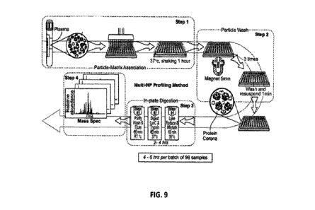
[0077] FIG. 9 shows a process for proteomic analysis. The process is tailored
for high-
throughput and automation that can be run in hours and across multiple samples
in parallel. The
process includes particle-matrix association, particle wash (x3), formation of
the protein corona,
in-plate digestion, and mass spectrometry. Using the process, it may take only
4 to 6 hours per
batch of 96 samples. One nanoparticle, or more, at a time may be incubated
with a sample.
[0078] FIG. 10 shows the protein counts (number of proteins identified from
corona analysis)
collected on pluralities of particles comprising from 1 particle type to 12
particle types. Each
particle from among a plurality of particles may be comprise unique materials,
surface
functionalization, and/or physical property (e.g., size or shape). Pooled
plasma from a group of
healthy subjects was used. Counts are the numbers of unique proteins collected
from a plurality
of particles and observed in about 2 hour mass spectrometry (MS) runs. 1318
proteins were
identified from the sample contacted with a plurality of particles comprising
12 particle types.
[0079] FIG. 11 shows the distribution of the presence-filtered, cluster
quality-filtered, median-
normalized MS feature intensities for the 56-sample NSCLC comparative study.
Each line
represents the density of the 10g.2 feature intensity for either a diseased
sample or a control
sample. Density is plotted from 0.00 to 0.15 on the y-axis, and log2 feature
intensity is plotted
-18-
CA 03146525 2022-2-1
WO 2021/026172
PCT/US2020/044908
from 15 to 35 on the x-axis. At the highest peak located near a log2 feature
intensity of about 28,
with densities ranging from about 0.13 to about 0.17, the two highest traces
correspond to
control samples, while the lowest trace corresponds to a diseased sample. The
remaining control
and diseased traces are distributed between the highest and lowest traces. At
the two shoulder
peaks, occurring at about 20 log2 feature intensity and about 23 10g2 feature
intensity, the highest
two traces are control traces and the lowest two traces are control traces at
the 20 10g2 feature
intensity peak, and the highest traces is a diseased trace at the 23 10g2
feature intensity.
[0080] FIG. 12 shows changed features in a non-small cell lung cancer (NSCLC)
pilot study
using poly(N-(3-(dimethylamino)propyl) methacrylamide) (PDMAPMA)-coated SPION
particles. Seven MS features were identified as statistically, significantly
different between 28
subjects with Stage IV NSCLC (with associated co-morbidities and treatment
effects) and 28
age- and gender-matched, apparently healthy subjects. The table at bottom is a
list of the seven
proteins that were significantly different. This includes 5 known proteins and
2 unknown
proteins. If a peptide-spectrum match was made for MS2 data associated with
the feature, that
peptide sequence (and charge) as well as the potential parent protein are
indicated; if an MS2
match was not associated with the feature, both the peptide and the protein
are marked as
"Unknown".
[0081] FIG. 13 shows correlation of the maximum intensities of particle corona
proteins and
plasma proteins to the published concentration of the same proteins. The blue
plotted lines are
linear regression models to the data and the shaded regions represent the
standard error of the
model fit. The dynamic range of the samples assayed with particles ("S-003,"
"S-007," and "S-
011", detailed in TABLE 1) exhibited a compressed dynamic range as compared to
the plasma
sample not assayed with particles ("Plasma"), as shown by the decrease in
slopes of the linear
fits. The slopes of each plot are 0.47, 0.19, 0.22, and 0.18 for, plasma
without particles, plasma
with S-003 particles, plasma with S-007 particles, and plasma with S-011
particles, respectively.
[0082] FIG. 14 shows the dynamic range compression of a protein corona
analysis assay with
mass spectrometry as compared to mass spectrometry without particle corona
formation. Protein
intensities of common proteins identified in particle corona in the plasma
samples assayed in
FIG. 13 ("Nanoparticle MS In Intensity") are plotted against the protein
intensity identified by
mass spectrometry of plasma without particles ("Plasma MS ln Intensity"). The
lightest dotted
line shows a slope of 1, indicating the dynamic range of mass spectrometry
without particles.
The slopes of the linear fits to the protein intensity was 0.12, 0.36, and
0.093 for S-003, S-007,
and S-011 panicles, respectively. The grayed area indicates the standard error
region of the
regression fit.
-19-
CA 03146525 2022-2-1
WO 2021/026172
PCT/US2020/044908
DETAILED DESCRIPTION
[0083] While various embodiments of the invention have been shown and
described herein, it
will be obvious to those skilled in the art that such embodiments are provided
by way of example
only. Numerous variations, changes, and substitutions may occur to those
skilled in the art
without departing from the invention. It should be understood that various
alternatives to the
embodiments of the invention described herein may be employed.
[0084] Whenever the term "at least," "greater than," or "greater than or equal
to" precedes the
first numerical value in a series of two or more numerical values, the term
"at least," "greater
than" or "greater than or equal to" applies to each of the numerical values in
that series of
numerical values. For example, greater than or equal to 1, 2, or 3 is
equivalent to greater than or
equal to 1, greater than or equal to 2, or greater than or equal to 3.
[0085] Whenever the term "no more than," "less than," or "less than or equal
to" precedes the
first numerical value in a series of two or more numerical values, the term
"no more than," "less
than," or "less than or equal to" applies to each of the numerical values in
that series of
numerical values. For example, less than or equal to 3, 2, or 1 is equivalent
to less than or equal
to 3, less than or equal to 2, or less than or equal to 1.
[0086] As used herein, a "feature" identified by mass spectrometry includes a
signal at a specific
combination of retention time and mh (mass-to-charge ratio), where each
feature has an
associated intensity. Some features are further fragmented in a second mass
spectrometry
analysis (MS2) for identification.
[0087] As used herein, the term "sensor element" refers to elements that are
able to bind to a
plurality of biomolecules when in contact with a sample and encompasses the
term "nanoscale
sensor element". A sensor element may be a particle, such as a nanoparticle,
or microparticle. A
sensor element may be a surface or a portion of a surface. A sensor element
may comprise a
panicle or plurality of particles. A sensor element may comprise a plurality
of surfaces capable
of adsorbing or binding biomolecules. A sensor element may comprise a porous
material, such as
a material into which biomolecules can intercalate.
[0088] As used herein, a "sensor array" may comprise a plurality of sensor
elements wherein the
plurality of sensor elements (e.g., particles) comprises multiple types of
sensor elements. The
sensor elements may be different types that differ from each other in at least
one
physicochemical property. A sensor array may be a substrate with a plurality
of partitions
containing a plurality of sensor elements (e.g., particles). For example, a
sensor array may
comprise a multi-well plate with a plurality of particles distributed between
the plurality of wells.
A sensor array may be a substrate comprising a plurality of partitions,
wherein the plurality of
-20-
CA 03146525 2022-2-1
WO 2021/026172
PCT/US2020/044908
partitions comprises a plurality of particles. In some embodiments, each
sensor element or
particle is able to bind a plurality of biomolecules in a sample to produce a
biomolecule corona
signature. In some embodiments, each sensor element (e.g., particle type) has
a distinct
biomolecule corona signature.
[0089] As used herein, the term "biomolecule corona" refers to the plurality
of different
biomolecules that bind to a sensor element. The term "biomolecule corona" may
refer to the
proteins, lipids and other plasma components that bind to particles (e.g.,
nanoparticles) when
they come into contact with biological samples or biological system. For use
herein, the term
"biomolecule corona" also encompasses both the soft and hard protein corona as
referred to in
Milani et al. "Reversible versus Irreversible Binding of Transferring to
Polystyrene
Nanoparticles: Soft and Hard Corona" ACS NANO, 2012, 6(3), pp. 2532-2541;
Mirshafiee et al.
"Impact of protein pre-coating on the protein corona composition and
nanoparticle cellular
uptake" Biomaterials vol. 75, January 2016 pp. 295-304, Mahmoudi et al.
"Emerging
understanding of the protein corona at the nano-bio interfaces" Nanotoday
11(6) December
2016, pp. 817-832, and Mahmoudi et al. "Protein-Nanoparticle Interactions:
Opportunities and
Challenges" Chem. Rev., 2011, 111(9), pp. 5610-5637, the contents of which are
incorporated
by reference in their entireties. As described therein, an adsorption curve
may show the build-up
of a strongly bound monolayer up to the point of monolayer saturation (at a
geometrically
defined protein-to-NP ratio), beyond which a secondary, weakly bound layer is
formed. While
the first layer is irreversibly bound (hard corona), the secondary layer (soft
corona) may exhibit
dynamic exchange. Proteins that adsorb with high affinity may form the "hard"
corona,
comprising tightly bound proteins that do not readily desorb, and proteins
that adsorb with low
affinity may form the "soft" corona, comprising loosely bound proteins. Soft
and hard corona
can also be characterized based on their exchange times. Hard corona may show
much larger
exchange times in the order of several hours. See, e.g., M. Rahman et al.
Protein-Nanoparticle
Interactions, Spring Series in Biophysics 15, 2013, incorporated by reference
in its entirety.
[0090] The term "biomolecule" refers to biological components that may be
involved in corona
formation, including, but not limited to, for example, proteins, polypeptides,
polysaccharides, a
sugar, a lipid, a lipoprotein, a metabolite, an oligonucleotide, metabolome or
combination
thereof It is contemplated that the biomolecule coronas of distinct particles
may contain some of
the same biomolecules, may contain distinct biomolecules with regard to the
other sensor
elements, and/or may differ in level or quantity, type or conformation of the
biomolecule that
binds to each sensor element. In one embodiment, the biomolecule is selected
from the group of
proteins, nucleic acids, lipids, and metabolomes.
-21-
CA 03146525 2022-2-1
WO 2021/026172
PCT/US2020/044908
[0091] The term "biomolecule corona signature" refers to the composition,
signature or pattern
of different biomolecules that are bound to each type of particle or separate
sensor element. The
signature may not only refers to the different biomolecules but also the
differences in the
amount, level or quantity of the biomolecule bound to the sensor element, or
differences in the
conformational state of the biomolecule that is bound to the particle or
sensor element. It is
contemplated that the biomolecule corona signatures of each distinct type of
sensor elements
may contain some of the same biomolecules, may contain distinct biomolecules
with regard to
the other sensor elements, and/or may differ in level or quantity, type or
conformation of various
biomolecules. The biomolecule corona signature may depend on not only the
physicochemical
properties of the sensor element (e.g., particle), but also the nature of the
sample and the duration
of exposure to the biological sample.
[0092] Disclosed herein are compositions and methods for multi-omic
analysis."Multi-omic(s)"
ormultiomic(s)" can refer to an analytical approach for analyzing biomolecules
at a large scale,
wherein the data sets are multiple omes, such as proteome, genome,
transcriptome, lipidome, and
metabolome. Non-limiting examples of multi-omic data include proteomic data,
genomic data,
lipidomic data, glycomic data, transcriptomic data, or metabolomics data.
[0093] "Biomolecule" in "biomolecule corona" can refer to any molecule or
biological
component that can be produced by, or is present in, a biological organism.
Non-limiting
examples of biomolecules include proteins (protein corona), polypeptides,
oligopeptides,
polyketides, polysaccharides, a sugar, a lipid, a lipoprotein, a metabolite,
an oligonucleotide, a
nucleic acid (DNA, RNA, micro RNA, plasmid, single stranded nucleic acid,
double stranded
nucleic acid), metabolome, as well as small molecules such as primary
metabolites, secondary
metabolites, and other natural products, or any combination thereof. In some
embodiments, the
biomolecule is selected from the group of proteins, nucleic acids, lipids, and
metabolomes.
[0094] Currently, there are a small number of protein-based biomarkers in use
today for clinical
diagnosis, and in spite of extensive efforts to analyze the plasma proteome
for the expansion of
markers, relatively few new candidates have been accepted as clinically useful
indicators. The
plasma proteome contains >10,000 proteins and potentially an order of
magnitude more protein
isoforms with a concentration range spanning over 10 orders of magnitude (from
mg/mL to
pg/mL). These attributes, combined with a lack of convenient molecular tools
for proteome
analysis, make comprehensive studies of the plasma proteome exceptionally
challenging
Approaches to overcome the broad dynamic range of proteins in biological
samples must be
capable of identifying and quantifying against a background of thousands of
unique proteins and
even more protein variants. However, there are no existing technologies that
are capable of
-22-
CA 03146525 2022-2-1
WO 2021/026172
PCT/US2020/044908
simultaneous measurement of proteins across the entire plasma concentration
range in a format
with a sufficient throughput and with a practical cost profile to allow for
appropriately-sized
studies with robust prospects for validation and replication. These challenges
not only limit the
discovery of novel disease biomarkers, but have been a bottleneck against the
adoption of
proteogenomics and protein annotation of genomic variants. Advances in mass
spectrometry
(MS) along with development of improved data analytics have offered tools for
deep and broad
proteomic analysis. Several attempts have been made to substantially improve
the detection of
low abundance proteins, such as depletion of highly abundant proteins, plasma
fractionation, and
peptide fractionation. It is now possible to identify over 4,500 proteins in
plasma. However,
current approaches are fairly complex and time-consuming (days to weeks), and
thus require a
tradeoff between depth of protein coverage and sample throughput.
Consequently, a simple and
robust strategy for comprehensive and rapid analysis of the available body of
information in the
proteome remains an unmet need.
[0095] Additionally, the earlier a disease is diagnosed, the more likely that
the disease can be
cured or successfully managed leading to a better prognosis for the patient.
When a disease is
treated early, it may be possible to prevent or delay problems from the
disease and may improve
the outcomes for the patient, including extending the patient's life and/or
quality of life.
[0096] Early diagnosis of cancer is crucial, as many types of cancers can be
successfully treated
in their early stages. For example, five-year survival after early diagnosis
and treatment of breast,
ovarian, and lung cancers is 90%, 90%, and 70%, respectively, compared to 15%,
5%, and 10%
for patients diagnosed at the most advanced stage of disease. Once cancer
cells leave their tissue
of origin, successful treatment using available established therapeutics
becomes very unlikely.
Although recognizing the warning signs of cancers and taking prompt action may
lead to early
diagnosis, the majority of cancers (e.g., lung) show symptoms only after
cancer cells have
already invaded the surrounding tissues and metastasized throughout the body.
For example,
more than 60% of patients with breast, lung, colon, and ovarian cancer have
concealed or even
metastatic colonies by the time their cancers are detected. Therefore, there
is an urgent need for
development of an effective approach for early detection of cancer. Such an
approach should
have the sensitivity to identify a cancer at various stages and the
specificity to give a negative
result when the person being tested is free of the cancer. There have been
extensive efforts to
develop methods for early detection of cancers; although huge numbers of risk
factors and
biomarkers have been introduced, a broadly relevant platform for early
detection of a wide range
of cancers remains elusive.
-23-
CA 03146525 2022-2-1
WO 2021/026172
PCT/US2020/044908
[0097] As various types of cancers can change the composition of blood
plasma¨even in their
early stages¨one promising approach for early detection is molecular blood
analysis for
biomarkers. Although this strategy has already worked for a few cancers (like
PSA for prostate
cancer), there are not yet specific biomarkers for early detection of the
majority of cancers. For
such cancers (e.g., lung), none of the defined candidate circulating
biomarkers has been
clinically validated, and very few have reached late-stage clinical
development. Therefore, there
is an urgent need for novel approaches to improve our ability to detect
cancer, as well as other
diseases, at very early stages.
Automated Sample Preparation
[0098] The present disclosure provides systems and methods for automated
sample preparation,
data generation, protein corona analysis. As is depicted in FIG. 1, the
systems and methods can
comprise (1) contacting a sample to particles (e.g., in a particle mixture) on
a sensor array,
substrate, plate, or within partitions on any of the foregoing, (2) allowing
biomolecules in the
sample to bind to the particles, (3) removing unbound sample from the
particles, and (4)
preparing a sample for analysis (e.g., using mass spectrometry ("MS")). For
example, in (1), a
method of the present disclosure can comprise contacting a biological sample
to a plurality of
particles. In (2), the sample may be incubated with the plurality of particles
so as to promote
biomolecule adsorption to the particles. In (3), unbound sample may be removed
while retaining
the particles and the biomolecules adsorbed to the particles. In (4) the
adsorbed biomolecules
may be desorbed from the particles and preparing them for mass spectrometric
analysis by which
example data may be generated.
[0099] The present disclosure provides automated systems, methods and kits for
biomolecule
corona preparation and analysis. The automated apparatus may perform at least
the
aforementioned data generating steps outlined in FIG. 1 using various units
illustrated in HG. 2.
The automated apparatus may contain a substrate with a plurality of partitions
containing sensor
elements 205 and a biological sample 210. The loading unit 215 on the
apparatus may transfer a
portion of the biological sample 210 into a partition on the substrate 205,
leading to adsorption of
biomolecules from the biological sample onto a sensor element in the partition
on the substrate.
The automated apparatus may then remove unbound biomolecules from the
partition, optionally
transferring the unbound sample into a waste receptacle 220. The remaining
biomolecules (e.g.,
biomolecules adsorbed to the sensor element) may be desorbed, collected, and
prepared for mass
spectrometric analysis. The reagents 225 may comprise a buffer, such as a
resuspension buffer
capable of desorbing biomolecules from a biomolecule corona or a denaturation
buffer capable
-24-
CA 03146525 2022-2-1
WO 2021/026172
PCT/US2020/044908
of denaturing or fragmenting a biomolecule. Reagents (e.g., a buffer or
protease) 225 may also
be loaded using the loading unit 215 to facilitate any of the foregoing.
[0100] In some aspects, the present disclosure provides an automated system
comprising a
network of units with differentiated functions in distinguishing states of a
complex biological
sample using a plurality of particles having surfaces with different
physicochemical properties
wherein: a first unit comprises a multichannel fluid transfer instrument for
transferring fluids
between units within the system; a second unit comprises a support for storing
a plurality of
biological samples; a third unit comprises a support for a sensor array plate
(e.g., a substrate
comprising a plurality of partitions comprising sensor elements, such as a 96
well plate
containing nanoparticles) possessing partitions that comprise the plurality of
particles having
surfaces with different physicochemical properties for detecting a binding
interaction between a
population of analytes within the complex biological sample and the plurality
of particles; a
fourth unit comprises supports for storing a plurality of reagents; a fifth
unit comprises supports
for storing a reagent to be disposed of; a sixth unit comprises supports for
storing consumables
used by the multichannel fluid transfer instrument; and wherein the system is
programed to
perform a series of steps comprising: contacting the biological sample with a
specified partition
of the sensor array; incubating the biological sample with the plurality of
particles contained
within the partition of the sensor array plate; removing all components from a
partition except
the plurality of particles and a population of analytes interacting with a
particle; and preparing a
sample for mass spectrometry.
[0101] An example of such an apparatus is provided in FIG. 3. The apparatus
comprises an
automated pipette that is able to transfer volumes between a biological sample
storage unit, a
substrate comprising a plurality of partitions comprising a plurality of
sensor elements, a waste
collection unit, a unit comprising a denaturation solution, and a unit
comprising a resuspension
solution. The automated apparatus can perform a biomolecule corona assay which
comprises
transferring a portion of the biological sample into a partition within the
substrate comprising a
sensor element, incubating the portion of the sample with the sensor element
to allow
biomolecules from the biological sample to bind to the sensor element,
removing contents from
the partition comprising biomolecules that are not bound to the sensor
elements, and then
preparing the biomolecules that remained within the partition for mass
spectrometric (MS)
analysis (e.g., LC-MS).
[0102] The loading may comprise a degree of mobility that enables access to
all other unit
within the system. The loading may comprise a capacity to perform pipetting
functions.
-25-
CA 03146525 2022-2-1
WO 2021/026172
PCT/US2020/044908
[0103] The system or apparatus of the present disclosure may comprise support
for a single
plate, a 6 well plate, a 12 well plate, a 96 well plate, a 192 well plate, a
384 well plate, or a rack
of microtubes. In some embodiments, the system or apparatus of the present
disclosure may
comprise a thermal unit capable of modulating the temperature of said support
and a sample. In
some embodiments, the system or apparatus of the present disclosure may
comprise a rotational
unit capable of physically agitating and/or mixing a sample.
[0104] In some embodiments, the plurality of particles comprises surfaces with
different
physicochemical properties for detecting a binding interaction between a
population of analytes
within the complex biological sample and the plurality of particles are
immobilized to a surface
with a partition of the sensory array. In some embodiments, the plurality of
particles comprises a
plurality magnetic nanoparticles in a solution with different physicochemical
properties for
detecting a binding interaction between a population of analytes within the
complex biological
sample and the plurality of particles. In some embodiments, the system
comprises a step wherein
the sensor array plate is transferred to an additional seventh unit that
comprises a magnetized
support and a thermal unit capable of modulating the temperature of said
support and a sample
and incubated for an additional amount of time.
[0105] In some embodiments, the fourth unit comprises a set of reagents for:
generating the
sensor array plate; washing an unbound sample; and/or preparing a sample for
mass
spectrometry. In some embodiments, contacting the biological sample with a
specified partition
of the sensor array comprises pipetting a specified volume of the biological
sample into the
specific partition of the sensor array. In some embodiments, contacting the
biological sample
with a specified partition of the sensor array comprises pipetting a volume
corresponding to a
1:1, 1:2: 1:3, 1:4, 1:5, 1:6, 1:7, 1:8, 1:9, 1:10, 1:15, or 1:20 ratio of a
plurality of particles in a
solution to the biological sample.
[0106] In some embodiments, contacting the biological sample with a specified
partition of the
sensor array comprises pipetting a volume of at least 10 microliters, at least
50 microliters, at
least 100 microliters, at least 250 microliters, at least 500 microliters, or
at least 1000 microliters
the biological sample into the specific partition of the sensor array.
Automated Apparatus
[0107] In some aspects, the present disclosure provides an automated apparatus
for generating a
subset of biomolecules from a biological sample, comprising: a substrate
comprising a plurality
of partitions, a first unit comprising the biological sample, and a loading
unit that is movable
across the substrate and is capable of transferring a volume (e.g., a volume
of buffer) between
different units of the apparatus. In some cases, the substrate is a multi-well
plate.
-26-
CA 03146525 2022-2-1
WO 2021/026172
PCT/US2020/044908
[0108] The plurality of partitions may comprise a plurality of sensor
elements. The plurality of
sensor elements may comprise particles. The plurality of sensor elements may
be particles (e.g.,
nanoparticles or micropartides).
[0109] A partition from among the plurality of partitions may comprise 1 to
100 types of sensor
elements (e.g., distinct particle types). A partition from among the plurality
of partitions may
comprise 2 to 50 types of sensor elements. A partition from among the
plurality of partitions may
comprise 2 to 5 types of sensor elements. A partition from among the plurality
of partitions may
comprise 3 to 8 types of sensor elements. A partition from among the plurality
of partitions may
comprise 4 to 10 types of sensor elements. A partition from among the
plurality of partitions may
comprise 5 to 12 types of sensor elements. A partition from among the
plurality of partitions may
comprise 6 to 15 types of sensor elements. A partition from among the
plurality of partitions may
comprise 8 to 20 types of sensor elements.
[0110] Two or more partitions from among the plurality of partitions may
comprise different
quantities of sensor elements. two or more partitions from among the plurality
of partitions may
comprise different types of sensor elements. A partition amongst a plurality
of partitions may
comprise a combination of types and/or quantities of sensor dement(s) that
differs from other
partitions in the plurality. A subset of partitions in a plurality of
partitions may each contain a
combination of distinct sensor elements that is distinct from other partitions
in the plurality.
[0111] Sensor elements may be stored in dry form inside of or within the
partitions. Dry sensor
elements may be reconstituted or rehydrated prior to use. Sensor elements may
also be stored
within solutions. For example, a substrate partition may comprise a solution
comprising a high
concentration of particles.
[0112] Partitions from among the plurality of partitions comprise different
concentrations or
amounts (e.g., by mass/molar amount per unit volume of sample) of sensor
elements. A partition
from among the plurality of partitions may comprise from 1 pM to 100 nIvI of
sensor elements. A
partition from among the plurality of partitions comprise may from 1 pM to 500
pM of sensor
elements. A partition from among the plurality of partitions may comprise from
10 pM to 1 nivl
of sensor elements. A partition from among the plurality of partitions may
comprise from 100
pM to 10 nM of sensor elements.A partition from among the plurality of
partitions may comprise
from 500 pM to 100 KIM of sensor elements. A partition from among the
plurality of partitions
may comprise from 50 pg/m1 to 3001,1Wm1 of sensor elements. A partition from
among the
plurality of partitions may comprise from 100 g/m1 to 500 Wm' of sensor
elements. A
partition from among the plurality of partitions may comprise from 250 pg/ml
to 750 pg/m1 of
sensor elements. A partition from among the plurality of partitions may
comprise from 400
-27-
CA 03146525 2022-2-1
WO 2021/026172
PCT/US2020/044908
pg/m1 to 1 mg/ml of sensor elements. A partition from among the plurality of
partitions may
comprise from 600 pg/ml to 1.5 mg/ml of sensor elements. A partition from
among the plurality
of partitions may comprise from 800 pg/ml to 2 mg/ml of sensor elements. A
partition from
among the plurality of partitions may comprise from 1 mg/ml to 3 mg/ml of
sensor elements_ A
partition from among the plurality of partitions may comprise from 2 mg/ml to
5 mg/ml of
sensor elements. A partition from among the plurality of partitions may
comprise more than 5
mg/m1 of sensor elements.
[0113] The loading unit may be configured to move between and transfer volumes
(e.g., a
volume of a solution or a powder) between any units, compartments, or
partitions within the
apparatus. The loading unit may be configured to move precise volumes (e.g.,
within 0.1%,
0.01%, 0.001% of the specified volume). The loading unit may be configured to
collect a volume
from the substrate or a compartment or partition within the substrate, and
dispense the volume
back into the substrate or compartment or partition within the substrate, or
to dispense the
volume or a portion of the volume into a different unit, compartment, or
partition. The loading
unit may be configured to move multiple volumes simultaneously, such as 2 to
400 separate
volumes. The loading unit may comprise a plurality of pipette tips.
[0114] The loading unit may be configured to move a volume of a liquid. The
volume may be
about 0.1 p.1,0.2 p1,0.3 p.1,0.4 p.1,0.5 p.1,0.6 p.1, 0.7 p1,0.8 p.1, 0.9 p.1,
1 p.1,2 p.1,3 p.1,4 p.1,5 p1,6
I, 7 1, 8 I, 9 I, 10 1, 12 I, 15 I, 20 1, 25 I, 30 I, 40 1, 50 1,
60 I, 70 I, 80 I, 90 1,
100 1, 120 p.1, 150 p.1, 180 p.1, 200 p1, 250 p1, 300 pl, 400 1, 500 1, 600
p.1, 800 I, 1 ml, or
more than 1 ml. The liquid may be a biological sample or a solution.
[0115] In some cases, the solution comprises a wash solution, a resuspension
solution, a
denaturing solution, a buffer, a reagent (e.g., a reducing reagent), or any
combination thereof. In
some cases, the solution comprises a biological sample.
[0116] In part owing to these functionalities, the loading unit can be capable
of partitioning a
sample. In some embodiments, this comprises dividing a sample into a number of
partitions. A
sample can be divided into at least 2, 3, 4, 5, 6, 7, 8, 9, 10, 12, 15, 20,
25, 30, 40, 50, 60, 70, 80,
90, 100, 120, 150, 180, 200, 250, 300, 350, 400, 500, or more partitions. A
sample can be
divided into 96, 192, or 384 partitions. The automated apparatus can comprise
multiple
substrates comprising partitions. The automated apparatus may comprise 1, 2,
3, 4, 5, or more
substrates comprising partitions. In some cases, the loading unit loads
different volumes of the
biological sample into different partitions. In some cases, the loading unit
loads identical
volumes into two or more partitions. The volume of biological sample loaded
into a partition
may be about 0.1 pl, 0.2 I, 0.3 p.1, 0.4 p.1, 0.5 1, 0.6 pl, 0.7 1, 0.8
p.l, 0.9 p.1, 1 1, 2 1, 3 pl, 4
-28-
CA 03146525 2022-2-1
WO 2021/026172
PCT/US2020/044908
pl, 90 I, 100 .1, 120 I, 150 I, 180 p.1, 200 p.1, 250 I, 300 p1,400 l,
500 I, 600 1, 800 I, 1
ml, or more than 1 mi. The volume of biological sample loaded into a partition
may be about 10
p1 to 400 1. The volume of biological sample loaded into a partition may be
about 5 pl to 150
1. The volume of biological sample loaded into a partition may be about 35 p.1
to 80 p.1. In some
cases, the loading unit may partition two or more biological samples. For
example, a sample
storage unit may comprise two biological samples that the system partitions
into one well plate.
In some embodiments, the loading unit can facilitate a transfer of the sample
for mass
spectrometry to a mass spectrometry unit.
[0117] The system may be configured to perform a dilution on a sample or a
sample partition. A
sample or sample partition may be diluted with buffer, water (e.g., purified
water), a non-
aqueous solvent, or any combination thereof. The diluent may be stored in the
automated
apparatus prior to dispensation into a substrate partition. The automated
apparatus may store a
plurality of diluents differing in pH, salinity, osmolarity, viscosity,
dielectric constant, or any
combination thereof. The diluents may be used to adjust the chemical
properties of a sample or
sample partition. The automated apparatus may dilute a sample or sample
partition by 2-fold, 3-
fold, 4-fold, 5-fold, 6-fold, 8-fold, 10-fold, 15-fold, 20-fold, 30-fold, 40-
fold, 50-fold, 75-fold,
100-fold, 150-fold, 200-fold, 300-fold, 400-fold, 500-fold or greater. The
automated apparatus
may perform different dilutions on two samples or sample partitions. The
system may perform
different dilutions on each partition from among a plurality of partitions.
For example, the
system may perform different dilutions on each of the 96 sample partitions in
a 96 well plate. In
some cases, the different dilutions comprise different degrees of dilution
(e.g., 2-fold vs. 4-fold).
In some cases, the different dilutions comprise dilution with different
solutions (e.g., different
buffers). In some cases, two sample partitions may be made to differ in one or
more chemical
properties, such as pH, salinity, or viscosity.
[0118] In some cases, the system may modify the chemical composition of a
sample or sample
partition. The system may modify or adjust the pH, salinity, osmolarity,
dielectric constant,
viscosity, buffer types, salt types, sugar types, detergent types, or any
combination thereof for a
sample or sample partition. Such modification or adjustments may comprise
mixing a reagent
from the fourth unit with a sample or sample partition. The system may
differently modify the
chemical composition of two samples or sample partitions.
101191 A system or automated apparatus of the present disclosure may also
comprise an
incubation element. The incubation element may contact, support, or hold
another component of
the automated apparatus (e.g., the substrate or a unit). The incubation unit
may contact, support,
-29-
CA 03146525 2022-2-1
WO 2021/026172
PCT/US2020/044908
or hold multiple components of the automated apparatus. The incubation element
may contact
the substrate to facilitate heat transfer between the incubation element and
the substrate. The
incubation unit may be configured to control the temperature of the one or
more components of
the automated apparatus, such as by heating or cooling. The incubation element
may be capable
of cooling a component of the apparatus to from 20 C to 1 C. The incubation
element may be
capable of heating a component of the apparatus to from 25 C to 100 'C. The
incubation
element may be capable of setting the temperature a component of the apparatus
to from 4 C to
37 C. The incubation element may be configured to heat or cool different
portions of a
component of the automated apparatus to different temperatures. For example,
the incubation
element may hold a first partition in the substrate at 30 C and a second
partition in the substrate
at 35 'C. The incubation element may control the temperature of a sample or
partition. The
incubation element may comprise a temperature sensor (e.g., a thermocouple)
for detecting the
temperature within a partition or container. The incubation element may
calibrate its heating or
cooling to the readout from the temperature sensor.
[0120] The incubation element may be configured to physically agitate a
component of the
automated apparatus. The agitation may be in the form of shaking or spinning,
vibrating,
rocking, sonicating, or any combination thereof The incubation element may be
capable of
providing multiple agitation intensities and/or frequencies. For example, the
incubation element
may comprise multiple settings for shaking at different frequencies and
amplitudes. The
incubation element may also be capable of stirring and or mixing a volume
(e.g., a portion of the
biological sample).
[0121] The automated apparatus may comprise a unit comprising a resuspension
solution. The
loading unit may be capable of transferring a volume of the resuspension
solution to a partition
from among the plurality of partitions of the substrate. In some cases, this
results in the dilution
of a sample present within the partition and can further result in the
desorption of a plurality of
biomolecules from a biomolecule corona disposed on a sensor element within the
partition. The
quantity of biomolecules desorbed from a biomolecule corona can depend on the
volume of the
resuspension solution added to the partition, the temperature of the
partition, the composition of
the resuspension solution (e.g., the salinity, osmolarity, viscosity,
dielectric constant, or pH), the
volume of the biological sample within the partition, and the sensor element
type and the
composition of biomolecules in the biomolecule corona. The transfer of a
volume of the
resuspension solution into a partition may result in the desorption of less
than 5% of the
biomolecules from a biomolecule corona. The transfer of a volume of the
resuspension solution
into a partition may result in the desorption of 10% to 20% of the
biomolecules from a
-30-
CA 03146525 2022-2-1
WO 2021/026172
PCT/US2020/044908
biomolecule corona. The transfer of a volume of the resuspension solution into
a partition may
result in the desorption of 20% to 30% of the biomolecules from a biomolecule
corona. The
transfer of a volume of the resuspension solution into a partition may result
in the desorption of
30% to 40% of the biomolecules from a biomolecule corona. The transfer of a
volume of the
resuspension solution into a partition may result in the desorption of 40% to
50% of the
biomolecules from a biomolecule corona. The transfer of a volume of the
resuspension solution
into a partition may result in the desorption of 50% to 60% of the
biomolecules from a
biomolecule corona. The transfer of a volume of the resuspension solution into
a partition may
result in the desorption of 60% to 70% of the biomolecules from a biomolecule
corona. The
transfer of a volume of the resuspension solution into a partition may result
in the desorption of
70% to 80% of the biomolecules from a biomolecule corona. The transfer of a
volume of the
resuspension solution into a partition may result in the desorption of 80% to
90% of the
biomolecules from a biomolecule corona. The transfer of a volume of the
resuspension solution
into a partition may result in the desorption of more than 90% of the
biomolecules from a
biomolecule corona.
[0122] In some cases, multiple rounds of desorption are performed. In each
round, the
supernatant comprising the desorbed biomolecules may be collected, analyzed,
or discarded. The
types and abundances of biomolecules in the supernatant may differ between
desorption rounds.
The automated apparatus may perform one or more desorption and discard cycles
(i.e., washes),
followed by one or more desorption cycles comprising sample collection and/or
analysis.
101231 The resuspension solution may be tailored to optimize enrichment of
particular
biomarkers. The resuspension solution may comprise a buffer, such as Tris-EDTA
(TE),
CHAPS, PBS, citrate, HEPES, MES, CUES, or another bio buffer. The resuspension
solution
may comprise Tris EDTA (TIE) 150mM KC1 0.05% CHAPS buffer. The resuspension
solution
may comprise 10 mM TrisHC1 pH 7.4, 1 mM EDTA. The resuspension solution may
also
contain or be highly purified water (e.g., distilled or deionized water).
Biomolecule desorption
may be augmented by heating or agitation by an incubation element. The
supernatant may be
transferred to a new partition following desorption. A resuspension solution
may be used to
dilute a sample.
[0124] The automated apparatus may comprise a unit comprising a denaturing
solution. The
denaturing solution may comprise a protease. The denaturing solution may
comprise a chemical
capable of performing peptide cleavage (e.g., cyanogen bromide, formic acid,
or hydroxylamine,
2-nitro-5-thiocyanatobenzoic acid). The denaturing solution may comprise a
chemical denaturant
such as guanidine, urea, sodium deoxycholate, acetonitrile, trichloroacetic
acid, acetic acid,
-31-
CA 03146525 2022-2-1
WO 2021/026172
PCT/US2020/044908
sulfosalicylic acid, sodium bicarbonate, ethanol, perchlorate, dodecyl
sulfate, or any combination
thereof. The denaturing solution may comprise a reductant, such as 2-
mercaptoethanol,
dithiothreitol, or tris(2-carboxyethyl)phosphine. The protease may be trypsin.
The denaturing
solution may be added to a partition following desorption. The denaturing
solution may be added
to a partition comprising biomolecule coronas.
[0125] The automated apparatus may comprise a magnet or array of magnets. The
automated
apparatus may capable of moving the substrate onto and off of the magnet or
array of magnets.
The array of magnets may be structured so that a plurality of magnets from the
array of magnets
can rest directly underneath underneath a plurality of partitions from the
substrate. The magnet
may be capable of immobilizing magnetic sensor elements (e.g., magnetic
particles such as
coated or uncoated super paramagnetic iron oxide nanoparticles) within a
partition on the
substrate. For example, the magnet may prevent magnetic nanoparticles from
being removed
from a partition during a wash step. The magnet may also create a pellet from
a collection of
magnetic particles. The magnet may create a particle pellet in less than 10
minutes. The magnet
may create a particle pellet in less than 5 minutes. The particle pellet may
comprise a particle
with a biomolecule corona
[0126] The automated apparatus may comprise a purification unit. The
purification unit may
comprise a plurality of partitions comprising an adsorbent or resin. The
purification unit may
comprise a solid-phase extraction array or plate. The solid-phase extraction
array or plate may
comprise a polar stationary phase material. The solid-phase extraction array
or plate may
comprise a non-polar stationary phase material. The solid-phase extraction
array or plate may
comprise a C18 stationary phase material (e.g., octadecyl group silica gel).
The automated
apparatus may comprises a unit with a conditioning solution for the
purification unit (e.g., a
conditioning solution for a solid-phase extraction material). The automated
apparatus may
comprise a unit with an elution solution for removing biomolecules from the
purification unit.
[0127] In some embodiments, components are removed from a partition, except
for the plurality
of sensor elements and a population of analytes interacting with the plurality
of sensor elements
(i.e., a wash step). In some instances, the automated apparatus may perform a
series of wash
steps. A wash step may remove biomolecules that are not bound to the sensor
elements within
the partition. A wash step may desorb a subset of biomolecules bound to sensor
elements within
a partition. For example, a wash step may result in the desorption and removal
of a subset of soft
corona analytes, while leaving the majority of hard corona analytes bound to
the sensor element.
[0128] In some aspects, the present disclosure provides an automated apparatus
to identify
proteins in a biological sample, the automated apparatus comprising: a sample
preparation unit; a
-32-
CA 03146525 2022-2-1
WO 2021/026172
PCT/US2020/044908
substrate comprising a plurality of channels; a plurality of pipettes; a
plurality of solutions, a
plurality of particles, and wherein the automated apparatus is configured to
form a protein corona
and digest the protein corona.
[0129] In some aspects, the present disclosure provides an automated apparatus
to identify
proteins in a biological sample, the automated apparatus comprising: a sample
preparation unit; a
substrate comprising a plurality of channels; a plurality of pipettes; a
plurality of solutions, a
plurality of nanoparticles, wherein the automated apparatus is configured to
form a protein
corona and digest the protein corona, and wherein at least one of the
solutions is TE 150mM KCl
0.05% CHAPS buffer.
[0130] In some embodiments, the sample preparation unit is configured to add
the plurality of
nanoparticles to the substrate with the plurality of pipettes. In some
embodiments, wherein the
sample preparation unit is configured to add the biological sample to the
substrate with the
plurality of pipettes. In some embodiments, the sample preparation unit is
configured to incubate
the plurality of nanoparticles and the biological sample to form the protein
corona.
[0131] In some embodiments, the sample preparation unit is configured to
separate the protein
corona from the supernatant to form a protein corona pellet. In some
embodiments, the sample
preparation unit is configured to reconstitute the protein corona pellet with
TE 150mM KCl
0.05% CHAPS buffer.
[0132] In some embodiments, the automated apparatus further comprises a
magnetic source. In
some embodiments, the automated apparatus is configured for BCA, gel, or
trypsin digestion of
the protein corona.
[0133] In some embodiments, the automated apparatus is enclosed. In some
embodiments, the
automated apparatus is sterilized before use. In some embodiments, the
automated apparatus is
configured to a mass spectrometry. In some embodiments, the automated
apparatus is
temperature controlled.
Assaying Methods
[0134] In some aspects, the present disclosure provides a method of identify
proteins in a
biological sample. In some cases the method comprises: adding the biological
sample to the
automated apparatus disclosed herein; generating proteomic data from the
automated apparatus;
and quantifying the proteomic data.
[0135] In some embodiments, the method comprises incubating a plurality of
biomolecules with
the biological sample in the automated apparatus to form a biomolecule corona.
In some
embodiments, incubating the biological sample with the plurality of sensor
elements (e.g.,
particles) contained within the partition of the substrate comprises an
incubation time of at least
-33-
CA 03146525 2022-2-1
WO 2021/026172
PCT/US2020/044908
about 10 seconds, at least about 15 seconds, at least about 20 seconds, at
least about 25 seconds,
at least about 30 seconds, at least about 40 seconds, at least about 50
seconds, at least about 60
seconds, at least about 90 seconds, at least about 2 minutes, at least about 3
minutes, at least
about 4 minutes, at least about 5 minutes, at least about 6 minutes, at least
about 7 minutes, at
least about 8 minutes, at least about 9 minutes, at least about 10 minutes, at
least about 15
minutes, at least about 20 minutes, at least about 25 minutes, at least about
30 minutes, at least
about 45 minutes, at least about 50 minutes, at least about 60 minutes, at
least about 90 minutes,
at least about 2 hours, at least about 3 hours, at least about 4 hours, at
least about 5 hours, at least
about 6 hours, at least about 7 hours, at least about 8 hours, at least about
9 hours, at least about
hours, at least about 12 hours, at least about 14 hours, at least about 15
hours, at least about
16 hours, at least about 17 hours, at least about 18 hours, at least about 19
hours, at least about
hours, or at least about 24 hours. In some cases, two wells will have two
different incubation
times. In some embodiments, incubating the biological sample with the
plurality of particles
contained within the partition of the substrate comprises an incubation
temperature between
about 4 C to about 370 C. In some embodiments, incubating the biological
sample with the
plurality of particles contained within the partition of the substrate
comprises an incubation
temperature between about 40 C to about 100 C.
[0136] The method, systems, and apparatus of the present disclosure may
comprise covering or
sealing a partition on the substrate. This may comprise covering a surface of
the apparatus with a
lid or a seal. The lid or seal may prevent solutions or species from leaving a
partition (e.g.,
evaporating from a partition). The automated apparatus may be configured to
place and/or
remove the lid or seal. The lid or seal may be pierceable (e.g., may comprise
a septum), thereby
allowing a syringe or needle to enter a substrate partition without removal of
the lid or seal.
[0137] In some cases, the system, apparatus and method of the present
disclosure further
comprise preparing analytes from the biomolecule corona for analysis (e.g.,
mass spectrometric
analysis). This can comprise separating the biomolecule corona from the
supernatant in the
automated apparatus. The biomolecule corona may be separated from the
supernatant by
removing the supernatant and then desorbing a plurality of proteins from the
biomolecule corona
into a desorbate solution (e.g., a resuspension solution). In some cases, a
first portion of
biomolecules from a biomolecule corona are desorbed from the biomolecule
corona and
discarded, and a second portion of biomolecules from a biomolecule corona are
desorbed from
the biomolecule corona and collected (e.g., for analysis). Multiple portions
of biomolecules from
a biomolecule corona may be separately, desorbed, collected, and analyzed.
-34-
CA 03146525 2022-2-1
WO 2021/026172
PCT/US2020/044908
[0138] In some cases, biomolecules within a biomolecule corona are denatured,
fragmented,
chemically modified, or any combination thereof. These treatments may be
performed on
desorbed biomolecules or on biomolecule coronas. The plurality of biomolecules
desorbed from
a biomolecule corona may comprise 1%, 2%, 3%, 4%, 5%, 6%, 8%, 10%, 12%, 15%,
20%,
25%, 30%, 40%, 50%, 60%, 70%, 80%, 90%, 95%, 98%, 99%, or greater than 99% of
the
biomolecules from the biomolecule corona. The desorption may performed for
different lengths
of time, including 5 seconds, 15 seconds, 30 seconds, 1 minute, 2 minutes, 3
minutes, 4 minutes,
minutes, 6 minutes, 8 minutes, 10 minutes, 12 minutes, 15 minutes, 20 minutes,
30 minutes, 40
minutes, 50 minutes, 1 hour, 1.5 hours, 2 hours, 3 hours, 4 hours, 5 hours, 6
hours, 8 hours, 12
hours, or longer. In some cases, the desorption comprises physical agitation,
such as shaking or
sonication. The percent of proteins desorbed from a particle corona may depend
on the
desorption time, the chemical composition of the desorbate solution into which
proteins are
desorbed (e.g., pH or buffer-type), the desorption temperature, the form and
intensity of physical
agitation applied, or any combination thereof Additionally, the types of
proteins desorbed from
a protein corona can be responsive to desorption conditions and methods. The
types of proteins
desorbed from a protein corona may differ by 1%, 2%, 3%, 4%, 5%, 6%, 8%, 10%,
12%, 15%,
20%, 25%, 30%, 40%, 50%, 60%, or more between two desorption conditions or
methods.
[0139] In some cases, preparing analytes from a biomolecule corona for
analysis comprises
digesting the biomolecule corona, a subset of biomolecules within the protein
corona, or
biomolecules desorbed from the biomolecule corona to form a digested sample in
the automated
apparatus. Preparing analytes from the biomolecule corona for analysis may
also comprise
chemically modifying a biomolecule from the biomolecule corona, such as
methylating or
reducing the biomolecule.
[0140] Desorbed biomolecules may be collected for further analysis (e.g., mass
spectrometric
analysis). The automated apparatus may perform the collecting, for example by
collecting a
volume of sample from a substrate partition comprising biomolecules desorbed
from
biomolecule coronas. A method may involve placing partition or plurality of
partitions (e.g., a
well plate) may be placed directly within an instrument for performing said
analysis.
[0141] A method may comprise multiple rounds of preparing analytes from a
biomolecule
corona for analysis. A method may comprise 1, 2, 3, 4, 5, 6, 7, 8, 9, 10, or
more rounds of
preparation. In some cases, each round produces a separate sample for analysis
(e.g., desorbed
biomolecules may be collected after each round and subjected to mass
spectrometric analysis).
Two rounds may comprise desorbing different pluralities of proteins from a
biomolecule corona.
Two rounds may also comprise different desorption methods or conditions, such
as different
-35-
CA 03146525 2022-2-1
WO 2021/026172
PCT/US2020/044908
desorbate solution volumes, different desorbate solution types (e.g.,
desorbate solutions
comprising different buffers or osmolarities), different temperatures, or
different types and
degrees of physical agitation. Two or more successive rounds of preparation
from a single
biomolecule corona (e.g., desorption and collection of a first subset of
biomolecules from a
biomolecule corona followed by desorption and collection of a second subset of
biomolecules
from a biomolecule corona) may generate two sets of biomolecules. This may
inform detection
or analysis of biomolecule interactions within a protein corona. As such,
multiple rounds of
preparation from a single biomolecule corona may be used to generate a number
biomolecule
subsets which exceed the number of partitions or types of sensor. For example,
a method
utilizing a substrate with 96 partitions (e.g., a 96 well plate) may generate
as many as 960 unique
biomolecule subsets if each partition comprises a unique combination of
particles and solution
conditions, and 10 rounds of analyte preparation are performed on each
partition.
[0142] A different number of rounds of analyte preparation may be performed in
separate
partitions. Partitions may also be subjected to different analyte preparation
conditions.
Performing more rounds of analyte preparation can increase the number of
proteins or types of
proteins collected for analysis (e.g., generate more proteins that fall within
concentration ranges
accessible for simultaneous mass spectrometric detection). The number proteins
or types of
proteins detected when multiple rounds of analyte preparation are performed
may be 10%, 20%,
30%, 40%, 50%, 60%, 70%, 80%, 90%, 100%, 150%, 200%, or more than 200% higher
than if a
single round of analyte preparation was performed.
[0143] In some cases, the method comprises immobilizing a sensor element
within a partition.
The immobilization may prevent the sensor element from being removed from the
partition when
a volume is removed from the partition (e.g., the loading unit removes 95% of
the solution from
the partition). Immobilization may be performed, for example, chemically
(e.g., covalent or non-
covalent binding to a substrate). Chemical immobilization may comprise
reacting a sensor
element with a surface of the partition. Chemical immobilization may also
comprise non-
covalently associating a sensor element with a surface of the partition. For
example, a sensor
element may comprise biotin moieties that bind to streptavidin bound to a
surface of a partition.
Immobilization may be achieved by applying a magnetic field to hold a magnetic
sensor element
within a partition. For example, a plurality of sensor elements may comprise a
plurality of
magnetic particles, and the substrate and the magnet may be in proximity such
that the one or
more magnetic particles are immobilized within a partition in the substrate.
Immobilization may
be achieved by providing a substrate with a sensor element formed or embedded
within a
-36-
CA 03146525 2022-2-1
WO 2021/026172
PCT/US2020/044908
partition on the substrate. For example, a sensor element may be a half-
particle formed on the
surface of a partition within the substrate.
[0144] In some cases, sensor element immobilization allows a biomolecule
corona to be
separated from the sensor element. This may comprise desorbing a plurality of
biomolecules
from a biomolecule corona associated with a sensor element, immobilizing the
sensor element
within the partition, and then collecting the solution with the plurality of
biomolecules from the
biomolecule corona, thereby separating at least a portion of a biomolecule
corona from a sensor
element.
[0145] FIG. 4 illustrates examples of methods comprising immobilization sensor
elements,
which can be performed by the automated apparatus of the present disclosure.
These methods
utilize particles 402 and 411 to capture a subset of biomolecules 403 and 404
in a sample.
[0146] Panel 400 shows a partition 401 containing particles 402 and
biomolecules. The particles
are suspended within the partition, and have adsorbed biomolecules 403 from
the sample,
thereby forming biomolecule coronas. A number of biomolecules 404 may not
adsorb to the
panicles, and will instead be suspended within the partition. Panel 410 shows
an alternative
method, comprising particles 411 that are formed on the surface of the
partition.
[0147] Panels 420 and 430 show two methods for immobilizing the particles. In
panel 420, the
particles are collected onto the bottom of the partition by a magnet 421. In
panel 430, the
particles are crosslinked to the partition via linkers 431. Both methods
result in the particles
becoming immobilized to the partition. Throughout the immobilization process,
particle-
adsorbed biomolecules 403 remain adsorbed to the particles, while the unbound
biomolecules
404 remain unbound from the particles.
[0148] Panel 440 shows the results of wash steps on the partitions from panels
410, 420, and
430. In all three cases, the wash removes unbound biomolecules from the
partition, while leaving
the immobilized particles and the biomolecules adsorbed to them. Panel 450
then shows
desorption of the biomolecule coronas, wherein a first plurality of
biomolecules 451 elute from
the particles, and a second plurality of biomolecules 403 remain adsorbed to
the particles. The
ratio of eluted to adsorbed biomolecules and the types of biomolecules eluted
from the particles
depends on the elution conditions (e.g., temperature, degree and type of
physical agitation,
solution conditions such as pH). The eluted biomolecules can be collected
(e.g., by the loading
unit) for further processing (e.g., fragmentation) or direct analysis.
[0149] FIG. 5 shows an example of a sample preparation method that can be
performed by the
automated apparatus of the present disclosure. This method utilizes sensor
elements 512 to
generate a subset of biomolecules from a biological sample 502. The biological
sample (shown
-37-
CA 03146525 2022-2-1
WO 2021/026172
PCT/US2020/044908
in panel 500), which is stored in a sample container 501, comprises a number
of biomolecules. A
volume of the sample can optionally be processed 504 (e.g., cells within the
sample can be lysed,
nucleic acids and proteins can be fragmented, the sample can be filtered to
remove large
biomolecules, etc.), and then added to a partition 511 comprising sensor
elements 512. As is
depicted in panel 520 a portion of the biomolecules 521 can bind to the sensor
elements,
separating them from a portion of biomolecules 522 that does not bind to the
sensor elements. As
is shown in panel 530, the sensor elements can then be immobilized within the
partition by
bringing the partition in contact with a magnet 531. The partition can then
undergo a wash cycle
(e.g., addition of buffer to the partition followed by removal of sample from
the partition),
resulting in the removal of the portion of biomolecules 522 that did not bind
to the sensor
elements (shown in panel 540). The bound biomolecules 521 can be eluted from
the sensor
elements and collected for further processing or analysis.
[0150] FIG. 6 illustrates a sample preparation method that can be performed by
the automated
apparatus of the present disclosure. This method utilizes sensor elements 512
that are formed on
the surface of a substrate partition 511 to collect biomolecules 503 from a
sample 502. The
biological sample is transferred 504 from a sample holding unit 501 to the
substrate partition
511. As is shown in panel 520, the sensor elements will adsorb a first portion
of the biomolecules
from the sample 521, while a second portion 522 will remain unbound. Panel 530
depicts the
removal of the unbound biomolecules, which leaves the sensor elements 512 and
sensor element-
bound biomolecules 521 within the partition. These biomolecules can
subsequently be desorbed
from the sensor elements and collected (e.g., by the loading unit) for further
processing or
analysis.
[0151] The methods disclosed herein may comprise filtering a sensor element
from a solution.
For example, the method may comprise desorbing a plurality of biomolecules
from a
biomolecule corona associated with a sensor element, and filtering the
solution such that the
sensor element is collected on the filter and the plurality of biomolecules
remain in solution. The
filtering may be performed after denaturation (e.g., digestion). The filtering
may also remove a
plurality of biomolecules or biological species such as intact proteins (e.g.,
undigested proteins
from the biological sample or proteases).
[0152] In some cases, the method comprises a purification step. The
purification step can
precede or follow preparation of analytes from a biomolecule corona. A
purification step may
comprise transferring a biological sample (e.g., biomolecules eluted and
collected from a
biomolecule corona) to a purification unit (e.g., a chromatography column) or
partition within a
purification unit. Purification may involve transferring a plurality of sample
partitions from the
-38-
CA 03146525 2022-2-1
WO 2021/026172
PCT/US2020/044908
substrate into separate partitions in the purification unit. The purification
unit may comprise a
solid-phase extraction plate. The purification step may remove reagents (e.g.,
chemicals and
enzymes) from the denaturation solution. Following purification, the
biological sample may be
recollected for further enrichment or chemical treatment within the substrate
or purification unit,
or may be collected for direct analysis (e.g., mass spectrometric analysis).
[0153] Collectively, the methods of the present disclosure enable a high
degree of profiling
depth for biological samples. The subset of biomolecules collected in the
methods of the present
disclosure may enable, without further manipulation or modification of said
subset of
biomolecules, mass spectrometric detection of at least 2%, at least 3%, at
least 4%, at least 5%,
at least 6%, at least 7%, at least 8%, at least 9%, at least 10%, at least
12%, at least 15 %, at least
20%, at least 25%, at least 30%, at least 40%, at least 50%, at least 60%, or
more than 60% of
the types of biomolecules in the biological sample from which the subset of
biomolecules were
collected. The subset of biomolecules may enable, without further manipulation
or modification
of said subset of biomolecules, mass spectrometric detection of at least 2%,
at least 3%, at least
4%, at least 5%, at least 6%, at least 7%, at least 8%, at least 9%, at least
10%, at least 12%, at
least 15 %, at least 20%, at least 25%, at least 30%, at least 40%, at least
50%, or more than 50%
of the types of proteins in a sample. The subset of biomolecules collected on
a sensor element or
prepared for analysis may enable, without further manipulation or modification
of said subset of
biomolecules, simultaneous mass spectrometric detection of two biomolecules
(e.g., proteins)
spanning 6, 7, 8, 9, 10, 11, 12 or more orders of magnitude in a sample. For
example, the two
biomolecules may be desorbed and collected at concentrations within 6 orders
of magnitude
within a single sample, fragmented, and then submitted for mass spectrometric
analysis.
[0154] In some cases, a type of sensor element (e.g., all sensor elements of a
given type that are
within contact of a single sample) adsorbs at least 100 to at least 300 types
of proteins upon
contacting a biological sample. A type of sensor element may adsorb at least
200 to at least 500
types of proteins upon contacting a biological sample. A type of sensor
element may adsorb at
least 300 to at least 800 types of proteins upon contacting a biological
sample. A type of sensor
element may adsorb at least 400 to at least 1000 types of proteins upon
contacting a biological
sample. A type of sensor element may adsorb at least 500 to at least 1200
types of proteins upon
contacting a biological sample.
[0155] In some cases, the proteins collected from a plurality of sensor
elements will be identified
on the level of protein groups. The plurality of protein groups collected on
sensor elements in a
partition may comprise from 1 to 20,000 protein groups. The plurality of
protein groups
collected on sensor elements in a partition may comprise from 100 to 10,000
protein groups. The
-39-
CA 03146525 2022-2-1
WO 2021/026172
PCT/US2020/044908
plurality of protein groups collected on sensor elements in a partition may
comprise from 100 to
5,000 protein groups. The plurality of protein groups collected on sensor
elements in a partition
may comprise from 300 to 2,200 protein groups. The plurality of protein groups
collected on
sensor elements in a partition may comprise from 1,200 to 2,200 protein
groups. The plurality of
protein groups collected on sensor elements in a partition may comprise from
20,000 to 25,000
protein groups. The plurality of protein groups collected on sensor elements
in a partition may
comprise from 25,000 to 30,000 protein groups. The plurality of protein groups
collected on
sensor elements in a partition may comprise from 30,000 to 50,000 protein
groups.
[0156] The methods of the present disclosure can result in the enrichment of
low abundance
biomolecules (e.g., proteins) from a biological sample. A low abundance
biomolecule may be a
biomolecule at a concentration of lOng/mL or less in a biological sample.
[0157] The methods of the present disclosure can result in the enrichment of
biomolecules (e.g.,
proteins) present at a concentration that is at least 6 orders of magnitude
lower than the
concentration of the most abundant biomolecule of the same type in the same
sample (e.g., a low
abundance protein may be a protein whose concentration is at least 6 orders of
magnitude lower
than the most abundant protein in the sample). Databases, such as the Car
database (Keshishian
et al., Mol. Cell Proteomies 14, 2375-2393 (2015), Plasma Proteome Database
(plasmaproteomedatabase.org)) characterizing the plasma proteome, may provide
a basis of
comparison such that one can ascertain whether a protein or biomolecule
detected is enriched
relative to other biomolecule(s) present in a plasma sample. Similar databases
may be used for
other types of biological samples.
[0158] In particular cases, the biological sample comprises blood, plasma, or
serum, and a
biomolecule corona comprises a lower proportion of albumin to non-albumin
proteins than the
biological sample. The ratio of albumin to non-albumin proteins may be 20%,
30%, 40%, 50%,
60%, or 70% lower in a biomolecule corona than in the sample from which
proteins were
adsorbed.
[0159] The concentration range of a plurality of biomolecules may be
compressed upon
formation of a biomolecule corona. For example, the automated apparatus may
increase the
number of types of biomolecules whose concentrations are within 6 orders of
magnitude of the
most concentrated biomolecule in the sample by at least 25%, 50%, 100%, 200%,
300%, 500%,
or 1000%. Analogously, the compressed dynamic range may comprise an increase
in the number
of types of proteins whose concentrations are within 6 orders of magnitude of
the most abundant
biomolecule in the sample. The automated apparatus may increase the number of
types of
proteins whose concentrations are within 6 orders of magnitude of the most
concentrated protein
-40-
CA 03146525 2022-2-1
WO 2021/026172
PCT/US2020/044908
in the sample by at least 25%, 50%, 100%, 200%, 300%, 500%, or 1000%. The
automated
apparatus may enrich a subset of biomolecules from a biological sample, and
the subset of
biomolecules may comprise at least 10% of the types of biomolecules from the
biological sample
within a 6 order of magnitude concentration range. The automated apparatus may
enrich a subset
of biomolecules from a biological sample, and the subset of biomolecules may
comprise at least
20% of the types of biomolecules from the biological sample within a 6 order
of magnitude
concentration range. The automated apparatus may enrich a subset of
biomolecules from a
biological sample, and the subset of biomolecules may comprise at least 30% of
the types of
biomolecules from the biological sample within a 6 order of magnitude
concentration range. The
automated apparatus may enrich a subset of biomolecules from a biological
sample, and the
subset of biomolecules may comprise at least 40% of the types of biomolecules
from the
biological sample within a 6 order of magnitude concentration range. The
automated apparatus
may enrich a subset of biomolecules from a biological sample, and the subset
of biomolecules
may comprise at least 50% of the types of biomolecules from the biological
sample within a 6
order of magnitude concentration range. The automated apparatus may enrich a
subset of
biomolecules from a biological sample, and the subset of biomolecules may
comprise at least
60% of the types of biomolecules from the biological sample within a 6 order
of magnitude
concentration range. The automated apparatus may enrich a subset of
biomolecules from a
biological sample, and the subset of biomolecules may comprise at least 70% of
the types of
biomolecules from the biological sample within a 6 order of magnitude
concentration range. The
automated apparatus may enrich a subset of biomolecules from a biological
sample, and the
subset of biomolecules may comprise at least 10% of the types of proteins from
the biological
sample within a 6 order of magnitude concentration range. The automated
apparatus may enrich
a subset of biomolecules from a biological sample, and the subset of
biomolecules may comprise
at least 20% of the types of proteins from the biological sample within a 6
order of magnitude
concentration range. The automated apparatus may enrich a subset of
biomolecules from a
biological sample, and the subset of biomolecules may comprise at least 30% of
the types of
proteins from the biological sample within a 6 order of magnitude
concentration range. The
automated apparatus may enrich a subset of biomolecules from a biological
sample, and the
subset of biomolecules may comprise at least 40% of the types of proteins from
the biological
sample within a 6 order of magnitude concentration range. The automated
apparatus may enrich
a subset of biomolecules from a biological sample, and the subset of
biomolecules may comprise
at least 50% of the types of proteins from the biological sample within a 6
order of magnitude
concentration range. The automated apparatus may enrich a subset of
biomolecules from a
-41-
CA 03146525 2022-2-1
WO 2021/026172
PCT/US2020/044908
biological sample, and the subset of biomolecules may comprise at least 60% of
the types of
proteins from the biological sample within a 6 order of magnitude
concentration range. The
automated apparatus may enrich a subset of biomolecules from a biological
sample, and the
subset of biomolecules may comprise at least 70% of the types of proteins from
the biological
sample within a 6 order of magnitude concentration range.
[0160] The methods and sensor elements of the present disclosure may be
tailored so that
biomolecule corona composition is invariant with respect to sample lipid
concentration. Changes
of at most 10% in the lipid concentration in a biological sample may result in
changes of less
than 5%, 2%, 1%, or 0.1% in the composition of the proteins in a biomolecule
corona. Changes
of at most 10% in the lipid concentration in a biological sample may result in
changes of less
than 5%, 2%, 1%, or 0.1% in the number of types of proteins in a biomolecule
corona. Changes
of at most 10% in the lipid concentration in a biological sample may result in
changes of less
than 5%, 2%, 1%, or 0.1% in the total number of proteins in a biomolecule
corona.
[0161] In some embodiments, the method further comprises washing the digested
sample in the
automated apparatus. In some embodiments, quantifying the proteomic data
comprises providing
the proteomic data to a mass spectrometer. In some embodiments, the biological
sample is a
biofluid. In some embodiments, the biofluid is serum or plasma.
[0162] In some cases, the entire assay time from a single sample, such as a
pooled plasma
sample, including sample preparation and LC-MS, can be about 8 hours. The
entire assay time
from a single sample, such as a pooled plasma sample, including sample
preparation and LC-
MS, can be about at least 1 hour, at least 2 hours, at least 3 hours, at least
4 hours, at least 5
hours, at least 6 hours, at least 7 hours, at least 8 hours, at least 9 hours,
at least 10 hours, under
20 hours, under 19 hours, under 18 hours, under 17 hours, under 16 hours,
under 15 hours, under
14 hours, under 13 hours, under 12 hours, under 11 hours, under 10 hours,
under 9 hours, under
8 hours, under 7 hours, under 6 hours, under 5 hours, under 4 hours, under 3
hours, under 2
hours, under 1 hour, at least 5 min to 10 min, at least 10 min to 20 min, at
least 20 min to 30 min,
at least 30 min to 40 min, at least 40 min to 50 min, at least 50 min to 60
min, at least 1 hour to
1.5 hours, at least 1.5 hour to 2 hours, at least 2 hour to 2.5 hours, at
least 2.5 hour to 3 hours, at
least 3 hour to 3.5 hours, at least 3.5 hour to 4 hours, at least 4 hour to
4.5 hours, at least 4_5 hour
to 5 hours, at least 5 hour to 5.5 hours, at least 5.5 hour to 6 hours, at
least 6 hour to 6.5 hours, at
least 6.5 hour to 7 hours, at least 7 hour to 7.5 hours, at least 7.5 hour to
8 hours, at least 8 hour
to 8.5 hours, at least 8.5 hour to 9 hours, at least 9 hour to 9.5 hours, or
at least 9.5 hour to 10
hours.
-42-
CA 03146525 2022-2-1
WO 2021/026172
PCT/US2020/044908
Dynamic Range
[0163] The biomolecule corona analysis methods described herein may comprise
assaying
biomolecules in a sample of the present disclosure across a wide dynamic
range. The dynamic
range of biomolecules assayed in a sample may be a range of measured signals
of biomolecule
abundances as measured by an assay method (e.g., mass spectrometry,
chromatography, gel
electrophoresis, spectroscopy, or immunoassays) for the biomolecules contained
within a
sample. For example, an assay capable of detecting proteins across a wide
dynamic range may be
capable of detecting proteins of very low abundance to proteins of very high
abundance. The
dynamic range of an assay may be directly related to the slope of assay signal
intensity as a
function of biomolecule abundance. For example, an assay with a low dynamic
range may have a
low (but positive) slope of the assay signal intensity as a function of
biomolecule abundance,
e.g., the ratio of the signal detected for a high abundance biomolecule to the
ratio of the signal
detected for a low abundance biomolecule may be lower for an assay with a low
dynamic range
than an assay with a high dynamic range. In specific cases, dynamic range may
refer to the
dynamic range of proteins within a sample or assaying method.
[0164] The biomolecule corona analysis methods described herein may compress
the dynamic
range of an assay. The dynamic range of an assay may be compressed relative to
another assay if
the slope of the assay signal intensity as a function of biomolecule abundance
is lower than that
of the other assay. For example, a plasma sample assayed using protein corona
analysis with
mass spectrometry may have a compressed dynamic range compared to a plasma
sample assayed
using mass spectrometry alone, directly on the sample or compared to provided
abundance
values for plasma proteins in databases (e.g., the database provided in
Keshishian et at., Mol.
Cell Proteomics 14, 2375-2393 (2015), also referred to herein as the "Carr
database"), as shown
in FIG. 13 and FIG. 14. The compressed dynamic range may enable the detection
of more low
abundance biomolecules in a biological sample using biomolecule corona
analysis with mass
spectrometry than using mass spectrometry alone.
[0165] In some embodiments, the dynamic range of a proteomic analysis assay
may be the ratio
of the signal produced by highest abundance proteins (e.g., the highest 10% of
proteins by
abundance) to the signal produced by the lowest abundance proteins (e.g., the
lowest 10% of
proteins by abundance). Compressing the dynamic range of a proteomic analysis
may comprise
decreasing the ratio of the signal produced by the highest abundance proteins
to the signal
produced by the lowest abundance proteins for a first proteomic analysis assay
relative to that of
a second proteomic analysis assay. The protein corona analysis assays
disclosed herein may
-43-
CA 03146525 2022-2-1
WO 2021/026172
PCT/US2020/044908
compress the dynamic range relative to the dynamic range of a total protein
analysis method
(e.g., mass spectrometry, gel electrophoresis, or liquid chromatography).
[0166] Provided herein are several methods for compressing the dynamic range
of a
biomolecular analysis assay to facilitate the detection of low abundance
biomolecules relative to
high abundance biomolecules. For example, a particle type of the present
disclosure can be used
to serially interrogate a sample. Upon incubation of the particle type in the
sample, a
biomolecule corona comprising forms on the surface of the particle type. If
biomolecules are
directly detected in the sample without the use of said particle types, for
example by direct mass
spectrometric analysis of the sample, the dynamic range may span a wider range
of
concentrations, or more orders of magnitude, than if the biomolecules are
directed on the surface
of the particle type. Thus, using the particle types disclosed herein may be
used to compress the
dynamic range of biomolecules in a sample. Without being limited by theory,
this effect may be
observed due to more capture of higher affinity, lower abundance biomolecules
in the
biomolecule corona of the particle type and less capture of lower affinity,
higher abundance
biomolecules in the biomolecule corona of the particle type.
[0167] A dynamic range of a proteomic analysis assay may be the slope of a
plot of a protein
signal measured by the proteomic analysis assay as a fiinction of total
abundance of the protein
in the sample. Compressing the dynamic range may comprise decreasing the slope
of the plot of
a protein signal measured by a proteomic analysis assay as a function of total
abundance of the
protein in the sample relative to the slope of the plot of a protein signal
measured by a second
proteomic analysis assay as a function of total abundance of the protein in
the sample. The
protein corona analysis assays disclosed herein may compress the dynamic range
relative to the
dynamic range of a total protein analysis method (e.g., mass spectrometry, gel
electrophoresis, or
liquid chromatography).
Automated Systems
[0168] Various aspects of the present disclosure provide an automated system
comprising an
automated apparatus configured to isolate a subset of biomolecules from a
biological sample, a
mass spectrometer configured to receive the subset of biomolecules and to
generate data
comprising mass spectrometric or tandem mass spectrometric signals, and a
computer
comprising one or more computer processors and a computer readable medium
comprising
machine-executable code that, upon execution of the code, generates a
biological fingerprint and
assigns a biological state based on the biological fingerprint.
[0169] In many cases, the automated apparatus comprises a sensor element or
plurality of sensor
elements which adsorb biomolecules from biological solutions, thereby forming
biomolecule
-44-
CA 03146525 2022-2-1
WO 2021/026172
PCT/US2020/044908
coronas. They type, amount, and categories of the biomolecules that make up
these biomolecule
coronas are strongly related to the physicochemical properties of the sensor
elements themselves
and the complex interactions between the different biomolecules themselves and
the sensor
elements. These interactions lead to the production of a unique biomolecule
corona signature for
each sensor element. In other words, depending on which biomolecules interact
with the sensor
element not only influences the makeup of the biomolecule corona but also can
alter which other
different biomolecules can also interact with that specific sensor element.
101701 Different sensor elements each with their own biomolecule corona
signature can be
contacted with a sample to produce a unique biomolecule fingerprint for that
sample. This
fingerprint can then be used to determine a disease state of a subject. A
plurality of sensor
elements may be able to bind a plurality of biomolecules in a sample to
produce a biomolecule
corona signature. A plurality of sensor elements may have distinct biomolecule
corona
signatures. In particular cases, each type of sensor element has a distinct
biomolecule corona
signature. For example, a plurality of particles comprising 5 pM of each of 5
types of particles
could have one biomolecule corona signature for each particle type.
[0171] The plurality of sensor elements when contacted with a sample produces
a plurality of
biomolecule corona signatures which together form a biomolecule fingerprint.
The "biomolecule
fingerprint" is the combined composition or pattern of biomolecules of at
least two biomolecule
corona signatures for the plurality of sensor elements. The biomolecule
fingerprint may comprise
at least 5, 10, 20, 40, 80, 150, or 200 distinct biomolecule corona
signatures.
[0172] In some cases, the automated system is configured so that the
biomolecule corona may be
assayed separately for each sensor element, allowing the biomolecule corona
signature to be
determined for each element. More broadly, the automated system may be
configured so that
each sample partition (e.g., each well in the substrate) can be assayed
separately, so that the
combined set of biomolecule corona signatures may be determined for each
partition.
[0173] Analogously, the computer may be configured to compare data from
multiple
biomolecule corona signatures, partitions, or separate subsets of biomolecules
collected from an
individual partition (e.g., through multiple rounds of desorption). This can
provide a profiling
sensitivity that is not possible with conventional methods. Many biological
states (such as many
pre-disease states) create minute variance in a patient's biological sample
(e.g., blood, urine, etc.)
that are often not discernible from biomarker analysis alone. The power of the
present
apparatuses, systems, methods, and sensor elements in part, stems from the
interdependence of
sensor element characteristics and biological sample composition on
biomolecule corona
composition, so that a small change in the populations, chemical states (e.g.,
post-translational
-45-
CA 03146525 2022-2-1
WO 2021/026172
PCT/US2020/044908
modification status), or even conformations of sparsely populated biomolecules
can have a major
impact on the biomolecule corona signature for a particular sensor element.
Furthermore, a
biological state which may not be evident from a single set of data may be
clearly elucidated by
the correlation between disparate biomolecule abundances across multiple
biomolecule corona
signatures or sample partition measurements. Thus, a combination of nearly
identical
biomolecule corona signatures can distinguish healthy subjects from cancer-
ridden subjects with
a high degree of accuracy.
[0174] In some cases, the computer is configured to process the data
comprising the intensity,
APEX, spectral count or number of peptides, Ion mobility behavior of the mass
spectrometric or
tandem mass spectrometric signal between a plurality of the distinct
biomolecule corona
signatures. The computer may be configured to process between 5,000 and
5,000,000 signals
between a plurality of the distinct biomolecule corona signatures or sample
partitions. The
computer may be configured to process between 10,000 and 5,000,000 signals
between a
plurality of the distinct biomolecule corona signatures or sample partitions.
The computer may
be configured to compare between 20,000 and 200,000 signals between a
plurality of the distinct
biomolecule corona signatures or sample partitions. The computer may be
configured to compare
between 400,000 and 1,000,000 signals between a plurality of the distinct
biomolecule corona
signatures or sample partitions. The computer may be configured to compare
between 600,000
and 2,000,000 signals between a plurality of the distinct biomolecule corona
signatures or
sample partitions. The computer may be configured to compare between 1,000,000
and
5,000,000 signals between a plurality of the distinct biomolecule corona
signatures or sample
partitions. In some cases, the signals comprise mass spectrometric or tandem
mass spectrometric
signals.
[0175] An aspect of the present disclosure provides methods for generating a
biomolecule
fingerprint from one or more sets of mass spectrometric data, tandem mass
spectrometric data,
chromatographic data, ion mobility data, or any combination thereof. In some
cases, mass
spectrometric data, tandem mass spectrometric data, chromatographic data, or
ion mobility data
may be used to determine the concentration of a biomolecule from a biological
sample. A
plurality of sample partitions may be subjected to a separate mass
spectrometric or tandem mass
spectrometric runs. A plurality of sample partitions may also be pooled and
collectively analyzed
in a single mass spectrometric or tandem mass spectrometric run. Multiple mass
spectrometric
runs may be coupled with multiple different chromatographic methods (e.g.,
different columns,
buffers, or gradients). A single mass spectrometric or tandem mass
spectrometric run is
performed in less than two hours, less than one hour, or less than half an
hour.
-46-
CA 03146525 2022-2-1
WO 2021/026172
PCT/US2020/044908
[0176] Aspects of the present disclosure provide methods for identifying
biological states and
biomolecules with high degrees of certainty and accuracy. The computer may be
configured to
identify a biomolecule or characterize an unidentified molecular feature based
on a mass
spectrometric or tandem mass spectrometric signal and or ion mobility and
chromatographic
behavior with a probability or certainty threshold of at least 95%. The
computer may associate a
biomolecule fingerprint with a biological state with at least 70% accuracy, at
least 75% accuracy,
at least 80% accuracy, at least 85% accuracy, at least 90% accuracy, at least
92% accuracy, at
least 95% accuracy, at least 96% accuracy, at least 97% accuracy, at least 98%
accuracy, at least
99% accuracy, or 100 % accuracy. The computer may associate a biomolecule
fingerprint with a
biological state with at least 70% sensitivity, at least 75% sensitivity, at
least 80% sensitivity, at
least 85% sensitivity, at least 90% sensitivity, at least 92% sensitivity, at
least 95% sensitivity, at
least 96% sensitivity, at least 97% sensitivity, at least 98% sensitivity, at
least 99% sensitivity, or
100 % sensitivity. The computer may be capable of distinguishing between two
biological states
associated with biological fingerprints that differ by less than 20%, 15%,
10%, or 8%, 5%, 3%,
2%, or 1%. In some aspects, a biomolecule identification is validated if a
threshold level of
diagnostic signals are detected. For example, if a threshold number of three
uniquely assignable
peptide fragment signals is provided for protein group identification in a
mass spectrometric
assay, then two peptide fragment signals corresponding to a particular protein
group will not be
counted.
Sensor Elements
[0177] As used herein, the term "sensor element" refers to elements that are
able to bind to a
plurality of biomolecules when in contact with a sample and encompasses the
term "particle".
The sensor element may be an element from about 5 nanometers (nm) to about
50000 nm in at
least one direction. Suitable sensor elements include, for example, but not
limited to a sensor
element from about 5 nm to about 50,000 nm in at least one direction,
including, about 5 nm to
about 40000 nm, alternatively about 5 nm to about 30000 nm, alternatively
about 5 nm to about
20,000 nm, alternatively about 5 nm to about 10,000 nm, alternatively about 5
nm to about 5000
nm, alternatively about 5 nm to about 1000 nm, alternatively about 5 nm to
about 500 nm,
alternatively about 5 nm to 50 nm, alternatively about 10 nm to 100 nm,
alternatively about 20
nm to 200 nm, alternatively about 30 urn to 300 nm, alternatively about 40 nm
to 400 nm,
alternatively about 50 nm to 500 nm, alternatively about 60 nm to 600 nm,
alternatively about 70
nm to 700 nm, alternatively about 80 um to 800 nm, alternatively about 90 nm
to 900 rim,
alternatively about 100 nm to 1000 nm, alternatively about 1000 rim to 10000
nm, alternatively
about 10000 nm to 50000 nm and any combination or amount in between (e.g. 5
nm, 10 nm, 15
-47-
CA 03146525 2022-2-1
WO 2021/026172
PCT/US2020/044908
nm, 20 nm, 25 nm, 30 nm, 35 nm, 40 nm, 45 nm, SO nm, 55 nm, 60 nm, 65 nm, 70
nm, 80 nm,
90 nm, 100 nm, 125 nm, 150 nm, 175 nm, 200 nm, 225 nm, 250 nm, 275 nm, 300 nm,
350 nm,
400 nm, 450 nm, 500 nm, 550 nm, 600 nm, 650 nm, 700 nm, 750 nm, 800 nm, 850
nm, 900 nm,
1000 nm, 1200 nm, 1300 nm, 1400 nm, 1500 nm, 1600 nm, 1700 nm, 1800 nm, 1900
nm, 2000
run, 2500 nm, 3000 nm, 3500 nm, 4000 nm, 4500 nm, 5000 nm, 5500 nm, 6000 nm,
6500 nm,
7000 nm, 7500 nm, 8000 nm, 8500 nm, 9000 nm, 10000 nm, 11000 nm, 12000 nm,
13000 nm,
14000 nm, 15000 nm, 16000 nm, 17000 nm, 18000 nm, 19000 nm, 20000 nm, 25000
nm, 30000
nm, 35000 nm, 40000 nm, 45000 nm, 50000 nm and any number in between). A
nanoscale
sensor element refers to a sensor element that is less than 1 micron in at
least one direction.
Suitable examples of ranges of nanoscale sensor elements include, but are not
limited to, for
example, elements from about 5 nm to about 1000 nm in one direction,
including, from example,
about 5 nm to about 500 nm, alternatively about 5 nm to about 400 nm,
alternatively about 5 nm
to about 300 nm, alternatively about 5 nm to about 200 nm, alternatively about
5 nm to about
100 nm, alternatively about 5 nm to about 50 nm, alternatively about 10 nm to
about 1000 nm,
alternatively about 10 nm to about 750 run, alternatively about 10 nm to about
500 nm,
alternatively about 10 nm to about 250 run, alternatively about 10 nm to about
200 nm,
alternatively about 10 nm to about 100 nm, alternatively about SO nm to about
1000 nm,
alternatively about 50 nm to about 500 nm, alternatively about 50 nm to about
250 nm,
alternatively about 50 nm to about 200 nm, alternatively about 50 nm to about
100 nm, and any
combinations, ranges or amount in-between (e.g. 5 nm, 10 nm, 15 nm, 20 nm, 25
nm, 30 nm, 35
run, 40 nm, 45 run, SO nm, 55 nm, 60 nm, 65 run, 70 nm, 80 rim, 90 nm, 100 nm,
125 nm, 150
run, 175 nm, 200 nm, 225 nm, 250 nm, 275 nm, 300 nm, 350 nm, 400 nm, 450 nm,
500 nm, 550
nm, 600 nm, 650 nm, 700 nm, 750 nm, 800 nm, 850 nm, 900 nm, 1000 nm, etc.) In
reference to
the sensor arrays described herein, the use of the term sensor element
includes the use of a
nanoscale sensor element for the sensor element and associated methods.
101781 The term "plurality of sensor elements" refers to more than one, for
example, at least two
sensor elements. In some embodiments, the plurality of sensor elements
includes at least two
sensor elements to at least 1015 sensor elements. In some embodiments, the
plurality of sensor
elements includes 106-107, 106-108, 106-109, 106-101 , 106-1011, 106-1012, 106-
1013, 106-1014, 106-
1015, 107-108, 107-109, 107-101 , 107-10", 107-1012, 107-1013, 107-1014, 107-
1015, 108-10 9, 108-
- -LIA,
-
101 , 108-1011, 108-1012, 108-1013, 108-1U
108-1015, 109-1U10, 109-
1011, 109-1012, 109-1013, 109-
1014, 109-1015, 101040", 101 -' 1 4
1 4
^12,
10013, 101 -1014, 10015, 1011-1012, 10-1013,
1011_
1014, 1011-1015, 1012-1013, 1012_1014, 10124015, 1013_1014, 1013_1
lu
or 1014-1015 different
sensor
elements.
-48-
CA 03146525 2022-2-1
WO 2021/026172
PCT/US2020/044908
[0179] In some embodiments, a plurality of sensor elements comprises a
plurality of types of
sensor elements. A plurality of sensor elements may comprise at least two to
at least 1000 types
of sensor elements, alternatively at least two to at least 50 types of sensor
elements, alternatively
at least 2 to 30 types of sensor elements, alternatively at least 2 to 20
types of sensor elements,
alternatively at least 2 to 10 types of sensor elements, alternatively at
least 3 to at least 50 types
of sensor elements, alternatively at least 3 to at least 30 types of sensor
elements, alternatively at
least 3 to at least 20 types of sensor elements, alternatively at least 3 to
at least 10 types of sensor
elements, alternatively at least 4 to at least 50 types of sensor elements,
alternatively at least 4 to
at least 30 types of sensor elements, alternatively at least 4 to at least 20
types of sensor
elements, alternatively at least 4 to at least 10 types of sensor elements,
and including any
number of types of sensor elements contemplated in between (e.g., at least 2,
3, 4, 5, 6, 7, 8, 9,
10, 11, 12, 13, 14, 15, 16, 17, 18, 19, 20, 21, 22, 23, 24, 25, 26, 27, 28,
29, 30, 31, 32, 33, 34, 35,
36, 37, 38, 39, 40, 41, 42, 43, 44, 45, 46, 47, 48, 49, 50, 60, 70, 80, 90,
100, 110, 120, 130, 140,
150, 160, 170, 180, 190, 200, 225, 250, 300, 350, 400, 450, 500, 550, 600,
650, 700, 750, 800,
etc.). The plurality of sensor elements may comprise at least 6 types of
sensor elements to at least
20 types of sensor elements, or alternatively at least 6 types of sensor
elements to at least 10
types of sensor elements.
[0180] In some cases, increasing the number of sensor elements can be a method
for increasing
the number of biomolecules (e.g., proteins) that can be identified in a given
sample. An example
of how increasing panel size may increase the number of identified proteins is
shown in FIG. 10.
This figure shows the number of proteins identified from corona analysis in
assays utilizing
panels with 1 to 12 particle types. In these assays, distinct proteins, as
opposed to protein groups,
were identified through mass spectrometric analysis. The number of types of
proteins identified
increased with increasing number of particle types, spanning from 419 unique
identified proteins
when one particle type was used to collect proteins, to 1318 unique identified
proteins for when
12 types of particles were used to collect proteins.
[0181] The sensor elements may be functionalized to have a wide range of
physicochemical
properties. Suitable methods of functionalizing the sensor elements are known
in the art and
depend on composition of the sensor element (e.g. gold, iron oxide, silica,
silver, etc.), and
include, but are not limited to, for example aminopropyl functionalized, amine
functionalized,
boronic acid functionalized, carboxylic acid functionalized, methyl
functionalized, succinimidyl
ester functionalized, PEG functionalized, streptavidin functionalized, methyl
ether
functionalized, triethoxylpropylaminosilane functionalized, thiol
functionalized, PCP
functionalized, citrate functionalized, lipoic acid functionalized, BPEI
functionalized, carboxyl
-49-
CA 03146525 2022-2-1
WO 2021/026172
PCT/US2020/044908
functionalized, hydroxyl functionalized, and the like. In one embodiment, the
sensor elements
may be functionalized with an amine group (-NH2 or a carboxyl group (COOK). In
some
embodiments, the nanoscale sensor elements are functionalized with a polar
functional group.
Non-limiting examples of the polar functional group comprise carboxyl group, a
hydroxyl group,
a thiol group, a cyano group, a nitro group, an ammonium group, an imidazolium
group, a
sulfonium group, a pyridinium group, a pyrrolidinium group, a phosphonium
group or any
combination thereof In some embodiments, the functional group is an acidic
functional group
(e.g., sulfonic acid group, carboxyl group, and the like), a basic functional
group (e g , amino
group, cyclic secondary amino group (such as pyrrolidyl group and piperidyl
group), pyridyl
group, imidazole group, guanidine group, etc.), a carbamoyl group, a hydroxyl
group, an
aldehyde group and the like. In some embodiments, the polar functional group
is an ionic
functional group. Non-limiting examples of the ionic function group comprise
an ammonium
group, an imidazolium group, a sulfonium group, a pyridinium group, a
pyrrolidinium group, a
phosphonium group. In some embodiments, the sensor elements are functionalized
with a
polymerizable functional group. Non-limiting examples of the polymerizable
functional group
include a vinyl group and a (meth)acrylic group. In some embodiments, the
functional group is
pyrrolidyl acrylate, acrylic acid, methaerylic acid, acrylamide, 2-
(dimethylamino)ethyl
methacrylate, hydroxyethyl methacrylate and the like.
101821 The physicochemical properties of the sensor elements may be modified
by modification
of the surface charge. For example, the surface can be modified to provide a
net neutral charge, a
net positive surface charge, a net negative surface charge, or a zwitterionic
charge. The charge of
the surface can be controlled either during synthesis of the element or by
post-synthesis
modification of the charge through surface functionalization. For polymeric
sensor elements
(e.g., polymeric particles), differences in charge can be obtained during
synthesis by using
different synthesis procedures, different charged comonomers, and in inorganic
substances by
having mixed oxidation states.
[0183] Non-limiting examples of the plurality of sensor elements include, but
are not limited to,
(a) a plurality of sensor elements made of the same material but differing in
physiochemical
properties, (b) a plurality of sensor elements where one or more sensor
element is made of a
different material with the same or differing physiochemical properties, (c) a
plurality of sensor
elements made of the same material differing in size, (d) a plurality of
sensor elements made of
different material with relatively the same size; (e) a plurality of sensor
elements made of
different material and made of different sizes, (f) a plurality of sensor
elements in which each
element is made of a different material, (g) a plurality of sensor elements
having different
-50-
CA 03146525 2022-2-1
WO 2021/026172
PCT/US2020/044908
charges, among others. The plurality of sensor elements can be in any suitable
combination of
two or more sensor elements in which each sensor element provides a unique
biomolecule
corona signature. For example, the plurality of sensor elements may include
one or more
liposomes and one or more particles described herein. In one embodiment, the
plurality of sensor
elements can be a plurality of liposomes with varying lipid content and/or
varying charges
(cationic/anionic/neutral). In another embodiment, the plurality of sensors
may contain one or
more nanoparticle made of the same material but of varying sizes and
physiochemical properties.
In another embodiment, the plurality of sensors may contain one or more
particle made of
differing materials (e.g. silica and polystyrene) with similar or varying
sizes and/or
physiochemical properties (e_g. modifications, for example, -NT-I2, -00011
functionalization).
These combinations are purely provided as examples and are non- limiting to
the scope of the
disclosure.
[0184] A sensor element may comprise a particle, such as a nanoparticle or a
microparticle. A
sensor element may be a particle, such as a nanoparticle or a microparticle. A
sensor element
may comprise a surface or a portion of a surface of a material. A sensor
element may comprise a
porous material (e.g., a polymer matrix) into which biomolecules can
intercalate. A sensor
element may comprise a material with projections, such as polymers, oligomers,
or metal
dendrites. A sensor element may comprise an aggregate of particles, such as a
nanoworm.
Particle Materials
[0185] A plurality of particles disclosed herein can be made of a variety of
different materials. A
plurality of particles can comprise specific types of nanoparticles to
identify a broad range of
proteins in the sample, or to selectively assay for a particular protein or
set of proteins of interest.
[0186] A plurality of particles may comprise at least 1 particle distinct
type, at least 2 distinct
particle types, at least 3 distinct particle types, at least 4 distinct
particle types, at least 5 distinct
particle types, at least 6 distinct particle types, at least 7 distinct
particle types, at least 8 distinct
particle types, at least 9 distinct particle types, at least 10 distinct
particle types, at least 11
distinct particle types, at least 12 distinct particle types, at least 13
distinct particle types, at least
14 distinct particle types, at least 15 distinct particle types, at least 16
distinct particle types, at
least 17 distinct particle types, at least 18 distinct particle types, at
least 19 distinct particle types,
at least 20 distinct particle types, at least 25 distinct particle types, at
least 30 distinct particle
types, at least 35 distinct particle types, at least 40 distinct particle
types, at least 45 distinct
particle types, at least 50 distinct particle types, at least 55 distinct
particle types, at least 60
distinct particle types, at least 65 distinct particle types, at least 70
distinct particle types, at least
75 distinct particle types, at least 80 distinct particle types, at least 85
distinct particle types, at
-51-
CA 03146525 2022-2-1
WO 2021/026172
PCT/US2020/044908
least 90 distinct particle types, at least 95 distinct particle types, at
least 100 distinct particle
types, from Ito 5 distinct particle types, from 5 to 10 distinct particle
types, from 10 to 15
distinct particle types, from 15 to 20 distinct particle types, from 20 to 25
distinct particle types,
from 25 to 30 distinct particle types, from 30 to 35 distinct particle types,
from 35 to 40 distinct
particle types, from 40 to 45 distinct particle types, from 45 to 50 distinct
particle types, from 50
to 55 distinct particle types, from 55 to 60 distinct particle types, from 60
to 65 distinct particle
types, from 65 to 70 distinct particle types, from 70 to 75 distinct particle
types, from 75 to 80
distinct particle types, from 80 to 85 distinct particle types, from 85 to 90
distinct particle types,
from 90 to 95 distinct particle types, from 95 to 100 distinct particle types,
from 1 to 100 distinct
particle types, from 20 to 40 distinct particle types, from 5 to 10 distinct
particle types, from 3 to
7 distinct particle types, from 2 to 10 distinct particle types, from 6 to 15
distinct particle types,
or from 10 to 20 distinct particle types. A plurality of particles may
comprise from 3 to 10
particle types. A plurality of particles may comprise from 4 to 11 distinct
particle types. A
plurality of particles may comprise from 5 to 15 distinct particle types. A
plurality of particles
may comprise from 5 to 15 distinct particle types. A plurality of particles
may comprise from 8
to 12 distinct particle types. A plurality of particles may comprise from 9 to
13 distinct particle
types. A plurality of particles may comprise 10 distinct particle types. The
particle types may
include nanoparticles.
101871 For example, the present disclosure a plurality of particles haying at
least 2 distinct
particle types, at least 3 different surface chemistries, at least 4 different
surface chemistries, at
least 5 different surface chemistries, at least 6 different surface
chemistries, at least 7 different
surface chemistries, at least 8 different surface chemistries, at least 9
different surface
chemistries, at least 10 different surface chemistries, at least 11 different
surface chemistries, at
least 12 different surface chemistries, at least 13 different surface
chemistries, at least 14
different surface chemistries, at least 15 different surface chemistries, at
least 20 different surface
chemistries, at least 25 different surface chemistries, at least 30 different
surface chemistries, at
least 35 different surface chemistries, at least 40 different surface
chemistries, at least 45
different surface chemistries, at least 50 different surface chemistries, at
least 100 different
surface chemistries, at least 150 different surface chemistries, at least 200
different surface
chemistries, at least 250 different surface chemistries, at least 300
different surface chemistries,
at least 350 different surface chemistries, at least 400 different surface
chemistries, at least 450
different surface chemistries, at least 500 different surface chemistries,
from 2 to 500 different
surface chemistries, from 2 to 5 different surface chemistries, from 5 to 10
different surface
chemistries, from 10 to 15 different surface chemistries, from 15 to 20
different surface
-52-
CA 03146525 2022-2-1
WO 2021/026172
PCT/US2020/044908
chemistries, from 2010 40 different surface chemistries, from 40 to 60
different surface
chemistries, from 60 to 80 different surface chemistries, from 80 to 100
different surface
chemistries, from 100 to 500 different surface chemistries, from 4 to 15
different surface
chemistries, or from 2 to 20 different surface chemistries.
[0188] The present disclosure provides a plurality of particles having at
least 2 different physical
properties, at least 3 different physical properties, at least 4 different
physical properties, at least
different physical properties, at least 6 different physical properties, at
least 7 different physical
properties, at least 8 different physical properties, at least 9 different
physical properties, at least
different physical properties, at least 11 different physical properties, at
least 12 different
physical properties, at least 13 different physical properties, at least 14
different physical
properties, at least 15 different physical properties, at least 20 different
physical properties, at
least 25 different physical properties, at least 30 different physical
properties, at least 35 different
physical properties, at least 40 different physical properties, at least 45
different physical
properties, at least 50 different physical properties, at least 100 different
physical properties, at
least 150 different physical properties, at least 200 different physical
properties, at least 250
different physical properties, at least 300 different physical properties, at
least 350 different
physical properties, at least 400 different physical properties, at least 450
different physical'
properties, at least 500 different physical properties, from 2 to 500
different physical properties,
from 2 to 5 different physical properties, from 5 to 10 different physical
properties, from 10 to 15
different physical properties, from 15 to 20 different physical properties,
from 20 to 40 different
physical properties, from 40 to 60 different physical properties, from 60 to
80 different physical
properties, from 80 to 100 different physical properties, from 100 to 500
different physical
properties, from 4 to 15 different physical properties, or from 2 to 20
different physical
properties.
[0189] Particles can be made from various materials. For example, nanoparticle
materials
consistent with the present disclosure include metals, polymers, magnetic
materials, and lipids.
Magnetic nanoparticles may be iron oxide nanoparticles. Examples of metal
materials include
any one of or any combination of gold, silver, copper, nickel, cobalt,
palladium, platinum,
iridium, osmium, rhodium, ruthenium, rhenium, vanadium, chromium, manganese,
niobium,
molybdenum, tungsten, tantalum, iron and cadmium, or any other material
described in
US7749299.
[0190] Examples of polymers include any one of or any combination of
polyethylenes,
polycarbonates, polyanhydrides, polyhydroxyacids, polypropylfumerates,
polycaprolactones,
polyamides, polyacetals, polyethers, polyesters, poly(orthoesters),
polycyanoacrylates, polyvinyl
-53-
CA 03146525 2022-2-1
WO 2021/026172
PCT/US2020/044908
alcohols, polyurethanes, polyphosphazenes, polyacrylates, polymethacrylates,
polycyanoacrylates, polyureas, polystyrenes, or polyamines, a polyalkylene
glycol (e.g.,
polyethylene glycol (PEG)), a polyester (e.g., poly(lactide-co-glycolide)
(PLGA), polylactic
acid, or polycaprolactone), or a copolymer of two or more polymers, such as a
copolymer of a
polyalkylene glycol (e.g., PEG) and a polyester (e.g., PLGA). In some
embodiments, the
polymer is a lipid-terminated polyalkylene glycol and a polyester, or any
other material disclosed
in US9549901. A polymer may also be a liposome.
[0191] Examples of lipids that can be used to form the nanoparticles of the
present disclosure
include cationic, anionic, and neutrally charged lipids. For example,
nanoparticles can be made
of any one of or any combination of dioleoylphosphatidylglycerol (DOPG),
diacylphosphatidylcholine, diacylphosphatidylethanolamine, ceramide,
sphingomyelin, cephalin,
cholesterol, cerebrosides and diacylglycerols, dioleoylphosphatidylcholine
(DOPC),
dimyristoylphosphatidylcholine (DMPC), and dioleoylphosphatidylserine (DOPS),
phosphatidylg,lycerol, cardiolipin, diacylphosphatidylserine,
diacylphosphatidic acid, N-
dodecanoyl phosphatidylethanolamines, N-succinyl phosphatidylethanolamines, N-
glutarylphosphatidylethanolamines, lysylphosphatidylglycerols,
palmitoyloleyolphosphatidylglycerol (POPG), lecithin, lysolecithin,
phosphatidylethanolamine,
lysophosphatidylethanolamine, dioleoylphosphatidylethanolamine (DOPE),
dipalmitoyl
phosphatidyl ethanolamine (DPPE), dimyristoylphosphoethanolamine (DMPE),
distearoyl-
phosphatidyl-ethanolamine (DSPE), palmitoyloleoyl-phosphatidylethanolamine
(POPE)
palmitoyloleoylphosphatidylcholine (POPC), egg phosphatidylcholine (EPC),
distearoylphosphatidylcholine (DSPC), dioleoylphosphatidylcholine (DOPC),
dipalmitoylphosphatidylcholine (DPPC), dioleoylphosphatidylglycerol (DOPG),
dipalmitoylphosphatidylglycerol (DPPG), palmitoyloleyolphosphatidylglycerol
(POPG), 16-0-
monomethyl PE, 16-0-dimethyl PE, 18-1-trans PE, palmitoyloleoyl-
phosphatidylethanolamine
(POPE), 1-stearoy1-2-oleoyl-phosphatidyethanolarnine (SOPE),
phosphatidylserine,
phosphatidylinositol, sphingomyelin, cephalin, cardiolipin, phosphatidic acid,
cerebrosides,
dicetylphosphate, and cholesterol, or any other material listed in US9445994.
[0192] In various cases, the core of the nanoparticles can include an organic
particle, an
inorganic particle, or a particle including both organic and inorganic
materials. For example, the
particles can have a core structure that is or includes a metal particle, a
quantum dot particle, a
metal oxide particle, or a core-shell particle. For example, the core
structure can be or include a
polymeric particle or a lipid-based particle, and the linkers can include a
lipid, a surfactant, a
polymer, a hydrocarbon chain, or an amphiphilic polymer. For example, the
linkers can include
-54-
CA 03146525 2022-2-1
WO 2021/026172
PCT/US2020/044908
polyethylene glycol or polyalkylene glycol, e.g., the first ends of the
linkers can include a lipid
bound to polyethelene glycol (PEG) and the second ends can include functional
groups bound to
the PEG. A particle may have a core-shell structure. In some cases, a particle
has a core
comprising a first material or composite and a plurality of shells comprising
different materials
or composites. In some cases, a particle has a magnetic core surrounded by a
non-magnetic or
plurality of non-magnetic shells. For example, a particle may comprise a
magnetic iron oxide
core surrounded by a non-magnetic polymer shell. In some cases, magnetic core
has a 10 nm to
500 nm diameter, and the shell has a 5 nm to 100 nm thickness.
[0193] Examples of particle types consistent with the present disclosure are
shown in TABLE 1
below. Additional examples of particles, such as magnetic core nanopartides
(MNP) and
corresponding surface chemistries are illustrated in FIG. 7.
TABLE 1: Particle Types
P# Description
Vendor
HX-13 Carboxylate (Citrate)
Seer
or S-
001
FIX-19 Phenol-formaldehyde coated
Seer
Of S-
002
HX-31 Polystyrene coated
Seer
or S-
004
HX-38 Polystyrene/carboxylate coated
Seer
or S-
005
HX-42 Silica coated, amine
Seer
or S-
006
FIX-57 Benzoic acid
Seer
Of S-
008
11X-58 PVBTMAC coated (Vinylbenzyltrimethylammonium chloride) Seer
or S-
009
FIX-59 Carboxylate, PAA coated
Seer
or S-
010
P-033 Carboxylate
Polysciences
P-039 Polystyrene Carboxyl
Micro Particles
P-041 Carboxylic acid
OceanNanoTech
P-047 Silica
OceanNanoTech
P-048 Carboxylic acid
OceanNanoTech
-55-
CA 03146525 2022-2-1
WO 2021/026172
PCT/US2020/044908
P# Description
Vendor
P-053 Amino
Spherotech
P-056 Silica Amino
Spherotech
P-063 Jeffamine
Spherotech
P-064 Polystyrene
Spherotech
P-065 Silica
Spherotech
P-069 Original coating
OceanNanoTech
P-073 Dextran based
Kisker Biotech
P-074 Silica Silanol
Kisker Biotech
HX-20 Silica-coated superparamagnetic iron oxide NPs (SPION)
Seer
or S-
003
HX-56 poly(N-(3-(dimethylamino)propyl) methacrylamide)
Seer
or S- (PDMAPMA)-coated SPION
007
11X-86 poly(oligo(ethylene glycol) methyl ether methacrylate)
Seer
or S- (POEGMA)-coated SPION
011
Properties of Particles
101941 Nanoparticles that are consistent with the present disclosure can be
made and used in
methods of forming protein coronas after incubation in a biofluid at a wide
range of sizes. For
example, the nanoparticles disclosed herein can be at least 10 nm, at least
100 nm, at least 200
nm, at least 300 nm, at least 400 nm, at least 500 nm, at least 600 nm, at
least 700 nm, at least
800 rim, at least 900 nm, from 10 nm to 50 rim, from 50 nm to 100 nm, from 100
nm to 150 nm,
from 150 nm to 200 nm, from 200 nm to 250 nm, from 250 nm to 300 nm, from 300
nm to 350
nm, from 350 nm to 400 nm, from 400 nm to 450 nm, from 450 nm to 500 nm, from
500 nm to
550 nm, from 550 nm to 600 nm, from 600 nm to 650 nm, from 650 nm to 700 nm,
from 700 nm
to 750 nm, from 750 nm to 800 nm, from 800 nm to 850 nm, from 850 nm to 900
rim, from 100
nm to 300 rim, from 150 rim to 350 nm, from 200 nm to 400 nm, from 250 nm to
450 nm, from
300 nm to 500 nm, from 350 nm to 550 nm, from 400 nm to 600 nm, from 450 nm to
650 nm,
from 500 nm to 700 nm, from 550 nm to 750 nm, from 600 nm to 800 nm, from 650
nm to 850
nm, from 700 nm to 900 nm, or from 10 nm to 900 nm.
[0195] Additionally, particles can have a homogenous size distribution or a
heterogeneous size
distribution. Polydispersity index (PDI), which can be measured by techniques
such as dynamic
light scattering, is a measure of the size distribution. A low PDI indicates a
more homogeneous
size distribution and a higher PDI indicates a more heterogeneous size
distribution. In some
cases, a plurality of particles has a PDI of 0.01 to 0.1, 0.1 to 0.5, 0.5 to
1, 1 to 5, 5 to 20, or
greater than 20.
-56-
CA 03146525 2022-2-1
WO 2021/026172
PCT/US2020/044908
[0196] Particles disclosed herein can have a range of different surface
charges. Particles can be
negatively charged, positively charged, or neutral in charge. In some
embodiments, particles
have a surface charge of -500 mV to -450 mV, -450 mV to -400 mV, -400 mV to -
350 mV, -350
mV to -300 mV, -300 mV to -250 mV, -250 mV to -200 mV, -200 mV to -150 mV, -
150 mV to -
100 mV, -100 mV to -90 mV, -90 my to -80 mV, -80 mV to -70 mV, -70 mV to -60
mV, -60
mV to -50 mV, -50 mV to -40 mV, -40 my to -30 mV, -30 mV to -20 my, -20 mV to -
10 mV, -
mV to 0 mV, 0 mV to 10 mV, 10 mV to 20 mV, 20 mV to 30 mV, 30 mV to 40 mV, 40
mV
to 50 mV, 50 mV to 60 mV, 60 mV to 70 mV, 70 mV to 80 mV, 80 mV to 90 mV, 90
mV to 100
my, 100 mV to 110 mV, 110 mV to 120 mV, 120 mV to 130 mV, 130 mV to 140 mV,
140 mV
to 150 my, 150 mV to 200 mV, 200 mV to 250 mV, 250 mV to 300 my, 300 mV to 350
mV,
350 mV to 400 my, 400 mV to 450 mV, 450 mV to 500 mV, -500 my to -400 mV, -400
my to -
300 my, -300 my to -200 mV, -200 my to -100 mV, -100 my to 0 mV, 0 my to 100
my, 100 my
to 200 my, 200 my to 300 mV, 300 my to 400 mV, or 400 my to 500 mV.
[0197] Various particle morphologies are consistent with the particle types in
panels of the
present disclosure. For example, particles may be spherical, colloidal, square
shaped, rods, wires,
cones, pyramids, or oblong.
Biomolecule Coronas
[0198] Provided herein are automated apparatuses, systems, methods, and sensor
elements
capable of generating biomolecule coronas comprising, consisting essentially
of or consisting of
a plurality of sensor elements wherein the plurality of sensor elements differ
from each other in
at least one physicochemical property. The plurality of sensor elements may
comprise a plurality
of particles (e.g., nanoparticles). The plurality of sensor elements may be a
plurality of particles.
A plurality of sensor elements may be able to bind a plurality of biomolecules
in a complex
biological sample to produce a biomolecule corona signature. A plurality of
sensor elements may
comprise a plurality of distinct biomolecule corona signatures.
[0199] A biomolecule of interest (e.g., a low abundance protein) may be
enriched in a
biomolecule corona relative to the untreated sample (e.g., a sample that is
not assayed using
particles). The biomolecule of interest may be a protein. The biomolecule
corona may be a
protein corona. A level of enrichment may be the percent increase or fold
increase in relative
abundance of the biomolecule of interest (e.g., number of copies of the
biomolecule of interest
versus the total number of biomolecules) in the biomolecule corona as compared
to the
biological sample from which the biomolecule corona was collected. A
biomolecule of interest
may be enriched in a biomolecule corona by increasing the abundance of the
biomolecule of
interest in the biomolecule corona as compared to the sample that has not been
contacted to the
-57-
CA 03146525 2022-2-1
WO 2021/026172
PCT/US2020/044908
sensor element. A biomolecule of interest may be enriched by decreasing the
abundance of a
biomolecule that is in high abundance biological sample.
[0200] A biomolecule corona analysis assay may be used to rapidly identify low
abundance
biomolecules in a biological sample (e.g., a biofluid). Biomolecule corona
analysis may be used
to identify at least about 500 low abundance biomolecules in a biological
sample in no more than
about 8 hours from first contacting the biological sample with a sensor
element (e.g., a particle).
Biomolecule corona analysis may identify at least about 1000 low abundance
biomolecules in a
biological sample in no more than about 8 hours from first contacting the
biological sample with
a sensor element Biomolecule corona analysis may identify at least about 500
low abundance
biomolecules in a biological sample in no more than about 4 hours from first
contacting the
biological sample with a sensor element Biomolecule corona analysis may
identify at least about
1000 low abundance biomolecules in a biological sample in no more than about 4
hours from
first contacting the biological sample with a sensor element.
[0201] A biomolecule corona signature may comprise a protein, a peptide, a
polysaccharide, an
oligosacchatide, a monosaccharide, a metabolite, a lipid, a nucleic acid, or
any combination
thereof. The biomolecule corona signature may be a protein corona signature.
The biomolecule
corona signature may be a polysaccharide corona signature. The biomolecule
corona signature
may be a metabolite corona signature. The biomolecule corona signature may be
a lipidomic
corona signature. The biomolecule corona signature may comprise the
biomolecules found in a
soft corona and a hard corona. The soft corona may be a soft protein corona.
The hard corona
may be a hard protein corona.
[0202] The biomolecule corona signature refers to the composition, signature
or pattern of
different biomolecules that are bound to each separate sensor element or each
nanoparticle. In
some cases, the biomolecule corona signature is a protein corona signature. In
another case, the
biomolecule corona signature is a polysaccharide corona signature. In yet
another case, the
biomolecule corona signature is a metabolite corona signature. In some cases,
the biomolecule
corona signature is a lipidomic corona signature. The signature can refer to
the different
biomolecules. It can also refer to the differences in the amount, level or
quantity of the
biomolecule bound to the sensor element or the nanoparticle, or differences in
the
conformational state of the biomolecule that is bound to the sensor element or
the particle. It is
contemplated that the biomolecule corona signatures of each sensor elements
may contain some
of the same biomolecules, may contain distinct biomolecules with regard to the
other sensor
elements or nanoparticles, and/or may differ in level or quantity, type or
confirmation of the
biomolecule. The biomolecule corona signature may depend on not only the
physicochemical
-58-
CA 03146525 2022-2-1
WO 2021/026172
PCT/US2020/044908
properties of the sensor element or the particle, but also the nature of the
sample and the duration
of exposure. In some embodiments, the biomolecule corona signature comprises
the
biomolecules found in a soft corona and a hard corona.
[0203] In some embodiments, a plurality of sensor elements includes a first
sensor element that
produces a first biomolecule corona signature and at least one second sensor
element (e.g., at
least one nanoparticle) that produces at least one second biomolecule corona
signature when the
sensor array is contacted with a complex biological sample. In some cases,
each type of sensor
element from among a plurality of sensor elements produces a different
biomolecule corona
signature.
[0204] The plurality of sensor elements when contacted with a sample produces
a plurality of
biomolecule corona signatures which together can form a biomolecule
fingerprint. The
"biomolecule fingerprint" refers to the combined composition or pattern of
biomolecules of at
least two biomolecule corona signatures for the plurality of sensor elements.
It is contemplated
that the biomolecule fingerprint can be made from at least two biomolecule
corona signatures to
as many different biomolecule signatures are assayed, e.g. at least 1000
different biomolecule
corona signatures. The biomolecule corona can be assayed separately for each
sensor element to
determine the biomolecule corona signature for each sensor element (e.g., each
nanoparticle or
each Liposome) and combined to form the biomolecule fingerprint. In some
cases, the
biomolecule fingerprint can be developed by assaying the two or more
biomolecule coronas
simultaneously.
Identified Proteins
[0205] The automated apparatuses, systems, methods, and sensor elements (e.g.,
panicles)
disclosed herein can be used to identify a number of biomolecules, proteins,
peptides, or protein
groups. Feature intensities, as disclosed herein, refers to the intensity of a
signal from an
analytical measurement, for example the intensity of a mass to charge ratio
from a mass
spectrometry run of a sample. Using the data analysis methods described
herein, feature
intensities of peptides and peptide fragments can be sorted into protein
groups. Protein groups
refer to two or more proteins that are identified by a shared peptide
sequence. Alternatively, a
protein group can refer to one protein that is identified using a unique
identifying sequence. For
example, if in a sample, a peptide sequence is assayed that is shared between
two proteins
(Protein 1: XYZZX and Protein 2: XYZYZ), a protein group could be the "XYZ
protein group"
having two members (protein 1 and protein 2). Alternatively, if the peptide
sequence is unique to
a single protein (Protein I), a protein group could be the "ZZX" protein group
having one
member (Protein I). Each protein group can be supported by more than one
peptide sequence_
-59-
CA 03146525 2022-2-1
WO 2021/026172
PCT/US2020/044908
Protein detected or identified according to the instant disclosure can refer
to a distinct protein
detected in the sample (e.g., distinct relative other proteins detected using
mass spectrometry).
Thus, analysis of proteins present in distinct coronas corresponding to the
distinct sensor element
types yields a high number of feature intensities. This number decreases as
feature intensities are
processed into distinct peptides, further decreases as distinct peptides are
processed into distinct
proteins, and further decreases as peptides are grouped into protein groups
(two or more proteins
that share a distinct peptide sequence).
[0206] The automated apparatuses, systems, methods, and sensor elements (e.g.,
particles)
disclosed herein can be used to identify at least at least 100 protein groups,
at least 200 protein
groups, at least 300 protein groups, at least 400 protein groups, at least 500
protein groups, at
least 600 protein groups, at least 700 protein groups, at least 800 protein
groups, at least 900
protein groups, at least 1000 protein groups, at least 1100 protein groups, at
least 1200 protein
groups, at least 1300 protein groups, at least 1400 protein groups, at least
1500 protein groups, at
least 1600 protein groups, at least 1700 protein groups, at least 1800 protein
groups, at least 1900
protein groups, at least 2000 protein groups, at least 2100 protein groups, at
least 2200 protein
groups, at least 2300 protein groups, at least 2400 protein groups, at least
2500 protein groups, at
least 2600 protein groups, at least 2700 protein groups, at least 2800 protein
groups, at least 2900
protein groups, at least 3000 protein groups, at least 3100 protein groups, at
least 3200 protein
groups, at least 3300 protein groups, at least 3400 protein groups, at least
3500 protein groups, at
least 3600 protein groups, at least 3700 protein groups, at least 3800 protein
groups, at least 3900
protein groups, at least 4000 protein groups, at least 4100 protein groups, at
least 4200 protein
groups, at least 4300 protein groups, at least 4400 protein groups, at least
4500 protein groups, at
least 4600 protein groups, at least 4700 protein groups, at least 4800 protein
groups, at least 4900
protein groups, at least 5000 protein groups, at least 10000 protein groups,
at least 20000 protein
groups, at least 100000 protein groups, from 100 to 5000 protein groups, from
200 to 4700
protein groups, from 300 to 4400 protein groups, from 400 to 4100 protein
groups, from 500 to
3800 protein groups, from 600 to 3500 protein groups, from 700 to 3200 protein
groups, from
800 to 2900 protein groups, from 900 to 2600 protein groups, from 1000 to 2300
protein groups,
from 1000 to 3000 protein groups, from 3000 to 4000 protein groups, from 4000
to 5000 protein
groups, from 5000 to 6000 protein groups, from 6000 to 7000 protein groups,
from 7000 to 8000
protein groups, from 8000 to 9000 protein groups, from 9000 to 10000 protein
groups, from
10000 to 11000 protein groups, from 11000 to 12000 protein groups, from 12000
to 13000
protein groups, from 13000 to 14000 protein groups, from 14000 to 15000
protein groups, from
15000 to 16000 protein groups, from 16000 to 17000 protein groups, from 17000
to 18000
-60-
CA 03146525 2022-2-1
WO 2021/026172
PCT/US2020/044908
protein groups, from 18000 to 19000 protein groups, from 19000 to 20000
protein groups, from
20000 to 25000 protein groups, from 25000 to 30000 protein groups, from 10000
to 20000
protein groups, from 10000 to 50000 protein groups, from 20000 to 100000
protein groups, from
2000 to 20000 protein groups, from 1800 to 20000 protein groups, or from 10000
to 100000
protein groups.
[0207] The automated apparatuses, systems, methods, and sensor elements (e.g.,
particles)
disclosed herein can be used to identify at least at least 100 proteins, at
least 200 proteins, at least
300 proteins, at least 400 proteins, at least 500 proteins, at least 600
proteins, at least 700
proteins, at least 800 proteins, at least 900 proteins, at least 1000
proteins, at least 1100 proteins,
at least 1200 proteins, at least 1300 proteins, at least 1400 proteins, at
least 1500 proteins, at least
1600 proteins, at least 1700 proteins, at least 1800 proteins, at least 1900
proteins, at least 2000
proteins, at least 2100 proteins, at least 2200 proteins, at least 2300
proteins, at least 2400
proteins, at least 2500 proteins, at least 2600 proteins, at least 2700
proteins, at least 2800
proteins, at least 2900 proteins, at least 3000 proteins, at least 3100
proteins, at least 3200
proteins, at least 3300 proteins, at least 3400 proteins, at least 3500
proteins, at least 3600
proteins, at least 3700 proteins, at least 3800 proteins, at least 3900
proteins, at least 4000
proteins, at least 4100 proteins, at least 4200 proteins, at least 4300
proteins, at least 4400
proteins, at least 4500 proteins, at least 4600 proteins, at least 4700
proteins, at least 4800
proteins, at least 4900 proteins, at least 5000 proteins, from 100 to 5000
proteins, from 200 to
4700 proteins, from 300 to 4400 proteins, from 400 to 4100 proteins, from 500
to 3800 proteins,
from 600 to 3500 proteins, from 700 to 3200 proteins, from 800 to 2900
proteins, from 900 to
2600 proteins, from 1000 to 2300 proteins, from 1000 to 3000 proteins, from
3000 to 4000
proteins, from 4000 to 5000 proteins, from 5000 to 6000 proteins, from 6000 to
7000 proteins,
from 7000 to 8000 proteins, from 8000 to 9000 proteins, from 9000 to 10000
proteins, from
10000 to 11000 proteins, from 11000 to 12000 proteins, from 12000 to 13000
proteins, from
13000 to 14000 proteins, from 14000 to 15000 proteins, from 15000 to 16000
proteins, from
16000 to 17000 proteins, from 17000 to 18000 proteins, from 18000 to 19000
proteins, from
19000 to 20000 proteins, from 20000 to 25000 proteins, from 25000 to 30000
proteins, or from
10000 to 20000 proteins.
[0208] The sensor elements disclosed herein can be used to identify the number
of distinct
proteins disclosed herein, and/or any of the specific proteins disclosed
herein, over a wide
dynamic range. For example, a plurality of particles disclosed herein
comprising distinct particle
types, can enrich for proteins in a sample, which can be identified using the
methods of the
present disclosure, over the entire dynamic range at which proteins are
present in a sample (e.g.,
-61-
CA 03146525 2022-2-1
WO 2021/026172
PCT/US2020/044908
a plasma sample). A particle panel may include any number of distinct particle
types disclosed
herein, and may enrich and identify biomolecules over a concentration range of
at least 2 to at
least 12 orders of magnitude in a sample.
Disease Detection
[0209] The systems and methods described herein can be used for detection of
markers in a
sample from a subject, which are consistent with a particular biological
(e.g., disease) state. The
biological state may be a disease, disorder, or tissue abnormality. The
disease state may be an
early phase or intermediate phase disease state.
[0210] The systems and methods of the present disclosure can be used to detect
a wide range of
disease states in a given sample. For example, the systems and methods of the
present disclosure
can be used to detect a cancer. The cancer may be brain cancer, lung cancer,
pancreatic cancer,
glioblastoma, meningioma, myeloma, or pancreatic cancer.
[0211] In some cases, a biomolecule fingerprint can be used to determine the
disease state of a
subject, diagnose or prognose a disease in a subject or identify unique
patterns of biomarkers that
are associated with a disease state or a disease or disorder. For example, the
changes in the
biomolecule fingerprint in a subject over time (days, months, years) allows
for the ability to track
a disease or disorder in a subject (e.g. disease state) which may be broadly
applicable to
determination of a biomolecule fingerprint that can be associated with the
early stage of a disease
or any other disease state. As disclosed herein, the ability to detect a
disease early on, for
example cancer, even before it fully develops or metastasizes allows for a
significant increase in
positive outcomes for those patients and the ability to increase life
expectancy and lower
mortality associated with that disease.
[0212] The automated apparatuses, systems, methods, and sensor elements (e.g.,
particles)
disclosed herein can provide a unique opportunity to be able to develop
biomolecule fingerprints
associated with the pre-stages or precursor states of the disease in a high-
throughput fashion. The
present disclosure provides for large scale, fast processing of samples to
generate biomolecule
fingerprints in a high throughput manner, thereby allowing for large scale
determination of
disease state of a subject, diagnosis or prognosis a disease in a subject or
identification of unique
patterns of biomarkers that are associated with a disease state or a disease
or disorder, across
many subjects.
[0213] In some embodiments, a method of detecting a disease or disorder in a
subject are
provided. The method comprises the steps of (a) obtaining a sample from the
subject; (b)
contacting the sample with a sensor array as described herein, and (c)
determining a biomolecule
fingerprint associated with the sample, wherein the biomolecule fingerprint
differentiates the
-62-
CA 03146525 2022-2-1
WO 2021/026172
PCT/US2020/044908
health of subject in a disease state, for example, from no disease or
disorder, having a precursor
of a disease or disorder, and having disease or disorder.
[0214] Determining whether a biomolecule fingerprint associated with the
sample may comprise
detecting the biomolecule corona signature for at least two sensor elements,
wherein the
combination of the at least two biomolecule corona signatures produces the
biomolecule
fingerprint. In some embodiments, the biomolecule corona signatures of the at
least two sensor
elements are assayed separately, and the results combined to determine the
biomolecule
fingerprint. In some embodiments the biomolecule corona signatures of the at
least two elements
are assayed at the same time or in the same sample.
[0215] The automated apparatuses, systems, sensor arrays, and methods
described herein can be
used to determine a disease state, and/or prognose or diagnose a disease or
disorder. The diseases
or disorders contemplated include, but are not limited to, for example,
cancer, cardiovascular
disease, endocrine disease, inflammatory disease, a neurological disease and
the like.
[0216] In one embodiment, the disease or disorder is cancer. The term "cancer"
is meant to
encompass any cancer, neoplastic and preneoplastic disease that is
characterized by abnormal
growth of cells, including tumors and benign growths. Cancer may, for example,
be lung cancer,
pancreatic cancer, or skin cancer. In suitable embodiments, the automated
apparatuses, systems,
sensor arrays, and methods described herein are not only able to diagnose
cancer (e.g. determine
if a subject (a) does not have cancer, (b) is in a pre-cancer development
stage, (c) is in early stage
of cancer, (d) is in a late stage of cancer) but in some embodiments is able
to determine the type
of cancer. As demonstrated in the examples below, a sensor array comprising
six sensor
elements was able to accurately determine the disease state of the presence or
absence of cancer.
Additionally, the Examples demonstrate that a sensor array comprising six
sensor elements was
able to distinguish between different cancer types (e.g. lung cancer,
glioblastoma, meningioma,
myeloma and pancreatic cancer).
[0217] The automated apparatuses, systems, sensor arrays, and methods of the
present disclosure
can additionally be used to detect other cancers, such as acute lymphoblastic
leukemia (ALL);
acute myeloid leukemia (AML); cancer in adolescents; adrenocortical carcinoma;
childhood
adrenocortical carcinoma; unusual cancers of childhood; ADS-related cancers;
kaposi sarcoma
(soft tissue sarcoma); AIDS-related lymphoma (lymphoma); primary cns lymphoma
(lymphoma); anal cancer; appendix cancer - see gastrointestinal carcinoid
tumors; astrocytomas,
childhood (brain cancer); atypical teratoid/rhabdoid tumor, childhood, central
nervous system
(brain cancer); basal cell carcinoma of the skin - see skin cancer; bile duct
cancer, bladder
cancer; childhood bladder cancer; bone cancer (includes ewing sarcoma and
osteosarcoma and
-63-
CA 03146525 2022-2-1
WO 2021/026172
PCT/US2020/044908
malignant fibrous histiocytoma); brain tumors; breast cancer; childhood breast
cancer; bronchial
tumors, childhood; burkitt Lymphoma - see non-hodgkin Lymphoma; carcinoid
tumor
(gastrointestinal); childhood carcinoid tumors; carcinoma of unknown primary;
childhood
carcinoma of unknown primary; cardiac (heart) tumors, childhood; central
nervous system;
atypical teratoid/rhabdoid tumor, childhood (brain cancer); embryonal tumors,
childhood (brain
cancer); germ cell tumor, childhood (brain cancer); primary cns lymphoma;
cervical cancer,
childhood cervical cancer; childhood cancers; cancers of childhood, unusual;
cholangiocarcinoma - see bile duct cancer; chordoma, childhood; chronic
lymphocytic leukemia
(CLL); chronic myelogenous leukemia (CIVIL); chronic myeloproliferative
neoplasms; colorectal
cancer; childhood colorectal cancer; craniopharyngioma, childhood (brain
cancer); cutaneous t-
cell Lymphoma - see lymphoma (mycosis fungoides and sezary syndrome); ductal
carcinoma in
situ (DCIS) - see breast cancer; embryonal tumors, central nervous system,
childhood (brain
cancer); endometrial cancer (uterine cancer); ependymoma, childhood (brain
cancer); esophageal
cancer; childhood esophageal cancer, esthesioneuroblastoma (head and neck
cancer); ewing
sarcoma (bone cancer); extracranial germ cell tumor, childhood; extragonadal
germ cell tumor;
eye cancer; childhood intraocular melanoma; intraocular melanoma;
retinoblastoma; fallopian
tube cancer; fibrous histiocytoma of bone, malignant, and osteosarcoma;
gallbladder cancer,
gastric (stomach) cancer; childhood isstric (stomach) cancer; gastrointestinal
carcinoid tumor;
gastrointestinal stromal tumors (GIST) (soft tissue sarcoma); childhood
gastrointestinal stromal
tumors; germ cell tumors; childhood central nervous system germ cell tumors
(brain cancer);
childhood extracranial germ cell tumors; extragonadal germ cell tumors;
ovarian germ cell
tumors; testicular cancer; gestational trophoblastic disease; hairy cell
leukemia; head and neck
cancer; heart tumors, childhood; hepatocellular (liver) cancer; hi
stiocytosis, langerhans cell;
hodgkin lymphoma; hypopharyngeal cancer (head and neck cancer); intraocular
melanoma;
childhood intraocular melanoma; islet cell tumors, pancreatic neuroendocrine
tumors; kaposi
sarcoma (soft tissue sarcoma); kidney (renal cell) cancer; Langerhans cell
histiocytosis; laryngeal
cancer (head and neck cancer); leukemia, lip and oral cavity cancer (head and
neck cancer); liver
cancer; lung cancer (non-small cell and small cell); childhood lung cancer;
lymphoma; male
breast cancer; malignant fibrous histiocytoma of bone and osteosarcoma;
melanoma, childhood
melanoma; melanoma, intraocular (eye); childhood intraocular melanoma; merkel
cell carcinoma
(skin cancer); mesothelioma, malignant; childhood mesothelioma; metastatic
cancer; metastatic
squamous neck cancer with occult primary (head and neck cancer); midline tract
carcinoma with
nut gene changes; mouth cancer (head and neck cancer), multiple endocrine
neoplasia
syndromes; multiple myeloma/plasma cell neoplasms; mycosis fungoides
(Lymphoma);
-64-
CA 03146525 2022-2-1
WO 2021/026172
PCT/US2020/044908
myelodysplastic syndromes, myelodysplastic/myeloproliferative neoplasms;
myelogenous
leukemia, chronic (cml); myeloid leukemia, acute (aml); myeloproliferative
neoplasms, chronic;
nasal cavity and paranasal sinus cancer (head and neck cancer); nasopharyngeal
cancer (head and
neck cancer); neuroblastoma; non-hodgkin lymphoma; non-small cell lung cancer;
oral cancer,
lip and oral cavity cancer and oropharyngeal cancer (head and neck cancer);
osteosarcoma and
malignant fibrous histiocytoma of bone; ovarian cancer; childhood ovarian
cancer; pancreatic
cancer; childhood pancreatic cancer; pancreatic neuroendocrine tumors (islet
cell tumors);
papillomatosis (childhood laryngeal); paraganglioma; childhood paraganglioma;
paranasal sinus
and nasal cavity cancer (head and neck cancer); parathyroid cancer; penile
cancer; pharyngeal
cancer (head and neck cancer); pheochromocytoma; childhood pheochromocytoma;
pituitary
tumor; plasma cell neoplasm/multiple myeloma; pleuropulmonary blastoma;
pregnancy and
breast cancer; primary central nervous system (CNS) lymphoma; primary
peritoneal cancer;
prostate cancer; rectal cancer; recurrent cancer; renal cell (kidney) cancer;
retinoblastoma;
rhabdomyosarcoma, childhood (soft tissue sarcoma); salivary gland cancer (head
and neck
cancer); sarcoma; childhood rhabdomyosarcoma (soft tissue sarcoma); childhood
vascular
tumors (soft tissue sarcoma); ewing sarcoma (bone cancer); kaposi sarcoma
(soft tissue
sarcoma); osteosarcoma (bone cancer); soft tissue sarcoma; uterine sarcoma;
sezary syndrome
(lymphoma); skin cancer; childhood skin cancer; small cell lung cancer; small
intestine cancer;
soft tissue sarcoma; squamous cell carcinoma of the skin - see skin cancer;
squamous neck
cancer with occult primary, metastatic (head and neck cancer); stomach
(gastric) cancer;
childhood stomach (gastric) cancer; t-cell lymphoma, cutaneous - see lymphoma
(mycosis
fungoides and sezary syndrome); testicular cancer; childhood testicular
cancer, throat cancer
(head and neck cancer); nasopharyngeal cancer, oropharyngeal cancer,
hypopharyngeal cancer;
thymoma and thymic carcinoma; thyroid cancer; transitional cell cancer of the
renal pelvis and
ureter (kidney (renal cell) cancer); carcinoma of unknown primary, childhood
cancer of
unknown primary; unusual cancers of childhood; ureter and renal pelvis,
transitional cell cancer
(kidney (renal cell) cancer; urethral cancer; uterine cancer, endometrial;
uterine sarcoma; vaginal
cancer; childhood vaginal cancer; vascular tumors (soft tissue sarcoma);
vulvar cancer; wilms
tumor and other childhood kidney tumors; or cancer in young adults.
102181 In some cases, the disease or disorder is cardiovascular disease. As
used herein, the terms
"cardiovascular disease" (CVD) or "cardiovascular disorder" are used to
classify numerous
conditions affecting the heart, heart valves, and vasculature (e.g., veins and
arteries) of the body
and encompasses diseases and conditions including but not limited to
atherosclerosis,
myocardial infarction, acute coronary syndrome, angina, congestive heart
failure, aortic
-65-
CA 03146525 2022-2-1
WO 2021/026172
PCT/US2020/044908
aneurysm, aortic dissection, iliac or femoral aneurysm, pulmonary embolism,
atrial fibrillation,
stroke, transient ischemic attack, systolic dysfunction, diastolic
dysfunction, myocarditis, atrial
tachycardia, ventricular fibrillation, endocarditis, peripheral vascular
disease, and coronary artery
disease (CAD). Further, the term cardiovascular disease refers to subjects
that ultimately have a
cardiovascular event or cardiovascular complication, referring to the
manifestation of an adverse
condition in a subject brought on by cardiovascular disease, such as sudden
cardiac death or
acute coronary syndrome, including, but not limited to, myocardial infarction,
unstable angina,
aneurysm, stroke, heart failure, non-fatal myocardial infarction, stroke,
angina pectoris, transient
ischemic attacks, aortic aneurysm, aortic dissection, cardiomyopathy, abnormal
cardiac
catheterization, abnormal cardiac imaging, stent or graft revascularization,
risk of experiencing
an abnormal stress test, risk of experiencing abnormal myocardial perfusion,
and death.
[0219] As used herein, the ability to detect, diagnose or prognose
cardiovascular disease, for
example, atherosclerosis, can include determining if the patient is in a pre-
stage of
cardiovascular disease, has developed early, moderate or severe forms of
cardiovascular disease,
or has suffered one or more cardiovascular event or complication associated
with cardiovascular
disease.
[0220] Atherosclerosis (also known as arteriosclerotic vascular disease or
ASVD) is a
cardiovascular disease in which an artery-wall thickens as a result of
invasion and accumulation
and deposition of arterial plaques containing white blood cells on the
innermost layer of the
walls of arteries resulting in the narrowing and hardening of the arteries.
The arterial plaque is an
accumulation of macrophage cells or debris, and contains lipids (cholesterol
and fatty acids),
calcium and a variable amount of fibrous connective tissue. Diseases
associated with
atherosclerosis include, but are not limited to, atherothrombosis, coronary
heart disease, deep
venous thrombosis, carotid artery disease, angina pectoris, peripheral
arterial disease, chronic
kidney disease, acute coronary syndrome, vascular stenosis, myocardial
infarction, aneurysm or
stroke. In one embodiment the automated apparatuses, compositions, and methods
of the present
disclosure may distinguish the different stages of atherosclerosis, including,
but not limited to,
the different degrees of stenosis in a subject.
[0221] In some cases, the disease or disorder is an endocrine disease. The
term "endocrine
disease" is used to refer to a disorder associated with dysregulation of
endocrine system of a
subject Endocrine diseases may result from a gland producing too much or too
little of an
endocrine hormone causing a hormonal imbalance, or due to the development of
lesions (such as
nodules or tumors) in the endocrine system, which may or may not affect
hormone levels.
Suitable endocrine diseases able to be treated include, but are not limited
to, e.g., Acromegaly,
-66-
CA 03146525 2022-2-1
WO 2021/026172
PCT/US2020/044908
Addison's Disease, Adrenal Cancer, Adrenal Disorders, Anaplastic Thyroid
Cancer, Cushing's
Syndrome, De Quervain's Thyroiditis, Diabetes, Follicular Thyroid Cancer,
Gestational
Diabetes, Goiters, Graves' Disease, Growth Disorders, Growth Hormone
Deficiency,
Hashimoto's Thyroiditis, Hurthle Cell Thyroid Cancer, Hyperglycemia,
Hyperparathyroidism,
Hyperthyroidism, Hypoglycemia, Hypoparathyroidism, Hypothyroidism, Low
Testosterone,
Medullary Thyroid Cancer, MEN 1, MEN 2A, MEN 2B, Menopause, Metabolic
Syndrome,
Obesity, Osteoporosis, Papillary Thyroid Cancer, Parathyroid Diseases,
Pheochromocytoma,
Pituitary Disorders, Pituitary Tumors, Polycystic Ovary Syndrome, Prediabetes,
Silent,
Thyroiditis, Thyroid Cancer, Thyroid Diseases, Thyroid Nodules, Thyroiditis,
Turner Syndrome,
Type 1 Diabetes, Type 2 Diabetes, and the like.
[0222] In some cases, the disease or disorder is an inflammatory disease. As
referred to herein,
inflammatory disease refers to a disease caused by uncontrolled inflammation
in the body of a
subject. Inflammation is a biological response of the subject to a harmful
stimulus which may be
external or internal such as pathogens, necrosed cells and tissues, irritants
etc. However, when
the inflammatory response becomes abnormal, it results in self-tissue injury
and may lead to
various diseases and disorders. Inflammatory diseases can include, but are not
limited to, asthma,
glomerulonephritis, inflammatory bowel disease, rheumatoid arthritis,
hypersensitivities, pelvic
inflammatory disease, autoimmune diseases, arthritis; necrotizing
enterocolitis (NEC),
gastroenteritis, pelvic inflammatory disease (ND), emphysema, pleurisy,
pyelitis, pharyngitis,
angina, acne vulgaris, urinary tract infection, appendicitis, bursitis,
colitis, cystitis, dermatitis,
phlebitis, rhinitis, tendonitis, tonsillitis, vasculitis, autoimmune diseases;
celiac disease; chronic
prostatitis, hypersensitivities, reperfusion injury; sarcoidosis, transplant
rejection, vasculitis,
interstitial cystitis, hay fever, periodontitis, atherosclerosis, psoriasis,
ankylosing spondylitis,
juvenile idiopathic arthritis, Behcet's disease, spondyloarthritis, uveitis,
systemic lupus
erythematosus, and cancer. For example, the arthritis includes rheumatoid
arthritis, psoriatic
arthritis, osteoarthritis or juvenile idiopathic arthritis, and the like.
[0223] The disease or disorder may be a neurological disease. Neurological
disorders or
neurological diseases are used interchangeably and refer to diseases of the
brain, spine and the
nerves that connect them. Neurological diseases include, but are not limited
to, brain tumors,
epilepsy, Parkinson's disease, Alzheimer's disease, ALS, arteriovenous
malformation,
cerebrovascular disease, brain aneurysms, epilepsy, multiple sclerosis,
Peripheral Neuropathy,
Post-Herpetic Neuralgia, stroke, frontotemporal dementia, demyelinating
disease (including but
are not limited to, multiple sclerosis, Devic's disease (i.e. neuromyelitis
optica), central pontine
myelinolysis, progressive multifocal leukoencephalopathy, leukodystrophies,
Guillain-Barre
-67-
CA 03146525 2022-2-1
WO 2021/026172
PCT/US2020/044908
syndrome, progressing inflammatory neuropathy, Charcot-Marie-Tooth disease,
chronic
inflammatory demyelinating polyneuropathy, and anti-MAG peripheral neuropathy)
and the like.
Neurological disorders also include immune-mediated neurological disorders
(IMNDs), which
include diseases with at least one component of the immune system reacts
against host proteins
present in the central or peripheral nervous system and contributes to disease
pathology. IMNDs
may include, but are not limited to, demyelinating disease, paraneoplastic
neurological
syndromes, immune-mediated encephalomyelitis, immune-mediated autonomic
neuropathy,
myasthenia gravis, autoantibody-associated encephalopathy, and acute
disseminated
encephalomyelitis.
[0224] Methods, systems, and/or apparatuses of the present disclosure may be
able to accurately
distinguish between patients with or without Alzheimer's disease. These may
also be able to
detect patients who are pm-symptomatic and may develop Alzheimer's disease
several years
after the screening. This provides advantages of being able to treat a disease
at a very early stage,
even before development of the disease.
[0225] The systems, methods, and apparatuses of the present disclosure can
detect a pre-disease
stage of a disease or disorder. A pre-disease stage is a stage at which the
patient has not
developed any signs or symptoms of the disease. A pre-cancerous stage would be
a stage in
which cancer or tumor or cancerous cells have not be identified within the
subject. A pre-
neurological disease stage would be a stage in which a person has not
developed one or more
symptom of the neurological disease. The ability to diagnose a disease before
one or more sign
or symptom of the disease is present allows for close monitoring of the
subject and the ability to
treat the disease at a very early stage, increasing the prospect of being able
to halt progression or
reduce the severity of the disease.
[0226] The automated apparatuses, systems, sensor arrays, and methods of the
present disclosure
in some embodiments are able to detect the early stages of a disease or
disorder. Early stages of
the disease can refer to when the first signs or symptoms of a disease may
manifest within a
subject. The early stage of a disease may be a stage at which there are no
outward signs or
symptoms. For example, in Alzheimer's disease an early stage may be a pre-
Alzheimer's stage in
which no symptoms are detected yet the patient will develop Alzheimer's months
or years later.
[0227] Identifying a disease in either pre-disease development or in the early
states may often
lead to a higher likelihood for a positive outcome for the patient. For
example, diagnosing cancer
at an early stage (stage 0 or stage 1) can increase the likelihood of survival
by over 80%. Stage 0
cancer can describe a cancer before it has begun to spread to nearby tissues.
This stage of cancer
is often highly curable, usually by removing the entire tumor with surgery.
Stage 1 cancer may
-68-
CA 03146525 2022-2-1
WO 2021/026172
PCT/US2020/044908
usually be a small cancer or tumor that has not grown deeply into nearby
tissue and has not
spread to lymph nodes or other parts of the body.
[0228] FIG. 8 presents a schematic overview of a cancer detection method that
can be
performed using the automated apparatus of the present disclosure. Whole blood
samples can be
collected from a range of patients, including healthy patients and patients
with different types
and stages of cancer. The whole blood can be fractionated into plasma samples,
and then
contacted with a plurality of types of particles, including positively
charged, negatively charged,
and neutral particles. Each particle type collects different types of proteins
from the plasma
samples, leading to each patient having a unique biomolecule fingerprint. The
biomolecule
fingerprint not only comprise the relative abundances of proteins on each
particle type, but also
the relative abundances of proteins across particle types. For example, an
increase in the
abundance of fibronectin on a first particle type may be a relevant indicator
only when the
abundance of complement component 4 is low on a second particle type. The
biomolecule
fingerprints can not only be used to determine which patients have cancer, but
also to determine
the stages and types of the cancers.
[0229] In some embodiments, the automated apparatuses, systems, sensor arrays,
and methods
are able to detect intermediate stages of the disease. Intermediate states of
the disease describe
stages of the disease that have passed the first signs and symptoms and the
patient is
experiencing one or more symptom of the disease. For example, for cancer,
stage II or In
cancers are considered intermediate stages, indicating larger cancers or
tumors that have grown
more deeply into nearby tissue. In some instances, stage II or III cancers may
have also spread to
lymph nodes but not to other parts of the body.
[0230] Further, the automated apparatuses, systems, sensor arrays, and methods
are able to
detect late or advanced stages of the disease. Late or advanced stages of the
disease may also be
called "severe" or "advanced" and usually indicates that the subject is
suffering from multiple
symptoms and effects of the disease. For example, severe stage cancer includes
stage IV, where
the cancer has spread to other organs or parts of the body and is sometimes
referred to as
advanced or metastatic cancer.
[0231] The methods of the present disclosure can include processing the
biomolecule fingerprint
of the sample against a collection of biomolecule fingerprints associated with
a plurality of
diseases and/or a plurality of disease states to determine if the sample
indicates a disease and/or
disease state. For example, samples can be collected from a population of
subjects over time.
Once the subjects develop a disease or disorder, the present disclosure allows
for the ability to
characterize and detect the changes in biomolecule fingerprints over time in
the subject by
-69-
CA 03146525 2022-2-1
WO 2021/026172
PCT/US2020/044908
computationally analyzing the biomolecule fingerprint of the sample from the
same subject
before they have developed a disease to the biomolecule fingerprint of the
subject after they have
developed the disease. Samples can also be taken from cohorts of patients who
all develop the
same disease, allowing for analysis and characterization of the biomolecule
fingerprints that are
associated with the different stages of the disease for these patients (e.g.
from pre-disease to
disease states).
[0232] In some cases, the apparatuses, systems, compositions, and methods of
the present
disclosure are able to distinguish not only between different types of
diseases, but also between
the different stages of the disease (e.g. early stages of cancer). This can
comprise distinguishing
healthy subjects from pre-disease state subjects. The pre-disease state may be
stage 0 or stage 1
cancer, a neurodegenerative disease, dementia, a coronary disease, a kidney
disease, a
cardiovascular disease (e.g., coronary artery disease), diabetes, or a liver
disease. Distinguishing
between different stages of the disease can comprise distinguishing between
two stages of a
cancer (e.g., stage 0 vs stage 1 or stage 1 vs stage 3).
Sample
102331 The panels of the present disclosure can be used to generate proteomic
data from protein
coronas and subsequently associated with any of the biological states
described herein. Samples
consistent with the present disclosure include biological samples from a
subject. The subject may
be a human or a non-human animal. Biological samples may be a biofluid. For
example, the
biofluid may be plasma, serum, CSF, urine, tear, or saliva. Said biological
samples can contain a
plurality of proteins or proteomic data, which may be analyzed after
adsorption of proteins to the
surface of the various sensor element (e.g., particle) types in a panel and
subsequent digestion of
protein coronas. Proteomic data can comprise nucleic acids, peptides, or
proteins.
[0234] A wide range of biological samples are compatible for use within the
automated
apparatuses of the present disclosure_ The biological sample may comprise
plasma, serum, urine,
cerebrospinal fluid, synovial fluid, tears, saliva, whole blood, milk, nipple
aspirate, ductal
lavage, vaginal fluid, nasal fluid, ear fluid, gastric fluid, pancreatic
fluid, trabecular fluid, lung
lavage, sweat, crevicular fluid, semen, prostatic fluid, sputum, fecal matter,
bronchial lavage,
fluid from swabbings, bronchial aspirants, fluidized solids, fine needle
aspiration samples, tissue
homogenates, lymphatic fluid, cell culture samples, or any combination thereof
The biological
sample may comprise multiple biological samples (e.g., pooled plasma from
multiple subjects, or
multiple tissue samples from a single subject). The biological sample may
comprise a single type
of biofluid or biomaterial from a single source.
-70-
CA 03146525 2022-2-1
WO 2021/026172
PCT/US2020/044908
[0235] The biological sample may be diluted or pre-treated. The biological
sample may undergo
depletion (e.g., the biological sample comprises serum) prior to use within
the automated
apparatus. The biological sample may also undergo physical (e.g.,
homogenization or sonication)
or chemical treatment prior to use within the automated apparatus. The
biological sample may be
diluted prior to use within the automated apparatus. The dilution medium may
comprise buffer or
salts, or be purified water (e.g., distilled water). Different partitions of a
biological sample may
undergo different degrees of dilution. A biological sample or sample partition
may undergo a
1.1-fold, 1.2-fold, 1.3-fold, 1.4-fold, 1.5-fold, 2-fold, 3-fold, 4-fold, 5-
fold, 6-fold, 8-fold, 10-
fold, 12-fold, 15-fold, 20-fold, 30-fold, 40-fold, 50-fold, 75-fold, 100-fold,
200-fold, 500-fold, or
1000-fold dilution.
[0236] In some embodiments, the panels of the present disclosure provide
identification and
measurement of particular proteins in the biological samples by processing of
the proteomic data
via digestion of coronas formed on sensor elements. Examples of proteins that
can be identified
and measured include highly abundant proteins, proteins of medium abundance,
and low-
abundance proteins. Examples of proteins that are highly abundant proteins
include albumin and
IgG.
[0237] In some embodiments, examples of proteins that can be measured and
identified include
albumin, immunoglobulin G (IgG), lysozyme, carcino embryonic antigen (CEA),
receptor
tyrosine-protein kinase erbB-2 (HER-2/neu), bladder tumor antigen,
thyroglobulin, alpha-
fetoprotein, prostate specific antigen (PSA), mucin 16 (CA125),carbohydrate
antigen 19-9
(CA19.9), carcinoma antigen 15-3 (CA15.3), leptin, prolactin, osteopontin,
insulin-like growth
factor 2 (IGF-11), 4F2 cell-surface antigen heavy chain (CD98), fascin, sPigR,
14-3-3 eta,
troponin I, B-type nattiuretic peptide, breast cancer type 1 susceptibility
protein (BRCA1), c-
Myc proto-oncogene protein (c-Myc), interleukin-6 (IL-6), fibrinogen.
Epidermal growth factor
receptor (EGFR), gastrin, PH, Granulocyte colony-stimulating factor (G CSF),
desmin, enolase 1
(NSE), folice-stimulating hormone (FSH), vascular endothelial growth factor
(VEGF), P21,
Proliferating cell nuclear antigen (PCNA), calcitonin, pathogenesis-related
proteins (PR),
luteinizing hormone (LH), somatostatin. S100, insulin. alpha-prolactin,
Adrenocorticotropic
hormone (ACTH), B-cell lymphoma 2 (Bcl 2), estrogen receptor alpha (ER alpha),
antigen k
(Ki-67), tumor protein (p53), cathepsin D, beta catenin, von Willebrand factor
(VWF), CD15, k-
ras, caspase 3, ENTH domain-containing protein (EPN), CD10, FAS, breast cancer
type 2
susceptibility protein (BRCA2), CD3OL, CD30, CGA, CRP, prothrombin, CD44,
APEX,
transferrin, GM-CSF, E-cadherin, interleukin-2 (1L-2), Bax, IFN-gamma, beta-2-
MG, tumor
necrosis factor alpha (TNF alpha), cluster of differentiation 340, trypsin,
cyclin D1, MG Li, XBP-
-71-
CA 03146525 2022-2-1
WO 2021/026172
PCT/US2020/044908
1, HG-1, YKL-40, S-gamma, NESP-55, netrin-1, geminin, GADD45A, CDK-6, CCL21,
breast
cancer metastasis suppressor 1 (BrMS1), 17betaliDI, platelet-derived growth
factor receptor A
(PDGRFA), P300/CBP-associated factor (Pcaf), chemokine ligand 5 (CCL5), matrix
metalloproteinase-3 (NIMP3), claudin-4, and claudin-3.
Methods of Analysis
[0238] The proteomic data of the sample can be identified, measured, and
quantified using a
number of different analytical techniques. For example, proteomic data can be
analyzed using
sodium dodecyl sulfate polyacrylamide gel electrophoresis (SDS-PAGE) or any
gel-based
separation technique. Peptides and proteins can also be identified, measured,
and quantified
using an immunoassay, such as an enzyme-linked immunosorbent assay (ELISA).
Alternatively,
proteomic data can be identified, measured, and quantified using mass
spectrometry, high
performance liquid chromatography, LC-MS/MS, and other protein separation
techniques.
[0239] In some cases, the method of determining the biomolecule fingerprint
comprises
detecting and determining the biomolecular corona signatures of the at least
two sensor elements_
This step can be done by separating the plurality of biomolecules attached to
each sensor element
(e.g. separating the biomolecule corona from the sensor element) and assaying
the plurality of
biomolecules to determine the composition of the plurality of biomolecule
coronas to determine
a biomolecule fingerprint. In some cases, the composition of each biomolecule
corona signature
of each sensor element is assayed independently, and the results are combined
to produce the
biomolecule fingerprint (e.g. each sensor element is in a separate channel or
compartment
wherein the specific composition of the biomolecule corona for that specific
sensor element can
be separately analyzed (e.g. either by detaching the biomolecules and assaying
by mass
spectrometry and/or chromatography or by detecting the plurality of
biomolecules still attached
to the sensor element by fluorescence, luminescence or other means). The at
least two sensor
elements may also be in the same partition and the composition of the
biomolecule corona for
the at least two sensor elements is assayed at the same time by dissociating
the biomolecule
corona from both sensor elements into one solution and assaying that solution
to determining a
biomolecule signature.
[0240] Methods of assaying the plurality of biomolecules that make up the
biomolecule corona
signature or the biomolecule fingerprint may include, but are not limited to,
for example, gel-
electrophoresis, liquid chromatography, mass spectrometry, nuclear magnetic
resonance
spectroscopy (NMR), Fourier transform infrared spectroscopy (FTIR), circular
dichroism,
Raman spectrometry, and a combination thereof. In some cases, the assaying
comprises an
analyte specific identification technique, such as ELISA, immunostaining, or
nucleic acid
-72-
CA 03146525 2022-2-1
WO 2021/026172
PCT/US2020/044908
capture by hybridization. In a preferred embodiment, the assaying comprises
liquid
chromatography, mass spectrometry or a combination thereof.
[0241] As disclosed herein, nucleic acids may be processed by standard
molecular biology
techniques for downstream applications. Embodiments of the methods and
compositions
disclosed herein relate to nucleic acid (polynucleotide) sequencing. In some
methods and
compositions described herein, the nucleotide sequence of a portion of a
target nucleic acid or
fragment thereof may be determined using a variety of methods and devices.
Examples of
sequencing methods include electrophoretic, sequencing by synthesis,
sequencing by ligation,
sequencing by hybridization, single-molecule sequencing, and real time
sequencing methods. In
some embodiments, the process to determine the nucleotide sequence of a target
nucleic acid or
fragment thereof may be an automated process. In some embodiments, capture
probes may
function as primers permitting the priming of a nucleotide synthesis reaction
using a
polynucleotide from the nucleic acid sample as a template. In this way,
information regarding the
sequence of the polynucleotides supplied to the array may be obtained. In some
embodiments,
polynucleotides hybridized to capture probes on the array may serve as
sequencing templates if
primers that hybridize to the polynucleotides bound to the capture probes and
sequencing
reagents are further supplied to the array. Methods of sequencing using arrays
have been
described previously in the art.
[0242] In some embodiments involving sequencing on a substrate such as an
array, paired end
reads may be obtained on nucleic acid clusters. Methods for obtaining paired
end reads are
described in WO/07010252 and WO/07091077, each of which is incorporated herein
by
reference in its entirety. Paired end sequencing facilitates reading both the
forward and reverse
template strands of each cluster during one paired-end read. Generally,
template clusters may be
amplified on the surface of a substrate (e.g. a flow-cell) by bridge
amplification and sequenced
by paired primers sequentially. Upon amplification of the template strands, a
bridged double
stranded structure may be produced. This may be treated to release a portion
of one of the
strands of each duplex from the surface. The single stranded nucleic acid may
be available for
sequencing, primer hybridization and cycles of primer extension. After the
first sequencing run,
the ends of the first single stranded template may be hybridized to the
immobilized primers
remaining from the initial cluster amplification procedure. The immobilized
primers may be
extended using the hybridized first single strand as a template to
resynthesize the original double
stranded structure. The double stranded structure may be treated to remove at
least a portion of
the first template strand to leave the resynthesized strand immobilized in
single stranded form.
The resynthesized strand may be sequenced to determine a second read, whose
location
-73-
CA 03146525 2022-2-1
WO 2021/026172
PCT/US2020/044908
originates from the opposite end of the original template fragment obtained
from the
fragmentation process.
[0243] Nucleic acid sequencing may be single-molecule sequencing or sequencing
by synthesis.
Sequencing may be massively parallel array sequencing (e.g., IlluminaTm
sequencing), which
may be performed using template nucleic acid molecules immobilized on a
support, such as a
flow cell. For example, sequencing may comprise a first-generation sequencing
method, such as
Maxam-Gilbert or Sanger sequencing, or a high-throughput sequencing (e.g.,
next-generation
sequencing or NGS) method. A high-throughput sequencing method may sequence
simultaneously (or substantially simultaneously) at least about 10,000,
100,000, 1 million, 10
million, 100 million, 1 billion, or more polynudeotide molecules. Sequencing
methods may
include, but are not limited to: pyrosequencing, sequencing-by synthesis,
single-molecule
sequencing, nanopore sequencing, semiconductor sequencing, sequencing-by-
ligation,
sequencing-by-hybridization, Digital Gene Expression (Helicos), massively
parallel sequencing,
e.g., Helicos, Clonal Single Molecule Array (Solexafillumina), sequencing
using PacBio,
SOLID, Ion Torrent, or Nanopore platforms.
[0244] A sensor element may comprise a complex with a first component and a
polymer
fluorophore or other quencher component chemically complementary to the first
component
where such a complex having an initial background or reference fluorescence.
Once the first
component comes into contact with a biomolecule (e.g., upon formation of a
biomolecule
corona), it can affect the quenching of the fluorophore and this change in
fluorescence can be
measured. After the sensor is irradiated and/or excited with a laser, the
effect and/or change in
fluorescence for each sensor element can be measured and compared to or
processed against the
background fluorescence to produce the biomolecule fingerprint.
Computer systems
[0245] The present disclosure provides computer control systems that are
programmed to
implement methods of the disclosure. This determination, analysis or
statistical classification is
done by methods known in the art, including, but not limited to, for example,
a wide variety of
supervised and unsupervised data analysis and clustering approaches such as
hierarchical cluster
analysis (HCA), principal component analysis (PCA), Partial least squares
Discriminant
Analysis (PLSDA), machine learning (also known as random forest), logistic
regression,
decision trees, support vector machine (SVM), k-nearest neighbors, naive
bayes, linear
regression, polynomial regression, SVM for regression, K-means clustering, and
hidden Markov
models, among others. The computer system can perform various aspects of
analyzing the
-74-
CA 03146525 2022-2-1
WO 2021/026172
PCT/US2020/044908
protein sets or protein corona of the present disclosure, such as, for
example,
comparing/analyzing the biomolecule corona of several samples to determine
with statistical
significance what patterns are common between the individual biomolecule
coronas to determine
a protein set that is associated with the biological state. The computer
system can be used to
develop classifiers to detect and discriminate different protein sets or
protein corona (e.g.,
characteristic of the composition of a protein corona). Data collected from
the presently
disclosed sensor array can be used to train a machine learning algorithm,
specifically an
algorithm that receives array measurements from a patient and outputs specific
biomolecule
corona compositions from each patient. Before training the algorithm, raw data
from the array
can be first denoised to reduce variability in individual variables.
102461 Machine learning can be generalized as the ability of a learning
machine to perform
accurately on new, unseen examples/tasks after having experienced a learning
data set. Machine
learning may include the following concepts and methods. Supervised learning
concepts may.
include AODE; Artificial neural network, such as Backpropagation,
Autoencoders, Hopfield
networks, Boltzmann machines, Restricted Boltzmann Machines, and Spiking
neural networks;
Bayesian statistics, such as Bayesian network and Bayesian knowledge base;
Case-based
reasoning; Gaussian process regression; Gene expression programming; Group
method of data
handling (GMDH); Inductive logic programming; Instance-based learning; Lazy
learning;
102471 Learning Automata; Learning Vector Quantization; Logistic Model Tree;
Minimum
message length (decision trees, decision graphs, etc.), such as Nearest
Neighbor Algorithm and
Analogical modeling; Probably approximately correct learning (PAC) learning;
Ripple down
rules, a knowledge acquisition methodology; Symbolic machine learning
algorithms; Support
vector machines; Random Forests; Ensembles of classifiers, such as Bootstrap
aggregating
(bagging) and Boosting (meta-algorithm); Ordinal classification; Information
fuzzy networks
(IFN); Conditional Random Field; ANOVA, Linear classifiers, such as Fisher's
linear
discriminant, Linear regression, Logistic regression, Multinomial logistic
regression, Naive
Bayes classifier, Perceptron, Support vector machines; Quadratic classifiers;
k-nearest neighbor;
Boosting; Decision trees, such as C4.5, Random forests, I03, CART, SLIQ
SPRINT; Bayesian
networks, such as Naive Bayes; and Hidden Markov models. Unsupervised learning
concepts
may include; Expectation-maximization algorithm; Vector Quantization;
Generative topographic
map; Information bottleneck method; Artificial neural network, such as Self-
organizing map;
Association rule learning, such as, Apriori algorithm, Eclat algorithm, and
FPgrowth algorithm;
Hierarchical clustering, such as Singlelinkage clustering and Conceptual
clustering; Cluster
analysis, such as, K-means algorithm, Fuzzy clustering, DBSCAN, and OPTICS
algorithm; and
-75-
CA 03146525 2022-2-1
WO 2021/026172
PCT/US2020/044908
Outlier Detection, such as Local Outlier Factor. Semi-supervised learning
concepts may include;
Generative models; Low-density separation; Graph-based methods; and Co-
training.
[0248] Reinforcement learning concepts may include; Temporal difference
learning; Q-leaming;
Learning Automata; and SARSA. Deep learning concepts may include; Deep belief
networks;
Deep Boltzmann machines; Deep Convolutional neural networks; Deep Recurrent
neural
networks; and Hierarchical temporal memory. A computer system may be adapted
to implement
a method described herein. The system includes a central computer server that
is programmed to
implement the methods described herein. The server includes a central
processing unit (CPU,
also "processor") which can be a single core processor, a multi core
processor, or plurality of
processors for parallel processing. The server also includes memory (e.g.,
random access
memory, read-only memory, flash memory); electronic storage unit (e.g. hard
disk);
communications interface (e.g., network adaptor) for communicating with one or
more other
systems; and peripheral devices which may include cache, other memory, data
storage, and/or
electronic display adaptors. The memory, storage unit, interface, and
peripheral devices are in
communication with the processor through a communications bus (solid lines),
such as a
motherboard. The storage unit can be a data storage unit for storing data. The
server is
operatively coupled to a computer network ("network") with the aid of the
communications
interface. The network can be the Internet, an intranet and/or an extranet, an
intranet and/or
extranet that is in communication with the Internet, a telecommunication or
data network. The
network in some cases, with the aid of the server, can implement a peer-to-
peer network, which'
may enable devices coupled to the server to behave as a client or a server.
[0249] The storage unit can store files, such as subject reports, and/or
communications with the
data about individuals, or any aspect of data associated with the present
disclosure.
[0250] The computer server can communicate with one or more remote computer
systems
through the network. The one or more remote computer systems may be, for
example, personal
computers, laptops, tablets, telephones, Smart phones, or personal digital
assistants.
[0251] In some applications the computer system includes a single server. In
other situations, the
system includes multiple servers in communication with one another through an
intranet,
extranet and/or the internet_
[0252] The server can be adapted to store measurement data or a database as
provided herein,
patient information from the subject, such as, for example, medical history,
family history,
demographic data and/or other clinical or personal information of potential
relevance to a
particular application. Such information can be stored on the storage unit or
the server and such
data can be transmitted through a network.
-76-
CA 03146525 2022-2-1
WO 2021/026172
PCT/US2020/044908
[0253] Methods as described herein can be implemented by way of machine (or
computer
processor) executable code (or software) stored on an electronic storage
location of the server,
such as, for example, on the memory, or electronic storage unit. During use,
the code can be
executed by the processor. In some cases, the code can be retrieved from the
storage unit and
stored on the memory for ready access by the processor. In some situations,
the electronic
storage unit can be precluded, and machine-executable instructions are stored
on memory.
[0254] Alternatively, the code can be executed on a second computer system.
[0255] Aspects of the systems and methods provided herein, such as the server,
can be embodied
in programming. Various aspects of the technology may be thought of as
"products" or "articles
of manufacture" typically in the form of machine (or processor) executable
code and/or
associated data that is carried on or embodied in a type of machine readable
medium. Machine-
executable code can be stored on an electronic storage unit, such memory
(e.g., read-only
memory, random-access memory, flash memory) or a hard disk. "Storage" type
media can
include any or all of the tangible memory of the computers, processors or the
like, or associated
modules thereof, such as various semiconductor memories, tape drives, disk
drives and the like,
which may provide non-transitory storage at any time for the software
programming. All or
portions of the software may at times be communicated through the Internet or
various other
telecommunication networks. Such communications, for example, may enable
loading of the
software from one computer or processor into another, for example, from a
management server
or host computer into the computer platform of an application server. Thus,
another type of
media that may bear the software elements includes optical, electrical, and
electromagnetic
waves, such as used across physical interfaces between local devices, through
wired and optical
landline networks and over various air-links. The physical elements that carry
such waves, such
as wired or wireless likes, optical links, or the like, also may be considered
as media bearing the
software. As used herein, unless restricted to non-transitory, tangible
"storage" media, terms such
as computer or machine "readable medium" can refer to any medium that
participates in
providing instructions to a processor for execution.
[0256] The computer systems described herein may comprise computer-executable
code for
performing any of the algorithms or algorithms-based methods described herein.
In some
applications the algorithms described herein will make use of a memory unit
that is comprised of
at least one database.
[0257] Data relating to the present disclosure can be transmitted over a
network or connections
for reception and/or review by a receiver. The receiver can be but is not
limited to the subject to
whom the report pertains; or to a caregiver thereof, e.g., a health care
provider, manager, other
-77-
CA 03146525 2022-2-1
WO 2021/026172
PCT/US2020/044908
health care professional, or other caretaker; a person or entity that
performed and/or ordered the
analysis. The receiver can also be a local or remote system for storing such
reports (e.g. servers
or other systems of a "cloud computing" architecture). In one embodiment, a
computer-readable
medium includes a medium suitable for transmission of a result of an analysis
of a biological
sample using the methods described herein.
102581 Aspects of the systems and methods provided herein can be embodied in
programming.
Various aspects of the technology may be thought of as "products" or "articles
of manufacture"
typically in the form of machine (or processor) executable code and/or
associated data that is
carried on or embodied in a type of machine readable medium. Machine
executable code can be
stored on an electronic storage unit, such as memory (e.g., read-only memory,
random-access
memory, flash memory) or a hard disk. "Storage" type media can include any or
all of the
tangible memory of the computers, processors or the like, or associated
modules thereof, such as
various semiconductor memories, tape drives, disk drives and the like, which
may provide
nontransitory storage at any time for the software programming. All or
portions of the software
may at times be communicated through the Internet or various other
telecommunication
networks. Such communications, for example, may enable loading of the software
from one
computer or processor into another, for example, from a management server or
host computer
into the computer platform of an application server. Thus, another type of
media that may bear
the software elements includes optical, electrical and electromagnetic waves,
such as used across
physical interfaces between local devices, through wired and optical landline
networks and over
various air-links. The physical elements that carry such waves, such as wired
or wireless links,
optical links or the like, also may be considered as media bearing the
software. As used herein,
unless restricted to non-transitory, tangible "storage" media, terms such as
computer or machine
"readable medium" refer to any medium that participates in providing
instructions to a processor
for execution.
[0259] Hence, a machine readable medium, such as computer- executable code,
may take many
forms, including but not limited to, a tangible storage medium, a carrier wave
medium or
physical transmission medium. Non-volatile storage media include, for example,
optical or
magnetic disks, such as any of the storage devices in any computer(s) or the
like, such as may be
used to implement the databases, etc. shown in the drawings. Volatile storage
media include
dynamic memory, such as main memory of such a computer platform. Tangible
transmission
media include coaxial cables; copper wire and fiber optics, including the
wires that comprise a
bus within a computer system. Carrier-wave transmission media may take the
form of electric or
electromagnetic signals, or acoustic or light waves such as those generated
during radio
-78-
CA 03146525 2022-2-1
WO 2021/026172
PCT/US2020/044908
frequency (RF) and infrared (1R) data communications. Common forms of computer-
readable
media therefore include for example: a floppy disk, a flexible disk, hard
disk, magnetic tape, any
other magnetic medium, a CD-ROM, DVD or DVD-ROM, any other optical medium,
punch
cards paper tape, any other physical storage medium with patterns of holes, a
RAM, a ROM, a
PROM and EPROM, a FLASH-EPROM, any other memory chip or cartridge, a carrier
wave
transporting data or instructions, cables or links transporting such a carrier
wave, or any other
medium from which a computer may read programming code and/or data. Many of
these forms
of computer readable media may be involved in carrying one or more sequences
of one or more
instructions to a processor for execution.
EXAMPLES
[0260] The following examples are included to further describe some aspects of
the present
disclosure and should not be used to limit the scope of the disclosure.
Example 1: Formation of protein coronas with magnetic nanoparticles and
biofluid with
full resuspension
102611 This exemplary procedure applies to creating protein coronas manually
in biofluid
samples using a panel of magnetic nanoparticles with full resuspension of
nanoparticles. The
systems and methods of the present disclosure may apply the procedures
described herein.
[0262] Materials:
[0263] The materials used in creating protein coronas is shown in TABLE 2.
TABLE 2: Equipment and reagents used in creating protein coronas
¶pp:bgr._t.nrcnvnshig)o-r:mjgkt:ANunthgp::
nmemannimenggennon onommunnimenenummemmanvisiminionnos
Reagent Grade Water TEKNOVA
W1210 or equivalent
Reagent Grade Water Corning
46-002-LF or equivalent
Microplate F-Bottom Greiner
655901
Aluminum Adhesive Plate VWR
29445-080 or equivalent
Sealers
Microplate Shaker VWR
12620-926 or equivalent
Vortexer VWR
33570 or equivalent
Analytical Balance Mettler Toledo
XP205
Single-channel pipettes (100- Rainin
L-1000 or equivalent
1000 pL)
-79-
CA 03146525 2022-2-1
WO 2021/026172
PCT/US2020/044908
MEREMERNEMWMCOMPMMaMflMMWMgMR-SRMMgEM=RMMM:U
Single-channel pipettes(20- Rainin
L-200 or equivalent
200ptL)
Multi-channel pipette (100- Rainin
E12-1200 or equivalent
1200pL)
Pipette tips (1000 L) Rainin
GPS-L1000 or equivalent
Pipette tips (20-200gL) Rainin
GPS-L250 or equivalent
50 mL Reagent Reservoirs VWR
82026-355 or equivalent
lx TE pH7.4 Quality Biological
351-010-131
CHAPS Fisher
BP571-5
Potassium Chloride (KC1) IT. Baker
4001-01
Coming 1L bottle Corning
430518
Nalgene Rapid Flow 1000mL Nalgene
567-0010 or 567-0020
0.1 m or 0.2 m filter set
[0264] Storage and handling:
[0265] The following reagents were stored at room temperature, as shown in
TABLE 3:
TABLE 3: Reagents stored at room temperature
naMMinininiafluMnisimman flgatun
ommaM,LIMõõ.M,__VMMOMmar
NMIIIIIIttEEMMEEMNMMMMEVMWMMSIMIIIMIEMRMWtttMEEMgNMMME
lx TE pH7.4 Quality
Biological 351-010-131
CHAPS Fisher
BP571-5
Potassium Chloride (KO) J.T. Baker
4001-01
Reagent Grade Water TEKNOVA
W1210
Reagent Grade Water Coming
46-002-LF
TE 150mM KCl 0.05% CHAPS Seer Inc.
SOP003
-80-
CA 03146525 2022-2-1
WO 2021/026172
PCT/US2020/044908
[0266] The following reagents were stored at about 2-8 C, as shown in TABLE 4:
TABLE 4: Reagents stored at 2-8 C
mmmmmmmmmmmmvmmmmmmwmmmmmwvmmmmmmmmmmmwmmmmmmmwgmmmm
=211111111t====1: Eill=111MPACC1111=11 1111111=111tItMinikell===
TE 150mM KO 0.05% CHAPS Seer Inc.
SOP003
[0267] Preparation:
102681 Biofluid samples were removed from the freezer and thawed thoroughly.
The
nanoparticles were sonicated and vortexed about 10 minutes before use. The TE
150mM KC1
0.05% CHAPS buffer was prepared before beginning the assay.
[0269] IA 150mAI KCl 0.05% CHAPS Buffer Preparation. 11.18g potassium chloride
and
500mg CHAPS were added to a Corning IL bottle. 998.3g of lx TE pH7.4 buffer
was added.
Using a house vacuum, the buffer was filtered with a 0.1pm or 0.2pm 1000mL
filter set. The
buffer can be stored at room temperature (for about 1 month) or at 2-8 C (for
longer than 1
month). The buffer was mixed well before use.
[0270] Nanoparticles Preparation. The nanoparticles (aqueous) were diluted in
Reagent Grade
water to appropriate designated concentration. For dry powder nanoparticles,
the dry powder
nanoparticles were measured out on a scale before adding the appropriate
volume of water to
create needed concentration.
[0271] Samples Preparation. The samples were removed from the freezer. The
samples were
thawed thoroughly, and the samples were centrifuged at 16,000G for about 2
minutes.
The samples were either diluted with TE 150mM KCl 0.05% CHAPS Buffer (1:5) or
kept as
neat.
102721 FIG. 9 illustrates a sample preparation method consistent with the
present disclosure.
This method comprises 4 steps that generate a subset of biomolecules from a
biological sample
and then use the subset of biomolecules to generate a biomolecule fingerprint.
The first step
comprises transferring a plasma sample into a plurality of partitions (e.g.,
wells within a well
plate) which comprises a plurality of sensor elements (e.g., magnetic
nanoparticles). The sample
is incubated within the partitions for 1 hour at 37 C with shaking, thereby
generating
biomolecule coronas on the sensor elements. The plurality of partitions is
then subjected to a
magnetic field that is sufficiently strong to immobilize the sensor elements
within the plurality of
partitions. The plurality of partitions are then subjected to three washes
(e.g., sequential addition
and removal of a resuspension buffer) to remove biomolecules that did not
adsorb to the sensor
elements. After the 3'd wash, the particles are resuspended in buffer,
resulting in the desorption
of a subset of biomolecules from the biomolecule coronas. The subset of
biomolecules is then
-81-
CA 03146525 2022-2-1
WO 2021/026172
PCT/US2020/044908
subjected to a set of denaturation and chemical treatment steps, including
heating to 95 C,
reduction and alkylation, protease digestion, and further washes. The subset
of biomolecules
isthen submitted for mass spectrometric analysis, which generates a
biomolecule fingerprint for
the sample.
[0273] Procedure:
[0274] The reagents and equipment were prepared as described in the previous
section (see
"Preparation"). 1001th of diluted nanoparticles were loaded into each well
using a multichannel
pipette. 1001aL of diluted samples per nanoparticle well were added using a
pipette. The wells
were mixed by aspiration with a pipette about 10 times. The plate was covered
with an adhesive
plate sealer and incubated for about 1 hours at 37 C on a plate shaker set to
300 rpm. After the
about 1-hour incubation, the adhesive plate sealer was removed, and the plate
was placed on a
magnet for about 5 minutes to form a nanoparticle corona pellet at the well
bottom. For washing,
the supernatant was removed with a multichannel pipette. About 20011.1_, of TE
150mM KC1
0.05% CHAPS Buffer was added using a pipette and fully resuspended the
nanoparticles. The
solution was placed back on the magnet for about 5 minutes. The washing step
was repeated 3
times. The nanoparticle pellet was resuspended in an appropriate reagent for
BCA, gel or trypsin
digestion.
Example 2: Trypsin gold digest
102751 Materials:
[0276] The materials used in the trypsin gold digest is shown in TABLE 5.
TABLE 5: Reagents used in the trypsin gold digest
tinoommumitgagclipmffimmomm; mmozupptiernnnnmamPartaumbermmne
Seppro Ammonium Bicarbonate Sigma
52454-200mL
Urea Fisher
BP169-500
DL-Dithiothreitol (DTT) Sigma
4381-5G
Iodoacetamide (IAA) GBiosciences
786-078
Trypsin Gold Promega
V5280
Acetic Acid VW11.
BDH3096-2.5LPC
102771 Preparation:
102781 50 mM ABC (Ammonium Bicarbonate). 0.25 mL of 2M ABC was added to 9.75
mL of
water to yield 10 mL of 50 mM ABC. The solution was vortexed and stored at 4 C
for up to a
week.
-82-
CA 03146525 2022-2-1
WO 2021/026172
PCT/US2020/044908
[0279] 8M Urea. 4.8 g of urea was weighed and 50 mM of ABC was added until
close to about
the 10 mL mark. The solution was vortexed and optionally swirled in 37 C
incubator to help
dissolve. 50 mM ABC was added to the 10 mL mark and vortexed.
[0280] 200mM DT7'. 0.031 g of DTT was weighed and 1 mL 50mM ABC was added. The
solution was vortexed and stored away from light at 4 C.
[0281] 200Inki IAA. 400 uL 50mM ABC was added to 0.015g of premeasured IAA.
The solution
was vortexed and stored at 4 C. The solution was made right before use.
[0282] hypsin Gold Reconstitution. The solution was prepared as per
manufacture PI
instructions. 100uL of 50mM Acetic Acid was added to 100ug of trypsin, and
vortexed. The
final concentration was lug/uL trypsin.
[0283] Sample/Trypsin Preparation
[0284] 40uL of 8M of urea was added to each sample. The solution was vortexed
and sonicated
for about 1 min. 2 uL of 200 mM DTT was added to each sample, and vortexed.
The solution
was incubated at room temperature for about 30min in the dark. 8 uL of 200mM
IAA was added
to each sample, and vortexed. The solution was incubated at room temperature
for about 30min
in the dark. 8 uL of 200 mM DTT was added to each sample, and vortexed. The
solution was
incubated at room temperature for about 30min in the dark. 50mM ABC was added
so that the
added urea was less than 2M. 110uL of 50mM ABC was added into 58uL of sample.
The
appropriate amount of trypsin was added to the samples. 3uL of reconstituted
trypsin was added
to each tube. The ratio of protein:trypsin = ¨30ug protein: lug trypsin. The
solution was
incubated at 37 C overnight. 17uL of 10%FA was added to stop digestion.
Example 3: Proteomic analysis of NSCLC samples and healthy controls
[0285] This example describes proteomic analysis of NSCLC samples and healthy
controls. To
demonstrate the utility of the corona analysis platform, the platform's
ability was evaluated using
a single particle type, poly(N-(3-(dimethylamino)propyl) methacrylamide)
(PDMAPMA)-coated
SPIONs, and serum samples from 56 subjects (28 with Stage IV NSCLC and 28 age-
and
gender-matched controls) to observe differences between the groups. The
selected subject
samples represented a reasonably balanced study to identify potential MS
features that are
different between the groups. Full data on subject annotation including
disease status and co-
morbidities are compiled in TABLE 5.
-83-
CA 03146525 2022-2-1
WO 2021/026172
PCT/US2020/044908
TABLE 5: Gender and age information for the patients from whom the serum
samples
were obtained.
Insara114111$ean, ..
maalinfiberonm
c : :õ:õz2mmunnn:ra====unnnnnuuunucnuuuuu:::
Healthy (Control) Female
71.1(7.7) 19
Male 72.4(11.1) 9
Non Small Cell Lung Female
70.7(7.5) 19
Cancer (Diseased) Male
75.6(116) 9
[0286] After collection and filtering of the MS1 features followed by 1og2
transformation of
their intensity, the datasets were median scaled without respect to class.
FIG. 11 shows the
normalized intensity distributions for all 56 subject datasets. All 56 sample
MS raw data files
from the NSCLC versus control study were processed by OpertMS pipeline scripts
to extract
MS1 features and their intensities and cluster them into feature groups based
on overlapping mz
and RT values within specified tolerances. Only those feature groups were
retained that 1) had at
least 50% presence of a feature in the group from at least one of the arms of
the comparison and
2) had a feature group cluster quality above the 25th percentile. The retained
features were
median normalized without respect to class and used for subsequent univariate
analytical
comparison. There were no outliers by inspection of the distributions and all
datasets were
retained for the univariate analysis.
[0287] There did not appear to be any outlier datasets by inspection.
Univariate comparison of
feature group intensifies between the classes was performed with a non-
parametric, Wilcoxon
Test (two-sided). The resulting p-value for the comparison was corrected for
multiple testing
using the method of Benjarnini-Hochberg. Using an adjusted p-value cut-off of
0.05, a total of
seven feature groups demonstrated statistical significance, as summarized in
FIG. 12.
[0288] All five of the proteins identified as differentially abundant between
the NSCLC-diseased
and control groups have previously been implicated in cancer if not actually
NSCLC itself.
PON1, or paraoxanase-I, has a complicated pattern in lung cancer including the
involvement of a
relatively common minor allele variant (Q192R) as a risk factor. At the
protein level, P0141 is
modestly decreased in lung adenocarcinoma. SAA1 is an acute phase protein that
has been
shown to be overexpressed in NSCLC in MS-related studies, and the identified
peptide was
found to be increased 5.4-fold in diseased subjects. The matrisome factor
tenascin C (TENA) has
been shown to be increased in primary lung tumors and associated lymph node
metastases
compared with normal tissue, and the associated MS feature was found to be
increased by 2-fold
in this study. Neural cell adhesion molecule 1 (NCAMI) serves as a marker for
diagnosing lung
neuroendocrine tumors. FIBA peptides were identified by MS analysis with
increased levels
-84-
CA 03146525 2022-2-1
WO 2021/026172
PCT/US2020/044908
correlating with advancing progression of lung cancer. Of special note are the
two unknown
features, Group2 and Group7, which show differences between control and
diseased subjects.
Crroup2 was found in 54 out of 56 subjects and had a modest 33% decrease in
diseased subjects.
In contrast, Group7 was found only in diseased subjects (14 out of 28 members
of the class).
These results demonstrated the potential utility for the particle corona to
aid in identifying
known and unknown markers for different disease states.
Example 4: Dynamic Range Compression of Plasma Using Protein Corona Analysis
[0289] This example describes dynamic range compression using particles to
collect proteins
from a plasma sample.
[0290] In order to evaluate the ability of particles to compress the measured
dynamic range,
measured and identified protein feature intensities were compared to the
published values for the
concentration of the same protein. First, the resulting peptide features for
each protein was
selected with the maximum MS-determined intensity of all possible features for
a protein (using
the OpenMS MS data processing tools to extract monoisotopic peak values), and
then the
intensities were modeled against the published abundance levels for those same
proteins. FIG.
13 shows correlation of the maximum intensities of particle corona proteins
and plasma proteins
to the published concentration of the same proteins. The blue plotted lines
are linear regression
models to the data and the shaded regions represent the standard error of the
model fit. The
dynamic range of the samples assayed with particles ("S-003," "S-007," and "S-
011", detailed in
TABLE 1) exhibited a compressed dynamic range as compared to the plasma sample
not assayed
with particles ("Plasma"), as shown by the decrease in slopes of the linear
fits. The slopes of
each plot are 0.47, 0.19, 0.22, and 0.18 for, plasma without particles, plasma
with S-003
particles, plasma with S-007 particles, and plasma with S-011 particles,
respectively. FIG. 14
shows the dynamic range compression of a protein corona analysis assay with
mass spectrometry
as compared to mass spectrometry without particle corona formation. Protein
intensities of
common proteins identified in particle coronas in the plasma samples assayed
in FIG. 13
("Nanoparticle MS In Intensity") are plotted against the protein intensity
identified by mass
spectrometry of plasma without particles ("Plasma MS In Intensity"). The
lightest dotted line
shows a slope of 1, indicating the dynamic range of mass spectrometry without
particles. The
slopes of the linear fits to the protein intensity was 0.12, 0.36, and 0.093
for S-003, 5-007, and 5-
011 particles, respectively. The grayed area indicates the standard error
region of the regression
fit.
[0291] By comparing the regression model slopes and the intensity span of the
measured data,
the biomolecule coronas contain more proteins at lower abundances (measured or
reported) than
-85-
CA 03146525 2022-2-1
WO 2021/026172
PCT/US2020/044908
does plasma. The dynamic range of those measured values was compressed (the
slope of the
regression model is reduced) for particle measurements as compared to plasma
measurements.
This was consistent with previous observations that particle can effectively
compress the
measured dynamic range for abundances in the resulting corona as compared to
the original
dynamic range in plasma, which could be attributable to the combination of
absolute
concentration of a protein, its binding affinity to particles, and its
interactions with neighboring
proteins. The results indicated that the biomolecule corona strategy
facilitated the identification
of a broad spectrum of plasma proteins, particularly those in the low
abundance that are
challenging for rapid detection by conventional proteomic techniques.
[0292] While preferred embodiments of the present invention have been shown
and described
herein, it will be obvious to those skilled in the art that such embodiments
are provided by way
of example only. Numerous variations, changes, and substitutions will now
occur to those
skilled in the art without departing from the invention. It should be
understood that various
alternatives to the embodiments of the invention described herein may be
employed in practicing
the invention. It is intended that the following claims define the scope of
the invention and that
methods and structures within the scope of these claims and their equivalents
be covered thereby.
-86-
CA 03146525 2022-2-1