Note: Descriptions are shown in the official language in which they were submitted.
CA 03146562 2022-01-07
WO 2021/007440 PCT/US2020/041416
SYSTEMS, DEVICES, AND METHODS FOR
BONE SUTURE ATTACHMENT AND SUPPORT
FIELD
[0001] The subject matter described herein relates generally to systems,
devices, and methods
for attaching and supporting a suture to a bone. In particular, described
herein are embodiments
of bracing apparatuses for bone suture attachment and support, as well as
methods and devices
relating thereto.
BACKGROUND
[0002] Joint arthropathies (diseases that compromise joint function) are
part of a steadily
growing worldwide trend in chronic musculoskeletal disorders. In 2012, the
Bone and Joint
Initiative published findings that one out of every two Americans were
diagnosed with
musculoskeletal conditions, accounting for hundreds of billions of dollars in
costs, which continue
to grow annually. In 2018, the World Health Organization (WHO) identified the
second largest
contributor to global disability as musculoskeletal conditions. The increasing
number of afflicted
people and a continued rise in treatment costs point to a critical need for
new technologies that
provide more effective solutions to manage musculoskeletal ailments.
[0003] Joint arthropathies caused by soft tissue damage (e.g., tendon,
ligament, and/or
fibrocartilage tears) make up the majority of cases within the broader
category of musculoskeletal
conditions. Shoulder pain stands among the most common musculoskeletal
complaint worldwide,
with rotator cuff tears being the leading cause of shoulder disability. Other
types of ligament,
tendon, and fibrocartilage injuries, such as labral tears, meniscus root
tears, Achilles tendon
avulsions, anterior cruciate ligament (ACL) ruptures, and lateral ankle
ligament tears, among
others, are somewhat less prevalent, but no less debilitating. Most of these
injuries, whether due
to tear size or lack of responsiveness to conservative treatment (e.g.
physical therapy), require
primary surgical repair. In 2014, the United States Agency for Healthcare
Research and Quality
(AHRQ) reported over 1.8 million invasive, therapeutic surgeries involving
"muscle, tendon, soft
tissue operating room procedures" and "incision or fusion of joint, or
destruction of joint lesion"
in the United States, which equates to 8.3% of the roughly 21.7 million total
ambulatory and
inpatient surgical procedures.
-1-
CA 03146562 2022-01-07
WO 2021/007440 PCT/US2020/041416
[0004] The goal of such repairs is to re-establish the position and
direction of force
transmission in these tissues, in order to restore stability and motion to
their respective joints. For
soft tissue injuries, this can be achieved by re-attaching the torn areas of
soft tissue (e.g., tendon,
ligament, and/or fibrocartilage) ¨ which naturally pulls away from its
anatomic insertion site upon
injury ¨ using a fixation method to create a stable connection and close
contact between tissue and
bone so that the interface can heal over time. The fixation method should be
mechanically and
structurally robust, because the biomechanical forces generated by muscles and
joint motion may
reach several hundred Newtons during physiological function. The fixation
method should also
be sufficient to withstand thousands of cycles of repetitive loading,
particularly in the lower
extremities.
[0005] Due to anatomical and functional variation, techniques used to
achieve soft tissue repair
can depend on the particular application. Two approaches for soft tissue
reattachment, for
example, include: (1) transosseous repair and (2) suture anchored repairs.
[0006] In a transosseous approach, a bone tunnel is created at a location
corresponding to the
injured tissue. A remaining portion of the torn tissue, a
synthetic/autologous/cadaveric tissue graft,
or a suture tethered to the injured tissue can be passed through the bone
tunnel and either tied back
around the surface of the bone or affixed by an interference screw or button
device. This approach,
is traditionally performed as open surgery, which may require a large incision
to give the surgeon
access to the bone and joint, or more recently with arthroscopic surgical
tools.
[0007] Due to anatomical limitations, a transosseous approach for rotator
cuff repairs involves
passing sutures through multiple tunnels that intersect at a location within
bone. At this
intersection, angular features place sutures at risk for abrasive failure.
Moreover, even in the
absence of sharp angles, sutures can cut through bone unpredictably, for
example, when the sutures
are being tensioned during surgery. In certain cases, a surgeon can insert a
cortical reinforcement,
such as a polymer grommet, into the entrance of the bone tunnel to reduce
failure risk. However,
this method can be disadvantageous for various reasons, including requiring
the subjective
judgment of the surgeon, and can result in a high variability in outcomes.
Moreover, there are
constraints in implementing multiple bone tunnels because of the long traverse
required.
[0008] Suture anchored repair is another approach to repair rotator cuff
tendons, as well as the
labrum, meniscus root, lateral ankle ligaments, and others. With respect to
suture anchored repair,
a metal or polymer suture anchor is secured by way of screw or interference
fit into a pilot hole
-2-
CA 03146562 2022-01-07
WO 2021/007440 PCT/US2020/041416
created in the bone. Sutures are tethered to the anchor and are used to tie
the tissue back to its
anatomic insertion site, thereby restoring function. In some cases, the anchor
can include suture
knots and/or deployable securing elements. In other cases, the anchor can be
an all-suture soft
anchor comprising a polymer textile sleeve through which a suture runs. Once
inserted into the
pilot hole, the sleeve bunches together when the suture line is pulled,
creating a plug that is slightly
wider than the pilot hole, to hold the suture in place.
[0009] Suture anchored repair relies on the interface between the anchor
and the bone to
maintain structural integrity of the repair. This can be disadvantageous in
that the anchors lack
the ability to achieve full biological integration, such that with time there
will be a risk of failure
if tissue healing remains inadequate. Even with a secure suture anchor, the
rate of re-tear in a
rotator cuff repair has been reported to be between 30% and 70%. Furthermore,
the size and
placement of anchors limit the sutures that can be used for repair. For
instance, if there are
complications or failures in a primary repair where a suture anchor is used,
surgeons are faced with
the dilemma of having constraints on anchor placement for the secondary
repair.
[0010] Thus, needs exist for systems, devices and methods that are more
mechanically and
structurally robust for attaching and supporting a bone suture.
SUMMARY
[0011] Provided herein are example embodiments of systems, devices and
methods for
attaching and supporting a suture to a bone. Generally, an implantable bracing
apparatus
comprising one or more curved tubes are described, wherein the one or more
curved tubes are
configured to be implanted within one or more bone tunnels.
[0012] In some example embodiments, for example, the implantable bracing
apparatus can
comprise a curved tube at least a portion of which is configured to be
implanted within a bone
tunnel, wherein the curved tube includes an outer surface configured to
interface with the bone
tunnel, an inner lumen configured to pass a suture therethrough and to prevent
the suture from
contacting at least a portion of the bone tunnel, a first open end
corresponding with a first entry
point of the bone tunnel, and a second open end corresponding with a second
entry point of the
bone tunnel.
[0013] In other example embodiments, an implantable bracing apparatus can
include a
plurality of curved tubes, wherein at least a portion of each of the plurality
of curved tubes is
-3-
CA 03146562 2022-01-07
WO 2021/007440 PCT/US2020/041416
configured to be implanted within a plurality of bone tunnels, and wherein a
first curved tube of
the plurality of curved tubes comprises an outer surface configured to
interface with a first bone
tunnel, an inner lumen configured to pass a suture therethrough and to prevent
the suture from
contacting at least a portion of the first bone tunnel, a first open end
corresponding with a first
entry point of the first bone tunnel, and a second open end corresponding with
a second entry point
of the first bone tunnel. In some embodiments, a second curved tube intersects
with the first curved
tube through a set of apertures, wherein the second curved tube is configured
to pass a second
suture therethrough. In other embodiments, the second curved tube can be
solid, positioned
adjacent to the first curved tube without intersecting it, and provided to
passively support the first
curved tube by distributing stress generated by the suture passing through the
first curved tube. In
other embodiments, the bracing apparatus can comprise three or four curved
tubes.
[0014] In some of the example embodiments, an implantable bracing apparatus
comprises a
cylindrical volume comprising one or more helices (e.g., a single helical
coil, a double helical coil,
etc.) or a latticework of struts.
[0015] In other example embodiments, a method of generating a bracing
apparatus in situ is
described, wherein the method comprises: introducing at least a portion of an
injector into a bone
tunnel, wherein the injector includes a plurality of perforations or holes;
injecting a fluidic agent
through the plurality of perforations or holes into the bone tunnel; causing
the fluidic agent to
undergo a phase transition from a liquid state to a substantially solid state,
wherein the substantially
solid state of the fluidic agent comprises a bracing apparatus; and retracing
the injector from the
bone tunnel. In some embodiments, the phase transition can be caused, for
example, by use of
LEDs of a visible, ultraviolet, or infrared wavelength, thermal or electrical
conductors, or chemical
diffusion agents. According to some embodiments, the fluidic agent can
comprise a self-
assembling polymer, a UV-cured resin, a thermosetting polymer, a chemically-
cured material, or
a chemically-crosslinked material.
[0016] In addition, example embodiments of tunneling devices are described
herein, wherein
the tunneling devices can be used with any of the bracing apparatuses also
described herein. In
some example embodiments, a tunneling device for creating a bone tunnel can
comprise: a housing
comprising at least one surface configured to interface with a bone material;
a channel disposed
within the housing; an impactor configured to travel along the channel,
wherein the impactor
comprises one or more pointed ends configured to strike the bone material; an
internal propulsion
-4-
CA 03146562 2022-01-07
WO 2021/007440 PCT/US2020/041416
mechanism configured to cause the impactor to travel along the channel in a
back-and-forth
motion, and to cause the one or more pointed ends of the impactor to strike
the bone material. In
some example embodiments, the internal propulsion mechanism can comprise a
piezoelectric
motor including a plurality of piezoceramic elements and a plurality of
bumpers disposed along
the channel. According to some embodiments, the internal propulsion mechanism
can be
configured to cause a first pointed tip at a first end of the impactor to
strike the bone material at a
first entry point. According to other embodiments, the internal propulsion can
also be configured
to cause a second pointed tip at a second end of the impactor to strike the
bone material at a second
entry point. Furthermore, in some embodiments, the impactor of the tunneling
device can
comprise a hollow cylinder configured to contain and release an implantable
bracing apparatus,
such as those described throughout the present disclosure.
[0017] The various configurations of these systems, methods and devices are
described by way
of the embodiments which are only examples. Other systems, devices, methods,
features,
improvements and advantages of the subject matter described herein are or will
become apparent
to one with skill in the art upon examination of the following figures and
detailed description. It
is intended that all such additional systems, devices, methods, features and
advantages be included
within this description, be within the scope of the subject matter described
herein, and be protected
by the accompanying claims. In no way should the features of the example
embodiments be
construed as limiting the appended claims, absent express recitation of those
features in the claims.
BRIEF DESCRIPTION OF THE FIGURES
[0018] The details of the subject matter set forth herein, both as to its
structure and operation,
may be apparent by study of the accompanying figures, in which like reference
numerals refer to
like parts. The components in the figures are not necessarily to scale,
emphasis instead being
placed upon illustrating the principles of the subject matter. Moreover, all
illustrations are
intended to convey concepts, where relative sizes, shapes and other detailed
attributes may be
illustrated schematically rather than literally or precisely.
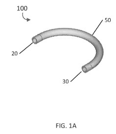
[0019] FIGS. 1A and 1B are a perspective view and a cross-sectional view of
an example
embodiment of an implantable bracing apparatus.
[0020] FIG. 1C is a perspective view of an example embodiment of an
implantable bracing
apparatus with a flange.
-5-
CA 03146562 2022-01-07
WO 2021/007440 PCT/US2020/041416
[0021] FIGS. 1D to 1G are perspective and side views of example embodiments
of end cap
accessories.
[0022] FIGS. 1H and 11 are progressive side views of an example embodiment
of a ridge
locking feature.
[0023] FIG. 1J is a partial cross-sectional view of an example embodiment
of an implantable
bracing apparatus.
[0024] FIGS. 2A and 2B are a perspective view and a cross-sectional view of
another example
embodiment of an implantable bracing apparatus.
[0025] FIGS. 3A and 3B are a perspective view and an overhead cross-
sectional view of
another example embodiment of an implantable bracing apparatus.
[0026] FIG. 3C is a partial cross-sectional view of another example
embodiment of an
implantable bracing apparatus.
[0027] FIGS. 4A and 4B are a perspective view and a partial side view of
another example
embodiment of an implantable bracing apparatus.
[0028] FIGS. 5A and 5B are a perspective view and an overhead view of
another example
embodiment of an implantable bracing apparatus.
[0029] FIGS. 6A and 6B are a perspective view and an overhead view of
another example
embodiment of an implantable bracing apparatus.
[0030] FIGS. 7A and 7B are a perspective view and a side view of another
example
embodiment of an implantable bracing apparatus.
[0031] FIGS. 8 and 9 are perspective views of example embodiments of
implantable bracing
apparatuses.
[0032] FIGS. 10A and 10B are a side view and a perspective view of another
example
embodiment of an implantable bracing apparatus.
[0033] FIGS. 11A to 11D are perspective views and a side view of other
example embodiments
of implantable bracing apparatuses.
[0034] FIGS. 12A and 12B are a top view and perspective view of a curved
tubular injector.
[0035] FIGS. 13A to 13F are partial cross-sectional views of curve tubular
injector at various
stages of operation.
[0036] FIG. 14 is a flow diagram of an example embodiment of a method for
generating a
bracing apparatus.
-6-
CA 03146562 2022-01-07
WO 2021/007440 PCT/US2020/041416
[0037] FIGS 15A to 15C are perspective views of an example embodiment of a
tunneling
device.
[0038] FIGS. 16A and 16B are perspective views of another example
embodiment of a
tunneling device.
[0039] FIGS. 17A to 17D are progressive diagrammatic views of an example
embodiment of
a tunneling device in various stages of operation.
[0040] FIGS. 18A to 18F are progressive diagrammatic views of another
example embodiment
of a tunneling device in various stages of operation.
DETAILED DESCRIPTION
[0041] Before the present subject matter is described in detail, it is to
be understood that this
disclosure is not limited to the particular embodiments described herein, as
such may, of course,
vary. It is also to be understood that the terminology used herein is for the
purpose of describing
particular embodiments only, and is not intended to be limiting, since the
scope of the present
disclosure will be limited only by the appended claims.
[0042] As used herein and in the appended claims, the singular forms "a,"
"an," and "the"
include plural referents unless the context clearly dictates otherwise.
[0043] Generally, embodiments of the present disclosure include systems,
devices, and
methods for attaching and supporting a bone suture. Accordingly, some
embodiments include
implantable bracing apparatuses to reinforce a hole or tunnel in a bone. These
various
embodiments can include elements through which sutures pass and/or to which
sutures may be
tethered. In certain embodiments, some or all elements of the bracing
apparatus may comprise
metal, natural or synthetic material, organic or inorganic material,
biodegradable or non-
biodegradable polymer, or a combination thereof
[0044] In some embodiments, a bracing apparatus can be a flexible or rigid
solid-walled
curved tube that is inserted into a pre-formed bone tunnel. The bracing
apparatus can further
comprise ends having one or more attachment features to which accessories can
be attached,
depending on the specific surgical application.
[0045] In other embodiments, the bracing apparatus can be a flexible or
rigid curved tube
having a non-solid wall. In some embodiments, for example, a bracing apparatus
can comprise a
coil having single or multiple helices, wherein the single or multiple helices
are configured to
-7-
CA 03146562 2022-01-07
WO 2021/007440 PCT/US2020/041416
compress and facilitate the insertion of the bracing apparatus. In other
embodiments, a bracing
apparatus can comprise a simple or complex latticework of struts, wherein the
latticework of struts
can be collapsible into a narrow configuration and, further, configured to
expand during
deployment while inside a bone tunnel.
[0046] According to another aspect of the embodiments, a method for
creating a bracing
apparatus in situ using phase transition polymers is provided. In some
embodiments, a liquid
polymer can be extruded from perforations in the wall of a straight or curved
injector. Upon
extrusion, the polymer can undergo a phase transition to a solid, where the
phase transition can be
induced by temperature, chemical or enzyme crosslinking/curing, photoreactive
crosslinking using
ultraviolet, infrared, or visible light, or other means. Following the phase
transition, the injector
is withdrawn, leaving the bracing apparatus within the bone tunnel or hole.
[0047] According to another aspect of the embodiments, a tunneling device
is provided for
creating a curved bone tunnel and inserting a bracing apparatus into the bone
tunnel. In certain
embodiments, the tunneling device can include a curved channel or guide tube
configured to guide
a pointed impactor along a predetermined path. In some embodiments, for
example, one or more
ends of the curved track or guide tube abut a target area of a bone surface,
wherein the target area
comprises one or more predetermined entry and exit points of a tunnel to be
created in the bone.
In some embodiments, a tunneling device can also include a means for
propelling the impactor,
for example, through the use of a pneumatic, magnetic, electrical, or
mechanical mechanism, or a
combination thereof
[0048] For each and every embodiment of a method disclosed herein, systems
and devices
capable of performing each of those embodiments are covered within the scope
of the present
disclosure. For example, embodiments of tunneling devices for creating a bone
tunnel are
disclosed, and these devices can each have one or more internal propulsion
mechanisms, piezo
motors, piezoceramic elements, bumpers, suture feed mechanisms, and other
components that can
perform any and all method steps, or facilitate the execution of any and all
method steps.
Example Embodiments of Implantable Bracing Apparatuses
[0049] FIGS. 1A and 1B depict a perspective view and a cross-sectional
view, respectively, of
an example embodiment of an implantable bracing apparatus 100. According to
one aspect of the
embodiment, apparatus 100 can comprise a curved tube at least a portion of
which is configured
-8-
CA 03146562 2022-01-07
WO 2021/007440 PCT/US2020/041416
to be implanted within a bone tunnel, wherein the curved tube includes an
inner lumen 40, an outer
surface 50, and two open ends 20, 30. Those of skill in the art will
appreciate that the curved tube
can have regional variations in radius of curvature along the length of the
tube, including areas
that are straight. Indeed, for certain applications, the entire length of tube
that serves as the bracing
apparatus may be straight. According to one aspect of the embodiments, outer
surface 50 is
configured to interface with the bone tunnel, and inner lumen 40 is configured
to pass a suture
therethrough and to prevent the suture from contacting at least a portion of
the bone tunnel.
According to another aspect of the embodiments, first open end 20 of apparatus
100 can correspond
with a first entry point of the bone tunnel, and second open end 30 can
correspond with a second
entry point of the bone tunnel.
[0050] As best seen in FIG. 1B, apparatus 100 can have a circular cross-
sectional geometry.
Those of skill in the art, however, will appreciate that the cross-sectional
geometry of bracing
apparatus 100 can be non-circular (e.g., oblong), and may have other geometric
features that
provide functional or structural utility. For example, in some embodiments,
grooves, fins, or
flanges integral to the apparatus can be disposed on outer surface 50 and/or
open ends 20, 30. In
example embodiment FIG. 1C, apparatus 150 is shown with one of the open ends
175 having an
integral flange, which constrains the apparatus from further entering the bone
tunnel. Furthermore,
some or all elements of bracing apparatus 100 can comprise a metallic
material, natural or synthetic
material, organic or inorganic material, biodegradable or non-biodegradable
polymer, or a
combination thereof. According to certain embodiments, for example, inner
lumen 40 can
comprise a coating of polyethylene or polytetrafluoroethylene composites to
reduce friction.
According to other embodiments, osteoconductive materials, such as
hydroxyapatite, can be
incorporated into inner lumen 40 and/or outer surface 50 to enhance
osseointegration and/or bone
ingrowth. Proteins, other biologics, or synthetic molecules can also be
tethered to either inner
lumen 40 or outer surface 50 to achieve the same or similar results. Further,
some or all elements
of bracing apparatus 100 can be subject to surface modification to enhance
osseointegration, such
as, for example, plasma treatment or electrochemical etching to generate
nanotextured surfaces.
[0051] In some embodiments, one or both open ends 20, 30 of bracing
apparatus 100 can be
configured to receive and mate with one or more end cap accessories. FIGS. 1D
to 1G depict
perspective and side views of two example embodiments of end cap accessories
3000 and 3100,
respectively. As shown in FIGS. 1D and 1E, end cap accessory 3000 features a
flanged head 3010
-9-
CA 03146562 2022-01-07
WO 2021/007440 PCT/US2020/041416
and a through hole 3020 to enable passage of a suture. The shaft of end cap
accessory 3000 can
be configured to mate with open ends 20, 30 of bracing apparatus 100, for
example, either by press
fit or snap fit via rounded ridges.
[0052] FIGS. 1F and 1G depict another embodiment of an end cap accessory
3100 with flanged
head 3010, and which also includes an eyelet 3160 at the end of the shaft.
According to another
aspect of the embodiments, a ridged feature 3150 disposed along the shaft is
configured to lock
end cap accessory 3100 with a corresponding ridge locking feature 3155 (shown
in FIGS. 1H and
11) at the ends 20, 30 of bracing apparatus 100.
[0053] FIGS. 1H and 11 depict progressive side views of the ridge locking
feature in operation,
as described with respect to the end cap accessory 3100 (shown in FIGS. 1F and
1G). According
to some embodiments, each of the one or more end cap accessories 3100 can
include a ridged
feature 3150 along an outer diameter of the shaft. As end cap accessory 3100
is moved in a
downward direction, as indicated by the downward arrow, ridged feature 3150
engages, by snap-
fit or press-fit mating, a corresponding ridge locking feature 3155 disposed
within end 20 of
bracing apparatus 100, which is shown deployed within bone 4020. Further, as
best seen in FIG.
11, when the ridge locking feature is engaged, a lower surface of flanged head
3010 of end cap
accessory 3100 abuts the top surface of open end 20 of bracing apparatus 100.
[0054] FIG. 1J is a partial cross-sectional view depicting implantable
bracing apparatus 100
with two end cap accessories 3000, as deployed within bone 4020. According to
the depicted
embodiment, an end cap accessory 3000 is disposed at each of open ends 20, 30
of bracing
apparatus 100. Soft tissue 4010, which is configured to be to be re-attached,
is situated next to
bone 4020. Suture 4050 passes through the soft tissue 4010, enters a first
through hole 3020 of a
first end cap accessory 3000, traverses one end 20 of bracing apparatus 100 to
the other open end
30, exits a second through hole 3020 of a second end cap accessory 3000, and
finally traverses soft
tissue 4010.
[0055] FIGS. 2A and 2B depict a perspective view and a cross-sectional
view, respectively, of
another example embodiment of an implantable bracing apparatus 200. Similar to
the
embodiments described with respect to FIG. 1A, bracing apparatus 200 can
comprise a curved
tube at least a portion of which is configured to be implanted within a bone
tunnel, wherein the
curved tube includes an inner lumen 240, an outer surface 250, and two open
ends 220, 230.
Although bracing apparatus 200 is depicted in FIG. 2B as having a circular
cross-section, those of
-10-
CA 03146562 2022-01-07
WO 2021/007440 PCT/US2020/041416
skill in the art will appreciate that the cross-sectional geometry of
apparatus 200 can be non-
circular (e.g., oblong), and may have other geometric features that provide
functional or structural
utility (e.g. grooves or fins on the outer surface or ends). Furthermore, some
or all elements of
bracing apparatus 200 can comprise a metallic material, natural or synthetic
material, organic or
inorganic material, biodegradable or non-biodegradable polymer, or a
combination thereof.
According to certain embodiments, for example, inner lumen 240 can comprise a
coating of
polyethylene or polytetrafluoroethylene composites to reduce friction.
According to other
embodiments, osteoconductive materials, such as hydroxyapatite, can be
incorporated into inner
lumen 240 and/or outer surface 250 to enhance osseointegration and/or bone
ingrowth. Proteins,
other biologics, or synthetic molecules can also be tethered to either inner
lumen 240 or outer
surface 250 to achieve the same or similar results. Further, some or all
elements of bracing
apparatus 200 can be subject to surface modification to enhance
osseointegration, such as, for
example, plasma treatment or electrochemical etching to generate nanotextured
surfaces.
[0056] In some embodiments, one or both open ends 220, 230 of bracing
apparatus 200 can
include a threaded portion configured to receive and couple with a functional
accessory, such as
screw cap 260. Although FIG. 2A depicts the threaded portion disposed on an
inner surface of
lumen 240, those of skill in the art will appreciate that the threaded end
portion can also be disposed
on outer surface 250 of bracing apparatus 200. Each threaded end portion can
be configured to
receive one or more of a screw cap 260, flanged end cap (e.g., as described
with respect to FIGS.
1C to 1D), anchor plate, or any other functional accessory to be mounted on
one or both of ends
220, 230.
[0057] FIGS. 3A and 3B depict a perspective view and an overhead cross-
sectional view,
respectively, of another example embodiment of an implantable bracing
apparatus 300 comprising
a plurality of curved tubes, wherein at least a portion of each curved tube is
configured to be
implanted within a bone tunnel. According to one aspect of the embodiments,
bracing apparatus
300 can include two curved tubes configured to intersect at a middle portion
305 along the length
of each curved tube. In some embodiments, the curved tubes can be similarly
dimensioned, e.g.,
having a similar diameter, wall thickness, etc. Apparatus 300 further includes
two pairs of open
ends (320, 325, 330, 335), each pair corresponding to a curved tube. As can be
seen in FIG. 3B,
apparatus 300 includes an inner lumen 40 and an outer surface 50. Although a
typical arrangement
-11-
CA 03146562 2022-01-07
WO 2021/007440 PCT/US2020/041416
is for the two intersecting segments to be orthogonal to one another, those of
skill in the art will
appreciate that the intersecting segments may be at any angle.
[0058] FIG. 3C depicts bracing apparatus 300 in a deployed state. Although
FIG. 3C depicts
bracing apparatus 300 without end cap accessories, those of skill in the art
will appreciate that
other embodiments of bracing apparatus 300 can be implemented with end cap
accessories, such
as those described with respect to FIGS. 1C to 1F. Referring back to FIG. 3C,
soft tissue 4010 to
be re-attached is adjacent to bone 4020, with first and second sutures 4050
and 4060 passing
through soft tissue 4010. First suture 4050 traverses soft tissue 4010, enters
open end 320 of
bracing apparatus 300, exits from open end 330, then passes again through soft
tissue 4010.
Similarly, second suture 4060 traverses soft tissue 4010, enters open end 325
of an orthogonal
segment of bracing apparatus 300, exits from the open end 335, then passes
again through soft
tissue 4010. Although FIG. 3C depicts bracing apparatus 300 implemented with
two sutures, those
of skill in the art will understand that a single suture can be used. For
example, in some
embodiments, suture 4050 enters end 320 and exits from end 330, wherein the
bracing segment
comprising ends 325, 335 is configured to serve as a passive support by
distributing stress
generated by suture 4050.
[0059] FIG. 4A is a perspective view of another example embodiment of an
implantable
bracing apparatus 400. According to one aspect of the embodiments, bracing
apparatus 400 can
comprise two intersecting curved tubes (405, 410), wherein the tubes are
dissimilar. As can be
seen in FIG. 4B, according to some embodiments, first curved tube 405 can
include a first pair of
centrally-located apertures 408. A second curved tube 410, which can have a
smaller diameter
relative to first curved tube 405, can be deployed by passing through
apertures 408. In addition,
according to some embodiments, second curved tube 410 can include a second
pair of centrally-
located apertures (not shown) configured to allow sutures to be passed through
first curved tube
405. Second curved tube 410 can also include one or more raised features to
ensure proper
alignment of the first and second pairs of apertures of the first and second
curved tube, respectively.
As best seen in FIG. 4B, the first pair of apertures 408 of first curved tube
405 can be disposed
along a middle portion of first curved tube 405.
[0060] FIGS. 5A and 5B depict a perspective view and an overhead view,
respectively, of
another example embodiment of an implantable bracing apparatus 500. Similar to
bracing
apparatus 300, the depicted embodiments comprise a plurality of curved tubes
of equal diameter
-12-
CA 03146562 2022-01-07
WO 2021/007440 PCT/US2020/041416
that intersect orthogonally at an apex 508 of the curved tubes. With respect
to bracing apparatus
500, there can be three curved tubes that intersect at an approximately 60
degree angle to each
other at apex 508, as best seen in FIG. 5B. Bracing apparatus 500 can be
deployed in a plurality
of bone tunnels according to a process similar to that of bracing apparatus
300 (as described with
respect to FIG. 3C), and further provides for the implementation of one, two,
or three sutures.
Those of skill in the art would also appreciate that the angles of
intersection between the curved
tubes can be greater or less than 60 degrees.
[0061] FIGS. 6A and 6B depict a perspective view and an overhead view,
respectively, of
another example embodiment of an implantable bracing apparatus 600. According
to some
embodiments, bracing apparatus 600 can comprise four curved tubes that
intersect at an apex 608,
wherein each curved tube is at a 45 degree angle to at least one adjacent
curved tube. As with the
previously described embodiments, bracing apparatus 600 can be deployed in a
plurality of bone
tunnels according to a process similar to that of bracing apparatus 300 (as
described with respect
to FIG. 3C), and further provides for the implementation of up to four
sutures. Those of skill in
the art will also appreciate that the angles of intersection between the
curved tubes can be greater
or less than 45 degrees.
[0062] FIGS. 7A and 7B depict a perspective view and a side view,
respectively, of another
example embodiment of a bracing apparatus 700, comprising a first curved tube
705 and a second
curved tube 710. As can be seen in the figures, bracing apparatus 700 can be
configured to be
deployed within two curved bone tunnels, wherein the first curved tube 705 is
deployed within a
first bone tunnel having a first depth within the bone, wherein the second
curved tube 710 is
deployed within a second bone tunnel having a second depth within the bone,
and wherein the first
depth is different from the second depth. In some embodiments, the first depth
is greater than the
second depth, and consequently, first curved tube 705 is deployed at a greater
depth within the
bone relative to second curved tube 710. According to another aspect of the
embodiments, first
curved tube 705 and second curved tube 710 "cross" at location 780, at an
angle relative to one
another. Although FIGS. 7A and 7B show the cross location 780 as an orthogonal
angle, those of
skill in the art will appreciate that the angle may be greater than or less
than 90 degrees. Each of
first curved tube 705 and second curved tube 710 may also have inner lumens
(not shown) and
outer surfaces 50, wherein the inner lumens are configured to pass one or more
sutures. According
-13-
CA 03146562 2022-01-07
WO 2021/007440 PCT/US2020/041416
to other embodiments, second curved tube 710 can be solid (e.g., without a
lumen), and configured
to serve as a passive support for first curved tube 705.
[0063] According to some embodiments, bracing apparatuses may also be
manufactured to
have flexibility, such as tubular structures with corrugated walls or with non-
solid walls, e.g.,
bracing apparatuses that encompass a cylindrical volume. FIG. 8 is a
perspective view of another
example embodiment of implantable bracing apparatus 800 comprising a
cylindrical volume
comprising a single helical coil. Those of skill in the art will appreciate
that the helical coil may
possess different chirality, pitch, radius of curvature, slant angle, wire
diameter, wire material, and
wire cross-sectional geometry. Furthermore, helical coil bracing apparatuses
may also possess
spatial variations of different parameters within a single embodiment. The
helical coil bracing
apparatus can be inserted into bone tunnels that have regional variations in
radius of curvature, as
well as regions that are straight. FIG. 9 is a perspective view of an
embodiment of implantable
bracing apparatus 900 comprising a cylindrical volume comprising a double
helix, which ¨ in a
manner similar to that of apparatus 800 ¨ may possess different physical and
material
characteristics, as well as spatial variations. FIGS. 10A and 10B depict a
side view and a
perspective view, respectively, of another example embodiment of implantable
bracing apparatus
1000, wherein bracing apparatus 1000 comprises a cylindrical volume comprising
four helical
coils (e.g., two right-hand and two left-hand coils). In a similar manner,
those of skill in the art
will appreciate that corrugated bracing apparatuses may possess either
concentric ring patterns of
ridges and valleys along the length of the apparatus, or helical patterns of
ridges and valleys that
vary in helix configurations just as those described for the helical coil
apparatuses above.
[0064] According to other embodiments, bracing apparatuses may also
comprise a cylindrical
volume comprising a latticework of struts, such as those depicted in FIGS. 11A
to 11D. FIG. 11A
shows a perspective view of an example embodiment of an implantable bracing
apparatus 1100,
wherein a lattice of triangular cells, as shown in exploded view 1100A, is
used to form a cylindrical
bracing structure. Each cell can have a thickness, b, and each side of
triangular cell can have a
length, h. Those of skill in the art will appreciate that the sides of the
triangular cell can have
similar or dissimilar lengths. FIG. 11B shows a perspective view of another
example embodiment
of an implantable bracing apparatus 1130, wherein a lattice of hexagonal
cells, as shown in
exploded view 1130A, is used to form a cylindrical bracing structure. Each
cell can have a
thickness, b, and each side of hexagonal cell can have a length, h. Those of
skill in the art will
-14-
CA 03146562 2022-01-07
WO 2021/007440 PCT/US2020/041416
appreciate that the sides of the hexagonal cell can have similar or dissimilar
lengths. FIG. 11C
shows a side view of yet another example embodiment of an implantable bracing
apparatus 1160,
where the apparatus includes a lattice comprising quadrilateral cells. FIG.
11D shows a
perspective view of an example embodiment of an implantable bracing apparatus
1190, wherein
the apparatus includes a lattice comprising multiple geometries (e.g.,
triangular and hexagonal).
These embodiments of the latticework are intended to be illustrative only and
are not meant to
limit the scope of the present disclosure. Indeed, those of skill in the art
will recognize that
latticework can be constructed from one or more different materials, according
to different
geometries, in order to achieve the intended result of facilitating the
insertion of the implantable
bracing apparatus. The latticework bracing apparatus can also be inserted into
bone tunnels that
have regional variations in radius of curvature, as well as regions that are
straight.
[0065] According to some embodiments, a bracing apparatus may be generated
in situ (at the
site of implantation). In particular, a curved tubular injector tip of an
instrument can be introduced
into a bone tunnel, and a natural or synthetic fluidic agent can be pumped
through the instrument
into the bone tunnel, where the agent subsequently undergoes a phase
transition into a solid. The
injector can be withdrawn from the bone tunnel after the phase transition,
leaving behind a solid
implanted bracing apparatus inside the bone tunnel.
[0066] FIGS. 12A and 12B depict a top view and a perspective view,
respectively, of an
example embodiment of a curved tubular injector 1200 configured to be inserted
into a bone tunnel
and introduce a fluidic agent therein. As can be seen in the figures, curved
tubular injector
comprises a plurality of perforations. Those of skill in the art will
appreciate that the number, size,
pattern, and spacing of perforations in curved tubular injector 1200, as well
as the material
composition, size, cross-sectional geometry, length, and shape of the tube may
vary to achieve the
intended function, with portions of the curved tubular injector having
variations in radius of
curvature and/or regions that are straight. The length and shape of the curved
tubular injector 1200
is dimensioned according to the desired penetration depth of the instrument
into the bone tunnel,
and the perforations along the length of the instrument are configured to
distribute a fluidic agent
into the bone tunnel. Additional features such as optical emitters including,
but not limited to,
LEDs of the visible, ultraviolet, or infrared wavelengths, thermal or
electrical conductors, or
additional chemical diffusion agents either coating or extrinsically
introduced through the
instrument may also be integrated in order to achieve its function to induce
the fluidic agent to
-15-
CA 03146562 2022-01-07
WO 2021/007440 PCT/US2020/041416
undergo phase transition whether by polymerization, setting, or curing. It is
important to note that
the injector need not be rigid and may be flexible and capable of being
manipulated to facilitate
insertion into the bone tunnel.
[0067] FIGS. 13A to 13F depict partial cross-sectional views of curved
tubular injector 1200,
like those described with respect to FIGS. 12A and 12B, in various stages of
operation, wherein
curved tubular injector 1200 is configured to generate an implanted bracing
apparatus in situ. FIG.
13A shows the step of introducing curved tubular injector 1200 into bone
tunnel 4025 within bone
4020. Bone tunnel 4025 can be created prior to introduction of curved tubular
injector 1200
through, for example, the methods and devices described herein with respect to
FIGS. 15A, 15B,
15C, 16A, and 16B. According to some embodiments, a single tubular arm, either
flexible or rigid,
can be used to maneuver injector 1200 into bone tunnel 4025. Curved tubular
injector 1200,
whether flexible or rigid, can be inserted into one end of bone tunnel 4025
(as shown in FIGS. 13A
to 13F) or, in the alternative, into both ends, in which case a first end and
a second end are
connected by bone tunnel 4025. According to one aspect of the embodiments,
bone tunnel 4025
is configured to receive fluidic agent 4035. In some embodiments, curved
tubular injector 1200
can be part of a motorized assembly, such as that of the tunneling devices
described with respect
to FIGS. 15A, 15B, 15C, 16A, and 16B.
[0068] FIGS. 13B, 13C, and 13D show the steps of injecting the natural or
synthetic fluidic
agent 4035, as it moves from injector 1200 into bone tunnel 4025. According to
one aspect of the
embodiments, a predetermined amount of fluidic agent 4035, based on volume
needed to fill the
bone tunnel, can either be manually injected (such as using a syringe or
similar instrument) or
automatically injected (such as with a motorized pump).
[0069] FIG. 13E shows the natural or synthetic fluidic agent 4035 in the
process of
spontaneously undergoing, or being activated/induced to undergo, phase
transition into a solid
bracing apparatus 4036 inside bone tunnel 4025. The nature of this phase
transition will depend
on the selected fluidic agent 4035 being used. Example embodiments of the
fluidic agent can be
one or more of a self-assembling polymer, a UV-cured resin, a thermosetting
polymer, a
chemically-cured material, a chemically-crosslinked material, among others. As
shown in FIG.
13F, the curved tubular injector 1200 has been retracted from bone tunnel
4025, leaving behind
the newly generated bracing structure 4036 after having either partially or
fully undergone phase
transition into a solid material.
-16-
CA 03146562 2022-01-07
WO 2021/007440 PCT/US2020/041416
[0070] FIG. 14 is a flow diagram depicting an example embodiment of a
method 1400 for
generating bracing apparatus 4036 in situ, in accordance with the embodiments
described with
respect to FIGS. 12A to 12B and 13A to 13F.
[0071] At Step 1410, an injector 1200 is introduced into bone tunnel 4025.
According to some
embodiments, injector 1200 can comprise a curved tubular injector having a
single tubular arm
that is either flexible or rigid. Bone tunnel 4025 can comprise a cavity
inside bone that can include
one or more openings at the bone surface. Injector 1200 can be inserted into
one end of bone
tunnel 4025 or, according to some embodiments, into both ends of bone tunnel
4025. In some
embodiments, injector 1200 can be part of a motorized assembly, such as the
tunneling devices
described herein with respect to FIGS. 15A, 15B, 15C, 16A, and 16B.
[0072] At Step 1420, fluidic agent 4035 is injected into bone tunnel 4025.
Example
embodiments of the fluidic agent can be one or more of a self-assembling
polymer, a UV-cured
resin, a thermosetting polymer, a chemically-cured material, a chemically-
crosslinked material,
among others. According to one aspect of the embodiments, a predetermined
amount of fluidic
agent 4035, based on volume needed to fill the bone tunnel, can either be
manually injected (such
as by using a syringe or similar instrument) or automatically injected (such
as with a motorized
pump).
[0073] At Step 1430, fluidic agent 4035 undergoes phase transition from
fluid 4035 into solid
bracing apparatus 4036 inside bone tunnel 4025. The nature of the phase
transition depends on
the selected fluidic agent 4035 being used.
[0074] At Step 1440, injector 1200 is retracted or withdrawn from bone
tunnel 4025, leaving
behind the newly generated bracing apparatus 4036 after having either
partially or fully undergone
phase transition into a solid material.
Example Embodiments of Tunneling Devices and Methods Relating Thereto
[0075] Example embodiments of tunneling devices for creating a bone tunnel,
and methods
relating thereto, will now be described.
[0076] FIG. 15A is a perspective view depicting an example embodiment of a
tunneling device
1500 for creating one or more tunnels in a bone material, which can be used
with any of the
previously described embodiments. According to some embodiments, tunneling
device 1500
includes a housing 1508 comprising at least one surface 1509 configured to
interface with a bone
-17-
CA 03146562 2022-01-07
WO 2021/007440 PCT/US2020/041416
material, a channel 1502 disposed within housing 1508, and a curved impactor
1503 configured to
travel along channel 1502 at high speeds. Channel 1502 is shown to be circular
in cross-section,
but those of skill in the art will appreciate that the cross-sectional shape
of channel 1502 and
impactor 1503, configured to travel therein, may be of any geometry. According
to some
embodiments, the path of channel 1502 within tunneling device 1500 can have an
arcuate
geometry. Furthermore, as seen in FIG. 15A, tunneling device 1500, including
housing 1508 and
channel 1502 disposed therein, comprises a semi-circular shape subtending an
angle of 180
degrees. Those of skill in the art, however, will appreciate that tunneling
device 1500, housing
1508, or channel 1502, may comprise a semi-circular shape subtending an angle
of more or less
than 180 degrees, or may have a non-circular shape. In addition, the length of
curved impactor
1503 may be either greater than or less than the length of channel 1502 of
tunneling device 1500.
[0077] According to another aspect of the embodiments, impactor 1503
includes one or more
pointed ends 1504, as seen in FIG. 15A, which are configured such that the
energy upon striking
a bone material can cause a bone tunnel to lengthen. In some embodiments, an
internal propulsion
mechanism 1501 transfers energy to impactor 1503 to generate a back-and-forth
motion within
channel 1502. Internal propulsion mechanism 1501 can comprise any technology
used to generate
motion including, but not limited to, one or more of a piezoelectric motor, an
electrical induction
motor, magnetic propulsion, pneumatic propulsion, hydraulic propulsion,
mechanical (e.g.,
linkages, gears, etc.), or any combination thereof
[0078] FIGS. 15B and 15C depict a perspective partial cross-sectional view
and a perspective
partial exploded view, respectively, of an example embodiment of tunneling
device 1500
comprising a piezoelectric motor 1501 configured to move impactor 1503.
According to one
aspect of the embodiments, piezoelectric motor 1501 can comprise a plurality
of piezoceramic
elements 1505 disposed along the length of channel 1502, wherein piezoceramic
elements 1505
are configured to expand and contract in response to electrical energy and
actuate one or more
bumpers 1506. According to another aspect of the embodiments, in response to
being actuated by
piezoceramic elements 1505, the one or more bumpers 1506 are then configured
to exert one or
more forces on impactor 1503, which can cause impactor 1503 to move according
to linear or
circular motions, thereby moving impactor 1503 along the path of channel 1502
according to a
predetermined back-and-forth motion. The repeated impact from impactor 1503,
whose trajectory
-18-
CA 03146562 2022-01-07
WO 2021/007440 PCT/US2020/041416
is defined by impactor 1503 travelling along path of channel 1502, then
results in a curved bone
tunnel.
[0079] As best seen in FIGS. 15B and 15C, according to some embodiments,
impactor 1503
can comprise a solid apparatus including one or more solid conical tips 1504
on each end. Those
of skill in the art, however, will appreciate that impactor 1503 can include
one or more tips 1504
having a different geometry including, but not limited to, a hollow cylinder,
hemisphere, or
truncated cone, along with other tip features, such as flutes. Additionally,
certain embodiments of
tunneling device 1500 may use any of the implantable bracing apparatuses
described herein, or
any components thereof, such as those described with respect to FIGS. 1A, 1B,
2A, 2B, 3A, 3B,
4A, 4B, 5A, 5B, 6A, 6B, 7A, 7B, 8, 9, 10A, 10B, 11A, 11B, 11C, or 11D, as the
impactor to form
a curved bone tunnel, such that the bracing apparatus may be left within the
tunnel after the tunnel
is formed. Additionally, according to some embodiments, piezomotor 1501 can
comprise one or
more of a piezo inertia motor, piezo ultrasonic resonance motor, or piezo walk
motor. Those of
skill in the art will recognize that the motion of piezo actuation can include
stacking, tubing,
expanding, shearing, walking, bending, bimorph flexing, and bimorph bending.
[0080] FIGS. 16A and 16B depict a perspective view and a perspective
exploded view,
respectively, of another embodiment of tunneling device 1600. According to one
aspect of the
embodiments, tunneling device 1600 can comprise a first subassembly 1607 and a
second
subassembly 1608 configured to couple with first subassembly 1607, wherein a
curved channel
1609 and impactor 1613 are disposed in first subassembly 1607 of tunneling
device 1600, and a
piezoelectric motor 1611 is disposed in second subassembly 1608 of tunneling
device 1600.
Although the cross-section of channel 1609 is shown to be circular in FIG.
16A, those of skill in
the art will appreciate that the channel cross-section may be of any geometry.
In addition, although
impactor 1613 is depicted as a hollow curved cylinder with a plurality of
pointed conical tips 1610,
those of skill in the art will recognize that impactor 1613 and tips 1610 can
comprise the same or
similar configurations as those described above with respect to FIGS. 15A,
15B, and 15C.
[0081] According to another aspect of the embodiments, a piezoelectric
motor 1611 disposed
in subassembly 1608 of tunneling device 1600 is configured to actuate a motion
to drive impactor
1613 in a back-and-forth motion to create a curved bone tunnel. In some
embodiments, impactor
1613 can comprise a needle having "pointy" cone-shaped tips 1610 for impaction
drilling. The
-19-
CA 03146562 2022-01-07
WO 2021/007440 PCT/US2020/041416
repeated impact from impactor 1613, whose trajectory is defined by impactor
1613 travelling along
path of channel 1609, then results in a curved bone tunnel.
[0082] According to another aspect of the embodiments, after impactor 1613
is propelled by
bumpers 1614 actuated by piezoelectric motor 1611 to create a bone tunnel, an
implantable bracing
apparatus 1612, such as any of the embodiments described herein with respect
to FIGS. 1A, 1B,
2A, 2B, 3A, 3B, 4A, 4B, 5A, 5B, 6A, 6B, 7A, 7B, 8, 9, 10A, 10B, 11A, 11B, 11C,
or 11D, is
subsequently inserted into the bone tunnel by tunneling device 1600. Bracing
apparatus 1612 can
be contained in a holding space within the impactor 1613, and deployed after
the removal of
impactor tips 1610. Bracing apparatus 1612 is then left to remain in the bone
tunnel.
[0083] In many of the embodiments disclosed herein, tunneling devices 1500
and 1600 can
also incorporate a feed mechanism (not shown) to insert a suture through the
bracing apparatus
that is situated in the bone tunnel. Because the bone tunnel and bracing can
be circular arcs, a
suture can easily pass through without the need for specialized hooks or
pincers to pull the suture
from the exit hole after a suture has been introduced through the entrance
hole.
[0084] FIGS. 17A to 17D are progressive diagrammatic views of an example
embodiment of
a tunneling device 1500 in various stages of operation, wherein tunneling
device 1500 is
configured to create a bone tunnel 4025 in a unidirectional manner. FIG. 17A
depicts an initial
stage wherein tunneling device 1500 is placed against the surface of bone 4020
at a predetermined
location identified as the tunneling site. According to one aspect of the
embodiments, tunneling
device 1500 is held stationary against the predetermined location of bone 4020
during the process
in which a bone tunnel is created.
[0085] FIG. 17B depicts a subsequent stage in which impactor 1503 is
actuated, and travels
along channel 1502 of tunneling device 1500, which can cause the pointed tip
1504 of impactor
1503 to make contact with the surface of bone 4020, as indicated by the arrow.
As described
above, impactor 1503 can be actuated using a piezomotor (not shown), such as,
for example, a
piezo inertia motor, piezo ultrasonic resonance motor, piezo walk motor, or
any similar mechanism
to cause the impactor 1503 to travel along channel 1502 at a high speed.
[0086] FIG. 17C depicts a stage of operation in which a continuous back-and-
forth actuation
(as indicated by the bi-directional arrow) of impactor 1503 along channel 1502
has caused pointed
tip 1504 of impactor 1503 to further progress into bone 4020, thereby creating
a partial bone tunnel
4025.
-20-
CA 03146562 2022-01-07
WO 2021/007440 PCT/US2020/041416
[0087] FIG. 17D depicts a near-final stage of operation in which the
continuous back-and-
forth actuation of impactor 1503 (as indicated by the bi-directional arrow)
along channel 1502 has
caused pointed tip 1504 of impactor 1503 to further progress into bone 4020,
thereby completing
the bone tunnel 4025. As can be seen in FIG. 17D, the completed bone tunnel
4025 can comprise
a first opening created by the entry of pointed tip 1504 (FIG. 17B) and a
second opening created
by the exit of pointed tip 1504 (FIG. 17D). According to another aspect of the
embodiments,
impactor 1503, having a curved or arcuate body, is configured to create bone
tunnel 4025, which
can similarly have a curved or arcuate shape.
[0088] FIGS. 18A to 18F are progressive diagrammatic views of another
example embodiment
of a tunneling device 1500 in various stages of operation, wherein tunneling
device 1500 is
configured to create a bone tunnel 4025 in a bi-directional manner.
[0089] FIG. 18A depicts an initial stage wherein tunneling device 1500 is
placed against the
surface of bone 4020 at a predetermined location identified as the tunneling
site. According to one
aspect of the embodiments, tunneling device 1500 is held stationary against
the predetermined
location of bone 4020 during the process in which a bone tunnel is created.
[0090] FIG. 18B depicts a subsequent stage in which impactor 1503 is
actuated and travels
along channel 1502 of tunneling device 1500 in a counter-clockwise direction
(as indicated by the
arrow), which can cause a first pointed tip 1504A of impactor 1503 to make
contact at a first entry
point on the surface of bone 4020. As described above, impactor 1503 can be
actuated using a
piezomotor (not shown), such as, for example, a piezo inertia motor, piezo
ultrasonic resonance
motor, piezo walk motor, or any similar mechanism to cause the impactor 1503
to travel along
channel 1502 at a high speed in a back-and-forth motion.
[0091] FIG. 18C depicts a subsequent stage in which impactor 1503 travels
along channel
1502 of tunneling device 1500 in a clockwise direction (as indicated by the
arrow), which can
cause a second pointed tip 1504B of impactor 1503 to make contact at a second
entry point on the
surface of bone 4020.
[0092] FIG. 18D depicts a subsequent stage of operation in which impactor
1503 travels along
channel of 1502 of tunneling device 1500 again in a counter-clockwise
direction (as indicated by
the arrow), causing first pointed tip 1504A of impactor 1503 to further
progress into bone 4020
through the first entry point, thereby creating a first partial bone tunnel
4025A.
-21-
CA 03146562 2022-01-07
WO 2021/007440 PCT/US2020/041416
[0093] FIG. 18E depicts a subsequent stage of operation in which impactor
1503 travels along
channel 1502 of tunneling device 1500 again in a clockwise direction (as
indicated by the arrow)
causing second pointed tip 1504B of impactor 1503 to further progress into
bone 4020 through the
second entry point, thereby creating a second partial bone tunnel 4025B.
[0094] FIG. 18F depicts a near-final stage of operation in which the
continuous back-and-forth
actuation of impactor 1503 along channel 1502 has caused first pointed tip
1504A of impactor
1503 to further progress into bone 4020 through the first entry point, thereby
connecting the two
partial bone tunnels (4025A, 4025B) to form a completed bone tunnel 4025. As
can be seen in
FIG. 18F, the completed bone tunnel 4025 can comprise a first opening created
by the entry of
first pointed tip 1504A and a second opening created by the entry of second
pointed tip 1504B.
According to another aspect of the embodiments, impactor 1503, having a curved
or arcuate body,
is configured to create bone tunnel 4025, which can similarly have a curved or
arcuate shape.
[0095] Although FIGS. 17A to 17D and FIGS. 18A to 18F are shown with
tunneling device
1500, those of skill in the art will understand that the methods described
herein can be utilized
with any of the disclosed tunneling devices, including the embodiments
described with respect to
FIGS. 15A, 15B, 15C, 16A, and 16D.
[0096] It should be noted that all features, elements, components,
functions, and steps
described with respect to any embodiment provided herein are intended to be
freely combinable
and substitutable with those from any other embodiment. If a certain feature,
element, component,
function, or step is described with respect to only one embodiment, then it
should be understood
that that feature, element, component, function, or step can be used with
every other embodiment
described herein unless explicitly stated otherwise. This paragraph therefore
serves as antecedent
basis and written support for the introduction of claims, at any time, that
combine features,
elements, components, functions, and steps from different embodiments, or that
substitute features,
elements, components, functions, and steps from one embodiment with those of
another, even if
the following description does not explicitly state, in a particular instance,
that such combinations
or substitutions are possible. It is explicitly acknowledged that express
recitation of every possible
combination and substitution is overly burdensome, especially given that the
permissibility of each
and every such combination and substitution will be readily recognized by
those of ordinary skill
in the art.
-22-
CA 03146562 2022-01-07
WO 2021/007440 PCT/US2020/041416
[0097] While the embodiments are susceptible to various modifications and
alternative forms,
specific examples thereof have been shown in the drawings and are herein
described in detail. It
should be understood, however, that these embodiments are not to be limited to
the particular form
disclosed, but to the contrary, these embodiments are to cover all
modifications, equivalents, and
alternatives falling within the spirit of the disclosure. Furthermore, any
features, functions, steps,
or elements of the embodiments may be recited in or added to the claims, as
well as negative
limitations that define the inventive scope of the claims by features,
functions, steps, or elements
that are not within that scope.
-23-