Note: Descriptions are shown in the official language in which they were submitted.
SIMULATED TISSUE STRUCTURE FOR SURGICAL TRAINING
CROSS-REFERENCE TO RELATED APPLICATION
[0001] This application claims priority to and benefit of U.S.
Provisional Patent
Application Serial No. 61/549,838 entitled "Simulated tissue structure for
surgical
training" filed on October 21, 2011 which is incorporated herein by reference
in its
entirety.
FIELD
[0002] This application is generally related to surgical training
tools, and in
particular, to anatomical models simulating organs or tissue for teaching and
practicing
various surgical techniques and procedures.
BACKGROUND
[0003] Medical students as well as experienced doctors learning new
surgical
techniques must undergo extensive training before they are qualified to
perform surgery
on human patients. The training must teach proper techniques employing various
medical devices for cutting, penetrating, clamping, grasping, stapling and
suturing a
variety of tissue types. The range of possibilities that a trainee may
encounter is great.
For example, different organs and patient anatomies and diseases are
presented. The
thickness and consistency of the various tissue layers will also vary from one
part of the
body to the next and from one patient to another. Accordingly, the skills
required of the
techniques and instruments will also vary. Furthermore, the trainee must
practice
techniques in readily accessible open surgical locations and in locations
accessed
laparoscopically.
[0004] Numerous teaching aids, trainers, simulators and model organs
are
available for one or more aspects of surgical training. However, there is a
need for
model organs or simulated tissue elements that are likely to be encountered in
endoscopic, laparoscopic, transanal, minimally invasive or other surgical
procedures
that include the removal of tumors or other tissue structures. In particular,
there is a
need for realistic model organs for the repeatable practice of removing a
tumor or other
- 1 -
Date recue/ date received 2022-01-25
undesired tissue followed by the closure of the target area by suturing or
stapling as
part of the same surgical procedure. In view of the above, it is an object of
this
invention to provide a surgical training device that realistically simulates
such particular
circumstances encountered during surgery.
SUMMARY
[0005] According to one aspect of the invention, a simulated tissue
structure
for surgical training is provided. The structure includes a defect layer
located above the
base layer. The defect layer includes at least one defect having two opposed
surfaces
that define at least one gap between the surfaces. A simulated tumor is
located above
the defect layer in such a way to overlay at least a portion of the defect. A
cover layer is
located above the base layer and overlays the tumor.
[0006] According to another aspect of the invention, a simulated
tissue
structure for surgical training is provided. The simulated tissue structure
includes at
least one simulated tissue module comprising a simulated tissue portion. The
structure
includes a module support having a first surface opposite from a second
surface and
defining a thickness therebetween. The module support includes at least one
module
receiving portion sized and configured to receive and connect with the at
least one
simulated tissue module. The simulated tissue module is insertable into and
removable
from the at least one module receiving portion and interchangeable with
another
simulated tissue module.
[0007] According to another aspect of the invention a method for
surgical
training is provided. The method includes the step of providing a simulated
tissue
structure comprising an artificial tumor located between a base layer and a
cover layer.
The base layer and the cover layer are made of elastomeric polymer that may
include
mesh reinforcement. The simulated tissue structure is placed inside a
simulated body
cavity of a surgical training device such that the simulated tissue structure
is at least
partially obscured from view by a user. The user removes the artificial tumor
from the
simulated tissue structure with instruments passed into the simulated body
cavity with
the simulated tissue structure obscured from the user and visualized on a
video monitor
providing a live feed of the simulated tissue structure inside the cavity via
a laparoscope
- 2 -
Date recue/ date received 2022-01-25
or endoscope. At least one defect is created substantially in the location of
the tumor.
The defect comprises two adjacent surfaces defining a gap. The gap is closed
by
bringing the two adjacent surfaces together with instruments such as sutures,
staples,
adhesive or other surgical means. Suturing the gap to bring the two adjacent
surfaces
together. In one variation, creating a defect includes providing a defect
layer in the
simulated tissue structure. Providing a defect layer includes providing a
defect layer
with a pre-formed defect or gap and placing the defect layer such that the
defect layer is
between the base layer and the cover layer and at least a portion of the
defect is
located underneath the artificial tumor. In another variation, creating a
defect includes
cutting at least one of the base layer and cover layer. Removing the
artificial tumor from
the simulated tissue structure includes removing the artificial tumor through
the defect
created by cutting.
BRIEF DESCRIPTION OF THE DRAWINGS
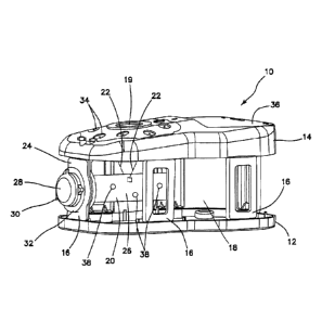
[0008] FIG. 1 illustrates a side view of a surgical training device
with a model
organ according to the present invention.
[0009] FIG. 2A illustrates a side cross-sectional view of a simulated
tissue
structure according to the present invention.
[0010] FIG. 2B illustrates a side cross-sectional view of a simulated
tissue
structure with tumor excised according to the present invention.
[0011] FIG. 2C illustrates a side cross-sectional view of a simulated
tissue
structure with an open suture according to the present invention.
[0012] FIG. 2D illustrates a side cross-sectional view of a simulated
tissue
structure with a closed suture according to the present invention.
[0013] FIG. 3A illustrates a top view of a defect layer having a
circular shaped
defect according to the present invention.
[0014] FIG. 3B illustrates a top view of a defect layer having an
elongated
defect according to the present invention.
[0015] FIG. 3C illustrates a top view of a defect layer having an
amorphous
defect according to the present invention.
- 3 -
Date recue/ date received 2022-01-25
[0016] FIG. 3D illustrates a top view of a defect layer having a two-
piece
defect according to the present invention.
[0017] FIG. 3E illustrates a top view of a multi-part defect layer
according to
the present invention.
[0018] FIG. 3F illustrates a top view of a defect layer having
multiple defects
according to the present invention.
[0019] FIG. 4 illustrates a top view of a simulated tissue structure
according to
the present invention.
[0020] FIG. 5 illustrates a side cross-sectional view of a simulated
tissue
structure according to the present invention.
[0021] FIG. 6A illustrates a perspective view of a modular tissue
structure and
support according to the present invention.
[0022] FIG. 6B illustrates a perspective view of a modular tissue
structure and
support according to the present invention.
[0023] FIG. 7 illustrates a cross-sectional view of a simulated
tissue structure
configured to mimic a human uterus according to the present invention.
[0024] FIG. 8 illustrates a top view of a modular tissue structure
according to
the present invention.
[0025] FIG. 9 illustrates a side view of a modular tissue structure
according to
the present invention.
[0026] FIG. 10A illustrates a perspective view of a simulated tissue
structure
according to the present invention.
[0027] FIG. 10B illustrates a perspective view of a simulated tissue
structure
according to the present invention.
[0028] FIG. 11A illustrates a perspective view of a simulated tissue
structure
according to the present invention.
[0029] FIG. 11B illustrates a perspective view of a simulated tissue
structure
according to the present invention.
[0030] FIG. 12 illustrates a perspective view of a suture needle and
a
simulated tissue structure according to the present invention.
- 4 -
Date recue/ date received 2022-01-25
DETAILED DESCRIPTION
[0031] A surgical training device 10 that is configured to mimic the
torso of a
patient such as the abdominal region is shown in FIG. 1. The surgical training
device
provides a simulated body cavity 18 substantially obscured from the user for
receiving model organs or simulated or live tissue 20. The body cavity 18 is
accessed
via a tissue simulation region 19 that is penetrated by the user employing
devices to
practice surgical techniques on the tissue or organ 20 found located in the
body cavity
18. Although the body cavity 18 is shown to be accessible through a tissue
simulation
region 19, a hand-assisted access device or single-site port device may be
alternatively
employed to access the body cavity 18 as described in U.S. Patent Application
Serial
No. 13/248,449 entitled "Portable Laparoscopic Trainer" filed on September 29,
2011
and incorporated herein by reference in its entirety. The surgical training
device 10 is
particularly well suited for practicing laparoscopic or other minimally
invasive surgical
procedures.
[0032] The surgical training device 10 includes a base 12 and a top
cover 14
connected to and spaced apart from the base 12 to define an internal body
cavity 18
between the top cover 14 and the base 12. At least one leg 16 interconnects
and
spaces apart the top cover 14 and base 12. A model organ or simulated tissue
20 is
disposed within the body cavity 18. The model organ 20 shown in FIG. 1 is a
partial
colon or intestine that is shown suspended from the top cover 14 by tethers 22
and
connected to at least one leg 24. The at least one leg 24 has an aperture (not
shown)
facing the internal cavity 20. The model colon 20 includes a tube 26 having a
proximal
end and a distal end. The proximal end of the tube 26 is interconnected with
the
aperture of the leg 16 such that the aperture provides an access port to the
lumen of the
tube 26. The access port and aperture is shown to be closed off in FIG. 1 with
an
access device 28 which in combination with a sealed distal end of the tube 26
provides
a model organ 20 that is adapted for insufflation with fluid deliverable via
an insufflation
port 30. An optional insert 32 made of soft material such as silicone creates
a realistic
interface for the access port. The distal end of the tube 26 extends into the
body cavity
18 and is suspended within the body cavity 18. The interior of the tube 26 of
the
simulated organ 20 is accessible via the access port of leg 24 or via the
tissue
- 5 -
Date recue/ date received 2022-01-25
simulation region 19 or instrument insertion ports 34. An endoscopic camera
inserted
into the body cavity 18 or into the organ 20 via the access port generates a
live image
for display on a fold out video screen 36 shown in the closed position in FIG.
1.
Although the simulated organ 20 of FIG. 1 is ideal for practicing procedures
related to
transanal minimally invasive surgery, any simulated organ or tissue portion
may be
employed. One particular aspect of the organ 20 is at least one tumor or
defect 38 is
provided and connected to the organ. As shown in FIG. 1, the tumor 38 is
connected to
the wall of the organ tube 26.
[0033] Turning now to FIG. 2A there is shown a partial side cross-
sectional
view of a portion of a simulated organ 20 that includes the tumor 38. The
simulated
organ or tissue 20 includes a base layer or organ wall 40. The organ wall 40
is made
from a material configured to mimic real live tissue such as silicone or other
polymer
and is dyed appropriately. One or more base layers 40 of varying thicknesses
and
colorations may be employed to comprise the entirety of the wall 40. In one
variation,
the organ wall 40 is rigid and made of polymeric material. Above the base
layer 40 is a
second layer or defect layer 42. The defect layer 42 is the same size or
smaller than
the base layer 40 forming a raised platform for the tumor 38. The defect layer
42 is
connected to the base layer 40 by adhesive or other means known to one having
ordinary skill in the art including being integrally formed with the base
layer 40 as a
single unit. The defect layer 42 is made of silicone and in one variation of
the same
color as the base layer 40 such that the defect layer 42 blends into the
background of
the base layer 40. The defect layer 42 includes at least one defect or gap 44.
In one
variation, the defect 44 is a pre-fabricated breach in the defect layer 42
that mimics an
incision, gap or other void in real tissue resulting from a tear, cut, removal
or other
surgical procedure that requires surgical attention by way of suturing,
stapling or the like
to close the defect. Such a situation arises most often in the removal of a
tumor 38
where surrounding tissue is also removed together with the tumor 38 to
preventatively
ensure the entirety of the tumor is excised leaving behind a remnant defect in
the tissue.
The defect 44 comprises two opposed sides or surfaces defining a gap
therebetween.
Although the adjacent sides or surfaces are shown to be vertical with respect
to the
base layer 40, the invention is not so limited and the juxtaposed surfaces or
sides can
- 6 -
Date recue/ date received 2022-01-25
have any shape and, for example, be curved. The defect 44 can be any shape as
will
be discussed with respect to FIGs. 3A-3F.
[0034] Turning now to FIG. 3A, there is shown a top view of a defect
layer 42
having a circular defect 44. A defect layer 42 with an elongated, oblong or
elliptically
shaped defect 44 is shown in the FIG. 3B. The defect 44 can be amorphic or any
shape
as shown in FIG. 3C. The defect layer 42 may be multi-part as shown in FIG. 3D
wherein the defect layer 42 includes two or more adjacent defect layer pieces
42a, 42b
juxtaposed to create at least one defect 44 therebetween. Another multi-part
defect
layer 42 is shown in FIG. 3E where a plurality of adjacent defect layer pieces
42a, 42b
and 42c form one or more defects 44 therebetween. Of course, a defect layer 42
may
include multiple defects 44a, 44b and 44c as shown in FIG. 3F. The defects 44
may all
be the same or have different shapes as shown in FIG. 3F. The shape, thickness
and
size of the defect allow the surgeon trainee to practice suturing across
defects of
varying difficulty. In one variation, the defect layer 42 is not of equal
thickness. Instead,
the thickness of the defect layer 42 varies at the defect location 48 to
increase the
difficulty of suturing or closing the defect.
[0035] Referring back to FIG. 2A, a tumor 38 is located above the
defect layer
42. The tumor 38 is preferably a different color from the base layer 40 or
defect layer
42 or both such that it is readily identifiable by the trainee. Preferably,
the tumor 38 is
made of silicone or other polymer material and is red, black, blue or dark
brown in color.
In general, the tumor 38 is of a darker color than the base or defect layers
40, 42 or
otherwise in contrast therewith when viewed through a scope. In one variation,
the
tumor 38 is connected to the defect layer 42 by adhesive or other means known
to one
of ordinary skill in the art. In another variation, the tumor 38 is not
connected or
attached to the defect layer 42 but is removably located thereon.
[0036] Still referencing FIG. 2A, the simulated tissue structure 20
includes a
cover layer 46 located above the tumor 38. In one variation, the cover layer
46 overlays
the tumor 38, defect layer 42 and the base layer 40. The cover layer 46 is
preferably
transparent or translucent in color and made of a polymer material such as
silicone. In
another variation, the cover layer 46 is the same color as the base layer 40
or defect
layer 42. The cover layer 46 is at least as thick as the base layer 40 or
defect layer 42
- 7 -
Date recue/ date received 2022-01-25
and in one variation is thinner than the defect layer 42 and in another
variation is thinner
than the base layer 40. The cover layer 46 is sized to cover the entire tumor
38 and
defect layer 42 and is big enough to contact the base layer 40 in one
variation. In
another variation, the cover layer 46 is sized to cover the entire tumor 38
and contact
the defect layer 40. The cover layer 46 is connected to the base layer 40,
defect layer
42, tumor 38 or any more than one of the three layers by way of adhesive or
other
means known to one of ordinary skill in the art. In another variation, the
cover layer 46
is smaller and connected to the defect layer 42 alone. In yet another
variation, the
cover layer 46 is connected to both the defect layer 42 and base layer 42 by
adhesive
or other means known to one of ordinary skill in the art. The cover layer 46
can be any
shape or sized and be configured to provide a smooth surface to the surgeon
instead of
a layered surface to the artificial tumor location. The cover layer 46, tumor
38, defect
layer 42 or base layer 40 includes surface texturing in one variation. Also,
the cover
layer 46 assists in keeping the tumor 38 and defect layer 42 sandwiched
between the
cover layer 46 and base layer 40 which is advantageous in a variation wherein
the
tumor 38 is not adhered to the defect layer 42. A top planar view of the base
layer 40,
defect layer 42, cover layer 46 and tumor 38 is shown in FIG. 4. In one
variation, any
one or more of the base layer 40, defect layer 42 and cover layer 46 is formed
of
silicone molded over a woven, fabric, or mesh material such as nylon or
cheesecloth so
that the silicone layer has an integrated mesh structural support or other
type of
reinforcement. Any one or more of the layers 38, 40, 42, 46 can include a
fabric or
mesh reinforcement combined with an elastic polymer such silicone. The mesh
support
aids in preventing the suture, staple, or suture needle from tearing through
at least one
of layers and especially the defect layer 42 when the suture is pulled to
close the gap
44.
[0037] In
FIG. 2B, the tumor 38 and a portion of the cover layer 46 are shown
excised from the base layer 40. The excision is performed by the trainee using
a
surgical instrument such as a scalpel or other medical instrument to remove
the tumor
38. The trainee will cut through the cover layer 46 around the tumor 38,
isolate the
tumor 38, lift and remove the tumor 38 away from the site to expose the
underlying
defect 44 as shown in FIG. 2B. Then, as shown in FIG. 2C the trainee sutures
the
- 8 -
Date recue/ date received 2022-01-25
defect 44 using a surgical suture 48 bringing the lips or edges of the defect
layer 42
together as shown in FIG. 2D, thereby, practicing the closing of a gap or
wound created
by the surgical removal of a tumor 38. Cutting the at least one layer to
create an
opening and removing the artificial tumor and suturing the gap is performed
while the
simulated tissue structure is disposed inside a simulated body cavity 18 of a
surgical
training device such that the simulated tissue structure is at least partially
obscured from
view by the user.
[0038] Turning now to FIG. 5, there is shown another variation in
which there
is no pre-formed gap or defect in the second or defect layer 42. Instead, upon
excising
the tumor 38, the defect is created by the user in one or more of the cover
layer 46,
defect layer 42, base layer 40 and any remaining tumor portion not removed by
the
user. The user would then practice suturing the created defect in any of these
layers
38, 40, 42, 46. In one such variation, one of the defect layer 42 or base
layer 40 is
omitted from the construct. In another variation, the tumor 38 is located on a
base layer
40 and the defect layer 42 is placed over the tumor 38 such that the defect
layer 42 is
above the tumor 38. In such a variation, a cover layer 46 may or may not be
included.
If a cover layer 46 is included it may be integrally formed together with the
defect layer
as a separate unitary layer. In any of the constructs described above with
respect to
FIGs. 2-5, the constructs may be flipped upside down or otherwise the layers
placed in
reverse or otherwise the construct being approachable by the user from either
the top or
bottom direction with the thicknesses and colors of the layers being adjusted
accordingly if necessary to provide the simulated effects of real tissue.
[0039] Turning now to FIGs. 6A and 6B, in any of the variations in
this
description, the simulated tissue construct can be modular such that it is not
integrally
formed with the entire simulated organ 20 but instead configured as a module
50 that is
removable and interchangeable. One or more modules 50 are supported or
contained
in a module support 52. A module support 52 includes a first surface 51, a
second
surface 53 and one or more tumor module receiving portions 54, 56, 58 formed
in the
support 52. The tumor support 52 can be rigid or pliable and made of polymeric
material. The tumor support 52 may also comprise a sheet of elastomeric
material.
The module receiving portions 54, 56, 58 are each sized and configured to
receive a
- 9 -
Date recue/ date received 2022-01-25
correspondingly sized and configured module 50. The modules 50 and module
receiving portions 54, 56, 48 in FIG. 6 are shown to be circular; however, the
tumor
module 50 can be any shape with a complementary shaped receiving portion
formed in
the module support 52. The thickness of the support 52 can vary providing the
construct with varying depths of tumor module 50 positioning. The module
receiving
portions 54, 56, 58 may include bottom walls onto which the tumor modules 50
may
rest. Alternatively, the tumor receiving portions 54, 56, 58 extend between
openings in
the first surface 51 and the second surface 53 with the modules 50 with tumor
38 being
connected between or at one of the openings at either surface 51, 53 or
suspended
within the tumor receiving portion. In one variation, a single tumor module 50
includes
one or more tumors 38. The module support 52 is loaded with one or more tumor
modules 50 and the simulated tissue construct 20 is inserted into the body
cavity 18 of
the surgical training device 10, framework or other torso model. It can be
placed on the
base 12 of the training device 10 or suspended within the body cavity 18 of
the training
device 10. The simulated tissue construct 20 and/or training device is
fashioned with
attachment mechanisms such as clips, fasteners, wires, hook-and-loop type
fasteners
and the like for placement, suspension or connection of the simulated tissue
construct
20 to a training device 10.
[0040] With
particular reference to FIG. 6B, there is shown a module support
52 that includes more than one layer. The module support 52 of FIG. 6B
includes a first
layer 57 connected to a second layer 55. In one variation, the first layer 57
is made of a
sheet of elastomeric material and the second layer 55 is made of any suitable
polymeric
material such as low-density elastomeric foam. The second layer 55 serves as a
support for the first layer 57. The second layer 55 also advantageously
provides depth
to the module support 52 permitting the tumors 38 within the modules 50 to be
placed
deeply into the module support 52 relative to the first surface 51. Module
receiving
portions 54, 56, 58 are formed in one or more than one of the first layer 57
and the
second layer 55. Module receiving portions 54, 56, 58 formed in the second
layer 55
may have a different shape than the shape the same module receiving portion
54, 56,
58 has in the first layer 57. In one variation, the tumor module 50 comprises
at least
only the simulated tumor 38 which is embedded or buried inside the second
layer 55
- 10 -
Date recue/ date received 2022-01-25
with at least one of the first layer 57 or second layer 55 constituting a
defect layer which
the user can practice closing. As an alternative, the first layer 57 does not
include a
module receiving portion but instead the first layer 57 serves as a cover
layer which the
user practices cutting through to access the tumor 38 located in a tumor
receiving
portion formed in the second layer 55. In such variation, the first layer 57
can be a
sheet of elastomeric material such as silicone and the second layer 55 is a
layer of low-
density elastomeric foam. The module support 52 is planar as shown in FIGs. 6A
and
6B or, alternatively, shaped to mimic a portion of the human anatomy, tissue
or organ.
[0041] For
example, FIG. 7 illustrates a support 52 that is shaped to mimic a
human uterus. The support 52 includes a first layer 57 connected to a second
layer 55.
In one variation, the first layer 57 is made of any suitable polymeric
material such as a
sheet of elastomeric material and the second layer 55 is made of any suitable
polymeric
material such as a low-density elastomeric foam. The second layer 55 serves as
a
support for the first layer 57 and advantageously permits the tumors 38 within
the
modules 50 or the tumors 38 by themselves to be connected to the support 52
and
realistically extend deeply into the support 52 and be dispersed throughout
the support
52 in various locations and orientations including being embedded into the
first layer 57
as shown in FIG. 7. Tumor or module receiving portions 61 are formed in at
least one
of the first layer 57 and second layer 55. The tumor receiving portions 61 may
be
pockets that are preformed in the second layer 55 or can be formed by the user
by
cutting slits into the second layer 55. In one variation, the tumors 38 are
configured to
mimic fibroid tumors commonly found in the human uterus. Examples of fibroid
tumors
that are simulated by the tumors 38 disposed in the support include but are
not limited
to one or more of the following types of fibroids: pedunculated submucosal
fibroids,
subserosal fibroids, submucosal fibroids, pedunculated subserosal fibroids and
intramural fibroids. The user can approach the support 52 to excise the
simulated
tumors 38 from the first surface 51 or the second surface 53 via the access
channel or
opening 63. In one variation, the opening 63 serves as the only opening to the
hollow
portion 59 or alternatively the support 52 can have a substantially C-shaped
planar
configuration with access available to the user from above or below the planar
C-
shaped structure.
- 11 -
Date recue/ date received 2022-01-25
[0042] In one variation, the module support 52 in any of the
variations is not
planar but is provided with a landscape that includes curves and other
structures,
mountains and valleys and various textures. The varying landscape provides the
user
with various levels of difficulty in approaching each tumor location requiring
the user to
navigate around artifacts and features that may obscure the tumor location.
These
structural artifacts in the tumor support 52 may be integrally formed with the
tumor
support 52 or also be modular in structure similar to the tumor modules 50
making the
anatomy landscape modules removable and interchangeable. Tumor modules 50 are
interchangeable with non-tumor modules that include, for example, features and
artifacts or textures made of silicone or other material extending outwardly
or inwardly
from the one or more of the upper and lower surfaces 51, 53 of the module
support 52.
The features in such non-tumor modules can have various shapes to mimic
anatomy
that includes adjacent organ structures or tissues. For example, a non-tumor
module
can include a tubular form of silicone to mimic an intestine. The non-tumor
and tumor
modules 50 are removably connected to the module support 52 by any means known
to
one skilled in the art enabling the user to discard a module after use and
then to
continue practicing by replacing the discarded module or moving to an adjacent
module
50 in the module support 52 or changing out a tumor module 50 for another
tumor
module 50 having a different feature or level of difficulty.
[0043] A variation of the tumor module 50 is shown in FIGs. 8 and 9.
The
tumor module 50 includes a simulated tissue portion 60 connected to a support
62. In
the variation shown, the support 62 includes a top frame 64 connected to a
bottom
frame 66. At least one of the top frame 64 and bottom frame 66 includes a
window.
The top frame 64 having a window 68 is shown in FIG. 8. The bottom frame 66
may or
may not include a window. If windows are provided in both the top frame 64 and
the
bottom frame 66, the windows are aligned at least in part. The support 62 is
sized and
configured to receive a simulated tissue portion 60 between the top frame 64
and the
bottom frame 66. The top frame 64 is connectable to the bottom frame 66 to
capture
the unitary simulated tissue portion 60 or a simulated tissue portion 60
formed from
multiple layers and, in one variation, separable. In one variation, the frames
64, 66 are
spaced apart from each other using spacers 70. Furthermore, at least one of
the top
- 12 -
Date recue/ date received 2022-01-25
and bottom frames 64, 66 includes one or more connecting features 72
configured to
secure the tumor module 50 to a tumor support 52 (not shown). In FIG. 9, the
connecting features 72 are shown as extending pegs for insertion into
corresponding
holes formed in the tumor support 52 to provide a snap-fit engagement. A
friction fit or
other fasteners or connecting means such as hook-and-loop type materials can
be
employed on the module 50 and module support 52 to connect the module 50 to
the
support 52 in a removable fashion.
[0044] Still referencing FIGs. 8 and 9, the simulated tissue portion
60 can be
any of the constructs described above with reference to FIGs. 2-5. With
windows
formed in both the first and second frames 64, 66, the simulated tissue
portion 60 can
be approached from either side of the module 50. Any layer described above as
a
cover layer may act as a top layer or as a bottom layer depending on from
which side or
direction the simulated tissue portion 60 is approached. For example, a base
layer may
also serve as a top layer or as a bottom layer depending on which side or
direction the
simulated tissue portion 60 is approached. In such, bi-directional constructs,
the
thicknesses and colors of the layers may be adjusted accordingly to provide
the desired
simulated effect.
[0045] The simulated tissue portion 60 in FIG. 9 includes a first
layer 74 and a
second layer 76. The first and second layers 74, 76 are made from a polymeric
material
configured to mimic real live tissue such as silicone or other polymer and can
include
dye of any one or more appropriate colors or mesh, fabric, or other
reinforcement. Each
of the layers 74, 76 includes a tumor receiving portion 78, 80, respectively.
Each tumor
receiving portion 78, 80 is a concavity, indent, half-pocket or a location of
reduced layer
thickness that is formed in the layers 74, 76. The tumor receiving portions
78, 80 are
substantially aligned to form a pocket for the tumor 38. Although each layer
74, 76 in
FIG. 9 is shown with a tumor receiving portion 78, 80, a single tumor
receiving portion is
formed in at least one of the first and second layers 74, 76 in one variation.
A tumor 38
is disposed within the pocket formed by one or more tumor receiving portions
78, 80
formed in the one or more layers 74, 76. The tumor 38 may be adhered to either
layer
74, 76 or free floating inside the pocket. As shown in FIG. 9, the tumor
receiving portion
formed in a layer can be considered to be one type of defect and the variation
of FIG. 9
- 13 -
Date recue/ date received 2022-01-25
describes a simulated tissue construct comprising two defect layers with a
tumor
therebetween. As a user approaches the simulated tissue portion 60, the user
will see
the target tumor location. Visualization of the target tumor 38 is enhanced by
the tumor
receiving portion being thinner in thickness relative to the rest of the layer
with the
thinning of the layer being provided by the concavity or pocket. The user will
then cut in
the general location of the tumor cutting into at least one of the layers 74,
76 to remove
the tumor 38. Cutting through one or more layers completes the creation of a
gap or full
defect which the user can then practice suturing or otherwise closing
together. In
another variation, there is no tumor receiving portion formed in the layers
74, 76. In
such a variation, at least one tumor is disposed between the two layers 74, 76
wherein
the layers 74, 76 have a substantially uniform thickness with the tumor 38
creating a
minor bulge in the layers.
[0046] Turning now to FIGs. 10A, 10B, 11A, 11B and 12, there is shown
another variation of a simulated tissue portion 86. The tissue portion 86 can
be integral
or modular as described above. The tissue portion 86 includes a base layer 88
formed
of any suitable polymeric material such as silicone or other elastomeric
polymer that
may or may not include a reinforcement material such as fabric, mesh, nylon or
other
reinforcement material or filler that will resist tearing while carrying
sutures or while
being sutured. The base layer 88 is connected to a defect layer 90 that is
overlaid onto
the base layer 88. The defect layer 90 includes a plurality of protrusions
extending
upwardly from the base layer 88. The defect layer 90 may be integrally formed
with the
base layer 88 or be a separate layer that is adhered to the base layer 88. As
can be
seen in FIGs. 10A, 11A and 12, the defect layer 90 is configured into a
lattice shaped
pattern such that the lattice is raised above the base layer 88 or projects
upwardly from
the base layer 88. A lattice pattern is exemplary and any shape may be formed
by the
defect layer 90 such that it contains a plurality of adjacent projections.
These
projections of the base layer 90 provide the user with locations to hook a
suture needle
into and as a platform to raise the tumor 38a, 38b above the base layer 88 for
easy
excision. The tumors 38a, 38b may be adhered to the defect layer 90 and a
cover layer
92 may be included in one variation. FIGs. 10A and 11A show the base layer 88,
defect
layer 90, tumors 38a, 38b and a cover layer 92 in a semi-exploded view of the
simulated
- 14 -
Date recue/ date received 2022-01-25
tissue portion 86 wherein the cover layer 92 is raised above the other layers.
The tumor
38a of FIG. 10a is substantially planar and is shown covered in FIG. 10B by
the cover
layer 92. Tumor 38b of FIG. 11A has greater height and is substantially
spherical in
shape and FIG. 11B shows the spherical tumor 38b covered with the cover layer
92
leaving a raised portion or protuberance in the construct. FIG, 12 shows the
tumor 38
being removed leaving a remnant defect 94 in the base layer 88 and a suture
needle
crossing the gap in the defect 94 with the defect having been accessed under
or
through the cover layer 92.
[0047] While certain embodiments have been particularly shown and
described with reference to exemplary embodiments thereof, it will be
understood by
those of ordinary skill in the art that various changes in form and details
may be made
therein without departing from the spirit and scope thereof as defined by the
following
claims.
- 15 -
Date recue/ date received 2022-01-25