Note: Descriptions are shown in the official language in which they were submitted.
CA 03146783 2022-01-10
WO 2021/005382 PCT/GB2020/051684
MEDICAL DEVICE
The present invention relates to a medical device having an electrode array.
The invention is
of particular relevance to implantable devices, for example those interfacing
with biological
tissue such as the nervous system for purposes such as recording cellular
activity for scientific
or diagnostic purposes, electrical stimulation, pain management,
rehabilitation, and brain-
machine interfaces
Various medical devices can incorporate electrode arrays, either for actively
stimulating tissue
or for passively sensing (or a combination of the two). In recent years,
implantable
bioelectronics devices for treating and diagnosing disease have emerged as a
prominent
component of modern healthcare. When used for the treatment of chronic
disorders,
implantable bioelectronics devices make use of electric pulses to, for
example, restore the
physiological function of organs (as in heart pacemakers and cochlear
implants) or alleviate
chronic side effect of neurodegenerative syndromes (as in deep brain
stimulators (DBS) to stop
tremor in Parkinson's disease). In addition to this, implantable bioelectronic
devices are used
clinically for acute (up to three weeks) recording/mapping of neural activity
in patients
undergoing surgical brain resection of epi I eptogenic tissue.
However, the risk and cost of the surgery to implant devices remains a
limiting factor.
By way of a particular example, clinically available spinal cord stimulators
(SC Ss) are used for
pain management. To date, SCS devices have been primarily used for chronic
pain
management caused by failed back surgery syndrome and angina, among other
disorders. Such
devices are implanted in the extradural space between the spinal cord and the
spine. They work
by creating local electric fields that interfere with the transmission of
nerve signals from their
source to where they are registered in the brain.
There are two types of commercially available stimulators: the linear and the
paddle designs.
The linear array of electrodes (e.g. electrodes arranged sequentially on a
single wire) can be
implanted percutaneously, through a needle, in a simple and cost-effective
procedure.
Unfortunately, the benefit of easy implantation for this type of device is
negated by both a very
limited spatial resolution and poor anatomical targeting capability these
slender wire-like
devices can provide. In contrast, the paddle are millimetres thick, presenting
electrodes e.g. in
1
CA 03146783 2022-01-10
WO 2021/005382 PCT/GB2020/051684
columns over a broader 'paddle-shaped' area than a single wire device, and
thus cover a larger
surface area of the spinal cord and provide a more specific and effective area
for the spinal
electric stimulation. However, implantation of the bulkier paddle designs
cannot be done so
simply, and so requires a risky and expensive surgical procedure under general
anaesthesia.
Inflatable devices are known but often suffer from a number of disadvantages.
In particular
they may require significant space to deploy from an uninflated state to an
inflated state, and/or
may have undesirable side-effects due to the expansion of the device caused by
the inflation
process. Packaging of inflatable devices has also been a challenge as it
requires the implant to
be sufficiently flexible to be rolled or folded into a compressed state that
is small enough for
percutaneous insertion. In contrast, clinically available devices such as
spinal cord stimulators
and electrocorticography arrays as well as other proposed inflatable devices
have components
such as thick metal electrodes or silicon chips that are too stiff to
elastically bend at a
sufficiently small radius.
As such, the existing options for such medical devices are not satisfactory.
The present
invention aims to at least partially address this problem.
A first aspect of the present invention provides a medical device, comprising:
a flexible
electrode array having a bend radius of no more than about 2mm; and a fluidic
component,
wherein the fluidic component is fluidically actuatable to cause the fluidic
component to
change configuration; wherein the fluidic component and the flexible electrode
array are
configured such that a change in configuration of the fluidic component causes
a change in
configuration of the flexible electrode array.
The electrode array of the above aspect is extremely flexible, having a bend
radius of no more
than 2mm, preferably no more than 1.5mm and more preferably no more than lmm.
Bend
radius, which is measured to the inside curvature, is the minimum radius that
a component (in
this case the electrode array) can be bent in at least one direction without
damaging it. The
bend radius as defined here refers to elastic deformation as opposed to
plastic deformation such
that an electrode array bent under an applied force to a radius greater than
the minimum bend
radius would return at least part way to its original shape with the removal
of the applied force.
In other words, the electrode array in the device of this aspect can be bent
to an inside curvature
of 2mm, for example by rolling when the device is being arranged for insertion
into a patient,
2
CA 03146783 2022-01-10
WO 2021/005382 PCT/GB2020/051684
and subsequently deployed (e.g. unrolled) to an expanded, less bent
configuration (e.g. a
substantially planar configuration) and still function exactly as it did prior
to bending.
Preferably the flexible electrode array has a bend radius of no more than
about 1.5mm, more
preferably no more than about lmm, more preferably no more than about 0.5mm.
Lower bend
radii for the flexible electrode can allow the electrode array to be rolled
into tighter (and thus
thinner) cylindrical structures for deployment, whilst still retaining the
functionality of the
electrode array when the device is deployed by a change in configuration of
the fluidic
component.
The device may have a proximal section and a distal section, the flexible
electrode array and
the fluidic component being arranged in the distal section. The distal section
may have a bend
radius of no more than about 2mm in a first direction (and preferably smaller,
for example
1.5mm, lmm or 0.5mm or less). It is generally the distal section of the device
which needs to
deploy in order for the electrode array to be arranged to perform its
function, for example when
implanted in a patient. Thus it may be the distal section which changes
configuration on
actuation of the fluidic component. Other parts of the device, such as
connectors to external
components such as tubes and wires which connect the device to further
apparatus such as an
implanted pulse generator and may be rigid (and may be required to be rigid)
can be arranged
in the proximal section and thus not affect the ability of the distal section
to change
configuration on fluidic actuation.
In certain arrangements, the medical device, and in particular the distal
section of the device,
may have different properties in different directions. For example, the distal
section may have
a bend radius in a second direction which is orthogonal to said first
direction which is more
than the bend radius in the first direction. This may apply to the whole of
the distal section or
to particular parts of the distal section (such as the flexible electrode
array). Such variations in
properties could for instance take the form of a device that is relatively
stiff or inelastic along
the axis of insertion to aid in positioning of the implant while still being
sufficiently flexible in
the orthogonal directions such that the device can be rolled or otherwise
compressed to allow
for implantation through a small incision.
In certain embodiments the medical device is elongate and the first direction
is substantially
perpendicular to the longitudinal axis of the device. This can allow the
flexible electrode array
3
CA 03146783 2022-01-10
WO 2021/005382 PCT/GB2020/051684
and/or distal section to be packaged in a manner which reduces the thickness
of the device (for
example in order to pass through a small incision, aperture, lumen or catheter
during
deployment of the device) and then deployed by fluid actuation into a larger
configuration
when in the desired position. Often it is desirable to reduce the thickness
dimensions of the
device for deployment through as small a gap as possible, whilst it is less
important to change
or reduce the length dimension of the device as this does not affect the size
of incision, aperture,
lumen or catheter needed.
In certain embodiments the device has a removable support element. The
removable support
element may provide rigidity to the device in one or more directions in order
to assist with
deployment of the device. For example the removable support element may be a
stiff element
which extends along some or all of the longitudinal extent of the device in
order to maintain
rigidity of the device during deployment (e.g. by preventing "crumpling" of
the device as it is
urged into a patient).
In certain embodiments the device is configured such that, when the removable
support element
is removed, the distal section has a bend radius of no more than about 2mm
(and preferably
less, for example, 1.5mm, lmm, 0.5mm or less) in each of the first and second
directions. Thus
the removable support element may provide temporary or removable support or
rigidity to the
device and can then be removed once that support is no longer needed.
A further aspect of the present invention provides a medical device
comprising: a flexible
electrode array; and a fluidic component, wherein the fluidic component is
fluidically
actuatable to cause the fluidic component to change configuration; wherein the
fluidic
component and the flexible electrode array are configured such that a change
in configuration
of the fluidic component causes a change in configuration of the flexible
electrode array, further
wherein the flexible electrode array and the fluidic component are arranged
such that a change
in configuration of the fluidic component causes the flexible electrode array
to transition
between a compressed configuration and an expanded configuration having a
greater projected
surface area than the compressed configuration.
In certain embodiments, in the compressed configuration the flexible electrode
array and,
optionally, the fluidic component, is rolled. Rolling the flexible electrode
array makes good
use of the available cross-section in a limited diameter incision, aperture,
lumen or catheter.
4
CA 03146783 2022-01-10
WO 2021/005382 PCT/GB2020/051684
Rolling is also facilitated by a device having a small bend radius in the
portions which change
configuration.
In certain embodiments the transition between the compressed configuration and
the expanded
configuration includes unrolling of the flexible electrode array. This
unrolling may be about an
axis parallel to the longitudinal extent of the device and/or about an axis
perpendicular to the
direction in which the electrode array and/or the fluidic component has a
small bend radius
(e.g. the first direction in the above aspect).
In the compressed configuration, the flexible electrode array and/or fluidic
component may be
substantially cylindrical and/or have a circular cross-section. Compressing
the electrode array
and/or fluidic component into a cylindrical form or such that it has a
circular cross-section
optimises the packing of the device into the available diameter for insertion
into a patient.
In the expanded configuration, the flexible electrode array may be
substantially planar.
Preferably, in the expanded configuration, the electrode array conforms to
organ or tissue that
it is intended to interact with, either in an active or passive manner. Such
conformation may
have a degree of curvature, but the overall configuration of the device may
still be substantially
planar compared, for example, to the compressed configuration.
Preferably, in the expanded configuration, the medical device has a thickness
of no more than
5mm, more preferably no more than 3mm, more preferably no more than 2mm, and
in some
embodiments may be lmm or less. The thickness of the device can be important
to ensure
reduced or minimal interaction with the surrounding tissue. Whilst expansion
of the electrode
array in the deployed configuration such that it has a greater projected area
than in the
compressed configuration is desirable for the electrode array to deploy across
a treatment or
detection area that is larger than that in which it is inserted into the
patient, expansion in the
thickness direction is generally less desirable and should be reduced and
avoided if possible.
In certain embodiments, the electrode array and/or fluidic component are
arranged such that
the electrode array can retain its deployed shape even if the fluidic
component is subsequently
partly or wholly deflated. This can assist in reducing the thickness of the
device in its deployed
configuration. In such embodiments, the thickness of the medical device in the
expanded or
CA 03146783 2022-01-10
WO 2021/005382 PCT/GB2020/051684
deployed configuration may be no more than 0.5mm, preferably no more than
0.2mm and more
preferably no more than 0.1mm.
Preferably the medical device is arranged to limit expansion in the thickness
of the device
during changes in configuration.
In certain embodiments the medical device further comprises a constraining
layer which is
arranged substantially parallel to the fluidic component and includes one or
more portions of
stiff or inelastic or low elasticity material which are arranged to prevent or
limit expansion of
the fluidic component in the thickness direction of the device during changes
in configuration.
Reference to "inelastic" in the following description will be understood to
include materials
with low levels of elasticity. Lower levels of elasticity are preferred for
the function of limiting
expansion, but some degree of elasticity may be desirable for other purposes.
The portions of stiff material may include a plurality of strips which are
arranged substantially
parallel to each other and wherein the parts of the constraining layer between
said strips are
more flexible.
Alternatively or additionally, the portions of stiff material may be arranged
so as not to impede
the change of configuration in directions other than the thickness direction.
In certain embodiments, the limitation on vertical expansion is achieved by
incorporating an
inelastic material into one or more layers above and/or below the fluidic
component. This
relatively inelastic material may resist deformation and therefore restrict
expansion in the
vertical direction. Likewise, a flexible but inelastic material above and/or
below the fluidic
component would prevent the fluidic chamber from stretching or ballooning to a
larger
volume. Such a material system could for instance take the form of thin layers
of parylene-C
or polyimide with or without layers of silicone.
Any such inelastic material can also be specifically configured to take
account of the
requirements for the overall flexibility of the device for the deployment
process. This could,
for example, be achieved by providing strips of stiff material with regions of
flexible material
between them, the strips being oriented perpendicular to the direction of
unrolling or
unfurling of the device during deployment, such that the flexible material
ensures that the
6
CA 03146783 2022-01-10
WO 2021/005382 PCT/GB2020/051684
device as a whole is still sufficiently flexible to deploy, while the stiff
strips prevent or reduce
the vertical expansion by increasing the force needed to cause such expansion.
Alternatively or additionally, a material could be used to form a layer in the
device above
and/or below the fluidic component which has anisotropic properties, such that
it is flexible
in the direction of rolling/unrolling, but stiff in the perpendicular (e.g.
longitudinal) direction.
In certain embodiments the fluidic component comprises a fluidic channel
extending within
the fluidic component and the fluidic component further comprises at least one
tie which joins
opposing sides of the fluidic channel so as to prevent or limit expansion of
the fluidic channel
in the thickness direction of the device during changes in configuration.
The tie(s) can be manufactured as part of the channel itself, or may be formed
by bonds or
welds between top and bottom layers of the fluidic channel. The ties may be
spot joins, with
a plurality of such joins distributed along the channel, or may be contiguous
along all or part
of the channel.
Alternatively or additionally the fluidic component may include a plurality of
independently
inflatable chambers wherein the chambers are sized so as to prevent or limit
expansion of the
fluidic channel in the thickness direction of the device during changes in
configuration. If the
individual chambers or sections of the fluidic component are sufficiently
small in cross
section, then vertical expansion may be prevented or restricted. Thus an
overall design of the
fluidic component in which a fluidic channel which is small in cross-section
may be
provided. A plurality of such channels may be arranged in parallel to each
other and be
joined at either end.
Alternatively or additionally the fluidic component further includes a
pressure valve arranged
fluidically between a first of said independently inflatable chambers and a
second of said
independently inflatable chambers, said pressure such that fluid will not pass
from the first
chamber to the second chamber until a predetermined fluid pressure is reached
in the first
chamber. The vertical expansion of the device can then be limited by the
design of the
geometry of the chambers and the pressure limits set by the valves.
7
CA 03146783 2022-01-10
WO 2021/005382 PCT/GB2020/051684
A further aspect of the present invention provides a medical device
comprising: a flexible
electrode array; and a fluidic component, wherein the fluidic component is
fluidically
actuatable to cause the fluidic component to change configuration; wherein the
fluidic
component and the flexible electrode array are configured such that a change
in configuration
of the fluidic component causes a change in configuration of the flexible
electrode array,
wherein the device has a proximal section and a distal section, the flexible
electrode array and
the fluidic component being arranged in the distal section, the device further
comprising: a
fluidic connector in fluid communication with the fluidic component and an
electrical
connector in electrical contact with the electrode array, said connectors
being provided in the
proximal section of the device for connection of the fluidic component and the
electrode array
to external devices.
The distal section of the device may be more flexible in at least one
direction than the proximal
section.
Thus the distal section of the device may contain the flexible and re-
configurable components
such as the fluidic component and the electrode array, whilst less flexible
(or inflexible)
components such as connectors can be located in the proximal end which,
preferably, does not
change configuration during deployment of the device.
The terms distal section and proximal section are intended to refer to the
relative arrangement
of the components described in this aspect. In particular, in certain
embodiments, it is not
envisaged that the device itself includes any wires or other connectors (e.g.
tubes) which serve
to connect the device to further apparatus or devices (such as controllers
and/or fluid and/or
power sources) external to, or at the skin level of the patient after
insertion of the device. Thus
the proximal section of the device may solely contain the components necessary
to make
connections to such items.
In such arrangements, the proximal section of the device may form a relatively
small proportion
of the device as a whole, for example no more than 20%, preferably no more
than 15%, more
preferably no more than 10%, more preferably no more than 5% of the total
volume of the
device in the deployed state (such that the distal section having the active
components
comprises 80%, 85%, 90% or 95% of the volume of the device respectively).
8
CA 03146783 2022-01-10
WO 2021/005382 PCT/GB2020/051684
The device may further comprise a conductive connector connecting the
electrode array to the
electrical connector and a first sheath which surrounds the conductive
connector. The first
sheath may be electrically insulating.
In certain embodiments, the device may further comprise a fluid channel
connecting the fluidic
component to the fluidic connector, wherein the first sheath also surrounds
the fluid channel.
The device may further comprise a second, removable sheath surrounding the
flexible electrode
array, the fluidic component, and the first sheath. The second sheath may
serve to protect the
fluidic component, electrode array and the connector(s) during insertion of
the device into a
patient and/or to prevent deformation of the device during insertion.
In particular, the flexible electrode array and the fluidic component may
arranged in a
compressed configuration within the second sheath, and the device is arranged
such that
actuation of the fluidic component after removal of the sheath causes the
fluidic component
and the flexible electrode array to change to an expanded configuration having
a greater
projected surface area than the compressed configuration.
The internal diameter of the second sheath is preferably 1 cm or less,
optionally 5 mm or less,
further optionally 2 mm or less.
According to another aspect of the invention, there is provided a medical
device, comprising
one or more of: a flexible electrode array; and a fluidic component, wherein
the fluidic
component is fluidically actuatable to cause the fluidic component to change
configuration;
wherein the fluidic component and the flexible electrode array are configured
such that a
change in configuration of the fluidic component causes a change in
configuration of the
flexible electrode array.
Optionally, the medical device is a bioelectric implant. The bioelectric
implant may be an
active implant, such as a spinal cord stimulator. The bioelectric implant is a
passive implant,
such as an electrocorticography sensor.
Optionally, the flexible electrode array comprises electrodes provided on a
flexible substrate.
The flexible substrate may be 500 p.m thick or less, optionally 200 p.m thick
or less, further
9
CA 03146783 2022-01-10
WO 2021/005382 PCT/GB2020/051684
optionally 100 p.m thick or less, further optionally 50 p.m thick or less,
further optionally 25 p.m
thick or less, further optionally 10 p.m thick or less and still further
optionally 5 p.m thick or
less. The flexible substrate may be made of a polymeric material, optionally a
thermoplastic,
and optionally comprising one or more of a poly-urethane, a silicone, a
parylene, a polyimide,
a polyamide, a cyclic olefin polymer, a cyclic olefin copolymer, a
polyacrylate, polyethylene
terephthalate and/or an epoxy.
Optionally, the flexible substrate comprises the fluidic component.
Optionally, the fluidic component comprises a fluidic inlet for supplying
fluid into the fluidic
component.
Optionally, the fluidic component comprises a fluidic channel connected to the
fluidic inlet,
the channel extending within the fluidic component.
Optionally, the fluidic channel is not rigid.
Optionally, the fluidic channel has: a maximum uninflated width dimension of 5
mm or less,
optionally 3 mm or less, further optionally 1 mm or less, further optionally
5001.tm or less,
further optionally 1001.tm or less, further optionally 501.tm or less, and
still further optionally
51.tm or less; and/or a maximum inflated thickness of no more than 5 mm,
optionally no more
than 2 mm, further optionally no more than 1 mm, and still further optionally
no more than
50011.m.
Optionally, the fluidic component is actuated by supplying fluid to the
fluidic channel.
Optionally, the fluidic channel has a branching and/or symmetrical structure
within the fluidic
component.
Optionally, the medical device can be configured to a first configuration have
diameter of 1 cm
or less, optionally 5 mm or less, further optionally 2 mm or less, and still
further optionally
1 mm or less.
CA 03146783 2022-01-10
WO 2021/005382 PCT/GB2020/051684
Optionally, the medical device can be actuated from said first configuration
to an expanded
configuration having a greater projected surface area than the first
configuration by the fluidic
actuation.
Optionally, the device is configured such that the fluidic actuation causes
the fluidic component
to unfurl or unfold, thereby unfurling or unfolding the flexible electrode
array.
Optionally, the fluidic component is separate or separable from the flexible
electrode array.
The fluidic component and the flexible electrode array in any of the above
devices may be
separate or separable. This can allow the fluidic component to be used in the
delivery and
deployment of the electrode array, but then be withdrawn leaving only the
array in situ in the
patient. This can significantly reduce the size of the device retained within
the patient, which
may provide for lower levels of disruption to surrounding tissue and organs
(and thus
potentially fewer side-effects from the implantation of the device).
The medical device of any of the above aspects may include one or more
components which
are imageable by X-ray such as a strip of a polymer material infused with
BaSO4. This allows
the position of the device to be checked and/or monitored during and/or after
the device has
been deployed in a patient.
Unless indicated otherwise, any of the features (including the optional or
preferred features)
described in relation to one of the above aspects are equally applicable in
combination with the
medical devices according to any of the other above-described aspects.
According to a further aspect of the invention, there is provided a method of
using a medical
device according to any of the previously described aspects (including some,
all or none of the
optional and preferred features of those aspects), the method comprising at
least one of:
supplying fluid to the fluidic component, so as to cause a change in
configuration of the fluidic
component; wherein the fluidic component, as it is changing configuration,
causes a change in
configuration of the flexible electrode array.
Optionally, the method further comprises removing the fluidic component from
the flexible
electrode array.
11
CA 03146783 2022-01-10
WO 2021/005382 PCT/GB2020/051684
Optionally, the method further comprises: configuring the bioelectric implant
in a first
configuration, suitable for deployment; deploying the bioelectric implant;
fluidically actuating
the bioelectric implant so as to change the bioelectric implant from a first
configuration into a
second configuration.
The bioelectric implant can be deployed percutaneously, or through a burr
hole, the burr hole
optionally being 20 mm or less in diameter, further optionally 10 mm or less,
further optionally
mm or less, and still further optionally 2 mm or less.
Optionally, the step of actuating further comprises bringing the electrodes of
the bioelectric
implant into contact or proximity with a target tissue.
According to a further aspect of the invention, there is provided a method of
treating a human
or animal body, the method comprising implanting a medical device or
bioelectric implant
according to any of the variations of the method of the above aspect.
The invention is described below, by way of example, with reference to the
accompanying
figures in which:
Fig. 1 is drawing of a medical device comprising a flexible electrode array
and a fluidics
component;
Fig. 2 shows examples of (A) longitudinal and (B) lateral unfolding/unfurling
of a medical
device such as presented in Fig. 1;
Fig 3 illustrates various patterns that may be used for the fluidics component
of the medical
device;
Fig. 4 illustrates a medical device according to an embodiment of the present
invention;
Fig. 5 illustrates a medical device according to an embodiment of the present
invention and the
sheathing of certain components of the device;
Fig. 6 illustrates the deployment of the device to a spinal cord location;
Fig. 7 shows, schematically, the cross-sectional configuration of a fluid
channel within a
medical device;
Fig. 8 illustrates the steps of a protocol for creating a flexible electrode
array;
Fig. 9 illustrates the steps of an alternative protocol for creating a
flexible electrode array;
12
CA 03146783 2022-01-10
WO 2021/005382 PCT/GB2020/051684
Fig. 10 illustrates the steps of a protocol for combining a fluidics component
with a flexible
electrode array;
Fig. 11 illustrates the steps of an alternative protocol for combining a
fluidics component with
a flexible electrode array; and
Fig. 12 illustrates the steps of a further alternative protocol for combining
a fluidics component
with a flexible electrode array.
The present disclosure relates to medical devices, particularly, implantable
bioelectronic
devices, which incorporate a fluidic component (it being understood that: a
'fluid' can be any
of a liquid, gas, gel or foam or combinations thereof; a 'fluidic component'
covers both
pneumatic and hydraulic components, as well as those actuated by gels or
foams, or
combinations thereof; and `fluidically actuatable' means that the component
may be actuated
by any of a liquid, gas, gel or foam or combinations thereof) that can be used
to actuate the
unfolding/unrolling of said device post implantation. By providing a flexible
device that can
be rolled up prior to implantation, the device may be deployed relatively
simply, e.g.
percutaneously. Once deployed, by being able to control the unfolding, the
device can be
positioned as needed and have a relatively large active surface area compared
to the size of the
device in the rolled configuration.
Such devices address the critical shortcomings of other implant technologies,
such as those
used in spinal cord stimulation (SCS) discussed above, in terms of reducing
surgical
invasiveness of implantation allowing for percutaneous implantation of large
implants.
In the discussion below, for ease of reference, the term "gathered" or
"compressed"
configuration is used to contrast with "expanded" configuration. The skilled
person will
understand that the gathered configuration can encompass any form or
combination of folding,
rolling, pleating etc.
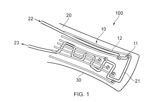
Figure 1 illustrates a medical device 100. The medical 100 may be a
bioelectric implant, for
example. The bioelectric implant 100 may be an active implant, such as a
spinal cord
stimulation (SCS) device. Alternatively, the bioelectric implant may be a
passive implant,
such as an electrocorticography sensor. In other applications, device 100 may
have both
active and passive functions. Other applications for such devices 100 include
for use in
peripheral nerve implants or recording/stimulating muscle activity.
13
CA 03146783 2022-01-10
WO 2021/005382 PCT/GB2020/051684
The medical device 100 comprises a flexible electrode array 10. The flexible
electrode array
comprises electrodes 11 connected to conductive lines 12, provided on a
flexible substrate
30. By way of non-limiting example, the flexible electrode array may be around
5 p.m thick.
The electrode array 10 is flexible so that it can change in configuration in
response, actuated
by the he fluidic component 20, as explained below. As such, herein, the
phrase "flexible
electrode array" is used to mean an array that can undergo such changes in
configuration.
That includes arrays which are entirely flexible, or semi-flexible (e.g.
including some parts or
features which are rigid or more rigid than other more flexible parts,
provided they can still
undergo the change in configuration actuated by the fluidic component).
The medical device 100 also includes a fluidic component 20. The fluidic
component 20 is
fluidically actuatable to cause the fluidic component 20 to change
configuration, as discussed
below. The fluidic component 20 can be a microfluidic component. In other
arrangements,
there may be one or more fluidic components, but a single fluidic component 20
is illustrated
for ease of understanding.
The fluidic component 20 and the flexible electrode array 10 are configured
such that a
change in configuration of the fluidic component 20 causes a change in
configuration of the
flexible electrode array 10.
In the illustrated embodiment, the change in configuration of the fluidic
component 20 causes
a change in configuration of the electrode array 10 because the substrate 30
of the electrode
array 10 comprises the fluidic component 20. As such, the fluidic component 20
and the
electrode array 10 are integrally connected.
However, in other configurations, the fluidic component 20 may be separate, or
separable
from, the electrode array 10. Indeed, as will be understood from the following
description,
the fluidic component 20 and the electrode array 10 may not be connected by
any other
means than the gathering of the components together before implantation. The
benefit of
having a separate, or separable, electrode array 10 and fluidic component 20
is that the fluidic
component 20 may be removed following the implantation of the electrode array
10.
However, in other scenarios it may be acceptable (or indeed preferable) to
keep the fluidic
component 20 in situ to remain part of the implanted device 100.
14
CA 03146783 2022-01-10
WO 2021/005382 PCT/GB2020/051684
The flexible substrate 30 may be 500 p.m thick or less, optionally 200 p.m
thick or less,
further optionally 100 p.m thick or less, further optionally 50 p.m thick or
less, further
optionally 25 p.m thick or less, further optionally 10 p.m thick or less and
still further
optionally 5 p.m thick or less. A thin substrate facilitates the creating a
small gathered
configuration of the medical device 100.
The flexible substrate 30 may be made of a polymeric material, optionally a
thermoplastic,
and optionally comprising one or more of a poly-urethane, a silicone, a
parylene, a
polyamide, a polyimide, a cyclic olefin polymer, a cyclic olefin copolymer, a
polyacrylate,
polyethylene terephthalate and/or an epoxy. Such materials are suitable for
implantation in
the body, and provide the flexibility to facilitate configuring the device in
a gathered
configuration that can be actuated into an expanded configuration.
In the illustrated embodiment of Figure 1, the fluidic component 20 comprises
a fluidic
channel 21 that extends through the substrate 30, with an inlet 22 and an
outlet 23. The inlet
22 (and outlet 23) may be embodied, for example, as a tube formed separately
and
subsequently connected to the fluidic channel 21.
The inlet 22 is for supplying fluid (i.e. liquid or gas) into the fluidic
component 20. In
general, there may be one or more such inlets 22. Such supply actuates the
fluidic component
20. The actuation may be the result of the supply increasing a fluid pressure
and/or an
amount of a fluid within the fluidic channel 21 of the fluidic component 20.
In some
arrangements, the supply of fluid may cause an inflation or straightening of
the channel 21
within the substrate 30, for example.
In some arrangements, there may be no specific outlet 23, separate to the
inlet 22. For
example, when using a gas as the actuating fluid, the gas may be supplied to
inlet 22 to
actuate the device 100, and removal of the supply may subsequently allow the
pressure to be
released within the channel 21 and gas to exit the channel 21 via the original
inlet 22. In
other arrangements, the channel 21 may extend from a dedicated inlet (or
inlets) 22 to one or
more separate outlets 23.
CA 03146783 2022-01-10
WO 2021/005382 PCT/GB2020/051684
In some arrangements, the fluidic component 20 may have independent fluidic
channels 21,
each with their own inlets 22 and outlets 23 (if present).
In either case, the route of the channel 21 through the substrate 30 can take
different forms.
The form of the route may be dictated by the manner in which the device 100
will be
arranged into the gathered configuration, and the manner in which it is
desired for the device
to transition into the expanded configuration. In some arrangements, the
channel 21 may
have a branching and/or symmetrical structure within the fluidic component 20.
Such
arrangements can provide an even distribution of the channel 21 throughout the
substrate 30,
which can be advantageous for even deployment of the device 100. The channel
21 may take
the form of a single chamber (e.g. having a 'balloon' or 'pillow' form when
inflated), or a
series of interconnected chambers of that sort. Larger chambers may also have
connecting
ties or 'pillars' from one side of the chamber to the other, to help control
the inflated shape
and resist over-inflation.
Fig. 3 illustrates various patterns (in plan view) that may be used for the
channel 21 of the
fluidics component, although other patterns are also possible. In the
patterns, black
represents the channel 21 and areas of white within the black areas indicate
areas where the
channel 21 does not extend, such as the ties or 'pillars' mentioned above, or
larger areas of
the substrate encircled by the channel 21.
As will be observed, the patterns of Figs. 3A, 3C, 3D, 3G and 3H have a single
inlet (at the
bottom of each pattern), which can function as both inlet and outlet. Fig. 3B
has two off-
centre lines at the bottom, which could both be simultaneously used as inlets
and
subsequently simultaneously used as outlets, or could be provided as dedicated
inlet and
outlet lines. Fig. 3E has a line approaching the pattern from the bottom, and
a line leading
away from the top ¨ this arrangement provides a natural 'flow-through'
arrangement in which
e.g. the bottom line can act as an inlet and the top line as the outlet, or
vice versa. Fig. 3F has
three lines approaching from the bottom; as for Fig. 3B, these could all be
used together as
inlets or outlets, depending on the need, or could be individually dedicated
as inlet or outlet
channels. For example, the central line may be the inlet, whilst the outer
lines are outlets, or
vice versa.
16
CA 03146783 2022-01-10
WO 2021/005382
PCT/GB2020/051684
It will also be observed that the patterns of Figs. 3A, 3B, 3C, 3F, 3G and 3H
are symmetrical
in nature. As mentioned above, this can assist with even unrolling/unfurling.
The design of
Figs. 3D and 3E are predominantly symmetrical too, other than the arrangement
of the single
inlet/outlet line of 3D, and the inlet/outlet line returning from the top of
the pattern to in Fig.
3E.
Considering the patterns individually, Fig. 3A illustrates a channel 21
forming a single
chamber. That chamber is of the 'balloon' or 'pillow' type, containing no
ties. The chamber
has a typical paddle shape that might correspond to the shape of an SCS
electrode array, for
example.
Fig. 3B illustrates a channel forming a large chamber, but compared to Fig. 3A
the Fig 3B
chamber is squared, of the sort of shape that may be useful for cortical
sensors (designed for
the surface of the brain). The chamber of Fig. 3B also contains ties or
'pillars' that connect
the upper and lower sides of the chamber. Those ties are shown as the white
circles and
ovals. The ties help control how the chamber inflates and reinforce the
chamber design.
Fig. 3C illustrates a branching channel design, with branches in both
directions (left and
right) from a central channel. The branches get thicker towards the top of the
pattern (i.e.
further from the inlet/outlet line at the bottom of the pattern). Such an
arrangement provides
less resistance to flow in the thicker branches, and can help encourage fluid
to fill the whole
pattern as it is introduced, rather than just fill from the end closest to the
inlet.
Fig. 3D is a branching design similar to Fig. 3C, but with the inlet line
offset to the side, such
that the branches extend from that line in one direction (i.e. to the right as
depicted).
Fig. 3E is a branching design in which the inlet branches, and then those
branches also
branch, before the various branches come back together again. This design
effectively
creates encircled areas of the substrate, bounded by the channel. Although the
channel does
not pass through those areas, the presence of the channel around those areas
means the
unfurling of that area is still actuatable.
17
CA 03146783 2022-01-10
WO 2021/005382
PCT/GB2020/051684
Fig. 3F is a multiply branching design which creates a network of channels and
encircled
areas. Fig. 3G is a similar design (although wider) with a different inlet
arrangement as
already discussed.
Fig. 3H is a branching design in which the initial branches are not
interconnected, but which
each subsequently branch further to form local networks of channel at
different positions
within the substrate. Such an approach might be desired, for example, to
provide a
concentration of the channel (i.e. the networked areas) in regions that will
correspond to
electrode locations, to ensure those areas are particularly well unfurled.
Although various arrangements have been discussed with respect to Fig. 3, the
skilled person
will recognise that those are illustrative only, and that other variations and
designs are also
possible including designs with multiple independent fluidic chambers.
In some arrangements, the fluidic channel 21 may be embedded wholly within the
substrate
30, such that the channel 21 is merely defined by the absence of the substrate
material within
the channel. In other arrangements, the channel may be formed of a different
material to the
surrounding substrate 30, or may be formed of the same material as the
substrate 30 but not
embedded directly within that substrate 30. As such, the fluidic channel may
be relatively
flexible or rigid compared to the substrate, depending on the method of
construction. In
either case, the fluidic channel may have a maximum uninflated width dimension
(i.e. a
maximum size across a cross-section through the channel 21 perpendicular to
the centreline
of the channel 21, before the channel is expanded by pressurisation or being
filled with fluid)
of 5 mm or less, optionally 3 mm or less, further optionally 1 mm or less,
further optionally
5001.tm or less, further optionally 1001.tm or less, further optionally 501.tm
or less, and still
further optionally 51.tm or less. The fluidic channel may also have a maximum
inflated
thickness (i.e. a maximum dimension following the expansion of the channel
after it is
pressurised/ filled with fluid to actuate the fluidic component) of no more
than 5 mm,
optionally no more than 2 mm, further optionally no more than 1 mm, and still
further
optionally no more than 50011.m.
Figure 2 illustrates how the flexible nature of the medical device 100 can be
exploited to
assist in its deployment. Because the electrode array 10 and the fluidic
component 20 are
both flexible, the entire device 100 can be gathered into a configuration that
can permit
18
CA 03146783 2022-01-10
WO 2021/005382 PCT/GB2020/051684
percutaneous deployment. In particular, the flexibility of the electrode array
and, preferably,
the fluidic component at least in the gathered configuration, can allow the
medical device to
be rolled without the functionality of the electrode array being affected.
The left hand side of Figure 2A illustrates, in side view, the device 100
rolled along the
longitudinal extent of the device 100. That is, the device 100 is rolled along
its longest axis.
The device 100 can be unrolled to a relatively flat configuration, shown on
the right.
Figure 2B shows an alternative arrangement (this time in plan view). On the
left, the device
100 is rolled or folded across the width (the shorter direction, within the
plane of the
electrode array 10 when expanded to a flat configuration) of the device.
Again, the device
may subsequently be unrolled or unfolded to provide the fully deployed device
as shown on
the right.
In both cases, the gathered configuration of the device 100 allows for the
possibility of the
device 100 to be implanted percutaneously. By providing a suitably thin and
flexible
substrate 30, even a device 100 with a relatively large expanded surface area
can be rolled
into a relatively narrow configuration that allows for percutaneous deployment
with a suitable
needle. Preferably, the gathered configuration is such that the maximum width
of the device
(i.e. in a cross section in the direction of gathering) in that configuration
is 1 cm or less,
optionally 5 mm or less, further optionally 2 mm or less. It is advantageous
for the maximum
width to be as small as possible, as this allows for a smaller diameter needle
to be used for the
percutaneous deployment. As such, it may be advantageous to roll the device
100 in the
narrower width dimension of the device 100 as opposed to the longer length
dimension, to
arrive at a smaller gathered width (as there will be less material to gather).
Although Fig. 2A illustrates an example with a single roll, it may be
advantageous to roll,
fold or otherwise gather the device from two directions, as shown in Fig. 2B,
e.g. from two
edges to a centre line. Such an arrangement can allow for a more even
deployment, as
discussed below. That is, it can allow the two sides to deploy at the same
time, thereby
avoiding twisting of the device 100 in situ as it is placed.
The method of gathering will be determined by the particular device, but it
can e.g. be
performed by hand, using a guide or otherwise, or may be automated. The
gathering may use
19
CA 03146783 2022-01-10
WO 2021/005382
PCT/GB2020/051684
a guide component (which may be integral to the device 100, or a separate
component) to
give additional stiffness/structure to the gathered device 100, to assist with
the percutaneous
delivery. Such a guide component may take the form of a wire or tubing, or a
bio-resorbable
shank, either within or around the gathered device 100. That is, the guide
component may
provide a relatively rigid 'backbone' or support around which the device 100
may be
gathered, and which may be subsequently used to help direct the device to its
deployment
location from within the gathered configuration. Alternatively, or in
combination, the guide
component may be a sheath or tube which the device is fed into as/after it is
gathered, so that
the guide component is outside of gathered device. In the case of an internal
guide
component, that component may or may not be removed once the device 100 is
deployed. In
the case of an external tube or sheath, the guide component must be withdrawn
or retracted
relative to the device enough to allow the change to the deployed
configuration (although, in
some cases this may be possible without any retraction at all, e.g. if
internal and external
guide components are used in combination).
In use, the device 100 may be gathered as discussed above, and then initially
deployed
according to methods known in the art. For example, an SCS device may be
deployed
percutaneously. Alternatively, a brain sensor can be deployed through a burr
hole in the
cranium. Such a burr hole can be 20 mm or less in diameter, further optionally
10 mm or
less, further optionally 5 mm or less, and still further optionally 2 mm or
less.
After the initial deployment, fluid may be supplied to inlet 22 to fill and/or
pressurise the
channel 21. As the channel 21 is filled/pressurised, it is urged into its
expanded
configuration, and therefore begins to unroll/unfold the fluidic component 20.
As such, the
transition of the fluidic component 20 from the gathered configuration to the
expanded
configuration is actuated by supplying fluid to the fluidic channel 21. This
transition brings
the device into contact with, or into suitable proximity with, the target
tissue.
The change in configuration of the fluidic component 20 causes a change in
configuration of
the associated flexible electrode array 10. In the embodiment of Fig. 1, the
electrode array 10
comprises the substrate 30 in which the fluidic component 20 is comprised. As
mentioned
above, in other arrangements, the fluidic component 20 and the electrode array
10 may be
separate, or separable, components that are each independently flexible. In
those
arrangements, by virtue of the separate/separable components being gathered
together, the
CA 03146783 2022-01-10
WO 2021/005382 PCT/GB2020/051684
actuation of the fluidic component still causes the change in configuration of
the electrode
array 10, even though the electrode array 10 and the fluidic component 20 do
not share the
same substrate 30, for example.
The fluidic actuation of the device 100 causes the device 100 to expand into a
configuration
having a greater projected surface area than the expanded configuration. The
expanded shape
and area of the electrode array varies depending on the application. For
example, the
electrode array for a brain sensor may be relatively square or circular and
have dimensions,
for example, up to 100 mm by 100 mm (i.e. a total area of 0.01 m2) or even
larger. SCS
devices, in contrast, may have similar total areas but are relatively long and
thin and may
have dimensions up to 30 mm by 300 mm, or larger. In either case, smaller
devices may be
used for more targeted sensing/stimulation. Moreover, the fluidic component 20
can act as a
support to help with the positioning of the expanded electrode array 10. The
fluidic
component 20 could be, for example filled with a self-curing gel or foam
following
deployment, to provide ongoing rigidity and support.
Once the device 100 has been deployed and positioned, the fluid provided to
the channel 21
may be removed. However, this is not necessary. For example, the fluid may be
a saline
solution or similar which provides no clinical risk in the unexpected scenario
that the fluid
somehow escapes from the device 100. Similarly optionally, the fluidic
component 20 may
itself be removed following the positioning of the electrode 10, provided that
the fluidic
component 20 and the electrode array 10 are separate or separable. For
example, if the
fluidic component 20 and the electrode array 10 are entirely separate, the
fluidic component
20 may be actuated to cause the change in configuration, thereby unfolding
both the fluidic
component and the electrode array 10, and following that unfolding the fluidic
component 20
may be freely removable.
Following the deployment and positioning of the implant 100, the implanted
device 100 may
be used in the desired capacity, whether that is a sensor or as a stimulator
in the treatment of
the patient. Such treatment can include therapy or diagnosis, or may be as
part of a method
of surgery.
Figure 4 shows a medical device 100 of an embodiment of the present invention
in an inflated
state. The components of the device 100 visible in Figure 4 are labelled using
the same
21
CA 03146783 2022-01-10
WO 2021/005382
PCT/GB2020/051684
numbering as in Figures 1 and 2. Generally, the electrode array 10 including a
plurality of
Ti/Au or Pt electrodes 11 can be seen covering the substrate 30 at the distal
or functional end
110 of the device.
At the proximal end of the device, a first section 120 provides one or more
fluid connectors
102 for fluidic connection for connection of the fluidic component 20 to an
external inflation
device. The fluid connectors 102 are medical grade polyethylene tubing
(although other
materials may be used as indicated above) and have an outside diameter of less
than about
1mm.
A second, more proximal, section 130 provides one or more electrical
connectors 101 for
electrical connection of the electrode array 10 to external electronics such
as a pulse
generator for stimulation or sensors for recording data from the electrodes.
The electrical
connectors 101 are three copper/polyimide flex cables each with a thickness of
about
0.07mm.
In the arrangement in Figure 4, the distal end 110 is the portion of the
device 100 whose
configuration can be changed from a gathered or compressed arrangement into a
larger,
deployed arrangement when the fluidic component is actuated. This distal end
110 is
generally flexible, whilst the first and second sections 120, 130 at the
proximal end of the
device may be less flexible or even rigid, thereby allowing for secure
connection from the
external sources to the fluidic component 20 and the electrode array 10. It
will be
appreciated that the fluid connectors 102 and the electrical connectors 101
will likely not
connect directly to the external sources but may be connector to further
elements such as
tubing or wires (not shown) which extend away from the medical device 100 and,
when the
device 100 is deployed within a patient, may extend outside of the patient
through a lumen.
The first and second sections 120, 130 are also not inflatable and do not
change shape or
configuration when the fluidic component is actuated.
In particular, the distal end 110 of the device 100, and in particular the
electrode array 10, has
a bend radius of no more than 2mm in the x-direction as shown in the axes in
Figure 4. This
means that the device can be readily rolled into a gathered configuration by
rolling about the
centre line of the device 100 which is parallel to the z direction in the
manner shown in, and
22
CA 03146783 2022-01-10
WO 2021/005382 PCT/GB2020/051684
described above in relation to, Figure 2B, and then deployed from that rolled
configuration
into the arrangement shown in Figure 4 when the fluidic component is actuated.
In the device 100 shown in Figure 4, the device is significantly less flexible
to bending about
axes parallel to the x direction shown (perpendicular to the z direction).
Thus the device 100
shown in Figure 4 is not suitable for deployment in the manner shown in, and
described
above in relation to, Figure 2A. This arrangement allows the device 100 to
have certain rigid
or less flexible components in the distal portion 110, provided that they are
aligned along the
longitudinal extent of the device 100.
For example, the distal portion 110 of the device 100 may have a support (not
shown) which
extends along the central longitudinal axis of the device in the z direction.
This support can
provide support and rigidity to the device 100 which may be needed, for
example, to facilitate
deployment and/or to ensure the device retains a desired longitudinal
configuration when
deployed. Despite this rigid or less-flexible support, the distal portion 110
of the device 100
can still be gathered into a compressed configuration by rolling the two sides
in to form two
coils (as viewed along the z direction) which meet at the central axis.
It will be appreciated that, in alternative embodiments, the device 100 may be
more flexible
in the z direction shown in Figure 4 and less flexible (or rigid) in the x
direction. This would
allow for rolling and deployment of the device in the manner shown in, and
described above
in relation to, Figure 2A. In such a device, rigid or less flexible components
in the distal
portion 110 could be aligned parallel to the x direction (i.e. transverse to
the longitudinal
extent of the device 100).
In a variation on such devices 100, the rigid or less flexible components in
the distal end 110
may be detachable or removable. For example, a rigid or stiff support may be
used which
extends along the longitudinal extent of the device 100 during deployment of
the device into
a patient to prevent the distal end of the device from being squashed or
deformed during
deployment. This support may then be removed once the device is in the desired
position. In
these variant devices, once all the rigid or less flexible components have
been removed or
detached from the distal end, the distal end may be flexible in both the x and
z directions and
may have similar bend radii in both directions.
23
CA 03146783 2022-01-10
WO 2021/005382 PCT/GB2020/051684
In alternative embodiments, the distal end 110 of the device 100 may have no
rigid or less
flexible components and thus be similarly flexible in both the x and z
directions. Such
devices may be configured so that deployment by unrolling or unfurling once
the device has
been deployed is possible in both the x and z directions.
Figure 5 illustrates how a device 100 such as that illustrated in Figure 4 and
described above
may be packaged for deployment. The device shown in Figure 5 is identical to
that shown in
Figure 4 and the individual components will not be described again.
Figure 5A shows how a first sheath or connection tubing 200 covers the fluidic
and electrical
connectors. The first sheath 200 is medical grade polyurethane (although, as
above,
alternative materials may be used) having an interior diameter of about 1.5mm
and a wall
thickness of about 0.07mm. The first sheath 200 covers and protects the
fluidic and electrical
connectors (and the further tubing and/or wires etc. that those connectors are
connected to).
Figure 5B shows the device 100 in a rolled configuration (double-rolled about
axes parallel to
the z direction as described in relation to Figure 4 above), along with the
first sheath 200,
both contained within a second sheath or deployment tubing 300. The second
sheath 300 is
medical grade polyurethane (as above, alternative materials may be used)
having an interior
diameter of about 1.82mm and a wall thickness of about 0.15mm.
It will be appreciated that, in order to fit into the second sheath 300 in a
double-roll
configuration without being damaged, the distal portion 110 of the device, and
thus the
fluidic component 20 and the electrode array 10 need to have a bend radius of
less than
0.455mm (1.82mm/2 = 0.91mm maximum available diameter space for each roll =>
0.91mm/2 = 0.455mm maximum radius for each roll).
Figure 6 shows the deployment of a medical device 100, such as that shown in
Figures 4 and
5, into the spinal cord area 400 of a patient according to an embodiment of
the present
invention. The device 100 shown in Figure 6 is an SCS device which is designed
to lie
alongside the spinal cord 410. In each of Figure 6A and 6B, the left hand side
drawing is a
side view of the patient along a cross-section through the spinal cord, whilst
the right hand
side drawing is a transverse cross section through the spinal cord at a point
where the device
is located.
24
CA 03146783 2022-01-10
WO 2021/005382 PCT/GB2020/051684
Figure 6A shows how the device 100 is inserted (for example using a sheath 300
or other
catheter delivery system) between vertebrae 420 so as to lie substantially
parallel to the spinal
cord 410. The right hand drawing shows how the device 100 starts to be
deployed by fluidic
actuation which causes the two rolls to unroll outwards away from the centre
line of the
device 100.
Figure 6B shows the device 100 in a deployed state in which the device is
fully unrolled or
unfurled and has a substantially planar configuration, although flexibly
conforming to the
curvature of the spinal cord 410 so that the electrode array of the device
lies adjacent to the
spinal cord.
It can be seen, particularly from the right hand drawings in Figures 6A and
6B, that the
epidural space 430 available for the device 100 to deploy into is limited in
the vertical
direction of Figures 6A and 6B (the anterior-posterior (AP) dimension in terms
of the
patient). In order for the device 100 to deploy into this space, it is
advantageous that the
device can unroll or unfurl such that its thickness in the vertical direction
does not
significantly exceed (if at all) the thickness of the device in that direction
when the device is
in the gathered or compressed configuration that it is initially inserted in.
Devices comprised
of multiple layers which unfold when deployed would be less suitable (if at
all) for
deployment in such spaces.
Whilst this limited space is particularly the case in the deployment of spinal
cord stimulators
and other devices into the spinal cord area 400, similar limitations apply in
the deployment of
other medical devices, for example to the brain area.
As well as meaning that the space for deployment of the device 100 is limited,
the restrictions
in this direction also mean that any expansion of the device in this direction
(i.e.
perpendicular to the direction of the unrolling) needs to be limited and
ideally does not
substantially exceed (if at all) the thickness of the device in the gathered
configuration that it
is inserted in. Excessive expansion in the vertical direction can lead to
damage to
surrounding tissue, obstruction of blood vessels or other complications.
CA 03146783 2022-01-10
WO 2021/005382 PCT/GB2020/051684
Simple inflation of a fluidic component such as that found in known inflatable
devices would
typically tend to result in a thin, flat deflated structure with a thickness
of, say, 20-500
microns inflating to adopt a bulbous shape, often having a circular or oval
cross-section of up
to lcm thickness. This would not be practical in the implementations discussed
above in
relation to Figure 6.
It has been suggested that the inflation thickness of devices could be
controlled by limiting
the amount or pressure of the inflation fluid injected into the device during
deployment.
However, in practice, a significant pressure build-up inside the fluidic
components of the
device is needed to initiate the deployment from the compressed configuration
to the
deployed configuration. As the force needed to cause expansion of the fluidic
component
(and therefore the device as a whole) in the vertical direction is typically
less that the force
needed to cause the device to deploy, the inflation necessary for deployment
in such devices
will inevitably lead to undesirable vertical expansion.
The devices 100 of certain embodiments of the present invention are designed
so as to limit
the expansion of the device in the vertical direction (i.e. a direction
perpendicular to the
direction of deployment of the device and/or a direction perpendicular to the
substantially
planar arrangement of the device in its deployed configuration). In certain
configurations, the
device is limited so that the thickness of the device in the vertical
direction in the deployed
configuration (and preferably at all stages during deployment) is never
greater than the
dimensions of the device in that same direction in the gathered configuration
prior to
deployment. In certain applications, this may be no more than a few
millimetres (e.g. 2, 3 or
5mm).
A variety of arrangements of the device 100 and/or the fluidic component 20
may be used to
achieve this. Two or more of the arrangements described further below may, of
course, be
combined in a particular embodiment.
In certain embodiments, the limitation on vertical expansion is achieved by
incorporating a
stiff (or alternatively inelastic or minimally elastic) material into one or
more layers above
and/or below the fluidic channel 21. This stiff material resists deformation
and therefore
restricts expansion in the vertical direction. Incorporation of such stiff
material needs to also
take account of the requirements for the overall flexibility of the device for
the deployment
26
CA 03146783 2022-01-10
WO 2021/005382 PCT/GB2020/051684
process. This could, for example, be achieved by providing strips of stiff
material with
regions of flexible material between them, the strips being oriented
perpendicular to the
direction of unrolling or unfurling of the device during deployment, such that
the flexible
material ensures that the device as a whole is still sufficiently flexible to
deploy, while the
stiff strips prevent or reduce the vertical expansion by increasing the force
needed to cause
such expansion.
In a variant of the above, a material could be used to form a layer in the
device above and/or
below the fluidic channel 21 which has anisotropic properties, such that it is
flexible in the
direction of rolling/unrolling, but stiff or inelastic in the perpendicular
(e.g. longitudinal)
direction.
In certain embodiments, the fluidic channel 21 itself is configured to limit
vertical expansion.
For example, the fluidic channel 21 may have a cross-section such as that
shown in Figure 7.
Figure 7A shows a schematic cross-section through the fluidic channel 21 when
the device
100 is in an uninflated (i.e. compressed/gathered) state (for convenience, the
channel is
shown planar, although in reality it is likely to be rolled/bent in that
state). The channel 21 is
divided into a plurality of parallel sub-channels 21a by a plurality of ties
or posts 21b that
physically bond the top and bottom layers 21c, 21d and thereby constrain
vertical expansion
of the fluidic channel. The ties or posts 21b can be manufactured as part of
the channel itself,
or may be formed by bonds or welds between the top and bottom layers 21c, 21d.
The ties or
posts 21b may be spot joins, with a plurality of such joins distributed along
the channel, or
may be contiguous along all or part of the channel 21.
As shown in Figure 7B, when the device is inflated, the expansion of the
fluidic channel 21 in
the vertical direction is restrained or restricted by the ties or posts 21b
and therefore, whilst
the individual sub-channels 21a can expand vertically, the overall expansion
of the device can
be controlled and limited.
In a similar fashion, if the fluidic channel 21 is sufficiently small in cross
section, then
vertical expansion may be prevented or restricted as for the individual sub-
channels 21a
shown in Figure 7. Thus an overall design of the fluidic channel 21 which is
small in cross-
section may be provided. A plurality of such channels may be arranged in
parallel to each
other and be joined at either end.
27
CA 03146783 2022-01-10
WO 2021/005382 PCT/GB2020/051684
In other embodiments, there are multiple fluidic chambers defined along the
fluidic channel
21 which are arranged to fill sequentially on inflation of the device.
Pressure-controlled
valves are arranged between each of the chambers such that a chamber will
inflate to a
predetermined pressure limit before the valve connecting to the next chamber
is forced open.
This process could be repeated throughout the device. The vertical expansion
of the device
can then be limited by the design of the geometry of the chambers and the
pressure limits set
by the valves.
Having discussed the configuration and use of the device 100, the following
sections consider
options for fabrication of such a device 100. The discussion presents two
options for how the
electrode array 10 may be formed, and then three separate options for how the
array may be
integrated with the fluidic component 20. Although these protocols refer to
specific
manufacturing techniques, the skilled person will understand that other
techniques may be
substitutable to produce the devices, depending on the desired materials etc.
Such processes
include for example photolithography process, casted elastomer processes,
digital
manufacturing processes (controlled extrusion, additive manufacture).
Fabrication of electrode array
As shown in Fig. 8, step 1, a clean and rigid substrate 41 (of any suitable
material, such as glass
or silicon wafer) can used for the deposition of a thin layer of flexible
material 42 (which will
ultimately form part of the electrode array 10). Suitable flexible materials
include, but are not
limited to, parylene, silicones, polyurethanes, other thermoplastic polymers,
etc. Prior to the
deposition of the flexible material 42, a release layer might be used to
minimise adhesion of
the flexible material film 42 with the rigid support 41 and ease release of
the final structure.
The thin layer of flexible material 42 can then serve (Fig. 8, steps 2 - 5) as
a base onto which
electrodes and conductive lines are patterned. The patterning may use, but is
not limited to
using, metals such as gold, iridium and/or platinum. The patterning can be
achieved through
lift-off techniques that will be familiar to those skilled in the art.
Briefly, a photoresist layer
43 can be spin coated (Fig. 8, step 2), soft baked and exposed (typically by
UV light) using a
contact aligner. The exposed photoresist 43 can then be developed (Fig. 8,
step 3) in the
appropriate developer. A layer of an adhesion promoter metal (typically
chromium or titanium)
28
CA 03146783 2022-01-10
WO 2021/005382 PCT/GB2020/051684
can then be deposited. That layer may be 5 to 10 nm thick, for example. That
can be followed
(Fig. 8, step 4) by the deposition of a relatively thick layer 44 of the
electrode/conductive
material ¨ e.g. gold or platinum. That layer 44 may be 100 nm, for example, or
thicker.
Multilayer deposition of different metals can also be performed. The final
metal patterns are
obtained (Fig. 8, step 5) through lift-off of the photoresist 43 in a suitable
photoresist removal
medium (aqueous solution or solvent/solvent mixture).
Although not illustrated, pattering of the electrodes and conductive lines
could instead be
performed via wet or dry etching of a metal layer. Another possible metal
pattering technique
is laser ablation of a conformal metal foil adhering to the base thin plastic
layer.
Following the creation of the patterned electrode array, the microfabrication
of the device 100
can continue with the deposition of a second film of the flexible material 42
(Fig. 8, step 6).
This layer serves as a passivation layer for the electrodes 11.
Optionally, an adhesion promoter might be used to improve adhesion between the
base layer
and the passivation layer of the flexible material 42. By way of example, a
typical adhesion
promoter for parylene is A-174 (Methacryloxypropyl trimethoxysilane).
Alternatively,
roughening of the surface of the base plastic layer can also improve adhesion
of the passivation
layer.
Next (Fig. 8, step 7) another photoresist 43 can be spin coated, exposed, and
developed using
appropriate developer, followed by e.g. reactive ion etching (Fig. 8, step 8)
to define the outline
of the device. Residual photoresist after the dry etching step can removed
using the appropriate
solvent. Alternatively, the device outline can be defined through laser
ablation of the two thin
plastic layers, for example.
A third sacrificial layer of flexible material 42 can then be deposited (Fig.
8, step 9) on this
structure. An anti-adhesion layer 45, e.g. a 2% v/v soap solution, can be spin
coated in between
the second and third layer of flexible material to minimize adhesion.
The fabrication can then continue (Fig. 8, step 10) with the deposition of a
layer of photoresist
44 to define the electrode area. The photoresist 44 can then be exposed and
developed, followed
by dry etching of the sacrificial and passivation layers of flexible material
42 (Fig. 8, step 11).
29
CA 03146783 2022-01-10
WO 2021/005382
PCT/GB2020/051684
This step is then followed (Fig. 8, step 12) by a lift-off of the remaining
photoresist 43 in the
suitable photoresist removal medium (aqueous solution or solvent/solvent
mixture).
After dry etching, an aqueous dispersion of a conducting polymer 46 can be
spin coated (Fig.
8, step 13) on the substrate. The conducting polymer 46 may be poly(3,4-
ethylenedioxythiophene)- poly(styrenesulfonate) (PEDOT:PSS), for example, and
may contain
additives (such as 5 vol% of ethylene glycol, 0.1 vol% dodecyl benzene
sulfonic acid, and 1
wt% of (3-glycidyloxypropyl) trimethoxysilane (GOPS)). Multiple depositions
can be used to
control the thickness of the conducting polymer. An, e.g. 1 min, step of soft
baking (e.g. at
110 C for the PEDOT:PSS system mentioned above) may be used in between each
deposition.
Finally (Fig. 8, step 14), the sacrificial third layer of flexible material 42
is removed to complete
the patterning of conductive polymer and hard baked (e.g. at 140 C for 1
hour).
The above described protocol includes the patterning using an organic material
(i.e. the
conductive polymer, such as PEDOT:PSS). However, the device 100 can be
manufactured
without the organic layer 46. In that case the workflow is slightly different,
and is illustrated in
Fig. 9 are as discussed in connection with Fig. 8 above. Then, following the
creation of the
etched device outline, a layer of photoresist 43 can be applied to define the
electrode area (Fig.
9, step 9), to allow the electrode area to be etched (Fig. 9, step 10) and the
remaining photoresist
43 to be removed.
Alternatively, other methods of pattering with organic material such as
PEDOT:PSS can be
used, such as via (1) dry etching, (2) dip coating, (3) photopolymerization or
(4)
electropolymerized on the electrode surface
Integration of electrode array and microfitlidics
A first strategy for integrating the electrode array created by the methods
discussed in
connection with Figs. 8 and 9 is illustrated in Fig. 10, steps 15' to step
21'. The device can be
coated with a water-soluble sacrificial layer 51 (Fig. 10, step 15'). This
could be an aqueous
PVA (poly-vinyl alcohol) solution, for example. The device can then be
released from the rigid
substrate 41 and reposition on the same or different rigid substrate 41 having
now the sacrificial
layer 51 facing down onto the rigid substrate 41 (Fig. 10, step 16'). A
removable patterning
CA 03146783 2022-01-10
WO 2021/005382 PCT/GB2020/051684
material 52 such as glycerol can be used to define the microfluidic structure
(Fig. 10, step 17'),
prior to deposition of another layer of flexible material (Fig. 10, step 18')
to seal the
microfluidics. The outline of the microfluidic is then defined (Fig. 10, step
19'). The
removable patterning material is then removed (Fig. 10, step 20') and a tubing
is positioned
and fixed at the inlet of the microfluidics. The final structure is then
released (Fig. 10, step 21').
A second strategy for integrating the electrode array created by the methods
discussed in
connection with Figs. 8 and 9 is illustrated in Fig. 11, steps 15" to step
18". The microfluidic
structure is designed using a CAD software. A thin and flexible double-sided
tape 53 is laser
cut (e.g. CO2 laser cut) to define the microfluidic structure (Fig. 11, step
15"). One side of
the tape can be permanently bonded to a pristine layer of the flexible
material 42 (which may
be held on a support 41). Next, the bioelectric device (e.g. as created
according to the methods
described in connection with Figs. 8 and 9) can be released from its own
substrate 41 (Fig. 11,
step 16"), and then aligned and bonded (Fig. 11, step 17") to the other side
of the tape in order
to seal the microfluidics. A tubing can then be positioned and fixed at the
inlet of the
microfluidics. The final structure can then be released (Fig. 11, step 18").
A third strategy for integrating the electrode array created by the methods
discussed in
connection with Figs. 8 and 9 is illustrated in Fig. 12, steps 15' to step
19". As in the second
strategy, the microfluidic structure can be designed using a CAD software, and
a thin and
flexible double-sided tape 53 is laser cut (e.g. CO2 laser cut) to define the
microfluidic structure
(Fig. 12, step 15'). One side of the tape can be permanently bonded to a
pristine layer of the
flexible material 42 (which may be held on a support 41). The microfluidic
device can then be
realised (Fig. 12, step 16") by positioning a further layer of the flexible
material 42 on the
other side of the laser cut double sided tape 53. An additional layer of the
double-sided tape
can be placed on the top side of the microfluidics (Fig. 12, step 17"),
followed by alignment
and bonding of the bioelectric device (Fig. 12, step 18'). A tubing can be
positioned and
fixed at the inlet of the microfluidics. The final structure can then be
released (Fig. 12, step
20').
As variations on the second and third strategies, instead of using double-
sided tape, a different
bonding strategy may be used, such as printing/stamping of viscous adhesive,
or laser welding
of plastic for example.
31
CA 03146783 2022-01-10
WO 2021/005382 PCT/GB2020/051684
The forgoing description is exemplary in nature only, and the skilled person
will understand
that changes and variations on the disclosed embodiments are possible within
the scope of the
claims. The claims define the invention.
Acknowledgement:
This work has received funding from the European Union's Horizon 2020 research
and
innovation programme under grant agreement N 732032.
32