Note: Descriptions are shown in the official language in which they were submitted.
CA 03146812 2022-01-10
WO 2021/011175
PCT/US2020/039845
PCT APPLICATION
STRONTIUM SEALED SOURCE
BACKGROUND OF THE DISCLOSURE
[0001] This PCT
application claims priority of U.S. patent application serial no.
16/513,032, filed July 16, 2019 which is a continuation-in-part application of
U.S. patent
application serial no. 15/571,310, filed on November 2, 2017, which claims
priority of
PCT/US2016/022437, filed March 15, 2016, which claims priority under 35 U.S.C.
119(e) of
U.S. provisional application serial no. 62/158,091, filed on May 7, 2015, the
contents of all of
which is hereby incorporated by reference in its entirety and for all
purposes.
Field of the Disclosure
[0002] The
disclosure pertains to a strontium-90 sealed source, such as may be used
with treatment of the eye or other medical, brachytherapeutic or industrial
processes. In
particular, a relatively constant absorbed dose rate is sought throughout a
target volume of
tissue of therapeutic interest that is to be treated with radiation
(hereinafter referred to as "a
flat radiation profile").
Description of the Prior Art
[0003] The
prior art of radiological or radioactive sources of various types for
medical, industrial and other processes is well-developed. For example, U.S.
Patent No.
8,430,804, entitled "Methods and Devices for Minimally-Invasive Extraocular
Delivery of
Radiation to the Posterior Portion of the Eye", issued on April 30, 2013 to
Brigatti et al., and
assigned on its face to Salutaris Medical Devices, Inc., discloses an
applicator for minimally-
invasive delivery of beta radiation from a radionuclide brachytherapy source
to the posterior
portion of the eye. In particular, this is adapted for the treatment of
various diseases of the
eye, such as, but not limited to, wet age-related macular degeneration. Other
prior art
includes U.S. Patent No. 9,873,001 entitled "Methods and Devices for Minimally-
Invasive
Delivery of Radiation to the Eye", issued on January 23, 2018 to Lutz et al.
and assigned on
its face to Salutaris Medical Devices, Inc.; PCT/U52014/056135 entitled
"Radiation System
1
CA 03146812 2022-01-10
WO 2021/011175
PCT/US2020/039845
with Emanating Source Surrounding an Internal Attenuation Component", filed on
March 18,
2016; U.S. Patent No. 7,070,554 entitled "Brachytherapy Devices and Methods of
Using
Them", issued on July 4, 2006 to White et al., and assigned on its face to
Theragenics
Corporation and U.S. Patent No. 6,443,881, entitled "Ophthalmic Brachytherapy
Device",
issued on September 3, 2002 to Finger.
[0004] While
this prior art is well-developed and suited for its intended purposes,
further improvements are sought in the radioactive sources used in the
disclosed devices. In
particular, a collimated distribution of radiation, rather than an isotropic
(spherical "47c")
distribution of radiation, would allow a radiological source to direct
radiation at the tissues
under treatment, while reducing radiation directed at surrounding tissues
which are not under
treatment and also while preventing excessive radiation to be directed to the
tissues under
treatment in the center of the emitted radiation beam.
OBJECTS AND SUMMARY OF THE DISCLOSURE
[0005] It is
therefore an object of the present disclosure to provide improvements in
the radiological sources used in brachytherapy and in other medical or
industrial applications.
In particular, it is an object of the present disclosure to provide improved
radiological sources
for known applicators for treatment of diseases of the eye, including, but not
limited to, wet
age-related macular degeneration. These radiological sources are intended to
concentrate the
radiation more uniformly on the diseased tissue, rather than using isotropic
radiation which
would expose more of the surrounding healthy tissue to unnecessary radiation
and could
overexpose tissue under treatment at the center of the radiation beam.
[0006] This and
other objects are attained by providing a beta radiological source,
typically containing strontium-90, wherein the radiological insert has
increased radioactivity
around its periphery and less radioactivity at its center. This may be
achieved by a toroidal or
annular shape, (such as a donut-type shape with a hole or aperture in the
middle) or with the
central portion of a disk having reduced thickness or reduced radioactivity
content. This may
further be achieved by a minus lens meniscus shape wherein the lower concave
surface has a
shorter radius of curvature than the upper concave surface, thereby resulting
in a raised
thinner portion and a lower thicker peripheral portion. This is further
achieved by providing
an encapsulation with increased shielding in the center of the face from which
the therapeutic
radiation is emitted, thereby substantially attenuating the radiation emitted
from the central
2
CA 03146812 2022-01-10
WO 2021/011175
PCT/US2020/039845
portion of a source. It is further possible to use a separate denser
attenuating disk in front of
the activity, either on the inside or outside of the encapsulation. Material
in the attenuating
disk may include, but is not limited to, silver, copper, lead, tungsten, gold
and/or iridium.
BRIEF DESCRIPTION OF THE DRAWINGS
[0007] Further objects and advantages of the disclosure will become
apparent from
the following description and from the accompanying drawings, wherein:
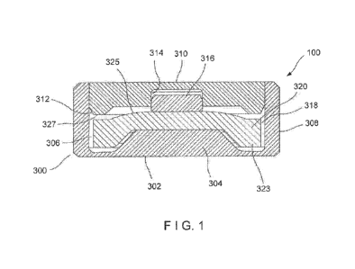
[0008] Figure 1 is a cross-sectional view of an embodiment of a
radiological source of
the present disclosure.
[0009] Figure 2 is an illustration relating to the radiation dose profile
generated by the
radiological source of Figure 1.
[00010] Figures 3A-3F illustrates various further embodiments of the
radiological
source of the present disclosure.
[00011] Figure 4 illustrates a placement of the radiological source with
respect to a
human eyeball during medical treatment.
[00012] Figure 5 illustrates a portion of Figure 4 in greater detail.
[00013] Figure 6 illustrates a still further embodiment of the radiological
source of the
present disclosure, including a minus-lens meniscus shape.
[00014] Figures 7A and 7B illustrate a still further embodiment of the
radiological
source of the present disclosure, wherein multiple elements are placed in a
quasi-toroidal
shape in one or two layers.
[00015] Figures 8A, 8B and 8C illustrate yet still further embodiments of
the
radiological source of the present disclosure.
DETAILED DESCRIPTION OF THE PREFERRED EMBODIMENTS
[00016] Referring now to the drawings in detail wherein like numerals refer
to like
elements throughout the several views, one sees that Figure 1 illustrates a
cross-sectional
view of an embodiment of the radiological source 100. The radiological source
100 is
substantially rotationally symmetric, including cylindrical, annular and
toroidal shapes. A
3
CA 03146812 2022-01-10
WO 2021/011175
PCT/US2020/039845
capsule body 300, typically made of titanium or stainless steel, includes a
lower floor 302
with a central plateau 304 thereby forming a toroidal channel 306 between the
central plateau
304 (thereby increasing the beta shielding in central portions of the lower
floor 302) and the
outer cylindrical wall 308 of the capsule body 300. The upper edge of outer
cylindrical wall
308 forms a circular opening for receiving outer lid 310 which is generally
cylindrical but
includes a chambered lower circular edge 312 and further includes a central
cylindrical blind
opening 314 for receiving telescoping inner lid 316, and typically forming a
tight friction or
interference fit therebetween in order to tightly position the radiological
insert 318 within the
capsule body 300. Outer lid 310, which is typically made of titanium or
stainless steel and
illustrated with an interior circumferential toroidal ridge 327, is typically
welded to capsule
body 300, using conventional standards of the industry. Strontium-90
radiological insert 318
(similar to insert 130 in previous embodiments) includes an upper circular or
disk-shaped
portion 320 which is engaged between a lower edge of telescoping inner lid 316
and central
plateau 304 of capsule body 300. This configuration is intended to reduce
rattling of the
strontium-90 radiological insert 318. The upper surface of strontium-90
radiological insert
318 includes a convex central region 325. This convex central region 325 is
intended to
reinforce the structure and avoid or minimize warping and possible
delamination during
production. Strontium-90 radiological insert 318 further includes a downwardly
extending
circumferential toroidal portion 323 which extends into toroidal channel 306
of capsule body
300.
[00017] The
toroidal shape of the strontium-90 radiological insert 318, with its
thickened periphery, leads to increased radiation emission around the
periphery and a reduced
radiation output within the center. This, in combination with the increased
beta shielding in
the central area of central plateau 304, results in a flat beam profile,
achieving a more
constant absorbed dose rate throughout a target volume of tissue of
therapeutic interest that is
located in front of the source as illustrated in Figure 2, wherein typical
values are given for a
radiological source 100 of a diameter of 4.05 millimeters and a maximum height
of 1.75
millimeters. In the given example, a intended therapeutic volume 400 with a
diameter of 3.0
millimeters and a depth of 1.438 to 2.196 millimeters (with a mean depth to
target of 1.817
millimeters from the lower surface of the radiological source 100) in a first
case or a depth of
1.353 to 2.111 millimeters (with a mean depth to target of 1.752 millimeters
from the lower
surface of the radiological source 100) in a second case. A radius of 11.50
millimeters is
4
CA 03146812 2022-01-10
WO 2021/011175
PCT/US2020/039845
typical for the sclera 2002 (outer covering) of a human eyeball 2000 (see also
Figures 4 and
5). Those skilled in the art, after review of this disclosure, will understand
that different
structural parameters will result in different radiation distributions, as may
be required by the
specific application.
[00018] It is
noted that the strontium-90 beta radiation insert 130 may be made of
various materials, such as a strontium ceramic, strontium glass, or a
collection of tightly
packed ceramic beads (of various possible shapes) or a refractory-metal
composite.
Refractory ceramics and glasses containing Strontium-90 can be made from a
wide variety of
materials in combination, such as those containing metal oxides of aluminum,
silicon,
zirconium, titanium, magnesium, calcium amongst others. It is envisioned that
other
additional materials may be selected from, but not limited to, such strontium-
90 compounds
as SrF2, Sr2P207, SrTiO3, Sr0, Sr2TiO4, SrZr03, SrCO3, Sr(Nb03)2, SrSiO3,
3SrO.A1203,
SrSO4, SrB6, SrS, SrBr2, SrC2, SrC12, SrI2 and SrW04. Additionally, beta
emitters based on
materials other than strontium-90 may also be compatible with this disclosure.
Such beta
emitters may include Copper-66, Lead-209, Praseodymium-145, Tellurium-127, Tin-
121,
Nickel-66, Yttrium-90, Bismuth-210, Erbium-169, Praseodymium-143, Phosphorus-
32,
Phosphorus-33, Strontium-89, Yttrium-91, Tungsten-188, Sulfur-35, Tin-123,
Calcium-45,
Berkelium-249, Ruthenium-106, Thulium-171, Promethium-147, Krypton-85,
Hydrogen-3,
Cadmium-113m, Plutonium-241, Strontium-90, Argon-42, Samarium-151, Nickel-63,
Silicon-32, Argon-39, Carbon-14, Technetium-99, Selenium-79, Beryllium-10,
Cesium-135,
Palladium-107, Rhenium-187, Indium-115 and Cadmium-113. In
particular, after
commercial and technical considerations (e.g., energy level and half-life),
the following are of
particular interest - Strontium-90/Yttrium-90, Strontium-89, Phosphorus-32,
Tin-123 and
Yttrium-91.
[00019] Figures
3A through 3F illustrate six further design embodiments of
radiological source 100 of the present disclosure. The radiological source 100
of Figure 3A is
very similar to Figure 1 and includes capsule body 300 includes a lower floor
302, the interior
wall of the lower floor 302 including a central plateau 304 on the interior
thereof thereby
forming a toroidal channel 306 between the central plateau 304 and the outer
cylindrical wall
308 of the capsule body 300. The upper edge of outer cylindrical wall 308
forms a circular
opening for receiving outer lid 310 which is generally cylindrical. Outer lid
310 is typically
welded to capsule body 300, using conventional standards of the industry.
Strontium-90
CA 03146812 2022-01-10
WO 2021/011175
PCT/US2020/039845
radiological insert 318 is toroidally shaped by rotating a rectangular cross-
section about the
central axis thereby resulting in a central passageway 319. Toroidally-shaped
radiological
insert 318 is positioned above the toroidal channel 306, and supported by
central plateau 304
and shoulder 308A, 308B formed within an interior of outer cylindrical wall
308. A
cylindrical disk-shaped spacer 320, typically made of titanium or stainless
steel, is positioned
between the radiological insert 318 and the lower surface of the outer lid
310. Additionally, a
cylindrical shielding insert 322, typically made from titanium or stainless
steel, inserted
within the central aperture 319. The shape of the strontium-90 radiological
insert 318 leads to
increased radiation output around the periphery, with a reduced radiation
output within the
central aperture 319. This, in combination with the increased shielding in the
central area of
central plateau 304 and the cylindrical shielding insert 322, results in a
flat beam profile,
achieving a more constant absorbed dose rate throughout a target volume of
tissue of
therapeutic interest that is located in front of the source (i.e.,
anisotropic) characteristic of the
resulting beta radiation.
[00020] The
embodiment of radiological source 100 in Figure 3B is similar to that of
Figure 3A. The interior wall of lower floor 302 is generally planar without
the central plateau
of Figure 3A. The toroidal-shaped strontium-90 radiological insert 318 is
secured to
cylindrical disk-shaped spacer 320 by a low-melting glass bond 321 or similar
configuration.
Cylindrical shielding insert 322 extends from spacer 320 to the inner wall of
lower floor 302,
thereby resulting in a configuration with a toroidal-shaped void 306' below
the toroidal-
shaped strontium-90 radiological insert 318. The shape of the strontium-90
radiological
insert 318 leads to an increased radiation source around the periphery, with a
removal of a
source of radiation within the central aperture 319. This, in combination with
the increased
shielding of the cylindrical shielding insert 322, results in a flat beam
profile, achieving a
more constant absorbed dose rate throughout a target volume of tissue of
therapeutic interest
that is located in front of the source).
[00021] The
embodiment of radiological source 100 in Figure 3C is similar to that of
Figure 3A. The toroidal-shaped strontium-90 radiological insert 318 includes a
central
cylindrical disk portion 318A and further includes upper and lower toroidal
portions 318B,
318C, respectively, extending around the circumference thereof Additionally,
spacer 320
further includes a downwardly extending cylindrical skirt 320A which outwardly
abuts the
circumference of toroidal-shaped strontium-90 radiological insert 318. Spacer
320 further
6
CA 03146812 2022-01-10
WO 2021/011175
PCT/US2020/039845
includes a central cylindrical aperture 320B which receives a variation of
shielding insert 322,
further including a downwardly extending frusto-conical portion 322A for
engaging against
central cylindrical disk portion 318A of strontium-90 radiological insert 318
and being
positioned within the upper toroidal portion 318B of strontium-90 radiological
insert 318.
This configuration engages the central cylindrical disk portion 318A between
the downwardly
extending frusto-conical portion 322A of shielding insert 322 and central
plateau 304.
Similar to the embodiment of Figure 3B, a toroidal-shaped void 306' is formed
between the
lower toroidal portion 318C of strontium-90 radiological insert 318 and the
inner wall of floor
302. The shape of the strontium-90 radiological insert 318 leads to an
increased radiation
source around the periphery, with a reduction in the radiation from
cylindrical disk portion
318A. This, in combination with the increased shielding of the central plateau
304, results in
a flat beam profile, achieving a more constant absorbed dose rate throughout a
target volume
of tissue of therapeutic interest that is located in front of the source).
[00022] The
embodiment of Figure 3D is similar to that of Figure 3B. However, the
interior of cylindrical wall 308 includes shoulders 308A, 308B for supporting
the toroidal-
shaped strontium-90 radiological insert 318 above the toroidal channel 306.
This may
eliminate the need for the low melting glass bond 321 or similar configuration
to affix the
toroidal-shaped strontium-90 radiological insert 318 to the spacer 320. The
shape of the
strontium-90 radiological insert 318 leads to an increased radiation source
around the
periphery, with a removal of a source of radiation within the central aperture
319. This, in
combination with the increased shielding of the cylindrical shielding insert
322, results in a
flat beam profile, achieving a more constant absorbed dose rate throughout a
target volume of
tissue of therapeutic interest that is located in front of the source).
[00023] The
embodiment of Figure 3E is similar to that of Figure 3C. The toroidal-
shaped strontium-90 radiological insert 318 includes a central cylindrical
disk portion 318A
and further includes a lower toroidal portion 318C extending around the
circumference
thereof The lack of a upper toroidal portion allows the spacer 320 to be
simplified to a
cylindrical disk shape. The shape of the strontium-90 radiological insert 318
leads to an
increased radiation source around the periphery, with a reduction in the
radiation from
cylindrical disk portion 318A. This, in combination with the increased
shielding of the
central plateau 304, results in a flat beam profile, achieving a more constant
absorbed dose
7
CA 03146812 2022-01-10
WO 2021/011175
PCT/US2020/039845
rate throughout a target volume of tissue of therapeutic interest that is
located in front of the
source).
[00024] The
embodiment of Figure 3F is similar to that of Figure 3E. The strontium-
90 radiological insert 318 is simplified to a disk shape, rather than a
toroidal shape.
Additionally, spacer 320 further includes a downwardly extending cylindrical
skirt 320A
which outwardly abuts the circumference of toroidal-shaped strontium-90
radiological insert
318. The strontium-90 radiological insert 318 is secured to cylindrical disk-
shaped spacer
320 by a low-melting glass bond 321 so as to be suspended above central
plateau 304 and
toroidal channel 306. It is envisioned that this embodiment could further have
the strontium-
90 radiological insert 318 contacting and being supported, at least in part,
by central plateau
304.
[00025] The
embodiment of Figure 6 is a Strontium-90 radiological insert 500 with a
(rotationally symmetric) minus-lens meniscus shape wherein there are two
different
curvatures on the upper and lower surfaces 502, 504. The upper surface 502 (or
"rear") is
convex, the lower surface 504 (or "face") is concave 504 and the radiological
insert 500 is
thinner at its center 510 (i.e., along the rotational axis) than at its edges
512, 514. Typically,
this minus-lens meniscus shape may be implemented by having a shorter radius
of curvature
for the lower surface 504 than for the upper surface 502. While not shown,
this radiological
insert 500 will typically be encased by an encapsulation or capsule body 300
similar to that
shown in Figures 3A-3F, possibly with increased shielding in a central portion
thereof (that is,
below the center 510) in order to achieve a flatter radiation profile. This
meniscus
configuration may be considered, from a mathematical point of view, to be mid-
way between
a cylindrical or flat disk and a toroidal "donut-shaped" configuration. The
configuration may
be termed "meniscus," "biconcave," or "planar concave."
[00026] The
embodiment of Figure 7A illustrates a Strontium-90 radiological insert
600 comprising a ring of disk-like sub-elements of Strontium-90 602 arranged
in a quasi-
toroidal shape.
Similarly, the embodiment of Figure 7B illustrates a Strontium-90
radiological insert 600 comprising a first ring of disk-like sub-elements of
Strontium-90 602
arranged in a quasi-toroidal shape, with second ring of disk-like sub-elements
of Strontium-
90 604, rotationally offset by the radius or one half of the expanse of one
disk from the first
ring, and axially offset, typically by the thickness of the sub-elements 602,
604. The first and
8
CA 03146812 2022-01-10
WO 2021/011175
PCT/US2020/039845
second rings are adjacent to each other and share a common rotational axis
606. The
embodiments of Figures 7A and 7B further include a sealed encapsulation.
[00027] The
embodiments of Figures 8A, 8B and 8C include a metallic, ceramic or
similar dish 700 into which fused Sr-90 glass 702 is melted and bonded. The Sr-
90 glass 702,
in a viscous state, is poured into the dish in an inverted position from that
shown in Figures
8A, 8B and 8C so as to form a meniscus 704 (the illustrated concave surface).
In order to
increase the amount of Sr-90 glass at the peripheral portions of the dish 700,
toroidal troughs
706 may be formed such as is illustrated in Figures 8A and 8B. These
embodiments of
Figures 8A, 8B and 8C further include a sealed encapsulation.
[00028] Further
alternatives to the present disclosure include fixation of the active
insert using glass, such as glass pre-melted into a stainless steel insert,
glass powder co-
compacted with a ceramic and glass powder mixed with a ceramic and then
compacted.
Additionally, alternatives include fixation of the active insert using
mechanical methods such
as soft materials such as copper, silver, aluminum, etc. or the use of springs
of various types
(wave, conical, folded disk, etc.). Further alternatives include active insert
centering features
to prevent positional errors such as tapered ceramic disks or a disk with an
aperture or
protrusion which interfaces with the capsule lid.
[00029]
Similarly, the various embodiments of the radiological sources which include a
cavity could be implemented by filling the cavity with radioactive
microspheres. Such shapes
would be defined by the shape of the cavity inside the source, while the
microspheres could
be immobilized using washers, spaces or similar devices during assembly.
Further alternative
embodiments include radioactive microspheres which are bonded using a fused
glass/enamel
bonding material to an insert (e.g., a metal or ceramic support) to immobilize
the
microspheres and define their shape.
[00030] In a
further aspect of this disclosure, aqueous ammonia solution (NH4OH) is
added to a mixed aqueous solution containing dissolved radioactive strontium
nitrate
905r(NO3)2 and dissolved silver nitrate (AgNO3) (gold or copper may be
substituted for silver
in some applications, mixtures of silver, gold or copper may also be used) and
a mixed
precipitate can form of sparingly soluble silver hydroxide Ag0H (some of which
may convert
to silver oxide Ag2O plus water in situ) and strontium hydroxide 905r(OH)2.
Soluble
ammonium nitrate NH4NO3 remains in solution. Excess ammonium hydroxide
produces a
water-soluble ammoniacal silver complex [Ag(NH3)20H] while the strontium
hydroxide
9
CA 03146812 2022-01-10
WO 2021/011175
PCT/US2020/039845
remains insoluble. The solution and/or the mixed precipitates can be
evaporated so that all
solids co-precipitate or crystalize out of solution to produce an intimate
mixture. These solids
are baked dry so that the ammonium nitrate decomposes and sublimes (above 250
Centigrade) leaving substantially nothing behind, silver hydroxide decomposes
to silver oxide
then further decomposes to silver metal and the strontium hydroxide decomposes
to strontium
oxide. What is left is an intimate mixture of silver metal and strontium oxide
(90Sr0 + Ag).
Because silver is a soft semi-precious metal, such an intimate mixture of
silver and
radioactive strontium oxide can be mechanically and/or thermally formed into
thin toroidal
insert shapes by processes such as pressing, forging, rolling, extrusion
and/or sintering.
[00031] Silver
hydroxide or silver oxide can be prepared and pressed into a disk shape
(toroidal or flat) at a pressure sufficient to bind the particles together to
produce a handleable
green-state disk (an organic or inorganic binder can be added if needed) but
at a pressure that
is low enough to leave porosity or microporosity within the disk. Aqueous
strontium nitrate
905r(NO3)2 can then be soaked into the disk and then dried down to achieve
intimate mixing.
The dried disk can be sintered to produce a fully dense cermet containing
strontium oxide
embedded or immobilized within the matrix formed of copper oxide, silver
oxide, copper
hydroxide, silver hydroxide, gold hydroxide (i.e., auric acid) or mixtures
thereof The
proportions of strontium and silver (or gold, copper or mixtures thereof) can
be varied,
resulting in different mechanical properties. Less strontium produces more
ductility but a
thicker more-attenuating disk. The typical range of composition can be 2-50
mol percent of
strontium oxide in silver, gold or copper, preferably 5-40 mol percent, more
preferably 10-30
mol percent. Cermet disks can be re-pressed or otherwise mechanically or
thermally treated
after sintering to further densify or remold the shape of the disks.
[00032] In a
further aspect of this disclosure, Strontium-90 compounds are
incorporated or mixed with aluminum to make a composite material. This may be
performed
by a method of incorporating Strontium-90 into aluminum by mixing or blending
strontium
fluoride (90SrF2) powder with aluminum powder, compressing the mixture into a
billet, then
heating it to about 10 Centigrade below the melting point of aluminum (660.3
Centigrade)
before extruding the billet through an aperture in a metal collar to produce a
wire of 90SrF2 +
Al. The resulting material can be formed into a toroidal disk or similar
configuration as
described in this disclosure.
CA 03146812 2022-01-10
WO 2021/011175
PCT/US2020/039845
[00033]
Strontium fluoride is a stable material. It melts at 1477 Centigrade and is
insoluble in water (KT value is approximately 2.0x10⁻¹ at 25 Centigrade). It
can be made
from commercially available strontium nitrate 90Sr(NO3)2 by adding soluble
ammonium
fluoride to a strontium nitrate solution, precipitating insoluble strontium
fluoride (90SrF2) and
mixing/blending the dried salt with aluminum powder before pressing the
mixture/blend into
a disk. Useful ratios of 90SrF2 to Al could typically be in the range 5-50% of
90SrF2, preferably
10-30% (by weight). The resulting material can be formed into a toroidal disk
or similar
configuration as described in this disclosure.
[00034]
Alternatively, an aqueous solution of 905r(NO3)2 could be absorbed into a disk
made of porous or microporous aluminum and then dried down and baked above the
decomposition temperature of905r(NO3)2 of 570 Centigrade but below the
melting point of
aluminum 660.3 Centigrade in a non-oxidizing atmosphere, to convert the
strontium nitrate
into strontium oxide. This could be achieved in a vacuum oven or under an
inert gas such as
argon or a reducing atmosphere such as an argon-hydrogen mixture. Other
soluble forms of
Strontium-90 could be absorbed and baked in similar ways. The resulting
material can be
formed into a toroidal disk or similar configuration as described in this
disclosure.
[00035] Thus the
several aforementioned objects and advantages are most effectively
attained. Although preferred embodiments of the invention have been disclosed
and
described in detail herein, it should be understood that this invention is in
no sense limited
thereby.
11