Note: Descriptions are shown in the official language in which they were submitted.
WO 2021/030447
PCT/US2020/045949
METHOD, SYSTEM AND APPARATUS FOR MULTI-OMIC SIMULTANEOUS
DETECTION OF PROTEIN EXPRESSION, SINGLE NUCLEOTIDE VARIATIONS,
AND COPY NUMBER VARIATIONS IN THE SAME SINGLE CELLS
CROSS REFERENCE
[0001] This application claims the benefit of and priority to U.S. Provisional
Patent
Application No. 62/885,490 filed August 12, 2019, the entire disclosure of
which is hereby
incorporated by reference in its entirety for all purposes.
BACKGROUND
[0002] Recent advancements in genomic analysis of tumors
have revealed that cancer
disease evolves by a reiterative process of somatic variation, clonal
expansion and selection.
Therefore, intra- and inter-tumor genomic heterogeneity have become a major
area of
investigation. While next-generation sequencing has contributed significantly
to the
understanding of cancer biology, the genetic heterogeneity of a tumor at the
individual
cellular level is masked with the average readout provided by a bulk
measurement. Very high
bulk sequence read depths are required to identify lower prevalence mutations.
Rare events
and mutation co-occurrence within and across select population of cells are
obscured with
such average signals. As such, there is difficulty in identifying
heterogeneous cell
populations in cells such as cancer cells, which renders cancer treatment
regimen less than
efficacious.
SUMMARY
[0003] Described herein are embodiments for performing
single-cell analysis of a
plurality of cells to determine cellular genotypes and phenotypes of
individual cells. In
various embodiments, the cellular genotypes and phenotypes of individual cells
are
informative for discovering subpopulations of cells characterized by those
genotypes and
phenotypes that may not have previously been known. This is especially useful
in the context
of cancer where heterogeneous cell populations are often present, but not
easily interrogated
or discovered. The identification of subpopulations of cells is informative
for improving the
understanding of disease biology, and subsequently the better design of
diagnostics and
therapies.
[0004] Particular embodiments disclosed herein involve
determining cellular genotypes
directly from cellular genomic DNA. Specifically, genomic DNA is directly
barcoded,
1
CA 03147367 2022-2-8
WO 2021/030447
PCT/US2020/045949
amplified, and sequenced to determine cellular genotype (e.g., SNV and CNV).
Such
methods involving the direct determination of cellular genotypes from genomic
DNA is
preferable in comparison to less direct methods. For example, less direct
methods involve
sequencing cDNA that has been reverse transcribed from RNA transcripts,
thereby providing
an indirect readout of cellular genotypes. The methods disclosed herein
involving direct
determination of cellular genotypes from genomic DNA includes the advantages
of: 1)
achieve broader understanding of cellular genotype across both coding and non-
coding
regions (whereas less direct methods only determine cellular genotype for
coding regions), 2)
avoiding reverse transcription, thereby improving accuracy in calling cell
mutations such as
SNVs and CNVs (e.g., avoids errors and/or processing artifacts that arise due
to reverse
transcription), 3) reduces costs of the single-cell workflow process that
arises from the
inclusion of reagents needed for reverse transcription (e.g., reverse
transcriptase).
[0005] Disclosed herein is a method for analyzing a
plurality of cells, the method
comprising: for one or more cells of the plurality of cells: encapsulating the
cell in an
emulsion comprising reagents, the cell comprising at least one DNA molecule
and at least
one analyte-bound antibody conjugated oligonucleotide; lysing the cell within
the emulsion
to generate a cell lysate comprising the at least one DNA molecule and the
oligonucleotide;
encapsulating the cell lysate comprising the at least one DNA molecule and the
oligonucleotide with a reaction mixture in a second emulsion; performing a
nucleic acid
amplification reaction within the second emulsion using the reaction mixture
to generate
amplicons, the amplicons comprising: a first amplicon derived from one of the
at least one
DNA molecule; and a second amplicon derived from the oligonucleotide;
sequencing the first
amplicon and the second amplicon; determining one or more mutations of the
cell using at
least the sequenced first amplicon; determining a presence or absence of an
analyte using at
least the second amplicon; and discovering a subpopulation of cells in the
plurality of cells,
the subpopulation of cells characterized by the one or more mutations and the
presence or
absence of the analyte.
[0006] In various embodiments, the one or more mutations
comprise a single nucleotide
variant (SNV) or a copy number variation (CNV). In various embodiments, the
one or more
mutations comprise a single nucleotide variant (SNV) and a copy number
variation (CNV).
In various embodiments, discovering the subpopulation of cells in the
plurality of cells
comprises clustering the one or more cells according to the identified SNV or
CNV.
2
CA 03147367 2022-2-8
WO 2021/030447
PCT/US2020/045949
[0007] In various embodiments, the SNV or CNV is
identified in a gene relevant in acute
lymphoblastic leukemia, acute myeloid leukemia, chronic lymphocytic leukemia,
chronic
myeloid leukemia, classic Hodgkin's Lymphoma, diffuse large B-cell lymphoma,
follicular
lymphoma, mantle cell lymphoma, multiple myeloma, myelodysplastic syndromes,
myeloid,
myeloproliferative neoplasms, T-cell lymphoma, breast invasive carcinoma,
colon
adenocarcinoma, glioblastoma multiforme, kidney renal clear cell carcinoma,
liver
hepatocellular carcinoma, lung adenocarcinoma, lung squannous cell carcinoma,
ovarian
cancer, pancreatic adenocarcinoma, prostate adenocarcinoma, or skin cutaneous
melanoma.
In various embodiments, the SNV or CNV is identified in any of ABL1, GNB1,
1CMT2D,
PLCG2, GNA13, ATM, BRAF, JAK3, ADO, DNMT3A, SERMNA1, XPOL PIM1,
CCND1, FLT3, STAT3, AKT1, FAT!, CTCF, TP53, NOTCH1, KRAS, ALK, MYB,
DNM2, DDX3X, CD79A, UBR5, PTEN, APC, PAX5, RUNX1, MAP2K1, CD79B, B1RC3,
1CMT2C, AR, CHD4, PHF6, POTI, CALR, TET2, ORAIL OVGP1, ZMYM3, MYC,
GATA2, CARD11, TP53BP1, TBL1XR1, BTK, WHSC1, MPL, FAS, CDH1, IICZF3,
LRFN2, EGR2, SOCS1, PTPN11, PLCG1, CDK4, WT1P, ZFHX4, MED12, TNFRSF14,
FAM46C, CDKN2A, BCOR, SORCS1, RPS15, TNFA1P3, TRF4, CBL, CSF1R, RPL22,
BTG1, STAT6, PIK3CA, GNAS, CTNNB1, ASXL2, BCL11B, EZH2, DDR2, ATRX,
MYD88, ARID1A, FGFR3, RAD21, EGFR, IICZFl, SMARCA4, SETD2, JA1C2, ERB2,
KLF9, ERG, CREBBP, RB1, CHEK2, ERBB3, ETV6, RPL10, BCL2, DIS3, ID111, ERB4,
NRAS, NFKBIE, NOTCH2, ESR1, HCN4, SF3B I, STAT5B, CCND3, U2AF1, FBXVV7,
CNOT3, EP300, CSF3R, FGFR1, USP9X, WT1, IDH2, FGFR2, 5LC25A33, SH2B3, NF1,
ZFP36L2, KIT, TRAF3, SETBP1, DNAH5, NCOR1, ABL1, ASXL1, GNAll, EPOR,
GNAQ, XBP1, CDKN1B, USH2A, NPM1, HNF1A, FREM2, LEF1, HRAS, OPN5, ZRSR2,
TSPYL2, LM02, JAK1, B2M, TAL1, MGA, NFICBIA, ARAF, ZEB2, KDR,1L7R,
SLC5A1, MYCN, PRDM1, MAP2K2, PH1P, MET, MLH1, REL, ZNF217, NOS1, MTOR,
1CDM6A, SPTBN5, SUZ12, UBA2, PDGFRA, PIK3R1, GATA3, CHD2, HDAC7, SMC1A,
RAF1, MDGA2, USP7, SPEN, RET, ZFR2, SMAD4, ITSN1, SMARCB1, BCORL1, SMC3,
SMO, RPL5, SRC, FOX01, STK11, EBF1, PIK3CD, KMT2A, RHOA, CXCR4, PPM1D,
VHL, LRP1B, and STAG2.
[0008] In various embodiments, determining presence or
absence of the analyte
comprises determining an expression level of the analyte, the analyte bound by
the antibody
conjugated to the oligonucleotide. In various embodiments, the analyte is any
of HLA-DR,
CD10,CD117, CD! lb. CD123, CD13,CD138, CD14, CD141, CD15,CD16, CD163, CD19,
3
CA 03147367 2022-2-8
WO 2021/030447
PCT/US2020/045949
CD193 (CCR3), CD lc, CD2, CD203c, CD209, CD22, CD25, CD3, CD30, CD303, CD304,
CD33, CD34, CD4, CD42b, CD45RA, CD5, CD56, CD62P (P-Selectin), CD64, CD68,
CD69, CD38, CD7, CD71, CD83, CD90 (Thy 1), Fe epsilon RI alpha, Siglec-8,
CD235a,
CD49d, CD45, CD8, CD45RO, mouse IgGl, kappa, mouse IgG2a, kappa, mouse IgG2b,
kappa, CD103, CD62L, CD! lc, CD44, CD27, CD81, CD319 (SLAMF7), CD269 (BCMA),
CD99, CD164, KCNJ3, CXCR4 (CD184), CD109, CD53, CD74, HLA-DR, DP, DQ, HLA-
A, B, C, ROR1, Annexin Al, or CD20.
[0009] In various embodiments, discovering the
subpopulation of cells in the plurality of
cells comprises clustering the one or more cells according to the determined
presence or
absence of the analyte.
[0010] In various embodiments, clustering the one or more
cells according to the
identified SNV or CNV or clustering the one or more cells according to the
determined
presence of the analyte comprises performing a dimensionality reduction
analysis selected
from any of principal component analysis (PCA), linear discriminant analysis
(LDA), T-
distributed stochastic neighbor embedding (t-SNE), or uniform manifold
approximation and
projection (UMAP).
[0011] In various embodiments, the disclosed method
further comprises: prior to
encapsulating the cell in the emulsion, exposing the cell to a plurality of
antibody-conjugated
oligonucleotides; and washing the cell to remove excess antibody conjugated
oligonucleotides. In various embodiments, the oligonucleotides conjugated to
the plurality of
antibodies comprise a PCR handle, a tag sequence, and a capture sequence. In
various
embodiments, the plurality of cells comprise cancer cells. In various
embodiments, the cancer
cells are any of acute lymphoblastic leukemia, acute myeloid leukemia, chronic
lymphocytic
leukemia, chronic myeloid leukemia, classic Hodgkin's Lymphoma, diffuse large
B-cell
lymphoma, follicular lymphoma, mantle cell lymphoma, multiple myeloma,
myelodysplastic
syndromes, myeloid, myeloproliferative neoplasms, T-cell lymphoma, breast
invasive
carcinoma, colon adenocarcinoma, glioblastoma multiforme, kidney renal clear
cell
carcinoma, liver hepatocellular carcinoma, lung adenocarcinoma, lung squamous
cell
carcinoma, ovarian cancer, pancreatic adenocarcinoma, prostate adenocarcinoma,
or skin
cutaneous melanoma.
[0012] In various embodiments, the method further
comprises encapsulating a first
barcode and a second barcode in the second emulsion along with the at least
one DNA
molecule, the oligonucleotide, and the reaction mixture. In various
embodiments, the first
4
CA 03147367 2022-2-8
WO 2021/030447
PCT/US2020/045949
nucleic acid comprises the first barcode. In various embodiments, the second
nucleic acid
comprises the second barcode. In various embodiments, the first barcode and
second barcode
share a same barcode sequence. In various embodiments, the first barcode and
second
barcode share different barcode sequences. In various embodiments, the first
barcode and
second barcode are releasably attached to a bead in the second emulsion.
BRIEF DESCRIPTION OF THE SEVERAL VIEWS OF THE DRAWINGS
[0013] These and other features, aspects, and advantages
of the present invention will
become better understood with regard to the following description, and
accompanying
drawings, where:
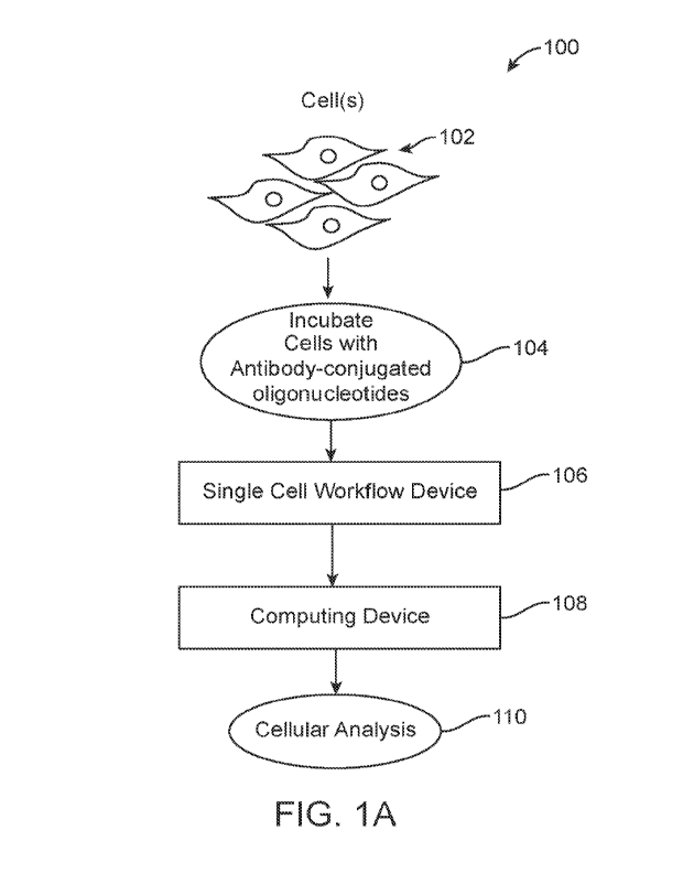
[0014] Figure (FIG.) lA depicts an overall system
environment including a single cell
workflow device and a computational device for conducting single-cell
analysis, in
accordance with an embodiment.
[0015] FIG. 1B shows an embodiment of processing single
cells to generate amplified
nucleic acid molecules for sequencing, in accordance with an embodiment.
[0016] FIG. 2 shows a flow process of determining
cellular genotypes and phenotypes
using sequence reads derived from individual cells and analyzing the cells
using the cellular
genotypes and phenotypes.
[0017] FIGs. 3A-3C shows the steps of analyte release in
the first emulsion, in
accordance with an embodiment.
[0018] FIG. 4A illustrates the priming and barcoding of
an antibody-conjugated
oligonucleotide, in accordance with an embodiment.
[0019] FIG. 48 illustrates the priming and barcoding of
genomic DNA, in accordance
with an embodiment.
[0020] FIGs. 5 and 6 show example gene targets and
protein targets analyzed using the
single cell workflow, in accordance with an embodiment.
[0021] FIG. 7 depicts an example computing device for
implementing system and
methods described in reference to FIGs. 1-6.
[0022] FIG. 8 depicts clustering of cells according to
expression of different proteins.
[0023] FIG. 9A depicts four different cell lines and SNVs
that differentiate the cell lines
from one another.
[0024] FIG. 98 depicts clustering of cells according to
protein expression, with an
additional overlay of cell genotype.
CA 03147367 2022-2-8
WO 2021/030447
PCT/US2020/045949
[0025] FIG. 10 depicts observed gene level copy numbers
for 13 genes across 4 cell lines
and the correlation of the observed gene level copy numbers to known levels in
the COSMIC
database.
[0026] FIG. 11 depicts clustering of cells according to
CNVs with an additional overlay
of cell typing by SNVs.
[0027] FIG. 12A depicts clustering and identification of
different subpopulations of cells
from a mixed population using one of SNV, CNV, or protein data obtained from
single cells.
[0028] FIG. 12B depicts clustering and identification of
different subpopulations of cells
from a mixed population using at least two of SNV, CNV, and protein data
obtained from
single cells.
DETAILED DESCRIPTION
Definitions
[0029] Terms used in the claims and specification are
defined as set forth below unless
otherwise specified.
[0030] The term "subject" or "patient" are used
interchangeably and encompass an
organism, human or non-human, mammal or non-mammal, male or female.
[0031] The term "sample" or "test sample" can include a
single cell or multiple cells or
fragments of cells or an aliquot of body fluid, such as a blood sample, taken
from a subject,
by means including venipuncture, excretion, ejaculation, massage, biopsy,
needle aspirate,
lavage sample, scraping, surgical incision, or intervention or other means
known in the art.
[0032] The term. "analyte" refers to a component of a
cell. Cell anal ytes can be
informative for understanding a state, behavior, or trajectory of a cell.
Therefore,
performing single-cell analysis of one or more analytes of a cell using the
systems and
methods described herein are informative for determining a state or behavior
of a cell.
Examples of an analyte include a nucleic acid (e.g., RNA, DNA, eDNA), a
protein, a
peptide, an. antibody, an antibody fragment, a polysaccharide, a sugar, a
lipid, a_ small
molecule, or combinations thereof. In particular embodiments, a single-cell
analysis
involves analyzing two different analytcs such as protein and DNA. In
particular
embodiments, a single-cell analysis involves analyzing three or more different
analytes of a
cell, such as RNA. DNA, and protein.
[0033] The phrase "cell phenotype" refers to the cell
expression of one or more proteins
(e.g., cellular proteomics). In various embodiments, a cell phenotype is
determined using a
6
CA 03147367 2022-2-8
WO 2021/030447
PCT/US2020/045949
single-cell analysis. In various embodiments, the cell phenotype can refer to
the expression
of a panel of proteins (e.g., a panel of proteins involved in cancer
processes). In various
embodiments, the protein panel includes proteins involved in any of the
following
hematologic malignancies: acute lymphoblastk leukemia, acute myeloid leukemia,
chronic
lymphocytie leukemia, chronic myeloid leukemia, classic Hodgkin's Lymphoma,
diffuse
large B-cell lymphoma, follicular lymphoma, mantle cell lymphoma, multiple
myeloma,
myelodysplastic syndromes, myeloid disease, myeloproliferative neoplasms, or T-
cell
lymphoma. In various embodiments, the protein panel includes proteins involved
in any of
the following solid tumors: breast invasive carcinoma, colon adenocarcinoma,
glioblastoma
multiforme, kidney renal clear cell carcinoma, liver hepatocellular carcinoma,
lung
adenocarcinoma, lung squamous cell carcinoma, ovarian cancer, pancreatic
adenocarcinoma, prostate adenocarcinoma, or skin cutaneous melanoma. Examples
proteins
in the panel can include any of HLA-DR, CD10, CD117, CD11b, CD123, CD13,
CD138,
CD14, CD141, CD15, CD16, CD163, CD19, CD193 (CCR3), CD1c, CD2, CD203c,
CD209, CD22, CD25, CD3, CD30, CD303, CD304, CD33, CD34, CD4, CD42b, CD45RA,
CD5, CD56, CD62P (P-Selectin), CD64, CD68, CD69, CD38, CD7, CD71, CD83, CD90
(Thy 1), Fe epsilon RI alpha, Siglee-8, CD235a, CD49d, CD45, CD8, CD45RO,
mouse
IgGl, kappa, mouse IgG2a, kappa, mouse IgG2b, kappa, CD103, CD62L, CD 1 le,
CD44,
CD27, CD81, CD319 (SLAMF7), CD269 (BCMA), CD99, CD164, KCNJ3, CXCR4
(CD184), CD109, CD53, CD74, HLA-DR, DP, DQ, HLA-A, B, C, ROR1, Annexin Al, or
CD20.
[0034]
The phrase "cell genotype" refers
to the genetic makeup of the cell and can refer
to one or more genes and/or the combination of alleles (e.g., homozygous or
heterozygous)
of a cell. The phrase cell genotype further encompasses one or more mutations
of the cell
including polymorphisms, single nucleotide polymorphisms (SNPs), single
nucleotide
variants (SNVs)), insertions, deletions, knock-ins, knock-outs, copy number
variations
(CNVs), duplications, translocations, and loss of heterozygosity (LOH). In
various
embodiments, a cell phenotype is determined using a single-cell analysis. In
various
embodiments, the cell phenotype can refer to the expression of a panel of
genes (e.g., a
panel of genes involved in cancer processes). In various embodiments, the
panel includes
genes involved in any of the following hematologic malignancies: acute
lymphoblastic
leukemia, acute myeloid leukemia, chronic lymphocytic leukemia, chronic
myeloid
7
CA 03147367 2022-2-8
WO 2021/030447
PCT/US2020/045949
leukemia, classic Hodgkin's Lymphoma, diffuse large B-cell lymphoma,
follicular
lymphoma, mantle cell lymphoma, multiple myeloma, myelodysplastic syndromes,
myeloid,
myeloproliferative neoplasms, or T-cell lymphoma. In various embodiments, the
panel
includes genes involved in any of the following solid tumors: breast invasive
carcinoma,
colon adenocarcinoma, glioblastoma multiforme, kidney renal clear cell
carcinoma, liver
hepatocellular carcinoma, lung adenocarcinoma, lung squamous cell carcinoma,
ovarian
cancer, pancreatic adenocarcinoma, prostate adenocarcinoma, or skin cutaneous
melanoma.
For example, for acute lymphoblastic leukemia, the following genes are
interrogated:
ASXL1, GATA2, KIT, PTPN11, TET2, DNMT3A, 113111, KRAS, RUNX1, TP53, EZH2,
ID112, NPM1, SF3B1, U2AF1, FLT3, JAK2, NRAS, SRSF2, or WT1.
[0035]
In some embodiments, the discrete
entities as described herein are droplets. The
terms "emulsion," "drop," "droplet," and "microdroplet" are used
interchangeably herein, to
refer to small, generally spherically structures, containing at least a first
fluid phase, e.g., an
aqueous phase (e.g., water), bounded by a second fluid phase (e.g., oil) which
is immiscible
with the first fluid phase. In some embodiments, droplets according to the
present disclosure
may contain a first fluid phase, e.g., oil, bounded by a second immiscible
fluid phase, e.g. an
aqueous phase fluid (e.g., water). In some embodiments, the second fluid phase
will be an
immiscible phase carrier fluid. Thus droplets according to the present
disclosure may be
provided as aqueous-in-oil emulsions or oil-in-aqueous emulsions. Droplets may
be sized
and/or shaped as described herein for discrete entities. For example, droplets
according to
the present disclosure generally range from 1 pm to 1000 pm, inclusive, in
diameter.
Droplets according to the present disclosure may be used to encapsulate cells,
nucleic acids
(e.g., DNA), enzymes, reagents, reaction mixture, and a variety of other
components. The
term emulsion may be used to refer to an emulsion produced in, on, or by a
microfluidic
device and/or flowed from or applied by a microfluidic device.
[0036] The term "antibody" encompasses monoclonal antibodies (including full
length
monoclonal antibodies), polyclonal antibodies, multispecific antibodies (e.g.,
bispecific
antibodies), and antibody fragments that are antigen-binding, e.g., an
antibody or an antigen-
binding fragment thereof. "Antibody fragment", and all grammatical variants
thereof, as used
herein are defined as a portion of an intact antibody comprising the antigen
binding site or
variable region of the intact antibody, wherein the portion is free of the
constant heavy chain
domains (i.e., CH2, CH3, and CH4, depending on antibody isotype) of the Fe
region of the
8
CA 03147367 2022-2-8
WO 2021/030447
PCT/US2020/045949
intact antibody. Examples of antibody fragments include Fab, Fab', Fabl-SH,
F(abt)2, and Fv
fragments; diabodies; any antibody fragment that is a polypeptide having a
primary structure
consisting of one uninterrupted sequence of contiguous amino acid residues
(referred to
herein as a "single-chain antibody fragment" or "single chain polypeptide").
[0037] "Complementarity" refers to the ability of a
nucleic acid to form hydrogen
bond(s) or hybridize with another nucleic acid sequence by either traditional
Watson-
Crick or other non-traditional types. As used herein "hybridization," refers
to the
binding, duplexing, or hybridizing of a molecule only to a particular
nucleotide sequence
under low, medium, or highly stringent conditions, including when that
sequence is
present in a complex mixture (e.g., total cellular) DNA or RNA. See e.g.,
Ausubel, et al.,
Current Protocols In Molecular Biology, John Wiley & Sons, New York, N.Y.,
1993. If
a nucleotide at a certain position of a polynucleotide is capable of forming a
Watson-
Crick pairing with a nucleotide at the same position in an anti-parallel DNA
or RNA
strand, then the polynucleotide and the DNA or RNA molecule are complementary
to
each other at that position. The polynucleotide and the DNA or RNA molecule
are
"substantially complementary" to each other when a sufficient number of
corresponding
positions in each molecule are occupied by nucleotides that can hybridize or
anneal with
each other in order to affect the desired process. A complementary sequence is
a
sequence capable of annealing under stringent conditions to provide a 3'-
terminal serving
as the origin of synthesis of complementary chain.
[0038] "Identity," as known in the art, is a relationship
between two or more
polypeptide sequences or two or more polynucleotide sequences, as determined
by
comparing the sequences. In the art, "identity" also means the degree of
sequence
relatedness between polypeptide or polynucleotide sequences, as determined by
the match
between strings of such sequences. "Identity" and "similarity" can be readily
calculated
by known methods, including, but not limited to, those described in
Computational
Molecular Biology, Lesk, A. M., ed., Oxford University Press, New York, 1988;
Biocomputing: Informatics and Genome Projects, Smith, D. W., ed., Academic
Press,
New York, 1993; Computer Analysis of Sequence Data, Part I, Griffin, A. M.,
and
Griffin, H. G., eds., Humana Press, New Jersey, 1994; Sequence Analysis in
Molecular
Biology, von Heinje, G., Academic Press, 1987; and Sequence Analysis Primer,
Gribskov, M. and Devereux, J., eds., M Stockton Press, New York, 1991; and
Carillo,
H., and Lipman, D., Siam J. Applied Math., 48:1073(1988). In addition, values
for
9
CA 03147367 2022-2-8
WO 2021/030447
PCT/US2020/045949
percentage identity can be obtained from amino acid and nucleotide sequence
alignments
generated using the default settings for the AlignX component of Vector NTI
Suite 8.0
(Informax, Frederick, Md.). Preferred methods to determine identity are
designed to give
the largest match between the sequences tested. Methods to determine identity
and
similarity are codified in publicly available computer programs. Example
computer
program methods to determine identity and similarity between two sequences
include,
but are not limited to, the GCG program package (Devereux, J., et al., Nucleic
Acids
Research 12(1): 387 (1984)), BLASTP, BLASTN, and FASTA (Atschul, S. F. et al.,
J.
Malec. Biol. 215:403-410 (1990)). The BLAST X program is publicly available
from
NCBI and other sources (BLAST Manual, Altschul, S., et al., NCBINLM NIH
Bethesda,
Md. 20894: Altschul, S., et al., J. Mol. Biol. 215:403-410(1990). The well-
known Smith
Waterman algorithm may also be used to determine identity.
[0039] The terms "amplify," "amplifying," "amplification
reaction" and their variants,
refer generally to any action or process whereby at least a portion of a
nucleic acid
molecule (referred to as a template nucleic acid molecule) is replicated or
copied into at
least one additional nucleic acid molecule. The additional nucleic acid
molecule
optionally includes sequence that is substantially identical or substantially
complementary to at least some portion of the template nucleic acid molecule.
The
template nucleic acid molecule can be single-stranded or double-stranded and
the
additional nucleic acid molecule can independently be single-stranded or
double-
stranded. In some embodiments, amplification includes a template-dependent in
vitro
enzyme-catalyzed reaction for the production of at least one copy of at least
some portion
of the nucleic acid molecule or the production of at least one copy of a
nucleic acid
sequence that is complementary to at least some portion of the nucleic acid
molecule.
Amplification optionally includes linear or exponential replication of a
nucleic acid
molecule. In some embodiments, such amplification is performed using
isothermal
conditions; in other embodiments, such amplification can include
thermocycling. In some
embodiments, the amplification is a multiplex amplification that includes the
simultaneous amplification of a plurality of target sequences in a single
amplification
reaction. At least some of the target sequences can be situated, on the same
nucleic acid
molecule or on different target nucleic acid molecules included in the single
amplification
reaction. In some embodiments, "amplification" includes amplification of at
least some
portion of DNA- and RNA-based nucleic acids alone, or in combination. The
CA 03147367 2022-2-8
WO 2021/030447
PCT/US2020/045949
amplification reaction can include single or double-stranded nucleic acid
substrates and
can further include any of the amplification processes known to one of
ordinary skill in
the art. In some embodiments, the amplification reaction includes polymerase
chain
reaction (PCR). In some embodiments, the amplification reaction includes an
isothermal
amplification reaction such as LAMP. In the present invention, the terms
"synthesis" and
"amplification" of nucleic acid are used. The synthesis of nucleic acid in the
present
invention means the elongation or extension of nucleic acid from an
oligonucleotide
serving as the origin of synthesis. If not only this synthesis but also the
formation of
other nucleic acid and the elongation or extension reaction of this formed
nucleic acid
occur continuously, a series of these reactions is comprehensively called
amplification.
The polynucleic acid produced by the amplification technology employed is
generically
referred to as an "amplicon" or "amplification product."
[0040] Any nucleic acid amplification method may be
utilized, such as a PCR-based
assay, e.g., quantitative PCR (qPCR), or an isothermal amplification may be
used to
detect the presence of certain nucleic acids, e.g., genes of interest, present
in discrete
entities or one or more components thereof, e.g., cells encapsulated therein.
Such assays
can be applied to discrete entities within a microfluidic device or a portion
thereof or any
other suitable location. The conditions of such amplification or PCR-based
assays may
include detecting nucleic acid amplification over time and may vary in one or
more ways.
[0041] A number of nucleic acid polymerases can be used
in the amplification
reactions utilized in certain embodiments provided herein, including any
enzyme that can
catalyze the polymerization of nucleotides (including analogs thereof) into a
nucleic acid
strand. Such nucleotide polymerization can occur in a template-dependent
fashion. Such
polymerases can include without limitation naturally occurring polymerases and
any
subunits and truncations thereof, mutant polymerases, variant polymerases,
recombinant,
fusion or otherwise engineered polymerases, chemically modified polymerases,
synthetic
molecules or assemblies, and any analogs, derivatives or fragments thereof
that retain the
ability to catalyze such polymerization. Optionally, the polymerase can be a
mutant
polymerase comprising one or more mutations involving the replacement of one
or more
amino acids with other amino acids, the insertion or deletion of one or more
amino acids
from the polymerase, or the linkage of parts of two or more polymerases.
Typically, the
polymerase comprises one or more active sites at which nucleotide binding
and/or
catalysis of nucleotide polymerization can occur. Some exemplary polymerases
include
11
CA 03147367 2022-2-8
WO 2021/030447
PCT/US2020/045949
without limitation DNA polymerases and RNA poly merases. The term "polymerase"
and
its variants, as used herein, also includes fusion proteins comprising at
least two portions
linked to each other, where the first portion comprises a peptide that can
catalyze the
polymerization of nucleotides into a nucleic acid strand and is linked to a
second portion
that comprises a second polypeptide. In some embodiments, the second
polypeptide can
include a reporter enzyme or a processivity-enhancing domain. Optionally, the
polymerase
can possess 5' exonuclease activity or terminal transferase activity. In some
embodiments,
the polymerase can be optionally reactivated, for example through the use of
heat,
chemicals or re-addition of new amounts of polymerase into a reaction mixture.
In some
embodiments, the polymerase can include a hot-start polymerase or an aptamer-
based
polymerase that optionally can be reactivated.
[0042] The terms "target primer" or "target-specific
primer" and variations thereof refer
to primers that are complementary to a binding site sequence. Target primers
are generally
a single stranded or double- stranded polynucleotide, typically an
oligonucleotide, that
includes at least one sequence that is at least partially complementary to a
target nucleic
acid sequence.
[0043] "Forward primer binding site" and "reverse primer
binding site" refers to the
regions on the template DNA and/or the amplicon to which the forward and
reverse
primers bind. The primers act to delimit the region of the original template
polynucleotide which is exponentially amplified during amplification. In some
embodiments, additional primers may bind to the region 5' of the forward
primer and/or
reverse primers. Where such additional primers are used, the forward primer
binding site
and/or the reverse primer binding site may encompass the binding regions of
these
additional primers as well as the binding regions of the primers themselves.
For example,
in some embodiments, the method may use one or more additional primers which
bind to
a region that lies 5' of the forward and/or reverse primer binding region.
Such a method
was disclosed, for example, in W00028082 which discloses the use of
"displacement
primers" or "outer primers."
[0044] A "barcode" nucleic acid identification sequence
can be incorporated into a
nucleic acid primer or linked to a primer to enable independent sequencing and
identification to be associated with one another via a barcode which relates
information
and identification that originated from molecules that existed within the same
sample.
There are numerous techniques that can be used to attach barcodes to the
nucleic acids
12
CA 03147367 2022-2-8
WO 2021/030447
PCT/US2020/045949
within a discrete entity. For example, the target nucleic acids may or may not
be first
amplified and fragmented into shorter pieces. The molecules can be combined
with
discrete entities, e.g., droplets, containing the barcodes. The barcodes can
then be
attached to the molecules using, for example, splicing by overlap extension.
In this
approach, the initial target molecules can have "adaptor" sequences added,
which are
molecules of a known sequence to which primers can be synthesized. When
combined
with the barcodes, primers can be used that are complementary to the adaptor
sequences
and the barcode sequences, such that the product amplicons of both target
nucleic acids
and barcodes can anneal to one another and, via an extension reaction such as
DNA
polymerization, be extended onto one another, generating a double- stranded
product
including the target nucleic acids attached to the barcode sequence.
Alternatively, the
primers that amplify that target can themselves be barcoded so that, upon
annealing and
extending onto the target, the amplicon produced has the barcode sequence
incorporated
into it. This can be applied with a number of amplification strategies,
including specific
amplification with PCR or non-specific amplification with, for example, MDA.
An
alternative enzymatic reaction that can be used to attach barcodes to nucleic
acids is
ligation, including blunt or sticky end ligation. In this approach, the DNA
barcodes are
incubated with the nucleic acid targets and ligase enzyme, resulting in the
ligation of the
barcode to the targets. The ends of the nucleic acids can be modified as
needed for
ligation by a number of techniques, including by using adaptors introduced
with ligase or
fragments to enable greater control over the number of barcodes added to the
end of the
molecule.
[0045] The term "identical" and their variants, as used
herein, when used in reference
to two or more sequences, refer to the degree to which the two or more
sequences (e.g.,
nucleotide or polypeptide sequences) are the same. In the context of two or
more
sequences, the percent identity or homology of the sequences or subsequences
thereof
indicates the percentage of all monomeric units (e.g., nucleotides or amino
acids) that are
the same at a given position or region of the sequence (i.e., about 70%
identity,
preferably 75%, 80%, 85%, 90%, 95%, 97%, 98% or 99% identity). The percent
identity
can be over a specified region, when compared and aligned for maximum
correspondence
over a comparison window, or designated region as measured using a BLAST or
BLAST
2.0 sequence comparison algorithms with default parameters described below, or
by
manual alignment and visual inspection. Sequences are said to be
"substantially identical"
13
CA 03147367 2022-2-8
WO 2021/030447
PCT/US2020/045949
when there is at least 85% identity at the amino acid level or at the
nucleotide level.
Preferably, the identity exists over a region that is at least about 25, 50,
or 100 residues in
length, or across the entire length of at least one compared sequence. A
typical algorithm
for determining percent sequence identity and sequence similarity are the
BLAST and
BLAST 2.0 algorithms, which are described in Altschul et al, Nuc. Acids Res.
25:3389-
3402 (1977). Other methods include the algorithms of Smith & Waterman, Adv.
Appl.
Math. 2:482 (1981), and Needleman & Wunsch, J. Mol. Biol. 48:443 (1970), etc.
Another indication that two nucleic acid sequences are substantially identical
is that the
two molecules or their complements hybridize to each other under stringent
hybridization
conditions.
[0046]
The terms "nucleic acid,"
"polynucleotides," and "oligonucleotides" refers to
biopolymers of nucleotides and, unless the context indicates otherwise,
includes modified
and unmodified nucleotides, and DNA and RNA, and modified nucleic acid
backbones.
For example, in certain embodiments, the nucleic acid is a peptide nucleic
acid (PNA) or
a locked nucleic acid (LNA). Typically, the methods as described herein are
performed
using DNA as the nucleic acid template for amplification. However, nucleic
acid whose
nucleotide is replaced by an artificial derivative or modified nucleic acid
from natural
DNA or RNA is also included in the nucleic acid of the present invention
insofar as it
functions as a template for synthesis of complementary chain. The nucleic acid
of the
present invention is generally contained in a biological sample. The
biological sample
includes animal, plant or microbial tissues, cells, cultures and excretions,
or extracts
therefrom. In certain aspects, the biological sample includes intracellular
parasitic
genomic DNA or RNA such as virus or mycoplasma. The nucleic acid may be
derived
from nucleic acid contained in said biological sample. For example, genomic
DNA, or
cDNA synthesized from mRNA, or nucleic acid amplified on the basis of nucleic
acid
derived from the biological sample, are preferably used in the described
methods. Unless
denoted otherwise, whenever a oligonucleotide sequence is represented, it will
be
understood that the nucleotides are in 5' to 3' order from left to right and
that "A" denotes
deoxyadenosine, "C" denotes deoxycytidine, "G" denotes deoxyguanosine, "T"
denotes
deoxythymidine, and "U' denotes uridine. Oligonucleotides are said to have "5'
ends" and
"3' ends" because mononucleotides are typically reacted to form
oligonucleotides via
attachment of the 5 phosphate or equivalent group of one nucleotide to the 3'
hydroxyl or
14
CA 03147367 2022-2-8
WO 2021/030447
PCT/US2020/045949
equivalent group of its neighboring nucleotide, optionally via a
phosphodiester or other
suitable linkage.
[0047] A template nucleic acid is a nucleic acid serving
as a template for synthesizing
a complementary chain in a nucleic acid amplification technique. A
complementary chain
having a nucleotide sequence complementary to the template has a meaning as a
chain
corresponding to the template, but the relationship between the two is merely
relative.
That is, according to the methods described herein a chain synthesized as the
complementary chain can function again as a template. That is, the
complementary chain
can become a template. In certain embodiments, the template is derived from a
biological
sample, e.g., plant, animal, virus, micro-organism, bacteria, fungus, etc. In
certain
embodiments, the animal is a mammal, e.g., a human patient. A template nucleic
acid
typically comprises one or more target nucleic acid. A target nucleic acid in
exemplary
embodiments may comprise any single or double-stranded nucleic acid sequence
that can
be amplified or synthesized according to the disclosure, including any nucleic
acid
sequence suspected or expected to be present in a sample.
[0048] Primers and oligonucleotides used in embodiments
herein comprise
nucleotides. A nucleotide comprises any compound, including without limitation
any
naturally occurring nucleotide or analog thereof, which can bind selectively
to, or can be
polymerized by, a polymerase. Typically, but not necessarily, selective
binding of the
nucleotide to the polymerase is followed by polymerization of the nucleotide
into a
nucleic acid strand by the polymerase; occasionally however the nucleotide may
dissociate
from the polymerase without becoming incorporated into the nucleic acid
strand, an event
referred to herein as a "non-productive" event. Such nucleotides include not
only naturally
occurring nucleotides but also any analogs, regardless of their structure,
that can bind
selectively to, or can be polymerized by, a polymerase. While naturally
occurring
nucleotides typically comprise base, sugar and phosphate moieties, the
nucleotides of the
present disclosure can include compounds lacking any one, some or all of such
moieties.
For example, the nucleotide can optionally include a chain of phosphorus atoms
comprising three, four, five, six, seven, eight, nine, ten or more phosphorus
atoms. In
some embodiments, the phosphorus chain can be attached to any carbon of a
sugar ring,
such as the S carbon. The phosphorus chain can be linked to the sugar with an
intervening
0 or S. In one embodiment, one or more phosphorus atoms in the chain can be
part of a
phosphate group having P and 0. In another embodiment, the phosphorus atoms in
the
CA 03147367 2022-2-8
WO 2021/030447
PCT/US2020/045949
chain can be linked together with intervening 0, NH, S. methylene, substituted
methylene, ethylene, substituted ethylene, CNH2, C(0), C(CH2), CH2CH2, or
C(OH)CH2R (where R can be a 4-pyridine or 1-imidazole). In one embodiment, the
phosphorus atoms in the chain can have side groups having 0, B113, or S. In
the
phosphorus chain, a phosphorus atom with a side group other than 0 can be a
substituted
phosphate group. In the phosphorus chain, phosphorus atoms with an intervening
atom
other than 0 can be a substituted phosphate group. Some examples of nucleotide
analogs
are described in Xu, U.S. Pat. No. 7,405,281.
100491 In some embodiments, the nucleotide comprises a
label and referred to herein as
a "labeled nucleotide"; the label of the labeled nuckotide is referred to
herein as a
"nucleotide label." In some embodiments, the label can be in the form of a
fluorescent
moiety (e.g. dye), luminescent moiety, or the like attached to the terminal
phosphate
group, i.e., the phosphate group most distal from the sugar. Some examples of
nucleotides that can be used in the disclosed methods and compositions
include, but are
not limited to, ribonucleotides, deoxyribonucleotides, modified
ribonucleotides, modified
deoxyribonucleotides, ribonucleotide polyphosphates, deoxyribonucleotide
polyphosphates, modified ribonucleotide polyphosphates, modified
deoxyribonucleotide
polyphosphates, peptide nucleotides, modifiedpeptide nucleotides,
metallonucleosides,
phosphonate nucleosides, and modified phosphate-sugar backbone nucleotides,
analogs,
derivatives, or variants of the foregoing compounds, and the like. In some
embodiments,
the nucleotide can comprise non-oxygen moieties such as, for example, thio- or
borano-
moieties, in place of the oxygen moiety bridging the alpha phosphate and the
sugar of the
nucleotide, or the alpha and beta phosphates of the nucleotide, or the beta
and gamma
phosphates of the nucleotide, or between any other two phosphates of the
nucleotide, or
any combination thereof.
100501 "Nucleotide 5'- triphosphate" refers to a
nucleotide with a triphosphate ester
group at the 5' position, and are sometimes denoted as "NTP", or "dNTP" and
"ddNTP"
to particularly point out the structural features of the ribose sugar. The
triphosphate ester
group can include sulfur substitutions for the various oxygens, e.g. a-thio-
nucleotide 5'-
triphosphates. For a review of nucleic acid chemistry, see: Shabarova, Z. and
Bogdanov,
A. Advanced Organic Chemistry of Nucleic Acids, VCH, New York, 1994.
16
CA 03147367 2022-2-8
WO 2021/030447
PCT/US2020/045949
Overview
[0051] Described herein are embodiments for performing
single-cell analyses for a
plurality of cells to determine cellular genotypes and phenotypes of
individual cells.
Generally, the single-cell analysis involves performing targeted DNA-seq to
generate
sequence reads derived from genomic DNA that are used to determine the cell
genotype (e.g.,
cell mutations such as CNVs and/or SNVs). The single-cell analysis further
involves
performing sequencing of oligonucleotides that are linked to antibodies, where
an antibody
exhibits binding affinity for a specific analyte expressed by a cell. Thus,
sequence reads
derived from the antibody-conjugated oligonucleotides are used to determine
the cell
phenotype (e.g., expression or presence of one or more analytes of the cell).
The
combination of cellular genotypes and phenotypes across cells in a population
(e.g., a
population of heterogeneous cancer cells) is useful for discerning
subpopulations of cells, a
subpopulation being characterized by a combination of a genotype and a
phenotype.
Subpopulations of cells may represent a subpopulation that was previously
unknown, or a
subpopulation that is unlikely to be detected using either cell genotype or
phenotype alone.
[0052] Reference is made to FIG. 1A, which depicts an
overall system environment 100
including a single cell workflow device 106 and a computational device 108 for
conducting
single-cell analysis, in accordance with an embodiment. A population of cells
102 are
obtained. In various embodiments, the cells 102 can be isolated from a test
sample obtained
from a subject or a patient. In various embodiments, the cells 102 are healthy
cells taken
from a healthy subject. In various embodiments, the cells 102 include diseased
cells taken
from a subject. In one embodiment, the cells 102 include cancer cells taken
from a subject
previously diagnosed with cancer. For example, cancer cells can be tumor cells
available in
the bloodstream of the subject diagnosed with cancer. As another example,
cancer cells can
be cells obtained through a tumor biopsy. Thus, single-cell analysis of the
tumor cells
enables characterization of cells of the subject's cancer. In various
embodiments, the test
sample is obtained from a subject following treatment of the subject (e.g.,
following a therapy
such as cancer therapy). Thus, single-cell analysis of the cells enables
characterization of
cells representing the subject's response to a therapy.
[0053] At step 104, the cells 102 are incubated with
antibodies. In various embodiments,
an antibody exhibits binding affinity to a target analyte. For example, an
antibody can
exhibit binding affinity to a target epitope of a target protein.
17
CA 03147367 2022-2-8
WO 2021/030447
PCT/US2020/045949
[0054] In various embodiments, the number of cells
incubated with antibodies can be 102
cells, 103 cells, 104 cells, 105 cells, 106 cells, or 107 cells. In various
embodiments, between
103 cells and 107 cells are incubated with antibodies. In various embodiments,
between 104
cells and 106 cells are incubated with antibodies. In various embodiments,
varying
concentrations of antibodies are incubated with cells. In various embodiments,
for an
antibody in the protein panel, a concentration of 0.1 nM, 0.5 nM, 1.0 nM, 2.0
nM, 3.0 nM,
4.0 nM, 5.0 nM, 6.0 nM, 7.0 nM, 8.0 nM, 9.0 nM, 10.0 nM, 20 nM, 30 nM, 40 nM,
50 nM,
60 nM, 70 nM, 80 nM, 90 nM, or 100 nM of the antibody is incubated with cells.
[0055] In various embodiments, cells 102 are incubated
with a plurality of different
antibodies. In one embodiment, amongst the plurality of different antibodies,
each antibody
exhibits binding affinity for an analyte of a panel. For example, each
antibody exhibits
binding affinity for a protein of a panel. Examples of proteins included in
protein panels are
described herein. The incubation of cells with antibodies leads to the binding
of the
antibodies against target epitopes. In various embodiments, a concentration of
0.1 nM, 0.5
nM, 1.0 nM, 2.0 nM, 3.0 nM, 4.0 nM, 5.0 nM, 6.0 nM, 7.0 nM, 8.0 nM, 9.0 nM,
10.0 nM, 20
nM, 30 nM, 40 nM, 50 nM, 60 nM, 70 nM, 80 nM, 90 nM, or 100 nM for narh
antibody of
the antibody panel is incubated with cells.
[0056] Following incubation, the cells 102 are washed
(e.g., with wash buffer) to remove
excess antibodies that are unbound.
[0057] In various embodiments, the antibodies are labeled
with one or more
oligonucleotides, also referred to as antibody oligonucleotides. Such
oligonucleotides can be
read out with microfluidic barcoding and DNA sequencing, thereby enabling the
detection of
cell analytes of interest. When an antibody binds its target, the antibody
oligonucleotide is
carried with it and thus allows the presence of the target analyte to be
inferred based on the
presence of the oligonucleotide tag. In some implementations, analyzing
antibody
oligonucleotides provides an estimate of the different epitopes present in the
cell.
[0058] The single cell workflow device 106 refers to a
device that processes individuals
cells to generate nucleic acids for sequencing. In various embodiments, the
single cell
workflow device 106 can encapsulate individual cells into emulsions, lyse
cells within the
emulsions, perform cell barcoding of cell lysate in a second emulsion, and
perform a nucleic
amplification reaction in the second emulsion. Thus, amplified nucleic acids
can be collected
and sequenced. In various embodiments, the single cell workflow device 106
further includes
a sequencer for sequencing the nucleic acids.
18
CA 03147367 2022-2-8
WO 2021/030447
PCT/US2020/045949
[0059] The computing device 108 is configured to receive
the sequenced reads from the
single cell workflow device 106. In various embodiments, the computing device
108 is
communicatively coupled to the single cell workflow device 106 and therefore,
directly
receives the sequence reads from the single cell workflow device 106. The
computing device
108 analyzes the sequence reads to generate a cellular analysis 110. In one
embodiment, the
computing device 108 analyzes the sequence reads to determine cellular
genotypes and
phenotypes. The computing device 108 uses the determined cellular genotypes
and
phenotypes to discover new cell subpopulations and/or to classify individual
cells into cell
subpopulations. Thus, in such embodiments, the cellular analysis 110 can refer
to the
identification of cell subpopulations or the classifications of cells into
cell subpopulations.
[0060] Reference is now made to FIG. 1B, which depicts
one embodiment of processing
single cells to generate amplified nucleic acid molecules for sequencing.
Specifically, MG.
1B depicts a workflow process including the steps of cell encapsulation 160,
analyte release
165, cell barcoding, and target amplification 175 of target nucleic acid
molecules.
[0061] Generally, the cell encapsulation step 160
involves encapsulating a single cell 102
with reagents 120 into an emulsion. In various embodiments, the emulsion is
formed by
partitioning aqueous fluid containing the cell 102 and reagents 120 into a
carrier fluid (e.g.,
oil 115), thereby resulting in an aqueous fluid-in-oil emulsion. The emulsion
includes
encapsulated cell 125 and the reagents 120. The encapsulated cell undergoes an
analyte
release at step 165. Generally, the reagents cause the cell to lyse, thereby
generating a cell
lysate 130 within the emulsion. In particular embodiments, the reagents 120
include
proteases, such as proteinase K, for lysing the cell to generate a cell lysate
130. The cell
lysate 130 includes the contents of the cell, which can include one or more
different types of
analytes (e.g., RNA transcripts, DNA, protein, lipids, or carbohydrates). In
various
embodiments, the different analytes of the cell lysate 130 can interact with
reagents 120
within the emulsion. For example, primers in the reagents 120, such as reverse
primers, can
prime the analytes.
[0062] The cell barcoding step 170 involves encapsulating
the cell lysate 130 into a
second emulsion along with a barcode 145 and/or reaction mixture 140. In
various
embodiments, the second emulsion is formed by partitioning aqueous fluid
containing the cell
lysate 130 into immiscible oil 135. As shown in FIG. 1B, the reaction mixture
140 and
barcode 145 can be introduced through a separate stream of aqueous fluid,
thereby
19
CA 03147367 2022-2-8
WO 2021/030447
PCT/US2020/045949
partitioning the reaction mixture 140 and barcode into the second emulsion
along with the
cell lysate 130.
[0063] Generally, a barcode 145 can label a target
analyte to be analyzed (e.g., a target
nucleic acid), which enables subsequent identification of the origin of a
sequence read that is
derived from the target nucleic acid. In various embodiments, multiple
barcodes 145 can
label multiple target nucleic acid of the cell lysate, thereby enabling the
subsequent
identification of the origin of large quantities of sequence reads.
[0064] Generally, the reaction mixture 140 enables the
performance of a reaction, such as
a nucleic acid amplification reaction. The target amplification step 175
involves amplifying
target nucleic acids. For example, target nucleic acids of the cell lysate
undergo amplification
using the reaction mixture 140 in the second emulsion, thereby generating
amplicons derived
from the target nucleic acids. Although FIG. 1B depicts cell barcoding 170 and
target
amplification 175 as two separate steps, in various embodiments, the target
nucleic acid is
labeled with a barcode 145 through the nucleic acid amplification step.
[0065] As referred herein, the workflow process shown in
FIG. 1B is a two-step
workflow process in which analyte release 165 from the cell occurs separate
from the steps of
cell barcoding 170 and target amplification 175. For example, analyte release
165 from a cell
occurs within a first emulsion followed by cell barcoding 170 and target
amplification 175 in
a second emulsion. In various embodiments, alternative workflow processes
(e.g., workflow
processes other than the two-step workflow process shown in FIG. 1B) can be
employed. For
example, the cell 102, reagents 120, reaction mixture 140, and barcode 145 can
be
encapsulated in an emulsion. Thus, analyte release 165 can occur within the
emulsion,
followed by cell barcoding 170 and target amplification 175 within the same
emulsion.
[0066] FIG. 2 is a flow process for determining cellular
genotypes and phenotypes using
sequence reads derived from individual cells and analyzing the cells using the
cellular
genotypes and phenotypes. Specifically, FIG. 2 depicts the steps of pooling
amplified nucleic
acids at step 205, sequencing the amplified nucleic acids, and determining a
cell trajectory for
a cell using the sequence reads. Generally, the flow process shown in FIG. 2
is a continuation
of the workflow process shown in FIG. 1B.
[0067] For example, after target amplification at step
175 of FIG. 1B, the amplified
nucleic acids 250A, 250B, and 250C are pooled at step 205 shown in FIG. 2. For
example,
emulsions of amplified nucleic acids are pooled and collected, and the
inuniscible oil of the
emulsions is removed. Thus, amplified nucleic acids from multiple cells can be
pooled
CA 03147367 2022-2-8
WO 2021/030447
PCT/US2020/045949
together. FIG. 2 depicts three amplified nucleic acids 250A, 250B, and 250C
but in various
embodiments, pooled nucleic acids can include hundreds, thousands, or millions
of nucleic
acids derived from analytes of multiple cells.
[0068] In various embodiments, each amplified nucleic
acid 250 includes at least a
sequence of a target nucleic acid 240 and a barcode 230. In various
embodiments, an
amplified nucleic acid 250 can include additional sequences, such as any of a
universal
primer sequence (e.g., an oligo-dT sequence), a random primer sequence, a gene
specific
primer forward sequence, a gene specific primer reverse sequence, or one or
more constant
regions (e.g., PCR handles).
[0069] In various embodiments, the amplified nucleic
acids 250A, 250B, and 250C are
derived from the same single cell and therefore, the barcodes 230A, 230B, and
230C are the
same. As such, sequencing of the barcodes 230 enables the determination that
the amplified
nucleic acids 250 are derived from the same cell. In various embodiments, the
amplified
nucleic acids 250A, 250B, and 250C are pooled and derived from different
cells. Therefore,
the barcodes 230A, 230B, and 230C are different from one another and
sequencing of the
barcodes 230 enables the determination that the amplified nucleic acids 250
are derived from
different cells.
[0070] At step 210, the pooled amplified nucleic acids
250 undergo sequencing to
generate sequence reads. For each amplified nucleic acid, the sequence read
includes the
sequence of the barcode and the target nucleic acid. Sequence reads
originating from
individual cells are clustered according to the barcode sequences included in
the amplified
nucleic acids. In various embodiments, one or more sequence reads for each
single cell are
aligned (e.g., to a reference genome). Aligning the sequence reads to the
reference genome
enables the determination of where in the genome the sequence read is derived
from. For
example, multiple sequence reads generated from DNA, when aligned to a
position of the
genome, can reveal one or more mutations present at or involving the position
of the genome.
In various embodiments, one or more sequence reads for each single cell do not
undergo
alignment. For example, sequence reads derived from antibody oligonucleotides
need not be
aligned to the reference genome, given that the antibody oligonucleotides are
not derived
from genomic DNA of the cell genome.
[0071] At step 220, aligned sequence reads for a single
cell are analyzed to determine the
cellular genotype and cellular phenotype of the single cell. For example,
sequence reads
generated from DNA transcripts are analyzed to determine one or more mutations
of the cell,
21
CA 03147367 2022-2-8
WO 2021/030447
PCT/US2020/045949
such as one or more CNVs and SNVs,. Sequence reads generated from antibody-
conjugated
oligonucleotides are used to determine the cellular phenotype, which can
include the presence
of absence of one or more proteins. In various embodiments, the quantity of
sequence reads
generated from antibody-conjugated oligonucleotides are correlated to an
expression level of
the one or more proteins. Taken together, the cellular genotype (e.g., one or
more SNVs and
CNVs) and cellular phenotype (e.g., presence/absence of proteins) provide a
simultaneous
view of the genornics and proteomics of a single cell.
[0072] At step 225, the cellular genotype and cellular
phenotype of the cell are analyzed.
In one embodiment, the cellular genotype and the cellular phenotype of the
cell are used to
classify the cell in a subpopulation that is characterized by the cellular
genotype and
phenotype. For example, a library of known cell subpopulations can be
characterized based
on combinations of genotypes and phenotypes. Therefore, the genotype and
phenotype of the
cell can be used to classify the cell in one or more cell populations that
share the same or
similar genotype and phenotype.
[0073] In one embodiment, the cellular genotype and
cellular phenotype of the cell is
used to identify cellular subpopulations. For example, the cell can be derived
from a
population of cells. In such embodiments, the cellular genotype and cellular
phenotype of the
cell is analyzed in conjunction with cellular genotypes and cellular
phenotypes of other cells
derived from the population of cells. In various embodiments, analyzing the
cellular
genotypes and cellular phenotypes of the population of cells involves
performing one or both
of a dimensional reduction analysis and a clustering analysis, such that cells
with similar
genotypes or phenotypes are localized within clusters. In various embodiments,
heterogeneous subpopulations of cells can be identified from individual
clusters. In various
embodiments, heterogenous subpopulations of cells can be identified from even
within the
clusters themselves.
[0074] Identifying subpopulations of cells with differing
combinations of genotypes and
phenotypes can be useful for discovering subpopulations of cells in cell
populations. As one
example, a subpopulation of cells can refer to a cancer cell subpopulation.
Thus, detection
and/or identification of the presence of a cancer cell subpopulation is useful
for diagnosing a
subject with cancer. As another example, the population of cells may be a
population of
cancer cells previously thought to be homogeneous. Thus, analyzing the
cellular genotypes
and phenotypes of cells in the cancer cells is helpful in understanding the
heterogeneity of the
22
CA 03147367 2022-2-8
WO 2021/030447
PCT/US2020/045949
cancer cells, which can be used to guide the development or selection of
treatments for
targeting the various subpopulations of cells.
Methods for Performing Single-Cell Analysis
Encapuslation, Analyte Release, Barcoding, and Amplification
[0075] Embodiments described herein involve encapsulating
one or more cells (e.g., at
step 160 in FIG. 1) to perform single-cell analysis on the one or more cells.
In various
embodiments, encapsulating a cell with reagents is accomplished by combining
an aqueous
phase including the cell and reagents with an immiscible oil phase. In one
embodiment, an
aqueous phase including the cell and reagents are flowed together with a
flowing immiscible
oil phase such that water in oil emulsions are formed, where at least one
emulsion includes a
single cell and the reagents. In various embodiments the immiscible oil phase
includes a
fluorous oil, a fluorous non-ionic surfactant, or both. In various
embodiments, emulsions can
have an internal volume of about 0.001 to 1000 picoliters or more and can
range from 0.1 to
1000 pm in diameter.
[0076] In various embodiments, the aqueous phase
including the cell and reagents need
not be simultaneously flowing with the immiscible oil phase. For example, the
aqueous
phase can be flowed to contact a stationary reservoir of the immiscible oil
phase, thereby
enabling the budding of water in oil emulsions within the stationary oil
reservoir.
[0077] In various embodiments, combining the aqueous
phase and the immiscible oil
phase can be performed in a microfluidic device. For example, the aqueous
phase can flow
through a microchannel of the microfluidic device to contact the immiscible
oil phase, which
is simultaneously flowing through a separate microchannel or is held in a
stationary reservoir
of the rnicrofluidic device. The encapsulated cell and reagents within an
emulsion can then
be flowed through the microfluidic device to undergo cell lysis.
[0078] Further example embodiments of adding reagents and
cells to emulsions can
include merging emulsions that separately contain the cells and reagents or
picoinjecting
reagents into an emulsion. Further description of example embodiments is
described in US
Application No. 14/420,646, which is hereby incorporated by reference in its
entirety.
[0079] The encapsulated cell in an emulsion is lysed to
generate cell lysate. In various
embodiments, a cell is lysed by lysing agents that are present in the
reagents. For example,
the reagents can include a detergent such as NP-40 and/or a protease. The
detergent and/or
the protease can lyse the cell membrane. In some embodiments, cell lysis may
also, or
instead, rely on techniques that do not involve a lysing agent in the reagent.
For example,
23
CA 03147367 2022-2-8
WO 2021/030447
PCT/US2020/045949
lysis may be achieved by mechanical techniques that may employ various
geometric features
to effect piercing, shearing, abrading, etc. of cells. Other types of
mechanical breakage such
as acoustic techniques may also be used. Further, thermal energy can also be
used to lyse
cells. Any convenient means of effecting cell lysis may be employed in the
methods
described herein.
[0080] Reference is now made to FIGs. 3A-3C, which depict
steps of releasing and
processing analytes within an emulsion (e.g., emulsion 300), in accordance
with a first
embodiment. FIG. 3A depicts emulsion 300A that includes both the cell 102 and
reagents
120 (as shown in FIG. 1B). Specifically, in FIG. 3A, the emulsion 300A
contains the cell
(which further includes DNA 302), antibody oligonucleotides 304 (from the
antibodies used
to bind cell proteins at step 104 in FIG. 1A), as well as proteases 310 that
are added from the
reagents. Within the emulsion 300A, the cell is lysed, as indicated by the
dotted line of the
cell membrane. In one embodiment, the cell is lysed by detergents included in
the reagents,
such as NP40 (e.g., 0.01% NP40).
[0081] FIG. 3B depicts the emulsion 300B as the proteases
302 digest the chromatin-
bound DNA 302, thereby releasing genomic DNA. In various embodiments, emulsion
300B
is exposed to elevated temperatures to enable the proteases 310 to digest the
chromatin. In
various embodiments, emulsion 300B is exposed to a temperature between 40 C
and 60 C.
In various embodiments, emulsion 30013 is exposed to a temperature between 45
C and
55 C. In various embodiments, emulsion 300B is exposed to a temperature
between 48 C
and 52 C. In various embodiments, emulsion 300B is exposed to a temperature of
50 C.
[0082] FIG. 3C depicts the free genomic DNA strands 306
and the antibody
oligonucleotides 304 residing within emulsion 300C. Proteases 310 are
inactivated. In
various embodiments, proteases 310 are inactivated by exposing emulsion 300C
to an
elevated temperature. In various embodiments, emulsion 300C is exposed to a
temperature
between 70 C and 90 C. In various embodiments, emulsion 300B is exposed to a
temperature between 75 C and 85 C. In various embodiments, emulsion 300B is
exposed to
a temperature between 78 C and 82 C. In various embodiments, emulsion 3001B is
exposed
to a temperature of 80 C.
[0083] In various embodiments, the antibody
oligonucleotide 304 and/or the free
genomic DNA 306 undergo priming within emulsion 300C. In various embodiments,
reverse
primers can hybridize with a portion of the antibody oligonucleotide 304
and/or the free
genomic DNA 306. For example, the reverse primer is a gene specific reverse
primer that
24
CA 03147367 2022-2-8
WO 2021/030447
PCT/US2020/045949
hybridizes with a portion of the free genomic DNA 306. Examples gene specific
primers are
described in further detail below. As another example, the reverse primer is a
PCR handle
that hybridizes with a portion of the antibody oligonucleotide 304, which is
described in
further detail below in relation to FIG. 4A. In various embodiments, the
priming of the
antibody oligonuckotide 304 can occur earlier, for example in emulsion 300A or
emulsion
300B, given that the reverse primers are included in the reagents, which are
introduced into
emulsion 300A along with the proteases 310.
[0084] In various embodiments, the antibody
oligonucleotide 304 and the free genomic
DNA 306 in emulsion 300C represent at least in part the cell lysate, such as
cell lysate 130
shown in FIG. 1B, which is subsequently encapsulated in a second emulsion for
barcoding
and amplification. Specifically, the step of cell barcoding 170 in FIG. 1
includes
encapsulating the cell lysate 130 with a reaction mixture 140 and a barcode
145. In various
embodiments, the reaction mixture 140 includes components for performing a
nucleic acid
reaction on target nucleic acids (e.g., antibody oligonucleotide and freed
genomic DNA). For
example, the reaction mixture 140 can include primers, enzymes for performing
nucleic acid
amplification, and dNTPs or ddNTPs for incorporation into amplified nucleic
acids.
[0085] In various embodiments, a cell lysate is
encapsulated with a reaction mixture and
a barcode by combining an aqueous phase including the reaction mixture and the
barcode
with the cell lysate and an immiscible oil phase. In one embodiment, an
aqueous phase
including the reaction mixture and the barcode are flowed together with a
flowing cell lysate
and a flowing immiscible oil phase such that water in oil emulsions are
formed, where at least
one emulsion includes a cell lysate, the reaction mixture, and the barcode. In
various
embodiments the immiscible oil phase includes a fluorous oil, a fluorous non-
ionic surfactant,
or both. In various embodiments, emulsions can have an internal volume of
about 0.001 to
1000 picoliters or more and can range from 0.1 to 1000 pm in diameter.
[0086] In various embodiments, combining the aqueous
phase and the immiscible oil
phase can be performed in a microfluidic device. For example, the aqueous
phase can flow
through a microchannel of the microfluidic device to contact the immiscible
oil phase, which
is simultaneously flowing through a separate mkrochannel or is held in a
stationary reservoir
of the microfluidic device. The encapsulated cell lysate, reaction mixture,
and barcode within
an emulsion can then be flowed through the microfluidic device to perform
amplification of
target nucleic acids.
CA 03147367 2022-2-8
WO 2021/030447
PCT/US2020/045949
[0087] Further example embodiments of adding reaction
mixture and barcodes to
emulsions can include merging emulsions that separately contain the cell
lysate and reaction
mixture and barcodes or picoinjecting the reaction mixture and/or barcode into
an emulsion.
Further description of example embodiments of merging emulsions or
picoinjecting
substances into an emulsion is found in US Application No. 14/420,646, which
is hereby
incorporated by reference in its entirety.
[0088] Once the reaction mixture and barcode are added to
an emulsion, the emulsion
may be incubated under conditions that facilitate the nucleic acid
amplification reaction. In
various embodiments, the emulsion may be incubated on the same microfluidic
device as was
used to add the reaction mixture and/or barcode, or may be incubated on a
separate device. In
certain embodiments, incubating the emulsion under conditions that facilitates
nucleic acid
amplification is performed on the same microfluidic device used to encapsulate
the cells and
lyse the cells. Incubating the emulsions may take a variety of forms. In
certain aspects, the
emulsions containing the reaction mix, barcode, and cell lysate may be flowed
through a
channel that incubates the emulsions under conditions effective for nucleic
acid
amplification. Flowing the microdroplets through a channel may involve a
channel that
snakes over various temperature zones maintained at temperatures effective for
PCR. Such
channels may, for example, cycle over two or more temperature zones, wherein
at least one
zone is maintained at about 65 C. and at least one zone is maintained at
about 95 C. As the
drops move through such zones, their temperature cycles, as needed for nucleic
acid
amplification. The number of zones, and the respective temperature of each
zone, may be
readily determined by those of skill in the art to achieve the desired nucleic
acid
amplification.
[0089] In various embodiments, following nucleic acid
amplification, emulsions
containing the amplified nucleic acids are collected. In various embodiments,
the emulsions
are collected in a well, such as a well of a microfluidic device. In various
embodiments, the
emulsions are collected in a reservoir or a tube, such as an Eppendorf tube.
Once collected,
the amplified nucleic acids across the different emulsions are pooled. In one
embodiment, the
emulsions are broken by providing an external stimuli to pool the amplified
nucleic acids. In
one embodiment, the emulsions naturally aggregate over time given the density
differences
between the aqueous phase and immiscible oil phase. Thus, the amplified
nucleic acids pool
in the aqueous phase.
26
CA 03147367 2022-2-8
WO 2021/030447
PCT/US2020/045949
[0090] In various embodiments, following pooling, the
amplified nucleic acids can
undergo further preparation for sequencing. For example, sequencing adapters
can be added
to the pooled nucleic acids. Example sequencing adapters are P5 and P7
sequencing
adapters. The sequencing adapters enable the subsequent sequencing of the
nucleic acids.
Example Barcodina of Antibodv-Conluzated Oli2onucleotide and
Genomic DNA
[0091] FIG. 4A illustrates the priming and barcoding of
an antibody-conjugated
oligonucleotide, in accordance with an embodiment. Specifically, FIG. 4A
depicts step 410
involving the priming of the antibody oligonucleotide 304 and further depicts
step 420 which
involves the barcoding and amplification of the antibody oligonucleotide 304.
In various
embodiments, step 410 occurs within a first emulsion during which cell lysis
occurs and step
420 occurs within a second emulsion during which cell barcoding and nucleic
acid
amplification occurs. In such embodiments, the primer 405 is provided in the
reagents and
the bead barcode is provided with the reaction mixture. In some embodiments,
both steps
410 and 420 occur within the second emulsion. In such embodiments, the primer
405 and the
bead barcode shown in FIG. 4A are provided with the reaction mixture.
[0092] The antibody oligonucleotide 304 is conjugated to
an antibody. In various
embodiments, an antibody oligonucleotide 304 includes a PCR handle, a tag
sequence (e.g.,
an antibody tag), and a capture sequence that links the oligonucleotide to the
antibody. In
various embodiments, the antibody oligonucleotide 304 is conjugated to a
region of the
antibody, such that the antibody's ability to bind a target epitope is
unaffected. For example,
the antibody oligonucleotide 304 can be linked to a Pc region of the antibody,
thereby leaving
the variable regions of the antibody unaffected and available for epitope
binding. In various
the antibody oligonucleotide 304 can include a unique molecular identifier
(UMI). In various
embodiments, the UMI can be inserted before or after the antibody tag. In
various
embodiments, the UMI can flank either end of the antibody tag. In various
embodiments, the
UMI enables the identification of the particular antibody oligonucleotide 304
and antibody
combination.
[0093] In various embodiments, the antibody
oligonucleotide 304 includes more than one
PCR handle. For example, the antibody oligonucleotide 304 can include two PCR
handles,
one on each end of the antibody oligonucleotide 304. In various embodiments,
one of the
PCR handles of the antibody oligonucleotide 304 is conjugated to the antibody.
Here,
27
CA 03147367 2022-2-8
WO 2021/030447
PCT/US2020/045949
forward and reverse primers can be provided that hybridize with the two PCR
handles,
thereby enabling amplification of the antibody oligonucleotide 304.
[0094] Generally, the antibody tag of the antibody
oligonucleotide 304 enables the
subsequent identification of the antibody (and corresponding protein). For
example, the
antibody tag can serve as an identifier e.g., a barcode for identifying the
type of protein for
which the antibody binds to. In various embodiments, antibodies that bind to
the same target
are each linked to the same antibody tag. For example antibodies that bind to
the same
epitope of a target protein are each linked to the same antibody tag, thereby
enabling the
subsequent determination of the presence of the target protein. In various
embodiments,
antibodies that bind different epitopes of the same target protein can be
linked to the same
antibody tag, thereby enabling the subsequent determination of the presence of
the target
protein.
[0095] In some embodiments, an oligonucleotide sequence
is encoded by its nucleobase
sequence and thus confers a combinatorial tag space far exceeding what is
possible with
conventional approaches using fluorescence. For example, a modest tag length
of ten bases
provides over a million unique sequences, sufficient to label an antibody
against every
epitope in the human proteome. Indeed, with this approach, the limit to
multiplexing is not
the availability of unique tag sequences but, rather, that of specific
antibodies that can detect
the epitopes of interest in a multiplexed reaction.
[0096] Step 410 depicts the priming of the antibody
oligonucleotide 304 by a primer 405.
As shown in FIG. 4, the primer 405 may include a PCR handle and a common
sequence.
Here, the PCR handle of the primer 405 is complementary to the PCR handle of
the antibody
oligonucleotide 304. Thus, the primer 405 primes the antibody oligonucleotide
304 given the
hybridization of the PCR handles. In various embodiments, extension occurs
from the PCR
handle of the antibody oligonucleotide 304 (as indicated by the dotted arrow).
In various
embodiments, extension occurs from the PCR handle of the primer 405, thereby
generating a
nucleic acid with the antibody tag and capture sequence.
[0097] Step 420 depicts the barcoding of the antibody
oligonucleotide 304. As shown in
FIG. 4, the barcode (e.g., cell barcode) is releasably attached to a bead and
is further linked to
a common sequence. Here, the common sequence linked to the cell barcode is
complementary to the common sequence linked to the PCR handle, antibody tag,
and capture
sequence. The antibody oligonucleotide is extended to include the common
sequence and
cell barcode.
28
CA 03147367 2022-2-8
WO 2021/030447
PCT/US2020/045949
[0098] In various embodiments, the antibody
oligonucleotide is amplified, thereby
generating amplicons with the cell barcode, common sequence, PCR handle,
antibody tag,
and capture sequence. In various embodiments, the capture sequence contains a
biotin
oligonucleotide capture site, which enables streptavidin bead enrichment prior
to library
preparation. In various embodiments, the barcoded antibody-oligonucleotides
can be enriched
by size separation from the amplified genomic DNA targets.
[0099] FIG_ 4B illustrates the priming and barcoding of
genomic DNA 455, in
accordance with an embodiment. Specifically, FIG. 4B depicts step 460
involving the
priming of the genomic DNA 455 and further depicts step 470 which involves the
barcoding
and amplification of the genomic DNA 455. In various embodiments, step 460
occurs within
a first emulsion during which cell lysis occurs and step 470 occurs within a
second emulsion
during which cell barcoding and nucleic acid amplification occurs. In such
embodiments, the
primer 465 is added in the reagents and the barcode and forward primers shown
in step 470
are added with the reaction mixture. In some embodiments, step 460 and step
470 both occur
within a single emulsion (e.g., a second emulsion) during which cell barcoding
and nucleic
acid amplification occurs. In such embodiments, the primer 465 shown in step
460 and the
barcode and forward primers shown in step 470 are added with the reaction
mixture.
[00100] At step 460, a primer 465 (as indicated by the dotted line) hybridizes
with a
portion of the genomic DNA 455. In various embodiments, the primer 465 is a
gene specific
primer that targets a sequence of a gene of interest. Therefore, the primer
465 hybridizes
with a sequence of the genomic DNA 455 corresponding to the gene of interest.
In various
embodiments the primer 465 further includes a PCR handle or is linked to a PCR
handle.
[00101] At step 470, a primer 475 (as indicated by the dotted line) hybridizes
with a
portion of the genomic DNA 455. In various embodiments, the primer 475
includes a PCR
handle or is linked to a PCR handle. In various embodiments, the primer 475 is
a gene
specific primer that targets another sequence of the gene of interest that
differs from the
sequence targeted by the primer 465. Additionally, a cell barcode (cell BC),
which is
releasably attached to a bead, is linked to a PCR handle which hybridizes with
the PCR
handle of the forward primer. Nucleic acid amplification generates amplicons,
each of which
include the cell barcode, PCR handle, forward primer, the gene sequence of
interest the
primer 465, and the PCR handle.
29
CA 03147367 2022-2-8
WO 2021/030447
PCT/US2020/045949
Sequencing and Read Alignment
[00102] Amplified nucleic acids (e.g., amplicons) are sequenced to obtain
sequence reads
for generating a sequencing library. Sequence reads can be achieved with
commercially
available next generation sequencing (NGS) platforms, including platforms that
perform any
of sequencing by synthesis, sequencing by ligation, pyrosequencing, using
reversible
terminator chemistry, using phospholinked fluorescent nucleotides, or real-
time sequencing.
As an example, amplified nucleic acids may be sequenced on an 11lumina MiSeq
platform.
[00103] When pyrosequencing libraries of NGS fragments are cloned in-situ
amplified by
capture of one matrix molecule using granules coated with oligonucleotides
complementary
to adapters. Each granule containing a matrix of the same type is placed in a
microbubble of
the "water in oil" type and the matrix is cloned amplified using a method
called emulsion
PCR. After amplification, the emulsion is destroyed and the granules are
stacked in separate
wells of a titration picoplate acting as a flow cell during sequencing
reactions. The ordered
multiple administration of each of the four dNTP reagents into the flow cell
occurs in the
presence of sequencing enzymes and a luminescent reporter, such as luciferase.
In the case
where a suitable dNTP is added to the 3 'end of the sequencing primer, the
resulting ATP
produces a flash of luminescence within the well, which is recorded using a
CCD camera. It
is possible to achieve a read length of more than or equal to 400 bases, and
it is possible to
obtain 106 readings of the sequence, resulting in up to 500 million base pairs
(megabytes) of
the sequence. Additional details for pyrosequencing are described in
Voelkerding et al.,
Clinical Chem., 55: 641-658, 2009; MacLean et al., Nature Rev. Microbial., 7:
287-296; US
patent Na 6,210,891; US patent No. 6,258,568; each of which is hereby
incorporated by
reference in its entirety.
[00104] On the Solexa / Illutnina platform, sequencing data is produced in the
form of
short readings. In this method, fragments of a library of NGS fragments are
captured on the
surface of a flow cell that is coated with oligonucleotide anchor molecules.
An anchor
molecule is used as a PCR primer, but due to the length of the matrix and its
proximity to
other nearby anchor oligonucleotides, elongation by PCR leads to the formation
of a "vault"
of the molecule with its hybridization with the neighboring anchor
oligonucleotide and the
formation of a bridging structure on the surface of the flow cell These DNA
loops are
denatured and cleaved. Straight chains are then sequenced using reversibly
stained
terminators. The nucleotides included in the sequence are determined by
detecting
fluorescence after inclusion, where each fluorescent and blocking agent is
removed prior to
CA 03147367 2022-2-8
WO 2021/030447
PCT/US2020/045949
the next dNTP addition cycle. Additional details for sequencing using the
11lumina platform
are found in Voelkerding et al., Clinical Chem., 55: 641-658, 2009; MacLean et
al., Nature
Rev. Microbiol., 7: 287-296; US patent No. 6,833,246; US patent No. 7,115,400;
US patent
No. 6,969,488; each of which is hereby incorporated by reference in its
entirety.
[00105] Sequencing of nucleic acid molecules using SOLiD technology includes
clonal
amplification of the library of NGS fragments using emulsion PCR. After that,
the granules
containing the matrix are immobilized on the derivatized surface of the glass
flow cell and
annealed with a primer complementary to the adapter oligonucleotide. However,
instead of
using the indicated primer for 3 'extension, it is used to obtain a 5
phosphate group for
ligation for test probes containing two probe-specific bases followed by 6
degenerate bases
and one of four fluorescent labels. In the SOLiD system, test probes have 16
possible
combinations of two bases at the 3 'end of each probe and one of four
fluorescent dyes at the
5' end. The color of the fluorescent dye and, thus, the identity of each
probe, corresponds to a
certain color space coding scheme. After many cycles of alignment of the
probe, ligation of
the probe and detection of a fluorescent signal, denaturation followed by a
second sequencing
cycle using a primer that is shifted by one base compared to the original
primer. In this way,
the sequence of the matrix can be reconstructed by calculation; matrix bases
are checked
twice, which leads to increased accuracy. Additional details for sequencing
using SOLiD
technology are found in Voelkerding et at, Clinical Chem., 55: 641-658, 2009;
MacLean et
al., Nature Rev. Microbiol., 7: 287-296; US patent No. 5,912,148; US patent
No. 6,130,073;
each of which is incorporated by reference in its entirety.
[00106] In particular embodiments, HeliScope from Helicos BioSciences is used.
Sequencing is achieved by the addition of polymerase and serial additions of
fluorescently-
labeled dNTP reagents. Switching on leads to the appearance of a fluorescent
signal
corresponding to dNTP, and the specified signal is captured by the CCD camera
before each
dNTP addition cycle. The reading length of the sequence varies from 25-50
nucleotides with
a total yield exceeding 1 billion nucleotide pairs per analytical work cycle.
Additional details
for performing sequencing using HeliScope are found in Voelkerding et at,
Clinical Chem.,
55: 641-658, 2009; MacLean et al., Nature Rev. Microbiol., 7: 287-296; US
Patent No.
7,169,560; US patent No. 7,282,337; US patent No. 7,482,120; US patent No.
7,501,245; US
patent No. 6,818,395; US patent No. 6,911,345; US patent No. 7,501,245; each
of which is
incorporated by reference in its entirety.
31
CA 03147367 2022-2-8
WO 2021/030447
PCT/US2020/045949
[00107] In some embodiments, a Roche sequencing system 454 is used. Sequencing
454
involves two steps. In the first step, DNA is cut into fragments of
approximately 300-800
base pairs, and these fragments have blunt ends. Oligonucleotide adapters are
then ligated to
the ends of the fragments. The adapter serves as primers for amplification and
sequencing of
fragments. Fragments can be attached to DNA-capture beads, for example,
streptavidin-
coated beads, using, for example, an adapter that contains a 5'-biotin tag.
Fragments attached
to the granules are amplified by PCR within the droplets of an oil-water
emulsion. The result
is multiple copies of cloned amplified DNA fragments on each bead. At the
second stage, the
granules are captured in wells (several picoliters in volume). Pyrosequencing
is carried out on
each DNA fragment in parallel. Adding one or more nucleotides leads to the
generation of a
light signal, which is recorded on the CCD camera of the sequencing
instrument. The signal
intensity is proportional to the number of nucleotides included.
Pyrosequencing uses
pyrophosphate (PPi), which is released upon the addition of a nucleotide. PPi
is converted to
ATP using ATP sulfurylase in the presence of adenosine 5 'phosphosulfate.
Luciferase uses
ATP to convert luciferin to oxyluciferin, and as a result of this reaction,
light is generated that
is detected and analyzed. Additional details for performing sequencing 454 are
found in
Margulies et al. (2005) Nature 437: 376-380, which is hereby incorporated by
reference in its
entirety.
[00108] Ion Torrent technology is a DNA sequencing method based on the
detection of
hydrogen ions that are released during DNA polymerization. The microwell
contains a
fragment of a library of NGS fragments to be sequenced. Under the microwell
layer is the
hypersensitive ion sensor IS PET. All layers are contained within a
semiconductor CMOS
chip, similar to the chip used in the electronics industry. When dNTP is
incorporated into a
growing complementary chain, a hydrogen ion is released that excites a
hypersensitive ion
sensor. If homopolymer repeats are present in the sequence of the template,
multiple dNTP
molecules will be included in one cycle. This results in a corresponding
amount of hydrogen
atoms being released and in proportion to a higher electrical signal. This
technology is
different from other sequencing technologies that do not use modified
nucleotides or optical
devices. Additional details for Ion Torrent Technology is found in Science 327
(5970): 1190
(2010); US Patent Application Publication Nos. 20090026082,
20090127589,20100301398,
20100197507, 20100188073, and 20100137143, each of which is incorporated by
reference
in its entirety.
32
CA 03147367 2022-2-8
WO 2021/030447
PCT/US2020/045949
[00109] In various embodiments, sequencing reads obtained from the NGS methods
can be
filtered by quality and grouped by barcode sequence using any algorithms known
in the art,
e.g., Python script barcodeCleanup.py . In some embodiments, a given
sequencing read may
be discarded if more than about 20% of its bases have a quality score (Q-
score) less than
Q20, indicating a base call accuracy of about 99%. In some embodiments, a
given sequencing
read may be discarded if more than about 5%, about 10%, about 15%, about 20%,
about
25%, about 30% have a Q-score less than Q10, Q20, Q30, Q40, Q50, Q60, or more,
indicating a base call accuracy of about 90%, about 99%, about 99.9%, about
99.99%, about
99.999%, about 99.9999%, or more, respectively.
[00110] In some embodiments, sequencing reads associated with a barcode
containing less
than 50 reads may be discarded to ensure that all barcode groups, representing
single cells,
contain a sufficient number of high-quality reads. In some embodiments, all
sequencing reads
associated with a barcode containing less than 30, less than 40, less than 50,
less than 60, less
than 70, less than 80, less than 90, less than 100 or more may be discarded to
ensure the
quality of the barcode groups representing single cells.
[00111] In various embodiments, sequence reads with common barcode sequences
(e.g.,
meaning that sequence reads originated from the same cell) may be aligned to a
reference
genome using known methods in the art to determine alignment position
information. For
example, sequence reads derived from genomic DNA can be aligned to a range of
positions
of a reference genome. In various embodiments, sequence reads derived from
genomic DNA
can align with a range of positions corresponding to a gene of the reference
genome. The
alignment position information may indicate a beginning position and an end
position of a
region in the reference genome that corresponds to a beginning nucleotide base
and end
nucleotide base of a given sequence read. A region in the reference genome may
be
associated with a target gene or a segment of a gene. Further details for
aligning sequence
reads to reference sequences is described in US Application No. 16/279,315,
which is hereby
incorporated by reference in its entirety. In various embodiments, an output
file having SAM
(sequence alignment map) format or BAM (binary alignment map) format may be
generated
and output for subsequent analysis, such as for determining cell trajectory.
Cellular Genotype and Phenotype
[00112] Sequencing reads of nucleic acids derived from genomic DNA and the
antibody
oligonucleotide are analyzed to determine cellular phenotypes and cellular
genotypes.
33
CA 03147367 2022-2-8
WO 2021/030447
PCT/US2020/045949
[00113] In various embodiments, determining a cell genotype refers to
determining one or
more mutations in the genome of the cell. In particular embodiments, the
Tapestri Insights
software is implemented to identify the one or more mutations in the genome of
the cell. In
one embodiment, the one or more mutations include single nucleotide changes
(e.g., SNVs)
or short sequences of nucleotide changes (e.g., short indels). Here, aligned
sequence reads
derived from genomic DNA of the cell are analyzed against the reference genome
to
determine differences between likely nucleotide bases present in the cell
mutations
corresponding nucleotide bases present in the reference genome. In various
embodiments,
identifying SNVs and/or short indels can be accomplished by implementing any
publicly
available SNV caller algorithms including, but not limited to: BWA, NovoAlign,
Torrent
Mapping Alignment Program (TMAP), VarScan2, qSNP, Shimmer, RADIA, SOAPsnv,
VarDict, SNVMix2, SPLINTER, SNVer, OutLyzer, Pisces, ISOWN, SomVarIUS, and
SiNVICT.
[00114] In one embodiment, the one or more mutations include structural
variants such as
CNVs and/or mutations that encompass long sequences (e.g., long indels). Here,
split-reads
and de-novo assembly methods can used to identify CNVs and/or longer indels.
In various
embodiments, the CNV caller workflow involves one or more of the following
steps: binning,
GC content correction, mappability correction, removal of outlier bins,
removal of outlier
cells, segmentation, and calling of absolute numbers. Further details of CNV
caller
workflows are described in Fan, X. et al, Methods for Copy Number Aberration
Detection
from Single-cell DNA Sequencing Data, bioRxiv 696179, which is hereby
incorporated by
reference in its entirety. In various embodiments, identifying CNVs and/or
long indels can be
accomplished by implementing any publicly available CNV caller including, but
not limited
to: HMMcopy, SeqSeg, CNV-seq, rSW-seq, FREEC, CNAseg, ReadDepth, CNVator,
seqCBS, seqCNA, m-HMM, Ginkgo, nbCNV, AneuFinder, SCNV, and CNV IFTV.
[00115] In various embodiments, sequence reads are pre-processed prior to
their use in
identifying one or more mutations of the cell genome. For example, reads from
a cell are
normalized by the cell's total read count and grouped by hierarchical
clustering based on
amplicon read distribution. Amplicon counts from the cell is divided by the
median of the
corresponding amplicons from a control group (e.g., a control cell cluster
with known
CNVs). Thus, normalized percentage of sequencing reads were used to calculate
CNVs for
each gene.
34
CA 03147367 2022-2-8
WO 2021/030447
PCT/US2020/045949
[00116] In various embodiments, sequence reads used to determine the cellular
genotype
can be derived from various regions of a cell genome. These regions of the
cell genome
include both coding regions and non-coding regions (e.g., introns, regulatory
elements,
transcription factor binding sites, chromosomal translocation junctions).
Therefore, one or
more mutations (e.g., SNVs, CNVs, and indels) can be identified in both coding
and non-
coding regions. The single-cell workflow analysis detailed above that directly
determines
cellular genotypes from genomic DNA enables the identification of mutations
from both
coding and non-coding regions, whereas less direct methods (e.g., those that
reverse
transcribe RNA) only identify mutations from coding regions.
[00117] To determine a cell phenotype, sequence reads derived from antibody-
conjugated
oligonucleotides are analyzed. Specifically, the sequence of the antibody tag
of the antibody
oligonucleotide is sequenced. The presence of the sequence read indicates that
the
corresponding antibody (on which the oligonucleotide was conjugated) had
previously been
bound to an analyte of the cell. In other words, the presence of the sequence
read indicates
that the cell expressed the target analyte.
[00118] In various embodiments, determining a cell phenotype involves
quantifying a
level of expression of a target analyte. In various embodiments, quantifying a
level of
expression of a target analyte involves normalizing the sequence reads derived
from
antibody-conjugated oligonucleotides. In various embodiments, normalizing the
sequence
reads involves performing a centered log ratio (CLR) transformation. In
various
embodiments, normalizing the sequence reads involves performing Denoised and
Scaled by
Background (DSB). Additional description of DSB normalization is found in
Mule, M. et al.
"Normalizing and denoising protein expression data from droplet-based single
cell profiling."
bioRxiv 2020.02.24.963603, which is hereby incorporated by reference in its
entirety.
[00119] In various embodiments, a cell phenotype can refer to the cell
expression of 1, 2,
3, 4, 5, 6, 7, 8, 9, 10, 11, 12, 13, 14, 15, 16,17, 18, 19, 20, 21, 22, 23,
24, 25, 26, 27, 28, 29,
30 ,31, 32, 33, 34, 35, 36, 37, 38, 39, 40, 41, 42, 43, 44, 45, 46, 47, 48,49,
50, 100, 500,
1000, 5000, or 10,000 target analytes. Therefore, the single-cell workflow
analysis can yield
an expression profile for a plurality of target analytes of a cell.
[00120] In various embodiments, the genotype and the phenotype of the cell can
be used to
classify the cell. For example, the cell can be classified within a population
of cells that share
at least the genotype, share at least the phenotype, or share at least both
the genotype and the
phenotype of the cell. In various embodiments, the single-cell workflow
analysis is
CA 03147367 2022-2-8
WO 2021/030447
PCT/US2020/045949
conducted on each cell in a population of cells. Therefore, the cell genotype
and cell
phenotype of each cell in the population can be used to classify each cell to
gain an
understanding as to the distribution of cells in the population. In various
embodiments, the
classified cells provide insight as to the subpopulations that are present. In
various
embodiments, classifying a cell involves comparing the genotype and phenotype
of the cell
against a library of known cell populations that are characterized by known
genotypes and
phenotypes. Therefore, if the cell shares a genotype, shares a phenotype, or
shares both a
genotype and phenotype with a known cell population, the cell can be
classified in a category
of the known cell population.
[00121] To provide an example, the population of cells can be obtained from a
subject
suspected of having cancer, each cell in the population can be analyzed using
the single-cell
workflow to determine each cell's genotype and phenotype. Cells are classified
according to
their genotypes and phenotypes by comparing to genotypes and phenotypes of
known
reference cells. Thus, classifying cells in the population using their
genotypes and
phenotypes reveals a distribution of cells which can guide the selection of a
cancer treatment
for the subject. For example, if a large proportion of cells in the population
are classified
with a known cell population that are known to be resistant to particular
therapies, then
alternative therapies that are more likely to be efficacious can be selected
for treating the
cancer.
[00122] In various embodiments, the genotype and the phenotype of the cell are
used to
identify subpopulations within a population of cells. This is useful for
discovering new
subpopulations that were not previously known. For example, a cell population
previously
thought be homogeneous can be analyzed to reveal multiple subpopulations of
cells with
different genotype and phenotype combinations. In various embodiments, a cell
population
may reveal two, three, four, five, six, seven, eight, nine, ten, eleven,
twelve, thirteen,
fourteen, fifteen, sixteen, seventeen, eighteen, nineteen, or twenty different
subpopulations.
[00123] In various embodiments, the single-cell workflow analysis is conducted
on each
cell in a population of cells and the cell genotypes and cell phenotypes of
cells in the
population are used to identify subpopulations of cells that are characterized
by genotypes
and phenotypes. In one embodiment, using the genotypes and phenotypes of the
cells to
identify subpopulations involves performing a dimensionality reduction
analysis. In one
embodiment, using the genotypes and phenotypes of the cells to identify
subpopulations
involves performing an unsupervised clustering analysis. In one embodiment,
using the
36
CA 03147367 2022-2-8
WO 2021/030447
PCT/US2020/045949
genotypes and phenotypes of the cells to identify subpopulations involves
performing a
dimensionality reduction analysis and an unsupervised clustering analysis.
[00124] Examples of unsupervised cluster analysis include hierarchical
clustering, k-
means clustering, clustering using mixture models, density based spatial
clustering of
applications with noise (DBSCAN), ordering points to identify the clustering
structure
(OPTICS), or combinations thereof. Examples of dimensionality reduction
analysis include
principal component analysis (PCA), kernel PCA, graph-based kernel PCA, linear
discriminant analysis, generalized discriminant analysis, autoencoder, non-
negative matrix
factorization, T-distributed stochastic neighbor embedding (t-SNE), or uniform
manifold
approximation and projection (UMAP) and dens-UMAP.
[00125] In particular embodiments, a dimensionality reduction analysis and
unsupervised
clustering is performed on at least one of either cellular genotypes or
cellular phenotypes of
cells in the population. Thus, clusters of cells are generated according to at
least one of either
cellular genotypes or cellular phenotypes of the cells. In particular
embodiments, clusters of
cells are generated according to detected SNVs for one or more genes. In
particular
embodiments, clusters of cells are generated according to detected SNVs for
two, three, four,
five, six, seven, eight, nine, ten, eleven, twelve, thirteen, fourteen,
fifteen, sixteen, seventeen,
eighteen, nineteen, twenty, twenty five, thirty, forty, fifty, sixty, seventy,
eighty, ninety, or
one hundred genes. In particular embodiments, clusters of cells are generated
according to
detected CNVs for one or more genes. In particular embodiments, clusters of
cells are
generated according to detected CNVs for two, three, four, five, six, seven,
eight, nine, ten,
eleven, twelve, thirteen, fourteen, fifteen, sixteen, seventeen, eighteen,
nineteen, twenty,
twenty five, thirty, forty, fifty, sixty, seventy, eighty, ninety, or one
hundred genes. In
particular embodiments, clusters of cells are generated according to levels of
analyte
expression for one or more analytes. In particular embodiments, clusters of
cells are
generated according to levels of analyte expression for two, three, four,
five, six, seven, eight,
nine, ten, eleven, twelve, thirteen, fourteen, fifteen, sixteen, seventeen,
eighteen, nineteen,
twenty, twenty five, thirty, forty, fifty, sixty, seventy, eighty, ninety, or
one hundred analytes.
[00126] In various embodiments individual cells in clusters are labeled using
the other of
the cellular genotypes or cellular phenotypes to reveal any subpopulations of
cells either
within clusters or across the clusters. As one example, cellular phenotypes
(e.g., analyte
expression) can be used to generate clusters of cells and cellular genotypes
(e.g., mutations)
37
CA 03147367 2022-2-8
WO 2021/030447
PCT/US2020/045949
are used to label cells in the clusters. As another example, cellular
genotypes are used to
generate clusters of cells and cellular phenotypes are used to label cells in
the clusters.
[00127] To provide a specific example, a dimensionality reduction analysis and
unsupervised clustering is performed on cellular phenotypes of cells.
Specifically,
dimensionality reduction analysis can be performed on normalized sequence read
values
(e.g., CLR values) derived from antibody oligonucleotides. Then, unsupervised
clustering is
performed on the CLR normalized sequence read values in the dimensionally
reduced space
to generate clusters of cells. Here, cells that have similar analyte
expression profiles may be
clustered in a common cluster whereas cells that have dissimilar analyte
expression profiles
may be clustered in different clusters. Cellular genotypes of the cells can be
used to label
individual cells within clusters. For example, individual cells within
clusters can be labeled
as having a particular mutation (e.g., a particular SNV on a gene or an
increase/decrease in
copy number for a particular gene). In some scenarios, individual cells within
clusters can be
labeled as having more than one mutation (e.g., SNVs on one or more genes or
increase/decrease in copy number of one or more genes).
[00128] As another example, a dimensionality reduction analysis and
unsupervised
clustering is performed on cellular genotypes of cells. Specifically,
dimensionality reduction
analysis can be performed according to mutations (e.g., SNVs and/or CNVs) of
one or more
genes identified within the cells. Then, unsupervised clustering is performed
in the
dimensionally reduced space to generate clusters of cells. Here, cells that
have similar
genotypes (e.g., mutations of one or more genes) may be clustered in a common
cluster
whereas cells that have dissimilar genotypes may be clustered in different
clusters. Cellular
phenotypes of the cells can be used to label individual cells within clusters.
For example,
individual cells within clusters can be labeled as expressing or not
expressing a particular
analyte. In some scenarios, individual cells within clusters can be labeled as
expressing more
than one analyte or not expressing more than one analyte.
[00129] In various embodiments, a dimensionality reduction analysis and
unsupervised
clustering is performed on both cellular genotypes and cellular phenotypes of
cells. Here,
cells that have similar genotypes (e.g., mutations of one or more genes) and
phenotypes may
be clustered in a common cluster whereas cells that have dissimilar genotypes
and
phenotypes may be clustered in different clusters.
[00130] Analyzing the labeled clusters of cells can, in some scenarios, reveal
subpopulations of cells that have particular combinations of genotypes (e.g.,
mutations) and
38
CA 03147367 2022-2-8
WO 2021/030447
PCT/US2020/045949
phenotypes (e.g., analyte expression). In one embodiment, a subpopulation of
cells can refer
to a cluster of cells that have a common phenotype and common genotype. For
example, a
subpopulation of cells can refer to a cluster of cells that express an analyte
and have a SNV at
a particular position of a gene. As another example, a subpopulation of cells
can refer to a
cluster of cells that do not an analyte and have an increased copy number of a
gene. Any
combination of cellular phenotype (e.g., expression or lack of expression of
an analyte) and
cellular genotype (e.g., presence or absence of one or more SNVs or
increase/decrease in
copy number of a gene) of a cluster of cells can be identified as a
subpopulation.
Cells and Cell Populations
[00131] Embodiments described herein involve the single-cell analysis of
cells. In various
embodiments, the cells are healthy cells. In various embodiments, the cells
are diseased cells.
Examples of diseased cells include cancer cells, such as cells of hematologic
malignancies or
solid tumors. Examples of hematologic malignancies include, but are not
limited to, acute
lymphoblastic leukemia, acute myeloid leukemia, chronic lymphocytic leukemia,
chronic
myeloid leukemia, classic Hodgkin's Lymphoma, diffuse large B-cell lymphoma,
follicular
lymphoma, mantle cell lymphoma, multiple myeloma, myelodysplastic syndromes,
myeloid,
myeloproliferative neoplasms, or T-cell lymphoma. Examples of solid tumors
include, but
are not limited to, breast invasive carcinoma, colon adenocarcinoma,
glioblastoma
multiforme, kidney renal clear cell carcinoma, liver hepatocellular carcinoma,
lung
adenocarcinoma, lung squamous cell carcinoma, ovarian cancer, pancreatic
adenocarcinoma,
prostate adenocarcinoma, or skin cutaneous melanoma.
[00132] In various embodiments, the single-cell analysis is performed on a
population of
cells. The population of cells can be a heterogeneous population of cells. In
one
embodiment, the population of cells can include both cancerous and non-
cancerous cells. In
one embodiment, the population of cells can include cancerous cells that are
heterogenous
amongst themselves. In various embodiments, the population of cells can be
obtained from a
subject. For example, a sample is taken from a subject, and the population of
cells in the
sample are isolated for performing single-cell analysis.
Targeted Panels
[00133] Embodiments disclosed herein include targeted DNA panels for
interrogating one
or more genes as well as protein panels for interrogating expression and/or
expression levels
of one or more proteins. In various embodiments, the targeted DNA panels and
the protein
39
CA 03147367 2022-2-8
WO 2021/030447
PCT/US2020/045949
panels are constructed for particular cancers (e.g., hematologic malignancies
and/or solid
tumors). FIGs. 5 and 6 show example gene targets and protein targets analyzed
using the
single cell workflow, in accordance with an embodiment. Specifically, the
genes identified in
FIG. 5A and the proteins identified in FIG. 5B may be target genes and
proteins for a single-
cell workflow for detecting or analyzing acute myeloid leukemia.
[00134] In various embodiments, the targeted gene panel includes 1, 2, 3, 4,
5, 6, 7, 8, 9,
10, 11, 12, 13, 14, 15, 16, 17, 18, 19, 20, 21, 22, 23, 24, 25, 26, 27, 28,
29, 30, 40, 50, 60, 70,
80, 90, 100, 125, 150, 175, 200, 250, 300, 350, 400, 450, 500, or 1000 genes.
In various
embodiments, the targeted protein panel includes at least 1, at least 2, at
least 5, at least 10, at
least 20, at least 30, at least 40, at least 50, at least 60, at least 70, at
least 80, at least 90, at
least 100, at least 200, at least 300, at least 400, at least 500, or at least
1000 genes.
[00135] In various embodiments, the targeted gene panel is specific for
detecting cancer
and includes one or more genes of ABL1, ADO, AKT1, ALK, APC, AR, ATM, BRAF,
CDH1, CDK4, CDKN2A, CSF1R, CTNNB1, DDR2, EGFR, ERBB2, ERBB3, ERBB4,
ESR1, EZH2, FBXVV7, FGFR1, FGFR2, FGFR3, FLT3, GNAll, GNAQ, GNAS, HNF1A,
HRAS, IDH1, TDH2, JAK1, JAK2, JAK3, ICDR, MT, ICRAS, MAP2K1, MAP21C2, MET,
MLH1, MPL, MTOR, NOTCH1, NRAS, PDGFRA, PIK3CA, VTEN, PTPN11, RAF1, RB1,
RET, SMAD4, SMARCB1, SMO, SRC, STK11, TP53, and VHL.
[00136] In various embodiments, the targeted gene panel is specific for
detecting or
analyzing acute lymphoblastic leukemia and includes one or more genes of GNB1,
DNMT3A, FAT1, MYB, PAX5, CHD4, ORAIl, TP53BP1, IKZF3, WTIP, BCOR, RPL22,
ASXL2, ATRX, =FL KLF9, ETV6, FLT3, HCN4, STAT5B, CNOT3, USP9X,
SLC25A33, ZFP36L2, DNAH5, EGFR, ABL1, CD1CN1B, FREM2, IDH2, TSPYL2,
ASXL1, DDX3X, TALL ZEB2, IL7R, BRAF, NOTCH1, ICRAS, RBI, CREBBP, MED12,
ZNF217, ICDM6A, JAK1, IDH1, PIK3R1, EZH2, GATA3, HDAC7, MDGA2, U5P7, ZFR2,
ITSN1, BCORL1, RPL5, SETD2, EBF1, K_MT2C, PTEN, 1CMT2D, SERPINAL CTCF,
DNM2, RUNX1, PHF6, OVGP1, TBL1XR1, LRFN2, ZFHX4, SORCS1, BTG1, BCL11B,
TP53, SMARCA4, ERG, RPL10, NRAS, PIK.3CA, CCND3, MYC, WT1, SH2B3, AKT1,
NCOR1, EPOR, XBP1, USH2A, LEF1, OPN5, JAK2, LM02, PTPN11, MGA, NP!, JAK3,
SLC5A1, MYCN, FBXW7, PHIP, CDICN2A, CBL, NOS!, SPTBN5, SUZ12, UBA2, and
EP300.
[00137] In various embodiments, the targeted gene panel is specific for
detecting or
analyzing chronic lymphocytic leukemia and includes one or more genes of ATM,
CHD2,
CA 03147367 2022-2-8
WO 2021/030447
PCT/US2020/045949
FBX-W7, NOTCH!, SPEN, BCOR, CREBBP, ICRAS, NRAS, TP53, B1RC3, CXCR4,
LRP1B, PLCG2, XPOI, BRAF, DDX3X, MAP2K1, POT1, ZMYM3, BTK, EGR2, MED12,
RPS15, CARD11, EZH2, MYD88, SETD2, CD79B, FAT!, NFKBIE, and SF3B1.
[00138] In various embodiments, the targeted gene panel is specific for
detecting or
analyzing chronic myeloid leukemia and includes one or more genes of DNMT3A,
CDKN2A, TP53, U2AF1, KIT, ABL1, SETBP1, TET2, ETV6, ASXL1, EZI12, FLT3, and
RUNXL
[00139] In various embodiments, the targeted gene panel is specific for
detecting or
analyzing Classic Hodgkin's Lymphoma and includes one or more genes of B2M,
NFKB1A,
SOCS1, TNFAIP3, MYB, PRDM1, STAT3, TP53, MYC, REL, and STAT6.
[00140] In various embodiments, the targeted gene panel is specific for
detecting or
analyzing diffuse large B-cell lymphoma and includes one or more genes of ATM,
CREBBP,
MYD88, STAT6, B2M, EP300, NOTCH!, TET2, BCL2, EZH2, NOTCH2, TNFAIP3,
BRAF, FOX01, PIK3CD, TNFRSF14, CARD11, GNA13, PIIVIL TP53, CD79A, CD79B,
ICNIT2D, MYC, PTEN, and SOCS1.
[00141] In various embodiments, the targeted gene panel is specific for
detecting or
analyzing follicular lymphoma and includes one or more genes of TNERSF14,
TNFAIP3,
STAT6, CD79B, AR1D1A, CARD11, CREBBP, BCL2, NOTCH2, EZH2, SOCS1, EP300,
TET2, ICMT2D, and TP53.
[00142] In various embodiments, the targeted gene panel is specific for
detecting or
analyzing mantle cell lymphoma and includes one or more genes of ATM, CCND1,
NOTCH, UBR5, BIRC3, ICMT2D, TP53, and WHSC1.
[00143] In various embodiments, the targeted gene panel is specific for
detecting or
analyzing multiple myleoma and includes one or more genes of BRAF,
FAM46C,IRF4,
PIK3CA, CCND1, FGFR3, JAK2, RB1, DIS3, FLT3, ICRAS, TP53, DNMT3A,IDH1,
NRAS, and TRAF3.
[00144] In various embodiments, the targeted gene panel is specific for
detecting or
analyzing myelodysplastic syndromes and includes one or more genes of ASXL1,
FLT3,
NP!, TP53, BCOR, GATA2, NRAS, U2AF1, CBL, IDH1, PTPN11, ZRSR2, DNMT3A,
1DH2, RUNX1, ETV6, JAK2, SF3B1, EZH2, KRAS, and TET2.
[00145] The various embodiments, the targeted gene panel is specific for
detecting or
analyzing myeloid disease and includes one or more genes of ASXL1, ERG, KDM6A,
NRAS, SMC1A, ATM, ETV6, KIT, PHF6, SMC3, BCOR, EZH2, KMT2A, PPM1D,
41
CA 03147367 2022-2-8
WO 2021/030447
PCT/US2020/045949
STAG2, BRAF, FLT3, KRAS, PTEN, STAT3, CALR, GATA2, MPL, PTPN11, TET2,
CBL, GNAS, MYC, RAD21, TP53, CHEIC2, IDH1, MYD88, RUNX1, U2AF1, CSF3R,
IDH2, NF1, SETBP1, WT1, DNMT3A, JAK2, NPM1, SF3B1, and ZRSR2.
[00146] In various embodiments, the targeted gene panel is specific for
detecting or
analyzing myeloproliferative neoplasms and includes one or more genes of
CSF3R, 1DH1,
JAK2, ARAF, CHEK2, MPL, KIT, CBL, SETBP1, SF3131, NRAS, TET2,
ASXL1,
CALR, DNMT3A, EZH2, TP53, RUNX1, NF1, ERBB4, PTPN11, ICRAS, and U2AF1.
[00147] In various embodiments, the targeted gene panel is specific for
detecting or
analyzing T-cell lymphoma and includes one or more genes of ALK, CDICN2A,
IDH2,
RHOA, ARID1A, DDX3X, JAK3, STAT3, ATM, DNMT3A, 1CMT2C, TET2, CARD11,
FAS PLCG1, and TP53.
[00148] In various embodiments, the targeted protein panel includes 1, 2, 3,
4, 5, 6, 7, 8, 9,
10, 11, 12, 13, 14, 15, 16, 17, 18, 19, 20, 21, 22, 23, 24, 25, 26, 27, 28,
29, 30, 40, 50, 60, 70,
80, 90, 100, 125, 150, 175, 200, 250, 300, 350, 400, 450, 500, or 1000
proteins. In various
embodiments, the targeted protein panel includes at least 1, at least 2, at
least 5, at least 10, at
least 20, at least 30, at least 40, at least 50, at least 60, at least 70, at
least 80, at least 90, at
least 100, at least 200, at least 300, at least 400, at least 500, or at least
1000 proteins. In
various embodiments, the targeted protein panel includes one or more proteins
of HLA-DR,
CD10,CD117, CD! lb. CD123, CD13,CD138, CD14, CD141, CD15,CD16, CD163, CD19,
CD193 (CCR3), CD1c, CD2, CD203c, CD209, CD22, CD25, CD3, CD30, CD303, CD304,
CD33, CD34, CD4, CD42b, CD45RA, CD5, CD56, CD62P (P-Selectin), CD64, CD68,
CD69, CD38, CD7, CD71, CD83, CD90 (Thy!), Pc epsilon RI alpha, Siglec-8,
CD235a,
CD49d, CD45, CD8, CD45RO, mouse IgGl, kappa, mouse IgG2a, kappa, mouse IgG2b,
kappa, CD103, CD62L, CD! lc, CD44, CD27, CD81, CD319 (SLAMF7), CD269 (BCMA),
CD99, CD164, KCNJ3, CXCR4 (CD184), CD109, CD53, CD74, HLA-DR, DP, DQ, HLA-
A, B, C, ROR1, Annexin Al, or CD20.
Barcodes and Barcoded Beads
[00149] Embodiments of the invention involve providing one or more barcode
sequences for labeling analytes of a single cell during step 170 shown in HG.
1. The one
or more barcode sequences are encapsulated in an emulsion with a cell lysate
derived
from a single cell. As such, the one or more barcodes label analytes of the
cell, thereby
enabling the subsequent determination that sequence reads derived from the
analytes
originated from the same single cell.
42
CA 03147367 2022-2-8
WO 2021/030447
PCT/US2020/045949
[00150] In various embodiments, a plurality of barcodes are added to an
emulsion with
a cell lysate. In various embodiments, the plurality of barcodes added to an
emulsion
includes at least 102, at least 103, at least 104, at least 105, at least 105,
at least 106, at least
107, or at least 108 barcodes. In various embodiments, the plurality of
barcodes added to
an emulsion have the same barcode sequence. For example, multiple copies of
the same
barcode label are added to an emulsion to label multiple analytes derived from
the cell
lysate, thereby enabling identification of the cell from which an analyte
originates from.
In various embodiments, the plurality of barcodes added to an emulsion
comprise a
'unique identification sequence' (UMI). A UMI is a nucleic acid having a
sequence which
can be used to identify and/or distinguish one or more first molecules to
which the UMI is
conjugated from one or more second molecules to which a distinct UMI, having a
different sequence, is conjugated. UMIs are typically short, e.g., about 5 to
20 bases in
length, and may be conjugated to one or more target molecules of interest or
amplification
products thereof. UMIs may be single or double stranded. In some embodiments,
both a
barcode sequence and a UMI are incorporated into a barcode. Generally, a UMI
is used
to distinguish between molecules of a similar type within a population or
group, whereas
a barcode sequence is used to distinguish between populations or groups of
molecules
that are derived from different cells. In some embodiments, where both a UMI
and a
barcode sequence are utilized, the UMI is shorter in sequence length than the
barcode
sequence. The use of barcodes is further described in US Patent Application
No.
15/940,850, which is hereby incorporated by reference in its entirety.
[00151] In some embodiments, the barcodes are single-stranded barcodes. Single-
stranded
barcodes can be generated using a number of techniques. For example, they can
be generated
by obtaining a plurality of DNA barcode molecules in which the sequences of
the different
molecules are at least partially different. These molecules can then be
amplified so as to
produce single stranded copies using, for instance, asymmetric PCR.
Alternatively, the
barcode molecules can be circularized and then subjected to rolling circle
amplification. This
will yield a product molecule in which the original DNA barcoded is
concatenated numerous
times as a single long molecule.
[00152] In some embodiments, circular barcode DNA containing a barcode
sequence
flanked by any number of constant sequences can be obtained by circularizing
linear DNA.
Primers that anneal to any constant sequence can initiate rolling circle
amplification by the
43
CA 03147367 2022-2-8
WO 2021/030447
PCT/US2020/045949
use of a strand displacing polymerase (such as Phi29 polymerase), generating
long linear
concatemers of barcode DNA.
[00153] In various embodiments, barcodes can be linked to a primer sequence
that enables
the barcode to label a target nucleic acid. In one embodiment, the barcode is
linked to a
forward primer sequence. In various embodiments, the forward primer sequence
is a gene
specific primer that hybridizes with a forward target of a nucleic acid. In
various
embodiments, the forward primer sequence is a constant region, such as a PCR
handle, that
hybridizes with a complementary sequence attached to a gene specific primer.
The
complementary sequence attached to a gene specific primer can be provided in
the reaction
mixture (e.g., reaction mixture 140 in FIG. 1). Including a constant forward
primer sequence
on barcodes may be preferable as the barcodes can have the same forward primer
and need
not be individually designed to be linked to gene specific forward primers.
[00154] In various embodiments, barcodes can be releasably attached to a
support
structure, such as a bead. Therefore, a single bead with multiple copies of
barcodes can
be partitioned into an emulsion with a cell lysate, thereby enabling labeling
of analytes of
the cell lysate with the barcodes of the bead. Example beads include solid
beads (e.g.,
silica beads), polymeric beads, or hydrogel beads (e.g., polyacrylamide,
agarose, or
alginate beads). Beads can be synthesized using a variety of techniques. For
example,
using a mix-split technique, beads with many copies of the same, random
barcode
sequence can be synthesized. This can be accomplished by, for example,
creating a
plurality of beads including sites on which DNA can be synthesized. The beads
can be
divided into four collections and each mixed with a buffer that will add a
base to it, such
as an A, T, G, or C. By dividing the population into four subpopulations, each
subpopulation can have one of the bases added to its surface. This reaction
can be
accomplished in such a way that only a single base is added and no further
bases are
added. The beads from all four subpopulations can be combined and mixed
together, and
divided into four populations a second time. In this division step, the beads
from the
previous four populations may be mixed together randomly. They can then be
added to the
four different solutions, adding another, random base on the surface of each
bead. This
process can be repeated to generate sequences on the surface of the bead of a
length
approximately equal to the number of times that the population is split and
mixed. If this
was done 10 times, for example, the result would be a population of beads in
which each
bead has many copies of the same random 10-base sequence synthesized on its
surface.
44
CA 03147367 2022-2-8
WO 2021/030447
PCT/US2020/045949
The sequence on each bead would be determined by the particular sequence of
reactors it
ended up in through each mix-split cycle. Additional details of example beads
and their
synthesis is described in International Application No. PCT/US2016/016444,
which is
hereby incorporated by reference in its entirety.
Reagents
[00155] Embodiments described herein include the encapsulation of a cell with
reagents
within an emulsion. Generally, the reagents interact with the encapsulated
cell under
conditions in which the cell is lysed, thereby releasing target analytes of
the cell. The
reagents can further interact with target analytes to prepare for subsequent
barcoding and/or
amplification.
[00156] In various embodiments, the reagents include one or more lysing agents
that cause
the cell to lyse. Examples of lysing agents include detergents such as Triton
X-100, Nonidet
P40 (NP40) as well as cytotoxins. In some embodiments, the reagents include
NP40
detergent which is sufficient to disrupt the cell membrane and cause cell
lysis, but does not
disrupt chromatin-packaged DNA. In various embodiments, the reagents include
0.01%,
0.05%, 0.1%, 0.2%, 0.3%, 0.4%, 0.5%, 0.6%, 0.7%, 0.8%, 0.9%, 1.0%, 1.1%, 1.2%,
1.3%,
1.4%, 1.5%, 1.6%, 1.7%, 1.8%, 1.9%, 2.0%, 3.0%, 3.1%, 3.2%, 3.3%, 3.4%, 3.5%,
3.6%,
3.7%, 3.8%, 3.9%, 4.0%, 4.1%, 4.2%, 4.3%, 4.4%, 4.5%, 4.6%, 4.7%, 4.8%, 4.9%,
or 5.0%
NP40 (v/v). In various embodiments, the reagents include at least at least
0.01%, at least
0.05%, 0.1%, at least 0.5%, at least 1%, at least 2%, at least 3%, at least
4%, or at least 5%
NP40 (v/v).
[00157] In various embodiments, the reagents further include proteases that
assist in the
lysing of the cell and/or accessing of genomic DNA. Examples of proteases
include
proteinase K, pepsin, protease-subtilisin Carlsberg, protease type X-bacillus
thermoproteolyticus, protease type XIII-aspergillus Saitoi. In various
embodiments, the
reagents includes 0.01 mg/mL, 0.05 mg/mL, 0.1 mg/mL, 0.2 mg/mL, 0.3 mg/mL, 0.4
mg/mL,
0.5 mg/mL, 0.6 mg/mL, 0.7 mg/mL, 0.8 mg/mL, 0.9 mg/mL, 1.0 mg/mL, 1.5 mg/mL,
2.0
mg/mL, 2.5 mg/mL, 3.0 mg/mL, 3.5 mg/mL, 4.0 mg/mL, 4.5 mg/mL, 5.0 mg/mL, 6.0
mg/mL, 7.0 mg/mL, 8.0 mg/mL, 9.0 mg/rnL, or 10.0 mg/mL of proteases. In
various
embodiments, the reagents include between 0.1 mg/mL and 5 mg/mL of proteases.
In
various embodiments, the reagents include between 0.5 mg/mL and 2.5 mg/mL of
proteases.
In various embodiments, the reagents include between 0.75 mg/mL and 1.5 mg/mL
of
CA 03147367 2022-2-8
WO 2021/030447
PCT/US2020/045949
proteases. In various embodiments, the reagents include between 0.9 nrighnL
and 1.1 mg/nriL
of proteases.
[00158] In various embodiments, the reagents can further include dNTPs,
stabilization
agents such as dithothreitol (DTT), and buffer solutions. In various
embodiments, the
reagents can include primers, such as reverse primers that hybridize with a
target analyte
(e.g., genomic DNA or an antibody oligonucleotide). In various embodiments,
such primers
can be gene specific primers. Example primers are described in further detail
below.
Reaction Mixture
[00159] As described herein, a reaction mixture is provided into an emulsion
with a cell
lysate (e.g., see cell barcoding step 170 in FIG. 1). Generally, the reaction
mixture includes
reactants sufficient for performing a reaction, such as nucleic acid
amplification, on analytes
of the cell lysate.
[00160] In various embodiments, the reaction mixture includes primers that are
capable of
acting as a point of initiation of synthesis along a complementary strand when
placed under
conditions in which synthesis of a primer extension product which is
complementary to a
nucleic acid strand is catalyzed. In various embodiments, the reaction mixture
includes the
four different deoxyribonucleoside triphosphates (adenosine, guanine,
cytosine, and
thymine). In various embodiments, the reaction mixture includes enzymes for
nucleic acid
amplification. Examples of enzymes for nucleic acid amplification include DNA
polymerase, thermo stable polymerases for thermal cycled amplification, or
polymerases for
multiple-displacement amplification for isothermal amplification. Other, less
common forms
of amplification may also be applied, such as amplification using DNA-
dependent RNA
polymerases to create multiple copies of RNA from the original DNA target
which
themselves can be converted back into DNA, resulting in, in essence,
amplification of the
target. Living organisms can also be used to amplify the target by, for
example, transforming
the targets into the organism which can then be allowed or induced to copy the
targets with or
without replication of the organisms.
[00161] In various embodiments, the contents of the reaction mixture are in a
suitable
buffer ("buffer" includes substituents which are cofactors, or which affect
pH, ionic strength,
etc.), and at a suitable temperature.
46
CA 03147367 2022-2-8
WO 2021/030447
PCT/US2020/045949
[00162] The extent of nucleic amplification can be controlled by modulating
the
concentration of the reactants in the reaction mixture. In some instances,
this is useful for fine
tuning of the reactions in which the amplified products are used.
Primers
[00163] Embodiments of the invention described herein use primers to conduct
the single-
cell analysis. For example, primers are implemented during the workflow
process shown in
FIG. 1. Primers can be used to prime (e.g., hybridize) with specific sequences
of nucleic
acids of interest, such that the nucleic acids of interest can be barcoded
and/or amplified.
Specifically, primers hybridize to a target sequence and act as a substrate
for enzymes (e.g.,
polymerases) that catalyze nucleic acid synthesis off a template strand to
which the primer
has hybridized. As described hereafter, primers can be provided in the
workflow process
shown in FIG. 1 in various steps. Referring again to FIG. 1, in various
embodiments, primers
can be included in the reagents 120 that are encapsulated with the cell 102.
In various
embodiments, primers can be included in the reaction mixture 140 that is
encapsulated with
the cell lysate 130. In various embodiments, primers can be included in or
linked with a
barcode 145 that is encapsulated with the cell lysate 130. Further description
and examples
of primers that are used in a single-cell analysis workflow process is
described in US
Application No. 16/749,731, which is hereby incorporated by reference in its
entirety.
[00164] In various embodiments, the number of distinct primers in any of the
reagents, the
reaction mixture, or with barcodes may range from about 1 to about 500 or
more, e.g., about
2 to 100 primers, about 2 to 10 primers, about 10 to 20 primers, about 20 to
30 primers, about
30 to 40 primers, about 40 to 50 primers, about 50 to 60 primers, about 60
1o70 primers,
about 70 to 80 primers, about 80 to 90 primers, about 90 to 100 primers, about
100 to 150
primers, about 150 to 200 primers, about 200 to 250 primers, about 250 to 300
primers, about
300 to 350 primers, about 350 to 400 primers, about 400 to 450 primers, about
450 to 500
primers, or about 500 primers or more.
[00165] For targeted DNA sequencing primers in the reagents (e.g., reagents
120 in FIG.
1) may include reverse primers that are complementary to a reverse target
sequence on a
nucleic acid of interest (e.g., DNA or RNA). In various embodiments, primers
in the reagents
may be gene-specific primers that target a reverse target sequence of a gene
of interest. In
various embodiments, primers in the reaction mixture (e.g., reaction mixture
140 in FIG. 1)
may include forward primers that are complementary to a forward target
sequence on a
47
CA 03147367 2022-2-8
WO 2021/030447
PCT/US2020/045949
nucleic acid of interest (e.g., DNA). In various embodiments, primers in the
reaction mixture
may be gene-specific primers that target a forward target of a gene of
interest. In various
embodiments, primers of the reagents and primers of the reaction mixture form
primer sets
(e.g., forward primer and reverse primer) for a region of interest on a
nucleic acid. Example
gene-specific primers can be primers that target any of the genes identified
in the "Targeted
Panels" section above.
[00166] The number of distinct forward or reverse primers for genes of
interest that are
added may be from about one to 500, e.g., about 1 to 10 primers, about 10 to
20 primers,
about 20 to 30 primers, about 30 to 40 primers, about 40 to 50 primers, about
50 to 60
primers, about 60 to 70 primers, about 70 to 80 primers, about 80 to 90
primers, about 90 to
100 primers, about 100 to 150 primers, about 150 to 200 primers, about 200 to
250 primers,
about 250 to 300 primers, about 300 to 350 primers, about 350 to 400 primers,
about 400 to
450 primers, about 450 to 500 primers, or about 500 primers or more.
[00167] In various embodiments, instead of the primers being included in the
reaction
mixture (e.g., reaction mixture 140 in FIG. 1) such primers can be included or
linked to a
barcode (e.g., barcode 145 in FIG. 1). In particular embodiments, the primers
are linked to an
end of the barcode and therefore, are available to hybridize with target
sequences of nucleic
acids in the cell lysate.
[00168] In various embodiments, primers of the reaction mixture, primers of
the reagents,
or primers of barcodes may be added to an emulsion in one step, or in more
than one step.
For instance, the primers may be added in two or more steps, three or more
steps, four or
more steps, or five or more steps. Regardless of whether the primers are added
in one step or
in more than one step, they may be added after the addition of a lysing agent,
prior to the
addition of a lysing agent, or concomitantly with the addition of a lysing
agent. When added
before or after the addition of a lysing agent, the primers of the reaction
mixture may be
added in a separate step from the addition of a lysing agent (e.g., as
exemplified in the two
step workflow process shown in FIG. 1).
[00169] A primer set for the amplification of a target nucleic acid typically
includes a
forward primer and a reverse primer that are complementary to a target nucleic
acid or the
complement thereof. In some embodiments, amplification can be performed using
multiple target-specific primer pairs in a single amplification reaction,
wherein each
primer pair includes a forward target-specific primer and a reverse target-
specific primer,
where each includes at least one sequence that is substantially complementary
or
48
CA 03147367 2022-2-8
WO 2021/030447
PCT/US2020/045949
substantially identical to a corresponding target sequence in the sample, and
each primer
pair having a different corresponding target sequence. Accordingly, certain
methods
herein are used to detect or identify multiple target sequences from a single
cell sample.
Example System and/or Computer Embodiments
[00170] Additionally described herein are systems and computer embodiments for
performing the single cell analysis described above. An example system can
include a single
cell workflow device and a computing device, such as single cell workflow
device 106 and
computing device 108 shown in FIG. 1A. In various embodiments, the single cell
workflow
device 106 is configured to perform the steps of cell encapsulation 160,
analyte release 165,
cell barcoding 170, target amplification 175, nucleic acid pooling 205, and
sequencing 210.
In various embodiments, the computing device 108 is configured to perform the
in silico
steps of read alignment 215, determining cellular genotype and phenotype 220,
and analyzing
cells using cellular genotypes and phenotypes.
[00171] In various embodiments, a single cell workflow device 106 includes at
least a
microfluidic device that is configured to encapsulate cells with reagents,
encapsulate cell
lysates with reaction mixtures, and perform nucleic acid amplification
reactions. For
example, the microfluidic device can include one or more fluidic channels that
are fluidically
connected. Therefore, the combining of an aqueous fluid through a first
channel and a carrier
fluid through a second channel results in the generation of emulsion droplets.
In various
embodiments, the fluidic channels of the microfluidic device may have at least
one cross-
sectional dimension on the order of a millimeter or smaller (e.g., less than
or equal to about 1
millimeter). Additional details of microchannel design and dimensions is
described in
International Patent Application No. PCT/US2016/016444 and US Patent
Application No.
14/420,646, each of which is hereby incorporated by reference in its entirety.
An example of
a microfluidic device is the Tapestrim Platform.
[00172] In various embodiments, the single cell workflow device 106 may also
include
one or more of: (a) a temperature control module for controlling the
temperature of one or
more portions of the subject devices and/or droplets therein and which is
operably connected
to the microfluidic device(s), (b) a detection module, i.e., a detector, e.g.,
an optical imager,
operably connected to the microfluidic device(s), (c) an incubator, e.g., a
cell incubator,
operably connected to the microfluidic device(s), and (d) a sequencer operably
connected to
the rnicrofluidic device(s). The one or more temperature and/or pressure
control modules
49
CA 03147367 2022-2-8
WO 2021/030447
PCT/US2020/045949
provide control over the temperature and/or pressure of a carrier fluid in one
or more flow
channels of a device. As an example, a temperature control module may be one
or more
thermal cycler that regulates the temperature for performing nucleic acid
amplification. The
one or more detection modules i.e., a detector, e.g., an optical imager, are
configured for
detecting the presence of one or more droplets, or one or more characteristics
thereof,
including their composition. In some embodiments, detector modules are
configured to
recognize one or more components of one or more droplets, in one or more flow
channel.
The sequencer is a hardware device configured to perform sequencing, such as
next
generation sequencing. Examples of sequencers include IIlumina sequencers
(e.g.,
MiniSeqTM, MiSeqTM, NextSeqTm 550 Series, or NextSeqTM 2000), Roche sequencing
system
454, and Thermo Fisher Scientific sequencers (e.g., Ion GeneStudio S5 system,
Ion Torrent
Genexus System).
[00173] FIG. 7 depicts an example computing device for implementing system and
methods described in reference to FIGs. 1-6. For example, the example
computing device
108 is configured to perform the in silico steps of read alignment 215 and
determining cell
trajectory 220. Examples of a computing device can include a personal
computer, desktop
computer laptop, server computer, a computing node within a cluster, message
processors,
hand-held devices, multi-processor systems, microprocessor-based or
programmable
consumer electronics, network PCs, minicomputers, mainframe computers, mobile
telephones, PDAs, tablets, pagers, routers, switches, and the like.
[00174] FIG. 7 illustrates an example computing device 108 for implementing
system and
methods described in FIGs. 1-5. In some embodiments, the computing device 108
includes at
least one processor 702 coupled to a chipset 704. The chipset 704 includes a
memory
controller hub 720 and an input/output (I/O) controller hub 722. A memory 706
and a
graphics adapter 712 are coupled to the memory controller hub 720, and a
display 718 is
coupled to the graphics adapter 712. A storage device 708, an input interface
714, and
network adapter 716 are coupled to the I/O controller hub 722. Other
embodiments of the
computing device 108 have different architectures.
[00175] The storage device 708 is a non-transitory computer-readable storage
medium
such as a hard drive, compact disk read-only memory (CD-ROM), DVD, or a solid-
state
memory device. The memory 706 holds instructions and data used by the
processor 702.
The input interface 714 is a touch-screen interface, a mouse, track ball, or
other type of input
interface, a keyboard, or some combination thereof, and is used to input data
into the
CA 03147367 2022-2-8
WO 2021/030447
PCT/US2020/045949
computing device 108. In some embodiments, the computing device 108 may be
configured
to receive input (e.g., commands) from the input interface 714 via gestures
from the user.
The graphics adapter 712 displays images and other information on the display
718. For
example, the display 718 can show an indication of a predicted cell
trajectory. The network
adapter 716 couples the computing device 108 to one or more computer networks.
[00176] The computing device 108 is adapted to execute computer program
modules for
providing functionality described herein. As used herein, the term "module"
refers to
computer program logic used to provide the specified functionality. Thus, a
module can be
implemented in hardware, firmware, and/or software. In one embodiment, program
modules
are stored on the storage device 708, loaded into the memory 706, and executed
by the
processor 702.
[00177] The types of computing devices 108 can vary from the embodiments
described
herein. For example, the computing device 108 can lack some of the components
described
above, such as graphics adapters 712, input interface 714, and displays 718.
In some
embodiments, a computing device 108 can include a processor 702 for executing
instructions
stored on a memory 706.
[00178] In various embodiments, methods described herein, such as methods of
aligning
sequence reads, methods of determining cellular genotypes and phenotypes,
and/or methods
of analyzing cells using cellular genotypes and phenotypes can be implemented
in hardware
or software, or a combination of both. In one embodiment, a non-transitory
machine-
readable storage medium, such as one described above, is provided, the medium
comprising a
data storage material encoded with machine readable data which, when using a
machine
programmed with instructions for using said data, is capable of displaying any
of the datasets
and execution and results of a cell trajectory of this invention. Such data
can be used for a
variety of purposes, such as patient monitoring, treatment considerations, and
the like.
Embodiments of the methods described above can be implemented in computer
programs
executing on programmable computers, comprising a processor, a data storage
system
(including volatile and non-volatile memory and/or storage elements), a
graphics adapter, an
input interface, a network adapter, at least one input device, and at least
one output device. A
display is coupled to the graphics adapter. Program code is applied to input
data to perform
the functions described above and generate output information. The output
information is
applied to one or more output devices, in known fashion. The computer can be,
for example,
a personal computer, microcomputer, or workstation of conventional design.
51
CA 03147367 2022-2-8
WO 2021/030447
PCT/US2020/045949
[00179] Each program can be implemented in a high level procedural or object
oriented
programming language to communicate with a computer system. However, the
programs can
be implemented in assembly or machine language, if desired. In any case, the
language can
be a compiled or interpreted language. Each such computer program is
preferably stored on a
storage media or device (e.g.. ROM or magnetic diskette) readable by a general
or special
purpose programmable computer, for configuring and operating the computer when
the
storage media or device is read by the computer to perform the procedures
described herein.
The system can also be considered to be implemented as a computer-readable
storage
medium, configured with a computer program, where the storage medium so
configured
causes a computer to operate in a specific and predefined manner to perform
the functions
described herein.
[00180] The signature patterns and databases thereof can be provided in a
variety of media
to facilitate their use. "Media" refers to a manufacture that contains the
signature pattern
information of the present invention. The databases of the present invention
can be recorded
on computer readable media, e.g. any medium that can be read and accessed
directly by a
computer. Such media include, but are not limited to: magnetic storage media,
such as floppy
discs, hard disc storage medium, and magnetic tape; optical storage media such
as CD-ROM;
electrical storage media such as RAM and ROM; and hybrids of these categories
such as
magnetic/optical storage media. One of skill in the art can readily appreciate
how any of the
presently known computer readable mediums can be used to create a manufacture
comprising
a recording of the present database information. "Recorded" refers to a
process for storing
information on computer readable medium, using any such methods as known in
the art. Any
convenient data storage structure can be chosen, based on the means used to
access the stored
information. A variety of data processor programs and formats can be used for
storage, e.g.
word processing text file, database format, etc.
Example Kit Embodiments
[00181] Also provided herein are kits for performing the single-cell workflow
for
determining cellular genotypes and phenotypes of populations of cells. The
kits may include
one or more of the following: fluids for forming emulsions (e.g., carrier
phase, aqueous
phase), barcoded beads, micro fluidic devices for processing single cells,
reagents for lysing
cells and releasing cell analytes, reagents and buffers for labeling cells
with antibodies,
52
CA 03147367 2022-2-8
WO 2021/030447
PCT/US2020/045949
reaction mixtures for performing nucleic acid amplification reactions, and
instructions for
using any of the kit components according to the methods described herein.
53
CA 03147367 2022-2-8
WO 2021/030447
PCT/US2020/045949
EXAMPLES
Example 1: Simultaneous Detection of Cell Surface Protein and Mutations in
Single Cells
[00182] A mixed population of Jurkat, K562, Mutz-8, and Raji cells were
treated with a
pool of oligonucleotide-conjugated antibodies that contain 9 monoclonal
antibodies of
interest plus mouse IgG lk antibody that served as negative control. Cells
were then washed
and loaded onto the Tapestri Platform to be analyzed with the Single-Cell DNA
AML V2
Panel (128 arnplicons covering 20 genes). Sequencing data for DNA genotype was
processed
with the Tapestri Pipeline software and further analyzed with the Tapestri
Insights software
to determined SNVs.
[00183] Antibody tag counts were normalized using centered log ratio (CLR)
transformation. A t-SNE plot was generated using the CLR values from all
protein targets.
Specifically, FIG. 8 depicts clustering of cells of a t-SNE plot according to
expression of
different proteins. As can be seen from FIG. 8, four different clusters of
cells with varying
protein expressions were identified. Each of the panels reflect CLR values for
each
respective protein.
[00184] SNV data derived from the cells were analyzed to confirm that the four
clusters
were the four different cell lines. FIG. 9A depicts four different cell lines
and known SNVs
that differentiate the cell lines from one another. As such, the SNV data
captured from a
single cell reveals whether that single cell is a K562 cell, a RAJI cell,
murz8 cell, or
JURKAT cell.
[00185] The SNV data from each cell was next combined with the clustered
protein
expression data shown in FIG. 8. Specifically, FIG. 9B depicts clustering of
cells according
to protein expression, with an additional overlay of cell genotype.
Specifically, the SNV data
reveals that cluster 910 corresponds to RAJI cells, cluster 920 corresponds to
JURICAT cells,
cluster 930 corresponds to K562 cells, and cluster 940 corresponds to MUTZ8
cells.
[00186] Altogether, the single-cell protein marker expression data
independently clustered
the cells into groups that matched up with the cell genotype data. This
demonstrates that the
single-cell workflow process is able to successfully classify individual cells
into cell
populations according to their phenotype (e.g., protein marker expression) and
genotype (e.g.,
SNVs).
54
CA 03147367 2022-2-8
WO 2021/030447
PCT/US2020/045949
Example 2: CNV analysis from Targeted DNA Sequencing
[00187] CNV data obtained from cells were analyzed to demonstrate that CNV
data could
be successfully used to differentiate between cells of the four different
populations. From the
targeted DNA sequencing data, the reads of each cell were first normalized by
the cell's total
read count and grouped by hierarchical clustering based on amplicon read
distribution. A
control cell cluster with known CNVs was then identified and amplicon counts
from all cells
were divided by the median of the corresponding amplicons from the control
group. In this
experiment, normalized percentage of sequencing reads from the amplicons in
the AML
panel were used to calculate CNVs for each gene tested. Jurkat cells were used
as a control
cell line with a known diploid status for all genes tested.
[00188] FIG. 10 depicts observed gene level copy numbers for 13 genes across 4
cell lines
and the correlation of the observed gene level copy numbers to known levels in
the COSMIC
database. Generally, FIG. 10 demonstrates that the single-cell workflow
process is able to
identify a quantity of CNVs for 13 genes across four different cell lines that
correlates with
publicly available known CNVs (e.g., from COSMIC database).
[00189] Specifically, FIG. 10 illustrates the observed copy number and their
comparison to
the copy numbers in the COSMIC database. As shown in the top row of panels,
the observed
copy numbers for each of the genes across JURICAT, K562, MUTZ8, and RAJI cells
were in
agreement with copy numbers in the COSMIC database. As noted before, increased
copy
numbers of the EZH2 gene is observed in K562 cells, which agrees with the
increase copy
numbers of the EZH2 gene in the COSMIC database. The same increases were
observed in
the COSMIC database for the FLT3, KIT, and TET2 genes in MUTZ8 cells, and the
ICRAS
gene in RAJI cells.
[00190] The bottom row of panels demonstrate a linear curve fit for the
observed copy
numbers (y-axis) versus the COSMIC copy number (x-axis). A unity linear fit
(slope = 1) is
shown in each of the panels for comparison purposes.
[00191] Altogether, this indicates that the single-cell workflow process
successfully
identifies copy numbers of genes for individual cells.
Example 3: Clustering Cell Types by CNV results
[00192] Cells were clustered using t-SNE clustering according to gene CNVs.
FIG. 11
depicts clustering of cells according to CNVs with an additional overlay of
cell typing by
SNVs. Cell typing by SNVs was conducted according to the known SNVs described
above
CA 03147367 2022-2-8
WO 2021/030447
PCT/US2020/045949
in relation to FIG. 9A. Specifically, in FIG. 11, CNV data were grouped on a t-
SNE plot and
cells different displayed based on SNV genotypes previously established for
each cell line.
[00193] FIG. 11 shows that the t-SNE clustering according to gene copy numbers
resolved
three separate clusters 1110, 1120, and 1130. When overlaid with SNV
genotyping, the
cluster 1110 corresponds to K562 cells, the cluster 1130 corresponds to MUTZ8
cells, and
the cluster 1120 corresponds to both JURKAT and RAJI cells. Thus, this
demonstrates that
the combination of SNV and CNV data enables the classification of cells
belonging to
different cell types.
Example 4: Phenotype and Genotype Analysis to Reveal Cell Subpopulations
[00194] Raji, IC562, TOM1 and KG1 cell lines were analyzed using the Tapestri
Single-
Cell DNA AML Panel for both SNVs/indels and CNVs. Cells were processed on the
Tapestri
Platform to simultaneously access protein expression using a panel of 6
antibodies conjugated
to analyte barcoded oligo tags. The targets consisted of CD19, CD33, CD45,
CD90, HLA-DR
and mouse IgGlic. For downstream analysis, only a select few SNVs/indels, CNVs
and
proteins were included.
[00195] Next, six AML patient samples were analyzed with a custom DNA panel of
31
genes relevant to AML, MPN, and MDS across 109 amplicons. In addition, a
custom protein
antibody panel was used targeting the following 6 proteins: CD3, CD! lb, CD34,
CD38,
CD45RA and CD90. Data were analyzed with custom Tapestri Pipeline software.
SNVs and
indels were identified using Tapestri Insights software, CNVs were analyzed
using the
Mision Bio "tapestri-cnv" package for R, and DNA + protein data were
integrated and
analyzed using the Mission Bio "tapestri-protein" package for R.
[00196] Raji, 1(562, TOM1 and KG1 cells were mixed together at equal ratios
and
analyzed for SNVs, indels, CNVs and proteins using the Tapestri Platform.
[00197] FIG. 12A depicts unsupervised clustering of four cell lines using one
of SNV,
CNV, and protein expression. Unsupervised clustering (e.g., UMAP) and
visualization of
each individual analyte resolved 3 cell lines using the SNV data (based on 4
variants). Here,
K562 and TOM1 cells were unable to be distinguished while RAJI and KG1 were
separately
clustered. Unsupervised clustering of CNVs similarly generated 3 clusters with
IC562 and
KG1 cells being separately clustered, but RAJI and TOM1 cells clustered
together.
Unsupervised clustering of protein expression distinguished the TOM1 cell
population, but
had overlapping clusters of K562, KG1, and RAJI cell populations.
56
CA 03147367 2022-2-8
WO 2021/030447
PCT/US2020/045949
[00198] FIG. 12B depicts unsupervised clustering of four cell lines using at
least two of
SNV, CNV, and protein expression. Generally, resolution of the cell lines
increased when
SNV or CNV were combined with protein data respectively, while combined SNV,
CNV and
protein data together led to the most distinct resolution of the 4 cell line
populations. Here,
unsupervised clustering using at least two of SNV, CNV, and protein was able
to further
resolve separate cell populations. Specifically, unsupervised clustering on
SNV and protein
was able to resolve distinct populations of RAJI cells and KG1 cells, with
minimal overlap of
K562 and TOM1 cell populations. Similarly, unsupervised clustering of CNV and
protein
was able to clearly resolve KG1 cells with with minimal overlap between RAH,
TOM1, and
K562 cells. Finally, unsupervised clustering of CNV, SNV, and protein fully
resolved the
four different cell lines. This result illustrates the power of using more
data from the same
cells with a multi-omks approach to gain the greatest resolution between cell
types. This
further demonstrates that subpopulations of cells that are mixed in a
heterogenous population
can be distinguished or identified using the single-cell workflow described
herein.
57
CA 03147367 2022-2-8