Note: Descriptions are shown in the official language in which they were submitted.
CA 03147432 2022-01-13
WO 2021/021678 PCT/US2020/043614
METHOD, APPARATUS, AND COMPUTER-READABLE MEDIUM FOR
ADAPTIVE NORMALIZATION OF ANALYTE LEVELS
CROSS-REFERENCE TO RELATED APPLICATIONS
[0001] The present application claims priority to U.S. provisional
application number
62/880,791, filed July 31, 2019, the entirety of which is incorporated herein
by reference.
BACKGROUND
[0002] Median normalization was developed to remove certain assay artifacts
from data
sets prior to analysis. Such normalization can remove sample or assay biases
that may be due
to differences between samples in overall protein concentration (due to
hydration state, for
example), pipetting errors, changes in reagent concentrations, assay timing,
and other sources
of systematic variability within a single assay run. In addition, it has been
observed that
proteomic assays (e.g., aptamer-based proteomic assays) may produce correlated
noise, and the
normalization process largely mitigates these artifactual correlations.
[0003] Median normalization relies on the notion that true biological
biomarkers (related
to underlying physiology) are relatively rare so that most protein
measurements in highly
multiplexed proteomic assays are unchanged in the populations of interest.
Therefore, the
majority of protein measurements within a sample and across the population of
interest can be
considered to be sampled from a common population distribution for that
analyte with a well-
defined center and scale. When these assumptions don't hold, median
normalization can
introduce artifacts into the data, muting true biological signals and
introducing systematic
differences in analytes that are not differentially expressed within the
sample set.
[0004] Certain pre-analytical variables related to sample collection and
processing have
been observed to violate the assumptions of median normalization since large
numbers of
analytes can be affected by under spinning samples or allowing cells to lyse
prior to separation
from the bulk fluid. Additionally, protein measurements from patients with
chronic kidney
disease have shown that many hundreds of protein levels are affected by this
condition, leading
- 1 -
CA 03147432 2022-01-13
WO 2021/021678 PCT/US2020/043614
to a build-up of circulating protein concentrations in these individuals
compared to someone
with properly functioning kidneys
[0005] Accordingly, there is a need for improvements in systems for
guarding against
introducing artifacts in data due to sample collection artifacts or excessive
numbers of disease
related proteomic changes while properly removing assay bias and decorrelating
assay noise.
BRIEF DESCRIPTION OF THE DRAWINGS
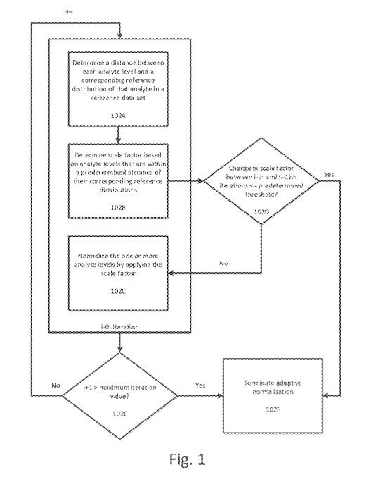
[0006] Fig. 1 illustrates a flowchart for determining the scale factor
based at least in part
on analyte levels that are within a predetermined distance of their
corresponding reference
distributions according to an exemplary embodiment.
[0007] Fig. 2 illustrates an example of a sample 200 having multiple
detected analytes
including 201A and 202A according to an exemplary embodiment including
reference
distribution 1 and reference distribution 2, respectively.
[0008] Fig. 3 illustrates the process for each iteration of the scale
factor application
process according to an exemplary embodiment.
[0009] Figs. 4A-4F illustrate an example of the adaptive normalization
process for a set
of sample data according to an exemplary embodiment.
[0010] Figs. 5A-5E illustrate another example of the adaptive normalization
process that
requires more than one iteration according to an exemplary embodiment.
[0011] Figs. 6A-6B illustrates the analyte levels for all samples after one
iteration of the
adaptive normalization process described herein.
[0012] Fig. 7 illustrates the components for determining a value of the
scale factor that
maximizes a probability that analyte levels that are within the predetermined
distance of their
corresponding reference distributions are part of their corresponding
reference distributions
according to an exemplary embodiment.
- 2 -
CA 03147432 2022-01-13
WO 2021/021678 PCT/US2020/043614
[0013] Figs. 8A-8C illustrate the application of Adaptive Normalization by
Maximum
Likelihood to the sample data in sample 4 shown in Figs.
[0014] Figs. 9A-9F illustrate the application of Population Adaptive
Normalization to the
data shown in Figs. 10A-10B according to an exemplary embodiment.
[0015] Fig. 9 illustrates another method for adaptive normalization of
analyte levels in
one or more samples according to an exemplary embodiment.
[0016] Fig. 10 illustrates a specialized computing environment for adaptive
normalization of analyte levels according to an exemplary embodiment.
[0017] Fig. 11 illustrates median coefficient of variation across all
aptamer-based
proteomic assay measurements for 38 technical replicates.
[0018] Fig. 12 illustrates the Kolmogorov¨Smirnov statistic against a
gender specific
biomarker for samples with respect to maximum allowable iterations.
[0019] Fig. 13 illustrates the number of QC samples by SampleID for plasma
and serum
used in analysis.
[0020] Fig. 14 illustrates the concordance of QC sample scale factors using
median
normalization and ANML
[0021] Fig. 15 illustrates CV Decomposition for control samples using
median
normalization and ANML. Lines indicate empirical cumulative distribution
function of CV for
each control samples within a plate (intra) between plates (inter) and total.
[0022] Fig. 16 illustrates median QC ratios using median normalization and
ANML.
[0023] Fig. 17 illustrates QC ratios in tails using median normalization
and ANML.
[0024] Fig. 18 illustrates scale factor concordance in time-to-spin samples
using SSAN
and ANML
- 3 -
CA 03147432 2022-01-13
WO 2021/021678 PCT/US2020/043614
[0025] Fig. 19 illustrates median analyte CV's across 18 donors in time-to-
spin under
varying normalization schemes.
[0026] Fig. 20 illustrates a concordance plot between scale factors from
Covance
(plasma) using SSAN and ANML.
[0027] Figure 21 shows the distribution of all pairwise analyte
correlations for Covance
samples before and after ANML.
[0028] Fig. 22 illustrates a comparison of distributions obtained from data
normalized
through several methods.
[0029] Fig. 23 illustrates metrics for smoking logic-regression classifier
model for hold-
out test set using data normalized with SSAN and ANML.
[0030] Fig. 24 illustrates Empirical CDFs for c-Raf measurements in plasma
and serum
samples colored by collection site.
[0031] Fig. 25 illustrates concordance plots of scale factors using
standard median
normalization vs. adaptive median normalization in plasma (top) and serum
(bottom).
[0032] Fig. 26 illustrates CDFs by site for an analyte that is not affected
by the site
differences for the standard normalization scheme and adaptive normalization.
[0033] Fig. 27 illustrates plasma sample median normalization scale factors
by dilution
and Covance collection site.
[0034] Fig. 28 where the distributions of median normalization scale
factors are shown
for increasing stringency in adaptive normalization
[0035] Fig. 29 shows typical behavior for a analyte which shows significant
differences
in RFU as a function of time-to-spin.
[0036] Fig. 30 illustrates median normalization scale factors by dilution
with respect to
time-to-spin.
- 4 -
CA 03147432 2022-01-13
WO 2021/021678 PCT/US2020/043614
[0037] Fig. 31 summarizes the effect of adaptive normalization on median
normalization
scale factors vs. time-to-spin.
[0038] Fig. 32 illustrates standard median normalization scale factors by
dilution and
disease state partitioned by GFR value.
[0039] Fig. 33 illustrates median normalization scale factors by dilution
and disease state
by standard median normalization (top) and adaptive normalization by cutoff.
[0040] Fig. 34 illustrates this with the CDF of Pearson correlation of all
analytes with
GFR (log/log) for various normalization procedures.
[0041] Fig. 35 illustrates the distribution of inter-protein Pearson
correlations for the
CKD data set for unnormalized data, standard median normalization and adaptive
normalization.
DETAILED DESCRIPTION
[0042] While methods, apparatuses, and computer-readable media are
described herein
by way of examples and embodiments, those skilled in the art recognize that
methods,
apparatuses, and computer-readable media for adaptive normalization of analyte
levels are not
limited to the embodiments or drawings described. It should be understood that
the drawings
and description are not intended to be limited to the particular forms
disclosed. Rather, the
intention is to cover all modifications, equivalents and alternatives falling
within the spirit and
scope of the appended claims. Any headings used herein are for organizational
purposes only
and are not meant to limit the scope of the description or the claims. As used
herein, the word
"can" is used in a permissive sense (i.e., meaning having the potential to)
rather than the
mandatory sense (i.e., meaning must). Similarly, the words "include,"
"including," "includes",
"comprise," "comprises," and "comprising" mean including, but not limited to.
[0043] Applicant has developed a novel method, apparatus, and computer-
readable
medium for adaptive normalization of analyte levels detected in samples. The
techniques
disclosed herein and recited in the claims guard against introducing artifacts
in data due to
- 5 -
CA 03147432 2022-01-13
WO 2021/021678 PCT/US2020/043614
sample collection artifacts or excessive numbers of disease related proteomic
changes while
properly removing assay bias and decorrelating assay noise.
[0044] This disclosed adaptive normalization techniques and systems remove
affected
analytes from the normalization procedure when collection biases exist within
the populations
of interest or an excessive number of analytes are biologically affected in
the populations being
studied, thereby preventing the introduction of bias into the data.
[0045] The directed aspect of adaptive normalization utilizes definitions
of comparisons
within the sample set that may be suspect for bias. These include distinct
sites in multisite
sample collections that have been shown to exhibit large variations in certain
protein
distributions and key clinical variates within a study. A clinical variate
that can be tested is the
clinical variate of interest in the analysis, but other confounding factors
may exist.
[0046] The adaptive aspect of adaptive normalization refers to the removal
of those
analytes from the normalization procedure that are seen to be significantly
different in the
directed comparisons defined at the outset of the procedure. Since each
collection of clinical
samples is somewhat unique, the method adapts to learn those analytes
necessary for removal
from normalization and sets of removed analytes will be different for
different studies.
[0047] Additionally, by removing affected analytes from median
normalization, the
present system and method minimizes the introduction of normalization
artifacts without
correcting the affected analytes. To the contrary, sample handling artifacts
are amplified by
such analysis, as will the underlying biology in the study. These effects are
discussed in
greater detail in the EXAMPLES section.
[0048] The disclosed techniques for adaptive normalization follow a
recursive
methodology to check for significant differences between user directed groups
on an analyte-
by-analyte level. A dataset is hybridization normalized and calibrated first
to remove initially
detected assay noise and bias. This dataset is then passed into the adaptive
normalization
process (described in greater detail below) with the following parameters:
[0049] (1) the directed groups of interest,
- 6 -
CA 03147432 2022-01-13
WO 2021/021678 PCT/US2020/043614
[0050] (2) the test statistic to be used for determining differences among
the directed
groups,
[0051] (3) a multiple test correction method, and
[0052] (4) a test significance level cutoff.
[0053] The set of user-directed groups can be defined by the samples
themselves, by
collection sites, sample quality metrics, etc., or by clinical covariates such
as Glomerular
Filtration Rate (GFR), case/control, event/no event, etc. Many test statistics
can be used to
detect artifacts in the collection, including Student's t-test, ANOVA, Kruskal-
Wallis, or
continuous correlation. Multiple test corrections include Bonferroni, Holm and
Benj amini-
Hochberg (BH), to name a few.
[0054] The adaptive normalization process is initiated with data that is
already
hybridization normalized and calibrated. Univariate test statistics are
computed for each
analyte level between the directed groups. The data is then median normalized
to a reference
(Covance dataset), removing those analytes levels with significant variation
among the defined
groups from the set of measurements used to produce normalization scale
factors. Through this
adaptive step, the present system will remove analyte levels that have the
potential to introduce
systematic bias between the defined groups. The resulting adaptive
normalization data is then
used to recompute the test statistics, followed by a new adaptive set of
measurements used to
normalize the data, and so on.
[0055] The process can be repeated over multiple iterations until one or
more conditions
are met. These conditions can include convergence, i.e., when analyte levels
selected from
consecutive iterations are identical, a degree of change of analyte levels
between consecutive
iterations being below a certain threshold, a degree of change of scale
factors between
consecutive iterations being below a certain threshold, or a certain number of
iterations
passing. The output of the adaptive normalization process can be a normalized
file annotated
with a list of excluded analytes/analyte levels, the value of the test
statistic, and the
corresponding statistical values (i.e., the adjusted p-value).
- 7 -
CA 03147432 2022-01-13
WO 2021/021678 PCT/US2020/043614
[0056] As will be explained further in the EXAMPLES sections, for a dataset
that
includes an extreme number of artifacts ¨ either biological or collection
related ¨ the present
system is able to filter artifacts and noise that is not detected by previous
median normalization
schemes.
[0057] Fig. 1 illustrates a method for adaptive normalization of analyte
levels in one or
more samples according to an exemplary embodiment. One or more analyte levels
corresponding to one or more analytes detected in the one or more samples are
received. Each
analyte level corresponds to a detected quantity of that analyte in the one or
more samples.
[0058] Fig. 2 illustrates an example of a sample 200 having multiple
detected analytes
according to an exemplary embodiment. As shown in Fig. 2, the larger circle
200 represents
the sample, and each of the smaller circles represents an analyte level for a
different analyte
detected in the sample. For example, circles 201A and 202A correspond to two
different
analyte levels for two different analytes. Of course, the quantity of analytes
shown in Fig. 2 is
for illustration purposes only, and the number of analyte levels and analytes
detected in a
particular sample can vary.
[0059] As shown in Fig. 2, sample 200 includes various analytes, such as
analyte 201A
and analyte 202A. Reference distribution 1 is a reference distribution
corresponding to analyte
201A and reference distribution 2 is a reference distribution corresponding to
analyte 202A.
The reference distributions can take any suitable format. For example, as
shown in Fig. 2, each
reference distribution can plot analyte levels of an analyte detected in a
reference population or
reference samples. Of course, the reference distribution can be plotted and/or
stored in a
variety of different ways. For example, the reference distribution can be
plotted on the basis of
a count of each of analyte level or range of analyte levels. Additionally, the
reference
distributions can be processed to extract mean, median, and standard deviation
values and
those stored values can be used in the distance determination process, as
discussed below.
Many variations are possible and these examples are not intended to be
limiting.
[0060] As shown in Fig. 2, the analyte level of each analyte in the sample
(such as
analytes 201A and 202A) are compared to the corresponding reference
distributions (such as
distributions 1 and 2) either directly or via statistical measure extracted
from the reference
- 8 -
CA 03147432 2022-01-13
WO 2021/021678 PCT/US2020/043614
distributions (such as mean, median, and/or standard deviation) to determine
the statistical
and/or mathematical distance between each analyte level in the sample and the
corresponding
reference distribution.
[0061] The one or more samples in which the analyte levels are detected can
include a
biological sample, such as a blood sample, a plasma sample, a serum sample, a
cerebral spinal
fluid sample, a cell lysates sample, and/or a urine sample. Additionally, the
one or more
analytes can include, for example, protein analyte(s), peptide analyte(s),
sugar analyte(s),
and/or lipid analyte(s).
[0062] The analyte level of each analyte can be determined in a variety of
ways. For
example, each analyte level can be determined based on applying a binding
partner of the
analyte to the one or more samples, the binding of the binding partner to the
analyte resulting
in a measurable signal. The measurable signal can then be measured to yield
the analyte level.
In this case, the binding partner can be an antibody or an aptamer. Each
analyte level can
additionally or alternatively be determined based on mass spectrometry of the
one or more
samples.
[0063] Returning to Fig. 1, at step 102C a scale factor is iteratively
applied to the one or
more analyte levels over one or more iterations until a change in the scale
factor between
consecutive iterations is less than or equal to a predetermined change
threshold 102D or until a
quantity of the one or more iterations exceeds a maximum iteration value
(102F).
[0064] The scale factor is a dynamic variable that is re-calculated for
each iteration. By
determining and measuring the change in the scale factor between subsequent
iterations, the
present system is able to detect when further iterations would not improve
results and thereby
terminate the process.
[0065] Additionally, a maximum iteration value can be utilized as a
failsafe, to ensure
that the scale factor application process does not repeat indefinitely (in an
infinite loop). The
maximum iteration value can be, for example, 10 iterations, 20 iterations, 30
iterations, 40
iterations, 50 iterations, 100 iterations, or 200 iterations.
- 9 -
CA 03147432 2022-01-13
WO 2021/021678 PCT/US2020/043614
[0066] Optionally, the maximum iteration value can be omitted and the scale
factor can
be iteratively applied to the one or more analyte levels over one or more
iterations until a
change in the scale factor between consecutive iterations is less than or
equal to a
predetermined change threshold, without consideration of the number of
iterations required.
[0067] The predetermined change threshold can be set by a user or set to
some default
value. For example, the predetermined change threshold can be set to a very
low decimal
value (e.g., 0.001) such that the scale factor is required to reach a
"convergence" where there is
very little measurable change in the scale factor between iterations in order
for the process to
terminate.
[0068] The change in the scale factor between subsequent iterations can
measured as a
percentage change. In this case, the predetermined change threshold can be,
for example, a
value between 0 and 40 percent, inclusive, a value between 0 and 20 percent,
inclusive, a value
between 0 and 10 percent, inclusive, a value between 0 and 5 percent,
inclusive, a value
between 0 and 2 percent, inclusive, a value between 0 and 1 percent,
inclusive, and/or 0
percent.
[0069] At step 102A a distance is determined between each analyte level in
the one or
more analyte levels and a corresponding reference distribution of that analyte
in a reference
data set.
[0070] This distance is a statistical or mathematical distance and can be
measure the
degree to which a particular analyte level differs from a corresponding
reference distribution of
that same analyte. Reference distributions of various analyte levels can be
pre-compiled and
stored in a database and accessed as required during the distance
determination process. The
reference distributions can be based upon reference samples or populations and
be verified to
be free of contamination or artifacts through a manual review process or other
suitable
technique.
[0071] The determination of a distance between each analyte level in the
one or more
analyte levels and a corresponding reference distribution of that analyte in a
reference data set
- 10 -
CA 03147432 2022-01-13
WO 2021/021678 PCT/US2020/043614
can include determining an absolute value of a Mahalanobis distance between
each analyte
level and the corresponding reference distribution of that analyte in the
reference data set.
[0072] The Mahalanobis distance is a measure of the distance between a
point P and a
distribution D. An origin point for computing this measure can be at the
centroid (the center of
mass) of a distribution. The origin point for computation of the Mahalanobis
distance ("M-
Distance") can also be a mean or median of the distribution and utilize the
standard deviation
of the distribution, as will be discussed further below.
[0073] Of course, there are other ways of measuring statistical or
mathematical distance
between an analyte level in the sample and a corresponding reference
distribution that can be
utilized. For example, determining a distance between each analyte level in
the one or more
analyte levels and a corresponding reference distribution of that analyte in a
reference data set
can include determining a quantity of standard deviations between each analyte
level and a
mean or a median of the corresponding reference distribution of that analyte
in the reference
data set.
[0074] Returning to Fig. 1, at step 102B a scale factor is determined based
at least in part
on analyte levels that are within a predetermined distance of their
corresponding reference
distributions.
[0075] This step includes a first sub-step of identifying all analyte
levels in the sample
that are within a predetermined distance threshold of their corresponding
reference
distributions. The predetermined distance that is used as a cutoff to identify
analyte levels to
be used in the scale factor determination process can be set by a user, set to
some default value,
and/or customized to the type of sample and analytes involved.
[0076] Additionally, the predetermined distance threshold will depend on
how the
statistical distance between the analyte level and the corresponding reference
distribution is
determined. In the case when an M-Distance is used, the predetermined distance
can be value
in a range between 0.5 to 6, inclusive, a value in a range between 1 to 4,
inclusive, a value in a
range between 1.5 to 3.5, inclusive, a value in a range between 1.5 to 2.5,
inclusive, and/or a
value in a range between 2.0 to 2.5, inclusive. The specific predetermined
distance used to
-11-
CA 03147432 2022-01-13
WO 2021/021678 PCT/US2020/043614
filter analyte levels from use in the scale factor determination process can
depend on the
underlying data set and the relevant biological parameters. Certain types of
samples may have
a greater inherent variation than others, warranting a higher predetermined
distance threshold,
while others may warrant a lower predetermined distance threshold.
[0077] Returning to Fig 1. At step 102A distance is calculated between each
analyte
level and the corresponding reference distribution for that analyte. The
corresponding
reference distribution can be looked up based upon an identifier associate
with the analyte and
stored in memory or based upon an analyte identification process that detects
each type of
analyte. The distance can be calculated, for example, as an M-Distance, as
discussed
previously. The M-Distance be computed on the basis of the mean, median,
and/or standard
deviation of the corresponding reference distribution so that the entire
reference distribution
does not need to be stored in memory. For example, the M-Distance between each
analyte
level in the sample and the corresponding reference distribution can be given
by:
[0078] M = (Xp¨ Pre f ,p)
C re f ,p
[0079] Where M is the Mahalanobis Distance ("M-Distance"), xp is the value
of an
analyte level in the sample, põf,p is the mean of the reference distribution
corresponding to
that analyte, and o-õf,p is the standard deviation of the reference
distribution corresponding to
that analyte.
[0080] Fig. 3 illustrates a flowchart for determining the scale factor
based at least in part
on analyte levels that are within a predetermined distance of their
corresponding reference
distributions according to an exemplary embodiment.
[0081] At step 301 an analyte scale factor is determined for each analyte
level that is
within the predetermined distance of the corresponding reference distribution.
This analyte
scale factor is determined based at least in part on the analyte level and a
mean or median value
of the corresponding reference distribution. For example, the analyte scale
factor for each
analyte can be based upon the mean of the corresponding reference
distribution:
- 12 -
CA 03147432 2022-01-13
WO 2021/021678 PCT/US2020/043614
[0082] f ,p
SFAnalyte = -
xp
[0083] Where SF
Analyteis the scale factor for each analyte that is within a predetermined
distance of its corresponding reference distribution, iiref,p is the mean of
the reference
distribution corresponding to that analyte, and xp is the value of an analyte
level in the sample.
[0084] The analyte scale factor can also be based upon the median of the
corresponding
reference distribution:
re f ,p
[0085] SFAnalyte = -
xp
[0086] Where SF
Analyteis the scale factor for each analyte that is within a predetermined
distance of its corresponding reference distribution, is the median of the
reference
distribution corresponding to that analyte, and xp is the value of an analyte
level in the sample.
[0087] At step 302 the overall scale factor for the sample is determined by
computing
either a mean or a median of analyte scale factors corresponding to analyte
levels that are
within the predetermined distance of their corresponding reference
distributions. The overall
scale factor is therefore given by one of:
[0088] SFOverall
= SFAnalyte
[0089] Or:
[0090] SFOverall = SF
G Analyte
[0091] Where SF
Overall is the overall scale factor (referred to herein as the "scale factor")
to be applied to the analyte levels in the sample, cw
Analyte is the mean of the analyte scale
factors, and a SFAnalyte is the median of the analyte scale factors.
[0092] At step 302 a determination is made whether the distance between the
analyte
level and the reference distribution is greater than the predetermined
distance threshold. If so,
the analyte level is flagged as an outlier at step 303 and the analyte level
is excluded from the
- 13 -
CA 03147432 2022-01-13
WO 2021/021678 PCT/US2020/043614
scale factor determination process at step 304. Otherwise, if the distance
between the analyte
level and the reference distribution is less than or equal to the
predetermined distance
threshold, then the analyte level is flagged as being within an acceptable
distance at step 305
and the analyte level is used in the scale factor determination process at
step 306.
[0093] The flagging of each analyte level can encoded and tracked by a data
structure for
each iteration of the scale factor application process, such as a bit vector
or other Boolean
value storing a 1 or 0 for each analyte level, the 1 or 0 indicating whether
the analyte level
should be used in the scale factor determination process. The corresponding
data structure can
the n be refreshed/re-encoded during a new iteration of the scale factor
application process.
[0094] When the scale factor determination process occurs at step 306, the
data structure
encoding the results of the distance threshold evaluation process in steps 301-
302 can be
utilized to filter the analyte levels in the sample to extract and/or identify
only those analyte
levels to be used in the scale factor determination process.
[0095] While the origin point for computing the predetermined distance for
each
reference distribution is shown as the centroid of the distribution for
clarity, it is understood
that other origin points can be utilized, such as the mean or median of the
distribution, or the
mean or median adjusted based upon the standard deviation of the distribution.
[0096] Returning to Fig. 1, at step 102D a determination is made regarding
whether the
change in scale factor between the determined scale factor and the previously
determined
scale factor (for a previous iteration) is less than or equal to a
predetermined threshold. If
the first iteration of the scaling process is being performed than this step
can be skipped.
This step compares the current scale factor with the previous scale factor
from the
previous iteration and determines whether the change between the previous
scale factor
and the current scale factor exceeds the predetermined threshold.
[0097] As discussed earlier, this predetermined threshold can be some user-
defined
threshold, such as a 1 % change, and/or can require nearly identical scale
factors (¨ 0%
change) such that the scale factor converges to a particular value.
- 14 -
CA 03147432 2022-01-13
WO 2021/021678 PCT/US2020/043614
[0098] If the change in scale factor between the ith and the (i-1)th
iterations is less than or
equal to the predetermined threshold, then at step 102F the adaptive
normalization process
terminates.
[0099] Otherwise, if the change in scale factor between the ith and the (i-
l)th iterations is
greater than the predetermined threshold, then the process proceeds to step
102C, where the
one or more analyte levels in the sample are normalized by applying the scale
factor. Note that
all analyte levels in the sample are normalized using this scale factor, and
not only the analyte
levels that were used to compute the scale factor. Therefore, the adaptive
normalization
process does not "correct" collection site bias, or differential protein
levels due to disease;
rather, it ensures that such large differential effects are not removed during
normalization since
that would introduce artifacts in the data and destroy the desired protein
signatures.
[00100] After the normalization step at 102C, at optional step 102E, a
determination is
made regarding whether repeating one more iteration of the scaling process
would exceed the
maximum iteration value (i.e., whether i+1 > maximum iteration value). If so,
the process
terminates at step 102F. Otherwise, the next iteration is initialized (i++)
and the process
proceeds back to step 102A for another round of distance determination, scale
factor
determination at step 102B, and normalization at step 102C (if the change in
scale factor
exceeds the predetermined threshold at 102D).
[00101] Steps 102A-102D are repeated for each iteration until the process
terminates at
step 102F (based upon either the change in scale factor falling within the
predetermined
threshold or the maximum iteration value being exceeded.
[00102] Figs. 4A-4F illustrate an example of the adaptive normalization
process for a set
of sample data according to an exemplary embodiment.
[00103] Fig. 4A illustrates a set of reference data summary statistics that
are to be used
for both calculation of scale factors and distance determination of analyte
levels to reference
distributions. The reference data summary statistics summarize the pertinent
statistical
measures for reference distributions corresponding to 25 different analytes.
- 15 -
CA 03147432 2022-01-13
WO 2021/021678 PCT/US2020/043614
[00104] Fig. 4B illustrates a set of sample data corresponding to analyte
levels of the 25
different analytes measured across ten samples. Each of the analyte levels are
expressed as
relative fluorescent units but is understood that other units of measurement
can be utilized.
[00105] The adaptive normalization process can iterate through each sample
by first
calculating the Mahalanobis distance (M-Distance) between each analyte level
and the
corresponding reference distribution, determining whether each M-Distance
falls within a
predetermined distance, calculating a scale factor (both at the analyte level
and overall),
normalizing the analyte levels, and then repeating the process until the
change in the scale
factor falls under a predefined threshold.
[00106] As an example, the tables in Figs. 4C-4F will utilize the
measurements in Sample
3 in Fig. 4B. As shown in Fig. 4C, an M-Di stance is calculated between each
analyte level in
sample 3 and the corresponding reference distribution. This M-Distance is
given by the
equation (discussed earlier):
p f ,p)
M =
Ore f ,p
[00107] Also shown in the table of Fig. 4C is a Boolean variable Within-
Cutoff, that
indicates whether the absolute value of the M-Distance for each analyte is
within the
predetermined distance required to be used in the scale factor determination
process. In this
case, the predetermined distance is set to 2. As shown in Fig. 4C, analytes 3,
6, 7, 11, 17, 18,
20, and 23 are greater than the cutoff distance of 121 and so these will not
be used in the
following scale factor determination step.
[00108] To determine the overall scale factor, a scale factor for each of
the remaining
analytes (the analytes having a Within-Cutoff value of TRUE) is determined as
discussed
previously. Fig. 4D illustrates the analyte scale factor for each of the
analytes. The median of
these analyte scale factors is then set to be the overall scale factor. Of
course, the mean of
these analyte scale factors can also be used as the overall scale factor.
- 16 -
CA 03147432 2022-01-13
WO 2021/021678 PCT/US2020/043614
[00109] In this case, the scale factor is given by:
[00110] SFOverall = median(SF
Analyte 1...p = 0.9343
[00111] Where SFAnalyte 1 is the analyte scale factor for each of the
analytes that are
used in the scale factor determination process.
[00112] The 25 analyte measurements for sample 3 are then multiplied by
this scale factor
and the process is repeated. New M-Distances are calculated for this
normalized data and
analytes that are within the predetermined distance threshold are determined,
as shown in Fig.
4E. Fig. 4F additionally illustrates the analyte scale factors for this next
iteration. Using the
above mentioned formula for the overall scale factor, the overall scale factor
for this iteration is
determined to be equal to 1 (the median of the analyte scale factors).
[00113] Since the overall scale factor is determined to be 1, the process
can be terminated,
since application of this scale factor will not produce any change to the data
and the next scale
factor will also be 1.
[00114] Figs. 5A-5E illustrate another example of the adaptive
normalization process that
requires more than one iteration according to an exemplary embodiment. These
figures use the
data corresponding to sample 4 in Figs. 4A-4B.
[00115] Fig. 5A illustrates the M-Distance values and the corresponding
Boolean "Within-
Cutoff' values of each of the analytes in sample 4. As shown in Fig. 5A,
analytes 1, 4, 6, 8,
12, 17, 19, and 21-25 are excluded from the scale factor determination
process.
[00116] Fig. 5B illustrates the analyte scale factors for each of the
remaining analytes.
The overall scale factor for this iteration is taken as the median of these
values, as discussed
previously, and is equal to 0.9663.
[00117] This scale factor is applied to the analyte levels to generate the
analyte levels
shown in Fig. 5C. Fig. 5C also illustrates the M-Distance determination and
cutoff
determination results for the second iteration of the normalization process.
In this case,
- 17 -
CA 03147432 2022-01-13
WO 2021/021678 PCT/US2020/043614
analytes 1, 4, 6, 10, 12, 17, 19, and 21-25 are excluded from the scale factor
determination
process.
[00118] Fig. 5D illustrates the analyte scale factors for each of the
remaining analytes.
The overall scale factor for this iteration is taken as the median of these
values, as discussed
previously, and is equal to 0.8903. As this scale factor has not yet converged
to a value of 1
(indicating no further change in scale factor), the process is repeated until
a convergence is
reached (or until the change in scale factor falls within some other
predefined threshold).
[00119] Fig. 5E illustrates the scale factor determined for each sample
shown in Figs. 4A-
4B across eight iterations of the scale factor determination and adaptive
normalization process.
As shown in Fig. 5E, the scale factor for sample 4 does not converge until the
fifth iteration of
the process.
[00120] The analyte level data for each of the samples will change after
each iteration
(assuming the determined scale factor is not 1). For example, Fig. 6A
illustrates the analyte
levels for all samples after one iteration of the adaptive normalization
process described herein.
Figs. 6A-6B illustrates the analyte levels for all samples after the adaptive
normalization
process is completed (in this example, after all scale factors have converged
to 1).
[00121] Referring back to Fig. 1, the scale factor determination step 102B
can be
performed in other ways. In particular, determining the scale factor based at
least in part on
analyte levels that are within a predetermined distance of their corresponding
reference
distributions can include determining a value of the scale factor that
maximizes a probability
that analyte levels that are within the predetermined distance of their
corresponding reference
distributions are part of their corresponding reference distributions.
[00122] Fig. 7 illustrates the requirements for determining a value of the
scale factor that
maximizes a probability that analyte measurements within a given sample are
derived from a
reference distribution.
[00123] In this case, the probability that each analyte level is part of
the corresponding
reference distribution can be determined based at least in part on the scale
factor, the analyte
- 18 -
CA 03147432 2022-01-13
WO 2021/021678 PCT/US2020/043614
level, a standard deviation of the corresponding reference distribution, and a
median of the
corresponding reference distribution.
[00124] At step 704 a value of the scale factor is determined that
maximizes a probability
that all analyte levels that are within the predetermined distance of their
corresponding
reference distributions are part of their corresponding reference
distributions. As shown in Fig.
7, this probability function utilizes a standard deviation of the
corresponding reference
distributions 702 and the analyte levels 703 in order to determine the value
of the scale factor
7015 that maximizes this probability.
[00125] Adaptive normalization that uses this technique for scale factor
determination is
referred to herein as Adaptive Normalization by Maximum Likelihood (ANML). The
primary
difference between ANN/IL and the previous technique for adaptive
normalization described
above (which operates on single samples and is referred to herein as Single
Sample Adaptive
Normalization (SSAN)), is the scale factor determination step.
[00126] Whereas medians were used to calculate the scale factor for SSAN,
ANN/IL
utilizes the information of the reference distribution to maximize the
probability the sample
was derived from the reference distribution:
E7-10-tref,p-xref,p)cire2f,p
l [00127] o a SF
- Overall =
El 2
iV 3=icrr-ef,p
[00128] This formula relies on the assumption that the reference
distribution follows a log
normal probability. Such an assumption allows for the simple closed form for
the scale factors
but is not necessary. As shown above, the overall scale factor for ANN/IL is a
weighted
variance average. The contribution to the scale factor, SFOverall, of analyte
measurements
which show large population variance will be weighted less than those coming
from smaller
population variances.
[00129] Figs. 8A-8C illustrate the application of Adaptive Normalization by
Maximum
Likelihood to the sample data in sample 4 shown in Figs. 4A-4B according to an
exemplary
embodiment. Fig. 4A illustrates the M-Distance values and With-Cutoff values
of each analyte
in a first iteration. As shown in Fig. 8A, the non-usable analytes from the
first iteration for
- 19 -
CA 03147432 2022-01-13
WO 2021/021678 PCT/US2020/043614
sample 4 are analytes 1, 4, 6, 8, 12, 17, 19, 21, 22, 23, 24, and 25. For the
calculation of the
scale factor we take the log10 transformed reference data, standard deviation,
and sample data
and apply the above-mentioned equation for scale factor determination:
vN r \ ¨2
Lp=ill-tref,p ¨Xref,p)(Tre f ,p
[00130] loa SF
- Overall = = 0.01072
M1c7r-e2f,p
[00131] Applying this exponent to the base of 10 we determine the scale
factor for this
sample/iteration as:
[00132] SFOverall = 10-0.010702 = 0.9756
[00133] Similar to the procedure of SSAN, this intermediate scale factor
would be applied
to the measurements from sample 4 and the process would be repeated for the
successive
iterations.
[00134] Fig. 8B illustrates the scale factors determined by the application
of ANN/IL to the
data in Figs. 4A-4B over multiple iterations. The differences in normalized
sample
measurements between the first iteration and after convergence is quite
distinct for those
samples requiring more than 1 iteration. These additional iterations show
benefits in data
generated with an aptamer-based proteomic assay, which will be described
further in the
examples section. As shown in Fig. 8B, these scale factors differ from those
determined by
SSAN (Fig. 5E). These differences are due to the weighted population variance
for each
analyte, which helps balance the scale factor calculation for those analytes
in which reference
population variance is large.
[00135] Fig. 8C illustrates the normalized analyte levels resulting from
the application of
ANN/IL to the data in Figs. 4A-4B over multiple iterations. As shown in Fig.
8C, the
normalized analyte levels differ from those determined by SSAN (Fig. 5B).
[00136] Another type of adaptive normalization that can be performed using
the disclosed
techniques is Population Adaptive Normalization (PAN). PAN can be utilized
when the one or
more samples comprise a plurality of samples and the one or more analyte
levels
- 20 -
CA 03147432 2022-01-13
WO 2021/021678 PCT/US2020/043614
corresponding to the one or more analytes comprise a plurality of analyte
levels corresponding
to each analyte.
[00137] When performing adaptive normalization using PAN, the distance
between each
analyte level in the one or more analyte levels and a corresponding reference
distribution of
that analyte in a reference data set is determined by determining a Student's
T-test,
Kolmogorov-Smirnov test, or a Cohen's D statistic between the plurality of
analyte levels
corresponding to each analyte and the corresponding reference distribution of
each analyte in
the reference data set.
[00138] For PAN, clinical data is treated as a group in order to censor
analytes that are
significantly different from the population reference data. PAN can be used
when a group of
samples is identified from having a subset of similar attributes such as being
collected from the
same testing site under certain collection conditions, or the group of samples
may have a
clinical distinction (disease state) that is distinct from the reference
distributions.
[00139] The power of population normalization schemes is the ability to
compare many
measurements of the same analyte against the reference distribution. The
general procedure of
normalization is similar to the above-described adaptive normalization methods
and again
starts of an initial comparison of each analyte measurement against the
reference distribution.
[00140] As explained above, multiple statistical tests can be used to
determine statistical
differences between analyte measurements from the test data and the reference
distribution
including Student's T-tests, Kolmogorov-Smirnov test, etc.
[00141] The following example utilizes the Cohen's D statistic for distance
measurement,
which a measurement of effect size between two distributions and is very
similar to the M-
distance calculation discussed previously:
Dp = _______________________________________
_Jo-2 G2
re f ,p ' x,p
-21 -
CA 03147432 2022-01-13
WO 2021/021678 PCT/US2020/043614
[00142] Where Dp is the Cohen's D statistic, pp is the reference
distribution median for
particular analyte, 5c; is the clinical data (sample) median across all
samples, and
0-2 is the pooled standard deviation (or median absolution deviation). As
shown
re f ,p x,p
above, Cohen's D is defined as the difference between the reference
distribution median and
clinical data median over a pooled standard deviation (or median absolution
deviation).
[00143] Figs. 9A-9F illustrate the application of Population Adaptive
Normalization to the
data shown in Figs. 4A-4B according to an exemplary embodiment. For the
reference data
shown in Fig. 4A and clinical data shown in Fig. 4B, 25 Cohen's D statistics
are calculated,
one corresponding to each analyte. Fig. 9A illustrates the Cohen's D statistic
for each analyte
across all samples. This calculation can be done in log10 transformed space to
enhance
normality for analyte measurements.
[00144] In an exemplary embodiment, the predetermined distance threshold
used to
determine if an analyte is to be included in the scale factor determination
process is a Cohen's
D of 0.5. Analytes outside of this window will be excluded from the
calculation of scale
factor. As shown in Fig. 9A, this results in analytes 1, 4, 5, 8, 17, 21, and
22 being excluded
from the scale factor calculation.
[00145] Fig. 9B illustrates the scale factors calculated for each analyte
across samples. A
difference between population adaptive normalization (PAN) and the previously
discussed
normalization methods is that in PAN each sample will include/exclude the same
analytes
during scale factor calculation. In PAN, the scale factor for all samples will
be determined on
the basis of the remaining analytes. In this example, the scale factor can be
given by the
median or the mean of the analyte scale factors of the remaining analytes.
Similar to the
above-described adaptive normalization methods, the scale factor can be
determined as a mean
or median of the individual analyte scale factors. If the median is used, then
the scale factor
for the data shown in Fig. 9B is 0.8876.
[00146] This scale factor is multiple with the data values shown in Fig. 4B
to generate
normalized data values, as shown in Fig. 9C. Fig. 9D illustrates the results
of the second
- 22 -
CA 03147432 2022-01-13
WO 2021/021678 PCT/US2020/043614
iteration of the scale factor determination process, including the Cohen's D
value for each
analyte and the Within-Cutoff value for each analyte.
[00147] For this iteration, analytes 1, 4, 5, 8, 16, 17, 20, and 22 are to
be excluded from
the scale factor determination process. In addition to the analytes excluded
in the first
iteration, the second iteration additionally excludes analyte 16 from the
calculation of scale
factors. The above-described steps are then repeated to removing the
additional analyte from
scale factor calculation for each sample.
[00148] Convergence of the adaptive normalization (a change in scale factor
less than a
predefined threshold) occurs when the analytes removed from the ith iteration
are identical to
the (i-l)th iteration and scale factors for all samples have converged. In
this example,
convergence requires five iterations. Fig. 9E illustrates the scale factors
for each of the
samples at each of the five iterations. Additionally, Fig. 9F illustrates the
normalized analyte
level data after convergence has occurred and all scale factors have been
applied.
[00149] The systems and methods described herein implement an adaptive
normalization
process which performs outlier detection to identify any outlier analyte
levels and exclude said
outliers from the scale factor determination, while including the outliers in
the scaling aspect of
the normalization.
[00150] The features of computing a scale factor and applying the scale
factor are also
described in greater detail with respect to the previous figures.
Additionally, the removal of
outlier analyte levels in the one or more analyte levels by performing outlier
analysis can be
implemented as described with respect to Figs. 1-3.
[00151] The outlier analysis method described in those figures and the
corresponding
sections of the specification is a distance based outlier analysis that
filters analyte levels based
upon a predetermined distance threshold from a corresponding reference
distribution.
[00152] However, other forms of outlier analysis can also be utilized to
identify outlier
analyte levels. For example, a density based outlier analysis such as the
Local Outlier Factor
("LOF") can be utilized. LOF is based on local density of data points in the
distribution. The
- 23 -
CA 03147432 2022-01-13
WO 2021/021678 PCT/US2020/043614
locality of each point is given by k nearest neighbors, whose distance is used
to estimate the
density. By comparing the local density of an object to the local densities of
its neighbors,
regions of similar density can be identified, as well as points that have a
lower density than
their neighbors. These are considered to be outliers.
[00153] Density-based outlier detection is performed by evaluating distance
from a given
node to its K Nearest Neighbors ("K-NN"). The K-NN method computes a Euclidean
distance
matrix for all clusters in the cluster system and then evaluates local
reachability distance from
the center of each cluster to its K nearest neighbors. Based on the said
distance matrix local
reachability distance, density is computed for each cluster and the Local
Outlier Factor
("LOF") for each data point is determined. Data points with large LOF value
are considered as
the outlier candidates. In this case, the LOF can be computed for each analyte
level in the
sample with respect to its reference distribution.
[00154] The step of normalizing the one or more analyte levels over one or
more iterations
can include performing additional iterations until a change in the scale
factor between
consecutive iterations is less than or equal to a predetermined change
threshold or until a
quantity of the one or more iterations exceeds a maximum iteration value, as
discussed
previously with respect to Fig. 1.
[00155] Fig. 10 illustrates a specialized computing environment for
adaptive
normalization of analyte levels according to an exemplary embodiment.
Computing
environment 1000 includes a memory 1001 that is a non-transitory computer-
readable medium
and can be volatile memory (e.g., registers, cache, RAM), non-volatile memory
(e.g., ROM,
EEPROM, flash memory, etc.), or some combination of the two.
[00156] As shown in Fig. 10, memory 1001 stores distance determination
software 1001A
for determining statistical/mathematical distances between analyte levels and
their
corresponding reference distributions, outlier detection software 1001B for
identifying analyte
levels that are outside the predefined distance threshold, scale factor
determination software
1001C for determining analyte scale factors and overall scale factors,
normalization software
1001D for applying the adaptive normalization techniques described herein to a
data set.
- 24 -
CA 03147432 2022-01-13
WO 2021/021678 PCT/US2020/043614
[00157] Memory 1001 additionally includes a storage 1001 that can be used
to store the
reference data distributions, statistical measures on the reference data,
variables such as the
scale factor and Boolean data structures, intermediate data values or
variables resulting from
each iteration of the adaptive normalization process.
[00158] All of the software stored within memory 1001 can be stored as
computer-
readable instructions, that when executed by one or more processors 1002,
cause the processors
to perform the functionality described herein.
[00159] Processor(s) 1002 execute computer-executable instructions and can
be a real or
virtual processor. In a multi-processing system, multiple processors or
multicore processors
can be used to execute computer-executable instructions to increase processing
power and/or to
execute certain software in parallel.
[00160] The computing environment additionally includes a communication
interface 503,
such as a network interface, which is used to monitor network communications,
communicate
with devices, applications, or processes on a computer network or computing
system, collect
data from devices on the network, and actions on network communications within
the
computer network or on data stored in databases of the computer network. The
communication
interface conveys information such as computer-executable instructions, audio
or video
information, or other data in a modulated data signal. A modulated data signal
is a signal that
has one or more of its characteristics set or changed in such a manner as to
encode information
in the signal. By way of example, and not limitation, communication media
include wired or
wireless techniques implemented with an electrical, optical, RF, infrared,
acoustic, or other
carrier.
[00161] Computing environment 1000 further includes input and output
interfaces 1004
that allow users (such as system administrators) to provide input to the
system and display or
otherwise transmit information for display to users. For example, the
input/output interface
1004 can be used to configure settings and thresholds, load data sets, and
view results.
[00162] An interconnection mechanism (shown as a solid line in Fig. 10),
such as a bus,
controller, or network interconnects the components of the computing
environment 1000.
- 25 -
CA 03147432 2022-01-13
WO 2021/021678 PCT/US2020/043614
[00163] Input and output interfaces 1004 can be coupled to input and output
devices. The
input device(s) can be a touch input device such as a keyboard, mouse, pen,
trackball, touch
screen, or game controller, a voice input device, a scanning device, a digital
camera, remote
control, or another device that provides input to the computing environment.
The output
device(s) can be a display, television, monitor, printer, speaker, or another
device that provides
output from the computing environment 1000. Displays can include a graphical
user interface
(GUI) that presents options to users such as system administrators for
configuring the adaptive
normalization process.
[00164] The computing environment 1000 can additionally utilize a removable
or non-
removable storage, such as magnetic disks, magnetic tapes or cassettes, CD-
ROMs, CD-RWs,
DVDs, USB drives, or any other medium which can be used to store information
and which
can be accessed within the computing environment 1000.
[00165] The computing environment 1000 can be a set-top box, personal
computer, a
client device, a database or databases, or one or more servers, for example a
farm of networked
servers, a clustered server environment, or a cloud network of computing
devices and/or
distributed databases.
[00166] As used herein, "nucleic acid ligand,"aptamer,"SOMAmer," and
"clone" are
used interchangeably to refer to a non-naturally occurring nucleic acid that
has a desirable
action on a target molecule. A desirable action includes, but is not limited
to, binding of the
target, catalytically changing the target, reacting with the target in a way
that modifies or alters
the target or the functional activity of the target, covalently attaching to
the target (as in a
suicide inhibitor), and facilitating the reaction between the target and
another molecule. In one
embodiment, the action is specific binding affinity for a target molecule,
such target molecule
being a three dimensional chemical structure other than a polynucleotide that
binds to the
aptamer through a mechanism which is independent of Watson/Crick base pairing
or triple
helix formation, wherein the aptamer is not a nucleic acid having the known
physiological
function of being bound by the target molecule. Aptamers to a given target
include nucleic
acids that are identified from a candidate mixture of nucleic acids, where the
aptamer is a
ligand of the target, by a method comprising: (a) contacting the candidate
mixture with the
- 26 -
CA 03147432 2022-01-13
WO 2021/021678 PCT/US2020/043614
target, wherein nucleic acids having an increased affinity to the target
relative to other nucleic
acids in the candidate mixture can be partitioned from the remainder of the
candidate mixture;
(b) partitioning the increased affinity nucleic acids from the remainder of
the candidate
mixture; and (c) amplifying the increased affinity nucleic acids to yield a
ligand-enriched
mixture of nucleic acids, whereby aptamers of the target molecule are
identified. It is
recognized that affinity interactions are a matter of degree; however, in this
context, the
"specific binding affinity" of an aptamer for its target means that the
aptamer binds to its target
generally with a much higher degree of affinity than it binds to other, non-
target, components
in a mixture or sample. An "aptamer,"SOMAmer," or "nucleic acid ligand" is a
set of copies
of one type or species of nucleic acid molecule that has a particular
nucleotide sequence. An
aptamer can include any suitable number of nucleotides. "Aptamers" refer to
more than one
such set of molecules. Different aptamers can have either the same or
different numbers of
nucleotides. Aptamers may be DNA or RNA and may be single stranded, double
stranded, or
contain double stranded or triple stranded regions. In some embodiments, the
aptamers are
prepared using a SELEX process as described herein, or known in the art. As
used herein, a
"SOMAmer" or Slow Off-Rate Modified Aptamer refers to an aptamer having
improved off-
rate characteristics. SOMAmers can be generated using the improved SELEX
methods
described in U.S. Pat. No. 7,947,447, entitled "Method for Generating Aptamers
with
Improved Off-Rates," the disclosure of which is hereby incorporated by
reference in its
entirety.
[00167] Greater detail regarding aptamer-base proteomic assays are
described, in U.S.
Patent Nos. 7,855,054, 7,964,356 and 8,945,830, US Patent Application No.
14/569,241, and
PCT Application PCT/U52013/044792, the disclosures of which are hereby
incorporated by
reference in their entirety.
EXAMPLES
[00168] IMPROVED PRECISION
[00169] Fig. 11 illustrates median coefficient of variation across all
aptamer-based
proteomic assay measurements for 38 technical replicates.
- 27 -
CA 03147432 2022-01-13
WO 2021/021678 PCT/US2020/043614
[00170] Applicant took 38 technical replicates from 13 aptamer based
proteomic assay
runs (Quality Control (QC)samples) and calculated coefficient of variation
(CV), defined as the
standard deviation of measurements over the mean/median of measurements, for
each analyte
across the aptamer-based proteomic assay menu. Using ANML, Applicant
normalized each
sample while controlling the maximum number of iterations each sample would be
allowed
under the normalization process.
[00171] The median CVs for the replicates show reduced CV as the maximum
number of
allowable iterations increases indicating increased precision as replicates
are allowed to
converge.
[00172] IMPROVED BIOMARKER DISCRIMINATION
[00173] Fig. 12 illustrates the Kolmogorov¨Smirnov statistic against a
gender specific
biomarker for samples with respect to maximum allowable iterations.
[00174] Applicant looked at the discriminatory power for a gender specific
biomarker
known in the aptamer-based proteomic assay menu. Applicant calculated a
Kolmogorov¨
Smirnov (K.S.) test to quantify the distance between the empirical
distribution functions of 569
female and 460 male samples to quantify the extent of separation between this
analyte shows
between male/female samples where a K.S. distance of 1 implies complete
separation of
distribution (good discriminatory properties) and 0 implies complete overlap
of the
distributions (poor discriminatory properties). As in the example above,
Applicant limited the
number of iterations each sample could run through before calculating the K.
S. distance of the
groups.
[00175] This data shows that the discriminatory characteristics of the
biomarker for
male/female gender determination are increased as samples are allowed to
converge in the
iterative normalization process.
[00176] APPLICATION OF ANML ON QC SAMPLES
- 28 -
CA 03147432 2022-01-13
WO 2021/021678 PCT/US2020/043614
[00177] 662 runs (BI, in Boulder) with 2066 QC samples. These replicates
comprise 4
different QC lots. Fig. 13 illustrates the number of QC samples by SampleID
for plasma and
serum used in analysis.
[00178] A new version of the normalization population reference was
generated (to make
it consistent with the ANML and generate estimates to the reference SDs). The
data described
above was hybridization normalized and calibrated as per standard procedures
for V4
normalization. At that point, it was median normalized to both the original
and the new
population reference (shows differences due to changes in the median values of
reference) and
using ANML (shows differences due to both the adaptive and maximum likelihood
changes in
normalization to a population reference.)
[00179] Normalization Scale Factors
[00180] A first comparison to make is to look at the scale factors
concordances between
different normalization references/methods. If there are only slight
differences, then good
concordance in all other metrics is to be expected. Figure 1 shows scale
factors for QC samples
in plasma and serum; which show good concordance between For QC 1710255 (for
which we
have, by far, the largest number of replicates), for the most part, there is
no large difference
(the dashed lines represent a difference of 0.1 in scale factors; so
differences are mostly below
0.05.)
[00181] Fig. 14 illustrates the concordance of QC sample scale factors
using median
normalization and ANML. Solid line indicates identity, dashed lines indicate
difference of 0.1
above/below identity.
[00182] CV's
[00183] We then computed the CV decomposition for control samples in plasma
and
serum samples in median normalization and ANML. Fig. 15 illustrates CV
Decomposition for
control samples using median normalization and ANML. Lines indicate empirical
cumulative
distribution function of CV for each control samples within a plate (intra)
between plates
(inter) and total.
- 29 -
CA 03147432 2022-01-13
WO 2021/021678 PCT/US2020/043614
[00184] There is little (if any) discernable difference between the two
normalization
strategies indicating that ANVIL does not change control sample
reproducibility.
[00185] QC Ratios to Reference
[00186] After ANML, we compute references for each of the QC lots, and use
these
reference values to compare to the median QC value in each run. Empirical
cumulative
distribution functions for QC samples in plasma and serum. Fig. 16 illustrates
median QC
ratios using median normalization and ANVIL. Each line indicates an individual
plate. These
ratios distributions show that when we had a "good" distribution, then it did
not change much
when using ANML. On the other hand, a couple of abnormal distributions
(plasma, in light
blue) get somewhat better under ANVIL. It does not seem like the tails are
much affected, but
to make sure we plot below the % in tail for both methods, as well as their
differences and
ratios. Fig. 17 illustrates QC ratios in tails using median normalization and
ANVIL. Each dot
indicates an individual plate, the yellow line indicates plate failure
criteria and he dotted lines
in the Delta plot are at +-0.5%, while the ones at the ratio plot at 0.9, 1.1.
[00187] We see that there is no change in failures (the only plotted run
that was over 15%
in tails remains there; the abnormal ones that were not plotted remain
abnormal.) Moreover,
differences in tails are well below 0.5% for almost all runs.
[00188] APPLICATION OF ANVIL ON DATASETS
[00189] We compared the effects of ANVIL against SSAN on clinical (Covance)
and
experimental (time-to-spin) datasets using consistent Mahalanobis distance
cutoff of 2.0 for
analyte exclusion during normalization.
[00190] Time-To-Spin
[00191] The time-to-spin experiment used 18 individuals each of 6 K2EDTA-
Plasma
blood collection tubes that were left to sit for 0, 0.5, 1.5, 3, 9, and 24
hours before processing.
Several thousand analytes show signal changes a function of processing time,
the same
analytes that show similar movement with clinical samples with uncontrolled or
with
processing protocols not in-line with SomaLogic's collection protocol. We
compared the scale
- 30 -
CA 03147432 2022-01-13
WO 2021/021678 PCT/US2020/043614
factors from SSAN against ALMN. Fig. 18 illustrates scale factor concordance
in time-to-spin
samples using SSAN and ANVIL. Each dot indicates an individual sample. There
is very good
agreement between the two methods.
[00192] This dataset is unique in that multiple measurements of the same
individual under
increasingly detrimental sample quality. While many analyte signals are
affected by time-to-
spin there are many thousands that are unaffected as well. The reproducibility
of these
measurements across increasing time-to-spin can be quantified across multiple
normalization
schemes; standard median normalization, single sample adaptive median
normalization, and
adaptive normalization by maximum likelihood. We calculated CV's for each of
the 18 donors
across time-to-spin, separating the analytes by their sensitivity to time-to-
spin. Fig. 19
illustrates median analyte CV's across 18 donors in time-to-spin under varying
normalization
schemes. Each dot indicates 1 individual joined by dashed lines across varying
normalization
[00193] The expectation for analytes that do not show sensitivity to time-
to-spin should be
high reproducibility for each donor across the 6 conditions and thus the
adaptive normalization
strategy should lower CVs.
[00194] ANML shows improved CVs against both standard median normalization
and
SSAN indicating that this normalization procedure is increasing
reproducibility against
detrimental sample handling artifacts. Conversely, analytes affected by time-
to-spin (Fig. 19)
which are amplified over the 6 time-to-spin conditions. This is consistent
with previous
observations that an adaptive normalization scheme will enhance true
biological effects. In this
case sample handling artifacts are magnified, however in other cases such as
chronic kidney
disease where many analytes are affected, we expect a similar broadening of
effect sizes for
those effected analytes.
[00195] Covance
[00196] We next tested ANVIL on Covance plasma samples which were used to
derive the
population reference. The comparison of scale factors obtained using the
single sample
adaptive schemes are presented by dilution group in Figure 20. Fig. 20
illustrates a
concordance plot between scale factors from Covance (plasma) using SSAN and
ANML. Each
-31-
CA 03147432 2022-01-13
WO 2021/021678 PCT/US2020/043614
dot indicates an individual, solid line indicates identity. Very good
agreement is again obtained
between the two methods.
[00197] A goal of normalization is to remove correlated noise that results
during the
aptamer-based proteomic assay. Figure 21 shows the distribution of all
pairwise analyte
correlations for Covance samples before and after ANML. The red curve shows
the correlation
structure of calibrated data which shows a distinct positive correlation bias
with little to no
negative correlations between analytes. After normalization this distribution
is re-centered with
distinct populations of positive and negative correlating analytes.
[00198] We next looked how ANML compared to SSAN on insight generation and
testing
using Covance smoking status. Fig. 22 illustrates a comparison of
distributions obtained from
data normalized through several methods. The distributions for tobacco users
(dotted lines) and
nonusers (solid lines) for these two analytes are virtually identical between
ANML and SSAN.
The distribution of alkaline phosphatase shown in Fig. 22 is a top predictor
of smoking use
status, which shows good discrimination under ANML.
[00199] We trained a logistic regression classifier for predicting smoking
status using a
complexity of 10 analytes under SAMN normalized data and ANML normalized data
using an
80/20 train/test split. A summary of performance metrics for each
normalization is shown in
Fig. 23, which illustrates metrics for smoking logic-regression classifier
model for hold-out test
set using data normalized with SSAN and ANML. Under ANML we see no loss, and
potentially a small gain, in performance for smoking prediction.
[00200] Adaptive normalization by maximum likelihood uses information of
the
underlying analyte distribution to normalize single samples. The adaptive
scheme guards
against the influence of analytes with large pre-analytic variations from
biasing signals from
unaffected analytes. The high concordance of scale factors between ANML and
single sample
normalization shows that while small adjustments are being made, they can
influence
reproducibility and model performance. Furthermore, data from control samples
show no
change in plate failures or reproducibility of QC and calibrator samples.
- 32 -
CA 03147432 2022-01-13
WO 2021/021678 PCT/US2020/043614
[00201] APPLICATION OF PAN ON DATASETS
[00202] The analysis begins with data that was hybridization normalized and
calibrated
internally. In all the following studies, unless otherwise noted, the adaptive
normalization
method uses Student's t-test for detecting differences in the defined groups
along with the BH
multiple test correction. Typically, the normalization is repeated with
different cutoff values to
examine the behavior. In all cases, adaptive normalization is compared to the
standard median
normalization scheme.
[00203] Covance
[00204] Covance collected plasma and serum samples from healthy individuals
across five
different collection sites: San Diego, Honolulu, Portland, Boise, and
Austin/Dallas. Only one
sample from the Texas site was assayed and so was removed from this analysis.
The 167
Covance samples for each matrix were run on the aptamer-based proteomic assay
(V3 assay;5k
menu). The directed groups here are defined by the first four collection
sites.
[00205] The number of analytes removed in Covance plasma samples using
adaptive
normalization is ¨2500 or half the analyte menu, whereas, measurements for
Covance serum
samples do not show any significant amount of site biases and less than 200
analytes were
removed. The empirical cumulative distribution functions (cdfs) by collection
site for analyte
measurement c-RAF illustrates the site bias observed for plasma measurements
and lack of
such bias in serum. Fig. 24 illustrates Empirical CDFs for c-Raf measurements
in plasma and
serum samples colored by collection site. Notable differences in plasma sample
distribution
(left) are collapsed in serum samples (right). Adaptive normalization only
removes analytes
within a study that are deemed problematic by statistical tests, so the plasma
and serum
normalization for Covance are sensibly tailored to the observed differences.
[00206] A core assumption with median normalization is that the clinical
outcome (or in
this case collection site) affects a relatively small number of analytes, say
<5%, to avoid
introducing biases in analyte signals. This assumption holds well for the
Covance serum
measurements and is clearly not valid for the Covance plasma measurements.
Comparison of
median normalization scale factors from our standard procedure with that of
adaptive
- 33 -
CA 03147432 2022-01-13
WO 2021/021678 PCT/US2020/043614
normalization reveals that for serum, adaptive normalization faithfully
reproduces scale factors
for the standard scheme. However, for plasma, many analyte measurements will
have site-
dependent biases introduced by using the standard normalization procedure.
Fig. 25 illustrates
concordance plots of scale factors using standard median normalization vs.
adaptive median
normalization in plasma (top) and serum (bottom). In plasma, several thousand
analytes show
significant site biases which is accounted for and corrected using the
adaptive scheme. In
serum, <200 analytes show significant site biases resulting in little to no
change in scale factors
between the two normalization schemes. Individual points represent scale
factors for each
sample colored by collection site. Black line indicates identity.
[00207] For example, consider analytes that are not signaling differently
among the four
sites in plasma. Due to the large number of other analytes that are signaling
higher in
Honolulu, Portland and San Diego samples, the measurements for these analytes
after standard
median normalization will be inflated for the Boise site while simultaneously
being deflated for
the remaining three sites, introducing a clear artifact in the data. This is
observed in the plasma
scale factors for Boise samples appearing below the diagonal while the rest
appear above the
diagonal in Fig. 25. To illustrate the bias that misapplication of standard
median normalization
can induce, CDFs by site for an analyte that is not affected by the site
differences are displayed
in Fig. 26 for the standard normalization scheme and adaptive normalization.
The adaptive
normalization performs well for guarding against introducing artifacts in the
data during
normalization due to collection site bias. For analytes that show strong site
bias, adaptive
normalization will preserve the differences while standard median
normalization tends to
dampen these differences, see c-RAF in Figure 26. The median RFUs for all
sites except Boise
are higher in the adaptive normalization set compared to standard.
[00208] The Covance results illustrate two key features of the adaptive
normalization
algorithm, (1) for datasets with no collection site or biological bias,
adaptive normalization
faithfully reproduces the standard median normalization results, as
illustrated for the serum
measurements. For situations in which multiple sites or pre-analytical
variation or other clinical
covariates affect many analyte measurements, adaptive normalization will
normalize the data
correctly by removing the altered measurements during scale factor
determination. Once a
scale factor has been computed, the entire sample is scaled.
- 34 -
CA 03147432 2022-01-13
WO 2021/021678 PCT/US2020/043614
[00209] In practice, artifacts in median normalization can be detected by
looking for bias
in the set of scale factors produced during normalization. With standard
median normalization,
there are significant differences in scale factor distributions among the four
collection sites ¨
with Portland and San Diego more similar than Boise and Honolulu. Fig. 27
illustrates plasma
sample median normalization scale factors by dilution and Covance collection
site. The bias in
scale factors by site is most evident for measurements in the 1% and 40% mix.
A simple
ANOVA test on the distribution of scale factors by site indicates
statistically significant
differences for the 1% and 40% dilution measurements with p-values of 2.4x10'
and 4.3x10'
while the measurements in the 0.005% dilution appear unbiased, with a p-value
of 0.45. The
ANOVA test for scale factor bias among the defined groups for adaptive
normalization provide
a key metric for assessing normalization without introduction of bias.
[00210] This is illustrated in Figure 28 where the distributions of median
normalization
scale factors are shown for increasing stringency in adaptive normalization,
from q-value
cutoff of 0.0 (standard median normalization), 0.05, 0.25, and 0.5. At a 0.05
cutoff, 2557
(-50%) of analytes were identified as showing variability with collection
site. Increasing the
cutoff to 0.25 and 0.5 identifies 3479 and 4133 analytes. However, the extent
to which
increasing the cutoff removes site specific difference in median scale factors
is negligible.
Measurements in the 1% dilution no longer show site specific differences in
scale factors while
site bias in the 40% dilution have been reduced significantly, by four logs in
q-value, and the
0.005% distribution was unchanged and unbiased to begin with.
[00211] Sample Handling/Time-to-Spin
[00212] Samples collected from 18 individuals in-house with multiple tubes
per individual
sat before spinning for 0, 0.5, 1.5, 3, 9, and 24 hours at room temperature.
Samples were run
using standard aptamer-based proteomic assay(.
[00213] Certain analyte's signals are dramatically affected by sample
handling artifacts.
For plasma samples, specifically, the duration that samples are left to sit
before spinning can
increase signal by over ten-fold over samples that are promptly processed.
Figure 29 shows
typical behavior for an analyte which shows significant differences in RFU as
a function of
time-to-spin.
- 35 -
CA 03147432 2022-01-13
WO 2021/021678 PCT/US2020/043614
[00214] Many of the analytes that are seen to increase in signal with
increasing time-to-
spin have been identified as analytes that are dependent on platelet
activation (data not shown).
Using measurements for analytes like this within median normalization
introduces dramatic
artifacts in the process, and entire samples that are unaffected by the spin
time can be
negatively altered. Conversely, Figure 29 also shows a sample analyte
insensitive to time-to-
spin whose measurements may become distorted by including analytes in the
normalization
procedure that are affected by spin time. It is critical to remove any
measurement that is
aberrant ¨ for whatever reason - from the normalization procedure to assure
the integrity of the
remaining measurements.
[00215] Standard median normalization across this time-to-spin data set
will lead to
significant, systematic differences in median normalization scale factors
across the time-to-spin
groups. Fig. 30 illustrates median normalization scale factors by dilution
with respect to time-
to-spin. Samples left for long periods of time before spinning result in
higher RFU values,
leading to lower median scale factors.
[00216] The scale factors for the 0.005% dilution are much less affected by
spin time than
the 1% and 40% dilutions. This is probably due to two distinctly different
reasons. The first is
that the number of highly abundant circulating analytes that are also in
platelets is relatively
small, therefore fewer plasma analytes in the 0.005% dilution are affected by
platelet
activation. In addition, extreme processing times may lead to cell death and
lysis in the
samples, releasing nuclear proteins that are quite basic (histones, for
example) and increase the
Non-Specific Binding (NSB) as evidenced by signals on negative controls. Due
to the large
dilution, the effect of NSB is not observed in 0.005% dilution. Median
normalization scale
factors for the 1% and 40% dilution exhibit quite strong bias with spin times.
Due to the
predominately increase in signal with increasing spin time, short spin time
samples have higher
scale factors than one ¨ signals are increased by median normalization - and
samples with
longer spin times have scale factors lower than one ¨ signals are reduced.
Such observed bias
in the normalization scale factors gives rise to bias in the measurements for
those analytes
unaffected by spin time, similar to that illustrated above in the Covance
samples.
- 36 -
CA 03147432 2022-01-13
WO 2021/021678 PCT/US2020/043614
[00217] Many analytes are affected by platelet activation in plasma
samples, so these data
represent an extreme test of the adaptive normalization method since both the
number of
affected analytes and the magnitude of the effect size is quite large. We
tested if our adaptive
normalization procedure could remove this inherent correlation between median
normalization
scale factors and the time-to-spin.
[00218] Adaptive normalization was run against the plasma time-to-spin
samples using
Kruskal-Wallis to test for significant differences, using BH to control for
multiple
comparisons. Bonferroni multiple comparisons correction was also used and
generated similar
results (not shown). At a cutoff of p=0.05, 1020, or 23%, of analytes were
identified as
showing significant changes with time-to-spin. Increasing the cutoff to 0.25
and 0.5 increases
the number of significant analytes to 1344 and 1598, respectively. The effect
of adaptive
normalization on median normalization scale factors vs. time-to-spin is
summarized in Fig. 31.
[00219] analytes within the 0.005% dilution were unbiased with the standard
median
normalization and their values were unaffected by adaptive normalization.
While at all cutoff
levels the variability in the scale factors with spin time for the 1% dilution
is removed, there is
still some residual bias in the 40% dilution, albeit it has been dramatically
reduced. There is
evidence to suggest that the residual bias may be due to NSB induced by
platelet activation
and/or cell lysis.
[00220] To summarize, using a fairly stringent cutoff of 0.25 for adaptive
normalization
does result in normalization across this sample set that decreases the bias
observed in the
standard normalization scheme but does not completely mitigate all artifacts.
This may be due
to NSB that is a confounding factor here and adaptive normalization removes
this signal on
average, resulting in the remaining bias in scale factors but potentially
removing bias in analyte
signals.
[00221] CKD/GFR (CL-13-069)
[00222] A final example of the usefulness of PBAN includes a dataset from a
single site
with presumably consistent collection but with quite large biological effects
due to the
underlying physiological condition of interest, Chronic Kidney Disease (CKD).
The CKD
- 37 -
CA 03147432 2022-01-13
WO 2021/021678 PCT/US2020/043614
study, comprising 357 plasma samples, was run on the aptamer-based proteomic
assay (V3
assay;1129-plex menu). Samples were collected along with Glomerular Filtration
Rate (GFR)
as a measure of kidney function where GFR ranges >90m1s/min/1.73m2 for healthy
individuals. GFR was measured for each sample using iohexol either pre or post
blood draw.
We made no distinction in the analysis for pre/post iohexol treatment however
paired samples
were removed from analysis.
[00223] Decreases in GFR result in increases to signals across most
analytes, thus,
standard median normalization becomes problematic. As the adaptive variable is
now
continuous the analysis was done by segmenting the data by GFR rates (>90
healthy, 60-90
mild disease, 40-60 disease, 0-40 severe disease) and passing these groups
within the adaptive
normalization procedure. With standard median normalization we observe
significant
differences of median normalization scale factors by disease (GFR) state
across all dilutions,
indicating a strong inverse correlation between GFR and protein levels in
plasma. Fig. 32
illustrates standard median normalization scale factors by dilution and
disease state partitioned
by GFR value. Although this effect exists in all three dilutions, it is
weakest in the 0.005%
mix, suggesting some of the observed bias is due to NSB as in the example
above.
[00224] Using adaptive normalization with the disease related directed
groups and a
p=0.05 cutoff, 738 (of 1211), or 61% of analyte measurements were excluded
from median
normalization. The number of analytes removed from normalization increases to
1081(89%)
and 1147 (95%) at p=0.25 and p=0.5, respectively. As in the two other studies,
adaptive
normalization removed correlations of the scale factors with disease severity
in the 0.005% and
1% dilutions using a conservative cutoff value of p=0.05, although residual,
yet significantly
reduced, correlation remains within the 40% dilution. At p=0.5 we have removed
all the GFR
bias but at the expense of having excluded nearly 95% of all analytes from
median
normalization. Fig. 33 illustrates median normalization scale factors by
dilution and disease
state by standard median normalization (top) and adaptive normalization by
cutoff
[00225] When the assumptions for standard median normalization are invalid,
artifacts
will be introduced into the data using standard median normalization. In this
extreme case,
where a large portion of analyte measurements are correlated with GFR,
standard median
- 38 -
CA 03147432 2022-01-13
WO 2021/021678 PCT/US2020/043614
normalization will attempt to force all measurements to appear to be drawn
from the same
underlying distribution, thus removing analyte correlations with GFR and
decreasing the
sensitivity of an analysis. Additional distortions are introduced by moving
analyte signals that
are unaffected by biology as a consequence of "correcting" the higher
signaling analytes in
CKD. These distortions are observed as analytes with positive correlation
between protein
levels and GFR, opposite the true biological signal.
[00226] Figure 34 illustrates this with the CDF of Pearson correlation of
all analytes with
GFR (log/log) for various normalization procedures. Standard median
normalization
(HybCalMed) shifts the distribution towards 0 ¨ introducing false positive
correlations between
analyte signals and GFR. Using adaptive normalization reduces this effect as a
function of the
chosen cutoff value.
[00227] In addition to preserving the true biological correlations between
GFR and analyte
levels, adaptive normalization also removes the assay induced protein-protein
correlations
resulting from the correlated noise in the aptamer-based proteomic assay, as
shown in Fig. 31.
The distribution of inter-protein Pearson correlations for the CKD data set
for unnormalized
data, standard median normalization and adaptive normalization are presented
in Figure 35.
[00228] The unnormalized data show inter-protein correlations centered on
¨0.2 and
ranging from ¨ -0.3 to +0.75. In the normalized data, these correlations are
sensibly centered at
0.0 and range from -0.5 to +0.5. Although many spurious correlations are
removed by adaptive
normalization, the meaningful biological correlations are preserved since
we've already
demonstrated that adaptive normalization preserves the physiological
correlations with protein
levels and GFR.
[00229] PBAN Method Analysis
[00230] The use of population-based adaptive normalization relies on the
meta data
associated with a dataset. In practice, it moves normalization from a standard
data workup
process into an analysis tool when clinical variables, outcomes, or collection
protocols affect
large numbers of analyte measurements. We've examined studies that have pre-
analytical
- 39 -
CA 03147432 2022-01-13
WO 2021/021678 PCT/US2020/043614
variation as well as an extreme physiological variation and the procedure
performs well using
bias in the scale factors as a measure of performance.
[00231] Aptamer-based proteomic assay data standardization, consisting of
hybridization
normalization, plate scaling, calibration, and standard median normalization
likely suffices for
samples collected and run in-house using well-adhered to SomaLogic sample
collection and
handling protocols. For samples collected remotely, such as the four sites
used in the Covance
study, this standardization protocol does not hold, as samples can show
significant site
differences (presumably from comparable sample populations between sites).
Each clinical
sample set needs to be examined for bias in median normalization scale factors
as a quality
control step. The metrics explored for such bias should include distinct sites
if known as well
as any other clinical variate that may result in violations of the basic
assumptions for standard
median normalization.
[00232] The Covance example illustrates the power of the adaptive
normalization
methodology. In the case of serum samples, little site-dependent bias was
observed in the
standard median normalization scale factors and the adaptive normalization
procedure
essentially reproduces the standard median normalization results. But in the
case of Covance
plasma samples, extreme bias was observed in the standard median normalization
scale factors.
The adaptive normalization procedure results in normalizing the data without
introducing
artifacts in the analyte measurements unaffected by the collection
differences. The power of
the adaptive normalization procedure lies in its ability to normalize data
from well collected
samples with few biomarkers as well as data from studies with severe
collection or biological
effects. The methodology easily adapts to include all the analytes that are
unaffected by the
metrics of interest while excluding only those analytes that are affected.
This makes the
adaptive normalization technique well suited for application to most clinical
studies.
[00233] Besides guarding against introducing normalization artifacts into
the aptamer-
based proteomic assay data, the adaptive normalization method removes spurious
correlation
due to the correlated noise observed in raw aptamer-based proteomic assay
data. This is well
illustrated in the CKD dataset where the unnormalized correlations are
centered to 0.0 while
the important biological correlations with protein levels and GFR are well
preserved.
- 40 -
CA 03147432 2022-01-13
WO 2021/021678 PCT/US2020/043614
[00234] Lastly, adaptive normalization works by removing analytes from the
normalization calculation that are not consistent across collection sites or
are strongly
correlated with disease state, but such differences are preserved and even
enhanced after
normalization. This procedure does not "correct" collection site bias, or
protein levels due to
GFR; rather, it ensures that such large differential effects are not removed
during normalization
since that would introduce artifacts in the data and destroy protein
signatures. The opposite is
true; most differences are enhanced after adaptive normalization while the
undifferentiated
measurements are made more consistent.
[00235] Conclusions
[00236] Applicant has developed a robust normalization procedure
(population based
adaptive normalization, aka PBAN) that reproduces the standard normalization
for data sets
with consistently collected samples with biological responses involving small
numbers of
analytes, say < 5% of the measurements. For those collections with site
dependent bias (pre-
analytical variation) or for studies of clinical populations where many
analytes are affected, the
adaptive normalization procedure guards against introducing artifacts due to
unintended
sample bias and will not mute biological responses. The analyses presented
here support the
use of adaptive normalization to guide normalization using key clinical
variables or collection
sites or both during normalization.
[00237] The three normalization techniques described herein have respective
advantages.
The appropriate technique is contingent on the extent of clinical and
reference data available.
For example, ANML can be used when the distributions of analyte measurements
for a
reference population is known. Otherwise, SSAN can be used as an approximation
to
normalize samples individually. Additionally, population adaptive
normalization techniques
are useful for normalizing specific cohorts of samples.
[00238] The combination of the adaptive and iterative process ensures
sample
measurements are re-centered around the reference distribution without the
potential influence
of analyte measurements outside of the reference distribution from biasing
scale factors.
-41 -
CA 03147432 2022-01-13
WO 2021/021678 PCT/US2020/043614
[00239] Having described and illustrated the principles of our invention
with reference to
the described embodiment, it will be recognized that the described embodiment
can be
modified in arrangement and detail without departing from such principles.
Elements of the
described embodiment shown in software can be implemented in hardware and vice
versa.
[00240] In view of the many possible embodiments to which the principles of
our
invention can be applied, we claim as our invention all such embodiments as
can come within
the scope and spirit of the following claims and equivalents thereto.
- 42 -