Note: Descriptions are shown in the official language in which they were submitted.
TITLE OF INVENTION
DEVICE AND METHOD FOR ALLOGRAFTING
This application is a division of Canadian Serial No. 2,942,052 filed March
12, 2015.
BACKGROUND
I. Field
The present disclosure relates to apparatus, systems, and methods for use in
tissue
grafting procedures, e.g., allografting procedures. For example, the present
disclosure relates
to apparatus, methods, and systems for use in tibial plateau allografting
procedures.
2. Discussion
Few options are available for long term treatment of tibial plateau articular
cartilage
and/or meniscal injuries, e.g., in young patients, which due to trauma or
degeneration, render
joint painful, insufficient, and beyond the ability of native tissue repair
techniques.
Current standard of care allogret and/or autograft cartilage transplantation
techniques are
sometimes inappropriatex unable to address tibial plateau cartilage defects,
e.g., due to
limitations of surgical exposure and current grafting techniques.
Additionally, meniscus
positioning and attachment to the tibia limits surgical options without
disruption of the
meniscus. Moreover, during meniscal transplantation, it is often difficult to
establish an
effective meniscus-bone interface with properties sufficient to withstand
physiological
loading due to the complex biologic and bionaechanical nature of the
interface.
Date Recue/Date Received 2022-02-03
As such, a need remains for apparatus, systems, and methods that aid in the
graft
implantation process, and have sufficient healing and functional performance
to be utilized in
a wide range of patients with joint pathology.
SUMMARY
In one aspect of the present disclosure, an allograft tibial plateau implant
system is
described, as well as corresponding methods of use, that utilize a tongue-and-
groove
interlocking mechanism. More specifically, the disclosed system and methods
create and use
an allograft tibial plateau implant with a bulging outward-curved ledge
(tongue member) that
is received by an implant recipient site prepared with a matching inward-
curved
groove/channel (groove member). This "tongue-and-groove" interlocking system
and
method may be utilized in a wide range of patients with joint pathology to
enhance healing
and functional performance.
In another aspect of the present disclosure, a recipient site cutting guide
for preparing
a recipient site in tissue is described. The recipient site cutting guide
comprises a height-
determination (H-G) arm and a blade arm with a cutting slot, one or more guide
holes, and
one or more fixation holes. In one embodiment, the height-determination (H-G)
arm and the
blade arm are arranged so as to define an "L" shape. The configurations,
dimensions, and
orientations of the cutting slot, the guide hole(s), and the fixation hole(s)
may he altered or
varied such that the recipient site may be created according to any desired
specifications, e.g.,
such that the recipient site and the groove/channel define a particular
length, height, and/or
width.
In certain embodiments, the cutting slot may be configured as an elongate
cavity
running horizontally, e.g., more than half-way, across the blade an in
parallel relation to the
height-determination (H-G) arm.
2
Date Recue/Date Received 2022-02-03
In certain embodiments, the guide hole(s) may be located near an end of the
cutting
slot, and a bottom curve/edge of the guide hole(s) may be set lower than the
cutting slot.
In certain embodiments, the fixation hole(s) may be located below the cutting
slot (on
an opposite side of the height-determination (H-G) arm).
During use of the recipient site cutting guide, the height-determination (II-
G) arm is
placed on a surface of a patient's articular cartilage such that the distance
between the height-
determination (H-G) arm and the cutting slot on the blade arm determines the
height of
recipient site to receive a harvested allograft implant.
Multiple cutting guides defining varying distances between the height-
determination
(H-G) arm and the cutting slot may be employed to create recipient sites of
varying heights to
accommodate different grafting requirements.
In another aspect of the present disclosure, a method of preparing a recipient
site, e.g.,
a patient's tibial plateau, for receipt of an allograft is disclosed (which
may comprise bone
and articular cartilage), with or without attached meniscus. The method
includes: i) selecting
a recipient site cutting guide with a desired distance being defined between a
height-
determination (H-G) arm and a cutting slot, ii) placing the height-
deteimination (H-G) arm on
a surface of the patient's articular cartilage, iii) securing the recipient
site cutting guide by
temporary fixation methods via one or more fixation holes; iv) making a shelf
locating cut
from anterior to posterior in a central aspect of the recipient bone; and v)
cutting along the
cutting slot to detach a portion of the recipient bone to be removed.
In another aspect of the invention, a method for preparing an allograft
recipient site,
e.g., a patient's tibial plateua, is described. The method includes: i)
selecting a recipient site
cutting guide comprising a II-G arm and a blade arm, which further comprises a
cutting slot,
one or more guide holes with a slot or other such opening, and one or more
fixation holes, so
that the height between the H-G arm and the cutting slot is compatible with
the allograft to be
3
Date Recue/Date Received 2022-02-03
implanted, ii) placing the H-G arm of the cutting guide on a surface of the
patient's articular
cartilage, iii) securing the cutting guide by temporary fixation methods via
the fixation
hole(s), iv) creating a channel from anterior to posterior by drilling (or
forming a channel)
through the guide hole(s), and v) removing a portion of the native tibial
plateau by cutting
along the cutting slot.
In another aspect of the present disclosure, a donor cutting guide is
disclosed that is
used to create an allograft implant (which may comprise bone and articular
cartilage), with or
without an attached meniscus, having a desired thickness and a bulging outward-
curved ledge
(tongue member) that is configured and dimensioned for positioning within the
groove/channel (groove member) created at the recipient site through use of
the recipient site
cutting guide.
In another aspect of the disclosure, a cutting guide is disclosed for use in a
surgical
procedure to removed damaged tissue from a patient and form a recipient site
configured and
dimensioned to receive a donor graft. The cutting guide includes a first arm
configured and
dimensioned for abutment with a section of the damages tissue to be removed,
and a second
arm connected to the first arm.
The second arm includes at least one slot that is configured and dimensioned
to
receive a first cutting implement, as well as at least one hole. The first arm
and the at least
one slot define a distance therebetween corresponding to a desired dimension
of the recipient
site, e. g. , the height of the recipient site.
In certain embodiments, the first arm and the second arm may subtend an angle
approximately equal to 900
.
The at least one hole includes a first hole that is configured and dimensioned
to
receive a second cutting implement. The first hole defines a periphery, and is
positioned
adjacent an end of the at least one slot.
4
Date Recue/Date Received 2022-02-03
In certain embodiments, a portion of the periphery defined by the first hole
may be
positioned below the at least one slot. Additionally, or alternatively, a
portion of the
periphery defined by the first hole may he positioned above the at least one
slot.
In certain embodiments, the at least one slot and the first hole may be
oriented such
that a central axis defined by the at least one slot bisects the first hole.
In certain embodiments, the at least one hole may further include a second
hole that is
configured and dimensioned to receive a fixation member to secure the cutting
guide in
relation to the damaged tissue.
In certain embodiments, the at least one slot may be linear in configuration.
In certain embodiments, the at least one slot may include a first slot and a
second slot.
In such embodiments, the first arm and the first slot define a first distance
therebetween, and
the first arm and the second slot define a second distance therebetween
greater than the first
distance.
In another aspect of the disclosure, a surgical cutting guide is disclosed for
use in
founing a donor graft from donor tissue. The cutting guide includes a body
defining a
channel extending along a first axis that is configured and dimensioned to
receive the donor
tissue, and a shaping member that is secured to the body such that the shaping
member is
rotatable in relation to the body about a second axis.
The body of the cutting guide defines an upper shelf and a lower shelf
positioned on
opposite sides of the shaping member.
The shaping member includes at least one vane having a linear portion and a
non-
linear portion, and extends into the channel such that as the donor tissue is
advanced through
the channel, the at least one vane shapes the donor tissue so as to form the
donor graft.
5
Date Recue/Date Received 2022-02-03
In certain embodiments, the shaping member may be secured to the body of the
cutting guide such that the second axis is transverse, e.g., orthogonal, in
relation to the first
axis.
The linear portion and the non-linear portion of the at least one vane are
configured
and dimensioned such that the donor graft defines a planar section and a
tongue member
positioned adjacent the planar section such that the tongue member extends
outwardly in
relation to the planar section.
In certain embodiments, the non-linear portion of the at least one vane may
define at
least one recess.
The cutting guide further includes a sled movable in relation to the body of
the cutting
guide to facilitate movement of the donor tissue through the channel.
In certain embodiments, the sled may include a textured surface to increase
friction
between the sled and the donor tissue during movement of the donor tissue
through the
channel.
In another aspect of the disclosure, a surgical system is disclosed for use
in: (i)
forming a donor graft from donor tissue; and (ii) removing damaged tissue from
a patient to
form a recipient site configured and dimensioned to receive the donor graft.
The system
includes a first cutting guide configured and dimensioned to form the donor
graft from the
donor tissue, and a second cutting guide configured and dimensioned to
facilitate formation
of the recipient site.
The first cutting guide includes a body defining a channel that is configured
and
dimensioned to receive the donor tissue, and a shaping member that is secured
to the body
such that the shaping member is rotatable in relation to the body.
The shaping member includes at least one vane having a linear portion and a
non-
linear portion, and extends into the channel whereby as the donor tissue is
advanced through
6
Date Recue/Date Received 2022-02-03
the channel, the at least one vane shapes the donor tissue to form the donor
graft to include a
planar section and a tongue member that extends outwardly in relation to the
planar section.
The second cutting guide includes a first ariri that is configured and
dimensioned for
abutment with a section of the damaged tissue to be removed, and a second arm
that is
connected to the first arm. The second arm includes at least one slot that is
configured and
dimensioned to receive a first cutting implement so as to define a planar
surface at the
recipient tissue corresponding in configuration and dimensions to the planar
section of the
donor graft. The second arm also includes at least one hole that is configured
and
dimensioned to receive a second cutting implement so as to define a channel at
the recipient
site configured and dimensioned to receive the tongue member of the donor
graft such that
the recipient site receives the donor graft in an interlocking fashion.
In certain embodiments, the first arm and the second arm may subtend an angle
of
approximately 90 .
The at least one hole includes a first hole defining a periphery that is
positioned
adjacent an end of the at least one slot.
In certain embodiments, a portion of the periphery defined by the first hole
is
positioned below the at least one slot.
In certain embodiments, the at least one hole includes a second hole
configured and
dimensioned to receive a fixation member to secure the second cutting guide in
relation to the
.. damaged tissue.
The channel defined by the body of the first cutting guide extends along a
first axis,
and the shaping member is rotatable in relation to the body of the first
cutting guide about a
second axis. In certain embodiments, the shaping member may be secured to the
body of the
first cutting guide such that the second axis is transverse, e.g., orthogonal,
in relation to the
first axis.
7
Date Recue/Date Received 2022-02-03
The linear portion of the at least one vane is configured and dimensioned to
shape the
planar section of the donor graft, and the non-linear portion of the at least
one vane is
configured and dimensioned to shape the tongue member of the donor graft.
In certain embodiments, the non-linear portion of the at least one vane may
define at
least one recess.
In certain embodiments, the at least one recess may be curvate in
configuration.
In another aspect of the disclosure, a method of performing a surgical
procedure is
disclosed that includes inserting donor tissue into a donor cutting guide,
which may include
bone and cartilage, as well as an attached meniscus, and advancing the donor
tissue into
contact with a shaping member rotatably secured to a body of the donor cutting
guide such
that a vane of the shaping member shapes the donor tissue into a donor graft
including a
planar section and a tongue member that extends outwardly in relation to the
planar section.
In certain embodiments, the method may further include harvesting the donor
tissue
from a donor site.
In certain embodiments, harvesting the donor tissue may include harvesting the
donor
tissue with an attached meniscus.
Inserting the donor tissue into the donor cutting guide includes positioning
the donor
tissue within a channel defined by the body of the donor cutting guide, and
more specifically,
on a lower shelf defined by the body of the donor cutting guide.
The disclosed method further includes advancing the donor tissue beyond the
shaping
member such that the donor tissue is positioned on an upper shelf defined by
the body of the
donor cutting guide after shaping into the donor graft.
Advancing the donor tissue includes repositioning a sled in contact with the
donor
tissue to thereby reposition the donor tissue.
8
Date Recue/Date Received 2022-02-03
Advancing the donor tissue into contact with the shaping member includes
shaping a
first portion of the donor tissue with a linear portion of the vane to thereby
form the planar
section of the donor graft, and shaping a second portion of the donor tissue
with a non-linear
portion of the vane to thereby form the tongue member of the donor graft.
In certain embodiments, shaping the second portion of the donor tissue may
include
shaping the donor tissue with a recess defined by the vane.
In certain embodiments, shaping the donor tissue with the recess may include
contacting the donor tissue with an arcuate surface defined by the recess.
The method further includes removing damaged tissue from a patient to form a
recipient site configured and dimensioned to receive the donor graft in an
interlocking
fashion.
Forming the recipient site includes positioning a first arm of a recipient
cutting guide
in abutment with a section of the damaged tissue to be removed, and a second
arm of the
recipient cutting guide in abutment with a section of tissue that will not be
removed.
In certain embodiments, forming the recipient site may further include
securing the
recipient cutting guide in relation to the damaged tissue, e.g., via
attachment of a fixation
member to the tissue that will not be removed through a hole in the recipient
cutting guide.
Forming the recipient site further includes forming a channel configured and
dimensioned to receive the tongue member of the donor graft, e.g., by passing
a cutting
implement through a hole in the recipient cutting guide.
Forming the recipient site further includes making a cut that intersects the
channel so
as to form a planar surface at the recipient site configured and dimensioned
for engagement
with the planar section of the donor graft, i.e., by passing a cutting
implement through a slot
extending through the second arm of the recipient cutting guide.
9
Date Recue/Date Received 2022-02-03
The method further includes positioning the donor graft such that the donor
graft
interlocks with the recipient site, e.g., such that the tongue member of the
donor graft is
positioned within the channel at the recipient site.
In certain embodiments, the method may further include securing the donor
graft to
the recipient site.
Other objects, features, and advantages of various illustrative embodiments of
the
present disclosure will become apparent with reference to the accompanying
drawings, and
the detailed description that follows.
BRIEF DESCRIPTION OF THE DRAWINGS
Various embodiments of the present disclosure are described herein with
reference to
the figures, wherein:
FIG. 1 is a front, elevational view illustrating a recipient cutting guide
according to
the principles of the present disclosure useful in the removal of damaged
tissue, e.g., bone
and cartilage, and the formation of a recipient site configured and
dimensioned to receive a
donor graft;
FIG. 2 is a side, elevational view of the presently disclosed recipient
cutting guide;
FIG. 3 is a side, elevational view of a donor graft following the removal of
damaged
tissue and formation of the recipient site;
FIG. 4 is a top, perspective view of the recipient site;
FIG. 5 is a side, elevational view illustrating placement of the donor graft
at the
recipient site;
FIG. 6 is a top, perspective view illustrating placement of the donor graft at
the
recipient site;
FIG. 7 is a side, elevational view of the presently disclosed recipient
cutting guide;
FIG. 8 is a front, elevational view of the presently disclosed recipient
cutting guide;
Date Recue/Date Received 2022-02-03
FIGS. 9 and 10 are top, perspective views of the presently disclosed recipient
cutting
guide;
FIGS. 11 and 12 are bottom, perspective views of the presently disclosed
recipient
cutting guide;
FIG. 13 is a side, elevational view of an alternate embodiment of the
presently
disclosed recipient cutting guide;
FIG. 14 is a top, perspective view of the recipient cutting guide shown in
FIG. 13;
FIG. 15 is a top, perspective view of a donor cutting guide useful in forming
the donor
graft shown in FIG. 3 from donor tissue;
FIG. 16 is an end view of the presently disclosed donor cutting guide and the
donor
tissue;
FIG. 17 is a longitudinal, cross-sectional view of the presently disclosed
donor cutting
guide and the donor tissue;
FIG. 18 is a partial, end view of the presently disclosed donor cutting guide;
FIG. 19 is a side, elevational view illustrating a shaping member of the
presently
disclosed donor cutting guide; and
FIG. 20 is a partial, end view illustrating a sled of the presently disclosed
donor
cutting guide together with the donor tissue.
DETAILED DESCRIPTION
In the following detailed description, reference is made to the accompanying
drawings, which depict non-limiting, illustrative embodiments of the present
disclosure.
Throughout the present disclosure, the term "tissue" should be understood as
including many
forms of biological structural material, including, but not limited to, bone
and cartilage.
Additionally, the term "damaged tissue" should be understood to encompass any
negatively
affected tissue, including, but not limited to inflamed tissue, scarred
tissue, and joint
11
Date Recue/Date Received 2022-02-03
pathology. Additionally, the terms "height," "width," "thickness," "above,"
"below,"
"closer," "further," and any variation(s) thereof used herein are relative in
character, and are
intended to he interpreted in accordance with the perspective shown in the
corresponding
figure(s).
The present disclosure relates to apparatus, systems, and methods adapted for
use in
grafting procedures, e.g., tibial plateau allografting (with or without
attached meniscus), and
provides numerous benefits over known apparatus, systems, and methods. For
example, the
apparatus, systems, and methods disclosed allow for maintenance of the
meniscus/hone
junction, and facilitate transplantation of healthy cartilage and a healthy
meniscus as a unit, as
opposed to being transplanted separately. Moreover, the apparatus disclosed
herein minimize
the amount of native tissue that is removed to form the recipient site, while
still allowing for
appropriate fixation of the donor graft.
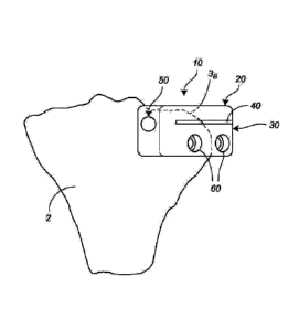
With reference now to FIGS. 1-12, a cutting guide 10 is illustrated for use
during a
surgical procedure in the preparation of a recipient site 5 (FIGS. 3-6) in
tissue 2, e.g., a
patient's tibial plateau, to receive a donor graft 1, e.g., a tibial plateau
allograft, and the
removal of damaged tissue.
The recipient site cutting guide 10 may include, e.g., be formed from, any
material
suitable for use in surgical practice, e.g., plastics, polymers, aluminum,
stainless steel,
titanium, and combinations thereof, and includes a generally horizontal first
arm, e.g., a
.. height-determination (H-G) arm, identified by the reference character 20,
and a generally
vertical second arm, e.g., a blade arm, identified by the reference character
30. In the
illustrated embodiment, the arms 20, 30 are integrally, e.g., monolithically,
formed. In
alternate embodiments, however, other suitable methods of attachment may be
employed to
connect the arms 20, 30, e.g., one or more connectors or fasteners may be
employed, or the
arms 20, 30 may be welded together.
12
Date Recue/Date Received 2022-02-03
The arms 20, 30 are connected such that the cutting guide 10 defines a
generally "L-
shaped" configuration, e.g., such that the arms 20, 30 subtend a fixed angle a
(FIG. 7) of
approximately 900, e.g., 60 -120 . In alternate embodiments, however, if
necessary or
desirable, the configuration of the cutting guide 10 may be varied to alter
the angle a. For
example, the arms 20, 30 may be arranged such that the angle a lies outside
the range of 60 -
1200 dependent upon the requirements of a particular surgical procedure.
Additionally, it is
envisioned that the arms 20, 30 may movably connected to one another such that
the angle a
may he adjusted by the user on an as-needed basis, e.g., during the course of
a surgical
procedure.
The second arm 30 includes a cutting slot 40, one or more guide holes, 50, and
one or
more fixation holes 60. Although illustrated as including a single cutting
slot 40, a single
guide hole 50, and a pair of fixation holes 60 in the embodiment illustrated
in FIGS. 7-12, the
number of cutting slots 40, guide holes 50, and fixation holes 60 may he
varied in alternate
embodiments of the cutting guide 10 without departing from the scope of the
present
disclosure.
The cutting slot 40 extends horizontally across the second arm 30, and is
configured
and dimensioned to receive a cutting implement, e.g., a sagittal saw (not
shown). In the
illustrated embodiment, the cutting slot 40 and the first arm 20 are shown as
extending in
parallel relation, i.e., along non-intersecting axes. In alternate
embodiments, however, the
cutting slot 40 and the first arm 20 may be arranged so as to extend along
intersecting axes.
Additionally, while the cutting slot 40 is illustrated as being linear in
configuration in the
embodiment shown in FIGS. 7-12, in alternate embodiments, the cutting slot 40
may be non-
linear in configuration. For example, the cutting slot 40 may include one or
more arcuate
and/or linear segments or sections.
13
Date Recue/Date Received 2022-02-03
The first arm 20 and the cutting slot 40 define a distance D (FIG. 7)
therebetween that
determines the vertical height (thickness) of the recipient site 5 (FIGS. 3-
6), as discussed in
further detail below. In general, the distance D will lie within the range of
approximately 2
cm to approximately 20 cm ( 25%). In most surgical applications, however, a
distance D of
approximately 6 cm to approximately 10 cm is standard.
The guide hole 50 is located adjacent an end of the cutting slot 40, and
defines a
periphery P (FIG. 8). Although illustrated as being separated from the cutting
slot 40, i.e.,
such that there is not communication between the guide hole 50 and the cutting
slot 40, in
alternate embodiments, the guide hole 50 and the cutting slot 40 may
intersect. Additionally,
while illustrated as circular in configuration in FIGS. 7-12, the guide hole
50 may define
alternate configurations in other embodiments of the cutting guide 10, e.g.,
the guide hole 50
may be elliptical, ovoid, rectangular, etc.
As seen in FIG. 8, for example, the guide hole 50 and the cutting slot 40 are
oriented
such that an upper portion of the periphery P of the guide hole 50 is located
above the cutting
slot 40 (closer to the first arm 20), and a lower portion of the periphery P
of the guide hole 50
is located below the cutting slot 40 (further from the first arm 20). For
example, it is
envisioned that a central axis Xcs (FIG. 8) of the cutting slot 40 may bisect
the guide hole 50.
With continued reference to FIGS. 7-12, the fixation holes 60 will be
discussed. The
fixation holes 60 are configured and dimensioned to removably receive fixation
members
(not shown), e.g., pins, screws, nails, or the like, which can be used to
secure the cutting
guide 10 to the tissue 2 (FIGS. 3-6), e.g., tibial bone, in which the
recipient site 5 is formed,
as discussed in further detail below. In the illustrated embodiment, the
fixation holes 60 are
located below the cutting slot 40. In alternate embodiments, however, the
specific location of
the fixation holes 60 may be altered or varied. For example, in one
embodiment, it is
envisioned that the fixation holes 60 may be located above the cutting slot
40, whereas in
14
Date Recue/Date Received 2022-02-03
another embodiment, it is envisioned that the cutting guide 10 may include one
fixation hole
60 located above the cutting slot 40, and another fixation hole 60 located
below, or in line
with, the cutting slot 40.
Although illustrated as being circular in configuration in the embodiment of
the
cutting guide 10 shown in FIGS. 7-12, the fixation holes 60 may define
alternate
configurations in other embodiments of the present disclosure. For example,
the fixation
holes 60 may be elliptical, ovoid, rectangular, etc.
With reference now to FIGS. 1-12, use of the recipient site cutting guide 10
will be
discussed in connection with the removal of damaged tissue, and formation of
the recipient
site 5 (FIGS. 3-6) in preparation to receive a donor graft 1.
Initially, the recipient site cutting guide 10 is selected according to the
requirements
of the procedure, e.g., such that the dimensions defined by the recipient site
5 correspond to
those of the donor graft 1, which may include an attached meniscus 4, as seen
in FIGS. 3, 5,
and 6. For example, the recipient site cutting guide 10 may be selected based
upon the
distance D (FIG. 7) defined between the first arm 20 and the cutting slot 40
such that the
recipient site 5 is dimensioned to define a particular height in
correspondence with the height
(thickness) of the donor graft 1. The selected recipient site cutting guide 10
is then
positioned such that the first arm 20 abuts a portion 3A (FIG. 2) of the
tissue 2 to be removed,
i.e., damaged tissue, located above the cutting slot 40, and the second ann 30
abuts a portion
3B of the tissue 2 that will not be removed, located below the cutting slot
40. The
configuration and dimensions of the recipient site cutting guide 10, e.g., the
location and
dimensions of the cutting slot 40, are such that the amount of native tissue 2
removed to form
the recipient site 5 is minimized, while still allowing for appropriate
fixation of the donor
graft 1.
Date Recue/Date Received 2022-02-03
After positioning the recipient site cutting guide 10, the cutting guide 10 is
then
secured to the tissue 2 by fixation members (not shown) inserted through the
fixation holes
60. Alternatively, the user may simply apply pressure to the recipient site
cutting guide 10 to
hold the recipient site cutting guide 10 in place.
Thereafter, a drill bit (not shown), or other such cutting implement, is
inserted into,
and advanced through, the guide hole 50 into contact with the tissue 2 to
create a channel 6
(FIGS. 3, 4), e.g., from anterior to posterior. A cut is then made along the
cutting slot 40
using a saggital saw (not shown), or other such cutting implement, until the
channel 6 is
reached so as to define an upper surface 7 of the recipient site 5. For
example, with reference
to FIGS. 1-6 in particular, due to the linear configuration of the cutting
slot 40 included in the
cutting guide 10, the cut made in the tissue 2 results in a planar
configuration at the upper
surface 7 of the recipient site 5. In alternate embodiments, however, i.e.,
embodiments
wherein the cuttings slot 40 is non-linear in configuration, the upper surface
7 of the recipient
site 5 may be formed so as to define a non-planar, or otherwise irregular
configuration.
After completion of the cut, the cutting implement is removed from the cutting
slot 40,
the fixation members (not shown) can be removed from the fixation holes 60,
and the portion
3A (FIG. 2) of the tissue 2 to be removed can be separated from the remainder
of the tissue 2,
revealing the recipient site 5, including the aforementioned channel 6 and
upper surface 7.
FIGS. 13 and 14 illustrate an alternate embodiment of the presently disclosed
recipient site cutting guide, which is referred to generally by the reference
character 110. The
cutting guide 110 is identical to the cutting guide 10 discussed above in
connection with
FIGS. 7-12, for example, but for any distinctions that are specifically noted.
Accordingly, a
discussion of certain features common to the cutting guides 10, 110 may be
omitted in the
interest of brevity.
16
Date Recue/Date Received 2022-02-03
To increase versatility of the cutting guide 110, and the creation of
recipient sites 5
(FIGS. 3-6) of various dimensions, e.g., heights, the cutting guide 110
includes a first arm
120, and a second arm 130 with a series of cutting slots 140, each of which is
located a
different distance from the first arm 120. For example, in the embodiment
illustrated in FIGS.
13 and 14, the second arm 130 includes cutting slots 140A, 140, 140c, wherein
the first arm
120 is spaced a distance DA from the cutting slot 140A, a distance DB from the
cutting slot
140B greater than the distance DA, and a distance Dc from the cutting slot
140c greater than
the distance DB.
The method of using the cutting guide 110 is identical to that of the cutting
guide 10,
but for the fact that the user has the ability to choose a specific cutting
slot, e.g., one of the
cutting slots 140A, 140B, 140c in the embodiment shown in FIGS. 13 and 14,
based upon the
requirements of the particular procedure. For example, dependent upon the
characteristics of
the patient, and/or those of the damaged tissue to be removed, the user may
elect to use one
of the cutting slots 140A, 140B, 140c as opposed to another to guide the
cutting implement
during formation of the upper surface 7 (FIGS. 3-7) of the recipient site 5.
In an alternate method of use, it is envisioned that more than one of the
cutting slots
140A, 140B, 140c may be employed during a surgical procedure. For example, an
initial cut
may be made using the cutting slot 140A, and thereafter, one ore more
additional cuts may be
made using the cutting slot 140B and/or the cutting slot 140c to allow for the
progressive
removal of the tissue and definition of the recipient site 5, e.g., to reduce
patient trauma
and/or inflammation at the recipient site 5.
With reference now to FIGS. 15-20, a donor implant cutting guide 200 will be
discussed useful in formation of the aforementioned donor graft 1 (FIGS. 3-6,
15). The
cutting guide 200 may include, e.g., be formed from, any material suitable for
use in surgical
17
Date Recue/Date Received 2022-02-03
practice, e.g., plastics, polymers, aluminum, stainless steel, titanium, and
combinations
thereof, and includes a body 202, a shaping member 204, and a movable sled
206.
The body 202 of the cutting guide 200 defines a channel 208 that is configured
and
dimensioned to receive donor tissue T, which may include an attached meniscus
4 (FIGS. 3, 5,
6, 15, 16). The channel 208 extends along an axis X, and is defined by
sidewalls 210, 212,
and respective lower and upper shelves 214, 216 positioned on opposite sides
of the shaping
member 204. The lower shelf 214 is spaced a first distance Di (FIG. 17) from a
bottom wall
218 of the body 202, and the upper shelf 216 is spaced a second, greater
distance D2 from the
bottom wall 218 of the body 202.
Although illustrated as extending in parallel relation to the bottom wall 218
of the
body 202 in the embodiment illustrated in FIGS. 15-20, in alternate
embodiments of the
cutting guide 200, the shelf 214 and/or the shelf 216 may extend at an angle
to the bottom
wall 218 so as to either assist or resist movement of the donor tissue T
through the channel
208. For example, either or both of the shelves 214, 216 may be angled toward
the shaping
member 204, or away from the shaping member 204.
The shaping member 204 resides within a well 220 defined by the body 202, and
is
secured to the body 202 such that the shaping member 204 is rotatable in
relation to the body
202 about a fixed axis Y (FIG. 15) that extends in transverse relation to the
axis X defined by
the channel 208. For example, in the embodiment seen in FIG. 15, the shaping
member 204
is oriented such that the axis Y is orthogonal in relation to the axis X. In
alternate
embodiments, however, the shaping member 204 may be oriented such that the
axes X, Y
subtend an angle other than 90 , e.g., 45 .
The shaping member 204 may be actuated, i.e., caused to rotate, by an
automated
mechanism, e.g., a motor (not shown), or alternatively, under manual power via
the
18
Date Recue/Date Received 2022-02-03
application of force by a user. For example, the shaping member 204 may be
rotated by a
crank (not shown) connected to the shaping member 204.
The shaping member 204 includes a drum 222, and one or more vanes 224. While
the
vanes 224 may include sharpened cutting edges 226 (FIGS. 18, 19), as
illustrated in the
embodiment seen in FIGS. 15-20, the vanes 224 may be devoid of any sharpened
edges in
alternate embodiments of the cutting guide 200. Additionally, while the
shaping member 204
is illustrated as including four (4) vanes 224 in the embodiment of the
cutting guide 200
shown in FIGS. 15-20, the number of vanes 224 may be increased or decreased in
alternate
embodiments of the cutting guide 200 without departing from the scope of the
present
disclosure, e.g., to reduce manufacturing costs.
The vanes 224 extend outwardly from the drum 222 into the channel 208.
Specifically, the shaping member 204 is positioned within the well 220 such
that the edges
226 of the vanes 224 align with the upper shelf 216, i.e., such that the
maximum linear
separation realized between the vanes 224 and the bottom wall 218 of the body
202 during
rotation of the shaping member 204 is equivalent to the distance D2 (FIG. 17)
defined
between the upper shelf 216 and the bottom wall 218.
The vanes 224 are configured and dimensioned to shape the donor tissue T into
the
donor graft 1 (FIGS. 3, 5, 6) in correspondence with the configuration of the
recipient site 5
(FIGS. 3-6). For example, in the embodiment of the cutting guide 200
illustrated in FIGS.
15-20, to facilitate shaping of the donor tissue T in the desired manner, the
vanes 224 include
a linear portion 228 (FIG. 19), and a non-linear portion 230 defining one or
more recesses
232. In the specific embodiment shown, the vanes 224 are illustrated as
including a single,
curvate recess 232 defining an arcuate surface 234 that extends inwardly,
toward the axis Y,
resulting in a generally C-shaped configuration. In alternate embodiments,
however, the
recesses 232 may be present in greater number, and/or may define alternative
configurations.
19
Date Recue/Date Received 2022-02-03
For example, each vane 224 may include a pair of recesses 232 that are
triangular in
configuration.
Additionally, or alternatively, it is envisioned that the non-linear portion
230 of the
vanes 224 may include one or more projections (not shown) extending outwardly,
away from
the axis Y.
With reference now to FIGS. 15 and 20 in particular, the sled 206 will be
discussed.
During operation of the cutting guide 200, the sled 206 is used to stabilize
and move the
donor tissue T through the channel 208 across the lower shelf 214 into contact
with the
shaping member 204 and onto the upper shelf 216. To facilitate movement of the
donor
tissue T, the sled 206 is configured and dimensioned to slide in relation to
the body 202 of the
cutting guide 200, and may be either fixedly or removably connected thereto in
any manner
facilitating movement in this manner. For example, the sled 206 may rest upon
upper
surfaces 236, 238 (FIG. 15) defined by the sidewalls 210, 212 of the body 202
such that the
sled 206 slides along the upper surfaces 236, 238 during movement.
In one embodiment, such as the embodiment shown in FIGS. 15 and 20, for
example,
the sled 206 includes a shoulder 240 that depends from an underside 242 (FIG.
20) thereof
which may be used to urge the donor tissue T into contact with one of the
sidewalls 210, 212
(FIG. 15) during movement of the donor tissue T through the channel 208 to
further stabilize
the donor tissue T, e.g., during shaping.
In one embodiment, seen in FIGS. 15 and 20 for example, the underside 242 of
the
sled 206 may include a textured surface 244 to increase friction between the
sled 206 and the
donor tissue T, and thus, control over the donor tissue T during movement
through the
channel 208. For example, the underside 242 of the sled 206 may include one or
more
protrusions 246 configured as detents, teeth, or the like. Alternatively, the
underside 242 of
the sled 206 may be non-textured.
-)0
Date Recue/Date Received 2022-02-03
Additionally, or alternatively, the sled 206 may include retaining structure
(not
shown), e.g., one or more pins, clamps, jaws, or the like, to secure the donor
tissue T to the
sled 206.
With reference now to FIGS. 3-6 and 15-20, use of the cutting guide 200 will
be
discussed in connection with formation of the aforementioned donor graft 1.
Initially, the donor tissue T is harvested from a larger section of tissue
(not shown),
e.g., through use of a saggital saw, scalpel etc., and is fed into the cutting
guide 200. If
necessary, the donor tissue T can be shaped or trimmed so as to fit within the
confines of the
channel 208 (FIG. 15) defined by the body 202 of the donor implant cutting
guide 200.
Specifically, the donor tissue T is positioned on the lower shelf 214, and is
stabilized using
the sled 206, i.e., the donor tissue T is positioned between the lower shelf
214 and the sled
206. Using the sled 206, the donor tissue T is advanced into contact with the
shaping
member 204 whereby the vanes 224 remove portions of the donor tissue T in
accordance with
a pattern determined by the configuration and dimensions thereof.
Specifically, in the
illustrated embodiment, the linear portion 228 (FIG. 19) of the vanes 224
shape a section of
the donor tissue T so as to define a planar section 8 (FIG. 3) that
corresponds in
configurations and dimensions to the upper surface 7 of the recipient site 5,
while the non-
linear portion 230 (FIG. 19) of the vanes 224 simultaneously shape an adjacent
section of the
donor tissue T so as to define a tongue member 9 (FIG. 3). The tongue member 9
corresponds in configurations and dimensions to the channel 6 defined by the
recipient site 5,
and extends transversely in relation to the length and width of the planar
section 8 of the
donor graft 1 such that the tongue member 9 extends outwardly in relation to
the planar
section 8.
As the donor tissue T passes by the shaping member 204, it is supported by the
upper
shelf 216. After shaping of the donor tissue T has been completed, i.e., when
the donor graft
/1
Date Recue/Date Received 2022-02-03
1 has been formed, the donor graft 1 is placed at the recipient site 5 (FIGS.
3-6). Specifically,
the planar section 8 of the donor graft 1 is positioned in abutment with the
upper surface 7 of
the recipient site 5, and the tongue member 9 is positioned within the channel
6, as shown in
FIGS. 3, 5 and 6, whereby the donor graft 1 and the recipient site 5 mate in
an interlocking
fashion so as to inhibit movement of the donor graft 1 in relation to the
recipient site 5, e.g.,
motion in the medial-lateral direction.
In various embodiments of the present disclosure, the configurations,
dimensions, and
orientations of the cutting slot 40, the guide hole 50, and the fixation holes
60 of the cutting
guide 10 (FIGS. 7-12) may be altered or varied, as can the configuration and
dimensions of
the vanes 224, the recesses 232, and the shelves 214, 216 of the cutting guide
200 (FIG. 15),
so as to create a recipient site 5 (FIG. 3) and a donor graft 1 that interlock
in any desired
manner.
With reference again to FIG. 3, following placement of the donor graft 1, the
donor
graft 1 can be attached to the recipient site 5 using either temporary or
permanent attachment
structures (not shown), e.g., fixation screws, bone plates, or the like.
While the present disclosure has been described in connection with specific
embodiments thereof, it will be understood that the subject matter of the
present disclosure is
capable of further modifications. For example, persons skilled in the art will
understand that
additional components and features may be added to any of the embodiments
discussed
herein above, and that those elements and features described in connection
with any one
embodiment may also be applicable to, or combined with, those of any other
embodiment,
without departing from the scope of the present disclosure.
The scope of the present disclosure is intended to cover any variations, uses,
and/or
adaptations of the presently disclosed subject matter in accordance with the
principles of the
present disclosure, including such departures from the present disclosure as
come within
99
Date Recue/Date Received 2022-02-03
known or customary practice within the art to which the present disclosure
pertains, and as
may be applied to the elements, components, and features set forth herein
above.
23
Date Recue/Date Received 2022-02-03