Note: Descriptions are shown in the official language in which they were submitted.
WO 2021/030680
PCT/US2020/046352
MULTISPECIFIC ANTIGEN-BINDING MOLECULES FOR CELL TARGETING
AND USES THEREOF
REFERENCE TO A SEQUENCE LISTING
[0001] This application incorporates by reference the Sequence Listing
submitted in Computer
Readable Form as file 10606W001-Sequence.txt, created on August 7, 2020 and
containing
64,570 bytes.
FIELD OF THE INVENTION
[0002] The present invention relates to alternative formats for multivalent
antigen-binding
proteins, and methods of use thereof. The multivalent antigen-binding
proteins, including bispecific
and multispecific molecules comprise a first polypeptide chain with both an N-
terminal and a C-
terminal antigen-binding domain that specifically binds a T-cell antigen
(e.g., CD3), and a second
polypeptide chain comprising at least one antigen-binding domain that binds a
target antigen (e.g.,
a tumor cell antigen).
BACKGROUND
[0003] Bispecific and multispecific antibodies and antigen-binding molecules
are known in the art
(see, e.g., Brinkmann and Kontermann, MABS, 9(2):182-212, 2017). Among such
known formats is
the FcFc* (Fig. 1A structure), a traditional bispecific antibody with Fab
antigen-binding domains on
either arm of the antibody and an Fc region with a modified CH3 domain that
changes Protein A
binding affinity to permit isolation of the heterodimer from the homodinneric
impurities (Id. at p. 184,
Figure 2, panel 7, last structure). This traditional bispecific antibody
format has been used to make
bispecific antibodies in which one arm of the antibody targets a tumor cell
antigen and the second
arm targets a T-cell antigen, such as CD3. Another conventional format is the
IgG-HC-scFv (Fig.
1B structure), a bispecific antibody in which two N-terminal Fab domains bind
a first antigen and
two scFv domains linked to the C-terminus of the Fc region bind a second
antigen (Id. at p. 184,
Figure 2, panel 10, first structure). There is a need in the art for new and
useful formats for
bispecific or multispecific antigen-binding molecules that improve desired
functionalifies. Although
Brinkmann et at generically references the "building blocks" for the
generation of any homodimeric
or heterodimeric antigen-binding molecule (p. 183, Figure 1), the
possibilities are virtually infinite,
and only those molecules shown in Figure 2 (p. 184) had reportedly been
prepared. Moreover,
Brinkmann doesn't contemplate specific antigen-binding domains, particularly a
molecule
comprising T-cell antigen binding domains at both the N-terminus and the C-
terminus of a single
polypeptide chain forming part of a multispecific molecule.
1
CA 03147791 2022-2-11
WO 2021/030680
PCT/US2020/046352
BRIEF SUMMARY OF THE INVENTION
[0004] In general, the present invention provides multispecific antigen-
binding molecules that bind
both a T-cell antigen (TCA) (e.g., CO3) and a target antigen (TA) (e.g., a
tumor associated antigen,
a viral or bacterial antigen), and which include a single polypeptide chain
that is multivalent (e.g.,
bivalent) with respect to T-cell antigen binding.
[0005] In one aspect, the present invention provides a multispecific antigen-
binding molecule,
comprising: (a) a first polypeptide comprising, from N-terminus to C-terminus
(i) a first antigen-
binding domain that specifically binds a T cell antigen, (ii) a first
multimerizing domain, and (iii) a
second antigen-binding domain that specifically binds a T cell antigen; and
(b) a second polypeptide
comprising, from N-terminus to C-terminus (i) a third antigen-binding domain
that specifically binds
a target antigen, and (ii) a second multimerizing domain, wherein the first
and the second
multimerizing domains associate with one another to form the molecule.
[0006] In some embodiments, the second polypeptide further comprises a fourth
antigen-binding
domain at the C-terminus of the second multimerizing domain. In some cases,
the fourth antigen-
binding domain specifically binds a target antigen. In some cases, the third
antigen-binding domain
and the fourth antigen-binding domain specifically bind distinct target
antigens. In some cases, the
distinct target antigens are expressed (or present) on the surface of the same
cell_ In some cases,
the distinct target antigens are expressed (or present) on the surface of
different cells. References,
herein, to a target antigen expressed (or present) on the surface of a cell
include both a protein
expressed by the cell that is embedded in or spans the cell's membrane, and a
peptide presented in
the context of the groove of a major histocompatibility complex (MHC) protein
by the cell. In some
cases, the third antigen-binding domain and the fourth antigen-binding domain
specifically bind the
same target antigen. In some embodiments, the fourth antigen-binding domain
specifically binds a
T cell antigen. In some cases, the first antigen-binding domain and the second
antigen-binding
domain specifically bind the same T-cell antigen. In some cases, the first
antigen-binding domain
and the second antigen-binding domain specifically bind distinct T-cell
antigens. In some
embodiments, the first antigen-binding domain specifically binds a first T-
cell antigen that is a co-
stimulatory molecule, and the second antigen-binding domain specifically binds
a second T-cell
antigen that is a check-point inhibitor. In some cases, the co-stimulatory
molecule is CO28 and the
check-point inhibitor is PD-1. In some cases, the first, second and fourth
antigen-binding domains
specifically bind the same T-cell antigen. In some cases, the first, second
and fourth antigen-
binding domains bind distinct T-cell antigens. In some cases, the first and
fourth antigen-binding
domains specifically bind the same T-cell antigen. In some cases, the second
and fourth antigen-
binding domains specifically bind the same T-cell antigen.
2
CA 03147791 2022-2-11
WO 2021/030680
PCT/US2020/046352
[0007] In various embodiments, one or more of the antigen-binding domains is a
Fab. In various
embodiments, one or more of the antigen-binding domains is a scFv. In some
embodiments, the
nnultispecific molecules contain both Fab and scFv antigen-binding domains. In
some cases, the
first antigen-binding domain and the third antigen-binding domain are Fabs. In
some cases, the
second antigen-binding domain is an scFv. In some cases, the fourth antigen-
binding domain is an
scFv. In some embodiments, the first, second and third antigen-binding domains
are Fabs. In
some cases, the first and third antigen-binding domains are Fab domains, and
the second antigen-
binding domain is an scFv domain. In some embodiments, the first, second,
third and fourth
antigen-binding domains are Fabs. In some cases, the first second, third and
fourth antigen-
binding domains are Fab domains. In some cases, the first and third antigen-
binding domains are
Fab domains, and the second and fourth antigen-binding domains are scFv
domains. In some
cases, the first, second, third and fourth antigen-binding domains are Fab
domains. In some cases,
the first and third antigen-binding domains are Fab domains, and the second
and fourth antigen-
binding domains are scFv domains. In some cases, the first, second, third and
fourth antigen-
binding domains are Fab domains.
[0008] In any embodiments in which the antigen-binding domain is an scFv
domain, the scFv
domain may comprise a heavy chain variable region (HCVR) comprising a cysteine
mutation at
residue 44, and a light chain variable region comprising a cysteine mutation
at residue 100 (Kabat
numbering). In some cases, the scFv comprises a HCVR and a LCVR joined
together via a
polypeptide linker of from 10 to 30 amino adds, optionally a (G4S)4 linker. In
some embodiments,
the scFv is connected to the C-terminus of the first and/or second
multimerizing domain via a linker
of from 5 to 25 amino acids, optionally a (G4S)3 linker.
[0009] In some embodiments, the T cell antigen is a T cell receptor complex
antigen (i.e., any of
the protein subunits that make up the T cell receptor complex). In some cases,
the T cell antigen is
CD3. In some cases, the T cell antigen is a co-stimulatory molecule or a check-
point inhibitor on a
T cell. In some embodiments, the T cell antigen is selected from the group
consisting of CO27,
CD28, 4-1BB and PD-1. In some embodiments, the T cell antigen is selected from
the group
consisting of CD3, CO27, CO28, 4-1BB and PD-1.
[0010] In some embodiments, the target antigen is a tumor-associated antigen_
In some
embodiments, the target antigen is a viral or bacterial antigen. In some
embodiments, the target
antigen is a fungal antigen or a parasite antigen.
[0011] In some embodiments, the first and second multimerizing domains are
immunoglobulin Fc
domains. In some cases, the first multimerizing domain and the second
multimerizing domain are
human IgG1 or human IgG4 Fc domains. In some cases, the first and second
multimerizing
domains comprise an innnnunoglobulin hinge domain, a CH2 domain and a CH3
domain of a human
3
CA 03147791 2022-2-11
WO 2021/030680
PCT/US2020/046352
IgG polypeptide (e.g., IgG1, IgG2, IgG3 or IgG4). In some cases, the first and
second multimerizing
domains comprise a hinge domain, a CH2 domain and a CH3 domain of a human IgG1
polypeptide. In some cases, the first and second multimerizing domains
comprise a hinge domain,
a CH2 domain and a CH3 domain of a human IgG4 polypeptide. In some
embodiments, the first
and second multimerizing domains associate with one another via disulfide
bonding.
[0012] In some embodiments, the first multimerizing domain or the second
multimerizing domain
comprises an amino acid substitution that reduces affinity for Protein A
binding compared to a wild-
type Fc domain of the same isotype. In some cases, the amino acid substitution
comprises an
H435R modification, or H435R and Y436F modifications (EU numbering). In some
cases, the first
multimerizing domain comprises the H435R and Y436F modifications. In some
cases, the second
multimerizing domain comprises the H435R and Y436F modifications. In some
embodiments, the
first polypeptide, the second polypeptide, or both the first and the second
polypeptides comprise a
modified hinge domain that reduces binding affinity for an Fcy receptor
relative to a wild-type hinge
domain of the same isotype.
[0013] In another aspect, the present invention provides a multispecific
antigen-binding molecule,
comprising: (a) a first polypeptide comprising, from N-terminus to C-terminus
(i) a first Fab that
specifically binds a T cell antigen, (ii) a first immunoglobulin Fc domain,
and (iii) a first scFv that
specifically binds a T cell antigen; and (b) a second polypeptide comprising,
from N-terminus to C-
terminus (i) a second Fab that specifically binds a target antigen, (ii) a
second immunoglobulin Fc
domain, and (iii) a second scFv that specifically binds a target antigen,
wherein the first and the
second immunoglobulin domains associate with one another via disulfide bonding
to form the
molecule.
[0014] In some embodiments, the second Fab and the second scFv specifically
bind distinct
target antigens. In some cases, the distinct target antigens are expressed on
the surface of the
same cell. In some embodiments, the second Fab and the second scFv
specifically bind the same
target antigen.
[0015] In another aspect, the present invention provides a nnultispecific
antigen-binding molecule,
comprising: (a) a first polypeptide comprising, from N-terminus to C-terminus
(i) a first Fab that
specifically binds a T cell antigen, (ii) a first immunoglobulin Fc domain,
and (iii) a second Fab that
specifically binds a T cell antigen; and (b) a second polypeptide comprising,
from N-terminus to C-
terminus (i) a third Fab that specifically binds a target antigen, (ii) a
second immunoglobulin Fc
domain, and (iii) a fourth Fab that specifically binds a target antigen,
wherein the first and the
second immunoglobulin domains associate with one another via disulfide bonding
to form the
molecule.
4
CA 03147791 2022-2-11
WO 2021/030680
PCT/US2020/046352
[0016] In some embodiments, the third Fab and the fourth Fab specifically bind
distinct target
antigens. In some cases, the distinct target antigens are expressed on the
surface of the same cell.
In some embodiments, the third Fab and the fourth Fab specifically bind the
same target antigen.
[0017] In another aspect, the present invention provides a multispecific
antigen-binding molecule,
comprising: (a) a first polypeptide comprising, from N-terminus to C-terminus
(i) a first Fab that
specifically binds a T cell antigen, (ii) a first immunoglobulin Fc domain,
and (iii) a first scFv that
specifically binds a T cell antigen; and (b) a second polypeptide comprising,
from N-terminus to C-
terminus (i) a second Fab that specifically binds a target antigen, (ii) a
second immunoglobulin Fc
domain, and (iii) a second scFv that specifically binds a T cell antigen,
wherein the first and the
second immunoglobulin domains associate with one another via disulfide bonding
to form the
molecule.
[0018] In another aspect, the present invention provides a multispecific
antigen-binding molecule,
comprising: (a) a first polypeptide comprising, from N-terminus to C-terminus
(i) a first Fab that
specifically binds a T cell antigen, (ii) a first immunoglobulin Fc domain,
and (iii) a second Fab that
specifically binds a T cell antigen; and (b) a second polypeptide comprising,
from N-terminus to C-
terminus (i) a second Fab that specifically binds a target antigen, and (ii) a
second immunoglobulin
Fc domain, wherein the first and the second immunoglobulin domains associate
with one another
via disulfide bonding to form the molecule.
[0019] In various embodiments, such as any of those mentioned above or herein,
the T cell
antigen is a T cell receptor complex antigen (La, any of the protein subunits
that make up the T cell
receptor complex). In some cases, the T cell antigen is CD3. In some cases,
the T cell antigen is a
co-stimulatory molecule or a check-point inhibitor on a T cell. In some
embodiments, the T cell
antigen is selected from the group consisting of 0D27, CO28, 4-1BB and PD-1.
In some
embodiments, the T cell antigen is selected from the group consisting of CD3,
CD27, CD28, 4-I BB
and PD-1.
[0020] In various embodiments, such as any of those mentioned above or herein,
the target
antigen is a tumor-associated antigen. In some embodiments, the target antigen
is a viral or
bacterial antigen. In some embodiments, the target antigen is a fungal antigen
or a parasite
antigen.
[0021] In some embodiments, such as any of those mentioned above or herein,
the first and
second multimerizing domains are immunoglobulin Fc domains. In some cases, the
first
multimerizing domain and the second multimerizing domain are human IgG1 or
human IgG4 Fc
domains. In some cases, the first and second multimerizing domains comprise an
immunoglobulin
hinge domain, a CH2 domain and a CH3 domain of a human IgG polypeptide (e.g.,
IgG1, IgG2,
IgG3 or IgG4). In some cases, the first and second multimerizing domains
comprise a hinge
CA 03147791 2022-2-11
WO 2021/030680
PCT/US2020/046352
domain, a CH2 domain and a CH3 domain of a human IgG1 polypeptide. In some
cases, the first
and second multimerizing domains comprise a hinge domain, a CH2 domain and a
CH3 domain of
a human IgG4 polypeptide. In some embodiments, the first and second
multimerizing domains
associate with one another via disulfide bonding.
[0022] In some embodiments, such as any of those mentioned above or herein,
the first
multimerizing domain or the second multimerizing domain comprises an amino add
substitution that
reduces affinity for Protein A binding compared to a wild-type Fe domain of
the same isotype. In
some cases, the amino add substitution comprises an H435R modification, or
H435R and Y436F
modifications (EU numbering). In some cases, the first multimerizing domain
comprises the H435R
and Y436F modifications. In some cases, the second multimerizing domain
comprises the H435R
and Y436F modifications. In some embodiments, the first polypeptide, the
second polypeptide, or
both the first and the second polypeptides comprise a modified hinge domain
that reduces binding
affinity for an Fcy receptor relative to a wild-type hinge domain of the same
isotype.
[0023] In another aspect, the present invention provides a pharmaceutical
composition
comprising any one of the multispecific molecules discussed above or herein,
and a
pharmaceutically acceptable carrier or diluent.
[0024] In another aspect, the present invention provides a method of treating
cancer, comprising
administering any one of the multispecific molecules discussed above or herein
to a subject in need
thereof.
[0025] In another aspect, the present invention provides a method of treating
an infection,
comprising administering any one of the multispecific molecules discussed
above or herein to a
subject in need thereof. In some cases, the infection is a bacterial
infection. In some cases, the
infection is a viral infection. In some cases, the infection is a fungal
infection. In some cases, the
infection is a parasite infection.
[0026] In various embodiments, the target antigen is present at a density of
from 10 to
10,000,000 copies per target cell. In various embodiments, the target antigen
is present at a
density of from 100 to 10,000,000 copies per target cell. In various
embodiments, the target
antigen is present at a density of from 100 to 1,000,000 copies per target
cell. In some
embodiments, the target antigen is present at a density of from 50 to 10,000.
In some
embodiments, the target antigen is present at a density of from 100 to 5000.
In some
embodiments, the target antigen is present at a density of from 100 to 20,000.
In some
embodiments, the target antigen is present at a density of from 500 to
1,000,000 copies per target
cell. In some embodiments, the target antigen is present at a density of from
1000 to 20,000 copies
per target cell. In some embodiments, the target antigen is present at a
density of greater than
20,000 copies per target cell. In various embodiments, the target antigen is
present at a density of
6
CA 03147791 2022-2-11
WO 2021/030680
PCT/US2020/046352
about 10, about 50, about 1001 about 200, about 300, about 400, about 500,
about 1000, about
2000, about 3000, about 4000, about 5000, about 6000, about 7000, about 8000,
about 9000,
about 10,000, about 15,000, about 20,000, about 25,000, about 50,000, about
75,000, about
100,000, about 200,000, about 300,000, about 400,000, about 500,000, about
600,000, about
700,000, about 800,000, about 900,000, about 1,000,000, about 2,000,000, about
3,000,000, about
4,000,000 or about 5,000,000 copies per target cell. As used herein, a "low
density antigen" is an
antigen where no more than 5000 copies of the antigen are found on a target
cell. References to a
low density antigen include cases in which a cell has no more than 4000, no
more than 3000, no
more than 2000, no more than 1000, no more than 900, no more than 800, no more
than 700, no
more than 600, no more than 500, no more than 400, no more than 300, no more
than 200, no
more than 100, or no more than 50 copies of the target antigen.
[0027] In various embodiments, the multispecific molecule is administered in
combination with a
second therapeutic agent to treat a disease or disorder. In some cases, the
second therapeutic
agent comprises a bispecific antigen-binding molecule comprising a first
antigen-binding domain
that binds a target antigen (TA) and a second antigen-binding domain that
binds a T-cell antigen. In
some cases, the target antigen is a tumor-cell antigen. In some embodiments,
the second
therapeutic agent comprises a bispecific anti-TA x anti-0O28 antibody. In some
embodiments, the
second therapeutic agent comprises a bispecific anti-EGFR x anti-0028
antibody. In some
embodiments, the second therapeutic agent comprises an antibody that binds a
check-point
inhibitor on a T cell. In some embodiments, the second therapeutic agent
comprises an anti-PD-1
antibody. In some cases, the multispecific molecule is administered in
combination with two or
more second therapeutic agents.
[0028] In another aspect, the present invention provides for use of any one of
the multispecific
molecules discussed above or herein in the manufacture of a medicament for
treating a disease or
disorder (e.g., a cancer, or an infection) in a subject in need thereof.
[0029] In another aspect, the present invention provides for use of any one of
the multispecific
molecules discussed above or herein in medicine, or to treat a disease or
disorder (e.g., a cancer,
or an infection).
[0030] In another aspect, the present invention provides a multispecific
molecule, as discussed
above or herein, for use in medicine, or to treat a disease or disorder (e.g.,
a cancer, or an
infection).
[0031] In any of the embodiments discussed above or herein, the target antigen
may be a peptide
in the context of the groove of a major histocompafibility complex (MHC)
protein.
[0032] In various embodiments, any of the features or components of
embodiments discussed
above or herein may be combined, and such combinations are encompassed within
the scope of
7
CA 03147791 2022-2-11
WO 2021/030680
PCT/US2020/046352
the present disclosure. Any specific value discussed above or herein may be
combined with
another related value discussed above or herein to recite a range with the
values representing the
upper and lower ends of the range, and such ranges are encompassed within the
scope of the
present disclosure.
[0033] Other embodiments will become apparent from a review of the ensuing
detailed
description.
BRIEF DESCRIPTION OF THE DRAWINGS
[0034] Figs. 1A and 1B illustrate known bispecific antibody and antigen-
binding molecule formats.
[0035] Figs. 1C, 1E, 1F, 1G, 1H, 11, 1J,1K, 11_, 1M, 1N, 10, 1P, 1Q, 1R and 1S
illustrate
bispecific or multispecific antigen-binding molecule formats in accordance
with embodiments of the
present invention. In each of these formats, a first polypeptide chain
comprises both an N-terminal
and a C-terminal antigen-binding domain (e.g., a Fab or scFv) that
specifically binds a T-cell
antigen (TCA) (e.g., CM), and a second polypeptide chain comprising at least
one antigen-binding
domain (e.g., a Fab or scFv) that binds a target antigen (TA) (e.g_, a tumor
cell antigen). Fig. 1D
illustrates a format in which the two antigen-binding domains that
specifically bind a T-cell antigen
(e.g., COS) are located on different polypeptide chains (at the N-terminus on
one polypeptide chain,
and at the C-terminus on the second polypeptide chain).
[0036] Fig. 2 shows T cell activation induced by molecules having each of the
formats illustrated
in Figs. 1A, 1B and 1C compared to a T cell-only control (ZERO) and a positive
control. None of
the molecules activated T cells in the absence of target cells.
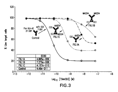
[0037] Fig. 3 shows the cytotoxic activity of molecules having each of the
formats illustrated in
Figs. 1A, 1B and 1C, in the presence of human PBMC and target cells (A375),
compared to a
positive control that induces maximal cell killing. The CD3-binding domains of
the molecules
comprise the variable regions of a 7221G anti-003 antibody. The molecule
having the structure of
Fig. 1C was significantly more potent than the molecules having the structures
of Figs. 1A and 1B.
[0038] Figs. 4A, 4B and 4C show the cytotoxic activity of molecules having
each of the formats
illustrated in Figs. 1A, 1B and 1C, in the presence of human PBMC and target
cells (A375), in
combination with an anti-PD-1 antibody (Fig. 4A), a co-stimulatory bispecific
EGFR x CO28
antibody (Fig. 4B), or both an anti-PD-1 antibody and a co-stimulatory
bispecific EGFR x CD28
antibody (Fig. 40) compared to a positive control that induces maximal cell
killing. The CD3-
binding domains of the molecules comprise the variable regions of a 7221G anti-
CD3 antibody.
The molecule having the structure of Fig. 1C was significantly more potent in
combination with
these additional antibodies than the molecules having the structures of Figs.
1A and 1B.
8
CA 03147791 2022-2-11
WO 2021/030680
PCT/US2020/046352
[0039] Fig. 5 shows the measured cytokine levels of the molecule having the
structure of Fig. 1C
(right panel) compared to the molecule having the structure of Fig. 1A (left
panel) at the point of
maximal antibody concentration shown in Figs. 4A, 4B and 4C. The CD3-binding
domains of the
molecules comprise the variable regions of a 7221G anti-CD3 antibody. The
molecule having the
structure of Fig. 1C does not show greater levels of cytokine release in spite
of the significantly
greater cytotoxic activity.
[0040] Figs. 6A, 6B, 6C and 60 show binding of the molecule having the
structure of Fig. 1C and
modified versions of this molecule (with inactive domains ¨ noted by an X in
the legend) to Raji cells
(Fig. GA) or A375 cells (Fig. GC) overexpressing a MAGEA4 peptide, or CD3+
Jurkat cells (Figs. 613
and 60). The CD3-binding domains of the molecules illustrated in Figs. 6A and
6B comprise the
variable regions of a 7195P anti-0O3 antibody. The CD3-binding domains of the
molecules
illustrated in Figs. 6C and 6D comprise the variable regions of a 7221G anti-
CD3 antibody. As
shown in Figs. 6A,6B, 6C and 60, the presence of two active antigen-binding
domains improved
binding to the target antigens, and similar binding was observed irrespective
of the source of the
anti-CD3 binding domains. As illustrated in these figures, binding was most
affected when the N-
terminal Fab domain was removed.
[0041] Figs. 7A and 7B show the cytotoxic activity of the same molecules shown
in Figs. 6A and
6B (Fig. 7A), and Figs. 6C and 6D (Fig. 7B). The molecule having the structure
of Fig. 1C showed
the greatest cytotoxic potency, followed by the molecules with two active T-
cell antigen (e.g., CO3)
binding domains. A similar pattern of cytotoxicity was observed irrespective
of the source of the
anti-CD3 binding domains.
[0042] Figs. 8A and 8B show binding of the molecule having the structure of
Fig. 1C and modified
versions of this molecule (with C-terminal Fab domains or inactive domains ¨
noted by an X in the
legend) to Raji cells overexpressing a MAGEA4 peptide (Fig. 8A) or CD3+ Jurkat
cells (Fig. 8B).
The CD3-binding domains of the molecules comprise the variable regions of a
7195P anti-CD3
antibody. As shown in Figs. 8A and 8B, C-terminal scFv domains provided
superior binding to the
target antigens compared to C-terminal Fab domains.
[0043] Fig. 9 shows the cytotoxic activity of the same molecules shown in
Figs. 8A and 8B. The
CD3-binding domains of the molecules comprise the variable regions of a 7195P
anti-CD3
antibody. The molecule having the structure of Fig. 1C showed the greatest
cytotoxic potency,
followed by the molecule having the structure of Fig. 1E.
[0044] Figs. 10A and 10B show binding of the molecules having the structures
of Figs. 1C and 1D
to A375 cells overexpressing a MAGEA4 peptide (Fig. 10A), or CO3+ Jurkat cells
(Fig. 10B). The
CD3-binding domains of the molecules comprise the variable regions of a 7221G
anti-0O3
antibody. The two molecules showed similar binding to both cell types relative
to one another.
9
CA 03147791 2022-2-11
WO 2021/030680
PCT/US2020/046352
[0045] Figs. 11A and 11B show the cytotoxic activity of the same molecules
shown in Figs. 10A
and 10B on A375 cells from two different donor sources. The CD3-binding
domains of the
molecules comprise the variable regions of a 7221G anti-CD3 antibody. The
molecule having the
structure of Fig. 1C was more potent than the molecule having the structure of
Fig. 112
[0046] Figs. 12A and 12B show the relative cytotoxic activity and potency of
molecules having the
structures of Fig. 1C and Fig. 1F, respectively, as compared to a molecule
having the structure of
Fig. 1A. The molecules were tested individually, and in combination with a co-
stimulatory bispecific
EGFR x CO28 antibody and an anti-PD-1 antibody, as discussed in Example 7. The
COS-binding
domains of the molecules comprise the variable regions of a 7195P anti-CD3
antibody. The
molecule having the structure of Fig. 1F targets two different epitopes of the
same target antigen
with the two TA antigen-binding domains, whereas the molecule having the
structure of Fig. 1C
targets the same epitope of the target antigen with the two TA antigen-binding
domains. The
molecule having the structure of Fig. 1F was more potent than the molecule
having the structure of
Fig. 1C, and both molecules were more potent that the molecule having the
structure of Fig. 1A. In
each case, the combination of these molecules with the co-stimulatory
bispecific antibody and the
anti-PD-1 antibody produced even greater cytotoxic potency, similar to the
results shown in Figs.
4A-4C.
[0047] Fig. 13 shows the relative binding affinity for molecules having the
structure of Fig. IF, in
which the CO3-binding domains are derived from anti-COB antibodies with
strong, moderate, or
weak binding affinity to CD3. The "strong" binding domains are derived from
the 7195P anti-CD3
antibody. The "moderate" binding domains are derived from the 7221G anti-0O3
antibody. The
"weak" binding domains are derived from the 7221G20 anti-CD3 antibody. The
references to, e.g.,
"strong / strong" refer, respectively, to the Fab anti-CD3 binding domain and
the scFc anfi-0O3
binding domain. As expected, binding to CD3-positive Jurkat cells correlates
with the strength of
the affinity of the anti-CD3 binding domains in the molecules.
[0048] Figs. 14A and 14B show the relative cytotoxic activity and potency of
the molecules shown
in Fig. 13 in MAGEA4-positive A375 cells. The molecules were tested
individually (Fig. 14A), and in
combination with a co-stimulatory bispecific EGFR x CD28 and an anti-PD-1
antibody (Fig. 14B), as
discussed in Example 8. There is a clear correlation between the strength of
the anti-CD3 binding
domains and the potency of the molecules. The "Control" is a positive control
that targets the
scaffold of all HLA molecules to provide a maximum cytotoxicity against which
to compare the other
molecules.
[0049] Figs. 15A and 15B show the relative cytotoxic activity and potency of
the molecules shown
in Fig. 13 in MAGEA4-positive ScaBER cells. The molecules were tested
individually (Fig. 15A),
and in combination with a co-stimulatory bispecific EGFR x CO28 and an anti-PD-
1 antibody (Fig.
CA 03147791 2022-2-11
WO 2021/030680
PCT/US2020/046352
15B), as discussed in Example 8. There is a clear correlation between the
strength of the anti-CD3
binding domains and the potency of the molecules. The "Control" is a positive
control that targets
the scaffold of all HLA molecules to provide a maximum cytotoxicity against
which to compare the
other molecules.
[0050] Figs. 16A, 16B and 16C show the relative binding affinity for molecules
having the
structures of Figs. 1A (Molecule C), 1C (Molecule B), and IF (Molecules A and
D) to NYESO-1-
positive cells (Fig. 16A), MAGEA4 (peptide 1)-positive cells (Fig. 16B) and
MAGEA4 (peptide 2)-
positive cells (Fig. 16C). As expected, Molecule D, without an NYESO-1 binding
domain does not
bind to the NYES0-1 expressing cells (Fig. 16A), and the molecules that lack
the relevant MAGEA4
binding domain do not bind to the MAGEA4-expressing cells, as shown in Figs.
16B and 16C.
The CD3-binding domains of the molecules comprise the variable regions of a
7195P anti-CD3
antibody. The "HLA Targeting Bispecific" positive control binds HLA molecules
and CD3. The
"Isotype Control Multispecific" is a molecule having the structure of Fig. 1C
with binding domains to
an irrelevant target antigen.
[0051] Figs. 17A and 17B show the relative cytotoxic activity and potency of
molecules having the
structures of Fig. 1C and Fig. 1F, respectively, as compared to a positive
control having the
structure of Fig. 1A, which binds HLA molecules and CD3. The isotype controls
included a
molecule with the structure of Fig. 1C with binding domains to an irrelevant
target antigen, and a
molecule with the structure of Fig. 1A with binding domains to CD3 and an
irrelevant target antigen.
The molecules were tested individually, and in combination with a co-
stimulatory bispecific EGFR x
CD28 antibody and an anti-PD-1 antibody, as discussed in Example 9. The CD3-
binding domains
of the molecules comprise the variable regions of a 7195P anti-CD3 antibody.
The molecule having
the structure of Fig. 1F targets two different antigens (NYESO-1 and MAGEA4)
with the two TA
antigen-binding domains, whereas the molecule having the structure of Fig. 1C
targets a single
antigen with both of the two TA antigen-binding domains. The molecule having
the structure of Fig.
IF and targeting two different antigens was more potent than the molecule
having the structure of
Fig. 1C. In each case, the combination of these molecules with the co-
stimulatory bispecific
antibody and the anti-PD-1 antibody produced even greater cytotoxic potency,
relative to the
molecule alone, similar to the results shown in Figs. 4A-4C.
[0052] Figs. 17C and 17D shown the relative T-cell activation of the molecules
discussed in
connection with Figs. 17A and 17B.
[0053] Figs. 18A and 18B show the relative cytotoxic activity and potency of
molecules having the
structures of Fig. 1C and Fig. 1F, respectively, as compared to a molecule
having the structure of
Fig. 1A. The positive control is a molecule with the structure of Fig. 1A that
binds human leukocyte
antigen (HLA) molecules and CD3. The isotype controls included a molecule with
the structure of
11
CA 03147791 2022-2-11
WO 2021/030680
PCT/US2020/046352
Fig. 1C with binding domains to an irrelevant target antigen, and a molecule
with the structure of
Fig. 1A with binding domains to CD3 and an irrelevant target antigen. The
molecules were tested
individually, and in combination with a co-stimulatory bispecific EGFR x CD28
antibody and an anti-
PD-1 antibody, as discussed in Example 9. The CD3-binding domains of the
molecules comprise
the variable regions of a 7195P anti-CD3 antibody. As shown in Fig. 18A, the
molecule having the
structure of Fig. IF (targeting two distinct epitopes of MAGEA4) is more
potent than the molecule
having the structure of Fig. 1C (targeting a single epitope with both TA-
binding domains), and both
molecules are more potent than the molecule having the structure of Fig. -1A.
Similarly, as shown in
Fig. 18B, the molecule having the structure of Fig. 1F (targeting two
different antigens) is more
potent than the molecule having the structure of Fig. 1C (targeting a single
antigen with both TA-
binding domains), and both molecules are more potent than the molecule having
the structure of
Fig. 1A. In each case, the combination of these molecules with the co-
stimulatory bispecific
antibody and the anti-PD-1 antibody produced even greater cytotoxic potency,
relative to the
molecule alone, similar to the results shown in Figs. 4A-4C.
[0054] Figs. 18C, 18D, 18E and 18F show the relative T-cell activation of the
molecules
discussed in connection with Figs. 18A and 18B.
[0055] Figs. 19A and 19B show the cytotoxic activity and potency, and T-cell
activation,
respectively, of a molecule having the structure of Fig. 1F relative to a
combination of two
molecules having the struture of Fig. 1A, in which the combination of the two
molecules binds the
same pair of target antigens as the molecule having the structure of Fig. 1F.
As shown in Figs. 19A
and 19B, the molecule having the structure of Fig. IF more potently kills the
tumor cells and
increases T-cell activation than does the combination of the two molecules
having the structure of
Fig. 1A.
DETAILED DESCRIPTION
[0056] Before the present invention is described in further detail, it is to
be understood that this
invention is not limited to particular methods and experimental conditions
described, as such
methods and conditions may vary. It is also to be understood that the
terminology used herein is
for the purpose of describing particular embodiments only, and is not intended
to be limiting, since
the scope of the present invention will be limited only by the appended
claims.
[0057] Unless defined otherwise, all technical and scientific terms used
herein have the same
meaning as commonly understood by one of ordinary skill in the art to which
this invention belongs.
As used herein, the term "about," when used in reference to a particular
recited numerical value,
means that the value may vary from the recited value by no more than 1%. For
example, as used
12
CA 03147791 2022-2-11
WO 2021/030680
PCT/US2020/046352
herein, the expression "about 100" includes 99 and 101 and all values in
between (e.g., 99.1, 99.2,
99.3, 99.4, etc.).
[0058] Although any methods and materials similar or equivalent to those
described herein can
be used in the practice or testing of the present invention, the preferred
methods and materials are
now described. All patents, applications and non-patent publications mentioned
in this specification
are incorporated herein by reference in their entireties.
Definitions
[0059] The term "T cell" refers to immune cells expressing CD3, including CD4+
cells (helper T
cells), CD8+ cells (cytotoxic T cells), regulatory T cells (Tregs), and tumor
infiltrating lymphocytes.
[0060] The expression "T-cell antigen" refers to a cell-surface expressed
protein present on a T
cell, and includes "co-stimulatory molecules." A "co-stimulatory molecule"
refers to a protein
expressed by a T cell that binds a cognate ligand or receptor (e.g., on an
antigen-presenting cell) to
provide a stimulatory signal, which, in combination with the primary signal
provided by engagement
of the T cell's TCR with a peptide/MHC, stimulates the activity of the T cell.
Stimulation of a T cell
can include activation, proliferation and/or survival of the T cell.
[0061] As used herein, the expression "cell surface-expressed" or "cell-
surface molecule" means
one or more protein(s) that is/are expressed on the surface of a cell in vitro
or in vivo, such that at
least a portion of the protein is exposed to the extracellular side of the
cell membrane and is
accessible to an antigen-binding portion of an antibody or an antigen-binding
domain of the
multispecific antigen-binding molecules discussed herein.
[0062] The expression "CO3," as used herein, refers to an antigen which is
expressed on T cells
as part of the multimolecular T cell receptor (TCR) and which consists of a
homodimer or
heterodimer formed from the association of two of four receptor chains: CD3-
epsilon, CD3-delta,
CD3-zeta, and CO3-gamma. All references to proteins, polypeptides and protein
fragments herein
are intended to refer to the human version of the respective protein,
polypeptide or protein fragment
unless explicitly specified as being from a non-human species. Thus, the
expression "CD3" means
human CD3 unless specified as being from a non-human species, e.g., "mouse
CD3," "monkey
CD3," etc.
[0063] As used herein, "an antibody that binds CD3" or an "anti-CD3 antibody"
includes
antibodies and antigen-binding fragments thereof that specifically recognize a
single CD3 subunit
(e.g., epsilon, delta, gamma or zeta), as well as antibodies and antigen-
binding fragments thereof
that specifically recognize a dimeric complex of two CO3 subunits (e.g.,
gamma/epsilon,
delta/epsilon, and zeta/zeta CD3 dimers). The antigen-binding domains of the
present invention
may bind soluble CD3 and/or cell surface expressed CD3. Soluble CD3 includes
natural CD3
13
CA 03147791 2022-2-11
WO 2021/030680
PCT/US2020/046352
proteins as well as recombinant CD3 protein variants such as, e.g., monomeric
and dimeric CD3
constructs, that lack a transmembrane domain or are otherwise unassociated
with a cell membrane.
[0064] As used herein, the expression "cell surface-expressed COY' means one
or more CO3
protein(s) that is/are expressed on the surface of a cell in vitro or in vivo,
such that at least a portion
of a CD3 protein is exposed to the extracellular side of the cell membrane and
is accessible to an
antigen-binding portion of an antibody. "Cell surface-expressed CD3" includes
CO3 proteins
contained within the context of a functional T cell receptor in the membrane
of a cell. The
expression "cell surface-expressed CD3" includes CO3 protein expressed as part
of a homodimer
or heterodimer on the surface of a cell (e.g., gamma/epsilon, delta/epsilon,
and zeta/zeta CD3
dimers). The expression, "cell surface-expressed CD3" also includes a CO3
chain (e.g., CD3-
epsilon, CD3-delta or CO3-gamma) that is expressed by itself, without other
COB chain types, on
the surface of a cell. A "cell surface-expressed CD3" can comprise or consist
of a CD3 protein
expressed on the surface of a cell which normally expresses CD3 protein.
Alternatively, "cell
surface-expressed CD3" can comprise or consist of CO3 protein expressed on the
surface of a cell
that normally does not express human CD3 on its surface but has been
artificially engineered to
express CO3 on its surface.
[0065] The term "antigen-binding domain" refers to that portion of a
multispecific molecule or a
corresponding antibody that binds specifically to a predetermined antigen
(e.g., CO3 or a tumor
associated antigen). References to a "corresponding antibody" refer to the
antibody from which the
CDRs or variable regions (HCVR and LCVR) used in a mulfispecific molecule are
derived. For
example, the Fig. 1C structured molecules discussed in the examples include
Fabs and scFvs with
variable regions derived from specific anti-CD3 antibodies and anti-MAGEA4
antibodies. These
antibodies are the "corresponding antibodies" to the respective multispecific
molecules.
[0066] The term "multispecific antigen-binding molecule" includes molecules
that bind two or
more (e.g., three or four) different epitopes or antigens. In some cases, the
multispecific antigen-
binding molecules are bispecific. In some cases, the multispecific antigen-
binding molecules are
trispecific. In some cases, the multispecific antigen-binding molecules are
tetraspecific.
[0067] The term "antibody" means any antigen-binding molecule or molecular
complex
comprising at least one complennentarity determining region (CDR) that
specifically binds to or
interacts with a particular antigen (e.g., CD3 or a target antigen (TA)). The
term "antibody" includes
innnnunoglobulin molecules comprising four polypepfide chains, two heavy (H)
chains and two light
(L) chains inter-connected by disulfide bonds, as well as multimers thereof
(e.g., IgM). The term
"antibody' also includes immunoglobulin molecules consisting of four
polypeptide chains, two heavy
(H) chains and two light (L) chains inter-connected by disulfide bonds. Each
heavy chain comprises
a heavy chain variable region (abbreviated herein as HCVR or VH) and a heavy
chain constant
14
CA 03147791 2022-2-11
WO 2021/030680
PCT/US2020/046352
region. The heavy chain constant region comprises three domains, CH1, C112 and
CH3. Each light
chain comprises a light chain variable region (abbreviated herein as LCVR or
Vi) and a light chain
constant region. The light chain constant region comprises one domain (CL1).
The VH and VL
regions can be further subdivided into regions of hypervariability, termed
complementarily
determining regions (CDRs), interspersed with regions that are more conserved,
termed framework
regions (FR). Each VH and VL is composed of three CDRs and four FRs, arranged
from amino-
terminus to carboxy-terminus in the following order FRI. CDR1, FR2, CDR2, FR3,
CDR3, FR4. In
different embodiments of the invention, the FRs of the anti-TA antibody or
anti-CD3 antibody (or
antigen-binding portion thereof) may be identical to the human germline
sequences, or may be
naturally or artificially modified. An amino acid consensus sequence may be
defined based on a
side-by-side analysis of two or more CDRs.
[0068] The term "antibody", as used herein, also includes antigen-binding
fragments of full
antibody molecules. The terms "antigen-binding portion" of an antibody,
"antigen-binding fragment"
of an antibody, and the like, as used herein, include any naturally occurring,
enzymatically
obtainable, synthetic, or genetically engineered polypeptide or glycoprotein
that specifically binds
an antigen to form a complex. Antigen-binding fragments of an antibody may be
derived, e.g., from
full antibody molecules using any suitable standard techniques such as
proteolytic digestion or
recombinant genetic engineering techniques involving the manipulation and
expression of DNA
encoding antibody variable and optionally constant domains. Such DNA is known
and/or is readily
available from, e_g_, commercial sources, DNA libraries (including, e.g.,
phage-antibody libraries), or
can be synthesized. The DNA may be sequenced and manipulated chemically or by
using
molecular biology techniques, for example, to arrange one or more variable
and/or constant
domains into a suitable configuration, or to introduce codons, create cysteine
residues, modify, add
or delete amino acids, etc.
[0069] Non-limiting examples of antigen-binding fragments include: (i) Fab
fragments; (ii) F(ab')2
fragments; (iii) Fd fragments; (iv) Fv fragments; (v) single-chain Fv (scFv)
molecules; (vi) dAb
fragments; and (vii) minimal recognition units consisting of the amino add
residues that mimic the
hypervariable region of an antibody (e.g., an isolated complementarity
determining region (CDR)
such as a CDR3 peptide), or a constrained FR3-CDR3-FR4 peptide. Other
engineered molecules,
such as domain-specific antibodies, single domain antibodies, domain-deleted
antibodies, chimeric
antibodies, CDR-grafted antibodies, diabodies, triabodies, tetrabodies,
nninibodies, nanobodies (e.g.
monovalent nanobodies, bivalent nanobodies, etc.), small modular
immunopharmaceuticals
(SMI Ps), and shark variable IgNAR domains, are also encompassed within the
expression "antigen-
binding fragment," as used herein.
CA 03147791 2022-2-11
WO 2021/030680
PCT/US2020/046352
[0070] An antigen-binding fragment of an antibody will typically comprise at
least one variable
domain. The variable domain may be of any size or amino acid composition and
will generally
comprise at least one CDR which is adjacent to or in frame with one or more
framework sequences.
In antigen-binding fragments having a VH domain associated with a VI_ domain,
the VH and VL
domains may be situated relative to one another in any suitable arrangement
For example, the
variable region may be dimeric and contain VH-VH, VH-V1 or VL-VI dimers.
Alternatively, the antigen-
binding fragment of an antibody may contain a monomeric VH or Vi. domain.
[0071] In certain embodiments, an antigen-binding fragment of an antibody may
contain at least
one variable domain covalently linked to at least one constant domain. Non-
limiting, exemplary
configurations of variable and constant domains that may be found within an
antigen-binding
fragment of an antibody of the present invention include: (i) VH-CHI; (ii) VH-
CH2; (iii) VH-CH3; (iv) VH-
CH1-CH2; (V) VH-CH1-CH2-CI3; (vi) VH-CH2-CH3; (Vii) VH-CL, (VW) VL-CH1; (ix)
V1..-CH2, (X) VL-CH3; (Xi)
VI-CHI-Cl-i2; (Xii) VL-Cl1-Cl2-CH3; (Xiii) Vi-CH2-CH3; and (xiv) VL-CL. In any
configuration of
variable and constant domains, including any of the exemplary configurations
listed above, the
variable and constant domains may be either directly linked to one another or
may be linked by a
full or partial hinge or linker region. A hinge region may consist of at least
2 (e.g., 5, 10, 15, 20, 40,
60 or more) amino acids which result in a flexible or semi-flexible linkage
between adjacent variable
and/or constant domains in a single polypeptide molecule. Moreover, an antigen-
binding fragment
of an antibody of the present invention may comprise a homo-dimer or hetero-
dimer (or other
multimer) of any of the variable and constant domain configurations listed
above in non-covalent
association with one another and/or with one or more monomeric VH or VL domain
(e.g., by disulfide
bond(s)).
[0072] In certain embodiments of the invention, the antibodies are human
antibodies. The term
"human antibody" is intended to include antibodies having variable and
constant regions derived
from human germline innmunoglobulin sequences. The human antibodies may
include amino acid
residues not encoded by human germline immunoglobulin sequences (e.g.,
mutations introduced
by random or site-specific nnutagenesis in vitro or by somatic mutation in
vivo), for example in the
CDRs and in particular CDR3. However, the term "human antibody", as used
herein, is not
intended to include antibodies in which CDR sequences derived from the
gemnline of another
mammalian species, such as a mouse, have been grafted onto human framework
sequences.
[0073] The antibodies discussed herein may, in some embodiments, be
recombinant human
antibodies. The term "recombinant human antibody" is intended to include all
human antibodies
that are prepared, expressed, created or isolated by recombinant means, such
as antibodies
expressed using a recombinant expression vector transfected into a host cell,
antibodies isolated
from a recombinant, combinatorial human antibody library, antibodies isolated
from an animal (e.g.,
16
CA 03147791 2022-2-11
WO 2021/030680
PCT/US2020/046352
a mouse) that is transgenic for human immunoglobulin genes (see e.g., Taylor
et al. (1992) Nucl.
Acids Res. 20:6287-6295) or antibodies prepared, expressed, created or
isolated by any other
means that involves splicing of human immunoglobulin gene sequences to other
DNA sequences.
Such recombinant human antibodies have variable and constant regions derived
from human
germline immunoglobulin sequences. In certain embodiments, however, such
recombinant human
antibodies are subjected to in vitro mutagenesis (or, when an animal
transgenic for human Ig
sequences is used, in vivo somatic mutagenesis) and thus the amino acid
sequences of the VH and
VI_ regions of the recombinant antibodies are sequences that, while derived
from and related to
human germline VH and VI_ sequences, may not naturally exist within the human
antibody germline
repertoire in vivo.
[0074] The antibodies referenced herein may be isolated antibodies. An
"isolated antibody," as
used herein, means an antibody that has been identified and separated and/or
recovered from at
least one component of its natural environment. For example, an antibody that
has been separated
or removed from at least one component of an organism, or from a tissue or
cell in which the
antibody naturally exists or is naturally produced, is an "isolated antibody."
An isolated antibody
also includes an antibody in situ within a recombinant cell. Isolated
antibodies are antibodies that
have been subjected to at least one purification or isolation step. An
isolated antibody may be
substantially free of other cellular material and/or chemicals.
[0075] The antibodies referenced herein may comprise one or more amino add
substitutions,
insertions and/or deletions in the framework and/or CDR regions of the heavy
and light chain
variable domains as compared to the corresponding germline sequences from
which the antibodies
were derived. Such mutations can be readily ascertained by comparing the amino
acid sequences
disclosed herein to germline sequences available from, for example, public
antibody sequence
databases.
[0076] The term "epitope" refers to an antigenic determinant that interacts
with a specific antigen
binding site in the variable region of an antibody molecule known as a
paratope. A single antigen
may have more than one epitope. Thus, different antibodies may bind to
different areas on an
antigen and may have different biological effects. Epitopes may be either
conformational or linear.
A conformational epitope is produced by spatially juxtaposed amino acids from
different segments
of the linear polypeptide chain. A linear epitope is one produced by adjacent
amino acid residues in
a polypeptide chain. In certain circumstance, an epitope may include moieties
of saccharides,
phosphoryl groups, or sulfonyl groups on the antigen.
[0077] A "multimerization domain" or "nnultimerizing domain" is any
macromolecule that has the
ability to associate (covalently or non-covalently) with a second
macromolecule of the same or
similar structure or constitution. For example, a multimerization domain may
be a polypeptide
17
CA 03147791 2022-2-11
WO 2021/030680
PCT/US2020/046352
comprising an immunoglobulin CH3 domain. A non-limiting example of a
multimerization domain is
an Fc portion of an immunoglobulin, e.g., an Fc domain of an IgG selected from
the isotypes IgG1,
IgG2, IgG3, and IgG4, as well as any allotype within each isotype group. In
certain embodiments,
the multimerization domain is an Fc fragment or an amino acid sequence of 1 to
about 200 amino
acids in length containing at least one cysteine residue. In other
embodiments, the multimerization
domain is a cysteine residue or a short cysteine-containing peptide. Other
multimerization domains
include peptides or polypeptides comprising or consisting of a leucine zipper,
a helix-loop motif, or a
coiled-coil motif. In some embodiments, the nnultimerizing domain is an
immunoglobulin Fc domain
and the multispecific antigen-binding molecules of the present invention are
formed by association
of two such Fc domains via interchain disulfide bonding as in a conventional
antibody.
[0078] The terms "nucleic acid" or "polynucleotide" refers to nucleotides
and/or polynucleotides,
such as deoxyribonucleic acid (DNA) or ribonucleic acid (RNA),
oligonucleotides, fragments
generated by the polymerase chain reaction (PCR), and fragments generated by
any of ligation,
scission, endonuclease action, and exonuclease action. Nucleic add molecules
can be composed
of monomers that are naturally-occurring nucleotides (such as DNA and RNA), or
analogs of
naturally-occurring nucleotides (e.g., enanfionneric forms of naturally-
occurring nucleotides), or a
combination of both. Modified nucleotides can have alterations in sugar
moieties and/or in
pyrimidine or purine base moieties. Sugar modifications include, for example,
replacement of one
or more hydroxyl groups with halogens, alkyl groups, amines, and azido groups,
or sugars can be
functionalized as ethers or esters. Moreover, the entire sugar moiety can be
replaced with sterically
and electronically similar structures, such as aza-sugars and carbocyclic
sugar analogs. Examples
of modifications in a base moiety include alkylated purines and pyrimidines,
acylated purines or
pyrinnidines, or other well-known heterocyclic substitutes. Nucleic acid
monomers can be linked by
phosphodiester bonds or analogs of such linkages. Nucleic acids can be either
single stranded or
double stranded.
[0079] The term "recombinant," as used herein, is intended to include all
molecules that are
prepared, expressed, created or isolated by recombinant means, such as
multispecific molecules
(e.g. bispecific molecules) expressed using a recombinant expression vector
transfected into a host
cell, multispecific molecules (e.g., bispecific molecules) isolated from an
animal (e.g., a mouse) that
is transgenic for human immunoglobulin genes (see e.g., Taylor et al. (1992)
Nucl. Acids Res.
20:6287-6295) or multispecific molecules prepared, expressed, created or
isolated by any other
means that involves splicing of human immunoglobulin and/or MHC gene sequences
to other DNA
sequences. Such recombinant multispecific molecules can include antigen-
binding domains having
variable and constant regions derived from human germline immunoglobulin
sequences.
18
CA 03147791 2022-2-11
WO 2021/030680
PCT/US2020/046352
[0080] The term "subject" or "patient' as used herein includes all members of
the animal kingdom
including non-human primates and humans. In one embodiment, patients are
humans with a
disease or disorder, e.g., an infection or a cancer.
[0081] The term "substantial identity" or "substantially identical," when
referring to a nucleic acid
or fragment thereof, indicates that, when optimally aligned with appropriate
nucleotide insertions or
deletions with another nucleic acid (or its complementary strand), there is
nucleotide sequence
identity in at least about 95%, and more preferably at least about 96%, 97%,
98% or 99% of the
nucleotide bases, as measured by any well-known algorithm of sequence
identity, such as FASTA,
BLAST or Gap, as discussed below. A nucleic acid molecule having substantial
identity to a
reference nucleic acid molecule may, in certain instances, encode a
polypeptide having the same or
substantially similar amino add sequence as the polypeptide encoded by the
reference nucleic add
molecule.
[0082] As applied to polypeptides, the term "substantial similarity" or
"substantially similar' means
that two peptide sequences, when optimally aligned, such as by the programs
GAP or BESTFIT
using default gap weights, share at least 95% sequence identity, even more
preferably at least 98%
or 99% sequence identity. Preferably, residue positions which are not
identical differ by
conservative amino acid substitutions. A "conservative amino acid
substitution" is one in which an
amino add residue is substituted by another amino acid residue having a side
chain (R group) with
similar chemical properties (e.g., charge or hydrophobicity). In general, a
conservative amino acid
substitution will not substantially change the functional properties of a
protein. In cases where two
or more amino add sequences differ from each other by conservative
substitutions, the percent
sequence identity or degree of similarity may be adjusted upwards to correct
for the conservative
nature of the substitution. Means for making this adjustment are well-known to
those of skill in the
art. See, e.g., Pearson (1994) Methods Mol. Biol. 24: 307-331, herein
incorporated by reference.
Examples of groups of amino acids that have side chains with similar chemical
properties include
(1) aliphatic side chains: glycine, alanine, valine, leucine and isoleucine;
(2) aliphatic-hydroxyl side
chains: serine and threonine; (3) amide-containing side chains: asparagine and
glutamine; (4)
aromatic side chains: phenylalanine, tyrosine, and tryptophan; (5) basic side
chains: lysine,
arginine, and histidine; (6) acidic side chains: aspartate and glutamate, and
(7) sulfur-containing
side chains are cysteine and methionine. Preferred conservative amino acids
substitution groups
are: valine-leucine-isoleucine, phenylalanine-tyrosine, lysine-arginine,
alanine-valine, glutamate-
aspartate, and asparagine-glutamine. Alternatively, a conservative replacement
is any change
having a positive value in the PAM250 log-likelihood matrix disclosed in
Gonnet et at (1992)
Science 256: 1443-1445, herein incorporated by reference. A "moderately
conservative"
replacement is any change having a nonnegative value in the PAM250 log-
likelihood matrix.
19
CA 03147791 2022-2-11
WO 2021/030680
PCT/US2020/046352
[0083] Sequence similarity for polypeptides, which is also referred to as
sequence identity, is
typically measured using sequence analysis software. Protein analysis software
matches similar
sequences using measures of similarity assigned to various substitutions,
deletions and other
modifications, including conservative amino acid substitutions. For instance,
GCG software
contains programs such as Gap and Bestht which can be used with default
parameters to
determine sequence homology or sequence identity between closely related
polypeptides, such as
homologous polypeptides from different species of organisms or between a wild
type protein and a
mutein thereof. See, e.g., GCG Version 6.1. Polypeptide sequences also can be
compared using
FASTA using default or recommended parameters, a program in GCG Version 6.1.
FASTA (e.g.,
FASTA2 and FASTA3) provides alignments and percent sequence identity of the
regions of the
best overlap between the query and search sequences (Pearson (2000) supra).
Another preferred
algorithm when comparing a sequence of the invention to a database containing
a large number of
sequences from different organisms is the computer program BLAST, especially
BLASTP or
TBLASTN, using default parameters. See, e.g., Altschul et al. (1990) J. Mol.
Biol. 215:403-410 and
Altschul et at (1997) Nucleic Acids Res. 25:3389-402, each herein incorporated
by reference.
[0084] The terms "vector" and "expression vector" include, but are not limited
to, a viral vector, a
plasmid, an RNA vector or a linear or circular DNA or RNA molecule which may
consist of
chromosomal, non-chromosomal, semi-synthetic or synthetic nucleic acids_ In
some cases, the
vectors are those capable of autonomous replication (episomal vector) and/or
expression of nucleic
acids to which they are linked (expression vectors). Large numbers of suitable
vectors are known
to those of skill in the art and are commercially available. Viral vectors
include retrovirus,
adenovirus, parvovirus (e.g., adenoassociated viruses), coronavirus, negative
strand RNA viruses
such as orthomyxovirus (e.g., influenza virus), rhabdovirus (e.g., rabies and
vesicular stonnatifis
virus), paramyxovirus (e.g. measles and Sendai), positive strand RNA viruses
such as picornavirus
and alphavirus, and double-stranded DNA viruses including adenovirus,
herpesvirus (e.g., Herpes
Simplex virus types 1 and 2, Epstein-Barr virus, cytomegalovirus), and
poxvirus (e.g., vaccinia,
fowlpox and canarypox). Other viruses include Norwalk virus, togavirus,
flavivirus, reoviruses,
papovavirus, hepadnavirus, and hepatitis virus, for example. Examples of
retroviruses include:
avian leukosis-sarcoma, mammalian C-type, B-type viruses, D type viruses, HTLV-
BLV group, and
lentivirus.
Multispecific Antigen-Binding Molecules
[0085] The multispecific antigen-binding molecules (e.g., bispecific or
trispecific or tetraspecific)
of the present invention comprise (a) a first polypeptide comprising, from N-
terminus to C-terminus
(i) a first antigen-binding domain that specifically binds a T cell antigen,
(ii) a first multimerizing
CA 03147791 2022-2-11
WO 2021/030680
PCT/US2020/046352
domain, and (iii) a second antigen-binding domain that specifically binds a T
cell antigen; and (b) a
second polypeptide comprising, from N-terminus to C-terminus (i) a third
antigen-binding domain
that specifically binds a target antigen, and (ii) a second multimerizing
domain, wherein the first and
the second multimerizing domains associate with one another (e.g., via
interchain disulfide bonding)
to form the molecule.
[0086] In some embodiments, the multispecific antigen-binding molecules (e.g.,
bispecific or
trispecific or tetraspecific) of the present invention comprise (a) a first
polypeptide comprising, from
N-terminus to C-terminus (i) a first antigen-binding domain that specifically
binds a T cell antigen,
(ii) a first multimerizing domain, and (iii) a second antigen-binding domain
that specifically binds a T
cell antigen; and (b) a second polypeptide comprising, from N-terminus to C-
terminus (i) a third
antigen-binding domain that specifically binds a target antigen, (ii) a second
multimerizing domain,
and (iii) a fourth antigen-binding domain that specifically binds a target
antigen, wherein the first and
the second multimerizing domains associate with one another (e.g., via
interchain disulfide bonding)
to form the molecule.
[0087] The antigen-binding domains referenced above and herein can be Fab
domains,
comprising a heavy chain variable region (HCVR) and a heavy chain CHI domain
paired with a
light chain variable region (LCVR) and a CL domain. The antigen-binding
domains referenced
above and herein can also be single chain variable fragment (scFv) domains,
comprising a HCVR
and LCVR connected together by a short peptide linker of, e.g., from about 10
to about 25 amino
acids. Specific linkers include (G4S)n linkers, wherein n=1-10, or n is 1, 2,
3, 4, 5, 6, 7, 8, 9, or 10.
In some cases, the linker between the HCVR and LCVR of each scFv is (G4S)4.
Unless otherwise
defined, the antigen-binding domains of the multispecific molecules of the
present invention can be
all Fab domains, all scFv domains, or a combination of Fab domains and scFv
domains. In some
cases, one or more of the antigen-binding domains is a Fab domain. In some
cases, one or more
of the antigen-binding domains is a scFv domain. In some cases, the first
antigen-binding domain
and the third antigen-binding domain are Fab domains. In some cases, the
second antigen-binding
domain is an scFv domain. In some cases, the fourth antigen-binding domain is
an scFv domain.
In some cases, the first and third antigen-binding domains are Fab domains,
and the second and
fourth antigen-binding domains are scFv domains. In some cases, the first,
second and third
antigen-binding domains are Fab domains. In some cases, the first, second,
third and fourth
antigen-binding domains are Fab domains.
[0088] In various embodiments, the scFv domains are connected to the C-
terminus of the
respective multimerizing domain via a linker peptide. In some cases, the
linker is between 1-10
amino acids long. In some embodiments, the linker is between 1-20 amino acids
long. In this
regard, the linker may be 1, 2, 3, 4, 5, 6, 7, 8, 9, 10, 11, 12, 13, 14, 15,
16, 17, 18, 19 or 20 amino
21
CA 03147791 2022-2-11
WO 2021/030680
PCT/US2020/046352
acids long. In some embodiments, the linker may be 21, 22, 23, 24, 25, 26, 27,
28, 29 or 30 amino
acids long. Ranges including the numbers discussed herein are also encompassed
within this
disclosure, e.g., a linker 10-30 amino acids long. In some embodiments, the
linkers are flexible
linkers. Suitable linkers can be readily selected and can be of any of a
suitable of different lengths,
such as from 1 amino acid (e.g., Gly) to 20 amino acids, from 2 amino acids to
15 amino acids, from
3 amino adds to 12 amino adds, including 4 amino adds to 10 amino adds, 5
amino adds to 9
amino acids, 6 amino acids to 8 amino acids, or 7 amino acids to 8 amino
acids, and may be 1, 2,
3, 4, 5, 6, or 7 amino adds. Exemplary flexible linkers include glycine
polymers (G)n, glycine-serine
polymers (GS)n, where n is an integer of at least one (e.g., from 1-20),
glycine-alanine polymers,
alanine-serine polymers, and other flexible linkers known in the art. Specific
linkers include (G4S)n
linkers, wherein n=1-10, or n is 1, 2, 3, 4, 5, 6, 7, 8 , 9, or 10. In some
cases, the linker between
each scFv domain and the C-terminus of the respective multimerizing domain is
(G4S)3.
[0089] In those embodiments in which one or more antigen-binding domains is an
scFv, the scFv
can be a stabilized scFv, in which one or more modifications is made to the
HCVR and/or LGVR
sequence in order to produce and maintain a proper conformation of the scFv.
In some
embodiments, the scFv indudes cysteine mutations at residue 44 of the HCVR and
residue 100 of
the LCVR (Kabat numbering) to produce inter-disulfide bonding between the
variable regions (see,
Zhao et aL, Int. J. Mal. Sci, 12:1-11, 2011; and Weatherill et aL, Protein
Engineering, Design and
Selection, 25(7):321-329, 2012). In some embodiments, the scFv includes
mutations at residue 39
of the HCVR and residue 38 of the LCVR (Kabat numbering) to modify the
glutamine residues to
glutamic acid or lysine residues to inhibit conformational isomerization (see,
lgawa et at, Protein
Engineering, Design and Selection, 23(8):667-677, 2010).
[0090] In various embodiments, the LCVR (and optionally the CL) of any of the
antigen-binding
domains can be a cognate LCVR that corresponds to the HCVR, or the LCVR can be
a universal
LCVR (and optionally CL) common to multiple antigen-binding domains. In some
embodiments, the
light chain of the Fab domains is a common light chain. In some embodiments,
the light chain of
the Fab domains is a cognate light chain corresponding to the target antigen
binding domain, and
the light chain is common to both Fab domains. In some embodiments, the LCVR
of the scFv
domains is a cognate LCVR. In some embodiments, the light chain of the Fab
domains is a
common light chain and the LCVR of the scFv domains is a cognate LCVR.
[0091] In some embodiments, the nnultispecific antigen-binding molecules of
the present invention
comprise: (a) a first polypeptide comprising, from N-terminus to C-terminus
(i) a first Fab that
specifically binds a T cell antigen, (ii) a first immunoglobulin Fc domain,
and (iii) a first scFv that
specifically binds a T cell antigen; and (b) a second polypeptide comprising,
from N-terminus to C-
terminus (i) a second Fab that specifically binds a target antigen, (ii) a
second innnnunoglobulin Fc
22
CA 03147791 2022-2-11
WO 2021/030680
PCT/US2020/046352
domain, and (iii) a second scFv that specifically binds a target antigen,
wherein the first and the
second immunoglobulin domains associate with one another via disulfide bonding
to form the
molecule. An exemplary structure for such a molecule is illustrated in Fig.
1C.
[0092] In some embodiments, the multispecific antigen-binding molecules of the
present invention
comprise: (a) a first polypeptide comprising, from N-terminus to C-terminus
(i) a first Fab that
specifically binds a T cell antigen, (ii) a first immunoglobulin Fc domain,
and (iii) a second Fab that
specifically binds a T cell antigen; and (b) a second polypeptide comprising,
from N-terminus to C-
terminus (i) a third Fab that specifically binds a target antigen, (ii) a
second immunoglobulin Fc
domain, and (iii) a fourth Fab that specifically binds a target antigen,
wherein the first and the
second immunoglobulin domains associate with one another via disulfide bonding
to form the
molecule. An exemplary structure for such a molecule is illustrated in Fig.
1E.
[0093] In some embodiments, the multispecific antigen-binding molecules of the
present invention
comprise: (a) a first polypeptide comprising, from N-terminus to C-terminus
(i) a first Fab that
specifically binds a T cell antigen, (ii) a first immunoglobulin Fc domain,
and (iii) a first scFv that
specifically binds a T cell antigen; and (b) a second polypeptide comprising,
from N-terminus to C-
terminus (i) a second Fab that specifically binds a first target antigen, (ii)
a second immunoglobulin
Fc domain, and (iii) a second scFv that specifically binds a second target
antigen different from the
first target antigen, wherein the first and the second immunoglobulin domains
associate with one
another via disulfide bonding to form the molecule. An exemplary structure for
such a molecule is
illustrated in Fig. IF.
[0094] In some embodiments, the multispecific antigen-binding molecules of the
present invention
comprise: (a) a first polypeptide comprising, from N-terminus to C-terminus
(i) a first Fab that
specifically binds a T cell antigen, (ii) a first immunoglobulin Fc domain,
and (iii) a second Fab that
specifically binds a T cell antigen; and (b) a second polypeptide comprising,
from N-terminus to C-
terminus (i) a third Fab that specifically binds a first target antigen, (ii)
a second immunoglobulin Fc
domain, and (iii) a fourth Fab that specifically binds a second target antigen
different from the first
target antigen, wherein the first and the second immunoglobulin domains
associate with one
another via disulfide bonding to form the molecule. An exemplary structure for
such a molecule is
illustrated in Fig. 1G.
[0095] In some embodiments, the multispecific antigen-binding molecules of the
present invention
comprise: (a) a first polypeptide comprising, from N-terminus to C-terminus
(i) a first Fab that
specifically binds a T cell antigen, (ii) a first immunoglobulin Fc domain,
and (iii) a first scFv that
specifically binds a T cell antigen; and (b) a second polypeptide comprising,
from N-terminus to C-
terminus (i) a second Fab that specifically binds a target antigen, (ii) a
second immunoglobulin Fc
domain, and (iii) a second scFv that specifically binds a T cell antigen,
wherein the first and the
23
CA 03147791 2022-2-11
WO 2021/030680
PCT/US2020/046352
second immunoglobulin domains associate with one another via disulfide bonding
to form the
molecule. An exemplary structure for such a molecule is illustrated in Fig.
1H.
[0096] In some embodiments, the nnultispecific antigen-binding molecules of
the present invention
comprise: (a) a first polypeptide comprising, from N-terminus to C-terminus
(i) a first Fab that
specifically binds a T cell antigen, (ii) a first immunoglobulin Fc domain,
and (iii) a second Fab that
specifically binds a T cell antigen; and (b) a second polypeptide comprising,
from N-terminus to C-
terminus (i) a third Fab that specifically binds a target antigen, (ii) a
second immunoglobulin Fc
domain, and (iii) a fourth Fab that specifically binds a T cell antigen,
wherein the first and the
second immunoglobulin domains associate with one another via disulfide bonding
to form the
molecule. An exemplary structure for such a molecule is illustrated in Fig. 1L
[0097] In some embodiments, the nnultispecific antigen-binding molecules of
the present invention
comprise: (a) a first polypeptide comprising, from N-terminus to C-terminus
(i) a first Fab that
specifically binds a T cell antigen, (ii) a first immunoglobulin Fc domain,
and (iii) a second Fab that
specifically binds a T cell antigen; and (b) a second polypeptide comprising,
from N-terminus to C-
terminus (i) a second Fab that specifically binds a target antigen, and (ii) a
second immunoglobulin
Fc domain, wherein the first and the second immunoglobulin domains associate
with one another
via disulfide bonding to form the molecule. An exemplary structure for such a
molecule is illustrated
in Fig. 1J.
[0098] In some embodiments, the multispecific antigen-binding molecules of the
present invention
comprise: (a) a first polypeptide comprising, from N-terminus to C-terminus
(i) a first Fab that
specifically binds a T cell antigen, (ii) a first immunoglobulin Fc domain,
and (iii) a second Fab that
specifically binds a T cell antigen; and (b) a second polypeptide comprising,
from N-terminus to C-
terminus (i) a third Fab that specifically binds a target antigen, and (ii) a
second immunoglobulin Fc
domain, wherein the first and the second immunoglobulin domains associate with
one another via
disulfide bonding to form the molecule. An exemplary structure for such a
molecule is illustrated in
Fig. 1K.
[0099] In some embodiments, the nnultispecific antigen-binding molecules of
the present invention
comprise: (a) a first polypeptide comprising, from N-terminus to C-terminus
(i) a first Fab that
specifically binds a first T cell antigen, (ii) a first immunoglobulin Fc
domain, and (iii) a first scFv that
specifically binds a second T cell antigen; and (b) a second polypeptide
comprising, from N-
terminus to C-terminus (i) a second Fab that specifically binds a target
antigen, (ii) a second
immunoglobulin Fc domain, and (iii) a second scFv that specifically binds a
target antigen, wherein
the first and the second immunoglobulin domains associate with one another via
disulfide bonding
to form the molecule. An exemplary structure for such a molecule is
illustrated in Fig. 1L.
24
CA 03147791 2022-2-11
WO 2021/030680
PCT/US2020/046352
[0100] In some embodiments, the multispecific antigen-binding molecules of the
present invention
comprise: (a) a first polypeptide comprising, from N-terminus to C-terminus
(i) a first Fab that
specifically binds a first T cell antigen, (ii) a first immunoglobulin Fc
domain, and (iii) a second Fab
that specifically binds a second T cell antigen; and (b) a second polypeptide
comprising, from N-
terminus to C-terminus (i) a third Fab that specifically binds a target
antigen, (ii) a second
immunoglobulin Fc domain, and (iii) a fourth Fab that specifically binds a
target antigen, wherein the
first and the second immunoglobulin domains associate with one another via
disulfide bonding to
form the molecule. An exemplary structure for such a molecule is illustrated
in Fig. 1M.
[0101] In some embodiments, the multispecific antigen-binding molecules of the
present invention
comprise: (a) a first polypeptide comprising, from N-terminus to C-terminus
(i) a first Fab that
specifically binds a first T cell antigen, (ii) a first immunoglobulin Fe
domain, and (iii) a first scFv that
specifically binds a second T cell antigen; and (b) a second polypeptide
comprising, from N-
terminus to C-terminus (i) a second Fab that specifically binds a first target
antigen, (ii) a second
immunoglobulin Fc domain, and (iii) a second scFv that specifically binds a
second target antigen
different from the first target antigen, wherein the first and the second
immunoglobulin domains
associate with one another via disulfide bonding to form the molecule. An
exemplary structure for
such a molecule is illustrated in Fig. IN.
[0102] In some embodiments, the multispecific antigen-binding molecules of the
present invention
comprise: (a) a first polypeptide comprising, from N-terminus to C-terminus
(i) a first Fab that
specifically binds a first T cell antigen, (ii) a first immunoglobulin Fc
domain, and (iii) a second Fab
that specifically binds a second T cell antigen; and (b) a second polypeptide
comprising, from N-
terminus to C-terminus (i) a third Fab that specifically binds a first target
antigen, (ii) a second
immunoglobulin Fc domain, and (iii) a fourth Fab that specifically binds a
second target antigen
different from the first target antigen, wherein the first and the second
immunoglobulin domains
associate with one another via disulfide bonding to form the molecule. An
exemplary structure for
such a molecule is illustrated in Fig. 10.
[0103] In some embodiments, the multispecific antigen-binding molecules of the
present invention
comprise: (a) a first polypeptide comprising, from N-terminus to C-terminus
(i) a first Fab that
specifically binds a first T cell antigen, (ii) a first immunoglobulin Fc
domain, and (iii) a first scFv that
specifically binds a second T cell antigen; and (b) a second polypeptide
comprising, from N-
terminus to C-terminus (i) a second Fab that specifically binds a target
antigen, (ii) a second
immunoglobulin Fc domain, and (iii) a second scFv that specifically binds a T
cell antigen (optionally
may bind the first T cell antigen, the second T cell antigen, or a third T
cell antigen), wherein the
first and the second immunoglobulin domains associate with one another via
disulfide bonding to
form the molecule. An exemplary structure for such a molecule is illustrated
in Fig. 1P.
CA 03147791 2022-2-11
WO 2021/030680
PCT/US2020/046352
[0104] In some embodiments, the multispecific antigen-binding molecules of the
present invention
comprise: (a) a first polypeptide comprising, from N-terminus to C-terminus
(i) a first Fab that
specifically binds a first T cell antigen, (ii) a first immunoglobulin Fc
domain, and (iii) a second Fab
that specifically binds a second T cell antigen; and (b) a second polypeptide
comprising, from N-
terminus to C-terminus (i) a third Fab that specifically binds a target
antigen, (ii) a second
immunoglobulin Fc domain, and (iii) a fourth Fab that specifically binds a T
cell antigen (optionally
may bind the first T cell antigen, the second T cell antigen, or a third T
cell antigen), wherein the
first and the second immunoglobulin domains associate with one another via
disulfide bonding to
form the molecule. An exemplary structure for such a molecule is illustrated
in Fig. 1Q.
[0105] In some embodiments, the multispecific antigen-binding molecules of the
present invention
comprise: (a) a first polypeptide comprising, from N-terminus to C-terminus
(i) a first Fab that
specifically binds a first T cell antigen, (ii) a first immunoglobulin Fc
domain, and (iii) a second Fab
that specifically binds a second T cell antigen; and (b) a second polypeptide
comprising, from N-
terminus to C-terminus (i) a second Fab that specifically binds a target
antigen, and (ii) a second
immunoglobulin Fc domain, wherein the first and the second immunoglobulin
domains associate
with one another via disulfide bonding to form the molecule. An exemplary
structure for such a
molecule is illustrated in Fig. 1R.
[0106] In some embodiments, the multispecific antigen-binding molecules of the
present invention
comprise: (a) a first polypeptide comprising, from N-terminus to C-terminus
(i) a first Fab that
specifically binds a first T cell antigen, (ii) a first immunoglobulin Fc
domain, and (iii) a second Fab
that specifically binds a second T cell antigen; and (b) a second polypeptide
comprising, from N-
terminus to C-terminus (i) a third Fab that specifically binds a target
antigen, and (ii) a second
immunoglobulin Fc domain, wherein the first and the second immunoglobulin
domains associate
with one another via disulfide bonding to form the molecule. An exemplary
structure for such a
molecule is illustrated in Fig. IS.
[0107] Unless otherwise defined, and when present, the fourth antigen-binding
domain can
specifically bind a target antigen or a T cell antigen. In some cases, the
third antigen-binding
domain and the fourth antigen-binding domain specifically bind distinct target
antigens (different
epitopes on the same protein, or different proteins). In some cases, the
distinct target antigens are
expressed on the surface of the same target cell (e.g., tumor cell). In some
cases, the third
antigen-binding domain and the fourth antigen-binding domain specifically bind
the same target
antigen (the same epitope on the same protein). In various embodiments, the
first and second
antigen-binding domains, and the fourth antigen-binding domain (when present,
and directed to a T-
cell antigen) can bind the same or distinct T-cell antigens, as illustrated in
the figures. In some
cases, the first, second and fourth antigen-binding domains specifically bind
distinct T-cell antigens
26
CA 03147791 2022-2-11
WO 2021/030680
PCT/US2020/046352
(different epitopes on the same protein, or different proteins). In some
cases, the first, second and
fourth antigen-binding domains specifically bind the same T-cell antigen (the
same epitope on the
same protein). In some cases, the distinct T-cell antigens are a co-
stimulatory molecule (e.g.,
CD28) and a check-point inhibitor (e.g., PD-1) on the surface of a T cell. In
such embodiments, the
multispecific molecules of the invention can provide a costimulatory signal to
the T cell as well as
prevent checkpoint inhibition. As used herein, reference to "same" target
antigen or T-cell antigen
does not necessarily mean that the antigen-binding domains are binding to the
same surface
molecule, but rather that the antigen-binding domains have the same
specificity (e.g., they each
bind CD3 or a TA). Similarly, references to a "distinct' target antigen or T-
cell antigen mean that it
is different from another target antigen (e.g., MAGEA4 vs. EGFR) or another T-
cell antigen (e.g.,
CD28 vs. PD-1), or is another epitope on the same protein.
[0108] In any of the embodiments discussed above or herein, the target antigen
can be a tumor-
associated antigen or an infection-associated antigen (e.g., a viral antigen,
a bacterial antigen, a
fungal antigen, or an antigen expressed by a parasite). In some cases, the
target antigen is a
tumor-associated antigen. In some cases, the target antigen is an infection-
associated antigen. In
some cases, the target antigen is a viral antigen. In some cases, the target
antigen is a bacterial
antigen. In some cases, the target antigen is a fungal antigen. In some cases,
the target antigen is
an antigen expressed by a parasite.
[0109] In some cases, the target antigen is a peptide in the context of the
groove (PIG) of a major
histocompatibility complex (MHC) protein. In some embodiments, the PiG is a
peptide consisting of
about 5 to about 40 amino add residues, from about 6 to about 30 amino add
residues, from about
8 to about 20 amino add residues, or about 9, 101 or 11 amino acid residues.
In some cases, the
PIG is a fragment of a tumor-associated antigen, a viral antigen, a bacterial
antigen, a fungal
antigen, or a parasite antigen. In various embodiments, the target antigen is
a peptide in the
context of the groove of any class, subtype or allele of human leukocyte
antigen, including any of
HLA-A, HLA-B, HLA-C, HLA-DR, HLA-DQ or HLA-DP. In some embodiments, the target
antigen is
a peptide/MHC complex. In some cases, the peptide in the peptide/MHC complex
is a fragment of
a tumor-associated antigen, a fragment of a bacterial antigen, a fragment of a
viral antigen, a
fragment of a fungal antigen, or a fragment of a parasite antigen.
[0110] In some cases, the antigen is a tumor-associated antigen or an antigen
expressed by a
tumor cell. In some embodiments, the tumor-associated antigen is selected from
the group
consisting of AFP, ALK, SAGE proteins, BIRC5 (survivin), BIRC7, I3-catenin,
brc-abl, BRCA1,
BORIS, CA9, carbonic anhydrase IX, caspase-8, CALR, CCR5, CD19, CD20 (M54A1),
CD22,
CD40, CD70, CDK4, CEA, cyclin-B1, CYP1B1, EGFR, EGFRvIll, ErbB2/Her2, ErbB3,
ErbB4,
ETV6-AML, EpCAM, EphA2, Fra-1, FOLR1, GAGE proteins (e.g., GAGE-1, -2), GD2,
GD3,
27
CA 03147791 2022-2-11
WO 2021/030680
PCT/US2020/046352
GloboH, glypican-3, GM3, gp100, Her2, HLA/B-raf, HLA/k-ras, HLA/MAGE-A3,
hTERT, IL-10,
LMP2, MAGE proteins (e.g., MAGE-1, -2, -3, -4, -6, and -12), MART-1,
mesothelin, ML-IAP, Mud,
Muc2, Muc3, Muc4, Muc5, Muc16 (CA-125), MUM1, NA17, NY-BR1, NY-BR62, NY-BR85,
NY-
ES01, p15, p53, PAP, PAX3, PAX5, PCTA-1, PLAC1, PRLR, PRAME, PSMA (FOLH1),
RAGE
proteins, Ras, RGS5, Rho, SART-1, SART-3, STEAP1, STEAP2, TAG-72, TGF-13,
TMPRSS2,
Thompson-nouvelle antigen (Tn), TRP-1, TRP-2, tyrosinase, and uroplakin-3.
[0111] In some cases, the antigen is a viral antigen or a bacterial antigen.
In some embodiments,
the viral antigen is associated with or expressed by a virus selected from the
group consisting of
adenovirus, astrovirus, chikungunya, cytomegalo, dengue, ebola, EBV,
hantavirus, HBsAg,
hepatitis A, hepatitis B, hepatitis C, hepatitis D, hepatitis E, herpes, HIV,
HPIV, HTLV, influenza,
Japanese encephalitis virus, lassa, measles, nnetapneunnovirus, mumps,
norovirus, oropauche,
HPV, parvovirus, rotavirus, RSV, rubella, SARS, TBEV, usutu, vaccina,
varicella, West Nile, yellow
fever, and zika, or the bacterial antigen is derived from a bacterium selected
from the group
consisting of methicillin-resistant Staphylococcus Aureus (MRSA), Clostridium
Difficile,
carbapenum-resistant Enterobacteriaceae, drug-resistant Neisseria Gonorrhoeae,
multidrug-
resistant Acinetobacter, drug-resistant Cannpylobacter, Fluconazole-resistant
Candida, extended-
spectrum 13-lactamase producing bacteria, Vancomycin-resistant enterococcus,
multidrug-resistant
pseudomonas Aeruginosa, drug-resistant non-typhoidal Salmonella, drug-
resistant Salmonella
serotype typhi, drug-resistant Shigella, drug-resistant Streptococcus
Pneumoniae, drug-resistant
tuberculosis, Vancomycin-resistant Staphylococcus Aureus, Erythomycin-
resistant group A
Streptococcus, and Clindamycin-resistant group B Streptococcus.
[0112] In any of the embodiments discussed above or herein, the T cell antigen
can be an
antigen expressed at the surface of a T cell, a T cell receptor complex
antigen, a co-stimulatory
molecule or a check point inhibitor on a T cell, CD3, CD27, CD28, 4-1 BB or PD-
1. In some cases,
the T cell antigen is a T cell receptor complex antigen. In some cases, the T
cell antigen is CD3. In
some cases, the T cell antigen is a co-stimulatory molecule or a check-point
inhibitor on a T cell. In
some cases, the T cell antigen is selected from the group consisting of CD27,
CD28, 4-1BB and
PD-1. In some cases, the T cell antigen is selected from the group consisting
of CD3, CO27, CD28,
4-I BR and PD-1. In some cases, the T cell antigen is selected from the group
consisting of CD28,
ICOS, HVEM, CO27, 4-1BB, 0X40, DR3, GITR, CD30, SLAM, CO2, 2B4, CD226, TIM1,
and TIM2.
[0113] In certain embodiments in which the T cell antigen is CD3, the CD3-
binding domain binds
to human CD3 and induces human T cell activation. In certain embodiments, the
CD3-binding
domain binds weakly to human CD3 and induces human T cell activation. In some
embodiments,
the CD3-binding domain binds weakly to human CD3 and induces tumor-associated
antigen-
expressing cell killing. In some embodiments, the CD3-binding domain binds or
associates weakly
28
CA 03147791 2022-2-11
WO 2021/030680
PCT/US2020/046352
with human and cynomolgus (monkey) CO3, yet the binding interaction is not
detectable by in vitro
assays known in the art. In some embodiments, the CD3-binding domain binds
with weak affinity to
human CD3. In some embodiments, the CD3-binding domain binds with moderate
affinity to
human CD3. In some embodiments, the CD3-binding domain binds with high
affinity to human
CD3. In some embodiments, the CD3-binding domain binds to human CD3 (e.g., at
25 C) with a KD
of less than about 15 nM as measured by surface plasmon resonance (e.g., mAb-
capture or
antigen-capture format) or a substantially similar assay. In some embodiments,
the CD3-binding
domain binds human CO3 with an KD value of greater than about 15 nM, greater
than about 20 nM,
greater than about 30 nM, greater than about 40 nM, greater than about 50 nM,
greater than about
60 nM, greater than about 100 nM, greater than about 200 nM, or greater than
about 300 nM, as
measured in a surface plasmon resonance binding assay (e.g., mAb-capture or
antigen-capture
format) or a substantially similar assay. In some embodiments, the antibodies
or antigen-binding
fragments of the present invention bind CD3 with a KD of less than about 5 nM,
less than about 2
nM, less than about 1 nM, less than about 800 pM, less than about 600 pM, less
than about 500
pM, less than about 400 pM, less than about 300 pM, less than about 200 pM,
less than about 180
pM, less than about 160 pM, less than about 140 pM, less than about 120 pM,
less than about 100
pM, less than about 80 pM, less than about 60 pM, less than about 40 pM, less
than about 20 pM,
or less than about 10 pM, as measured by surface plasmon resonance, e.g.,
using an assay format
as defined in Example 3 herein (e.g., mAb-capture or antigen-capture format),
or a substantially
similar assay.
[0114] In some embodiments, the CD3-binding domain exbibits an EC50 value of
less than less
than about 50 nM, less than about 40 nM, less than about 30 nM, less than
about 20 nM, less than
about 10 nM, less than about 5 nM, less than about 4 nM, less than about 3 nM,
less than about 2
nM, less than about 1 nM, less than 900 pM, less than 800 pM, less than 700
pM, less than 600
pM, or less than 500 pM, as measured in an in vitro flow cytometry binding
assay. In some
embodiments, the CO3-binding domain exhibits an EC50 value of about or greater
than about 1 nM,
2 nM, 3 nM, 4 nM, 5 nM, 10 nM, 25 nM, 50 nM, 100 nM, 500 nM or 1 pM, as
measured in an in vitro
flow cytometry binding assay.
[0115] In any of the embodiments, the CD3-binding domain can comprise any of
the HCVR/LCVR
or CDR (e.g., the six CDRs contained within a pair of HCVR/LCVR sequences)
amino acid
sequences of the anb-CD3 antibodies disclosed in WO 2014/047231 (9250-WD) or
1NO
2017/053856 (10151VV)01), including the antibodies identified as 7195P, 7221G,
7221G5 and
7221G20. In various embodiments, an anti-CD3 antibody identified as a "strong
binder has an
affinity for human CO3 in the single digit nanomolar range (e.g., from 1-9 nM)
as measured in a
surface plasmon resonance assay (e.g., at 25 C in an antigen-capture format
with measurements
29
CA 03147791 2022-2-11
WO 2021/030680
PCT/US2020/046352
conducted on a T200 BIACORE instrument). In various embodiments, an anti-CD3
antibody
identified as a "moderate binder" has an affinity for human CD3 in the double
digit nanomolar range
(e.g., from 10-99 nM, optionally from 10-50 nM or 10-25 nM) as measured in a
surface plasmon
resonance assay. In various embodiments, an anti-CD3 antibody identified as a
"weak binder has
an affinity for human CD3 in the three digit nanomolar range (e.g., from 100-
999 nM, optionally from
100-500 nM or from 500 nM to 1 pM) as measured in a surface plasmon resonance
assay. In
various embodiments, an anti-CD3 antibody identified as a "very weak binder"
has an affinity for
human CD3 that is greater than 10 pM or is undetectable as measured in a
surface plasmon
resonance assay.
[0116] In any of the embodiments, the CD3-binding domain can comprise any of
the HCVR/LCVR
or CDR (e.g., the six CDRs contained within a pair of HCVR/LCVR sequences)
amino add
sequences set forth in the following tables (the "G" versions are taken from
WO 2017/053856) In
some embodiments, the CD3-binding domains (e.g., in the Fab arm of a molecule
having the
structure of Fig. 1C or 1F) comprise a cognate light chain corresponding to
the target antigen
binding domain. In other words, the cognate light chain of the target antigen
binding domain is
common to both the target antigen-binding domain and the CD3-binding domain
(e.g., in the N-
terminal Fab domains of the structure of Fig. 1C or 1F).
Table 1: Heavy Chain Amino Acid Sequence Identifiers
Antibody
SEQ ID NOs:
CD-VH
Designation
HCVR CDR1
CDR2 CDR3
CD3-VH-G 2 4
6 8
CD3-VH-G2 10 12
14 16
CD3-VH-G3 18 20
22 24
CD3-VH-G4 26 28
30 32
CD3-VH-G5 34 36
38 40
CD3-VH-G8 42 44
46 48
CD3-VH-G9 50 52
54 56
CO3-VH-G10 58 60
62 64
CD3-VH-G11 66 68
70 72
CD3-VH-G12 74 76
78 80
CD3-VH-G13 82 84
86 88
CD3-VH-G14 90 92
94 96
CD3-VH-G15 98 100
102 104
CD3-VH-G16 106 108
110 112
CA 03147791 2022-2-11
WO 2021/030680
PCT/US2020/046352
CO3-VH-G17 114 116 118 120
CD3-VH-G18 122 124 126 128
CD3-VH-G19 130 132 134 136
CO3-VH-G20 138 140 142 144
CD3-VH-G21 146 148 150 152
7195P 154 156 158 160
Table 2: Heavy Chain Nucleic Acid Sequence Identifiers
SEQ ID NOs:
Antibody
CD3-VH
Designation HCVR CDR1 CDR2
CDR3
CD3-VH-G 1 3
5 7
CO3-VH-G2 9 11 13 15
CO3-VH-G3 17 19 21 23
CO3-VH-G4 25 27 29 31
CD3-VH-G5 33 35 37 39
CO3-VH-G8 41 43 45 47
CD3-VH-G9 49 51 53 55
CO3-VH-G10 57 59 61 63
CO3-VH-G11 65 67 69 71
CO3-VH-G12 73 75 77 79
CO3-VH-G13 81 83 85 87
CO3-VH-G14 89 91 93 95
CO3-VH-G15 97 99 101 103
CO3-VH-G16 105 107 109 111
CO3-VH-G17 113 115 117 119
CO3-VH-G18 121 123 125 127
CO3-VH-G19 129 131 133 135
CO3-VH-G20 137 139 141 143
CO3-VH-G21 145 147 149 151
7195P 153
155 157 159
Table 3: Light Chain Amino Acid Sequence Identifiers
Antibody
SEQ ID NOs:
ULC
Designation LCVR CDR1 CDR2
CDR3
31
CA 03147791 2022-2-11
WO 2021/030680
PCT/US2020/046352
I VK1-39JK5 1 162 1 164 I 166 I 168 1
Table 4: Light Chain Nucleic Acid Sequence Identifiers
Antibody
SEQ ID NOs:
ULC
Designation LCVR CDR1
CDR2 CDR3
VK1-39JK5 161 163
165 167
[0117] Each of the antibodies set forth in Table 1 comprises a common light
chain variable region
comprising the amino acid sequence set forth in Table 3. Each of the "G"
designated antibodies
may also be referred to herein with a "7221" prefix, e.g., 7221G, 7221G5,
7221G20, etc. In the
scFv versions of the antigen-binding domains, the amino add residue at
position 44 of the heavy
chain variable region may be replaced with a cysteine residue, for example, as
shown in SEQ ID
NO: 169 (the modified heavy chain corresponding to 7195P) or SEQ ID NO: 170
(the modified
heavy chain corresponding to 7221G).
[0118] The multispecific antigen-binding molecules (e.g., bispecific or
trispecific or tetraspecific)
of the present invention comprise two polypeptide chains, each of which
includes a multimerizing
domain that facilitates association of the two polypeptide chains (e.g., via
interchain disulfide
bonding) to form a single nnultispecific antigen-binding molecule. In any of
the embodiments
discussed above or herein, the first and second multimerizing domains can be
immunoglobulin Fc
domains (e.g. of human IgG isotype). In some cases, the first and second
multimerizing domains
associate with one another via disulfide bonding. In some embodiments, the
first multimerizing
domain and the second multimerizing domain are human IgG1 or human IgG4 Fc
domains. In
some cases, the first and second multimerizing domains comprise a hinge
domain, a CH2 domain
and a CH3 domain of human IgG1 or human IgG4.
[0119] In some embodiments, the first multimerizing domain or the second
multimerizing domain
comprises an amino acid substitution that reduces affinity for Protein A
binding compared to a wild-
type Fc domain of the same isotype (e.g., human IgG1 or human IgG4). In some
cases, the amino
acid substitution comprises an H435R modification, or H435R and Y436F
modifications (EU
numbering). In some cases, the first multimerizing domain comprises the H435R
and Y436F
modifications. In some cases, the second multimerizing domain comprises the
H435R and Y436F
modifications.
[0120] In some embodiments, the first polypeptide, the second polypeptide, or
both the first and
the second polypeptides comprise a modified hinge domain that reduces binding
affinity for an Fcy
32
CA 03147791 2022-2-11
WO 2021/030680
PCT/US2020/046352
receptor relative to a wild-type hinge domain of the same isotype (e.g., human
IgG1 or human
IgG4).
[0121] In various embodiments in which the multimerizing domain comprises a
heavy chain
constant region including a hinge domain, the constant region may be chimeric,
combining
sequences derived from more than one immunoglobulin isotype. For example, a
chimeric Fc
domain can comprise part or all of a CH2 sequence derived from a human IgG1,
human IgG2 or
human IgG4 CH2 region, and part or all of a CH3 sequence derived from a human
IgG1, human
IgG2 or human IgG4. A chimeric Fc domain can also contain a chimeric hinge
region. For
example, a chimeric hinge may comprise an "upper hinge" sequence, derived from
a human IgG1,
a human IgG2 or a human IgG4 hinge region, combined with a "lower hinge"
sequence, derived
from a human IgG1, a human IgG2 or a human IgG4 hinge region. A particular
example of a
chimeric Fc domain that can be included in any of the antigen-binding
molecules set forth herein
comprises, from N- to C-terminus: [IgG4 CHI] - [IgG4 upper hinge] - [IgG2
lower hinge] - [IgG4
CH2] - [IgG4 CH3]. Another example of a chimeric Fc domain that can be
included in any of the
antigen-binding molecules set forth herein comprises, from N- to C-terminus:
[IgG1 CH1] - [IgG1
upper hinge] - [IgG2 lower hinge] - [IgG4 CH2] - [IgG1 CH3]. These and other
examples of
chimeric Fc domains that can be included in any of the antigen-binding
molecules of the present
invention are described in 1/110 2014/121087 (8550-WO). Chimeric Fc domains
having these
general structural arrangements, and variants thereof, can have altered Fc
receptor binding, which
in turn affects Fc effector function.
[0122] In various embodiments in which the multimerizing domain comprises a
heavy chain
constant region including a hinge domain, positions 233-236 within the hinge
domain may be G, G,
G and unoccupied; G, G, unoccupied, and unoccupied; G, unoccupied, unoccupied,
and
unoccupied; or all unoccupied, with positions numbered by EU numbering.
Optionally, the heavy
chain constant region comprises from N-terminal to C-terminal the hinge
domain, a CH2 domain
and a CH3 domain. Optionally, the heavy chain constant region comprises from N-
terminal to C-
terminal a CHI domain, the hinge domain, a CH2 domain and a CH3 domain_
Optionally, the CHI
region, if present, remainder of the hinge region, if any, CH2 region and CH3
region are the same
human isotype. Optionally, the CHI region, if present, remainder of the hinge
region, if any, CH2
region and CH3 region are human IgG1. Optionally, the CH1 region, if present,
remainder of the
hinge region, if any, CH2 region and CH3 region are human IgG2. Optionally,
the CH1 region if
present, remainder of the hinge region, if any, CH2 region and CH3 region are
human IgG4.
Optionally, the constant region has a CH3 domain modified to reduce binding to
protein A. These
and other examples of multimerizing heavy chain constant regions that can be
included in any of
33
CA 03147791 2022-2-11
WO 2021/030680
PCT/US2020/046352
the antigen-binding molecules of the present invention are described in VVO
2016/161010
(10140VV001).
[0123] In embodiments of the present invention, the association of one
multimerizing domain with
another multimerizing domain facilitates the association between the two
antigen-binding domains,
thereby forming a multispecific antigen-binding molecule. The multimerizing
domain may be any
macromolecule, protein, polypeptide, peptide, or amino acid that has the
ability to associate with a
second multimerizing domain of the same or similar structure or constitution.
For example, a
multimerizing domain may be a polypeptide comprising an immunoglobulin CH3
domain. A non-
limiting example of a multimerizing component is an Fc portion of an
immunoglobulin (comprising a
CH2-CH3 domain), e.g., an Fc domain of an IgG selected from the isotypes IgG1,
IgG2, IgG3, and
IgG4, as well as any allotype within each isotype group.
[0124] In some embodiments, the first and second multimerizing domains may be
of the same
IgG isotype such as, e.g., IgG1/IgG1, IgG2/IgG2, IgG4/19G4. Alternatively, the
first and second
multimerizing domains may be of different IgG isotypes such as, e.g.,
IgG1/IgG2, IgG1/IgG4,
IgG2/IgG4, etc.
[0125] In certain embodiments, the multimerizing domain is an Fc fragment or
an amino acid
sequence of from 1 to about 200 amino acids in length containing at least one
cysteine residue. In
other embodiments, the multimerizing domain is a cysteine residue, or a short
cysteine-containing
peptide. Other multimerizing domains include peptides or polypeptides
comprising or consisting of
a leucine zipper, a helix-loop motif, or a coiled-coil motif.
[0126] The multimerizing domains, e.g., Fc domains (with or without a hinge),
may comprise one
or more amino acid changes (e.g., insertions, deletions or substitutions) as
compared to the wild-
type, naturally occurring version of the Fc domain. For example, the invention
includes bispecific
antigen-binding molecules comprising one or more modifications in the Fc
domain that results in a
modified Fc domain having a modified binding interaction (e.g., enhanced or
diminished) between
Fc and FcRn. In one embodiment, the bispecific antigen-binding molecule
comprises a modification
in a CH2 or a CH3 region, wherein the modification increases the affinity of
the Fc domain to FcRn in
an acidic environment (e.g., in an endosome where pH ranges from about 5.510
about 6.0). Non-
limiting examples of such Fc modifications include, e.g., a modification at
position 250 (e.g., E or
Q); 250 and 428 (e.g., L or F); 252 (e.g., UY/FNV or T), 254 (e.g., S or T),
and 256 (e.g., S/R/Q/E/D
or T); or a modification at position 428 and/or 433 (e.g., UR/S/P/Q or K)
and/or 434 (e.g., H/F or Y);
or a modification at position 250 and/or 428; or a modification at position
307 or 308 (e.g., 308F,
V308F), and 434. In one embodiment, the modification comprises a 428L (e.g.,
M428L) and 434S
(e.g., N4348) modification; a 428L, 2591 (e.g., V259I), and 308F (e.g., V308F)
modification; a 433K
(e.g., H433K) and a 434 (e.g., 434Y) modification; a 252, 254, and 256 (e.g.,
252Y, 254T, and
34
CA 03147791 2022-2-11
WO 2021/030680
PCT/US2020/046352
256E) modification; a 2500 and 428L modification (e.g., T2500 and M428L); and
a 307 and/or 308
modification (e.g., 308F or 308P).
[0127] The present invention also includes multispecific antigen-binding
molecules comprising a
first Ig CH3 domain and a second Ig CH3 domain, wherein the first and second
Ig CH3 domains differ
from one another by at least one amino acid, and wherein at least one amino
acid difference
reduces binding of the bispecific antibody to Protein A as compared to a bi-
specific antibody lacking
the amino acid difference. In one embodiment, the first Ig CH3 domain binds
Protein A and the
second Ig CH3 domain contains a mutation that reduces or abolishes Protein A
binding such as an
H95R modification (by !MGT exon numbering; H435R by EU numbering). The second
CH3 may
further comprise a Y96F modification (by IMGT; Y436F by EU). See, for example,
US Patent No.
8,586,713. Further modifications that may be found within the second CH3
include: D16E, L18M,
N448, K52N, V57M, and V82I (by IMGT; D356E, L358M, N3845, K392N, V397M, and
V422I by
EU) in the case of IgG1 antibodies; N44S, K52N, and V82I (IMGT; N384S, K392N,
and V422I by
EU) in the case of IgG2 antibodies; and 015R, N44S, K52N, V57M, R69K, E790,
and V82I (by
IMGT; Q355R, N384S, K392N, V397M, R409K, E419Q, and V422I by EU) in the case
of IgG4
antibodies.
Preparation of Antigen-Binding Domains and Construction of Bispecific
Molecules
[0128] Antigen-binding domains specific for particular antigens can be
prepared by any antibody
generating technology known in the art. Once obtained, different antigen-
binding domains, specific
for two or more different antigens (e.g., CO3 and a target antigen), can be
appropriately arranged
relative to one another to produce the structures of the multispecific antigen-
binding molecules of
the present invention using routine methods. In certain embodiments, one or
more of the individual
components (e.g., heavy and light chains or parts thereof) of the
multispecific antigen-binding
molecules of the invention are derived from chimeric, humanized or fully human
antibodies.
Methods for making such antibodies are well known in the art For example, one
or more of the
heavy and/or light chains of the multispecific antigen-binding molecules of
the present invention can
be prepared using VELOCIMMUNETm technology. Using VELOCIM MUN En" technology
(or any
other human antibody generating technology), high affinity chimeric antibodies
to a particular
antigen (e.g., CD3 or a target antigen) are initially isolated having a human
variable region and a
mouse constant region. The antibodies are characterized and selected for
desirable
characteristics, including affinity, selectivity, epitope, etc. The mouse
constant regions are replaced
with a desired human constant region to generate fully human heavy and/or
light chains that can be
incorporated into the multispecific antigen-binding molecules of the present
invention.
CA 03147791 2022-2-11
WO 2021/030680
PCT/US2020/046352
[0129] Genetically engineered animals may be used to make human multispecific
antigen-binding
molecules. For example, a genetically modified mouse can be used which is
incapable of
rearranging and expressing an endogenous mouse immunoglobulin light chain
variable sequence,
wherein the mouse expresses only one or two human light chain variable domains
encoded by
human immunoglobulin sequences operably linked to the mouse kappa constant
gene at the
endogenous mouse kappa locus. Such genetically modified mice can be used to
produce fully
human multispecific antigen-binding molecules comprising two different heavy
chains that associate
with an identical light chain that comprises a variable domain derived from
one of two different
human light chain variable region gene segments. (See, e.g., US 2011/0195454).
Fully human
refers to an antibody, or antigen-binding fragment or immunoglobulin domain
thereof, comprising an
amino acid sequence encoded by a DNA derived from a human sequence over the
entire length of
each polypeptide of the antibody or antigen-binding fragment or immunoglobulin
domain thereof. In
some instances, the fully human sequence is derived from a protein endogenous
to a human. In
other instances, the fully human protein or protein sequence comprises a
chimeric sequence
wherein each component sequence is derived from human sequence. While not
being bound by
any one theory, chimeric proteins or chimeric sequences are generally designed
to minimize the
creation of immunogenic epitopes in the junctions of component sequences, e.g.
compared to any
wild-type human immunoglobulin regions or domains.
[0130] In various embodiments, the methods and techniques discussed above are
used to
generate antibodies to a T-cell antigen and a target antigen, and the antigen-
binding domains of
these antibodies (e.g., the HCVR, LCVR, or CDRs) are used to produce the
multispecific antigen-
binding molecules as discussed herein or having, e.g., the structures
illustrated in Figs. 1C and 1E-
1S.
Binding Properties of the Antigen-Binding Domains
[0131] As used herein, the term "binding" in the context of the binding of an
antibody (e.g.. a
corresponding antibody), immunoglobulin, antigen-binding domain or
multispecific antigen-binding
molecule to, e.g., a predetermined antigen, such as a cell surface protein or
fragment thereof,
typically refers to an interaction or association between a minimum of two
entities or molecular
structures, such as an antigen-binding domain / antigen interaction.
[0132] For instance, binding affinity typically corresponds to a KD value of
about 10-7 M or less,
such as about 10-8 M or less, such as about 10-8 M or less when determined by,
for instance,
surface plasmon resonance (SPR) technology in a BlAcore 3000 instrument using
the antigen as
the ligand and the antibody, Ig, antibody-binding domain or multispecific
antigen-binding molecule
as the analyte (or anti-ligand). Flow cytometry assays are also routinely
used.
36
CA 03147791 2022-2-11
WO 2021/030680
PCT/US2020/046352
[0133] Accordingly, the antibody (e.g., a corresponding antibody), antigen-
binding domain or
multispecific antigen-binding molecule of the invention binds to the
predetermined antigen or cell
surface molecule having an affinity corresponding to a KD value that is at
least ten-fold lower than
its affinity for binding to a non-specific antigen (e.g., BSA, casein).
According to the present
invention, the affinity of an antibody (e.g., a corresponding antibody),
antigen-binding domain or
multispecific antigen-binding molecule corresponding to a Ko value that is
equal to or less than ten-
fold lower than a non-specific antigen may be considered non-detectable
binding, however such an
antibody may be paired with a second antigen binding arm for the production of
a bispecific
antibody of the invention.
[0134] The term "KD" (M) refers to the dissociation equilibrium constant of a
particular antibody (or
antigen-binding domain)-antigen interaction, or the dissociation equilibrium
constant of an antibody
(or antigen-binding domain) or antibody-binding fragment binding to an
antigen. There is an inverse
relationship between KB and binding affinity, therefore the smaller the KD
value, the higher, i.e.
stronger, the affinity. Thus, the terms "higher affinity" or "stronger
affinity" relate to a higher ability to
form an interaction and therefore a smaller KID value, and conversely the
terms "lower affinity" or
"weaker affinity" relate to a lower ability to form an interaction and
therefore a larger KD value. In
some circumstances, a higher binding affinity (or KO of a particular molecule
(eg antibody or
antigen-binding domain) to its interactive partner molecule (e.g. antigen X)
compared to the binding
affinity of the molecule (e.g. antibody or antigen-binding domain) to another
interactive partner
molecule (e.g. antigen Y) may be expressed as a binding ratio determined by
dividing the larger KD
value (lower, or weaker, affinity) by the smaller KD (higher, or stronger,
affinity), for example
expressed as 5-fold or 10-fold greater binding affinity, as the case may be.
[0135] The term "kd" (sec -1 or 1/s) refers to the dissociation rate constant
of a particular antibody
(or antigen-binding domain)-antigen interaction, or the dissociation rate
constant of an antibody or
antibody-binding domain. Said value is also referred to as the ken value.
[0136] The term "ka" (M-1 x sec-1 or 1/M) refers to the association rate
constant of a particular
antibody (or antigen-binding domain)-antigen interaction, or the association
rate constant of an
antibody or antibody-binding domain.
[0137] The term "KA" (M-1 or 1/M) refers to the association equilibrium
constant of a particular
antibody (or antigen-binding domain)-antigen interaction, or the association
equilibrium constant of
an antibody or antibody-binding domain. The association equilibrium constant
is obtained by
dividing the ka by the kd.
[0138] The term "EC50" or "EC50" refers to the half maximal effective
concentration, which
includes the concentration of an antibody (or antigen-binding domain or
multispecific molecule)
which induces a response halfway between the baseline and maximum after a
specified exposure
37
CA 03147791 2022-2-11
WO 2021/030680
PCT/US2020/046352
time. The EC50 essentially represents the concentration of an antibody (or
antigen-binding domain
or multispecific molecule) where 50% of its maximal effect is observed. In
certain embodiments,
the EC50 value equals the concentration of a multispecific molecule of the
invention that gives half-
maximal binding to cells expressing CO3 or target antigen (e.g., tumor-
associated antigen), as
determined by e.g. a flow cytometry binding assay. Thus, reduced or weaker
binding is observed
with an increased EC50, or half maximal effective concentration value.
[0139] In one embodiment, decreased binding can be defined as an increased
EC50 molecule
concentration which enables binding to the half-maximal amount of target
cells.
[0140] In another embodiment, the EC50 value represents the concentration of a
molecule of the
invention that elicits half-maximal depletion of target cells by T cell
cytotoxic activity. Thus,
increased cytotoxic activity (e.g. T cell-mediated tumor cell killing) is
observed with a decreased
EC50, or half maximal effective concentration value.
pH-Dependent Binding
[0141] The present invention includes antigen-binding domains and
multispecific antigen-binding
molecules with pH-dependent binding characteristics. For example, a molecule
of the present
invention may exhibit reduced binding to a T-cell antigen or a target antigen
at acidic pH as
compared to neutral pH. Alternatively, molecules of the invention may exhibit
enhanced binding to
a T-cell antigen or a target antigen at acidic pH as compared to neutral pH.
The expression "acidic
pH" includes pH values less than about 6.2, e.g., about 6.0, 5.95, 5,9, 5.85,
5.8, 5.75, 5.7, 5.65, 5.6,
5.55, 5.5, 5.45, 5.4, 5.35, 5.3, 5.25, 5.2, 5.15, 5.1, 5.05, 5.0, or less. As
used herein, the
expression "neutral pH" means a pH of about 7.0 to about 7.4. The expression
"neutral pH"
includes pH values of about 7.0, 7.05, 7.1, 7.15, 7.2, 7.25, 7.3, 7.35, and
7.4.
[0142] In certain instances, "reduced binding ... at acidic pH as compared to
neutral pH" is
expressed in terms of a ratio of the KD value of the molecule (or antigen-
binding domain) binding to
its antigen at acidic pH to the KID value of the molecule (or antigen-binding
domain) binding to its
antigen at neutral pH (or vice versa). For example, a molecule or antigen-
binding domain may be
regarded as exhibiting "reduced binding to a T-cell antigen or a target
antigen at acidic pH as
compared to neutral pH" for purposes of the present invention if the molecule
or antigen-binding
domain exhibits an acidic/neutral KD ratio of about 3.0 or greater. In certain
exemplary
embodiments, the acidic/neutral KD ratio for a molecule or antigen-binding
domain of the present
invention can be about 3.0, 3.5, 4.0, 4.5, 5.0, 5.5, 6.0, 6.5, 7.0, 7.5, 8.0,
8.5, 9.0, 9.5, 10.0, 10.5,
11.0, 11.5, 12.0, 12.5, 13.0, 13.5, 14.0, 14.5, 15.0, 20Ø 25.0, 30.0, 40.0,
50.0, 60.0, 70.0, 100.0 or
greater.
38
CA 03147791 2022-2-11
WO 2021/030680
PCT/US2020/046352
[0143] Multispecific molecules with pH-dependent binding characteristics may
be obtained, e.g.,
by screening a population of corresponding antibodies for reduced (or
enhanced) binding to a
particular antigen at acidic pH as compared to neutral pH. Additionally,
modifications of the
antigen-binding domain at the amino add level may yield molecules with pH-
dependent
characteristics. For example, by substituting one or more amino adds of an
antigen-binding
domain (e.g., within a CDR) with a histidine residue, a molecule with reduced
antigen-binding at
acidic pH relative to neutral pH may be obtained.
Biological Characteristics of the Multispecific Antigen-Binding Molecules
[0144] The present invention can include multispecific antigen-binding
molecules and antigen-
binding domains thereof that are capable of simultaneously binding to a human
1-cell antigen (e.g.,
CD3) and a human target antigen or antigens (e.g., a tumor-associated
antigen).
[0145] The present invention can include multispecific antigen-binding
molecules that bind a
human T-cell antigen (e.g., 003) and induce T cell activation in the presence
of target cells_ For
example, in some embodiments, the present invention includes multispecific
antigen-binding
molecules that bind a human T-cell antigen (e.g., CO3) and induce T cell
cytotoxic activity in the
presence of cells expressing the target antigen or target antigens (e.g., a
tumor-associated
antigen).
[0146] The present invention can include multispecific antigen-binding
molecules that bind a
human T-cell antigen (e.g., CO3) and induce T cell activation without
increasing cytokine production
relative to a conventionally structured bispecific anti-CD3 x anti-TA antibody
(e.g., Fig. 1A).
[0147] The present invention can include multispecific antigen-binding
molecules that are capable
of depleting or reducing cell populations in which the cells express the
target antigen or target
antigens. The multispecific antigen-binding molecules of the present invention
are capable of
inducing 1-cell mediated cytotoxicity more potently than molecules having
conventional bispecific
antibody formats (e.g., Figs. 1A and 1B).
[0148] The present invention can include multispecific antigen-binding
molecules that bind a
human T-cell antigen (e.g., CD3) and two distinct target antigens (e.g., a
molecule having the
structure of Fig. 1F), and induce cytotoxic activity and/or T-cell activation
in the presence of cells
expressing the two target antigens.
[0149] Many cancers express a variety of intracellular antigens that are
processed inside the cell
by the proteosome and associated peptides are presented at the surface of the
cell in the context of
HLA molecules. Targeting peptides from different proteins may be used to
increase the specificity
of the multispecific molecules of the present invention. In some cases,
cancers characterized by
PiG antigens or low density cancer antigens escape conventional cancer
therapies because they
39
CA 03147791 2022-2-11
WO 2021/030680
PCT/US2020/046352
are often present in low target copy numbers within tumors. Additionally,
solid tumors characterized
by PiGs or low density cancer antigens can be more resistant to therapy and
more difficult to treat
because they are not cell surface antigens, but are present in grooves within
the cancer related
peptide. Thus, use of a multispecific molecule of the present invention
targeting two distinct
antigens (e.g., low density antigens) can effectively target PiGs and/or low
density cancer antigens
to increase/enhance efficacy of therapy in cancers, especially those cancers
characterized by solid
tumors.
[0150] In various embodiments, the multispecific antigen-binding molecules of
the present
invention are capable of inducing T-cell mediated cytotoxicity in cell
populations when the density of
the target antigen ranges from about 100 copies per cell to about 1 million
copies per cell or more.
In some cases, the target antigen is present at a copy number/cell of about
100, about 200, about
300, about 400, about 500, about 1000, about 2000, about 3000, about 4000,
about 5000, about
6000, about 7000, about 8000, about 9000, about 10000, about 15000, about
20000, about 25000,
about 30000, about 35000, about 40000, about 45000, about 50000, about 75000,
about 100000
(i.e., 100K), about 200K, about 300K, about 400K, about 500K, about 600K,
about 700K, about
800K, about 900K, about 1 million, about 2 million, about 3 million, about 4
million, about 5 million,
or about 10 million.
[0151] Without intending to be bound by theory, the inventors postulate that
the improved
cytotoxic potency of the molecular format of the present invention is a
function of the presence of
two T-cell antigen (e.g., CO3) binding domains on a single chain of the
molecule. In particular, it is
hypothesized that the geometry of the molecular structures of the present
invention selectively
induces lytic synapse formation at low concentrations without inducing
stimulatory synapse
formation, the latter of which is responsible for cytokine production from
cytotoxic T lymphocytes.
Epitope Mapping and Related Technologies
[0152] The epitope on the T-cell antigen (e.g., CD3) and/or the target antigen
(e.g., a tumor-
associated antigen) to which the antigen-binding molecules of the present
invention bind may
consist of a single contiguous sequence of 3 or more (e.g., 3, 4, 5, 6, 7, 8,
9, 10, 11, 12, 13, 14, 15,
16, 17, 18, 19, 20 or more) amino acids of a protein. Alternatively, the
epitope may consist of a
plurality of non-contiguous amino adds (or amino acid sequences) of the
protein. The molecules of
the invention may interact with, e.g., amino adds contained within a single
CO3 chain (e.g., 0D3-
epsilon, CO3-delta or CD3-gamma), or may interact with amino acids on two or
more different CD3
chains. The term "epitope," as used herein, refers to an antigenic determinant
that interacts with a
specific antigen binding site in the variable region of an antigen-binding
domain known as a
paratope. A single antigen may have more than one epitope. Thus, different
antigen-binding
CA 03147791 2022-2-11
WO 2021/030680
PCT/US2020/046352
domains may bind to different areas on an antigen and may have different
biological effects.
Epitopes may be either conformational or linear. A conformational epitope is
produced by spatially
juxtaposed amino acids from different segments of the linear polypeptide
chain. A linear epitope is
one produced by adjacent amino acid residues in a polypeptide chain. In
certain circumstances, an
epitope may include moieties of saccharides, phosphoryl groups, or sulfonyl
groups on the antigen.
[0153] Various techniques known to persons of ordinary skill in the art can be
used to determine
whether an antigen-binding domain of a molecule "interacts with one or more
amino acids" within a
polypeptide or protein. Exemplary techniques include, e.g., routine cross-
blocking assay such as
that described Antibodies, Harlow and Lane (Cold Spring Harbor Press, Cold
Spring Harb., NY),
alanine scanning mutational analysis, peptide blots analysis (Reineke, 2004,
Methods Mol Biol
248:443-463), and peptide cleavage analysis. In addition, methods such as
epitope excision,
epitope extraction and chemical modification of antigens can be employed
(Tomer, 2000, Protein
Science 9:487-496). Another method that can be used to identify the amino
acids within a
polypeptide with which an antigen-binding domain of a molecule interacts is
hydrogen/deuterium
exchange detected by mass spectrometry. In general terms, the
hydrogen/deuterium exchange
method involves deuterium-labeling the protein of interest, followed by
binding the molecule to the
deuterium-labeled protein. Next, the protein/molecule complex is transferred
to water to allow
hydrogen-deuterium exchange to occur at all residues except for the residues
protected by the
molecule (which remain deuterium-labeled). After dissociation of the molecule,
the target protein is
subjected to protease cleavage and mass spectrometry analysis, thereby
revealing the deuterium-
labeled residues which correspond to the specific amino acids with which the
molecule interacts.
See, e.g., Ehring (1999) Analytical Biochemistry 267(4252-259; Engen and Smith
(2001) Anal.
Chem. 73:256A-265A. X-ray crystallography of the antigen/molecule complex may
also be used for
epitope mapping purposes.
Bioequivalents
[0154] The present invention includes multispecific antigen-binding molecules
that are
bioequivalent to any of the exemplary multispecific antigen-binding molecules
set forth herein. Two
antigen-binding proteins are considered bioequivalent if, for example, they
are pharmaceutical
equivalents or pharmaceutical alternatives whose rate and extent of absorption
do not show a
significant difference when administered at the same molar dose under similar
experimental
conditions, either single does or multiple dose. Some antigen-binding proteins
will be considered
equivalents or pharmaceutical alternatives if they are equivalent in the
extent of their absorption but
not in their rate of absorption and yet may be considered bioequivalent
because such differences in
the rate of absorption are intentional and are reflected in the labeling, are
not essential to the
41
CA 03147791 2022-2-11
WO 2021/030680
PCT/US2020/046352
attainment of effective body drug concentrations on, e.g., chronic use, and
are considered medically
insignificant for the particular drug product studied.
[0155] In one embodiment, two antigen-binding proteins are bioequivalent if
there are no clinically
meaningful differences in their safety, purity, and potency.
[0156] In one embodiment, two antigen-binding proteins are bioequivalent if a
patient can be
switched one or more times between the reference product and the biological
product without an
expected increase in the risk of adverse effects, including a clinically
significant change in
irnmunogenicity, or diminished effectiveness, as compared to continued therapy
without such
switching.
[0157] In one embodiment, two antigen-binding proteins are bioequivalent if
they both act by a
common mechanism or mechanisms of action for the condition or conditions of
use, to the extent
that such mechanisms are known.
[0158] Bioequivalence may be demonstrated by in vivo and in vitro methods.
Bioequivalence
measures include, e.g., (a) an in vivo test in humans or other mammals, in
which the concentration
of the antigen-binding protein or its metabolites is measured in blood,
plasma, serum, or other
biological fluid as a function of time; (b) an in vitro test that has been
correlated with and is
reasonably predictive of human in vivo bioavailability data; (c) an in vivo
test in humans or other
mammals in which the appropriate acute pharmacological effect of the antigen-
binding protein (or
its target) is measured as a function of time; and (d) in a well-controlled
clinical trial that establishes
safety, efficacy, or bioavailability or bioequivalence of an antigen-binding
protein.
[0159] Bioequivalent variants of the exemplary multispecific antigen-binding
molecules set forth
herein may be constructed by, for example, making various substitutions of
residues or sequences
or deleting terminal or internal residues or sequences not needed for
biological activity. For
example, cysteine residues not essential for biological activity can be
deleted or replaced with other
amino acids to prevent formation of unnecessary or incorrect intramolecular
disulfide bridges upon
renaturation. In other contexts, bioequivalent antigen-binding proteins may
include variants of the
exemplary niultispecific antigen-binding molecules set forth herein comprising
amino acid changes
which modify the glycosylation characteristics of the molecules, e.g.,
mutations which eliminate or
remove glycosylation.
Species Selectivity and Species Cross-Reactivity
[0160] According to certain embodiments of the invention, antigen-binding
molecules are provided
which bind to human T cell antigen (e.g., CD3) but not to the same antigen
from other species.
Also provided are antigen-binding molecules which bind to human target
antigens (e.g.., tumor
antigens) but not to the same target antigens from other species. The present
invention also
42
CA 03147791 2022-2-11
WO 2021/030680
PCT/US2020/046352
includes antigen-binding molecules that bind to human antigens and
corresponding antigens from
one or more non-human species.
[0161] According to certain exemplary embodiments of the invention, antigen-
binding molecules
are provided which bind to human CD3 and/or a human tumor antigen and may bind
or not bind, as
the case may be, to one or more of mouse, rat, guinea pig, hamster, gerbil,
pig, cat, dog, rabbit,
goat, sheep, cow, horse, camel, cynomolgus, marmoset, rhesus or chimpanzee CD3
and/or tumor
antigen. For example, in a particular exemplary embodiment of the present
invention, multispecific
antigen-binding molecules are provided comprising a first antigen-binding
domain that binds human
CD3 and cynomolgus CO3, and a second antigen-binding domain that specifically
binds a human
tumor antigen.
Immunoconjugates
[0162] The present invention encompasses antigen-binding molecules conjugated
to a
therapeutic moiety ("immunoconjugate"), such as a cytotoxin, a
chemotherapeutic drug, an
immunosuppressant or a radioisotope. Cytotoxic agents include any agent that
is detrimental to
cells. Examples of suitable cytotoxic agents and chemotherapeutic agents for
forming
immunoconjugates are known in the art, (see for example, WO 05/103081).
Therapeutic Formulation and Administration
[0163] The present invention provides pharmaceutical compositions comprising
the multispecific
antigen-binding molecules of the present invention. The pharmaceutical
compositions of the
invention are formulated with suitable carriers, excipients, and other agents
that provide improved
transfer, delivery, tolerance, and the like. A multitude of appropriate
formulations can be found in
the formulary known to all pharmaceutical chemists: Rennington's
Pharmaceutical Sciences, Mack
Publishing Company, Easton, PA. These formulations include, for example,
powders, pastes,
ointments, jellies, waxes, oils, lipids, lipid (cationic or anionic)
containing vesicles (such as
LIPOFECTIN-na, Life Technologies, Carlsbad, CA), DNA conjugates, anhydrous
absorption pastes,
oil-in-water and water-in-oil emulsions, emulsions carbowax (polyethylene
glycols of various
molecular weights), semi-solid gels, and semi-solid mixtures containing
carbowax_ See also Powell
et al. "Compendium of excipients for parenteral formulations" PDA (1998) J
Pharm Sci Technol
52:238-311.
[0164] The dose of antigen-binding molecule administered to a patient may vary
depending upon
the age and the size of the patient, target disease, conditions, route of
administration, and the like.
The preferred dose is typically calculated according to body weight or body
surface area. When a
multispecific antigen-binding molecule of the present invention is used for
therapeutic purposes in
43
CA 03147791 2022-2-11
WO 2021/030680
PCT/US2020/046352
an adult patient, it may be advantageous to intravenously administer the
multispecific antigen-
binding molecule of the present invention normally at a single dose of about
0.01 to about 20 mg/kg
body weight, more preferably about 0.02 to about 7, about 0.03 to about 5, or
about 0.05 to about 3
mg/kg body weight. Depending on the severity of the condition, the frequency
and the duration of
the treatment can be adjusted. Effective dosages and schedules for
administering a nnultispecific
antigen-binding molecule may be determined empirically; for example, patient
progress can be
monitored by periodic assessment, and the dose adjusted accordingly. Moreover,
interspecies
scaling of dosages can be performed using well-known methods in the art (e.g.,
Mordenti et all,
1991, Pharmaceut Res. 8:1351).
[0165] Various delivery systems are known and can be used to administer the
pharmaceutical
composition of the invention, e.g., encapsulation in liposonnes,
nnicroparticles, nnicrocapsules,
recombinant cells capable of expressing the mutant viruses, receptor mediated
endocytosis (see,
e.g., Wu et al., 1987, J. Biol. Chem. 262:4429-4432). Methods of introduction
include, but are not
limited to, intradermal, intramuscular, intraperitoneal, intravenous,
subcutaneous, intranasal,
epidural, and oral routes. The composition may be administered by any
convenient route, for
example by infusion or bolus injection, by absorption through epithelial or
nnucocutaneous linings
(e.g., oral mucosa, rectal and intestinal mucosa, etc.) and may be
administered together with other
biologically active agents. Administration can be systemic or local.
[0166] A pharmaceutical composition of the present invention can be delivered
subcutaneously or
intravenously with a standard needle and syringe. In addition, with respect to
subcutaneous
delivery, a pen delivery device readily has applications in delivering a
pharmaceutical composition
of the present invention. Such a pen delivery device can be reusable or
disposable. A reusable
pen delivery device generally utilizes a replaceable cartridge that contains a
pharmaceutical
composition. Once all of the pharmaceutical composition within the cartridge
has been
administered and the cartridge is empty, the empty cartridge can readily be
discarded and replaced
with a new cartridge that contains the pharmaceutical composition. The pen
delivery device can
then be reused. In a disposable pen delivery device, there is no replaceable
cartridge. Rather, the
disposable pen delivery device comes prefilled with the pharmaceutical
composition held in a
reservoir within the device. Once the reservoir is emptied of the
pharmaceutical composition, the
entire device is discarded.
[0167] Numerous reusable pens and autoinjector delivery devices have
applications in the
subcutaneous delivery of a pharmaceutical composition of the present
invention. Examples
include, but are not limited to AUTOPENTm (Owen Mumford, Inc., Woodstock, UK),
DISETRONICTm
pen (Disetronic Medical Systems, Bergdorf, Switzerland), HUMALOG MIX 75/25Tm
pen,
HUMALOGTm pen, HUMALIN 70/30Tm pen (Eli Lilly and Co., Indianapolis, IN),
NOVOPENTm I, II
44
CA 03147791 2022-2-11
WO 2021/030680
PCT/US2020/046352
and III (Novo Nordisk, Copenhagen, Denmark), NOVOPEN JUNIOR Tm (Novo Nordisk,
Copenhagen, Denmark), BD Tm pen (Becton Dickinson, Franklin Lakes, NJ),
OPTIPEN, OPTIPEN
PROTm, OPTIPEN STARLET, and OPTICLIKTm (sanofi-aventis, Frankfurt, Germany),
to name
only a few. Examples of disposable pen delivery devices having applications in
subcutaneous
delivery of a pharmaceutical composition of the present invention include, but
are not limited to the
SOLOSTARTIA pen (sanofi-aventis), the FLEXPENTm (Novo Nordisk), and the
KVVIKPENim (Eli
Lilly), the SURECLICIel Autoinjector (Amgen, Thousand Oaks, CA), the PENLETTm
(Haselmeier,
Stuttgart, Germany), the EPIPEN (Dey, L.P.), and the HUM IRA Pen (Abbott Labs,
Abbott Park
IL), to name only a few.
[0168] In certain situations, the pharmaceutical composition can be delivered
in a controlled
release system. In one embodiment, a pump may be used (see Langer, supra;
Sefton, 1987, CRC
Crit. Ref. Biomed. Eng. 14:201). In another embodiment, polymeric materials
can be used; see,
Medical Applications of Controlled Release, Langer and Wise (eds.), 1974, CRC
Pres., Boca Raton,
Florida. In yet another embodiment, a controlled release system can be placed
in proximity of the
composition's target, thus requiring only a fraction of the systemic dose
(see, e.g., Goodson, 1984,
in Medical Applications of Controlled Release, supra, vol. 2, pp. 115-138).
Other controlled release
systems are discussed in the review by Langer, 1990, Science 249:1527-1533.
[0169] The injectable preparations may include dosage forms for intravenous,
subcutaneous,
intracutaneous and intramuscular injections, drip infusions, etc These
injectable preparations may
be prepared by methods publicly known. For example, the injectable
preparations may be
prepared, e.g., by dissolving, suspending or emulsifying the antigen-binding
molecule or its salt
described above in a sterile aqueous medium or an oily medium conventionally
used for injections.
As the aqueous medium for injections, there are, for example, physiological
saline, an isotonic
solution containing glucose and other auxiliary agents, etc., which may be
used in combination with
an appropriate solubilizing agent such as an alcohol (e.g., ethanol), a
polyalcohol (e.g., propylene
glycol, polyethylene glycol), a nonionic surfactant [e.g., polysorbate 80, HCO-
50 (polyoxyethylene
(50 nnol) adduct of hydrogenated castor oil)], etc. As the oily medium, there
are employed, e.g.,
sesame oil, soybean oil, etc., which may be used in combination with a
solubilizing agent such as
benzyl benzoate, benzyl alcohol, etc. The injection thus prepared is
preferably filled in an
appropriate ampoule.
[0170] Advantageously, the pharmaceutical compositions for oral or parenteral
use described
above are prepared into dosage forms in a unit dose suited to fit a dose of
the active ingredients.
Such dosage forms in a unit dose include, for example, tablets, pills,
capsules, injections
(ampoules), suppositories, etc. The amount of the aforesaid antigen-binding
molecule contained is
generally about 5 to about 500 mg per dosage form in a unit dose; especially
in the form of
CA 03147791 2022-2-11
WO 2021/030680
PCT/US2020/046352
injection, it is preferred that the aforesaid antigen-binding molecule is
contained in about 5 to about
100 mg and in about 10 to about 250 mg for the other dosage forms.
Therapeutic Uses of the Antigen-Binding Molecules
[0171] The present invention includes methods comprising administering to a
subject in need
thereof a therapeutic composition comprising a multispecific antigen-binding
molecule that
specifically binds a T-cell antigen (e.g., CO3) and a target antigen (e.g., a
tumor-associated
antigen). The therapeutic composition can comprise any of the multispecific
antigen-binding
molecules as disclosed herein and a pharmaceutically acceptable carrier or
diluent. As used
herein, the expression "a subject in need thereof' means a human or non-human
animal that
exhibits one or more symptoms or indicia of cancer, or who otherwise would
benefit from an
inhibition or reduction in target antigen activity or a depletion of target-
antigen positive cells (e.g.,
tumor cells).
[0172] The multispecific antigen-binding molecules of the invention (and
therapeutic compositions
comprising the same) are useful, inter alia, for treating any disease or
disorder in which stimulation,
activation and/or targeting of an immune response would be beneficial. In
particular, the
multispecific antigen-binding molecules of the present invention may be used
for the treatment,
prevention and/or amelioration of any disease or disorder associated with or
mediated by target
antigen expression or activity or the proliferation of target-antigen positive
cells. The mechanism of
action by which the therapeutic methods of the invention are achieved includes
killing of the cells
expressing the target antigen in the presence of T cells.
[0173] The multispecific antigen-binding molecules of the present invention
may be used to treat
a disease or disorder associated with target antigen expression including,
e.g., a cancer.
Analytic/diagnostic methods known in the art, such as tumor scanning, etc.,
may be used to
ascertain whether a patient harbors a tumor cell that is positive for the
target antigen. In some
cases, the cancer is selected from a solid tumor, cervical cancer, head and
neck squamous cell
carcinoma, melanoma, prostate cancer, acute myeloid leukemia, pancreatic
cancer, colon cancer,
acute lymphocytic leukemia, a non-Hodgkin's lymphoma, gastric cancer, post-
transplant
lymphoproliferative disorder, ovarian cancer, lung cancer, squannous cell
carcinoma, non-small cell
lung cancer esophageal cancer, bladder cancer, nasopharyngeal cancer, uterine
cancer, liver
cancer, testicular cancer, or breast cancer
[0174] The present invention also includes methods for treating residual
cancer in a subject As
used herein, the term "residual cancer means the existence or persistence of
one or more
cancerous cells in a subject following treatment with an anti-cancer therapy.
46
CA 03147791 2022-2-11
WO 2021/030680
PCT/US2020/046352
[0175] According to certain aspects, the present invention provides methods
for treating a
disease or disorder associated with target antigen expression (e.g., a cancer)
comprising
administering one or more of the multispecific antigen-binding molecules
described elsewhere
herein to a subject after the subject has been determined to have a target
antigen positive cancer.
For example, the present invention includes methods for treating a cancer
comprising administering
a multispecific antigen-binding molecule to a patient 1 day, 2 days, 3 days, 4
days, 5 days, 6 days,
1 week, 2 weeks, 3 weeks or 4 weeks, 2 months, 4 months, 6 months, 8 months, 1
year, or more
after the subject has received other innmunotherapy or chemotherapy.
Combination Therapies and Formulations
[0176] The present invention provides methods which comprise administering a
pharmaceutical
composition comprising any of the exemplary multispecific antigen-binding
molecules described
herein in combination with one or more additional therapeutic agents.
Exemplary additional
therapeutic agents that may be combined with or administered in combination
with an antigen-
binding molecule of the present invention include, e.g., an anti-tumor agent
(e.g. chemotherapeutic
agents). In certain embodiments, the second therapeutic agent may be a
monoclonal antibody, an
antibody drug conjugate, a bispecific antibody conjugated to an anti-tumor
agent, a checkpoint
inhibitor, or combinations thereof. Other agents that may be beneficially
administered in
combination with the antigen-binding molecules of the invention include
cytokine inhibitors,
including small-molecule cytokine inhibitors and antibodies that bind to
cytokines such as IL-1, IL-2,
IL-3, IL-4, IL-5, IL-6, IL-8, IL-9, IL-10, IL-11, IL-12, IL-13, IL-17, IL-18,
orbo their respective
receptors. The pharmaceutical compositions of the present invention (e.g.,
pharmaceutical
compositions comprising a multispecific antigen-binding molecule as disclosed
herein) may also be
administered as part of a therapeutic regimen comprising one or more
therapeutic combinations
selected from a monoclonal antibody that may interact with a different antigen
on the cell surface, a
bispecific antibody that has one arm that binds to an antigen on the tumor
cell surface and the other
arm binds to an antigen on a T cell, an antibody drug conjugate, a bispecific
antibody conjugated
with an anti-tumor agent, a checkpoint inhibitor, for example, one that
targets, PD-1 or CTLA-4, or
combinations thereof. In certain embodiments, the checkpoint inhibitors may be
selected from PD-1
inhibitors, such as pembrolizunnab (Keytruda), nivolurnab (Opdivo), or
cerniplinnab (REGN2810). In
certain embodiments, the checkpoint inhibitors may be selected from PD-L1
inhibitors, such as
atezolizumab (Tecentriq), avelumab (Bavencio), or Durvalumab (Imfinzi)). In
certain embodiments,
the checkpoint inhibitors may be selected from CTLA-4 inhibitors, such as
ipilimumab (Yervoy).
Other combinations that may be used in conjunction with an antibody of the
invention are described
above.
47
CA 03147791 2022-2-11
WO 2021/030680
PCT/US2020/046352
[0177] The present invention also includes therapeutic combinations comprising
any of the
antigen-binding molecules mentioned herein and an inhibitor of one or more of
VEGF, Ang2, DLL4,
EGFR, ErbB2, ErbB3, ErbB4, EGFRvIll, cMet, IGF1R, IL-10, B-raf, PDGFR-a, PDGFR-
8, FOLH1
(PSMA), PRLR, STEAP1, STEAP2, TMPRSS2, MSLN, CAS, uroplakin, or any of the
aforementioned cytokines, wherein the inhibitor is an aptamer, an anfisense
molecule, a ribozyme,
an siRNA, a peptibody, a nanobody, an antibody, a bispecific antibody or an
antibody fragment
(e.g., Fab fragment; F(abs)2 fragment; Fd fragment; Fv fragment; scFv; dAb
fragment; or other
engineered molecules, such as diabodies, triabodies, tetrabodies, minibodies
and minimal
recognition units). The antigen-binding molecules of the invention may also be
administered and/or
co-formulated in combination with antivirals, antibiotics, analgesics,
corticosteroids and/or NSAIDs.
The antigen-binding molecules of the invention may also be administered as
part of a treatment
regimen that also includes radiation treatment and/or conventional
chemotherapy.
[0178] The additional therapeutically active component(s) may be administered
just prior to,
concurrent with, or shortly after the administration of an antigen-binding
molecule of the present
invention; (for purposes of the present disclosure, such administration
regimens are considered the
administration of an antigen-binding molecule "in combination with" an
additional therapeutically
active component).
[0179] The present invention includes pharmaceutical compositions in which an
antigen-binding
molecule of the present invention is co-formulated with one or more of the
additional therapeutically
active component(s) as described elsewhere herein.
Administration Regimens
[0180] According to certain embodiments of the present invention, multiple
doses of a
multispecific antigen-binding molecule may be administered to a subject over a
defined time course.
The methods according to this aspect of the invention comprise sequentially
administering to a
subject multiple doses of an antigen-binding molecule of the invention. As
used herein,
"sequentially administering" means that each dose of an antigen-binding
molecule is administered
to the subject at a different point in time, e.g., on different days separated
by a predetermined
interval (ag., hours, days, weeks or months). The present invention indudes
methods which
comprise sequentially administering to the patient a single initial dose of an
antigen-binding
molecule, followed by one or more secondary doses of the antigen-binding
molecule, and optionally
followed by one or more tertiary doses of the antigen-binding molecule.
[0181] The terms "initial dose," "secondary doses," and "tertiary doses,"
refer to the temporal
sequence of administration of the antigen-binding molecule of the invention.
Thus, the "initial dose"
is the dose which is administered at the beginning of the treatment regimen
(also referred to as the
48
CA 03147791 2022-2-11
WO 2021/030680
PCT/US2020/046352
"baseline dose"); the "secondary doses" are the doses which are administered
after the initial dose;
and the "tertiary doses" are the doses which are administered after the
secondary doses. The
initial, secondary, and tertiary doses may all contain the same amount of the
antigen-binding
molecule, but generally may differ from one another in terms of frequency of
administration. In
certain embodiments, however, the amount of an antigen-binding molecule
contained in the initial,
secondary and/or tertiary doses varies from one another (e.g., adjusted up or
down as appropriate)
during the course of treatment In certain embodiments, two or more (e.g., 2,
3, 4, or 5) doses are
administered at the beginning of the treatment regimen as "loading doses"
followed by subsequent
doses that are administered on a less frequent basis (e.g., "maintenance
doses").
[0182] In one exemplary embodiment of the present invention, each secondary
and/or tertiary
dose is administered 1 to 26 (e.g., 1, 134, 2, 2%, 3, 31,4, 4, 4%, 51 5%, 6,
6%, 7, 7%, 8, 8%, 9, 9%,
10, 10%, 11, 11%, 12, 12%, 13, 13%, 14, 14%, 15, 15%, 16, 16%, 17, 171/2, 18,
181/2, 19, 19%, 20,
20%, 21, 21%, 22, 22%, 23, 23%, 24, 24%, 25, 25%, 26, 26%, or more) weeks
after the immediately
preceding dose. The phrase "the immediately preceding dose," as used herein,
means, in a
sequence of multiple administrations, the dose of antigen-binding molecule
which is administered to
a patient prior to the administration of the very next dose in the sequence
with no intervening doses.
[0183] The methods according to this aspect of the invention may comprise
administering to a
patient any number of secondary and/or tertiary doses of an antigen-binding
molecule. For
example, in certain embodiments, only a single secondary dose is administered
to the patient. In
other embodiments, two or more (e.g., 2, 3, 4, 5, 6, 7, 8, or more) secondary
doses are
administered to the patient. Likewise, in certain embodiments, only a single
tertiary dose is
administered to the patient. In other embodiments, two or more (e.g., 2, 3, 4,
5, 6, 7, 8, or more)
tertiary doses are administered to the patient
[0184] In embodiments involving multiple secondary doses, each secondary dose
may be
administered at the same frequency as the other secondary doses. For example,
each secondary
dose may be administered to the patient 1 to 2 weeks after the immediately
preceding dose.
Similarly, in embodiments involving multiple tertiary doses, each tertiary
dose may be administered
at the same frequency as the other tertiary doses. For example, each tertiary
dose may be
administered to the patient 2 to 4 weeks after the immediately preceding dose.
Alternatively, the
frequency at which the secondary and/or tertiary doses are administered to a
patient can vary over
the course of the treatment regimen. The frequency of administration may also
be adjusted during
the course of treatment by a physician depending on the needs of the
individual patient following
clinical examination.
49
CA 03147791 2022-2-11
WO 2021/030680
PCT/US2020/046352
EXAMPLES
[0185] The following examples are put forth so as to provide those of ordinary
skill in the art with
a complete disclosure and description of how to make and use the methods and
compositions of
the invention, and are not intended to limit the scope of what the inventors
regard as their invention.
Efforts have been made to ensure accuracy with respect to numbers used (e.g.,
amounts,
temperature, etc.) but some experimental errors and deviations should be
accounted for. Unless
indicated otherwise, parts are parts by weight, molecular weight is average
molecular weight,
temperature is in degrees Centigrade, and pressure is at or near atmospheric.
[0186] Method for Bindina by Flow Cytometry: In the following examples,
binding for the
various molecules was determined using the following flow cytometry method.
Flow cytometric
analysis was utilized to determine binding of MAGEA4xCD3 nnultispecific
molecules to RANH LA-
A2/B2M/MAGEA4(peptide a), A375/hHLA-A2/B2M/MAGEA4(peptide b), RAJI/HLA-
A2/B2M/NY-
ES0-1, and JURKAT cells, followed by detection with an APC-Iabeled anti-human
IgG antibody.
Briefly, lx 105 cells/well were incubated for 30 minutes at 4 C with a serial
dilution of
MAGEA4xCD3 multispecific molecules or lsotype control (a human IgG4 stealth
antibody that binds
a human antigen with no cross-reactivity to human MAGEA4 or CD3). After
incubation, the cells
were washed twice with cold PBS containing 1% filtered FBS and a PE-conjugated
anti-human
secondary antibody was added to the cells and incubated for an additional 30
minutes. Wells
containing no antibody or secondary only were used as a control. After
incubation, cells were
washed, re-suspended in 200 pL cold PBS containing 1% filtered FBS and
analyzed by flow
cytometry on a BD FAGS Canto II.
[0187] Method for Cvtotoxicity Assay: In the following examples, cytotoxicity
of the various
molecules was determined using the following cytotoxicity assay. In order to
monitor the killing of
MAGEA4+ cells in the presence of MAGEA4xCD3 as single agents or in combination
with an
EGFRxCD28 bispecific antibody and/or a PD-1 antibody, A375 cells, ScaBER
cells, NCI-H1755
metastatic (from liver) cells, and NCI-H1755 cells were labeled with 1uM of
the fluorescent tracking
dye Violet Cell Tracker. After labeling, cells were plated overnight at 37 C.
Separately, human
PBMCs were plated in supplemented RPM! media at 1x10⁵ cells/mL and incubated
overnight at
37 C in order to enrich for lymphocytes by depleting adherent macrophages,
dendritic cells, and
some monocytes. The next day, target cells were co-incubated with adherent
cell-depleted naïve
PBMC (Effector/Target cell 10:1 ratio), a serial dilution of MAGEA4xCD3
nnultispecific molecules
and a fixed concentration of EGFRxCD28 and/or anti-PD1 antibodies for 96 hours
at 37 C. Cells
were removed from cell culture plates using Trypsin-EDTA dissociation buffer,
and analyzed by
FAGS on a FACS BD LSRFortessa-X20. For FAGS analysis, cells were stained with
a dead/live
Near IR Reactive (Invitrogen) dye. 5E05 counting beads were added to each well
immediately
CA 03147791 2022-2-11
WO 2021/030680
PCT/US2020/046352
before FAGS analysis. 1E05 beads were collected for each sample. For the
assessment of
specificity of killing, cells were gated on live Violet labeled populations.
Percent of live population
was recorded and used for the calculation of survival.
Example 1: T Cell Activation is Dependent on the Presence of Target Cells
[0188] T cell activation was evaluated for each of the molecular formats
illustrated in Figs. 1A, 1B
and 1C. T cell activation and upregulation of the PD-1 marker were assessed by
incubating cells
with directly conjugated antibodies to CD2, CD4, CD8, CD25 and PD-1, and by
reporting the
percent of late activated (CD25+/CD8+) T cells and PD-1+/CD4+ T cells out of
total T cells (CD2+).
[0189] As shown in Fig. 2, the exemplary multispecific molecule of the present
invention (Fig. 1C
structure) did not activate T cells in the absence of target cells. The "ZERO"
represent a T cell only
control.
Example 2: Cytotoxicity of Multispecific Molecules Relative to Conventional
Formats
[0190] Cytotoxicity of an exemplary multispecific molecule of the present
invention (Fig. 1C
structure) was measured as discussed above, and compared to the cytotoxicity
of conventionally
formatted molecules having the same antigen-binding domains (Figs. 1A and 1B).
The CD3-
binding domains used in this example have a moderate binding affinity to human
CD3. The target
antigen binding domain used in this example binds to a MAGEA4 (Melanoma-
Associated Antigen
A4) peptide. The "Control" is a positive control that targets the scaffold of
all HLA molecules to
provide a maximum cytotoxicity against which to compare the other formats.
[0191] As illustrated in Fig. 3, the exemplary multispecific molecule of the
present invention (Fig.
1C structure) more potently killed target cells than did the molecules having
conventional bispecific
formats (Fig. 1A structure, and Fig. 1B structure).
Example 3: Cytotoxicity of Multispecific Molecules Relative to Conventional
Formats in
Combination with an Anti-PD-1 Antibody, a Co-Stimulatory Bispecific Antibody,
or Both
[0192] Cytotoxicity of an exemplary multispecific molecule of the present
invention (Fig. 1C
structure) was measured as discussed above, and compared to the cytotoxicity
of conventionally
formatted molecules having the same antigen-binding domains (Figs. 1A and 1B)
in combination
with an anti-PD-1 antibody, a co-stimulatory bispecific EGFR x CD28 antibody,
or both an anti-PD-1
antibody and a costinnulatory bispecific EGFR x CD28 antibody. The positive
control, and the CD3
and target antigen-binding domains were as discussed above in Example 2_
[0193] As illustrated in Figs. 4A, 4B and 4C, the addition of an anti-PD-1
antibody, a co-
stimulatory bispecific EGFR x CD28 antibody, or both, further enhanced the
potency of the
exemplary multispecific molecule of the present invention (Fig. 1C structure).
The solid lines
51
CA 03147791 2022-2-11
WO 2021/030680
PCT/US2020/046352
represent the cytotoxicity of the single agent (as shown in Fig. 3), and the
dashed lines represent
the cytotoxicity of the respective combination.
[0194] In addition to cytotoxicity, the supernatant of the assay wells from
the human PBMC assay
were assessed for Th1/Th2 cytokine release using the BD cytometric bead array
human kit and
following the manufacturers protocol. As illustrated in Fig. 5, the greater
cytotoxicity of the
exemplary multispecific molecules of the present invention (Fig. 1C structure)
did not result in any
greater cytokine release as compared to the conventional bispecific antibody
format (Fig. 1A
structure).
[0195] This set of experiments confirms that: (a) at maximum concentration in
the cytotoxicity
assay, the molecule having the structure of Fig. 1C exhibited greater potency
than did the molecule
having the structure of Fig. 1A with comparable levels of cytokine release;
(b) the EC50 for the
cytotoxicity of the molecule having the structure of Fig. 1C (single agent)
was lower than that
observed for the molecule having the structure of Fig. 1A (single agent); (c)
at maximum
concentration in the cytotoxicity assay, the molecule having the structure of
Fig. 1A exhibited
greater potency in combination with an anti-PD-1 antibody than did the
molecule having the
structure of Fig. 1A (anti-PD-1 combo) with comparable levels of cytokine
release; (d) the EC50 for
the cytotoxicity of the molecule having the structure of Fig. 1C in
combination with an anti-PD-1
antibody was lower than that observed for the molecule having the structure of
Fig. 1A (anti-PD-1
combo); (e) at maximum concentration in the cytotoxicity assay, the molecule
having the structure
of Fig. 1A exhibited greater potency in combination with an anti-EGFR x CD28
bispecific antibody
than did the molecule having the structure of Fig. 1A (anti-EGFR x CD28 combo)
with comparable
levels of cytokine release; (f) the EC50 for the cytotoxicity of the molecule
having the structure of
Fig. 1C in combination with an anti-EGFR x CO28 bispecific antibody was lower
than that observed
for the molecule having the structure of Fig. 1A (anti-EGFR x CD28 combo); (g)
at maximum
concentration in the cytotoxicity assay, the molecule having the structure of
Fig. 1A exhibited
greater potency in combination with an anti-PD-1 antibody and an anti-EGFR x
CD28 bispecific
antibody than did the molecule having the structure of Fig. 1A (triple combo)
with comparable levels
of cytokine release; (h) the EC50 for the cytotoxicity of the molecule having
the structure of Fig. 1C
in combination with an anti-PD-1 antibody and an anti-EGFR x CD28 bispecific
antibody was lower
than that observed for the molecule having the structure of Fig. 1A (triple
combo).
Example 4: Potency of the Multispecific Molecules is Enhanced by Two Effector
Binding
Domains
[0196] Binding of an exemplary multispecific molecule (Fig. 1C structure) to
target cells
overexpressing a MAGEA4 peptide and CD3+ Jurkat cells was measured as
discussed above.
52
CA 03147791 2022-2-11
WO 2021/030680
PCT/US2020/046352
Binding to these cells was also evaluated for modifications of the Fig. 1C
structure in which one or
more of the antigen-binding domains was made inactive. The inactive domains
are illustrated with
an "X" in the legend of the figures.
[0197] As illustrated in Figs. 6A-6D, the binding data showed that the
combination of two antigen-
binding domains (e.g., a single Fab and a single scFv) bound to the target
cells with greater affinity
(lower EC50) than did a molecule with a single Fab domain or molecule with a
single scFv domain.
As expected, the isotype control molecule showed no binding. No distinction in
the binding pattern
was observed irrespective of the source of the anti-CD3 binding domain.
[0198] In addition to binding, cytotoxicity of these molecules was also
determined using the
method discussed above. As illustrated in Figs. 7A and 7B, the exemplary
multispecific molecule
(Fig. 1C structure) of the present invention showed the greatest cytotoxic
potency, followed by the
two modified molecules comprising two T-cell antigen (003) binding domains but
only a single
target antigen (MAGEA4) binding domain (scFv or Fab). Again, the same
cytotoxic pattern was
observed irrespective of the source of the anti-CD3 binding domain. The
negative control (Fig. 1A
format) comprised an irrelevant target antigen binding domain.
Example 5: C-Terminal scFv Domains Enhance Potency of the Multispecific
Molecules
Relative to C-Terminal Fab Domains
[0199] Binding of an exemplary multispecific molecule (Fig. 1C structure) to
target cells
overexpressing a MAGEA4 peptide and CD3+ Juricat cells was measured as
discussed above.
Binding to these cells was also evaluated for modifications of the Fig. 1C
structure to replace the C-
terminal scFv domains with Fab domains (Fig. lE structure), or in which the N-
terminal Fab
domains were made inactive. The inactive domains are illustrated with an "X"
in the legend of the
figures.
[0200] Similarly to the binding discussed in Example 4, and as illustrated in
Figs. 8A and 8B, the
binding data showed that the combination of two antigen-binding domains (e.g.,
a single Fab and a
single scFv, or two Fabs) bound to the target cells with greater affinity
(lower EC50) than did a
molecule with a single Fab domain or molecule with a single scFv domain. As
shown in the tables
of Figs. 8A and 8B, the molecules having the structures of Fig. 1C and Fig. lE
bind with
comparable binding titrations.
[0201] In addition to binding, cytotoxicity of these molecules was also
determined using the
method discussed above. As illustrated in Fig. 9, the exemplary multispecific
molecule (Fig. 1C
structure) of the present invention showed the greatest cytotoxic potency,
followed by the modified
molecule comprising C-terminal Fab domains in place of the two scFv domains_
53
CA 03147791 2022-2-11
WO 2021/030680
PCT/US2020/046352
Example 6: Single Chain Bivalency for T-Cell Antigen Enhances Potency of the
Multispecific
Molecules Relative to Multiple Chain Bivalency
[0202] Binding of an exemplary nnulfispecific molecule (Fig. 1C structure) to
target cells
overexpressing a MAGEA4 peptide and CD3+ Jurkat cells was measured as
discussed above.
Binding to these cells was also evaluated for the molecule having the
structure illustrated in Fig. 1D,
in which the MAGEA4-binding domain and the CD3-binding domain are swapped such
that the two
sets of antigen-binding domains are located on two separate polypeptide
chains.
[0203] As illustrated in Figs. 10A and 10B, the binding data showed similar
binding of the two
molecular structures to each of the two cell types.
[0204] In addition to binding, cytotoxicity of these molecules was also
determined using the
method discussed above. As illustrated in Figs. 11A and 11B, the exemplary
nnultispecific molecule
(Fig. 1C structure) of the present invention showed the greater cytotoxic
potency relative to the
molecule having the structure of Fig. 1D, confirming that the presence of two
T-cell antigen binding
domains on a single polypeptide chain provides enhanced cytotoxic potency.
Example 7: Relative Cytotoxicity of Multispecific Molecules Targeting One or
Two Antigens
Relative to Conventional Formats Alone or in Combination with an Anti-PD-1
Antibody and a
Co-Stimulatory Bispecific Antibody
[0205] Cytotoxicity of two exemplary multispecific molecules of the present
invention (Fig. 1C and
Fig. 1F structures) was measured as discussed above, and compared to the
cytotoxicity of a
conventionally formatted molecule (Fig. 1A), alone or in combination with an
anti-PD-1 antibody and
a co-stimulatory bispecific EGFR x CD28 antibody. This example uses a positive
control with
greater specificity than that used in prior Examples to show the greater
distinction between the
molecules having the structures of Figs. 1C and 1F, and the combinations of
these molecules with
the co-stimulatory bispecific antibody and the anti-PD-1 antibody. The CD3
antigen-binding
domains used in this example have a strong binding affinity to human CD3, and
the target antigen-
binding domains (MAGEA4a) were as discussed above in Example 2. The negative
control (Fig.
1A format) comprised an irrelevant target antigen binding domain. The second
target antigen
binding domain (MAGEA4b) used in this example for the molecule having the
structure of Fig. 1F
binds to an epitope of MAGEA4 that is completely distinct from the epitope
bound by the first target
antigen binding domain.
[0206] As illustrated in Figs. 12A and 12B, the multispecific molecule that
targets two different low
density antigens on tumor cells shows increased potency relative to the
nnultispecific molecule that
targets only a single tumor antigen, and both molecules show greater potency
than the
conventionally formatted molecule having the structure of Fig. 1A. The
addition of an anti-PD-1
54
CA 03147791 2022-2-11
WO 2021/030680
PCT/US2020/046352
antibody and a co-stimulatory bispecific EGFR x CO28 antibody further enhanced
the potency of
the exemplary multispecific molecules of the present invention (Figs. 1C and
1F structures).
Example 8: Relative Cytotoxicity of Multispecific Molecules Correlates to the
Affinity of the
T-Cell Antigen Binding Domain
[0207] Exemplary multispecific molecules having the structure of Fig. 1F (as
shown in Fig. 13)
were prepared with anti-CD3 binding domains of varying affinity. Five
molecules were prepared
according to the following parameters:
Molecule A with CD3 arms 7195P (strong) tab and 7195P (strong) scfv;
Molecule B with CD3 arms 7221G (moderate) tab and 7221G (moderate) scfv;
Molecule C with CO3 arms 7221G20 (weak) tab and 7221G20 (weak) scfv;
Molecule D with CO3 arms 7221G20 (weak) tab and 7221G (moderate) scfv; and
Molecule E with CD3 arms 7221G (moderate) fab and 7195P (strong) scfv.
[0208] The range of binding titration to T cells from these five molecules was
tested by flow
cytometry, and correlates with the strength of the CD3 binding domains, as
shown in Fig. 13 relative
to an isotype control.
[0209] In a cytotoxicity assay targeting two different MAGEA4+ cell lines
(A375 and ScaBER), the
potency of the molecules was shown to decrease when the strength of the
effector arm (e.g., anti-
CD3 binding domain) decreases, as single agent, or in combination with an EGFR
x CD28
bispecific antibody and an anti-PD1 antibody, as shown in Figs. 14A, 14B, 15A
and 158. Each of
the molecules contained the same target antigen binding domains (to non-
overlapping MAGEA4
peptide 1 and MAGEA4 peptide 2).
Example 9: Relative Cytotoxicity of Multispecific Molecules Targeting Two
Antigens Relative
to Conventional Formats Alone or in Combination with an Anti-PD-1 Antibody and
a Co-
Stimulatory Bispecific Antibody
[0210] Cytotoxicity of three exemplary multispecific molecules of the present
invention (Fig. 1C
and Fig. 1F structures) was measured as discussed above, and compared to the
cytotoxicity of a
conventionally formatted molecule (Fig. 1A), alone or in combination with an
anti-PD-1 antibody and
a co-stimulatory bispecific EGFR x CO28 antibody. This example uses a positive
control with the
structure of Fig. 1A that binds CD3 and HLA. The CD3 antigen-binding domains
used in this
example have a strong binding affinity to human CD3 (derived from 7195P), and
the target antigen-
binding domains are to one or two non-overlapping MAGEA4 (Melanoma-Associated
Antigen A4)
peptides (MAGEA4Aa and MAGEA4b) or to a peptide of NY-ESO-1 (New York
esophageal
CA 03147791 2022-2-11
WO 2021/030680
PCT/US2020/046352
squamous cell carcinoma 1). Two isotype negative controls (Fig. 1A format and
Fig. 1C format)
comprising irrelevant target antigen binding domains were also included.
[0211] As illustrated in Figs. 16A, 16B and 16C, the molecules bound, as
expected, to NY-ESO-1,
MAGEA4a, or MAGEA4b expressing cells by flow cytometry.
[0212] As illustrated in Figs. 17A and 17B, the multispecific molecules
targeting two distinct
antigens (Molecule A) or two different epitopes of a single antigen (Molecule
B) potently induced
cytotoxicity of both metastatic non-small cell lung cancer (NSCLC) cells (Fig.
17A) and NSCLC cells
(Fig. 17B), with the multispecific molecule targeting two distinct antigens
(Molecule A) showing
increased potency relative to the multispecific molecule targeting two
distinct epitopes of the same
antigen (Molecule B). The addition of an anti-PD-1 antibody and a co-
stimulatory bispecific EGFR x
CD28 antibody further enhanced the potency of the exemplary multispecific
molecules of the
present invention (Figs. 1F structures). The relative induction of T-cell
activation of these molecules
was also evaluated, and is shown in Figs. 17C (in metastatic NSCLC cells) and
17D (in NSCLC
cells).
[0213] The relative cytotoxic activity and potency of multispecific molecules
targeting one or two
antigens (distinct epitopes or distinct antigens), and having the structures
of Figs. 1C and IF, was
compared to the cytotoxicity of a conventionally formatted molecule (Fig. 1A),
alone or in
combination with an anti-PD-1 antibody and a co-stimulatory bispecific EGFR x
CD28 antibody.
The positive control and the isotype controls were as discussed above in this
Example. As
illustrated in Figs. 18A and 18B, the mulfispecific molecules were more potent
than the
conventionally formatted molecules, and the multispecific molecules targeting
two distinct epitopes
(Fig. 18A) or two distinct antigens (Fig. 18B) were more potent than the
multispecific molecules
targeting the same antigen at both target antigen-binding domains. The
relative induction of T-cell
activation of these molecules is shown in Figs. 18C, 18D, 18E and 18F.
Example 10: Relative Cytotoxicity of Multispecific Molecule Targeting Two
Antigens Relative
to a Combination of Conventionally Formatted Molecules Targeting the Same
Antigens
[0214] Cytotoxicity of an exemplary multispecific molecule of the present
invention (Fig. 1F
structure) targeting two different antigens was measured as discussed above,
and compared to the
cytotoxicity of a combination of conventionally formatted molecules (Fig. 1A
structure) targeting the
same two antigens, alone or in combination with an anti-PD-1 antibody and a co-
stimulatory
bispecific EGFR x CD28 antibody.
[0215] The cytotoxicity assay targeted MAGEA4 expressing-SCaBER cells
(Bladder), and
demonstrated that the multispecific molecule targeting both MAGEA4a and
MAGEA4b (which are
non-overlapping peptides of MAGEA4) was more potent than the combination of
conventionally-
56
CA 03147791 2022-2-11
WO 2021/030680
PCT/US2020/046352
formatted bispecific antibodies targeting the same two MAGEA4 peptides, as
shown in Fig. 19A.
The addition of an anti-PD-1 antibody and a co-stimulatory bispecific EGFR x
CD28 antibody further
enhanced the potency of the exemplary multispecific molecule of the present
invention (Fig_ 1F
structure). The relative induction of T-cell activation by these same
molecules is shown in Fig_ 19B.
[0216] The present invention is not to be limited in scope by the specific
embodiments described
herein. Indeed, various modifications of the invention in addition to those
described herein will
become apparent to those skilled in the art from the foregoing description.
Such modifications are
intended to fall within the scope of the appended claims.
57
CA 03147791 2022-2-11