Note: Descriptions are shown in the official language in which they were submitted.
WO 2021/046316
PCT/US2020/049352
ANTI-DRUG ANTIBODY ASSAY
CROSS REFERENCE TO RELATED APPLICATIONS
100011 This application claims the benefit of and priority to U.S. Provisional
Application No.
62/896,361, filed September 5, 2019 and U.S. Provisional Application No.
62/928,567, filed
October 31, 2019, the entire contents of which being incorporated herein by
reference in their
entireties.
TECHNICAL FIELD
100021 The present disclosure relates to an immunoassay for detection of anti-
drug antibodies
(ADAs), such as anti-C1-Inhibitor antibodies (C11NH-ADA) in a test sample. The
immunoassay comprises the steps of (i) acidic dissociation of the anti-drug
antibodies from
the drug within a provided test sample; (ii) neutralization of the test sample
containing the
dissociated drug-ADA complexes; (iii) incubation of the sample with excess
binding affinity-
labeled drug; (iv) capture of the resulting ADA-binding affinity labeled drug
complexes on
an affinity binding substrate surface; (v) addition of tagged anti-human
antibodies; and (vi)
quantification of captured affinity-labeled drug-ADA complexes. The
immunoassay provides
a means for testing the immunogenicity and efficacy of drug treatment
protocols.
BACKGROUND
100031 Hereditary angioedema (HAE) is a rare disorder that is associated with
excess
bradykinin generation resulting from a deficiency of Cl-Inhibitor (CIINH).
Cl1NH is a
serpin important in control of plasma serine proteases that release bradykinin
from high
molecular weight kininogen Excess bradykinin within a subject can lead to
swelling which
can be life threatening, especially when it occurs in the airways.
100041 Several protein replacement therapies are currently on the market for
treatment of
HAE. These include, for example, administration of recombinant C1INH (Haegarda
,
Ruconest , Berinert , C1INRYZEC). Methods of treating HAE using gene therapy
have
been proposed, whereby a functional copy of ClINH would be delivered to a
subset of cells
of a patient with HAE, thereby producing sufficient HAE to reduce or alleviate
symptoms.
See W02016/191746. Unwanted immunogenicity is an immune response sometimes
observed with administration of a therapeutic protein leading to production of
anti-drug-
antibodies (ADAs) which can inactivate the therapeutic effects of the
treatment and, in some
cases, inducing adverse effects. The development of ADAs to a gene therapy
expression
product is a concern because expression of the therapeutic protein is
prolonged or permanent.
1
CA 03148161 2022-2-15
WO 2021/046316
PCT/US2020/049352
100051 Clinical trials have reported low incidence of anti-drug antibodies
(ADA) to ClINH
in treated subjects; however, the drug tolerance of these assays has not been
described. High
levels of soluble analyte are widely known for interference in ADA assays;
normal serum
concentrations of ClINH range from 180-200 tag/mL (1.7-2 itM). Post-marketing
commitments for ClINH replacement therapies will include a requirement to
develop and
validate a well-controlled and sensitive assay for ClINH immunogenicity.
Accordingly,
highly drug-tolerant assays for ADA to C1INTI in human test samples are needed
SUMMARY
100061 For regulatory purposes, it is advantageous to be able to detect and
quantify the
development in a subject of anti-drug antibodies against a drug treatment.
100071 The present disclosure relates to an immunoassay for detection of the
presence of
anti-drug antibodies (ADA) in a sample. The immunoassay comprises the steps of
(i)
providing a test sample with an acidic environment for dissociation of anti-
drug antibodies
from the drug within a provided test sample; (ii) neutralization of the test
sample containing
the dissociated drug-ADA complexes; (iii) incubation of the sample with excess
binding
affinity-labeled drug; (iv) capture of the resulting ADA-binding affinity
labeled drug
complexes within the test sample on an affinity binding substrate surface; (v)
addition of
tagged anti-human antibodies; and (vi) quantification of captured antibody-
drug-ADA
complexes.
100081 The present disclosure relates to an anti-drug antibody immunoassay for
detection of
the presence of anti-C1-Inhibitor antibodies (C1INH-ADA) in a sample. The
assay comprises
the steps of (i) providing a test sample with an acidic environment for the
dissociation of
C1INH-ADA complexes within the provided test sample; (ii) neutralization of
the test sample
containing the dissociated CHNH-ADA complexes; (iii) incubation of the test
sample with
excess of affinity-labeled ClINH resulting in formation of labeled ClINH-ADA
complexes;
(iv) capture of the resulting labeled C1INH-ADA complexes within the test
sample on a
functionalized substrate surface; (v) addition of tagged anti-human
immunoglobulin
secondary antibodies that bind to the captured labeled CHNH-ADA complexes; and
(vi)
quantification of captured labeled ClINH-ADA complexes.
100091 The present disclosure relates, more specifically, to an anti-drug
antibody
immunoassay for detection of the presence of anti-CI-Inhibitor antibodies
(C1INH-ADA) in
a sample. The assay comprises the steps of (i) providing a test sample with an
acidic
environment for the dissociation of ClINH-ADA complexes within the provided
test sample;
2
CA 03148161 2022-2-15
WO 2021/046316
PCT/US2020/049352
(ii) neutralization of the test sample containing the dissociated CIINH-ADA
complexes; (iii)
incubation of the test sample with excess biotin-labeled ClINH resulting in
formation of
biotin-labeled ClINH-ADA complexes; (iv) capture of the resulting biotin-
labeled C1INH-
ADA complexes within the test sample on a functionalized streptavidin
substrate surface; (v)
addition of tagged anti-human immunoglobulin secondary antibodies that bind to
the
streptavidin captured biotin-labeled C1lNH-ADA complexes; and (vi)
quantification of
captured biotin-labeled ClINH-ADA complexes.
[0010] A test sample to be measured refers to a sample possibly containing
drug-ADA
complexes and, for example, is a sample collected from a subject being treated
with a given
drug In other embodiments, the sample is obtained from a subject who has not
recently been
exposed to the drug or obtained from the subject prior to the planned
administration of the
drug. A subject may be a mammal, for example a human, with a disease or
suspected of
having a disease for which drug treatment is, or will be, administered.
However, in some
instances, the term "subject", as used herein, refers to laboratory animal of
an animal model
study. In embodiments the sample is, or can be derived from, a bodily fluid or
body tissue. A
test sample may comprise a material selected from the group consisting of body
fluids, blood,
whole blood, plasma, serum, mucus secretions, saliva, lymph fluid or an
immunoglobulin-
enriched fraction derived from one or more of these tissues.
[0011] In a specific step of the disclosed immunoassay, the test sample
suspected of having
ADAs is exposed to an acidic environment to dissociate drug-ADA complexes
within the
sample. The acidic environment is one that is sufficiently acidic to result in
dissociation of
the drug-ADA complex of interest and can be determined by one of skill in the
art Typically,
such an acidic environment is in the pH range of pH 2.0 to pH 5.0, preferably
pH 2.0 to pH
3.0, preferably pH 2.6. The sample is then neutralized and labeled DINH is
added at the
same time to the reaction.
[0012] In certain aspects, affinity-binding pairs comprising a first member of
a binding pair
and a second member of a binding pair are used for capture of drug-ADA
complexes on a
substrate surface. In such affinity binding pairs, the first member of the
binding pair has
binding affinity for the second member of a binding pair. Such affinity
binding pairs may be
selected, for example, from the group consisting biotin/streptavidin,
biotin/avidin,
GST/glutathione, His-tag/Nickel, calmodulin binding protein/calmodulin,
maltose binding
protein/maltose, enzyme-enzyme substrate, and receptor-ligand binding pairs.
[0013] In an embodiment, the binding affinity label associated with the drug
comprises a first
member of a binding pair and the affinity binding substrate surface for use in
capture of the
3
CA 03148161 2022-2-15
WO 2021/046316
PCT/US2020/049352
labeled drug-ADA complex comprises a second member of a binding pair. In a
specific
embodiment, the affinity binding pair comprises a biotin/streptavidin binding
pair. In such an
instance, the drug, e.g., C1INH, is labeled with biotin and the binding
substrate surface
comprises streptavidin molecules to which the biotin binds. In one aspect, the
binding
substrate surface is Meso Scale Discovery (MSD)-Gold streptavidin-coated
plates.
[0014] For detection of drug-ADA complexes bound to the binding substrate
surface of the
assay, tagged anti-human secondary immunoglobulin antibodies are used in the
practice of
the assay. A secondary antibody is an antibody which binds to other
antibodies, for example,
a mouse antibody which binds human antibodies (a mouse anti-human
immunoglobulin
secondary antibody). Such anti-human secondary antibodies include anti-human
immunoglobulin antibodies that are labeled with a phosphorescent moiety,
luminescent
moiety, electrochemiluminescent moiety, chromatic moiety, a radioactive
isotope or an
enzyme. In one embodiment, the detectable label comprises an
electrochemiluminescent label
comprising a sulfo-TAG.
100151 The present disclosure further provides kits that are assembled for
determining the
presence or absence of ADAs in a test sample. The kits may comprise
instructions and, in a
container, reagents including (i) for contacting the sample with an acid
solution; (ii) for
neutralization of the sample. The kit will further comprise one or more of the
following
reagents (i) a binding affinity labeled drug; (ii) an affinity binding
substrate for capture of
ADA-binding affinity labeled drug complexes; and tagged anti-human secondary
antibodies.
BRIEF DESCRIPTION OF 'THE DRAWINGS
[0016] Various embodiment of the peptide compositions and methods are
described herein
with reference to the drawings wherein:
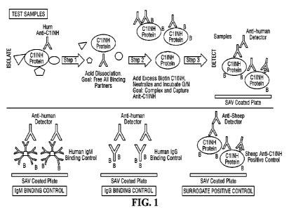
[0017] FIG. 1 is a schematic of the anti-drug antibody assay for Cl Esterase
Inhibitor.
[0018] FIG. 2 illustrates the steps of the anti-drug antibody assay.
[0019] FIG. 3 is a graph depicting screening cut points. The observed ECL
values were
analyzed by two operators over four runs.
[0020] FIG. 4 is a graph depicting confirmatory cut points. The observed
percentage
inhibition was analyzed by two operators over four runs.
[0021] FIG. 5 shows electrochemiluminescence (ECL) values in unspiked sera.
ECL values
are plotted against different test human plasmas.
[0022] FIG. 6 shows electrochemiluminescence (ECL) values in low positive
control (LPG)
(75ng/mL). ECL values are plotted against different test human plasmas.
4
CA 03148161 2022-2-15
WO 2021/046316
PCT/US2020/049352
100231 FIG. 7 shows electrochemiluminescence (ECL) values for high positive
control HPC
(1000 ng/mL). ECL values are plotted against different test human plasmas.
100241 FIG. 8 shows electrochemiluminescence (ECL) values for IgG coat
controls. The
ECL values were observed over twenty-four runs.
100251 FIG. 9 shows electrochemiluminescence (ECL) values for IgM coat
controls. The
ECL values were observed over twenty-four runs.
100261 FIG. 10, shows screening sensitivity and confirmatory sensitivity assay
results.
100271 FIG. 11 summarizes assay results demonstrating screening drug tolerance
and
confirmatory drug tolerance.
DETAILED DESCRIPTION
100281 Unless otherwise defined, all technical and scientific terms used
herein have the same
meaning as commonly understood by one of ordinary skill in the art to which
this disclosure
belongs. Although methods and materials similar or equivalent to those
described herein can
be used in the practice or testing of the present disclosure, suitable methods
and materials are
described herein.
Abbreviations
ADA Anti-Drug Antibody
CHNH Cl Esterase Inhibitor, or Cl-inhibitor
CI Confidence Interval
CCP Confirmation Assay Cutpoint
CF Cut point Factor
CV C Coefficient of variation
df Degrees of freedom
ECL Electrochemiluminescence
Fail
FPER False-Positive Error Rate
hIgG Human IgG
hIgM Human IgM
HPC High positive control
LPC Low positive control
Max Maximum
CA 03148161 2022-2-15
WO 2021/046316
PCT/US2020/049352
Min Minimum
MRD Minimum required dilution
MSD MesoScale Discovery
Number of samples
NA Not applicable
NC Negative control
NF Normalization Factor
Pass
pAb Polyclonal antibody
PC Positive Control
PSCP Plate specific cut point
SCP Screening Assay Cutpoint
shigG Sheep IgG
SD Standard deviation
100291 Immunogenicity of drug products, particularly protein drug products,
leading to
development of ADAs can be a problematic in drug treatment protocols because
of the
potential serious side effects and reduction in drug efficacy resulting from
such
immunogenicity. Development of ADAs can also result in uncertainty in
interpretation of
clinical and pre-clinical data related to toxicity, pharmacokinetic and
pharrnacodynamics.
100301 For a drug found in high circulating concentrations, any circulating
ADAs are
typically bound to the circulating drug (drug interference) making the ADA
unavailable for
detection. Accordingly, development of drug tolerant immunogenicity assays
present
challenges in detection of ADAs. The following disclosure provides an
immunoassay based
on acid dissociation of drug-ADA complexes within a sample; neutralization of
the sample
simultaneous with or closely followed by contact with affinity-labeled drug;
capture of ADA-
affinity labeled drug complexes on a an affinity binding substrate; and
detection of captured
complexes via use of tagged anti-human immunoglobulin secondary antibodies.
100311 The immunoassay comprises the steps of (i) providing a test sample with
an acidic
environment for dissociation of anti-drug antibodies from the drug within a
test sample; (ii)
neutralization of the test sample containing the dissociated drug-ADA
complexes (iii)
incubation of the sample with excess affinity-labeled drug; (iv) capture of
the resulting ADA-
affinity labeled drug complexes on an affinity binding substrate surface; (v)
addition of
tagged anti-human immunoglobulin secondary antibodies that bind to the
captured ADA-
6
CA 03148161 2022-2-15
WO 2021/046316
PCT/US2020/049352
affinity labeled drug complexes; and (vi) quantification of captured antibody-
drug-ADA
complexes.
[0032] In one aspect, the present disclosure relates to an anti-drug antibody
immunoassay for
detection of the presence of anti-CI-Inhibitor antibodies (ClINH-ADA) in a
sample. The
assay comprises the steps of (i) adding an acid solution to a test sample for
dissociation of
C 'MTH-ADA complexes in the test sample; (ii) neutralization of the test
sample containing
the dissociated ClINH-ADA; (iii) incubation of the test sample with excess
affinity labeled
ClINH resulting in formation of ADA- affinity labeled ClINH complexes; (iv)
contact of the
test sample with an affinity functionalized binding substrate surface
resulting in capture of
the ADA- affinity labeled CHNH complexes; (vi) addition of tagged anti-human
immunoglobulin secondary antibodies that bind to the captured ADA-affinity
labeled ClINH
complexes; and quantification of captured ADA-affinity-labeled ClINH
complexes.
[0033] The present disclosure relates more specifically to an anti-drug
antibody
immunoassay for detection of the presence of anti-CI-Inhibitor antibodies
(ClINH-ADA) in
a sample. The assay comprises the steps of (1) adding an add solution to a
test sample for
dissociation of ClINH-ADA complexes in the test sample; (ii) neutralization of
the test
sample containing the dissociated ClINH-ADA; (iii) incubation of the test
sample with
excess biotinylated ClINIT resulting in formation of ADA-biotinylated C BINH
complexes;
(iv) contact of the test sample with a streptavidin functionalized surface
resulting in capture
of the ADA-biotinylated ClINH complexes; (vi) addition of tagged anti-human
secondary
antibodies that bind to the streptavidin captured ADA¨biotinylated CH-NH
complexes; and
quantification of captured antibody-C1INH-ADA complexes.
[0034] The disclosure provides immunoassays for detection of anti-drug
antibodies with
specificity towards a wide variety of different drug products. In certain
aspects, the drug
products are protein-based products that have been developed to treat a wide
variety of
clinical indications, including cancers, autoimmunity/inflammation, exposure
to infectious
agents, and genetic disorders. Such therapeutic proteins include, for example,
antibodies
(including antibody fragments and fusion proteins), coagulation factors,
hormones, growth
factors, cytoldnes, enzymes and plasma proteins to name a few. In addition,
the
immunoassays may be used to detect anti-drug antibodies against nucleic acid-
based drugs
including RNA and DNA based drug products.
[0035] A test sample to be measured refers to a sample possibly containing
drug-ADA
complexes and, for example, is a sample collected from a subject being treated
with the drug.
The term "drug" as used herein refers to a chemical substance, including for
example proteins
7
CA 03148161 2022-2-15
WO 2021/046316
PCT/US2020/049352
or peptides, that are used to treat, cure, prevent, or diagnose a disease or
to promote well-
being in a treated subject. In a specific aspect, the sample may possibly
contain ClINH-ADA
complexes and, for example, is a sample collected from a subject for which
CIINH is being
administered. In other embodiments, the sample is obtained from a subject who
has not
recently been exposed to the drug, e.g., ClINH, or obtained from the subject
prior to the
planned administration of the drug. A subject may be a mammal, for example a
human, with
a disease or suspected of having a disease for which drug treatment is being,
or is to be,
administered. However, in some instances, the term "subject", as used herein,
refers to
laboratory animal of an animal model study.
100361 The term "sample" includes any biological specimen obtained from a
subject. In some
embodiments, the sample is derived from a bodily fluid or body tissue. A test
sample may
comprise a material selected from the group consisting of body fluids, blood,
whole blood,
plasma, serum, mucus secretions, saliva, tears, fine needle aspirate, lymph
fluid or an
immunoglobulin-enriched fraction derived from one or more of these tissues.
One skilled in
the art will appreciate that samples such as serum samples can be diluted
prior to the analysis.
100371 The immunoassay provided herein comprises a step wherein the sample to
be tested is
exposed to an acid solution for dissociation of the drug-ADA complexes found
within the
sample. In particular, a subject's sample can be incubated with an amount of
acid that is
sufficient to provide for the measurement of the presence or level of drug-ADA
complexes.
In a specific embodiment, exposure to an acid solution results in dissociation
of ClINH-ADA
complexes within the sample.
100381 The acid solution to be utilized may be any acid solution that results
in dissociation of
the complexes within the sample. The amount of acid solution to be utilized is
an amount that
provides an acid environment sufficient to result in dissociation of the drug-
ADA complex of
interest and can be determined by one of skill in the art. Typically, such an
acidic
environment is in the pH range of pH 1.0 to pH 5.0, preferably pH 2.0 to pH
3.0, more
preferably pH 2.6. For the assays disclosed herein, the test sample is
contacted with an acid
solution at a concentration of between about 0.1 M to about 1 M, more
preferably 0.3 M. The
acid solution can comprise an organic acid, an inorganic acid, or a mixture
thereof In some
aspects, the acid solution comprises an acid selected from the group
consisting of citric acid,
glutamic acid, acetic acid, glycine/HC1 and any combinations thereof In an
exemplary
embodiment, the add solution comprises acetic acid. In embodiments, the sample
is
contacted with an acid for an amount of time that is sufficient to dissociate
drug-ADA
complexes. Additional methods, well known to those skilled in the art, for
dissociation of
8
CA 03148161 2022-2-15
WO 2021/046316
PCT/US2020/049352
drug-ADA complexes may also be used. For example, dissociation may be achieved
through
application of heat, with or without EDTA.
100391 Following the acidic dissociation, the sample is neutralized and
labeled drug, e.g.
labeled ClINH is added. The step of neutralizing the acid comprises raising
the pH of the
sample to allow the formation of a complex between the labeled drug and ADAs
as described
herein. In some embodiments, the acid is neutralized by the addition of one or
more
neutralizing agents such as, for example, strong bases, weak bases, buffer
solutions, and
combinations thereof. One skilled in the art will appreciate that
neutralization reactions do
not necessarily require a resultant pH of 7 but rather a pH that allows the
formation of labeled
drug/ADA complexes.
100401 In certain aspects, affinity-binding pairs comprising a first member of
a binding pair
and a second member of a binding pair are used for capture of drug-ADA
complexes on an
affinity binding substrate surface. In such affinity binding pairs, the first
member of the
binding pair has binding affinity for the second member of a binding pair.
Such affinity
binding pairs for use in the methods provided herein include, for example,
biotin/streptavidin,
biotin/avidin, biotin/neutravidin, biotin/captavidin, epitope/antibody,
protein
A/immunoglobulin, protein G/immunoglobulin, protein L/immunoglobulin,
GST/glutathione,
His-tag/Nickel, antigen/antibody, FLAG/MI antibody, maltose binding
protein/maltose,
calmodulin binding protein/calmodulin, enzyme-enzyme substrate, and receptor-
ligand
binding pairs. In a specific embodiment, the affinity binding pair comprises a
biotin/streptavidin binding pair.
100411 In one aspect, the excess drug to be added to the test sample is
labeled with a first
member of the binding pair and the binding substrate surface comprises the
cognate second
member of the binding pair. In a specific embodiment, the affinity binding
pair comprises a
biotin/streptavidin binding pair. In such an instance, the drug, e.g., ClINH
is labeled with
biotin and the binding substrate surface comprises streptavidin molecules. A
binding
substrate surface may be a tube, cuvette, microtiter plate, beads or
microparticles. Such
substrates include, but are not limited to, those made of polystyrene,
polycarbonate,
polyvinyltoluene, polypropylene, polyethylene, polyvinyl chloride, nylon,
polymethacrylate,
latex, gelatin, agarose, cellulose, sepharose, glass, metal, ceramic, a
magnetic substance, or
the like.
[0042] In one aspect, the streptavidin surface is a streptavidin coated
microtiter plate. In one
aspect, the binding substrate surface is Meso Scale Discovery (MSD)-Gold
streptavidin-
coated plates.
9
CA 03148161 2022-2-15
WO 2021/046316
PCT/US2020/049352
100431 The immunoassay disclosed herein, comprises the step of detecting
captured ADA-
binding affinity labeled drug complexes. In embodiments, any directly or
indirectly labeled
reagent that binds to the captured ADA-binding affinity labeled drug complexes
may be used.
In an embodiment tagged anti-human antibodies may be used in the practice of
the assay for
detection of ADA-binding affinity labeled drug complexes. The tagged anti-
human
immunoglobulin antibodies include, polyclonal, monoclonal and fragments of
antibodies that
recognize and bind to human antibodies. Such anti-human antibodies include
anti-human
antibodies tagged with a detectable label. In addition, aptamers, such as
oligonucleotide or
peptide molecules that bind to a specific target molecule, may be used..
100441 The detectable label may comprise, for example, a label selected from
the group
consisting of a hapten, radioactive isotope, an enzyme, a fluorescent label, a
chemiluminescent label, and electro-chemiluminescent label. Methods for
coupling detection
reagents such as antibodies, e.g., anti-human antibodies, to detectable labels
are well known
in the art, as are methods for imaging using detectable labels. Such labeled
reagents may
employ a wide variety of labels. Detection of the formation of captured ADA-
binding affinity
labeled drug complexes can be facilitated by attaching a detectable substance
to the detection
reagent, such as an anti-human antibody. Suitable detection means include the
use of labels
such as radionucleotides, enzymes, coenzymes, fluorescers, chemiluminescers,
chromogens,
enzyme substrates or co-factors, enzyme inhibitors, prosthetic group
complexes, free radicals,
particles, dyes, and the like. Examples of suitable enzymes include
horseradish peroxidase,
alkaline phosphatase,f3-galactosidase, or acetylcholinesterase; examples of
suitable prosthetic
group complexes include streptavidin/biotin and avidin/biotin; examples of
suitable
fluorescent materials include umbelliferone, fluorescein, fluorescein
isothiocyanate,
rhodamine, dichlorotriazinylamine fluorescein, dansyl chloride or
phycoerythrin; an example
of a luminescent material is luminol; examples of bioluminescent materials
include
luciferase, luciferin, and aequorin; and examples of suitable radioactive
material include 1251,
131-,
1 35, or 3H. Such labeled reagents may be used in a variety of well-known
assays, such as
radioimmunoassays, enzyme immunoassays, e.g., ELISA, fluorescent immunoassays,
and the
like.
100451 Labeled antibodies can be tagged with such labels by known methods. For
instance,
coupling agents such as aldehydes, carbodiimides, dimaleimide, imidates,
succinimides, bid-
diazotized benzadine and the like are used to tag the antibodies with the
above-described
fluorescent, chemiluminescent, and enzyme labels. An enzyme is typically
combined with an
CA 03148161 2022-2-15
WO 2021/046316
PCT/US2020/049352
antibody using bridging molecules such as carbodiimides, periodate,
diisocyanates,
glutaraldehyde and the like. Various labeling techniques are described in
Morrison, Methods
in Enzymology 32b, 103 (1974), Syvanen et al., I Bid. Chem. 284, 3762 (1973)
and Bolton
and Hunter, Biochem J. 133, 529(1973).
100461 In an embodiment, the anti-human antibodies are labeled with an
electrochemiluminescence moiety. Electrochemiluminescent labels generate light
when
stimulated by electricity in the appropriate chemical environment In one
embodiment, the
detectable label comprises an electrochemiluminescent label comprising a sulfo-
TAG label
and allows for ultra-sensitive detection. In such an instance, the sulfo-TAG
labeled antibodies
are used in conjunction with a binding substrate surface comprising Meso Scale
Discovery
(MSD)-Gold streptavidin-coated plates. Electricity is applied to the plate
electrodes by
an MSD instrument leading to light emission by SULFO-TAG labels. Light
intensity is then
measured to quantify ADAs present in the test sample.
100471 Methodologies and techniques for performing the above ADA assays, such
as assay
conditions, assay buffers, washing steps, solid supports, suitable tags/labels
and methods for
linking them to the detection agent, techniques for detecting/measuring the
detectable label,
and equipment for performing the assays are known to those skilled in the art.
100481 The present disclosure further provides kits that are assembled for
determining the
presence or absence of ADAs in a test sample. The kits may comprise control
samples and/or
instructions and, in a container, reagents including (i) for contacting the
sample with an acid
solution; (ii) for neutralization of the sample. The kit will further comprise
one or more of the
following reagents (i) a binding affinity labeled drug; (ii) an affinity
binding substrate for
capture of ADA-binding affinity labeled drug complexes; and tagged anti-human
antibodies.
EXAMPLE
100491 Persons skilled in the art will understand that the structures and
methods specifically
described herein and shown in the accompanying figures are non-limiting
exemplary
embodiments, and that the description, disclosure, and figures should be
construed merely as
exemplary of particular embodiments. It is to be understood, therefore, that
this disclosure is
not limited to the precise embodiments described, and that various other
changes and
modifications may be effected by one skilled in the art without departing from
the scope or
spirit of this disclosure. Additionally, the elements and features shown or
described in
connection with certain embodiments may be combined with the elements and
features of
certain other embodiments without departing from the scope of this disclosure,
and that such
11
CA 03148161 2022-2-15
WO 2021/046316
PCT/US2020/049352
modifications and variations are included within the scope of this disclosure.
Accordingly,
the subject matter of this disclosure is not limited by what has been
particularly shown and
described.
100501 Hereditary angioedema (HAE) is a rare disorder that leads to swelling
consequent to
excess bradykinin generation. When this occurs in the airways, attacks can be
life-
threatening. Excess bradykinin generation results from a deficiency of Cl-
Inhibitor
(ClINH), a serpin important in control of plasma serine proteases that release
bradykinin
from high molecular weight kininogen. Clinical trials of several protein
replacement
therapeutics have reported low incidence of anti-drug antibodies (ADA) to
ClINH in
subjects; however, the drug tolerance of these assays has not been described.
High levels of
soluble analyte are known for interference in ADA assays; normal serum
concentrations of
range from 180-200 lig/mL (1.7-2 M). Accordingly, there is a need to develop
and
validate a well-controlled and sensitive assay for ClINTH immunogenicity.
Described below
is a highly drug-tolerant assay for ADA to C11NH in human serum.
100511 Specifically, a dual secondary antibody-based ADA assay (based on
Affinity Capture
Elution without the need for two solid phases) for human anti-ClINH antibody
is disclosed.
As described in detail below, ClINH-ADA complexes in undiluted serum samples
were acid-
dissociated. Released antibody was neutralized and incubated with excess
biotinylated
ClINH. The resulting ADA-biotinylated ClINH complexes were captured on Meso
Scale
Discovery (MSD)-Gold streptavidin-coated plates. Sulfo-tagged anti- human
(h)IgG was
allowed to bind to captured complexes and quantitated using standard MSD
protocols.
Sample dilution after acidification and neutralization was 20-fold. Since
human anti-hClINH
is unavailable, sheep anti-hC HMI (PC) served as surrogate positive control.
Detector
reagent concentrations (analyte-specific anti-hIgG/IgM and PC-specific anti-
sheep IgG) were
optimized to ensure equivalent sensitivities using control wells incubated
with biotinylated
hIgG or hIgIVI.
100521 A dual secondary antibody-based ADA assay (based on Affinity Capture
Elution
without the need for two solid phases) for human anti-ClINH antibody was
developed (see,
FIG. 1 and FIG. 2) ClINH-ADA complexes in undiluted plasma samples were acid-
dissociated. Released antibody was neutralized and incubated with excess
biotinylated
ClINH. The resulting ADA-biotinylated ClINH complexes were captured on Meso
Scale
Discovery (MSD)-Gold streptavi din-coated plates. Sulfo-tagged anti- human (h)
IgG was
allowed to bind to captured complexes and quantitated using standard MSD
protocols.
Sample dilution after acidification and neutralization was 20-fold. Since
human anti-hCllNH
12
CA 03148161 2022-2-15
WO 2021/046316
PCT/US2020/049352
is unavailable, sheep anti-hC IINH (PC) served as surrogate positive control.
Detector
reagent concentrations (analyte-specific anti-hIgG/IgM and PC-specific anti-
sheep IgG) were
optimized to ensure equivalent sensitivities using control wells incubated
with biotinylated
hIgG or hIg114. Specific details of the utilized materials and methods are
described below.
[0053] Samples and QC were removed from the freezer and placed at RT to thaw.
A 0.742
mg/mL stock of biotinylated C1-INH was diluted to 1.0 pg/mL in neutralization
buffer (30%
1M Tris HCL pII 9.5 in blocker Casein in TBS) ("labeled neutralization
buffer"). 5.0 pL of
each sample and QC were transferred to a dilution plate. 45.0 pL 300 mM acetic
acid was
added to each well of the dilution plate containing a sample or QC. Next,
samples were
incubated for 10 minutes at 25+2 C on setting 2 of Labline Titre Plate Shaker
or 100 rpm to
allow C1-1NH-ADA dissociation. 50.0 ill labeled neutralization buffer was then
added to
each well of the dilution plate containing the acidified sample and QC.
Incubation was done
for 16-20 hours at 25+2 C on setting 2 of Labline Titre Plate Shaker or 100
rpm.
[0054] MSD SAY plates and buffers were placed at RT for at least 15 minutes
prior to use.
Blocking of MSD plates was done as follows: 150 pl of blocker casein in TBS
was added to
each well and incubation was for 1-hour th 10 minutes on 25+2 C on (setting
0).
[0055] Preparation of Human Detection Controls (volume for two plates) was as
follows. To
prepare a 10.0 pg/ml intermediate solution, a 1.00 mg/mL stock Biotin-Tagged-
Human IgG
was diluted to 10.0 pg/mL in Blocker Casein in TBS. The 10.0 Wm1 intermediate
biotin-
tagged-human IgG was diluted to the working concentration of 100 ng/mL in
Blocker Casein
in TBS. To prepare a 10.0 pig/ml intermediate solution, a 1.00 mg/mL stock
Biotin-Tagged-
Human IgM was diluted to 10.0 ug/mL in Blocker Casein in TBS. The 10.0 pg/ml
intermediate biotin-tagged-human IgM was diluted to the working concentration
of 100
ng/mL in Blocker Casein in TBS.
[0056] The blocked assay plate was washed 3X, on a plate washer, with ELISA
Wash Buffer
(0.05% Tween 20 in 1X PBS) by adding 300pL of buffer to each well. The blocked
assay
plate was inverted and tapped on absorbent paper after the final wash.
Neutralized samples
and QC were removed from the shaker.
100571 Sample Step was as follows. 25.0pL of each neutralized QC and/or
sample,
100ng/mL Biotin tagged IgG and Biotin tagged IgM were transferred to the
respective wells
of assay plate per plate map was done. Incubation was done for 1-hour th10
minutes on
25+2 C Jitterbug with shaking (setting 0).
[0058] Detection Preparation was as follows. The 500 pg/mL stock Sulfo-tagged-
anti-sheep
AB detection antibody was diluted to 500 ng/mL in Blocker Casein in TBS. The
1.62 mg/mL
13
CA 03148161 2022-2-15
WO 2021/046316
PCT/US2020/049352
stock Sulfo-tagged-Fab anti-hu-IgG+IgM detection antibody was diluted to 16.2
pig/rnL. The
16.2 pg/mL Intermediate sulfo-tag-Fab-anti-hu-IgG+IgM detection antibody was
diluted to
162 ng/mL in Blocker Casein TBS. The 162 ng/mL Intermediate sulfo-tag-Fab-anti-
hu-IgG-F
IgNI detection antibody was diluted to 1.0 ng/mL in Blocker Casein TBS.
100591 The assay plate was washed 3X on a plate washer with ELISA Wash Buffer
by
adding 300 p.I., of buffer to each well. The plate was then inverted and
tapped on absorbent
paper after the final wash.
100601 The detection step was as follows. 50.0 pL of each detection antibody
was added to
the respective wells per plate map. Incubation was carried out for 1 hour 10
minutes on 25
2 C Jitterbug with shaking (setting 0).
100611 The assay plate was then washed 3X on a plate washer with ELISA Wash
Buffer by
adding 300pL of buffer to each well. The plate was inverted and tapped on
absorbent paper
after the final wash. The stop step was performed by addition of 150 pi of MSD
Read Buffer
to each well. The plate was then read.
100621 The Confirmation Assay procedure was conducted as follows: C1-INH
(Stock at
0.5mg/mL) was diluted to 80.0 pg/mL in labeled Neutralization Buffer. 50 1_,
of drug spiked
labeled neutralization buffer was added per well to dilution plate containing
acid treated
samples according to plate map. Incubation was conducted 16-20 hours at 25+2'
on setting 2
of LablineTitre Plate Shaker or 100 rpm. The screening assay was then
performed as
described above.
100631 The cut point determination was conducted using normal plasma samples
obtained
from 30 individual male and female humans. Each of the samples was mm at the
MRD of
1/20 (unspiked) and at the MRD in the presence of 80 pg/mL human ClINH
(spiked). Two
operators each ran all 30 samples spiked and unspiked, in two independent
runs, for a total of
four runs.
100641 Uncorrected ECL values from unspiked samples were used to define the
plate-specific
cut point factor. Percent inhibition by spiked drug was calculated for each
sample and used
to define the % inhibition cut point. To identify the statistical outliers,
the "outlier box-plot
criteria" was used. More precisely, all samples in an individual run above Q3
+ 1.5*(Q3-Q1)
or below Q1 - 1.5*(Q3-Q1), where Q3 and Q1 represented respectively the 75th
and 25th
percentiles, were considered as Analytical outliers. Following removal of
analytical outliers,
all samples above Q3 + 1.5*(Q3-Q1) or below Q1 - 1.5*(Q3-Q1), where Q3 and Q1
represented respectively the 75th and 25th percentiles, on averaged (mean)
data, were
14
CA 03148161 2022-2-15
WO 2021/046316
PCT/US2020/049352
considered as Biological outliers and all such were removed from the data. The
outliers were
evaluated using ECL values for CP and percentage inhibitions for CCP_
[0065] Following elimination of the outliers (analytical and biological) and
prior to the
evaluation of the CP, the distribution normality of the original data was
tested via Shapiro-
Wilk's test to decide which scale (original or log) to use for the CP
determination. For the
screen and titer assays, the result of this test was found to be significant
at the 10% level
(Shapiro-Wilk W value is greater than the P value). Therefore, the
nonpaninetlic method
was used on the log-transformed data and the screening cut point was
determined as the
antilog of the 95th percentile value. The screening cut point factor (NF) of
1.457 was
determined as the average (CPrun / Median NCrun) from the four runs (FIG. 3)
[0066] The data with outliers removed was used for the calculation of the CCP
and was
evaluated by Shapiro-Wilk' s test for each run and was evaluated using die
ratio of (s/us)
where "s" denotes spiked and "us" denotes unspiked values. If the result of
this test was not
found to be significant at the 10% level and the distribution was found to be
symmetrical,
then the parametric method was used to calculate the CCP. More precisely, the
CCP was
defined as mean (percentage inhibition) + 3.09*SD (percentage inhibition).
This is the case
for runs 1 and 3. Runs 2 and 4 were found to be non-normally distributed and
therefore the
nonparametfic method was used on the log-transformed data and the confirmatory
cut point
was determined as the antilog of the 99.9th percentile value. The average cut
point from the
four runs is 34.0% Inhibition. (FIG. 4)
[0067] Precision evaluation (% CV) of the ECL values obtained in the screening
assay for the
HPC (1000 ng/mL) and LPC (75ng/mL) were <20% CV (20.3 and 15.8%, respectively)
over
6 runs by two analysts. (FIG. 6 and FIG. 7) To assess the precision of the
assay at lower
ranges, a second study was performed with 11:PC (1000 ng/mL). LPC (75 ng/mL),
LPC1 (50
ng/mL), LPC2 (25 ng/mL) and LPC3 (123 ng/mL). The data demonstrate precision
of <
25% CV for all levels of PC and for the NC.
[0068] Precision of the human IgG and human IgM coating controls ECL signal
was also
<20%. (FIG. 8 and FIG. 9)
[0069] Precision of the % Inhibition values calculated for the HPC and LPC in
six runs by
two analysts was 1.5 and 7.7% CV, respectively. An additional precision study
with lower
PC concentrations also maintained precision of <20% CV.
[0070] The sensitivity of the screening assay was determined using the
surrogate positive
control, sheep anti-C IINH pAb, spiked into normal human plasma at
concentrations ranging
from 312.5-2.4 ng/mL (before application of the MRD of 20) prepared four
independent
CA 03148161 2022-2-15
WO 2021/046316
PCT/US2020/049352
times and analyzed in two runs each by two different analysts. The
concentration associated
with the cut point was determined and the sensitivity defined as the 95th C.I.
The data indicate
the method sensitivity is 8.8 ng/mL. (FIG. 10)
100711 The sensitivity of the confirmatory assay was determined using the
surrogate positive
control, sheep anti-ClINH pAb, spiked into normal human plasma at
concentrations ranging
from 31.3-0.24 ng/mL (before application of the MRD of 20) prepared four
independent
times and analyzed in two runs each by two different analysts. The
concentration associated
with the cut point was determined and the sensitivity defined as the 95th C.I.
The data indicate
the method sensitivity is 9.3 ng/mL (FIG.10).
100721 It is important to note that due to potential affinity and avidity
differences between the
pAb used in this study and those of actual subjects, the antibodies used for
this study do not
fully represent the antibody repertoire of preclinical or clinical samples,
and therefore the
actual sensitivity value may vary for preclinical or clinical samples.
100731 The screening assay method was demonstrated to be selective to the
detection of low
(75 ng/mL) and high (1000 ng/mL) levels of anti-drug (ClINH) antibodies. In
this
evaluation, individual normal plasma samples (n=12) spiked with polyclonal
anti-hClINH
antibody scored positive in the screening assay and confirmed positive in the
confirmatory
assay. Unspiked controls scored negative. (FIG.5)
[0074] The ability of the assay to detect anti-hC1INH antibodies when hC11NH
is present
(drug tolerance) was evaluated by spiking plasma samples containing known
concentrations
of anti-hClINH (HPC and LPC) with hClINH (1000 ng/mL to 15.6 ng/mL). Drug
tolerance
is defined as the greatest amount of hClINH present in a sample that still
allows the sample
to score positive for antibody. The screening assay is tolerant to 417 pg/mL
of CHNH at
75 ng/mL level of antibody. The confirmatory assay is tolerant to 250 pg/mL of
ClINH at
the 75 ng/mL level of antibody. As expected, increasing levels of anti-drug
antibody show
increased tolerance to circulating ClINH. FIG. 11 demonstrates the conclusions
determined
for the disclosed qualified clINH ADA assay. Below is a table representing the
qualification
parameters.
100751 The qualified method complies with the 2019 FDA Guidance-required
screening
sensitivity of <100 ng/mL ADA in the presence of normal plasma levels of C
1INH with
acceptable intra- and inter-assay precision. The assay is therefore acceptably
drug-tolerant
and has acceptable precision and selectivity.
16
CA 03148161 2022-2-15
WO 2021/046316
PCT/US2020/049352
Parameter Result
PC Precision (% CV) = LPC (75
ng/mL): 16% w/o Drug
22% with 80,000 ng/mL Dmg
= I-1PC (1000 ng/mL): 20% w/o Drug
25% with 80,000 ng/mL Drug
Sensitivity (LOD) - Screening
assay: 8.8 ng/mL
(surrogate PC sheep arhClINH) = Confirmation
assay: 9.3 ng/mL
Selectivity in Human Serum - Unspiked
matrix lots: 100% negative
(12 Matrix Lots) = HPC and LPC
matrix lots: 100% positive
- SCP Factor -
1.457 (non-parametric, 5% FPER)
= CCP = 34%
Inhibition (0.1% FPER)
Drug Tolerance for hClINH at LPC = Screening assay: 417 gg/mL
= Confirmatory assay: 250 ttg/mL
IgG and IgM Binding Controls = All >PSCP
17
CA 03148161 2022-2-15