Note: Descriptions are shown in the official language in which they were submitted.
CA 03148219 2022-01-20
WO 2021/016641
PCT/US2020/070312
SYSTEMS AND METHODS FOR PERFORMING
TRANS-ABDOMINAL FETAL OXIMETRY OR PULSE OXIMETRY
Related Applications
[0001] This application is an INTERNATIONAL PCT application of U.S.
Provisional
Patent Application Number 62/878,243 filed on July 24, 2019 entitled "SYSTEMS,
DEVICES, AND METHODS FOR PERFORMING TRANS-ABDOMINAL FETAL
OXIMETRY AND/OR TRANS-ABDOMINAL FETAL PULSE OXIMETRY USING
PHYSIOLOGICAL CHARACTERISITICS AND/OR A CALIBRATION FACTOR" and
U.S. Provisional Patent Application Number 62/971,152 filed on February 6,2020
entitled "SYSTEMS, DEVICES, AND METHODS FOR PERFORMING FETAL
OXIMETRY AND/OR FETAL PULSE OXIMETRY USING FETAL DEPTH AND/OR A
MATERNAL HEMOGLOBIN OXYGEN SATURATION LEVEL" all of which are
hereby incorporated, in their entireties, herein.
Field of Invention
[0002] The present invention is in the field of medical devices and, more
particularly,
in the field of trans-abdominal fetal oximetry and trans-abdominal fetal pulse
oximetry.
Backdround
[0003] Oximetry is a method for determining the oxygen saturation of
hemoglobin in
a mammal's blood. Typically, 90% (or higher) of an adult human's hemoglobin is
saturated with (i.e., bound to) oxygen while only 30-60% of a fetus's blood is
saturated with oxygen. Pulse oximetry is a type of oximetry that uses changes
in
blood volume through a heartbeat cycle to internally calibrate hemoglobin
oxygen
saturation measurements of the arterial blood.
[0004] Current methods of monitoring fetal health, such as monitoring fetal
heart
rate, are inefficient at determining levels of fetal distress and, at times,
provide false
positive results indicating fetal distress that may result in the unnecessary
performance of a Cesarean delivery.
Summary
[0005] Systems, devices, and methods for performing trans-abdominal fetal
oximetry
and/or trans-abdominal fetal pulse oximetry using physiological
characteristics
1
CA 03148219 2022-01-20
WO 2021/016641
PCT/US2020/070312
and/or a calibration factor are herein disclosed. In some embodiments, a
physiological characteristic of a pregnant mammal may be received by, for
example,
a computer or processor. An impact of the physiological characteristic on a
behavior
of an optical signal projected into the abdomen of the pregnant mammal may
then be
determined. Exemplary impacts include absorption and scattering of the optical
signal. A calibration factor for the optical signal may then be determined
responsively to the determined impact of the physiological characteristic. In
some
cases, determining a calibration factor may include querying a database using
the
physiological characteristic for a corresponding calibration factor. The
determined
calibration factor may then be stored in a database.
[0006] On some occasions, the processor may further receive a composite
detected
electronic signal from a detector communicatively coupled to the processor.
The
composite electronic signal may correspond to an optical signal emitted from
the
pregnant mammal's abdomen and a fetus contained therein that has been detected
by the detector and converted into the composite detected electronic signal.
The
emitted optical signal may be a portion of light projected into the pregnant
mammal's
abdomen and onto the fetus contained therein. A fetal signal may then be
generated
by isolating a portion of the composite detected electronic signal that
corresponds to
light that was incident upon the fetus. Isolation of the fetal signal from a
composite
(maternal and fetal signal) may be accomplished a number of ways including,
but not
limited to, filtering via, for example, bandpass or Kalman filters,
amplification, and/or
processing using one or more input signals such as fetal heart rate, maternal
heart
rate, maternal pulse oxygenation values, and/or maternal respiratory values to
remove a portion of the composite signal contributed by the pregnant mammal
and/or amplify a portion of the composite signal contributed by the fetus.
Some of
these techniques may also be used remove noise (e.g., ambient light,
harmonics,
etc.) from the composite and/or fetal signal. The calibration factor may then
be
applied to the fetal signal to generate a calibrated fetal signal and the
calibrated fetal
signal may be processed to determine a fetal hemoglobin oxygen saturation
level for
the fetus. The fetal hemoglobin oxygen saturation level may then be
communicated
to a user such as a doctor, midwife, or nurse.
[0007] In some embodiments, an indication of whether the fetal signal
corresponds to
pre-ductal or post-ductal blood may be received by the processor. Often times,
this
indication is input by a clinician based on a location of the detector
detecting the
2
CA 03148219 2022-01-20
WO 2021/016641
PCT/US2020/070312
composite signal on the pregnant mammal's abdomen that corresponds to a
location
on the fetus (e.g., head, thorax, or limb) from which the composite signal is
generated. This indication may later be provided or displayed to a user along
with
the fetal hemoglobin oxygen saturation level so that the user may determine
whether
the fetal hemoglobin oxygen saturation level is dangerously low for the fetus.
[0008] In some embodiments, a maternal detected electronic signal may be
received
from a detector communicatively coupled to the processor. The maternal
detected
electronic signal may correspond to an optical signal emitted from the
pregnant
mammal's abdomen (that has not traveled deep enough into the abdomen to reach
to the fetus) that has been detected by the detector and converted into the
maternal
detected electronic signal. In some embodiments, the maternal detected
electronic
signal may be a short separation signal that only passes through maternal
tissue.
The emitted optical signal may be a portion of light projected, by a light
source, into
the pregnant mammal's abdomen. Then, the maternal detected electronic signal
may be analyzed to determine the physiological characteristic of the pregnant
mammal. The determined physiological characteristic and/or the calibration
factor
for the pregnant mammal may be stored in a database.
[0009] The received physiological characteristic may be intrinsic or extrinsic
and may
be, for example, the pregnant mammal's age, the pregnant mammal's weight, and
the pregnant mammal's body mass index. At times, the physiological
characteristic
is received from a clinician based on his or her observations, an ultra-sound
device,
a Doppler device, an image of the pregnant mammal's abdomen, a Fitzpatrick
scale
reading, manually-operated calipers, a blood measurement device, an oximeter,
a
pulse oximeter, and/or a scale.
[00010] In one embodiment, the received physiological characteristic is a
skin
color, or melanin concentration, of the pregnant mammal and the determination
of
the impact of the physiological characteristic on the behavior of the optical
signal
may include determining how much of the optical signal is absorbed by the
pregnant
mammal's melanin/skin color.
[00011] Additionally, or alternatively, the received physiological
characteristic
may be a thickness of a muscle layer in the pregnant mammal's abdomen. When
this is the case, the determination of the impact of the physiological
characteristic on
the behavior of the optical signal may include determining how much of the
optical
signal is absorbed by the muscle layer in the pregnant mammal's abdomen.
3
CA 03148219 2022-01-20
WO 2021/016641
PCT/US2020/070312
[00012] Additionally, or alternatively the received physiological
characteristic is
a thickness of an adipose layer in the pregnant mammal's abdomen, further
wherein
the determination of the impact of the physiological characteristic on the
behavior of
the optical signal includes determining how much of the optical signal is
scattered by
the adipose layer in the pregnant mammal's abdomen.
[00013] Additionally, or alternatively, the received physiological
characteristic
may be a body mass index for the pregnant mammal and the determination of the
impact of the physiological characteristic on the behavior of the optical
signal may
include determining how much of the optical signal is scattered or absorbed by
the
pregnant mammal's abdomen due to her body mass index.
[00014] Additionally, or alternatively, the received physiological
characteristic
may be a thickness of the pregnant mammal's abdomen (also referred to herein
as
fetal depth). In this case, the determination of the impact of the
physiological
characteristic on the behavior of the optical signal may include determining
how
much of the optical signal is absorbed by the pregnant mammal's
abdomen/abdominal tissue.
[00015] Additionally, or alternatively, the received physiological
characteristic
may be a thickness of the pregnant mammal's abdomen and the determination of
the
impact of the physiological characteristic on the behavior of the optical
signal may
include determining how much of the optical signal is scattered by the
pregnant
mammal's abdomen.
[00016] Additionally, or alternatively, the received physiological
characteristic
may include a hemoglobin concentration of the pregnant mammal's blood. In
these
situations, the determination of the impact of the physiological
characteristic on the
behavior of the optical signal may include determining how much of the optical
signal
is absorbed by the pregnant mammal's hemoglobin.
[00017] Additionally, or alternatively, the received physiological
characteristic
may be a hemoglobin oxygen saturation of the pregnant mammal's blood the
determination of the impact of the physiological characteristic on the
behavior of the
optical signal may include determining how much of the optical signal is
absorbed by
the pregnant mammal's oxygenated and/or deoxygenated hemoglobin.
[00018] In another embodiment, a maternal detected electronic signal may
be
received from a detector communicatively coupled to a processor, the maternal
detected electronic signal may correspond to an optical signal emitted from
the
4
CA 03148219 2022-01-20
WO 2021/016641
PCT/US2020/070312
pregnant mammal's abdomen that has been detected by the detector and converted
into the maternal detected electronic signal. The emitted optical signal may
be a
portion of light projected (by a light source) into the pregnant mammal's
abdomen.
The maternal detected electronic signal may then be analyzed to determine a
physiological characteristic of the pregnant mammal. A calibration factor for
the
optical signal emanating from the pregnant mammal may then be determined
responsively to the analysis. In some embodiments, the physiological
characteristic
of the pregnant mammal may be associated with the calibration factor and this
association may be stored in a database.
[00019] In some instances, a composite detected electronic signal may be
received from a detector communicatively coupled to the processor. The
composite
detected electronic signal may correspond to an optical signal emitted from
the
pregnant mammal's abdomen and a fetus contained therein that has been detected
by the detector and converted into the composite detected electronic signal.
The
emitted optical signal may be a portion of light projected, by a light source,
into the
pregnant mammal's abdomen and onto the fetus contained therein. A fetal signal
may then be generated by isolating a portion of the composite detected
electronic
signal that corresponds to light that was incident upon the fetus. A
calibrated fetal
signal may be generated by applying the calibration factor to the fetal
signal. Then,
a fetal hemoglobin oxygen saturation level may be determined using the
calibrated
fetal signal and the fetal hemoglobin oxygen saturation may be communicated to
a
user via, for example, displaying the fetal hemoglobin oxygen saturation on a
display
device.
[00020] In some embodiments, determining the calibration factor for the
optical
signal responsively to the impact includes querying a database for a
calibration
factor that corresponds to the physiological characteristic and receiving the
queried-
for calibration factor from the database.
[00021] In some instances, an indication of whether the fetal signal
corresponds to pre-ductal or post-ductal blood may be received from, for
example, a
clinician or doctor and this indication may be provided along with the fetal
hemoglobin oxygen saturation level to the user.
[00022] On some occasions, a maternal detected electronic signal may be
received from a detector communicatively coupled to the processor. The
maternal
detected electronic signal may correspond to an optical signal emitted from
the
CA 03148219 2022-01-20
WO 2021/016641
PCT/US2020/070312
pregnant mammal's abdomen that has been detected by the detector and converted
into the maternal detected electronic signal that has not passed through, or
been
incident upon, the fetus. Thus, it is an optical signal that only passes
through the
maternal abdomen and does not penetrate far enough into the abdomen to be
incident on the fetus. The maternal detected electronic signal may then be
analyzed
and/or processed, and the physiological characteristic of the pregnant mammal
may
be determined responsively to the analysis.
[00023] In some cases, the determined physiological characteristic is a
skin
color of the pregnant mammal and the calibration factor may pertain to how
much of
the optical signal is absorbed by the pregnant mammal's skin color.
Additionally, or
alternatively, the determined physiological characteristic may be a thickness
of a
muscle layer in the pregnant mammal's abdomen and the calibration factor may
pertain to how much of the optical signal is absorbed by the muscle layer in
the
pregnant mammal's abdomen.
[00024] Additionally, or alternatively, the determined physiological
characteristic
may be a thickness of an adipose layer in the pregnant mammal's abdomen and
the
calibration factor may pertain to how much of the optical signal may be
scattered by
the adipose layer in the pregnant mammal's abdomen. Additionally, or
alternatively,
the determined physiological characteristic may be a thickness of the pregnant
mammal's abdomen and the calibration factor may pertain to how much of the
optical signal may be absorbed by the pregnant mammal's by the pregnant
mammal's abdomen. Additionally, or alternatively, the determined physiological
characteristic may be a thickness of the pregnant mammal's abdomen and the
calibration factor may pertain to how much of the optical signal is scattered
by the
pregnant mammal's abdomen. Additionally, or alternatively, the determined
physiological characteristic may be a hemoglobin concentration of the pregnant
mammal's blood and the calibration factor may pertain to how much of the
optical
signal is absorbed by the pregnant mammal's hemoglobin. Additionally, or
alternatively, the determined physiological characteristic may be a hemoglobin
oxygen saturation of the pregnant mammal's blood and the calibration factor
may
pertain to how much of the optical signal is absorbed by the pregnant mammal's
oxygenated and deoxygenated hemoglobin.
6
CA 03148219 2022-01-20
WO 2021/016641
PCT/US2020/070312
Brief Description of the Fiqures
[00025] FIG. 1A is a block diagram illustrating an exemplary system for
determining a level of oxygen saturation for fetal hemoglobin and/or whether
meconium is present in the amniotic fluid of a pregnant mammal, consistent
with
some embodiments of the present invention;
[00026] FIG. 1B is a block diagram of an exemplary processor-based system
that may store data and/or execute instructions for the processes disclosed
herein,
consistent with some embodiments of the present invention;
[00027] FIG. 2A is a block diagram illustrating an exemplary fetal probe,
consistent with some embodiments of the present invention;
[00028] FIG. 2B is a block diagram illustrating another exemplary fetal
probe,
consistent with some embodiments of the present invention;
[00029] FIG. 3A provides an illustration of exemplary dimensions for
layers of
tissue within two different maternal abdomens with their respective fetuses,
consistent with some embodiments of the present invention;
[00030] FIG. 3B provides an illustration of exemplary dimensions for
layers of
tissue within two different maternal abdomens with their respective fetuses,
consistent with some embodiments of the present invention;
[00031] FIG. 30 provides a midsagittal plane view of pregnant mammal's
abdomen with fetal hemoglobin probe positioned thereon, consistent with some
embodiments of the present invention;
[00032] FIG. 4A illustrates an exemplary fetal hemoglobin probe in contact
with
a pregnant mammal's abdomen showing the different layers of maternal abdominal
tissue, consistent with some embodiments of the present invention;
[00033] FIG. 4B illustrates another exemplary fetal hemoglobin probe in
contact
with a pregnant mammal's abdomen, consistent with some embodiments of the
present invention;
[00034] FIG. 40 illustrates an exemplary fetal probe configured to detect
two
short separation signals and one long separation signal in contact with a
pregnant
mammal's abdomen where the layers of the maternal abdomen are depicted as a
single layer, consistent with some embodiments of the present invention;
[00035] FIG. 4D illustrates an exemplary fetal probe configured to detect
two
short separation signals and one long separation signal in contact with a
pregnant
7
CA 03148219 2022-01-20
WO 2021/016641
PCT/US2020/070312
mammal's abdomen where some of the layers of the maternal abdomen are shown,
consistent with some embodiments of the present invention;
[00036] FIG. 5 provides a flowchart illustrating a process for determining
a fetal
hemoglobin oxygen saturation level, consistent with some embodiments of the
present invention;
[00037] FIG. 6 provides a flowchart illustrating a process for determining
a
physiological characteristic of a pregnant mammal using a received optical
signal
consistent with some embodiments of the present invention;
[00038] FIG.7A is a flowchart illustrating an exemplary process for
determining
a fetal depth and/or a fetal hemoglobin oxygen saturation level, in accordance
with
some embodiments of the present invention;
[00039] FIG.7B provides a flowchart illustrating an exemplary process for
determining a fetal depth, in accordance with some embodiments of the present
invention;
[00040] FIG.70 provides a graph showing a scatter plot of a change in
percent
transmission of light for lst_Nth fetal signals as a function of
source/detector distance,
in accordance with some embodiments of the present invention;
[00041] FIG.7D provides a graph showing a scatter plot of a change in
percent
transmission of light for 1st-Nth maternal signals as a function of
source/detector
distance, in accordance with some embodiments of the present invention;
[00042] FIG. 8 is a flowchart illustrating an exemplary process for
determining
a fetal depth and/or a fetal hemoglobin oxygen saturation level, in accordance
with
some embodiments of the present invention;
[00043] FIG. 9 is a flowchart illustrating an exemplary process for
determining a
fetal depth and/or a fetal hemoglobin oxygen saturation level, in accordance
with
some embodiments of the present invention;
[00044] FIG. 10 provides a flowchart illustrating a process for
determining a
fetal hemoglobin oxygenation saturation level using physiological
characteristics of
the pregnant mammal determined using one or more maternal detected electronic
signal(s), in accordance with some embodiments of the present invention;
[00045] FIG. 11 provides a flowchart illustrating a process 1100 for
determining
an influence of a physiological characteristic on the behavior of light
traversing
through the abdomen of a pregnant mammal and/or her fetus, in accordance with
some embodiments of the present invention;
8
CA 03148219 2022-01-20
WO 2021/016641
PCT/US2020/070312
[00046] FIG. 12 is a flowchart illustrating an exemplary process for
determining
a fetal hemoglobin oxygen saturation level using a maternal hemoglobin oxygen
saturation level and/or a fetal depth, in accordance with some embodiments of
the
present invention;
[00047] FIG. 13 provides a flowchart illustrating a process for
determining a
fetal hemoglobin oxygenation saturation level using calibration factor and/or
physiological characteristic of the pregnant mammal and/or fetus, consistent
with
some embodiments of the present invention;
[00048] FIG. 14 provides a flowchart illustrating a process for
determining a
fetal hemoglobin oxygenation saturation level, consistent with some
embodiments of
the present invention;
[00049] FIG. 15A provides a flowchart illustrating a first part of a
process for
determining a composite fetal hemoglobin oxygenation saturation level,
consistent
with some embodiments of the present invention; and
[00050] FIG. 15B provides a flowchart illustrating a second part of a
process for
determining a composite fetal hemoglobin oxygenation saturation level,
consistent
with some embodiments of the present invention.
WRITTEN DESCRIPTION
[00051] Behavior of light projected into the abdomen of a pregnant mammal
may be impacted (e.g., absorbed and/or scattered) by the abdominal tissue of
the
pregnant mammal. This may impact how much light incident on the maternal
abdomen is incident on a fetus within the pregnant mammal's abdomen and/or a
clarity of a signal received from the maternal abdomen and/or a signal that
was
incident on the fetus. Knowing how much light is incident on a fetus may be
important for various reasons. For example, a value for the intensity of light
incident
on a fetus and/or a percent transmission of light through the pregnant
mammal's
abdomen may be used to calculate fetal hemoglobin oxygen saturation using the
oximetry calculations and/or the Beer-Lambert Law. Also, understanding the
behavior (absorption and/or scattering, which may also be referred to herein
as
absorption coefficients, or (MA)), and/or scattering coefficients (N(A)) for
different
wavelengths of light) of light projected into a pregnant mammal's abdomen may
be
used to determine, for example, an impact of the pregnant mammal's abdominal
9
CA 03148219 2022-01-20
WO 2021/016641
PCT/US2020/070312
tissue's interaction with light traveling through from her abdomen may lead to
greater
accuracy when calculating fetal hemoglobin oxygen saturation.
[00052] How much light reaches a fetus is often times not linearly related
to
how much light is projected into the pregnant mammal's abdomen. Each pregnant
mammal and fetus combination is different in terms of the geometry of their
respective tissue layers and/or intrinsic characteristics such as hemoglobin
oxygen
saturation and/or blood profusion through tissue, which makes approximations
for
how much light reaches a fetus or other one size fits all calculations or
corrections
for the calculation of fetal hemoglobin oxygen saturation often times
inaccurate.
Thus, calibrating calculations using physiological characteristics of a
pregnant
mammal and/or pregnant mammal/fetus combination may assist with more
accurately calculating fetal hemoglobin oxygen saturation.
[00053] Transabdominal fetal oximetry and/or fetal pulse oximetry is often
performed using near infrared (NIR) light. NIR light projected into a pregnant
mammal's abdomen may be absorbed by, for example, the melanin in the pregnant
mammal's skin, the pregnant mammal's myoglobin (muscle) tissue, and the
hemoglobin in the pregnant mammal's blood, deoxygenated hemoglobin absorbs
more light than oxygenated hemoglobin. Thus, knowing the how much melanin is
in
a pregnant mammal's skin, a concentration of her myoglobin layers, and/or her
hemoglobin oxygen saturation can assist with predicting how much light, or
photons,
her hemoglobin is likely to absorb. Knowing this absorption characteristic
(which
may be expressed as an absorption coefficient ([1.,(2)) in a mathematical
equation ¨
examples of which are provided herein) may make calculating the fetal
hemoglobin
oxygen saturation via, for example, one or more methods disclosed herein more
accurate.
[00054] In addition, the intensity of light projected into the pregnant
mammal's
abdomen often decays exponentially with distance (in this case the distance
between the maternal epidermis and the fetus' epidermis, or fetal depth) via,
for
example, the Inverse Square Law wherein the intensity of light incident on the
fetus
is proportional to the fetal depth.
[00055] NIR light projected into a pregnant mammal's abdomen may be
scattered by, for example, adipose tissue present in the maternal abdomen and
positioned between a fetal hemoglobin oxygen saturation probe and a fetus.
CA 03148219 2022-01-20
WO 2021/016641
PCT/US2020/070312
[00056] Thus, the factors of the pregnant mammal's melanin content,
hemoglobin oxygen saturation, myoglobin concentration, and /or adipose tissue
thickness may impact how much light is incident upon the fetus. It is
important to
understand one or more of these physiological characteristics of the pregnant
mammal in order to understand how much light is incident on the fetus so that
analysis of light reflected from the fetus and subsequent calculations of
fetal
hemoglobin oxygen saturation is accurate.
[00057] In some cases, calculations of hemoglobin oxygen saturation are
performed using certain assumptions including, but not limited to, a
pathlength for
different wavelengths of light through tissue is the same (or so close as to
have a
negligible impact) and/or that light's scattering behavior as it passes
through tissue is
of negligible importance. While these assumptions may be appropriate for
simplified
applications (e.g., determining a user's hemoglobin oxygenation via projection
of
light through a finger or ear lobe), they may not always hold true (i.e.,
produce
accurate results) when projecting light deeper into tissue as is the case when
projecting light into a maternal abdomen in order to determine a hemoglobin
oxygen
saturation level for the pregnant mammal's fetus because, for example, a
deeper
probing geometry when probing a maternal abdomen may exaggerate the path-
length difference for discordant wavelengths. Because these assumptions may
not
always hold true in this context, measurements or other calibration factors
that factor
in how layers of maternal tissue may impact light's behavior when passing
through
the tissue may improve the accuracy of determining hemoglobin oxygen
saturation
levels for a fetus. Examples of such measurements and/or calibration factors
will be
discussed below.
[00058] FIG. 1 provides an exemplary system 100 for detecting and/or
determining fetal hemoglobin oxygen saturation levels. The components of
system
100 may be coupled together via wired and/or wireless communication links. In
some instances, wireless communication of one or more components of system 100
may be enabled using short-range wireless communication protocols designed to
communicate over relatively short distances (e.g., BLUETOOTHO, near field
communication (NFC), radio-frequency identification (RFID), and Wi-Fi) with,
for
example, a computer or personal electronic device (e.g., tablet computer or
smart
phone) as described below.
11
CA 03148219 2022-01-20
WO 2021/016641
PCT/US2020/070312
[00059] System 100 includes a light source 105 and a detector 160 that, at
times, may be housed in a single housing, which may be referred to as fetal
hemoglobin probe 115. Light source 105 may include a single, or multiple light
sources and detector 160 may include a single, or multiple detectors.
[00060] Light sources 105 may transmit light at light of one or more
wavelengths, including NIR, into the pregnant mammal's abdomen. Light sources
105 may be, for example, a LED, and/or a LASER, a tunable light bulb and/or a
tunable LED that may be coupled to a fiber optic cable. On some occasions, the
light
sources may be one or more fiber optic cables optically coupled to a laser and
arranged in an array. In some instances, the light sources 105 may be tunable
or
otherwise user configurable while, in other instances, one or more of the
light
sources may be configured to emit light within a pre-defined range of
wavelengths.
Additionally, or alternatively, one or more filters (not shown). These
filters/polarizers
may also be tunable or user configurable.
[00061] An exemplary light source 105 may have a relatively small form
factor
and may operate with high efficiency, which may serve to, for example,
conserve
space and/or limit heat emitted by the light source 105. In one embodiment,
light
source 105 is configured to emit light in the range of 770-850nm. In some
examples,
light source 105 may be configured so that it does not emit light that may,
for
example, irritate or burn the skin of the patient and/or harm the fetus. This
may be
achieved by, for example, configuring and/or instructing light source 105 to
emit a
high-intensity/high-power pulse of light for a short time duration. This high-
intensity/high-power pulse of light may be used to, for example, improve a
likelihood
that detectors like detector 160 positioned relatively far away from the light
source
will receive sufficient light to detect following the light's transmission
into the
pregnant mammal's abdomen and emission therefrom in a manner that does not
harm the pregnant mammal or her fetus. Additionally, or alternatively, one or
more
light source(s) 105 may be configured to emit light in a time division
multiplexed
manner so that, for example, signals received from each of a plurality of
detectors,
like detector 160, may be distinguished from one another. Light emitted in a
time
division multiplexed manner may be utilized for detectors that are relatively
close to
the light source(s) 105.
[00062] Detector 160 may be configured to detect a light signal emitted
from
the pregnant mammal and/or the fetus via, for example, transmission and/or
back
12
CA 03148219 2022-01-20
WO 2021/016641
PCT/US2020/070312
scattering. Detector 160 may convert this light signal into an electronic
signal, which
may be communicated to a computer or processor and/or an on-board transceiver
that may be capable of communicating the signal to the computer/processor.
This
emitted light might then be processed in order to determine how much light, at
various wavelengths, passes through the fetus and/or is reflected and/or
absorbed
by the fetal oxyhemoglobin and/or de-oxyhemoglobin so that a fetal hemoglobin
oxygen saturation level may be determined. This processing will be discussed
in
greater detail below.
[00063] Exemplary detectors include, but are not limited to, cameras,
traditional
photomultiplier tubes (PMTs), silicon PMTs, avalanche photodiodes, and silicon
photodiodes. In some embodiments, the detectors will have a relatively low
cost
(e.g., $50 or below), a low voltage requirement (e.g., less than 100 volts),
and non-
glass (e.g., plastic) form factor. In other embodiments, (e.g., contactless
pulse
oximetry) a sensitive camera may be deployed to receive light emitted by the
pregnant mammal's abdomen. For example, detector 160 may be a sensitive
camera adapted to capture small changes in fetal skin tone caused by changes
in
cardiovascular pressure associated with fetal myocardial contractions. In
these
embodiments, detector 160 and/or fetal hemoglobin probe 115 may be in contact
with the pregnant mammal's abdomen, or not, as this embodiment may be used to
perform so-called contactless pulse oximetry. In these embodiments, light
sources
105 may be adapted to provide light (e.g., in the visible spectrum, near-
infrared, etc.)
directed toward the pregnant mammal's abdomen so that the detector 160 is able
to
receive/detect light emitted by the pregnant mammal's abdomen and fetus. The
emitted light captured by detector 160 may be communicated to computer 150 for
processing to convert the images to a measurement of fetal hemoglobin oxygen
saturation according to, for example, one or more of the processes described
herein.
[00064] A fetal hemoglobin probe 115, light source 105, and/or detector
160
may be of any appropriate size and, in some circumstances, may be sized so as
to
accommodate the size of the pregnant mammal using any appropriate sizing
system
(e.g., waist size and/or small, medium, large, etc.). Exemplary lengths for a
fetal
hemoglobin probe 115 include a length of 4cm-40cm and a width of 2cm-10cm. In
some circumstances, the size and/or configuration of a fetal hemoglobin probe
115,
or components thereof, may be responsive to skin pigmentation of the pregnant
mammal and/or fetus. In some instances, the fetal hemoglobin probe 115 may be
13
CA 03148219 2022-01-20
WO 2021/016641
PCT/US2020/070312
applied to the pregnant mammal's skin via tape or a strap that cooperates with
a
mechanism (e.g., snap, loop, etc.) (not shown). In some embodiments, fetal
hemoglobin probe 115 may be configured as a multiparameter unit that may be
configured to, for example, communicate both ways with, for example, computer
150
and/or a processor to, for example, integrate, share, and/or store data
amongst the
different components of system 100.
[00065] System 100 includes a number of optional independent
sensors/probes
designed to monitor various aspects of maternal and/or fetal health and may be
in
contact with a pregnant mammal. These probes/sensors are a NIRS adult
hemoglobin probe 125, a pulse oximetry probe 130, a Doppler and/or ultrasound
probe 135, and a uterine contraction measurement device 140. Not all
embodiments
of system 100 will include all of these components. In some embodiments,
system
100 may also include an electrocardiography (ECG) machine (not shown) that may
be used to determine the pregnant mammal's and/or fetus' heart rate and/or an
intrauterine pulse oximetry probe (not shown) that may be used to determine
the
fetus' heart rate. The Doppler and/or ultrasound probe 135 may be configured
to be
placed on the abdomen of the pregnant mammal and may be of a size and shape
that approximates a silver U.S. dollar coin and may provide information
regarding
fetal position, orientation, and/or heart rate. Pulse oximetry probe 130 may
be a
conventional pulse oximetry probe placed on pregnant mammal's hand and/or
finger
to measure the pregnant mammal's hemoglobin oxygen saturation. NIRS adult
hemoglobin probe 125 may be placed on, for example, the pregnant mammal's 2nd
finger and may be configured to, for example, use near infrared spectroscopy
to
calculate the ratio of adult oxyhemoglobin to adult de-oxyhemoglobin. NIRS
adult
hemoglobin probe 125 may also be used to determine the pregnant mammal's heart
rate.
[00066] Optionally, system 100 may include a uterine contraction
measurement
device 140 configured to measure the strength and/or timing of the pregnant
mammal's uterine contractions. In some embodiments, uterine contractions will
be
measured by uterine contraction measurement device 140 as a function of
pressure
(e.g., measured in e.g., mmHg) overtime. In some instances, the uterine
contraction
measurement device 140 is and/or includes a tocotransducer, which is an
instrument
that includes a pressure-sensing area that detects changes in the abdominal
contour
14
CA 03148219 2022-01-20
WO 2021/016641
PCT/US2020/070312
to measure uterine activity and, in this way, monitors frequency and duration
of
contractions.
[00067] In another embodiment, uterine contraction measurement device 140
may be configured to pass an electrical current through the pregnant mammal
and
measure changes in the electrical impedance as the uterus contracts.
Additionally,
or alternatively, uterine contractions may also be measured via near infrared
spectroscopy using, for example, light received/detected by detector 160
because
uterine contractions, which are muscle contractions, are oscillations of the
uterine
muscle between a contracted state and a relaxed state. Oxygen consumption of
the
uterine muscle during both of these stages is different and these differences
may be
detectable using NIRS.
[00068] Measurements and/or signals from NIRS adult hemoglobin probe 125,
pulse oximetry probe 130, Doppler and/or ultrasound probe 135, and/or uterine
contraction measurement device 140 may be communicated to receiver 145 for
communication to computer 150 and display on display device 155 and, in some
instances, may be considered secondary signals. As will be discussed below,
measurements provided by NIRS adult hemoglobin probe 125, pulse oximetry probe
130, a Doppler and/or ultrasound probe 135, uterine contraction measurement
device 140 may be used in conjunction with fetal hemoglobin probe 115 to
isolate a
fetal pulse signal and/or fetal heart rate from a maternal pulse signal and/or
maternal
heart rate. Receiver 145 may be configured to receive signals and/or data from
one
or more components of system 100 including, but not limited to, fetal
hemoglobin
probe 115, NIRS adult hemoglobin probe 125, pulse oximetry probe 130, Doppler
and/or ultrasound probe 135, and/or uterine contraction measurement device
140.
Communication of receiver 145 with other components of system may be made
using wired or wireless communication.
[00069] In some instances, one or more of NIRS adult hemoglobin probe 125,
pulse oximetry probe 130, a Doppler and/or ultrasound probe 135, uterine
contraction measurement device 140 may include a dedicated display that
provides
the measurements to, for example, a user or medical treatment provider. It is
important to note that not all of these probes may be used in every instance.
For
example, when the pregnant mammal is using fetal hemoglobin probe 115 in a
setting outside of a hospital or treatment facility (e.g., at home or work)
then, some of
the probes (e.g., NIRS adult hemoglobin probe 125, pulse oximetry probe 130, a
CA 03148219 2022-01-20
WO 2021/016641
PCT/US2020/070312
Doppler and/or ultrasound probe 135, uterine contraction measurement device
140)
of system 100 may not be used.
[00070] In some instances, receiver 145 may be configured to process or
pre-
process received signals so as to, for example, make the signals compatible
with
computer 150 (e.g., convert an optical signal to an electrical signal),
amplify a
received signal, and/or improve signal to noise ratio (SNR) by, for example,
performing Fast Fourier transforms (FFT), bandwidth narrowing, and/or phase
correlation filtering. In some instances, receiver 145 may be resident within
and/or a
component of computer 150. In some embodiments, computer 150 may amplify or
otherwise condition the received detected signal so as to, for example,
improve the
signal-to-noise ratio.
[00071] Receiver 145 may communicate received, pre-processed, and/or
processed signals to computer 150. Computer 150 may act to process the
received
signals, as discussed in greater detail below, and facilitate provision of the
results to
a display device 155. Exemplary computers 150 include desktop and laptop
computers, servers, tablet computers, personal electronic devices, mobile
devices
(e.g., smart phones), Internet of things (loT) that may enable remote
patient/pregnant mammal monitoring, and the like. Exemplary display devices
155
are computer monitors, tablet computer devices, and displays provided by one
or
more of the components of system 100. In some instances, display device 155
may
be resident in receiver 145 and/or computer 150. Computer 150 may be
communicatively coupled to database 170, which may be configured to store
information regarding physiological characteristic and/or combinations of
physiological characteristic of pregnant mammals and/or their fetuses, impacts
of
physiological characteristic on light behavior, information regarding the
calculation of
hemoglobin oxygen saturation levels, calibration factors, and so on. In some
embodiments, database 170 may be local (e.g., coupled to computer 150) and/or
remote (e.g., a cloud-computing database).
[00072] In some embodiments, a pregnant mammal may be electrically
insulated from one or more components of system 100 by, for example, an
electricity
isolator 120. Exemplary electricity insulators 120 include circuit breakers,
ground
fault switches, and fuses.
[00073] System 100 may also include an electrocardiography (ECG) machine
175, and/or a ventilatory/respiratory signal source 180. ECG 175 may be used
to
16
CA 03148219 2022-01-20
WO 2021/016641
PCT/US2020/070312
determine the pregnant mammal's and/or fetus's heart rate. In some
embodiments,
ECG 175 may be a fetal ECG that is used internally via, for example, placement
in
the birth canal may be used to determine the fetus's heart rate.
[00074] In some embodiments, system 100 may include a
ventilatory/respiratory signal source 180 that may be configured to monitor
the
pregnant mammal's respiratory rate and provide a respiratory signal indicating
the
pregnant mammal's respiratory rate to, for example, computer 150.
Additionally, or
alternatively, ventilatory/respiratory signal source 180 may be a source of a
ventilatory signal obtained via, for example, cooperation with a ventilation
machine.
Exemplary ventilatory/respiratory signal sources180 include, but are not
limited to, a
carbon dioxide measurement device, a stethoscope and/or electronic acoustic
stethoscope, a device that measures chest excursion for the pregnant mammal,
and
a pulse oximeter. A signal from a pulse oximeter may be analyzed to determine
variations in the PPG signal that may correspond to respiration for the
pregnant
mammal. Additionally, or alternatively, ventilatory/respiratory signal source
180 may
provide a respiratory signal that corresponds to a frequency with which gas
(e.g., air,
anesthetic, etc.) is provided to the pregnant mammal during, for example, a
surgical
procedure. This respiratory signal may be used to, for example, determine a
frequency of respiration for the pregnant mammal.
[00075] In some embodiments, measurements provided by NIRS adult
hemoglobin probe 125, pulse oximetry probe 130, a Doppler and/or ultrasound
probe
135, uterine contraction measurement device 140, ECG 175, and/or
ventilatory/respiratory signal source 180 may be used in conjunction with
fetal probe
115 to isolate a fetal pulse signal and/or fetal heart rate from a maternal
pulse signal
and/or maternal heart rate.
[00076] FIG. 1B provides an example of a processor-based system 151 that
may store and/or execute instructions for the processes described herein.
Processor-based system 151 may be representative of, for example, computing
device 150. Note, not all of the various processor-based systems which may be
employed in accordance with embodiments of the present invention have all of
the
features of system 151. For example, certain processor-based systems may not
include a display inasmuch as the display function may be provided by a client
computer communicatively coupled to the processor-based system or a display
function may be unnecessary. Such details are not critical to the present
invention.
17
CA 03148219 2022-01-20
WO 2021/016641
PCT/US2020/070312
[00077] System 151 includes a bus 12 or other communication mechanism for
communicating information, and a processor 14 coupled with the bus 12 for
processing information. System 151 also includes a main memory 16, such as a
random-access memory (RAM) or other dynamic storage device, coupled to the bus
12 for storing information and instructions to be executed by processor 14.
Main
memory 16 also may be used for storing temporary variables or other
intermediate
information during execution of instructions to be executed by processor 14.
System
151 further includes a read only memory (ROM) 18 or other static storage
device
coupled to the bus 12 for storing static information and instructions for the
processor
14. A storage device 10, which may be one or more of a hard disk, flash memory-
based storage medium, a magnetic storage medium, an optical storage medium
(e.g., a Blu-ray disk, a digital versatile disk (DVD)-ROM), or any other
storage
medium from which processor 14 can read, is provided and coupled to the bus 12
for
storing information and instructions (e.g., operating systems, applications
programs
and the like).
[00078] System 151 may be coupled via the bus 12 to a display 22, such as
a
flat panel display, for displaying information to a user. An input device 24,
such as a
keyboard including alphanumeric and other keys, may be coupled to the bus 12
for
communicating information and command selections to the processor 14. Another
type of user input device is cursor control device 26, such as a mouse, a
trackball, or
cursor direction keys for communicating direction information and command
selections to processor 14 and for controlling cursor movement on the display
22.
Other user interface devices, such as microphones, speakers, etc. are not
shown in
detail but may be involved with the receipt of user input and/or presentation
of
output.
[00079] The processes referred to herein may be implemented by processor
14
executing appropriate sequences of processor-readable instructions stored in
main
memory 16. Such instructions may be read into main memory 16 from another
processor-readable medium, such as storage device 10, and execution of the
sequences of instructions contained in the main memory 16 causes the processor
14
to perform the associated actions. In alternative embodiments, hard-wired
circuitry
or firmware-controlled processing units (e.g., field programmable gate arrays)
may
be used in place of or in combination with processor 14 and its associated
computer
18
CA 03148219 2022-01-20
WO 2021/016641
PCT/US2020/070312
software instructions to implement the invention. The processor-readable
instructions may be rendered in any computer language.
[00080] System 151 may also include a communication interface 28 coupled
to
the bus 12. Communication interface 28 may provide a two-way data
communication channel with a computer network, which provides connectivity to
the
plasma processing systems discussed above. For example, communication
interface 28 may be a local area network (LAN) card to provide a data
communication connection to a compatible LAN, which itself is communicatively
coupled to other computer systems. The precise details of such communication
paths are not critical to the present invention. What is important is that
system 151
can send and receive messages and data through the communication interface 28
and in that way communicate with other controllers, etc.
[00081] FIG. 2A is a block diagram illustrating an exemplary fetal probe
115A
with housing 111A that houses a light source 105 and a plurality of detectors
160A-
160D arranged in an exemplary array. Housing 111A may be any housing
configured to house components of fetal probe 115A including light sources
105, the
plurality of detectors 160A-160D, an optional power source 121 (e.g., a
battery), a
fetal depth probe 138, a maternal probe 133, a communication device (e.g.,
antenna
or transceiver) 142, a processor 151, a power port 141, and/or a communication
port
131. Exemplary fetal probe 115A includes a light source 105 substantially
aligned
with along the Y-axis with four detectors 160A-160D. In some embodiments, the
gain, or sensitivity, of a detector 160A-160D may vary with its position
relative to light
source 105 so that, for example, detectors positioned further away from light
source
105 (e.g., detectors 160A and 160B) have a greater gain/sensitivity than
detectors
positioned closer to light source 105 (e.g., detectors 160C and 160D).
[00082] In one example, fetal probe 115A may include a light source 105
configured to emit light of a plurality of wavelengths such as 735nm, 760nm,
810nm,
808nm, and/or 850nm and each of detectors 160A-160D may be configured to
detect light/photons of each of these wavelengths. An exemplary distance
between
light source 105 and detector 160A is 3cm, an exemplary distance between light
source 105 and detector 160B is 5cm, an exemplary distance between light
source
105 and detector 160C is 7cm, and an exemplary distance between light source
105
and detector 160D is 10cm.
19
CA 03148219 2022-01-20
WO 2021/016641
PCT/US2020/070312
[00083] FIG. 2B is a block diagram illustrating an exemplary fetal probe
115B
with a plurality of light sources 105 and detectors 160A-160U arranged in an
exemplary array within a housing 111B. Housing 111B may be any housing
configured to house components of fetal probe 115B including the plurality of
light
sources 105, the plurality of detectors 160A-160U, optional power source 121
(e.g.,
a battery), fetal depth probe 138, maternal pulse oximetry probe 133,
communication
device (e.g., antenna or transceiver) 142, processor 151, power port 141,
and/or
communication port 131. Exemplary fetal probe 115A includes a light source 105
substantially aligned with along the Y-axis with four detectors 160A-160D.
[00084] Exemplary fetal probe 115B includes a row of three light sources
105
positioned in the approximate center, along the Y-axis, of housing 111B. The
plurality of light sources 105 may be substantially aligned with one another
along the
X-axis. Housing may further include nine detectors 160A-160I positioned above
the
light sources 105 in three rows with three columns and nine detectors 160K-
160R
positioned below the light sources 105 in three rows with three columns each.
In
some embodiments, the gain, or sensitivity, of a detector 160E-160R may vary
with
its position relative to a light source 105 so that, for example, detectors
positioned
further away from light source 105 have a greater gain/sensitivity as
explained above
with regard to fetal probe 115A.
[00085] The arrangement sources and detectors of FIGs. 2A and 2B are
provided by way of example only and is not intended to limit an arrangement
and/or
number of light sources 105 and/or detectors 160 that may be used. Any
arrangement thereof may be used to detect optical signals and convert them
into the
detected electronic signal(s) discussed herein.
[00086] FIGs. 3A and 3B provide illustrations 301 and 302, respectively,
of
some layers of tissue present in two different maternal abdomens with their
respective fetuses included in the illustration. Information used to generate
illustrations 301 and 302 may be received from, for example, ultrasound
imaging
devices (e.g., Doppler/ultrasound probe 135) and/or MRI images.
[00087] Illustrations 301 and 302 provide exemplary dimensions for some
layers of maternal tissue positioned proximate to a placement of a fetal
hemoglobin
probe 115 as well as the fetus including a depth of the fetus within the
respective
pregnant mammal's abdomen. A depth of a fetus may be understood as, for
example, a distance between the epidermis of the pregnant mammal and the
CA 03148219 2022-01-20
WO 2021/016641
PCT/US2020/070312
epidermis of the fetus and/or the aggregate width of the layers of maternal
tissue and
amniotic fluid. Illustration 301 shows maternal abdominal tissue for a fetus
that has
reached 29 weeks of gestation. The layers of tissue shown in illustration 301
include
a subcutaneous fat layer 305A, an abdominal muscle (skeletal muscle) layer
310A,
an intraperitoneal fat layer 315A, a uterine wall (smooth muscle) layer 320A,
an
amniotic fluid layer 325A, and a fetus 330A. Measurements for a width of each
of
these layers and are taken at a position proximate to (e.g., underneath) fetal
hemoglobin probe 115. The approximate location for where width measurements
are taken is represented by a line connecting a top and bottom of the layer of
interest. For example, in FIG. 3A, a width of subcutaneous fat layer 305A is
represented by line 1, a width of abdominal muscle layer 310 is represented by
line
2, a width of intraperitoneal fat layer 315A is represented by line 3, a width
of uterine
wall layer 320A is represented by line 4, and a width of amniotic fluid layer
325A is
represented by line 5. Approximate dimensions for these layers of maternal
tissue
that are positioned proximate to (e.g., underneath) fetal hemoglobin probe 115
are:
Subcutaneous fat layer 305A: 10.2 mm (represented by line 1);
Abdominal muscle layer 310A: 7.1 mm (represented by line 2);
I ntraperitoneal fat layer 315A: 2.0 mm (represented by line 3);
Uterine wall layer 320A: 3.1 mm (represented by line 4);
Amniotic fluid layer 325A: 3.6 mm (represented by line 5); and
Fetus 330A.
A total distance from the maternal epidermis to the epidermis of fetus 330A
(i.e., fetal
depth) in this example is 28mm.
[00088] The fetus shown in illustration 302 of FIG. 3B has reached 35
weeks of
gestation. The layers of tissue shown in illustration 302 include a
subcutaneous fat
layer 305B, an abdominal muscle (skeletal muscle) layer 310B, an
intraperitoneal fat
layer 315B, a uterine wall (smooth muscle) layer 320B, and a fetus 330B.
Measurements for a width of each of these layers and are taken at a position
proximate to (e.g., underneath) fetal hemoglobin probe 115. The approximate
location for where width measurements are taken is represented by a line
connecting
a top and bottom of the layer of interest. For example, in FIG. 3B, a width of
subcutaneous fat layer 305B is represented by line 1, a width of abdominal
muscle
layer 310 is represented by line 2, a width of intraperitoneal fat layer 315B
is
represented by line 3, and a width of uterine wall layer 320B is represented
by line 2.
21
CA 03148219 2022-01-20
WO 2021/016641
PCT/US2020/070312
Approximate dimensions for the layers of maternal tissue that are positioned
proximate to (e.g., underneath) fetal hemoglobin probe 115 are:
Subcutaneous fat layer 305B: 11.3 mm (represented by line 1);
Abdominal muscle layer 310B: 3.1 mm (represented by line 2);
Intraperitoneal fat layer 315B: 3.1 mm (represented by line 3);
Uterine wall layer 320B: 2.3 mm (represented by line 4); and
Fetus 330B.
[00089] A total distance from maternal skin to fetus (i.e., fetal depth)
in this
example is 19.8mm. Because the fetus is more developed and larger at 35 week's
gestation, a width of the amniotic fluid is negligible and is not included in
this
example. In addition, for illustrations 301 and 302, a width of the skin of
the
pregnant mammal is also negligible at approximately 1-1.5mm.
[00090] In some embodiments, the fetus 330A and/or fetal layer 330B may be
divided into one or more additional layer(s) (not shown). These layers may
pertain
to, for example, one or more of vernix, hair, skin, bone, etc. In some
embodiments,
information regarding one or more of these layers (e.g., melanin content of
fetal skin
and/or hair color) may be deduced from, for example, parentage of the fetus,
genetic
testing of the fetus, and/or direct observation of the fetus via, for example,
an optic
scope and/or transvaginal examination.
[00091] FIG. 30 illustrates provides a midsagittal plane view of pregnant
mammal's 305 abdomen with fetal hemoglobin probe 115 positioned thereon. As
shown in FIG. 3, the pregnant mammal's abdomen 305 includes an approximation
of
a fetus 330, a uterus 340, and maternal tissue (e.g., skin, muscle, etc.) 330.
Fetal
hemoglobin probe 115 may be positioned anywhere on the pregnant mammal's
abdomen and, in some instances, more than one fetal hemoglobin probe 115 may
be placed on the pregnant mammal's abdomen. FIG. 30 also shows a first optical
signal 420A being projected into the pregnant mammal's abdomen where the depth
of penetration of first optical signal 420A is only to the edge of the uterine
wall 340
and then is back scattered, or transmitted through, into a detector of fetal
hemoglobin probe 115 like detector 160. FIG. 30 further shows a second optical
signal 420B being projected into the pregnant mammal's abdomen and penetrates
fetus 330 prior to being detected by detector 160. First optical signal 420A
may
include light of a single wavelength or a plurality of wavelengths that may
be, for
example, red or NIR. In some embodiments, first optical signal may include
light of
22
CA 03148219 2022-01-20
WO 2021/016641
PCT/US2020/070312
two distinct wavelengths or ranges of wavelengths, one red and one NIR. Second
optical signal 420B may include light of a single wavelength or a plurality of
wavelengths that may be, for example, red or NIR. The wavelength(s) of second
optical signal 420B may be different from those of first optical signal 420A
and/or
projected into the pregnant mammal's abdomen at different times so that second
optical signal 420B may be distinguished from first optical signal 420A during
processing of detected portions of first and second optical signals 420A and
420B,
respectively. In some embodiments, first and second optical signals 420A and
420B
may include light of two distinct wavelengths or ranges of wavelengths, one
red and
one NIR that are slightly different from one another. For example, first
optical signal
420A may be red and second optical signal 420B may be NIR, both first and
second
optical signals 420A and 420B may be red or NIR. In these examples, the
wavelengths of first and second optical signals 420A and 420B may be selected
so
that any differences in their respective path lengths will be negligible. The
two
wavelengths may enable pulse oximetry calculations using, for example,
differences
in absorption, or ( ,(A)), and/or scattering (N(A)) of the optical signal
using, for
example, the Lambert-Beer or modified Lambert-Beer calculations as, for
example,
described herein.
[00092] FIG. 4A illustrates an exemplary fetal hemoglobin probe 1150 in
contact with a pregnant mammal's abdomen in a manner similar to that shown in
FIG. 3. FIG. 4A also shows a plurality of layers of tissue. More specifically,
FIG. 4A
shows a first layer that represents a maternal skin layer 415, a second layer
that
represents a maternal subcutaneous fat layer 421, a third layer that
represents a
maternal abdominal muscle (skeletal muscle) layer 425, a fourth layer that
represents a maternal intraperitoneal fat layer 430, a fifth layer that
represents a
uterine wall (smooth muscle) layer 435, a sixth layer that represents an
amniotic fluid
layer 440, and a seventh layer that represents the fetus 330.
[00093] Fetal hemoglobin probe 1150 includes a first light source 105A
that
emits first light beam 420A1, a second light source 105B that emits second
light
beam 420, and a detector 160. First and/or second light beams 420A1 and/or
420B1
may include light of a single, or multiple, wavelengths and may be within, for
example, the red, NIR, or infra-red spectrum. In some circumstances,
characteristics
of light beam 420A1 may be different from the wavelength of light beam 420B1
23
CA 03148219 2022-01-20
WO 2021/016641
PCT/US2020/070312
and/or may be projected into the pregnant mammal's abdomen at a different time
to
enable distinguishing light projected from the two light sources when it is
received by
detector 160 and processed according to one or more of the processes described
herein. In some embodiments, fetal hemoglobin probe 1150 may include a filter
(not
shown) for detector 160 that may be attenuated to so that detector 160 detects
and
equal amount of light from first and second light sources 105A and 105B.
[00094] In many instances, a depth of light propagation through the
pregnant
mammal's abdomen is dependent on a distance between a light source and a
detector. In some embodiments, the position of first light source 105A and/or
second
light source 105B may be adjusted (e.g., moved closer to, or further away
from,
detector 160) so as to, for example, adjust a depth of penetration for the
light emitted
therefrom. The adjustment may be facilitated by, for example, a track or other
positioning device included in fetal hemoglobin probe 1150 (not shown). In
some
instances, the positioning of first light source 105A and/or second light
source 105B
may be adjusted responsively to a depth of fetus 330 within the pregnant
mammal's
abdomen (i.e., a measurement of the width of maternal tissue 405 positioned
between the fetal hemoglobin probe 1150 and the fetus 330). A measurement of a
depth of fetus 330 within the pregnant mammal's abdomen may be provided by,
for
example, an ultrasound or Doppler probe like Doppler/ultrasound probe 135
and/or
an MRI image, an illustration of a portion of which is shown in illustrations
301 and
302.
[00095] In some embodiments, first light source 105A may be positioned
relative to detector 160 so that light emitted from first light source (i.e.,
light beam
420A1) only propagates through the maternal tissue 405 and does not reach
fetus
330. Second light source 105B may be positioned further away (relative to
first light
source 105A) from detector 160 so that light projected by second light source
105B
(i.e., light beam 420131) projects deeper into the pregnant mammal's abdomen
than
light beam 420A1 and back scattering therefrom and/or transmission
therethrough
are detected by detector 160. Stated differently, light source 105A may be
positioned
so light beam 420A1 only projects into maternal tissue 405 so that the portion
of light
beam 420A1 detected by detector 160 may only be back scattered from and/or
transmitted through from maternal tissue 405 and not the fetus 330 while light
source
105B may be positioned so light beam 420B1 projects into both maternal tissue
405
and fetus 330 so that the portion of light beam 420B1 detected by detector 160
may
24
CA 03148219 2022-01-20
WO 2021/016641
PCT/US2020/070312
be back scattered from and/or transmitted through from maternal tissue 405 and
the
fetus 330. This positioning of first light source 105A may facilitate short
separation
(SS) measurements and the path of first light beam 420A1 and/or the detected
amounts of first light beam 420A1 by detector 160 may be referred to herein as
a SS
channel. This positioning of second light source 105B may facilitate long
separation
(LS) measurements and the path of second light beam 420B1 and/or the detected
amounts of second light beam 420B1 by detector 160 may be referred to herein
as a
LS channel.
[00096] FIG. 4B illustrates an exemplary fetal probe 115B positioned on a
pregnant mammal's abdomen. The maternal tissue of the pregnant mammal's
abdomen is represented as maternal tissue 405 and a fetus within the pregnant
mammal's abdomen is represented as fetus 410.
[00097] Fetal probe 115D has one light source 105 and six detectors 160A,
160B, 1600, 160D, 160E, and 160F, each of which have a different position
relative
to source 105 with first detector 160 A being the closest to source 105 and
sixth
detector 160F being the furthest away from source 105. A position of a
detector
160A-160F relative to source 105 may be referred to herein as a
source/detector
distance. In some examples, detectors 160A-160F may be arranged linearly and
may be positioned 1cm apart from one another so that first detector 160A is
positioned 1cm away from source 105, second detector 160B is positioned 1cm
away from first detector 160A, third detector 1600 is positioned 1cm away from
second detector 160B, fourth detector 160D is positioned 1cm away from third
detector 1600, fifth detector 160E is positioned 1cm away from fourth detector
160D,
and sixth detector 160F is positioned 1cm away from fifth detector 160E.
[00098] Source 105 may project an optical signal 420 into the pregnant
mammal's abdomen 405 and a resultant optical signal may be detected by one or
more of detector(s) 160A-160F. It is expected that the detectors positioned
closer to
source 105 will detect a portion of the optical signal that has been incident
on the
pregnant mammal's abdomen 405 but not fetus 330 and, in some embodiments,
first detector 160A and/or second detector 160B may be positioned via, for
example,
setting of a source/detector distance, so that a majority, if not all, of an
optical signal
420A2 and 420B1 detected by first and second detectors 160A and 160B,
respectively, has only been incident of the pregnant mammal's abdomen 405
(i.e., is
not incident on the fetus). Third-sixth detectors 1600-160F may detect
portions of
CA 03148219 2022-01-20
WO 2021/016641
PCT/US2020/070312
the optical signal 4200, 420D, 420E, and 420F that are incident on the
pregnant
mammal 405 and fetus 330 as shown in FIG. 4. In some cases, third detector
1600
may be positioned 3-5cm away from the light source and sixth detector 160F may
be
positioned 6-10cm away from the light source. Additionally, or alternatively,
third-
sixth detectors 1600-160F may be positioned within 4-10cm of the light source.
[00099] As the source/detector distance increases a proportion of the
optical
signal that corresponds to light that was incident on fetus 330 increases.
Thus,
optical signal 420F may include a higher proportion of light that was incident
on the
fetus than, for example, optical signal 420E or 420D.
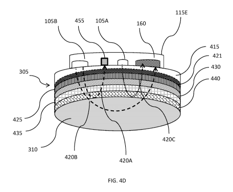
[000100] FIGS. 40 and 4D illustrate an exemplary fetal probe 115E in
contact
with a pregnant mammal's abdomen in a manner similar to that shown in FIGs. 4A
and 4B with layers of maternal tissue similar to those shown in FIG. 4A. The
embodiment shown in FIG. 40 utilizes the simplified layer of maternal tissue
450 and
the embodiment shown in FIG. 4D shows many layers of maternal tissue with
layers
of maternal tissue similar to those shown in FIG. 4A and fetal probe 115E is
configured to enable double short separation (SS) analysis of light back
scattered
from and/or transmitted through the pregnant mammal's abdomen and the fetus
contained therein.
[000101] Fetal probe 115E includes a first light source 105A that emits
first
optical signal 4200, a small detector 455, a second light source 105B that
emits
second optical signal 420, and detector 160. A first portion of second optical
signal
420A3 may be detected by small detector 455 and a second portion of second
optical signal 420B may be detected by detector 160. First and/or second light
beams 4200 and/or 420 may include light of a single, or multiple, wavelengths
and
may be within, for example, the red, near infra-red, and/or broadband
spectrum. In
some circumstances, the wavelength for optical signal 4200 may be different
from
the wavelength of optical signal 420 and/or may be projected at different
times to
enable differentiation between light projected from the two light sources when
it is
received by detector 160 and processed according to one or more of the
processes
described herein. Small detector 455 may be similar to detector 160 but may
have,
for example, a smaller size and/or decreased sensitivity. In some instances,
small
detector 455 may be a small fiber detector. In some embodiments, fetal probe
115E
may include a filter (not shown) for detector 160 that may be attenuated to so
that
26
CA 03148219 2022-01-20
WO 2021/016641
PCT/US2020/070312
detector 160 detects and equal amount of light from first and second light
sources
105A and 105B.
[000102] In some embodiments, the position of first light source 105A
and/or
second light source 105B may be adjusted (e.g., moved closer to, or further
away
from, detector 160) so as to, for example, adjust a depth of penetration for
the light
emitted therefrom that is detected by detector 160. The adjustment may be
facilitated by, for example, manual manipulation and/or placement of a
detector
and/or moving a detector along a track or other positioning device included in
and/or
associated with fetal probe 115E (not shown). In some instances, the
positioning of
first light source 105A and/or second light source 105B may be adjusted
responsively to a depth of fetus 330 within the pregnant mammal's abdomen
(i.e., a
measurement of the width of maternal tissue 450 positioned between the fetal
probe
115E and the fetus 330). A measurement of a depth of fetus 330 within the
pregnant
mammal's abdomen may be provided by, for example, an ultrasound or Doppler
probe like Doper/ultrasound probe 135 and/or an image of the pregnant mammal's
abdomen, illustrations of which are shown in FIGs. 3A and 3B.
[000103] In some embodiments, first light source 105A may be positioned
relative to detector 160 so that light emitted from first light source (i.e.,
optical signal
4200) only propagates through the maternal tissue 305 and does not reach fetus
330. Second light source 105B may be positioned further away (relative to
first light
source 105A) from detector 160 so that light projected by second light source
105B
(i.e., optical signal 420) projects deeper into the pregnant mammal's abdomen
than
optical signal 4200 so that it reaches fetus 330 so that light back scattered
from
and/or transmitted through the fetus may be detected by detector 160. Small
detector 455 may be positioned between first and second light sources 105A and
105B so that light (i.e., optical signal 420) only propagates through the
maternal
tissue 450 prior to detection by small detector 455 and does not reach fetus
330.
This positioning of first light source 105A may facilitate collection of a
first set of short
separation (SS) measurements and the path of first optical signal 4200 and/or
the
detected amounts of first optical signal 4200 by detector 160 may be referred
to
herein as a first SS channel. This positioning of second light source 105B may
facilitate long separation (LS) measurements and the path of second optical
signal
420 and/or the detected amounts of second optical signal 420 by detector 160
may
be referred to herein as a LS channel. This positioning of small detector 455
may
27
CA 03148219 2022-01-20
WO 2021/016641
PCT/US2020/070312
facilitate a second set of short separation (SS) measurements and the path of
first
optical signal 4200 and/or the detected amounts of first optical signal 4200
by
detector 160 may be referred to herein as a second SS channel. Thus, fetal
probe
115E provides for SS measurements of both the first and second light sources
105A
and 105B.
[000104] FIG. 5 provides a flowchart illustrating a process 500 for
determining a
fetal hemoglobin oxygen saturation level. Process 500 may be executed by, for
example, any of the system or system components described herein.
[000105] Initially, a detected composite electronic signal may be received
from a
photo-detector (e.g., detector 160) by a processor and/or computer like
computer
150 (step 505). The detected composite electronic signal may be received from,
for
example, a photo-detector, a transceiver coupled to the photo-detector, and/or
a fetal
hemoglobin probe such as fetal hemoglobin probe 115. The
[000106] The detected composite electronic signal may correspond to an
optical
signal of a plurality of wavelengths emanating (via, for example,
transmission, back
scattering, and/or reflection) from the abdomen of a pregnant mammal and/or
her
fetus. Light incident upon, and exiting from, the pregnant mammal's abdomen
may
be generated by one or more light sources, like light source 105 and may be of
any
acceptable frequency or wavelength (e.g., near infra-red (NIR)) and/or
combination
of frequencies and/or wavelengths. In some embodiments (e.g., when multiple
detectors are used), the received detected composite electronic signals may
include
and/or be associated with a detector identifier (e.g., code) so that a
position of a
particular detected composite electronic signal may be known. This location
may
then be used to analyze the received detected composite electronic signals to
determine various factors of the detected light and/or imaged tissue.
[000107] In step 510, a pathlength and/or a degree of scattering and/or
absorption of each wavelength of light emanating from the pregnant mammal's
abdomen may be measured and/or determined via, for example, analysis of the
detected first optical signal. In some embodiments, execution of step 510 may
include using the Modified Beer Lambert Law, which is reproduced below as
Equation 1. Equation 1 may be used to, for example, determine an absorption
coefficient fora particular wavelength ( ,(A)), a change in absorption
coefficient,
and/or an effective mean path length factor (DPF) for a particular wavelength
(A).
28
CA 03148219 2022-01-20
WO 2021/016641
PCT/US2020/070312
1 AI(A)
Alla = Equation 1
r*DPF(A) 10
Where:
4,00 = the change in an absorption coefficient for a given wavelength X;
r = a distance between a light source and detector;
DPF = differential pathlength factor for the given wavelength X;
= actual intensity of the light of the given wavelength X as it changes over
the course of each heart beat pulse; and
lo = incident intensity of the light of the given wavelength X.
At times, lo may be an average intensity measured over the time of trace
(i.e., the
time within which the measurement is taken and/or a composite electronic
signal is
detected). In some instances, values for 4,00 and/or DPF may be a calibration
factor (step 515) for other calculations described herein (e.g., equations 5,
6, 7a
and/or 7b). At times, the DPF is estimated based on, for example,
characteristics of
light at wavelength X. Additionally, or alternatively, DPF may be deduced from
a
spectral fitting of experimentally determined values for ¨AI(A) .
Additionally, or
alternatively, DPF may be determined via measuring and/or calculating the
amount
scattering of light for a given wavelength. In some circumstances when, for
example, directing light into a portion of the body that has an inhomogeneous
geometry (such as the abdomen of a pregnant mammal), the DFP may be
dependent on, for example, physical characteristics and/or intrinsic
properties of the
pregnant mammal and/or her fetus including, but not limited to, fetal depth,
lipid
concentration in tissue, width of layers of tissue, a location on the fetus
being
exposed to the light, a melanin content of the pregnant mammal and/or fetus,
and so
on. Additionally, or alternatively, the DFP may be dependent on a property of
the
detector such as a color of the sensor surface of the detector and/or a
sensitivity of
the detector.
[000108] In some embodiments, an absorption coefficient may be determined
for
different layers of maternal tissue (also referred to herein as individual
absorption
coefficients) using, for example, optical properties of the type of tissue
and/or
29
CA 03148219 2022-01-20
WO 2021/016641
PCT/US2020/070312
maternal geometry (e.g., width of a tissue layer) and/or tissue density using
Equation
1. For example, a series of absorption coefficients for different types and/or
features
of maternal tissue may be experimentally-determined and/or modeled based on
characteristics of the layers of maternal tissue. Exemplary characteristics
include,
but are not limited to, tissue type, tissue density, tissue layer thickness,
position of
the tissue layer (e.g., DFP and/or distance between source and detector
(represented as r with regard to Equation 1)), and/or optical properties
(e.g.,
scattering and/or absorption coefficients for known widths of tissue that may
be
modified, or otherwise adjusted, based on, for example, geometry of a
particular
pregnant mammal and/or fetus). In some embodiments, these individual
absorption
coefficients may then be aggregated together to generate a total absorption
coefficient that more closely approximates the absorption of light for a
particular
situation (e.g., particular pregnant mammals, particular locations on a
pregnant
mammal's abdomen, etc.).
[000109] In some embodiments, steps 505- 515 may be performed on an
individual, per-pregnant mammal basis in order to, for example, tailor, or
calibrate,
instruments and/or calculations to account for, among other things, individual
physiology, instrument (e.g. light source and/or detector) placement, noise,
etc.
Additionally, or alternatively, in other embodiments, steps 505- 515 may be
performed a plurality (e.g., hundreds, thousands, etc.) of times so that a
plurality of
calibration factors may be determined. In some cases, this plurality of
determined
calibration factors may be used to, for example, determine average calibration
factors for pregnant mammals that may, or may not be associated with one or
more
of the pregnant mammal's physiological characteristics. For example, in one
embodiment, calibration factors for 10,000 pregnant women may be determined.
These calibration factors then may be used to determine, for example, a
universal
calibration factor for all pregnant women (e.g., average calibration factor
for the
10,000 pregnant women) and/or may be grouped according to one or more
physiological factors including, but not limited to, gestational age of the
fetus,
maternal weight, maternal height, maternal melanin content, etc.
[000110] Optionally, in step 520, one or more physiological
characteristics, or
parameters, of the pregnant mammal and/or fetus may be received and/or
determined (via, for example, analysis of ultrasound information or an image
of the
pregnant mammal, illustrations of which are provided by FIGs. 3A and 3B).
CA 03148219 2022-01-20
WO 2021/016641
PCT/US2020/070312
Exemplary physiological characteristics include, but are not limited to,
melanin
content and/or degree and/or type of skin pigmentation of woman's and/or
fetus'
skin, fetal depth, fetal gestational age, and/or a width of one or more layers
of
maternal or fetal tissue, abdominal wall thickness for the pregnant mammal, a
percentage of hemoglobin blood concentration, a degree of blood perfusion in
tissue
which may be taken via, for example, a DC tissue measurement. In some cases,
the
physiological characteristics may be grouped as intrinsic or geometrical
characteristics, wherein exemplary intrinsic characteristics are blood or
tissue
oxygenation and/or hemoglobin concentration levels and body mass index (BMI)
abdominal wall thickness and/or a thickness of a layer of the abdominal wall
may be
geometrical characteristics.
[000111] The one or more physiological characteristics of the pregnant
mammal
and/or fetus may be associated (e.g., indexed in a database table) with the
calibration factor (step 525) in, for example, an index or look-up table. In
some
embodiments, these associations may be used to select a calibration factor
that is
appropriate for a particular pregnant mammal and/or fetus as will be discussed
in
greater detail below with regard to FIG. 13. The calibration factor, the one
or more
physiological characteristics of the pregnant mammal and/or fetus, and/or the
association(s) therebetween may then be stored in, for example, a database
like
database 170 and/or a memory resident within a computer such as computer 150
(step 530). At times, calibration factors may be aggregated together and
correlated
to physiological properties; so that assumptions/calibration factors may be
determined and/or applied without the need to individually calculate
calibration factor
for each pregnant mammal.
[000112] FIG. 6 provides a flowchart illustrating a process 600 for
determining a
fetal hemoglobin oxygenation saturation level using physiological
characteristics of
the pregnant mammal determined using a maternal detected electronic signal.
[000113] Process 600 may be performed by, for example, system 100 and/or
components thereof. Process 600 may be executed in-situ during, for example, a
labor and delivery of the fetus and/or a wellness checkup for the pregnant
mammal.
In some cases, process 600 may be executed on a continuous, periodic, and/or
as-
needed basis over a period of time such as the labor and delivery of the fetus
so
that, for example, the fetal hemoglobin oxygenation saturation level may be
calibrated and recalibrated over time as needed and/or when conditions for the
fetus
31
CA 03148219 2022-01-20
WO 2021/016641
PCT/US2020/070312
and/or pregnant mammal change as may be the case when, for example, the fetus
travels through the birth canal.
[000114] Initially, one or more maternal detected electronic signal(s)
corresponding to an optical signal emanating from the abdomen of a pregnant
mammal may be received (step 605). The optical signal may be generated by a
light
source like light source 105, incident on the pregnant mammal's abdomen, pass
through a portion of the maternal abdominal tissue, and be reflected, or
backscattered, through the maternal tissue where it is detected by a detector
like
detector 160. Exemplary optical signals like the optical signal that may be
detected
by a detector and received in step 605 are shown in FIG. 30 as first and
second
optical signals 325A and 420B.
[000115] In step 610, the maternal detected electronic signal may be
analyzed to
determine one or more physiological characteristics of the pregnant mammal
(step
615). The analysis may be, for example, a frequency domain and/or time of
flight
analysis performed by a fetal hemoglobin probe configured to acquire time of
flight
measurements for photons projected into the maternal abdomen. In some
embodiments, the determination of a physical characteristics for the pregnant
mammal may be performed by determining how the pregnant mammal's tissue
responds to incident light via measurement of, for example, absorption and/or
scattering of the light in step 610. In some embodiments, the absorption
and/or
scattering of light may be expressed as an absorption coefficient or
scattering
coefficient, respectively, and may be used in one or more equations described
herein. Then, in step 620, a calibration factor for the physiological
characteristic may
be determined (step 620). In some cases, steps 610 and 615 may be performed to
determine one or more intrinsic physiological characteristics of the pregnant
mammal
that may be uniform across a portion of that pregnant mammal's abdomen. Such
intrinsic physiological characteristics include, but are not limited to,
melanin content
and/or degree and/or type of skin pigmentation of woman's skin, fetal depth, a
width
of one or more layers of maternal or fetal tissue, abdominal wall thickness
for the
pregnant mammal, hemoglobin oxygen saturation, a degree of blood perfusion in
abdominal tissue, whether the pregnant mammal is anemic, tissue oxygen
saturation, and hemoglobin concentration levels.
[000116] A determination (i.e., calculation) and/or selection of a
calibration factor
in step 620 may be made by, for example, assessing how much light from the
32
CA 03148219 2022-01-20
WO 2021/016641
PCT/US2020/070312
incident optical signal is absorbed and/or scattered by the pregnant mammal's
abdominal tissue and/or a time of flight for photons of the optical signal to
travel
through the pregnant mammal's abdominal tissue and be detected by the
detector.
In some embodiments, the determination/selection of a calibration factor in
step 620
may be performed by querying a database like database 170 and/or a memory
resident within a computer such as computer 150 for calibration factors that
correspond to the physiological characteristic determined in step 615. The
database
may be populated with correlated physiological characteristics and calibration
factors
via process 500. In some embodiments, associations between the results of
executing steps 605, 610, 615, and/or 620 may be mapped to one another (step
625) and the results of executing steps 605, 610, 615, and/or 620 may be
stored in,
for example, a database like database 170 and/or a memory resident within a
computer such as computer 150.
[000117] In one example, the physiological characteristic determined in
step 615
may be a pregnant mammal's a skin color, pigmentation and/or melanin content,
which may be determined by, for example determining how much of light incident
on
the pregnant mammal's abdomen is absorbed by the pregnant mammal's skin. The
skin color of the pregnant mammal's skin may influence how much of the
incident
light is absorbed, which may impact how much of the light incident on the
pregnant
mammal's abdomen travels through the maternal tissue and is incident on the
fetus
may be associated with a known, or calculated, factor, which is determined
and/or
selected in step 620. For example, execution of 620 may include querying a
database for calibration factors associated with how much light the pregnant
mammal's skin absorbed. In this example, the calibration factor may be an
absorption coefficient that may be associated with the pregnant mammal's
absorption rate and/or skin color.
[000118] In another example, the maternal detected electronic signal
received in
step 605 may be analyzed in step 610 to determine the physiological
characteristic a
concentration or thickness of the myoglobin, or muscle, layers of the pregnant
mammal's abdomen. This analysis may be performed using, for example, a fetal
hemoglobin probe configured as a frequency domain NIRS system and/or a fetal
hemoglobin probe configured as to obtain a time of flight for photons
projected into
the maternal abdomen that penetrate the maternal myoglobin layers and are
reflected back to a detector. The myoglobin tissue of the pregnant mammal may
33
CA 03148219 2022-01-20
WO 2021/016641
PCT/US2020/070312
absorb light projected into the maternal abdomen and a measurement and/or
calculation of how much light that is absorbed (e.g., not detected) by the
pregnant
mammal's abdominal tissue may be used to determine the physiological
characteristic of how thick and/or concentrated the maternal myoglobin
layer(s) are.
In step 620, this physiological characteristic may be used to, for example
calculate a
calibration factor and/or query a database of calibration factors to find a
calibration
factor associated with the physiological characteristic of the myoglobin
thickness
and/or concentration determined for the pregnant mammal.
[000119] In another example, the maternal detected electronic signal
received in
step 605 may be analyzed in step 610 to determine the physiological
characteristic a
total thickness of pregnant mammal's abdomen (which may also be referred to
herein as fetal depth) which may vary over the course of a pregnancy due to
the
increasing size of the fetus. Additionally, or alternatively, the thickness of
the
pregnant mammal's abdominal tissue may vary of the course of the pregnancy
and/or during labor and delivery of the fetus because of pre-eclampsia or
eclampsia,
which can cause edema that causes the pregnant mammal's abdominal thickness to
change. The analysis of step 610 for this example may be performed using, for
example, a fetal hemoglobin probe configured as a frequency domain NIRS system
and/or a fetal hemoglobin probe configured as to obtain a time of flight for
photons
projected into the maternal abdomen that penetrate the maternal abdominal
layers
and are reflected back to a detector. The abdominal tissue of the pregnant
mammal
may absorb light projected into the maternal abdomen and a measurement and/or
calculation of how much light that is absorbed (e.g., not detected) by the
pregnant
mammal's abdominal tissue may be used to determine the physiological
characteristic of how thick the pregnant mammal's abdominal tissue is. In step
620,
this physiological characteristic may be used to, for example calculate a
calibration
factor and/or query a database of calibration factors to find a calibration
factor
associated with the physiological characteristic of the myoglobin
concentration
and/or thickness determined for the pregnant mammal.
[000120] In yet another example, the maternal detected electronic signal
received in step 605 may be analyzed in step 610 to determine the
physiological
characteristic a thickness of the adipose, or fat, layers of the pregnant
mammal's
abdomen. This analysis may be performed using, for example, a fetal hemoglobin
probe configured as a frequency domain NIRS system and/or a fetal hemoglobin
34
CA 03148219 2022-01-20
WO 2021/016641
PCT/US2020/070312
probe configured as to obtain a time of flight for photons projected into the
maternal
abdomen that penetrate the maternal adipose layers and are reflected back to a
detector. The adipose tissue of the pregnant mammal may scatter light
projected
into the maternal abdomen and a measurement and/or calculation of how much
light
is scattered (e.g., not detected) by the pregnant mammal's adipose tissue may
be
used to determine the physiological characteristic of how thick the maternal
adipose
layer(s) are. In step 620, this physiological characteristic may be used to,
for
example calculate a calibration factor and/or query a database of calibration
factors
to find a calibration factor associated with the physiological characteristic
of the
adipose thickness determined for the pregnant mammal.
[000121] FIG. 7A is a flowchart illustrating a process 700 for determining
a fetal
depth and/or a level of oxygen saturation for fetal hemoglobin. Process 700
may be
performed by, for example, system 100 and/or components thereof.
[000122] Optionally, in step 705, a plurality of first detected electronic
signals,
each of which correspond to an optical signal of one or more wavelengths
projected
into the pregnant mammal's abdomen by, for example, one or more light sources
like
light source 105 and exiting therefrom via, for example, reflection, back
scattering,
and/or transmission (i.e., passing through the maternal abdomen) may be
received
by, for example, a computer or processor such as computer 150. Each of the
first
detected signals of the plurality of first detected signals may be received
from a
different detector like detectors 160A-160V as shown and discussed above with
regard to FIGs. 3A, 3B, and 3. The received first detected electronic signals
may be
associated with a detector identifier. Each detector may have a different
source/detector distance. For example, a probe like fetal probe 115A, 115B,
115D,
and/or 115E may have a source and a plurality of detectors with each detector
having a different source/detector distance. In some embodiments, an optical
signal
detected by the detectors arranged further away from the source may have a
higher
proportion of light that was incident on the fetus than detectors arranged
relatively
close to the source.
[000123] An exemplary range of wavelengths for the optical signals that
correspond to the first detected electronic signals is between 600 and 1000nm
and
may be similar to one or more of optical signals 420. In some embodiments, the
optical signal may be a broadband optical signal (e.g., white light and/or a
range of,
for example, 10, 15, or 20 wavelengths) and the received first detected signal
may
CA 03148219 2022-01-20
WO 2021/016641
PCT/US2020/070312
correspond to an optical signal of a plurality of wavelengths. In some
embodiments,
the optical signal, or a portion thereof, may be of a set, or known,
wavelength that
may be at an isosbestic point for light directed into human tissue to
determine a ratio
of oxygenated and de-oxygenated hemoglobin for the human's blood such as 808
nm. Light at this wavelength is reflected from oxygenated and de-oxygenated
hemoglobin in the same way.
[000124] When step 705 is executed, each of the first detected electronic
signals
received in step 705 may be processed to isolate a portion thereof that
corresponds
to light that has been incident upon the fetus (step 710). This isolated
portion of
each of the first detected electronic signal may be referred to herein as a
first fetal
signal. Step 710 may be executed using any appropriate method of isolating a
fetal
signal from a corresponding first detected electronic signal including the
methods
disclosed herein. Appropriate methods include, but are not limited to,
reducing noise
in the signal via, for example, application of filtering or amplification
techniques,
determining a portion of the first detected electronic signal that is
contributed by the
pregnant mammal and then subtracting, or otherwise removing, that portion of
the
first detected electronic signal from the received first detected electronic
signals
and/or receiving information regarding fetal a heart rate and using that
information to
lock in (via, for example, a lock-in amplifier) on a portion of the received
first detected
electronic signals generated by the fetus.
[000125] Optionally, execution of step 710 may include pre-processing one
or
more of the first detected electronic signals in order to, for example, remove
noise
from the signal and/or confounding effects of the pregnant mammal's anatomy or
physiological signals (e.g., a respiratory signal) from the first detected
electronic
signals. Execution of the pre-processing may include, but is not limited to,
application of filtering techniques to the first detected electronic signals,
application
of amplification techniques to the first detected electronic signals,
utilization of a lock-
in amplifier on the first detected electronic signals, and so on. In some
embodiments, the pre-processing may include application of a filter (e.g.,
bandpass
or Kalman) to one or more of the detected electronic signal(s) to reduce noise
or
hum in the first detected electronic signals that may be caused by, for
example,
electronic noise generated by equipment generating and/or detecting the first
detected electronic signals and/or environmental equipment that may, in some
instances, be coupled to the pregnant mammal.
36
CA 03148219 2022-01-20
WO 2021/016641
PCT/US2020/070312
[000126] Optionally, in step 715, an indication of a hemoglobin oxygen
saturation level for the pregnant mammal may be received from, for example, a
pulse oximetry probe like pulse oximetry probe 130, a maternal pulse oximetry
probe
like maternal probe 133, and/or a NIRS adult hemoglobin probe like NIRS adult
hemoglobin probe 125, and/or determined using, for example, a processor
executing
process 700 using, for example, the initial detected electronic signals.
Additionally,
or alternatively, an indication of a tissue oxygen saturation level for the
pregnant
mammal may be received and/or determined in step 715. The pregnant mammal's
tissue oxygen saturation level may be received from, for example, a diffuse
optical
tomography (DOT) instrument and/or may be determined by applying DOT to the
initial detected electronic signals. Additionally, or alternatively, an
indication of a
hemoglobin and/or tissue oxygen saturation level for the pregnant mammal may
be
determined using one or more of the first detected electronic signals received
in step
705 and, for example, the Beer-Lambert Law as discussed above with regard to
Equation 1 above.
[000127] In some instances, the pregnant mammal's a hemoglobin and/or
tissue
oxygen saturation level may be used to determine how much light is incident on
the
fetus as discussed below.
[000128] In step 720, a fetal depth may be received from, for example,
Doppler/ultrasound probe 135 and/or may be determined using, for example, the
first
detected electronic signals of step 705, first fetal signals of step 710,
and/or maternal
hemoglobin and/or tissue saturation level of step 715.
[000129] When fetal depth is determined in step 720, the depth of the fetus
may
be determined by comparing an intensity of the initial fetal signals to one
another to
determine a change in intensity for each of the initial fetal signals. In some
cases,
this comparison may incorporate a position and/or source/detector distance for
each
detector providing a corresponding first detected electronic signal and a
depth of a
fetus may be determined by quantifying a drop-off, or decrease in intensity of
the
fetal signal, as the source/detector distance increases for detectors
positioned
further away from the source. This decrease in intensity as a function of
source/detector distance may be used to determine a depth of the fetus.
[000130] FIG.7B provides a flowchart illustrating an exemplary process for
executing step 720 to determine a fetal depth. Initially, in step 750, a first
initial
detected electronic signal may be analyzed to determine if it includes an
initial fetal
37
CA 03148219 2022-01-20
WO 2021/016641
PCT/US2020/070312
signal. The first initial detected electronic signal may correspond to an
optical signal
like first optical signal 420A detected by a first detector like first
detector 160A. The
analysis of step 750 may be based on the processing/isolation of step 710 and
may
include a yes or no determination of whether or not there is any initial fetal
signal to
isolate from the first initial detected electronic signal. When there is an
initial fetal
signal included in the first initial detected electronic signal, an intensity
of the initial
fetal signal may be determined (step 755).
[000131] Whether or not there is an initial fetal signal included in the
first initial
detected electronic signal, in step 760 it may be determined if a second
initial
detected electronic signal includes an initial fetal signal. The second
initial detected
electronic signal may correspond to an optical signal like second optical
signal 420B
detected by a second detector like second detector 160B. Step 760 may be
executed in a manner similar to the execution of step 750. When there is an
initial
fetal signal included in the second initial detected electronic signal, an
intensity of the
initial fetal signal may be determined (step 755).
[000132] Whether or not there is an initial fetal signal included in the
second
initial detected electronic signal, in step 765 it may be determined if a
third initial
detected electronic signal includes an initial fetal signal. The third initial
detected
electronic signal may correspond to an optical signal like third optical
signal 4200
detected by a third detector like third detector 1600. Step 765 may be
executed in a
manner similar to the execution of step 750 and/0r760. When there is an
initial fetal
signal included in the third initial detected electronic signal, an intensity
of the initial
fetal signal may be determined (step 755).
[000133] A process like step 765 may be repeated N times until it is
determined
whether the last of the initial detected electronic signal of the plurality of
initial
detected electronic signal received in step 705 includes an initial fetal
signal (step
770). The Nth initial detected electronic signal may correspond to an optical
signal
like sixth optical signal 420F detected by a sixth detector like third
detector 160F.
Step 770 may be executed in a manner similar to the execution of step 750,760,
and/0r765. When there is an initial fetal signal included in the Nth initial
detected
electronic signal, an intensity of the initial fetal signal may be determined
(step 755).
[000134] In step 775 a fetal depth may be determined by analyzing the
determined intensities of each of the respective initial fetal signals
determined in step
755. In some embodiments, step 775 may be executed by plotting (using, for
38
CA 03148219 2022-01-20
WO 2021/016641
PCT/US2020/070312
example, a scatter plot) the intensities on a graph showing fetal signal
intensity as a
function of the detector detecting the respective 1st-Nth fetal signal and/or
as a
function of a distance between a light source generating the optical signal
and the
detector detecting the respective 1st-Nth initial detected electronic signals.
FIG.70
provides an example of a graph 702 showing a scatter plot of optical intensity
of 1st-
Nth fetal signals in Watts/cm as a function of source/detector distance in cm
that
may be determined by execution of step 755. In the case of FIG.70, graph 702
corresponds to initial detected electronic signals detected by first through
sixth
detectors 160A-160F and their respective distance from light source 105. As
may be
seen in graph 702, a change in percent transmission of light for a signal,
which may
sometimes be related to the intensity of the signal, detected a distance away
from
the light source predominantly follows an inverse proportionality. The change
in
percent transmission of light and/or intensity of light incident on the fetus
may vary in
a nonlinear manner dependent on the fetal depth and the source-detector
distance
as shown graph 702 of FIG. 70. In some embodiments, an intensity and/or change
in percent transmission of light of light reaches the fetus may be dependent
upon a
fetal depth according to an inverse proportionality as shown in FIG. 7D, which
includes a graph 703 that shows a change in percent transmission of light as a
function of source-detector distance measured in cm for a maternal signal
and/or a
maternal portion of the signal.
[000135] Because the first fetal signal is detected by third detector with
a
source/detector distance of 3cm, an exemplary depth of the fetus may be
approximately 25mm. A fetal depth may be determined using the rate of decay of
the intensity of the fetal signal as the source/detector distance increases.
This rate
of decay may correspond to a slope of a linear regression the scatter plot.
[000136] In some embodiments, a depth of a fetus may be determined by
analyzing light at the isosbestic point of 808nm. Light at this wavelength is
reflected
from oxygenated and de-oxygenated hemoglobin in the same way and, as such,
scattering and absorption of the light at this wavelength will be the same for
both
oxygenated and de-oxygenated hemoglobin and will not change based on, for
example, a hemoglobin oxygen saturation level of the pregnant mammal's and/or
fetus's blood. Thus, determinations of scattering and/or absorption (which may
be
expressed as a scattering and or absorption coefficient, respectively) of the
optical
39
CA 03148219 2022-01-20
WO 2021/016641
PCT/US2020/070312
signal by maternal tissue may not be required when executing step 720 using
light at
the isosbestic point of 808nm.
[000137] In step
725, a second detected electronic signal that corresponds to a
second optical signal may be received. The second detected electronic signal
may
correspond to a second optical signal that exits the abdomen of the pregnant
mammal and may resemble the first detected electronic signal received in step
705.
The second detected electronic signal may then be processed to isolate a
portion
thereof that was incident on the fetus (step 730). The isolated portion of the
second
detected electronic signal may be referred to herein as a second fetal signal.
In
some embodiments, execution of step 730 may resemble execution of step 710.
[000138] In step
735, a factor for analyzing the second fetal signal to determine
a fetal hemoglobin oxygen saturation level may be selected using the fetal
depth.
For example, the fetal depth may be used to determine and/or select a
differential
path length factor (DFP) for a particular wavelength and/or a correlation
factor for
use in calculations to determine a hemoglobin oxygen saturation level for the
fetus
(step 740) using, for example, the modified Beer-Lambert law, which is
presented as
Equation 1 above, for each wavelength.
[000139] The
fetal depth may then be used to determine and/or select the DFP
for a particular wavelength. A value for lo for each wavelength of light in
the incident
fetal optical signal may be, for example, an intensity of light projected into
the
pregnant mammal's abdomen and/or an intensity of the light incident on the
fetus as
may be determined via a process disclosed herein. In embodiments where a
hemoglobin and/or tissue oxygen saturation level of the pregnant mammal is
received and/or determined in step 715, the hemoglobin and/or tissue oxygen
saturation level may be used to determine how much, or an intensity of, light
emitted
by a light source that is directed into the abdomen of the pregnant mammal is
absorbed by maternal tissue or hemoglobin. A correlation between the
hemoglobin
and/or tissue oxygen saturation level of the pregnant mammal and how much of
the
incident light she may absorb for each wavelength of light may be known and/or
empirically determined and these correlations may be stored in, for example, a
look
up table of a database like database 170 such that when a hemoglobin and/or
tissue
oxygen saturation level for a pregnant mammal is received and/or determined in
step
715, it may be used to look up a corresponding level of light absorption
(e.g., a
percentage or ratio) for the pregnant mammal. This value (the level of light
CA 03148219 2022-01-20
WO 2021/016641
PCT/US2020/070312
absorption for the pregnant mammal) may then be applied (e.g., subtracted or
multiplied) to an initial intensity of a light source when it is projecting
light into the
pregnant mammal's abdomen to determine the initial intensity of light incident
on the
fetus (lo). Al (A) may be the change in the measured intensity of light
incident on the
fetus (lo) at wavelength A and an intensity of a detected fetal signal for
light of
wavelength A.
[000140] Once the absorption coefficient (or a change in the absorption
coefficient) is determined via Equation 1, an indication of fetal hemoglobin
oxygen
saturation may be determined via, for example, calculations using Equation 2,
provided below:
Ao(X)= AcHbO*EHbO(X)+AcHb*EHb (X)
Equation 2
where:
A pa(A) = the change in the absorption coefficient for a given wavelength A
over a defined time period;
AcHbo = a change in the concentration of oxygenated hemoglobin (HbO) over
the defined time period;
AcHb = a change in the concentration of deoxygenated hemoglobin (Hb) over
the defined time period;
EHbo(X) = the extinction coefficient for oxygenated hemoglobin (HbO) for the
given wavelength; and
EHb(X) = the extinction coefficient for deoxygenated hemoglobin (Hb) for the
given wavelength.
[000141] Equation 1 may be solved for two or more wavelength pairs by
inputting the change in intensity I, as a function of wavelength X. From this,
changes
in absorption coefficients, Alla, may be determined using Equation 2 by
inputting
known extinction coefficients, EHbO(X) and EHb (X) for a particular
wavelength, which
may be looked up in, for example, a look-up table stored on, for example,
computer
150. The wavelength pairs used to perform the calculations of Equation 2 may
be
any pair of wavelengths included in the spectrum of wavelengths of the optical
signal
incident upon the pregnant mammal's abdomen. In some embodiments, the
41
CA 03148219 2022-01-20
WO 2021/016641
PCT/US2020/070312
calculation of Equation 2 may be performed many times (e.g., 10s, 100s, or
1000s),
in different combinations of wavelengths, in order to arrive at multiple
values for
AcHb0 and AcHb which may be weighted and/or averaged according to one or more
criteria to arrive at robust values (e.g., statistically valid and/or with an
acceptable
level of confidence and error rate) for AcHb0 and AcHb. Additionally, or
alternatively, the calculation of Equation 2 may be performed many times
(e.g., 10s,
100s, or 1000s), to fit a plurality of wavelengths at the same time to the
equation.
[000142] The values for AcHb0 and AcHb generated via Equation 2 are
relative
values, not absolute values, for the concentrations of oxygenated and
deoxygenated
hemoglobin in the fetus's blood, which may be useful in monitoring changes in
the
fetal hemoglobin oxygen saturation levels of the fetus over time. In some
embodiments, the determination of step 1235 may also include determining an
overall oxygen saturation for the fetus's hemoglobin by determining a ratio of
the
change in concentration of oxygenated hemoglobin to the change in
concentration of
total hemoglobin, which may be the sum of oxygenated and deoxygenated
hemoglobin. Additionally, or alternatively, the fetal hemoglobin oxygen
saturation
may be determined using another method, which may be disclosed herein.
[000143] Once the fetal hemoglobin oxygen saturation level is determined in
step 740, provision of an indication of same to a user may be facilitated by,
for
example, display on a display device like display device 155 (step 745).
[000144] FIG. 8 is a flowchart illustrating a process 800 for determining a
fetal
depth using a time of flight for photons incident on a fetus and/or a level of
oxygen
saturation for fetal hemoglobin. Process 800 may be performed by, for example,
system 100 and/or components thereof.
[000145] Optionally, in step 805, a plurality of first detected electronic
signals,
each of which correspond to an optical signal of one or more wavelengths
projected
into the pregnant mammal's abdomen by, for example, one or more light sources
like
light source 105 and exiting therefrom via, for example, reflection, back
scattering,
and/or transmission (i.e., passing through the maternal abdomen) may be
received
by, for example, a computer or processor such as computer 150. The first
detected
electronic signals received in step 805 may resemble the first detected
electronic
signals received in step 705. In some embodiments, a time between when the
optical signal is projected into the pregnant mammal's abdomen and when it is
received by a detector may be received in step 805. Additionally, or
alternatively, the
42
CA 03148219 2022-01-20
WO 2021/016641
PCT/US2020/070312
plurality of first detected electronic signals may include timestamp
information
corresponding to when, for example, the optical signal projected into the
pregnant
mammal's abdomen and/or when the first detected electronic signal is received
by a
respective detector.
[000146] When step 805 is executed, each of the first detected electronic
signals
received in step 805 may be processed to isolate a portion thereof that
corresponds
to light that has been incident upon the fetus (step 810). Execution of step
810 may
resemble execution of step 710.
[000147] In step 815, an indication of a time of flight for photons of the
optical
signal that are incident on the fetus may be received or, when steps 805 and
810 are
performed, determined using a time between when photons of the optical signal
leave the light source and are received by a detector. The time of flight may
be
determined by calculating a length of time between when the optical signal is
projected into the pregnant mammal's abdomen and when it is received by a
detector. When the plurality of first detected electronic signals include
timestamp
information corresponding to when the optical signal projected into the
pregnant
mammal's abdomen and when the first detected electronic signal is received by
a
respective detector, a determination of the time of flight for photons of the
optical
signal that are incident on the fetus may be made by determining a difference,
or
length of time, between the timestamp for when the optical signal projected
into the
pregnant mammal's abdomen and the timestamp for when the first detected
electronic signal is received by a respective detector.
[000148] In step 820, a fetal depth may be received from, for example,
Doppler/ultrasound probe 135 and/or may be determined using the, for example,
the
first detected electronic signals of step 805, first fetal signals of step
810, and/or the
time of flight of step 815. When fetal depth is determined in step 820, a
depth of the
fetus may be determined by calculating a distance traveled by the optical
signal in
the time between when the optical signal is projected into the pregnant
mammal's
abdomen and when it is received at the detector according to Equation 3,
below.
D=s1
Equation 3
Where:
D = distance traveled;
s= speed of light; and
43
CA 03148219 2022-01-20
WO 2021/016641
PCT/US2020/070312
t = time between when the optical signal is projected into the pregnant
mammal's abdomen and when it is received at the detector
The fetal distance may be calculated by dividing a value for the distance
traveled by
the light (D) by 2 because the distance calculated via Equation 3 is the
distance the
light travels to the fetus and back to the detector.
[000149] In step 825, a second detected electronic signal that corresponds
to a
second optical signal may be received. The second detected electronic signal
may
correspond to a second optical signal that exits the abdomen of the pregnant
mammal and may resemble the first detected electronic signal received in step
805.
The second detected electronic signal may then be processed to isolate a
portion
thereof that was incident on the fetus (step 830). The isolated portion of the
second
detected electronic signal may be referred to herein as a second fetal signal.
In
some embodiments, execution of step 830 may resemble execution of step 810
and/or step 730.
[000150] In step 835, a factor for analyzing the second detected electronic
signal
to determine a fetal hemoglobin oxygen saturation level may be selected using
the
fetal depth. For example, the fetal depth may be used to determine and/or
select a
differential path length factor (DFP) for a particular wavelength and/or a
correlation
factor for use in calculations to determine a hemoglobin oxygen saturation
level for
the fetus (step 840) using, for example, Equations 1 and 2, provided and
discussed
above. In some embodiments, execution of step 835 may resemble execution of
step 735. Additionally, or alternatively, the fetal hemoglobin oxygen
saturation may
be determined using another method, which may be disclosed herein.
[000151] Once the fetal hemoglobin oxygen saturation level is determined in
step 840, provision of an indication of same to a user may be facilitated by,
for
example, display on a display device like display device 155 (step 845).
[000152] FIG. 9 provides a flowchart illustrating a process 900 for
determining a
fetal hemoglobin oxygenation saturation level using physiological
characteristics of
the pregnant mammal determined using a maternal detected electronic signal.
Process 900 may be performed by, for example, system 100 and/or components
thereof. Process 900 may be executed in-situ during, for example, a labor and
delivery of the fetus and/or a wellness checkup for the pregnant mammal. In
some
cases, process 900 may be executed on a continuous, periodic, and/or as-needed
basis over a period of time such as the labor and delivery of the fetus so
that, for
44
CA 03148219 2022-01-20
WO 2021/016641
PCT/US2020/070312
example, the fetal hemoglobin oxygenation saturation level may be calibrated
and
recalibrated over time as needed and/or when conditions for the fetus and/or
pregnant mammal change as may be the case when, for example, the fetus travels
through the birth canal.
[000153] In step 925, a detected composite electronic signal may be
received
from the detector by a processor and/or computer like computer 150. The
detected
composite electronic signal may be received from, for example, a photo-
detector, a
transceiver coupled to the photo-detector, and/or a fetal hemoglobin probe
such as
fetal hemoglobin probe 115. The detected composite electronic signal may
correspond to an optical signal emanating from the abdomen of a pregnant
mammal
and/or her fetus. Light incident upon, and exiting from, the pregnant mammal's
abdomen may be generated by one or more light sources like light sources 105
and
may be of any acceptable frequency or wavelength (e.g., near infra-red (NIR))
and/or
combination of frequencies and/or wavelengths. In some embodiments, an
indication of whether the fetal hemoglobin oxygen saturation level is pre-
ductal or
post-ductal may also be received in step 925 in a manner similar to, for
example, the
receipt of an indication of whether the fetal hemoglobin oxygen saturation
level is
pre-ductal or post-ductal in step 1305. Step 925 may be executed at any point
following execution of step 920. However, in many cases, step 925 may be
performed immediately, or soon (e.g., 5 seconds, 30 seconds, 1 minute)
following
execution of step 920 to, for example, account for dynamic changes to the
physiological characteristics of the pregnant mammal's abdomen.
[000154] Next, the received detected composite electronic signal may be
analyzed to isolate a portion of the signal that has corresponds to light that
was
incident upon the fetus, thereby generating a fetal signal (step 930). Step
930 may
be executed in a manner similar to the execution of, for example, step 810
and/or
710 or any other method disclosed herein. Then, the calibration factor
selected
and/or determined in step 920 may be applied to the fetal signal in order to
calibrate
the fetal signal (step 935). Once the fetal signal has been calibrated, an
indication of
the fetal hemoglobin oxygen saturation level may be determined (step 940) and
provided to a user (step 945). Optionally, execution of step 945 may include
an
indication of whether the fetal hemoglobin oxygen saturation level is pre-
ductal or
post-ductal to the user in manner similar to the execution of step 1355.
Execution of
steps 935-945 may be similar to execution of steps 1345-1350 disclosed below
CA 03148219 2022-01-20
WO 2021/016641
PCT/US2020/070312
and/or steps 830-835. Additionally, or alternatively, the fetal hemoglobin
oxygen
saturation may be determined using another method, which may be disclosed
herein.
[000155] FIG. 10 provides a flowchart illustrating a process 1000 for
determining
a fetal hemoglobin oxygenation saturation level using physiological
characteristics of
the pregnant mammal determined using one or more maternal detected electronic
signal(s). Process 1000 may be used to account for changes in maternal
geometries
and/or geometrical physiological characteristics across, for example, the
surface
area of a fetal hemoglobin probe like fetal hemoglobin probe 115. Process 1000
may be performed by, for example, system 100 and/or components thereof.
Process
1000 may be executed in-situ during, for example, a labor and delivery of the
fetus
and/or a wellness checkup for the pregnant mammal. In some cases, process 1000
may be executed on a continuous, periodic, and/or as-needed basis over a
period of
time (e.g., 1-24 hours) such as the labor and delivery of the fetus so that,
for
example, the fetal hemoglobin oxygenation saturation level may be calibrated
and
recalibrated over time as needed and/or when conditions for the fetus and/or
pregnant mammal change as may be the case when, for example, the fetus travels
through the birth canal.
[000156] Initially, in step 1005, one or more maternal detected electronic
signal(s) corresponding to one or more optical signal(s) emanating from the
abdomen of a pregnant mammal may be received (step 1005). The optical signal
may be generated by a light source like light source 105, incident on the
pregnant
mammal's abdomen, pass through a portion of the maternal abdominal tissue, and
be reflected, or backscattered, through the maternal tissue where it is
detected by a
detector like detector 160. Each optical signal may be associated with a
location of
and/or identifier for each detector that detected the respective optical
signal. For
example, each detector that detects a maternal detected electronic signal may
be
associated with an identifier (e.g., detector 1, detector 2, etc.), a location
that may be
a location (e.g., coordinates) of the detector on a fetal hemoglobin probe 115
and/or
a location on the maternal abdomen (e.g., 1 inch directly below the navel, 1
inch
below the navel and 1 inch to the left of the midsagittal line, etc.).
Exemplary optical
signals like the optical signal that may be detected by a detector and
received in step
1005 are shown in, for example, FIG. 30 as first and second optical signals
420A
and 420B and are shown as optical signals 420 in FIGs. 4A-4D.
46
CA 03148219 2022-01-20
WO 2021/016641
PCT/US2020/070312
[000157] In step 1010, each of the maternal detected electronic signals may
be
analyzed to determine one or more extrinsic or geometrical physiological
characteristics of the pregnant mammal (step 1015). Exemplary extrinsic
physiological characteristics include, but are not limited to, melanin content
and/or
degree and/or type of skin pigmentation of pregnant mammal's skin, a width of
one
or more layers of maternal or fetal tissue, abdominal wall thickness for the
pregnant
mammal. Exemplary dimensions for the thickness of an abdominal wall and/or
layers of the pregnant mammal's abdominal wall are provided above with regard
to
FIGs. 3A and 3B.
[000158] In step 1020, a calibration factor may be selected and/or
determined
for each maternal detected electronic signal. The determination (i.e.,
calculation)
and/or selection of a calibration factor may be made by, for example,
assessing how
much light from the incident optical signal is absorbed and/or scattered by
the
pregnant mammal's abdominal tissue and/or a time of flight for photons of the
optical
signal to travel through the pregnant mammal's abdominal tissue and be
detected by
the detector. In some embodiments, the selection of a calibration factor may
be
performed by querying a database like database 170 and/or a memory resident
within a computer such as computer 150 for calibration factors that correspond
to the
physiological characteristic determined in step 915. The database may be
populated
with correlated physiological characteristics and calibration factors via
process 500.
In some embodiments, the results of executing steps 1005, 1010, 1015, and/or
1020
may be stored in, for example, a database like database 170 and/or a memory
resident within a computer such as computer 150.
[000159] In step 1025, the physiological characteristics and/or calibration
factors
may be associated with the detector that detected each of the respective
maternal
detected electronic signals received in step 1005. Additionally, or
alternatively, in
step 1025, the physiological characteristics and/or calibration factors may be
associated with a location on the pregnant mammal's abdomen.
[000160] In one example, a set of results from executing steps 1005-1025 is
provided by the values in Table 1, below which provides a detector identifier,
a value
for the physiological characteristic of abdominal thickness, and a calibration
factor
corresponding to the abdominal thickness physiological characteristic.
47
CA 03148219 2022-01-20
WO 2021/016641
PCT/US2020/070312
Detector Identifier Abdominal Thickness Calibration Factor
Detector 1 19.81mm Cl
Detector 2 19.83mm C2
Detector 3 19.85mm C3
TABLE 1
[000161] In step 1030, one or more detected composite electronic signal(s)
may
be received by a processor and/or computer like computer 150. The one or more
detected composite electronic signal(s) may be received from the detector(s)
that
provided the maternal detected electronic signals received in step 1005 and
each of
the detected composite electronic signal(s) may be associated with a location
of
and/or identifier for the detector that detected each respective detected
composite
electronic signal(s). . In some embodiments, an indication of whether the
fetal
hemoglobin oxygen saturation level is pre-ductal or post-ductal may also be
received
in step 1030 in a manner similar to, for example, the receipt of an indication
of
whether the fetal hemoglobin oxygen saturation level is pre-ductal or post-
ductal in
step 1305.
[000162] The detected composite electronic signal may be received from, for
example, a photo-detector, a transceiver coupled to the photo-detector, and/or
a fetal
hemoglobin probe such as fetal hemoglobin probe 115. The detected composite
electronic signal may correspond to an optical signal emanating from the
abdomen
of a pregnant mammal and/or her fetus. Light incident upon, and exiting from,
the
pregnant mammal's abdomen may be generated by one or more light sources like
light sources 105 and may be of any acceptable frequency or wavelength (e.g.,
near
infra-red (NIR)) and/or combination of frequencies and/or wavelengths. Step
1030
may be executed at any point following execution of step 1025. However, in
many
cases, step 1030 may be performed immediately, or soon (e.g., 5 seconds, 30
seconds, 1 minute, 1 hour) following execution of step 1025 to, for example,
account
for dynamic changes to the physiological characteristics of the pregnant
mammal's
abdomen.
[000163] Next, the received detected composite electronic signal(s) may be
analyzed to isolate a portion of the respective composite electronic signal
that has
corresponds to light that was incident upon the fetus, thereby generating a
corresponding number of fetal signal(s) (step 1035). Step 1035 may be executed
in
48
CA 03148219 2022-01-20
WO 2021/016641
PCT/US2020/070312
a manner similar to the execution of step(s) 710 and/or 810 and/or in
accordance
with other method(s) disclosed herein. Then, the calibration factor correlated
with
the detector that detected each respective detected composite electronic
signal may
be applied to the fetal signal corresponding to each respective detected
composite
electronic signal (step 1040). Execution of step 1040 may resemble execution
of
step 935 as discussed above.
[000164] Continuing the example above, application of the calibration
factors to
fetal signals received from the first, second, and third detectors in step
1040
corresponds to first, second, and third fetal signals (i.e., fetal signal 1,
fetal signal 2,
and fetal signal 3), which corresponds to calibration factors P1, P2, and P3,
respectively as shown in Table 2, below where the detector the calibration
factor of a
first, second, and third physiological characteristic corresponds to the
calibration
factor for the first, second, and third fetal signal, respectively, as shown
in Table 2.
Detector Identifier Fetal Signal Identifier Calibration Factor
Detector 1 Fetal Signal 1 P1
Detector 2 Fetal Signal 2 P2
Detector 3 Fetal Signal 2 P3
TABLE 2
[000165] In some embodiments, the fetal signals disclosed herein and/or
fetal
signal 1, fetal signal 2, and/or fetal signal 3 may be a signal that includes
light of a
plurality of wavelengths and a calibration factor for each of these
wavelengths may
be separately determined and/or applied to the individual wavelengths (or
groups of
similar wavelengths) included in each fetal signal. Additionally, or
alternatively, the
fetal signals disclosed herein and/or fetal signal 1, fetal signal 2, and/or
fetal signal 3
may each be detected by a separate detector and/or be detected at a different
time
by the same detector. For example, a fetal pulse may be extracted from a
plurality
fetal signals (each of which may be detected by a separate detector) and the
calibration(s) of the fetal signals may be influenced differently in each
detector
channel by different proportionalities of the different physical
characteristics, which
may be governed by the following relationship: if detector 1 signal = function
D1 (D1,
P1, P2, P3, ...) where D1 is a vector of coefficients associated with the
detector 1
geometry, wavelength, etc. and P1 is a vector associated with physical
49
CA 03148219 2022-01-20
WO 2021/016641
PCT/US2020/070312
characteristics P1, P2 is a vector associated with physical characteristics
P2, and so
forth. In this example, the function D1 may be considered as a tensor.
[000166] Once each of the fetal signals have been calibrated, an indication
of
the fetal hemoglobin oxygen saturation level may be determined (step 1045)
using,
for example, one or more of the methods disclosed herein. In some cases, step
1050 may be executed by individually determining a fetal hemoglobin oxygen
saturation level for each fetal signal and then averaging the individually
determined
fetal hemoglobin oxygen saturation level into an average a fetal hemoglobin
oxygen
saturation level. Additionally, or alternatively, each fetal signal may be a
mix, or
combination, of different signals coming from the various simultaneously
occurring
physical characteristics and, in some cases, each calibration factor 01, 02,
etc., may
be a vector and for the family of detector signals, it would be a tensor
matrix.
[000167] Once the fetal hemoglobin oxygen saturation level is determined,
the
fetal hemoglobin oxygen saturation level may be provided to a user (step
1050).
Optionally, execution of step 1050 may include an indication of whether the
fetal
hemoglobin oxygen saturation level is pre-ductal or post-ductal to the user in
manner
similar to the execution of step 1355. In some embodiments, execution of steps
1045 and 1050 may be similar to execution of steps p 1345-p 1350, steps 830-
835,
and/or steps 940 and 945.
[000168] FIG. 11 provides a flowchart illustrating a process 1100 for
determining
an influence of a physiological characteristic on the behavior of light (e.g.,
scattering,
absorption, etc.) traversing through the abdomen of a pregnant mammal and/or
her
fetus. Process 1100 may be executed by, for example, system 100 and/or a
component combination of components thereof. Exemplary physiological
characteristics include, but are not limited to, type of tissue, a depth of
tissue, a width
of a layer of tissue, how many layers of tissue the light is illuminating,
skin
pigmentation, density of tissue or a layer of tissue, a depth of the fetus
within the
maternal abdomen, composition of tissue or a layer of tissue, and so on.
[000169] In step 1105, a physiological characteristic of a pregnant mammal
and/or her fetus may be received. In some cases, the physiological
characteristic
may be determined via, for example, analyzing an image of the pregnant mammal
and/or her fetus. The image may be generated by one or more imaging techniques
including, but not limited to, MRI or ultrasound imaging technologies.
Illustrations of
exemplary images that may be analyzed to determine one or more physiological
CA 03148219 2022-01-20
WO 2021/016641
PCT/US2020/070312
characteristics include those of FIGs. 3A and 3B. Additionally, or
alternatively, the
physiological characteristic may not be based upon an image, such as body mass
index, skin color, age, and so on.
[000170] In step 1110, it may be determined how the physiological
characteristic
may influence the behavior of light directed into and/or passing through the
maternal
abdomen and/or fetus. The light directed into and/or passing through the
maternal
abdomen and/or fetus may be of a single or multiple (e.g., a broad or narrow
range)
wavelength(s) and the determination may be based upon and/or factor in one or
more wavelengths of interest. In general, light's behavior (e.g., scattering)
is
dependent upon tissue morphology and particle size/density for matter (e.g.,
water,
lipids, etc.) within tissue. Different layers of tissue typically have
different
morphology, particle size and/or particle density. For example, when a
physical
characteristic of a layer of tissue is a higher than average amount of fat
(i.e., high
lipid count) within the tissue, then one might expect the amount of scattering
of the
light to be higher than average due to the higher than average lipid content.
In some
embodiments, this influence may be described by a scattering coefficient
(e.g.,
ps(A)), an absorption coefficient (e.g., pa(A)), an adjustment to a scattering
coefficient,
and/or an adjustment to an absorption coefficient that may be input into an
equation
pertaining to, and/or incorporating, the behavior of light as it passes
through a
medium or a plurality of mediums such as the Beer-Lambert Law and/or Modified
Beer-Lambert Law.
[000171] The determination of step 1110 may be done using, for example,
experimental observations gathered from one or more pregnant mammals and/or
mathematical modeling performed for a plurality of theoretical pregnant
mammals
and/or theoretically modeled physiological characteristics. For example,
experimental observations may be used to correlate observed optical signal
behavior
that may be detected upon exiting from the abdomen of a pregnant mammal under
study (via, for example, reflection, back scattering, and/or transmission) and
physiological characteristics of the pregnant mammal/pregnant mammal's
abdomen.
Additionally, or alternatively, detected light scattering and/or detected
optical signals
for a cohort of pregnant mammals may be analyzed to determine how various
physiological characteristics of the pregnant mammals in the cohort may impact
light
scattering. For instance, light scattering and/or detected optical signals may
be
analyzed along with physiological characteristics such as a width of a
subcutaneous
51
CA 03148219 2022-01-20
WO 2021/016641
PCT/US2020/070312
fat layer for each pregnant mammal of the cohort to determine whether the
width of
the subcutaneous fat layer impacts the behavior of the detected light and, if
so, how
the light's behavior is impacted and to what degree. This process may be
repeated
with, for example, a plurality of physiological characteristics of the
pregnant
mammals within the cohort, a plurality of tissue layers of the pregnant
mammal's
abdomen for each pregnant mammal of the cohort in isolation (i.e., one at a
time)
and/or in combination (i.e., determine how a plurality of physiological
characteristics
may impact the behavior of light). In some embodiments, the determinations of
step
1110 may be dependent on the frequency/wavelength of light being observed or
measured. In some cases, the determinations of step 1110 may consider how
different intrinsic and/or tissue properties and geometric properties of the
pregnant
mammal and/or fetus may impact different wavelengths/ For instance, a
particular
physiological characteristic may impact the behavior of light of a first
wavelength
(e.g., 700nm) differently than it impacts light of a second wavelength (e.g.,
800nm)
by, for example, having greater/lesser scattering and/or absorption of the
light of a
first wavelength when compared with light of a second wavelength. Thus, the
impact
of a physiological characteristic may be determined for different
frequencies/wavelengths and/or different ranges of frequencies/wavelengths of
light.
[000172] Additionally, or alternatively, when step 1110 is executed using
mathematical modeling, one or more known, understood, estimated, and/or
assumed
ways light may behave when passing through a material (e.g., skin, water,
water with
a known lipid count, skeletal muscle, fat, smooth muscle, water with a known
electrolyte count, etc.) may be used to mathematically model how light may
behave
when passing through the material, and/or a plurality of materials (e.g.,
skin, fat,
muscle, etc.) in a physiological context, such as through a pregnant mammal's
abdomen. Exemplary programs that may be used to perform the mathematical
modeling include, but are not limited to, Monte Carlo simulations and NIRFAST
for
finite element modeling.
[000173] In one example, the physiological characteristic received in step
1105
may be a skin color, pigmentation and/or melanin content that is input by, for
example, a clinician or doctor. In some embodiments, a skin color may be
quantified
using the Fitzpatrick scale and may be input into a processor or computer
executing
process 1100 by the clinician. In step 1110, it may be determined how the
quantified
skin color of the pregnant mammal may influence the behavior of light incident
on the
52
CA 03148219 2022-01-20
WO 2021/016641
PCT/US2020/070312
pregnant mammal's abdomen and/or emanating from her skin. The skin color of
the
pregnant mammal's skin may influence how much of the incident light is
absorbed,
which may impact how much of the light incident on the pregnant mammal's
abdomen travels through the maternal tissue and is incident on the fetus
according
to a known, or calculated, factor, which is determined in step 1110.
[000174] In another example, the physiological characteristic received in
step
1105 may be a density, concentration, and/or thickness of the myoglobin (which
may
be collectively referred to herein as myoglobin concentration), or muscle,
layers of
the pregnant mammal's abdomen. The myoglobin tissue of the pregnant mammal
may absorb light projected into the maternal abdomen and a measurement of how
concentrated a myoglobin layer is may be used to determine how much light is
absorbed (e.g., not detected) by the pregnant mammal's myoglobin tissue. In
step
620, this physiological characteristic may be used to, for example calculate a
calibration factor and/or query a database of calibration factors to find a
calibration
factor associated with the physiological characteristic of the myoglobin
concentration
determined for the pregnant mammal.
[000175] In another example, the physiological characteristic received in
step
1105 may be a total thickness of pregnant mammal's abdomen (which may also be
referred to herein as fetal depth) which may vary over the course of a
pregnancy due
to the increasing size of the fetus. In some cases, the thickness of the
pregnant
mammal's abdomen may be determined via, for example, a body mass index (BMI)
calculation, analysis of an image, exemplary illustrations of which are shown
in
illustrations 301 and 302 and/or analysis of an ultrasound image.
Additionally, or
alternatively, the thickness of the pregnant mammal's abdominal tissue may
vary of
the course of the pregnancy and/or during labor and delivery of the fetus
because of
pre-eclampsia or eclampsia. The abdominal tissue of the pregnant mammal may
absorb light projected into the maternal abdomen and a measurement and/or
calculation of how much light that is absorbed (e.g., not detected) by the
pregnant
mammal's abdominal tissue may be used to determine the physiological
characteristic of how thick the pregnant mammal's abdominal tissue is. In step
1115,
this physiological characteristic may be used to, for example determine a
calibration
factor and/or query a database of calibration factors to find a calibration
factor
associated with the physiological characteristic of the abdominal thickness
determined for the pregnant mammal.
53
CA 03148219 2022-01-20
WO 2021/016641
PCT/US2020/070312
[000176] In yet another example, the physiological characteristic received
in step
1105 may be a thickness of the adipose, or fat, layers of the pregnant
mammal's
abdomen. The adipose tissue of the pregnant mammal may scatter light projected
into the maternal abdomen and a measurement and/or calculation of how thick an
adipose layer is may be used to determine how much light the adipose layer may
scatter. In step 1115, this physiological characteristic may be used to, for
example
calculate a calibration factor and/or query a database of calibration factors
to find a
calibration factor associated with the physiological characteristic of the
adipose
thickness determined for the pregnant mammal.
[000177] In a different example, the physiological characteristic received
in step
1105 may be an amount of hemoglobin circulating in the pregnant mammal's blood
(i.e., hemoglobin concentration). An amount of hemoglobin circulating in the
blood of
a pregnant mammal may be determined via a blood measurement such a
hemoglobin concentration measurement, a hematocrit measurement, and/or total
blood volume measurement to quantify this the maternal hemoglobin
concentration.
In some cases, a measurement of hemoglobin concentration may be measured
using a device like the Masimo's SpHb device which is configure to non-
invasively
measure hemoglobin concentration.
[000178] Anemia is a condition that causes a decrease
in hemoglobin concentrations in the pregnant mammal's blood and a condition
such
as polycythemia vera causes an increase in hemoglobin concentration in the
pregnant mammal's blood. The hemoglobin concentration in the pregnant mammal's
blood may absorb light projected into the maternal abdomen and a value for the
hemoglobin concentration in the pregnant mammal's blood may be used to
determine how much light the mother's hemoglobin may absorb, wherein an anemic
pregnant mammal's blood would not absorb as much light as a pregnant mammal
with a normal value for hemoglobin concentration, which may impact how much
light
is incident on the fetus. Likewise, the increased hemoglobin concentration of
a
pregnant mammal with polycythemia vera may absorb more light than a pregnant
mammal with a normal value for hemoglobin concentration, which may impact how
much light is incident on the fetus. In step 1115, this physiological
characteristic may
be used to, for example calculate a calibration factor and/or query a database
of
calibration factors to find a calibration factor associated with the
physiological
characteristic of the adipose thickness determined for the pregnant mammal.
54
CA 03148219 2022-01-20
WO 2021/016641
PCT/US2020/070312
[000179] In another example, the physiological characteristic received in
step
1105 may be a hemoglobin oxygen saturation measurement for the pregnant
mammal that may be measured by, for example, analysis of a direct arterial
blood
sample, approximated using a venous blood sample, a pulse oximeter like pulse
oximeter 130 and/or a NIRS adult hemoglobin probe like NIRS adult hemoglobin
probe. Conditions such as pneumonia, asthma, COVID-19, cardiovascular
conditions that may affect blood oxygen saturation, and high altitude can all
cause a
drop in the maternal hemoglobin oxygen saturation. The hemoglobin oxygen
saturation in the pregnant mammal's blood may determine how much light the
oxygenated and/or deoxygenated hemoglobin absorbs and a value for the
hemoglobin oxygen saturation of the pregnant mammal's blood may be used to
determine how much light the mother's oxygenated/deoxygenated hemoglobin may
absorb, which may impact how much light is incident on the fetus.
[000180] In step 1120, the determined influence(s) of the one or more
physiological characteristics and/or combination(s) of physiological
characteristics on
light's behavior may be stored in a database, like database 170. At times, a
physiological characteristic may be indexed within the database to a
corresponding
determination of the physiological characteristic's influence on light, or a
particular
wavelength of light. For example, a physiological characteristic that is
static over a
period of time (e.g., anemia, hypertension, respiratory illness, and/or a
traditionally
low blood oxygen level for the pregnant mammal) may be indexed to and/or
correlated with a corresponding determination of the physiological
characteristic's
influence on light, or a particular wavelength of light (e.g., a calibration
factor).
[000181] FIG. 12 provides a flowchart illustrating an exemplary process
1200 for
determining a fetal hemoglobin oxygen saturation level using an indication of
a
maternal hemoglobin oxygen saturation level and/or a fetal depth. Process 1200
may be executed by, for example, system 100 and/or a component thereof.
[000182] In step 1205, an indication of a hemoglobin oxygen saturation
level for
a pregnant mammal may be received from, for example, a pulse oximetry probe
like
pulse oximetry probe 130, a maternal pulse oximetry probe like maternal probe
133,
and/or a NIRS adult hemoglobin probe like NIRS adult hemoglobin probe 125
and/or
determined using, for example, a processor executing process 1200 using, for
example, the first detected electronic signals. Additionally, or
alternatively, an
indication of a tissue oxygen saturation level for the pregnant mammal may be
CA 03148219 2022-01-20
WO 2021/016641
PCT/US2020/070312
received and/or determined in step 715. The pregnant mammal's tissue oxygen
saturation level may be received from, for example, a diffuse optical
tomography
(DOT) instrument and/or may be determined by applying DOT to the first
detected
electronic signals. In some embodiments, execution of step 1205 may resemble
execution of step 715.
[000183] In step 1210, an intensity value for an optical signal incident on
the
pregnant mammal's abdomen may be received. This intensity value may be known
from, for example, a manufacturer of a light source being used to generate the
optical signal and/or may be experimentally determined. In step 1215, a
portion of
the incident optical signal that may be absorbed by the pregnant mammal and
therefore may not be incident on the fetus and/or how much of the incident
signal
may be incident on the fetus may be determined and/or received. The portion of
the
incident optical signal that may be incident on the fetus may be referred to
herein as
the incident fetal optical signal. Step 1215 may be determined by using a
light
absorption rate (e.g., Apa(A)) of the pregnant mammal that may be based on her
hemoglobin and/or tissue oxygenation level received in step 1205. In some
embodiments, the determination of step 1215 may resemble execution of step
720.
[000184] In step 1220, a detected electronic signal may be received from a
photo-detector, like detector 160. The detected electronic signal may
correspond to
an optical signal of one or more wavelengths incident upon, and exiting from,
an
abdomen of a pregnant mammal and her fetus that has been detected by the
detector over a period of time. Detection of the optical signal may include
counting
photons of different wavelengths that are received by the detector and/or
incident on
optical fibers coupled to the detector. On some occasions, the received
detected
electronic signals may resemble the detected electronic signals received in
step 705
and/0r725 of process 700 and/or steps 805 and/or 825 of process 800, discussed
above.
[000185] Optionally, in some embodiments, a fetal depth (e.g., a distance
between the epidermis of the pregnant mammal's abdomen and the epidermis of
the
fetus) may be received and/or determined (step 1225). The fetal depth may be
received from, for example, a Doppler/ultrasound probe like Doppler/ultrasound
probe 135 and/or a fetal depth probe like fetal depth probe 138. The fetal
depth may
be determined via, for example, execution of process 700 and/or 800 described
above with regard to FIGs.7 and 8, respectively. When a fetal depth is
received
56
CA 03148219 2022-01-20
WO 2021/016641
PCT/US2020/070312
and/or determined in step 1225, the fetal depth may be used to determine
and/or
select a calibration factor and/or a differential path length factor (DPF) for
one or
more wavelength(s) of light included in the fetal signal and/or incident
optical signal.
[000186] In step 1230, a portion of the detected electronic signal of step
1220
that has been incident upon the fetus may be isolated from the detected
electronic
signal according to, for example, one or more methods disclosed herein. This
isolated portion of the received detected electronic signal may be referred to
herein
as a fetal signal. Step 1230 may be executed using any appropriate method of
isolating the fetal signal from the detected electronic signal. Appropriate
methods
include, but are not limited to, reducing noise in the signal via, for
example,
application of filtering or amplification techniques, determining a portion of
the
detected electronic signal that is contributed by the pregnant mammal and then
subtracting, or otherwise removing, that portion of the detected electronic
signal from
the received detected electronic signals and/or receiving information
regarding fetal
a heart rate and using that information to lock in (via, for example, a lock-
in amplifier)
on a portion of the received detected electronic signals generated by the
fetus.
[000187] In step 1235, the fetal signal may be analyzed to determine a
fetal
hemoglobin oxygen saturation level using, for example, Equations 1 and 2,
described above and/or one or more of the methods disclosed herein. In some
embodiments, execution of step 1235 may resemble execution of 5tep5740 and/or
840 of process(es) 700 and 800, respectively. Once the fetal hemoglobin oxygen
saturation level is determined in step 1235, provision of an indication of
same to a
user may be facilitated by, for example, display on a display device like
display
device 155.
[000188] FIG. 13 provides a flowchart illustrating a process 1300 for
determining
a fetal hemoglobin oxygenation saturation level using calibration factor
and/or
physiological characteristic of the pregnant mammal and/or fetus. Process 1300
may be performed by, for example, system 100 and/or components thereof.
[000189] Initially, a detected composite electronic signal may be received
from a
photo-detector (e.g., detector 160) by a processor and/or computer like
computer
150 (step 1305). The detected composite electronic signal may be received
from, for
example, a photo-detector, a transceiver coupled to the photo-detector, and/or
a fetal
hemoglobin probe such as fetal hemoglobin probe 115. The detected composite
electronic signal may correspond to an optical signal emanating from the
abdomen
57
CA 03148219 2022-01-20
WO 2021/016641
PCT/US2020/070312
of a pregnant mammal and/or her fetus. Light incident upon, and exiting from,
the
pregnant mammal's abdomen may be generated by one or more light sources like
light sources 105 and may be of any acceptable frequency or wavelength (e.g.,
near
infra-red (NIR)) and/or combination of frequencies and/or wavelengths. In some
embodiments, (e.g., when multiple detectors are used) the received detected
composite electronic signals may be include and/or be associated with a
detector
identifier so that a position of a particular detected composite electronic
signal may
be known. This location may then be used to analyze the received detected
composite electronic signals to determine various factors of the detected
light and/or
imaged tissue.
[000190] In some embodiments, an indication of whether the fetal hemoglobin
oxygen saturation level is pre-ductal or post-ductal may also be received in
step
1305. This indication may, in some cases, be an indication of where, on the
fetus,
the optical signal is reflecting from wherein a measurement from the head of
the
fetus would indicate a pre-ductal measurement (i.e., higher fetal oxygen
saturation
level) and measurement from the body of the fetus (e.g., back or buttocks)
would
indicate a post-ductal measurement were a lower fetal oxygen saturation level
is
expected. The indication of where on the fetus the measurement is reflecting
from
may be provided by, for example, a user who enters this information following
examination of an image, like an ultrasound or MRI image of the pregnant
mammal's
abdomen.
[000191] Optionally, information regarding the fetus and/or pregnant mammal
may be received (step 1310). Exemplary information includes, but is not
limited to,
fetal heart rate, a fetal ECG signal, a maternal heart rate, a maternal ECG
signal,
uterine contraction information for the pregnant mammal, maternal hemoglobin
oxygen saturation, and/or a maternal respiration signal.
[000192] Next, the received detected composite electronic signal may be
analyzed to isolate a portion of the signal that has corresponds to light that
was
incident upon the fetus, thereby generating a fetal signal (step 1315). Step
1315
may be executed using any appropriate method of isolating the fetal signal
from the
received detected composite electronic signal. Appropriate methods include,
but are
not limited to application of filtering or amplification techniques,
determining a portion
of the detected composite electronic signal that is contributed by the
pregnant
mammal and then subtracting or otherwise removing that portion of the detected
58
CA 03148219 2022-01-20
WO 2021/016641
PCT/US2020/070312
composite electronic signal from the detected composite electronic signal
and/or
receiving information regarding fetal heart rate and using that information to
lock in
(via, for example, a boxcar and/or gated integrator and/or a lock-in
amplifier) on a
portion of the detected composite electronic signal that may be generated
and/or
influenced by the fetus. When information is received in step 1310, execution
of step
1315 may include using the information received in step 1310 to generate the
fetal
signal.
[000193] In step 1320, it may be determined if the pregnant mammal is
associated with a calibration factor. The pregnant mammal may be associated
with
a calibration factor via, for example, execution of process 500 that may be
done
proximate in time (e.g., contemporaneously, within a few minutes, hours, days,
and/or weeks) to the receipt of the detected composite electronic signal in
step 1305.
In some cases, the calibration factor may be specific to the pregnant mammal
and/or
week of gestation for her fetus. When the pregnant mammal is associated with a
calibration factor, process 1300 may proceed to step 1345, and the calibration
factor
may be received.
[000194] Additionally, or alternatively, one or more physiological
characteristics
of the pregnant mammal and/or fetus may be requested and/or determined (step
1325). A physiological characteristic may be determined via, for example,
analysis
of an image of the pregnant mammal's abdomen and/or a measurement of a fetal
depth. Exemplary requests may take the form of, for example, provision of a
question or request to a user fetal hemoglobin probe 115. The physiological
characteristic may be received via any appropriate means including, but not
limited
to, direct entry by a user via an interface (e.g., key pad or microphone),
querying a
database for medical/physiological information regarding the pregnant mammal,
etc.
In some instances, the physiological characteristic will be demographic (e.g.,
age or
skin tone) and/or related to the pregnancy (e.g., weeks of gestation, position
of the
fetus within the abdomen, etc.). In step 1330, a physiological characteristic
for the
pregnant mammal and/or fetus may be received. Then a database, like database
170, may be queried using the received physiological characteristic to
determine
and/or select one or more calibration factor(s) that is/are appropriate for
the pregnant
mammal and/or fetus (step 1335).
[000195] The queried-for calibration factor(s) may be received in step 1340
and
applied to the fetal signal in order to, calibrate, or otherwise improve
(e.g., clarify or
59
CA 03148219 2022-01-20
WO 2021/016641
PCT/US2020/070312
improve accuracy of) the fetal signal and the calibrated signal may then be
used to
determine a level of fetal hemoglobin oxygen saturation thereby generating a
calibrated fetal signal (step 1345). In some embodiments, execution of step
1345
may include using each of the calibration factors received in step 1340 to
generate a
corresponding calibration curve that considers one or more physiological
characteristic of the pregnant mammal and/or fetus and then applying these
calibration curve(s) to the fetal signal to calibrate and/or improve the fetal
signal.
[000196] In step 1350, the calibrated fetal signal may be analyzed to
determine
the fetal hemoglobin oxygen saturation and provision of an indication of the
level of
fetal hemoglobin oxygen saturation to a user may be facilitated by, for
example,
computer 150 and/or a display device (step 1355). Optionally, execution of
step
1355 may include an indication of whether the fetal hemoglobin oxygen
saturation
level is pre-ductal or post-ductal to the user. The indication of whether the
fetal
hemoglobin oxygen saturation level is pre-ductal or post-ductal may help a
clinician
determine whether the fetal hemoglobin oxygen saturation level is low enough
to
cause concern (e.g., indicate that there is a possibility of fetal acidosis)
or to warrant
further intervention like a Caesarian section. Further details regarding the
execution
of step 1340 are provided below with regard to the discussion of process 1400,
and,
in particular execution of steps 1425 and 1430.
[000197] FIG. 14 provides a flowchart illustrating a process 1400 for
determining
a fetal hemoglobin oxygenation saturation level using physiological
characteristics of
the pregnant mammal and/or fetus. Process 1400 may be performed by, for
example, system 100 and/or components thereof.
[000198] Initially, a detected composite electronic signal may be received
from a
photo-detector (e.g., detector 160) by a processor and/or computer like
computer
150 (step 1405). The detected composite electronic signal may be received
from, for
example, a photo-detector, a transceiver coupled to the photo-detector, and/or
a fetal
hemoglobin probe such as fetal hemoglobin probe 115. In some embodiments, an
indication of whether the fetal hemoglobin oxygen saturation level is pre-
ductal or
post-ductal may also be received in step 1405 in a manner similar to, for
example,
the receipt of an indication of whether the fetal hemoglobin oxygen saturation
level is
pre-ductal or post-ductal in step 1405.
[000199] The detected composite electronic signal may correspond to an
optical
signal emanating from the abdomen of a pregnant mammal and/or her fetus. Light
CA 03148219 2022-01-20
WO 2021/016641
PCT/US2020/070312
incident upon, and exiting from, the pregnant mammal's abdomen may be
generated
by one or more light sources like light sources 105 and may be of any
acceptable
frequency or wavelength (e.g., near infra-red (NIR)) and/or combination of
frequencies and/or wavelengths. In some embodiments, (e.g., when multiple
detectors are used) the received detected composite electronic signals may
include
and/or be associated with a detector identifier so that a position of receipt
for a
particular detected composite electronic signal may be known. This location
may
then be used to analyze the received detected composite electronic signals to
determine various factors of the detected light and/or imaged tissue.
[000200] In step 1410, a physiological characteristic regarding a pregnant
mammal and/or her fetus may be received. Exemplary physiological
characteristics
include, but are not limited to, fetal depth within the abdomen, fetal
position, skin
pigmentation of the fetus and/or pregnant mammal, uterine thickness, skin
thickness,
fetal tissue type, fetal tissue thickness, a density and/or thickness of
various layers of
tissue (e.g., skin, fat, uterus, subcutaneous fat, amniotic fluid, fetus,
etc.) included in
the maternal abdomen, and so on. In some embodiments, the physiological
characteristic may be determined from analysis of, for example, an image
(e.g.,
ultrasound or MRI) of the maternal abdomen, examples of which are provided in
the
illustrations of FIGs. 3A and 3B. Additionally, or alternatively, the
physiological
characteristic may be directly entered by, for example, a physician, user,
and/or
operator via, for example, measuring the physiological characteristic and/or
measuring an aspect of an image of the pregnant mammal's abdomen.
[000201] In step 1415, a database, such as database 170, may be queried for
information regarding how the physiological characteristic may impact the
behavior
of light traversing through the pregnant mammal's abdomen and/or her fetus.
This
determination may be the result of execution of process(es) 500, p 1300,
and/or
1100.
[000202] In step 1420, the signal received in step 1405 may be analyzed to
determine how the physiological characteristic may impact the light's behavior
when
entering, traveling through, and/or exiting the pregnant mammal's abdomen.
[000203] Next, the received detected composite electronic signal may be
analyzed to isolate a portion of the signal that corresponds to light that was
incident
upon the fetus (step 1425). In some instances, this isolated portion of the
signal may
be referred to herein as a "fetal signal." Step 1425 may be executed using any
61
CA 03148219 2022-01-20
WO 2021/016641
PCT/US2020/070312
appropriate method of isolating the fetal signal from the received detected
composite
electronic signal. Often times, the fetal signal is associated with the fetal
pulsatile
signal, which may be used to isolate the fetal signal from the composite
electronic
signal because the fetal signal (which is optical and included in the optical
signal)
may correspond, in time, with the fetal pulsatile signal. This correspondence
may be
used to extract the fetal signal from the composite electronic signal.
Appropriate
methods include, but are not limited to, reducing noise in the signal via, for
example,
application of filtering or amplification techniques, determining a portion of
the
detected composite electronic signal that is contributed by the pregnant
mammal and
then subtracting or otherwise removing that portion of the detected composite
electronic signal from the detected composite electronic signal and/or
receiving
information regarding fetal heart rate and using that information to lock in
(via, for
example, a lock-in amplifier) on a portion of the detected composite
electronic signal
that may be generated and/or influenced by the fetus.
[000204] In step 1430, the fetal signal may be analyzed to determine a
fetal
hemoglobin oxygen saturation level. In some embodiments, two (or more)
different
detected composite electronic signals (also referred to herein as a first
detected
composite electronic signal and a second detected composite electronic signal)
may
be received in step 1405. The first and second detected composite electronic
signals may be of two different wavelengths and/or ranges of wavelengths and
may
be analyzed to create a first fetal signal and a second fetal signal in step
1425. The
first and second fetal signals may be analyzed and processed to determine a
value
of the PPD pulse amplitude at end diastole for each fetal signal thereby
determining
a first and second PPD pulse amplitude at end diastole, which may be referred
to
herein as ID1 and ID2, respectively. In some instances, the PPD pulse
amplitude at
end diastole may be understood and/or referred to as an AC signal or value.
Then,
the first and second fetal signals may be analyzed and processed to determine
a
value of the PPD pulse amplitude during systole for each fetal signal thereby
determining a first and second PPD pulse amplitude during systole, which may
be
referred to herein as IS1 and IS2, respectively. In some instances, the PPD
pulse
amplitude during systole may be understood and/or referred to as an DC signal
or
value.
[000205] Then, a ratio of ratios (also referred to as "R") may be
determined via
performing the following calculation via Equation 4a and/or 4b, wherein:
62
CA 03148219 2022-01-20
WO 2021/016641
PCT/US2020/070312
Vr, t is I
[(4T )
Equation 4a
R = f(ls, ID) Equation
4b
In some instances, R may be an average value determined via, a determining a
plurality of values for IDi, ID2, Isi, and Is2 and then calculating an average
value for
ID2, Isi, and Is2, which may be input into Equation(s) 5a, 5b, and/or 50,
discussed
below. Additionally, or alternatively, R may be determined by performing the
calculation using Equation(s) 4a and/or 4b a plurality of times (e.g., 70,
110, 120,
etc.) to determine a plurality of R values that may then be averaged to
determine an
average R value.
[000206] In some
instances, the R value determined/calculated via execution of
process 1400 may be done on a case-by-case basis for each individual pregnant
mammal or fetus to customize, or personalize, the R value for each situation
or fetus.
In some embodiments, the R value may be relatable to the intrinsic saturation
Sp02
values determined from independent control data. This specificity may be of
clinical
importance because it provides a more accurate determination of R than when a
value for R is determined by a pulse oximeter or DOT manufacturer as an
average
across all situations. In some cases, a R value is provided by a pulse
oximeter
manufacturer and it is based on an evaluation of experimentally determined
results.
A problem with this approach is that it presumes conditions under which the
pulse
oximeter will be used from patient to patient or situation to situation will
be relatively
uniform as is the case with a finger or ear lobe, which are traditional
locations on the
body where pulse oximetry measurements are taken. However, such an assumption
cannot be made with sufficient certainty in the case of a pregnant mammal and
her
fetus, which does not exhibit the predictability or uniformity required to
have sufficient
confidence in a generalized R value determined by a manufacturer under average
conditions. At times, determining R may be done a plurality of times during a
monitoring session on, for example, a continuous, periodic or as-needed basis
to
specifically tailor the R value to a point in time or situation. For example,
an R value
determination may be executed every hour, half-hour, or minute during labor
and
63
CA 03148219 2022-01-20
WO 2021/016641
PCT/US2020/070312
delivery of the fetus in order to adjust R values when, for example, a fetus
and/or the
pregnant mammal or her uterus moves and/or muscles expand/contract.
Alternatively, an R value may be a measured (independent) quantity that may
not be
provided by a manufacturer of the oximetry device. A calibration curve
associated
with these R values may relate to the true Sp02 values is empirically derived
and
incorporated by the manufacturer.
[000207] At times, the determination of the fetal hemoglobin oxygen
saturation
level may include determining an extinction coefficient for oxygenated
hemoglobin
(60) and deoxygenated hemoglobin (Ed) for the first and second fetal signals.
The
extinction coefficient of hemoglobin may be understood as an absorption
coefficient
(e.g., pa(A)) of the tissue under study divided by the hemoglobin
concentration. The
absorption coefficient may be received and/or understood via, for example,
execution of process 1100 and/or step 1415. Once the extinction coefficients
are
determined (via, for example, looking them up in a table and/or a database
like
database 170), they may be plugged into the following equation (Equation 5a)
to
determine the fetal hemoglobin oxygen saturation (Sp02):
Sp0, = ¨ R(1, SII)F,&,3a
,
Rth 4)(6o1 ¨cip)+(di ¨cod
Equation 5a
Where:
6c11 = the extinction coefficient for deoxygenated hemoglobin for Xi;
6c12 = the extinction coefficient for deoxygenated hemoglobin for X2,
601 = the extinction coefficient for oxygenated hemoglobin for Xi;
602 = the extinction coefficient for oxygenated hemoglobin for X2,
Ii = the path length for Xi; and
12 = the path length for X2.
Incorporating one or more of the calibration factors into this calculation may
be done
via Equations 5b and/or Sc, below , wherein:
Sp02 = g(li, 12, 601, 602, P1 , P2 ....) Equation
5b
Spa = h(g, 01,02 ....) Equation
5c
64
CA 03148219 2022-01-20
WO 2021/016641
PCT/US2020/070312
Where:
601 = the extinction coefficient for oxygenated hemoglobin for Xi;
602 = the extinction coefficient for oxygenated hemoglobin for X2,
Ii = the path length for Xi;
12 = the path length for X2,
Pi = a calibration factor for a first physiological characteristic;
P2 = a calibration factor for a second physiological characteristic;
g = the generalized equation dependent on the parameters: Ii 12 , Ed1, Ed2,
C1,
02, C3 , etc.;
Ci = a first detected electronic signal;
C2 = a second detected electronic signal; and
h = an empirical parametrization equation for relating the physical
characteristics and the measured signals Ci
Equation 5b allows for the application of one or more calibration factors for
one or more physiological characteristics may be incorporated into the
calculation of
fetal Sp02. Equation 5c allows for the application of one or more calibration
factors
for one or more physiological characteristics to one or more detected
electronic
signals into a calculation of fetal Sp02.
[000208] Once determined, provision of the fetal oxygen hemoglobin
saturation
(Sp02) value to a user (e.g., doctor, nurse, or patient) may be facilitated
(step 1435)
via, for example, providing the indication to a display device (e.g., display
device
155), or a computer (e.g., computer 150) screen or screen of a device (e.g.,
fetal
hemoglobin probe 115).
[000209] Optionally, execution of step 1435 may include an indication of
whether
the fetal hemoglobin oxygen saturation level is pre-ductal or post-ductal to
the user
in a manner similar to execution of step 1355. The indication of whether the
fetal
hemoglobin oxygen saturation level is pre-ductal or post-ductal may help a
clinician
determine whether the fetal hemoglobin oxygen saturation level is low enough
to
cause concern (e.g., indicate that there is a possibility of fetal acidosis)
or to warrant
further intervention like a Caesarian section. Further details regarding the
execution
of step 1340 are provided below with regard to the discussion of process 1400,
and,
in particular execution of steps 1425 and 1430.
CA 03148219 2022-01-20
WO 2021/016641
PCT/US2020/070312
[000210] Additionally, or alternatively, execution of step(s) 1425 and/or
1430
may include using one or more impacts of a physiological characteristic on the
detected composite electronic signal determined in step 1420 in the form of,
for
example, adjustment of a scattering coefficient and/or absorption coefficient.
For
example, the detected composite electronic signals received in step 1405 may
be
the result of light of two different wavelengths being projected into the
maternal
abdomen. Exemplary values for the first wavelength (X1) range between 760nm
and
805nm and exemplary values for the second wavelength (X2) range between 808nm
and 830nm. Often, the light of X1 and X2 will be monochromatic or within a
narrow
band of the electromagnetic spectrum. Light of both wavelengths may be
incident
upon the pregnant mammal's abdomen and collected via optical cables and passed
to one or more detectors like detector 160 and/or may be directly detected by
detector 160. The data collected by the detectors may then be interpreted
and/or
processed via Equation 6, 7a, and 7b below to determine changes in absorption
coefficients and changes in oxyhemoglobin saturation (A[Hb0]) and
deoxyhemoglobin saturation (A[Hb]), respectively.
= WW1..
Equation 6
A[Hti] ____________________________ and
,At
bc' 1160
_AI
A[Hbo] = _________________________________________
cThc,x2 ,At
Hb0
(2a,1))
Equations (7a)
and (7b)
66
CA 03148219 2022-01-20
WO 2021/016641
PCT/US2020/070312
[000211] The value of 4,00 may be used in, for example, Equation 6, above,
to
determine an extinction coefficient for oxygenated hemoglobin (60) and/or
deoxygenated hemoglobin (Ed ) for the wavelength X. These values, for two
different
wavelengths X1 and X2 may then be put into Equations 7a and 7b to determine
relative changes in oxyhemoglobin saturation (A[HbO]) and deoxyhemoglobin
saturation (A[Hb]).
[000212] A reconstruction algorithm may be applied to account for path
length
differences between, for example, a source and detector position, a fetal
depth, etc.
and may be used to reconstruct predicted changes in the absorption coefficient
Ai
la
at a detector. Then, Equations 7a and 7b may be solved to determine changes in
oxyhemoglobin saturation (A[HbO]) and deoxyhemoglobin saturation (A[Hb]) for
the
fetus. In one exemplary embodiment, these values (A[HbO] and A[Hb]) may be
used
to determine a relative fetal hemoglobin oxygen level. Additionally, or
alternatively,
values for A[HbO] and A[Hb] may be used to generate a two- or three-
dimensional
map of the pregnant mammal's abdomen which show relative changes in
oxyhemoglobin saturation (A[HbO]) and deoxyhemoglobin saturation (A[Hb]). The
changes in oxyhemoglobin saturation (A[HbO]) and deoxyhemoglobin saturation
(A[Hb]) may be shown using, for example, grey scale, color-coding and the
images
may be topographic, cross-sectional, and/or volumetric. While this process
does not
provide an absolute value for fetal hemoglobin oxygen saturation, it does
provide a
relative value for fetal hemoglobin oxygen saturation which may be used to
monitor
fetal hemoglobin oxygen saturation over time to determine changes thereto that
may
indicate the fetus is in distress as may be the case with a rapidly or slowly
declining
fetal hemoglobin oxygen saturation level.
[000213] FIG. 15 provides a flowchart illustrating a process 1500 for
determining
a composite fetal hemoglobin oxygenation saturation level using physiological
characteristics of the pregnant mammal and/or fetus. Process 1500 may be
performed by, for example, system 100 and/or components thereof. In some
embodiments, process 1500 may be executed using a fetal hemoglobin probe like
the fetal hemoglobin probe(s) 115 disclosed herein.
[000214] In step 1505, a first maternal detected electronic signal may be
received by a processor. The first maternal detected electronic signal may be
received from a first detector communicatively coupled to the processor. The
first
67
CA 03148219 2022-01-20
WO 2021/016641
PCT/US2020/070312
maternal detected electronic signal may correspond to a first optical signal
emitted
from a first location on the pregnant mammal's abdomen that has been detected
by
a first detector positioned proximate to (on top of) the first location on the
pregnant
mammal's abdomen and converted into the first maternal detected electronic
signal.
The first emitted optical signal may be a portion of light projected, by a
first light
source, into the pregnant mammal's abdomen. In some embodiments, execution of
step 1505 may resemble execution of step 905.
[000215] In step 1510, the first maternal detected electronic signal may be
analyzed to determine optionally determine a physiological characteristic
(step
1515). Then, a first calibration factor for the first optical signal emanating
from the
pregnant mammal at the first location may be determined responsively to the
analysis (step 1520). In some embodiments, execution of steps 1510, 1515,
and/or
1520 may resemble execution of steps 910, 915, and/or 920, respectively.
[000216] In step 1525, a first maternal detected electronic signal may be
received by a processor. The first maternal detected electronic signal may be
received from a first detector communicatively coupled to the processor. The
first
maternal detected electronic signal may correspond to a first optical signal
emitted
from a first location on the pregnant mammal's abdomen that has been detected
by
a first detector positioned proximate to (on top of) the first location on the
pregnant
mammal's abdomen and converted into the first maternal detected electronic
signal.
The first emitted optical signal may be a portion of light projected, by a
first light
source, into the pregnant mammal's abdomen. In some embodiments, execution of
step 1525 may resemble execution of step 1505 but with a different maternal
detected electronic signal (i.e., the second maternal detected electronic
signal).
[000217] In step 1530, the first maternal detected electronic signal may be
analyzed to determine optionally determine a physiological characteristic
(step
1535). Then, a first calibration factor for the first optical signal emanating
from the
pregnant mammal at the first location may be determined responsively to the
analysis (step 1540). In some embodiments, execution of steps 1510, 1515,
and/or
1520 may resemble execution of steps 1510, 1515, and/or 1520, respectively but
with a different maternal detected electronic signal (i.e., the second
maternal
detected electronic signal).
[000218] In some embodiments, the first and/or second physiological
characteristic(s) and/or the first and/or second calibration factor(s) for the
pregnant
68
CA 03148219 2022-01-20
WO 2021/016641
PCT/US2020/070312
mammal may be stored in a database. At times, an association between the first
physiological characteristic of the pregnant mammal and the first calibration
factor
and/or an association between the second physiological characteristic of the
pregnant mammal and the second calibration factor may be made and this
association may be stored in the database.
[000219] In step 1545, a first composite detected electronic signal may be
received from the first detector. The first detector may be located proximate
to the
first location on the pregnant mammal's abdomen. In some embodiments like when
the pregnant mammal is wearing a fetal hemoglobin probe over a duration of
time,
the first composite detected electronic signal is received shortly (e.g.,
0.5s, is, 1
minute, etc.) after step 1505 is executed. The first composite detected
electronic
signal may correspond to a third optical signal emitted from the pregnant
mammal's
abdomen and a fetus contained therein that has been detected by the first
detector
and converted into the first composite detected electronic signal. The third
emitted
optical signal may be a portion of light projected by, for example, the first
and/or a
third light source into the pregnant mammal's abdomen and onto the fetus
contained
therein.
[000220] The first composite signal may be analyzed or processed using one
or
more of the processes described herein to isolate a portion of the first
composite
electronic signal that corresponds to light that was incident upon the fetus
thereby
generating a first fetal signal (step 1550) using, for example, one or more of
the
methods disclosed herein. A first calibrated fetal signal may then be
generated by
applying the first calibration factor to the first fetal signal (step 1555)
and a first fetal
hemoglobin oxygen saturation level may then be determined (step 1560) using
the
first calibrated fetal signal. The first fetal hemoglobin oxygen saturation
level may be
determined using, for example, any of the methods disclosed herein.
[000221] In step 1565, a second composite detected electronic signal may be
received from the second detector. The second detector may be located
proximate
to the second location on the pregnant mammal's abdomen. In some embodiments
like when the pregnant mammal is wearing a fetal hemoglobin probe over a
duration
of time, the second composite detected electronic signal is received shortly
(e.g.,
0.5s, is, 1 minute, etc.) after step 1505 is executed. The second composite
detected electronic signal may correspond to a third optical signal emitted
from the
pregnant mammal's abdomen and a fetus contained therein that has been detected
69
CA 03148219 2022-01-20
WO 2021/016641
PCT/US2020/070312
by the second detector and converted into the second composite detected
electronic
signal. The third emitted optical signal may be a portion of light projected
by, for
example, the second and/or a third light source into the pregnant mammal's
abdomen and onto the fetus contained therein.
[000222] The second composite signal may be analyzed or processed using one
or more of the processes described herein to isolate a portion of the second
composite electronic signal that corresponds to light that was incident upon
the fetus
thereby generating a second fetal signal (step 1570) using, for example, one
or more
of the methods disclosed herein. A second calibrated fetal signal may then be
generated by applying the second calibration factor to the second fetal signal
(step
1575) and a second fetal hemoglobin oxygen saturation level may then be
determined (step 1580) using the second calibrated fetal signal. The second
fetal
hemoglobin oxygen saturation level may be determined using, for example, any
of
the methods disclosed herein.
In step 1585, a composite fetal hemoglobin oxygen saturation level may be
determined using the first and second fetal hemoglobin oxygen saturation
levels.
Step 1585 may be executed by, for example, taking an average value of the
first and
second fetal hemoglobin oxygen saturation level. In some embodiments, process
1500 and/or steps 1545-1580 may be repeated on a continuous, periodic, and/or
as-
needed basis so that multiple fetal hemoglobin oxygen saturation levels may be
determined overtime. In these embodiments, the composite fetal hemoglobin
oxygen saturation level may include more fetal hemoglobin oxygen saturation
levels
than the first and second fetal hemoglobin oxygen saturation levels determined
in
steps 1560 and 1580 and these values may, in some cases, be averaged and/or
the
composite fetal hemoglobin oxygen saturation level may be a time weighted
average
of all fetal hemoglobin oxygen saturation levels determined over a given
period of
time (e.g., 5 minutes, 15 minutes, 1 hour, etc.). In step 1590, an indication
of the
composite fetal hemoglobin oxygen saturation level may be communicated to a
user.
In some cases, an indication the first and/or second fetal hemoglobin oxygen
saturation level(s) may also be provided in step 1590. Additionally, or
alternatively,
an indication of whether the fetal blood used to determine the fetal
hemoglobin
oxygen saturation levels is pre-ductal or post-ductal may also be provided in
step
1590.
CA 03148219 2022-01-20
WO 2021/016641
PCT/US2020/070312
[000223] For the embodiments herein described, the light directed into the
pregnant mammal's abdomen and the fetus may be of at least two separate
wavelengths and/or frequencies (e.g., red, infrared, near-infrared, etc.) and
the
received detected electronic signals may correspond to light of these
different
wavelengths.
[000224] Hence, systems, devices, and methods for determining fetal oxygen
level have been herein disclosed. In some embodiments, use of the systems,
devices, and methods described herein may be particularly useful during the
labor
and delivery of the fetus (e.g., during the first and/or second stage of
labor) because
it is difficult to assess fetal health during the labor and delivery process.
[000225] In some embodiments, two or more of the processes, or portions
thereof, may be combined in any order and executed together.
71