Note: Descriptions are shown in the official language in which they were submitted.
WO 2020/073121
PCT/CA2019/051434
Title: Conformation-specific epitopes in alpha-synuclein, antibodies thereto
and methods
related thereof
Cross reference to related applications
[0001] The present application is a PCT application and claims priority from
U.S. provisional
application no. 62/742, 408 filed on October 7, 2018, U.S. provisional
application no. 62/780,599 filed
on December 17, 2018, U.S. provisional application no. 62/820,701 filed on
March 19, 2019, and U.S.
provisional application no. 62/864,060 filed on June 20, 2019, each of which
are hereby incorporated
by reference in their entirety.
Field
[0002] The present disclosure relates to alpha-synuclein (also referred to as
a-syn or a-
synuclein) epitopes and antibodies thereto, and more specifically to
conformational alpha-synuclein
epitopes that are selectively accessible in disease related alpha-synuclein,
and related antibody
compositions and uses thereof.
Background
[0003] Alpha-synuclein (a-syn or a-synuclein), is a 140 amino acid protein
found mainly in
the presynaptic terminals of neurons, and is thought to play functional roles
in maintaining the supply
of synaptic vesicles in presynaptic terminals by clustering synaptic vesicles,
and in regulating the
release of dopamine [eLife 2013;2:e00592 doi: 10.7554/eLife.00592]. At least
three isoforms of
synuclein are produced through alternative splicing. The most common form of
the protein is the full-
length protein of 140 amino acids. Other isoforms are a-syn-126, which lacks
residues 41-54 due to
loss of exon 3, and a-syn-112, which lacks residue 103-130 due to loss of exon
5.
[0004] Monomeric alpha-synuclein in solution is considered to be an
intrinsically disordered
protein, lacking a single stable 3D structure. N-terminal residues 1-60 of a-
syn are amphipathic and
contain four 11-residue repeats including the consensus sequence KTKEGV (SEQ
ID NO: 6). This
sequence has a structural alpha helix propensity similar to apolipoprotein-
binding domains. Residues
61-95 constitute a central hydrophobic region which is referred to as the non-
amyloid-I3 component or
NAC region, and is known to be involved in protein aggregation [PNAS December
1, 1993 90 (23)
11282-11286; doi.org/10.1073/pnas.90.23.11282]. Residues 96-140 constitute a
highly acidic and
proline-rich region with no distinct structural propensity.
[0005] The a-syn monomer in solution is intrinsically disordered. The monomer
bound to
membranes has partial helical structure [Ulmer, T.S., Bax, A., Cole, N.B.,
Nussbaum, R.L (2005) J Biol
Chem 280 9595-9603; Rao, J.N., Jao, C.C., Hegde, B.G., Langen, R., Ulmer, T.S.
(2010) J Am Chem
Soc 132 8657-86681 a-syn monomers bound to membranes induce curvature in the
membrane [Varkey
et al. J Biol Chem v285, no. 42, pp32486-32493,(2010) DOI:
10.1074/jbc.M110.1395761. a-Synuclein
may exist in a stably folded teiramer that resists aggregation
Idoi:10.1038/nature10324] or as a
monomer, at least in the CNS (Fauvet et al., 2012, DOI:
10.1074/jbc.M111.318949).
[0006] It has been recently shown that a Parkinson's-like disease develops in
mice expressing
a mutant alpha-syn that cannot tetramerize (Nuber et al, 2018).
1
CA 03148562 2022-2-17
WO 2020/073121
PCT/CA2019/051434
[0007] Under pathological conditions associated with Parkinson's disease,
dementia with
Lewy bodies, and multiple system atrophy (collectively known as
synudeinopathies), a-synuclein
aggregates to form insoluble fibrils characteristic of Lewy bodies and Lewy
neurites. Alpha-synuclein is
the primary structural component of Lewy body fibrils. Alpha-synuclein
pathology is also found in both
sporadic and familial cases with Alzheimer's disease [doi:10.1007/500401-002-
0596-7]. Point mutations
in the gene for a-Syn are associated with inherited forms of Parkinson's
disease, including A53T, A30P,
E46K, H500, and G51D. Overexpression by genomic duplication and triplication
of the SNCA gene
encoding a-Syn also appear to cause Parkinson's disease.
[0008] Alpha-synuclein pathological aggregates located in the presynapse are
thought to be
a cause of synaptic dysfunction [doi:10.1007/s00401-010-0711-0]. Small
molecule compounds that
inhibit aggregation of alpha-synuclein have thus been developed as a strategy
for treating
synucleinopathies [REF DOI: 10.1021/bi0600749].
[0009] Antibodies that specifically recognize phospho-5129 of a-synuclein
immunostain Lewy
bodies, indicating S129 is selectively and extensively phosphorylated in
synucleinopathy lesions.
[0010] Antibodies have been raised to alpha-synuclein and immunogens related
thereto
described.
[0011] U. S_ Patent Publication No US20160244515A1 describes human anti-alpha-
synuclein
antibodies.
[0012] U.S. Patent Publication No. US20150232524A1 discloses compositions,
comprising
one or more immunogens having at least two regions including an alpha-
synuclein B cell epitope and
at least one T helper cell epitope.
[0013] U.S_ Patent Publication Na US20140295465A1 describes use of an anti-
alpha
synuclein antibody to diagnose an elevated level of alpha synuclein in the
brain.
[0014] Oligomeric alpha-synuclein may be a form of the protein that causes
neuronal death
[Brown DR 2010, DOI: 10.1002/iub.316]. a-Syn has been detected in the
cerebrospinal fluid (CSF) of
Parkinson's disease patients. Oligomers, thought to be formed as prefibrillar
intermediates, may be the
preferentially toxic component of a-Syn [Karpinar et al. 2009, DOI:
10.1038/emboj.2009.257]. Pre-
fibrillar alpha-synuclein variants with impaired beta-structure increase neuro-
toxicity in Parkinson's
disease models [EMBO J. 28, 3256-3268; Outeiro.. McLean, (2008)1. Formation of
toxic oligomeric
alpha-synuclein species can occur intracellularly in living cells [PLoS ONE 3,
e1867; Danzer..Kostka,
(2007)]. Different species of alpha-synuclein oligomers induce calcium influx
and seeding [J. Neurosci.
27, 9220-9232].
[0015] Oligomers have no well-defined structure and are conformationally
plastic, and are
present at concentrations far below that of the functional monomer or
tetramer. The low concentration
of misfolded, oligomeric alpha-syn makes this target elusive. Antibodies or
drugs targeting healthy
alpha-syn could be harmful for the cell.
2
CA 03148562 2022-2-17
WO 2020/073121
PCT/CA2019/051434
[0016] Attempts to raise antibodies for oligomeric alpha-synuclein have been
reported. U.S.
Patent Publication No. U520160199522A1 reports raising antibodies using
preparations of soluble
protofibril/oligomer human alpha-synuclein modified with 4-hydroxy-2-nonenal
(HNE) or alpha, beta-
unsaturated alkenal 4-oxo-2-nonenal (ONE). No evidence of their usefulness for
human samples was
provided.
[0017] The survival of neurons with intracellular Lewy bodies suggests that
the presence of
intracytoplasmic a-Syn aggregates is not grossly toxic to all cells
[Spillantiniet. al (1997) Alpha-synuclein
in Lewy bodies. Nature 388, 839-8401.
[0018] Fibril structures of full length human a-synuclein have been obtained
by solid-state
NMR (PDB 2N0A) [doi:10.1038/nsmb.3194, Solid-state NMR structure of a
pathogenic fibril of full-
length human a-synudein, Tuttle et al Nature SMB 2016].
[0019] Antibodies that preferentially or
selectively bind misfolded oligomeric alpha-
synudein over monomeric a-Syn, and/or insoluble fibrillar a-Syn, are
desirable_
Summary.
[0020] Described herein are conformational epitopes in misfolded oligomeric a-
synuclein.
[0021] An aspect includes a cyclic compound comprising an a-synuclein peptide
comprising
and/or consisting of 3 or more residues of EKTKEQ (SEQ ID NO: 1), optionally
comprising and/or
consisting of residues EKTK (SEQ ID NO: 2) or a part thereof, of residues KTKE
(SEQ ID NO: 3) or a
part thereof, or of residues TKEQ (SEQ ID NO: 4) or a part thereof, the part
thereof comprising at least
3 amino acids,
[0022] The a-synuclein peptides incorporated into the cyclic compound are
conformational
epitopes and can be used as immunogens. The epitopes are selectively exposed
in misfolded
oligomeric species of a-synuclein, and for example unavailable or less
available in natively folded a-
synudein monomer and/or native tetramer.
[0023] Another aspect includes an antibody that specifically binds an epitope
in the a-Syn
peptide of the cyclic compound described herein and/or in misfolded oligomeric
a-synuclein compared
to a corresponding linear compound and/or a native a-Syn and/or insoluble
fibrillarct-Syn. The antibody
may be raised using an immunogen or composition comprising an immunogen
described herein.
[0024] The epitope is a conformational epitope, for example, the epitope is
selectively
presented or accessible in misfolded oligomeric a-Syn. The a-Syn peptide can
be 3 or more residues
of EKTKEQ (SEQ ID NO: 1), optionally 4 or more residues, 5 or more residues or
6 residues, or can be
specifically EKT, KTK, TKE, KEQ, EKTK (SEQ ID NO: 2), KTKE (SEQ ID NO: 3),
TKEQ (SEQ ID NO:
4), EKTKE (SEQ ID NO: 8) or KTKEQ (SEQ ID NO: 9).
[0025] In an embodiment, the antibody comprises a heavy chain variable region
and/or a light
chain variable region, the heavy chain variable region comprising
complementarity determining regions
3
CA 03148562 2022-2-17
WO 2020/073121
PCT/CA2019/051434
CDR-H1, CDR-H2 and CDR-H3, and the light chain variable region comprising
complementally
determining regions CDR-L1, CDR-L2 and CDR-L3, with the amino acid sequence of
one or more of
said CDRs being selected from the amino acid sequences set forth below.
CDR-H1: SEQ ID NOs: 61, 67, 73, 79, 91 or 180;
CDR-H2: SEQ ID NOs: 62, 68, 74, 80, 92 or 181;
CDR-H3: SEQ ID NOs: 63, 69, 75, 81, 93 or 182;
CDR-L1: SEQ ID NOs: 64, 70, 76, 94 or 183;
CDR-L2: SEQ ID NOs: 65, 71 or 77; or
CDR-L3: SEQ ID NOs: 66, 7Z 78, 84, 96 or 184.
[0026] In an embodiment, the CDRs are: In an embodiment, the CDRs are:
CDR-H1: SEQ ID NO: 67; CDR-H2: SEQ ID NO: 68; CDR-H3: SEQ ID NO: 69;
CDR-L1: SEQ ID NO: 70; CDR-L2: SEQ ID NO: 71; and CDR-L3: SEQ ID NO: 72.
[0027] In an embodiment, the CDRs are:
CDR-H1: SEQ ID NO: 73; CDR-H2 SEQ ID NO: 74; CDR-H3: SEQ ID NO: 75;
CDR-L1: SEQ ID NO: 76; CDR-L2: SEQ ID NO: 77; and CDR-L3: SEQ ID NO: 78.
[0028] In an embodiment, the CDRs are:
CDR-H1: SEQ ID NO: 79; CDR-H2: SEQ ID NO: 80; CDR-H3: SEQ ID NO: 81;
CDR-L1: SEQ ID NO: 76; CDR-L2: SEQ ID NO: 77; and CDR-L3: SEQ ID NO: 84.
[0029] In an embodiment, the CDRs are:
CDR-H1: SEQ ID NO: 79; CDR-H2: SEQ ID NO: 80; CDR-H3: SEQ ID NO: 81;
CDR-L1: SEQ ID NO: 76; CDR-L2: SEQ ID NO: 77; and CDR-L3: SEQ ID NO: 84.
[0030] In an embodiment, the CDRs are:
CDR-H1: SEQ ID NO: 91; CDR-H2: SEQ ID NO: 92; CDR-H3: SEQ ID NO: 93;
CDR-Ll: SEQ ID NO: 94; CDR-L2: SEQ ID NO: 71; and CDR-L3: SEQ ID NO: 96.
[0031] In an embodiment, the CDRs are:
CDR-H1: SEQ ID NO: 180; CDR-H2: SEQ ID NO: 181; CDR-H3: SEQ ID NO: 18Z
CDR-L1: SEQ ID NO: 183; CDR-L2: SEQ ID NO: 77; and CDR-L3: SEQ ID NO: 184.
[0032] A further aspect includes a nucleic acid described herein.
[0033] A further aspect is a vector comprising a nucleic acid described
herein_
[0034] Another aspect includes a recombinant cell producing an antibody,
nucleic acid or
vector described herein. A further aspect includes a composition comprising a
component (e.g. cyclic
compound, antibody, nucleic acid, vector, recombinant cell etc and
combinations thereof) described
herein.
[0035] Another aspect provides an assay for detecting whether a test sample
comprises
misfolded oligomeric a-Syn comprising
4
CA 03148562 2022-2-17
WO 2020/073121
PCT/CA2019/051434
a. contacting the test sample with an antibody or immunoconjugate described
herein under conditions permissive to produce an antibody:misfolded oligomeric
a-Syn polypeptide
complex; and
b. detecting the presence or absence of any complex;
wherein the presence of detectable complex is indicative that the test sample
may contain misfolded
oligomeric a-Syn polypeptide.
[0036] The misfolded oligomeric a-Syn detected for example comprises a
conformational
epitope described herein selectively accessible in the misfolded oligomeric a-
Syn polypeptide
compared a native a-Syn, for example the epitope can selectively presented or
accessible in misfolded
oligomeric a-Syn.
[0037] A further aspect includes a method of inhibiting misfolded a-synuclein
toxicity
comprising administering to a cell population or a subject in need thereof an
effective amount of an
antibody, immunoconjugate or composition described herein.
[0038] Yet another aspect is a method of treating an a-synucleinopathy
comprising
administering an antibody, immunoconjugate or composition or combination of
any of the foregoing
described herein to a subject in need thereof. These antibodies for example as
demonstrated herein,
selectively bind to misfolded oligomeric a-synuolein and/or soluble a-
synuclein fibrils (e.g. toxic
misfolded species) compared to monomeric, tetrameric (e.g. physiological or
native species) and/or
insoluble fibril a-synuclein species.
[0039] Other features and advantages of the present disclosure will become
apparent from
the following detailed description. It should be understood, however, that the
detailed description and
the specific examples while indicating preferred embodiments of the disclosure
are given by way of
illustration only, since various changes and modifications within the spirit
and scope of the disclosure
will become apparent to those skilled in the art from this detailed
description.
Brief description of the drawinas
[0040] Various embodiments of the present disclosure will now be described in
relation to the
drawings in which:
[0041] Figs. 1A-C are graphs describing the predicted epitope. Fig. 1A shows
the predicted
likelihood of exposure as a function of sequence, based on solvent accessible
surface area (SASA).
The graph of Fig. 1A represents the epitope predictions arising from stressed
fibril structure PDB 2N0A,
using the increase in SASA (ASASA) as a criterion to choose epitopes. The EKTK
(SEQ ID NO: 2)
(residues 57-60) and TKEQ (SEQ ID NO: 4) (residues 59-62) epitopes emerge as a
prediction for PDB
structure 2N0A (Fig. 1A). Fig. 1B shows the epitope predictions arising from
structure PDB 2N0A, using
the loss of native contacts as a criterion for epitope choice. The EKTK
epitope (SEQ ID NO: 2) emerges
as a prediction using this metric. Fig. 1C shows epitope predictions made by
several metrics, including
increased SASA (ASASA), increased root mean squared fluctuations (RMSF) of the
atomic positions.
which represents the increased dynamics of the epitope, and the decrease in
the number of native
CA 03148562 2022-2-17
WO 2020/073121
PCT/CA2019/051434
contacts, Acontacts. These 3 different metrics predict epitopes EKTK (SEQ ID
NO: 2), KTKE (SEQ ID
NO: 3), TKEQ (SEQ ID NO: 4), and their subsequences. That is, for one or more
chains in the fibril
structure, EKTK (SEQ ID NO: 2), KTKE (SEQ ID NO: 3), and TKEQ (SEQ ID NO: 4)
satisfy all three of
the above criteria, while neighboring regions do not satisfy this requirement.
[0042] Fig. 2A shows a rendering of a conformation of a monomer of a-Syn in
the context of
the unbiased fibril (PDB 2N0A). This structure is taken from an equilibrium
simulation of 5 chains of a-
Syn with 100 mM NaCI. Residues K58 and K60 are approximately parallel in this
structural ensemble.
There is a close contact between the He3 atom of K60 (which is weakly positive
charged, Q:1.05) and
the Ne2 of Q62 (which is negatively charged, Q=-0.64).
[0043] Fig. 2B shows a snapshot of the structure of a monomer of a-Syn in the
biased fibril
ensemble. Residues K58 and K60 are no longer parallel in this ensemble, and
the contact between K60
and Q62 is no longer present. This suggests that K60 and Q62 may be more
accessible for binding in
the biased or "stressed" fibrils and oligomeric species of a-Syn compared to
the unbiased fibril.
[0044] Figs. 3A-C show schematic representations of different conformations of
alpha-
synudein. Panel A shows a snapshot of the EKTK (SEQ ID NO: 2) in the context
of the unbiased fibril
(PDB 2N0A). The figure also shows the SASA of this region of sequence; the
SASA is minimal since
the epitope is largely buried. Panel B shows the centroid structure for the
ensemble of the cyclic peptide
cyclo(CGGGGEKTKGG) (SEQ ID NO: 5). The sidechains in the cyclic peptide show
increased SASA
relative to the sidechains in the fibril. Panel C shows the side-chain
orientations of both instances of
the epitope in the centroid structure of the isolated native monomer ensemble.
The orientations of T59
and K60, relative to K58, are significantly different in the isolated monomer
ensemble than in the cyclic
peptide ensemble. The conformation of the epitope is different from the bulk
of the conformations in the
cyclic peptide ensemble.
[0045] Fig. 4, Panel A shows a snapshot of the TKEQ (SEQ ID NO: 4) in the
context of the
unbiased fibril (PDB 2N0A). The figure also shows the SASA of this region of
sequence; the SASA is
minimal since the epitope is largely buried. Panel B shows the centroid
structure for the ensemble of
the cyclic peptide cyclo(CGTKEQGGGG) (SEQ ID NO: 7). The sidechains in the
cyclic peptide show
increased SASA relative to the sidechains in the fibril. Panel C shows the
side-chain orientations of the
epitope in the centroid structure of the isolated native monomer ensemble. The
orientations of the side
chains are significantly different in the isolated monomer ensemble than are
the orientations of the
corresponding side chains in the cyclic peptide ensemble.
[0046] Fig. 5, Panel A plots the ensemble-averaged solvent accessible surface
area (SASA)
for the for the EKTK (SEQ ID NO: 2) epitope in the fibril ensemble,
stressed/biased fibril ensemble, and
cyclic peptide ensemble cyclo(CGGGGEKTKGG) (SEQ ID NO: 5). The residues show a
monotonic
increase in surface exposure between unbiased fibril, biased fibril, and
cyclic peptide. Panel B plots
the ensemble-averaged solvent accessible surface area (SASA) for the for the
TKE0 (SEQ ID NO: 4)
epitope in the unbiased fibril ensemble, stressed/biased fibril ensemble, and
cyclic peptide ensemble
cyclo(CGTKEQGGGG) (SEQ ID NO: 7). Residues T59, E61, and Q62 show the largest
increases in
6
CA 03148562 2022-2-17
WO 2020/073121
PCT/CA2019/051434
surface exposure between unbiased fibril and the cyclic peptide. The increase
in SASA from unbiased
fibril to biased fibril is nearly uniform across the epitope. The increase in
SASA for epitopes TKEQ (SEQ
ID NO: 4) and EKTK (SEQ ID NO: 2) are also shown in Fig. 5, and in Fig. 1
Panels A, C. Panel C plots
a histogram of the RMSD to the centroid of the cyclic peptide equilibrium
distribution, for the cyclic
peptide scaffold cylco(CGTKEQGGGG) (SEQ ID NO: 7). Most conformations are very
similar to the
centroid conformation and the distribution peaks at around 1.3 Angstrom. Also
shown is the RMSD
corresponding to the conformations of the epitopes in the centroid
conformation of the monomer
ensemble and fibril ensemble. Finally, the RMSD of the epitopes in the
conformations of PDB structures
of alpha helical, micelle-bound alpha-synuclein, 1XQ8 and 2KKVV are shown.
These conformations are
dissimilar from most cyclic conformations.
[0047] Figs. 6A-H are a series of graphs. Fig. 6A is a series of graphs that
show binding of
antibodies to alpha-synuclein monomer by SPR. Strong binding is seen with pan
antibody 406, and low
level of binding is seen with Syn-F1 which favours aggregated alpha-synuclein
and no binding is seen
with test antibodies. Fig. 6B is a series of graphs that show binding of
antibodies to alpha synuclein
oligomer by SPR. Fig. 6C is a series of graphs that show binding of antibodies
to alpha synuclein
oligomer by sandwich SPR. Fig. 60 is a series of graphs that show binding to
alpha-synuclein oligomers.
Fig. 6E is a series of graphs showing antibody binding response to soluble LBD
brain extract Fig. 6F
is a series of graphs showing antibody binding in HMW and LMVV soluble LBD
brain fractions. The first
darker bar for each condition is HMW (-140-700 kDa) and the second lighter bar
is LMW (-8-70 kDa).
Fig. 6G is a graph that shows the degree of cross-reactive binding of the
antibodies with small soluble
fibrils. Fig. 6H is a graph that shows a comparison in the binding profile of
a test antibody of the
disclosure and pan antibody 406.
[0048] Fig. 7A-F are a series of dot blots.
[0049] Figs. 7G and H are a series of graphs. Fig. 7G is a graph plotting the
fold reactivity in
dementia with Lewy Bodies (DLB) brain over normal brain.Fig. 7H is a graph
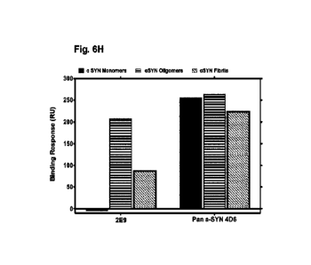
showing the total amount
of alpha synuclein in brain samples.
[0050] Figs. 8 A to H are graphs and images showing alpha-syn toxicity
inhibited by various
test antibodies.
[0051] Figs. 9A-B are a series of graphs. Fig. 9A is a graph showing antibody
selective binding
to synthetic alpha-syn oligomers but not to monomers or physiological
tetramers by SPR. Fig. SB is a
graph that shows antibody selective binding to alpha-syn oligomers and
sonicated fibrils.
[0052] Figs. 10 A to E are images showing that alpha-syn test antibodies
prefentially stain
small aggregates of alpha-syn over dense Lewy bodies by IHC and
immunofluorescent staining.
[0053] Figs. 10 F to K are images showing that alpha-syn test antibodies do
not give rise to
detectable staining of normal brain by IHC.
[0054] Fig. 11 is a graph showing the representative ELISA results for
antibody 2E9.
7
CA 03148562 2022-2-17
WO 2020/073121
PCT/CA2019/051434
[0055] Fig. 12A-B are a series of graphs. Fig. 12A is a graph that shows test
antibody binding
to DLB soluble brain extract. Fig. 12B is a graph showing that test antibody
binding to soluble DLB
extract is epitope-specific.
[0056] Figs. 13A to I are graphs and images showing that test antibodies
reduce PFF induced
formation of alpha-synuclein aggregates.
[0057] Fig. 14 A to H are graphs and images showing that test antibodies
reduce PFF induced
aggregation and phosphorylation of endogenous alpha-synuclein.
[0056] Fig. 15A-B are graphs where Fig. 15A is a graph showing that test
antibodies inhibit in-
vitro propagation of alpha-synuclein aggregation and Fig. 15B is a graph
showing that the cyclic peptide
comprising an alpha-syn peptide comprising a conformational el:dope is
sufficient to replicate the
seeding activity of pre-formed fibrils and is neutralized by test antibody
2E9.
[0059] Figs. 16A and B are bar graphs showing binding response of test
antibodies to brain
extract from a multiple system atrophy (MSA) patient
[0060] Fig. 16C is a bar graph showing binding response of test antibodies to
a prion enriched
fraction of brain extract.
[0061] Figs. 17A to C are a series graphs illustrating the binding profile of
test antibody 12G1.
[0062] Figs. 18A-C are a series of graphs illustrating the binding profile of
test antibody 9D8.
[0063] Figs. 19A-C are a series of graphs depicting the binding profile of
test antibody 10D5.
[0064] Fig. 20 is graph showing the quantitation of misfolded oligomeric a¨syn
in a biosample.
Detailed description of the Disclosure
[0065] Generation of conformation-specific antibodies was accomplished.
[0066] Antibodies raised to native protein regions tend not to be selective
for misfolded protein
such as non-native oligomeric species, and thus may bind to native functional
protein as well as
misfolded protein.
[0067] As described herein, to develop antibodies that may be selective for
misfolded
oligomeric forms of a-Syn, the inventors sought to identify regions of a-Syn
sequence that are prone to
disruption in the context of the fibril, and that may thus be exposed on the
surface of the misfolded
protein oligomers that could act as catalytic substrates for misfolding.
[0068] As described in the Examples, computational simulations, using
molecular dynamics
with standardized force-fields, were employed. An experimentally-validated
structural model of the fibril
structure was globally biased away from its reported conformation to be
partially unfolded, using
molecular dynamics, to yield regions of contiguous primary sequence that are
prone to be disordered
upon an external challenge in an anomalous cellular environment.
[0069] It was hypothesized that these weakly-stable regions may be selectively
exposed in
misfolded pathogenic species such as non-native oligomers.
8
CA 03148562 2022-2-17
WO 2020/073121
PCT/CA2019/051434
[0070] As described the Examples, the inventors have identified conformational
epitopes. The
inventors designed cyclic compounds comprising the identified epitopes to
mimic the putative selective
epitope by satisfying several criteria such as a higher exposed surface area,
loss of contact interactions
present in the fibril, and/or conformations that did not readily align by root
mean squared deviation
(RMSD) to the isolated monomeric ensemble, but would align more favorably to a
biased, partially
disordered fibril ensemble. As further shown in the Examples, monoclonal
antibodies produced using
immunogens comprising these cyclic compounds produced antibodies that
preferentially bound
misfolded oligomeric alpha-synuclein and inhibited alpha-synuclien induced
neural toxicity.
I. Definitions
[0071] As used herein, the term "a-Syn" alternately referred to as -a-
synuclein", or "Alpha-
Synuclein", or "alpha-syn" as used herein means all forms of a-Syn including
wildtype sequence a-Syn
and mutated forms, monomeric a-Syn, and aggregates thereof such as misfolded
oligomers and soluble
fibrillar forms of a-Syn, from all species, particularly human a-Syn (i.e. hua-
Syn). Human a-Syn is a
protein of typically 140 amino acid residues and the amino acid sequence (e.g.
Uniprot Accession
number P37840) and the nucleotide sequence (e.g. Accession number HGNC: 11138)
have been
previously characterized
[0072] 'Wild type" as used herein refers to the primary amino acid sequence of
non-mutant or
naturally occurring protein in humans.
[0073] "Native alpha-synudein polypeptide" or "a native a-Syn" as used herein
refers to the
alpha-synuclein monomer whether associated with membrane or cytosolic as well
as other multimers
found in normal cells, such as tetramer, and for example as can be predicted
when using one of the
chains from the PDB fibril (2N0A) as described herein. Native alpha-synuclein
polypeptide can be
detected using pan antibodies in for example brains not afflicted by a
synucleinopathy.
[0074] Models of the native a-Syn tetramer
[pnas.orn/ccii/doi/10.1073/pnas.11132601081
show that it is stabilized by interactions that include residues in the above
epitopes, specifically, inter-
chain salt-bridges between K60-E57 and between K34-E57. As well, 062 exhibited
among the largest
paramagnetic relaxation effects indicating that it is strongly interacting in
the tetramer vs. the isolated
monomer. These interactions may result in the sequestration of the epitope in
the naturally occurring
native tetrameric form, so that antibodies targeting the epitope would select
for non-native species (e.g.
misfolded oligomeric alpha-synuclein).
[0075] "Structured fibrir, "un-stressed fibril", or "unbiased fibril" as used
herein refers to the
expected conformations that would be observed in thermal equilibrium for a
fibril of alpha-synuclein,
e.g. for which PDB 2NOA would be a representative example of.
[0076] "Misfolded ofigomer", "non-native oligomer as used herein refers to the
secondary and
tertiary structure of a multisubunit polypeptide or polypeptide aggregation,
and indicates that the
oligomeric polypeptide, or a subunit therein has adopted a conformation (e.g.
at one or more locations)
that is different from that typically adopted by the native monomer and/or
tetramer. Although misfolding
can be caused by mutations in a protein, such as amino acid deletion,
substitution, or addition, wild-
9
CA 03148562 2022-2-17
WO 2020/073121
PCT/CA2019/051434
type sequence protein can also be misfolded in disease, and expose disease-
specific or selective
epitopes for instance, as a result of a change in microenvironmental
conditions, or oligomer formation
that may be on- or off-pathway to fibril formation (e.g insoluble fibrils).
Accordingly, "misfolded
oligomeric a-Syn polypeptide", "misfolded alpha-Syn" or "misfolded oligomeric
a-Syn" when referring to
the polypeptide herein refers to a-Syn polypeptide oligomers that displays a
conformation that is
different from nascently folded monomeric a-Syn and/or natively folded
tetrameric alpha-synuclein and
includes for example non-native oligomers, soluble fibrils, protofibrils, and
fibril fragments. Soluble
fibrils include for example a-syn fibril species that are found in the
supernatant of a sample subjected
to uttracentrifugation at 100, 000xg for 1 hour Soluble fibrils can be
produced by sonicating fibrils which
produces fragments. For example, misfolded oligomeric a-Syn can include a
conformation that is
partially-ordered, containing parts of the fibril structure, and partially-
disordered, containing polymer
segments of amino acids that have alternate conformations than either monomer,
tetramer and/or fibril
a-Syn_ Misfolded oligomeric alpha-synuclein as shown herein includes
conformational epitopes that are
selectively presented or accessible for binding wherein the epitope sequence
in misfolded oligomeric
alpha-synuclein can be conformationally different than the corresponding
sequence in the context of
the isolated monomer, as measured for example by side chain orientation or by
root mean-squared
deviation (RMSD). Misfolded alpha-synuclein may comprise at least one of the
residues E57, K58, T59,
K60, E61, or Q62 in an alternate conformation than occupied by E57, K58, T59,
K60, E61, and/or Q62
in a non-misfolded proteinic conformation such as native monomer and/or
tetramer or in insoluble fibrils
such as those found in Lewy body deposits. Soluble a-synuclein fibrils refers
to smaller fibrils or
fragments for example fibrils that are sonicated as described in the Examples
and which are in solution
as well as disease associated smaller fibrils that are not present in Lewy
bodies which comprise
insoluble fibrils.
LOOT?] The term "mutant ci-Syn" refers to forms of a-Syn, and particularly
endogenous forms
of a-Syn that occur as a result of genetic mutation that result for instance
in amino acid substitution,
such as those substitutions characteristic for instance of familial
Parkinson's disease.
[0078] The term "EKTK (SEQ ID NO: 2)" means the amino acid sequence: glutamic
acid,
lysine, threonine, lysine, as shown in SEQ ID NO: 2. The term "TKEQ (SEQ ID
NO: 4)" means the
amino acid sequence: threonine, lysine, glutamic acid, glutamine, as shown in
SEQ ID NO: 4. Similarly
EKT, KTK, TKE, KEQ, EKTKEQ (SEQ ID NO: 1), EKTKE (SEQ ID NO: 8), KTKE (SEQ ID
NO: 3),
KTKEQ (SEQ ID NO: 9) refer to the amino acid sequences identified by the 1-
letter amino acid code.
Depending on the context, the reference of the amino acid sequence can refer
to a sequence in a-Syn
or an isolated peptide, such as the amino acid sequence of the epitope portion
of a cyclic compound
The sequences EKTK (SEQ ID NO: 2) and TKEQ (SEQ ID NO: 4) consist of residues
57-60 and
residues 59-62 in the a-Syn amino acid primary sequence, respectively (e.g.
Uniprot Accession number
P37840).
[0079] The term "epitope in EKTKEQ (SEQ ID NO: 1)" as used herein refers to
any part thereof
that is specifically bound by an antibody. For example the antibody may
specifically bind the side chains
and/or backbones of a combination of several residues in the epitope,
including several of E57, and/or
CA 03148562 2022-2-17
WO 2020/073121
PCT/CA2019/051434
E61, and/or K58, and/or K60, and/or 062, and/or T59, or a particular part of
these residues, or a
combination of any of the foregoing. The epitope can be a conformational
epitope.
[0080] The term "epitope" as used herein means a sequence of amino acids in an
antigen
wherein the amino acids (or a subset thereof) in the sequence are specifically
recognized by an antibody
or binding fragment, for example an antibody or binding fragment described
herein. An epitope can
comprise one or more antigenic determinants. For example, an antibody
generated against an isolated
peptide corresponding to a conformational epitope recognizes part or all of
said epitope sequence.
[0081] The term "epitope selectively presented or accessible in misfolded
oligomeric a-Syn"
as used herein refers to a conformational epitope that is selectively
presented or accessible on
misfolded oligomeric a-Syn polypeptide as present in synucleinopathies such as
Parkinson's disease
and Lewy Body Dementia (e.g. disease-associated misfolded a-Syn) whether in
multimeric, oligomeric,
or aggregated forms, but not on the molecular surface of the nascent monomeric
peptide or tetrameric
forms of a-Syn as found normally in viva
[0082] As used herein, the term "conformational epitope" refers to a sequence
of amino acids
or an antigenic determinant thereof that has a particular three-dimensional
structure in a species of a
protein wherein at least an aspect of the three-dimensional structure is
present or is more accessible
to antibody binding compared to in another species such as an isolated monomer
(native) or other
native structure. Antibodies which specifically bind a conformational epitope
recognize the spatial
arrangement of one or more of the amino acids of that conformation-specific
epitope. For example, a
conformational epitope in EKTKEQ (SEQ ID NO: 1) can refer to a conformation of
one or more amino
acids or parts thereof of EKTKEQ (SEQ ID NO: 1) that is recognized by
antibodies selectively, for
example at least 2 fold, 3 fold, 5 fold, 10 fold, 50 fold, 100 fold, 250 fold,
500 fold or 1000 fold or greater
more selectivity as compared to another conformation, optionally the
corresponding region in the a-Syn
monomer or insoluble fibril or for example antibodies raised using a
corresponding linear peptide or part
thereof.
[0083] Reference to the "cyclic peptide" herein can refer to a fully
prateinaceous cyclic
compound (e.g. wherein the linker is 2, 3, 4, 5, 6, 7 or 8 amino acids). It is
understood that properties
described for the cyclic peptide determined in the examples can be
incorporated in other compounds
(e.g. cyclic compounds) comprising non-amino acid linker molecules.
[0084] The term "amino acid" includes all of the naturally occurring amino
adds as well as
modified L-amino acids. The atoms of the amino acid can for example include
different isotopes. For
example, the amino acids can comprise deuterium substituted for hydrogen,
nitrogen-15 substituted for
nitrogen-14, and carbon-13 substituted for carbon-12 and other similar
changes.
[0085] The term "antibody" as used herein is intended to include monoclonal
antibodies,
polyclonal antibodies, single chain, single domain, humanized and other
chimeric antibodies as well as
binding fragments thereof. The antibody may be from recombinant sources and/or
produced in
transgenic animals. The antibody in an embodiment comprises a heavy chain
variable region or a heavy
chain comprising a heavy chain complementally determining region 1, heavy
chain complementarity
11
CA 03148562 2022-2-17
WO 2020/073121
PCT/CA2019/051434
determining region 2 and heavy chain complementarity determining region 3, as
well as a light chain
variable region or light chain comprising a light chain complementarity
determining region 1, light chain
complementarity determining region 2 and light chain complementarity
determining region 3. Also
included are human antibodies that can be produced through using biochemical
techniques or isolated
from a library. Humanized or chimeric antibody may include sequences from one
or more than one
isotype or class. Reference to antibody or antibodies of the disclosure refers
to antibody or antibodies
described herein that are for example raised using an immunogen described
herein and/or selective for
an epitope described herein for example KTKE (SEQ ID NO: 3), EKTK (SEQ ID NO:
2) or TKEQ (SEQ
ID NO: 4) or a part thereof in the context for example of the epitope,
misfoldecl oligomeric alpha-
synuclein and/or a cyclic compound comprising one of said epitope sequences.
[0086] The term "heavy chain complementarity determining region" as used
herein refers to
regions of hypervariability within the heavy chain variable region of an
antibody molecule. The heavy
chain variable region has three complementarity determining regions termed
heavy chain
complementarity determining region 1 (CDR-H1), heavy chain complementarity
determining region 2
(CDR-H2) and heavy chain complementarity determining region 3 (CDR-H3) from
the amino terminus
to carboxy terminus.
[0087] The term "heavy chain variable region" as used herein refers to the
variable domain of
the heavy chain comprising the heavy chain complementarity determining region
1, heavy chain
complementarity determining region 2 and heavy chain complementarity
determining region 3. One or
more amino acids or nucleotides can be modified for example replaced with a
conservative substitution,
for example outside the CDR sequences. The variable region comprises framework
region 1 (FR1),
followed by CDR1, followed by framework region 2 (FR2), followed by CDR2,
followed by framework
region 3 (FR3), followed by CDR3, followed by framework region 4 (FR4).
[0088] The term "light chain
complementarity determining region" as used herein refers
to regions of hypervariability within the light chain variable region of an
antibody molecule. Light chain
variable regions have three complementarity determining regions termed light
chain complementarity
determining region 1, light chain complementarity determining region 2 and
light chain complementarity
determining region 3 from the amino terminus to the carboxy terminus.
[0089] The term "light chain variable
region" as used herein refers to the variable
domain of the light chain comprising the light chain complementarity
determining region 1, light chain
complementarity determining region 2 and light chain complementarity
determining region 3. The
variable region comprises framework region 1 (FR1), followed by CDR1, followed
by framework region
2 (FR2), followed by CDR2, followed by framework region 3 (FR3), followed by
CDR3, followed by
framework region 4 (FR4).
[0090] The phrase "isolated antibody" refers to antibody produced in vivo or
in vitro that has
been removed from the source that produced the antibody, for example, an
animal, hybridoma or other
cell line (such as recombinant cells that produce antibody). The isolated
antibody is optionally "purified",
which means at least: 80%, 85%, 90%, 95%, 98% or 99% purity.
12
CA 03148562 2022-2-17
WO 2020/073121
PCT/CA2019/051434
[0091] The term "binding fragment" as used herein to a part or portion of an
antibody or
antibody chain comprising fewer amino acid residues than an intact or complete
antibody or antibody
chain and which binds the antigen or competes with intact antibody. Exemplary
binding fragments
include without limitations Fab, Fab', F(alS)2, scFv, dsFv, ds-scFv,
nanobodies, minibodies, diabodies,
and multimers thereof. Fragments can be obtained via chemical or enzymatic
treatment of an intact or
complete antibody or antibody chain. Fragments can also be obtained by
recombinant means. For
example, F(a131)2 fragments can be generated by treating the antibody with
pepsin. The resulting F(ati)2
fragment can be treated to reduce disulfide bridges to produce Fab' fragments.
Papain digestion can
lead to the formation of Fab fragments. Fab, Fab' and F(a131)2, scFv, dsFv, ds-
scFv, dimers, minibodies,
diabodies, bispecific antibody fragments and other fragments can also be
constructed by recombinant
expression techniques.
[0092] When an antibody is said to bind to an epitope in, such as EKTKEO (SEQ
ID NO: 1),
or TKEQ (SEQ ID NO: 4), what is meant is that the antibody specifically binds
to a polypeptide or
compound containing the specified residues or a part thereof for example at
least 1 residue or at least
2 residues. Such an antibody does not necessarily contact every residue of
EKTK (SEQ ID NO: 2) or
TKEQ (SEQ ID NO: 4), and every single amino acid substitution or deletion
within said epitope does
not necessarily significantly affect or equally affect binding affinity.
[0093] The term "detectable label" as used herein refers to moieties such as
peptide
sequences, fluorescent proteins that can be appended or introduced into a
peptide or compound
described herein and which is capable of producing, either directly or
indirectly, a detectable signal_ For
example, the label may be radio-opaque, positron-emitting radionuclide (for
example for use in PET
imaging), or a radioisotope, such as 3H, 13N, 14C, 18p, 32p, 35S, 1231, 1251,
1311-
, a fluorescent (fluorophore)
or chemiluminescent (chromophore) compound, such as fluorescein
isothiocyanate, rhodamine or
luciferin; an enzyme, such as alkaline phosphatase, beta-galactosidase or
horseradish peroxidase; an
imaging agent; or a metal ion. The detectable label may be also detectable
indirectly for example using
secondary antibody.
[0094] The term "greater affinity" as used herein refers to a degree of
antibody binding where
an antibody X binds to target Y more strongly (Km) and/or with a smaller
dissociation constant (Koff)
than to target Z, and in this context antibody X has a greater affinity for
target Y than for Z. Likewise,
the term "lesser affinity" herein refers to a degree of antibody binding where
an antibody X binds to
target Y less strongly and/or with a larger dissociation constant than to
target Z, and in this context
antibody X has a lesser affinity for target Y than for Z. The affinity of
binding between an antibody and
its target antigen, can be expressed as KA equal to 1/Ko where KD is equal to
kodkoff. The Icon and koff
values can be measured using surface plasmon resonance (measurable for example
using a Biacore
system).
[0095] Also as used herein, the term "immunogenic" refers to substances which
elicit the
production of antibodies, activate lymphocytes and other reactive immune cells
directed against an
antigenic portion of the immunogen.
13
CA 03148562 2022-2-17
WO 2020/073121
PCT/CA2019/051434
[0096] An "immunogen" as used herein means a substance which provokes an
immune
response and/or causes production of an antibody and can comprise for example
cyclic peptides
described herein, conjugated as multiantigenic peptide and/or fused to an
immunogenicity enhancing
agent such as Keyhole Limpet Hemocyanin (KLH). In addition to the conjugates
described herein,
immunogenic peptide mimetics which elicit cross-reactive antibodies to the
epitopes identified, e.g.
EKTKEQ (SEQ ID NO: 1), EKTK (SEQ ID NO: 2), TKEQ (SEQ ID NO: 4) or KTKE (SEQ
ID NO: 3). To
serve as a useful imrnunogen, the ci-Syn peptide desirably incorporates a
minimum of about 3, 4, 5, 6,
or 7 a-Syn residues, comprising E57, K58, T59, K60, E61, and/or Q62.
[0097] The term "inhibiting" as used herein for example in the context of an
antibody of the
disclosure inhibiting alpha-syn phosphorylation means reducing the amount of
alpha-syn
phosphorylation in the presence of the antibody by at least 10%, at least 20%,
at least 30% compared
to in the absence of the antibody.
[0098] The term "corresponding linear compound" with regard to a cyclic
compound refers to
a compound, optionally a peptide, comprising or consisting of the same
sequence or chemical moieties
as the cyclic compound but in linear (non-cyclized) form.
[0099] The term 'nucleic acid sequence" as used herein refers to a sequence of
nucleoside
or nucleotide monomers consisting of naturally occurring bases, sugars and
intersugar (backbone)
linkages. The term also includes modified or substituted sequences comprising
non-naturally occurring
monomers or portions thereof. The nucleic acid sequences of the present
application may be
deoxyribonucleic acid sequences (DNA) or ribonucleic acid sequences (RNA) and
may include naturally
occurring bases including adenine, guanine, cytosine, thymidine and uracil.
The sequences may also
contain modified bases. Examples of such modified bases include aza and deaza
adenine, guanine,
cytosine, thymidine and uracil; and xanthine and hypoxanthine. The nucleic
acid can be either double
stranded or single stranded, and represents the sense or antisense strand.
Further, the term "nucleic
acid" includes the complementary nucleic acid sequences as well as codon
optimized or synonymous
codon equivalents. The term "isolated nucleic acid sequences" as used herein
refers to a nucleic acid
substantially free of cellular material or culture medium when produced by
recombinant DNA
techniques, or chemical precursors, or other chemicals when chemically
synthesized. An isolated
nucleic acid is also substantially free of sequences which naturally flank the
nucleic acid (i.e. sequences
located at the 5' and 3' ends of the nucleic acid) from which the nucleic acid
is derived.
[00100] The term "vector' as used herein comprises any intermediary vehicle
for a nucleic acid
molecule which enables said nucleic acid molecule, for example, to be
introduced into prokaryotic
and/or eukaryotic cells and/or integrated into a genome, and include plasmids,
phagemids,
bacteriophages or viral vectors such as retroviral based vectors, Adeno
Associated viral vectors and
the like. The term "plasmid" as used herein generally refers to a construct of
extrachromosomal genetic
material, usually a circular DNA duplex, which can replicate independently of
chromosomal DNA.
[00101] The term "host cell' refers to a cell into which a recombinant DNA
expression vector
can be introduced to produce a recombinant cell. The host cell can be a
bacterial cell such as E. coli as
14
CA 03148562 2022-2-17
WO 2020/073121
PCT/CA2019/051434
well as any type of microbial, yeast, fungi, insect or mammalian host cell.
Mammalian host cell can be
a human cell.
[00102] The term "pharmaceutically acceptable" means that the carrier,
diluent, or excipient is
compatible with the other components of the formulation and not substantially
deleterious to the
recipient thereof.
[00103] The term "administered" as used herein means administration of a
therapeutically
effective dose of a compound or composition of the disclosure to a cell or
subject.
[00104] As used herein, the phrase "effective amount" means an amount
effective, at dosages
and for periods of time necessary to achieve a desired result. Effective
amounts when administered to
a subject may vary according to factors such as the disease state, age, sex,
weight of the subject.
Dosage regime may be adjusted to provide the optimum therapeutic response.
[00105] The term "treating" or "treatment" as used herein and as is well
understood in the art,
means an approach for obtaining beneficial or desired results, including
clinical results. Beneficial or
desired clinical results can include, but are not limited to, alleviation or
amelioration of one or more
symptoms or conditions, diminishment of extent of disease, stabilized (i.e.
not worsening) state of
disease, preventing spread of disease, delay or slowing of disease
progression, amelioration or
palliation of the disease state, diminishment of the reoccurrence of disease,
and remission (whether
partial or total), whether detectable or undetectable. 'Treating" and
'Treatment" can also mean
prolonging survival as compared to expected survival if not receiving
treatment "Treating" and
"treatment" as used herein also include prophylactic treatment. For example, a
subject with early stage
PD can be treated to prevent progression. Such a subject can be treated with a
compound, antibody,
immunogen, immunoconjugate or composition described herein to prevent
progression.
[00106] As used herein "specifically binds" in reference to an antibody means
that the antibody
binds to its target antigen with greater affinity than it does to a
structurally or conformationally different
antigen and/or to an antigen with modified or mutated sequence. For example a
multivalent antibody
binds its target with KD of at least 5e-5, at least le-6, at least le-7, at
least le-8, or at least le-9.
Affinities greater than at least 1e-7 are preferred. An antigen binding
fragment such as Fab fragment
comprising one variable domain, may bind its target with for example a 10 fold
or 100 fold less
affinity/avidity than a multivalent interaction with a non-fragmented
antibody.
[00107] The term "selective" or "selectively binds" as used herein with
respect to an antibody
that preferentially binds a form of a-Syn (e.g. misfolded conformations such
as misfolded oligomers and
small soluble fibrils relative to isolated native monomer or native tetramers
and/or insoluble fibrillar
alpha-synuclein) means that the binding protein binds the form with at least 2
fold, 3 fold, or at least 5
fold, at least 10 fold, at least 100 fold, at least 250 fold, at least 500
fold or at least 1000 fold or more
greater affinity. Accordingly an antibody that is more selective for a
particular conformation (e.g.
misfolded oligomers) preferentially binds the particular form of a-Syn with at
least 3 fold etc. greater
affinity compared to another form.
CA 03148562 2022-2-17
WO 2020/073121
PCT/CA2019/051434
[00108] The term "linker" as used herein means a
chemical moiety, preferably
poorly
immunogenic or non-immunogenic, that can be covalently linked directly or
indirectly to the a-Syn
peptide N- and/or C- termini comprising at least 3 amino acids of EKTKEQ (SEQ
ID NO: 1), optionally
EKTK (SEQ ID NO: 2), KTKE (SEQ ID NO: 3) or TKEQ (SEQ ID NO: 4) epitope
peptide, which is linked
to the peptide N- and/or C- termini. The linker ends can for example be joined
to produce a cyclic
compound. The linker can comprise one or more functionalizable moieties such
as one or more cysteine
residues. The linker can be linked via the functionalizable moieties to a
carrier protein or an immunogen
enhancing agent such as keyhole limpet hemocyanin (KLH). The cyclic compound
comprising the linker
is of longer length than the peptide itself. That is, when cyclized the
peptide with a linker (for example
of 3 amino acid residues) makes a larger closed circle than the peptide
without a linker. The linker may
include, but is not limited to, non-immunogenic moieties such as amino acids G
and A, or PEG repeats.
[00109] The term "functionalizable moiety" as used herein refers to a chemical
entity with a
"functional group" which as used herein refers to a group of atoms or a single
atom that will react with
another group of atoms or a single atom (so called "complementary functional
group") to form a chemical
interaction between the two groups or atoms. In the case of cysteine, the
functional group can be ¨SH
which can be reacted to form a disulfide bond. Accordingly the linker can for
example be CCC. The
reaction with another group of atoms can be covalent or a strong non-covalent
bond, for example as in
the case as biotin-streptavidin bonds, which can have Kd¨le-14. A strong non-
covalent bond as used
herein means an interaction with a Kd of at least le-9, at least le-10, at
least le-11, at least le-12, at
least le-13 or at least le-14.
[00110] Proteins and/or other agents may be coupled to the cyclic compound,
either to aid in
immunogenicity, or to act as a probe in in vitro studies. For this purpose,
any functionalizable moiety
capable of reacting (e.g. making a covalent or non-covalent but strong bond)
may be used. In one
specific embodiment, the functionalizable moiety is a cysteine residue which
is reacted to form a
disulfide bond with an unpaired cysteine on a protein of interest, which can
be, for example, an
immunogenicity enhancing agent such as Keyhole limpet hemocyanin (KLH), or a
carrier protein such
as Bovine serum albumin (BSA) used for in vitro immunoblots or
immunohistochemical assays.
[00111]The term "animal" or "subject" as used herein includes all members of
the animal
kingdom including mammals, including humans.
[00112] Compositions or methods "comprising" or "including" one or more
recited elements
may include other elements not specifically recited. For example, a
composition that "comprises" or
"includes" an antibody may contain the antibody alone or in combination with
other ingredients.
[00113] In understanding the scope of the present disclosure, the term
"consisting" and its
derivatives, as used herein, are intended to be close ended terms that specify
the presence of stated
features, elements, components, groups, integers, and/or steps, and also
exclude the presence of other
unstated features, elements, components, groups, integers and/or steps.
[00114]The recitation of numerical ranges by endpoints herein includes all
numbers and
fractions subsumed within that range (e.g. Ito 5 includes 1, 1.5, 2, 2.75, 3,
3.90, 4, and 5). It is also to
16
CA 03148562 2022-2-17
WO 2020/073121
PCT/CA2019/051434
be understood that all numbers and fractions thereof are presumed to be
modified by the term "about."
Further, it is to be understood that "a," "an," and "the" include plural
referents unless the content clearly
dictates otherwise. The term "about" means plus or minus OA to 50%, 5-50%, or
10-40%, preferably
10-20%, more preferably 10% or 15%, of the number to which reference is being
made.
[00115] Further, the definitions and embodiments described in particular
sections are intended
to be applicable to other embodiments herein described for which they are
suitable as would be
understood by a person skilled in the art. For example, in the following
passages, different aspects of
the invention are defined in more detail. Each aspect so defined may be
combined with any other aspect
or aspects unless clearly indicated to the contrary. In particular, any
feature indicated as being preferred
or advantageous may be combined with any other feature or features indicated
as being preferred or
advantageous.
[00116] The singular forms of the articles "a," "an," and "the" include plural
references unless
the context clearly dictates otherwise. For example, the term "a compound" or
"at least one compound"
can include a plurality of compounds, including mixtures thereof.
Epitopes and Epitope Compounds
[00117] The inventors have identified sequences in a-Syn protein including
EKTK (SEQ ID NO:
2), KTKE (SEQ ID NO: 3) and TKEQ (SEQ ID NO: 4) at amino acids 57-60, 58-61
and 59-62
respectively that may be conformational epitopes, such that for examplethat
EKTK (SEQ ID NO: 2) and
TKEQ (SEQ ID NO: 4) or a part of each of thereof may be selectively accessible
to antibody binding in
misfolded oligomeric species of a-Syn.
[00118] Based on one or more conformational differences identified between the
epitopes
identified in monomeric, fibril and/or biased a-Syn fibril ensembles, the
inventors have designed
conformationally restricted compounds and immunogens for producing antibodies.
[00119] As shown in the Examples antibodies raised using said immunogens are
useful for
detecting or targeting misfolded oligomeric a-Syn.
[00120] As described in the Examples, cyclic compounds such as cyclic peptides
cyclo(CGGGGEKTKGG) (SEQ ID NO: 5), cyclo(CGTKEQGGGG) (SEQ ID NO: 7),
cyclo(CGGGEKTKGG) (SEQ ID NO: 10) and cyclo(CGGGGTKEQGG)(SEQ ID NO: 11) were
identified
to capture the conformational differences of the corresponding epitope in
misfolded oligomeric species
of a-Syn relative to monomeric and/or insoluble fibril species. For example,
RMSD structural alignment
for amino acids in the cyclic 10-mer cyclo(CGTKEQGGGG) (SEQ ID NO: 7) were
found to be
significantly different than the corresponding quantities in the monomeric
ensemble. This suggests that
the cyclic compound may provide for a conformational epitope that is
conformationally-distinct from the
sequence presented in the nascent monomeric a-Syn and/or insoluble fibril.
[00121]Accordingly, the present disclosure identifies conformational epitopes
in a-Syn for
example peptides EKTK (SEQ ID NO: 2 ), and KTKE (SEQ ID NO: 3) and TKEQ (SEQ
ID NO: 4) or a
part thereof such as EK corresponding to amino acids residues 57-58 on a-Syn
and KEQ corresponding
to amino acids 60-62 on a-Syn. As demonstrated in the Examples, EKTK (SEQ ID
NO: 2) and TKEQ
17
CA 03148562 2022-2-17
WO 2020/073121
PCT/CA2019/051434
(SEQ ID NO: 4) were identified as regions prone to disorder in a-Syn. The
residues EKTK (SEQ ID NO:
2) and TKEQ (SEQ ID NO: 4) emerged in a prediction using the Collective
Coordinates method as
described in the Examples.
[00122] An aspect includes a compound comprising an a-Syn peptide comprising
at least 3
amino acids of EKTKEQ (SEQ ID NO: 1), optionally EKTK (SEQ ID NO: 2), KTKE
(SEQ ID NO: 3) or
TKEQ (SEQ ID NO: 4), and/or part of any of the foregoing such as KEQ. In an
embodiment, the a-Syn
peptide is selected from EKTK (SEQ ID NO: 2), KTKE (SEQ ID NO: 3), TKEQ (SEQ
ID NO: 4), EKTKE
(SEQ ID NO: 8), EKT, KTK, TKE, KEQ, or KTKEQ (SEQ ID NO: 9).
[00123] The a-Syn peptide can also include an additional 1, 2 or 3 amino acids
in a-Syn either
N-terminal and/or C-terminal to EKTKEQ (SEQ ID NO: 1) (or an internal sequence
thereof such as EKT
or EKTK (SEQ ID NO: 2)) for 1, 2 or 3 N-terminal amino acid residues, and/or)
with 1, 2 or 3 C-terminal
amino acid residues. In some embodiments, the maximum length of the a-Syn
peptide is 9 amino acids,
8 amino acids or 7 amino acids.
[00124] In an embodiment, the a-Syn peptide comprises or consists of KEQ, TKEQ
(SEQ ID
NO: 4), KTKEQ (SEQ ID NO: 9) or EKTKEQ (SEQ ID NO: 1).
[00125] In an embodiment, the compound further comprises a linker. The linker
can comprise
one or more functionalizable moieties_ The linker can for example comprise 1,
2, 3, 4, 5, 6, 7 or 8 amino
acids and/or equivalently functioning molecules such as polyethylene glycol
(PEG) moieties, and/or a
combination thereof In an embodiment, the linker amino acids are selected from
non-immunogenic or
poorly immunogenic amino acid residues such as G and A, for example the linker
can be GG, GGG,
GAG, G(PEG)G, PEG-PEG(also referred to as PEG2)-GG and the like. One or more
functionalizable
moieties e.g. amino acids with a functional group may be included for example
for coupling the
compound to an agent or detectable tag or a carrier such as BSA or an
irnmunogenicity enhancing
agent such as KLH. The functionalizable moiety can be an amino acid such as
cysteine. In an
embodiment, the linker comprises up to or a maximum of 1, 2, 3, 4, 5, 6, 7 or
8 amino acids.
[00126] In an embodiment, the linker comprises GC-PEG, PEG-GC, GCG or PEG2-CG.
In
another embodiment, the linker comprises GCGGGG (SEQ ID NO: 12), GGCGG (SEQ ID
NO: 13),
GGCGGGG (SEQ ID NO: 14), GGGCGG (SEQ ID NO: 15) or GGGGCGG (SEQ ID NO: 16).
Other
linkers are provided (presented in constructs comprising the alpha-Syn
peptide) in Tables 2-4.
[00127] Proteinaceous portions of compounds (or the compound wherein the
linker is also
proteinaceous) may be prepared by chemical synthesis using techniques well
known in the chemistry
of proteins such as solid phase synthesis or synthesis in homogenous solution.
[00128] The compound can be linear and can be used for example for selecting
antibodies that
bind preferentially to the corresponding cyclic compound_ Preferably, the
compound is a conformational
compound, such as a cyclic compound. As shown in the Examples this can be
accomplished using a
cyclic peptide comprising the a-Syn peptide.
[00129] An aspect therefore provides a cyclic compound comprising an a-Syn
peptide
comprising at least 3 amino acids of EKTKEQ (SEQ ID NO: 1), optionally EKTK
(SEQ ID NO: 2), KTKE
18
CA 03148562 2022-2-17
WO 2020/073121
PCT/CA2019/051434
(SEQ ID NO: 3) or TKEQ (SEQ ID NO: 4), and/or part of any of the foregoing and
a linker, wherein the
linker is covalently coupled directly or indirectly to the a-Syn peptide. As
shown in the Examples,
residues in the cyclic peptide are in an alternate conformation compared to
the corresponding residue
in monomer or fibril ensembles. In an embodiment, the cyclic compound
comprises an a-Syn peptide
and linker described herein. In an embodiment, the cyclic compound comprises
an a-Syn peptide
comprising EKT, KEQ, EKTK (SEQ ID NO: 2), KTKE (SEQ ID NO: 3), or TKEQ (SEQ ID
NO: 4 ) and
up to 6 a-Syn residues (e.g. 1 or 2 amino acids N and/or C terminus to EKTK
(SEQ ID NO: 2), KTKE
(SEQ ID NO: 3), or TKEQ (SEQ ID NO: 4) and a linker, wherein the linker is
covalently coupled directly
or indirectly to the peptide N-terminus residue and the C-terminus residue of
the a-Syn peptide. The
exposure of the residues in the cyclic peptide can be different than
corresponding residues, in the
monomeric and/or fibril ensembles and cellular monomeric and insoluble
fibrillar a-Syn. For example in
the cyclic compound, at least one of E57, K58, T59, K60, E61, or Q62, has more
surface exposure
than the conformation occupied in the monomeric ensemble_
[00130] In embodiments wherein the peptide comprising EKTK (SEQ ID NO: 2),
EKT, EKTKE
(SEQ ID NO: 8), KTKEQ (SEQ ID NO: 9), KEQ or TKEQ (SEQ ID NO: 4) includes 1,2
or 3 additional
residues found in a-Syn that are N- and/or C- terminal to EKTK (SEQ ID NO: 2)
or TKEQ (SEQ ID NO:
4) the linker in the cydized compound is covalently linked to the N- and/or C-
termini of the a-Syn
additional residues. Similarly, where the a-Syn peptide is EKTK (SEQ ID NO: 2)
the linker is covalently
linked to residues E and K, where the a-Syn peptide is TKEQ (SEQ ID NO: 4),
the linker is covalently
linked to residues T and Q, and where the a-Syn peptide is KTKEQ (SEQ ID NO:
9), the linker is
covalently linked to residues K and Q.
[00131] In an embodiment the cyclic compound comprises a peptide comprising or
consisting
of EKTK (SEQ ID NO: 2), KTKE (SEQ ID NO: 3), or TKEQ (SEQ ID NO: 4) and a
linker, wherein the
linker is coupled to the N- and C- termini of the peptide.
[00132] In an embodiment, the alternate conformation is a more solvent-exposed
conformation,
for one or more of the residues E57, K58, T59, K60, E61, or Q62.
[00133] In one embodiment, the cyclic compound is a cyclic peptide. In another
embodiment,
the cyclic peptide comprises or consists of the sequence of any one of SEQ ID
NOs: 5, 7 and 10-60. In
one embodiment, the cyclic peptide comprises or consists of the sequence of
CGGGGEKTKGG (SEQ
ID NO: 5). In another embodiment, the cyclic peptide comprises or consists of
the sequence of
CGTKEQGGGG (SEQ ID NO: 7). In another embodiment, the cyclic peptide comprises
or consists of
the sequence of CGGGEKTKGG (SEQ ID NO: 10).
[00134] The cyclic peptides and corresponding linear peptides can for example
be referenced
by identifying the positions of the linker residues relative to the a-Syn
peptide and the functionalizable
moiety. For example, CGGGGEKTKGG (SEQ ID NO: 5) can be referred to as the 4,2
construct,
CGTKEQGGGG (SEQ ID NO: 7), can be referred to as the 1,4 construct and
CGGGEKTKGG can be
referred to as the 3,2 construct.
19
CA 03148562 2022-2-17
WO 2020/073121
PCT/CA2019/051434
[00135] Methods for making cyclized peptides are known in the art and include
SS-cyclization
or amide cyclization (head-to-tail, or backbone cyclization). Methods are
further described in in the
Example section. For example, a peptide with "C" residues at its N- and C-
termini, e.g. CGGEKTKGGC
(SEQ ID NO: 17), can be reacted by SS-cyclization to produce a cyclic peptide.
The cyclic compound
can be synthesized as a linear molecule with the linker covalently attached to
the N-terminus or C-
terminus of the peptide comprising the a-Syn peptide, optionally EKTK (SEQ ID
NO: 2), KTKE (SEQ ID
NO: 3), or TKEQ (SEQ ID NO: 4) or related epitope, prior to cyclization.
Alternatively, part of the linker
is covalently attached to the N-terminus and part is covalently attached to
the C-terminus prior to
cyclization. In either case, the linear compound is cyclized for example in a
head to tail cyclization (e.g.
amide bond cyclization).
[00136] As described in the Examples, cyclic compounds were assessed for their
relatedness
to the conformational epitopes identified, synthesized and used to prepare
immunogens and used to
raise antibodies selective for misfolded oligomeric c*-Syn. The epitopes EKTK
(SEQ ID NO: 2), KTKE
(SEQ ID NO: 3), or TKEQ (SEQ ID NO: 4) and/or parts thereof, as described
herein may be a potential
target in misfolded propagating strains of a-Syn, and antibodies that
recognize the conformational
epitope as shown herein are useful for detecting misfolded species and
inhibiting such propagating
strains.As mentioned the above cyclic compounds comprising the a-Syn peptides
can be used as an
immunogen to raise antibodies.
[00137] Accordingly another aspect includes an immunogen comprising a
conformational
compound, optionally a cyclic compound, such as a cyclic peptide, described
herein. In an embodiment,
the immunogen comprises an immunogenicity enhancing agent such as Keyhole
Limpet Hemocyanin
(KLH) or carrier such as bovine serum albumin (BSA) or ovalbumin_ The
immunogenicity enhancing
agent can be coupled to the compound either directly, such as through an amide
bound, or indirectly
through a chemical linker. Alternatively the immunogen may be a multi
antigenic peptide (MAP).
[00138] The immunogen can be produced by conjugating the cyclic compound
containing the
constrained a-Syn epitope peptide to an immunogenicity enhancing agent such as
KLH or a carrier
such as BSA using for example the method described in Lateef et al 2007,
herein incorporated by
reference. In an embodiment, the method described in Examples 3 and 4 is used.
Ill. Antibodies
[00139] The compounds and particularly the cyclic compounds comprising any 3
amino acid
residues of EKTKEQ (SEQ ID NO: 1) such as alpha-Syn peptide EKTK (SEQ ID NO:
2), KTKE (SEQ
ID NO: 3), or TKEQ (SEQ ID NO: 4) described herein can be used to raise
antibodies that selectively
bind the compound comprising the alpha-Syn peptide relative to the
corresponding linear compound,
and/or also bind an epitope in the alpha-Syn peptide in misfolded forms of
alpha-Syn including
misfolded oligomeric alpha-Syn relative to monomeric and/or alpha-Syn
insoluble fibrils. As shown in
the Examples, the cyclic compounds exhibit one or more spatial conformations
that are dissimilar to
unbiased fibrillar alpha-Syn and which resemble partially unfolded fibrillar
alpha-Syn (biased alpha-
Syn). Further, it is demonstrated that antibodies raised using said compounds
are selective for cyclic
peptides and also bind misfolded alpha-syn such as misfolded oligomeric alpha-
syn selectively relative
CA 03148562 2022-2-17
WO 2020/073121
PCT/CA2019/051434
to native species, indicating that they preferentially recognize a
conformation of these residues in the
misfolded a-Syn. For example, as shown in the examples, the antibodies raised
preferentially bind
misfolded oligomeric species relative to monomeric and insoluble fibril
species.
[00140] Similarly, cyclic compounds comprising for example EKT, KTK, TKE, KEQ,
EKTK
(SEQ ID NO: 2), KTKE (SEQ ID NO: 3), TKEQ (SEQ ID NO: 4), EKTKE (SEQ ID NO: 8)
or KTKEQ
(SEQ ID NO: 9) and/or other related epitope sequences described herein can be
used to raise
antibodies that selectively bind for example to EKT, KTK, TKE, KEQ, EKTK (SEQ
ID NO: 2), KTKE
(SEQ ID NO: 3), TKEQ (SEQ ID NO: 4), EKTKE (SEQ ID NO: 8) or KTKEQ (SEQ ID NO:
9) etc. in the
context of misfolded oligomeric alpha-Syn.
[00141]Accordingly, an aspect includes an antibody that binds an epitope in an
a-Syn peptide,
the a-Syn peptide comprising or consisting of EKTKEQ (SEQ ID NO: 1), or a
related epitope thereof
such as a part thereof comprising at least 3 or at least 4 amino acids. In an
embodiment, ciSyn peptide
selected from EKT, KTK, TKE, KEQ, EKTK (SEQ ID NO: 2), KTKE (SEQ ID NO: 3) and
TKEQ (SEQ
ID NO: 4).
[00142] In an embodiment, epitope is a conformational epitope.
[00143] The a-Syn peptide may be in a cyclic compound and/or in a misfolded a-
Syn such as
misfolded oligomeric a-Syn. In an embodiment, the antibody selectively binds a
cyclic compound
comprising the a-Syn peptide relative to a corresponding linear compound. In
another embodiment, the
antibody selectively binds a-Syn peptide in a misfolded a-Syn such as
oligomeric a-Syn relative to
monomeric or insoluble fibrillar a-Syn.
[00144] In an embodiment, the antibody is isolated.
[00145] In an embodiment, the antibody does not selectively bind isolated
native monomeric
alpha-Syn relative to misfolded forms such as misfolded oligomeric alpha-Syn.
Binding including
selective binding can be measured using, for example, an ELISA or surface
plasmon resonance
measurement, for example as described herein.
[00146] Accordingly a further aspect is an antibody which specifically or
selectively binds a
conformational epitope in an a-Syn peptide in a cyclic compound comprising
said a-Syn peptide or in
misfolded oligomeric a-Syn, wherein the a-Syn peptide or the epitope comprises
or consists of
EKTKEQ (SEQ ID NO: 1), EKTK (SEQ ID NO: 2), KTKE (SEQ ID NO: 3), or TKEQ (SEQ
ID NO: 4), or
a part thereof such as EKT, KTK, TKE or KEQ. In some embodiments, the a-Syn
peptide or the epitope
is EKTK (SEQ ID NO: 2), KTKE (SEQ ID NO: 3), or TKEQ (SEQ ID NO: 4). In one
embodiment, the a-
Syn peptide or epitope is EKTK (SEQ ID NO: 2). In another embodiment, the a-
Syn peptide or epitope
is KTKE (SEQ ID NO: 3). In yet another embodiment, the a-Syn peptide or
epitope is TKEQ (SEQ ID
NO: 4).
[00147] In an embodiment, the epitope comprises or consists of at least two
consecutive amino
acid residues predominantly involved in binding to the antibody, wherein the
at least two consecutive
amino acids are EK, or KT, or TK, or KE, or EQ embedded correspondingly within
EKTK (SEQ ID NO:
2), or KTKE (SEQ ID NO: 3), or TKEQ (SEQ ID NO: 4).
21
CA 03148562 2022-2-17
WO 2020/073121
PCT/CA2019/051434
[00148] In another embodiment, the epitope is a conformational epitope and
consists of EKTK
(SEQ ID NO: 2), KTKE (SEQ ID NO: 3), or TKEQ (SEQ ID NO: 4). In an embodiment,
the antibody
selectively binds EKTK (SEQ ID NO: 2), KTKE (SEQ ID NO: 3), or TKEQ (SEQ ID
NO: 4) in a cyclic
peptide, optionally cyclo(CGTKEQGGGG) (SEQ ID NO: 7), cyclo(CGGTKEQGG) SEQ ID
NO: 48,
cyclo(CGGTKEQGGGG) SEQ ID NO:49, cyclo(CGGGEKTKGG) SEQ ID NO: 10, or
cyclo(CGGGGEKTKGG) SEQ ID NO: 5 relative to a corresponding linear compound.
[00149] In an embodiment, the antibody selectively binds the a-Syn peptide or
epitope in a
cyclic compound, the a-Syn peptide comprising or consisting of EKTK (SEQ ID
NO: 2), KTKE (SEQ ID
NO: 3), or TKEQ (SEQ ID NO: 4), optionally in the context of cyclo(CGTKEQGGGG)
(SEQ ID NO: 7)
or other cyclic peptide sequence listed in Table 2-4 relative to the
corresponding linear peptide and/or
monomeric or insoluble fibrillar a-Syn. For example, in an embodiment the
antibody selectively binds
TKEQ (SEQ ID NO: 4) in a cyclic compound, optionally a cyclic peptide such as
cyclo(CGTKEQGGGG)
(SEQ ID NO: 7) and has at least 2 fold, at least 5 fold, at least 10 fold at
least 20 fold, at least 30 fold,
at least 40 fold, at least 50 fold, at least 100 fold, at least 500 fold, at
least 1000 fold more selective
greater selectivity (e.g. binding affinity) for TKEQ (SEQ ID NO: 4) in the
cyclic conformation compared
to TKEQ (SEQ ID NO: 4) in the corresponding linear peptide andtor monomeric or
insoluble fibrillar a-
Syn, for example as measured by ELISA or surface plasmon resonance, optionally
using a method
described herein.
[00150] In an embodiment, the antibody selectively binds the a-Syn peptide or
epitope in
rnisfolded oligomeric a-Syn polypeptide relative to native a-Syn. In an
embodiment, the selectivity is at
least 2 fold, at least 3 fold, at least 5 fold, at least 10 fold, at least 20
fold, at least 30 fold, at least 40
fold, at least 50 fold, at least 100 fold, at least 500 fold, at least 1000
fold more selective for misfolded
oligomeric a-Syn polypeptide over a monomeric or insoluble fibrillar a-Syn.
[00151] In an embodiment, the antibody comprises a heavy chain variable region
and/or a light
chain variable region, the heavy chain variable region comprising
complementarity determining regions
CDR-H1, CDR-H2 and CDR-H3, and the light chain variable region comprising
complementarity
determining regions CDR-L1, CDR-L2 and CDR-L3 with the amino acid sequences of
one or more of
said CDRs being selected from the amino acid sequences set forth below.
CDR-H1: SEQ ID NOs: 61, 67, 73, 79 91 or
180;
CDR-H2: SEQ ID NOs: 62, 68, 74, 80 92 or
181;
CDR-H3: SEQ ID NOs: 63, 69, 75, 81 93 or
182;
SEQ ID NOs: 64, 70, 76 94 or 183;
CDR-L2: SEQ ID NOs: 65, 71 01 77; or
CDR-L3: SEQ ID NOs: 66, 72, 78, 84, 96 or
184.
22
CA 03148562 2022-2-17
WO 2020/073121
PCT/CA2019/051434
[00152] In an embodiment, the antibody comprises a heavy chain variable region
and/or a light
chain variable region, the heavy chain variable region comprising
complementarity determining regions
CDR-H1, CDR-H2 and CDR-H3, and the light chain variable region comprising
complementarity
determining regions CDR-L1, CDR-L2 and CDR-L3 with the amino acid sequence of
CDR3 selected
from SEQ ID NOs: 63, 69, 75, 81 93 or 182; wherein the antibody selectively
binds an a-Syn peptide or
epitope described herein in misfolded oligomeric a-Syn relative to native a-
Syn.
[00153] In one embodiment, the antibody comprises complementarity determining
regions
CDR-H1, CDR-H2, CDR-H3, CDR-L1, CDR-L2 and CDR-L3 with the amino acid
sequences of one or
more of said CDRs being selected from the amino acid sequences of SEQ ID NOs:
61-66.
[00154] In a specific embodiment, the antibody comprises complementarity
determining
regions CDR-H1, CDR-H2, CDR-H3, CDR-L1, CDR-L2 and CDR-L3 having amino acid
sequences
SEQ ID NO: 61, 62, 63, 64, 65 and 66, respectively.
[00155] In another embodiment, the antibody comprises complementarity
determining regions
CDR-H1, CDR-H2, CDR-H3, CDR-L1, CDR-L2 and CDR-L3 with the amino acid
sequences of one or
more of said CDRs being selected from the amino acid sequences of SEQ ID NOs:
67-72.
[00156] In a specific embodiment, the antibody comprises complementarity
determining
regions CDR-H1, CDR-H2, CDR-H3, CDR-L1, CDR-L2 and CDR-L3 having amino acid
sequences
SEQ ID NO: 67, 68, 69, 70, 71 and 72, respectively.
[00157] In another embodiment, the antibody comprises complementarity
determining regions
CDR-H1, CDR-H2, CDR-H3, CDR-L1, CDR-L2 and CDR-L3 with the amino acid
sequences of one or
more of said CDRs being selected from the amino acid sequences of SEQ ID NOs:
73-78.
[00158] In a specific embodiment, the antibody comprises complementarity
determining
regions CDR-H1, CDR-H2, CDR-H3, CDR-L1, CDR-L2 and CDR-L3 having amino acid
sequences
SEQ ID NO: 73, 74, 75, 76, 77 and 78, respectively.
[00159] In another embodiment, the antibody comprises complementarily
determining regions
CDR-H1, CDR-H2, CDR-H3, CDR-L1, CDR-L2 and CDR-L3 with the amino acid
sequences of one or
more of said CDRs being selected from the amino acid sequences of SEQ ID NOs:
76-77, 79-81, and
84.
[00160] In a specific embodiment, the antibody comprises complementarity
determining
regions CDR-H1, CDR-H2, CDR-H3, CDR-L1, CDR-L2 and CDR-L3 having amino acid
sequences
SEQ ID NO: 76-77, 79, 80, 81, and 84, respectively.
[00161] In another embodiment, the antibody comprises complementarity
determining regions
CDR-H1, CDR-H2, CDR-H3, CDR-L1, CDR-L2 and CDR-L3 with the amino acid
sequences of one or
more of said CDRs being selected from the amino acid sequences of SEQ ID NOs:
79-81, 76-77 and
84.
23
CA 03148562 2022-2-17
WO 2020/073121
PCT/CA2019/051434
[00162] In a specific embodiment, the antibody comprises complementarity
determining
regions CDR-H1, CDR-H2, CDR-H3, CDR-L1, CDR-L2 and CDR-L3 having amino acid
sequences
SEQ ID NO: 79, 80, 81, 76, 77 and 84, respectively.
10016311n another embodiment, the antibody comprises complementarity
determining regions
CDR-H1, CDR-H2, CDR-H3, CDR-L1, CDR-L2 and CDR-L3 with the amino acid
sequences of one or
more of said CDRs being selected from the amino acid sequences of SEQ ID NOs:
71, 91-94, and 96.
[0016411n a specific embodiment, the antibody comprises complementarity
determining
regions CDR-H1, CDR-H2, CDR-H3, CDR-L1, CDR-L2 and CDR-L3 having amino acid
sequences
SEQ ID NO: 91, 92, 93, 94, 71 and 96, respectively.
[0016511n another embodiment, the antibody comprises complementarity
determining regions
CDR-H1, CDR-H2, CDR-H3, CDR-L1, CDR-L2 and CDR-L3 with the amino acid
sequences of one or
more of said CDRs being selected from the amino acid sequences SEQ ID NO: 180,
181,182, 183, 77
and184.
[0016611n a specific embodiment, the antibody comprises complementarity
determining
regions CDR-H1, CDR-H2, CDR-H3, CDR-L1, CDR-L2 and CDR-L3 having amino acid
sequences
SEQ ID NO: 180, 181,182, 183, 77 and 184, respectively.
[00167] In another embodiment, the antibody comprises a heavy chain variable
region
comprising an amino acid sequence of any one of SEQ ID NOs: 133, 135, 137,
139, 141, 143 and 190;
or an amino acid sequence having at least 80%, 90%, 95% or 98% sequence
identity to any one of
SEQ ID NOs: 133, 135, 137, 139, 141, 143 and 190, wherein the CDR sequences
are maintained.
[001613] In another embodiment, the antibody comprises a heavy chain variable
region
comprising an amino acid sequence of SEQ ID NO: 133; or an amino acid sequence
having at least
80%, 90%, 95% or 98% sequence identity to SEQ ID NO: 133. In another
embodiment, the antibody
comprises a heavy chain variable region comprising an amino acid sequence of
SEQ ID NO: 135; or
an amino acid sequence having at least 80%, 90%, 95% or 98% sequence identity
to SEQ ID NO: 135.
In another embodiment, the antibody comprises a heavy chain variable region
comprising an amino
acid sequence of SEQ ID NO: 137; or an amino acid sequence having at least
80%, 90%, 95% or 98%
sequence identity to SEQ ID NO: 137. In another embodiment, the antibody
comprises a heavy chain
variable region comprising an amino acid sequence of SEQ ID NO: 139; or an
amino acid sequence
having at least 80%, 90%, 95% or 98% sequence identity to SEQ ID NO: 139. In
another embodiment,
the antibody comprises a heavy chain variable region comprising an amino acid
sequence of SEQ ID
NO: 141; or an amino acid sequence having at least 80%, 90%, 95% or 98%
sequence identity to SEQ
ID NO: 141. In another embodiment, the antibody comprises a heavy chain
variable region comprising
an amino acid sequence of SEQ ID NO: 143; or an amino acid sequence having at
least 80%, 90%,
95% or 98% sequence identity to SEQ ID NO: 143. In a further embodiment, the
antibody comprises a
heavy chain variable region comprising an amino acid sequence of SEQ ID NO:
190; or an amino acid
sequence having at least 80%, 90%, 95% or 98% sequence identity to SEQ ID NO:
190.
24
CA 03148562 2022-2-17
WO 2020/073121
PCT/CA2019/051434
[00169] In an embodiment, the light chain variable region comprises an amino
acid sequence
of any one of SEQ ID NOs: 134, 136, 138, 140, 142, 144 and 191; or an amino
acid sequence having
at least 80%, 90%, 95% or 98% sequence identity to any one of SEQ ID NOs: 134,
136, 138, 140, 142,
144 and 191, wherein the CDR sequences are maintained.
[00170] In another embodiment, the light
chain variable region comprises an amino
acid sequence of SEQ ID NO: 134; or an amino acid sequence having at least
80%, 90%, 95% or 98%
sequence identity to any one of SEQ ID NO: 134. In another embodiment, the
light chain variable
region comprises an amino acid sequence of SEQ ID NO: 136; or an amino acid
sequence having at
least 80%, 90%, 95% or 98% sequence identity to any one of SEQ ID NO: 136. In
another embodiment,
the light chain variable region comprises an amino acid sequence of SEQ ID NO:
138; or an amino acid
sequence having at least 80%, 90%, 95% or 98% sequence identity to any one of
SEQ ID NO: 138. In
another embodiment, the light chain variable region comprises an amino acid
sequence of SEQ ID NO:
140; or an amino acid sequence having at least 80%, 900/c, 95% or 98% sequence
identity to any one
of SEQ ID NO: 140. In another embodiment, the light chain variable region
comprises an amino acid
sequence of SEQ ID NO: 142; or an amino acid sequence having at least 80%,
90%, 95% or 98%
sequence identity to any one of SEQ ID NO: 142. In another embodiment, the
light chain variable region
comprises an amino acid sequence of SEQ ID NO: 144; or an amino acid sequence
having at least
80%, 90%, 95% or 98% sequence identity to any one of SEQ ID NO: 144. In
another embodiment, the
light chain variable region comprises an amino acid sequence of SEQ ID NO:
191; or an amino acid
sequence having at least 80%, 90%, 95% or 98% sequence identity to any one of
SEQ ID NO: 191.
[00171] In another embodiment, the heavy
chain variable region and light chain
variable region are amino acid sequences of SEQ ID NO: 133, or an amino acid
sequence having at
least 80%, 90%, 95% or 98% sequence identity to SEQ ID NO: 133; and SEQ ID NO:
134, or an amino
acid sequence having at least 80%, 90%, 95% or 98% sequence identity to SEQ ID
NO: 134,
respectively. In another embodiment, the heavy chain variable region and light
chain variable region
are amino acid sequences of SEQ ID NO: 135, or an amino acid sequence having
at least 80%, 90%,
95% or 98% sequence identity to SEQ ID NO: 135; and SEQ ID NO: 136, or an
amino acid sequence
having at least 80%, 90%, 95% or 98% sequence identity to SEQ ID NO: 136,
respectively. In another
embodiment, the heavy chain variable region and light chain variable region
are amino acid sequences
of SEQ ID NO: 137, or an amino acid sequence having at least 80%, 90%, 95% or
98% sequence
identity to SEQ ID NO: 137; and SEQ ID NO: 138, or an amino acid sequence
having at least 80%,
90%, 95% or 98% sequence identity to SEQ ID NO: 138, respectively. In another
embodiment, the
heavy chain variable region and light chain variable region are amino acid
sequences of SEQ ID NO:
139, or an amino acid sequence having at least 80%, 90%, 95% or 98% sequence
identity to SEQ ID
NO: 139; and SEQ ID NO: 140, or an amino acid sequence having at least 80%,
90%, 95% or 98%
sequence identity to SEQ ID NO: 140, respectively. In another embodiment, the
heavy chain variable
region and light chain variable region are amino acid sequences of SEQ ID NO:
141, or an amino acid
sequence having at least 80%, 90%, 95% or 98% sequence identity to SEQ ID NO:
141; and SEQ ID
NO: 142, or an amino acid sequence having at least 80%, 90%, 95% or 98%
sequence identity to SEQ
ID NO: 142, respectively. In another embodiment, the heavy chain variable
region and light chain
CA 03148562 2022-2-17
WO 2020/073121
PCT/CA2019/051434
variable region are amino acid sequences of SEQ ID NO: 143, or an amino acid
sequence having at
least 80%, 90%, 95% or 98% sequence identity to SEQ ID NO: 143; and SEQ ID NO:
144, or an amino
acid sequence having at least 80%, 90%, 95% or 98% sequence identity to SEQ ID
NO: 144,
respectively. In another embodiment, the heavy chain variable region and light
chain variable region
are amino acid sequences of SEQ ID NO: 190, or an amino acid sequence having
at least 80%, 90%,
95% or 98% sequence identity to SEQ ID NO: 190; and SEQ ID NO: 191, or an
amino acid sequence
having at least 80%, 90%, 95% or 98% sequence identity to SEQ ID NO: 191,
respectively.
[00172] Tables 13 and 14 below set out the
nucleic acid and amino acid sequences of
complementarity determining regions (CDRs) and of the heavy and light chains,
respectively, of each
of the antibody clones 2E9, 9D8, 12G1, 3C11, 121312, 10D5 and 11B6, as
determined according to
IgBLAST. CDRs of heavy and light chains in Table 14 are shown in bold. In some
embodiments, the
CDR set, variable region of the heavy and/or the light is as set out therein.
In other embodiments, the
antibody or nucleic acid comprises the sequence set out therein.
[0017311n an embodiment the antibody is selected from the group consisting of
a monoclonal
antibody, an immunoglobulin molecule, a Fab, a Fab', a F(ab)2, a F(a1312, a
Fv, a disulfide linked Fv, a
scFv, a disulfide linked scFv, a single chain antibody, single domain
antibody, a diabody, a dimer, a
minibody, a bispecific antibody fragment, a chimeric antibody, a humanized
antibody and a polyclonal
antibody.
[00174] In an embodiment, the antibody is a monoclonal antibody.
[00175] In an embodiment, the antibody is a humanized antibody.
[00176] In an embodiment, the antibody is a single chain antibody, optionally
a humanized
single chain antibody.
[00177] In an embodiment, the antibody is a binding fragment such as a Fab
fragment,
optionally a humanized Fab fragment
[00178] To produce monoclonal antibodies, antibody producing cells
(lymphocytes) can be
harvested from a subject immunized with an immunogen described herein, and
fused with myeloma
cells by standard somatic cell fusion procedures thus immortalizing these
cells and yielding hybridoma
cells. Such techniques are well known in the art, (e.g. the hybridoma
technique originally developed by
Kohler and Milstein (Nature 256:495-497 (1975)) as well as other techniques
such as the human B-cell
hybridoma technique (Kozbor et al, Immunol.Today 4:72 (1983)), the EBV-
hybridoma technique to
produce human monoclonal antibodies (Cole et al., Methods Enzymol, 121 : 140-
67 (1986)), and
screening of combinatorial antibody libraries (Huse et al., Science 246:1275
(1989)). Hybridoma cells
can be screened immunochemically for production of antibodies specifically
reactive with the desired
epitopes and the monoclonal antibodies can be isolated.
[00179] Specific antibodies, or antibody fragments, reactive against
particular antigens or
molecules, may also be generated by screening expression libraries encoding
immunoglobulin genes,
or portions thereof, expressed in bacteria with cell surface components. For
example, complete Fab
fragments, VH regions and FV regions can be expressed in bacteria using phage
expression libraries
26
CA 03148562 2022-2-17
WO 2020/073121
PCT/CA2019/051434
(see for example Ward et al., Nature 41:544-546 (1989); Huse et al., Science
246:1275-1281 (1989);
and McCafferty et al., Nature 348:552-554(1990).
[00180] The humanization of antibodies from non-human species has been well
described in
the literature. See for example EP-B1 0 239400 and Carter & Merchant 1997
(Curr Opin Biotechnol 8,
449-454, 1997 incorporated by reference in their entirety herein). Humanized
antibodies are also readily
obtained commercially (eg. Scotgen Limited, 2 Holly Road, Twickenham,
Middlesex, Great Britain.).
[00181] Humanized forms of rodent antibodies are readily generated by CDR
grafting
(Riechmann et al. Nature, 332:323-327, 1988). In this approach the six CDR
loops comprising the
antigen binding site of the rodent monoclonal antibody are linked to
corresponding human framework
regions. CDR grafting often yields antibodies with reduced affinity as the
amino acids of the framework
regions may influence antigen recognition (Foote & Winter. J Mol Biol, 224:
487-499, 1992). To
maintain the affinity of the antibody, it is often necessary to replace
certain framework residues by site
directed mutagenesis or other recombinant techniques and may be aided by
computer modeling of the
antigen binding site (Co et al. J Immunol, 152: 2968-2976, 1994).
[00182] Humanized forms of antibodies are optionally obtained by resurfacing
(Pedersen et al.
J Mol Biol, 235: 959-973, 1994). In this approach only the surface residues of
a rodent antibody are
humanized.
[00183] Human antibodies specific to a particular antigen may be identified by
a phage display
strategy (Jespers et al_ Bio/Technology, 12: 899-903, 1994). In one approach,
the heavy chain of a
rodent antibody directed against a specific antigen is cloned and paired with
a repertoire of human light
chains for display as Fab fragments on filamentous phage. The phage is
selected by binding to antigen.
The selected human light chain is subsequently paired with a repertoire of
human heavy chains for
display on phage, and the phage is again selected by binding to antigen. The
result is a human antibody
Fab fragment specific to a particular antigen. In another approach, libraries
of phage are produced
where members display different human antibody fragments (Fab or Fv) on their
outer surfaces (Dower
et al., WO 91/17271 and McCafferty et al., WO 92/01047). Phage displaying
antibodies with a desired
specificity are selected by affinity enrichment to a specific antigen. The
human Fab or Fv fragment
identified from either approach may be recloned for expression as a human
antibody in mammalian
cells.
[00184] Human antibodies are optionally obtained from transgenic animals (US
Patent Not
6,150,584: 6,114,598: and 5,770,429). In this approach the heavy chain joining
region (JH) gene in a
chimeric or germ-line mutant mouse is deleted. Human germ-line immunoglobulin
gene array is
subsequently transferred to such mutant mice. The resulting transgenic mouse
is then capable of
generating a full repertoire of human antibodies upon antigen challenge.
[00185] Humanized or human antibodies are selected from any class of
immunoglobulins
including: IgM, IgG, IgD, IgA or IgE; and any isotype, including: IgG1, IgG2,
IgG3 and IgG4. The
humanized or human antibody may include sequences from one or more than one
isotype or class.
Further, these antibodies are typically produced as antigen binding fragments
such as Fab, Fab' F(ab.)2,
27
CA 03148562 2022-2-17
WO 2020/073121
PCT/CA2019/051434
Fd, Fv and single domain antibody fragments, or as single chain antibodies in
which the heavy and light
chains are linked by a linker Also, the human or humanized antibodies may
exist in monomeric or
polymeric form. The humanized antibody optionally comprises one non-human
chain and one
humanized chain (i.e. one humanized heavy or light chain).
[00186] Additionally, antibodies specific for the epitopes described herein
are readily isolated
by screening antibody phage display libraries. For example, an antibody phage
library is optionally
screened by using a disease specific epitope of the current disclosure to
identify antibody fragments
specific for the disease specific epitope. Antibody fragments identified are
optionally used to produce a
variety of recombinant antibodies that are useful with different embodiments
of the present disclosure.
Antibody phage display libraries are commercially available, for example,
through Xoma (Berkeley,
California) Methods for screening antibody phage libraries are well known in
the art.
[00187] Another aspect is an immunoconjugate comprising an antibody herein
disclosed and a
moiety such as a detectable label or particle.
[00188] The detectable label can for example be a polypeptide that is fused to
the antibody
(e.g. fusion moiety) such as a fluorescent protein or a purification tag such
as a FLAG tag, histidine tag,
optionally cleavable. For example, the detectable label could be streptavidin,
or a fluorescent dye (e.g.
Cy3, Cy4, Cy5).
[00189] The detectable label can also be positron-emitting radionuclide. Other
detectable labels
are described elsewhere herein.
[00190] The particle can for example be a magnetic particle, such as a
magnetic bead, a gold
particle, resin or agarose to which the antibody is conjugated. One or more
different antibodies
described herein can be conjugated to the particle. The antibodies can be
conjugated to particles for
example by covalent linking through an amine, carboxyl or maleimide functional
group.
[00191] A further aspect is a nucleic acid encoding the amino acid residues of
the compound
or immunogen herein described.
[00192] A further aspect is a nucleic acid encoding an antibody or
immunoconjugate described
herein, such as a single chain, single domain and/or humanized antibody,
optionally comprised in a
vector Nucleic acid sequences can be determined by sequencing immunoglobulin
gene transcripts
expressed for example by hybridomas using cDNA generated therefrom using
standard RT-PCR and
sequencing for example using standard dye-terminator capillary sequencing.
[00193] The nucleic acid can also encode any part thereof such as a CDR,
comprising for
example at least 10 nucleotides. The nucleic acid can also be a primer for
amplifying one or more
sequences described herein.
[00194] The nucleic acid may comprise other comprise such as signal sequences
and the like.
[00195] In an embodiment, the nucleic acid encoding an antibody comprises a
nucleic acid
sequence which encodes a heavy chain variable region, the heavy chain variable
region comprising
28
CA 03148562 2022-2-17
WO 2020/073121
PCT/CA2019/051434
corn plementarity determining regions CDR-H1, CDR-H2 and CDR-H3 with the amino
acid sequence of
one or more of said CDRs selected from those set forth below
CDR-H1: SEQ ID NOs: 61, 67, 73, 79, 91 01
180;
CDR-H2: SEQ ID NOs: 62, 68, 74, 80, 9201
181;
CDR-H3: SEQ ID NOs: 63, 69, 75, 81 93 or
182;
CDR-L1: SEQ ID NOs: 64, 70, 76, 94 or 183;
CDR-L2: SEQ ID NOs: 65, 71 or 77; or
CDR-L3: SEQ ID NOs: 66, 72, 78, 84 96 01
184.
[00196] In an embodiment, the nucleic acid encoding an antibody comprises a
nucleic acid
sequence which encodes a heavy chain variable region, the heavy chain variable
region comprising
corn plementarity determining regions CDR-H1, CDR-H2 and CDR-H3 with the amino
acid sequence of
CDR-H3 selected from SEQ ID NOs: 63, 69, 75, 81 93 and 182_
[00197] In an embodiment, the nucleic acid encoding an antibody comprises a
nucleic acid
sequence which encodes a heavy chain variable region, the heavy chain variable
region comprising
corn plementarity determining regions CDR-H1, CDR-H2 and CDR-H3 with the amino
acid sequence of
one or more of said CDRs being encoded by the nucleic acid sequences set forth
below.
CDR-H1: SEQ ID NOs: 97, 103, 109, 115, 127
or 185;
CDR-H2: SEQ ID NOs: 98, 104, 110, 116, 128
or 186; or
CDR-H3: SEQ ID NOs: 99, 105, 111, 117, 123,
129 or 187.
[00198] For example, the complementarity determining regions CDR-H1, CDR-H2
and CDR-
H3 can be encoded by the nucleic acid sequences SEQ ID NOs: 97-99,
respectively. For example, the
complementarity determining regions CDR-H1, CDR-H2 and CDR-H3 can be encoded
by the nucleic
acid sequences SEQ ID NOs: 103-105 respectively. For example, the
complementarity determining
regions CDR-H1, CDR-H2 and CDR-H3 can be encoded by the nucleic acid sequences
SEQ ID NOs:
109-111. For example, the complementarity determining regions CDR-H1, CDR-H2
and CDR-H3 can
be encoded by the nucleic acid sequences SEQ ID NOs: 115-117 respectively. For
example, the
complementarity determining regions CDR-H1, CDR-H2 and CDR-H3 can be encoded
by the nucleic
acid sequences SEQ ID NOs: 115-116, and 123 respectively. For example, the
complementarity
determining regions CDR-H1, CDR-H2 and CDR-H3 can be encoded by the nucleic
acid sequences
selected from SEQ ID NOs: 127-129 respectively. For example, the
complementarity determining
regions CDR-H1, CDR-H2 and CDR-H3 can be encoded by the nucleic acid sequences
SEQ ID NOs:
185-187 respectively.
[00199] In another embodiment, the nucleic acid encoding an antibody comprises
a nucleic
acid which encodes a light chain variable region, the light chain variable
region comprising
29
CA 03148562 2022-2-17
WO 2020/073121
PCT/CA2019/051434
complementarity determining regions CDR-L1, CDR-L2 and CDR-L3 with the amino
acid sequence of
one or more of said CDRs being encoded by the nucleic add sequences set forth
below.
CDR-L1: SEQ ID NOs: 1001 106, 112, 115, 130
or 188;
CDR-L2: SEQ ID NOs: 101, 107 or 113; or
CDR-L3: SEQ ID NOs: 102, 108, 114, 120,
132, or 189.
[00200] For example, the complementarity determining regions CDR-L1, CDR-L2
and CDR-L3
can be encoded by the nucleic acid sequences SEQ ID NOs: 100-102,
respectively. For example, the
complementarity determining regions CDR-L1, CDR-L2 and CDR-L3 can be encoded
by the nucleic
acid sequences SEQ ID NOs: 106-108 respectively. For example, the
complementarily determining
regions CDR-L1, CDR-L2 and CDR-L3 can be encoded by o the nucleic acid
sequences SEQ ID NOs:
112-114 respectively. For example, the complementarity determining regions CDR-
L1, CDR-L2 and
CDR-L3 can be encoded by the nucleic acid sequences SEQ ID NOs: 112-113,
and120 respectively.
For example, the complementarity determining regions CDR-L1, CDR-L2 and CDR-L3
can beencoded
by the nucleic acid sequences SEQ ID NOs: 112-113, and 120 respectively. For
example, the
complementarity determining regions CDR-L1, CDR-L2 and CDR-L3 can be encoded
by the nucleic
acid sequences SEQ ID NOs: 107,130, and 132 respectively. For example, the
complementarity
determining regions CDR-L1, CDR-L2 and CDR-L3 can be encoded by the nucleic
acid sequences
SEQ ID NOs: 188, 113, and 189 respectively.
[00201] In an embodiment, the nucleic acid encodes an antibody and comprises
i) a first nucleic
acid molecule encoding a heavy chain variable region, the heavy chain variable
region comprising
complementarity determining regions CDR-H1, CDR-H2 and CDR-H3 and ii) a second
nucleic acid
molecule encoding a light chain variable region comprising complementarity
determining regions CDR-
Ll , CDR-L2 and CDR-L3, with the amino acid sequence of one or more of said
CDRs being encoded
by the nucleic acid sequences set forth below.
CDR-H1: SEQ ID NOs: 97, 103, 109, 115, 127
or 185:
CDR-H2 SEQ ID NOs: 98, 104, 110, 116, 128
or 186:
CDR-H3: SEQ ID NOs: 99, 105, 111, 117,
123,129 or 187;
CDR-Ll: SEQ ID NOs: 100, 106, 112, 130 or
188;
CDR-L2: SEQ ID NOs: 101, 107 or 113; or
CDR-L3: SEQ ID NOs: 102, 108, 114, 120, 132
or 189.
[00202] The first nucleic acid molecule and the second nucleic acid molecule
may be fused as
an expression cassette or comprised in a vector as separate expression
cassette&
CA 03148562 2022-2-17
WO 2020/073121
PCT/CA2019/051434
[00203] The nucleic acid can comprise the CDR sets described herein.
[00204] In a specific embodiment, the CDRs CDR-H1, CDR-H2, CDR-H3, CDR-L1, CDR-
L1,
CDR-L2 and CDR-L3 are encoded by SEQ ID NOs: 97, 98, 99, 100, 101 and 102,
respectively.
[00205] In a specific embodiment, the CDRs CDR-H1, CDR-H2, CDR-H3, CDR-L1, CDR-
L1,
CDR-L2 and CDR-L3 are encoded by SEQ ID NOs: 103, 104, 105, 106, 107 and 108,
respectively.
[00206] In a specific embodiment, the CDRs CDR-H1, CDR-H2, CDR-H3, CDR-L1, CDR-
L1,
CDR-L2 and CDR-L3 are encoded by SEQ ID NOs: 109, 110, 111, 112, 113 and 114,
respectively.
[00207] In a specific embodiment, the CDRs CDR-H1, CDR-H2, CDR-H3, CDR-L1, CDR-
L1,
CDR-L2 and CDR-L3 are encoded by SEQ ID NOs: 115, 116, 117, 112, 113 and 120,
respectively.
[00208] In a specific embodiment, the CDRs CDR-H1, CDR-H2, CDR-H3, CDR-L1, CDR-
L1,
CDR-L2 and CDR-L3 are encoded by SEQ ID NOs: 115, 116, 123, 112, 113 and 120,
respectively.
[00209] In a specific embodiment, the CDRs CDR-H1, CDR-H2, CDR-H3, CDR-L1, CDR-
L1,
CDR-L2 and CDR-L3 are encoded by SEQ ID NOs: 127, 128, 129, 130, 107 and 132,
respectively.
[00210] In a specific embodiment, the CDRs CDR-H1, CDR-H2, CDR-H3, CDR-L1, CDR-
L1,
CDR-L2 and CDR-L3 are encoded by SEQ ID NOs: 185-188, 113 and 189,
respectively.
[00211] In another embodiment, the heavy chain variable region of the antibody
is encoded by
a nucleic acid sequence comprising any one of SEQ ID NOs: 145, 147, 149, 151,
153, 155 and 192; a
sequence with at least 80%, 90%, 95%, 98% or 99% sequence identity to any of
the foregoing wherein
the amino acid sequence of the CDR regions are maintained; a sequence encoding
any one of SEQ ID
NOs: 133, 135, 137, 139, 141, 143 and 190; or encoding an amino acid sequence
having at least 80%,
90%, 95% 98% or 99% sequence identity to any one of SEQ ID NOs: 133, 135, 137,
139, 141, 143 and
190, wherein the CDR amino acid sequences are maintained.
[00212] In a further embodiment, the light chain variable region of the
antibody is encoded by
a nucleic acid sequence comprising any one of SEQ ID NOs: 146, 148, 150, 152,
154, 156 and 193, a
sequence with at least 80%, 90%, 95%, 98% or 99% sequence identity to any of
the foregoing wherein
the amino acid sequence of the CDR regions are maintained; a sequence encoding
any one of SEQ ID
NOs: 134, 136, 138, 140, 142, 144 and 191; or encoding an amino acid sequence
having at least 80%,
90%, 95% or 98% sequence identity to any one of SEQ ID NOs: 134, 136, 138,
140, 142, 144 and 191,
wherein the CDR sequences are maintained.
[00213] In an embodiment, the nucleic acid is an isolated nucleic acid.
[00214] The vector can be any vector, including vectors suitable for producing
an antibody or
expressing a peptide sequence described herein.
[00215] The nucleic acid molecules may be incorporated in a known manner into
an appropriate
expression vector which ensures expression of the protein. Possible expression
vectors include but
are not limited to cosmids, plasmids, or modified viruses (e.g. replication
defective retroviruses,
31
CA 03148562 2022-2-17
WO 2020/073121
PCT/CA2019/051434
adenoviruses and adeno-associated viruses). The vector should be compatible
with the host cell used.
The expression vectors are suitable for transformation of a host cell, which
means that the expression
vectors contain a nucleic acid molecule encoding antibodies described herein.
For example, the nucleic
acid molecules encoding the heavy chain variable region and the light chain
variable region may be
inserted into separate vectors. For example, the nucleic acid molecules
encoding the heavy chain
variable region and the light chain variable region may be inserted into the
same expression vector.
[00216] In an embodiment, the vector comprises one or more of the nucleic acid
sequences
encoding any amino acid sequence described herein or comprising any nucleic
add described herein
for example one or more of SEQ ID NOs: 97-117, 120, 123, 127-130,132, 146-166
and 186-189.
[00217] In an embodiment, the vector is suitable for expressing for example
single chain
antibodies by gene therapy. The vector can be adapted for specific expression
in neural tissue, for
example using neural specific promoters and the like. In an embodiment, the
vector comprises an IRES
and allows for expression of a light chain variable region and a heavy chain
variable region. Such
vectors can be used to deliver antibody in vivo.
[00218] Suitable regulatory sequences may be derived from a variety of
sources, including
bacterial, fungal, viral, mammalian, or insect genes.
[00219] Examples of such regulatory sequences include: a transcriptional
promoter and
enhancer or RNA polymerase binding sequence, a ribosomal binding sequence,
including a translation
initiation signal. Additionally, depending on the host cell chosen and the
vector employed, other
sequences, such as an origin of replication, additional DNA restriction sites,
enhancers, and sequences
conferring inducibility of transcription may be incorporated into the
expression vector.
[00220] In an embodiment, the regulatory sequences direct or increase
expression in neural
tissue and/or cells.
[00221] In an embodiment, the vector is a viral vector
[00222] The recombinant expression vectors may also contain a marker gene
which facilitates
the selection of host cells transformed, infected or transfected with a vector
for expressing an antibody
or epitope peptide described herein.
[00223] The recombinant expression vectors may also contain expression
cassettes which
encode a fusion moiety (i.e. a "fusion protein") which provides increased
expression or stability of the
recombinant peptide; increased solubility of the recombinant peptide; and aid
in the purification of the
target recombinant peptide by acting as a ligand in affinity purification,
including for example tags and
labels described herein. Further, a proteolytic cleavage site may be added to
the target recombinant
protein to allow separation of the recombinant protein from the fusion moiety
subsequent to purification
of the fusion protein. Typical fusion expression vectors include pGEX (Amrad
Corp., Melbourne,
Australia), pMAL (New England Biolabs, Beverly, MA) and pRIT5 (Pharmacia,
Piscataway, NJ) which
fuse glutathione S-transferase (GST), maltose E binding protein, or protein A,
respectively, to the
recombinant protein.
32
CA 03148562 2022-2-17
WO 2020/073121
PCT/CA2019/051434
[00224] Systems for the transfer of genes for example into neurons and neural
tissue both in
vitro and in vivo include vectors based on viruses, most notably Herpes
Simplex Virus, Adenovirus,
Adeno-associated virus (AAV) and retroviruses including lentiviruses.
Alternative approaches for gene
delivery include the use of naked, plasmid DNA as well as liposome¨DNA
complexes. Another
approach is the use of AAV plasmids in which the DNA is polycation-condensed
and lipid entrapped
and introduced into the brain by intracerebral gene delivery (Leone et al. US
Application No.
2002076394).
[00225] In an embodiment, the vector comprises a nucleic acid sequence
comprising a signal
sequence (permitting intracellular antibody expression). Any signal peptide
suitable for expression of a
secretable chain precursor that enables proper externalization with folding
and disulfide formation to
elaborate the desired antibody as a secreted, dimerized and processed protein
may be used. For
example the signal sequence is chosen from any one of SEQ ID NOs: 157 to 164,
169-177, and 179.
[00226] In an embodiment, the vector comprises a nucleic acid sequence deleted
of signal
sequence, optionally one in Table 15.
[00227] Also provided in another aspect is a cell expressing an antibody
described herein.
[00228] In an embodiment, the cell is a fused cell such as a hybridoma.
[00229] In an embodiment, the cell is a recombinant cell. In an embodiment,
the cell is a
mammalian cell, optionally a CHO cell.
[00230]The recombinant cell can be generated using any cell suitable for
producing a
polypeptide, for example suitable for producing an antibody and/or binding
fragment thereof
[00231] Suitable host cells include a wide variety of prokaryotic and
eukaryotic host cells. For
example, the proteins may be expressed in bacterial cells such as E. coli,
insect cells (using
baculovirus), yeast cells or mammalian cells.
[00232] More particularly, bacterial host cells suitable for producing
recombinant antibody
producing cells include E. coli, B. subtilis, Salmonella typhimurium, and
various species within the genus
Pseudomonas, Streptomyces, and Staphylococcus, as well as many other bacterial
species well known
to one of ordinary skill in the art. Suitable bacterial expression vectors
preferably comprise a promoter
which functions in the host cell, one or more selectable phenotypic markers,
and a bacterial origin of
replication_ Representative promoters include the 11-lactamase (penicillinase)
and lactose promoter
system, the trp promoter and the tac promoter. Representative selectable
markers include various
antibiotic resistance markers such as the kanamycin or ampicillin resistance
genes. Suitable expression
vectors include but are not limited to bacteriophages such as lambda
derivatives or plasmids such as
pBR322, the pUC plasmids pUC18, pUC19, pUC118, pUC119, and pNH8A, pNH16a,
pNH18a, and
Bluescript M13 (Stratagene, La Jolla, Calif.).
[00233] Suitable yeast and fungi host cells include, but are not limited to
Saccharomyces
cerevisiae, Schizosaccharomyces pombe, the genera Pichia or Kluyveromyces and
various species of
the genus Aspergillus. Examples of vectors for expression in yeast S.
cerivisiae include pYepSec1,
33
CA 03148562 2022-2-17
WO 2020/073121
PCT/CA2019/051434
pMFa, pJRY88, and pYES2 (Invitrogen Corporation, San Diego, CA). Protocols for
the transformation
of yeast and fungi are well known to those of ordinary skill in the art.
[00234] Suitable mammalian cells include, among others: COS (e.g., ATCC No.
CRL 1650 or
1651), BHK (e.g. ATCC No. CRL 6281), CHO (ATCC No. CCL 61)1 HeLa (e.g., ATCC
No. CCL 2), 293
(ATCC No. 1573), NSA cells and any derivatives of these lines.
[00235] In an embodiment, the mammalian cells used to produce a recombinant
antibody are
selected from CHO, HEK293 cells or Freestylem 293-F cells (Life technologies).
FreeStyle 293-F cell
line is derived from the 293 cell line and can be used with the FreeStylelm
MAX 293 Expression System,
FreeStylen" 293 Expression System or other expression systems.
[00236] Suitable expression vectors for directing expression in mammalian
cells generally
include a promoter (e.g., derived from viral material such as polyoma,
Adenovirus 2, cytomegalovirus
and Simian Virus 40), as well as other transcriptional and translational
control sequences.
[00237] Suitable insect cells include cells and cell lines from Bombyx or
Spodatera species.
Baculovirus vectors available for expression of proteins in cultured insect
cells (SF 9 cells) include the
pAc series and the pVL series.
[00238] The recombinant expression vectors may also contain genes which encode
a fusion
moiety (i.e. a "fusion protein") which provides increased expression or
stability of the recombinant
peptide; increased solubility of the recombinant peptide; and aid in the
purification of the target
recombinant peptide by acting as a ligand in affinity purification, including
for example tags and labels
described herein. Further, a proteolytic cleavage site may be added to the
target recombinant protein
to allow separation of the recombinant protein from the fusion moiety
subsequent to purification of the
fusion protein. Typical fusion expression vectors include pGEX (Amrad Corp.,
Melbourne, Australia),
pMAL (New England Biolabs, Beverly, MA) and pRIT5 (Pharmacia, Piscataway, NJ)
which fuse
glutathione S-transferase (GST), maltose E binding protein, or protein A,
respectively, to the
recombinant protein.
[00239] In an embodiment, expression of the antibody or binding fragment
thereof is under the
control of an inducible promoter. Examples of inducible non-fusion expression
vectors include pTrc
(ThermoFisher Scientific) and pET 11d.
[00240] The recombinant expression vectors may also contain a marker gene
which facilitates
the selection of host cells transformed or transfected with a recombinant
molecule of the invention.
Examples of selectable marker genes are genes encoding a protein such as G418
and hygromycin
which confer resistance to certain drugs, B-galactosidase, chloramphenicol
acetyltransferase, firefly
luciferase, or an immunoglobulin or portion thereof such as the Fc portion of
an immunoglobulin
preferably IgG. Transcription of the selectable marker gene is monitored by
changes in the
concentration of the selectable marker protein such as B-galactosidase,
chloramphenicol
acetyltransferase, or firefly luciferase. If the selectable marker gene
encodes a protein conferring
antibiotic resistance such as neomycin resistance transformant cells can be
selected with G418. Cells
that have incorporated the selectable marker gene will survive, while the
other cells die. This makes it
34
CA 03148562 2022-2-17
WO 2020/073121
PCT/CA2019/051434
possible to visualize and assay for expression of recombinant expression
vectors of the invention and
in particular to determine the effect of a mutation on expression and
phenotype. It will be appreciated
that selectable markers can be introduced on a separate vector from the
nucleic acid of interest. Other
selectable markers include fluorescent proteins such as GFP which may be
cotransduced with the
nucleic acid of interest.
IV. Compositions
[00241] A further aspect is a composition comprising a compound, an immunogen,
an
immunoconjugate, an antibody, a nucleic acid, a vector and/or a cell described
herein. The
composition comprise 2 or more, 3 more or any combination of components
described herein. For
example the composition can comprise two or more antibodies, two or more
immunoconjugates, two
or more immunogens etc.
[00242] In an embodiment, the composition comprises a diluent.
[00243] Suitable diluents for polypeptides, including antibodies and/or cells
include but are not
limited to saline solutions, pH buffered solutions and glycerol solutions or
other solutions suitable for
freezing polypeptides and/or cells. Suitable diluents for nucleic acids
include but are not limited to water,
saline solutions and ethanol.
[00244] In an embodiment, the composition comprises a pharmaceutically
acceptable carrier,
diluent, and/or excipient. In an embodiment, the composition is a
pharmaceutical composition, for
example for a method described herein such as for treating a subject with a
synucleinopathy or in need
of inhibiting misfolded a-synuclein toxicity.
[00245] One or more antibodies can be administered in combination or with
other treatments
for a condition or disease described herein
[00246] The compositions described herein can be prepared by per se known
methods for the
preparation of pharmaceutically acceptable compositions that can be
administered to subjects,
optionally as a vaccine, such that an effective quantity of the active
substance is combined in a mixture
with a pharmaceutically acceptable vehicle.
[00247] In an embodiment comprising a compound or immunogen described herein,
the
composition comprises an adjuvant.
[00248]Adjuvants that can be used for example, include Intrinsic adjuvants
(such as
lipopolysaccharides) normally are the components of killed or attenuated
bacteria used as vaccines.
Extrinsic adjuvants are immunomodulators which are typically non-covalently
linked to antigens and are
formulated to enhance the host immune responses. Aluminum hydroxide, aluminum
sulfate and
aluminum phosphate (collectively commonly referred to as alum) are routinely
used as adjuvants. A
wide range of extrinsic adjuvants can provoke potent immune responses to
immunogens. These
include saponins such as Stimulons (0521, Aquila, Worcester, Mass.) or
particles generated therefrom
such as ISCOMs and (immunostimulating complexes) and ISCOMATRIX, complexed to
membrane
protein antigens (immune stimulating complexes), pluronic polymers with
mineral oil, killed
CA 03148562 2022-2-17
WO 2020/073121
PCT/CA2019/051434
mycobacteria and mineral oil, Freund's complete adjuvant, bacterial products
such as muramyl
dipeptide (MDP) and lipopolysaccharide (LPS), as well as lipid A, and
liposomes.
[00249] In an embodiment, the adjuvant is aluminum hydroxide. In another
embodiment, the
adjuvant is aluminum phosphate_ Oil in water emulsions include squalene;
peanut oil; MF59 (WO
90/14387): SAF (Syntex Laboratories, Palo Alto, Calif.): and RibiTM (Ribi
Immunochem, Hamilton,
Mont). Oil in water emulsions may be used with immunostimulating agents such
as muramyl peptides
(for example, N-acetylmuramyl-L-threonyl-D-isoglutamine (thr-MDP), -acetyl-
normuramyl-L-alanyl-D-
isog luta mine (nor-MOP), N-acetylmuramyl-L-alanyl-D-isoglutamyl-L-alanine-2-
(11-2'dipalm
glycero-3-hydroxyphosphorylox y)-ethyla mine (MTP-PE), N-acetylglucsaminyl-N-
acetylmuramyl-L-Al-
D-isoglu-L-Ala-dipalmitoxy propylamide (DTP-DPP) theramide(TM)), or other
bacterial cell wall
corn ponents.
[00250]The adjuvant may be administered with an immuogen as a single
composition.
Alternatively, an adjuvant may be administered before, concurrent and/or after
administration of the
immunogen.
[00251] In an embodiment, the composition comprises an antibody described
herein. In another
embodiment, the composition comprises an antibody described herein and a
diluent. In an embodiment,
the composition is a sterile composition.
[00252] Pharmaceutical compositions include, without limitation, lyophilized
powders or
aqueous or non-aqueous sterile injectable solutions or suspensions, which may
further contain
antioxidants, buffers, bacteriostats and solutes that render the compositions
substantially compatible
with the tissues or the blood of an intended recipient Other components that
may be present in such
compositions include water, surfactants (such as Tween), alcohols, polyols,
glycerin and vegetable oils,
for example_ Extemporaneous injection solutions and suspensions may be
prepared from sterile
powders, granules, tablets, or concentrated solutions or suspensions_ The
composition may be
supplied, for example but not by way of limitation, as a lyophilized powder
which is reconstituted with
sterile water or saline or other pharmaceutically acceptable diluent prior to
administration to the patient.
[00253] Pharmaceutical compositions may comprise a pharmaceutically acceptable
carrier.
Suitable pharmaceutically acceptable carriers include essentially chemically
inert and nontoxic
compositions that do not interfere with the effectiveness of the biological
activity of the pharmaceutical
composition. Examples of suitable pharmaceutical carriers include, but are not
limited to, water, saline
solutions, glycerol solutions, ethanol, N-(1(2,3-dioleyloxy)propyl)N,N,N-
trimethylammonium chloride
(DOTMA), diolesylphosphotidyl-ethanolamine (DOPE), and liposomes. Such
compositions should
contain a therapeutically effective amount of the compound, together with a
suitable amount of carrier
so as to provide the form for direct administration to the patient
[00254] The term 'compound as used herein" can refer for example to the
peptide, immunogen,
antibody, immunoconjugate etc.
36
CA 03148562 2022-2-17
WO 2020/073121
PCT/CA2019/051434
[00255] Another aspect includes an antibody complex comprising an antibody
described herein
and alph-syn (e.g. misfolded alpha-syn oligomers or soluble fibrils). The
complex may be in solution.
V. Kits
[00256] A further aspect relates to a kit comprising i) an antibody or
immunoconjugate, ii) a
nucleic acid or vector, iii) a peptide, cyclic compound or immunogen, iv) a
composition and/or v)
recombinant cell described herein, comprised for example in a vial such as a
sterile vial or other housing
and optionally a reference agent and/or instructions for use thereof.
[00257] The kit can comprise an antibody described herein and a particle, such
as a bead, a
plate, such as multiwell plate for an immunoassay or other matrices. The
antibody can be conjugated
thereto or provided separately with one or more coupling reagents.
[00258] The kit can comprise one or more reagents such as a test sample
preparatory or
dilution solution, a complex formation solution, or a wash solution for
washing unbound antibody, one
or more detection reagents or a coupling.
[00259]The reagent may be a reagent described herein, for example described in
the
Examples.
[00260] The kit can for example be for use with a method or methods described
herein.
[00261] The compounds, immunogens, antibodies, immunoconjugates , nucleic
acids, vectors,
cells compositions and kits described herein can be used for inhibiting
misfolded alpha-syn toxicity, for
treating a synucleinopathy or other method or assay described herein. They can
also be used in the
manufacture of a medicament for the inhibiting misfolded alpha-syn toxicity,
for treating a
synucleinopathy or other method or assay described herein.
VI. Methods and Assays
[00262] Included are methods for making the compounds, immunogens, nucleic
acid, vectors,
antibodies and immunoconjugates described herein.
[00263] In particular, provided are methods of making an antibody selective
for a
conformational epitope in EKTKEQ (SEQ ID NO: 1), EKTK (SEQ ID NO: 2), KTKE
(SEQ ID NO: 3),
TKEQ (SEQ ID NO: 4), or related epitope.
[00264] In one embodiment, the method comprises administering a cyclic
compound or
immunogen described herein or a composition comprising the cyclic compound or
the immunogen to a
subject and isolating antibody and/or cells expressing antibody specific for
the cyclic compound or
immunogen administered, optionally selecting and/or isolating one or more
antibodies that selectively
bind misfolded oligomeric alpha-Syn polypeptide.
[00265] As mentioned above, the antibody herein described comprises a heavy
chain variable
region and/or a light chain variable region, the heavy chain variable region
comprising complementarity
determining regions CDR-H1, CDR-H2 and CDR-H3, and the light chain variable
region comprising
37
CA 03148562 2022-2-17
WO 2020/073121
PCT/CA2019/051434
complementarity determining regions CDR-L1, CDR-L2 and CDR-L3 with the amino
acid sequence of
said CDRs being selected from the amino acid sequences set forth below.
CDR-H1: SEQ ID NOs: 61, 67, 73, 79, 91 01
180;
CDR-H2: SEQ ID NOs: 62, 68, 74, 80, 92 or
181;
CDR-H3: SEQ ID NOs: 63, 69, 75, 81, 93 or
182;
CDR-L1: SEQ ID NOs: 64, 70, 76, 94 or 183;
CDR-L2: SEQ ID NOs: 65, 71 or 77; or
CDR-L3: SEQ ID NOs: 66, 72, 78, 84, 96 or
184.
[00266] The antibody can comprise any of the CDR sets or variable regions
described herein.
[00267] In another embodiment the produced antibody is isolated and purified.
[00268] In another embodiment, the isolated and purified antibody is affinity
matured. Affinity
maturation can be performed as described for the initial selection, with
antigen adsorbed to plastic
plates, using a for example a phage library comprising variants of the CDR
sequences_
[00269] A person skilled in the art will appreciate that several methods can
be used to produce
antibodies with specific binding affinity to misfolded oligomeric arsynuclein.
A method that can be used
is a phage display method.
[00270] A further aspect provides an assay for detecting whether a test sample
comprises
misfolded oligomeric a-Syn for example, wherein EKTK (SEQ ID NO: 2), KTKE (SEQ
ID NO: 3), or
TKEQ (SEQ ID NO: 4) or related conformational epitope comprises at least one
of the residues E57,
K58, 159, K60, E61, or Q62 is in an alternate conformation than occupied by
E57, K58, T59, K60, E61,
and/or Q62 in a non-misfolded proteinic conformation (e.g. native monomers and
tetramers) or insoluble
fibrils.
[00271] In an embodiment, the assay comprises:
a. contacting the test sample with the antibody described herein under
conditions
permissive to produce an antibody:misfolded oligomeric a-Syn polypeptide
complex; and
b. detecting the presence of any complex;
wherein the presence of detectable complex is indicative that the test sample
may contain
misfolded oligomeric a-Syn polypeptide.
[00272] In another embodiment the assay comprises:
a. contacting a test sample of said subject with an antibody or
immunoconjugate
described herein, under conditions permissive to produce an antibody-antigen
complex;
b. quantitating the amount of the antibody-antigen complex in the test sample;
and
c. comparing the amount of antibody-antigen complex in the test sample to a
control.
38
CA 03148562 2022-2-17
WO 2020/073121
PCT/CA2019/051434
For example, the comparison to a control can indicate if the test sample
comprises misfolded
a-Syn such as misfolded oligomeric a-Syn.
[00273] In an embodiment, the test sample comprises brain tissue or an extract
thereof, saliva,
and/or CSF. In an embodiment, the test sample is obtained from a human subject
[00274] The control can be a range or cut-off value derived from a control
population known as
having or not having the disease. The assay can also include a negative
control or a positive control
(such as recombinant misfolded oligomeric alpha-syn or cyclic peptide).
[00275] For example, a negative control sample can be included in the assay to
provide
background values for the assay. Results obtained with test samples can be
compared to the values
obtained with a control population (e.g. the control). The control can for
example be a range or a cut-
off value determined from controlsubjects that are for example age matched and
which are known as
not having or having the synucleinopathy of the subject from whom the test
sample is obtained.
Comparisons can also be made to one or more previous levels in methods
involving monitoring disease
progression.
[00276] In some embodiments, the test sample is from a subject with comprising
a genetic
mutation in the alpha-synuclein gene.
[00277] In another embodiment, the test sample is from a subject with or
suspected of having
a synucleinopathy, optionally Parkinson's disease, dementia with Lewy bodies,
or multiple system
atrophy.
[00278]A number of methods can be used to determine if misfolded oligomeric a-
Syn
polypeptide is present in a test sample using the antibodies described herein,
including immunoassays
such as flow cytometry, dot blot, Western blots, ELISA, and
immunoprecipitation followed by SDS-
PAGE immunocytochemistry and other detection platform (e.g. SIMOA, MSD, etc).
[00279] Detection of misfolded oligomeric a-Syn polypeptide in a test sample
using a number
of methods could be used for diagnosis and treatment tracking of
synucleinopathies, including
Parkinson's disease (PD), dementia with Lewy bodies (DLB), and multiple system
atrophy (MSA).
Assays that can detect in the range of for example 1-1000 pg/ml of a sample
may be useful. For
example, the "Single Molecule Counting" (SMCTM) platform from EMD Millipore is
ultrasensitive and
suitable for detection of low abundance biomarkers such as misfolded
oligomeric a-Syn. .
[00280] In some embodiments, immunoconjugates comprising particles such as
magnetic
beads coated with an a-Syn- antibody or antibodies described herein are used
to capture a-Syn from
the test samples. As the antibodies described herein selectively bind
toxigenic oligomeric species
relative to monomeric species, the method limits interference by
physiologically abundant monomers.
The measuring step can comprise detecting the captured misfolded oligomeric a-
Syn with a pan-a-
Syn antibody that comprises for example a label such as any of the detectable
labels described
elsewhere, followed by elution of the bound detector antibody for quantitation
of signal from the label.
The signal determined is proportional to the amount of misfolded oligomeric a-
Syn polypeptide in the
sample such that the amount can be calculated from a standard curve.
Comparison to a range or cut-
39
CA 03148562 2022-2-17
WO 2020/073121
PCT/CA2019/051434
off value obtained with normal control samples can be used for diagnostic
purposes while longitudinal
measurements can be used to assess efficacy of treatment The feasibility of
this approach has been
established using soluble MSA brain extract (Example 19).
[00281]As described in the Examples surface plasmon resonance can be used to
assess
conformation specific binding.
[002821A further aspect includes a method of inducing an immune response in a
subject,
comprising administering to the subject a compound, immunogen and/or
composition comprising a
compound described herein; and optionally isolating cells and/or antibodies
that specifically bind the
compound or immunogen administered. The antibodies can be tested using one or
more assays
described in the Examples.
[002831R is also demonstrated in the Examples, that antibodies of the
disclosure, were able to
inhibit the toxicity of misfolded alpha-Syn oligomers in a Parkinson's disease
rat dopaminergic neural
assay. Further, it is also demonstrated herein that antibodies of the
disclosure can prevent alpha-
synuclein aggregation and phosphorylation induced by exposure to small soluble
fibrils (sonicated
synthetic preformed fibrils (PFFs)) in a hippocampal neuron culture model of
Parkinson's disease. For
example as shown in Example 11, the antibodies of the disclosure were able to
reduce the amount of
synthetic alpha-syn internalized and the recruitment of endogenous alpha-syn
to a pathological
phosphorylated form induced by the exposure to PFFs.
[00284] Recently it was also shown that mice expressing the familial PD E46K
mutation plus
2 homologous E¨>l< mutations in adjacent KTK EGV sequence had disrupted
tetramers and produced
an increased number of monomers (Nuber 2018). The inability to form
physiological tetramers in these
mice caused a Parkinson's-like disorder.
[00285J Accordingly another aspect is a method of inhibiting misfolded alpha-
syn toxicity and/or
propagation comprising administering an effective amount of an antibody,
immunoconjugate or
composition comprising said antibody or immunoconjugate described herein to a
cell population and/or
subject in need thereof, wherein the antibody selectively binds misfolded
oligomeric alpha-Syn but not
physiological tetramers, for example determined using an assay described
herein or inhibits misfolded
oligomeric alpha-Syn toxicity for example as assessed in a dopaminergic neural
toxicity assay, for
example such as the model assay described in the examples.
[00286]Also provided is use of an effective amount of an antibody,
immunoconjugate or
composition comprising said antibody or immunoconjugate described herein for
inhibiting misfolded
alpha-syn toxicity in a cell population or subject in need thereof, wherein
the antibody selectively binds
misfolded oligomeric alpha-Syn and/or an epitope sequence in the context of an
immunogen described
herein.
[0028711n an embodiment method or use is for inhibiting cell to cell
transmission of misfolded
alpha-syn and/or reducing the amount of endogenous aggregated phosphorylated
alpha-syn.
[00288] In an embodiment, the cell population is a neural cell population. In
an embodiment,
the neural cell population is an in vitro model of Parkinson's disease.
CA 03148562 2022-2-17
WO 2020/073121
PCT/CA2019/051434
[00289] In an embodiment, the subject is a human.
[002901 In an embodiment, the human has or is suspected of having, or has an
increased
likelihood of developing Parkinson's disease (PD), Lewy Body disease (LBD,
also referred to as
dementia with Lewy Bodies or DLB) or multiple system atrophy (MSA)
(collectively known as
synucleinopathies). For example, a person who carries a mutation associated
with famililal PD is
considered to have an increased likelihood of developing PD.
[002911 Also provided is a method of treating a a-synudeinopathy, the method
comprising
administering to a subject in need thereof an effective amount of an antibody
or immunoconjugate of
the disclosure described herein, or a composition comprising said antibody or
immunoconjugate.
[00292] In an embodiment, the a-synucleinopathy is PD, LBD or multiple system
atrophy
(MSA). Alpha-synuclein has also been implicated in Alzheimer's disease (AD).
Accordingly a further
aspect is a method of treating a subject with comprising administering to a
subject in need thereof an
effective amount of an antibody or immunoconjugate of the disclosure described
herein, or a
composition comprising said antibody or immunoconjugate, optionally in
combination with another AD
treatment The another AD treatment can for example be an antibody described in
any of
WO/2017/079833, WO/2017/079834, WO/2017/079831, WO/2017/079832 and
W0/2017/079835 each
of which ere filed on September 11,2016 and which are herin incorporated by
reference.
[00293] The antibody can for example be comprised in a composition as
described herein for
example in combination with a pharmaceutically acceptable carrier, diluent
and/or excipient and
formulated for example in vesicles for improving delivery. Combinations of
antibodies (e.g. 2 or more
antibodies) and/or immunoconjugates can also be used.
[00294] The compositions, antibodies, immunogens and immunoconjugates
described herein
can be administered for example, by parenteral, intravenous, subcutaneous,
intramuscular, intracranial,
intraventricular, intrathecal, intraorbital, ophthalmic, intraspinal,
intracisternal, intraperitoneal,
intranasal, aerosol or oral administration.
[00295] In certain embodiments, the composition is administered systemically.
[00298] Other embodiments contemplate the co-administration of the
compositions, antibodies,
and immunoconjugates described herein with biologically active molecules known
to facilitate the
transport across the blood brain barrier.
[00297] Also contemplated in certain embodiments, are methods for
administering the
compositions, antibodies, and immunoconjugates described herein across the
blood brain barrier such
as those directed at transiently increasing the permeability of the blood
brain barrier as described in US
patent 7,012,061 "Method for increasing the permeability of the blood brain
barrier', herein incorporated
by reference.
[00298] The above disclosure generally describes the present application. A
more complete
understanding can be obtained by reference to the following specific examples.
These examples are
described solely for the purpose of illustration and are not intended to limit
the scope of the application.
41
CA 03148562 2022-2-17
WO 2020/073121
PCT/CA2019/051434
Changes in form and substitution of equivalents are contemplated as
circumstances might suggest or
render expedient Although specific terms have been employed herein, such terms
are intended in a
descriptive sense and not for purposes of limitation.
[00299] The following non-limiting examples are illustrative of the present
disclosure:
Examples
Example 1
[00300] Molecular-dynamics-based simulations which impose a global coordinate
bias on a
protein (or peptide-aggregate) to force the protein (or peptide-aggregate) to
misfold and then predict
the most likely unfolded regions of the partially unstructured protein (or
peptide aggregate) were used
to identify epitopes that are selectively or preferentially displayed in
misfolded alpha-synuclein. Biasing
simulations were performed and the change in solvent accessible surface area
(SASA) corresponding
to each residue was measured (compared to that of the initial fibril structure
of the protein under
consideration). SASA represents the surface area that is accessible to H20. A
positive change in SASA
(compared to that of the initial structure of the protein under consideration)
may be considered to be
indicative of unfolding in the region of the associated residue index. Two
other methods were used in
addition to SASA to identify candidate epitopes. These were the loss of fibril
contacts, defined by non-
hydrogen atoms within a cut-off length, and root mean squared fluctuations
(RMSF), measuring the
extent of deviations about the average in a structural ensemble; here an
increase in RMSF for some
amino acids indicates an increase in the dynamics of those amino acids.
[430301] The methods were applied to the a-Syn fibril (PDB entry 2N0A).
[00302] A structure of 10 chains of ci-Syn fibril has been determined and is
listed on the protein
databank as PDB entry 2N0A. The PDB 2NOA structure or any part of it can be
equilibrated on a
computer to obtain an equilibrium ensemble, which was used for all
measurements of the fibril
conformations of the epitopes in the fibril structure of a-Syn, referred to
herein variably as "'structured
fibril' or "unbiased fibril structure of a-Syn", "fibril ensemble of a-Syn",
"equilibrium fibril ensemble of a-
Syn", or "a-Syn fibril structural ensemble".
[00303] The monomer ensemble can be obtained for example by taking as a
starting structure
one of the chains from the PDB fibril (2N0A). A Pivot algorithm is then used
to induce large
conformational changes to the configuration, and generate 1500 different
unfolded structures to be used
as initial configurations. For each of these 1500 structures, a 3ns
equilibration simulation was
performed, and a snapshot configuration for each Ins was collected and added
into the monomer
ensemble-3 equilibrated snapshots for each initial condition. The net result
is a monomer ensemble
with 4500 configurations.
[00304] Simulations were performed for this initial structure using the
collective coordinates
method as described in W0/2017/079836 and the CHARMM force-field parameters
described in: K.
Vanommeslaeghe, E. Hatcher, C.Acharya, S. Kundu, S. Zhong, J. Shim, E. Darian,
0. Guvench, P.
Lopes, I. Vorobyov, and A. D. Mackerel!. Charmm general force field: A force
field for drug-like
42
CA 03148562 2022-2-17
WO 2020/073121
PCT/CA2019/051434
molecules compatible with the CHARMM all-atom additive biological force
fields. Journal of
Computational Chemistry, 31(4):671-690, 2010; and P. Bjelkmar, P. Larsson, M.
A Cuendet B. Hess,
and E. Lindahl. Implementation of the CHAR MM force field in GROMACS: analysis
of protein stability
effects from correlation maps, virtual interaction sites, and water models. J.
Chem. Theo. Comp., 6:459-
466, 2010, both of which are hereby incorporated herein by reference, with
TIP3P water as solvent.
I. EPITOPE PREDICTIONS
[00305] The epitopes EKTK (SEQ ID NO: 2),
KTKE (SEQ ID NO: 3), and TKEQ (SEQ
ID NO: 4) emerge as predicted epitopes from the PDB structure 2NOA using the
collective coordinates
approach as shown for example in Fig. 1A and Fig. 1B. The EKTK (SEQ ID NO: 2),
KTKE (SEQ ID
NO: 3), and TKEQ (SEQ ID NO: 4) epitopes emerge as a prediction for PDB
structure 2NOA when
considering either increased SASA, loss of fibril contacts, or increased RMSF
(Fig. 1 Panel C). Cyclic
compounds comprising epitopes EKTK (SEQ ID NO: 2), KTKE (SEQ ID NO: 3) or TKEQ
(SEQ ID NO:
4) in an amino acid scaffold (e.g. comprising a linker) were assessed for
their suitability for presenting
the epitope as described in Example 2 and used for further analysis.
[00306] For the plots in Figs. 1-5 discussed herein, the data are obtained
from equilibrium
simulations in explicit solvent (TIP3P) using the Charmm36m force field as
mentioned above.
[00307] Fig_ 2 shows the conformation an a-Syn monomer in the context of the
unbiased fibril_
This structure is the centroid conformation taken from an equilibrium
simulation of 5 chains of a-Syn
with 100 mM NaCI. Residues K58 and K60 are approximately parallel in this
structural ensemble_ There
is a close contact between the Ha3 atom of K60 (which is weakly positive
charged, 0=0.05) and the
NE2 of Q62 (which is negatively charged, Q=-0.64). Panel B shows a snapshot of
the structure of an
a-Syn monomer in the biased ensemble. Residues K58 and K60 are no longer
parallel in this ensemble,
and the contact between K60 and Q62 is no longer present
II. SOLVENT-EXPOSURE OF THE EPITOPE
[00308] Fig. 3 Panels A-C illustrate
snapshots of the epitope indicating solvent
accessible surface area (SASA) of each residue in centroids of the equilibrium
ensemble of the
unbiased fibril ensemble; the identified cyclic peptide; and the isolated
(native) monomer for the
sequence EKTK. (SEQ ID NO: 2). Fig. 4 Panels A-C gives a snapshots of
indicating solvent accessible
surface area (SASA) of each residue in the the unstressed fibril ensemble;
equilibrium ensemble of the
cyclic peptide; and the isolated monomer ensemble for the sequence TKEQ (SEQ
ID NO: 4). Fig. 1A
and 1C shows that the SASA of residues in EKTKEQ (SEQ ID NO: 1), including in
EKTK (SEQ ID NO:
2), KTKE (SEQ ID NO: 3), and TKEQ (SEQ ID NO: 4) in the biased fibril ensemble
is increased over
the unbiased fibril. Fig. 5 A andB show that the SASA of the cyclic peptide is
increased over that in the
unbiased or biased ensemble (with the exception of K60 for TKEQ (SEQ ID NO:
4), indicating more
surface would be exposed and thus accessible to antibody binding. For TKEQ
(SEQ ID NO: 4) the
increase in exposure is most significant for residues E61 and Q62, which shows
the largest increase in
SASA over the unbiased ensemble. For E61, this difference between the cyclic
to unbiased fibril is
43
CA 03148562 2022-2-17
WO 2020/073121
PCT/CA2019/051434
101A2, while for 062, this difference is 67A2. For 159, the difference between
the cyclic to unbiased
fibril for TKEQ (SEQ ID NO: 4) is 80A2 while for EKTK (SEQ ID NO: 2) the
difference is 65A2.
III. THE ENSEMBLE OF CYCLIC PEPTIDE CONFORMATIONS CLUSTERS DIFFERENTLY
THAN THE ENSEMBLE OF EITHER LINEAR OR FIBRIL CONFORMATIONS
[00309] Fig. 5C plots a histogram of the RMSD to the centroid of the cyclic
peptide equilibrium
distribution, for the cyclic peptide scaffolds cyclo(CGTKEQGGGG) (SEC! ID NO:
7). Most
conformations are very similar to the centroid conformation and the
distribution peaks at around 1.3
Angstrom. Also shown is the RMSD corresponding to the conformations of the
epitopes in the centroid
conformation of the native monomer ensemble and the fibril ensemble. Finally,
the RMSD of the
epitopes in the conformations of PDB structures of alpha helical, micelle-
bound alpha-synuclein, 1X08
and 2KKW, are shown. This figure shows that these conformations are dissimilar
from most cyclic
conformations. The dissimilarity between the epitope conformation in cyclic
peptide ensemble and its
conformation in either the fibril ensemble or the isolated native monomer
ensemble can be quantified
by using the Jensen-Shannon distance. This distance gives an effective
separation between any two
pairs of ensembles, which may be recast as an effective separation between two
Gaussian ensembles.
Cyclic peptide conformations cyclo(CGGGGEKTKGG) (SEQ ID NO: 5) of the epitope
EKTK (SEQ ID
NO: 2) are distinct from those in the fibril: the effective distance between
two gaussians representing
these ensembles would be 7.8 standard deviations. Many cyclic peptide
conformations of the epitope
are also distinct from those in the isolated native monomer ensemble: the
effective distance between
two gaussians representing these ensembles would be 3.1 standard deviation&
[00310] Similarly, the cyclic peptide conformations Cyclo(CGTKEQGGGG) (SEQ ID
NO: 7) of
the epitope TKEQ (SEQ ID NO: 4) are distinct from those in the fibril: the
effective distance between
two gaussians representing these ensembles would be 7.8 standard deviations.
Most cyclic peptide
conformations of the epitope are also distinct from those in the isolated
native monomer ensemble: the
effective distance between two gaussians representing these ensembles would be
5.2 standard
deviations.
[00311] The conformations of alpha-helical membrane-bound alpha-synuclein (PDB
IDs 1XQ8
and 2KKW) are also distinct from most conformations in the cyclic peptide
ensemble, as can be seen
for example in Fig. 5C for the TKEQ (SEQ ID NO: 4) epitope. The degree of
similarity was quantified
by calculating the embedding depth of the epitope in these PDB structures into
the cyclic peptide
ensemble. Lower embedding depths indicate that less of the ensemble is outside
of the PDB structure,
and thus a larger percentage of cyclic conformations are distinct from those
in the PDB structure& For
EKTK (SEQ ID NO: 2), the embedding depth for the epitope in 2KKW is 20%, and
the embedding depth
for the epitope in 1QX8 is 38%. For TKEQ (SEQ ID NO: 4), the embedding depth
for the epitope in
2KKVV is 16%, and the embedding depth for the epitope in 1QX8 is 22%. These
numbers indicate that
much of the cyclic peptide ensemble is conformationally-distinct from the
conformations of the epitope
in alpha helical membrane-bound alpha-synuclein; antibodies raised to the
cyclic peptide are thus
unlikely to bind to this native form of the epitope.
44
CA 03148562 2022-2-17
WO 2020/073121
PCT/CA2019/051434
Example 2
[00312] Scaffolding that can be used to present the identified epitopes in a
cyclic conformation
were assessed. Table 2 below gives several cyclic epitope scaffolds for EKTK
(SEQ ID NO: 2), obtained
by flanking the epitope with a variable number of glycine amino acids N- and C-
terminal to the epitope.
Suitability is assessed by measuring the Jenson-Shannon-distance between the
ensembles of the
cyclic peptide, the equilibrium ensembles of the alpha-synuclein monomer, beta-
synuclein monomer,
and gamma-synuclein monomer, and the equilibrium ensemble of the stressed
(i.e. biased) fibril.
Similarity to the stressed/biased fibril is desired, while dissimilarity to
the monomer ensembles is also
desired to avoid interference with in vivo function. Cyclic peptide scaffolds
that are predicted to be
suitable based on these criteria are described in Table 2.
Table 2. Cyclic peptides for epitope EKTK (SEQ ID NO: 2)
Cyclic peptide
Cyclic peptide
NO: SEQ ID
ID NO: SEQ
CGGGGEKTKGG 5
CGGGEKTKGGG 24
CGGGEKTKGG 10
CGGGGEKTKGGG 25
CGEKTKGGG 18
CGEKTKGGGG 26
CGEKTKGG 19
CGGEKTKG 27
CGGEKTKGGG 20
CGGGEKTKGGGG 28
CGGGEKTKG 21
CGGEKTKGG 29
CGEKTKG 22
CGGEKTKGGGG 30
CGGGGEKTKGGGG 23
CGGGGEKTKG 31
[00313] A similar analysis was conducted for epitope KTKE (SEQ ID NO: 3).
Suitable scaffolds
are provided in Table 3.
Table 3. Cyclic peptides for epitope KTKE (SEQ ID NO: 3)
Cyclic peptide SEQ ID Cyclic peptide SEQ ID NO:
NO:
CGGGGKTKEGG 32
CGKTKEG 40
CGGKTKEGG 33
CGGGGKTKEGGGG 41
CGGGGKTKEG 34
CGGGKTKEG 42
CGKTKEGG 35
CGGKTKEGGG 43
CGGKTKEG 36
CGKTKEGGG 44
CGGGKTKEGGGG 37
CGKTKEGGGG 45
OGGGGKTKEGGG 38
CGGKTKEGGGG 46
CA 03148562 2022-2-17
WO 2020/073121
PCT/CA2019/051434
CGGGKTKEGGG 39
[00314] A similar analysis was conducted for epitope TKEQ (SEQ ID NO: 4).
Suitable scaffolds are provided in Table 4.
Table 4. TKEQ (SEQ ID NO: 4) epitope scaffolds.
cyclic peptide
cyclic peptide
NO: SEQ ID
NO: SEQ ID
CGTKEQGGGG 7
CGTKEQGG 53
CGGGGTKEOGG 11
CGGGTKEQGGG 54
CGGTKEQGGG 47
CGGGTKEQGG 55
CGGTKEQGG 48
CGTKEQG 56
CGGTKEQGGGG 49
CGTKEQGGG 57
CGGTKEQG 50
CGGGTKEQGGGG 58
CGGGGTKEQGGGG 51
CGGGGTKEQG 59
CGGGGTKEQGGG 52
CGGGTKEQG 60
Example 3
Cyclic compound construction comprising a conformationally constrained epitope
[00315] Compounds comprising conformationally constrained epitope sequences
can be
prepared by making linear peptides comprising or consisting an epitope
described herein such as
EKTKEQ (SEQ ID NO: 1), EKTK (SEQ ID NO: 2), KTKE (SEQ ID NO: 3), or TKEQ (SEQ
ID NO: 4) or
a part thereof such as KEQ, and a linker sequence and cyclized to make cyclic
compounds such as
Cydo(CGTKEQGGGG) (SEQ ID NO: 7) or cyclo(CGGTKEQGGGG) (SEQ ID NO: 49). For
example,
the cyclic compounds can be made by cyclizing linear peptides head to tail.
[00316] For example, a peptide corresponding to an epitope such as EKTK (SEQ
ID NO: 2),
KTKE (SEQ ID NO: 3), or TKEQ (SEQ ID NO: 4) or a part thereof such as KEQ can
be synthesized
with or conjugated to a a linker, preferably comprising 1, 2, 3, or 4 amino
acids and/or PEG units C
terminal and/or N terminal to the epitope sequence. When the linker is
composed of amino acid
sequence, it can be synthesized using known methods such as Fmoc based solid
phase peptide
synthesis alone or in combination with other methods, PEG molecules can be
coupled to amine groups
at the N terminus for example using coupling chemistries described in Hamley
2014
[Biornacrornolecules, 2014, 15 (5), pp 1543-1559, DOI: 10.1021/bm500246w] and
Roberts et al 2012
[ Advanced Drug Delivery Reviews, Volume 64, Supplement, December 2012, Pages
116-127;
M.J.RobertsM.D.BentleyJ.M.Harris dolorg/10,1016/taddr 2012,09,0251 each
incorporated herein by
reference. The compounds may be cyclizeii by covalently bonding 1) the amino
terminus and the
carboxy terminus of the peptide-linker to form a peptide bond (e.g. cyclizing
the backbone), 2) the
amino or carboxy terminus with a side chain in the peptide+linker or 3) two
side chains in the
peptide+linker.
46
CA 03148562 2022-2-17
WO 2020/073121
PCT/CA2019/051434
[00317] The bonds in the cyclic compound may be all regular peptide bonds
(homodetic cyclic
peptide) or include other types of bonds such as ester, ether, amide or
disulfide linkages (heterodetic
cyclic peptide).
[00318] Peptides may be cyclized by oxidation of thiol- or mercaptan-
containing residues at the
N-terminus or C-terminus, or internal to the peptide, including for example
cysteine and homocysteine.
For example two cysteine residues flanking the peptide may be oxidized to form
a disulphide bond.
Oxidative reagents that may employed include, for example, oxygen (air),
dimethyl sulphoxide, oxidized
glutathione, cystine, copper (II) chloride, potassium ferricyanide,
thallium(III) lrifluro acetate, or other
oxidative reagents such as may be known to those of skill in the art and used
with such methods as are
known to those of skill in the art.
[00319] Methods and compositions related to cyclic peptide synthesis are
described in US
Patent Publication 2009/0215172 US Patent publication 2010/0240865, US Patent
Publication
2010/0137559, and US Patent 7,569,541 describe various methods for
cyclization. Other examples are
described in PCT Publication W001/92466, and Andreu et al., 1994. Methods in
Molecular Biology
35:91-169.
[00320] The linker can comprise one or more cysteine residues flanking and/or
inserted in the
linker. The peptide can be structured into a cyclic conformation by creating a
disulfide linkage between
the non-native cysteines residues added to the N- and C-termini of the
peptide.
[00321]The cyclic peptide can be linked to a carrier, optionally a BSA moiety
or an
immunogenicity enhancing agent such as KLH.
Example 4
[00322] The following linear and cyclic peptides were prepared:
Table 1. Cyclic peptides for immunogens and corresponding linear peptide
Linear CGTKEQGGGG (SEQ ID NO: 7)
(1, 4 linker)
Cylo(CGTKEQGGGG) (SEQ ID NO: 7)
(1, 4 linker)
Linear CGGTKEQGG (SEQ ID NO: 48)
(2, 2 linker)
Cyclo(CGGTKEQGG) (SEQ ID NO: 48)
(2, 2 linker)
Linear CGGTKEQGGGG (SEQ ID NO: 49)
(2, 4 linker)
Cyclo(CGGTKEQGGGG) (SEQ ID NO: 49)
(2, 4 linker)
Linear CGGGEKTKGG (SEQ ID NO: 10)
(3, 2 linker)
Cyclo(CGGGEKTKGG) (SEQ ID NO: 10)
(3, 2 linker)
Linear CGGGGEKTKGGG (SEQ ID NO: 25)
(4, 3 linker)
Cyclo(CGGGGEKTKGGG) (SEQ ID NO: 25)
(4, 3 linker)
47
CA 03148562 2022-2-17
WO 2020/073121
PCT/CA2019/051434
[00323] Peptide synthesis was performed by CPC Scientific Inc. (Sunnyvale CA,
USA).The
peptides were synthesized by standard conventional Fmoc-based solid-phase
peptide synthesis on 2-
chlorotrityl chloride resin, followed by cleavage from the resin. Peptide
sequence was confirmed by
electrospray MS and purity was assessed by HPLC to confirm at least 95% pure.
Cyclization was
performed via a head-to-tail (C-G) amide bond. Non-cyclized, linear CGHHQKG
peptide was also
produced by CPC Scientific.
Immunoden Construction
[00324] The cyclic compounds in Table 1 were then conjugated to KLH (for
immunizing) or BSA
(for screening) via maleimide-based coupling (CPC Scientific Inc, Sunnyvale
CA).
Example 5
Antibody Generation and Selection
[00325] The linked peptides were used for mouse monoclonal antibody
production, following
protocols approved by the Canadian Council on Animal Care (Immunoprecise
Antibodies LTD (Victoria
BC, Canada)).
Immunization
[00326] Briefly, fifty day old female BALB/c mice (Charles River Laboratories,
Quebec) were
immunized. A series of subcutaneous aqueous injections containing antigen but
no adjuvant were given
over a period of 19 days. Mice were immunized with 100pg per mouse per
injection of a 0.5mg/mL
solution in sterile saline of cyclic peptide-KLH. All mice were euthanized on
Day 19 and lymphocytes
were harvested for hybridoma cell line generation.
Fusion / Hybridoma Development
[00327] Lymphocytes were isolated and fused with murine SP2/0 myeloma cells in
the
presence of poly-ethylene glycol (PEG 1500). Fused cells were cultured using
HAT selection. This
method uses a semi-solid methylcellulose-based HAT selective medium to combine
the hybridoma
selection and cloning into one step. Single cell-derived hybridomas were grown
to form monoclonal
colonies on the semi-solid media. 10 days after the fusion event, resulting
hybridoma clones were
transferred to 96-well tissue culture plates and grown in HT containing medium
until mid-log growth was
reached (5 days).
Hybridoma Analysis (Screening)
[00328] Tissue culture supernatants from the hybridomas were tested by
indirect ELISA on
screening antigen (cyclic peptide-BSA and linear peptide-BSA) and probed for
both IgG and IgM
antibodies using a Goat anti-IgG/IgM(H&L)-HRP secondary and developed with TMB
substrate.
[00329] Positive cultures were retested on screening antigen to confirm
secretion and on an
irrelevant antigen (Human Transferrin). Clones were isotyped by antibody
trapping ELISA to determine
48
CA 03148562 2022-2-17
WO 2020/073121
PCT/CA2019/051434
if they are IgG or IgM isotype and tested by indirect ELISA on other cyclic
peptide-BSA conjugates
comprising the same epitope to evaluate cross-reactivity.
/sot ypinq
[00330] The hybridoma antibodies were isotyped using antibody trap
experiments. Trap plates
were coated with 1:10,000 Goat anti-mouse IgG/10M(H&L) antibody at 100uL/well
carbonate coating
buffer pH9.6 overnight at 4C. Primary antibody (hybridoma supematants) was
added at 100 ug/mL.
Secondary Antibody was added at 1:5,000. Goat anti-mouse IgGy-HRP or 1:10,000
Goat anti-mouse
IgMp-HRP was added at 100uUwell in PBS-Tween for 1 hour at 37C with shaking.
All washing steps
were performed for 30 mins with PBS-Tween. The substrate TMB was added at
50uUwell, developed
in the dark and stopped with equal volume 1M HCI.
Results
TKEQ (SEQ ID NO: 4) constructs
[00331] Mice immunized with cyclo(CGTKEQGGGG)-KLH (SEQ ID NO: 7),
cyclo(CGGTKEQGGGG)-KLH (SEQ ID NO: 49) and cyclo(CGGTKEQGG)-KLH (SEQ ID NO:
48)
produced clones that were selective for the cyclic peptide (free or bound to
BSA) relative to the linear
construct. Clones were tested for reactivity to cyclic peptide-BSA at least 3
times for each clone with
similar results.
Table 5. Binding characteristics of hybridoma clones for mice immunized with
cyclo(CGTKEQGGGG)-KL.H (SEQ ID NO: 7)
81111 2E9 4D7 6C4 6C6 5H6 81312 10F3 3C4 81310
(1,4) -C -BSA 1.883 1S14 1.657 2.396
1.987 2.123 2.445 2.028 .. 1.979 .. 2.228
(1,4)-C -Free 1.682 2.197 2.002 1.974
2.041 2.008 1.836 1.216 1.222 0.388
(1,4) -L -BSA 0.054 0.054 0.052 0.045 0.102 0.058
0.049 0.061 0.067 0.149
HT
0.058 0.054 0.062 0.104 0.137 0.065 0.045 0.065 0.113 0.043
(1,4)-C -BSA
1.603 1.748 2.567 2.582 2.307 2.533
2.402 2.215 2.129 2.194
HSA
0.063 0.044 0.043 0.045 0.083 0.050
0.046 0.058 0.044 0.045
Trap IgGy 1.055 0.831 1.260 1.120
1.066 1.122 1.249 0.776 0.731 1.159
Trap Ig M u 0.059 0.059 0.044 0.153 0.081
0.043 0.107 0.058 0.058 0.043
Table 5. continued
1/4 365 5G4 10114 3G11
10135 7H5
(1,4) -C -BSA 2.255 2.141 2.715 2.427 2.036 1.990
(1,4) -C -Free 2.080 2.049 0.047 1.122 1.254 1.969
(1,4) -L -BSA 0.109 0.048 0.045 0.064 0.087 0.067
HT
0.089 0.055 0.052 0.052 0.060 0.068
(1,4) -C -BSA 1.731 1.817 2.310 2.650 2.158 2.290
HSA 0.060 0.043 0.048
0.060 0.065 0.059
Trap IgGy 0.713 1.081 1.052
0.653 1.250 1.041
49
CA 03148562 2022-2-17
WO 2020/073121
PCT/CA2019/051434
Trap IgMu I 0.061 0.045 0.045 I 0.062
0.051 I 0.044
BSA
0.065 0.044 0.044 0.060 0.066 0.058
Table 6. Binding characteristics of hybridoma clones for mice immunized with
cyclo(CGGTKEQGG)-KLH (SEQ ID NO: 48)
1H1 1A2 3B3 8D6 7F6 8B12 11A6 12G6 9C12 9A7
HT 0.053 0.071 0.048 0.074 0.073 0.053 0.169
0.054 0.06 0.047
Hum. SA 0.055 0.069 0.053 0.065 0.064 0.049 0.05
0.086 0.052 0.051
Trap IgGy 0.744 0.545 0.701 0.628 1.04 0.633 0.885 0.767 0.713 0.708
TrapIghlu 0.061 0.091 0.058 0.059 0.062 0.1 0.055 0.06 0.074 0.059
BSA 0.053 0.117 0.049 0.071 0.063 0.042 0.042
0.043 0.041 0.044
(2,2)-C-BSA 1.971 1.927 2.145 1.797 1.795 1.936 1.926 1.751 2.126 2.33
L-BSA 0.042 0.069 0.041 0.065 0.08 0.73 0.064 0.062
0.043 0.048
Table 7. Binding characteristics of hybridoma clones for mice immunized with
cyclo(CGGTKEQGGGG)-KLH (SEQ ID NO: 49)
1Al2 5612 7C4 9D8 10A10 2D2 7B3 5F8 2H9 12G1
(2,4)-C-
1.825 1.863 1.836 1.182 1.623
1.793 1.395 1.676 1.662 1.01
BSA
1.556 1.263 1.263 0.227 1.27 1.302 0.427 1.068 0.051 0.437
FREE
0.055 0.07 0.053 0.109 0.076 0.051 0.072 0.053 0.088 0.113
BSA
HT 0.057 0.052 0.054 0.06 0.048 0.052 0.048 0.047 0.059 0.071
HAS 0.055 0.048 0.055 0.054 0.048 0.048 0.047 0.047 0.048 0.052
IgG 0.577 0.453 0.593 0.42 0.497 0.549 0.181 0.438 0.444 0.573
IgM 0.051 0.053 0.05 0.049 0.051 0.052 0.048 0.049 0.051 0.053
BSA 0.077 0.066 0.059 0.055 0.058 0.053 0.089 0.058 0.069 0.06
Table 7. (Continued)
5E9 8A9 1C7 4A10
(2,4)-C-BSA 1.781 1.671 1.934 1.91
(2,4)-C-FREE 0.123 0.09 1.462 0.141
(2,4)-L-BSA 0.06 0.055 0.065 0.048
HT 0.06 0.057 0.074
0.055
HAS 0056 0.048 0.096
0.047
(2,4)-C-BSA 1.685 1.745 1.862 1.638
IgG 0.467 0.516 0.483
0.504
IgM 0062 0.052 0.051
0.044
CA 03148562 2022-2-17
WO 2020/073121
PCT/CA2019/051434
I BSA 1 0.189 1 0.052 1 0.117 1 0.06 I
[00334 Hybridoma antibodies were also tested for their ability to selectively
bind cyclic peptides
that comprised different linkers shown in Tables 2-4.
Table 8. Hybridoma antibodies raised against cyclo(CGTKEOGGGG) (SEQ ID NO: 7)
8H11 2E9 4D7 6C4 6C6 5H6 8612 10F3 3C4 8610
(1,4) -C -BSA 1.883 1.914 1.657 2.396 1.987 2.123
2.445 2.028 1.979 2.228
(1,4) -C -Free 1.682 2.197 2.002 1.974 2.041 2.008
1.836 1.216 1.222 0.388
(1,4)-I -BSA 0.054 0.054 0.052
0_045 0.102 0.058 0.049 01061 0.067 0.149
(2,2) -C -BSA 0.051
0.047 0.047 0.041 0.060 0.062 0,125 0.043 0.044 1.755
(2,2) -L -BSA 0.057 0.051 0.042 0.040 0.050 0.041
0.051 0.047 0.043 0.057
(2,4) -C -BSA 0.053 0.055 0.058 0.041 0.085
1.997 1.461 2.365 1.670 1.732
(2,4) -L -BSA
0.055 0.048 0.045 0.043 0.058 0.043 0.053 0.044 0.044 0.066
Table 8. (continued)
385 5G4 10H4 3611 10D5 71-15
(1,4) -C -BSA 2.255 2.141 1.119 2.427 2.036 1.990
(1,4) -C -Free 2.080 2.049 0.047 1.122 1.254 1.969
(1,4) -L -BSA 0.109 0.048 0.045 0.064 0.087 0.067
(2,2) -C -BSA 0.082 0.046 0.173 0.044 0.064 0.292
(2,2)-I -BSA 0.059 0.041 0.040 0.042 0.045 0.072
(2,4) -C -BSA 0.183 0.058 0.049 2.036 1.953 0.732
(2,4)-1 -BSA 0.071 0.046 0.042 0.045 0.050 0.073
[00333] Results for hybridoma antibody 2E9 to selectively bind cyclic peptide
relative to linear
or unrelated peptides HT, BSA and x-reactive self (a potentially cross-
reactive human protein from in
silico analysis) are shown in Fig.11. Also shown is its lack of reactivity to
alpha-syn, beta-syn and
gamma-syn monomers.
Table 9. Hybridoma antibodies raised against cyclo(CGOTKEOGG) (SEC) ID NO: 48)
1H1 1A2 3B3 8D6 7F6 81312 11A6 1266 9C12 9A7
(2, 2) C-BSA 1.971 1.927 2.145 1.797
1.795 1.936 1.926 1.751 2.126 2.33
(2, 2) L-BSA 0.042 0.069 0.041 0.065
0.08 0.073 0.064 0.062 0.043 0.048
1,4-C-BSA 0.063 0.075 0.056 0.059 0.066 0.070 0.069 0.063 0.345 1.695
1,4-L-BSA 0.057 0.080 0.053 0.047 0.052 0.070 0.043 0.044 0.041 0.044
2,4-C-BSA 0.173 0.106 0.061 0.046 0.735 2.287 0.946 0.955 0.070 0.890
2,4-L-BSA 0.060 0.087 0.065 0.047 0.056 0.063 0.048 0.054 0.055 0.043
Beta-Syn
0.057 0.075 0.055 0.062 0.051 0.058 0.053 0.048 0.053 0.057
Gamma-Syn 0.050 0.077 0.061 0.044 0.051 0.063 0.041 0.041 0.042 0.044
51
CA 03148562 2022-2-17
WO 2020/073121
PCT/CA2019/051434
Table 10. Hybridoma antibodies raised against cyclo(CGGTKEQGGGG) (SEQ ID NO:
49)
1Al2 51312 7C4 9D8 10A10 2D2 763 5F8 2H9 1261
(2,4)-I -BSA 0.041 0.051
0.068 0.046 0.046 0.070 0.053 0.046 0.088 0.066
(2,4) -C -BSA 1.177 1.189 1.031 0.726 1.013 1.16 1.045
1.149 0.888 0.756
(1,4) -C -BSA 0.156 0.143 0.169 0.048 0.157 1.957
1.735 0.201 0.049 2.066
(1,4) -L -BSA 0.042 0.073 0.043 0.043 0.040
0.047 0.042 0.041 0.042 0.070
(2,2)-C -BSA 0.062 0.073 0.068 0.048
0.069 0.079 0.054 0.073 0.395 1.612
(2,2)-I -BSA 0.044 0.057 0.043 0.045
0.042 0.046 0.043 0.043 0.059 0.047
Table 10. (continued)
5E9 8A9 1C7 4A10
(2,4)-L-BSA 0.046 0,045 0.056 0.046
(2,4)-C-BSA 1.168 1.134 1.17 1.177
(1,4)-C-BSA 0_047 11049 11175 0.215
(1,4)-L-BSA 0.042 0.047 0.045 0.043
(2,2)-C-BSA 0.046 0.049 0.075 0.046
(2,2)-L-BSA 0.045 0.043 0.225 0.046
Example 6
EKTK epitope (SEQ ID NO: 2)
[00334] Similarly, the binding selectivity of hybridoma antibodies raised
against
cyclo(CGGGEKTKGG) (SEQ ID NO: 10) to free and BSA bound cyclic peptide
relative to linear or
unrelated peptides HT, BSA and human complement factor H (HCFH) is shown in
Table 11. Also shown
is reactivity to cyclic and linear peptides of CGGGGEKTKGG (SEQ ID NO: 5).
Table 11. Binding Characteristics of antibodies raised against
cyclo(CGGGEKTKGG) (SEQ ID
NO: 10)
2B11 1163 11F11 12E2 3C11 121312
BSA 0.067 0.067 0.05 0.057
0.052 0.055
HCFH 0.075 0.082 0.055 0.068
0.064 0.059
HT
0.053 0.069 0.061 0.056 0.054 0.045
Trap IgG 0.867 0.876 0.657 1.106 0.419
0.957
Trap IgM 0.067 0.048 0.052 0.049 0.115
0.048
ASPEP(3,2)-C-BSA 1.446 1.349 0.804 1.399 1.171 1.285
ASPEP(3,2)-L-BSA 0.046 0.046 0.045 0.046 0.057 0.047
ASPEP(4,2)-C-BSA 0.053 0.045 0.043 0.051 0.567 0.851
ASPEP(4,2)-L-BSA 0.044 0.048 0.041 0.048 0.049 0.045
ASPEP refers to alpha-Syn peptide_
52
CA 03148562 2022-2-17
WO 2020/073121
PCT/CA2019/051434
[00335] Further, the binding selectivity of hybrid oma antibodies raised
against
cyclo(CGGGGEKTKGG) (SEQ ID NO: 5) to free and BSA bound cyclic peptide
relative to linear or
unrelated peptides HT, BSA and HCFH is shown in Table 12. Also shown is
reactivity to cyclic and
linear peptides of CGGGEKTKGG (SEQ ID NO: 10).
Table 12. Binding Characteristics of antibodies raised against
cyclo(CGGGGEKTKGG) (SEC, ID
NO: 5)
7F6 1166 2D6 3F2
4,2-Free-C 1.65 1.269 1.491 1.702
HT 0.07 0.051 0.043
0.036
BSA 0.05 0.041 0.045
0.041
FICFH 0.048 0.042
0.048 0.046
Trap IgG 0.783 0.866
0.839 0.678
Trap IgM 0.051 0.05 0.065
0.07
ASPEP(4,2)-C-BSA 1.758 1.586 1.604 1.802
ASPEP(4,2)- L-BSA 0.051 0.045 0.062 0.066
ASPEP(3,2)-C-BSA 0.055 0.047 0.592 0.215
ASPEP(3,2)-L-BSA 0.056 0.044 0.059 0.045
[003361Mouse anti-alpha-synuclein hybridomas were cross-tested for reactivity
to beta-
synudein and gamma-synuclein by indirect ELISA.
[003371 ELISA plates were coated with 0.1microgram/well beta-synuclein or
gamma-
synudein antigen at 100microLfwell in carbonate coating buffer (ph 9.6) 0/N at
4 C. Plates were
blocked with 3% skim milk powder in PBS for 1 hour at RT. Hybridoma antibody
(100 microUwell) was
added and plates incubated for 1 hour at 37 C w/shaking. Secondary antibody at
1:5000 (goat anti-
mouse IgGy-HRP) was added at 100 microUwell in PBS-Tween for 1 hour at 37 C
w/shaking. All
washing steps were performed for 30 mins with PBS-Tween. TMB substrate was
added at
50microUwell, developed in the dark and stopped with an equal volume of 1M
HCL.
Results
[00338] Hybridomas raised using cyclo(CGTKEQGGGG) (SEQ ID NO: 7) were tested.
Counts
for both beta-synuclein and gamma-synuclein were similar to background.
[00339] Similarly hybridomas raised against cyclo(CGGTKEQGG) (SEQ ID NO: 48)
and
cyclo(CGGTKEQGGGG) (SEQ ID NO: 49) were tested. Counts for both beta-synuclein
and gamma-
synudein were similar to background.
[00340] Ten hybridomas raised against cyclo(CGGGEKTKGG) (SEQ ID NO: 10) were
tested.
Counts for both beta-synuclein and gamma-synuclein were similar to background.
[003411 Similarly hybridomas raised against cyclo(CGGGGEKTKGG) (SEQ ID NO: 5)
were
tested. Counts for both beta-synuclein and gamma-synuclein were similar to
background.
53
CA 03148562 2022-2-17
WO 2020/073121
PCT/CA2019/051434
Example 7
Anti- misfolded a-Svn antibodies characterization
[00342] Antibodies were tested for their ability to bind native monomeric a-
Syn polypeptide as
well as misfolded oligomeric a-Syn polypeptide using surface plasmon
resonance.
Surface plasmon resonance analysis of biological samples.
[00343] Homogenization: Human neurological tissue samples were weighed and
subsequently
submersed in a volume of fresh, ice cold TBS (supplemented with 5mM EGTA, 5mM
EDTA (both from
Sigma) and EDTA-free protease inhibitor cocktail from Roche Diagnostics, Laval
QC, Canada) such
that the final concentration of tissue was 20% (w/v). Tissue is homogenized in
this buffer using a
mechanical probe homogenizer (3 x 30 sec pulses with 30 sec pauses in between,
all performed on
ice). TBS homogenized samples were then subjected to ultracentrifugation
(70,000xg for 90 min).
Supernatants were collected, aliquoted and stored at -80 C. The protein
concentration of TESS
homogenates was determined using a BCA protein assay (Pierce Biotechnology
Inc, Rockford IL, USA).
[00344] Surface Plasmon Resonance Analysis: neurological tissue samples from
PD and LBD
patients were analyzed. Test antibodies, positive control antibody (41)6) and
IgG isotype control were
immobilized at high densities (-10,000 RU) (approximately 9500 to 13,000RUs)
on flow cells of a sensor
chip. Diluted samples were injected sequentially over the surfaces for
approximately 300-900 seconds,
followed by 150 seconds of dissociation in buffer and surface regeneration. In
some experiments, 4D6
(BioLegend), was used to detect captured material. Binding responses were
double-referenced by
subtraction of IgG reference surface binding and normalized with assay buffer,
and the different groups
of samples compared.
[00345] Test antibodies included clones: 2E9, 365, 2D2, 8B12, 9A7. Control
antibodies used
were pan-Alpha Syn Antibody (4D6) (Biolegend) and Mouse IgG1 Isotype Control.
[00346] Analytes included SynAging Alpha-Syn Oligomers, unfractionated Lewy
Body Disease
(LBD) (also referred to as dementia with Lowy Bodies (DLB)) soluble brain
extracts, high molecular
weight (HMW) and low molecular weight (LMW) LBD and PD fractions were
analyzed.
[00347] Unfractionated soluble brain extract was diluted 1:4 and HMW and LMW
fractions were
diluted to 10Oug/mItotal protein.
[00348] Analytes injected over immobilized antibodies for 15 minutes at
1Oul/min. Antibody 4D6
injected over captured analyte for 5 minutes at IOW/min
Results
[00349] 4D6 (pan alpha-syn antibody) showed strong binding to SynAging a-syn
oligomers.
Test done 2E9 shows robust binding to SynAging a-syn oligomers. Test clones
2D2 and 8312 show
weaker binding (Fig. 61]). The left panel shows direct binding of captured
alpha synuclein. The right
panel shows alpha synuclein captured by the antibody along the x axis and
detected with pan antibody
4D6
[00350] As shown in the right panel of Fig. 6E, test antibodies bound alpha-
syn directly in
unfractionated soluble LBD brain extract. Subsequent detection with a pan
alpha-syn antibody 4D6
54
CA 03148562 2022-2-17
WO 2020/073121
PCT/CA2019/051434
confirmed the presence of alpha-syn in the material captured by the test
antibodies (left panel). Test
clone 2E9 and, to a lesser extent, clones 2D2 and 8B12 bind a-syn in both
unfractionated (Fig. 6E) as
well as LMW and HMW fractions of brain extracts (Fig. 6F).
[00351] Test clones were also assessed for binding soluble fibrils. In a
separate SPR run test
antibodies were immobilized and a soluble sonicated fibril preparation was
injected. A majority of the
test antibodies showed some degree of cross-reactivity with small soluble
fibrils, as shown in Fig. 6G.
[00352] Fig. 6H compares the binding profile of test clone 2E9 vs the pan
alpha-syn 406
antibody. Clone 2E9 does not bind to monomers, exhibits a robust binding to
soluble oligomers and
cross-reactivity with small soluble fibrils.
Example 8
Bindina to misfolded aloha-synuclein oliaomers
[00353] Additional SPR experiments were carried out on a Wastach protein
spotted and IBIS
96X SPR biosensor. Test mAbs along with control mAbs Syn-F1 and 4D6 were amine
coupled to a
200M biosensor surface using standard NHS/EDC activation. Controls Syn-Fl and
4D6 were
immobilized each in 4 positions. The test mAbs were immobilized each in two
positions. Oligomeric
alpha-synuclein was purchased from SynAging (Nancy France).
[00354] Alpha synuclein monomer purchased from rPeptide (Georgia, USA) was
tested up to
500 uM in a 3 fold concentration series. Syn-F1 shows weak binding to alpha
monomer. 4D6
(commercial pan mAb) showed higher levels of binding to alpha synuclein (see
Fig. 6A). Test antibodies
showed no binding to alpha synuclein monomer (Fig. 6A)_
[00355] SynAging alpha-synuclein oligomers were tested in a 3 fold titration
up to 6 uM.
Synaging oligomer bound well to the Syn-F1 and 406 surfaces. It also bound
well to clone 2E9 (Fig.
66).
[00356] Test mAb 2E9 and control 41)6 were also tested at 1/10 dilution over
the surfaces with
captured oligomers. 2E9 was injected first followed by 406 second (see
arrows). 2E9 (raised against
cyclo(CGTKEQGGGG) (SEQ ID NO: 7)) binds to the surfaces that have SynAging
oligomers on them
and not surfaces that do not. 406 also binds surfaces that have SynAging
oligomers on them and not
surfaces that do not See Fig. 6. Binding was also detected with clone 3B5
(raised against
cyclo(CGTKEQGGGG)(SEQ ID NO: 7)), 8B12, 9A7 (raised against cyclo(CGGTKEQGG)
(SEQ ID NO:
48)) and 202 (raised against cyclo(CGGTKEQGGGG) (SEQ ID NO: 49)).
[00357] Fig. 6C demonstrates that other clones also specifically bound
SynAging oligomers.
[00358] In a separate SPR run with a MASS2 instrument (Sierra Biosensors),
antibodies were
directly immobilized onto sensor chip via amine coupling and alpha-syn analyte
was injected over the
chip to measure binding response. All tested clones, particularly 1C7, 806,
8612, 908, 12612, 202 and
2E9, selectively bind alpha-syn oligomers with little or no binding to
monomers or physiological
tetramers (Fig. 9). Control 406 reacted with all species of alpha-syn. Control
1 (Prasinezumab, Human
IgG, PRX002 / RG7935, Creative Biolabs) behaved in a similar fashion to a pan
alpha-syn antibody and
CA 03148562 2022-2-17
WO 2020/073121
PCT/CA2019/051434
binds all species. Control 2 (BAN0805, mAb49/G, Mouse IgG, alpha-syn antibody,
Creative Bioabs)
binds alpha-syn oligomers with some reactivity against monomers.
[00359] Test antibody 2E9 results are shown in comparison with other alpha-syn-
directed
antibodies, in Fig. 9B. Control antibodies 1 and 3 (NI-202.12F4 and PRX002
from Creative Biolabs)
behaved in a similar fashion to a pan alpha-syn antibody and bind all species.
Control antibody 2 (mAb
49/G) primarily binds alpha-syn oligomers and sonicated fibrils.
Example 9
[00360] Several antibodies were also tested in dot blot assays using dementia
with Lewy
Bodies (LBD) brain homogenate and control brain homogenate. A LBD frontal
cortex high speed pellet
TritonX extract (NDBB220_HP-TX) and a control brain ("72_HP-TX) were tested
using Syn-F1
aggregation/fibril preferring antibody (0.5 pig/m1), 4D6 pan alpha-Syn
antibody (BioLegend, 1 jig/m1)
and test antibodies 2E9, 12B12, 3C11 and 2D6 (4 pg/m1). Loading was confirmed
by staining for 13-actin
(abm, 1 jig/m1). Nitrocellulose membrane was dotted with either 10 pg of
control brain or 10 ji.g of LBD
brain. For the loading reference, 10 jig 13-actin was dotted.
[00361] As shown in Fig. 7A Syn-F1 bound LBD preferentially compared to
control brain. As
shown also in Fig. 7A, 4136 robustly binds to both LBD brain and control
brain_ Strong binding to LBD
was exhibited by test antibodies 2E9, 12B12, 3C11 and 21)6 (Figs 7B ¨E). The
beta-actin control
confirmed that the amount of protein in each dot was equivalent (Fig. 7F).
[00362] The relative amount of staining for LBD relative to control brain
extract was assessed
for antibodies Syn-F1, 4D6, 2E9, 12B12 and 3C11. As shown in Fig. 7G, the test
antibodies
preferentially bound LBD by 15 to 26 fold. This was several fold higher than
that seen with Syn-F1 or
4D6. The total amount of alpha-Syn detected by the pan alpha-syn antibody 4D6
in control and LBD
brain is shown in Fig. 7H.
Example 10
Neuroprotectiye effect of antibodies against ct-Syn toxicity in rat primary
dopamineraic
neurons model of Parkinson's Disease
[00363] The neuroprotective effect of several antibodies was also tested on
rat primary
dopaminergic neurons injured by exposure to alpha-syn oligomers using an in
vitro Parkinson's disease
model.
Methods
[00283] Rat dopaminergic neurons were cultured as described by Schinelli et
al., 1988. Briefly
pregnant female rats of 15 days gestation were killed by cervical dislocation
(Rats Wistar; Janvier) and
the foetuses removed from the uterus. The embryonic midbrains were removed and
placed in ice-cold
medium of Leibovitz 15 (L15; PanBiotech, Ref PO4-27055, Batch: 4511117)
containing 2% of Penicillin-
Streptomycin (PS; PanBiotech, ref: P06-07100, Batch: 7050218) and 1% of bovine
serum albumin
(BSA; PanBiotech, Ref: P06- 1391100, Batch: H170807). The midbrains were
dissociated by
trypsinisation. Cells were then mechanically dissociated by 3 passages through
a 10 ml pipette. Cells
56
CA 03148562 2022-2-17
WO 2020/073121
PCT/CA2019/051434
were re-suspended in a defined culture medium consisting of Neurobasal
(Invitrogen, Ref. 11570556,
Batch: 1944312) supplemented with B27 2%; (Invitrogen, ref: 17504, batch:
1950376), L-glutamine (2
mM; PanBiotech, Ref: PO4-80100, Batch: 8440517) and 2% of PS, 1Ong/mL of brain
derived
neurotrophic factor (BDNF) (ReproTech, Ref: 450-02, Batch: 081761) and lng/mL
of glial cell-derived
neurotrophic factor GDNF (PanBiotech, Ref: CB-1116001, Batch: H170806). Viable
cells were counted
in a Neubauer cytometer using the trypan blue exclusion test. The cells were
seeded at a density of
40000 cells/well in 96 well-plates (pre-coated with poly-D-lysine; Greiner,
Ref: 655940, batch:
E170938V) and were cultured at 37 C in a humidified air (95%)/002 (5%)
atmosphere
[00284] Half of the medium was changed every 2 days with fresh medium. In
these conditions,
after 5 days of culture, astrocytes are present in the culture and release
growth factor allowing neuron
differentiation.
Alpha-synuclein preparation and cell culture injury
[002851 Briefly, a¨syn peptide (rPeptide, ref: S 1001-1, batch: 080817A5) was
reconstituted in defined culture medium at 4 pM and slowly shaken at +37 C
for 3 days in dark to
generate oligomers. The control medium was prepared in the same conditions. A
second a-syn
oligomer preparation from SynAging was also tested. After 6 days of culture,
the test antibodies and
the alpha-syn toxin (oligomers) were pre-incubated together for 30 min at room
temperature before
adding the mixture to the neuronal cultures. The culture medium was removed
and a-syn oligomer
preparation was added. Test compounds were left in during a-syn intoxication.
The following conditions
were tested:
Control (vehicle) / vehicle for 4 days
a-synuclein oligomer / vehicle 4 days injuries
a-synuclein oligomer (0.5 pM) + test antibodies at 2 concentrations (0.05 pM
and 0.25 pM)
a-synuclein oligomer (0.5 pM ) + BDNF 50 ng/mL
test antibodies alone at the highest concentration (0.25 pM) assessed
[00286] One culture was performed (6 wells per condition) to assess the
dopaminergic neuronal
survival.
Total number of TH positive neurons
[00287] After 4 days of intoxication in presence or absence of test compounds,
cells were
fixed by a solution of 4% paraformaldehyde (Sigma, ref 6148, batch: SZBE2390V)
for 20 min at room
temperature, cells from the control conditions were fixed as well following
the same procedure. The
cells were then permeabilized and non- specific sites were blocked with a
solution of phosphate buffered
saline (PBS; PanBiotech; ref: PO4-36500, Batch: 2300518) containing 0.1% of
saponin (Sigma; ref:
S7900, Batch: BCBJ8417V) and 1% fetal calf serum (FCS) for 15 min at room
temperature. Cells were
incubated with Monoclonal Anti-Tyrosine Hydroxylase antibody produced in
chicken (TH, antibodies-
abcam; ref: ab76442, Batch: GR3190915) PBS containing 1% FCS, 0.1 % saponin,
for 2 h at room
temperature. Antibody against TH stained dopaminergic neuron.
57
CA 03148562 2022-2-17
WO 2020/073121
PCT/CA2019/051434
[00288] The antibody was revealed with Alexa Fluor 488 goat anti-chicken IgG
(Molecular
probe, ref: 13417227, Batch: SC2359411A) in PBS with 1% FCS, 0.1 % saponin,
for 1 h at room
temperature. Nuclei of cells were labelled by a fluorescent marker (Hoechst
solution, Sigma; ref: 61155,
Batch: 046M4048V) in the same solution.
[00289] For each condition, 20 pictures per well were taken using InCell
AnalyzerTM 2200 (GE
Healthcare) with 20x magnification. Images of each culture well were taken in
same condition. Analysis
of cell bodies of TH positive neurons was performed using Developer software
(GE healthcare). A total
of 6 data per experimental condition were provided_
Statistics
[00290] The data were expressed as mean +/- s.e.mean (of 6 data per condition,
1 culture).
A global analysis of the data was performed using a one-way analysis of
variance (ANOVA) following
by Dunnett's test. The level of significance is set at p < 0.05.
Results
[00291] As shown in Fig. 8A, SynAging alpha synuclein oligomer preparation
induced a large
decrease of dopaminergic neurons survival (p<0.001, ***, 58.43% of the
control). Similar experiments
produced similar levels of decrease in neuron survival (e.g. 56.07% of the
control). BDNF at 5Ong/mL
rescued the neurons from cell death (p<0.001, *", 97.19% of the control). In
similar experiemnts,
BDNF rescued neurons from cell death at a similar level, e.g. 99.42% of the
control).
[00292]Test antibody 1Al2 at 250nM (which was raised against
cyclo(CGGTKEQGGGG)
(SEQ ID NO: 49) shows a statistically significant effect on dopaminergic
neuron survival (p<0.05, *,
83.15% of the control).
[00293]Test antibody 3C11 at 50 nM and 12612 at 50 nM are able to rescue
dopaminergic
neurons from oligomer-induced cell death in a statistically significant manner
(**, p<0.01, 89.60% and
*, 79.77% of the control respectively. Fig_ 8A)
[00294]Test antibody 2D6 at 250nM and 11136 at 250nM (which were both raised
against
cyclo(CGGGGEKTKGG) (SEQ ID NO: 5) are able to rescue dopaminergic neurons from
oligomer-
induced cell death in a statistically significant manner (fl, p<0.01, 83.80%
and t, p<0.05, 81.01% of the
control respectively).
[00295] Antibodies 1Al2, 3C11, 12612, 2D6 and 1166 were able to rescue
dopaminergic
neurons from oligomer-induced cell death in a statistical significant manner.
Antibody 2E9 approached
statistical significance.
[002961 Results with additional clones are shown in Fig. 8B. Figs. 8C-G are
exemplary
immunohistochemistry images showing dopaminergic neurons (stained for TH) and
nuclei (stained with
Hoechst solution) as described in the methods. Fig. 8C is control untreated
neurons, showing
dopaminergic neuron processes. Fig. 8D are cells treated with alpha-synuclein
oligomers. No neuron
processes are detectable. Neuron loss is prevented by antibodies of the
application. Fig. 8E are cells
treated with 2E9 antibody and a-synuclein oligomers; Fig. 8F are cells treated
with 12G1 antibody and
58
CA 03148562 2022-2-17
WO 2020/073121
PCT/CA2019/051434
a-synuclein oligomers and Fig. 8G are cells treated with 12612 antibody and a-
synuclein oligomers, all
showing protection of dopaminergic neurons from oligomer toxicity.
[00297] An alpha-synuclein oligomer preparation was shown in repeated tests to
induce a large
decrease of dopaminergic neurons survival (p<0.01, ***, 62.98 to 64.50% of the
control). Moreover,
BDNF at 50ng/mL is able to rescue neurons from the cell death induced by the
preparation (p<0.001,
***, 96.31-103.76% of the control).
[00298] Test antibodies 9D8 at 250nM and 12G1 at 250nM are able to rescue
dopaminergic
neuron death in a statistically significant manner (", p<0.01, 96.47% and
p<0.05, 90.59% of the
control respectively).
[00299] Test antibodies 12612 (at 250nM and 50nM) and antibody 10D5 at 250nM
are able to
rescue dopaminergic neuron death in a statistically significant manner (n,
p<0.01, 92.63%; *, p<0.05,
91.24% and *, p<0.05, 88.94% of the control respectively).
[00300] Test antibody 8612 (at 250nM and 50nM) is able to rescue dopaminergic
neuron death
in a statistically significant manner (**, p<0.01, 101.44% and *, p<0.05,
99.52% of the control
respectively).
[00301] Test antibody 7F6 at 250nM is able to rescue dopaminergic neuron death
in a
statistically significant manner ("3 p<0.01, 98.39% of the control
respectively).
Example 11
Effect of antibodies against alpha-svnuclein aggregates using preformed
fibrils (PFFs)
in hippocampal neurons culture model of Parkinson's disease.
[00302] Sonicated synthetic preformed fibrils (PFFs; small soluble fibrils)
have been shown to
recruit endogenous a-syn and induce LB/LN pathology in vitro and in vivo,
implicating propagation and
cell-to-cell transmission of pathological a-syn as mechanisms for the
progressive spread of LBs/LNs
(Costanzo and Zurzolo, 2013; Guo and Lee, 2014).
[00303] The cell-to-cell spread of misfolded disease protein may involve their
release followed
by internalization. lmmunotherapy may treat this neurodegenerative disease by
neutralizing them in the
extracellular space (Prusiner, 2012; Jucker and Walker, 2013).
[00304] The effect of test antibodies on internalization of synthetic alpha-
syn and recruitment
of endogenous alpha-syn to a pathological phosphorylated form was also tested
using PFFs in
hippocampal neuron cultures.
Methods
[00305] Rat hippocampal neurons were cultured as described by Harrison (1990).
Pregnant
females (VVistar, Janvier) at 17 days of gestation were killed by cervical
dislocation. Hippocampi were
rapidly and aseptically dissected from each brain in ice-cold medium of
Leibovitz (L15, Panbiotech, Ref,
PO4-27055, batch: 4511117), followed by removal of meninges and mincing to
small pieces. The
hippocampal tissues were next digested by trypsinisation (Trypsin EDTA 1X;
PanBiotech, ref P10-
59
CA 03148562 2022-2-17
WO 2020/073121
PCT/CA2019/051434
023100, batch 8970318) for 20 min at 37 C. The reaction was stopped by the
addition of DMEM
(Panbiotech, Ref PO4-03600, batch: 5181217) containing DNAase I grade 11 (0.1
mg/ml Panbiotech,
ref: P60-37780100, batch: H170706) and 10% of foetal calf serum (FCS,
Invitrogen, ref: 10270-098,
batch 42G2068K). Cells were mechanically dissociated by 3 passages through a
10 ml pipette. Cells
were then centrifuged at 515 x g for 10 min at 4 C. The supernatant was
discarded and the pellet was
re-suspended in a defined culture medium consisting of Neurobasal (Nb,
Invitrogen, ref 21103049,
batch 1979084) supplemented with 2 % of B27 (Invitrogen, ref 17504-044, batch:
1969926), 2 mM of
L-glutamine (PanBiotech, ref PO4-80100, batch: 8440517), 2% of PS solution and
10 ngiml of BDNF
(Peprotech, ref: 450-02, batch: 021861). Viable cells were counted in a
Neubauer cytometer using the
trypan blue exclusion test. In this condition, after 3 days of culture, the
hippocampal neurons culture
contains less than 5 % of astrocytes.
[00306] The cells were seeded at a density of 20 000 cells/well in 96 well-
plates (wells are pre-
coated with poly-D-lysine (Greiner)) and cultured at 37 C in a humidified air
(95%)/CO2 (5%)
atmosphere. Half of the medium was changed every 2 days with fresh medium. The
culture was used
after 7 days of culture.
Alpha-synuclein preparation and cell culture injury
[00307] Human a¨syn peptide (Proteos) was prepared as described by Volpicelli-
Daley et
al.(2014). Human a¨syn peptide was thawed and centrifuged at 12 000g at 4 C
10min to pellet any
aggregated material. Supernatant was used for the generation of PFFs. The
concentration was adjusted
to 5mg/mL and 500pL of it was shaking at 37 C at 1 000RPM during 7 days_ At
this step, PFFs were
aliquoted and stored at -80 C until use.
[00308]The a-syn PFFs preparation was used on primary hippocampal neurons
after 7 days
of culture.
[00309] PFFs were diluted at 0.1mg/m1 in sterile PBS and the suspension was
sonicated with
60 pulses of 0.5 seconds at 10% power. Then sonicated PFFs solution was
diluted at 1pg/mL in
hippocampal neurons medium and added on neurons cells culture.
[00310]The test antibodies and the alpha-syn toxin (sonicated PFFs) were pre-
incubated
together for 30 min at room temperature before adding the mixture to the
neuronal cultures.
[00311] Cells were incubated with test compounds at the same time. The
following conditions
were done:
Control (vehicle) / vehicle for 14 days
a-synuclein PFFs (1pg/mL, 14 days)
a-synuclein PFFs (1pg/mL, 14 days) + test antibodies (0.25pM and 0.05pM)
Medium was changed once a week without new addition of fibrils.
One culture and 6 wells by condition were done.
Alpha synuclein aggregate evaluation
CA 03148562 2022-2-17
WO 2020/073121
PCT/CA2019/051434
[00312] After 14 days of intoxication, cells were fixed by a solution of 4%
paraformaldehyde
(Sigma, ref 6148, batch: SZBE2390V) / 4% sucrose (Sigma, Ref: S7903-250G,
Batch: BCBV9208) /
1% triton X-100 (Sigma) for 15 min at room temperature. The cells were then
permeabilized and blocked
by a solution of phosphate buffered saline (PBS; PanBiotech, ref: PO4-36500,
Batch: 6760918)
containing 3% Bovine Serum Albumin (BSA, Dutcher, Ref: P06-1391100, batch:
H160810) and 0.1%
of triton 15min at room temperature (RT).
[00313] For human a-synuclein quantification, cells were incubated overnight
at 4 C in blocking
buffer (PBS, 3%BSA) with:
Chicken primary antibody against Microtubule associated Protein (MAP2, Abeam,
retab5392,
batch: GR3209140-2)
Rabbit primary antibody anti-a-synuclein (Thermofisher, ref 701085, batch
1920377-3) at
1/500.
[00314] For endogenous pathologic a-synuclein quantification, cells were
incubated overnight
at 4 C in blocking buffer with:
Chicken primary antibody against Microtubule associated Protein (MAP2, Abcam,
retab5392,
batch: GR3209140-2)
Rabbit primary antibody anti- Phosphorylatd Ser 129 a-synuclein (abcam, ref
ab51253, batch:
GR3232346-1) at 1/500.
[00315] These antibodies were revealed with
Alexa Fluor 633 goat anti-rabbit IgG
(Molecular probe, ref: A21070, Batch: 1700326) and Alexa Fluor 568 goat anti-
chicken (Molecular
probe, ref: A110041, batch: 1776042) in PBS 3% BSA, for lh at room
temperature. Nuclei of cells were
labeled by a fluorescent marker (Hoechst solution, SIGMA, ref: B1155, Batch:
046M4048V) in the same
solution.
[00316] For each condition, multiple
pictures per well were taken using InCell
AnalyzerTM 2200 (GE Healthcare) with 20x magnification. Analysis of MAP2
positive neurons and a-
synudein aggregates were performed using Developer software (GE healthcare).
All values were
expressed as mean SEM. Statistical analyses were done on different
conditions.
Statistics
[00317] The data were expressed as mean
SEM (of 6 data per condition, 1 culture).
A global analysis of the data was performed using a one-way analysis of
variance (ANOVA) following
by Dunnett's test. The level of significance is set at p < 0.05.
Results
[00318] The effect of test antibodies on
human alpha synuclein aggregates in
hippocampal neurons injured by PFFs preparation is shown in Figs. 13-14.
[00319] As shown in Fig. 13A, the PFFs
preparation induces a large and significant
increase of human alpha synuclein aggregates (p<0.001, It**, 20376% of the
control).
61
CA 03148562 2022-2-17
WO 2020/073121
PCT/CA2019/051434
[00320] Antibodies 2E9 (", p<0.001, 12168%
of the control at 0.25pM; *, p<0.05
15387% of the control at 0.05pM), 9D8 (***, p<0.001, 9181% of the control at
0.25pM), and 12G1 (*-",
p<0.001, 11694% of the control at 0.251J M and 13156% of the control at
0.05pM) are able to decrease
human a-synuclein aggregates in a statistically significant manner.
[00321] As shown in Fig. 13B, the PFFs
preparation induces a large and significant
increase of human alpha synuclein aggregates (p<0.001, ***, 53941% of the
control).Antibodies 12612
(*, p<0.05, 40717% of the control at 0.25pM and 41007% of the control at
0.05pM), and 1Al2 (**,
p<0.01, 38641% of the control at 0.25pM and ", p<0.001 30064% of the control
at 0.05pM) are able
to decrease human a-syn aggregates in a statistically significant manner.
[00322] As shown in Fig. 13C, the PFFs
preparation induces a large and significant
increase of human alpha synuclein aggregates (p<0.001, "*, 41528% of the
control).
[00323] Antibodies 3C11 (*, p<0.05, 28829%
of the control at 0.05pM) and 1166 (",
p<0.001, 23690% of the control at 0.25p M and ", p<0.01 24897% of the control
at 0.05pM) are able to
decrease human a-syn aggregates in a statistically significant manner.
[00324] As shown in Fig. 131, the effect of
the 1005 and 1C7 antibodies on
internalization of pre-formed a-Syn fibrils (PFFs) was tested according to the
protocol described in this
Example_ Both antibodies significantly decreased PFF uptake and induction of
aggregation_ For mean
+ SEM, * denotes p<0.05, **denotes p<0.01, ***denotes p<0.001, and # denotes
PFFs alone.
[00325] Exemplary images are shown in Figs.
13D-H. Fig. 13D shows control cells
stained for neuronal marker MAP2 illustrating the long neuronal processes and
cell bodies of the
neurons. Nuclei are stained as described in the methods. Fig. 13E shows cells
treated with a-synuclein
PFF. a-synuclein PFF is visible as bright punctate staining denoting
aggregates. Figs. 13F-H, shows
cells whwere the a-synuclein PFF is first incubated with test antibodies.
Aggregates are visibily less
numerous.
[00326] The effect of test antibodies on
the recruitment of phosphorylated endogenous
rat alpha synuclein aggregates in hippocampal neurons exposed to human PFFs
alpha synuclein
preparation is shown in Figs. 14A and 14B.
[00327] As observed on Fig. 14A, PFFs
preparation induces a large and significant
increase of endogenous phosphorylated alpha synuclein aggregates (p<0.001, n*,
215.26% of the
control).
[00328] Antibodies 2E9 at 0.05pM (**,
p<0.01, 131.13% of the control) and 12G1 at
0.2501 and 0.05pM (*, p<0.05 144.81% and 147.77% of the control respectively)
are able to decrease
endogenous phosphorylated a-syn aggregates in a statistically significant
manner.
[00329] As observed on Fig. 14B, PFFs
preparation induces a large and significant
increase of endogenous phosphorylated alpha synuclein aggregates (p<0.001,
***, 225.18% of the
control).
62
CA 03148562 2022-2-17
WO 2020/073121
PCT/CA2019/051434
[00330] Antibody 12012 at 0.25pM and 0.05pM
is able to statistically decrease
endogenous phosphorylated a-syn aggregates (*, p<0.05 140.44% and 144.88% of
the control
respectively).
[00331] Antibodies 3C11 at 0.05pM (*,
p<0.05, 152.89% of the control), and 1106 at
0.25pM (*, p<0.05 156.71% of the control respectively) are able to
statistically decrease endogenous
phosphorylated c*-syn aggregates.
[00332] As shown in Fig. 14H, The effect of
the 1005 and 1C7 antibodies on
recruitment of endogenous a-Syn to a pathological phosphorylated form was
tested according to the
protocol described in this Example. Both antibodies significantly decreased
PFF uptake and induction
of aggregation. For mean + SEM, **denotes p<0.01, *-"r* denotes p<0.001, and #
denotes PFFs alone.
[00333] Exemplary images are shown in Figs.
14 C-G. Fig. 14C shows control cells.
Fig. 140 shows PFF treated cells showing extensive phosphorylated a-syn
aggregate staining.
Examples of phosphorylated aggregates inside the neurons are identified by
arrows. Figs. 14E-G show
that pre-incubation of PFFs with test antibodies dramatically decreases
endogenous phosphorylated a-
syn aggregate staining.
[00334] Tested antibodies 2E9, 12G1, 12B12,
3C11 and 11B6 are able to reduce
endogenous phosphorylated oksyn aggregates in a statistically significant
manner
Example 12
Immunohistochemistry (IHC) stamina and immunofluorescence of LBD brain and
normal
brain
[00283] Frozen sections from the brain frontal cortex of a patient with Lewy
body dementia
(LBO) were exposed to the test antibodies (2E9, 12012 or 3C11) or control
antibodies at a concentration
of 4 pg/ml. Similarly, frozen sections from the brain frontal cortex of a
normal individual were exposed
to the test antibodies (12012, 12G1, 3C11, 2E9, 1106 and 908) at a
concentration of 10 pig/ml. Bound
antibody was detected by the addition of horseradish peroxidase-conjugated
sheep anti-mouse IgG
(ECL, 1:1000 dilution) or rabbit anti-human IgG (Abeam, 1:5000 dilution).
Diaminobezidine (DAB)
chromogen reagent, the HRP enzyme substrate (Vector Laboratories), was then
added to the sections
to produce a brown color. The sections were counterstained with hematoxylin to
visualize the cells and
cell nuclei (bluish purple staining). For immunofluorescence, detection of
bound antibody was
performed using Alexa fluor 568-conjugated goat anti-mouse IgG (Invitrogen) at
a 1:1000 working
concentration with DAPI counterstaining.
Results
[002134111HC staining demonstrates that alpha-syn antibodies according to the
present
disclosure preferentially bind to small aggregates over dense Lewy bodies
(insoluble fibril deposits), as
shown in Fig. 10 A (2E9) and Fig. 10B (12012) and Fig. 10C (3C11). Fig. 100
shows pan alpha-syn
4D6 antibody staining of Lewy bodies and Fig. 10E shows mouse IgG1 control.
The arrows in Fig. 10A,
B and C point to staining of small aggregates with test antibodies whereas in
Fig. 10D the arrows identify
63
CA 03148562 2022-2-17
WO 2020/073121
PCT/CA2019/051434
that Lewy Bodies are stained by the pan alpha syn antibody. The test
antibodies show greaterselectivity
for small disease promoting aggregates over Lewy Bodies. Immunofluorescense
staining for the test
antibody was also performed (Fig. 10A left top panel) consistent with the
results seen with IHC.
[00285] IHC staining also demonstrates that none the test antibodies 12012,
12G1, 3C11, 2E9,
11B6 and 9D8 gave rise to detectable staining of normal brain (100X
magnification) as shown in Fig.
10F (12B12), Fig. 10G (12G1), Fig. 10H (3C11), Fig. 101(2E9), Fig. 10J (11B6)
and Fig. 10K (9D8).
Example 13
SeecificiW - Licand blocking
[00286] The binding specificity of test antibodies to alpha-syn in soluble DLB
brain extract was
also tested in a ligand blocking assay. As shown in Fig. 12A, test antibodies
bound to DLB brain extract.
As shown in Fig. 12B, the binding of test antibodies to DLB extract is epitope-
specific, i.e., it is inhibited
by exposure to a peptide comprising the epitope sequence used to raise the
antibody. Pan alpha-syn
4D6, control 1 (Human IgG, Prasinezumab, PRX002 / RG7935, Creative Labs) and
control 2 (Mouse
IgG, BAN0805, mAb49/G, Creative Labs) antibodies recognize a different epitope
so their binding to
DLB extract is not blocked by the test antibody peptide comprising epitope
sequence.
Example 14
[00287] Prion-like propagation of aggregated alpha-synuclein (a-Syn) underlies
the progression
of Parkinson's disease (PD), Lewy-body dementia (LBD), and multiple systems
atrophy (MSA). a-Syn
oligomers and small soluble fibrils have been implicated in the neurotoxicity
and propagation of a-Syn,
respectively (Fusco 2017 Science; Choi 2018 Cell Reports). Epitopes were
identified that would allow
for targeting of these pathogenic species while sparing normal a-Syn monomers
and physiological
tetramers (Nuber 2018 Neuron).
[00288] Methods: Using Collective Coordinates (described in described in
WO/2017/079836
and Peng et al 2018), conformational epitopes were identified which were
predicted to be exposed on
a-Syn oligomers, and to a lesser extent fibril fragments/protofibrils, but not
on large fibrils, physiological
tetramers, or a-Syn monomers. Cyclic peptide scaffolds reproducing the
conformational epitopes were
used to generate mouse monoclonal antibodies which were then screened for
selectivity of binding and
biological activity in vitro,
[00289] Results: Using surface plasmon resonance (SPR), antibody candidates
were identified
that showed selective binding to synthetic a-Syn oligomers and soluble
sonicated fibrils, with [ale or no
binding to monomers or physiological tetramers. Recognition of native a-Syn
aggregates in LBD brain
extracts was confirmed by SPR and dot blot. Immunohistochemistry confirmed
minimal binding to Lewy
bodies. In vitro, the antibodies protected primary rodent neurons against the
toxicity of a-Syn oligomers
and inhibited mechanisms involved in a-Syn propagation i.e. uptake of
sonicated preformed fibrils and
induction of phosphorylated a-Syn aggregates.
64
CA 03148562 2022-2-17
WO 2020/073121
PCT/CA2019/051434
[00290] Conclusions: The "tuning" of epitopes with Collective Coordinates
allowed for the
generation of selective antibodies with protective activity against pathogenic
a-Syn.
Example 15
Antibody Sequencing
[00291] Variable regions of the heavy and light chain immunoglobulin gene for
mouse
hybridoma clones 2E9, 9D8, 12G1, 3C11, 12612, 1005 and 1166 were identified
and sequenced.
Method
[002921 Total RNA was isolated from the hybridoma cells and reverse-
transcribed into cDNA
using either isotype-specific anti-sense primers or universal primers.
Antibody fragments of heavy chain
and light chain were amplified by rapid amplification of cDNA ends (RACE).
Amplified antibody
fragments were cloned into a standard cloning vector separately. Colony PCR
was performed to screen
for clones with inserts of correct sizes. Per hybridoma cell line, 5 clones
were selected and sequenced
for both heavy and light chains. Sequence alignment was performed with the 5
clones to confidently
determine the heavy and light chain sequences for each monoclonal antibody.
Analysis of amino acid and nucleic acid sequences
[0029311-able& 13 and 14 below set out the nucleic acid and amino acid
sequences of
complementarity determining regions (CDRs) and of the heavy and light chains,
respectively, of each
of the antibody clones 2E9, 9D8, 12G1, 3C11, 12612, 10D5 and 11E16, as
determined according to
IgBLAST. CDRs of heavy and light chains in Table 14 are shown in bold.
Table 13. CDR sequences
Peptide Clone Chain CDR Amino Acid
SEQ Nucleic Acid Sequence SEO ID
Scaffold Sequence
ID NO NO
1,4 2E9 Heavy CDR1 GFDFSRYW
GGAT TC GAT TT TAG TAG
IgG1
61 ATACTGG 97
CDR2 iNnissir I
AT TAAT CCACATAGCAG
62 TACGATA
98
CDR3 GRGDYVDY
GGAAGAGGAGAC TACGT
63 TGACTAC
99
Light C DR1 QS L LY SRNQICI \ TY
CAGAGCCTT TTATATAG
Kappa
TAGP,AAT CAAAAGAACT
64 AC
100
CDR2 WAS
65 TGGGCAT CC 101
C DR3 QQYYSYPRT
CAGCAATAT TATAGCTA
66 TCCTCGGACG
102
1005 Heavy CORI GFN I KDYY
GGCTTCAACATTAAAGACT
IgG1
180 ACTAT 185
CDR2 IDPENDNT
ATTGATCCTGAGAATGATA
181 ATACT
186
C DR3 AMGGFTY
GCTATGGGGGGTTTTACTT
182 AC
187
Light CDR1 QSLLHSDGKTY
CA GAG CCTCTTACATAGTG
kappa
183 ATGGAAAGACATAT 188
CDR2 LVS
77 CTGGTGTCT 113
C OR3 WQGTHFPRT
TGGCAAGGTACACATTTTC
184 CTCGGACG
189
2,4 9D8 CDR1 GFS LS TSGMG
GGGTT T TCACTGAGCAC
67 TTCTGGTATGGGT
103
CA 03148562 2022-2-17
WO 2020/073121
PCT/CA2019/051434
Heavy COR2 IWWDGDK
AT T TGGTGGGATGGTGA
IgG1 68
CAAG 104
CDR3 TR I VVPN E'LFTY
ACTCGAATAGTAGTTCC
TAAC T T CCTGTT TACT T
69 AC
105
Light CDR1 QS I VQSNGNTY
CAGAGCATTGTACAAAG
Kappa 70
TAATGGAAACACCTAT 106
CDR2 ENS 71
AAAGT T TCC 107
CDR3 FQGSHVP FT
TT TCAAGGTTC.ACATGT
72
TCCAT TCACG 108
12G1 Heavy CDR1 GY T ET TAG
GGGTATACCTTCACAAC
IgG1 73
TGCTGGA 109
CDR2 IN THSGV P
ATAAATACCCACTCTGG
74 AGTGCCA
110
CDR3 ARTSWAFY
GCGAGAACTTCCTGGCTC
75
TCCT TAG 111
Light CDR1 QSLLDSDGKTY
CAGAGCCTCTTAGATAG
Kappa 76
TGATGGAAAGACATAT 112
CDR2 LVS 77
CTGGTGTCT 113
CDR3 WQGTHFPQT
TGGCAAGGTACACATTT
78 TCCTCAGACG
114
Table 13. CDR sequences (continued)
Peptide Clone Chain CDR Amino Acid SEQ
Nucleic Acid Sequence SEQ ID
Scaffold Sequence ID NO
NO
3,2 3C11 Heavy CDR1 GEN I ICDTY 79
GGCT T CAACATTAAAGACACCTAT 115
IgG1 CDR2 I DPANGN T 80
AT TGAT CCTGCGAATGGTAATACT 116
CDR3 SNWDY FDY 81
TCTAAC TGGGAT TACT T TGACTAC 117
Light CDR1 QSLLDSDGKTY
CAGAGCCTCTTAGATAGTGATGGAAAGA
Kappa 76
CATAT 112
CDR2 LVS 17
CTGGTGTCT 113
CDR3 SQGTHFPRT 84
TCGCAAGGTACACAT T T TCC T CGGACG 120
12812 Heavy CDR1 GEN II<DTY 79
GGC T T CAACAT TAAAGACAC C TAT 115
IgG1 CDR2 I DPANGN T 80
AT TGAT CCTGCGAATGGTAATACT 116
CDR3 SNWDY FDY 81
TCGAAC TGGGAT TACT T TGACTAC 123
Light CDR1 QSLLDSDGKTY
CAGAGCCTCTTAGATAGTGATGGAAAGA
Kappa 76
CATAT 112
CDR2 LVS 77
CTGGTGTCT 113
CDR3 SQGTHFPRT 84
TCGCAAGGTACACATTTTCCTCGGACG 120
4,2 1188 Heavy CDR1 GY T FS SYW 91
GGCTACACATTCAGTAGTTACTGG in
IgG1 CDR2 I EPGSGSA 92
AT T T T CCCTGGAAGTGGTAGTGCT 128
CDR3 TSRWY PDY FEY
ACAAGTAGATGGTATCCTGACTACTTTG
93 AATAT
129
Light CDR1 QSLVHSNGNTY
CAGAGCCTTGTACACAGTAATCTGAAACA
Kappa 94
CCTAT 130
CDR2 KVS 71
AAAGT T TCC 107
CDR3 SOSTM/PY T 96
TCTCAAAGTACACATGTTCCGTACACG 132
Table 14. Sequences of variable heavy and light chains
Peptide Clone Chain Amino acid SEQ
Nucleic acid sequence SEQ
Scaffold sequence ID NO
ID
NO
1,4 2E9 Heavy EVICL LE 5 G GGLVQ 133
GAGGTGAAGCT TCTCGAGTCTGGAGGTGGCCTGGTGCAG 145
IgG1 PGGS LKLSCAASG
CCTGGAGGAT CCCTGAAACTCTCCTGTGCAGCCTCAGGA
FDFSRYPIHNWVRQ
TTOGATTTTAGTAGATACTGGATGAAT TGGGTCCGGCAG
APGKGLEWIGErN
GCTCCAGGGAAAGGGCTAGAATGGATTGGAGAAATTAAT
PlISSTINYAPS LK
CCACATAGCAGTALraTAAACTATGCGCCATCTCTAAAG
DKEI I SRDNAKNT
GATAAATTCATCATCTCCAGAGACAACGCCAAAAATACG
66
CA 03148562 2022-2-17
WO 2020/073121
PCT/CA2019/051434
LY LQMSKVR S E DT
CTGTACCTGCAAATGAGOAAAGTGAGATCTGAGGACACA
ALYY CGRGDYVDY GCC
CT T TATTAC T GT G GAAGAiGGAGACTACGTTGACTAC
WGLGTTLTVSS
TGGGGCCTAGGCACCAC TCTCACAGTCTCCT CA
Light D VMSQSPSS LAV 134 GACAT
TGTGAT GT CACAGTCTCCATCCTCCC TAGC TGTG 14 6
kappa SVGEKVTMSCKSS TCAGT
TGGAGAGAAGGT TACTAT GAGCTGCAAGTCCAGT
QSLLYSRITQICNYL
CAGAGCCTTTTATATAGTAGAAATCAAAAGAACTACTTG
AWYQQKPGQS PKL
GCCTGGTACCAGCAGAAACCAGGGCAGTCTCCTAAACTG
L I YWASTRES GVP C T
GAT T TACTGGGCATCCAC TAG GGAAT C TGGGGT CC C T
DRFTGSGSGT DFT GAT
CGCT TCACAGGCAGTGGATCTGGGACAGAT T T CAC T
LT I S SVKADD LAV
CTCACCATCAGCAGTGTGAAGGCTGATGACCTGGCAGTT
Y Y CQQYYSYPRT E TAT
TACTGTCAGCAATATTATAGCTATCCTCGGACGTTC
GGGTKLE IK GGT
GGAGGCACCAAGCTGGAAATCAAA
1 005 Heavy EVQLQQSGAELVRP 190
GAGGTTCAGCTGCAGCAGTCTGGGGCTGAGCTTGTGAGGCC 192
IgG1 GALVKLSCKGSGFNI
AGGGGCCTTAGTCAAGTTGTCCTGCAAAGGTTCTGGCTTCAA
KDYYMSWVKQRPE
CATTAAAGACTACTATATGAGTTGGGTGAAGCAGAGGCCTG
QGLEWIGWIDPEND
AACAGGGCCTGGAATGGATTGGATGGATTGATCCTGAGAAT
NTIYDSKFQGKASITA
GATAATACTATATATGACTCGAAGTTCCAGGGCAAGGCCAGT
DTSSINITAYLQFSSLTP
ATAACAGCAGACACTTCCTCCAACACAGCCTACCTGCAGTTCA
EDTAVYYCANIGGFT
GCAGCCTGACACCTGAGGACACTGCCGTCTATTACTGTGCTA
YW GQGTLVTVSA
TGGGGGGTTTTACTTACTGGGGCCAAGGGACTC1GGTCACT
GTCTCTGCA
Light DVVMTQTPLTLSVTI 191 GATGTTGTGATGACCCAGACTCCACTCACTTTGFCGGTTACCA 193
Kappa GQpASISCKSSQSLLH
TTGGACAACCAGCCTCCATCTCTTGCAAGTCAAGTCAGAGCC
SDGICYLNWLLQRP
TCTTACATAGTGATGGAAAGACATATTTGAATTGGTTGTTAC
GOSPKRHYLVSKLDS
AGAGGCCAGGCCAGTCTCCAAAGCGCCTAATCTATCTGGTGT
GVPDRFTGSGSGTDF
CTAAACTGGACTCTGGAGTCCCTGACAGGTTCACTGGCAGTG
TLKISRVEAEDLGVYY
GATCAGGGACAGATTTCACACTGAAAATCAGCAGAGTGGAG
CWQGTHFPRTFGGG
GCTGAGGATTTGGGAGTTTATTATTGCTGGCAAGGTACACAT
TKLEIK
TTTCCTCGGACGTTCGGTGGAGGCACCAAGCTGGAAATCAAA
2,4 9D8 Heavy QVT LKESGPG I LQ 135 CAGGT
TACTCTGAAAGAGTCTGGCCCTGGGATATTGCAG 117
igG1 PSQTLSLTCSFSG
CCCTCCCAGACCCTCAGTCTGACTTGT TCTTTCTCTGGG
FSIS TSGICV GW I T TT
TCACTGAGCACT TCTGGTAT GGGTG TAGGC T GGAT T
RQPSGKGLEWLAH CGT
CAGCCTTCAGGGAAGGGTCTGGAGTGGCTGGCACAC
IWNDGDICRYN PAL ATT
TGGTGGGATGGTGACAAGCGC TATAACC CAGC CC T G
RSR LT I SKDT SSN
AGGAGCCGACT GACAATCTCCAAGGATACCTCCAGCAAC
QVFLKIASVDTAD CAGGT
T ICC? CAAGAT C GC CAG T GT GGACAC T GCAGAT
TATY Y CTRIVVPN ACT
GC CACATAC TAC T G TACTCGAATAGTAGTTCC MAC
FLFTYWGQGT LVT
TTCCTGTTTACTTACTGGGGCCAAGGGACTCTG
VSA
GTCACTGTCTCTGCA
Light DVLMTQT P LS LPV 136 GAT
GT T T TGAT GACCCAAACTCCACTCTCCCTGCCTGTC 14 8
Kappa SLGDQAS I SCRSS AGT
CT IGGAGATCRAGCCTCCATCTCT TGCAGATCTAGT
QSIVQSNGNTYLE
CAGAGCATTGTACAAAGTAATGGAAACACCTATT TAGAA
WY LQKPGQSPNL L
TGGTACT TGCAGARACCAGGCCAGTCTCCAAACCTCCTG
YKVSNRFSGVPD
ATCTACAAAGTTTCCAACCGAT T T TCTGGGGTCCCAGAC
RFSGSGSGTDFTL AGGTT
CAGTGGCAGTGGATCAGGGACAGATTTCACACTC
KI SRVEAEDLGVY AAGAT
CAGCAGAGTGGAGGCTGAGGATCTGGGAGT T TAT
Y CFQGSFIVPFT FG
TACTGCTTTCAAGGTTCACATGT TCCATTCACGT TCGGC
SGTKLE 1K
TCGGGGACAAAGT TGGAAATAAAA
12G1 Heavy Q I QLVQSGPE LRK 137 CAGAT
CCAGT GG T GCAGT C T G GACC T GAGC T GAG GAAG 119
IgG1 PGETVRI SCKASG CCT
GGAGAGACAGTCAGGATCTC CTGCAAGGCT TC TGGG
YTETTAGFIQWVQK
TATACCTTCACAACTGCTGGAAT GCAGTGGGTGCAAAAG
MPGKGLKWI GWIN
ATGCCAGGAAAGGGT TTGAAGTGGATTGGCTGGAT.AAAT
THSGVPICIAE DEK
ACCCACTCTGGAGTGCCAAAATATGCAGAAGACTT CAAG
GRFAESLETSAST
GGACGGT TTGCCT TC TC TT TGGAA/ACCTCCGCCAG CAC T
AY LQ I SNLKNE DT
GCATAT T TACAGATAAGCAACC T CAPAAATGAGGACACG
ATY FCARTSWAPY
GCTACGTATTT CTGTGCGAGAACTTCCTGGGCTCCTTAC
WGQGT LVTVSA
TGGGGCCAAGGGACTCTGGTCACTGTCTCTGCA
67
CA 03148562 2022-2-17
WO 2020/073121
PCT/CA2019/051434
Light DVVMTQT PLT LSV 138
GATGT TGTGAT GACCCAGACTCCACTCAC TT TGTCGGTT 150
Kappa TI GQPAS I SCKSS
ACCAT TGGACAACCAGCCTCCATCTCTTGCAAGTCAAGT
QSLLDSDGKTYLN
CAGAGCCTCTTAGATAGTGATGGAAAGACATAT T TGAAT
WLLQR PGQS P KR L TGGT
T GT TACAGAGGCCAGGCCAGTCTCCAAAGCGCCTA
YLVSKLDSGVPD
ATCTATCTGGTGTCTAAACTGGACTCTGGAGTCCC TGAC
RFTGSGSGTDETL AGGT
T CAC T GG CAG T GGAT CAGGGACAGAC T T CACAC T G
KI SRVEAEDLGVY
AAAAT CAGCAGAGT GGAGGC T GAGGAT T T GGGAGT T TAT
YCWQGTHFPQTFG TAT
T G CTGGCAAGGTACACAT TT TCCTCAGACGT TCGGT
GGTKLEIK
GGAGGCACCAAGCTGGAAATCAAA
Table 14. Sequences of variable heavy and light chains (continued)
Peptide Clone Chain Amino acid SEQ
Nucleic acid sequence SEQ
Scaffold sequence ID NO
ID NO
3,2 3C11 Heavy SEVQLQQSGAELV 139
GAGGT TCAGCT GCAGCAGT C T GGGGCAGAAC T T GT GAA 151
IgG1 KPGASVKLSC TAS
GCCAGGGGCCT CAGTCAAGT T GT CC TGCACAGC T TCTG
GFNIKDTY I HIAMK
GCTTCAACATTAAAGACACCTATATACAC TGGAT GAAG
QR PE QGLEWI GRI
CAGAGGCCTGAACAGGGCCTGGAGTGGAT TGGAAGGAT
DPANGNT ICY D PE F
TGATCCTGCGAATGGTAATACTAAATATGACCCGGAGT
QDKAT TAADTSSN
TCCAGGACAAGGCCACTATAGCAGCAGACACATCC TOO
TAY LQLSSLTSED
AACACAGCCTACCTGCAGCTCAGCAGCCTGACATC TGA
TAVYYCSNWDYFD
GGACACTGCCGTCTATTACTGTTCTAACTGGGATTACT
YWGQGTALTVSS
TTGACTACTGGGGCCAAGGCACCGCTCTCACAGTC TOO
TCA
Light DVVMTQT PLT LSV 140 GAT
GT TGT GAT GACCCAGACTCCACTCAC TT TGTCGGT 152
Kappa TI GQPAS I SCKSS
TACCAT TGGACAGCCAGCCTCCATCTCTTGCAAGTCAA
QSLLDSDGKTYLN
GTCAGAGCCTCTTAGATAGTGATGGAAAGACATAT TTG
WLLQR PGQS PKR L
AATTGGTTGTTACAGAGGCCAGGCCAGTC TCCAAAGCG
I YLVSKLDSGVPD
CCTAATCTATCTGGTGTCTAAAC TGGACTCTGGAGTCC
RETGSGSGTDETL
CTGACAGGTTCACTGGCAGTGGATCAGGGACAGAT TTC
KI SRVEAEDLGVY
ACACT GAAAAT CAGCAGAGTGGAGGCTGAGGATT TGGG
YCSQGTHFPRTFG ACTT
TAT TAT T GC TCGCAAGGTACACATT T TCCTCGGA
GGTKLEIK
CGTTC GGTGGAGGCACCAAGCTGGAAATCAAA
121312 Heavy EVQLQQSGAE LVK 141
GAGGT T CAC C T GCAGCAGT C T GGGGCAGAAC T T GT GAA 153
IgG1 PGASVKLSCTASG
GCCAGGGGCCT CAGTCAAGT T G T CC T GCACAGC T TCTG
FNIKD TY I HIANKQ
GCTTCAACATTAAAGACACCTATATACAC TGGGTGAAG
R PEQGLEWT GRID
CAGAGGCCTGAACAGGGCCTGGAGTGGAT TGGAAGGAT
PANGHTNY DPKFQ
TGATCCTGCGAATGGTAATACTAAT TATGACCCGAAGT
DICAT I TAUT SSNT
TCCAGGACAAGGCCACTATAACAGCAGACACATCC TCC
AY LQFSS LT SEDT
AACACAGCCTACCTGCAGTTCAGCAGCCTGACATC TGA
AVYYCSNWDYFDY G
GACAC TGCC G T C TAT TAC T GTTCGAACT GGGATTACT
WGQGT T L TVS S
TTGACTACTGGGGCCAAGGCACCACTCTCACAGTC TOO
TCA
Light DVVLTQTPLTLSV 142 GATGTTGTGCTGACCCAGACTCCACTCACTTTGTCGGT 154
Kappa T I GQPAS I PCKSS
TACCAT TGGACAGCCAGCCTCCATCCCTTGCAAGTCAA
QSLLDSDGKTYLN
GTCAGAGCCTCTT.AGATAGTGATGGAAAGACATAT TTG
WLLQR PGQS P KR L
AATTGGTTGTTACAGAGGCCAGGCCAGTC TCCAAAGCG
YLVSKLDSGVPD
CCTAATCTATCTGGTGTCTAAAC TGGACTCTGGAGTCC
RETGSGSGTDETL
CTGACAGGTTCACTGGCAGTGGATCAGGGACAGAT TTC
KI SRVEAEDLGVY ACAC
T GAAAAT CAGCAGAGTGGAGGCTGAGGATT TGGG
YCSQGTHFPRTFG AGTT
TAT TAT T GC TCGCAAGGTACACATT T TCCTCGGA
GGTKLE TIC CGT
TC GGTGGAGGCACCAAGCTGGAAATCAAA
4,2 11136 Heavy QVQLQQSGAELMEC 143
CAGGT T CAGC T GCAGCAGT C T GGAGC T GAGC T GAT GAA 155
IgG1 PGASVKISCICATG
GCCTGGGGCCT CAGTGAAGATAT CC TGCAAGGC TACTG
YTFSSYWVEWVKL
GCTACACATTCAGTAGTTACTGGGTAGAGTGGGTAAAG
RPGHGLEWIGEIF
CTGAGGCCTGGACATGGCCT TGAGTGGAT TGGAGAGAT
68
CA 03148562 2022-2-17
WO 2020/073121
PCT/CA2019/051434
PGSGSANY NE KFK
TTTCCCTGGAAGTGGTAGTGCTAAT TACAATGAGAAGT
GKAT FTADTSSNT
TCAAGGGCAAGGCCACATTCACTGCAGATACATCCTCC
AYMQ LSSLTSE DS
AACACAGCCTACATGCAACTCAGCAGCCTGACATCTGA
AVYY CTSRWYPDY GGAT
T CTGCCGTCTAT TACT GTACAAGTAGATGGTATC
FEYWGQGT TLTVS
CTGACTACTTTGAATAT TGGGGCCAAGGCACCACTCTC
ACAGTCTCCTCA
Light DVVMTQT P LS LPV 144 GATGT
TGTGAT GACCCAAACTCCACTCTCCCTGCCTGT 156
kappa SLGDQAS I SCRSS CAGTC
T TGGAGATCAAGCCT CCATCTCT T GCAGAT CTA
QSLVHSNGNTYLH
GTCAGAGCCTTGTACACAGTAATGGAAACACCTATT TA
WY LQKPGQSPKL L CAT
TGGTACCT GCAGAAGCCAGGCCAGTCTCCAAAGCT
YKVSNRFSGVPD
CCTGATC TACAAA.GTTT CCAACC GATT TTCTGGGGTCC
RFSGSGSGTDFT L
CAGACAGGTTCAGTGGCAGTGGATCAGGGACAGATTTC
KI SR VEAE DLGVY
ACACTCAAGAT CAGCAGAGTGGAGGCTGAGGATCTGGG
FCSQSTHVPYTEG AGT T
TAT TTCT GCTCTCAAAGTACACATGTTCCGTACA
GGTKLE I R CGT
TCGGAGGGGGGACCAAGCTGGAAATAAGA
[00294] Table 15 belowshows signal sequences that may be
linked to the antibody chain, optionally
at the amino terminus.
Table 15. Signal sequences
Amino acid SEQ ID Nucleic add
SEQ ID
NO
NO
MDFGLIFFI VAL LKGVQC ATGGATTT TGGGCTGAT T
TTTTTTAT T GTTGCT CT T T
157 TAAAAGGGGTCCAGT GT
169
MD S QAQV LML L L LWVS GT C G
ATGGATTCACAGGCCCAGGTTCTTATGTTACTGCTGC
158 TATGGGTATCTGGTACCTGTGGG
170
MGRLT SS FLLLI VPAY VLS ATGGGCAGGCT TACT TCT
TCATTCTTGCTACTGATTG
159 TCCCTGCATATGTCCTGTCC
171
MKLPVRL LVLMFWI PASS S ATGAAGTTGCCTGT
TAGGCTGTTGGTGCTGATGTTCT
160 GGATTCCTGCT TCCAGCAGT
172
MEWLWNL LFLMAAAQS I QA AT GGAA.T GGC TGT GGAAC
TTGCTATT TCTCATGGCAG
161 CAGCTCAAAGTATCCAAGCA
173
MSPAQFL FL LVFWIRETNG ATGAGT CCTGCCCAGT
TCCTGT TTCTGT TAGTGT TCT
162 GGATTCGGGAAACCAACGGT
174
MKCSWV I F FLINAVVTGVN ATGAAATGCAGCTGGGT
TATCTTCTTCCTGATGGCAG
163 TGGTTACAGGGGTCAAT TCA
175
MSPAQFL FL LVLWIRETNG
ATGAGTCCTGCCCAGTTCCTGTTTCTGTTAGTGCTCT
164 GGATTCGGGAAACCAACGGT
176
MKCSWV I EFLMAVVTGVN5 ATGAAATGCAGCTGGGT
TATCTTCTTCCTGATGGCAG
165 TGGTTACAGGGGTCAAT TCG
177
MSPAQFL FL LVLWIRETNG
ATGAGTCCTGCCCAGTTCCTGTTTCTGTTAGTGCTCT
164 GGATTCGGGAAACCAACGGT
176
MEWTWVF LFLLSVTAGGHS
ATGGAATGGACCTGGGICITTCTCTTCCTCCTGTCAG
157 TAACTGCAGGTGGCCACTCC
179
MKT, PVR L LVLMFWI PASS S ATGAAGTTGCCTGT
TAGGCTGTTGGTGCTGATGTTCT
160 GGATTCCTGCT TCCAGCAGT
172
Assessment of CDR and antibody sequencing consensus
[00295] VH and VL sequences were confirmed through sequencing of 5 cloning
vectors
containing amplified antibody fragments for the variable IgG1 heavy chain and
variable kappa light
chain. For antibodies 2E9, 9D8, 12G1, 3C11, 121312, 10D5 and 1166, 100%
alignment was obtained
across the 5 sequencing traces, providing confident assurance of the reported
framework and CDR1,
CDR2 and CDR3 regions of both heavy and light chains. No alternate nucleotides
and/or amino acids
were identified for the reported heavy and light chain sequences.
69
CA 03148562 2022-2-17
WO 2020/073121
PCT/CA2019/051434
Example 16
Relative Bindina of alpha-Svn species
[00296] Purified antibodies 2E9, 12612 and 12(31 and 3 alpha-syn comparator
antibodies
purchased from Creative Biolabs) were immobilized onto a sensorchip surface
for SPR analysis.
Appoximately 5000RUs of each antibody was immobilized. Serial dilutions of
alpha-syn monomers,
tetramers or sonicated preformed fibrils (soluble fibrils) were injected over
the antibodies. Binding
interactions were measured and relative binding of soluble fibrils :monomers
and tetramer forms
computed.
[00297] The ratio of antibody binding to soluble fibril versus native monomer
and tetramer of
the test antibodies are presented below.
Table 16: Test antibody binding affinity to monomer versus tetramer
Antibody Soluble
fibril: Soluble fibril:
monomer
tetramer
2E9 More than 10 fold
No binding to
greater binding affinity for tetramer
under study
soluble fibril relative to conditions, ratio could not be
monomer
calculated
12612 More than 10 fold
More than 20 fold
greater binding affinity for greater binding affinity for
soluble fibril relative to soluble fibril relative to
monomer
tetramer
12G1 More than 50 fold
No binding to
greater binding affinity for tetramer
under study
soluble fibril relative to conditions, ratio could not be
monomer
calculated
[00298] The test antibodies unlike the three comparator antibodies, exhibited
more than 10 fold
greater binding for the soluble fibril alpha-syn relative to either the
monomer or tetramer species.
Example 17
In vitro propagation of alpha-synuclein aggregation
[00299] Alpha-synuclein monomers (100 uM) were incubated with 10 nM soluble
human pre-
formed fibrils (huPFF) that act as a seed to trigger aggregation.
[00300] As shown in Fig. 15A, monomers incubated alone did not aggregate under
the
conditions tested. Aggregation gives rise to the formation of beta-sheets
which are bound by Thioflavin-
T (25 uM), giving rise to a fluorescence signal proportional to the amount of
aggregation. The 2E9
antibody added at 0.1 nM (1:100 molar ratio of 2E9:huPFF) inhibited the
propagation of aggregation.
Seeding by cyclic peptide and inhibition by 2E9
[00301] Sonicated, pre-formed fibrils of a-Syn (PFFs) are known to act as a
seed to trigger
aggregation_ The ability of cyclic peptide (CGTKEQGGGG) (SEQ ID NO: 7) alone
to replicate the
CA 03148562 2022-2-17
WO 2020/073121
PCT/CA2019/051434
seeding activity of PFFs was tested using the Thioflavin-T assay described in
this Example.
Aggregation gives rise to the formation of beta-sheets which are bound by
Thioflavin-T (25 uM),
producing a fluorescence signal proportional to the amount of aggregation.
[00302] a-Syn monomers (100 uM ) were incubated with 100nM of the BSA-
conjugated cyclic
peptide (SEQ ID NO: 7; the cyclic peptide used to raise antibody 2E9) as a
seed, or BSA-conjugated
corresponding linear peptide as a control. As shown in Fig. 15B, monomers
incubated alone or in the
presence of linear peptide did not aggregate. In contrast, the cyclic peptide
possessed a conformation
capable of acting as a seed and inducing progressive aggregation of a-Syn
overtime. The 2E9 antibody
added at 0.1 nM inhibited the propagation of aggregation (Fig. 15B).
Example 18
Binding of antibodies to brain extract from multiple system atrophy (MSA)
patient
[00303] Antibodies were immobilized directly onto sensor chips via amine
coupling. A pan
alpha-synuclein antibody (4D6) was used as a positive control and murine IgG1
(m !gel) was used as
a negative isotype control. Soluble brain extract from the cerebellum of a 50
year old female MSA
patient (200 ug/ml) was injected over the immobilized antibodies for 8 min,
followed by a 5 min
dissociation. Binding responses (in terms of response units, RU) at 30s into
the dissociation phase are
shown in Fig. 16k As can be seen, the test antibodies 2E9, 12G1, 11B6, 12B12,
3C11, 9D8 and 10D5
showed binding response above the background binding response obtained with m
IgG1, and the
binding response was greater than that seen with the control pan alpha-syn
(4D6) antibody_
SPR analysis of binding to unfractionated human soluble MSA brain extract
[00304] SPR analysis of unfractionated human soluble MSA brain extract from
the cerebellum
of a 50-year-old female with multiple system atrophy (MSA) was conducted as
described in this
Example. Test antibodies, positive control antibody (pan a¨Syn 406),
comparator anti-a¨Syn
antibodies from Creative Biolabs (mAb49/G, NI-202.12F4, PRX002) and murine IgG
isotype control
(mIgG1) were immobilized at high densities (approximately 12,000 to 20,000
RUs) on flow cells of a
sensor chip. Brain soluble extract diluted to 200 ug/ml was injected over the
surfaces for approximately
8 minutes at 10 ul/min, followed by a 5-minute dissociation (quadruplicate
measurements). As shown
in Fig. 16B, all test antibodies and the comparators showed a binding response
above the background
binding response obtained with m IgG1. The binding responses of the test
antibodies were also greater
than that seen with the control pan alpha-syn (4D6) antibody. The NI-202.12F4
antibody binds N-
terminal residues 1-10, the PRX002 antibody binds residues 118-126.
SPR analysis of binding to a "prion-enriched" fraction from human MSA brain
extract
[00305]A brain sample from the cerebellum of a 50-year-old female with MSA was
homogenized as described above (Example 7). The homogenate was then processed
as described by
Aoyagi et al (Science Translational Medicine, eaat8462, 2019) to isolate a
"prion-enriched fraction"
containing self-propagating species of a¨Syn. Briefly, 2% sarkosyl and 0.5%
benzonase were added
71
CA 03148562 2022-2-17
WO 2020/073121
PCT/CA2019/051434
to the homogenate and incubated at 37 C for 2hrs with shaking. Phosphotungstic
acid (PTA) was then
added at a final concentration of 2% and the mixture was incubated at 37 C
overnight with shaking.
The material was then centrifuged for 30min at 16,100 x g and the resulting
pellet was resuspended in
2% sarkosyl and 2% PTA and incubated at 37 C for 1 hour with shaking (to wash
off residual sarkosyl-
soluble proteins). The mixture was then centrifuged for 30min at 16,100 x g
and the final pellet was
resuspended in PBS for SPR analysis.
[00306] Test antibodies, positive control antibody (pan
a¨Syn 4D6) and m urine IgG isotype control
(mIgG1) were immobilized at high densities (approximately 18,000 to 24,000
RUs) on flow cells of a
sensor chip. The re-suspended pellet material (estimated protein concentration
of 250-375 ug/ml) was
injected over the surfaces for approximately 8 minutes at 10 ul/min, followed
by a dissociation period of
approximately 200s (duplicate measurements). Binding responses were double-
referenced by
subtraction of IgG reference surface binding and normalized with assay buffer.
Results are shown in
Fig. 16C. AIII test antibodies showed a binding response above the background
binding response
obtained with mIgG1. The binding responses of the test antibodies were
comparable or greater than
that seen with the control pan alpha-syn (4D6) antibody which is expected to
bind both the prions and
any remaining contaminating a-Syn species remaining in the prion-enriched
prep.
Example 19
Measurement of misfolded a-svn oligomers in biosamples
[00307] The 12G1 antibody was used in the EMD Millipore SMOTM platform. To
generate a
standard curve, magnetic particles were coated with 123 ug/m1 of 12G1 antibody
and exposed to
various concentrations of a-syn oligomers ranging from 0-156 pg/ml. Captured a-
Syn was then
detected using a labeled pan a-Syn antibody (4D6) at a concentration of 1,500
ng/ml. The standard
curve shows the signal (Response Units) from the eluted detector antibody at
the different a-Syn
concentrations. 12G1-coated magnetic particles were then exposed to MSA brain
extract (1,277 ug/m1
total protein) following the same protocol. The signal obtained was used to
derive the amount of a-Syn
oligomers present in the sample using the standard curve. The amount was
estimated to be
approximately 113 pg/ml (Fig. 20).
Example 20
Analysis of selectivity using the Millipore "Single Molecule Counting" (SMCTM)
platform
[00308]The binding of test antibodies 12G1 (Fig. 17A-C), 9D8 (Fig. 18A-C), and
10D5
(Fig.19A-C) to a-Syn monomers vs oligomers vs sonicated fibrils was evaluated
on the Millipore SMC-rm
platform to determine the lower limit of quantitation (LLoQ) and relative
selectivity of the antibodies for
these species. Briefly, magnetic particles were coated with the test antibody
at 12.5 uWm1(12(31, 10D5)
or 25 ug/m1(908). The coated particles were exposed to a wide range of a-Syn
concentrations going
up to 1400 ng/ml for monomers, 10,000 pg/ml for oligomers and up to 1,000
pg/ml for soluble, sonicated
72
CA 03148562 2022-2-17
WO 2020/073121
PCT/CA2019/051434
fibrils. Captured a-Syn was then detected using a labeled pan a-Syn antibody
(4D6) at a concentration
of 1,500 ng/ml. The binding curves show the signal (Response Units) from the
eluted detector antibody
for the different a-Syn concentrations. The LLoQ is defined as the
interpolated value at which the signal
is 2.5X background
[00309] All 3 antibodies tested showed much greater reactivity with ct-Syn
oligomers and fibrils
(the pathogenic species of a-Syn) compared to monomers. Based on the LLoQ
values, the fold
selectivity for oligomers vs monomers ranged from 9,300-35,000X, and the fold
selectivity for soluble
fibrils vs monomers ranged from 11.200-175,000X. Specifically, 12G1 fold
selectivity for oligomers
versus monomers was 35,000X, and for fibrils versus monomers was 175,0000X
(Fig. 17A-C). 9D8
fold selectivity for oligomers versus monomers was 9,300X, and for fibrils
versus monomers was
93,300X (Fig. 18A-C). 1005 fold selectivity for oligomers versus monomers was
9,300X, and for fibrils
was 11,200X Fig. 19A-C).
Example 21
SPR affinity measurements
[00310] For SPR analysis of the binding parameters of antibodies to various cc-
Syn species
(monomers, physiologic tetramers, oligomers and soluble fibrils), the
antibodies were immobilized at
approximately 2,000-4,500 RUs on flow cells of a sensor chip. The a-Syn
analytes diluted 2-fold from
1,000¨ 0.5 nM (12-point dilution series) were injected sequentially over the
surfaces for approximately
4 minutes followed by approximately 5 min of dissociation in buffer and
surface regeneration. Binding
parameters were calculated using steady state ( monomers, physiologic
tetramers) or kinetic (oligomers,
sonicated fibrils) curve fitting and a Langmuir 1:1 binding model. Results are
summarized in Table 17.
[00311] Compared to other a-Syn antibodies obtained commercially (Creative
Biolabs, clones
PRX002 and NI-202.12F4), the antibodies tested (2E9, 12G1, 121312) showed
greater selectivity for
pathogenic a-Syn species, with negligible binding to monomers and physiologic
tetramers, but strong
affinity for oligomers and sonicated fibrils_
Tabe 17: SPR affinity measurements ¨ Quantitative values supporting the
selectivity for
oligomers and soluble fibrils
73
CA 03148562 2022-2-17
WO 2020/073121
PCT/CA2019/051434
Monomers* Physiologic
Oligorriers! Sonicated Fibrils!
Tetramers*
Antibody KO uM)
KD UM "ppm KB ur6.1
iniciippi KU) (uM
2E9 2.0111-
C13 8.65E-04 0.431 6.67E+03 6.29E-05 0.009
12G1 9.66E+02
1.55E403 1.608 2.82E+03 5.62E-05 0.020
121312 132E+03
1301-03 0.985 233E+03 2.09E-05 0.008
PRX002 010 0.05 2.51E+04
1.81E-03 0.072 1.95E+04 2.09E-04 01)11
NI-202.12F4 8.6 14.00 75,4E+03
1 00E-03 0.131 3_10E+03 3_75E-05 0.012
-Steady state feting, #Kinetic fitting
-1 Binding too weak to measure KD
[00312] While the present application has been
described with reference to what are presently
considered to be the preferred examples, it is to be understood that the
application is not limited to the
disclosed examples. To the contrary, the application is intended to cover
various modifications and
equivalent arrangements included within the spirit and scope of the appended
claims.
[00313] All publications, patents and patent
applications are herein incorporated by reference in
their entirety to the same extent as if each individual publication, patent or
patent application was
specifically and individually indicated to be incorporated by reference in its
entirety. Specifically, the
sequences associated with each accession numbers provided herein including for
example accession
numbers and/or biomarker sequences (e.g protein and/or nucleic acid) provided
in the Tables or
elsewhere, are incorporated by reference in its entirely.
[00314] The scope of the claims should not be limited
by the preferred embodiments and examples,
but should be given the broadest interpretation consistent with the
description as a whole.
74
CA 03148562 2022-2-17
WO 2020/073121
PCT/CA2019/051434
References
Aoyagi et al. (2019). Ap and tau prion-like activities decline with longevity
in the Alzheimer's
disease human brain. Sci. Trans!. Med., eaat8462.
Schinelli, S., Zuddas, A., Kopin, I. J., Barker, J. L., & DI Porzio, U.
(1988). 1-Methyl-4-phenyl-
1,2,3,6-tetrahydropyridine metabolism and 1-methyl-4- phenylpyridinium uptake
in dissociated cell
cultures from the embryonic mesencephalon. J Neurochem., 50, 1900-1907.
Costanzo, M., and Zurzolo, C. (2013). The cell biology of prion-like spread of
protein
aggregates: mechanisms and implication in neurodegeneration. Biochem. J. 452,
1-17.
Guo, J.L., and Lee, V.M. (2014). Cell-to-cell transmission of pathogenic
proteins in
neurodegenerative diseases. Nat Med. 20, 130-138.
Harrison NL. (1990) On the presynaptic action of baclofen at inhibitory
synapses between
cultured rat hippocampalneurones. J Physiol. 1990 Mar;422:433-46.
Jucker M and Walker LC. (2013) Self-propagation of pathogenic protein
aggregates in
neurodegenerative diseases. Nature. Sep 5;501(7465):45-51.
Prusiner, S.B. (2012). Cell biology. A unifying role for prions in
neurodegenerative diseases.
Science 336, 1511-1513.
L, Luk K.,Lee V. (2014) Addition of exogenous a-Synuclein Pre-formed fibrils
to Primary Neuronal Cultures to seed recruitment of endogenous a-Synuclein to
Lewy body and Lewy
Neurite-like aggregates. Nat Protoc. 9(9): 2135-2146.
Nuber, SiIke, Molly Rajsombath, Georgia Minakaki ... Barbara Caldarone, Ulf
Dettmer, and
Dennis J. Selkoe. Abrogating Native a-Synuclein Tetramers in Mice Causes a
LDOPA-Responsive
Motor Syndrome Closely Resembling Parkinson's Disease. NEURON October 10,
2018.
Peng 2018 Journal Physical Chemistry B. Prediction of Misfolding-Specific
Epitopes in SOD1
Using Collective Coordinates Xubiao Peng, Neil R. Cashman, and Steven S.
Plotkin, The Journal of
Physical Chemistry 22018 122 (49), 11662-11676.
CA 03148562 2022-2-17