Note: Descriptions are shown in the official language in which they were submitted.
WO 2021/041373
PCT/US2020/047733
TREATMENT OF DIABETIC RETINOPATHY
WITH FULLY-HUMAN POST-TRANSLATIONALLY MODIFIED ANTI-VEGF Fab
CROSS-REFERNECE TO RELATED APPLICATIONS
[0001] This application claims the benefit of U.S.
Provisional Application No. 62/891,799
filed August 26, 2019, U.S. Provisional Application No. 62/902,352 filed
September 18, 2019
and U.S. Provisional Application No. 63/004,258 filed April 2, 2020, the
content of each of
which is incorporated herein by reference in its entirety.
REFERENCE TO SEQUENCE LISTING SUBMITTED ELECTRONICALLY
100021 This application incorporates by reference a
Sequence Listing submitted with this
application as text file entitled "12656-127-228 Sequence Listing.TXT" created
on August 12,
2020 and having a size of 97,447bytes.
1. INTRODUCTION
[0003] Compositions and methods are described for the
delivery of a fully human post-
translationally modified (HuPTM) monoclonal antibody ("mAb") or the antigen-
binding
fragment of a mAb against vascular endothelial growth factor ("VEGF") ¨ such
as, e.g., a fully
human-glycosylated (HuGly) anti-VEGF antigen-binding fragment ¨ to the
retina/vitreal humour
in the eye(s) of human subjects diagnosed with ocular diseases, in particular
an ocular disease
caused by increased neovascularization, for example, diabetic retinopathy
(DR).
2. BACKGROUND OF THE INVENTION
[0004] Diabetic eye disease is a leading cause of visual
impairment in working-age adults in
the United States; the prevalence rate in adults with diabetes aged 40 and
older is approximately
28.4% (4.2 million adults) (AA0 PPP Retina/Vitreous Panel, Hoskins Center for
Quality Eye
Care, "Diabetic retinopathy PPP - Updated 2017"). Given the increasing rates
of diabetes across
the United States and other developed countries, the societal impact of
diabetic retinopathy (DR)
and the impact on blindness is expected to rise. Retina specialists recognize
that they play a
critical role in the prevention, diagnosis, and management of diabetic eye
disease, which can
often precede other systemic complications of diabetes mellitus. The potential
to limit sight-
-1-
CA 03149401 2022-2-24
WO 2021/041373
PCT/US2020/047733
threatening diabetic complications in the working-age population could have
significant impact
on public health.
[0005] Diabetic retinopathy is an ocular complication of
diabetes, characterized by
microaneurysms, hard exudates, hemorrhages, and venous abnormalities in the
non-proliferative
form and neovascularization, preretinal or vitreous hemorrhages, and
fibrovascular proliferation
in the proliferative form. Hyperglycemia induces microvascular retinal
changes, leading to
blurred vision, dark spots or flashing lights, and sudden loss of vision (Cai
& McGinnis, 2016,
Journal of Diabetes Research, Vol. 2016, Article ID 3789217).
[0006] Diabetic retinopathy ranges from mild
nonproliferative disease to severe proliferative
disease. The most common early clinically visible manifestations of
nonproliferative diabetic
retinopathy (NPDR) include microaneurysm formation and intraretinal
hemorrhages.
Microvascular damage leads to retinal capillary nonperfusion, cotton wool
spots, increased
numbers of hemorrhages, venous abnormalities, and intraretinal microvascular
abnormalities. At
any stage in the course of the disease, increased vasopermeability can result
in retinal thickening
(edema) and/or exudates that may lead to a loss in central visual acuity (VA).
The proliferative
diabetic retinopathy (PDR) stage results from closure of arterioles and
venules with secondary
proliferation of new vessels on the retina, optic disc, or anterior segment.
Common
complications of DR that risk a patient's vision and require either urgent
medical or surgical
intervention include center involved-diabetic macular edema (CI-DME),
tractional retinal
detachments, epiretinal membranes, and vitreous hemorrhage. The risk of these
complications
usually increases as the severity of DR increases, although DME can be present
at any stage of
DR (Aiello et al., 1994, N Engl J Med. 331(22): 1480-1487). The link between
diabetic ischernia
and subsequent proliferation of angiogenic factors including vascular
endothelial growth factor
(VEGF) has been established,
[0007] In the landmark Early Treatment Diabetic Retinopathy
Study (ETDRS) study from
the 1990s, patients with baseline severe NPDR had an approximate 50% risk of
progression to
PDR and a 15% risk of developing high-risk PDR. Furthermore, for patients with
very severe
NPDR, their risk of worsening to high-risk PDR increases to 75% within 1 year.
Given that the
average age of patients in diabetic eye studies is around 50 years, avoiding
conversion to PDR
and its associated sight-threatening complications can improve patient quality
of life for several
-2-
CA 03149401 2022-2-24
WO 2021/041373
PCT/US2020/047733
decades. As a result, the decision about prophylactic treatment of NPDR and
non high-risk PDR
(mild to moderate PDR) is an ongoing discussion within the retina community.
3. SUMMARY OF THE INVENTION
100081 Compositions and methods are described for the
delivery of a fully human post-
translationally modified (HuPTM) antibody against VEGF to the retina/vitreal
humour in the
eye(s) of patients (human subjects) diagnosed with an ocular disease, in
particular an ocular
disease caused by increased neovascularization, for example, diabetic
retinopathy (DR). In
certain aspects, described herein are compositions and methods for the
subretinal administration
of a fully human post-translationally modified (HuPTM) antibody against VEGF
to the
subretinal space in the eye(s) of patients (human subjects) diagnosed with
diabetic retinopathy
(DR). Antibodies include, but are not limited to, monoclonal antibodies,
polyclonal antibodies,
recombinantly produced antibodies, human antibodies, humanized antibodies,
chimeric
antibodies, synthetic antibodies, tetrameric antibodies comprising two heavy
chain and two light
chain molecules, antibody light chain monomers, antibody heavy chain monomers,
antibody
light chain dimers, antibody heavy chain dimers, antibody light chain-heavy
chain pairs,
intrabodies, heteroconjugate antibodies, monovalent antibodies, antigen-
binding fragments of
full-length antibodies, and fusion proteins of the above. Such antigen-binding
fragments include,
but are not limited to, single-domain antibodies (variable domain of heavy
chain antibodies
(VHIls) or nanobodies), Fabs, F(ab')25, and scFvs (single-chain variable
fragments) of full-length
anti-VEGF antibodies (preferably, full-length anti-VEGF monoclonal antibodies
(mAbs)
(collectively referred to herein as "antigen-binding fragments"). In a
preferred embodiment, the
fully human post-translationally modified antibody against VEGF is a fully
human post-
translationally modified antigen-binding fragment of a monoclonal antibody
(mAb) against
VEGF ("HuPTMFabVEGFi"). In a further preferred embodiment, the HuPTMFabVEGFi
is a
fully human glycosylated antigen-binding fragment of an anti-VEGF mAb
("HuGlyFabVEGFi").
In an alternative embodiment, full-length mAbs can be used. In a preferred
embodiment,
delivery is accomplished via gene therapy ¨ e.g., by administering a viral
vector or other DNA
expression construct encoding an anti-VEGF antigen-binding fragment or mAb (or
a
hyperg,lycosylated derivative (see, e.g., FIG. 3)) to the suprachoroidal
space, subretinal space
(from a transvitreal approach or with a catheter through the suprachoroidal
space), intraretinal
space, vitreous cavity, and/or outer surface of the sclera (La, juxtascleral
administration) in the
-3-
CA 03149401 2022-2-24
WO 2021/041373
PCT/US2020/047733
eye(s) of patients (human subjects) diagnosed with diabetic retinopathy (DR),
to create a
permanent depot in the eye that continuously supplies the human PTM, e.g.,
human-
glycosylated, transgene product. In a preferred embodiment, the viral vector
used for delivering
the transgene should have a tropism for human retinal cells or photoreceptor
cells. Such vectors
can include non-replicating recombinant adeno-associated virus vectors
("rAAV"), particularly
those bearing an AAV8 capsid are preferred. In a specific embodiment, the
viral vector or other
DNA expression construct described herein is Construct I, wherein the
Construct I comprises the
following components: (1) AAV8 inverted terminal repeats that flank the
expression cassette; (2)
control elements, which include a) the CB7 promoter, comprising the CMV
enhancer/chicken 13-
actin promoter, b) a chicken I3-actin intron and c) a rabbit 13-globin poly A
signal; and (3) nucleic
acid sequences coding for the heavy and light chains of anti-VEGF antigen-
binding fragment,
separated by a self-cleaving furin (F)/F2A linker, ensuring expression of
equal amounts of the
heavy and the light chain polypeptides In another specific embodiment, the
viral vector or other
DNA expression construct described herein is Construct II, wherein the
Construct II comprise
the following components: (1) AAV2 inverted terminal repeats that flank the
expression cassette;
(2) control elements, which include a) the CB7 promoter, comprising the CMV
enhancer/chicken
I3-actin promoter, b) a chicken 13-actin intron and c) a rabbit 13-globin poly
A signal; and (3)
nucleic acid sequences coding for the heavy and light chains of anti-VEGF
antigen-binding
fragment, separated by a self-cleaving furin (F)/F2A linker, ensuring
expression of equal
amounts of the heavy and the light chain polypeptides.
[0009] In certain aspects, described herein are methods of
treating a human subject
diagnosed with diabetic retinopathy (DR), comprising delivering to the retina
of said human
subject a therapeutically effective amount of anti-hVEGF antigen-binding
fragment produced by
human retinal cells. In a specific aspect, described herein are methods of
treating a human
subject diagnosed with diabetic retinopathy (DR) comprising delivering to the
retina of said
human subject a therapeutically effective amount of anti-hVEGF antigen-binding
fragment
produced by human retinal cells, by administering to the suprachoroidal space,
subretinal
space(with vitrectomy, or without vitrectomy), intraretinal space, vitreous
cavity, or outer
surface of the sclera in the eye of said human subject (e.g., by
suprachoroidal injection (for
example, via a suprachoroidal drug delivery device such as a microinjector
with a microneedle),
subretinal injection via transvitreal approach (a surgical procedure),
subretinal administration via
-4-
CA 03149401 2022-2-24
WO 2021/041373
PCT/US2020/047733
the suprachoroidal space (for example, a surgical procedure via a subretinal
drug delivery device
comprising a catheter that can be inserted and tunneled through the
suprachoroidal space toward
the posterior pole, where a small needle injects into the subretinal space),
or a posterior
juxtascleral depot procedure (for example, via a juxtascleral drug delivery
device comprising a
cannula whose tip can be inserted and kept in direct apposition to the sclera]
surface)) an
expression vector encoding the anti-hVEGF antigen-binding fragment. In a
specific aspect,
described herein are methods of treating diabetic retinopathy (DR), comprising
delivering to the
retina of said human subject a therapeutically effective amount of anti-hVEGF
antigen-binding
fragment produced by human retinal cells, by the use of a suprachoroidal drug
delivery device
such as a microinjector.
100101 In certain aspects, described herein are methods of
treating a human subject
diagnosed with diabetic retinopathy (DR), comprising delivering to the retina
of said human
subject a therapeutically effective amount of anti-hVEGF antigen-binding
fragment produced by
human photoreceptor cells (e.g., cone cells and/or rod cells), horizontal
cells, bipolar cells,
amacrine cells, retina ganglion cells (e.g., midget cells, parasol cells,
bistratified cells, giant
retina ganglion cells, photosensitive ganglion cells, and/or Muller glia),
and/or retinal pigment
epithelial cells in the external limiting membrane. In a specific aspect,
described herein are
methods of treating a human subject diagnosed with diabetic retinopathy (DR),
comprising
delivering to the retina of said human subject a therapeutically effective
amount of anti-hVEGF
antigen-binding fragment produced by human photoreceptor cells (e.g., cone
cells and/or rod
cells), horizontal cells, bipolar cells, amacrine cells, retina ganglion cells
(e.g., midget cells,
parasol cells, bistratified cells, giant retina ganglion cells, photosensitive
ganglion cells, and/or
Muller glia), and/or retinal pigment epithelial cells in the external limiting
membrane, by
administering to the suprachoroidal space, subretinal space, intraretinal
space, vitreous cavity, or
outer surface of the sclera in the eye of said human subject (e.g., by
suprachoroidal injection (for
example, via a suprachoroidal drug delivery device such as a microinjector
with a microneedle),
subretinal injection via the transvitreal approach (a surgical procedure),
subretinal administration
via the suprachoroidal space (for example, a surgical procedure via a
subretinal drug delivery
device comprising a catheter that can be inserted and tunneled through the
suprachoroidal space
toward the posterior pole, where a small needle injects into the subretinal
space), or a posterior
juxtascleral depot procedure (for example, via a juxtascleral drug delivery
device comprising a
-5-
CA 03149401 2022-2-24
WO 2021/041373
PCT/US2020/047733
cannula whose tip can be inserted and kept in direct apposition to the scleral
surface)) an
expression vector encoding the anti-hVEGF antigen-binding fragment. In a
specific aspect,
described herein are methods of treating a human subject diagnosed with
diabetic retinopathy
(DR), comprising delivering to the retina of said human subject a
therapeutically effective
amount of anti-hVEGF antigen-binding fragment produced by human photoreceptor
cells (e.g.,
cone cells and/or rod cells), horizontal cells, bipolar cells, amacrine cells,
retina ganglion cells
(e.g., midget cells, parasol cells, bistratified cells, giant retina ganglion
cells, photosensitive
ganglion cells, and/or Muller glia), and/or retinal pigment epithelial cells
in the external limiting
membrane, by the use of a suprachoroidal drug delivery device such as a
microinjector.
100111 In certain aspects, described herein are methods of
treating a human subject
diagnosed with diabetic retinopathy (DR), comprising delivering to the eye of
said human
subject a therapeutically effective amount of anti-hVEGF antigen-binding
fragment produced by
human retinal cells. In a specific aspect, described herein are methods of
treating a human
subject diagnosed with diabetic retinopathy (DR), comprising delivering to the
eye of said
human subject a therapeutically effective amount of anti-hVEGF antigen-binding
fragment
produced by human retinal cells, by administering to the suprachoroidal space,
subretinal space,
intraretinal space, vitreous cavity or outer surface of the sclera in the eye
of said human subject
(e.g., by suprachoroidal injection (for example, via a suprachoroidal drug
delivery device such as
a microinjector with a microneedle), subretinal injection via the transvitreal
approach (a surgical
procedure), subretinal administration via the suprachoroidal space (for
example, a surgical
procedure via a subretinal drug delivery device comprising a catheter that can
be inserted and
tunneled through the suprachoroidal space toward the posterior pole, where a
small needle injects
into the subretinal space), or a posterior juxtascleral depot procedure (for
example, via a
juxtascleral drug delivery device comprising a cannula whose tip can be
inserted and kept in
direct apposition to the scleral surface)) an expression vector encoding the
anti-hVEGF antigen-
binding fragment In a specific aspect, described herein are methods of
treating a human subject
diagnosed with diabetic retinopathy (DR), comprising delivering to the eye of
said human
subject a therapeutically effective amount of anti-hVEGF antigen-binding
fragment produced by
human retinal cells, by the use of a suprachoroidal drug delivery device such
as a microinjector,
100121 In certain aspects, described herein are methods of
treating a human subject
diagnosed with diabetic retinopathy (DR), comprising delivering to the eye of
said human
-6-
CA 03149401 2022-2-24
WO 2021/041373
PCT/US2020/047733
subject a therapeutically effective amount of anti-hVEGF antigen-binding
fragment produced by
human photoreceptor cells (e.g., cone cells and/or rod cells), horizontal
cells, bipolar cells,
amacrine cells, retina ganglion cells (e.g., midget cells, parasol cells,
bistratified cells, giant
retina ganglion cells, photosensitive ganglion cells, and/or Willer glia),
and/or retinal pigment
epithelial cells in the external limiting membrane. In a specific aspect,
described herein are
methods of treating a human subject diagnosed with diabetic retinopathy (DR) ,
comprising
delivering to the eye of said human subject a therapeutically effective amount
of anti-hVEGF
antigen-binding fragment produced by human photoreceptor cells (e.g., cone
cells and/or rod
cells), horizontal cells, bipolar cells, amacrine cells, retina ganglion cells
(e.g., midget cells,
parasol cells, bistratified cells, giant retina ganglion cells, photosensitive
ganglion cells, and/or
Muller glia), and/or retinal pigment epithelial cells in the external limiting
membrane, by
administering to the suprachoroidal space, subretinal space, intraretinal
space, vitreous cavity or
outer surface of the sclera in the eye of said human subject (e.g., by
suprachoroidal injection (for
example, via a suprachoroidal drug delivery device such as a microinjector
with a microneedle),
subretinal injection via the transvitreal approach (a surgical procedure),
subretinal administration
via the suprachoroidal space (for example, a surgical procedure via a
subretinal drug delivery
device comprising a catheter that can be inserted and tunneled through the
suprachoroidal space
toward the posterior pole, where a small needle injects into the subretinal
space), or a posterior
juxtascleral depot procedure (for example, via a juxtascleral drug delivery
device comprising a
cannula whose tip can be inserted and kept in direct apposition to the scleral
surface)) an
expression vector encoding the anti-hVEGF antigen-binding fragment. In a
specific aspect,
described herein are methods of treating a human subject diagnosed with
diabetic retinopathy
(DR), comprising delivering to the eye of said human subject a therapeutically
effective amount
of anti-hVEGF antigen-binding fragment produced by human photoreceptor cells
(e.g., cone
cells and/or rod cells), horizontal cells, bipolar cells, amacrine cells,
retina ganglion cells (e.g.,
midget cells, parasol cells, bistratified cells, giant retina ganglion cells,
photosensitive ganglion
cells, and/or Muller glia), and/or retinal pigment epithelial cells in the
external limiting
membrane, by the use of a suprachoroidal drug delivery device such as a
microinjector.
100131 In certain aspects, described herein are methods of
treating a human subject
diagnosed with diabetic retinopathy (DR), comprising delivering to the eye of
said human
subject a therapeutically effective amount of anti-hVEGF antibody produced by
human retinal
-7-
CA 03149401 2022-2-24
WO 2021/041373
PCT/US2020/047733
cells. In a specific aspect, described herein are methods of treating a human
subject diagnosed
with diabetic retinopathy (DR), comprising delivering to the eye of said human
subject a
therapeutically effective amount of anti-hVEGF antibody produced by human
retinal cells, by
administering to the suprachoroidal space, subretinal space, intraretinal
space, vitreous cavity or
outer surface of the sclera in the eye of said human subject (e.g., by
suprachoroidal injection (for
example, via a suprachoroidal drug delivery device such as a microinjector
with a microneedle),
subretinal injection via the transvitreal approach (a surgical procedure),
subretinal administration
via the suprachoroidal space (for example, a surgical procedure via a
subretinal drug delivery
device comprising a catheter that can be inserted and tunneled through the
suprachoroidal space
toward the posterior pole, where a small needle injects into the subretinal
space), or a posterior
juxtascleral depot procedure (for example, via a juxtascleral drug delivery
device comprising a
cannula whose tip can be inserted and kept in direct apposition to the scleral
surface)) an
expression vector encoding the anti-hVEGF antibody.
100141 In certain aspects, described herein are methods of
treating a human subject
diagnosed with retinopathy (DR), comprising delivering to the eye of said
human subject a
therapeutically effective amount of anti-hVEGF antibody produced by human
photoreceptor
cells (e.g., cone cells and/or rod cells), horizontal cells, bipolar cells,
amacrine cells, retina
ganglion cells (e.g., midget cells, parasol cells, bistratified cells, giant
retina ganglion cells,
photosensitive ganglion cells, and/or Muller glia), and/or retinal pigment
epithelial cells in the
external limiting membrane. In a specific aspect, described herein are methods
of treating a
human subject diagnosed with diabetic retinopathy (DR), comprising delivering
to the eye of
said human subject a therapeutically effective amount of anti-hVEGF antibody
produced by
human photoreceptor cells (e.g., cone cells and/or rod cells), horizontal
cells, bipolar cells,
amacrine cells, retina ganglion cells (e.g., midget cells, parasol cells,
bistratified cells, giant
retina ganglion cells, photosensitive ganglion cells, and/or Muller g,lia),
and/or retinal pigment
epithelial cells in the external limiting membrane, by administering to the
suprachoroidal space,
subretinal space, intraretinal space, vitreous cavity or outer surface of the
sclera in the eye of said
human subject (e.g., by suprachoroidal injection (for example, via a
suprachoroidal drug delivery
device such as a microinjector with a microneedle), subretinal injection via
the transvitreal
approach (a surgical procedure), subretinal administration via the
suprachoroidal space (for
example, a surgical procedure via a subretinal drug delivery device comprising
a catheter that
-8-
CA 03149401 2022-2-24
WO 2021/041373
PCT/US2020/047733
can be inserted and tunneled through the suprachoroidal space toward the
posterior pole, where a
small needle injects into the subretinal space), or a posterior juxtascleral
depot procedure (for
example, via a juxtascleral drug delivery device comprising a cannula whose
tip can be inserted
and kept in direct apposition to the scleral surface) an expression vector
encoding the anti-
hVEGF antibody.
[0015] In certain aspects, described herein are methods of
treating a human subject
diagnosed with diabetic retinopathy (DR), comprising delivering to the retina
of said human
subject a therapeutically effective amount of anti-hVEGF antibody produced by
human retinal
cells. In a specific aspect, described herein are methods of treating a human
subject diagnosed
with diabetic retinopathy (DR), comprising delivering to the retina of said
human subject a
therapeutically effective amount of anti-hVEGF antibody produced by human
retinal cells, by
administering to the suprachoroidal space, subretinal space, intraretinal
space, vitreous cavity or
outer surface of the sclera in the eye of said human subject (e.g., by
suprachoroidal injection (for
example, via a suprachoroidal drug delivery device such as a microinjector
with a microneedle),
subretinal injection via the transvitreal approach (a surgical procedure),
subretinal administration
via the suprachoroidal space (for example, a surgical procedure via a
subretinal drug delivery
device comprising a catheter that can be inserted and tunneled through the
suprachoroidal space
toward the posterior pole, where a small needle injects into the subretinal
space), or a posterior
juxtascleral depot procedure (for example, via a juxtascleral drug delivery
device comprising a
cannula whose tip can be inserted and kept in direct apposition to the scleral
surface)) an
expression vector encoding the anti-hVEGF antibody.
100161 In certain aspects, described herein are methods of
treating a human subject
diagnosed with diabetic retinopathy (DR), comprising delivering to the retina
of said human
subject a therapeutically effective amount of anti-hVEGF antibody produced by
human
photoreceptor cells (e.g., cone cells and/or rod cells), horizontal cells,
bipolar cells, amacrine
cells, retina ganglion cells (e.g., midget cells, parasol cells, bistratified
cells, giant retina ganglion
cells, photosensitive ganglion cells, and/or Muller glia), and/or retinal
pigment epithelial cells in
the external limiting membrane. In a specific aspect, described herein are
methods of treating a
human subject diagnosed with diabetic retinopathy (DR), comprising delivering
to the retina of
said human subject a therapeutically effective amount of anti-hVEGF antibody
produced by
human photoreceptor cells (e.g., cone cells and/or rod cells), horizontal
cells, bipolar cells,
-9-
CA 03149401 2022-2-24
WO 2021/041373
PCT/US2020/047733
amacrine cells, retina ganglion cells (e.g., midget cells, parasol cells,
bistratified cells, giant
retina ganglion cells, photosensitive ganglion cells, and/or Muller glia),
and/or retinal pigment
epithelial cells in the external limiting membrane, by administering to the
suprachoroidal space,
subretinal space, intraretinal space, vitreous cavity or outer surface of the
sclera in the eye of said
human subject (e.g., by suprachoroidal injection (for example, via a
suprachoroidal drug delivery
device such as a microinjector with a microneedle), subretinal injection via
the transvitreal
approach (a surgical procedure), subretinal administration via the
suprachoroidal space (for
example, a surgical procedure via a subretinal drug delivery device comprising
a catheter that
can be inserted and tunneled through the suprachoroidal space toward the
posterior pole, where a
small needle injects into the subretinal space), or a posterior juxtascleral
depot procedure (for
example, via a juxtascleral drug delivery device comprising a cannula whose
tip can be inserted
and kept in direct apposition to the scleral surface)) an expression vector
encoding the anti-
hVEGF antibody.
100171 In a specific aspect, the method comprises
performing a vitrectomy on the eye of said
human patient. In a specific aspect, the vitrectomy is a partial vitrectomy.
100181 In a specific aspect, the administering step is by
injecting the recombinant viral vector
into the vitreous cavity using an intravitreal drug delivery device In a
specific aspect, the
intravitreal drug delivery device is a microinjector.
100191 Described herein are anti-human vascular endothelial
growth factor (hVEGF)
antibodies, for example, anti-hVEGF antigen-binding fragments, produced by
human retinal
cells. Human VEGF (hVEGF) is a human protein encoded by the VEGF (VEGFA,
VEGFB,
VEGFC, or VEGFD) gene. An exemplary amino acid sequence of hVEGF may be found
at
GenBank Accession No AAA35789.1 An exemplary nucleic acid sequence of hVEGF
may be
found at GenBank Accession No. M32977.1.
100201 In certain aspects of the methods described herein,
the antigen-binding fragment
comprises a heavy chain comprising the amino acid sequence of SEQ ID NO. 2 or
SEQ ID NO.
4, and alight chain comprising the amino acid sequence of SEQ ID NO. 1, or SEQ
ID NO, 3.
100211 In certain aspects of the methods described herein,
the antigen-binding fragment
comprises light chain CDRs 1-3 of SEQ ID NOs: 14-16 and heavy chain CDRs 1-3
of SEQ ID
NOs:17-19 or SEQ ID NOs: 20, 18, and 21.
100221 In a specific embodiment of the methods described
herein, the antigen-binding
-10-
CA 03149401 2022-2-24
WO 2021/041373
PCT/US2020/047733
fragment comprises light chain CDRs 1-3 of SEQ ID NOs: 14-16 and heavy chain
CDRs 1-3 of
SEQ ID NOs: 20, 18, and 21, wherein the second amino acid residue of the light
chain CDR3
(i.e., the second Q in QQYSTVPWTF (SEQ ID NO. 16)) does not carry one or more
of the
following chemical modifications: oxidation, acetylation, deamidation, and
pyroglutamation
(pyro Glu). In a specific embodiment, the antigen-binding fragment comprises
light chain CDRs
1-3 of SEQ ID NOs: 14-16 and heavy chain CDRs 1-3 of SEQ NOs: 20, 18, and 21,
wherein
the eighth and eleventh amino acid residues of the light chain CDR1 (La, the
two Ns in
SASQDISNYLN (SEQ ID NO. 14) each carries one or more of the following chemical
modifications: oxidation, acetylation, deamidation, and pyroglutamation (pyro
Glu), and the
second amino acid residue of the light chain CDR3 (i.e., the second Q in
QQYSTVPWTF (SEQ
ID NO. 16)) does not carry one or more of the following chemical
modifications: oxidation,
acetylation, deamidation, and pyroglutamation (pyro Glu). In a specific
embodiment, the
antigen-binding fragment comprises light chain CDRs 1-3 of SEQ ID NOs: 14-16
and heavy
chain CDRs 1-3 of SEQ ID NOs: 20, 18, and 21, wherein the second amino acid
residue of the
light chain CDR3 (Le., the second Q in QQYSTVPWTF (SEQ ID NO. 16)) is not
acetylated. In
a specific embodiment, the antigen-binding fragment comprises light chain CDRs
1-3 of SEQ ID
NOs: 14-16 and heavy chain CDRs 1-3 of SEQ ID NOs: 20, 18, and 21, wherein the
eighth and
eleventh amino acid residues of the light chain CDR1 (i.e., the two Ns in
SASQDISNYLN (SEQ
ID NO. 14) each carries one or more of the following chemical modifications:
oxidation,
acetylation, deamidation, and pyroglutamation (pyro Glu), and the second amino
acid residue of
the light chain CDR3 (i.e., the second Q in QQYSTVPWTF (SEQ NO. 16)) is not
acetylated.
In a preferred embodiment, the chemical modification(s) or lack of chemical
modification(s) (as
the case may be) described herein is determined by mass spectrometry.
100231 In a specific embodiment of the methods described
herein, the antigen-binding
fragment comprises light chain CDRs 1-3 of SEQ ID NOs: 14-16 and heavy chain
CDRs 1-3 of
SEQ ID NOs: 20, 18, and 21, wherein the last amino acid residue of the heavy
chain CDR1 (Le.,
the N in GYDFTHYGMN (SEQ ID NO. 20)) does not carry one or more of the
following
chemical modifications: oxidation, acetylation, deamidation, and
pyroglutamation (pyro Glu).
In a specific embodiment, the antigen-binding fragment comprises light chain
CDRs 1-3 of SEQ
ID NOs: 14-16 and heavy chain CDRs 1-3 of SEQ ID NOs: 20, 18, and 21, wherein
the ninth
amino acid residue of the heavy chain CDR1 (Le., the M in GYDETHYGMN (SEQ ID
NO. 20))
-11-
CA 03149401 2022-2-24
WO 2021/041373
PCT/US2020/047733
carries one or more of the following chemical modifications: acetylation,
deamidation, and
pyroglutamation (pyro Glu), the third amino acid residue of the heavy chain
CDR2 (i.e., the N in
WINTYTGEPTYAADFICR (SEQ ID NO. 18) carries one or more of the following
chemical
modifications: acetylation, deamidation, and pyroglutamation (pyro Glu), and
the last amino
acid residue of the heavy chain CDR1 (i.e., the N in GYDFTHYGMN (SEQ ID NO.
20)) does
not carry one or more of the following chemical modifications: oxidation,
acetylation,
deamidation, and pyroglutamation (pyro Glu). In a specific embodiment, the
antigen-binding
fragment comprises light chain CDRs 1-3 of SEQ NOs: 14-16 and heavy chain CDRs
1-3 of
SEQ ID NOs: 20, 18, and 21, wherein the last amino acid residue of the heavy
chain CDR1 (La,
the N in GYDFTHYGMN (SEQ ID NO. 20)) is not acetylated. In a specific
embodiment, the
antigen-binding fragment comprises light chain CDRs 1-3 of SEQ ID NOs: 14-16
and heavy
chain CDRs 1-3 of SEQ ID NOs; 20, 18, and 21, wherein the ninth amino acid
residue of the
heavy chain CDR1 (i.e., the M in GYDFTHYGMN (SEQ ID NO. 20)) carries one or
more of the
following chemical modifications: acetylation, deamidation, and
pyroglutamation (pyro Glu),
the third amino acid residue of the heavy chain CDR2 (he., the N in
WINTYTGEPTYAADFKR
(SEQ ID NO. 18) carries one or more of the following chemical modifications:
acetylation,
deamidation, and pyroglutamation (pyro Glu), and the last amino acid residue
of the heavy chain
CDR1 (Le., the N in GYDFTHYGMN (SEQ ID NO. 20)) is not acetylated. In a
preferred
embodiment, the chemical modification(s) or lack of chemical modification(s)
(as the case may
be) described herein is determined by mass spectrometry.
100241 In a specific embodiment of the methods described
herein, the antigen-binding
fragment comprises light chain CDRs 1-3 of SEQ ID NOs: 14-16 and heavy chain
CDRs 1-3 of
SEQ ID NOs: 20, 18, and 21, wherein the last amino acid residue of the heavy
chain CDR1
the N in GYDFTHYGMN (SEQ ID NO. 20)) does not carry one or more of the
following
chemical modifications: oxidation, acetylation, deamidation, and
pyroglutamation (pyro Glu),
and the second amino acid residue of the light chain CDR3 (La, the second Q in
QQYSTVPWTF (SEQ ID NO, 16)) does not carry one or more of the following
chemical
modifications: oxidation, acetylation, deamidation, and pyroglutamation (pyro
Glu). In a
specific embodiment, the antigen-binding fragment comprises light chain CDRs 1-
3 of SEQ ID
NOs: 14-16 and heavy chain CDRs 1-3 of SEQ ID NOs: 20, 18, and 21, wherein:
(1) the ninth
amino acid residue of the heavy chain CDR1 (i.e., the M in GYDFTHYGMN (SEQ ID
NO. 20))
-12-
CA 03149401 2022-2-24
WO 2021/041373
PCT/US2020/047733
carries one or more of the following chemical modifications: acetylation,
deamidation, and
pyroglutamation (pyro Glu), the third amino acid residue of the heavy chain
CDR2 (i.e., the N in
WINTYTGEPTYAADFICR (SEQ ID NO. 18) carries one or more of the following
chemical
modifications: acetylation, deamidation, and pyroglutamation (pyro Glu), and
the last amino
acid residue of the heavy chain CDR1 (i.e., the N in GYDFTHYGMN (SEQ ID NO.
20)) does
not carry one or more of the following chemical modifications: oxidation,
acetylation,
deamidation, and pyroglutamation (pyro Glu); and (2) the eighth and eleventh
amino acid
residues of the light chain CDR1 (i.e., the two Ns in SASQDISNYLN (SEQ ID NO.
14) each
carries one or more of the following chemical modifications: oxidation,
acetylation,
deamidation, and pyroglutamation (pyro Glu), and the second amino acid residue
of the light
chain CDR3 (La, the second Q in QQYSTVPWTF (SEQ ID NO. 16)) does not carry one
or
more of the following chemical modifications: oxidation, acetylation,
deamidation, and
pyroglutamation (pyro Glu). In a specific embodiment, the antigen-binding
fragment comprises
light chain CDRs 1-3 of SEQ ID NOs: 14-16 and heavy chain CDRs 1-3 of SEQ ID
NOs: 20, 18,
and 21, wherein the last amino acid residue of the heavy chain CDR1 (i.e., the
N in
GYDFTHYGMN (SEQ ID NO. 20)) is not acetylated, and the second amino acid
residue of the
light chain CDR3 (he., the second Q in QQYSTVPWTF (SEQ ID NO. 16)) is not
acetylated. In
a specific embodiment, the antigen-binding fragment comprises light chain CDRs
1-3 of SEQ ID
NOs: 14-16 and heavy chain CDRs 1-3 of SEQ ID NOs: 20, 18, and 21, wherein:
(1) the ninth
amino acid residue of the heavy chain CDR1 (i.e., the M in GYDFTHYGMN (SEQ ID
NO. 20))
carries one or more of the following chemical modifications: acetylation,
deamidation, and
pyroglutamation (pyro Glu), the third amino acid residue of the heavy chain
CDR2 (i.e., the N in
WINTYTGEPTYAADFICR (SEQ ID NO. 18) carries one or more of the following
chemical
modifications: acetylation, deamidation, and pyroglutamation (pyro Glu), and
the last amino
acid residue of the heavy chain CDR1 (i.e., the N in GYDFTHYGMN (SEQ ID NO.
20)) is not
acetylated; and (2) the eighth and eleventh amino acid residues of the light
chain CDR1 (La, the
two Ns in SASQDISNYLN (SEQ ID NO. 14) each carries one or more of the
following
chemical modifications: oxidation, acetylation, deamidation, and
pyroglutamation (pyro Glu),
and the second amino acid residue of the light chain CDR3 (La, the second Q in
QQYSTVPWTF (SEQ ID NO. 16)) is not acetylated. In a preferred embodiment, the
chemical
modification(s) or lack of chemical modification(s) (as the case may be)
described herein is
-13-
CA 03149401 2022-2-24
WO 2021/041373
PCT/US2020/047733
determined by mass spectrometry.
100251 In certain aspects, described herein are methods of
treating a human subject
diagnosed with diabetic retinopathy (DR), comprising: delivering to the eye of
said human
subject, a therapeutically effective amount of an antigen-binding fragment of
a mAb against
hVEGF, said antigen-binding fragment containing a a2,6-sialylated glycan. In a
specific aspect,
described herein are methods of treating a human subject diagnosed with
diabetic retinopathy
(DR), comprising: delivering to the eye of said human subject, a
therapeutically effective amount
of an antigen-binding fragment of a mAb against hVEGF, said antigen-binding
fragment
containing a a2,6-sialylated glycan, by administering to the suprachoroidal
space, subretinal
space, intraretinal space, vitreous cavity or outer surface of the sclera in
the eye of said human
subject (e.g., by suprachoroidal injection (for example, via a suprachoroidal
drug delivery device
such as a microinjector with a microneedle), subretinal injection via the
transvitreal approach (a
surgical procedure), subretinal administration via the suprachoroidal space
(for example, a
surgical procedure via a subretinal drug delivery device comprising a catheter
that can be
inserted and tunneled through the suprachoroidal space toward the posterior
pole, where a small
needle injects into the subretinal space), or a posterior juxtascleral depot
procedure (for example,
via a juxtascleral drug delivery device comprising a cannula whose tip can be
inserted and kept
in direct apposition to the scleral surface)) an expression vector encoding
the antigen-binding
fragment of a mAb against hVEGF.
100261 In certain aspects, described herein are methods of
treating a human subject
diagnosed with diabetic retinopathy (DR), comprising: delivering to the eye of
said human
subject, a therapeutically effective amount of a glycosylated antigen-binding
fragment of a mAb
against hVEGF, wherein said antigen-binding fragment does not contain
detectable NeuGc
and/or a-Gal antigen (i.e., as used herein, "detectable" means levels
detectable by standard
assays described infra). In a specific embodiment, described herein are
methods of treating a
human subject diagnosed with diabetic retinopathy (DR), comprising: delivering
to the eye of
said human subject, a therapeutically effective amount of a glycosylated
antigen-binding
fragment of a mAb against hVEGF, by administering to the suprachoroidal space,
subretinal
space, intraretinal space, vitreous cavity, or outer surface of the sclera in
the eye of said human
subject (e.g., by suprachoroidal injection (for example, via a suprachoroidal
drug delivery device
such as a microinjector with a microneedle), subretinal injection via the
transvitreal approach (a
-14-
CA 03149401 2022-2-24
WO 2021/041373
PCT/US2020/047733
surgical procedure), subretinal administration via the suprachoroidal space
(for example, a
surgical procedure via a subretinal drug delivery device comprising a catheter
that can be
inserted and tunneled through the suprachoroidal space toward the posterior
pole, where a small
needle injects into the subretinal space), or a posterior juxtascleral depot
procedure (for example,
via a juxtascleral drug delivery device comprising a cannula whose tip can be
inserted and kept
in direct apposition to the scleral surface)) an expression vector encoding
the glycosylated
antigen-binding fragment of a mAb against hVEGF, wherein said antigen-binding
fragment does
not contain detectable NeuGc and/or a-Gal antigen.
100271 In certain aspects, described herein are methods of
treating a human subject
diagnosed with diabetic retinopathy (DR), wherein the method comprises:
administering to the
suprachoroidal space, subretinal space, intraretinal space, vitreous cavity,
or outer surface of the
sclera in the eye of said human subject an expression vector encoding an
antigen-binding
fragment of a mAb against hVEGF (e.g., by suprachoroidal injection, subretinal
injection via the
transvitreal approach (a surgical procedure),subretinal administration via the
suprachoroidal
space, or a posterior juxtascleral depot procedure), wherein expression of
said antigen-binding
fragment is a2,6-sialylated upon expression from said expression vector in a
human,
immortalized retina-derived cell
100281 In certain aspects, described herein are methods of
treating a human subject
diagnosed with diabetic retinopathy (DR), wherein the method comprises.
administering or
delivering to the retina of said human subject via the suprachoroidal space in
the eye of said
human subject (e.g., via a suprachoroidal drug delivery device such as a
microinjector with a
microneedle) an expression vector encoding an antigen-binding fragment of a
mAb against
hVEGF, wherein expression of said antigen-binding fragment is a2,6-sialylated
upon expression
from said expression vector in a human, immortalized retina-derived cell.
100291 In certain aspects, described herein are methods of
treating a human subject
diagnosed with retinopathy (DR), wherein the method comprises: administering
to the subretinal
and/or intraretinal space of said human subject via the suprachoroidal space
in the eye of said
human subject (e.g., via a subretinal drug delivery device comprising a
catheter that can be
inserted and tunneled through the suprachoroidal space) an expression vector
encoding an
antigen-binding fragment of a mAb against hVEGF, wherein expression of said
antigen-binding
fragment is a2,6-sialylated upon expression from said expression vector in a
human,
-15-
CA 03149401 2022-2-24
WO 2021/041373
PCT/US2020/047733
immortalized retina-derived cell. In certain aspects, described herein are
methods of treating a
human subject diagnosed with diabetic retinopathy (DR), wherein the method
comprises:
administering to the suprachoroidal space, subretinal space, intraretinal
space, vitreous cavity, or
outer surface of the sclera in the eye of said human subject an expression
vector encoding an
antigen-binding fragment against hVEGF (e.g., by suprachoroidal injection,
subretinal injection
via the transvitreal approach (a surgical procedure), subretinal
administration via the
suprachoroidal space, or a posterior juxtascleral depot procedure), wherein
expression of said
antigen-binding fragment is a2,6-sialylated upon expression from said
expression vector in a
human, immortalized retina-derived cell, wherein said antigen-binding fragment
does not contain
detectable NeuGc and/or a-Gal antigen.
100301 In certain aspects, described herein are methods of
treating a human subject
diagnosed with diabetic retinopathy (DR), wherein the method comprises:
administering or
delivering to the retina of said human subject via the suprachoroidal space in
the eye of said
human subject (e.g., via a suprachoroidal drug delivery device such as a
microinjector with a
microneedle) an expression vector encoding an antigen-binding fragment against
hVEGF,
wherein expression of said antigen-binding fragment is a2,6-sialylated upon
expression from
said expression vector in a human, immortalized retina-derived cell, wherein
said antigen-
binding fragment does not contain detectable NeuGc and/or a-Gal antigen.
100311 In certain aspects, described herein are methods of
treating a human subject
diagnosed with diabetic retinopathy (DR), wherein the method comprises:
administering to the
subretinal space and/or intraretinal of said human subject via the
suprachoroidal space in the eye
of said human subject (e.g., via a subretinal drug delivery device comprising
a catheter that can
be inserted and tunneled through the suprachoroidal space toward the posterior
pole, where a
small needle injects into the subretinal space) an expression vector encoding
an antigen-binding
fragment against hVEGF, wherein expression of said antigen-binding fragment is
a2,6-sialylated
upon expression from said expression vector in a human, immortalized retina-
derived cell,
wherein said antigen-binding fragment does not contain detectable NeuGc and/or
a-Gal antigen.
100321 In certain aspects, described herein are methods of
treating a human subject
diagnosed with diabetic retinopathy (DR), comprising: administering to the
suprachoroidal
space, subretinal space, intraretinal space, vitreous cavity, or outer surface
of the sclera in the eye
of said human subject, a therapeutically effective amount of a recombinant
nucleotide expression
-16-
CA 03149401 2022-2-24
WO 2021/041373
PCT/US2020/047733
vector encoding an antigen-binding fragment of a mAb against hVEGF (e.g. by
suprachoroidal
injection, subretinal injection via the transvitreal approach (a surgical
procedure), subretinal
administration via the suprachoroidal space, or a posterior juxtascleral depot
procedure), so that a
depot is formed that releases said antigen-binding fragment containing a a2,6-
sialylated glycan.
[0033] In certain aspects, described herein are methods of
treating a human subject
diagnosed with diabetic retinopathy (DR), comprising: administering or
delivering to the retina
of said human subject via the suprachoroidal space in the eye of said human
subject (e.g., via a
suprachoroidal drug delivery device such as a microinjector with a
microneedle), a
therapeutically effective amount of a recombinant nucleotide expression vector
encoding an
antigen-binding fragment of a mAb against hVEGF, so that a depot is formed
that releases said
antigen-binding fragment containing a a2,6-sialylated glycan.
[0034] In certain aspects, described herein are methods of
treating a human subject
diagnosed with diabetic retinopathy (DR), comprising: administering to the
subretinal and/or
intraretinal space of said human subject via the suprachoroidal space in the
eye of said human
subject (e.g., via a subretinal drug delivery device comprising a catheter
that can be inserted and
tunneled through the suprachoroidal space toward the posterior pole, where a
small needle injects
into the subretinal space), a therapeutically effective amount of a
recombinant nucleotide
expression vector encoding an antigen-binding fragment of a mAb against hVEGF,
so that a
depot is formed that releases said antigen-binding fragment containing a a2,6-
sialylated glycan.
[0035] In certain aspects, described herein are methods of
treating a human subject
diagnosed with diabetic retinopathy (DR), comprising: administering to the
suprachoroidal
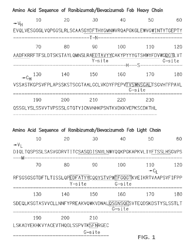
space, subretinal space, intraretinal space, vitreous cavity, or outer surface
of the sclera in the eye
of said human subject, a therapeutically effective amount of a recombinant
nucleotide expression
vector encoding an antigen-binding fragment of a mAb against hVEGF (e.g., by
suprachoroidal
injection, subretinal injection via the transvitreal approach (a surgical
procedure), subretinal
administration via the suprachoroidal space, or a posterior juxtascleral depot
procedure), so that a
depot is formed that releases said antigen-binding fragment wherein said
antigen-binding
fragment is glycosylated but does not contain detectable NeuGc and/or a-Gal
antigen.
[0036] In certain aspects, described herein are methods of
treating a human subject
diagnosed with diabetic retinopathy (DR), comprising: administering or
delivering to the retina
of said human subject via the suprachoroidal space in the eye of said human
subject (e.g., via a
-17-
CA 03149401 2022-2-24
WO 2021/041373
PCT/US2020/047733
suprachoroidal drug delivery device such as a microinjector with a
microneedle), a
therapeutically effective amount of a recombinant nucleotide expression vector
encoding an
antigen-binding fragment of a mAb against hVEGF, so that a depot is formed
that releases said
antigen-binding fragment wherein said antigen-binding fragment is glycosylated
but does not
contain detectable NeuGc and/or a-Gal antigen.
[0037] In certain aspects, described herein are methods of
treating a human subject
diagnosed with diabetic retinopathy (DR), comprising: administering to the
subretinal and/or
intraretinal space of said human subject via the suprachoroidal space in the
eye of said human
subject (e.g., via a subretinal drug delivery device comprising a catheter
that can be inserted and
tunneled through the suprachoroidal space toward the posterior pole, where a
small needle injects
into the subretinal space), a therapeutically effective amount of a
recombinant nucleotide
expression vector encoding an antigen-binding fragment of a mAb against hVEGF,
so that a
depot is formed that releases said antigen-binding fragment wherein said
antigen-binding
fragment is glycosylated but does not contain detectable NeuGc and/or a-Gal
antigen. In certain
aspects, described herein are methods of treating a human subject diagnosed
with diabetic
retinopathy (DR), comprising administering to the subretinal space and/or
intraretinal space of
said human subject via the suprachoroidal space in the eye of said human
subject an expression
vector encoding an anti-human vascular endothelial growth factor (hVEGF)
antibody. In a
specific aspect, the expression vector is administered via subretinal delivery
in a single dose
about 1.6 x 10" GC/eye at a concentration of 6.4 x 10" GC/mL or about 2.5 x
10" GC/eye at a
concentration of 1.0 x 10¹² GC/mL.
100381 In certain aspects, described herein are methods of
treating a human subject
diagnosed with diabetic retinopathy (DR), comprising: administering to the
subretinal and/or
intraretinal space of said human subject via the suprachoroidal space in the
eye of said human
subject (e.g., via a subretinal drug delivery device comprising a catheter
that can be inserted and
tunneled through the suprachoroidal space toward the posterior pole, where a
small needle injects
into the subretinal space), a therapeutically effective amount of a
recombinant nucleotide
expression vector encoding an antigen-binding fragment of a mAb against hVEGF,
so that a
depot is formed that releases said antigen-binding fragment wherein said
antigen-binding
fragment is glycosylated but does not contain detectable NeuGc and/or a-Gal
antigen. In certain
aspects, described herein are methods of treating a human subject diagnosed
with diabetic
-18-
CA 03149401 2022-2-24
WO 2021/041373
PCT/US2020/047733
retinopathy (DR), comprising administering to the subretinal and/or
intraretinal space of said
human subject via the suprachoroidal space in the eye of said human subject an
expression
vector encoding an anti-human vascular endothelial growth factor (hVEGF)
antibody. In a
specific aspect, the expression vector is administered via subretinal delivery
in a single dose
about 1.6 x 10¹¹ GC/eye at a concentration of 6.2 x 10¹¹ GC/mL or about 2.5 x
1011 GC/eye at a
concentration of 1.0 x 10¹² GC/mL. In a specific aspect, the expression vector
is administered
via subretinal delivery in a single dose about 1.55 x 10¹¹ GC/eye at a
concentration of 6.2 x 10¹¹
GC/mL or about 2.5 x 10¹¹ GC/eye at a concentration of 1.0 x 10¹² GC/mL.
100391 In a specific aspect, the anti-hVEGF antibody
comprises a heavy chain comprising
the amino acid sequence of SEQ ID NO. 2 or SEQ ID NO. 4, and a light chain
comprising the
amino acid sequence of SEQ ID NO. 1, or SEQ ID NO. 3. In a specific aspect,
the expression
vector is an AAV8 vector.
100401 In certain aspects of the methods described herein,
the antigen-binding fragment
transgene encodes a leader peptide_ A leader peptide may also be referred to
as a signal peptide
or leader sequence herein.
100411 In certain aspects, described herein are methods of
treating a human subject
diagnosed with diabetic retinopathy (DR), comprising administering to the
suprachoroidal
space, subretinal space, intraretinal space, vitreous cavity, or outer surface
of the sclera in the eye
of said human subject, a therapeutically effective amount of a recombinant
nucleotide expression
vector encoding an antigen-binding fragment of a mAb against hVEGF (e.g., by
suprachoroidal
injection, subretinal injection via the transvitreal approach (a surgical
procedure), subretinal
administration via the suprachoroidal space, or a posterior juxtascleral depot
procedure)), so that
a depot is formed that releases said antigen-binding fragment containing a
a2,6-sialylated glycan;
wherein said recombinant vector, when used to transduce PER.C6 or RIP cells in
culture results
in production of said antigen-binding fragment containing a a2,6-sialylated
glycan in said cell
culture.
100421 In certain aspects, described herein are methods of
treating a human subject
diagnosed with diabetic retinopathy (DR) (in particular, wet AMID),
comprising: administering
or delivering to the retina of said human subject via the suprachoroidal space
in the eye of said
human subject (e.g., via a suprachoroidal drug delivery device such as a
microinjector with a
microneedle), a therapeutically effective amount of a recombinant nucleotide
expression vector
-19-
CA 03149401 2022-2-24
WO 2021/041373
PCT/US2020/047733
encoding an antigen-binding fragment of a mAb against hVEGF, so that a depot
is formed that
releases said antigen-binding fragment containing a a2,6-sialylated glycan;
wherein said
recombinant vector, when used to transduce PER.C6 or RPE cells in culture
results in production
of said antigen-binding fragment containing a a2,6-sialylated glycan in said
cell culture.
100431 In certain aspects, described herein are methods of
treating a human subject
diagnosed with diabetic retinopathy (DR), comprising: administering to the
subretinal and/or
intraretinal space of said human subject via the suprachoroidal space in the
eye of said human
subject (e.g., via a subretinal drug delivery device comprising a catheter
that can be inserted and
tunneled through the suprachoroidal space toward the posterior pole, where a
small needle injects
into the subretinal space), a therapeutically effective amount of a
recombinant nucleotide
expression vector encoding an antigen-binding fragment of a mAb against hVEGF,
so that a
depot is formed that releases said antigen-binding fragment containing a a2,6-
sialylated glycan;
wherein said recombinant vector, when used to transduce PER.C6 or RPE cells in
culture results
in production of said antigen-binding fragment containing a a2,6-sialylated
glycan in said cell
culture.
100441 In certain aspects, described herein are methods of
treating a human subject
diagnosed with diabetic retinopathy (DR), comprising: administering to the
suprachoroidal
space, subretinal space, intraretinal space, vitreous cavity, or outer surface
of the sclera in the eye
of said human subject, a therapeutically effective amount of a recombinant
nucleotide expression
vector encoding an antigen-binding fragment of a mAb against hVEGF (e.g., by
suprachoroidal
injection, subretinal injection via the transvitreal approach (a surgical
procedure), subretinal
administration via the suprachoroidal space, or a posterior juxtascleral depot
procedure), so that a
depot is formed that releases said antigen-binding fragment wherein said
antigen-binding
fragment is glycosylated but does not contain detectable NeuGc and/or a-Gal
antigen; wherein
said recombinant vector, when used to transduce PER.C6 or RPE cells in culture
results in
production of said antigen-binding fragment that is glycosylated but does not
contain detectable
NeuGc and/or a-Gal antigen in said cell culture.
100451 In certain aspects, described herein are methods of
treating a human subject
diagnosed with diabetic retinopathy (DR), comprising: administering to the
subretinal and/or
intraretinal space of said human subject via the suprachoroidal space in the
eye of said human
subject (e.g., via a subretinal drug delivery device comprising a catheter
that can be inserted and
-20-
CA 03149401 2022-2-24
WO 2021/041373
PCT/US2020/047733
tunneled through the suprachoroidal space toward the posterior pole, where a
small needle injects
into the subretinal space), a therapeutically effective amount of a
recombinant nucleotide
expression vector encoding an antigen-binding fragment of a mAb against hVEGF,
so that a
depot is formed that releases said antigen-binding fragment wherein said
antigen-binding
fragment is glycosylated but does not contain detectable NeuGc and/or a-Gal
antigen; wherein
said recombinant vector, when used to transduce PER.C6 or RPE cells in culture
results in
production of said antigen-binding fragment that is glycosylated but does not
contain detectable
NeuGc and/or a-Gal antigen in said cell culture.
100461 In certain aspects, described herein are methods of
treating a human subject
diagnosed with diabetic retinopathy (DR), comprising: administering to the
subretinal and/or
intraretinal space of said human subject via the suprachoroidal space in the
eye of said human
subject (e.g., via a subretinal drug delivery device comprising a catheter
that can be inserted and
tunneled through the suprachoroidal space toward the posterior pole, where a
small needle injects
into the subretinal space), a therapeutically effective amount of a
recombinant nucleotide
expression vector encoding an antigen-binding fragment of a mAb against hVEGF,
so that a
depot is formed that releases said antigen-binding fragment wherein said
antigen-binding
fragment is glycosylated but does not contain detectable NeuGc and/or a-Gal
antigen; wherein
said recombinant vector, when used to transduce PER.C6 or RPE cells in culture
results in
production of said antigen-binding fragment that is glycosylated but does not
contain detectable
NeuGc and/or a-Gal antigen in said cell culture.
100471 In certain aspects of the methods described herein,
the human subject has a Best-
corrected visual acuity (BCVA) of > 69 ETDRS letters (approximate Snellen
equivalent 20/40 or
better).
100481 In certain aspects of the methods described herein,
the BCVA is the BCVA in the eye
to be treated in the human subject.
100491 In certain aspects of the methods described herein,
delivering to the eye comprises
delivering to the retina, choroid, and/or vitreous humor of the eye. In
certain aspects of the
methods described herein, the antigen-binding fragment comprises a heavy chain
that comprises
one, two, three, or four additional amino acids at the C-terminus.
100501 Subjects to whom such gene therapy is administered
should be those responsive to
anti-VEGF therapy. In particular embodiments, the methods encompass treating
patients who
-21-
CA 03149401 2022-2-24
WO 2021/041373
PCT/US2020/047733
have been diagnosed with retinopathy (DR) and identified as responsive to
treatment with an
anti-VEGF antibody. In more specific embodiments, the patients are responsive
to treatment
with an anti-VEGF antigen-binding fragment. In certain embodiments, the
patients have been
shown to be responsive to treatment with an anti-VEGF antigen-binding fragment
injected
intravitreally prior to treatment with gene therapy. In specific embodiments,
the patients have
previously been treated with LUCENTIS (ranibizumab), EYLEA (aflibercept),
and/or
AVASTINO (bevacizumab), and have been found to be responsive to one or more of
said
LUCENTIS (ranibizumab), EYLEA (atlibercept), and/or AVASTINO (bevacizumab).
100511 Subjects to whom such viral vector or other DNA
expression construct is delivered
should be responsive to the anti-hVEGF antigen-binding fragment encoded by the
transgene in
the viral vector or expression construct. To determine responsiveness, the
anti-VEGF antigen-
binding fragment transgene product (e.g., produced in cell culture,
bioreactors, etc.) may be
administered directly to the subject, such as by intravitreal injection.
100521 In certain aspects of the methods described herein,
the antigen-binding fragment
comprises a heavy chain that does not comprise an additional amino acid at the
C-terminus.
100531 In certain aspects of the methods described herein
produces a population of antigen-
binding fragment molecules, wherein the antigen-binding fragment molecules
comprise a heavy
chain, and wherein 0.5%, 1%, 2%, 3%, 4%, 5%, 10%, or 20%, or less of the
population of
antigen-binding fragment molecules comprises one, two, three, or four
additional amino acids at
the C-terminus of the heavy chain. In certain aspects of the methods described
herein produces a
population of antigen-binding fragment molecules, wherein the antigen-binding
fragment
molecules comprise a heavy chain, and wherein 0.5%, 1%, 2%, 3%, 4%, 5%, 10%,
or 20%, or
less but more than 0% of the population of antigen-binding fragment molecules
comprises one,
two, three, or four additional amino acids at the C-terminus of the heavy
chain.
100541 In certain aspects of the methods described herein
produces a population of antigen-
binding fragment molecules, wherein the antigen-binding fragment molecules
comprise a heavy
chain, and wherein 0.5-1%, 0.5%-2%, 0.5%-3%, 0.5%-4%, 0.5%-5%, 0.5%40%, 0.5%-
20%,
1%-2%, 1%-3%, 1%-4%, 1%-5%, 1%40%, 1%-20%, 2%-3%, 2%-4%, 2%-5%, 2%40%, 2%-
20%, 3%-4%, 3%-5%, 3%-10%, 3%-20%, 4%-5%, 4%40%, 4%-20%, 5%40%, 5%-20%, or
10%-20% of the population of antigen-binding fragment molecules comprises one,
two, three, or
four additional amino acids at the C-terminus of the heavy chain.
-22-
CA 03149401 2022-2-24
WO 2021/041373
PCT/US2020/047733
100551 The HuPTMFabVEGFi, e.g., HuGlyFabVEGFi, encoded by
the transgene can
include, but is not limited to an antigen-binding fragment of an antibody that
binds to hVEGF,
such as bevacizumab; an anti-hVEGF Fab moiety such as ranibizumab; or such
bevacizumab or
ranibizumab Fab moieties engineered to contain additional glycosylation sites
on the Fab domain
(e.g., see Courtois et al, , 2016, mAbs 8: 99-112 which is incorporated by
reference herein in its
entirety for it description of derivatives of bevacizumab that are
hyperglycosylated on the Fab
domain of the full length antibody).
100561 The recombinant vector used for delivering the
transgene should have a tropism for
human retinal cells or photoreceptor cells. Such vectors can include non-
replicating recombinant
adeno-associated virus vectors ("rAAV"), particularly those bearing an AAV8
capsid are
preferred. However, other viral vectors may be used, including but not limited
to lentiviral
vectors, vaccinia viral vectors, or non-viral expression vectors referred to
as "naked DNA"
constructs. Preferably, the HuPTMFabVEGFi, e.g., HuGlyFabVEGFi, transgene
should be
controlled by appropriate expression control elements, for example, the CB7
promoter (a chicken
13-actin promoter and CMV enhancer), the RPE65 promoter, or opsin promoter to
name a few,
and can include other expression control elements that enhance expression of
the transgene
driven by the vector (e.g., introns such as the chicken j3-actin intron,
minute virus of mice
(MVM) intron, human factor IX intron (e.g., FIX truncated intron 1), 13-globin
splice
donor/immunoglobulin heavy chain spice acceptor intron, adenovirus splice
donor
/immunoglobulin splice acceptor intron, SV40 late splice donor /splice
acceptor (19S/16S)
intron, and hybrid adenovirus splice donor/IgG splice acceptor intron and
polyA signals such as
the rabbit 13-globin polyA signal, human growth hormone (hGH) polyA signal,
SV40 late polyA
signal, synthetic polyA (SPA) signal, and bovine growth hormone (bGH) polyA
signal). See,
e.g., Powell and Rivera-Soto, 2015, Discov_ Med., 19(102):49-57.
100571 Gene therapy constructs are designed such that both
the heavy and light chains are
expressed. More specifically, the heavy and light chains should be expressed
at about equal
amounts, in other words, the heavy and light chains are expressed at
approximately a 1:1 ratio of
heavy chains to light chains. The coding sequences for the heavy and light
chains can be
engineered in a single construct in which the heavy and light chains are
separated by a cleavable
linker or 1RES so that separate heavy and light chain polypeptides are
expressed. See, e.g.,
Section 5.2.4 for specific leader sequences and Section 5.2.5 for specific
1RES, 2A, and other
-23-
CA 03149401 2022-2-24
WO 2021/041373
PCT/US2020/047733
linker sequences that can be used with the methods and compositions provided
herein.
100581 In certain embodiments, gene therapy constructs are
supplied as a frozen sterile,
single use solution of the AAV vector active ingredient in a formulation
buffer. In a specific
embodiment, the pharmaceutical compositions suitable for subretinal
administration comprise a
suspension of the recombinant (e.g., rHuGlyFabVEGFi) vector in a formulation
buffer
comprising a physiologically compatible aqueous buffer, a surfactant and
optional excipients. In
a specific embodiment, the construct is formulated in Dulbecco's phosphate
buffered saline and
0.001% Plutonic F68, pH = 7.4.
100591 In certain embodiments, gene therapy constructs are
supplied as a frozen sterile,
single use solution of the AAV vector active ingredient in a formulation
buffer. In a specific
embodiment, the pharmaceutical compositions suitable for suprachoroidal,
subretinal,
juxtascleral, intravitreal, subconjunctival, and/or intraretinal
administration comprise a
suspension of the recombinant (e.g., rHuGlyFabVEGFO vector in a formulation
buffer
comprising a physiologically compatible aqueous buffer, a surfactant and
optional excipients.
100601 Therapeutically effective doses of the recombinant
vector should be administered
subretinally and/or intraretinally (e.g., by subretinal injection via the
transvitreal approach (a
surgical procedure), or subretinal administration via the suprachoroidal
space) in a volume
ranging from 0.1 mL to 0.5 mL, preferably in 0.1 to 0.30 mL (100 ¨ 300 pi),
and most
preferably, in a volume of 0.25 mL (250 RI). Therapeutically effective doses
of the recombinant
vector should be administered suprachoroidally (e.g., by suprachoroidal
injection) in a volume of
100 pi or less, for example, in a volume of 50-100 pl. Therapeutically
effective doses of the
recombinant vector should be administered to the outer surface of the sclera
(e.g., by a posterior
juxtascleral depot procedure) in a volume of 500 pl or less, for example, in a
volume of 10-20 pi,
20-50 pl. 50-100 pl. 100-200 pl. 200-300 pi, 300-400 p1, or 400-500 pl.
Subretirtal injection is a
surgical procedure performed by trained retinal surgeons that involves a
vitrectomy with the
subject under local anesthesia, and subretinal injection of the gene therapy
into the retina (see,
e.g., Campochiaro et at, 2017, Hum Gen Ther 28(1):99-111, which is
incorporated by reference
herein in its entirety). In a specific embodiment, the subretinal
administration is performed via
the suprachoroidal space using a suprachoroidal catheter which injects drug
into the subretinal
space, such as a subretinal drug delivery device that comprises a catheter
which can be inserted
and tunneled through the suprachoroidal space to the posterior pole, where a
small needle injects
-24-
CA 03149401 2022-2-24
WO 2021/041373
PCT/US2020/047733
into the subretinal space (see, e.g., Baldassarre et at, 2017, Subretinal
Delivery of Cells via the
Suprachoroidal Space: Janssen Trial. In: Schwartz et at (eds) Cellular
Therapies for Retinal
Disease, Springer, Cham; International Patent Application Publication No. WO
2016/040635
Al; each of which is incorporated by reference herein in its entirety).
Suprachoroidal
administration procedures involve administration of a drug to the
suprachoroidal space of the
eye, and are normally performed using a suprachoroidal drug delivery device
such as a
microinjector with a microneedle (see, e.g., Hariprasad, 2016, Retinal
Physician 13: 20-23;
Goldstein, 2014, Retina Today 9(5): 82-87; each of which is incorporated by
reference herein in
its entirety). The suprachoroidal drug delivery devices that can be used to
deposit the expression
vector in the suprachoroidal space according to the invention described herein
include, but are
not limited to, suprachoroidal drug delivery devices manufactured by Clearside
Biomedical,
Inc. (see, for example, Hariprasad, 2016, Retinal Physician 13: 20-23) and
MedOne
suprachoroidal catheters. The subretinal drug delivery devices that can be
used to deposit the
expression vector in the subretinal space via the suprachoroidal space
according to the invention
described herein include, but are not limited to, subretinal drug delivery
devices manufactured by
Janssen Pharmaceuticals, Inc. (see, for example, International Patent
Application Publication No.
WO 2016/040635 Al). In a specific embodiment, administration to the outer
surface of the
sclera is performed by a juxtascleral drug delivery device comprising a
cannula whose tip can be
inserted and kept in direct apposition to the sclera' surface. See Section
5.3.2 for more details of
the different modes of administration. Suprachoroidal, subretinal,
juxtascleral, intravitreal,
subconjunctival, and/or intraretinal administration should result in delivery
of the soluble
transgene product to the retina, the vitreous humor, and/or the aqueous humor,
The expression
of the transgene product (e.g., the encoded anti-VEGF antibody) by retinal
cells, e.g., rod, cone,
retinal pigment epithelial, horizontal, bipolar, amaaine, ganglion, and/or
Muller cells, results in
delivery and maintenance of the transgene product in the retina, the vitreous
humor, and/or the
aqueous humor. In a specific embodiment, doses that maintain a concentration
of the transgene
product at a Cmin of at least 0.330 pig/mL in the Vitreous humour, or 0.110
gg/mL in the
Aqueous humour (the anterior chamber of the eye) for three months are desired,
thereafter,
Vitreous Cmin concentrations of the transgene product ranging from 1,70 to
6.60 itg/mL, and/or
Aqueous Cmin concentrations ranging from 0.567 to 2.20 pg/mL should be
maintained.
However, because the transgene product is continuously produced, maintenance
of lower
-25-
CA 03149401 2022-2-24
WO 2021/041373
PCT/US2020/047733
concentrations can be effective. The concentration of the transgene product
can be measured in
patient samples of the vitreous humour and/or aqueous from the anterior
chamber of the treated
eye. Alternatively, vitreous humour concentrations can be estimated and/or
monitored by
measuring the patient's serum concentrations of the transgene product ¨ the
ratio of systemic to
vitreal exposure to the transgene product is about 1:90,000. (Kg., see,
vitreous humor and serum
concentrations of ranibizumab reported in Xu L, et al., 2013, Invest. Opthal.
Vis. Sci. 54: 1616-
1624, at p. 1621 and Table 5 at p. 1623, which is incorporated by reference
herein in its entirety).
100611 In a specific embodiment, the subretinal
administration is performed with a subretinal
drug delivery device that comprises the micro volume injector delivery system,
which is
manufactured by Altaviz (see FIGs 9A and 9B) (see, e.g. International Patent
Application
Publication No. WO 2013/177215, United States Patent Application Publication
No.
2019/0175825, and United States Patent Application Publication No.
2019/0167906) that can be
used for any administration route described herein for eye administration. The
micro volume
injector delivery system may include a gas-powered module providing high force
delivery and
improved precision, as described in United States Patent Application
Publication No.
2019/0175825 and United States Patent Application Publication No,
2019/0167906. In addition,
the micro volume injector delivery system may include a hydraulic drive for
providing a
consistent dose rate, and a low-force activation lever for controlling the gas-
powered module
and, in turn, the fluid delivery. In certain embodiment, the micro volume
injector delivery
system can be used for micro volume injector is a micro volume injector with
dose guidance and
can be used with, for example, a suprachoroidal needle (for example, the
Clearside needle), a
subretinal needle, an intravitreal needle, a juxtascleral needle, a
subconjunctival needle, and/or
intraretinal needle. The benefits of using micro volume injector include: (a)
more controlled
delivery (for example, due to having precision injection flow rate control and
dose guidance), (b)
single surgeon, single hand, one finger operation; (c) pneumatic drive with 10
'LILL increment
dosage; (d) divorced from the vitrectomy machine; (e) 400 it syringe dose; (0
digitally guided
delivery; (g) digitally recorded delivery; and (h) agnostic tip (for example,
the MedOne 38g
needle and the Dore 41g needle can be used for subretinal delivery, while the
Clearside needle
and the Visionisti OY adaptor can be used for subretinal delivery).
100621 In certain embodiments of the methods described
herein, the recombinant vector is
administered suprachoroidally (e.g., by suprachoroidal injection). In a
specific embodiment,
-26-
CA 03149401 2022-2-24
WO 2021/041373
PCT/US2020/047733
suprachoroidal administration (e.g., an injection into the suprachoroidal
space) is performed
using a suprachoroidal drug delivery device. Suprachoroidal drug delivery
devices are often
used in suprachoroidal administration procedures, which involve administration
of a drug to the
suprachoroidal space of the eye (see, e.g., Hariprasad, 2016, Retinal
Physician 13: 20-23;
Goldstein, 2014, Retina Today 9(5): 82-87; Baldassare et al., 2017; each of
which is
incorporated by reference herein in its entirety). The suprachoroidal drug
delivery devices that
can be used to deposit the recombinant vector in the suprachoroidal space
according to the
invention described herein include, but are not limited to, suprachoroidal
drug delivery devices
manufactured by Clearside Biomedical, Inc. (see, for example, Hariprasad,
2016, Retinal
Physician 13: 20-23) and MedOne suprachoroidal catheters. In another
embodiment, the
suprachoroidal drug delivery device that can be used in accordance with the
methods described
herein comprises the micro volume injector delivery system, which is
manufactured by Altaviz
(see FIGs. 9A and 9B ) (see, e.g. International Patent Application Publication
No. WO
2013/177215, United States Patent Application Publication No. 2019/0175825,
and United States
Patent Application Publication No. 2019/0167906) that can be used for any
administration route
described herein for eye administration. The micro volume injector delivery
system may include
a gas-powered module providing high force delivery and improved precision, as
described in
United States Patent Application Publication No. 2019/0175825 and United
States Patent
Application Publication No. 2019/0167906. In addition, the micro volume
injector delivery
system may include a hydraulic drive for providing a consistent dose rate, and
a low-force
activation lever for controlling the gas-powered module and, in turn, the
fluid delivery. The
micro volume injector is a micro volume injector with dose guidance and can be
used with, for
example, a suprachoroidal needle (for example, the Clearside needle) or a
subretinaI needle.
The benefits of using micro volume injector include: (a) more controlled
delivery (for example,
due to having precision injection flow rate control and dose guidance), (b)
single surgeon, single
hand, one finger operation; (c) pneumatic drive with 10 pL increment dosage;
(d) divorced from
the vitrectomy machine; (e) 400 tuL syringe dose; (0 digitally guided
delivery; (g) digitally
recorded delivery; and (h) agnostic tip (for example, the MedOne 38g needle
and the Dorc 41g
needle can be used for subretinal delivery, while the Clearside needle and
the Visionisti OY
adaptor can be used for suprachoroidal delivery). In another embodiment, the
suprachoroidal
drug delivery device that can be used in accordance with the methods described
herein is a tool
-27-
CA 03149401 2022-2-24
WO 2021/041373
PCT/US2020/047733
that comprises a normal length hypodermic needle with an adaptor (and
preferably also a needle
guide) manufactured by Visionisti OY, which adaptor turns the normal length
hypodermic
needle into a suprachoroidal needle by controlling the length of the needle
tip exposing from the
adapter (see FIG. 8) (see, for example, U.S. Design Patent No. D878,575; and
International
Patent Application. Publication No. WO/2016/083669) In a specific embodiment,
the
suprachoroidal drug delivery device is a syringe with a 1 millimeter 30 gauge
needle (see FIG.
5). During an injection using this device, the needle pierces to the base of
the sclera and fluid
containing drug enters the suprachoroidal space, leading to expansion of the
suprachoroidal
space. As a result, there is tactile and visual feedback during the injection.
Following the
injection, the fluid flows posteriorly and absorbs dominantly in the choroid
and retina This
results in the production of therapeutic product from all retinal cell layers
and choroidal cells.
Using this type of device and procedure allows for a quick and easy in-office
procedure with low
risk of complications. A max volume of 100 id can be injected into the
suprachoroidal space.
100631 In a specific embodiment, the intravitreal
administration is performed with a
intravitreal drug delivery device that comprises the micro volume injector
delivery system,
which is manufactured by Altaviz. (see FIGs 9A and 98) (see, e.g.
International Patent
Application Publication No. WO 2013/177215) , United States Patent Application
Publication
No. 2019/0175825, and United States Patent Application Publication No.
2019/0167906) that
can be used for any administration route described herein for eye
administration. The micro
volume injector delivery system may include a gas-powered module providing
high force
delivery and improved precision, as described in United States Patent
Application Publication
No. 2019/0175825 and United States Patent Application Publication No.
2019/0167906. In
addition, the micro volume injector delivery system may include a hydraulic
drive for providing
a consistent dose rate, and a low-force activation lever for controlling the
gas-powered module
and, in turn, the fluid delivery. The micro volume injector is a micro volume
injector with dose
guidance and can be used with, for example, a intravitreal needle. The
benefits of using micro
volume injector include: (a) more controlled delivery (for example, due to
having precision
injection flow rate control and dose guidance), (b) single surgeon, single
hand, one finger
operation; (c) pneumatic drive with 10 it increment dosage; (d) divorced from
the vitrectomy
-28-
CA 03149401 2022-2-24
WO 2021/041373
PCT/US2020/047733
machine; (e) 400 I, syringe dose; (f) digitally guided delivery; (g)
digitally recorded delivery;
and (h) agnostic tip.
100641 In a specific embodiment, the juxtascleral
administration is performed with a
juxtascleral drug delivery device that comprises the micro volume injector
delivery system,
which is manufactured by Altaviz. (see FIGs. 9A and 9B) (see, e.g.
International Patent
Application Publication No. WO 2013/177215) , United States Patent Application
Publication
No. 2019/0175825, and United States Patent Application Publication No.
2019/0167906) that
can be used for any administration route described herein for eye
administration. The micro
volume injector delivery system may include a gas-powered module providing
high force
delivery and improved precision, as described in United States Patent
Application Publication
No. 2019/0175825 and United States Patent Application Publication No.
2019/0167906. In
addition, the micro volume injector delivery system may include a hydraulic
drive for providing
a consistent dose rate, and a low-force activation lever for controlling the
gas-powered module
and, in turn, the fluid delivery. Micro volume injector is a micro volume
injector with dose
guidance and can be used with, for example, a subretinal needle. The benefits
of using micro
volume injector include: (a) more controlled delivery (for example, due to
having precision
injection flow rate control and dose guidance), (b) single surgeon, single
hand, one finger
operation; (c) pneumatic drive with 10 tiL increment dosage; (d) divorced from
the vitrectomy
machine; (e) 400 I, syringe dose, (f) digitally guided delivery, (g)
digitally recorded delivery,
and (h) agnostic tip.
100651 In certain embodiments, dosages are measured by
genome copies per ml or the
number of genome copies administered to the eye of the patient (e.g., by
suprachoroidal injection
(for example, via a suprachoroidal drug delivery device such as a
microinjector with a
microneedle), subretinal injection via the transvitreal approach (a surgical
procedure), or
subretinal administration via the suprachoroidal space). In certain
embodiments, 2.4 x 101'
genome copies per ml to 1 x101-3 genome copies per ml are administered. In a
specific
embodiment, 2.4 x 10¹¹ genome copies per ml to 5 x10'1 genome copies per ml
are administered.
In another specific embodiment, 5 x 10¹¹ genome copies per ml to 1 x10¹²
genome copies per ml
are administered. In another specific embodiment, 1 x 10¹² genome copies per
ml to 5 x10¹²
genome copies per ml are administered. In another specific embodiment, 5 x
1012 genome
copies per ml to 1 x10¹³ genome copies per ml are administered. In another
specific
-29-
CA 03149401 2022-2-24
WO 2021/041373
PCT/US2020/047733
embodiment, about 2.4 x 10¹¹ genome copies per ml are administered. In another
specific
embodiment, about 5 x 10¹¹ genome copies per ml are administered. In another
specific
embodiment, about 1 x 10¹² genome copies per ml are administered. In another
specific
embodiment, about 5 x 10¹² genome copies per ml are administered. In another
specific
embodiment, about 1 x 10¹³ genome copies per ml are administered. In certain
embodiments, 1
x 109 to 1 x 10¹² genome copies are administered. In specific embodiments, 3 x
109 to 2.5 x 10¹¹
genome copies are administered. In specific embodiments, 1 x 10⁹ to 2.5 x 10¹¹
genome copies
are administered. In specific embodiments, 1 x 10⁹ to 1 x 10¹¹ genome copies
are administered.
In specific embodiments, 1 x 10⁹ to 5 x 10⁹ genome copies are administered. In
specific
embodiments, 6 x 10⁹ to 3 x 10¹⁰ genome copies are administered. In specific
embodiments, 4 x
1010 to 1 x 10¹¹ genome copies are administered. In specific embodiments, 2 x
1011 to 1 x 10¹²
genome copies are administered. In a specific embodiment, about 3 x 10⁹ genome
copies are
administered (which corresponds to about 1.2 x 101 genome copies per ml in a
volume of 250
pi). In another specific embodiment, about 1 x 10¹⁰ genome copies are
administered (which
corresponds to about 4 x 10¹⁰ genome copies per ml in a volume of 250 RD. In
another specific
embodiment, about 6 x 10¹⁰ genome copies are administered (which corresponds
to about 2.4 x
1011 genome copies per ml in a volume of 250 1). In another specific
embodiment, about 1.6 x
1011 genome copies are administered (which corresponds to about 6.2 x 10¹¹
genome copies per
ml in a volume of 250 pi). In another specific embodiment, about 1.6 x 10¹¹
genome copies are
administered (which corresponds to about 6.4 x 10¹¹ genome copies per ml in a
volume of 250
1). In another specific embodiment, about 1.55 x 10¹¹ genome copies are
administered (which
corresponds to about 6.2 x 10¹¹ genome copies per ml in a volume of 250 1).
In another specific
embodiment, about 2.5 x 10¹¹ genome copies (which corresponds to about 1.0 x
1012 in a volume
of 250 pl) are administered.
100661 In certain embodiments, about 3.0 x 10¹³ genome
copies per eye are administered. In
certain embodiments, up to 3.0 x 10¹³ genome copies per eye are administered.
100671 In certain embodiments, about 6.0 x 10¹⁰ genome
copies per eye are administered. In
certain embodiments, about 1.6 x 10¹¹ genome copies per eye are administered.
In certain
embodiments, about 2.5 x 10¹¹ genome copies per eye are administered. In
certain
embodiments, about 5.0 x 10¹¹ genome copies per eye are administered. In
certain
embodiments, about 3 x 10¹² genome copies per eye are administered. In certain
embodiments,
-30-
CA 03149401 2022-2-24
WO 2021/041373
PCT/US2020/047733
about 1.0 x 10¹² genome copies per ml per eye are administered. In certain
embodiments, about
2.5 x 10¹² genome copies per ml per eye are administered.
100681 In certain embodiments, about 6.0 x 10¹⁰ genome
copies per eye are administered by
subretinal injection. In certain embodiments, about 1.6 x 10" genome copies
per eye are
administered by subretinal injection. In certain embodiments, about 2.5 x 10"
genome copies
per eye are administered by subretinal injection. In certain embodiments,
about 3.0 x 10¹³
genome copies per eye are administered by subretinal injection. In certain
embodiments, up to
3.0 x 10¹³ genome copies per eye are administered by subretinal injection.
1001691 In certain embodiments, about 2,5 x 10" genome
copies per eye are administered by
suprachoroidal injection. In certain embodiments, about 5.0 x 10" genome
copies per eye are
administered by suprachoroidal injection. In certain embodiments, about 3 x
1012 genome copies
per eye are administered by suprachoroidal injection. In certain embodiments,
about 2.5 x
genome copies per eye are administered by a single suprachoroidal injection.
In certain
embodiments, about 5.0 x 10¹¹ genome copies per eye are administered by double
suprachoroidal injections. In certain embodiments, about 3.0 x 10¹³ genome
copies per eye are
administered by suprachoroidal injection. In certain embodiments, up to 3.0 x
1013 genome
copies per eye are administered by suprachoroidal injection. In certain
embodiments, about 2_5 x
1012 genome copies per ml per eye are administered by a single suprachoroidal
injection in a
volume of 100 I. In certain embodiments, about 2.5 x 10¹² genome copies per
ml per eye are
administered by double suprachoroidal injections, wherein each injection is in
a volume of 100
1.
100701 As used herein and unless otherwise specified, the
term "about" means within plus or
minus 10% of a given value or range In certain embodiments, the term "about"
encompasses
the exact number recited.
1001711 The invention has several advantages over standard
of care treatments that involve
repeated ocular injections of high dose boluses of the VEGF inhibitor that
dissipate over time
resulting in peak and trough levels. Sustained expression of the transgene
product antibody, as
opposed to injecting an antibody repeatedly, allows for a more consistent
levels of antibody to be
present at the site of action, and is less risky and more convenient for
patients, since fewer
injections need to be made, resulting in fewer doctor visits. Consistent
protein production may
leads to better clinical outcomes as edema rebound in the retina is less
likely to occur.
-31-
CA 03149401 2022-2-24
WO 2021/041373
PCT/US2020/047733
Furthermore, antibodies expressed from transgenes are post-translationally
modified in a
different manner than those that are directly injected because of the
different microenvironment
present during and after translation. Without being bound by any particular
theory, this results in
antibodies that have different diffusion, bioactivity, distribution, affinity,
phannacokinetic, and
immunogenicity characteristics, such that the antibodies delivered to the site
of action are
"biobetters" in comparison with directly injected antibodies.
100721 In addition, antibodies expressed from transgenes in
vivo are not likely to contain
degradation products associated with antibodies produced by recombinant
technologies, such as
protein aggregation and protein oxidation. Aggregation is an issue associated
with protein
production and storage due to high protein concentration, surface interaction
with manufacturing
equipment and containers, and purification with certain buffer systems. These
conditions, which
promote aggregation, do not exist in transgene expression in gene therapy.
Oxidation, such as
methionine, ttyptophan, and histidine oxidation, is also associated with
protein production and
storage, and is caused by stressed cell culture conditions, metal and air
contact, and impurities in
buffers and excipients. The proteins expressed from transgenes in vivo may
also oxidize in a
stressed condition. However, humans, and many other organisms, are equipped
with an
antioxidation defense system, which not only reduces the oxidation stress, but
sometimes also
repairs and/or reverses the oxidation. Thus, proteins produced in vivo are not
likely to be in an
oxidized form. Both aggregation and oxidation could affect the potency,
phannacokinetics
(clearance), and immunogenicity.
100731 Without being bound by theory, the methods and
compositions provided herein are
based, in part, on the following principles:
(i) Human retinal cells are secretory cells that possess the cellular
machinery for post-
translational processing of secreted proteins ¨ including glycosylation and
tyrosine-0-
sulfation, a robust process in retinal cells. (See, e.g., Wang et at, 2013,
Analytical
Biochent 427: 20-28 and Adamis eta!,, 1993, BBRC 193: 631-638 reporting the
production of glycoproteins by retinal cells; and Kanan el al., 2009, Exp. Eye
Res. 89:
559-567 and Kanan 84. Al-Ubaidi, 2015, Exp. Eye Res. 133: 126-131 reporting
the
production of tyrosine-sulfated glycoproteins secreted by retinal cells, each
of which is
incorporated by reference in its entirety for post-translational modifications
made by
human retinal cells).
-32-
CA 03149401 2022-2-24
WO 2021/041373
PCT/US2020/047733
(ii) Contrary to the state of the art understanding, anti-VEGF antigen-binding
fragments,
such as ranibizumab (and the Fab domain of full length anti-VEGF mAbs such as
bevacizumab) do indeed possess N-linked glycosylation sites. For example, see
FIG. 1
which identifies non-consensus asparaginal ("N") glycosylation sites in the CH
domain
(TVSWNE65SGAL) and in the CL domain (QSGN158SQE), as well as glutamine ("Q")
residues that are glycosylation sites in the Vii domain (Q115GT) and Vt.
domain
(TFQ10krni) of ranibizumab (and corresponding sites in the Fab of
bevacizumab). (See,
e.g., Valliere-Douglass et al., 2009, J. Biol. Chem. 284: 32493-32506, and
Valliere-
Douglass et al., 2010, J. Biol. Chem, 285: 16012-16022, each of which is
incorporated by
reference in its entirety for the identification of N-linked glycosylation
sites in
antibodies).
(iii) While such non-canonical sites usually result in low level glycosylation
(e.g., about 1-
5%) of the antibody population, the functional benefits may be significant in
immunoptivileged organs, such as the eye (See, e.g., van de Bovenkamp et al.,
2016, J.
Immunol. 196:1435-1441). For example, Fab glycosylation may affect the
stability, half-
life, and binding characteristics of an antibody. To determine the effects of
Fab
glycosylation on the affinity of the antibody for its target, any technique
known to one of
skill in the art may be used, for example, enzyme linked immunosorbent assay
(ELISA),
or surface plasmon resonance (SPR). To determine the effects of Fab
glycosylation on
the half-life of the antibody, any technique known to one of skill in the art
may be used,
for example, by measurement of the levels of radioactivity in the blood or
organs (e.g.,
the eye) in a subject to whom a radiolabelled antibody has been administered.
To
determine the effects of Fab glycosylation on the stability, for example,
levels of
aggregation or protein unfolding, of the antibody, any technique known to one
of skill in
the art may be used, for example, differential scanning calotimetry (DSC),
high
performance liquid chromatography (HPLC), e.g., size exclusion high
performance liquid
chromatography (SEC-HPLC), capillary electrophoresis, mass spectrometry, or
turbidity
measurement. Provided herein, the HuPTMFabVEGFi, e.g., HuGlyFabVEGFi,
transgene
results in production of a Fab which is 0.5%, 1%, 2%, 3%, 4%, 5%, 6%, 7%, 8%,
9%, or
10% or more glycosylated at non-canonical sites. In certain embodiments, 0.5%,
1%,
2%, 3%, 4%, 5%, 6%, 7%, 8%, 9%, or 10% or more Fabs from a population of Fabs
are
-33-
CA 03149401 2022-2-24
WO 2021/041373
PCT/US2020/047733
glycosylated at non-canonical sites. In certain embodiments, 0.5%, 1%, 2%, 3%,
4%,
5%, 6%, 7%, 8%, 9%, or 10% or more non-canonical sites are glycosylated. In
certain
embodiments, the glycosylation of the Fab at these non-canonical sites is 25%,
50%,
100%, 200%, 300%, 400%, 500%, or more greater than the amount of glycosylation
of
these non-canonical sites in a Fab produced in HEK293 cells.
(iv) In addition to the glycosylation sites, anti-VEGF Fabs such as
ranibizumab (and the Fab
of bevacizumab) contain tyrosine ("Y") sulfation sites in or near the CDRs;
see FIG. 1
which identifies tyrosine-O-sulfation sites in the VH(EDTAVY94Y95) and VI_
(EDFATY') domains of ranibizumab (and corresponding sites in the Fab of
bevacizumab). (See, e.g., Yang etal., 2015, Molecules 20:2138-2164, esp. at p.
2154
which is incorporated by reference in its entirety for the analysis of amino
acids
surrounding tyrosine residues subjected to protein tyrosine sulfation. The
"rules" can be
summarized as follows. Y residues with E or D within +5 to -5 position of Y,
and where
position -1 of Y is a neutral or acidic charged amino acid ¨ but not a basic
amino acid,
e.g, R, K, or H that abolishes sulfation). Human IgG antibodies can manifest a
number
of other post-translational modifications, such as N-terminal modifications, C-
terminal
modifications, degradation or oxidation of amino acid residues, cysteine
related variants,
and glycation (See, e.g., Liu et at, 2014, mAbs 6(5):1145-1154).
(v) Glycosylation of anti-VEGF Fabs, such as ranibizumab or the Fab fragment
of
bevacizumab by human retinal cells will result in the addition of glycans that
can
improve stability, half-life and reduce unwanted aggregation and/or
immunogenicity of
the transgene product. (See, e.g., Bovenkamp et al., 2016, J. Immunol. 196:
1435-1441
for a review of the emerging importance of Fab glycosylation). Significantly,
glycans
that can be added to HuPTMFabVEGFi, e.g., HuGlyFabVEGFi, provided herein, are
highly processed complex-type biantennary N-glycans that contain 2,6-sialic
acid (e.g.,
see FIG. 2 depicting the glycans that may be incorporated into HuPTMFabVEGFi,
e.g,
HuGlyFabVEGFO and bisecting GlcNAc, but not NGNA (N-Glycolylneuraminic acid,
Neu5Gc). Such glycans are not present in ranibizumab (which is made in E. coh
and is
not glycosylated at all) or in bevacizumab (which is made in CHO cells that do
not have
the 2,6-sialyltransferase required to make this post-translational
modification, nor do
CHO cells product bisecting GlcNAc, although they do add Neu5Gc (NGNA) as
sialic
-34-
CA 03149401 2022-2-24
WO 2021/041373
PCT/US2020/047733
acid not typical (and potentially immunogenic) to humans instead of Neu5Ac
(MANIA)).
See, e.g., Dumont etal., 2015, Crit. Rev. Biotechnol. (Early Online, published
online
September 18, 2015, pp. 1-13 at p. 5) Moreover, CHO cells can also produce an
immunogenic glycan, the a-Gal antigen, which reacts with anti-a-Gal antibodies
present
in most individuals, and at high concentrations can trigger anaphylaxis. See,
e.g.,
Bosques, 2010, Nat Biotech 28: 1153-1156. The human glycosylation pattern of
the
HuPTMFabVEGFi, e.g., HuGlyFabVEGFi, provided herein, should reduce
immunogenicity of the transgene product and improve efficacy.
(vi)Tyrosine-sullation of anti-VEGF Fabs, such as ranibizumab or the Fab
fragment of
bevacizumab ¨ a robust post-translational process in human retinal cells ¨
could result in
transgene products with increased avidity for VEGF. Indeed, tyrosine-sulfation
of the
Fab of therapeutic antibodies against other targets has been shown to
dramatically
increase avidity for antigen and activity. (See, e.g., Loos et al., 2015, PNAS
112. 12675-
12680, and Choe et al., 2003, Cell 114: 161-170). Such post-translational
modifications
are not present on ranibizumab (which is made in E. coil a host that does not
possess the
enzymes required for tyrosine-sulfation), and at best is under-represented in
bevacizumab
¨ a CHO cell product. Unlike human retinal cells, CHO cells are not secretory
cells and
have a limited capacity for post-translational tyrosine-sulfation. (See, e.g.,
Mikkelsen &
Ezban, 1991, Biochemistry 30: 1533-1537, esp. discussion at p. 1537).
100741 For the foregoing reasons, the production of
HuPTMFabVEGFi, e.g.,
HuGlyFabVEGFi, should result in a "biobetter" molecule for the treatment of
diabetic
retinopathy (DR) accomplished via gene therapy ¨ e.g., by administering a
viral vector or other
DNA expression construct encoding HuPTMFabVEGFi, e.g., HuGlyFabVEGFi, to the
suprachoroidal space, subretinal space, intraretinal space, vitreous cavity,
or the outer surface of
the sclera in the eye(s) of patients (human subjects) diagnosed with diabetic
retinopathy (DR)
(e.g., by suprachoroidal injection (for example, via a suprachoroidal drug
delivery device such as
a microinjector with a microneedle), subretinal injection via the transvitreal
approach (a surgical
procedure), subretinal administration via the suprachoroidal space, or a
posterior juxtascleral
depot procedure), to create a permanent depot in the eye that continuously
supplies the fully-
human post-translationally modified, e.g., human-glycosylated, sulfated
transgene product
produced by transduced retinal cells. The cDNA construct for the FabVEGFi
should include a
-35-
CA 03149401 2022-2-24
WO 2021/041373
PCT/US2020/047733
signal peptide that ensures proper co- and post-translational processing
(glycosylation and
protein sulfation) by the transduced retinal cells. Such signal sequences used
by retinal cells may
include but are not limited to:
= MNFLLSWVHW SLALLLYLHH AKWSQA (VEGF-A signal peptide) (SEQ ID
NO: 5)
= mERAAPSRRV PLPLLLLGGL ALLAAGVDA (Fibulin-1 signal peptide) (SEQ
ID NO: 6)
= MAPLRPLLIL ALLAWVALA (Vitronectin signal peptide) (SEQ ID NO: 7)
= MRLLAKIICLMLWAICVA (Complement Factor H signal peptide) (SEQ ID
NO: 8)
= mRLLAFLSLL ALVLQETGT (Opticin signal peptide) (SEQ ID NO: 9)
= MKWVTFISLLFLFSSAYS (Albumin signal peptide) (SEQ ID NO: 22)
= MAFLWLLSCWALLGTTFG (Chymotrypsinogen signal peptide) (SEQ ID NO:
23)
= MYRMQLLSCIALILALVTNS (Interleukin-2 signal peptide) (SEQ ID NO: 24)
= MNLLLILTFVAAAVA (Trypsinogen-2 signal peptide) (SEQ ID NO: 25).
100751 See, e.g., Stem et al., 2007, Trends Cell. Mol.
Biol., 2:1-17 and Dalton & Barton,
2014, Protein Sci, 21 517-525, each of which is incorporated by reference
herein in its entirety
for the signal peptides that can be used.
100761 As an alternative, or an additional treatment to
gene therapy, the HuPTMFabVEGFi
product, e.g., HuGlyFabVEGFi glycoprotein, can be produced in human cell lines
by
recombinant DNA technology, and administered to patients diagnosed with
diabetic retinopathy
(DR) by intravitreal or subretinal injection. The HuPTMFabVEGFi product, e.g.,
glycoprotein,
may also be administered to patients with diabetic retinopathy (DR). Human
cell lines that can
be used for such recombinant glycoprotein production include but are not
limited to human
embryonic kidney 293 cells (HEK293), fibrosarcoma HT-1080, HKB-11, CAP, HuH-7,
and
retinal cell lines, PER.C6, or RPE to name a few (e.g., see Dumont et al.,
2015, Crit. Rev.
Biotechnol. (Early Online, published online September 18, 2015, pp. 1-13)
"Human cell lines for
biopharmaceutical manufacturing: history, status, and future perspectives"
which is incorporated
by reference in its entirety for a review of the human cell lines that could
be used for the
recombinant production of the HuPTMFabVEGFi product, e.g., HuGlyFabVEGFi
glycoprotein).
-36-
CA 03149401 2022-2-24
WO 2021/041373
PCT/US2020/047733
To ensure complete glycosylation, especially sialylation, and tyrosine-
sulfation, the cell line used
for production can be enhanced by engineering the host cells to co-express a-
2,6-
sialyltransferase (or both a-2,3- and a-2,6-sialyltransferases) and/or TPST-1
and TPST-2
enzymes responsible for tyrosine-O-sulfation in retinal cells.
100771 Combinations of delivery of the HuPTMFabVEGFi, e.g.,
HuGlyFabVEGFi, to the
eye/retina accompanied by delivery of other available treatments are
encompassed by the
methods provided herein. The additional treatments may be administered before,
concurrently or
subsequent to the gene therapy treatment. Available treatments for diabetic
retinopathy (DR)
that could be combined with the gene therapy provided herein include but are
not limited to laser
photocoagulation, photodynamic therapy with verteporfin, and intravitreal
(IVT) injections with
anti-VEGF agents, including but not limited to pegaptanib, ranibizumab,
aflibercept, or
bevacizumab. Additional treatments with anti-VEGF agents, such as biologics,
may be referred
to as "rescue" therapy.
100781 Unlike small molecule drugs, biologics usually
comprise a mixture of many variants
with different modifications or forms that have a different potency,
pharmacokinetics, and safety
profile It is not essential that every molecule produced either in the gene
therapy or protein
therapy approach be fully glycosylated and sulfated. Rather, the population of
glycoproteins
produced should have sufficient glycosylation (from about I% to about 10% of
the population),
including 2,6-sialylation, and sulfation to demonstrate efficacy. The goal of
gene therapy
treatment provided herein is to slow or arrest the progression of retinal
degeneration, and to slow
or prevent loss of vision with minimal intervention/invasive procedures.
Efficacy may be
monitored by measuring BCVA (Best-Corrected Visual Acuity), intraocular
pressure, slit lamp
biomicroscopy, indirect ophthalmoscopy, SD-OCT (SD-Optical Coherence
Tomography),
electroretinography (ERG). Signs of vision loss, infection, inflammation and
other safety events,
including retinal detachment may also be monitored. Retinal thickness may be
monitored to
determine efficacy of the treatments provided herein. Without being bound by
any particular
theory, thickness of the retina may be used as a clinical readout, wherein the
greater reduction in
retinal thickness or the longer period of time before thickening of the
retina, the more efficacious
the treatment. Retinal thickness may be determined, for example, by SD-OCT. SD-
OCT is a
three-dimensional imaging technology which uses low-coherence interferometry
to determine
the echo time delay and magnitude of backscattered light reflected off an
object of interest. OCT
-37-
CA 03149401 2022-2-24
WO 2021/041373
PCT/US2020/047733
can be used to scan the layers of a tissue sample (e.g., the retina) with 3 to
15 tun axial
resolution, and SD-OCT improves axial resolution and scan speed over previous
forms of the
technology (Schuman, 2008, Trans. Am. Opthamol. Soc. 106:426-458). Retinal
function may be
determined, for example, by ERG. ERG is a non-invasive electrophysiologic test
of retinal
function, approved by the FDA for use in humans, which examines the light
sensitive cells of the
eye (the rods and cones), and their connecting ganglion cells, in particular,
their response to a
flash stimulation.
100791 In preferred embodiments, the antigen-binding
fragments do not contain detectable
NeuGc and/or a-Gal. The phrase "detectable NeuGc and/or a-Gal" used herein
means NeuGc
and/or a-Gal moieties detectable by standard assay methods known in the art.
For example,
NeuGc may be detected by HPLC according to Hara et at, 1989, "Highly Sensitive
Determination of N-Acetyl-and N-Glycolylneuraminic Acids in Human Serum and
Urine and
Rat Serum by Reversed-Phase Liquid Chromatography with Fluorescence
Detection." J.
Chromatogr., B: Biomed. 377: 111-119, which is hereby incorporated by
reference for the
method of detecting NeuGc. Alternatively, NeuGc may be detected by mass
spectrometry. The
a-Gal may be detected using an ELISA, see, for example, Galili et at, 1998, "A
sensitive assay
for measuring alpha-Gal epitope expression on cells by a monoclonal anti-Gal
antibody?'
Transplantation. 65(8)1129-32, or by mass spectrometry, see, for example,
Ayoub et at, 2013,
"Correct primary structure assessment and extensive glyco-profiling of
cetuximab by a
combination of intact, middle-up, middle-down and bottom-up ESI and MALDI mass
spectrometry techniques." Landes Bioscience. 5(5): 699-710. See also the
references cited in
Platts-Mills et at, 2015, "Anaphylaxis to the Carbohydrate Side-Chain Alpha-
gal" Immunol
Allergy din North Am. 35(2): 247-260.
100801 In certain aspects, also provided herein are anti-
VEGF antigen-binding fragments
(te., antigen-binding fragments that immunospecifically binds to VEGF)
comprising light chain
CDRs 1-3 of SEQ ID NOs: 14-16 and heavy chain CDRs 1-3 of SEQ ID NOs: 20,18,
and 21,
wherein the second amino acid residue of the light chain CDR3 (i.e., the
second Q in
QQYSTVPWTF (SEQ ID NO. 16)) does not carry one or more of the following
chemical
modifications: oxidation, acetylation, deamidation, and pyroglutamation (pyro
(flu). In a
specific embodiment, the antigen-binding fragment comprises light chain CDRs 1-
3 of SEQ ID
NOs: 14-16 and heavy chain CDRs 1-3 of SEQ ID NOs: 20,18, and 21, wherein the
eighth and
-38-
CA 03149401 2022-2-24
WO 2021/041373
PCT/US2020/047733
eleventh amino acid residues of the light chain CDR1 (i.e., the two Ns in
SASQDISNYLN (SEQ
ID NO. 14) each carries one or more of the following chemical modifications:
oxidation,
acetylation, deamidation, and pyroglutamation (pyro Glu), and the second amino
acid residue of
the light chain CDR3 the second Q in QQYSTVPWTF (SEQ
ID NO. 16)) does not carry
one or more of the following chemical modifications: oxidation, acetylation,
deamidation, and
pyroglutamation (pyro Glu). In a specific embodiment, the antigen-binding
fragment comprises
light chain CDRs 1-3 of SEQ ID NOs: 14-16 and heavy chain CDRs 1-3 of SEQ ID
NOs: 20, 18,
and 21, wherein the second amino acid residue of the light chain CDR3 (i.e.,
the second Q in
QQYSTVPWTF (SEQ ID NO. 16)) is not acetylated. In a specific embodiment, the
antigen-
binding fragment comprises light chain CDRs 1-3 of SEQ ID NOs: 14-16 and heavy
chain CDRs
1-3 of SEQ ID NOs: 20, 18, and 21, wherein the eighth and eleventh amino acid
residues of the
light chain CDR1 (i.e., the two Ns in SASQDISNYLN (SEQ ID NO. 14) each carries
one or
more of the following chemical modifications: oxidation, acetylation,
deamidation, and
pyroglutamation (pyro Glu), and the second amino acid residue of the light
chain CDR3 (i.e., the
second Q in QQYSTVPWTF (SEQ ID NO. 16)) is not acetylated. The anti-VEGF
antigen-
binding fragments provided herein can be used in any method according to the
invention
described herein. In a preferred embodiment, the chemical modification(s) or
lack of chemical
modification(s) (as the case may be) described herein is determined by mass
spectrometry.
100811
In certain aspects, also provided
herein are anti-VEGF antigen-binding fragments
comprising light chain CDRs 1-3 of SEQ ID NOs: 14-16 and heavy chain CDRs 1-3
of SEQ ID
NOs: 20, 18, and 21, wherein the last amino acid residue of the heavy chain
CDR1 (i.e., the N in
GYDFTHYGMN (SEQ ID NO, 20)) does not carry one or more of the following
chemical
modifications: oxidation, acetylation, deamidation, and pyroglutamation (pyro
Glu). In a
specific embodiment, the antigen-binding fragment comprises light chain CDRs 1-
3 of SEQ ID
NOs: 14-16 and heavy chain CDRs 1-3 of SEQ ID NOs: 20, 18, and 21, wherein the
ninth amino
acid residue of the heavy chain CDR1
the M in GYDFTHYGMN (SEQ ID
NO. 20)) carries
one or more of the following chemical modifications: acetylation, deamidation,
and
pyroglutamation (pyro Glu), the third amino acid residue of the heavy chain
CDR2 (La, the N in
WINTYTGEPTYAADF1CR (SEQ ID NO. 18) carries one or more of the following
chemical
modifications: acetylation, deamidation, and pyroglutamation (pyro Glu), and
the last amino
acid residue of the heavy chain CDR1 (i.e., the N in GYDFTHYGMN (SEQ ID NO.
20)) does
-39-
CA 03149401 2022-2-24
WO 2021/041373
PCT/US2020/047733
not carry one or more of the following chemical modifications: oxidation,
acetylation,
deamidation, and pyroglutamation (pyro Glu). In a specific embodiment, the
antigen-binding
fragment comprises light chain CDRs 1-3 of SEQ ID NOs: 14-16 and heavy chain
CDRs 1-3 of
SEQ ID NOs: 20, 18, and 21, wherein the last amino acid residue of the heavy
chain CDR1
the N in GYDFTHYGMN (SEQ ID NO. 20)) is not acetylated. In a specific
embodiment, the
antigen-binding fragment comprises light chain CDRs 1-3 of SEQ ID NOs: 14-16
and heavy
chain CDRs 1-3 of SEQ ID NOs: 20, 18, and 21, wherein the ninth amino acid
residue of the
heavy chain CDR1 (i.e., the M in GYDFTHYGMN (SEQ ID NO. 20)) carries one or
more of the
following chemical modifications: acetylation, deamidation, and
pyroglutamation (pyro Glu),
the third amino acid residue of the heavy chain CDR2 (i.e., the N in
WINTYTGEPTYAADFICR
(SEQ ID NO. 18) carries one or more of the following chemical modifications:
acetylation,
deamidation, and pyroglutamation (pyro Glu), and the last amino acid residue
of the heavy chain
CDR1 (i.e., the N in GYDFTHYGMN (SEQ ID NO. 20)) is not acetylated. The anti-
VEGF
antigen-binding fragments provided herein can be used in any method according
to the invention
described herein. In a preferred embodiment, the chemical modification(s) or
lack of chemical
modification(s) (as the case may be) described herein is determined by mass
spectrometry.
100821
In certain aspects, also provided
herein are anti-VEGF antigen-binding fragments
comprising light chain CDRs 1-3 of SEQ ID NOs: 14-16 and heavy chain CDRs 1-3
of SEQ ID
NOs: 20, 18, and 21, wherein the last amino acid residue of the heavy chain
CDR1 (i.e., the N in
GYDFTHYGMN (SEQ ID NO, 20)) does not carry one or more of the following
chemical
modifications: oxidation, acetylation, deamidation, and pyroglutamation (pyro
Glu), and the
second amino acid residue of the light chain CDR3 (i.e., the second Q in
QQYSTVPWTF (SEQ
ID NO. 16)) does not carry one or more of the following chemical
modifications: oxidation,
acetylation, deamidation, and pyroglutamation (pyro Glu). In a specific
embodiment, the
antigen-binding fragment comprises light chain CDRs 1-3 of SEQ ID NOs: 14-16
and heavy
chain CDRs 1-3 of SEQ ID NOs: 20, 18, and 21, wherein: (1) the ninth amino
acid residue of the
heavy chain CDR1 (i.e., the M in GYDFTHYGMN (SEQ ID NO. 20)) carries one or
more of the
following chemical modifications: acetylation, deamidation, and
pyroglutamation (pyro Glu),
the third amino acid residue of the heavy chain CDR2 (i.e., the N in
WINTYTGEPTYAADFKR
(SEQ ID NO. 18) carries one or more of the following chemical modifications:
acetylation,
deamidation, and pyroglutamation (pyro Glu), and the last amino acid residue
of the heavy chain
-40-
CA 03149401 2022-2-24
WO 2021/041373
PCT/US2020/047733
CDR1 (i.e., the N in GYDFTHYGMN (SEQ ID NO. 20)) does not carry one or more of
the
following chemical modifications: oxidation, acetylation, deamidation, and
pyroglutamation
(pyro Gin); and (2) the eighth and eleventh amino acid residues of the light
chain CDR1 (i.e., the
two Ns in SASQDISNYLN (SEQ ID NO. 14) each carries one or more of the
following
chemical modifications: oxidation, acetylation, deamidation, and
pyroglutamation (pyro Glu),
and the second amino acid residue of the light chain CDR3 (i.e., the second Q
in
QQYSTVPWTF (SEQ ID NO. 16)) does not carry one or more of the following
chemical
modifications: oxidation, acetylation, deamidation, and pyroglutamation (pyro
Glu). In a
specific embodiment, the antigen-binding fragment comprises light chain CDRs 1-
3 of SEQ ID
NOs: 14-16 and heavy chain CDRs 1-3 of SEQ ID NOs: 20, 18, and 21, wherein the
last amino
acid residue of the heavy chain CDR1 (i.e., the N in GYDFTHYGMN (SEQ ID NO.
20)) is not
acetylated, and the second amino acid residue of the light chain CDR3 (i.e.,
the second Q in
QQYSTVPWTF (SEQ ID NO. 16)) is not acetylated. In a specific embodiment, the
antigen-
binding fragment comprises light chain CDRs 1-3 of SEQ ID NOs: 14-16 and heavy
chain CDRs
1-3 of SEQ ID NOs: 20, 18, and 21, wherein: (1) the ninth amino acid residue
of the heavy chain
CDR1 (Le., the M in GYDFTHYGMN (SEQ ID NO. 20)) carries one or more of the
following
chemical modifications: acetylation, deamidation, and pyroglutamation (pyro
Glu), the third
amino acid residue of the heavy chain CDR2 (i.e., the N in WINTYTGEPTYAADFICR
(SEQ ID
NO. 18) carries one or more of the following chemical modifications:
acetylation, deamidation,
and pyroglutamation (pyro Glu), and the last amino acid residue of the heavy
chain CDR1 (i.e.,
the N in GYDFTHYGMN (SEQ ID NO. 20)) is not acetylated; and (2) the eighth and
eleventh
amino acid residues of the light chain CDR1 (La, the two Ns in SASQDISNYLN
(SEQ ID NO.
14) each carries one or more of the following chemical modifications:
oxidation, acetylation,
deamidation, and pyroglutamation (pyro Glu), and the second amino acid residue
of the light
chain CDR3 (i.e., the second Q in QQYSTVPWTF (SEQ ID NO. 16)) is not
acetylated. The
anti-VEGF antigen-binding fragments provided herein can be used in any method
according to
the invention described herein. In a preferred embodiment, the chemical
modification(s) or lack
of chemical modification(s) (as the case may be) described herein is
determined by mass
spectrometry.
100831 Another contemplated administration route is
subretinal administration via the
suprachoroidal space, using a subretinal drug delivery device that has a
catheter inserted and
-41-
CA 03149401 2022-2-24
WO 2021/041373
PCT/US2020/047733
tunneled through the suprachoroidal space to inject into the subretinal space
toward the posterior
pole, where a small needle injects into the subretinal space. This route of
administration allows
the vitreous to remain intact and thus, there are fewer complication risks
(less risk of gene
therapy egress, and complications such as retinal detachments and macular
holes), and without a
vitrectomy, the resulting bleb may spread more diffusely allowing more of the
surface area of the
retina to be transduced with a smaller volume. The risk of induced cataract
following this
procedure is minimized, which is desirable for younger patients. Moreover,
this procedure can
deliver bleb under the fovea more safely than the standard transvitreal
approach, which is
desirable for patients with inherited retinal diseases effecting central
vision where the target cells
for transduction are in the macula. This procedure is also favorable for
patients that have
neutralizing antibodies (Nabs) to AAVs present in the systemic circulation
which may impact
other routes of delivery. Additionally, this method has shown to create blebs
with less egress out
the retinotomy site than the standard transvitreal approach.
100841 Juxtascleral administration provides an additional
administration route which avoids
the risk of intraocular infection and retinal detachment, side effects
commonly associated with
injecting therapeutic agents directly into the eye.
100851 In certain aspects, provided herein is a method of
treating a human subject diagnosed
with diabetic retinopathy (DR), comprising administering to the subretinal
space in the eye of
said human subject an expression vector encoding an anti-human vascular
endothelial growth
factor (hVEGF) antibody, wherein the expression vector is administered via
subretinal delivery
in a single dose about 1.6 x 10" GC/eye at a concentration of 6.2 x 10" GC/mL
or about 2.5 x
10" GC/eye at a concentration of 1.0 x 10¹² GC/mL, wherein the anti-hVEGF
antibody
comprises a heavy chain comprising the amino acid sequence of SEQ ID NO 2 or
SEQ ID NO
4, and a light chain comprising the amino acid sequence of SEQ ID NO. 1, or
SEQ ID NO. 3;
and wherein the expression vector is an AAV8 vector.
100861 In certain aspects, provided herein is a method of
treating a human subject diagnosed
with diabetic retinopathy (DR), comprising administering to the subretinal
space in the eye of
said human subject an expression vector encoding an anti-human vascular
endothelial growth
factor (hVEGF) antibody, wherein the expression vector is administered via
subretinal delivery
in a single dose about 1.55 x 10¹¹ GC/eye at a concentration of 6.2 x 10¹¹
GC/mL or about 2.5 x
10" GC/eye at a concentration of 1.0 x 10¹² GC/mL, wherein the anti-hVEGF
antibody
-42-
CA 03149401 2022-2-24
WO 2021/041373
PCT/US2020/047733
comprises a heavy chain comprising the amino acid sequence of SEQ ID NO. 2 or
SEQ ID NO.
4, and a light chain comprising the amino acid sequence of SEQ ID NO. 1, or
SEQ ID NO. 3;
and wherein the expression vector is an AAV8 vector.
100871 In certain aspects, provided herein is a method of
treating a human subject diagnosed
with diabetic retinopathy (DR), comprising administering to the subretinal
space in the eye of
said human subject an expression vector encoding an anti-human vascular
endothelial growth
factor (hVEGF) antibody, wherein the expression vector is administered via
subretinal delivery
in a single dose about 1.6 x 10¹¹ GC/eye at a concentration of 6.4 x 10¹¹
GC/mL or about 2.5 x
10" GC/eye at a concentration of 1.0 x 10¹² G-C/mL, wherein the anti-hVEGF
antibody
comprises a heavy chain comprising the amino acid sequence of SEQ ID NO. 2 or
SEQ ID NO.
4, and a light chain comprising the amino acid sequence of SEQ ID NO. 1, or
SEQ ID NO, 3;
and wherein the expression vector is an AAV8 vector.
100881 In certain aspects, provided herein is a single dose
composition comprising 1.6 x 10¹¹
GC at a concentration of 6.2 x 10¹¹ GC/mL or 2.5 x 10¹¹GC at a concentration
of 1.0 x 10¹²
GC/mL of an expression vector encoding an anti-human vascular endothelial
growth factor
(hVEGF) antibody in a formulation buffer (pH=7.4), wherein the formulation
buffer comprises
Dulbecco's phosphate buffered saline and 0.0001% Pluronic F68, wherein the
anti-hVEGF
antibody comprises a heavy chain comprising the amino acid sequence of SEQ ID
NO. 2 or SEQ
ID NO. 4, and a light chain comprising the amino acid sequence of SEQ ID NO.
1, or SEQ ID
NO. 3; and wherein the wherein the expression vector is an AAV8 vector.
100891 In certain aspects, provided herein is a single dose
composition comprising 1.55 x
1011G-C at a concentration of 6_2 x 1011GC/mL or 2.5 x 10¹¹GC at a
concentration of 1.0 x 10¹²
GC/mL of an expression vector encoding an anti-human vascular endothelial
growth factor
(hVEGF) antibody in a formulation buffer (p11=7.4), wherein the formulation
buffer comprises
Dulbecco's phosphate buffered saline and 0.0001% Pluronic F68, wherein the
anti-hVEGF
antibody comprises a heavy chain comprising the amino acid sequence of SEQ ID
NO. 2 or SEQ
ID NO. 4, and a light chain comprising the amino acid sequence of SEQ ID NO.
1, or SEQ ID
NO. 3; and wherein the wherein the expression vector is an AAV8 vector.
100901 In certain aspects, provided herein is a single dose
composition comprising 1.6 x 10¹¹
GC at a concentration of 6.4 x 10" GC/mL or 2.5 x 10" GC at a concentration of
1.0 x 10¹²
GC/mL of an expression vector encoding an anti-human vascular endothelial
growth factor
-43-
CA 03149401 2022-2-24
WO 2021/041373
PCT/US2020/047733
(hVEGF) antibody in a formulation buffer (p11=7.4), wherein the formulation
buffer comprises
Dulbecco's phosphate buffered saline and 0.0001% Pluronic F68, wherein the
anti-hVEGF
antibody comprises a heavy chain comprising the amino acid sequence of SEQ ID
NO. 2 or SEQ
ID NO. 4, and a light chain comprising the amino acid sequence of SEQ ID NO.
1, or SEQ ID
NO. 3; and wherein the wherein the expression vector is an AAV8 vector.
[0091] In certain aspects, provided herein is a single dose
composition comprising about
6.0x 10¹⁰ genome copies per eye, L6 x 1011 genome copies per eye, 2.5 x 10¹¹
genome copies
per eye, 5.0 x 10¹¹ genome copies per eye, or 3.0 x 10¹² genome copies per eye
of an expression
vector encoding an anti-human vascular endothelial growth factor (hVEGF)
antibody in a
formulation buffer (pH=7.4), wherein the formulation buffer comprises
Dulbecco's phosphate
buffered saline and 0.0001% Pluronic F68, wherein the anti-hVEGF antibody
comprises a heavy
chain comprising the amino acid sequence of SEQ ID NO. 2 or SEQ ID NO. 4, and
a light chain
comprising the amino acid sequence of SEQ ID NO. 1, or SEQ ID NO. 3; and
wherein the
wherein the expression vector is an AAV8 vector.
[0092] In certain embodiments, provided herein is a method
for treating a subject with
diabetic retinopathy (DR), wherein the subject has at least one eye with DR,
the method
comprising the steps of
(1) determining the subject's ETDRS-DR Severity Scale (DRSS) Level, and
(2) if the subject's ETDRS-DRSS is Level 47, 53, 61 or 65 then administering
to the
subretinal space or the suprachoroidal space in the eye of the human subject
an expression
vector encoding an anti-human vascular endothelial growth factor (hVEGF)
antibody.
100931 In some embodiments, the method further comprises
obtaining or having obtained a
biological sample from the subject, and determining that the subject has a
serum level of
hemoglobin A1c of less than or equal to 10%.
100941 In some embodiments, the method prevents progression
to proliferative stages of
retinopathy in the subject
100951 In certain embodiments, provided herein is a method
for treating a subject with
diabetic retinopathy, wherein the subject has at least one eye with moderately-
severe non-
proliferative diabetic retinopathy (NPDR), the method comprising the steps of
(1) determining the subject's ETDRS-DR Severity Scale (DRSS) Level, and
(2) if the subject's ETDRS-DRSS is Level 47, then administering to the
subretinal space or
-44-
CA 03149401 2022-2-24
WO 2021/041373
PCT/US2020/047733
the suprachoroidal space in the eye of the human subject an expression vector
encoding an
anti-human vascular endothelial growth factor (hVEGF) antibody.
[0096] In certain embodiments, provided herein is a method
for treating a subject with
diabetic retinopathy, wherein the subject has at least one eye with severe
NPDR, the method
comprising the steps of:
(1) determining the subject's ETDRS-DR Severity Scale (DRSS) Level, and
(2) if the subject's ETDRS-DRSS is Level 53, then administering to the
subretinal space or
the suprachoroidal space in the eye of the human subject an expression vector
encoding an
anti-human vascular endothelial growth factor (hVEGF) antibody.
[0097] In certain embodiments, provided herein is a method
for treating a subject with
diabetic retinopathy, wherein the subject has at least one eye with mild
proliferative diabetic
retinopathy (PDR), the method comprising the steps of:
(1) determining the subject's ETDRS-DR Severity Scale (DRSS) Level, and
(2) if the subject's ETDRS-DRSS is Level 61, then administering to the
subretinal space or
the suprachoroidal space in the eye of the human subject an expression vector
encoding an
anti-human vascular endothelial growth factor (hVEGF) antibody.
100981 In certain embodiments, provided herein is a method
for treating a subject with
diabetic retinopathy, wherein the subject has at least one eye with moderate
PDR, the method
comprising the steps of:
(1) determining the subject's ETDRS-DR Severity Scale (DRSS) Level, and
(2) if the subject's ETDRS-DRSS is Level 65, then administering to the
subretinal space or
the suprachoroidal space in the eye of the human subject an expression vector
encoding an
anti-human vascular endothelial growth factor (hVEGF) antibody.
[0099] ETDRS- DR severity scale (DRSS) Levels are
determined using standard 4-widefield
digital stereoscopic fundus photographs or equivalent; they may also be
measured by
monoscopic or stereo photography in accordance with Li et al, 2010, Retina
Invest Ophthalmol
Vis Sci. 2010;51:3184-3192, or an analogous method.
[00100] In certain embodiments of the methods described herein, the method
further
comprises, after the administering step, a step of monitoring temperature of
the surface of the eye
using an infrared thermal camera. In a specific embodiment, the infrared
thermal camera is an
FL1R T530 infrared thermal camera. In a specific embodiment, the infrared
thermal camera is an
-45-
CA 03149401 2022-2-24
WO 2021/041373
PCT/US2020/047733
FUR T420 infrared thermal camera. In a specific embodiment, the infrared
thermal camera is an
FUR T440 infrared thermal camera. In a specific embodiment, the infrared
thermal camera is an
Fluke Ti400 infrared thermal camera. In a specific embodiment, the infrared
thermal camera is
an FLIRE60 infrared thermal camera. In a specific embodiment, the infrared
resolution of the
infrared thermal camera is equal to or greater than 75,000 pixels. In a
specific embodiment, the
thermal sensitivity of the infrared thermal camera is equal to or smaller than
0.05 C at 30 C. In
a specific embodiment, the field of view (F0V) of the infrared thermal camera
is equal to or
lower than 25 x 25 .
3.1 ILLUSTRATIVE EMBODIMENTS
1. A method of treating a human subject diagnosed with diabetic retinopathy
(DR),
comprising administering to the subretinal space in the eye of said human
subject an expression
vector encoding an anti-human vascular endothelial growth factor (hVEGF)
antibody, wherein the
expression vector is administered via subretinal delivery in a single dose
about 1.6 x 10¹¹ GC/eye
at a concentration of 6.2 x wit GC/mL or about 15 x 1011 GC/eye at a
concentration of 1.() x 1012
GC/mL, wherein the anti-hVEGF antibody comprises a heavy chain comprising the
amino acid
sequence of SEQ ID NO. 2 or SEQ ID NO. 4, and a light chain comprising the
amino acid sequence
of SEQ ID NO. 1, or SEQ ID NO. 3; and wherein the expression vector is an AAV8
vector.
2, The method of paragraph 1, wherein the administering is by injecting the
expression vector into the subretinal space using a subretinal drug delivery
device.
3. The method of any one of paragraphs 1-2, wherein the administering
delivers a
therapeutically effective amount of the anti-hVEGF antibody to the retina of
said human subject.
4. The method of paragraph 3, wherein the therapeutically effective amount
of the
anti-hVEGF antibody is produced by human retinal cells of said human subject.
5. The method of paragraph 4, wherein the therapeutically effective amount
of the
anti-hVEGF antibody is produced by human photoreceptor cells, horizontal
cells, bipolar cells,
amacrine cells, retina ganglion cells, and/or retinal pigment epithelial cells
in the external
limiting membrane of said human subject.
6. The method of paragraph 5, wherein the human photoreceptor cells are
cone cells
and/or rod cells.
7. The method of paragraph 6, wherein the retina ganglion cells are midget
cells,
parasol cells, bistratified cells, giant retina ganglion cells, photosensitive
ganglion cells, and/or
-46-
CA 03149401 2022-2-24
WO 2021/041373
PCT/US2020/047733
Muller glia.
8. The method of any one of paragraphs 1-7, wherein the expression vector
comprises the CB7 promoter.
9. The method of paragraph 8, wherein the expression vector is Construct II
10. A single dose composition comprising 1.6 x 10¹¹GC at a concentration of
61 x
1011GC/tnL or 2.5 x 10¹¹GC at a concentration of 1.0 x 10¹² GC/mL of an
expression vector
encoding an anti-human vascular endothelial growth factor (hVEGF) antibody in
a formulation
buffer (pH=7.4), wherein the formulation buffer comprises Dulbecco's phosphate
buffered saline
and 0.001% Pluronic F68, wherein the anti-hVEGF antibody comprises a heavy
chain
comprising the amino acid sequence of SEQ ID NO. 2 or SEQ ID NO. 4, and a
light chain
comprising the amino acid sequence of SEQ ID NO. 1, or SEQ ID NO. 3; and
wherein the
wherein the expression vector is an AAV8 vector.
11. The composition of paragraph 10, wherein the expression vector is II.
12. The method of any one of paragraphs 1-9, which further comprises, after
the
administering step, a step of monitoring the post ocular injection thermal
profile of the injected
material in the eye using an infrared thermal camera.
13. The method of paragraph 12, wherein the infrared thermal camera is a
FUR
T530 infrared thermal camera.
14. A method of treating a human subject diagnosed with DR, comprising
administering to the subretinal space in the eye of said human subject an
expression vector
encoding an anti-human vascular endothelial growth factor (hVEGF) antibody,
wherein about
2.5 x 10¹¹ genome copies per eye of the expression vector are administered by
double
suprachoroidal injections, wherein the anti-hVEGF antibody comprises a heavy
chain
comprising the amino acid sequence of SEQ ID NO. 2 or SEQ ID NO. 4, and a
light chain
comprising the amino acid sequence of SEQ ID NO. 1, or SEQ ID NO. 3; and
wherein the
expression vector is an AAV8 vector.
15. A method of treating a human subject diagnosed with DR, comprising
administering to the subretinal space in the eye of said human subject an
expression vector
encoding an anti-human vascular endothelial growth factor (hVEGF) antibody,
wherein about
5.0 x 10¹¹ genome copies per eye of the expression vector are administered by
double
suprachoroidal injections, wherein the anti-hVEGF antibody comprises a heavy
chain
-47-
CA 03149401 2022-2-24
WO 2021/041373
PCT/US2020/047733
comprising the amino acid sequence of SEQ ID NO. 2 or SEQ ID NO. 4, and a
light chain
comprising the amino acid sequence of SEQ ID NO. 1, or SEQ ID NO. 3; and
wherein the
expression vector is an AAV8 vector.
16. The method of any one of paragraphs 14-15, wherein the administering
delivers a
therapeutically effective amount of the anti-hVEGF antibody to the retina of
said human subject.
17. The method of paragraph 16, wherein the therapeutically effective
amount of the
anti-hVEGF antibody is produced by human retinal cells of said human subject.
18. The method of paragraph 17, wherein the therapeutically effective
amount of the
anti-hVEGF antibody is produced by human photoreceptor cells, horizontal
cells, bipolar cells,
amacrine cells, retina ganglion cells, and/or retinal pigment epithelial cells
in the external
limiting membrane of said human subject.
19. The method of paragraph 18, wherein the human photoreceptor cells are
cone
cells and/or rod cells.
20. The method of paragraph 19, wherein the retina ganglion cells are
midget cells,
parasol cells, bistratified cells, giant retina ganglion cells, photosensitive
ganglion cells, and/or
Muller g,lia.
21 The method of any one of paragraphs 14-20,
wherein the expression vector
comprises the CB7 promoter.
22. The method of paragraph 21, wherein the expression vector is Construct
if
23. The method of any one of paragraphs 14-22, which further comprises,
after the
administering step, a step of monitoring the post ocular injection thermal
profile of the injected
material in the eye using an infrared thermal camera.
24. The method of paragraph 23, wherein the infrared thermal camera is a
FUR
T530 infrared thermal camera.
25. A single dose composition comprising about 6.0x 10¹⁰ genome copies per
eye, 1.6
x 10" genome copies per eye, 2.5 x 10¹¹ genome copies per eye, 5.0 x 10¹¹
genome copies per
eye, or 3.0 x 10" genome copies per eye of an expression vector encoding an
anti-human
vascular endothelial growth factor (hVEGF) antibody in a formulation buffer
(pH=7.4), wherein
the formulation buffer comprises Dulbecco's phosphate buffered saline and
0.0001% Pluronic
F68, wherein the anti-hVEGF antibody comprises a heavy chain comprising the
amino acid
sequence of SEQ ID NO. 2 or SEQ 1D NO. 4, and a light chain comprising the
amino acid
-48-
CA 03149401 2022-2-24
WO 2021/041373
PCT/US2020/047733
sequence of SEQ ID NO. 1, or SEQ ID NO. 3; and wherein the wherein the
expression vector is
an AAV8 vector.
26. The composition of paragraph 16, wherein the expression vector is
Construct II.
27. The method of any one of paragraphs 1-9 and 12-24, wherein the method
does not
result in shedding of the expression vector.
28. The method of any one of paragraphs 1-9 and 12-24, wherein less than
1000, less
than 500, less than 100, less than 50 or less than 10 expression vector gene
copies/5 pL are
detectable by quantitative polymerase chain reaction in a biological fluid at
any point after
administration.
29. The method of any one of paragraphs 1-9 and 12-24, wherein 210
expression
vector gene copies/51AL or less are detectable by quantitative polymerase
chain reaction in a
biological fluid at any point after administration.
30. The method of any one of paragraphs 1-9 and 12-24, wherein less than
1000, less
than 500, less than 100, less than 50 or less than 10 vector gene copies/5 Le
are detectable by
quantitative polymerase chain reaction in a biological fluid by 1, 2, 3, 4, 5,
6, 7, 8, 9, 10, 11, 12,
13 or 14 weeks after administration.
31 The method of any one of paragraphs 1-9 and 12-
24, wherein no vector gene
copies are detectable in a biological fluid by week 14 after administration of
the vector.
32. The method of any one of paragraphs 28-31,
wherein the biological fluid is tears,
serum or urine.
4. BRIEF DESCRIPTION OF THE DRAWINGS
[00101] FIG. 1. The amino acid sequence of ranibizumab (top) showing 5
different residues
in bevacizumab Fab (below). The starts of the variable and constant heavy
chains (VH and CH)
and light chains (Vi., and Vc) are indicated by arrows (4), and the CDRs are
underscored. Non-
consensus glycosylation sites ("Gsite") tyrosine-O-sulfation sites ("Ysite")
are indicated.
[00102] FIG. 2. Glycans that can be attached to HuGlyFabVEGFi. (Adapted from
Bondt et
al., 2014, Mol & Cell Proteomics 13.1: 3029-3039).
[00103] FIG. 3. The amino acid sequence of hyperglycosylated variants of
ranibizumab
(above) and bevacizumab Fab (below). The starts of the variable and constant
heavy chains (VH
and CH) and light chains (W, and Vc) are indicated by arrows (4), and the CDRs
are
-49-
CA 03149401 2022-2-24
WO 2021/041373
PCT/US2020/047733
underscored. Non-consensus glycosylation sites ("Gsite") and tyrosine-O-
sulfation sites
("Ysite") are indicated. Four hyperglycoslated variants are indicated with an
asterisk (*).
[00104] FIG. 4. Schematic of AAV8-antiVEGFfab genome
[00105] FIG. 5. A suprachoroidal drug delivery device manufactured by
Clearside
Biomedical, Inc.
[00106] FIG. 6. A subretinal drug delivery device comprising a catheter that
can be inserted
and tunneled through the suprachoroidal space toward the posterior pole, where
a small needle
injects into the subretinal space, manufactured by Janssen Pharmaceuticals,
Inc.
[00107] FIGS. 7A-7D. Illustration of the posterior juxtascleral depot
procedure.
[00108] FIG. 8. Clustal Multiple Sequence Alignment of AAV capsids 1 ¨9 (SEQ
ID NOs:
41-51). Amino acid substitutions (shown in bold in the bottom rows) can be
made to AAV9 and
AAV8 capsids by "recruiting" amino acid residues from the corresponding
position of other
aligned AAV capsids. Sequence regions designated by "HVR" = hypervariable
regions.
[00109] FIGs. 9A and 9R A micro volume injector drug delivery device
manufactured by
Altaviz.
[00110] FIGs. 10A and 10B A drug delivery device manufactured by Visionisti
OY.
Specifically, FIG. 10A depicts the injection adapter, which is able to convert
30g short
hypodermic needles into a suprachoroidal/subretinal needles. The device is
able to control the
length of the needle tip exposed from the distal tip of the adapter.
Adjustments can be made at
L. The device has the ability to adjust for suprachoroidal delivery and/or ab-
externo
subretinal delivery. FIG. 8B depicts a needle adaptor guide which is able to
keep the lids open
and hold the needle at the optimal angle and depth for delivery. The needle
adapter is locked
into the stabilizing device The needle adapter is an all-in-one tool for
standardized and
optimized in-office suprachoroidal and/or subretinal injections.
5. DETAILED DESCRIPTION OF THE INVENTION
[00111] Compositions and methods are described for the delivery of a fully
human post-
translationally modified (HuPTM) antibody against VEGF to the retina/vitreal
humour in the
eye(s) of patients (human subjects) diagnosed with diabetic retinopathy (DR).
Antibodies
include, but are not limited to, monoclonal antibodies, polyclonal antibodies,
recombinantly
produced antibodies, human antibodies, humanized antibodies, chimeric
antibodies, synthetic
antibodies, tetrameric antibodies comprising two heavy chain and two light
chain molecules,
-50-
CA 03149401 2022-2-24
WO 2021/041373
PCT/US2020/047733
antibody light chain monomers, antibody heavy chain monomers, antibody light
chain dimers,
antibody heavy chain dimers, antibody light chain-heavy chain pairs,
intrabodies,
heteroconjugate antibodies, monovalent antibodies, and antigen-binding
fragments of full-length
antibodies, and fusion proteins of the above. Such antigen-binding fragments
include, but are
not limited to,single-domain antibodies (variable domain of heavy chain
antibodies (VHF1s) or
nanobodies), Fabs, F(ab')25, and scFvs (single-chain variable fragments) of
full-length anti-
VEGF antibodies (preferably, full-length anti-VEGF monoclonal antibodies
(mAbs))
(collectively referred to herein as "antigen-binding fragments"). In a
preferred embodiment, the
fully human post-translationally modified antibody against VEGF is a fully
human post-
translationally modified antigen-binding fragment of a monoclonal antibody
(mAb) against
VEGF ("HuPTMFabVEGFi"). In a further preferred embodiment, the HuPTMFabVEGFi
is a
fully human glycosylated antigen-binding fragment of an anti-VEGF mAb
("HuGlyFabVEGFi").
See, also, International Patent Application Publication No. WO/2017/180936
(International
Patent Application No. PCT/US2017/027529, filed April 14, 2017), International
Patent
Application Publication No. WO/2017/181021 (International Patent Application
No.
PCT/US2017/027650, filed April 14, 2017), and International Patent Application
Publication No
W02019/067540 (International Patent Application No PCT/1JS2018/052855, filed
September
26, 2018),each of which is incorporated by reference herein in its entirety,
for compositions and
methods that can be used according to the invention described herein. In an
alternative
embodiment, full-length mAbs can be used. Delivery may be accomplished via
gene therapy
¨ e.g., by administering a viral vector or other DNA expression construct
encoding an anti-
VEGF antigen-binding fragment or mAb (or a hyperglycosylated derivative) to
the
suprachoroidal space, subretinal space (from a transvitreal approach or with a
catheter through
the suprachoroidal space), intrarefinal space, vitreous cavity, and/or outer
surface of the sclera
(i.e., juxtascleral administration) in the eye(s) of patients (human subjects)
diagnosed with
diabetic retinopathy (DR), to create a permanent depot in the eye that
continuously supplies the
human PTM, e.g., human-glycosylated, transgene product. See, e.g.,
administration modes
described in Section 5.3.2.
1001121 In certain embodiments, the patients have been shown to be responsive
to treatment
with an anti-VEGF antigen-binding fragment injected intravitreally prior to
treatment with gene
therapy. In specific embodiments, the patients have previously been treated
with LUCENTIS
-51-
CA 03149401 2022-2-24
WO 2021/041373
PCT/US2020/047733
(ranibizumab), EYLEA (aflibercept), and/or AVASTIN (bevacizumab), and have
been found
to be responsive to one or more of said LUCENTIS (ranibizumab), EYLEA
(aflibercept),
and/or AVASTIN (bevacizumab).
1001131 Subjects to whom such viral vector or other DNA expression construct
is delivered
should be responsive to the anti-VEGF antigen-binding fragment encoded by the
transgene in the
viral vector or expression construct. To determine responsiveness, the anti-
hVEGF antigen-
binding fragment transgene product (e.g., produced in cell culture,
bioreactors, etc.) may be
administered directly to the subject, such as by intravitreal injection.
[00114] The HuPTMEabVEGFi, e.g., HuGlyFabVEGFi, encoded by the transgene can
include, but is not limited to an antigen-binding fragment of an antibody that
binds to hVEGF,
such as bevacizumab; an anti-hVEGF Fab moiety such as ranibizumab; or such
bevacizumab or
ranibizumab Fab moieties engineered to contain additional glycosylation sites
on the Fab domain
(e.g., see Courtois etal., 2016, mAbs 8: 99-112 which is incorporated by
reference herein in its
entirety for it description of derivatives of bevacizumab that are
hyperglycosylated on the Fah
domain of the full length antibody).
1001151 The recombinant vector used for delivering the transgene should have a
tropism for
human retinal cells or photoreceptor cells. Such vectors can include non-
replicating recombinant
adeno-associated virus vectors ("rAAV"), particularly those bearing an AAV8
capsid are
preferred. However, other viral vectors may be used, including but not limited
to lentiviral
vectors, vaccinia viral vectors, or non-viral expression vectors referred to
as "naked DNA"
constructs. Preferably, the HuPTN1FabVEGFi, e.g., HuGlyFabVEGFi, transgene
should be
controlled by appropriate expression control elements, for example, the CB7
promoter (a chicken
I3-actin promoter and CMV enhancer), the RPE65 promoter, or opsin promoter to
name a few,
and can include other expression control elements that enhance expression of
the transgene
driven by the vector (e.g., introns such as the chicken I3-actin intron,
minute virus of mice
(MVM) intron, human factor DC intron (e.g., FIX truncated intron 1), 13-globin
splice
donor/immunoglobulin heavy chain spice acceptor intron, adenovirus splice
donor
/immunoglobulin splice acceptor intron, SV40 late splice donor /splice
acceptor (19S/16S)
intron, and hybrid adenovirus splice donor/IgG splice acceptor intron and
polyA signals such as
the rabbit I3-globin polyA signal, human growth hormone (hGH) polyA signal,
SV40 late polyA
signal, synthetic polyA (SPA) signal, and bovine growth hormone (bGH) polyA
signal). See,
-52-
CA 03149401 2022-2-24
WO 2021/041373
PCT/US2020/047733
e.g., Powell and Rivera-Soto, 2015, Discov_ Med., 19(102):49-57.
[00116] In preferred embodiments, gene therapy constructs are designed such
that both the
heavy and light chains are expressed. More specifically, the heavy and light
chains should be
expressed at about equal amounts, in other words, the heavy and light chains
are expressed at
approximately a 1:1 ratio of heavy chains to light chains. The coding
sequences for the heavy
and light chains can be engineered in a single construct in which the heavy
and light chains are
separated by a cleavable linker or IRES so that separate heavy and light chain
polypeptides are
expressed. See, e.g., Section 5.2.4 for specific leader sequences and Section
5.2.5 for specific
TRES, 2A, and other linker sequences that can be used with the methods and
compositions
provided herein.
[00117] In certain embodiments, gene therapy constructs are supplied as a
frozen sterile,
single use solution of the AAV vector active ingredient in a formulation
buffer. In a specific
embodiment, the pharmaceutical compositions suitable for subretinal
administration comprise a
suspension of the recombinant (e.g., rHuGlyFabVEGFO vector in a formulation
buffer
comprising a physiologically compatible aqueous buffer, a surfactant and
optional excipients. In
a specific embodiment, the construct is formulated in Dulbecco's phosphate
buffered saline and
0001% Pluronic F68, pH = 7.4
[00118] Therapeutically effective doses of the recombinant vector should be
administered
subretinally and/or intraretinally (e.g., by subretinal injection via the
transvitreal approach (a
surgical procedure), or subretinal administration via the suprachoroidal
space) in a volume
ranging from 01 mL to 05 mL, preferably in 0.1 to 0,30 mL (100¨ 300 pi), and
most
preferably, in a volume of 0.25 mL (250 RE): Therapeutically effective doses
of the recombinant
vector should be administered suprachoroidally (e.g., by suprachoroidal
injection) in a volume of
100 pl or less, for example, in a volume of 50-100 pl. Therapeutically
effective doses of the
recombinant vector should be administered to the outer surface of the sclera
in a volume of 500
1 or less, for example, in a volume of 500 id or less, for example, in a
volume of 10-20 pl, 20-50
50-100 pl, 100-200 pl, 200-300 pl, 300-400 pl, or 400-500 pl. Subretinal
injection is a
surgical procedure performed by trained retinal surgeons that involves a
partial vitrectomy with
the subject under local anesthesia, and injection of the gene therapy into the
retina (see, e.g.,
Campochiaro et at, 2017, Hum Gen Ther 28(499-111, which is incorporated by
reference
herein in its entirety). In a specific embodiment, the subretinal
administration is performed via
-53-
CA 03149401 2022-2-24
WO 2021/041373
PCT/US2020/047733
the suprachoroidal space using a subretinal drug delivery device that
comprises a catheter which
can be inserted and tunneled through the suprachoroidal space to the posterior
pole, where a
small needle injects into the subretinal space (see, e.g., Baldassarre et al.,
2017, Subretinal
Delivery of Cells via the Suprachoroidal Space: Janssen Trial. In: Schwartz et
al. (eds) Cellular
Therapies for Retinal Disease, Springer, Cham, International Patent
Application Publication No.
WO 2016/040635 Al; each of which is incorporated by reference herein in its
entirety).
Suprachoroidal administration procedures involve administration of a drug to
the suprachoroidal
space of the eye, and are normally performed using a suprachoroidal drug
delivery device such
as a microinjector with a microneedle (see, e.g., Hariprasad, 2016, Retinal
Physician 13: 20-23;
Goldstein, 2014, Retina Today 9(5): 82-87; each of which is incorporated by
reference herein in
its entirety). The suprachoroidal drug delivery devices that can be used to
deposit the expression
vector in the suprachoroidal space according to the invention described herein
include, but are
not limited to, suprachoroidal drug delivery devices manufactured by Clearside
Biomedical,
Inc. (see, for example, Hariprasad, 2016, Retinal Physician 13: 20-23). The
subretinal drug
delivery devices that can be used to deposit the expression vector in the
subretinal space via the
suprachoroidal space according to the invention described herein include, but
are not limited to,
subretinal drug delivery devices manufactured by Janssen Pharmaceuticals, Inc.
(see, for
example, International Patent Application Publication No. WO 2016/040635 Al).
In a specific
embodiment, administration to the outer surface of the sclera is performed by
a juxtascleral drug
delivery device that comprises a cannula, whose tip can be inserted and kept
in direct apposition
to the scleral surface. See Section 5.3.2 for more details of the different
modes of administration.
Suprachoroidal, subretinal, juxtascleral, intravitreal, subconjunctival,
and/or intraretinal
administration should result in delivery of the soluble transgene product to
the retina, the
vitreous humor, and/or the aqueous humor_ The expression of the transgene
product (e.g., the
encoded anti-VEGF antibody) by retinal cells, e.g., rod, cone, retinal pigment
epithelial,
horizontal, bipolar, amacrine, ganglion, and/or Muller cells, results in
delivery and maintenance
of the transgene product in the retina, the vitreous humor, and/or the aqueous
humor. In a
specific embodiment, doses that maintain a concentration of the transgene
product at a Cmili of at
least 0,330 g/mL in the vitreous humour, or 0.110 pg/mL in the aqueous humour
(the anterior
chamber of the eye) for three months are desired; thereafter, vitreous Cann
concentrations of the
transgene product ranging from 1.70 to 6.60 pg/mL, and/or aqueous Cmin
concentrations ranging
-54-
CA 03149401 2022-2-24
WO 2021/041373
PCT/US2020/047733
from 0.567 to 2.20 pg/mL should be maintained. However, because the transgene
product is
continuously produced, maintenance of lower concentrations can be effective.
In a specific
embodiment, the concentration of the transgene product can be measured in
patient samples of
the vitreous humour and/or aqueous from the anterior chamber of the treated
eye. Alternatively,
vitreous humour concentrations can be estimated and/or monitored by measuring
the patient's
serum concentrations of the transgene product ¨ the ratio of systemic to
vitreal exposure to the
transgene product is about 1:90,000. (E.g., see, vitreous humor and serum
concentrations of
ranibizumab reported in Xu L, eat at, 2013, Invest. Opthal. Vis. Sci. 54: 1616-
1624, at p. 1621
and Table 5 at p. 1623, which is incorporated by reference herein in its
entirety).
[00119] Vector transgenes have the potential to spread to unintended
recipients from shedding
(release of vectors that did not infect the target cells and were cleared from
the body via feces or
bodily fluids), mobilization (transgene replication and transfer out of the
target cell), or germ line
transmission (genetic transmission to offspring through semen). Vector
shedding may be
determined for example by measuring vector DNA in biological fluids such as
tears, serum or
urine using quantitative polymerase chain reaction. In some embodiments, no
vector gene copies
are detectable in a biological fluid (es , tears, serum or urine) at any time
point after
administration of the vector_ In some embodiments, less than 1000, less than
500, less than 100,
less than 50 or less than 10 vector gene copies/5 !AL are detectable by
quantitative polymerase
chain reaction in a biological fluid (e.g., tears, serum or urine) at any
point after administration.
In specific embodiments, 210 vector gene copies/5 it.L or less are detectable
in serum. In some
embodiments, less than 1000, less than 500, less than 100, less than 50 or
less than 10 vector
gene copies/5 itL are detectable by quantitative polymerase chain reaction in
a biological fluid
(e.g., tears, serum or urine) by 1, 2, 3, 4, 5, 6, 7, 8, 9, 10, 11, 12, 13 or
14 weeks after
administration. In specific embodiments, no vector gene copies are detectable
in serum by week
14 after administration of the vector.
[00120] The invention has several advantages over standard of care treatments
that involve
repeated ocular injections of high dose boluses of the VEGF inhibitor that
dissipate over time
resulting in peak and trough levels. Sustained expression of the transgene
product antibody, as
opposed to injecting an antibody repeatedly, allows for a more consistent
levels of antibody to be
present at the site of action, and is less risky and more convenient for
patients, since fewer
injections need to be made, resulting in fewer doctor visits. Consistent
protein production may
-55-
CA 03149401 2022-2-24
WO 2021/041373
PCT/US2020/047733
leads to better clinical outcomes as edema rebound in the retina is less
likely to occur.
Furthermore, antibodies expressed from transgenes are post-translationally
modified in a
different manner than those that are directly injected because of the
different microenvironment
present during and after translation. Without being bound by any particular
theory, this results in
antibodies that have different diffusion, bioactivity, distribution, affinity,
pharmacokinetic, and
immunogenicity characteristics, such that the antibodies delivered to the site
of action are
"biobetters" in comparison with directly injected antibodies.
[00121] In addition, antibodies expressed from transgenes in vivo are not
likely to contain
degradation products associated with antibodies produced by recombinant
technologies, such as
protein aggregation and protein oxidation. Aggregation is an issue associated
with protein
production and storage due to high protein concentration, surface interaction
with manufacturing
equipment and containers, and purification with certain buffer systems. These
conditions, which
promote aggregation, do not exist in transgene expression in gene therapy.
Oxidation, such as
methionine, tryptophan, and histidine oxidation, is also associated with
protein production and
storage, and is caused by stressed cell culture conditions, metal and air
contact, and impurities in
buffers and excipients The proteins expressed from transgenes in vivo may also
oxidize in a
stressed condition. However, humans, and many other organisms, are equipped
with an
antioxidation defense system, which not only reduces the oxidation stress, but
sometimes also
repairs and/or reverses the oxidation. Thus, proteins produced in vivo are not
likely to be in an
oxidized form. Both aggregation and oxidation could affect the potency,
phannacokinetics
(clearance), and immunogenicity.
[00122] Without being bound by theory, the methods and compositions provided
herein are
based, in part, on the following principles:
(i) Human retinal cells are secretory cells that possess the cellular
machinery for post-
translational processing of secreted proteins ¨ including glycosylation and
tyrosine-0-
sulfation, a robust process in retinal cells. (See, e.g., Wang et at, 2013,
Analytical
Biochem. 427: 20-28 and Adamis etal., 1993, BBRC 193: 631-638 reporting the
production of glycoproteins by retinal cells; and Kanan et al., 2009, Exp. Eye
Res. 89:
559-567 and Kanan 8c Al-Ubaidi, 2015, Exp. Eye Res. 133: 126-131 reporting the
production of tyrosine-sulfated glycoproteins secreted by retinal cells, each
of which is
incorporated by reference in its entirety for post-translational modifications
made by
-56-
CA 03149401 2022-2-24
WO 2021/041373
PCT/US2020/047733
human retinal cells).
(ii) Contrary to the state of the art understanding, anti-VEGF antigen-binding
fragments,
such as ranibizumab (and the Fab domain of full length anti-VEGF mAbs such as
bevacizumab) do indeed possess N-linked glycosylation sites. For example, see
FIG. 1
which identifies non-consensus asparaginal ("N") glycosylation sites in the CH
domain
(TVSWNE65SGAL) and in the CL domain (QSGN158SQE), as well as glutamine ("Q")
residues that are glycosylation sites in the Vii domain (Q115GT) and VL domain
(TFQio GT) of ranibizumab (and corresponding sites in the Fab of bevacizumab).
(See,
e.g., Valliere-Douglass et al., 2009, J. Biol, Chem, 284: 32493-32506, and
Valliere-
Douglass etal., 2010, J. Biol. Chem. 285: 16012-16022, each of which is
incorporated by
reference in its entirety for the identification of N-linked glycosylation
sites in
antibodies).
(iii)While such non-canonical sites usually result in low level glycosylation
(e.g., about 1-
5%) of the antibody population, the functional benefits may be significant in
immunoprivileged organs, such as the eye (See, e.g., van de Bovenkamp et aL,
2016, J.
Immunol. 196:1435-1441). For example, Fab glycosylation may affect the
stability, half-
life, and binding characteristics of an antibody. To determine the effects of
Fab
glycosylation on the affinity of the antibody for its target, any technique
known to one of
skill in the art may be used, for example, enzyme linked immunosorbent assay
(ELISA),
or surface plasmon resonance (SPR). To determine the effects of Fab
glycosylation on
the half-life of the antibody, any technique known to one of skill in the art
may be used,
for example, by measurement of the levels of radioactivity in the blood or
organs (e.g.,
the eye) in a subject to whom a radiolabeled antibody has been administered.
To
determine the effects of Fab glycosylation on the stability, for example,
levels of
aggregation or protein unfolding, of the antibody, any technique known to one
of skill in
the art may be used, for example, differential scanning calorimetry (DSC),
high
performance liquid chromatography (HPLC), e.g., size exclusion high
performance liquid
chromatography (SEC-HPLC), capillary electrophoresis, mass spectrometry, or
turbidity
measurement. Provided herein, the HuPTMFabVEGFi, e.g., HuGlyFabVEGFi,
transgene
results in production of a Fab which is 0.5%, 1%, 2%, 3%, 4%, 5%, 6%, 7%, 8%,
9%, or
10% or more glycosylated at non-canonical sites. In certain embodiments, 0.5%,
1%,
-57-
CA 03149401 2022-2-24
WO 2021/041373
PCT/US2020/047733
2%, 3%, 4%, 5%, 6%, 7%, 8%, 9%, or 10% or more Fabs from a population of Fabs
are
glycosylated at non-canonical sites. In certain embodiments, 0.5%, 1%, 2%, 3%,
4%,
5%, 6%, 7%, 8%, 9%, or 10% or more non-canonical sites are glycosylated. In
certain
embodiments, the glycosylation of the Fab at these non-canonical sites is 25%,
50%,
100%, 200%, 300%, 400%, 500%, or more greater than the amount of glycosylation
of
these non-canonical sites in a Fab produced in HEIC293 cells.
(iv)In addition to the glycosylation sites, anti-VEGF Fabs such as ranibizumab
(and the Fab
of bevacizumab) contain tyrosine ("Y") sulfation sites in or near the CDRs;
see FIG. 1
which identifies tyrosine-O-sulfation sites in the Vu (EDTAVY"Y') and VI,
(EDFATY86) domains of ranibizumab (and corresponding sites in the Fab of
bevacizumab). (See, e.g., Yang etal., 2015, Molecules 20.2138-2164, esp. at p.
2154
which is incorporated by reference in its entirety for the analysis of amino
acids
surrounding tyrosine residues subjected to protein tyrosine sulfation. The
"rules" can be
summarized as follows: Y residues with E or D within +5 to -5 position of Y,
and where
position -1 of Y is a neutral or acidic charged amino acid ¨ but not a basic
amino acid,
e.g., R, K, or H that abolishes sulfation). Human IgG antibodies can manifest
a number
of other post-translational modifications, such as N-terminal modifications, C-
terminal
modifications, degradation or oxidation of amino acid residues, cysteine
related variants,
and glycation (See, e.g., Liu etal., 2014, mAbs 6(5):1145-1154).
(v) Glycosylation of anti-VEGF Fabs, such as ranibizumab or the Fab fragment
of
bevacizumab by human retinal cells will result in the addition of glycans that
can
improve stability, half-life and reduce unwanted aggregation and/or
immunogenicity of
the transgene product. (See, e.g., Bovenkamp et at, 2016, J. Immunol. 196:
1435-1441
for a review of the emerging importance of Fab glycosylation). Significantly,
glycans
that can be added to HuPTMFabVEGFi, e.g., HuGlyFabVEGFi, provided herein, are
highly processed complex-type biantennary N-glycans that contain 2,6-sialic
acid (e.g.,
see FIG. 2 depicting the glycans that may be incorporated into HuPTMFabVEGFi,
e.g.,
HuGlyFabVEGFO and bisecting GleNAc, but not NGNA (N-Glycolylneuraminic acid,
Neu5Gc). Such glycans are not present in ranibizumab (which is made in K colt
and is
not glycosylated at all) or in bevacizumab (which is made in CHO cells that do
not have
the 2,6-sialyltransferase required to make this post-translational
modification, nor do
-58-
CA 03149401 2022-2-24
WO 2021/041373
PCT/US2020/047733
CHO cells product bisecting GlcNAc, although they do add Neu5Gc (NGNA) as
sialic
acid not typical (and potentially immunogenic) to humans instead of Neu5Ac
(NANA)).
See, e.g., Dumont etal., 2015, Crit. Rev. Biotechnol. (Early Online, published
online
September 18, 2015, pp. 1-13 at p. 5) Moreover, CHO cells can also produce an
immunogenic glycan, the a-Gal antigen, which reacts with anti-a-Gal antibodies
present
in most individuals, and at high concentrations can trigger anaphylaxis. See,
e.g.,
Bosques, 2010, Nat Biotech 28: 1153-1156. The human glycosylation pattern of
the
HuPTMFabVEGFi, e.g., HuGlyFabVEGFi, provided herein, should reduce
immunogenicity of the transgene product and improve efficacy.
(vi)Tyrosine-sulfation of anti-VEGF Fabs, such as ranibizumab or the Fab
fragment of
bevacizumab ¨ a robust post-translational process in human retinal cells ¨
could result in
transgene products with increased avidity for VEGF. Indeed, tyrosine-sulfation
of the
Fab of therapeutic antibodies against other targets has been shown to
dramatically
increase avidity for antigen and activity. (See, e.g., Loos et at, 2015, PNAS
112: 12675-
12680, and Choe etal., 2003, Cell 114: 161-170). Such post-translational
modifications
are not present on ranibizumab (which is made in E. cola' a host that does not
possess the
enzymes required for tyrosine-sulfation), and at best is under-represented in
bevacizumab
¨ a CHO cell product. Unlike human retinal cells, CHO cells are not secretory
cells and
have a limited capacity for post-translational tyrosine-sulfation. (See, e.g.,
Mikkelsen &
Ezban, 1991, Biochemistry 30: 1533-1537, esp. discussion at p. 1537).
[00123] For the foregoing reasons, the production of HuPTMFabVEGFi, e.g.,
HuGlyFabVEGFi, should result in a "biobetter molecule for the treatment of
diabetic
retinopathy (DR) accomplished via gene therapy ¨ e.g., by administering a
viral vector or other
DNA expression construct encoding HuPTMFabVEGFi, e.g., HuGlyFabVEGFi, to the
suprachoroidal space, subretinal space, or outer surface of the sclera in the
eye(s)of patients
(human subjects) diagnosed with diabetic retinopathy (DR), (e.g., by
suprachoroidal injection,
subretinal injection via the transvitreal approach (a surgical procedure),
subretinal administration
via the suprachoroidal space, or a posterior juxtascleral depot procedure), to
create a permanent
depot in the eye that continuously supplies the fully-human post-
translationally modified, e.g.,
human-glycosylated, sulfated transgene product produced by transduced retinal
cells. The
cDNA construct for the FabVEGFi should include a signal peptide that ensures
proper co- and
-59-
CA 03149401 2022-2-24
WO 2021/041373
PCT/US2020/047733
post-translational processing (glycosylation and protein sulfation) by the
transduced retinal cells.
Such signal sequences used by retinal cells may include but are not limited
to:
= MNFLLSWVHW SLALLLYLHH AKWSQA (VEGF-A signal peptide) (SEQ 1D
NO: 5)
= MERAAPSRRV PLPLLLLGGL ALLAAGVDA (Fibulin-1 signal peptide) (SEQ
ID NO: 6)
= MAPLRPLLIL ALLAWVALA (Vitronectin signal peptide) (SEQ ID NO: 7)
= MRLLAKIICLMLWAICVA (Complement Factor H signal peptide) (SEQ ID
NO: 8)
= MRLLAFLSLL ALVLQETGT (Opticin signal peptide) (SEQ ID NO: 9)
= MKWVTFISLLFLFSSAYS (Albumin signal peptide) (SEQ ID NO: 22)
= MAFLWLLSCWALLGTTFG (Chymotrypsinogen signal peptide) (SEQ ID NO:
23)
= MYRMQLLSCIALILALVTNS (Interleukin-2 signal peptide) (SEQ ID NO: 24)
= MNLLLILTFVAAAVA (Trypsinogen-2 signal peptide) (SEQ ID NO: 25).
1001241 See, e.g., Stem et al., 2007, Trends Cell. Mol. Biol., 2:1-17 and
Dalton & Barton,
2014, Protein Sci, 23: 517-525, each of which is incorporated by reference
herein in its entirety
for the signal peptides that can be used.
1001251 As an alternative, or an additional treatment to
gene therapy, the HuPTMFabVEGFi
product, e.g., HuGlyFabVEGFi glycoprotein, can be produced in human cell lines
by
recombinant DNA technology, and administered to patients diagnosed with
diabetic retinopathy
(DR) by by intravitreall injection. The HuPTMFabVEGFi product, e.g.,
glycoprotein, may also
be administered to patients with diabetic retinopathy (DR). Human cell lines
that can be used for
such recombinant glycoprotein production include but are not limited to human
embryonic
kidney 293 cells (HEK293), fibrosarcoma HT-1080, IIKB-11, CAP, Hull-7, and
retinal cell
lines, PER.C6, or RPE to name a few (e.g., see Dumont et al., 2015, Crit. Rev.
Biotechnol
(Early Online, published online September 18, 2015, pp. 1-13) "Human cell
lines for
biopharmaceutical manufacturing: history, status, and future perspectives"
which is incorporated
by reference in its entirety for a review of the human cell lines that could
be used for the
recombinant production of the HuPTMFabVEGFi product, e.g., HuGlyFabVEGFi
glycoprotein).
To ensure complete glycosylation, especially sialylation, and tyrosine-
sulfation, the cell line used
-60-
CA 03149401 2022-2-24
WO 2021/041373
PCT/US2020/047733
for production can be enhanced by engineering the host cells to co-express a-
2,6-
sialyltransferase (or both a-2,3- and a-2,6-sialyltransferases) and/or TPST-1
and TPST-2
enzymes responsible for tyrosine-O-sulfation in retinal cells.
[00126] Combinations of delivery of the HuPTMFabVEGFi, e.g., HuGlyFabVEGFi, to
the
eye/retina accompanied by delivery of other available treatments are
encompassed by the
methods provided herein. The additional treatments may be administered before,
concurrently or
subsequent to the gene therapy treatment. Available treatments for diabetic
retinopathy (DR)
that could be combined with the gene therapy provided herein include but are
not limited to laser
photocoagulation, photodynamic therapy with verteporfin, and intravitreal
(IVT) injections with
anti-VEGF agents, including but not limited to pegaptanib, ranibizumab,
aflibercept, or
bevacizumab. Additional treatments with anti-VEGF agents, such as biologics,
may be referred
to as "rescue" therapy.
[00127] Unlike small molecule drugs, biologics usually comprise a mixture of
many variants
with different modifications or forms that have a different potency,
pharmacokinetics, and safety
profile. It is not essential that every molecule produced either in the gene
therapy or protein
therapy approach be fully g,lycosylated and sulfated. Rather, the population
of glycoproteins
produced should have sufficient glycosylation (from about 1% to about 10% of
the population),
including 2,6-sialylation, and sulfation to demonstrate efficacy. The goal of
gene therapy
treatment provided herein is to slow or arrest the progression of retinal
degeneration, and to slow
or prevent loss of vision with minimal intervention/invasive procedures.
Efficacy may be
monitored by measuring BCVA (Best-Corrected Visual Acuity), intraocular
pressure, slit lamp
biomicroscopy, indirect ophthalmoscopy, SD-OCT (SD-Optical Coherence
Tomography),
electroretinography (ERG). Signs of vision loss, infection, inflammation and
other safety events,
including retinal detachment may also be monitored. Retinal thickness may be
monitored to
determine efficacy of the treatments provided herein. Without being bound by
any particular
theory, thickness of the retina may be used as a clinical readout, wherein the
greater reduction in
retinal thickness or the longer period of time before thickening of the
retina, the more efficacious
the treatment. Retinal thickness may be determined, for example, by SD-OCT. SD-
OCT is a
three-dimensional imaging technology which uses low-coherence interferometry
to determine
the echo time delay and magnitude of backscattered light reflected off an
object of interest. OCT
can be used to scan the layers of a tissue sample (e.g., the retina) with 3 to
15 tam axial
-61-
CA 03149401 2022-2-24
WO 2021/041373
PCT/US2020/047733
resolution, and SD-OCT improves axial resolution and scan speed over previous
forms of the
technology (Schuman, 2008, Trans. Am. Opthamol. Soc. 106:426-458). Retinal
function may be
determined, for example, by ERG. ERG is a non-invasive electrophysiologic test
of retinal
function, approved by the FDA for use in humans, which examines the light
sensitive cells of the
eye (the rods and cones), and their connecting ganglion cells, in particular,
their response to a
flash stimulation.
5.1 N-GLYCOSYLATION, TYROSINE SULFATION, AND 0-
GLYCOSYLATION
[00128] The amino acid sequence (primary sequence) of the anti-VEGF antigen-
binding
fragment of a HuPTMFabVEGFi, e.g., HuGlyFabVEGFi, used in the methods
described herein
comprises at least one site at which N-glycosylation or tyrosine sulfation
takes place_ In certain
embodiments, the amino acid sequence of the anti-VEGF antigen-binding fragment
comprises at
least one N-glycosylation site and at least one tyrosine sulfation site. Such
sites are described in
detail below. In certain embodiments, the amino acid sequence of the anti-VEGF
antigen-
binding fragment comprises at least one 0-glycosylation site, which can be in
addition to one or
more N-glycosylation sites and/or tyrosine sulfation sites present in said
amino acid sequence.
5.1.1 N-Glycosylation
Reverse Glycosylation Sites
[00129] The canonical N-glycosylation sequence is known in the art to be Asn-X-
Ser(or Thr),
wherein X can be any amino acid except Pro. However, it recently has been
demonstrated that
asparagine (Asn) residues of human antibodies can be glycosylated in the
context of a reverse
consensus motif, Ser(or Thr)-X-Asn, wherein X can be any amino acid except
Pro. See Valliere-
Douglass et at, 2009, J. Biol. Chem. 284:32493-32506; and Valliere-Douglass et
al., 2010, J.
Biol. Chem. 285:16012-16022. As disclosed herein, and contrary to the state of
the art
understanding, anti-VEGF antigen-binding fragments for use in accordance with
the methods
described herein, e.g., ranibizumab, comprise several of such reverse
consensus sequences_
Accordingly, the methods described herein comprise use of anti-VEGF antigen-
binding
fragments that comprise at least one N-glycosylation site comprising the
sequence Ser(or Thr)-
X-Asn, wherein X can be any amino acid except Pro (also referred to herein as
a "reverse N-
glycosylation site").
[00130] In certain embodiments, the methods described herein comprise use of
an anti-VEGF
-62-
CA 03149401 2022-2-24
WO 2021/041373
PCT/US2020/047733
antigen-binding fragment that comprises one, two, three, four, five, six,
seven, eight, nine, ten, or
more than ten N-glycosylation sites comprising the sequence Ser(or Thr)-X-Asn,
wherein X can
be any amino acid except Pro. In certain embodiments, the methods described
herein comprise
use of an anti-VEGF antigen-binding fragment that comprises one, two, three,
four, five, six,
seven, eight, nine, ten, or more than ten reverse N-glycosylation sites, as
well as one, two, three,
four, five, six, seven, eight, nine, ten, or more than ten non-consensus N-
glycosylation sites (as
defined herein, below).
[00131] In a specific embodiment, the anti-VEGF antigen-binding fragment
comprising one
or more reverse N-glycosylation sites used in the methods described herein is
ranibizumab,
comprising a light chain and a heavy chain of SEQ ID NOs. 1 and 2,
respectively. In another
specific embodiment, the anti-VEGF antigen-binding fragment comprising one or
more reverse
N-glycosylation sites used in the methods comprises the Fab of bevacizumab,
comprising a light
chain and a heavy chain of SEQ ID NOs. 3 and 4, respectively.
Non-Consensus Gotosylation Sites
[00132] In addition to reverse N-glycosylation sites, it recently has been
demonstrated that
glutamine (Gin) residues of human antibodies can be glycosylated in the
context of a non-
consensus motif, Gln-Gly-Thr_ See Valliere-Douglass et at, 2010, J Biol. Chem.
285:16012-
16022. Surprisingly, anti-VEGF antigen-binding fragments for use in accordance
with the
methods described herein, e.g., ranibizumab, comprise several of such non-
consensus sequences.
Accordingly, the methods described herein comprise use of anti-VEGF antigen-
binding
fragments that comprise at least one N-glycosylation site comprising the
sequence Gln-Gly-Thr
(also referred to herein as a "non-consensus N-glycosylation site").
[00133] In certain embodiments, the methods described herein comprise use of
an anti-VEGF
antigen-binding fragment that comprises one, two, three, four, five, six,
seven, eight, nine, ten, or
more than ten N-g,lycosylation sites comprising the sequence Gln-Gly-Thr.
[00134] In a specific embodiment, the anti-VEGF antigen-binding fragment
comprising one
or more non-consensus N-glycosylation sites used in the methods described
herein is
ranibizumab (comprising a light chain and a heavy chain of SEQ ID NOs. 1 and
2, respectively).
In another specific embodiment, the anti-VEGF antigen-binding fragment
comprising one or
more non-consensus N-glycosylation sites used in the methods comprises the Fab
of
bevacizumab (comprising a light chain and a heavy chain of SEQ ID NOs. 3 and
4, respectively).
-63-
CA 03149401 2022-2-24
WO 2021/041373
PCT/US2020/047733
Engineered N-Glyeosylation Sites
[00135] In certain embodiments, a nucleic acid encoding an anti-VEGF antigen-
binding
fragment is modified to include 1, 2, 3, 4, 5, 6, 7, 8, 9, 10, or more N-
glycosylation sites
(including the canonical N-glycosylation consensus sequence, reverse N-
glycosylation site, and
non-consensus N-glycosylation sites) than would normally be associated with
the
HuGlyFabVEGFi (e.g., relative to the number of N-glycosylation sites
associated with the anti-
VEGF antigen-binding fragment in its unmodified state). In specific
embodiments, introduction
of glycosylation sites is accomplished by insertion of N-glycosylation sites
(including the
canonical N-glycosylation consensus sequence, reverse N-glycosylation site,
and non-consensus
N-glycosylation sites) anywhere in the primary structure of the antigen-
binding fragment, so
long as said introduction does not impact binding of the antigen-binding
fragment to its antigen,
VEGF. Introduction of glycosylation sites can be accomplished by, e.g., adding
new amino
acids to the primary structure of the antigen-binding fragment, or the
antibody from which the
antigen-binding fragment is derived (La, the glycosylation sites are added, in
full or in part), or
by mutating existing amino acids in the antigen-binding fragment, or the
antibody from which
the antigen-binding fragment is derived, in order to generate the N-
glycosylation sites (i.e.,
amino acids are not added to the antigen-binding fragment/antibody, but
selected amino acids of
the antigen-binding fragment/antibody are mutated so as to form N-
glycosylation sites). Those
of skill in the art will recognize that the amino acid sequence of a protein
can be readily modified
using approaches known in the art, e.g., recombinant approaches that include
modification of the
nucleic acid sequence encoding the protein.
[00136] In a specific embodiment, an anti-VEGF antigen-binding fragment used
in the
method described herein is modified such that, when expressed in retinal
cells, it can be
hyperg,lycosylated. See Courtois et at, 2016, mAbs 8:99-112 which is
incorporated by reference
herein in its entirety. In a specific embodiment, said anti-VEGF antigen-
binding fragment is
ranibizumab (comprising a light chain and a heavy chain of SEQ TO NOs. 1 and
2, respectively).
In another specific embodiment, said anti-VEGF antigen-binding fragment
comprises the Fab of
bevacizumab (comprising a light chain and a heavy chain of SEQ ID NOs. 3 and
4, respectively).
N-Glycosylation of anti- VEGF antigen-binding fragments
[00137] Unlike small molecule drugs, biologics usually comprise a mixture of
many variants
with different modifications or forms that have a different potency,
pharmacokinetics, and safety
-64-
CA 03149401 2022-2-24
WO 2021/041373
PCT/US2020/047733
profile. It is not essential that every molecule produced either in the gene
therapy or protein
therapy approach be fully glycosylated and sulfated. Rather, the population of
glycoproteins
produced should have sufficient glycosylation (including 2,6-sialylation) and
sulfation to
demonstrate efficacy. The goal of gene therapy treatment provided herein is to
slow or arrest the
progression of retinal degeneration, and to slow or prevent loss of vision
with minimal
intervention/invasive procedures.
[00138] In a specific embodiment, an anti-VEGF antigen-binding fragment, e.g.,
ranibizumab,
used in accordance with the methods described herein, when expressed in a
retinal cell, could be
glycosylated at 100% of its N-glycosylation sites However, one of skill in the
art will appreciate
that not every N-glycosylation site of an anti-VEGF antigen-binding fragment
need be N-
glycosylated in order for benefits of glycosylation to be attained. Rather,
benefits of
glycosylation can be realized when only a percentage of N-glycosylation sites
are glycosylated,
and/or when only a percentage of expressed antigen-binding fragments are
glycosylated.
Accordingly, in certain embodiments, an anti-VEGF antigen-binding fragment
used in
accordance with the methods described herein, when expressed in a retinal
cell, is glycosylated at
10% - 20%, 20% - 30%, 30% - 40%, 40% - 50%, 50% - 60%, 60% - 70%, 70% - 80%,
80% -
90%, or 90% - 100% of it available N-glycosylation sites. In certain
embodiments, when
expressed in a retinal cell, 10% - 20%, 20% - 30%, 30% - 40%, 40% - 50%, 50% -
60%, 60% -
70%, 70% - 80%, 80% - 90%, or 90% - 100% of the an anti-VEGF antigen-binding
fragments
used in accordance with the methods described herein are glycosylated at least
one of their
available N-glycosylation sites.
[00139] In a specific embodiment, at least 10%, 20% 30%, 40%, 50%, 60%, 70%,
75%, 80%,
85%, 90%, 95%, or 99% of the N-glycosylation sites present in an anti-VEGF
antigen-binding
fragment used in accordance with the methods described herein are glycosylated
at an Asn
residue (or other relevant residue) present in an N-glycosylation site, when
the anti-VEGF
antigen-binding fragment is expressed in a retinal cell. That is, at least
50%, 60%, 70%, 75%,
80%, 85%, 90%, 95%, or 99% of the N-glycosylation sites of the resultant
HuGlyFabVEGFi are
glycosylated.
[00140] In another specific embodiment, at least 10%, 20% 30%, 40%, 50%, 60%,
70%, 75%,
80%, 85%, 90%, 95%, or 99% of the N-glycosylation sites present in an anti-
VEGF antigen-
binding fragment used in accordance with the methods described herein are
glycosylated with an
-65-
CA 03149401 2022-2-24
WO 2021/041373
PCT/US2020/047733
identical attached glycan linked to the Asn residue (or other relevant
residue) present in an N-
glycosylation site, when the anti-VEGF antigen-binding fragment is expressed
in a retinal cell.
That is, at least 50%, 60%, 70%, 75%, 80%, 85%, 90%, 95%, or 99% of the N-
glycosylation
sites of the resultant HuGlyFabVEGFi an identical attached glycan.
[00141] When an anti-VEGF antigen-binding fragment, e.g., ranibizumab, used in
accordance
with the methods described herein is expressed in a retinal cell, the N-
glycosylation sites of the
of the antigen-binding fragment can be glycosylated with various different
glycans. N-glycans
of antigen-binding fragments have been characterized in the art. For example,
Bondi etal.,
2014, Mol. & Cell. Proteotnics 13.11:3029-3039 (incorporated by reference
herein in its entirety
for it disclosure of Fab-associated N-glycans) characterizes glycans
associated with Fabs, and
demonstrates that Fab and Fe portions of antibodies comprise distinct
glycosylation patterns,
with Fab glycans being high in galactosylation, sialylation, and bisection
(e.g., with bisecting
GlcNAc) but low in fucosylation with respect to Fc glycans. Like Bondt, Huang
et al., 2006,
Anal. Biochem. 349:197-207 (incorporated by reference herein in its entirety
for it disclosure of
Fab-associated N-glycans) found that most glycans of Fabs are sialylated.
However, in the Fab
of the antibody examined by Huang (which was produced in a murine cell
background), the
identified sialic residues were N-Glycolylneuraminic acid ("Neu5Gc" or
"NeuGc") (which is not
natural to humans) instead of N-acetylneuraminic acid ("Neu5Ac," the
predominant human sialic
acid). In addition, Song et al., 2014, Anal. Chem. 86.5661-5666 (incorporated
by reference
herein in its entirety for it disclosure of Fab-associated N-glycans)
describes a library of N-
glycans associated with commercially available antibodies.
[00142] Importantly, when the anti-VEGF antigen-binding fragments, e.g.,
ranibizumab, used
in accordance with the methods described herein are expressed in human retinal
cells, the need
for in vitro production in prokaryotic host cells (e.g., K colt) or eukaryotic
host cells (e.g., CHO
cells) is circumvented. Instead, as a result of the methods described herein
(e.g., use of retinal
cells to express anti-hVEGF antigen-binding fragments), N-glycosylation sites
of the anti-VEGF
antigen-binding fragments are advantageously decorated with glycans relevant
to and beneficial
to treatment of humans. Such an advantage is unattainable when CHO cells or E.
colt are
utilized in antibody/antigen-binding fragment production, because e.g., CHO
cells (1) do not
express 2,6 sialyltransferase and thus cannot add 2,6 sialic acid during N-
glycosylation and (2)
can add Neu5Gc as sialic acid instead of Neu5Ac; and because E. coil does not
naturally contain
-66-
CA 03149401 2022-2-24
WO 2021/041373
PCT/US2020/047733
components needed for N-glycosylation. Accordingly, in one embodiment, an anti-
VEGF
antigen-binding fragment expressed in a retinal cell to give rise to a
HuGlyFabVEGFi used in the
methods of treatment described herein is glycosylated in the manner in which a
protein is N-
glycosylated in human retinal cells, e.g., retinal pigment cells, but is not
glycosylated in the
manner in which proteins are glycosylated in CHO cells. In another embodiment,
an anti-VEGF
antigen-binding fragment expressed in a retinal cell to give rise to a
HuGlyFabVEGFi used in the
methods of treatment described herein is glycosylated in the manner in which a
protein is N-
glycosylated in human retinal cells, e.g., retinal pigment cells, wherein such
glycosylation is not
naturally possible using a prokaryotic host cell, e.g., using E. colt
[00143] In certain embodiments, a HuGlyFabVEGFi, e.g., ranibizumab, used in
accordance
with the methods described herein comprises one, two, three, four, five or
more distinct N-
glycans associated with Fabs of human antibodies. In a specific embodiment,
said N-glycans
associated with Fabs of human antibodies are those described in Bondt etal.,
2014, Mol. & Cell.
Proteomics 13.11:3029-3039, Huang et at, 2006, Anal. Biochem. 349:197-207,
and/or Song et
at, 2014, Anal. Chem. 86:5661-5666. In certain embodiments, a HuGlyFabVEGFi,
e.g.,
ranibizumab, used in accordance with the methods described herein does not
comprise detectable
NeuGc and/or a-Gal antigen
[00144] In a specific embodiment, the HuGlyFabVEGFi, e.g., ranibizumab, used
in
accordance with the methods described herein are predominantly glycosylated
with a glycan
comprising 2,64inked sialic acid. In certain embodiments, HuGlyFabVEGFi
comprising 2,6-
linked sialic acid is polysialylated, i.e., contains more than one sialic
acid. In certain
embodiments, each N-glycosylation site of said HuGlyFabVEGFi comprises a
glycan
comprising 2,6-linked sialic acid, La, 100% of the N-g,lycosylation site of
said HuGlyFabVEGFi
comprise a glycan comprising 2,6-linked sialic acid. In another specific
embodiment, at least
20%, 30%, 40%, 50%, 60%, 70%, 75%, 80%, 85%, 90%, 95%, or 99% of the N-
glycosylation
sites of a HuGlyFabVEGFi used in accordance with the methods described herein
are
glycosylated with a glycan comprising 2,6-linked sialic acid. In another
specific embodiment, at
least 10% - 20%, 20% - 30%, 30% - 40%, 40% - 50%, 50% - 60%, 60% - 70%, 70% -
80%, 80%
- 90%, or 90% - 99% of the N-glycosylation sites of a HuGlyFabVEGFi used in
accordance with
the methods described herein are glycosylated with a glycan comprising 2,6-
linked sialic acid.
In another specific embodiment, at least 20%, 30%, 40%, 50%, 60%, 70%, 75%,
80%, 85%,
-67-
CA 03149401 2022-2-24
WO 2021/041373
PCT/US2020/047733
90%, 95%, or 99% of the antigen-binding fragments expressed in a retinal cell
in accordance
with methods described herein (i.e., the antigen-binding fragments that give
rise to
HuGlyFabVEGFi, e.g., ranibizumab) are glycosylated with a glycan comprising
2,6-linked sialic
acid. In another specific embodiment, at least 10% - 20%, 20% - 30%, 30% -
40%, 40% - 50%,
50% - 60%, 60% - 70%, 70% - 80%, 80% - 90%, or 90% - 99% of the antigen-
binding fragments
expressed in a retinal cell in accordance with methods described herein (i.e.,
the Fabs that give
rise to HuGlyFabVEGFi, e.g., ranibizumab) are glycosylated with a glycan
comprising 2,6-
linked sialic acid. In another specific embodiment, said sialic acid is
Neu5Ac. In accordance
with such embodiments, when only a percentage of the N-glycosylation sites of
a
HuGlyFabVEGFi are 2,6 sialylated or polysialylated, the remaining N-
glycosylation can
comprise a distinct N-glycan, or no N-glycan at all (i.e., remain non-
glycosylated)
[00145] When a HuGlyFabVEGFi is 2,6 polysialylated, it comprises multiple
sialic acid
residues, e.g., 2, 3, 4, 5, 6, 7, 8, 9, 10 or more than 10 sialic acid
residues. In certain
embodiments, when a HuGlyFabVEGFi is polysialylated, it comprises 2-5, 5-10,
10-20, 20-30,
30-40, or 40-50 sialic acid residues. In certain embodiments, when a
HuGlyFabVEGFi is
polysialylated, it comprises 2,6-linked (sialic acid)", wherein n can be any
number from 1-100.
[00146] In a specific embodiment, the HuGlyFabVEGFi, e.g., ranibizumab, used
in
accordance with the methods described herein are predominantly glycosylated
with a glycan
comprising a bisecting GlcNAc In certain embodiments, each N-glycosylation
site of said
HuGlyFabVEGFi comprises a glycan comprising a bisecting GlcNAc, i.e., 100% of
the N-
glycosylation site of said HuGlyFabVEGFi comprise a glycan comprising a
bisecting GlcNAc.
In another specific embodiment, at least 20%, 30%, 40%, 50%, 60%, 70%, 75%,
80%, 85%,
90%, 95%, or 99% of the N-glycosylation sites of a HuGlyFabVEGFi used in
accordance with
the methods described herein are glycosylated with a glycan comprising a
bisecting GlcNAc. In
another specific embodiment, at least 10% - 20%, 20% - 30%, 30% - 40%, 40% -
50%, 50% -
60%, 60% - 70%, 70% - 80%, 80% - 90%, or 90% - 99% of the N-glycosylation
sites of a
HuGlyFabVEGFi used in accordance with the methods described herein are
glycosylated with a
glycan comprising a bisecting GlcNAc. In another specific embodiment, at least
20%, 30%,
40%, 50%, 60%, 70%, 75%, 80%, 85%, 90%, 95%, or 99% of the antigen-binding
fragments
expressed in a retinal cell in accordance with methods described herein (i.e.,
the antigen-binding
fragments that give rise to HuGlyFabVEGFi, e.g., ranibizumab) are glycosylated
with a glycan
-68-
CA 03149401 2022-2-24
WO 2021/041373
PCT/US2020/047733
comprising a bisecting GleNAc. In another specific embodiment, at least 10% -
20%, 20% -
30%, 30% - 40%, 40% - 50%, 50% - 60%, 60,6 - 70%, 70% - 80%, 80% - 90%, or 90%
- 99% of
the antigen-binding fragments expressed in a retinal cell in accordance with
methods described
herein (i.e., the antigen-binding fragments that give rise to HuGlyFabVEGFi,
e.g., ranibizumab)
are glycosylated with a glycan comprising a bisecting GleNAc.
1001471 In certain embodiments, the HuGlyFabVEGFi, e.g., ranibizumab, used in
accordance
with the methods described herein are hyperglycosylated, i.e., in addition to
the N-glycosylation
resultant from the naturally occurring N-g,lycosylation sites, said
HuGlyFabVEGFi comprise
glycans at N-glycosylation sites engineered to be present in the amino acid
sequence of the
antigen-binding fragment giving rise to HuGlyFabVEGFi. In certain embodiments,
the
HuGlyFabVEGFi, e.g., ranibizumab, used in accordance with the methods
described herein is
hyperglycosylated but does not comprise detectable NeuGc and/or arGal antigen.
1001481 Assays for determining the glycosylafion pattern of antibodies,
including antigen-
binding fragments are known in the art. For example, hydrazinolysis can be
used to analyze
glycans. First, polysaccharides are released from their associated protein by
incubation with
hydrazine (the Ludger Liberate Hydrazinolysis Glycan Release Kit, Oxfordshire,
UK can be
used) The nucleophile hydrazine attacks the glycosidic bond between the
polysaccharide and
the carrier protein and allows release of the attached glycans. N-acetyl
groups are lost during
this treatment and have to be reconstituted by re-N-acetylation. Glycans may
also be released
using enzymes such as glycosidases or endoglycosidases, such as PNGase F and
Endo H, which
cleave cleanly and with fewer side reactions than hydrazines. The free glycans
can be purified
on carbon columns and subsequently labeled at the reducing end with the
fluorophor 2-amino
benzamide The labeled polysaccharides can be separated on a GlycoSep-N column
(GL
Sciences) according to the HPLC protocol of Royle et al, Anal Biochem 2002,
304(0:70-90.
The resulting fluorescence chromatogram indicates the polysaccharide length
and number of
repeating units. Structural information can be gathered by collecting
individual peaks and
subsequently performing MS/MS analysis. Thereby the monosaccharide composition
and
sequence of the repeating unit can be confirmed and additionally in
homogeneity of the
polysaccharide composition can be identified. Specific peaks of low or high
molecular weight
can be analyzed by MALDI-MS/MS and the result used to confirm the glycan
sequence. Each
peak in the chromatogram corresponds to a polymer, e.g., glycan, consisting of
a certain number
-69-
CA 03149401 2022-2-24
WO 2021/041373
PCT/US2020/047733
of repeat units and fragments, e.g, sugar residues, thereof The chromatogram
thus allows
measurement of the polymer, e.g., glycan, length distribution. The elution
time is an indication
for polymer length, while fluorescence intensity correlates with molar
abundance for the
respective polymer, e.g., glycan. Other methods for assessing glycans
associated with antigen-
binding fragments include those described by Bondt etal., 2014, Mol. & Cell.
Proteomics
13.11:3029-3039, Huang etal., 2006, Anal. Biochem. 349:197-207, and/or Song
etal., 2014,
Anal. Chem. 86:5661-5666.
[00149] Homogeneity or heterogeneity of the glycan patterns associated with
antibodies
(including antigen-binding fragments), as it relates to both glycan length or
size and numbers
glycans present across glycosylation sites, can be assessed using methods
known in the art, e.g.,
methods that measure glycan length or size and hydrodynamic radius. HPLC, such
as Size
exclusion, normal phase, reversed phase, and anion exchange HPLC, as well as
capillary
electrophoresis, allows the measurement of the hydrodynamic radius. Higher
numbers of
glycosylation sites in a protein lead to higher variation in hydrodynamic
radius compared to a
carrier with less glycosylation sites. However, when single glycan chains are
analyzed, they may
be more homogenous due to the more controlled length. Glycan length can be
measured by
hydrazinolysis, SDS PAGE, and capillary gel electrophoresis. In addition,
homogeneity can also
mean that certain glycosylation site usage patterns change to a
broader/narrower range. These
factors can be measured by Glycopeptide LC-MS/MS.
Benefits of N-Glyeosylation
[00150] N-glycosylation confers numerous benefits on the HuGlyFabVEGFi used in
the
methods described herein. Such benefits are unattainable by production of
antigen-binding
fragments in E. colt, because E. colt does not naturally possess components
needed for N-
glycosylation. Further, some benefits are unattainable through antibody
production in, e.g., CHO
cells, because CHO cells lack components needed for addition of certain
glycans (e.g., 2,6 sialic
acid and bisecting GlcNAc) and because CHO cells can add glycans, ag, Neu5Gc
not typical to
humans. See, e.g., Song a at, 2014, Anal. Chem, 86:5661-5666. Accordingly, by
virtue of the
discovery set forth herein that anti-VEGF antigen-binding fragments, e.g.,
ranibizumab,
comprise non-canonical N-glycosylation sites (including both reverse and non-
consensus
glycosylation sites), a method of expressing such anti-VEGF antigen-binding
fragments in a
manner that results in their glycosylation (and thus improved benefits
associated with the
-70-
CA 03149401 2022-2-24
WO 2021/041373
PCT/US2020/047733
antigen-binding fragments) has been realized. In particular, expression of
anti-VEGF antigen-
binding fragments in human retinal cells results in the production of
HuGlyFabVEGFi (e.g.,
ranibizumab) comprising beneficial glycans that otherwise would not be
associated with the
antigen-binding fragments or their parent antibody.
[00151] While non-canonical glycosylation sites usually result in low level
glycosylation
(e.g., 1-5%) of the antibody population, the functional benefits may be
significant in
immunoprivileged organs, such as the eye (See, e.g., van de Bovenkamp et al.,
2016, J.
Immunol. 196:1435-1441). For example, Fab glycosylation may affect the
stability, half-life,
and binding characteristics of an antibody. To determine the effects of Fab
glycosylation on the
affinity of the antibody for its target, any technique known to one of skill
in the art may be used,
for example, enzyme linked immunosorbent assay (ELISA), or surface plasmon
resonance
(SPR). To determine the effects of Fab glycosylation on the half-life of the
antibody, any
technique known to one of skill in the art may be used, for example, by
measurement of the
levels of radioactivity in the blood or organs (e.g., the eye) in a subject to
whom a radiolabeled
antibody has been administered. To determine the effects of Fab glycosylation
on the stability,
for example, levels of aggregation or protein unfolding, of the antibody, any
technique known to
one of skill in the art may be used, for example, differential scanning
calorimetry (DSC), high
performance liquid chromatography (HPLC), e.g., size exclusion high
performance liquid
chromatography (SEC-HPLC), capillary electrophoresis, mass spectrometry, or
turbidity
measurement. Provided herein, the HuGlyFabVEGFi transgene results in
production of an
antigen-binding fragment which is 0.5%, 1%, 2%, 3%, 4%, 5%, 6%, 7%, 8%, 9%, or
10% or
more glycosylated at non-canonical sites. In certain embodiments, 0.5%, 1%,
2%, 3%, 4%, 5%,
6%, 7%, 8%, 9%, or 10% or more antigen-binding fragments from a population of
antigen-
binding fragments are glycosylated at non-canonical sites. In certain
embodiments, 0.5%, 1%,
2%, 3%, 4%, 5%, 6%, 7%, 8%, 9%, or 10% or more non-canonical sites are
glycosylated. In
certain embodiments, the glycosylation of the antigen-binding fragment at
these non-canonical
sites is 25%, 50%, 100%, 200%, 300%, 400%, 500%, or more greater than the
amount of
glycosylation of these non-canonical sites in an antigen-binding fragment
produced in HEK293
cells.
[00152] The presence of sialic acid on HuGlyFabVEGFi used in the methods
described herein
can impact clearance rate of the HuGlyFabVEGFi, e.g., the rate of clearance
from the vitreous
-71-
CA 03149401 2022-2-24
WO 2021/041373
PCT/US2020/047733
humour. Accordingly, sialic acid patterns of a HuGlyFabVEGFi can be used to
generate a
therapeutic having an optimized clearance rate. Method of assessing antigen-
binding fragment
clearance rate are known in the art. See, e.g., Huang et al., 2006, Anal.
Biochem. 349:197-207.
[00153] In another specific embodiment, a benefit conferred by N-glycosylation
is reduced
aggregation. Occupied N-glycosylation sites can mask aggregation prone amino
acid residues,
resulting in decreased aggregation. Such N-glycosylation sites can be native
to an antigen-
binding fragment used herein, or engineered into an antigen-binding fragment
used herein,
resulting in HuGlyFabVEGFi that is less prone to aggregation when expressed,
e.g., expressed in
retinal cells. Methods of assessing aggregation of antibodies are known in the
art. See, e.g.,
Courtois et at, 2016, mAbs 8:99-112 which is incorporated by reference herein
in its entirety.
[00154] In another specific embodiment, a benefit conferred by N-glycosylation
is reduced
immunogenicity. Such N-glycosylation sites can be native to an antigen-binding
fragment used
herein, or engineered into an antigen-binding fragment used herein, resulting
in
HuGlyFabVEGFi that is less prone to immunogenicity when expressed, e.g.,
expressed in retinal
cells.
[00155] In another specific embodiment, a benefit conferred by N-glycosylation
is protein
stability. N-g,lycosylation of proteins is well-known to confer stability on
them, and methods of
assessing protein stability resulting from N-glycosylation are known in the
art. See, e.g., Sola
and Griebenow, 2009, J Pharin Sci., 98(4): 1223-1245.
[00156] In another specific embodiment, a benefit conferred by N-glycosylation
is altered
binding affinity. It is known in the art that the presence of N-glycosylation
sites in the variable
domains of an antibody can increase the affinity of the antibody for its
antigen. See, e.g.,
Bovenkamp et at, 2016, J. Immunol, 196:1435-1441. Assays for measuring
antibody binding
affinity are known in the art. See, e.g., Wright et al., 1991, EMBO J. 10:2717-
2723; and
Leibiger et al., 1999, Biochem. J. 338:529-538.
5.1.2 Tyrosine Sulfation
[00157] Tyrosine sulfation occurs at tyrosine (Y) residues with glutamate (E)
or aspartate (13)
within +5 to -5 position of Y, and where position -1 of Y is a neutral or
acidic charged amino
acid, but not a basic amino acid, e.g., arginine (R), lysine (K), or histidine
(H) that abolishes
sulfation. Surprisingly, anti-VEGF antigen-binding fragments for use in
accordance with the
methods described herein, e.g., ranibizumab, comprise tyrosine sulfation sites
(see Fig. 1).
-72-
CA 03149401 2022-2-24
WO 2021/041373
PCT/US2020/047733
Accordingly, the methods described herein comprise use of anti-VEGF antigen-
binding
fragments, e.g., HuPTMFabVEGFi , that comprise at least one tyrosine sulfation
site, such the
anti-VEGF antigen-binding fragments, when expressed in retinal cells, can be
tyrosine sulfated.
[00158] Importantly, tyrosine-sulfated antigen-binding fragments, e.g.,
ranibizumab, cannot
be produced in E. colt, which naturally does not possess the enzymes required
for tyrosine-
sulfation. Further, CHO cells are deficient for tyrosine sulfation¨they are
not secretory cells and
have a limited capacity for post-translational tyrosine-sulfation. See, e.g.,
Mikkelsen & Ezban,
1991, Biochemistry 30: 1533-1537. Advantageously, the methods provided herein
call for
expression of anti-VEGF antigen-binding fragments, e.g., HuPTMFabVEGFi , for
example,
ranibizumab, in retinal cells, which are secretory and do have capacity for
tyrosine sulfation. See
Kanan et at, 2009, Exp. Eye Res. 89: 559-567 and Kanan & Al-Ubaidi, 2015, Exp.
Eye Res.
133: 126-131 reporting the production of tyrosine-sulfated glycoproteins
secreted by retinal cells.
[00159] Tyrosine sulfation is advantageous for several reasons. For example,
tyrosine-
sulfation of the antigen-binding fragment of therapeutic antibodies against
targets has been
shown to dramatically increase avidity for antigen and activity. See, e.g.,
Loos et at, 2015,
PNAS 112: 12675-12680, and Choe et al., 2003, Cell 114: 161-170. Assays for
detection
tyrosine sulfation are known in the art See, e.g., Yang et al , 2015,
Molecules 20:2138-2164.
5.1.3 0-Glycosylation
[00160] 0-glycosylation comprises the addition of N-acetyl-galactosarnine to
serine or
threonine residues by the enzyme. It has been demonstrated that amino acid
residues present in
the hinge region of antibodies can be 0-glycosylated. In certain embodiments,
the anti-VEGF
antigen-binding fragments, e.g., ranibizumab, used in accordance with the
methods described
herein comprise all or a portion of their hinge region, and thus are capable
of being 0-
glycosylated when expressed in human retinal cells. The possibility of 0-
glycosylation confers
another advantage to the HuPTMFabVEGFi, e.g., HuGlyFabVEGFi, provided herein,
as
compared to, e.g., antigen-binding fragments produced in E. coil, again
because the E coil
naturally does not contain machinery equivalent to that used in human 0-
glycosylation.
(Instead, 0-glycosylation in E co/i has been demonstrated only when the
bacteria is modified to
contain specific 0-glycosylation machinery. See, e.g., Faridmoayer et al.,
2007, J. Bacteriol.
189:8088-8098.) 0-glycosylated HuPTMFabVEGFi, e.g., HuGlyFabVEGFi, by virtue
of
possessing glycans, shares advantageous characteristics with N-glycosylated
HuGlyFabVEGFi
-73-
CA 03149401 2022-2-24
WO 2021/041373
PCT/US2020/047733
(as discussed above).
5.2 CONSTRUCTS AND FOFtIVIULATIONS
[00161] For use in the methods provided herein are viral vectors or other DNA
expression
constructs encoding an anti-VEGF antigen-binding fragment or a
hyperglycosylated derivative of
an anti-VEGF antigen-binding fragment. The viral vectors and other DNA
expression constructs
provided herein include any suitable method for delivery of a transgene to a
target cell (e.g.,
retinal pigment epithelial cells). The means of delivery of a transgene
include viral vectors,
liposomes, other lipid-containing complexes, other macromolecular complexes,
synthetic
modified mRNA, unmodified mRNA, small molecules, non-biologically active
molecules (e.g.,
gold panicles), polymerized molecules (e.g., dendrimers), naked DNA, plasmids,
phages,
transposons, cosmids, or episomes. In some embodiments, the vector is a
targeted vector, e.g., a
vector targeted to retinal pigment epithelial cells.
[00162] In some aspects, the disclosure provides for a nucleic acid for use,
wherein the
nucleic acid encodes a HuPTMFabVEGFi, e.g., HuGlyFabVEGFi operatively linked
to a
promoter selected from the group consisting of: the CB7 promoter (a chicken 13-
actin promoter
and CMV enhancer), cytomegalovirus (CMV) promoter, Rous sarcoma virus (RSV)
promoter,
MMT promoter, EF-1 alpha promoter, UB6 promoter, chicken beta-actin promoter,
CAG
promoter, RPE65 promoter and opsin promoter. In a specific embodiment,
HuPTMFabVEGFi is
operatively linked to the CB7 promoter.
[00163] In certain embodiments, provided herein are recombinant vectors that
comprise one
or more nucleic acids (e.g. polynucleotides). The nucleic acids may comprise
DNA, RNA, or a
combination of DNA and RNA. In certain embodiments, the DNA comprises one or
more of the
sequences selected from the group consisting of promoter sequences, the
sequence of the gene of
interest (the transgene, e.g., an anti-VEGF antigen-binding fragment),
untranslated regions, and
termination sequences. In certain embodiments, viral vectors provided herein
comprise a
promoter operably linked to the gene of interest.
[00164] In certain embodiments, nucleic acids (e.g., polynucleofides) and
nucleic acid
sequences disclosed herein may be codon-optimized, for example, via any codon-
optimization
technique known to one of skill in the an (see, e.g., review by Quax et at,
2015, Mol Cell
59:149-161).
[00165] In a specific embodiment, the construct described herein is Construct
I, wherein the
-74-
CA 03149401 2022-2-24
WO 2021/041373
PCT/US2020/047733
Construct I comprises the following components: (1) AAV8 inverted terminal
repeats that flank
the expression cassette; (2) control elements, which include a) the CB7
promoter, comprising the
CMV enhancer/chicken 13-actin promoter, b) a chicken 13-actin intron and c) a
rabbit 13-globin
poly A signal; and (3) nucleic acid sequences coding for the heavy and light
chains of anti-VEGF
antigen-binding fragment, separated by a self-cleaving furin (F)/F2A linker,
ensuring expression
of equal amounts of the heavy and the light chain polypeptides.
1001661 In another specific embodiment, the construct described herein is
Construct
wherein the Construct II comprises the following components: (1) AAV2 inverted
terminal
repeats that flank the expression cassette; (2) control elements, which
include a) the CB7
promoter, comprising the CMV enhancer/chicken 13-actin promoter, b) a chicken
13-actin intron
and c) a rabbit 13-globin poly A signal; and (3) nucleic acid sequences coding
for the heavy and
light chains of anti-VEGF antigen-binding fragment, separated by a self-
cleaving furin (F)/F2A
linker, ensuring expression of equal amounts of the heavy and the light chain
polypeptides. In a
specific embodiment, the construct described herein is illustrated in FIG. 4.
5.2.1 mRNA
1001671 In certain embodiments, the vectors provided herein are modified mRNA
encoding
for the gene of interest (e.g., the transgene, for example, an anti-VEGF
antigen-binding fragment
moiety) The synthesis of modified and unmodified mRNA for delivery of a
transgene to retinal
pigment epithelial cells is taught, for example, in Hansson et al., J. Biol.
Chem., 2015,
290(9)3661-5672, which is incorporated by reference herein in its entirety. In
certain
embodiments, provided herein is a modified mRNA encoding for an anti-VEGF
antigen-binding
fragment moiety.
5.2.2 Viral vectors
1001681 Viral vectors include adenovirus, adeno-associated virus (AAV, e.g.,
AAV8),
lentivirus, helper-dependent adenovirus, herpes simplex virus, poxvirus,
hemagglutinin virus of
Japan (1-1V.1), alphavirus, vaccinia virus, and retrovirus vectors. Retroviral
vectors include
murine leukemia virus (MLV)- and human immunodeficiency virus (111V)-based
vectors.
Alphavirus vectors include semliki forest virus (SFV) and sindbis virus (SIN).
In certain
embodiments, the viral vectors provided herein are recombinant viral vectors.
In certain
embodiments, the viral vectors provided herein are altered such that they are
replication-deficient
-75-
CA 03149401 2022-2-24
WO 2021/041373
PCT/US2020/047733
in humans. In certain embodiments, the viral vectors are hybrid vectors, e.g.,
an AAV vector
placed into a "helpless" adenoviral vector. In certain embodiments, provided
herein are viral
vectors comprising a viral capsid from a first virus and viral envelope
proteins from a second
virus. In specific embodiments, the second virus is vesicular stomatitus virus
(VSV). In more
specific embodiments, the envelope protein is VSV-G protein.
[00169] In certain embodiments, the viral vectors provided herein are HIV
based viral vectors.
In certain embodiments, HIV-based vectors provided herein comprise at least
two
polynucleotides, wherein the gag and poi genes are from an HIV genome and the
env gene is
from another virus.
[00170] In certain embodiments, the viral vectors provided herein are herpes
simplex virus-
based viral vectors. In certain embodiments, herpes simplex virus-based
vectors provided herein
are modified such that they do not comprise one or more immediately early (lE)
genes, rendering
them non-cytotoxic.
[00171] In certain embodiments, the viral vectors provided herein are IVILV
based viral
vectors. In certain embodiments, 11/MV-based vectors provided herein comprise
up to 8 kb of
heterologous DNA in place of the viral genes.
[00172] In certain embodiments, the viral vectors provided herein are
lentivirus-based viral
vectors. In certain embodiments, lentiviral vectors provided herein are
derived from human
lentiviruses. In certain embodiments, lentiviral vectors provided herein are
derived from non-
human lentiviruses. In certain embodiments, lentiviral vectors provided herein
are packaged into
a lentiviral capsid. In certain embodiments, lentiviral vectors provided
herein comprise one or
more of the following elements: long terminal repeats, a primer binding site,
a polypurine tract,
alt sites, and an encapsidation site.
[00173] In certain embodiments, the viral vectors provided herein are
alphavirus-based viral
vectors. In certain embodiments, alphavirus vectors provided herein are
recombinant,
replication-defective alphaviruses. In certain embodiments, alphavirus
replicons in the
alphavirus vectors provided herein are targeted to specific cell types by
displaying a functional
heterologous ligand on their virion surface.
[00174] In certain embodiments, the viral vectors provided herein are AAV
based viral
vectors. In preferred embodiments, the viral vectors provided herein are AAV8
based viral
vectors. In certain embodiments, the AAV8 based viral vectors provided herein
retain tropism
-76-
CA 03149401 2022-2-24
WO 2021/041373
PCT/US2020/047733
for retinal cells. In certain embodiments, the AAV-based vectors provided
herein encode the
AAV rep gene (required for replication) and/or the AAV cap gene (required for
synthesis of the
capsid proteins). Multiple AAV serotypes have been identified. In certain
embodiments, AAV-
based vectors provided herein comprise components from one or more serotypes
of AAV. In
certain embodiments, AAV based vectors provided herein comprise capsid
components from one
or more of AAV1, AAV2, AAV3, AAV4, AAV5, AAV6, AAV7, AAV8, AAV9, AAV10,
AAV11, or AAVrh10. In preferred embodiments, AAV based vectors provided herein
comprise
components from one or more of AAV8, AAV9, AAV10, AAV11, or AAVrh10 serotypes.
[00175] Provided in particular embodiments are AAV8 vectors comprising a viral
genome
comprising an expression cassette for expression of the transgene, under the
control of
regulatory elements and flanked by ITRs and a viral capsid that has the amino
acid sequence of
the AAV8 capsid protein or is at least 95%, 96%, 97%, 98%, 99% or 99.9%
identical to the
amino acid sequence of the AAV8 capsid protein (SEQ ID NO: 48) while retaining
the
biological function of the AAV8 capsid. In certain embodiments, the encoded
AAV8 capsid has
the sequence of SEQ ID NO: 48 with 1, 2, 3, 4, 5, 6, 7, 8, 9, 10, 11, 12, 13,
14, 15, 16, 17, 18, 19,
20, 21, 22, 23, 24, 25, 26, 27, 28, 29 or 30 amino acid substitutions and
retaining the biological
function of the AAV8 capsid. FIG. 8 provides a comparative alignment of the
amino acid
sequences of the capsid proteins of different AAV serotypes with potential
amino acids that may
be substituted at certain positions in the aligned sequences based upon the
comparison in the row
labeled SUBS. Accordingly, in specific embodiments, the AAV8 vector comprises
an AAV8
capsid variant that has 1,2, 3, 4, 5, 6, 7, 8, 9, 10, 11, 12, 13, 14, 15, 16,
17, 18, 19, 20, 21, 22, 23,
24, 25, 26, 27, 28, 29 or 30 amino acid substitutions identified in the SUBS
row of FIG. 8 that
are not present at that position in the native AAV8 sequence.
[00176] In certain embodiments, the AAV that is used in the methods described
herein is
Anc80 or Anc80L65, as described in Zinn etal., 2015, Cell Rep. 12(6): 1056-
1068, which is
incorporated by reference in its entirety. In certain embodiments, the AAV
that is used in the
methods described herein comprises one of the following amino acid insertions:
LGETTRP or
LALGETTRP, as described in United States Patent Nos, 9,193,956; 9458517; and
9,587,282 and
US patent application publication no. 2016/0376323, each of which is
incorporated herein by
reference in its entirety. In certain embodiments, the AAV that is used in the
methods described
herein is AAV.7m8, as described in United States Patent Nos. 9,193,956;
9,458,517; and
-77-
CA 03149401 2022-2-24
WO 2021/041373
PCT/US2020/047733
9,587,282 and US patent application publication no. 2016/0376323, each of
which is
incorporated herein by reference in its entirety. In certain embodiments, the
AAV that is used in
the methods described herein is any AAV disclosed in United States Patent No.
9,585,971, such
as AAV-PHP.B. In certain embodiments, the AAV that is used in the methods
described herein
is an AAV disclosed in any of the following patents and patent applications,
each of which is
incorporated herein by reference in its entirety: United States Patent Nos.
7,906,111, 8,524,446;
8,999,678; 8,628,966; 8,927,514; 8,734,809; US 9,284,357; 9,409,953;
9,169,299; 9,193,956;
9458517; and 9,587,282 US patent application publication nos. 2015/0374803;
2015/0126588;
2017/0067908; 2013/0224836; 2016/0215024; 2017/0051257; and International
Patent
Application Nos. PCT/US2015/034799; PCT/EP2015/053335.
[00177] AAV8-based viral vectors are used in certain of the methods described
herein.
Nucleic acid sequences of AAV based viral vectors and methods of making
recombinant AAV
and AAV capsids are taught, for example, in United States Patent No. 7,282,199
B2, United
States Patent No. 7,790,449B2, United States Patent No. 8,318,480 B2, United
States Patent No.
8,962,332 B2 and International Patent Application No. PCT/EP2014/076466, each
of which is
incorporated herein by reference in its entirety. In one aspect, provided
herein are AAV (e.g.,
AAV8)-based viral vectors encoding a transgene (e.g., an anti-VFGF antigen-
binding fragment).
In specific embodiments, provided herein are AAV8-based viral vectors encoding
an anti-VEGF
antigen-binding fragment. In more specific embodiments, provided herein are
AAV8-based viral
vectors encoding ranibizumab.
[00178] In certain embodiments, a single-stranded AAV (ssAAV) may be used
supra. In
certain embodiments, a self-complementary vector, e.g., scAAV, may be used
(see, e.g., Wu,
2007, Human Gene Therapy, 18(2):171-82, McCarty et al, 2001, Gene Therapy, Vol
8, Number
16, Pages 1248-1254; and U.S. Patent Nos. 6,596,535; 7,125,717; and 7,456,683,
each of which
is incorporated herein by reference in its entirety).
[00179] In certain embodiments, the viral vectors used in the methods
described herein are
adenovirus based viral vectors. A recombinant adenovirus vector may be used to
transfer in the
anti-VEGF antigen-binding fragment. The recombinant adenovirus can be a first
generation
vector, with an El deletion, with or without an E3 deletion, and with the
expression cassette
inserted into either deleted region. The recombinant adenovirus can be a
second generation
vector, which contains full or partial deletions of the E2 and E4 regions. A
helper-dependent
-78-
CA 03149401 2022-2-24
WO 2021/041373
PCT/US2020/047733
adenovirus retains only the adenovirus inverted terminal repeats and the
packaging signal (phi).
The transgene is inserted between the packaging signal and the 3'ITR, with or
without stuffer
sequences to keep the genome close to wild-type size of approx. 36 kb. An
exemplary protocol
for production of adenoviral vectors may be found in Alba et al., 2005,
"Gutless adenovirus: last
generation adenovirus for gene therapy," Gene Therapy 12:S18-S27, which is
incorporated by
reference herein in its entirety.
[00180] In certain embodiments, the viral vectors used in the methods
described herein are
lentivirus based viral vectors. A recombinant lentivirus vector may be used to
transfer in the
anti-VEGF antigen-binding fragment. Four plasmids are used to make the
construct: Gag/pol
sequence containing plasmid, Rev sequence containing plasmids, Envelope
protein containing
plasmid (i.e. VSV-G), and Cis plasmid with the packaging elements and the anti-
VEGF antigen-
binding fragment gene.
[00181] For lentiviral vector production, the four plasmids are co-transfected
into cells (i.e.,
HEK293 based cells), whereby polyethylenimine or calcium phosphate can be used
as
transfection agents, among others. The lentivirus is then harvested in the
supernatant
(lentiviruses need to bud from the cells to be active, so no cell harvest
needs/should be done).
The supernatant is filtered (0.45 pm) and then magnesium chloride and
benzonase added_ Further
downstream processes can vary widely, with using TFF and column chromatography
being the
most GMP compatible ones. Others use ultracentrifizgation with/without column
chromatography. Exemplary protocols for production of lentiviral vectors may
be found in
Lesch etal., 2011, "Production and purification of lentiviral vector generated
in 293T suspension
cells with baculoviral vectors," Gene Therapy 18:531-538, and Ausubel et al.,
2012, "Production
of CGMP-Grade Lentiviral Vectors," Bioprocess Int. 10(2):32-43, both of which
are
incorporated by reference herein in their entireties.
[00182] In a specific embodiment, a vector for use in the methods described
herein is one that
encodes an anti-VEGF antigen-binding fragment (e.g, ranibizumab) such that,
upon introduction
of the vector into a relevant cell (e.g., a retinal cell in vivo or in vitro),
a glycosylated and or
tyrosine sulfated variant of the anti-VEGF antigen-binding fragment is
expressed by the cell. In
a specific embodiment, the expressed anti-VEGF antigen-binding fragment
comprises a
glycosylation and/or tyrosine sulfation pattern as described in Section 5.1,
above.
-79-
CA 03149401 2022-2-24
WO 2021/041373
PCT/US2020/047733
5.2.3 Promoters and Modifiers of Gene Expression
1001831 In certain embodiments, the vectors provided herein comprise
components that
modulate gene delivery or gene expression (e.g., "expression control
elements"). In certain
embodiments, the vectors provided herein comprise components that modulate
gene expression.
In certain embodiments, the vectors provided herein comprise components that
influence binding
or targeting to cells. In certain embodiments, the vectors provided herein
comprise components
that influence the localization of the polynucleotide (e.g., the transgene)
within the cell after
uptake. In certain embodiments, the vectors provided herein comprise
components that can be
used as detectable or selectable markers, e.g., to detect or select for cells
that have taken up the
polynucleotide.
1001841 In certain embodiments, the viral vectors provided herein comprise one
or more
promoters. In certain embodiments, the promoter is a constitutive promoter In
certain
embodiments, the promoter is an inducible promoter. Inducible promoters may be
preferred so
that transgene expression may be turned on and off as desired for therapeutic
efficacy. Such
promoters include, for example, hypoxia-induced promoters and drug inducible
promoters, such
as promoters induced by rapamycin and related agents. Hypoxia-inducible
promoters include
promoters with HIF binding sites, see, for example, Schodel, et at, 2011,
Blood 117(23):e207-
e217 and Kenneth and Rocha, 2008, Biochem J. 414:19-29, each of which is
incorporated by
reference for teachings of hypoxia-inducible promoters. In addition, hypoxia-
inducible
promoters that may be used in the constructs include the erythropoietin
promoter and N-WASP
promoter (see, Tsuchiya, 1993, J. Biochem. 113:395 for disclosure of the
erythropoietin
promoter and Salvi, 2017, Biochemistry and Biophysics Reports 9:13-21 for
disclosure of N-
WASP promoter, both of which are incorporated by reference for the teachings
of hypoxia-
induced promoters). Alternatively, the constructs may contain drug inducible
promoters, for
example promoters inducible by administration of rapamycin and related analogs
(see, for
example, International Patent Application Publication Nos. W094/18317, WO
96/20951, WO
96/41865, WO 99/10508, WO 99/10510, WO 99/36553, and WO 99/41258, and U.S.
Patent No.
US 7,067,526 (disclosing rapamycin analogs), which are incorporated by
reference herein for
their disclosure of drug inducible promoters). In certain embodiments the
promoter is a hypoxia-
inducible promoter. In certain embodiments, the promoter comprises a hypoxia-
inducible factor
(FIEF) binding site. In certain embodiments, the promoter comprises a HIF-la
binding site. In
-80-
CA 03149401 2022-2-24
WO 2021/041373
PCT/US2020/047733
certain embodiments, the promoter comprises a IIIF-2a binding site. In certain
embodiments,
the HIF binding site comprises an RCGTG motif. For details regarding the
location and
sequence of HIF binding sites, see, e.g., Schadel, et al., Blood, 2011,
117(23):e207-e217, which
is incorporated by reference herein in its entirety. In certain embodiments,
the promoter
comprises a binding site for a hypoxia induced transcription factor other than
a HIF transcription
factor. In certain embodiments, the viral vectors provided herein comprise one
or more IRES
sites that is preferentially translated in hypoxia. For teachings regarding
hypoxia-inducible gene
expression and the factors involved therein, see, e.g., Kenneth and Rocha,
Biochem J., 2008,
414:19-29, which is incorporated by reference herein in its entirety.
[00185] In certain embodiments, the promoter is a CB7 promoter (see Dinculescu
et al., 2005,
Hum Gene Ther 16: 649-663, incorporated by reference herein in its entirety).
In some
embodiments, the CB7 promoter includes other expression control elements that
enhance
expression of the transgene driven by the vector. In certain embodiments, the
other expression
control elements include chicken I3-actin intron and/or rabbit I3-globin polA
signal. In certain
embodiments, the promoter comprises a TATA box. In certain embodiments, the
promoter
comprises one or more elements. In certain embodiments, the one or more
promoter elements
may be inverted or moved relative to one another. In certain embodiments, the
elements of the
promoter are positioned to function cooperatively. In certain embodiments, the
elements of the
promoter are positioned to function independently. In certain embodiments, the
viral vectors
provided herein comprise one or more promoters selected from the group
consisting of the
human CMV immediate early gene promoter, the SV40 early promoter, the Rous
sarcoma virus
(RS) long terminal repeat, and rat insulin promoter. In certain embodiments,
the vectors
provided herein comprise one or more long terminal repeat (LTR) promoters
selected from the
group consisting of AAV, MLV, MMTV, SV40, RSV, HIV-1, and HIV-2 LTRs. In
certain
embodiments, the vectors provided herein comprise one or more tissue specific
promoters (e.g., a
retinal pigment epithelial cell-specific promoter). In certain embodiments,
the viral vectors
provided herein comprise a RPE65 promoter. In certain embodiments, the vectors
provided
herein comprise a VMD2 promoter.
[00186] In certain embodiments, the viral vectors provided herein comprise one
or more
regulatory elements other than a promoter. In certain embodiments, the viral
vectors provided
herein comprise an enhancer_ In certain embodiments, the viral vectors
provided herein
-81-
CA 03149401 2022-2-24
WO 2021/041373
PCT/US2020/047733
comprise a repressor. In certain embodiments, the viral vectors provided
herein comprise an
intron or a chimeric intron. In certain embodiments, the viral vectors
provided herein comprise a
polyadenylation sequence.
5.2.4 Signal Peptides
[00187] In certain embodiments, the vectors provided herein comprise
components that
modulate protein delivery. In certain embodiments, the viral vectors provided
herein comprise
one or more signal peptides. Signal peptides may also be referred to herein as
"leader
sequences" or "leader peptides". In certain embodiments, the signal peptides
allow for the
transgene product (e.g., the anti-VEGF antigen-binding fragment moiety) to
achieve the proper
packaging (e.g. glycosylation) in the cell_ In certain embodiments, the signal
peptides allow for
the transgene product (e.g., the anti-VEGF antigen-binding fragment moiety) to
achieve the
proper localization in the cell. In certain embodiments, the signal peptides
allow for the
transgene product (e.g the anti-VEGF antigen-binding fragment moiety) to
achieve secretion
from the cell. Examples of signal peptides to be used in connection with the
vectors and
transgenes provided herein may be found in Table 1.
[00188] Table 1. Signal peptides for use with the vectors provided herein.
SEQ Signal
Peptide Sequence
ID NO.
VEGF-A signal peptide MNFLLSWVHW SLALLLYLHH
AKWSQA
6 Fibulin-1 signal peptide
MERAAPSRR.V PLPLLLLGGL
ALL AAGVDA
7 Vitronectin signal peptide
MAPLRPLLIL ALLAWVALA
8 Complement Factor H signal peptide
MRLLAKIICLMLWAICVA
9 Opticin
signal peptide MRLLAFLSLL ALVLQETGT
22 Albumin
signal peptide MKWVTFISLLFLFSSAYS
23 Chymotrypsinogen signal peptide
MAFLWLLSCWALLGTTFG
24 Interleukin-2 signal peptide
MYRMQLLSCIALILALVTNS
25 Trypsinogen-2 signal peptide
MNLLLILTFVAAAVA
-82-
CA 03149401 2022-2-24
WO 2021/041373
PCT/US2020/047733
5.2.5 Polycistronic Messages ¨ IRES and F2A linkers
[00189] Internal ribosome entry sites. A single construct can be engineered to
encode both the
heavy and light chains separated by a cleavable linker or IRES so that
separate heavy and light
chain polypeptides are expressed by the transduced cells. In certain
embodiments, the viral
vectors provided herein provide polycistronic (e.g., bicistronic) messages.
For example, the viral
construct can encode the heavy and light chains separated by an internal
ribosome entry site
(IRES) elements (for examples of the use of IRES elements to create
bicistronic vectors see, e.g.,
Gurtu et at, 1996, Biochem. Biophys. Res. Comm. 229(1):295-8, which is herein
incorporated
by reference in its entirety). IRES elements bypass the ribosome scanning
model and begin
translation at internal sites. The use of IRES in AAV is described, for
example, in Furling et
al., 2001, Gene Ther 8(11): 854-73, which is herein incorporated by reference
in its entirety. In
certain embodiments, the bicistronic message is contained within a viral
vector with a restraint
on the size of the polynucleotide(s) therein. In certain embodiments, the
bicistronic message is
contained within an AAV virus-based vector (e.g., an AAV8-based vector).
[00190] Furin-F2A linkers. In other embodiments, the viral vectors provided
herein encode
the heavy and light chains separated by a cleavable linker such as the self-
cleaving furin/F2A
(F/F2A) linkers (Fang et at, 2005, Nature Biotechnology 23: 584-590, and Fang,
2007, Mol
Ther 15: 1153-9, each of which is incorporated by reference herein in its
entirety).
[00191] For example, a furin-F2A linker may be incorporated into an expression
cassette to
separate the heavy and light chain coding sequences, resulting in a construct
with the structure:
Leader ¨ Heavy chain ¨ Furin site ¨ F2A site ¨ Leader ¨ Light chain ¨ PolyA.
[00192] The F2A site, with the amino acid sequence LLNFDLLKLAGDVESNPGP (SEQ 1D
NO: 26) is self-processing, resulting in "cleavage" between the final G and P
amino acid
residues. Additional linkers that could be used include but are not limited
to:
= T2A:(GSG)EGRGSLLTCGDVEENPGP(SEQIDNO: 27);
= P2A:(GSG)ATNFSLLKQAGDVEENPGP(SEQIDNO: 28);
= E2A:(GSG)QCTNYALLKLAGDVESNPGP(SEQ1DNO: 29);
= F2A:(GSG)VKQTLNFDLLKLAGDVESNPGP(SEQ1DNO: 30).
[00193] A peptide bond is skipped when the ribosome encounters the F2A
sequence in the
open reading frame, resulting in the termination of translation, or continued
translation of the
downstream sequence (the light chain). This self-processing sequence results
in a string of
-83-
CA 03149401 2022-2-24
WO 2021/041373
PCT/US2020/047733
additional amino acids at the end of the C-terminus of the heavy chain.
However, such
additional amino acids are then cleaved by host cell Furin at the firrin
sites, located immediately
prior to the F2A site and after the heavy chain sequence, and further cleaved
by
carboxypeptidases. The resultant heavy chain may have one, two, three, or more
additional
amino acids included at the C-terminus, or it may not have such additional
amino acids,
depending on the sequence of the Furin linker used and the carboxypeptidase
that cleaves the
linker in vivo (See, e.g., Fang et at, 17 April 2005, Nature Biotechnol.
Advance Online
Publication; Fang et at, 2007, Molecular Therapy 15(6):1153-1159; Luke, 2012,
Innovations in
Biotechnology, Ch, 8, 161-186). Furin linkers that may be used comprise a
series of four basic
amino acids, for example, RKRR, RRRR, RRICR, or RKICR. Once this linker is
cleaved by a
carboxypeptidase, additional amino acids may remain, such that an additional
zero, one, two,
three or four amino acids may remain on the C-terminus of the heavy chain, for
example, R, RR,
RK, RKR, RRR, RRK, RICK, RKRR, RRRR, RRKR, or RIC_KR. In certain embodiments,
one
the linker is cleaved by an carboxypeptidase, no additional amino acids
remain. In certain
embodiments, 0.5%, 1%, 2%, 3%, 4%, 5%, 10%, 15%, or 20%, or less but more than
0% of the
antibody, e.g., antigen-binding fragment, population produced by the
constructs for use in the
methods described herein has one, two, three, or four amino acids remaining on
the C-terminus
of the heavy chain after cleavage. In certain embodiments, 0.5-1%, 0.5%-2%,
0.5%-3%, 0.5%-
4%, 0.5%-5%, 0.5%-10%, 0.5%-20%, 104_2%, 10/0-3%, 10/0-4ni,
1%-5%, 1%-10%, 1%-20%,
2%-3%, 2%-4%, 2%-5%, 2%-10%, 2%-20%, 3%-4%, 3%-5%, 3%-10%, 3%-20%, 4%-5%, 4%-
10%, 4%-20%, 5%40%, 5%-20%, or 10%-20% of the antibody, e.g., antigen-binding
fragment,
population produced by the constructs for use in the methods described herein
has one, two,
three, or four amino acids remaining on the C-terminus of the heavy chain
after cleavage. In
certain embodiments, the furin linker has the sequence R-X-IJR-R, such that
the additional
amino acids on the C-terminus of the heavy chain are R, RX, RXIK, R_XR, RXICR,
or RXRR,
where X is any amino acid, for example, alanine (A). In certain embodiments,
no additional
amino acids may remain on the C-tenninus of the heavy chain.
[00194] In certain embodiments, an expression cassette described herein is
contained within a
viral vector with a restraint on the size of the polynucleotide(s) therein. In
certain embodiments,
the expression cassette is contained within an AAV virus-based vector (e.g.,
an AAV8-based
vector).
-84-
CA 03149401 2022-2-24
WO 2021/041373
PCT/US2020/047733
5.2.6 Untranslated regions
[00195] In certain embodiments, the viral vectors provided herein comprise one
or more
untranslated regions (UTRs), e.g., 3' and/or 5' UTRs. In certain embodiments,
the UTRs are
optimized for the desired level of protein expression. In certain embodiments,
the UTRs are
optimized for the mRNA half life of the transgene. In certain embodiments, the
UTRs are
optimized for the stability of the mRNA of the transgene. In certain
embodiments, the UTRs are
optimized for the secondary structure of the mRNA of the transgene.
5.2.7 Inverted terminal repeats
[00196] In certain embodiments, the viral vectors provided herein comprise one
or more
inverted terminal repeat (ITR) sequences. ITR sequences may be used for
packaging the
recombinant gene expression cassette into the virion of the viral vector. In
certain embodiments,
the ITR is from an AAV, e.g., AAV8 or AAV2 (see, e.g., Yan et al., 2005, J.
Virol., 79(1):364-
379; United States Patent No. 7,282,199 B2, United States Patent No. 7,790,449
B2, United
States Patent No. 8,318,480 B2, United States Patent No. 8,962,332 B2 and
International Patent
Application No. PCT/EP2014/076466, each of which is incorporated herein by
reference in its
entirety).
5.2.8 Transgenes
[00197] The HuPTMFabVEGFi, e.g., HuGlyFabVEGFi encoded by the transgene can
include,
but is not limited to an antigen-binding fragment of an antibody that binds to
VEGF, such as
bevacizumab; an anti-VEGF Fab moiety such as ranibizumab; or such bevacizumab
or
ranibizumab Fab moieties engineered to contain additional g,lycosylation sites
on the Fab domain
(e.g., see Courtois et at, 2016, mAbs 8: 99-112 which is incorporated by
reference herein in its
entirety for it description of derivatives of bevacizumab that are
hyperglycosylated on the Fab
domain of the full length antibody).
[00198] In certain embodiments, the vectors provided
herein encode an anti-VEGF antigen-
binding fragment transgene. In specific embodiments, the anti-VEGF antigen-
binding fragment
transgene is controlled by appropriate expression control elements for
expression in retinal cells:
In certain embodiments, the anti-VEGF antigen-binding fragment transgene
comprises
bevacizumab Fab portion of the light and heavy chain cDNA sequences (SEQ ID
NOs. 10 and
11, respectively). In certain embodiments, the anti-VEGF antigen-binding
fragment transgene
-85-
CA 03149401 2022-2-24
WO 2021/041373
PCT/US2020/047733
comprises ranibizumab light and heavy chain cDNA sequences (SEQ ID NOs. 12 and
13,
respectively). In certain embodiments, the anti-VEGF antigen-binding fragment
transgene
encodes a bevacizumab Fab, comprising a light chain and a heavy chain of SEQ
ID NOs: 3 and
4, respectively. In certain embodiments, the anti-VEGF antigen-binding
fragment transgene
encodes an antigen-binding fragment comprising a light chain comprising an
amino acid
sequence that is at least 85%, 86%, 87%, 88%, 89%, 90%, 91%, 92%, 93%, 94%,
95%, 96%,
97%, 98% or 99% identical to the sequence set forth in SEQ ID NO: 3. In
certain embodiments,
the anti-VEGF antigen-binding fragment transgene encodes an antigen-binding
fragment
comprising a heavy chain comprising an amino acid sequence that is at least
85%, 86%, 87%,
88%, 89%, 90%, 91%, 92%, 93%, 94%, 95%, 96%, 97%, 98% or 99% identical to the
sequence
set forth in SEQ ID NO: 4. In certain embodiments, the anti-VEGF antigen-
binding fragment
transgene encodes an antigen-binding fragment comprising a light chain
comprising an amino
acid sequence that is at least 85%, 86%, 87%, 88%, 89%, 90%, 91%, 92%, 93%,
94%, 95%,
96%, 97%, 98% or 99% identical to the sequence set forth in SEQ ID NO: 3 and a
heavy chain
comprising an amino acid sequence that is at least 85%, 86%, 87%, 88%, 89%,
90%, 91%, 92%,
93%, 94%, 95%, 96%, 97%, 98% or 99% identical to the sequence set forth in SEQ
ID NO: 4.
In certain embodiments, the anti-VEGF antigen-binding fragment transgene
encodes a
hyperglycosylated ranibizumab, comprising a light chain and a heavy chain of
SEQ ID NOs: 1
and 2, respectively. In certain embodiments, the anti-VEGF antigen-binding
fragment transgene
encodes an antigen-binding fragment comprising a light chain comprising an
amino acid
sequence that is at least 85%, 86%, 87%, 88%, 89%, 90%, 91%, 92%, 93%, 94%,
95%, 96%,
97%, 98% or 99% identical to the sequence set forth in SEQ ID NO: 1. In
certain embodiments,
the anti-VEGF antigen-binding fragment transgene encodes an antigen-binding
fragment
comprising a heavy chain comprising an amino acid sequence that is at least
85%, 86%, 87%,
88%, 89%, 90%, 91%, 92%, 93%, 94%, 95%, 96%, 97%, 98% or 99% identical to the
sequence
set forth in SEQ ID NO: 2 In certain embodiments, the anti-VEGF antigen-
binding fragment
transgene encodes an antigen-binding fragment comprising a light chain
comprising an amino
acid sequence that is at least 85%, 86%, 87%, 88%, 89%, 90%, 91%, 92%, 93%,
94%, 95%,
96%, 97%, 98% or 99% identical to the sequence set forth in SEQ ID NO: 1 and a
heavy chain
comprising an amino acid sequence that is at least 85%, 86%, 87%, 88%, 89%,
90%, 91%, 92%,
93%, 94%, 95%, 96%, 97%, 98% or 99% identical to the sequence set forth in SEQ
ID NO: 2.
-86-
CA 03149401 2022-2-24
WO 2021/041373
PCT/US2020/047733
[00199] In certain embodiments, the anti-VEGF antigen-binding fragment
transgene encodes
a hyperglycosylated bevacizumab Fab, comprising a light chain and a heavy
chain of SEQ ID
NOs: 3 and 4, with one or more of the following mutations: L118N (heavy
chain), E195N (light
chain), or Q160N or Q160S (light chain). In certain embodiments, the anti-VEGF
antigen-
binding fragment transgene encodes a hyperglycosylated ranibizumab, comprising
a light chain
and a heavy chain of SEQ NOs: 1 and 2, with one or more of the following
mutations:
L118N (heavy chain), E195N (light chain), or Q160N or Q160S (light chain). The
sequences of
the antigen-binding fragment transgene cDNAs may be found, for example, in
Table 2. In
certain embodiments, the sequence of the antigen-binding fragment transgene
cDNAs is obtained
by replacing the signal sequence of SEQ ID NOs: 10 and 11 or SEQ ID NOs: 12
and 13 with one
or more signal sequences listed in Table 1.
[00200] In certain embodiments, the anti-VEGF antigen-binding fragment
transgene encodes
an antigen-binding fragment and comprises the nucleotide sequences of the six
bevacizumab
CDRs. In certain embodiments, the anti-VEGF antigen-binding fragment transgene
encodes an
antigen-binding fragment and comprises the nucleotide sequences of the six
ranibizumab CDRs.
In certain embodiments, the anti-VEGF antigen-binding fragment transgene
encodes an antigen-
binding fragment comprising a heavy chain variable region comprising heavy
chain CDRs 1-3 of
ranibizumab (SEQ ID NOs: 20, 18, and 21). In certain embodiments, the anti-
VEGF antigen-
binding fragment transgene encodes an antigen-binding fragment comprising a
light chain
variable region comprising light chain CDRs 1-3 of ranibizumab (SEQ ID NOs: 14-
16). In
certain embodiments, the anti-VEGF antigen-binding fragment transgene encodes
an antigen-
binding fragment comprising a heavy chain variable region comprising heavy
chain CDRs 1-3 of
bevacizumab (SEQ ID NOs: 17-19). In certain embodiments, the anti-VEGF antigen-
binding
fragment transgene encodes an antigen-binding fragment comprising a light
chain variable
region comprising light chain CDRs 1-3 of bevacizumab (SEQ ID NOs: 14-16). In
certain
embodiments, the anti-VEGF antigen-binding fragment transgene encodes an
antigen-binding
fragment comprising a heavy chain variable region comprising heavy chain CDRs
1-3 of
ranibizumab (SEQ ID NOs: 20, 18, and 21) and a light chain variable region
comprising light
chain CDRs 1-3 of ranibizumab (SEQ lID NOs: 14-16), In certain embodiments,
the anti-VEGF
antigen-binding fragment transgene encodes an antigen-binding fragment
comprising a heavy
chain variable region comprising heavy chain CDRs 1-3 of bevacizumab (SEQ ID
NOs: 17-19)
-87-
CA 03149401 2022-2-24
WO 2021/041373
PCT/US2020/047733
and a light chain variable region comprising light chain CDRs 1-3 of
bevacizumab (SEQ
NOs: 14-16).
1002011 In certain embodiments, the anti-VEGF antigen-binding fragment
transgene encodes
an antigen-binding fragment comprising a light chain variable region
comprising light chain
CDRs 1-3 of SEQ ID NOs: 14-16, wherein the second amino acid residue of the
light chain
CDR3 (i.e., the second Q in QQYSTVPWTF (SEQ ID NO. 16)) does not carry one or
more of
the following chemical modifications: oxidation, acetylation, deamidation, and
pyroglutamation
(pyro Glu). In a specific embodiment, the anti-VEGF antigen-binding fragment
transgene
encodes an antigen-binding fragment comprising a light chain variable region
comprising light
chain CDRs 1-3 of SEQ ID NOs: 14-16, wherein the eighth and eleventh amino
acid residues of
the light chain CDR1 (Le., the two Ns in SASQDISNYLN (SEQ ID NO. 14) each
carries one or
more of the following chemical modifications: oxidation, acetylation,
deamidation, and
pyroglutamation (pyro Glu), and the second amino acid residue of the light
chain CDR3 (i.e., the
second Q in QQYSTVPWTF (SEQ ID NO. 16)) does not carry one or more of the
following
chemical modifications: oxidation, acetylation, deamidation, and
pyroglutamation (pyro Glu).
In a specific embodiment, the anti-VEGF antigen-binding fragment transgene
encodes an
antigen-binding fragment comprising a light chain variable region comprising
light chain CDRs
1-3 of SEQ ID NOs: 14-16, wherein the second amino acid residue of the light
chain CDR3 (i.e.,
the second Q in QQYSTVPWTF (SEQ ID NO. 16)) is not acetylated. In a specific
embodiment,
the anti-VEGF antigen-binding fragment transgene encodes an antigen-binding
fragment
comprising a light chain variable region comprising light chain CDRs 1-3 of
SEQ II) NOs: 14-
16, wherein the eighth and eleventh amino acid residues of the light chain
CDR1 (i.e., the two Ns
in SASQDISNYLN (SEQ ID NO. 14) each carries one or more of the following
chemical
modifications: oxidation, acetylation, deamidation, and pyroglutamation (pyro
Glu), and the
second amino acid residue of the light chain CDR3 (i.e., the second Q in
QQYSTVPWTF (SEQ
ID NO. 16)) is not acetylated. In a preferred embodiment, the chemical
modification(s) or lack
of chemical modification(s) (as the case may be) described herein is
determined by mass
spectrometry.
1002021 In certain embodiments, the anti-VEGF antigen-binding fragment
transgene encodes
an antigen-binding fragment comprising a heavy chain variable region
comprising heavy chain
CDRs 1-3 of SEQ ID NOs: 20, 18, and 21, wherein the last amino acid residue of
the heavy
-88-
CA 03149401 2022-2-24
WO 2021/041373
PCT/US2020/047733
chain CDR1 (i.e., the N in GYDFTHYGMN (SEQ ID NO. 20)) does not carry one or
more of
the following chemical modifications: oxidation, acetylation, deamidation, and
pyroglutamation
(pyro Gin). In a specific embodiment, the anti-VEGF antigen-binding fragment
transgene
encodes an antigen-binding fragment comprising a heavy chain variable region
comprising
heavy chain CDRs 1-3 of SEQ ID NOs: 20, 18, and 21, wherein the ninth amino
acid residue of
the heavy chain CDR1 (i.e., the M in GYDFTHYGMN (SEQ ID NO. 20)) carries one
or more of
the following chemical modifications: acetylation, deamidation, and
pyroglutamation (pyro
Glu), the third amino acid residue of the heavy chain CDR2 (i.e., the N in
WINTYTGEPTYAADFICR (SEQ ID NO. 18) carries one or more of the following
chemical
modifications: acetylation, deamidation, and pyroglutamation (pyro Glu), and
the last amino
acid residue of the heavy chain CDR1 (i.e., the N in GYDFTHYGMN (SEQ ID NO.
20)) does
not carry one or more of the following chemical modifications: oxidation,
acetylation,
deamidation, and pyroglutamation (pyro Glu). In a specific embodiment, the
anti-VEGF
antigen-binding fragment transgene encodes an antigen-binding fragment
comprising a heavy
chain variable region comprising heavy chain CDRs 1-3 of SEQ ID NOs: 20, 18,
and 21,
wherein the last amino acid residue of the heavy chain CDR1 (Le., the N in
GYDFTHYGMN
(SEQ ID NO. 20)) is not acetylated. In a specific embodiment, the anti-VEGF
antigen-binding
fragment transgene encodes an antigen-binding fragment comprising a heavy
chain variable
region comprising heavy chain CDRs 1-3 of SEQ ID NOs: 20, 18, and 21, wherein
the ninth
amino acid residue of the heavy chain CDR1 (i.e., the M in GYDFTHYGMN (SEQ 1D
NO. 20))
carries one or more of the following chemical modifications: acetylation,
deamidation, and
pyroglutamation (pyro Glu), the third amino acid residue of the heavy chain
CDR2 (i.e., the N in
WINTYTGEPTYAADFICR (SEQ ID NO. 18) carries one or more of the following
chemical
modifications: acetylation, deamidation, and pyroglutamation (pyro Glu), and
the last amino
acid residue of the heavy chain CDR1 (i.e., the N in GYDFTHYGMN (SEQ ID NO.
20)) is not
acetylated. In a preferred embodiment, the chemical modification(s) or lack of
chemical
modification(s) (as the case may be) described herein is determined by mass
spectrometry.
[00203] In certain embodiments, the anti-VEGF antigen-binding fragment
transgene encodes
an antigen-binding fragment comprising a light chain variable region
comprising light chain
CDRs 1-3 of SEQ ID NOs: 14-16 and a heavy chain variable region comprising
heavy chain
CDRs 1-3 of SEQ ID NOs: 20, 18, and 21, wherein the second amino acid residue
of the light
-89-
CA 03149401 2022-2-24
WO 2021/041373
PCT/US2020/047733
chain CDR3 (i.e., the second Q in QQYSTVPWTF (SEQ ID NO. 16)) does not carry
one or
more of the following chemical modifications: oxidation, acetylation,
deamidation, and
pyroglutamation (pyro Glu), and wherein the last amino acid residue of the
heavy chain CDR1
(i.e., the N in GYDFTHYGMN (SEQ ID NO. 20)) does not carry one or more of the
following
chemical modifications: oxidation, acetylation, deamidation, and
pyroglutamation (pyro Glu).
In a specific embodiment, the anti-VEGF antigen-binding fragment transgene
encodes an
antigen-binding fragment comprising a light chain variable region comprising
light chain CDRs
1-3 of SEQ ID NOs: 14-16 and a heavy chain variable region comprising heavy
chain CDRs 1-3
of SEQ ID NOs: 20, 18, and 21, wherein: (1) the ninth amino acid residue of
the heavy chain
CDR1 (i.e., the M in GYDFTHYGMN (SEQ ID NO. 20)) carries one or more of the
following
chemical modifications: acetylation, deamidation, and pyroglutamation (pyro
Glu), the third
amino acid residue of the heavy chain CDR2 (i.e., the N in WINTYTGEPTYAADFKR
(SEQ
NO. 18) carries one or more of the following chemical modifications:
acetylation, deamidation,
and pyroglutamation (pyro Glu), and the last amino acid residue of the heavy
chain CDR1 (i.e.,
the N in GYDFTHYGMN (SEQ ID NO. 20)) does not carry one or more of the
following
chemical modifications: oxidation, acetylation, deamidation, and
pyroglutamation (pyro Glu);
and (2) the eighth and eleventh amino acid residues of the light chain CDR1
(i.e., the two Ns in
SASQDISNYLN (SEQ ID NO, 14) each carries one or more of the following chemical
modifications: oxidation, acetylation, deamidation, and pyroglutamation (pyro
Glu), and the
second amino acid residue of the light chain CDR3 (i.e., the second Q in
QQYSTVPWTF (SEQ
ID NO, 16)) does not carry one or more of the following chemical
modifications: oxidation,
acetylation, deamidation, and pyroglutamation (pyro Glu). In a specific
embodiment, the anti-
VEGF antigen-binding fragment transgene encodes an antigen-binding fragment
comprising a
light chain variable region comprising light chain CDRs 1-3 of SEQ ID NOs: 14-
16 and a heavy
chain variable region comprising heavy chain CDRs 1-3 of SEQ ID NOs: 20, 18,
and 21,
wherein the second amino acid residue of the light chain CDR3 (La, the second
Q in
QQYSTVPWTF (SEQ ID NO, 16)) is not acetylated, and wherein the last amino acid
residue of
the heavy chain CDR1 (i.e., the N in GYDFTHYGMN (SEQ ID NO. 20)) is not
acetylated. In a
specific embodiment, the antigen-binding fragment comprises a heavy chain CDR1
of SEQ
NO. 20, wherein: (1) the ninth amino acid residue of the heavy chain CDR1
(i.e., the M in
GYDFTHYGMN (SEQ ID NO. 20)) carries one or more of the following chemical
-90-
CA 03149401 2022-2-24
WO 2021/041373
PCT/US2020/047733
modifications: acetylation, deamidation, and pyroglutamation (pyro Glu), the
third amino acid
residue of the heavy chain CDR2 (i.e., the N in WINTYTGEPTYAADFICR (SEQ 1D NO.
18)
carries one or more of the following chemical modifications: acetylation,
deamidation, and
pyroglutamation (pyro Glu), and the last amino acid residue of the heavy chain
CDR1 (i.e., the N
in GYDFTHYGMN (SEQ ID NO. 20)) is not acetylated; and (2) the eighth and
eleventh amino
acid residues of the light chain CDR1 (i.e., the two Ns in SASQDISNYLN (SEQ ID
NO. 14)
each carries one or more of the following chemical modifications: oxidation,
acetylation,
deamidation, and pyroglutamation (pyro Glu), and the second amino acid residue
of the light
chain CDR3 (Le., the second Q in QQYSTVPWTF (SEQ ID NO. 16)) is not
acetylated. In a
preferred embodiment, the chemical modification(s) or lack of chemical
modification(s) (as the
case may be) described herein is determined by mass spectrometry.
1002041 In certain aspects, also provided herein are anti-VEGF antigen-binding
fragments
comprising light chain CDRs 1-3 of SEQ ID NOs: 14-16 and heavy chain CDRs 1-3
of SEQ ID
NOs: 20, 18, and 21, and transgenes encoding such antigen-VEGF antigen-binding
fragments,
wherein the second amino acid residue of the light chain CDR3 (i.e., the
second Q in
QQYSTVPWTF (SEQ ID NO. 16)) does not carry one or more of the following
chemical
modifications: oxidation, acetylation, deamidation, and pyroglutamation (pyro
Gin). In a
specific embodiment, the antigen-binding fragment comprises light chain CDRs 1-
3 of SEQ ID
NOs: 14-16 and heavy chain CDRs 1-3 of SEQ ID NOs: 20, 18, and 21, wherein the
eighth and
eleventh amino acid residues of the light chain CDR1 (i.e., the two Ns in
SASQDISNYLN (SEQ
ID NO. 14) each carries one or more of the following chemical modifications:
oxidation,
acetylation, deamidation, and pyroglutamation (pyro Glu), and the second amino
acid residue of
the light chain CDR3 (La, the second Q in QQYSTVPWTF (SEQ ID NO. 16)) does not
carry
one or more of the following chemical modifications: oxidation, acetylation,
deamidation, and
pyroglutamation (pyro Glu). In a specific embodiment, the antigen-binding
fragment comprises
light chain CDRs 1-3 of SEQ ID NOs: 14-16 and heavy chain CDRs 1-3 of SEQ ID
NOs: 20, 18,
and 21, wherein the second amino acid residue of the light chain CDR3 (i.e.,
the second Q in
QQYSTVPWTF (SEQ ID NO. 16)) is not acetylated. In a specific embodiment, the
antigen-
binding fragment comprises light chain CDRs 1-3 of SEQ ID NOs: 14-16 and heavy
chain CDRs
1-3 of SEQ ID NOs: 20, 18, and 21, wherein the eighth and eleventh amino acid
residues of the
light chain CDR1 (i.e., the two Ns in SASQDISNYLN (SEQ ID NO. 14) each carries
one or
-91-
CA 03149401 2022-2-24
WO 2021/041373
PCT/US2020/047733
more of the following chemical modifications: oxidation, acetylation,
deamidation, and
pyroglutamation (pyro Glu), and the second amino acid residue of the light
chain CDR3 (La, the
second Q in QQYSTVPWTF (SEQ ID NO. 16)) is not acetylated. The anti-VEGF
antigen-
binding fragments and transgenes provided herein can be used in any method
according to the
invention described herein. In a preferred embodiment, the chemical
modification(s) or lack of
chemical modification(s) (as the case may be) described herein is determined
by mass
spectrometry.
1002051 In certain aspects, also provided herein are anti-VEGF antigen-binding
fragments
comprising light chain CDRs 1-3 of SEQ ID NOs: 14-16 and heavy chain CDRs 1-3
of SEQ ID
NOs: 20, 18, and 21, and transgenes encoding such antigen-VEGF antigen-binding
fragments,
wherein the last amino acid residue of the heavy chain CDR1 (i.e., the N in
GYDFTHYGMN
(SEQ ID NO. 20)) does not carry one or more of the following chemical
modifications:
oxidation, acetylation, deamidation, and pyroglutamation (pyro (flu). In a
specific embodiment,
the antigen-binding fragment comprises light chain CDRs 1-3 of SEQ ID NOs: 14-
16 and heavy
chain CDRs 1-3 of SEQ ID NOs: 20, 18, and 21, wherein the ninth amino acid
residue of the
heavy chain CDR1 (i.e., the M in GYDFTHYGMN (SEQ ID NO. 20)) carries one or
more of the
following chemical modifications: acetylation, deamidation, and
pyroglutamation (pyro Glu),
the third amino acid residue of the heavy chain CDR2 (ix., the N in
WINTYTGEPTYAADFKR
(SEQ ID NO. 18) carries one or more of the following chemical modifications:
acetylation,
deamidation, and pyroglutamation (pyro Glu), and the last amino acid residue
of the heavy chain
CDR1 (i.e., the N in GYDFTHYGMN (SEQ ID NO. 20)) does not carry one or more of
the
following chemical modifications: oxidation, acetylation, deamidation, and
pyroglutamation
(pyro Glu). In a specific embodiment, the antigen-binding fragment comprises
light chain CDRs
1-3 of SEQ ID NOs: 14-16 and heavy chain CDRs 1-3 of SEQ ID NOs: 20, 18, and
21, wherein
the last amino acid residue of the heavy chain CDR1 (La, the N in GYDFTHYGMN
(SEQ ID
NO. 20)) is not acetylated. In a specific embodiment, the antigen-binding
fragment comprises
light chain CDRs 1-3 of SEQ ID NOs: 14-16 and heavy chain CDRs 1-3 of SEQ ID
NOs: 20, 18,
and 21, wherein the ninth amino acid residue of the heavy chain CDR1 (La, the
M in
GYDFTHYGMN (SEQ ID NO, 20)) carries one or more of the following chemical
modifications: acetylation, deamidation, and pyroglutamation (pyro Glu), the
third amino acid
residue of the heavy chain CDR2 (La, the N in WINTYTGEPTYAADFKR (SEQ ID NO.
18)
-92-
CA 03149401 2022-2-24
WO 2021/041373
PCT/US2020/047733
carries one or more of the following chemical modifications: acetylation,
deamidation, and
pyroglutamation (pyro Glu), and the last amino acid residue of the heavy chain
CDR1 (i.e., the N
in GYDFTHYGMN (SEQ ID NO. 20)) is not acetylated. The anti-VEGF antigen-
binding
fragments and transgenes provided herein can be used in any method according
to the invention
described herein, In a preferred embodiment, the chemical modification(s) or
lack of chemical
modification(s) (as the case may be) described herein is determined by mass
spectrometry.
[00206] In certain aspects, also provided herein are anti-VEGF antigen-binding
fragments
comprising light chain CDRs 1-3 of SEQ ID NOs: 14-16 and heavy chain CDRs 1-3
of SEQ ID
NOs: 20, 18, and 21, and transgenes encoding such antigen-VEGF antigen-binding
fragments,
wherein the last amino acid residue of the heavy chain CDR1 (i.e., the N in
GYDFTHYGMN
(SEQ ID NO. 20)) does not carry one or more of the following chemical
modifications:
oxidation, acetylation, deamidation, and pyroglutamation (pyro (flu), and the
second amino acid
residue of the light chain CDR3 (i.e., the second Q in QQYSTVPWTF (SEQ ID NO.
16)) does
not carry one or more of the following chemical modifications: oxidation,
acetylation,
deamidation, and pyroglutamation (pyro Glu). In a specific embodiment, the
antigen-binding
fragment comprises light chain CDRs 1-3 of SEQ ID NOs: 14-16 and heavy chain
CDRs 1-3 of
SEQ ID NOs: 20, 18, and 21, wherein: (1) the ninth amino acid residue of the
heavy chain CDR1
(Le., the M in GYDFTHYGMN (SEQ ID NO. 20)) carries one or more of the
following chemical
modifications: acetylation, deamidation, and pyroglutamation (pyro Glu), the
third amino acid
residue of the heavy chain CDR2 (i.e., the N in W1NTYTGEPTYAADFKR (SEQ ID NO,
18)
carries one or more of the following chemical modifications: acetylation,
deamidation, and
pyroglutamation (pyro Glu), and the last amino acid residue of the heavy chain
CDR1 (te., the N
in GYDFTHYGMN (SEQ ID NO. 20)) does not carry one or more of the following
chemical
modifications: oxidation, acetylation, deamidation, and pyroglutamation (pyro
Glu); and (2) the
eighth and eleventh amino acid residues of the light chain CDR1 (Le., the two
Ns in
SASQDISNYLN (SEQ ID NO. 14) each carries one or more of the following chemical
modifications: oxidation, acetylation, deamidation, and pyroglutamation (pyro
Glu), and the
second amino acid residue of the light chain CDR3 (i.e., the second Q in
QQYSTVPWTF (SEQ
ID NO. 16)) does not carry one or more of the following chemical
modifications: oxidation,
acetylation, deamidation, and pyroglutamation (pyro Glu). In a specific
embodiment, the
antigen-binding fragment comprises light chain CDRs 1-3 of SEQ ID NOs: 14-16
and heavy
-93-
CA 03149401 2022-2-24
WO 2021/041373
PCT/US2020/047733
chain CDRs 1-3 of SEQ ID NOs: 20, 18, and 21, wherein the last amino acid
residue of the
heavy chain CDR1 (La, the N in GYDFTHYGMN (SEQ ID NO. 20)) is not acetylated,
and the
second amino acid residue of the light chain CDR3 (i.e., the second Q in
QQYSTVPWTF (SEQ
ID NO. 16)) is not acetylated. In a specific embodiment, the antigen-binding
fragment
comprises light chain CDRs 1-3 of SEQ ID NOs: 14-16 and heavy chain CDRs 1-3
of SEQ ID
NOs: 20, 18, and 21, wherein: (1) the ninth amino acid residue of the heavy
chain CDR1 (i.e., the
M in GYDFTHYGMN (SEQ ID NO. 20)) carries one or more of the following chemical
modifications: acetylation, deamidation, and pyroglutamation (pyro Glu), the
third amino acid
residue of the heavy chain CDR2 (La, the N in WINTYTGEPTYAADFKR (SEQ ID NO.
18)
carries one or more of the following chemical modifications: acetylation,
deamidation, and
pyroglutamation (pyro (lilu), and the last amino acid residue of the heavy
chain CDR1 (La, the N
in GYDFTHYGMN (SEQ ID NO. 20)) is not acetylated; and (2) the eighth and
eleventh amino
acid residues of the light chain CDR1 (i.e., the two Ns in SASQDISNYLN (SEQ ID
NO. 14)
each carries one or more of the following chemical modifications: oxidation,
acetylation,
deamidation, and pyroglutamation (pyro Glu), and the second amino acid residue
of the light
chain CDR3 (Le., the second Q in QQYSTVPWTF (SEQ ID NO. 16)) is not
acetylated. The
anti-VEGF antigen-binding fragments and transgenes provided herein can be used
in any method
according to the invention described herein. In a preferred embodiment, the
chemical
modification(s) or lack of chemical modification(s) (as the case may be)
described herein is
determined by mass spectrometry.
[00207] Table 1 Exemplary transgene sequences
SEQ VEGF
Sequence
ID antigen-
NO. binding
fragment
1 ranibizum
DIQLTQSPSSLSASVGDRVTITCSASQDISNYLNWYWKPGKAPKVLIYFTSS
ab Fab
LHSGVPSRFSGSGSGTDFTLTISSLQPEDFATYYCQQYSTVPWTFGQGTKVEI
Amino
KRTVAAPSVFIFPPSDEQLKSGTASVVCLLNNFYPREAKVQWKVDNALQSONS
Acid
QESVTEQDSKDSTYSLSSTLTLSKADYEKHKVYACEVTHOGLSSPVTICSENRG
Sequence EC
(Light
chain)
2 ranibizum
EVOLVESGGGLVQPGGSLRLSCAAEGYDFTHYGMNWVRQAPGKGLEWVGWINT
ab Fab
YTGEPTYAADFKRRFTESLDTSKSTAYLQMNSLRAEDTAVYYCAKYPYYYGTS
Amino
HWYFDVWGQGTLVTVSSASTKGPSVFPLAPSSKSTSGGTAALGOINKDYFPEP
Acid
VTVSWNSGALTSGVHTFPAVLQSSGLYSLSSVVTVPSSSLGTQTYICNVNHKP
Sequence SNTKVDKKVEPKSCOKTHL
-94-
CA 03149401 2022-2-24
WO 2021/041373
PCT/US2020/047733
SEQ VEGF
Sequence
ID antigen-
NO. binding
fragment
(Heavy
chain)
3 bevacizum
DIQMTQSPSSLSASVGDPVTITCSASQDISNYLNWYQQKPGKAPKVLIYFTSS
ab Fab
LHSGVPSRFSGSGSGTDFTLTISSLQPEDFATYYCQQYSTVPWTFGQGTKVEI
Amino
KRTVAAPSVFIFPPSDEQLKSGTASVVOLLNNFYPREAKVQWKVDNALQSGNS
Acid
QESVTEQDSKDSTYSLSSTLTLSKADYEKHKVYACEVTHQGLSSPVTKSFNRG
Sequence EC
(Light
chain)
4 bevacizum EVQLVESGGGLVQPGGSLRLSCAASGYTFTNYGMNWVRQAPGKGLEWVGWINT
ab Fab
YTGEPTYAADFKRRFTFSLDTSKSTAYLQMNSLRAEDTAVYYCAKYPHYYGSS
Amino
HWYEDVWGQGTLVTVSSASTKGPSVFPLAPSSKSTSGGTAALGCLVKDYFPEP
Acid
VTVSWNSGALTSGVHTFPAVLQSSGLYSLSSVVTVPSSSLGTQTYICNVNHKP
Sequence SNTKVDKKVEPKSCDKTHL
(Heavy
chain)
bevacizum gctagcgcca ccatgggctg gtcctgcatc atcctgttcc
ab cDNA tggtggccac cgccaccggc gtgcactccg
acatccagat
(Light gacccagtcc ccctcctccc tgtccgcctc
cgtgggcgac
chain) cgggtgacca tcacctgctc cgcctcccag
gacatctcca
actauctgaa ctggtaccag cagaagcccg guaaggcccc
caaggtgctg atctacttca cctcctccct gcactccggc
gtgccctccc ggttctccgg ctccggctcc ggcaccgact
tcaccctgac catctcctcc ctgcagcccg aggacttcgc
cacctactac tgccagcagt actccaccgt gccctggacc
ttcggccagg gcaccaaggt ggagatcaag cggaccgtgg
ccgccccctc cgtgttcatc ttccccccct ccgacgagca
gctgaagtcc ggcaccgcct ccgtggtgtg cctgctgaac
aacttctacc cccgggaggc caaggtgcag tggaaggtgg
acaacgccct gcagtccggc aactcccagg agtccgtgac
cgagcaggac tccaaggact ccacctactc cctgtcctcc
accctgaccc tgtccaaggc cgactacgag aagcacaagg
tgtacgcctg cgaggtgacc caccagggcc tgtcctcccc
cgtgaccaag tccttcaacc ggggcgagtg ctgagcggcc gcctcgag
11 bevacizum gctagcgcca ccatgggctg gtcctgcatc atcctgttcc
ab cDNA tggtggccac cgccaccggc gtgcactccg
aggtgcagct
(Heavy ggtggagtcc ggcggcggcc tggtgcagcc
cggcggctcc
chain) ctgcggctgt cctgcgccgc ctccggctac
accttcacca
actacggcat gaactgggtg cggcaggccc ccggcaaggg
cctggagtgg gtgggctgga tcaacaccta caccggcgag
cccacctacg ccgccgactt caagcggcgg ttcaccttct
ccctggacac ctccaagtcc accgcctacc tgcagatgaa
ctccctgcgg gccgaggaca ccgccgtgta ctactgcgcc
aagtaccccc actactacgg ctcctcccac tggtacttcg
-95-
CA 03149401 2022-2-24
i7Z-ZZOZT0i7617HOVD
-96-
imbeeboobb iebobboobe 000blobnin 0-4064364aq
Val CP
ber4ob4ob4o booeboob4o 6qope4eeeb qe4eo4obeb wnzTqTuea
CT
DDE qb5440beeb
e4eeqob4ee bobb4bo4ee 4444beeeeo oe44bb3o4b
eobeb43455 beo4eopoe4 4beeboE400 .64e454Beee
OPDPEUP'064 Eq.4UteDime eDbeb4ODDU bqoppeobeo
bebqoobrip ionnobnqn5 rprobelebb enerboorql
boneeebeeo obeqeeqbbo bebeobqoeo bqeeqebqqb
vere5b45uob qbeeueobee b4bob3o4u4 4444u-eq.-ee5
4obqp4b44q bq4bobeepb ooeobbobee eeb4obeoee
bqubqbeqop booqqqqqeq qqqqbobebo oeobuobqqb
opeqboeeeq qeeeb4qbee eopeobbbeo qbb44400eb
b4boo44boo eobe4e4beo beo4b44e44 e400eeob44
44e5ee55oo 6eob4005eo befleopebq 003e4444e5 (aouanbas
oopo51546e1 65o5e456o5 e4.444boobe boo44b4bbo
TeubTs
be4eobqoob eobeope444 4e-444Pb-404 4beeeb3oeo
e b
beeeqbbboo eee5eo5eo4 eq5544ee54 o4e44eeobe uTsTadmoo
q4e4eb5eop 6eeo6o5e45 4DDeq4PDOP .44546oTe64
uTego
bb4Tbobeeo bobebqoobe obeb000beb epooebqobe
gmbTa)
opmeqpspos bq4eo6eo6q 65q6e4qq46 564e44864o
VNGOqe
eob3ob-44oe obboebeeee Po-444446Pb bqeop43beb wnzTciTuei
31
ooboobbobe b4beeobboo 000qb2opo4
1540004bee5 epopeoe4oe ooeeoeobqo oobbeboeob
qebqbooqob qopqoqqbqb oeuobbbuob eobbqbb000
qbeeoebbqboneblobeeo oqoelbqoaq in-no-040E6 oebooioebb
4364b03000 opoepoebee oeqopeopeb eb000beoob
boepooqbeb b546e6546o oboTeoefloo qopooe4o44
obb5ee5455 4005400e54 poo4b455eo oeebeeooeb
4ebebbebbb poo400p000 tgoopeoeqb qbbep000be
bbb0000beo obbbeeoobb PPD040qPOD P.6Pe6P6D3P
ODDDabODDEI 4DDObbee3e eooqb4bbee obqbee3e4b
ebbeeobboe e54obbqoe5 beoopoE4ob 45ooeb4o54
booqb4bb4b ofIDOP4000 D4aEre3P1.6E obebbebbbo
opobeepoeb eepoboeuoe o54bbebb4b ob5oubb460
eablopeol qbeebqbbeb opooebbebo epooqbqboe
5b45b45b4b ob400eb4b5 eboopooebb 0004o4e5qe
54oppeoe55 eepoobeepo 00=04454o 04qb4b3o4o
opobbobbbq o54obe5000 oob000cbqo opoopobqoo
P3P003P6PP 3eb3b4004b peopobebb4 bbeebeeoeb
bqbbeeopeo eepoq000be epeopeebqb pereobloqeo
e400ebeopo eobbe43304 0343340036 45=264E64
booqoo4b4o oo4oe4bqoo bboo400qbe ob4ob4boob
3p3344o3e3 eob4bobboo 400eb4co3b 0553043ee5
b400454boo e54b000be5 000044oe4o eBbeeb45b4
oob4o6b64o pobooboopo 65o5boo400 poo46eep34
oo400poobb qop000qqbq 603400=66 beeopeop4o
obooqooqbq booeb4bb4o opeobbbeoo b5564b46oe
4uemBeag
BuTPuTS 'OK
-uebTque ai
eouenbes
AOSA Coss
EELLMOZOZSIVIDd
CLIWORZOZ OM
V/43 2021/041373
PCT/US2020/047733
SEQ VEGF
Sequence
ID antigen-
NO. binding
fragment
(Heavy cagctggttg aaagcggtgg tggtctggtt
cagcctggtg
chain gtagcctgcg tctgagctgt gcagcaagcg gttatgattt
comprisin tacccattat ggtatgaatt gggttcgtca ggcaccgggt
a aaaggtctgg aatgggttgg ttggattaat
acctataccg
signal gtgaaccgac ctatgcagca gattttaaac
gtcgttttac
sequence) ctttagcctg gataccagca aaagcaccgc atatctgcag
atgaatagcc tgcgtgcaga agataccgca gtttattatt
gtgccaaata tccgtattac tatggcacca gccactggta
tttcgatgtt tggggtcagg gcaccctggt taccgttagc
agcgcaagca ccaaaggtcc gagcgttttt ccgctggcac
cgagcagcaa aagtaccagc ggtggcacag cagcactggg
ttgtctggtt aaagattatt ttccggaacc ggttaccgtg
agctggaata gcggtgcact gaccagcggt gttcatacct
ttccggcagt tctgcagagc agcggtctgt atagcctgag
cagcgttgtt accgttccga gcagcagcct gggcacccag
acctatattt gtaatgttaa tcataaaccg agcaatacca
aagtggataa aaaagttgag ccgaaaagct gcgataaaac
ccatctgtaa tagggtacc
bevacizum SASQDISNYLN
ab Light FTSSLHS
Chain QQYSTVPWT
CDRs
(14, 15,
and 16)
bevacizum GYTFTNYGMN
ab Heavy WINTYTGEPTYAADFKR
Chain YPHYYGSSHWYFDV
CDRs
(17, 18,
and 19)
ranibizum SASQDISNYLN
ab Light FTSSLHS
Chain QQYSTVPWT
CDRs
(14, 15,
and 16)
ranibizum GYDFTHYGMN
ab Heavy WINTYTGEPTYAADFKR
Chain YPYYYGTSHWYFDV
CDRs
(20, 18,
and 21)
-97-
CA 03149401 2022-2-24
WO 2021/041373
PCT/US2020/047733
5.2.9 Constructs
1002081 In certain embodiments, the viral vectors provided herein comprise the
following
elements in the following order: a) a constitutive or a hypoxia-inducible
promoter sequence, and
b) a sequence encoding the transgene (e.g., an anti-VEGF antigen-binding
fragment moiety). In
certain embodiments, the sequence encoding the transgene comprises multiple
ORFs separated
by 1RES elements. In certain embodiments, the ORFs encode the heavy and light
chain domains
of the anti-VEGF antigen-binding fragment. In certain embodiments, the
sequence encoding the
transgene comprises multiple subunits in one ORF separated by F/F2A sequences.
In certain
embodiments, the sequence comprising the transgene encodes the heavy and light
chain domains
of the anti-VEGF antigen-binding fragment separated by an F/F2A sequence. In
certain
embodiments, the viral vectors provided herein comprise the following elements
in the following
order: a) a constitutive or a hypoxia-inducible promoter sequence, and b) a
sequence encoding
the transgene (e.g., an anti-VEGF antigen-binding fragment moiety), wherein
the transgene
comprises the signal peptide of VEGF (SEQ ID NO: 5), and wherein the transgene
encodes a
light chain and a heavy chain sequence separated by an IRES element. In
certain embodiments,
the viral vectors provided herein comprise the following elements in the
following order: a) a
constitutive or a hypoxia-inducible promoter sequence, and b) a sequence
encoding the transgene
(e.g., an anti-VEGF antigen-binding fragment moiety), wherein the transgene
comprises the
signal peptide of VEGF (SEQ NO: 5), and wherein the transgene encodes a light
chain and a
heavy chain sequence separated by a cleavable F/F2A sequence.
1002091 In certain embodiments, the viral vectors provided herein comprise the
following
elements in the following order: a) a first ITR sequence, b) a first linker
sequence, c) a
constitutive or a hypoxia-inducible promoter sequence, d) a second linker
sequence, e) an intron
sequence, f) a third linker sequence, g) a first UTR sequence, h) a sequence
encoding the
transgene (e.g., an anti-VEGF antigen-binding fragment moiety), i) a second
UTR sequence, j) a
fourth linker sequence, k) a poly A sequence, 1) a fifth linker sequence, and
m) a second ITR
sequence.
1002101 In certain embodiments, the viral vectors provided herein comprise the
following
elements in the following order: a) a first ITR sequence, b) a first linker
sequence, c) a
constitutive or a hypoxia-inducible promoter sequence, d) a second linker
sequence, e) an intron
sequence, f) a third linker sequence, g) a first UTR sequence, h) a sequence
encoding the
-98-
CA 03149401 2022-2-24
WO 2021/041373
PCT/US2020/047733
transgene (e.g., an anti-VEGF antigen-binding fragment moiety), i) a second
UTR sequence, j) a
fourth linker sequence, k) a poly A sequence, 1) a fifth linker sequence, and
m) a second ITR
sequence, wherein the transgene comprises the signal peptide of VEGF (SEQ ID
NO: 5), and
wherein the transgene encodes a light chain and a heavy chain sequence
separated by a cleavable
F/F2A sequence.
1002111 In a specific embodiment, the construct described herein is Construct
I, wherein the
Construct I comprises the following components: (1) AAV8 inverted terminal
repeats that flank
the expression cassette; (2) control elements, which include a) the CB7
promoter, comprising the
CMV enhancer/chicken 13-actin promoter, b) a chicken 13-actin intron and c) a
rabbit 13-globin
poly A signal; and (3) nucleic acid sequences coding for the heavy and light
chains of anti-VEGF
antigen-binding fragment, separated by a self-cleaving furin (F)/F2A linker,
ensuring expression
of equal amounts of the heavy and the light chain polypeptides.
1002121 In another specific embodiment, the construct described herein is
Construct
wherein the Construct II comprises the following components: (1) AAV2 inverted
terminal
repeats that flank the expression cassette; (2) control elements, which
include a) the CB7
promoter, comprising the CMV enhancer/chicken 13-actin promoter, b) a chicken
13-actin intron
and c) a rabbit 13-globin poly A signal; and (3) nucleic acid sequences coding
for the heavy and
light chains of anti-VEGF antigen-binding fragment, separated by a self-
cleaving furin (F)/F2A
linker, ensuring expression of equal amounts of the heavy and the light chain
polypeptides.
5.2.10 Manufacture and testing of vectors
1002131 The viral vectors provided herein may be manufactured using host
cells. The viral
vectors provided herein may be manufactured using mammalian host cells, for
example, A549,
WEHI, 10T1/2, BHK, MDCK, COSI, COS7, BSC 1, BSC 40, BMT 10, VERO, W138, HeLa,
293, Saos, C2C12, L, HT1080, HepG2, primary fibroblast, hepatocyte, and
myoblast cells. The
viral vectors provided herein may be manufactured using host cells from human,
monkey,
mouse, rat, rabbit, or hamster.
1002141 The host cells are stably transformed with the sequences encoding the
transgene and
associated elements (i.e., the vector genome), and the means of producing
viruses in the host
cells, for example, the replication and capsid genes (e.g., the rep and cap
genes of AAV). For a
method of producing recombinant AAV vectors with AAV8 capsids, see Section IV
of the
-99-
CA 03149401 2022-2-24
WO 2021/041373
PCT/US2020/047733
Detailed Description of U.S. Patent No. 7,282,199132, which is incorporated
herein by reference
in its entirety. Genome copy titers of said vectors may be determined, for
example, by
TAQMAN analysis. Virions may be recovered, for example, by CsCl2
sedimentation.
[00215] In vitro assays, e.g., cell culture assays, can be used to measure
transgene expression
from a vector described herein, thus indicating, e.g., potency of the vector.
For example, the
PER.C60 Cell Line (Lonza), a cell line derived from human embryonic retinal
cells, or retinal
pigment epithelial cells, e.g., the retinal pigment epithelial cell line hTERT
RPE-1 (available
from ATCC ), can be used to assess transgene expression. Once expressed,
characteristics of
the expressed product (i.e., HuGlyFabVEGFi) can be determined, including
determination of the
glycosylation and tyrosine sulfation patterns associated with the
HuGlyFabVEGFi.
Glycosylation patterns and methods of determining the same are discussed in
Section 5.1.1,
while tyrosine sulfation patterns and methods of determining the same are
discussed in Section
5.1.2. In addition, benefits resulting from glycosylation/sulfation of the
cell-expressed
HuGlyFabVEGFi can be determined using assays known in the art, e.g., the
methods described
in Sections 5.1.1 and 5.1.2.
5.2.11 Compositions
[00216] Compositions are described comprising a vector encoding a transgene
described
herein and a suitable carrier. A suitable carrier (e.g, for suprachoroidal,
subretinal, juxtascleral,
intravitreal, subconjunctival, and/or intraretinal administration) would be
readily selected by one
of skill in the art.
[00217] In certain embodiments, gene therapy constructs are supplied as a
frozen sterile,
single use solution of the AAV vector active ingredient in a formulation
buffer. In a specific
embodiment, the pharmaceutical compositions suitable for subretinal
administration comprise a
suspension of the recombinant (e.g., rHuGlyFabVEGFO vector in a formulation
buffer
comprising a physiologically compatible aqueous buffer, a surfactant and
optional excipients. In
a specific embodiment, the gene therapy construct is formulated in Dulbecco's
phosphate
buffered saline and 0.001% Pluronic F68, pH = 7.4.
5.3 GENE THERAPY
[00218] Methods are described for the administration of a therapeutically
effective amount of
a transgene construct to human subjects having an ocular disease, in
particular an ocular disease
-100-
CA 03149401 2022-2-24
WO 2021/041373
PCT/US2020/047733
caused by increased neovascularization. More particularly, methods for
administration of a
therapeutically effective amount of a transgene construct to patients having
diabetic retinopathy
(DR), in particular, for suprachoroidal, subretinal, juxtascleral,
intravitreal, subconjunctival,
and/or intraretinal administration (e.g., by suprachoroidal injection,
subretinal injection via the
transvitreal approach (a surgical procedure), subretinal administration via
the suprachoroidal
space, or a posterior juxtascleral depot procedure), are described.
[00219] Methods are described for suprachoroidal, subretinal, juxtascleral,
intravitreal,
subconjunctival, and/or intraretinal administration of a therapeutically
effective amount of a
transgene construct to patients diagnosed with diabetic retinopathy (e.g., by
suprachoroidal
injection, subretinal injection via the transvitreal approach (a surgical
procedure), or subretinal
administration via the suprachoroidal space).
[00220] Also provided herein are methods for suprachoroidal, subretinal,
juxtascleral,
intravitreal, subconjunctival, and/or intraretinal of a therapeutically
effective amount of a
transgene construct (e.g., by suprachoroidal injection, subretinal injection
via the transvitreal
approach (a surgical procedure), subretinal administration via the
suprachoroidal space, or a
posterior juxtascleral depot procedure) and methods of administration of a
therapeutically
effective amount of a transgene construct to the retinal pigment epithelium.
5.3.1 Target Patient Populations
[00221] The subjects treated in accordance with the methods described herein
can be any
mammals such as rodents, domestic animals such as dogs or cats, or primates,
e.g. non-human
primates. In a preferred embodiment, the subject is a human. In certain
embodiments, the
methods provided herein are for the administration to patients diagnosed with
an ocular disease,
in particular an ocular disease caused by increased neovascularization. In
certain embodiments,
the methods provided herein are for the administration to patients diagnosed
with diabetic
retinopathy (DR).
[00222] In certain embodiments, the methods provided herein are for the
administration to
patients diagnosed with severe diabetic retinopathy. In certain embodiments,
the methods
provided herein are for the administration to patients diagnosed with
attenuated diabetic
retinopathy.
1002231 In certain embodiments, the methods provided herein are for the
administration to
patients diagnosed with moderately-severe NPDR. In certain embodiments, the
methods
-101-
CA 03149401 2022-2-24
WO 2021/041373
PCT/US2020/047733
provided herein are for the administration to patients diagnosed with severe
NPDR. In certain
embodiments, the methods provided herein are for the administration to
patients diagnosed with
mild PDR. In certain embodiments, the methods provided herein are for the
administration to
patients diagnosed with moderate PDR.
[00224] In certain embodiments, the methods provided herein are for the
administration to
patients whose ETDRS-DRSS Levels are 47, 53, 61 or 65. In certain embodiments,
the methods
provided herein are for the administration to patients whose ETDRS-DRSS Levels
are Level 47.
In certain embodiments, the methods provided herein are for the administration
to patients whose
ETDRS-DRSS Levels are Level 53. In certain embodiments, the methods provided
herein are
for the administration to patients whose ETDRS-DRSS Levels are Level 61. In
certain
embodiments, the methods provided herein are for the administration to
patients whose ETDRS-
DRSS Levels are Level 65.
[00225] In certain embodiments, the subject treated in accordance with the
methods described
herein is female. In certain embodiments, the subject treated in accordance
with the methods
described herein is male. In certain embodiments, the subject treated in
accordance with the
methods described herein can be of any age In certain embodiments, the subject
treated in
accordance with the methods described herein is 18 years old or older. In
certain embodiments,
the subject treated in accordance with the methods described herein is between
18-89 years of
age. In certain embodiments, the subject treated in accordance with the
methods described
herein has DR secondary to diabetes mellitus Type 1. In certain embodiments,
the subject
treated in accordance with the methods described herein has DR secondary to
diabetes mellitus
Type 2. In certain embodiments, the subject treated in accordance with the
methods described
herein is 18 years old or older with DR secondary to diabetes mellitus Type 1
or Type 2 In
certain embodiments, the subject treated in accordance with the methods
described herein is
between 18-89 years of age with DR secondary to diabetes mellitus Type 1 or
Type 2.
[00226] In a specific embodiment, the subject treated in accordance with the
methods
described herein is a woman without childbearing potential.
[00227] In specific embodiments, the subject treated in accordance with the
methods
described herein is phakic. In other specific embodiments, the subject treated
in accordance with
the methods described herein is pseudophakic.
[00228] In certain embodiments, the subject treated in accordance with the
methods described
-102-
CA 03149401 2022-2-24
WO 2021/041373
PCT/US2020/047733
herein has a hemoglobin Al c < 10% (as confirmed by laboratory assessments).
[00229] In certain embodiments, the subject treated in accordance with the
methods described
herein has best-corrected visual acuity (BCVA) in the eye to be treated of >
69 ETDRS letters
(approximate Snellen equivalent 20/40 or better).
[00230] In certain embodiments, provided herein is a method for treating a
subject with
diabetic retinopathy (DR), wherein the subject has at least one eye with DR,
the method
comprising the steps of:
(1) determining the subject's ETDRS-DR Severity Scale (DRSS) Level, and
(2) if the subject's ETDRS-DRSS is Level 47, 53, 61 or 65 then administering
to the
subretinal space or the suprachoroidal space in the eye of the human subject
an expression
vector encoding an anti-human vascular endothelial growth factor (hVEGF)
antibody.
[00231] In some embodiments, the method further comprises obtaining or having
obtained a
biological sample from the subject, and determining that the subject has a
serum level of
hemoglobin A1c of less than or equal to 10%.
[00232] In some embodiments, the method prevents progression to proliferative
stages of
retinopathy in the subject
[00233] In certain embodiments, provided herein is a method for treating a
subject with
diabetic retinopathy, wherein the subject has at least one eye with moderately-
severe non-
proliferative diabetic retinopathy (NPDR), the method comprising the steps of:
(1) determining the subject's ETDRS-DR Severity Scale (DRSS) Level, and
(2) if the subject's ETDRS-DRSS is Level 47, then administering to the
subretinal space or
the suprachoroidal space in the eye of the human subject an expression vector
encoding an
anti-human vascular endothelial growth factor (hVEGF) antibody.
[00234] In certain embodiments, provided herein is a method for treating a
subject with
diabetic retinopathy, wherein the subject has at least one eye with severe
NPDR, the method
comprising the steps of:
(1) determining the subject's ETDRS-DR Severity Scale (DRSS) Level, and
(2) if the subject's ETDRS-DRSS is Level 53, then administering to the
subretinal space or
the suprachoroidal space in the eye of the human subject an expression vector
encoding an
anti-human vascular endothelial growth factor (hVEGF) antibody.
[00235] In certain embodiments, provided herein is a method for treating a
subject with
-103-
CA 03149401 2022-2-24
WO 2021/041373
PCT/US2020/047733
diabetic retinopathy, wherein the subject has at least one eye with mild
proliferative diabetic
retinopathy (PDR), the method comprising the steps of:
(1) determining the subject's ETDRS-DR Severity Scale (DRSS) Level, and
(2) if the subject's ETDRS-DRSS is Level 61, then administering to the
subretinal space or
the suprachoroidal space in the eye of the human subject an expression vector
encoding an
anti-human vascular endothelial growth factor (hVEGF) antibody.
[00236] In certain embodiments, provided herein is a method for treating a
subject with
diabetic retinopathy, wherein the subject has at least one eye with moderate
PDR, the method
comprising the steps of:
(1) determining the subject's ETDRS-DR Severity Scale (DRSS) Level, and
(2) if the subject's ETDRS-DRSS is Level 65, then administering to the
subretinal space or
the suprachoroidal space in the eye of the human subject an expression vector
encoding an
anti-human vascular endothelial growth factor (hVEGF) antibody.
[00237] ETDRS- DR severity scale (DRSS) Levels are determined using standard 4-
widefield
digital stereoscopic fundus photographs or equivalent; they may also be
measured by
monoscopic or stereo photography in accordance with Li et al., 2010, Retina
Invest Ophthalmol
Vis Sci 2010;51:3184-3192, or an analogous method.
5.3.2 Dosage and Mode of Administration
[00238] Therapeutically effective doses of the recombinant vector should be
administered
subretinally and/or intraretinally (e.g, by subretinal injection via the
transvitreal approach (a
surgical procedure), or via the suprachoroidal space) in a volume ranging from
0.1 mL to 0.5
mL, preferably in 0.1 to 0.30 mL (100 ¨ 300 1), and most preferably, in a
volume of 0.25 mL
(250 pip. Therapeutically effective doses of the recombinant vector should be
administered
suprachoroidally (e.g., by suprachoroidal injection) in a volume of 100 pl or
less, for example, in
a volume of 50-100 pl. Therapeutically effective doses of the recombinant
vector should be
administered to the ourter surface of the sclera in a volume of 500 pl or
less, for example, in a
volume of 500 pl or less, for example, in a volume of 10-20 I, 20-50 pl, 50-
100 pl. 100-200 I,
200-300 I, 300-400 pi, or 400-500 I. Therapeutically effective doses of the
recombinant
vector may also be administered to the outer surface of the sclera in two or
more injections of a
volume of 500 I or less, for example, a volume of 10-20 1, 20-50 1, 50-100
p1, 100-200 pi,
-104-
CA 03149401 2022-2-24
WO 2021/041373
PCT/US2020/047733
200-300 1, 300-400 1, 01 400-500 pl. The two or more injections may be
administered during
the same visit.
[00239] In certain embodiments, the recombinant vector is administered
suprachoroidally
(e.g., by suprachoroidal injection). In a specific embodiment, suprachorodial
administration
(e.g., an injection into the suprachoroidal space) is performed using a
suprachoroidal drug
delivery device. Suprachoroidal drug delivery devices are often used in
suprachoroidal
administration procedures, which involve administration of a drug to the
suprachoroidal space of
the eye (see, e.g., Hariprasad, 2016, Retinal Physician 13: 20-23; Goldstein,
2014, Retina Today
9(5): 82-87; Baldassare et al., 2017; each of which is incorporated by
reference herein in its
entirety). The suprachoroidal drug delivery devices that can be used to
deposit the expression
vector in the subretinal space according to the invention described herein
include, but are not
limited to, suprachoroidal drug delivery devices manufactured by Clearside
Biomedical, Inc.
(see, for example, Hariprasad, 2016, Retinal Physician 13: 20-23) and MedOne
suprachoroidal
catheters.
[00240] In a specific embodiment, the suprachoroidal drug delivery device is a
syringe with a
1 millimeter 30 gauge needle (see FIG. 5) During an injection using this
device, the needle
pierces to the base of the sclera and fluid containing drug enters the
suprachoroidal space,
leading to expansion of the suprachoroidal space. As a result, there is
tactile and visual feedback
during the injection. Following the injection, the fluid flows posteriorly and
absorbs dominantly
in the choroid and retina. This results in the production of transgene protein
from all retinal cell
layers and choroidal cells. Using this type of device and procedure allows for
a quick and easy
in-office procedure with low risk of complications. A max volume of 100 R1 can
be injected into
the suprachoroidal space.
[00241] In certain embodiments, the recombinant vector is administered
subretinally via the
suprachoroidal space by use of a subretinal drug delivery device. In certain
embodiments, the
subretinal drug delivery device is a catheter which is inserted and tunneled
through the
suprachoroidal space around to the back of the eye during a surgical procedure
to deliver drug to
the subretinal space(see Fig. 6). This procedure allows the vitreous to remain
intact and thus,
there are fewer complication risks (less risk of gene therapy egress, and
complications such as
retinal detachments and macular holes), and without a vitrectomy, the
resulting bleb may spread
more diffusely allowing more of the surface area of the retina to be
transduced with a smaller
-105-
CA 03149401 2022-2-24
WO 2021/041373
PCT/US2020/047733
volume. The risk of induced cataract following this procedure is minimized,
which is desirable
for younger patients. Moreover, this procedure can deliver bleb under the
fovea more safely than
the standard transvitreal approach, which is desirable for patients with
inherited retinal diseases
effecting central vision where the target cells for transduction are in the
macula. This procedure
is also favorable for patients that have neutralizing antibodies (Nabs) to
AAVs present in the
systemic circulation which may impact other routes of delivery (such as
suprachoroidal and
intravitreal). Additionally, this method has shown to create blebs with less
egress out the
retinotomy site than the standard transvitreal approach. The subretinal drug
delivery device
originally manufactured by Janssen Pharmaceuticals, Inc. now by Orbit
Biomedical Inc. (see, for
example, Subretinal Delivery of Cells via the Suprachoroidal Space: Janssen
Trial. In: Schwartz
et al. (eds) Cellular Therapies for Retinal Disease, Springer, Chain;
International Patent
Application Publication No. WO 2016/040635 Al) can be used for such purpose.
[00242] In certain embodiments, the recombinant vector is administered to the
outer surface
of the sclera (for example, by the use of a juxtascleral drug delivery device
that comprises a
cannula, whose tip can be inserted and kept in direct apposition to the
scleral surface). In a
specific embodiment, administration to the outer surface of the sclera is
performed using a
posterior juxtascleral depot procedure, which involves drug being drawn into a
blunt-tipped
curved cannula and then delivered in direct contact with the outer surface of
the sclera without
puncturing the eyeball. In particular, following the creation of a small
incision to bare sclera, the
cannula tip is inserted (see FIG. 7A). The curved portion of the cannula shaft
is inserted, keeping
the cannula tip in direct apposition to the sclera( surface (see FIG. 7B-7D).
After complete
insertion of the cannula (FIG. 7D), the drug is slowly injected while gentle
pressure is
maintained along the top and sides of the cannula shaft with sterile cotton
swabs. This method of
delivery avoids the risk of intraocular infection and retinal detachment, side
effects commonly
associated with injecting therapeutic agents directly into the eye.
[00243] Doses that maintain a concentration of the transgene product at a Cmin
of at least
0.330 gg/mL in the Vitreous humour, or 0.110 pg/mL in the Aqueous humour (the
anterior
chamber of the eye) for three months are desired; thereafter, Vitreous Cmin
concentrations of the
transgene product ranging from 1.70 to 6.60 mg/mL, and/or Aqueous Cmin
concentrations
ranging from 0.567 to 2.20 pg/mL should be maintained. However, because the
transgene
product is continuously produced (under the control of a constitutive promoter
or induced by
-106-
CA 03149401 2022-2-24
WO 2021/041373
PCT/US2020/047733
hypoxic conditions when using an hypoxia-inducible promoter), maintenance of
lower
concentrations can be effective. Vitreous humour concentrations can be
measured directly in
patient samples of fluid collected from the vitreous humour or the anterior
chamber, or estimated
and/or monitored by measuring the patient's serum concentrations of the
transgene product ¨ the
ratio of systemic to vitreal exposure to the transgene product is about
1:90,000. (E.g., see,
vitreous humor and serum concentrations of ranibizumab reported in Xu L, et
al., 2013, Invest.
Opthal. Vis. Sci. 54: 1616-1624, at p. 1621 and Table 5 at p. 1623, which is
incorporated by
reference herein in its entirety).
[00244] In certain embodiment, described herein is an micro volume injector
delivery system,
which is manufactured by Altaviz (see FIGs. 7A and 7B) (see, e.g.
International Patent
Application Publication No. WO 2013/177215, United States Patent Application
Publication No.
2019/0175825, and United States Patent Application Publication No.
2019/0167906) that can be
used for any administration route described herein for eye administration. The
micro volume
injector delivery system may include a gas-powered module providing high force
delivery and
improved precision, as described in United States Patent Application
Publication No.
2019/0175825 and United States Patent Application Publication No,
2019/0167906. In addition,
the micro volume injector delivery system may include a hydraulic drive for
providing a
consistent dose rate, and a low-force activation lever for controlling the gas-
powered module
and, in turn, the fluid delivery. In certain embodiment, the micro volume
injector delivery
system can be used for micro volume injector is a micro volume injector with
dose guidance and
can be used with, for example, a suprachoroidal needle (for example, the
Clearside needle), a
subretinal needle, an intravitreal needle, a juxtascleral needle, a
subconjunctival needle, and/or
intraretinal needle. The benefits of using micro volume injector include: (a)
more controlled
delivery (for example, due to having precision injection flow rate control and
dose guidance), (b)
single surgeon, single hand, one finger operation; (c) pneumatic drive with 10
'LILL increment
dosage; (d) divorced from the vitrectomy machine; (e) 400 RL syringe dose; (0
digitally guided
delivery; (g) digitally recorded delivery; and (h) agnostic tip (for example,
the MedOne 38g
needle and the Dore 41g needle can be used for subretinal delivery, while the
Clearside needle
and the Visionisti OY adaptor can be used for subretinal delivery).
[00245] In certain embodiments of the methods described herein, the
recombinant vector is
administered suprachoroidally (e.g., by suprachoroidal injection). In a
specific embodiment,
-107-
CA 03149401 2022-2-24
WO 2021/041373
PCT/US2020/047733
suprachoroidal administration (e.g., an injection into the suprachoroidal
space) is performed
using a suprachoroidal drug delivery device. Suprachoroidal drug delivery
devices are often
used in suprachoroidal administration procedures, which involve administration
of a drug to the
suprachoroidal space of the eye (see, e.g., Hariprasad, 2016, Retinal
Physician 13: 20-23;
Goldstein, 2014, Retina Today 9(5): 82-87; Baldassare et al., 2017; each of
which is
incorporated by reference herein in its entirety). The suprachoroidal drug
delivery devices that
can be used to deposit the recombinant vector in the suprachoroidal space
according to the
invention described herein include, but are not limited to, suprachoroidal
drug delivery devices
manufactured by Clearside Biomedical, Inc. (see, for example, Hariprasad,
2016, Retinal
Physician 13: 20-23) and MedOne suprachoroidal catheters. In another
embodiment, the
suprachoroidal drug delivery device that can be used in accordance with the
methods described
herein comprises the micro volume injector delivery system, which is
manufactured by Altaviz
(see FIGs. 7A and 7B ) (see, e.g. International Patent Application Publication
No. WO
2013/177215, United States Patent Application Publication No. 2019/0175825,
and United States
Patent Application Publication No. 2019/0167906) that can be used for any
administration route
described herein for eye administration. The micro volume injector delivery
system may include
a gas-powered module providing high force delivery and improved precision, as
described in
United States Patent Application Publication No. 2019/0175825 and United
States Patent
Application Publication No. 2019/0167906. In addition, the micro volume
injector delivery
system may include a hydraulic drive for providing a consistent dose rate, and
a low-force
activation lever for controlling the gas-powered module and, in turn, the
fluid delivery. The
micro volume injector is a micro volume injector with dose guidance and can be
used with, for
example, a suprachoroidal needle (for example, the Clearside needle) or a
subretinaI needle.
The benefits of using micro volume injector include: (a) more controlled
delivery (for example,
due to having precision injection flow rate control and dose guidance), (b)
single surgeon, single
hand, one finger operation; (c) pneumatic drive with 10 pL increment dosage;
(d) divorced from
the vitrectomy machine; (e) 400 tuL syringe dose; (0 digitally guided
delivery; (g) digitally
recorded delivery; and (h) agnostic tip (for example, the MedOne 38g needle
and the Dorc 41g
needle can be used for subretinal delivery, while the Clearside needle and
the Visionisti OY
adaptor can be used for suprachoroidal delivery). In another embodiment, the
suprachoroidal
drug delivery device that can be used in accordance with the methods described
herein is a tool
-M-
CA 03149401 2022-2-24
WO 2021/041373
PCT/US2020/047733
that comprises a normal length hypodermic needle with an adaptor (and
preferably also a needle
guide) manufactured by Visionisti OY, which adaptor turns the normal length
hypodermic
needle into a suprachoroidal needle by controlling the length of the needle
tip exposing from the
adapter (see FIG. 8) (see, for example, U.S. Design Patent No. D878,575; and
International
Patent Application. Publication No. WO/2016/083669) In a specific embodiment,
the
suprachoroidal drug delivery device is a syringe with a 1 millimeter 30 gauge
needle (see FIG.
1). During an injection using this device, the needle pierces to the base of
the sclera and fluid
containing drug enters the suprachoroidal space, leading to expansion of the
suprachoroidal
space. As a result, there is tactile and visual feedback during the injection.
Following the
injection, the fluid flows posteriorly and absorbs dominantly in the choroid
and retina This
results in the production of therapeutic product from all retinal cell layers
and choroidal cells.
Using this type of device and procedure allows for a quick and easy in-office
procedure with low
risk of complications. A max volume of 100 p.l can be injected into the
suprachoroidal space.
1002461 In a specific embodiment, the intravitreal administration is performed
with a
intravitreal drug delivery device that comprises the micro volume injector
delivery system,
which is manufactured by Altaviz (see FIGs. 7A and 713) (see, e.g.
International Patent
Application Publication No. WO 2013/177215) , United States Patent Application
Publication
No. 2019/0175825, and United States Patent Application Publication No.
2019/0167906) that
can be used for any administration route described herein for eye
administration. The micro
volume injector delivery system may include a gas-powered module providing
high force
delivery and improved precision, as described in United States Patent
Application Publication
No. 2019/0175825 and United States Patent Application Publication No.
2019/0167906. In
addition, the micro volume injector delivery system may include a hydraulic
drive for providing
a consistent dose rate, and a low-force activation lever for controlling the
gas-powered module
and, in turn, the fluid delivery. The micro volume injector is a micro volume
injector with dose
guidance and can be used with, for example, a intravitreal needle. The
benefits of using micro
volume injector include: (a) more controlled delivery (for example, due to
having precision
injection flow rate control and dose guidance), (b) single surgeon, single
hand, one finger
operation; (c) pneumatic drive with 10 it increment dosage; (d) divorced from
the vitrectomy
machine; (e) 400 !IL syringe dose; (f) digitally guided delivery; (g)
digitally recorded delivery;
and (h) agnostic tip. In a specific embodiment, the subretinal administration
is performed with a
-109-
CA 03149401 2022-2-24
WO 2021/041373
PCT/US2020/047733
subretinal drug delivery device that comprises the micro volume injector
delivery system, which
is manufactured by Altaviz (see FIGs. 7A and 7B) (see, e.g. International
Patent Application
Publication No. WO 2013/177215, United States Patent Application Publication
No.
2019/0175825, and United States Patent Application Publication No.
2019/0167906) that can be
used for any administration route described herein for eye administration. The
micro volume
injector delivery system may include a gas-powered module providing high force
delivery and
improved precision, as described in United States Patent Application
Publication No.
2019/0175825 and United States Patent Application Publication No.
2019/0167906. In addition,
the micro volume injector delivery system may include a hydraulic drive for
providing a
consistent dose rate, and a low-force activation lever for controlling the gas-
powered module
and, in turn, the fluid delivery. Micro volume injector is a micro volume
injector with dose
guidance and can be used with, for example, a subretinal needle. The benefits
of using micro
volume injector include: (a) more controlled delivery (for example, due to
having precision
injection flow rate control and dose guidance), (b) single surgeon, single
hand, one finger
operation; (c) pneumatic drive with 10 [EL increment dosage; (d) divorced from
the vitrectomy
machine; (e) 400 I, syringe dose; (f) digitally guided delivery; (g)
digitally recorded delivery;
and (h) agnostic tip (for example, the MedOne 38g needle and the Dorc 41g
needle can be used
for subretinal delivery, while the Clearside needle and the Visionisti OY
adaptor can be used
for suprachoroidal delivery).
1002471 In certain embodiments, the recombinant vector is administered to the
outer surface
of the sclera (for example, by the use of a juxtascleral drug delivery device
that comprises a
cannula, whose tip can be inserted and kept in direct apposition to the
scleral surface). In a
specific embodiment, administration to the outer surface of the sclera is
performed using a
posterior juxtascleral depot procedure, which involves drug being drawn into a
blunt-lipped
curved cannula and then delivered in direct contact with the outer surface of
the sclera without
puncturing the eyeball. In particular, following the creation of a small
incision to bare sclera, the
cannula tip is inserted (see FIG. 7A). The curved portion of the cannula shaft
is inserted, keeping
the cannula tip in direct apposition to the scleral surface (see FIGS. 7B-
71D), After complete
insertion of the cannula (FIG. 7D), the drug is slowly injected while gentle
pressure is
maintained along the top and sides of the cannula shaft with sterile cotton
swabs. This method of
delivery avoids the risk of intraocular infection and retinal detachment, side
effects commonly
-110-
CA 03149401 2022-2-24
WO 2021/041373
PCT/US2020/047733
associated with injecting therapeutic agents directly into the eye. In a
specific embodiment, the
juxtascleral administration is performed with a juxtascleral drug delivery
device that comprises
the micro volume injector delivery system, which is manufactured by Altaviz
(see FIGs. 7A and
7B) (see, e.g. International Patent Application Publication No. WO 2013/177215
, United States
Patent Application Publication No, 2019/0175825, and United States Patent
Application
Publication No. 2019/0167906) that can be used for any administration route
described herein for
eye administration. The micro volume injector delivery system may include a
gas-powered
module providing high force delivery and improved precision, as described in
United States
Patent Application Publication No 2019/0175825 and United States Patent
Application
Publication No. 2019/0167906. In addition, the micro volume injector delivery
system may
include a hydraulic drive for providing a consistent dose rate, and a low-
force activation lever for
controlling the gas-powered module and, in turn, the fluid delivery. Micro
Volume Injector is a
micro volume injector with dose guidance and can be used with, for example, a
juxtascleral
needle. The benefits of using micro volume injector include: (a) more
controlled delivery (for
example, due to having precision injection flow rate control and dose
guidance), (b) single
surgeon, single hand, one finger operation; (c) pneumatic drive with 10 pL
increment dosage; (d)
divorced from the vitrectomy machine; (e) 400 pL syringe dose; (0 digitally
guided delivery; (g)
digitally recorded delivery; and (h) agnostic tip.
1002481 In certain embodiments, dosages are measured by genome copies per ml
or the
number of genome copies administered to the eye of the patient (e.g.,
administered
suprachoroidally, subretinally, intravitreally, juxtasclerally,
subconjunctivally, and/or
intraretinally (e.g., by suprachoroidal injection, subretinal injection via
the transvitreal approach
(a surgical procedure), subretina1 administration via the suprachoroidal
space, or a posterior
juxtascleral depot procedure). In certain embodiments, 2.4 x 10¹¹ genome
copies per ml to 1
x1013 genome copies per ml are administered. In a specific embodiment, 2.4 x
1011 genome
copies per ml to 5 x10¹¹ genome copies per ml are administered. In another
specific
embodiment, 5 x 10¹¹ genome copies per ml to 1 x10¹² genome copies per ml are
administered.
In another specific embodiment, 1 x 10¹² genome copies per ml to 5 x10¹²
genome copies per ml
are administered. In another specific embodiment, 5 x 10¹² genome copies per
ml to 1 x10¹¹
genome copies per ml are administered. In another specific embodiment, about
2.4 x 10¹¹
genome copies per ml are administered. In another specific embodiment, about 5
x 1011 genome
-111-
CA 03149401 2022-2-24
WO 2021/041373
PCT/US2020/047733
copies per ml are administered. In another specific embodiment, about 1 x 10¹²
genome copies
per ml are administered. In another specific embodiment, about 5 x 10¹² genome
copies per ml
are administered. In another specific embodiment, about 1 x 10¹³ genome copies
per ml are
administered. In certain embodiments, 1 x 10⁹ to 1 x 10¹² genome copies are
administered. In
specific embodiments, 3 x 10⁹ to 2.5 x 10¹¹ genome copies are administered. In
specific
embodiments, 1 x 10⁹ to 2.5 x 10¹¹ genome copies are administered. In specific
embodiments, 1
x 109 to 1 x 10¹¹ genome copies are administered. In specific embodiments, 1 x
109 to 5 x 10⁹
genome copies are administered. In specific embodiments, 6 x 10⁹ to 3 x 10¹⁰
genome copies are
administered. In specific embodiments, 4 x 10¹⁰ to 1 x 10¹¹ genome copies are
administered. In
specific embodiments, 2 x 10¹¹ to 1 x 10¹² genome copies are administered. In
a specific
embodiment, about 3 x 10⁹ genome copies are administered (which corresponds to
about 1.2 x
101 genome copies per ml in a volume of 250 pp. In another specific
embodiment, about 1 x
1010 genome copies are administered (which corresponds to about 4 x 10¹⁰
genome copies per ml
in a volume of 250 1), In another specific embodiment, about 6 x 10¹⁰ genome
copies are
administered (which corresponds to about 2.4 x 10¹¹ genome copies per ml in a
volume of 250
I). In another specific embodiment, about 1.6 x 10¹¹ genome copies are
administered (which
corresponds to about 6.2 x 10¹¹ genome copies per ml in a volume of 250 I).
In another specific
embodiment, about 1.55 x 10¹¹ genome copies are administered (which
corresponds to about 6.2
x 1011 genome copies per ml in a volume of 250 pi). In another specific
embodiment, about 1.6
x 1011 genome copies are administered (which corresponds to about 6.4 x 10¹¹
genome copies
per ml in a volume of 250 p.1). In another specific embodiment, about 2.5 x
1011 genome copies
(which corresponds to about 1.0 x 10¹² in a volume of 250 I) are
administered.
[00249] In certain embodiments, about 3.0 x 10¹³ genome copies per eye are
administered. In
certain embodiments, up to 3.0 x 10¹³ genome copies per eye are administered.
[00250] In certain embodiments, about 6.0 x 10¹⁰ genome copies per eye are
administered. In
certain embodiments, about 1.6 x 10¹¹ genome copies per eye are administered.
In certain
embodiments, about 2.5 x 10¹¹ genome copies per eye are administered. In
certain
embodiments, about 5.0 x 10¹¹ genome copies per eye are administered. In
certain
embodiments, about 3 x 10¹² genome copies per eye are administered. In certain
embodiments,
-112-
CA 03149401 2022-2-24
WO 2021/041373
PCT/US2020/047733
about 1.0 x 10¹² genome copies per ml per eye are administered. In certain
embodiments, about
2.5 x 10¹² genome copies per ml per eye are administered.
[00251] In certain embodiments, about 6.0 x 10¹⁰ genome copies per eye are
administered by
subretinal injection. In certain embodiments, about 1.6 x 10" genome copies
per eye are
administered by subretinal injection. In certain embodiments, about 2.5 x 10¹¹
genome copies
per eye are administered by subretinal injection. In certain embodiments,
about 3.0 x 10¹³
genome copies per eye are administered by subretinal injection. In certain
embodiments, up to
3.0 x 10¹³ genome copies per eye are administered by subretinal injection.
[00252] In certain embodiments, about 2.5 x 10¹¹ genome copies per eye are
administered by
suprachoroidal injection. In certain embodiments, about 5.0 x 10¹¹ genome
copies per eye are
administered by suprachoroidal injection. In certain embodiments, about 3 x
1012 genome copies
per eye are administered by suprachoroidal injection. In certain embodiments,
about 2.5 x
genome copies per eye are administered by a single suprachoroidal injection.
In certain
embodiments, about 5.0 x 10¹¹ genome copies per eye are administered by double
suprachoroidal injections. In certain embodiments, about 3.0 x 10¹³ genome
copies per eye are
administered by suprachoroidal injection. In certain embodiments, up to 3.0 x
1013 genome
copies per eye are administered by suprachoroidal injection. In certain
embodiments, about 2_5 x
1012 genome copies per ml per eye are administered by a single suprachoroidal
injection in a
volume of 100 I. In certain embodiments, about 2.5 x 10¹² genome copies per
ml per eye are
administered by double suprachoroidal injections, wherein each injection is in
a volume of 100
1.
[00253] As used herein and unless otherwise specified, the term "about" means
within plus or
minus 10% of a given value or range
[00254] In certain embodiments, the term "about" encompasses the exact number
recited.
[00255] In certain embodiments, an infrared thermal camera can be used to
detect changes in
the thermal profile of the ocular surface after the administering of a
solution which is cooler than
body temperature to detect changes in the thermal profile of the ocular
surface that allows for
visualization of the spread of the solution, e.g., within the SCS, and can
potentially determine
whether the administration was successfully completed. This is because in
certain embodiments
the formulation containing the recombinant vector to be administered is
initially frozen, brought
to room temperature (68-72 F), and thawed for a short period of time (e.g.,
at least 30 minutes)
-113-
CA 03149401 2022-2-24
WO 2021/041373
PCT/US2020/047733
before administration, and thus the formulation is colder than the human eye
(about 92 F) (and
sometimes even colder than room temperature) at the time of injection. The
drug product is
typically used within 4 hours of thaw and the warmest the solution would be is
room
temperature. In a preferred embodiment, the procedure is videoed with infrared
video.
1002561 Infrared thermal cameras can detect small changes in temperature. They
capture
infrared energy through a lens and convert the energy into an electronic
signal. The infrared light
is focused onto an infrared sensor array which converts the energy into a
thermal image. The
infrared thermal camera can be used for any method of administration to the
eye, including any
administration route described herein, for example, suprachoroidal
administration, subretinal
administration, subconjunctival administration, intravitreal administration,
or administration with
the use of a slow infusion catheter in to the suprachoroidal space. In a
specific embodiment, the
infrared thermal camera is an FLIR T530 infrared thermal camera. The FLIR T530
infrared
thermal camera can capture slight temperature differences with an accuracy of
+3.6 F. The
camera has an infrared resolution of 76,800 pixels. The camera also utilizes a
24 lens capturing
a smaller field of view. A smaller field of view in combination with a high
infrared resolution
contributes to more detailed thermal profiles of what the operator is imaging.
However, other
infrared camera can be used that have different abilities and accuracy for
capturing slight
temperature changes, with different infrared resolutions, and/or with
different degrees of lens.
1002571 In a specific embodiment, the infrared thermal camera is an FLIR T420
infrared
thermal camera. In a specific embodiment, the infrared thermal camera is an
FLIR T440 infrared
thermal camera. In a specific embodiment, the infrared thermal camera is an
Fluke Ti400
infrared thermal camera. In a specific embodiment, the infrared thermal camera
is an FLIRE60
infrared thermal camera, In a specific embodiment, the infrared resolution of
the infrared
thermal camera is equal to or greater than 75,000 pixels. In a specific
embodiment, the thermal
sensitivity of the infrared thermal camera is equal to or smaller than 0.05 C
at 30 C. In a
specific embodiment, the field of view (FOV) of the infrared thermal camera is
equal to or lower
than 25 x 25 .
1002581 In certain embodiments, an iron filer is used with the infrared
thermal camera to
detect changes in the thermal profile of the ocular surface. In a preferred
embodiment, the use of
an iron filter is able to a generate pseudo-color image, wherein the warmest
or high temperature
parts are colored white, intermediate temperatures are reds and yellows, and
the coolest or low
-114-
CA 03149401 2022-2-24
WO 2021/041373
PCT/US2020/047733
temperature parts are black. In certain embodiments, other types of filters
can also be used to
generate pseudo-color images of the thermal profile.
[00259] The thermal profile for each administration method can be different.
For example, in
one embodiment, a successful suprachoroidal injection can be characterized by:
(a) a slow, wide
radial spread of the dark color, (b) very dark color at the beginning, and (c)
a gradual change of
injectate to lighter color, i.e., a temperature gradient noted by a lighter
color. In one
embodiment, an unsuccessful suprachoroidal injection can be characterized by:
(a) no spread of
the dark color, and (b) a minor change in color localized to the injection
site without any
distribution. In certain embodiments, the small localized temperature drop is
result from cannula
(low temperature) touching the ocular tissues (high temperature). In one
embodiment, a
successful intravitreal injection can be characterized by: (a) no spread of
the dark color, (b) an
initial change to very dark color localized to the injection site, and (c) a
gradual and uniform
change of the entire eye to darker color. In one embodiment, an extraocular
efflux can be
characterized by: (a) quick flowing streams on outside on the exterior surface
of the eye, (b) very
dark color at the beginning, and (c) a quick change to lighter color.
5.3.3 Sampling and Monitoring of Efficacy
[00260] Effects of the methods of treatment provided herein on visual deficits
may be
measured by BCVA (Best-Corrected Visual Acuity), intraocular pressure, slit
lamp
biomicroscopy, and/or indirect ophthalmoscopy. Extraocular movement may also
be assessed.
The intraocular pressure measurements may be conducted using Tonopen or
Goldmann
applanation tonometry. The slit lamp examination may include an evaluation of
the lids/lashes,
conjunctiva/sclera, cornea, anterior chamber, iris, lens, and/or vitreous
body.
[00261] In specific embodiments, effects of the methods provided herein on
visual deficits
may be measured by whether the human patient's eye that is treated by a method
described
herein achieves BCVA of greater than 43 letters post-treatment (e.g., 46-50
weeks or 98-102
weeks post-treatment). A BCVA of 43 letters corresponds to 20/160 approximate
Suellen
equivalent. In a specific embodiment, the human patient's eye that is treated
by a method
described herein achieves BCVA of greater than 43 letters post-treatment
(e.g., 46-50 weeks or
98-102 weeks post-treatment).
[00262] In specific embodiments, effects of the methods
provided herein on visual deficits
may be measured by whether the human patient's eye that is treated by a method
described
-115-
CA 03149401 2022-2-24
WO 2021/041373
PCT/US2020/047733
herein achieves BCVA of greater than 84 letters post-treatment (e.g., 46-50
weeks or 98-102
weeks post-treatment). A BCVA of 84 letters corresponds to 20/20 approximate
Snellen
equivalent. In a specific embodiment, the human patient's eye that is treated
by a method
described herein achieves BCVA of greater than 84 letters post-treatment
(e.g., 46-50 weeks or
98-102 weeks post-treatment). The BCVA testing may be conducted at a distance
of 4 meters
using ETDRS charts. For participants with reduced vision (inability to read?
20 letters correctly
at 4 meters), the BCVA testing may be conducted at a distance of 1 meter.
[00263] Effects of the methods of treatment provided herein on physical
changes to eye/retina
may be measured by SD-OCT (SD-Optical Coherence Tomography).
[00264] Efficacy may be monitored as measured by electroretinography (ERG).
[00265] Effects of the methods of treatment provided herein may be monitored
by measuring
signs of vision loss, infection, inflammation and other safety events,
including retinal
detachment.
[00266] Retinal thickness may be monitored to determine efficacy of the
treatments provided
herein. Without being bound by any particular theory, thickness of the retina
may be used as a
clinical readout, wherein the greater reduction in retinal thickness or the
longer period of time
before thickening of the retina, the more efficacious the treatment. Retinal
function may be
determined, for example, by ERG. ERG is a non-invasive electrophysiologic test
of retinal
function, approved by the FDA for use in humans, which examines the light
sensitive cells of the
eye (the rods and cones), and their connecting ganglion cells, in particular,
their response to a
flash stimulation. Retinal thickness may be determined, for example, by SD-
OCT. SD-OCT is a
three-dimensional imaging technology which uses low-coherence interferometry
to determine
the echo time delay and magnitude of backscaftered light reflected off an
object of interest. OCT
can be used to scan the layers of a tissue sample (e.g., the retina) with 3 to
15 tam axial
resolution, and SD-OCT improves axial resolution and scan speed over previous
forms of the
technology (Schuman, 2008, Trans Am. Opthamol. Soc. 106:426-458).
[00267] Effects of the methods provided herein may also be measured by a
change from
baseline in National Eye Institute Visual Functioning Questionnaire, the Rasch-
scored version
(NEI-VFQ-28-R) (composite score; activity limitation domain score; and socio-
emotional
functioning domain score). Effects of the methods provided herein may also be
measured by a
change from baseline in National Eye Institute Visual Functioning
Questionnaire 25-item version
-116-
CA 03149401 2022-2-24
WO 2021/041373
PCT/US2020/047733
(NEI-VFQ-25) (composite score and mental health subscale score). Effects of
the methods
provided herein may also be measured by a change from baseline in Macular
Disease Treatment
Satisfaction Questionnaire (MacTSQ) (composite score; safety, efficacy, and
discomfort domain
score; and information provision and convenience domain score).
[00268] In specific embodiments, the efficacy of a method described herein is
reflected by an
improvement in vision at about 4 weeks, 12 weeks, 6 months, 12 months, 24
months, 36 months,
or at other desired timepoints. In a specific embodiment, the improvement in
vision is
characterized by an increase in BCVA, for example, an increase by 1 letter, 2
letters, 3 letters, 4
letters, 5 letters, 6 letters, 7 letters, 8 letters, 9 letters, 10 letters, 11
letters, or 12 letters, or more
In a specific embodiment, the improvement in vision is characterized by a 5%,
10%, 15%, 20%,
30%, 40%, 50% or more increase in visual acuity from baseline.
1002691 In specific embodiments, the efficacy of a method described herein is
reflected by an
reduction in central retinal thickness (CRT) at about 4 weeks, 12 weeks, 6
months, 12 months, 24
months, 36 months, or at other desired timepoint, for example, a 5%, 10%, 15%,
20%, 30%,
40%, 50% or more decrease in central retinal thickness from baseline.
[00270] In s specific embodiments, there is no inflammation in the eye after
treatment or little
inflammation in the eye after treatment (for example, an increase in the level
of inflammation
by10%, 5%, 2%, 1% or less from baseline) Effects of the methods provided
herein on visual
deficits may be measured by OptoKinetic Nystagmus (OKN).
[00271] Without being bound by theory, this visual acuity screening uses the
principles of the
OKN involuntary reflex to objectively assess whether a patient's eyes can
follow a moving
target. By using OKN, no verbal communication is needed between the tester and
the patient.
As such, OKN can be used to measure visual acuity in pre-verbal and/or non-
verbal patients In
certain embodiments, OKN is used to measure visual acuity in patients that are
1 month old, 2
months old, 3 months old, 4 months old, 5 months old, 6 months old, 7 months
old, 8 months
old, 9 months old, 10 months old, 11 months old, 1 year old, 1.5 years old, 2
years old, 2.5 years
old, 3 years old, 3.5 years old, 4 years old, 4.5 years old, or 5 years old.
In certain embodiments,
an iPad is used to measure visual acuity through detection of the OKN reflex
when a patient is
looking at movement on the iPad.
[00272] Without being bound by theory, this visual acuity screening uses the
principles of the
OKN involuntary reflex to objectively assess whether a patient's eyes can
follow a moving
-117-
CA 03149401 2022-2-24
WO 2021/041373
PCT/US2020/047733
target. By using OKN, no verbal communication is needed between the tester and
the patient.
As such, OKN can be used to measure visual acuity in pre-verbal and/or non-
verbal patients. In
certain embodiments, OKN is used to measure visual acuity in patients that are
less than 1.5
months old, 2 months old, 3 months old, 4 months old, 5 months old, 6 months
old, 7 months
old, 8 months old, 9 months old, 10 months old, 11 months old, 1 year old, 1.5
years old, 2 years
old, 2.5 years old, 3 years old, 3.5 years old, 4 years old, 4.5 years old, or
5 years old. In another
specific embodiment, OKN is used to measure visual acuity in patients that are
1-2 months old,
2-3 months old, 3-4 months old, 4-5 months old, 5-6 months old, 6-7 months
old, 7-8 months
old, 8-9 months old, 9-10 months old, 10-11 months old, 11 months to 1 year
old, 1-1.5 years
old, 1.5-2 years old, 2-2.5 years old, 2.5-3 years old, 3-3.5 years old, 3.5-4
years old, 4-4.5 years
old, or 4.5-5 years old. In another specific embodiment, OKN is used to
measure visual acuity in
patients that are 6 months to 5 years old. In certain embodiments, an iPad is
used to measure
visual acuity through detection of the OKN reflex when a patient is looking at
movement on the
iPad.
1002731 If the human patient is a child, visual function can be assessed using
an optokinetic
nystagmus (OKN)-based approach or a modified OKN-based approach.
[00274] Vector shedding may be determined for example by measuring vector DNA
in
biological fluids such as tears, serum or urine using quantitative polymerase
chain reaction. In
some embodiments, no vector gene copies are detectable in urine at any time
point after
administration of the vector. In some embodiments, less than 1000, less than
500, less than 100,
less than 50 or less than 10 vector gene copies/5 pL are detectable by
quantitative polymerase
chain reaction in a biological fluid (e.g., tears, serum or urine) at any
point after administration.
In specific embodiments, 210 vector gene copies/5 pf, or less are detectable
in serum. In some
embodiments, less than 1000, less than 500, less than 100, less than 50 or
less than 10 vector
gene copies/5 pL are detectable by quantitative polymerase chain reaction in a
biological fluid
(e.g., tears, serum or urine) by 1, 2, 3, 4, 5, 6, 7, 8, 9, 10, 11, 12, 13 or
14 weeks after
administration. In specific embodiments, no vector gene copies are detectable
in a biological
fluid (e.g., tears, serum or urine) by Week 14 after administration of the
vector. In some
embodiments, no vector gene copies are detectable in a biological fluid (e.g.,
tears, serum or
urine) at any time point after administration of the vector.
[00275] In some embodiments, patients treated in accordance with a method
provided herein
-118-
CA 03149401 2022-2-24
WO 2021/041373
PCT/US2020/047733
are monitored for the development of Center Involved-Diabetic Macular Edema
(CI-DME),
cataracts, neovascularization, retinal detachment, diabetes complications,
vessel regression, area
of leakage, and/or area of retinal nonperfusion. Development of CI-DME,
cataracts,
neovascularization, retinal detachment, diabetes complications, vessel
regression, area of
leakage, and area of retinal nonperfusion may be assessed by any method known
in the art or
provided herein. Diabetic complications developed in a subject may require
panretina1
photocoagulation (PRP), anti-VEGF therapy and/or surgical intervention).
Diabetic
complications may be sight-threatening. Cataracts developed in a subject may
require surgery.
In some embodiments, the vital signs (e.g., heart rate, blood pressure) of a
patient treated in
accordance with the methods provided herein may be monitored.
[00276] The safety of a method of treatment described herein may be assessed
by assays
known in the art In certain embodiments, the safety of a method of treatment
described herein is
assessed by serum chemistry measurements of, e.g., levels of glucose, blood
urea nitrogen,
creatinine, sodium, potassium, chloride, carbon dioxide, calcium, total
protein albumin total
bilirubin, direct bilirubin, alkaline phosphatase, alanine aminotransferase,
aspartate
aminotransferase, and/or creatine kinase. In certain embodiments, the safety
of a method of
treatment described herein is assessed by hematological measurements of, e.g ,
platelets,
hematocrit, hemoglobin, red blood cells, white blood cells, neutrophils,
lymphocytes, monocytes,
eosinophils, basophils, mean corpuscular volume, mean corpuscular hemoglobin
and/or mean
corpuscular hemoglobin concentration. In certain embodiments, the safety of a
method of
treatment described herein is assessed by urinalysis, e.g., a dipstick test
for levels of glucose,
ketones, protein, and/or blood (if warranted, a microscopic evaluation may be
completed). In
certain embodiments, the safety of a method of treatment described herein is
assessed by
measurements of coagulation (e.g., prothrombin time and/or partial
thromboplastin time) or by
measurements of hemoglobin Alc.
[00277] In certain embodiments, the effects of a method provided herein are
determined by
statistical analysis. Statistical inference may be done at a significance
level of 2-sided a = 0.2.
Statistical endpoints may be summarized with a corresponding 80% confidence
interval.
[00278] The effects of a method provided herein may be determined by Fisher's
Exact test,
wherein a treated population is tested against a historical rate of response
(e.g., 5%) in an
untreated population.
-119-
CA 03149401 2022-2-24
WO 2021/041373
PCT/US2020/047733
5.4 COMBINATION THERAPIES
[00279] The methods of treatment provided herein may be
combined with one or more
additional therapies. In one aspect, the methods of treatment provided herein
are administered
with laser photocoagulation. In one aspect, the methods of treatment provided
herein are
administered with photodynamic therapy with verteporfin.
[00280] In one aspect, the methods of treatment provided herein are
administered with
intravitreal (IVT) injections with anti-VEGF agents, including but not limited
to
HuPTMFabVEGFi, e.g., HuGlyFabVEGFi produced in human cell lines (Dumont et at,
2015,
supra), or other anti-VEGF agents such as pegaptanib, ranibizumab,
aflibercept, or bevacizumab.
[00281] The additional therapies may be administered before, concurrently or
subsequent to
the gene therapy treatment.
[00282] The efficacy of the gene therapy treatment may be indicated by the
elimination of or
reduction in the number of rescue treatments using standard of care, for
example, intravitreal
injections with anti-VEGF agents, including but not limited to HuPTMFabVEGFi,
e.g.,
HuGlyFabVEGFi produced in human cell lines, or other anti-VEGF agents such as
pegaptanib,
ranibizumab, aflibercept, or bevacizumab.
Table 3. TABLE OF SEQUENCES
SEQ Description
Sequence
ID
NO:
1 Ranibizumab D IQLTQS P SS L SASVGDRVTI TC
SASQDISNYLNWYQQKPGKAPKVLIYFT SSLH
S GVP S RFS GS GS GTDFTLTI SSLQPEDFATYYCQQYSTVPWTFGQGTKVEIKRTV
Fab Amino
AAP SVFI FPP SDEQLKSGTASVVCLLNNFYPREAKVQWKVDNALQSGNSQESVTE
Acid Sequence
QDSKDSTYSLSSTLTLSKADYEKHKVYACEVTHQGLSSPVTKS FNRGEC
(Light chain)
2 Ranibizumab EVQLVESGGGLVQP GGSLRL S
CAASGYDFTHYGMNWVRQAP GKGLEWVGWINTYT
GE PT YAADFK RR FT FS L DTS KSTAY LQMN S LRAEDTAVYYCAKYP YYYGT S HWY F
Fab Amino
DVW GQ GT LVTVS SAS T KGPSVFP LAPS S KST S GGTAAL GC LVKDYFPEPVTVSWN
Acid Sequence
S GALT SGVHT FEAVLQS SGLYS LS SVVTVP SSSLGTQTY I CNVN H K PSNTKVDKK
(Heavy chain)
VEPKSCDKTHL
3 Bevacizumab D IQMTQS P SS L SASVGDRVTI T CSASQDI
SNYLNWYQQKPGKAP KVL I YFT SSLH
-120-
CA 03149401 2022-2-24
WO 2021/041373
PCT/US2020/047733
SEQ Description
Sequence
ID
NO:
Fab Amino S GVP SRES GS GS GTDFTLTI
SSLQPEDFATYYCQQYSTVPWTFGQGTKVEIKRTV
AAP SV F I FP P SDEQLKSGTASVVCLLNNFYP REAKVQWKVDNALQSGNSQESVTE
Acid Sequence
Q DS KDS TYSL S S TLT LSKADYEKHKVYACEVTHQGLS S PVTKS FNRGEC
(Light chain)
4 Bevacizumab EVQLVE S GGGLVQ P GG S LRL S CARS GYT
FTNYGMNWVRQAP GKGLEWVGWINTYT
GEPTYAADFKRRFTFS LDTS KSTAYLQMNS LRAE DTAVYYCAKYP =GS SHWYF
Fab Amino
DVWGQGTLVTVSSASTKGPSVFPLAPSSKSTSGGTAALGCLVKDYFPEPVTVSWN
Acid Sequence
SGALTSGVHTFPAVLQSSGLYSLSSVVTVPSSSLGTQTYICNVNHKPSNTKVDKK
(Heavy chain)
VEPKSCDKTHL
VEGF-A signal MNFLLSWVHW SLALLLYLHH AKWSQA
peptide
6 Fibuhn-1 MERAAPSRRV PLPLLLLGGL ALLAAGVDA
signal peptide
7 Vitronectin MAPLRPLLIL ALLAWVALA
signal peptide
8 Complement MRLLAKIICLMIMAICVA
Factor H signal
peptide
9 Opticin signal MRLLAFLSLL ALVLQETGT
peptide
Bevacizumab gctagcgcca ccatgggctg gtcctgcatc atcctgttcc tggtggccac
cDNA cgccaccggc gtgcactccg
acatccagat gacccagtcc ccctcctccc
tgtccgcctc cgtgggcgac cgggtgacca tcacctgctc cgcctcccag
(Light chain)
gacatctcca actacctgaa ctggtaccag cagaagcccg gcaaggcccc
caaggtgctg atctacttca cctcctccct gcactccggc gtgccctccc
ggttctccgg ctccggctcc ggcaccgact tcaccctgac catctcctcc
ctgcagcccg aggacttcgc cacctactac tgccagcagt actccaccgt
gccctggacc ttcggccagg gcaccaaggt ggagatcaag cggaccgtgg
ccgccccctc cgtgttcatc ttccccccct ccgacgagca gctgaagtcc
ggcaccgcct ccgtggtgtg cctgctgaac aacttctacc cccgggaggc
caaggtgcag tggaaggtgg acaacgccct gcagtccggc aactcccagg
agtccgtgac cgagcaggac tccaaggact ccacctactc cctgtcctcc
-121-
CA 03149401 2022-2-24
VM) 20211041373
PCT/US2020/047733
SEQ Description
Sequence
ID
NO:
accctgaccc tgtccaaggc cgactacgag aagcacaagg tgtacgcctg
cgaggtgacc caccagggcc tgtcctcccc cgtgaccaag tccttcaacc
ggggcgagtg ctgagcggcc gcctcgag
11 Devacizumab gctagcgcca ccatgggctg gtcctgcatc
atcctgttcc tggtggccac
cDNA (Heavy cgccaccggc gtgcactccg aggtgcagct ggtggagtcc ggcggcggcc
tggtgcagcc cggcggctcc ctgcggctgt cctgcgccgc ctccggctac
chain)
accttcacca actacggcat gaactgggtg cggcaggccc ccggcaaggg
cctggagtgg gtgggctgga tcaacaccta caccggcgag cccacctacg
ccgccgactt caagcggcgg ttcaccttct ccctggacac ctccaagtcc
accgcctacc tgcagatgaa ctccctgcgg gccgaggaca ccgccgtgta
ctactgcgcc aagtaccccc actactacgg ctcctcccac tggtacttcg
acgtgtgggg ccagggcacc ctggtgaccg tgtcctccgc ctccaccaag
ggcccctccg tgttccccct ggccccctcc tccaagtcca cctccggcgg
caccgccgcc ctgggctgcc tggtgaagga ctacttcccc gagcccgtga
ccgtgtcctg gaactccggc gccctgacct ccggcgtgca caccttcccc
gccgtgctgc agtcctccgg cctgtactcc ctgtcctccg tggtgaccgt
gccctcctcc tccctgggca cccagaccta catctgcaac gtgaaccaca
agccctccaa caccaaggtg gacaagaagg tggagcccaa gtcctgcgac
aagacccaca cctgcccccc ctgccccgcc cccgagctgc tgggcggccc
ctccgtgttc ctgttccccc ccaagcccaa ggacaccctg atgatctccc
ggacccccga ggtgacctgc gtggtggtgg acgtgtccca cgaggacccc
gaggtgaagt tcaactggta cgtggacggc gtggaggtgc acaacgccaa
gaccaagccc cgggaggagc agtacaactc cacctaccgg gtggtgtccg
tgctgaccgt gctgcaccag gactggctga acggcaagga gtacaagtgc
aaggtgtcca acaaggccct gcccgccccc atcgagaaga ccatctccaa
ggccaagggc cagccccggg agccccaggt gtacaccctg cccocctccc
gggaggagat gaccaagaac caggtgtccc tgacctgcct ggtgaagggc
ttctacccct ccgacatcgc cgtggagtgg gagtccaacg gccagcccga
gaacaactac aagaccaccc cccccgtgct ggactccgac ggctccttct
tcctgtactc caagctgacc gtggacaagt cccggtggca gcagggcaac
gtgttctcct gctccgtgat gcacgaggcc ctgcacaacc actacaccca
gaagtccctg tccctgtccc ccggcaagtg agcggccgcc
12 Rambizumab gagctccatg gagtttttca aaaagacggc
acttgccgca ctggttatgg
gttttagtgg tgcagcattg gccgatatcc agctgaccca gagcccgagc
-122-
CA 03149401 2022-2-24
WO 2021/041373
PCT/US2020/047733
SEQ Description
Sequence
ID
NO:
cDNA (Light agcctgagcg caagcgttgg tgatcgtgtt accattacct gtagcgcaag
chain ccaggatatt agcaattatc tgaattggta tcagcagaaa ccgggtaaag
caccgaaagt tctgatttat tttaccagca gcctgcatag cggtgttccg
comprising a
agccgtttta gcggtagcgg tagtggcacc gattttaccc tgaccattag
signal
cagcctgcag ccggaagatt
ttgcaaccta ttattgtcag cagtatagca
sequence)
ccgttccgtg gacctttggt
cagggcacca aagttgaaat taaacgtacc
gttgcagcac cgagcgtttt tatttttccg cctagtgatg aacagctgaa
aagcggcacc gcaagcgttg tttgtctgct gaataatttt tatccgcgtg
aagcaaaagt gcagtggaaa gttgataatg cactgcagag cggtaatagc
caagaaagcg ttaccgaaca ggatagcaaa gatagcacct atagcctgag
cagcaccctg accctgagca aagcagatta tgaaaaacac aaagtgtatg
cctgcgaagt tacccatcag ggtctgagca gtccggttac caaaagtttt
aatcgtggcg aatgctaata gaagcttggt acc
13 Ranibizumab gagctcatat gaaatacctg ctgccgaccg ctgctgctgg tctgctgctc
cDNA (Heavy ctcgctgccc agccggcgat ggccgaagtt cagctggttg aaagcggtgg
tggtctggtt cagcctggtg gtagcctgcg tctgagctgt gcagcaagcg
chain
gttatgattt tacccattat ggtatgaatt gggttcgtca ggcaccgggt
comprising a aaaggtctgg aatgggttgg ttggattaat acctataccg gtgaaccgac
signal
ctatgcagca gattttaaac
gtcgttttac ctttagcctg gataccagca
sequence)
aaagcaccgc atatctgcag
atgaatagcc tgcgtgcaga agataccgca
gtttattatt gtgccaaata tccgtattac tatggcacca gccactggta
tttcgatgtt tggggtcagg gcaccctggt taccgttagc agcgcaagca
ccaaaggtcc gagcgttttt ccgctggcac cgagcagcaa aagtaccagc
ggtggcacag cagcactggg ttgtctggtt aaagattatt ttccggaacc
ggttaccgtg agctggaata gcggtgcact gaccagcggt gttcatacct
ttccggcagt tctgcagagc agcggtctgt atagcctgag cagcgttgtt
accgttccga gcagcagcct gggcacccag acctatattt gtaatgttaa
tcataaaccg agcaatacca aagtggataa aaaagttgag ccgaaaagct
gcgataaaac ccatctgtaa tagggtacc
14 Bevacizumab SAS QDI SNYLN
and
Ranibizumab
Light Chain
-123-
CA 03149401 2022-2-24
WO 2021/041373
PCT/US2020/047733
SEQ Description
Sequence
ID
NO:
CDR1
15 Bevacizumab FTS SLHS
and
Ranibizumab
Light Chain
CDR2
16 Bevacizumab QQYSTVPWT
and
Ranibizumab
Light Chain
CDR3
17 Bevacizumab GYT FTNYGMN
Heavy Chain
CDR1
18 Bevacizumab WINTYTGEPTY.AADFKR
and
Ranibizumab
Heavy Chain
CDR2
19 Bevacizumab YPHYYGSSHWYFDV
Heavy Chain
CDR3
20 Ranibizumab GYDFTHYGMN
Heavy Chain
CDR1
21 Ranibizumab Y PYYYGT S HWY FDV
Heavy Chain
CDR3
-124-
CA 03149401 2022-2-24
WO 2021/041373
PCT/US2020/047733
SEQ Description Sequence
ID
NO:
22 Albumin signal MKWVT FI S LL FLFS SAYS
peptide
23 Chymottypsino MAFLWLLSCWALLGTT FG
gen signal
peptide
24 Interleukin-2 MYRMQLLSCIALILALVTNS
signal peptide
25 Trypsinogen-2 MNLLLI LT FVAAAVA
signal peptide
26 F2A site LLNFDLLKLAGDVESNPGP
27 T2A site ( GS G ) EGRGSLLTCGDVEENPGP
28 P2A site ( GS G ) ATNFS LLKQAGDVEENP GP
29 E2A site ( GS G ) QCTNYALLKLAGDVESNPGP
30 F2A site ( GS G )
VKQTLNFDLLKLAGDVESNPGP
31 Furin linker RKRR
32 Furin linker RRRR
33 Furin linker RRKR
34 Furin linker RKKR
35 Furin linker
36 Furin linker RXKR
37 Furin linker RXRR
38 Ranibizumab MDIQLTQ S PS SLSASVGDRVTIT C SAS QDI
SNYLNWYQQKPGKAPKVLIYFTSSL
HSGVP SRFSGSGSGTDFT LT I SS LQ PEDFATYYCQQYSTVPWTFGQGTKVEI KRT
Fab amino acid VAAPSVFI FP P S DEQLKS GTASVVC LLNNFYP REAKVQWKVDNALQ S GNSQE
SVT
EQDSKDSTYSLSSTLTLSKADYEKHKVYACEVTHQGLSSPVTKSFNRGEC
sequence (Light
chain)
39 Ranibizumab MEVQLVESGGGLVQPGGSLRLSCAASGYDFTHYGMNWVRQAPGKGLEWVGWINTY
TGEPTYAADFKRRFTFSLDTSKSTAYLQMNSLRAEDTAVYYCAKYPYYYGT SHWY
Fab amino acid FDVWGQGTLVTVSSASTKGPSVFPLAPSSKST SGGTAALGCLVKDYFPEPVTVSW
NSGALTSGVHT FPAVLQSSGLYSLSSVVTVP SSSLGTQTYI CNVNHKPSNTKVDK
KVEPKSCDKTHLRKRR
-125-
CA 03149401 2022-2-24
WO 2021/041373
PCT/US2020/047733
SEQ Description
Sequence
ID
NO:
sequence
(Heavy chain)
40 Ranibizumab MEVQLVESGGGLVQPGGSLRLSCAASGYDFTHYGMNWVRQAPGKGLEWVGWINTY
T GE PTYAADFKRRFT FSLDTS KSTAYLQMNSLRAEDTAVYYCAKYPYYYGT S HWY
Fab amino acid FDVWGQGTLVTVSSASTKGPSVFPLAPSSKST SGGTAALGCLVKDYFPEPVTVSW
NSGALTSGVHT FPAVLQSSGLYSLSSVVTVP S S LGTQTYI CNVNHKPSNTKVDK
sequence KVEPKSCDKTHL
(Heavy chain)
41 AAV1 MAADGYLPDWLEDNLSEGI
REWWDLKPGAPKPKANQQKQDDGRGLVLPGYKYLGP
FNGLDKGEPVNAADAAALEHDKAYDQQLKAGDNPYLRYNHADAE FQERLQEDTS F
GGNLGRAVFQ.AKKRVLEPLGLVEEGAKTAPGKKRPVEQSPQEPDSSSGIGKTGQQ
PAKKRLNFGQTGDSESVPDPQPLGEPPATRAAVGPTTMASGGGAPMADNNEGADG
VGNASGNWHC DS TWLGDRVI TT ST RTWALPTYNNHLYKQ I SSASTGASNDNHYFG
YST PWGYFDENRFHCHFSPRDWQRLINNNWGFRPKRLNFKLENIQVKEVTTNDGV
TT IANNLT STVQVFS DS EYQLP YVLGSAHQGC L P P FPADVFMI PQYGYLTLNNGS
QAVGRS FYCLEYFP SQMLRTGNNFTFS YT FEEVPFHSSYAHSQSLDRLMNPLI D
QYLYYLNRTQNQSGSAQNKDLLFSRGSP.AGMSVQPKNWLPGPCYRQQRVSKTKTD
NNN SN FTWTGASKYNLNGRES I I N PGTAMASH K DDEDK FFPMS GVMI FGKESAGA
SNTALDNVMITDEEEIKATNPVATERFGTVAVNFQS SSTDPATGDVHAMGALPGM
VWQDRDVYLQGP IWAKI PHTDGHFH PS PLMGGFGLKNP P PQ I LI KNT PVPAN P PA
EFSATKFA-SFITQYSTGQVSVEIEWELQKENSKRWNPEVQYTSNYAKSANVDFTV
DNNGLYTEPRP I GTRYLTRPL
42 AAV2 MAADGYLPDWLEDTLSEGI RQWWKLKP GP
P PPKPAERHKDDSRGLVLPGYKYLGP
FNGLDKGEPVNEADAAALEHDIcRYDRQLDSGDNPYLKYNHADAEFQERLKEDTS F
GGNLGRAVFQAKKRVLEPLGLVEEPVKTAPGKKRPVEHSPVEPDSSSGTGKAGQQ
PARKRLNFGQTGDADSVP D POP LGQ P PAAP SGLGTNTMATGSGAPMADNNEGADG
VGNSSGNWHCDS TWMGDRVI TT STRTWALPTYNNHLYKQ I SS QSGASNDNHYFGY
ST PWGY FDFN RFHCH FS P RDWQRL I NNNWGFRP KRLN FK L FN I QVK EVTQN DGTT
TIANNLTSTVQVFTDSEYQLPYVLGSAHQGCLPPFPADVFMVPQYGYLTLNNGSQ
AVGRSSFYCLEYFPSQMLRTGNNFT FS YT FEDVPFHS SYAHS QSLDRLMNP LIDQ
YLYYLSRTNT P SGTTTQ S RLQFSQAGAS DI RDQ S RNWL PGPCYRQQ RVS KT SADN
NNSEYSWTGATKYHLNGRDSLVNPGPAMASHKDDEEKFFPQSGVLI FGKQGSEKT
NVD I EKVMIT D EEE I RTTNPVAT EQYGSVSTNLQRGNRQAATADVNTQGVLPGMV
WQDRDVYLQGP IWAKI PHTDGHFHP SPLMGGFGLKHP P PQI LI KNT PVPANPSTT
FSAAKFASFITQYSTGQVSVEIEWELQKENSKRWNPEIQYTSNYNKSVNVDFTVD
TNGVYSEP RP I GTRYLTRNL
43 AAV3-3 MAADGYL PDWLEDNLS EGI REWWALK
PGVPQPKANQQHQDNRRGLVL PGYKYLGP
GNGLDKGEPVNEADAAALEHDKAYDQQLKAGDNPYLKYNHADAE FQERLQEDTS F
GGNLGRAVFQAKKRI LEP LGLVEEAAKTAPGKKGAVDQS PQEP DS SGVGKSGKQ
PARKRLNFGQTGDSESVPDPQPLGEPPAAPT SLGSNTMASGGGAPMADNNEGADG
VGN SSGNWHC DS QWLGDRVI TT ST RTWALPTYNNHLYKQ I SS QSGASNDNH Y FGY
STPWGYFDFNRFHCHFS PRDWQRL I NNNWGFRPKKLS FKL FNI QVRGVTQNDGTT
T IANN LT STVQVFTDS EYQL PYVLGSAHQGCL pp FPADVFMVPQYGYLTLNNGSQ
AVGRS S FYCLEYFPSQMLRTGNNFQ FS YT FEDVPFHS SYAHS QSLDRLMNP LIDQ
YLYYLNRTQGTT SGTTNQSRLLFSQAGPQSMSLQARNWLPGPCYRQQRLSKTAND
NNN SN FPWTAASKYHLNGRDSLVNPGPAMASHKDDEEKFFPMHGNL I FGKEGTTA
-126-
CA 03149401 2022-2-24
WO 2021/041373
PCT/US2020/047733
SEQ Description
Sequence
ID
NO:
SNAELDNVMITDEEEIRTTNPVATEQYGTVANNLQS SNTAPTTGTVNHQGALPGM
VWQDRDVYLQGP IWAKI PHTDGHFHPSPLMGGFGLKHP PPQIMIKNTPVPANPPT
T FS RAKFASFITQYSTGQVSVEIEWELQKENSKRWNPEIQYTSNYNKSVNVDFTV
DTNGVYSEPRP I GTRYLTRNL
44 AAV4-4 MT DGYLP DWLEDN LS EGVREWWALQ P
GAP KPKANQQHQDNARGLVLP GYKYL GP G
NGLDKGEPVNAADAAALEHDKAYDQQLKAGDNPYLKYNHADAEFQQRLQGDTSFG
GNLGRAVFQAKKRVLEPLGLVEQAGETAP GKKRPLI ES PQQPDS S TGIGKKGKQ P
AKKKLVFEDETGAGDGP P EGS T S GAMS DDS EMRAAAGGAAVEGGQGADGVGNAS G
DWHCDSTWSEGHVTTT STRTWVLPTYNNHLYKRLGESLQSNTYNGFSTPWGYFDF
NRFHCHFSPRDWQRLINNNWGMRPKAMRVKI FNIQVKEVTTSNGETTVANNLTST
VQI FAD S S YEL P YVMDAGQEGS L P P FPNDVFMVPQYGYCGLVTGNTSQQQTDRNA
FYCLEYFPSQMLRTGNNFEI TYS FEKVPFHSMYAHSQSLDRLMN P L I DQYLWGLQ
STTTGTTLNAGTATTNFTKLRPTNFSNFKKNWL PGP S I KQQGFSKTANQNYKI PA
T GS DSLIKYETHS TLDGRWSALT PGPPMATAGPADSKFSNSQLI FAGPKQNGNTA
TVPGT L I FTS EEELAATNATDTDMWGNLPGGDQSNSNLPTVDRLTALGAVPGMVW
QNRDIYYQGP IWAKIPHTDGHFHP PLI GGFGLKHP P PQ I FIKNT PVPANPATTF
SST PVNS FI TQYSTGQVSVQ I DWEI QKERS KRWN PEVQ FT SNYGQQNSLLWAPDA
AGKYT EPRAI GT RYLTHHL
45 A AV5 MS FVDH P P DWLEEVGEGLRE
FLGLEAGP P KP KPNQQHQDQARGLVL PGYNYLGP G
NGLDRGEPVNRADEVAREHDI S YNEQLEAGDNPYLKYNHADAEFQEKLADDTSFG
GNLGKAVFQAKKRVLEP FGLVEEGAKTAPTGKRI DDHFPKRKKARTEEDSKPSTS
S DAEAG P S GS QQLQ I PAQ PAS S LGADTMSAGGGG P LGDNNQGADGVGNAS GDWH C
D S TWMGDRVVT KS T RTWVL P S YNNHQYREI KS GSVDGSNANAYFGYS T PWCYFD F
NRFHSHWSPRDWQRLINNYWGFRPRSLRVKI FNI QVKEVTVQDST TT IANNLTST
VQVFTDDDYQLPYVVGNGTEGCLPAFPPQVFTLPQYGYATLNRDNTENPTERSSF
FCLEYFP S KMLRTGNNFE FT YNFEEVP FHS FAP S QNL FKLAN P LVDQYLYRFVS
TNNTGGVQFNKNLAGRYANTYKNWFPGPMGRTQGWNLGSGVNRASVSAFATTNRM
ELEGASYQVP PQPNGMTNNLQGSNTYALENTMI FNSQPANPGT TAT YLEGNMLI T
S ES ET Q PVNRVAYNVG GQMATNNQ S S T TA.PAT GT YN LQ E I VP GSVWMERDVYLQ G
P IWAK I PETGAH FHPS PAMGGFGLKHP P PMML I KNTPVPGNITSFSDVPVSS FIT
QYS TGQVTVEMEWELKKENS KRWN PEI QYTNNYNDPQFVD FAPDS TGEYRTTRP I
GTRYLTRPL
46 AAV6
MAADGYLPDWLEDNLSEGIREWWDLKPGAPKPKANQQKQDDGRGLVLPGYKYLGP
FNGLDKGEPVNAADAAALEHDKAYDQQLKAGDNPYLRYNHADAEFQERLQEDTSF
GGNLGRAVFQAKKRVLEP FGLVEEGAKTAPGKKRPVEQSPQEPDSSSGIGKTGQQ
PAKKRLN FGQT GD S ESVP D PQP LGE P PAT PAAVGPTTMAS GGGAPMADNNEGAD G
VGNASGNWHCDSTWLGDRVI TT STRTWALPTYNNHLYKQ I SSASTGASNDNHYFG
Y ST PWGYFDFNRFHCHFSPRDWQRLINNNWGFRPKRLNFKLFNIQVKEVTTNDGV
TTIANNLTSTVQVFSDSEYQLPYVLGSAHQGCLPPFPADVFMI PQYGYLTLNNGS
QAVGRSSFYCLEYFPSQMLRTGNNFTFS YT FEDVPFHSSYAHSQSLDRLMNPLI D
QYLYYLNRTQNQSGSAQNKDIALFSRGSPAGMSVQPKNWLPGPCYRQQRVSKTKTD
NNNSNFTWT GAS KYNLNGRES I INPGTAMASHKDDKDKFFPMS GVMI FGKE SAGA
SNTALDNVMI T DEEEI KATNPVAT ERFGTVAVNLQS S ST D PATGDVHVMGAL PGM
VWQDRDVYLQGP IWAKI PHTDGHFHPSPLMGGFGLKHP P PQI LI KNT PVPANPPA
EFSAT K FAS FI TQYSTGQVSVEI EWELQKENSKRWNP EVQYTSN YAK SANVD FTV
DNNGLYTEPRP I GTRYLTRPL
-127-
CA 03149401 2022-2-24
WO 2021/041373
PCT/US2020/047733
SEQ Description
Sequence
ID
NO:
47 AAV7
MAADGYLPDWLEDNLSEGIREWWDLKPGAPKPKANQQKQDNGRGLVLPGYKYLGP
FNGLDKGEPVNAADAAALEHDKAYDQQLKAGDNPYLRYNHADAEFQERLQEDTSF
GGNLGRAVFQAKKRVLEPLGLVEEGAKTABAKKRPVEPSPQRSPDSSTGIGKKGQ
QPARKRLNFGQTGDSESVPDPQPLGEPPAAPSSVGSGTVAAGGGAPMADNNEGAD
GVGNASGNWHCDSTWLGDRVITTSTRTWALPTYNNHLYKQISSETAGSTNDNTYF
GYSTPWGYFDENREHCHFSPRDWQRLINNNWGFRPKKLRFKLENIQVKEVTTNDG
VTTIANNLTSTIQVFSDSEYQLPYVLGSABQGCLPPFPADVFMIPQYGYLTLNNG
SQSVGRSSFYCLEYFPSWIRTGNNFEFSYSFEDVPFHSSYAHSQSLDRLMNPLI
DQYLYYLARTQSNPGGTAGNRELQFYQGGPSTMAEQAKNWLPGPCFRQQRVSKTL
DQNNNSNFAWTGATKYHLNGRNSLVNPGVAMATHKDDEDRFFPSSGVLIFGKTGA
TNKTTLENVLMTNEEEIRPTNPVATEEYGIVSSNLQAANTAAQTQVVNNQGALPG
MVWQNRDVYLQGPIWAKIPHTDGNFHPSPLMGGEGLICHPPPQILIKNTPVPANPP
KVETPAKFASFITQYSTGQVSVEIEWELQKENSKRWNPEIQYTSNFEKQTGVDFA
VDSQGVY S EP RP I GT RYLTRNIL
48 AAV8
MAADGYLPDWLEDNLSEGIREWWALKPGAPKPKANQQKQDDGRGLVLPGYKYLGP
FNGLDKGEPVNAADAAAL EHDKAYDQQLQAGDN PYLRYNHADAEFQ ERLQEDTS F
GGNLGRAVFQAKKRVLEPLGLVEEGAKTAPGKKRPVEPSPQRS P DS STGI GKKGQ
Q PARKRLN FGQT GDS ESVPD PQP LGE P PAAP SGVGPNTMAAGGGAPMADNN EGAD
GVGSS SGNWHCDS TWLGDRVI TT STRTWALPTYNNHLYKQI SNGTSGGATNDNTY
FGY ST PWGYFDENREHCHFS PRDWQRLINNNWGFRPKRL 5 FKLFN IQVKEVTQNE
GTKT IANN LT ST I QVFT D S EYQLPYVLGSAHQGCL P P FPADVFMI PQYGYLTLNN
GSQAVGRSSFYCLEYFPSQMLRTGNNFQFTYTFEDVPFHSSYAHSQSLDRLMNPL
I DQYLYYLSRTQTTGGTANTQT LGFSQGGPNTMANQAKNW L PGP CYRQQRVS TTT
GQNNNSNFAWTAGT KYHLNGRNS LANPGIAMATHKDDEERFFP SNGI LI FGKQNA
ARDNADY S DVMLTS EEE I KTTNPVATEEYGIVADNLQQQNTAPQI GTVNSQGAL P
GMVWQNRDVYLQGPIWAKI PHT DGNFHPS P LMGGFGLKHP PPQI LIKNTPVPADP
PTTFNQSKLNSFITQYSTGQVSVEIEWELQKENSKRWNPEIQYTSNYYKSTSVDF
AVNTEGVYSEPRPI GT RYLTRNL
49 hu31
MAADGYLPDWLEDTLSEGIRQWWKLKPGPPPPKPAERHKDDSRGLVLPGYKYLGP
GNGLDKGEPVNAADAAALEHDI(AYDQQLKAGDNPYLKYNHADAEFQERLKEDTS F
GGNLGRAVFQAKKRLLEPLGLVEEPLAKTA2GKKRPVEQSPQEPDSSAGIGKSGSQ
PAK KKLN FGQTGDTESVP D PQP I GE P PAAP SGVGS LTMASGGGAPVADNNEGADG
VGS S S GNWHCD S QWLGDRVI TT STRTWALPTYNNHLYKQ I SNSTSGGSSNDNAYF
GYS T PWGYFDFN RFHCH FS PRDWQRL I NNNWGFRPKRLN FKL FN I QVKEVTDNNG
VKT IANNLTS TVQVFT DSDYQLPYVLGSAHEGCLPP FPADVFMI P QYGYLT LND G
GQAVGRS S FYCLEYFP SQMIRT GNN FQFSYEFENVP FHS SYAHSQSLDRLMNPL I
DQYLYYL S KT I NGS GQNQQTLKFSVAGPSNMAVQGRN YI PGPSYRQQRVSTTVTQ
NNNSEFAWPGA S SWA LNGRNS LMNPGPAMASHKEGEDRFFPLS GSL I FGKQGTGR
DNVDADKVMI TN EEEI KTTNPVATESYGQVATNHQSAQAQAQTGWVQNQGI L PGM
VWQDRDVYLQGPIWAKI PHTDGNFHPS PLMGGFGMKHP P PQI LI KNT PVPADPPT
AFNKDKLNSFITQYSTGQVSVEIEWELQKENSKRWNPEIQYTSNYYKSNNVEFAV
STEGVYSEPRP I GTRYLTRNL
50 hu32
MAADGYLPDWLEDTLSEGIRQWWKLKPGPPPPKPAERHKDDSRGLVLPGYKYLGP
GNGLDKGEPVNAADAAALEHDKAYDQQLKAGDNPYLKYNHADAEFQERLKEDTS F
GGNLGRAVFQAKKRLLEPLGLVEEAAKTAPGKKRPVEQSPQEPDSSAGIGKSGSQ
PAK KKLN FGQTGDTESVP D PQP I GE P PAAP SGVGS LTMASGGGAPVADNNEGADG
VGS S S GNWHCD S QWLGDRVI TT STRTWALPTYNNHLYKQ I SNSTSGGSSNDNAYF
GYS T PWGYFDFN RFHCH FS PRDWQRL I NNNWGFRPKRLN FEL FN I QVKEVTDNNG
VKTIANNLTSTVQVFTDSDYQLPYVLGSAHEGCLPPFPADVFMIPQYGYLTLNDG
SQAVGRS S FYCLEYFP SQMI,RT GNNFQFSYEFENVP FHS SYAHSQSLDRLMNPL I
DQYLYYLS KT I NGS GQNQQTLICFSVAGPSNMAVOGRNYI PGPSYRQQRVSTTVTQ
-128-
CA 03149401 2022-2-24
WO 2021/041373
PCT/US2020/047733
SEQ Description
Sequence
ID
NO:
NNNSEFAWPGASSWALNGRNSLMNPGPAMAEHKEGEDRFFPLSGSLIFGKQGTGR
DNVDADKVMITNEEEIKTTNPVATESYGQVATNHQSAQAQAQTGWVQNQGILPGM
VWQDRDVYLQGPIWAKIPHTDGNFHPSPLMGGEGMKHPPPQILIKNTPVTABPPT
AHNKDKLNSFITQYSTGQVSVEIEWELQKENSKRWNPEIQYTSNYYKSNNVEFAV
NTEGVYSEPRPIGTRYLTRNL
51 AAV9 MAADGYL P DWLEDNLS EG I
REWWALKPGAPQPKANQQHQDNARGLVL PGYKYLG P
GNGLDKGEPVNAADAAALEHDKAYDOGLKAGDNPYLKYNHADAEFQERLKEDTSF
G GNLGRAVFQAKKRLLE PLGLVEEAAKTAP GKKRPVEQS PQEPDS SAG' GKS GAQ
PAKKRLN FGQTGDTESVPDPQP I GE PPAAP SGVGSLTMASGGGAPVADNNEGADG
VGS S S GNWHCD S QWLGDRVI TT STRTWALPTYNNHLYKQ I SNST SGGSSNDNAYF
G YS T PWGYFDFNRFHCHFSPRDWQRLINNNWGFRPKRLNFKLFN I QVKEVT DNNG
VKT IANNLTSTVQVFTDSDYQLPYVLGSAHEGCLPPFPADVFMI PQYGYLTLNDG
SQAVGRSSFYCLEYFP SQMLRTGNNFQFS YEFENVP FHS SYAHSQSLDRLMNPL I
DQYLYYL S KT INGSGQNQQTLKFSVAGPSNMAVQGRNYI PCPS YRQQRVSTTVTQ
NNN SEFAWPGAS SWALNGRNS LIMN PGPAMASHKEGEDRFFPLS GSL I FGKQGTGR
DNVDADKVMI TN EEEI KTTNPVAT ESYGQVATNHQSAQAQAQTGWVQNQGI L PGM
VWQDRDVYLQGP IWAKI PHTDGNFHPSPLMGGEGMKHP P PQI LI KNT PVPADPPT
AFNKDKLNS FITQYSTGQVSVEI EWELQKENSKRWNP EI QYTSNYYKSNNVEFAV
NTEGVYSEPRP I GTRYLTRNL
6. EXAMPLES
6.1 EXAMPLE 1: Bevacizumab Fab cDNA-Based Vector
[00283] A bevacizumab Fab cDNA-based vector is constructed comprising a
transgene
comprising bevacizumab Fab portion of the light and heavy chain cDNA sequences
(SEQ ID
NOs 10 and 11, respectively). The transgene also comprises nucleic acids
comprising a signal
peptide chosen from the group listed in Table 1. The nucleotide sequences
encoding the light
chain and heavy chain are separated by IRES elements or 2A cleavage sites to
create a
bicistronic vector. Optionally, the vector additionally comprises a hypoxia-
inducible promoter.
6.2 EXAMPLE 2: Ranibizumab cDNA-Based Vector
[00284] A ranibizumab Fab cDNA-based vector is constructed comprising a
transgene
comprising ranibizumab Fab light and heavy chain cDNAs (the portions of SEQ ID
NOs.12 and
13, respectively not encoding the signal peptide). The transgene also
comprises nucleic acids
comprising a signal peptide chosen from the group listed in Table 1. The
nucleotide sequences
encoding the light chain and heavy chain are separated by IRES elements or 2A
cleavage sites to
create a bicistronic vector. Optionally, the vector additionally comprises a
hypoxia-inducible
-129-
CA 03149401 2022-2-24
WO 2021/041373
PCT/US2020/047733
promoter.
6.3 EXAMPLE 3: Hyperglycosylated Bevacizumab Fab cDNA-Based Vector
[00285] A hyperglycosylated bevacizumab Fab cDNA-based vector is constructed
comprising
a transgene comprising bevacizumab Fab portion of the light and heavy chain
cDNA sequences
(SEQ ID NOs. 10 and 11, respectively) with mutations to the sequence encoding
one or more of
the following mutations: L118N (heavy chain), E195N (light chain), or Q160N or
Q160S (light
chain). The transgene also comprises nucleic acids comprising a signal peptide
chosen from the
group listed in Table 1, The nucleotide sequences encoding the light chain and
heavy chain are
separated by 1RES elements or 2A cleavage sites to create a bicistronic
vector. Optionally, the
vector additionally comprises a hypoxia-inducible promoter.
6.4 EXAMPLE 4: Hyperglycosylated Ranibizumab cDNA-based vector
[00286] A hyperglycosylated ranibizumab Fab cDNA-based vector is constructed
comprising
a transgene comprising ranibizumab Fab light and heavy chain cDNAs (the
portions of SEQ ID
NOs.12 and 13, respectively not encoding the signal peptide), with mutations
to the sequence
encoding one or more of the following mutations: Li 18N (heavy chain), E195N
(light chain), or
Q160N or Q160S (light chain), The transgene also comprises nucleic acids
comprising a signal
peptide chosen from the group listed in Table 1. The nucleotide sequences
encoding the light
chain and heavy chain are separated by TRES elements or 2A cleavage sites to
create a
bicistronic vector. Optionally, the vector additionally comprises a hypoxia-
inducible promoter.
6.5 EXAMPLE 5: Ranibizumab Based HuGlyFabVEGFi
[00287] A ranibizumab Fab cDNA-based vector (see Example 2) is expressed in
the
PER,C60 Cell Line (Lanza) in the AAV8 background. The resultant product,
ranibizumab-
based HuGlyFabVEGFi is determined to be stably produced. N-glycosylation of
the
HuGlyFabVEGFi is confirmed by hydrazinolysis and MS/MS analysis. See, e.g.,
Bondt etal.,
Mol. & Cell. Proteomics 13.11:3029-3039. Based on glycan analysis,
HuGlyFabVEGFi is
confirmed to be N-glycosylated, with 2,6 sialic acid a predominant
modification. Advantageous
properties of the N-glycosylated HuGlyFabVEGFi are determined using methods
known in the
art, The HuGlyFabVEGFi can be found to have increased stability and increased
affinity for its
antigen (VEGF). See Sala and Griebenow, 2009, J Phanrn Sci., 98(4): 1223-1245
for methods of
-130-
CA 03149401 2022-2-24
WO 2021/041373
PCT/US2020/047733
assessing stability and Wright et al., 1991, EMBO J. 10:2717-2723 and Leibiger
et at, 1999,
Biochem. J. 338:529-538 for methods of assessing affinity.
6.6 EXAMPLE 6: An Open-label Phase 2a Dose Assessment of
Construct II Gene
Therapy in Participants with Diabetic Retinopathy
[00288] This example provides an overview of a phase 2a, dose assessment of
Construct II
gene therapy in participants with diabetic retinopathy (DR). The sustained,
stable expression of
the Construct 11 transgene product following a 1-time gene therapy treatment
for DR could
potentially reduce the treatment burden of currently available therapies while
maintaining vision
with a favorable benefit:risk profile. The current proof of concept study is
intended to evaluate
the safety and efficacy of Construct II gene therapy at 2 different dose
levels in participants with
DR.
6.6.1 Objectives and Endpoints
Table 4: Primary and Secondary Objectives and Endpoints
-131-
CA 03149401 2022-2-24
WO 2021/041373
PCT/US2020/047733
I Objectives I
Endpoints
Primary
Efficacy = To evaluate the effect of =
Proportion of participants achieving a 2-step
Construct II on the
or greater improvement in ETDRS-DRSS on
ETDRS-DRSS at
4-widefield digital stereoscopic fundus
Week 24
photography at Week 24
Secondary
Efficacy = To evaluate the effect of =
Proportion of participants achieving a 2-step
Construct II on the
or greater improvement in ETDRS-DRSS on
ETDRS-DRSS at
4-widefield digital stereoscopic fundus
additional time points
photography at Week 12
= Proportion of participants achieving a 0-step
(no change), 1-step, 2-step, or 3-step
improvement in ETDRS-DRSS on
4-widefield digital stereoscopic fundus
photography at Week 12 and Week 24
= Proportion of participants achieving a 1-step
or greater, or a 3-step or greater improvement
in ETDRS-DRSS on 4-widefield digital
stereoscopic fundus photography at Week 12
and Week 24
= Proportion of participants with a 1-step or
greater, a 2-step or greater, or a 3-step or
greater worsening in ETDRS-DRSS on
4-widefield digital stereoscopic fundus
photography at Week 12 and Week 24
= Proportion of participants graded as Level 61
or 65 (PDR) at baseline achieving regression
to Level 47 or 53 (NPDR)
Safety/ = To assess the safety, =
Proportion of participants with cataracts
Inununogenicity tolerability, and
meeting the protocol-specified criteria for
immunogenic ity of
removal at either Week 18 or Week 24, or at
Construct II
an unscheduled visit prior to Week 18
= Incidences of ocular and systemic AEs
= Immunogenicity measurements (serum
neutralizing antibodies to AAV8 and serum
antibodies to Construct II TP) over 24
weeks
Safety/Efficacy = To evaluate the =
Proportion of participants requiring any
need for additional
additional intervention for diabetic
SOC intervention
complications to Week 24
due to diabetic =
Proportion of participants with any sight-
complications
threatening diabetes complications to Week 24
= Proportion of participants developing diabetic
complications (eg, CI-DME or
neovascularization) requiring anti-VEGF
-132-
CA 03149401 2022-2-24
WO 2021/041373
PCT/US2020/047733
Objectives
Endpoints
treatment per SOC through Week 24; for this
population, the following endpoints will be
evaluated:
o Number of anti-VEGF injections received
up to Week 24
o Duration of time from study intervention
(Day 1) to first anti-VEGF adminisuation
per SOC
= Proportion of participants developing diabetic
complications (eg, neovascularization due to
DR) requiring PRP per SOC through
Week 24; for this population, the following
endpoints will be evaluated:
o Duration of time from study intervention
(Day 1) to first PRP
o Proportion of participants requiring more
than 1 PRP
= Proportion of participants developing diabetic
complications (eg, retinal detachment)
requiring surgical intervention (pneumatic
retinopexy, cryopexy, or scleral buckle) per
SOC; for this population, the following
endpoint will be evaluated:
= Duration of time from study intervention
(Day 1) to surgical intervention
Pharmacodyna = To measure =
Aqueous Construct II TP concentrations at
mics aqueous Construct
Week 4, Week 12, and Week 24
II TP concentrations
Exploratory
Efficacy/Safety = To evaluate the =
Proportion of participants with visual stability
effect of Construct
(within 5 ETDRS letters or 5 ETDRS
II on vision
letters) from baseline to Week 24
outcomes in all =
Proportion of participants with vision gain
evaluable
or vision loss > 5 ETDRS letters from
participants
baseline to Week 24
= To
evaluate the = Mean change in CST on SD-OCT at Week 12
effect of Construct
and Week 24
II on anatomic =
Proportion of participants achieving < 250 pin
outcomes evaluated
CST on SD-OCT at Week 12 and Week 24
using SD-OCT in =
Proportion of participants with clinically
all evaluable
significant macular thickening in CST
participants
> 30 Elm from baseline at Week 12 and
Week 24
= Mean change in macular volume and
percent reduction in macular volume from
baseline based on SD-OCT, as determined
by the CRC
= To
assess evidence = Proportion of participants graded as Level
-133-
CA 03149401 2022-2-24
WO 2021/041373
PCT/US2020/047733
Objectives
Endpoints
of vessel regression
61 or 65 at baseline with evidence of vessel
for participants with
regression at Week 24 based on FA
baseline PDR
(Level 61 or 65)
= To
assess changes = Proportion of participants graded as Level
in the area of
61 or 65 at baseline with change in area of
leakage for
leakage from baseline to Week 24 based on
participants with
FA
baseline PDR
(Level 61 or 65)
= To
assess changes = Mean change from baseline in the area of
from baseline in the
retinal nonperfusion at Week 24 based on
area of retinal
FA in all evaluable participants
nonperfusion in all
evaluable
participants
Biomarkers = To measure =
VEGF-A concentration in aqueous fluid at
aqueous VEGF-A
assessed time points
concentration
AAV8 = adeno-associated virus serotype 8; AE = adverse event; CI-DME = center
involved-diabetic
macular edema; CRC = central reading center, CST = central subfield thickness;
DR = diabetic
retinopathy; DRSS = Diabetic Retinopathy Severity Scale; ETDRS = Early
Treatment Diabetic
Retinopathy Study; FA = fluorescein angiography; PDR = proliferative diabetic
retinopadiy; PRP =
panretinal photocoagulation; SD-OCT = spectral domain-optical coherence
tomography; SOC = standard
of care; TP = transgene product; VEGF = vascular endothelial growth factor
6.6.2 Inclusion Criteria
1002891 Participants must meet all the following criteria in order to be
eligible for this study.
All ocular criteria refer to the study eye: (1) men or women? 18 years of age
with DR secondary
to diabetes mellitus Type 1 or 2. Participants must have a hemoglobin Ale <
10% (as confirmed
by laboratory assessments obtained at Screening or by a documented laboratory
report dated
within 60 days prior to Screening); (2) participant deemed to be an
appropriate surgical
candidate, per the investigator; (3) study eye with moderately-severe NPDR,
severe NPDR, mild
PDR, or moderate PDR (ETDRS-DRSS Levels 47, 53, 61, or 65 using standard 4-
widefield
digital stereoscopic fundus photographs, as determined by the CRC) for which
PRP or anti-
VEGF injections can be safely deferred, in the opinion of the investigator,
for at least 6 months
after Screening; (4) no evidence in the study eye of high-risk characteristics
typically associated
with vision loss, per the investigator, including the following: (i) new
vessels within 1-disc area
of the optic nerve, or vitreous or preretinal hemorrhage associated with less
extensive new
vessels at the optic disc, or with new vessels elsewhere that are half a disc
area or more in size,
and (ii) no evidence in the study eye of anterior segment (eg, iris or angle)
neovascularization on
-134-
CA 03149401 2022-2-24
WO 2021/041373
PCT/US2020/047733
clinical examination; (5) best-corrected visual acuity (BCVA) in the study eye
of > 69 ETDRS
letters (approximate Snellen equivalent 20/40 or better); note: if both eyes
are eligible, the study
eye must be the participant's worse-seeing eye, as determined by the
investigator prior to
enrollment; (6) prior history of CI-DME in the study eye is acceptable if no
intravitreal anti-
VEGF or short-acting steroid injections have been given within the last 6
months, AND no more
than 10 documented injections have been given in the 3 years prior to
Screening; (7) women
must be postmenopausal > 1 year or surgically sterilized. If not, women must
have a negative
serum pregnancy test at Screening, have negative confirmatory urine; pregnancy
test results at
Day 1 (Construct It surgery day), and be willing to have additional pregnancy
tests during the
study; (8) women of childbearing potential, their male partners, and sexually
active male
participants with female partners of childbearing potential must be willing to
use a highly
effective method of contraception from Screening until 24 weeks after vector
administration.
Cessation of birth control after this point must be discussed with a
responsible physician; (9)
must be willing and able to comply with all study procedures and be available
for the duration of
the study; (10) must be willing and able to provide written, signed informed
consent.
6.6.3 Exclusion Criteria
1002901 Participants are excluded from the study if any of the following
criteria apply: (1)
presence of any active CI-DME, as determined by the investigator, on clinical
examination or
within the center subfield of the study eye using the following threshold:
Heidelberg Spectralis'
320 gm, (2) neovascularization in the study eye from a cause other than DR,
per investigator, (3)
evidence in the study eye, as determined by the investigator, of ischemia in
the study eye
involving > 50% of the peripheral retina, or the fovea or papillomacular area
on baseline FA; (4)
evidence in the study eye of optic nerve pallor on clinical exam or optic disc
neovascularization
on baseline FA, as determined by investigator, (5) any evidence of or
documented history of PRY
in the study eye, or any evidence of focal or grid laser outside the posterior
pole in the study eye;
(6) ocular or periocular infection in the study eye that may interfere with
the surgical procedure;
(7) any ocular condition in the study eye that could require surgical
intervention within the 6
months after Screening (vitreous hemorrhage, cataract that does not meet the
inclusion criteria,
retinal traction, epiretinal membrane, etc) or any condition in the study eye
that may, in the
opinion of the investigator, increase the risk to the participant, require
either medical or surgical
intervention during the study to prevent or treat vision loss, or interfere
with the study procedures
-135-
CA 03149401 2022-2-24
WO 2021/041373
PCT/US2020/047733
or assessments; (8) active or history of retinal detachment in the study eye;
(9) presence of an
implant in the study eye at Screening (excluding intraocular lens [IOL]); (10)
pentacam Nuclear
Staging score?: 1 as scanned by the Pentacam device and verified by the CRC,
or not meeting
other baseline cataract criteria as outlined in Section 6.6.5(c); (11)
documented existing cortical
or posterior subcapsular cataract on either clinical examination by
investigator, or lens imaging
as determined by the CRC, and/or having a nuclear lens image grade above AREDS
level 2
(mild nuclear opacities), as determined by the CRC; (12) advanced glaucoma in
the study eye
(le, uncontrolled, despite 2 or more drop treatments or an intervention such
as a tube or shunt), as
assessed through consultation with the participant's glaucoma specialist or
documented history
of glaucoma surgery; (13) history of intraocular surgery in the study eye
within 12 weeks prior to
Screening, yttrium aluminum garnet capsulotomy is permitted if performed >10
weeks prior to
Screening, (14) history of intravitreal therapy in the study eye, including
anti-VEGF therapy,
within 6 months prior to Screening, and documentation of more than 10 prior
anti-VEGF or
short-acting steroid intravitreal injections in the study eye for DME within 3
years of Screening;
(15) any prior intravitreal steroid injection in the study eye within 6 months
prior to Screening,
administration in the study eye of Ozurdex within 12 months prior to
Screening, or
administration in the study eye of Iluvien within 36 months prior to
Screening; (16) any prior
systemic anti-VEGF treatment within the 6 months prior to or plans to use
systemic anti-VEGF
therapy during the next 6 months after Screening; (17) history of therapy
known to have caused
retinal toxicity, or concomitant therapy with any drug that may affect VA or
with known retinal
toxicity, e.g., chloroquine or hydroxychloroquine; (18) myocardial infarction,
cerebrovascular
accident, or transient ischemic attacks within the 6 months prior to
Screening; (19) uncontrolled
hypertension (systolic blood pressure [BP] > 180 mmHg, diastolic BP> 100 mmHg)
despite
maximal medical treatment; note that if BP is brought below 180/100 mmHg and
stabilized by
antihypertensive treatment as determined by the investigator and/or primary
care physician, the
participant can be rescreened for eligibility; (20) a systemic condition that,
in the opinion of the
investigator, would preclude participation in the study (poor glycemic
control, uncontrolled
hypertension, etc); (21) any concomitant treatment that, in the opinion of the
investigator, may
interfere with the ocular surgical procedure or the healing process; (22)
history of malignancy or
hematologic malignancy that may compromise the immune system requiring
chemotherapy
and/or radiation in the 5 years prior to Screening. Localized basal cell
carcinoma will be
-136-
CA 03149401 2022-2-24
WO 2021/041373
PCT/US2020/047733
permitted; (23) has a serious, chronic, or unstable medical or psychological
condition that, in the
opinion of the investigator, may compromise the participant's safety or
ability to complete all
assessments and follow-up in the study; (24) any participant with the
following laboratory values
at Screening will be withdrawn from the study: (i) aspartate aminotransferase
(AST) / alanine
aminotransferase (ALT) > 2,5 x upper limit of normal (ULN), (ii) total
bilirubin > 1.5 x ULN,
unless the participant has a previously known history of Gilbert's syndrome
and a fractionated
bilirubin that shows conjugated bilirubin <35% of total bilirubin, (iii)
prothrombin time > 1.5 x
ULN, unless the participant is anticoagulated. Participants who are
anticoagulated will be
monitored by local labs and managed per local practice to hold or bridge
anticoagulant therapy
for the study procedure; consultation with the Medical Monitor is required if
the participant is
anticoagulated, (iv) hemoglobin <10 g/dL for male participants and <9 g/dL for
female
participants, (v) Platelets <100 x 103/pL, (vi) estimated glomerular
filtration rate < 30
mL/min/1.73 m2, (25) history of chronic renal failure requiring dialysis or
kidney transplant,
(26) initiation of intensive insulin treatment (pump or multiple daily
injections) within the 6
months prior to Screening or plans to do so within 6 months of Screening; (27)
currently taking
anticoagulation therapy for which holding anticoagulation therapy for
Construct II administration
is not indicated or considered to be unsafe in the opinion of the treating
investigator (le, retinal
surgeon), as well as the physician prescribing anticoagulation for the
participant, as verified by
the Medical Monitor; (28) participation in any other gene therapy study,
including Construct
or receipt of any investigational product within 30 days prior to enrollment
or 5 half-lives of the
investigational product, whichever is longer, or any plans to use an
investigational product
within 6 months following enrollment; (29) known hypersensitivity to
ranibizumab or any of its
components
6.6.4 Study Intervention
[00291] Study intervention is defined as any investigational intervention(s),
marketed
product(s), placebo, or medical device(s) intended to be administered to a
study participant
according to the study protocol.
[00292] Eligible participants will be assigned to receive a single dose of
either Construct II
(Dose 1) or a single dose of Construct II (Dose 2). All participants will
receive study intervention
on Day 1 via subretinal delivery in an operating room.
[00293] Table 5: Summary of Study Intervention(s)
-137-
CA 03149401 2022-2-24
WO 2021/041373
PCT/US2020/047733
Arm Name Construct II Dose 1
Construct II Dose 2
Type Gene therapy
(AAV8.CB7.CI.amd42.rBG)
Dose Formulation Solution
Unit Dose Strength 6.2 x 10" GC/mL
1.0 x 10¹²GC/mL
Dosage Level(s) 250 pL
250 ILL
(1.6 x 101/ GC/eye)
(2.5 x 101IGC/eye)
one-time dose
one-time dose
Route of Administration Subretinal delivery
Physical Description Construct II
investigational product is supplied as a frozen, sterile,
single- use solution of the AAV vector active ingredient
(AAV8.CB7.CIaind42.rBG) in a formulation buffer. The vector is
formulated in Dulbecco's phosphate buffered saline and 0.001%
Pluronic F68, pH = 74. The solution appears clear to opalescent,
colorless, and free of visible particulates at room temperature.
Packaging and Labeling Study intervention will be
supplied as a sterile, single-use solution in
2-mL Crystal Zenith vials sealed with latex free rubber stoppers and
aluminum flip-off seals. Each vial will be labeled as required per
applicable regulatory requirements.
1002941 Participants in this study will be randomized (1:1) at Screening using
an interactive
response technology system to receive Construct II (Dose 1) or Construct II
(Dose 2).
6.6.5 Prior and Concomitant Therapy
(a) Medications and Therapies
1002951 The following medications are prohibited prior to entry into the
study:
= Any prior systemic or ocular anti-VEGF treatment in the study eye within
the 6
months prior to Screening.
= More than 10 prior, documented, anti-VEGF or short-acting steroid
intravitreal
injections in the study eye for DMIE within 3 years of Screening.
= Any prior intravitreal short-acting steroid injection in the study eye
within 6 months
prior to Screening, administration in the study eye of Ozurdex within 12
months prior to
Screening, or administration in the study eye of Iluvien within 36 months
prior to Screening.
= Initiation of intensive insulin treatment (pump or multiple daily
injections) within the
6 months prior to Screening; for participants meeting this criterion,
modification of the regimen
is permitted during the study, as recommended and documented by their primary
care provider or
other treatment provider.
= Participants must not have used any concomitant treatment that, in the
opinion of the
-138-
CA 03149401 2022-2-24
WO 2021/041373
PCT/US2020/047733
investigator, could interfere with Construct 11 administration or the healing
process.
= Participants are prohibited from taking anticoagulation therapy for which
holding
anticoagulation therapy for Construct II administration is not indicated or
considered to be
unsafe in the opinion of the treating investigator (le, retinal surgeon), as
well as the physician
prescribing anticoagulation for the participant.
= Participants must not have used any investigational product within 30
days prior to
enrollment or within 5 half-lives of the investigational product, whichever is
longer.
[00296] The following concomitant medications are prohibited during the study:
= Anti-VEGF therapy in the study eye during the 6 months after Screening,
except in
the situations described in Section 6.6.5(b) for treatment of ocular diabetes
complications.
= Initiation of intensive insulin treatment (pump or multiple daily
injections) is not
allowed during the study, as indicated previously, modification of the
treatment regimen is
allowed during the study if initiation of treatment occurred at least 6 months
prior to Screening.
[00297] Postoperative care for participants receiving Construct II is
described in the
Procedures Manual. There are no other restrictions on prior or concomitant
therapy in this study.
(b) Treatment of Ocular Diabetes Complications
[00298] All complications of ocular diabetes will be managed in accordance
with each study
centers SOC
[00299] During the study, participants who develop diabetic complications
requiring anti-
VEGF treatment per SOC may be administered therapy as required. If needed, the
study centers
will provide their own supply of FDA-approved anti-VEGF therapy. Development
of CI-DME
must be recorded as an AE and the number of anti-VEGF injections received, and
the timing of
all administrations, must also be recorded in the source documents and eCRF.
[00300] Participants who develop diabetic complications requiring PRP SOC must
have the
time of PRP recorded in the source documents and eCRF.
[00301] Participants who develop diabetic complications requiring surgical
intervention SOC
(either pneumatic retinopexy, cryopexy, or scleral buckle) must have the type
of intervention and
the time of intervention recorded in the source documents and eCRF.
(c) Intervention for Cataract Formation
[00302] Screening
-139-
CA 03149401 2022-2-24
WO 2021/041373
PCT/US2020/047733
[00303] During the Screening visit, a series of assessments will be completed
to determine
eligibility and establish the participant's baseline cataract status. These
assessments include the
following: (1) assessing the participant's symptoms per SOC; (2) performing a
clinical
examination to determine whether any signs of cortical cataract or posterior
subcapsular cataract
are present; (3) imaging with the Oculus Pentacam Nuclear Staging system; and
(4) imaging the
participant's lens with standardized anterior segment photographs, which will
be submitted to the
CRC for grading and confirmation of study eligibility. Participants with
cataracts at the
Screening visit who meet Exclusion Criterion #11 must not be enrolled.
[00304] On-study Cataract Evaluation and Intervention
[00305] During the study, the cataract surgeon will continue to assess
participants for the
presence of cataracts meeting the criteria for removal specified below.
[00306] The criterion for medically indicated cataract extraction, which is to
be reported as an
AE, is as follows. the retina investigator is unable to adequately view and/or
image the retina in
order to safely monitor and manage diabetic eye disease and/or general retinal
status.
[00307] If the criterion for medically indicated cataract extraction is met at
any postbaseline
visit, an unscheduled visit for cataract extraction surgery will be scheduled
within 5 business
days by the study coordinator with the cataract surgeon.
[00308] If the criterion for medically indicated cataract extraction is not
met, but the
participant meets 2 or more of the following secondary criteria at any
postbaseline visit, the
study coordinator will schedule the participant for an unscheduled visit for
cataract extraction
surgery to be performed within 10 business days by the cataract surgeon. The
secondary criteria,
which are also to be reported as AEs, are as follows:
= Vision change: A decrease in BCVA of? 5 ETDRS letters, relative to the
best value
recorded during the study (baseline or postbaseline), that is also associated,
per the cataract
surgeon, with changes from baseline in the lens.
= Refractive shift: A change in refractive error > 1 diopter during BCVA
recorded at
any study visit (relative to the refractive error at baseline) that is also
associated, per the cataract
surgeon, with changes from baseline in the lens.
= Structural: A change of > 1 grade from baseline on the Pentacam Nuclear
Staging
score, reflecting increased pacification within the lens from baseline.
= Participant-reported: Visual function change from baseline as reported by
the
-140-
CA 03149401 2022-2-24
WO 2021/041373
PCT/US2020/047733
participant.
= CRC Imaging: A change of > 1 grade/subfield from baseline on the nuclear,
cortical,
or posterior subcapsular scales (AREDS cataract scale [see the Procedures
Manual for details]),
as determined by the CRC.
[00309] A monofocal, 1-piece acrylic IOL is the lens of choice for use in this
study. In some
instances, a toric (astigmatism-correcting) IOL could be considered, but any
difference in cost
between a monofocal IOL and a toric lens is the responsibility of the
participant unless otherwise
approved by the Sponsor and the Medical Monitor. Multifocal or other premium
IOLs are
excluded during the study, as they may diminish the ability to accurately
track any changes in
retinal pathology. Silicone optic IOLs will not be used because of their
potential to complicate
any subsequent retinal procedures. The cataract surgeon may provide the
participant with a
recommendation that is most likely to provide optimal postoperative VA and
visual function.
[00310] A postoperative, SOC protocol intended to limit complications will be
followed. The
preferred SOC protocol includes: fluroquinolone drops 4-times daily for 1
week, llevro
(nepafenac) 2-times daily for 1 month, and a steroid taper with prednisolone
acetate starting with
4-times daily for 1 week, tapering down 1 week at a time to 3-times daily, 2-
times daily, and,
finally, 1-time daily. For participant safety, alternative postoperative
protocols may be used
where appropriate, and with approval by the Medical Monitor.
6.7 EXAMPLE 7: An Open-label Phase 2a Dose Assessment of
Construct 11 Gene
Therapy in Participants with Diabetic Retinopathy
[00311] This example is an updated version of Example 6 and provides an
overview of a
phase 2a, dose assessment of Construct II gene therapy in participants with
diabetic retinopathy
(DR). The sustained, stable expression of the Construct II transgene product
following a one-
time gene therapy treatment for DR could potentially reduce the treatment
burden of currently
available therapies while maintaining vision with a favorable benefit:risk
profile. The current
proof of concept study is intended to evaluate the safety and efficacy of
Construct II gene
therapy at 2 different dose levels in participants with DR.
6.7.1 Objectives and Endpoints
-141-
CA 03149401 2022-2-24
WO 2021/041373
PCT/US2020/047733
1003121 Table 6: Primary and Secondary Objectives and Endpoints
I Objectives I
Endpoints
Primary
Efficacy = To evaluate the effect of =
Proportion of participants achieving a 2-step or
Construct II on the
greater improvement in ETDRS-DRSS on 4-
ETDRS-DRSS at
widefield digital stereoscopic fitndus
Week 24
photography at Week 24
Secondary
Efficacy = To evaluate the effect of =
Proportion of participants achieving a 2-step or
Construct II on the
greater improvement in ETDRS-DRSS on 4-
ETDRS-DRSS at
widefield digital stereoscopic fundus
additional time points
photography at Week 12
= Proportion of participants achieving a 0-step
(no change), 1-step, 2-step, or 3-step
improvement in ETDRS-DRSS on
4-widefield digital stereoscopic fimdus
photography at Week 12 and Week 24
= Proportion of participants achieving a 1-step or
greater, or a 3-step or greater improvement in
ETDRS-DRSS on 4-widefield digital
stereoscopic fundus photography at Week 12
and Week 24
= Proportion of participants with a 1-step or
greater, a 2-step or greater, or a 3-step or
greater worsening in ETDRS-DRSS on
4-widefield digital stereoscopic fimdus
photography at Week 12 and Week 24
= Proportion of participants graded as Level 61 or
65 (PDR) at baseline achieving regression to
Level 47 or 53 (NPDR)
Safety/ = To assess the safety, =
Proportion of phakic participants with
Immuno- tolerability, and
cataracts meeting the protocol-specified
genicity immunogenicity of
criteria for removal at either Week 18 or Week
Construct II
24, or at an unscheduled visit prior to Week 18
= Incidences of ocular and systemic AEs
= finmunogenicity measurements (serum
neutralizing antibodies to AAV8 and serum
antibodies to Construct II TP) over 24 weeks
Safety/ = To evaluate the need =
Proportion of participants requiring any
Efficacy for additional SOC
additional intervention for diabetic
intervention due to
complications to Week 24
diabetic complications =
Proportion of participants with any sight-
threatening diabetes complications to Week 24
= Proportion of participants developing diabetic
complications (eg, CI-DME or
neovascularization) requiring anti-VEGF
treatment per SOC through Week 24; for this
population, the following endpoints will be
-142-
CA 03149401 2022-2-24
WO 2021/041373
PCT/US2020/047733
Objectives
Endpoints
evaluated:
o Number of anti-VEGF injections received
up to Week 24
o Duration of time from study intervention
(Day 1) to first anti-VEGF administration
per SOC
= Proportion of participants developing diabetic
complications (eg, neovascularization due to
DR) requiring PRP per SOC through
Week 24; for this population, the following
endpoints will be evaluated:
o Duration of time from study intervention
(Day 1) to first PRP
o Proportion of participants requiring more
than 1 PRP
= Proportion of participants developing diabetic
complications (eg, retinal detachment) requiring
surgical intervention (pneumatic retinopexy,
ciyopexy, or scleral buckle) per SOC; for this
population, the following endpoint will be
evaluated:
= Duration of time from study intervention (Day
1) to surgical intervention
Pharmacod = To measure aqueous =
Aqueous Construct II TP concentrations at
ynamics mid serum Construct II
assessed time points
TP concentrations =
Serum Construct II TP concentrations at
assessed time points
Exploratory
Efficacy/S = To evaluate the effect =
Proportion of participants with visual stability
afety of Construct II on
(within 5 ETDRS letters or 15 ETDRS letters)
vision outcomes in all
from baseline to Week 24
evaluable participants =
Proportion of participants with vision gain or
vision loss > 5 ETDRS letters from baseline to
Week 24
= To
evaluate the effect = Mean change in CST on SD-OCT at Week 12
of Construct II on
and Week 24
anatomic outcomes =
Proportion of participants achieving 290 pm
evaluated using SD-
CST on SD-OCT at Week 12 and Week 24
OCT in all evaluable =
Proportion of participants with clinically
participants
significant macular thickening in CST
> 30 um from baseline at Week 12 and
Week 24
= Mean change in macular volume and percent
reduction in macular volume from baseline
based on SD-OCT, as determined by the CRC
= To
assess evidence of = Proportion of participants graded as Level 61
vessel regression for or
65 at baseline with evidence of vessel
participants with
regression at Week 24 based on FA
-143-
CA 03149401 2022-2-24
WO 2021/041373
PCT/US2020/047733
Objectives
Endpoints
baseline PDR (Level
61 or 65)
= To
assess changes in = Proportion of participants graded as Level 61
the area of leakage for or
65 at baseline with change in area of
participants with
leakage from baseline to Week 24 based on
baseline PDR (Level FA
61 or 65)
= To
assess changes = Mean change from baseline in the area of
from baseline in the
retinal nonperfusion at Week 24 based on FA
area of retinal in
all evaluable participants
nonperfusion in all
evaluable participants
Biomadcer = To measure aqueous = VEGF-
A concentration in aqueous fluid at
VEGF-A concentration
assessed time points
AAV8 = adeno-associated virus semtype 8; AE = adverse event; CI-DME = center
involved-diabetic
macular edema; CRC = central reading center, CST = central subfield thickness;
DR = diabetic
retinopathy; DRSS = Diabetic Retinopathy Severity Scale; ETDRS = Early
Treatment Diabetic
Retinopathy Study; FA = fluorescein angiography; PDR = proliferative diabetic
retinopathy; PRP =
panretinal photocoagulation; SD-OCT = spectral domain-optical coherence
tomography; SOC = standard
of care; TP = transgene product; VEGF = vascular endothelial growth factor
6.7.2 Inclusion Criteria
[00313] Participants must meet all the following criteria in order to be
eligible for this study.
All ocular criteria refer to the study eye: (1) men or women between 18-89
years of age with DR
secondary to diabetes mellitus Type 1 or 2. Participants must have a
hemoglobin Al c < 10% (as
confirmed by laboratory assessments obtained at Screening or by a documented
laboratory report
dated within 60 days prior to Screening); (2) participant deemed to be an
appropriate surgical
candidate, per the investigator; (3) study eye with moderately-severe NPDR,
severe NPDR, mild
PDR, or moderate PDR (ETDRS-DRSS Levels 47, 53, 61, 01 65 using standard 4-
widefield
digital stereoscopic fundus photographs, as determined by the CRC) for which
PRP or anti-
VEGF injections can be safely deferred, in the opinion of the investigator,
for at least 6 months
after Screening; (4) no evidence in the study eye of high-risk characteristics
typically associated
with vision loss, per the investigator, including the following: (i) new
vessels within 1-disc area
of the optic nerve, or vitreous or preretinal hemorrhage associated with less
extensive new
vessels at the optic disc, or with new vessels elsewhere that are half a disc
area or more in size,
and (ii) no evidence in the study eye of anterior segment (eg, iris or angle)
neovascularization on
clinical examination; (5) best-corrected visual acuity (BCVA) in the study eye
of > 69 ETDRS
letters (approximate Snellen equivalent 20/40 or better); note: if both eyes
are eligible, the study
eye must be the participant's worse-seeing eye, as determined by the
investigator prior to
-144-
CA 03149401 2022-2-24
WO 2021/041373
PCT/US2020/047733
enrollment; (6) prior history of CI-DME in the study eye is acceptable if no
intravitreal anti-
VEGF or short-acting steroid injections have been given within the last 6
months, AND no more
than 10 documented injections have been given in the 3 years prior to
Screening; (7) sexually
active male participants with female partners of childbearing potential must
be willing to use
condoms plus a medically accepted form of partner contraception from Screening
until 24 weeks
after vector administration; (9) must be willing and able to comply with all
study procedures and
be available for the duration of the study; (10) must be willing and able to
provide written,
signed informed consent.
6.7.3 Exclusion Criteria
[00314] Participants are excluded from the study if any of the following
criteria apply: (1)
women of childbearing potential, defined as neither postmenopausal nor
surgically sterile.
Postmenopausal is defined to be documented 12 consecutive months without
menses. Surgically
sterile is defined as having bilateral tubal ligation/bilateral salpingectomy,
bilateral tubal
occlusive procedure, hysterectomy, or bilateral oophorectomy; (2) presence of
any active CI-
DME, as determined by the investigator, on clinical examination or within the
center subfield of
the study eye using the following threshold: Heidelberg Spectralis: 320 pm;
(3)
neovascularization in the study eye from a cause other than DR, per
investigator; (4) evidence in
the study eye, as determined by the investigator, of ischemia in the study eye
involving > 50% of
the peripheral retina, or the fovea or papillomacular area on baseline FA; (5)
evidence in the
study eye of optic nerve pallor on clinical exam, as determined by
investigator; (6) any evidence
of or documented history of PRP or retinal laser in the study eye; (7) ocular
or peniocular
infection in the study eye that may interfere with the surgical procedure; (8)
any ocular condition
in the study eye that could require surgical intervention within the 6 months
after Screening
(vitreous hemorrhage, cataract that does not meet the inclusion criteria,
retinal traction, epiretinal
membrane, etc) or any condition in the study eye that may, in the opinion of
the investigator,
increase the risk to the participant, require either medical or surgical
intervention during the
study to prevent or treat vision loss, or interfere with the study procedures
or assessments; (9)
active or history of retinal detachment in the study eye; (10) presence of an
implant in the study
eye at Screening (excluding intraocular lens [IOL]); (11) for phakic
participants, Pentacam
Nuclear Staging score? 1 as scanned by the Pentacam device and verified by the
CRC, or not
meeting other baseline cataract criteria as outlined in Section 6.7.5(c); (12)
advanced glaucoma
-145-
CA 03149401 2022-2-24
WO 2021/041373
PCT/US2020/047733
in the study eye (ie, uncontrolled, despite 2 or more drop treatments or an
intervention such as a
tube or shunt), as assessed through consultation with the participant's
glaucoma specialist or
documented history of glaucoma surgery; (13) history of intraocular surgery in
the study eye
within 12 weeks prior to Screening; yttrium aluminum garnet (YAG) capsulotomy
is permitted if
performed >10 weeks prior to Screening; (14) history of intravitreal therapy
in the study eye,
including anti-VEGF therapy, within 6 months prior to Screening, and
documentation of more
than 10 prior anti-VEGF or short-acting steroid intravitreal injections in the
study eye for DME
within 3 years of Screening; (15) any prior intravitreal steroid injection in
the study eye within 6
months prior to Screening, administration in the study eye of Ozurdex within
12 months prior
to Screening, or administration in the study eye of Iluvien within 36 months
prior to Screening;
(16) any prior systemic anti-VEGF treatment within the 6 months prior to or
plans to use
systemic anti-VEGF therapy during the next 6 months after Screening, (17)
history of therapy
known to have caused retinal toxicity, or concomitant therapy with any drug
that may affect VA
or with known retinal toxicity, e.g., chloroquine or hydroxychloroquine; (18)
myocardial
infarction, cerebrovascular accident, or transient ischemic attacks within the
6 months prior to
Screening; (19) uncontrolled hypertension (systolic blood pressure [BP] > 180
mmHg, diastolic
BP> 100 mmHg) despite maximal medical treatment; note that if BP is brought
below 180/100
mmHg and stabilized by antihypertensive treatment as determined by the
investigator and/or
primary care physician, the participant can be rescreened for eligibility;
(20) a systemic condition
that, in the opinion of the investigator, would preclude participation in the
study (poor glycemic
control, uncontrolled hypertension, etc); (21) any concomitant treatment that,
in the opinion of
the investigator, may interfere with the ocular surgical procedure or the
healing process; (22)
history of malignancy or hematologic malignancy that may compromise the immune
system
requiring chemotherapy and/or radiation in the 5 years prior to Screening.
Localized basal cell
carcinoma will be permitted; (23) has a serious, chronic, or unstable medical
or psychological
condition that, in the opinion of the investigator, may compromise the
participant's safety or
ability to complete all assessments and follow-up in the study; (24) any
participant with the
following laboratory values at Screening will be withdrawn from the study: (i)
aspartate
aminotransferase (AST) / alanine aminotransferase (ALT) > 2,5 x upper limit of
normal (ULN),
(ii) total bilirubin > 1.5 x ULN, unless the participant has a previously
known history of
Gilbert's syndrome and a fractionated bilirubin that shows conjugated
bilirubin <35% of total
-146-
CA 03149401 2022-2-24
WO 2021/041373
PCT/US2020/047733
bilirubin, (iii) prothrombin time > 1.5 x ULN, unless the participant is
anticoagulated.
Participants who are antic,oagulated will be monitored by local labs and
managed per local
practice to hold or bridge anticoagulant therapy for the study procedure;
consultation with the
Medical Monitor is required if the participant is anticoagulated, (iv)
hemoglobin <10 g/dL for
male participants and < 9 g/dL for female participants, (v) Platelets <100 x
1034tL, (vi)
estimated glomerular filtration rate < 30 mL/min/1.73 m2; (25) history of
chronic renal failure
requiring dialysis or kidney transplant; (26) initiation of intensive insulin
treatment (pump or
multiple daily injections) within the 6 months prior to Screening or plans to
do so within 6
months of Screening; (27) currently taking anticoagulation therapy for which
holding
anticoagulation therapy for Construct II administration is not indicated or
considered to be
unsafe in the opinion of the treating investigator (ie, retinal surgeon), as
well as the physician
prescribing anticoagulation for the participant, as verified by the Medical
Monitor; (28)
participation in any other gene therapy study, including Construct II, or
receipt of any
investigational product within 30 days prior to enrollment or 5 half-lives of
the investigational
product, whichever is longer, or any plans to use an investigational product
within 6 months
following enrollment; (29) known hypersensitivity to ranibizumab or any of its
components.
6.7.4 Study Intervention
[00315] Study intervention is defined as any investigational intervention(s),
marketed
product(s), placebo, or medical device(s) intended to be administered to a
study participant
according to the study protocol.
[00316] Eligible participants will be assigned to receive a single dose of
either Construct II
(Dose 1) or a single dose of Construct II (Dose 2). All participants will
receive study intervention
on Day 1 via subretinal delivery in an operating room.
[00317] Table 7: Summary of Study Intervention(s)
Arm Name Construct II Dose 1
Construct II Dose 2
Type Gene therapy
(AAVS.CB7.CI.amd42.rBG)
Dose Formulation Solution
Unit Dose Strength 6_2 x 1011GC/mL
1.0 x 10¹² GC/na,
Dosage Level(s) 250 pLL
250 ILL
(1.6 x 10¹¹ GC/eye)
(2.5 x 10" GC/eye)
one-time dose
one-time dose
-147-
CA 03149401 2022-2-24
WO 2021/041373
PCT/US2020/047733
Route of Administration Subretinal delivery
Physical Description Construct II
investigational product is supplied as a frozen, sterile,
single- use solution of the AAV vector active ingredient
(AAV8.CB7.CI.amd42.rBG) in a formulation buffer. The solution
appears clear to opalescent, colorless, and free of visible particulates at
room temperature.
Packaging and Labeling Study intervention will be
supplied as a sterile, single-use solution in
2-mL Crystal Zenith vials sealed with latex free rubber stoppers and
aluminum flip-off seals. Each vial will be labeled as required per
applicable regulatory requirements.
1003181 Participants in this study will be randomized (1:1) at Screening using
an interactive
response technology system to receive Construct II (Dose 1) or Construct II
(Dose 2).
6.7.5 Prior and Concomitant Therapy
(a) Medications and Therapies
1003191 The following medications are prohibited prior to entry into the
study:
= Any prior systemic or ocular anti-VEGF treatment in the study eye within
the 6
months prior to Screening.
= More than 10 prior, documented, anti-VEGF or short-acting steroid
intravitreal
injections in the study eye for DME within 3 years of Screening.
= Any prior intravitreal short-acting steroid injection in the study eye
within 6 months
prior to Screening, administration in the study eye of Ozurdex within 12
months prior to
Screening, or administration in the study eye of Iluvien within 36 months
prior to Screening.
= Initiation of intensive insulin treatment (pump or multiple daily
injections) within the
6 months prior to Screening; for participants meeting this criterion,
modification of the regimen
is permitted during the study, as recommended and documented by their primary
care provider or
other treatment provider.
= Participants must not have used any concomitant treatment that, in the
opinion of the
investigator, could interfere with Construct II administration or the healing
process_
= Participants are prohibited from taking anticoagulation therapy for which
holding
anticoagulation therapy for Construct II administration is not indicated or
considered to be
unsafe in the opinion of the treating investigator (le, retinal surgeon), as
well as the physician
prescribing anticoagulation for the participant.
= Participants must not have used any investigational product within 30
days prior to
-148-
CA 03149401 2022-2-24
WO 2021/041373
PCT/US2020/047733
enrollment or within 5 half-lives of the investigational product, whichever is
longer.
[00320] The following concomitant medications are prohibited during the study:
= Anti-VEGF therapy in the study eye during the 6 months after Screening,
except in
the situations described in Section 6.7.5(b) for treatment of ocular diabetes
complications.
= Initiation of intensive insulin treatment (pump or multiple daily
injections) is not
allowed during the study; as indicated previously, modification of the
treatment regimen is
allowed during the study if initiation of treatment occurred at least 6 months
prior to Screening.
[00321] Postoperative care for participants receiving Construct II is
described in the
Procedures Manual. There are no other restrictions on prior or concomitant
therapy in this study.
(b) Treatment of Ocular Diabetes Complications
[00322] MI complications of ocular diabetes will be managed in accordance with
each study
centers SOC and must be documented as an AE.
[00323] During the study, participants who develop diabetic complications
requiring anti-
VEGF treatment per SOC may be administered therapy as required. If needed, the
study centers
will provide their own supply of FDA-approved anti-VEGF therapy. The number of
anti-VEGF
injections received, and the timing of all administrations, must also be
recorded in the source
documents and eCRF.
[00324] Participants who develop diabetic complications requiring PRP SOC must
have the
time of PRP recorded in the source documents and eCRF
[00325] Participants who develop diabetic complications requiring surgical
intervention SOC
(either pneumatic retinopexy, cryopexy, or scleral buckle) must have the type
of intervention and
the time of intervention recorded in the source documents and eCRF.
(c) Intervention for Cataract Formation
[00326] Baseline Screening for Phakic Participants
[00327] During the Screening visit, a series of assessments will be completed
to determine
eligibility and establish the participant's baseline cataract status for
phakic participants only.
These assessments include the following: (1) assessing the participant's
symptoms per SOC; (2)
performing a clinical examination to determine whether any clinically
significant cataract, per
cataract investigator, is present; (3) imaging the lens nucleus with the
Oculus Pentacam Nuclear
Staging (PNS) system. Pentacam grade <1 is acceptable for inclusion into the
study. Pentacam
-149-
CA 03149401 2022-2-24
WO 2021/041373
PCT/US2020/047733
eligibility should be determined at the site, and Pentacam scan should be
submitted to the CRC
for verification; and (4) imaging the participant's cortex and posterior
capsule of the lens with
standardized red reflex anterior segment photographs, which will be submitted
to the CRC for
grading and confirmation of study eligibility. Any subject with either
cortical or posterior
subcapsular lens image grade > Level 2 AREDS (mild opacities) will not be
eligible.
[00328] On-study Cataract Evaluation and Intervention for Phakic Participants
[00329] During the study, the retina investigator and cataract investigator
will continue to
assess participants for the presence of cataracts meeting the criteria for
removal specified below.
[00330] The criterion for medically indicated cataract extraction, which is to
be reported as an
AE, is as follows: the retina investigator is unable to adequately view and/or
image the retina in
order to safely monitor and manage diabetic eye disease and/or general retinal
status.
[00331] If the criterion for medically indicated cataract extraction is met at
any postbaseline
visit, an unscheduled visit for cataract extraction surgery will be scheduled
as soon as possible by
the study coordinator with the cataract investigator.
[00332] If the criterion for medically indicated cataract extraction is not
met, but the
participant meets either of the following two secondary criteria at any
postbaseline visit, (BCVA
decrease or participant-reported, described below), the study coordinator
should schedule an
unscheduled visit as soon as possible to obtain confirmatory Pentacam and CRC-
graded lens
photos (if not already available at that visit):
1. BCVA decrease: a decrease in BCVA of> 5 ETDRS letters, relative to the
best value
recorded during the study (baseline or postbasdine) believed to be the result
of worsening of
cataract.
2. Participant-reported: visual symptoms resulting in lifestyle impairment
as reported by
the participant believed to be the result of worsening of cataract.
[00333] If during the unscheduled visit, a change in nuclear sclerosis from
baseline on
Pentacam Nuclear Staging of > 1 grade or CRC-graded cortical or posterior
subcapsular red
reflex lens imaging of moderate cataract (ie, 5% involvement of central 5 mm)
is confirmed, the
secondary criteria for cataract extraction gas been met. This should be
reported as an AE, and
the study coordinator should schedule the unscheduled visit for cataract
extraction as soon as
possible by the cataract investigator.
[00334] A monofocal, 1-piece acrylic IOL is the lens of choice for use in this
study. In some
-150-
CA 03149401 2022-2-24
WO 2021/041373
PCT/US2020/047733
instances, a toric (astigmatism-correcting) IOL could be considered, but any
difference in cost
between a monofocal IOL and a toric lens is the responsibility of the
participant unless otherwise
approved by the Sponsor and the Medical Monitor. Multifocal or other premium
IOLs are
excluded during the study, as they may diminish the ability to accurately
track any changes in
retinal pathology. Silicone optic IOLs will not be used because of their
potential to complicate
any subsequent retinal procedures. The cataract surgeon may provide the
participant with a
recommendation that is most likely to provide optimal postoperative VA and
visual function.
1003351 A postoperative, SOC protocol intended to limit complications will be
followed. The
preferred SOC protocol includes: fluroquinolone drops 4-times daily for 1
week, Ilevro
(nepafenac) 2-times daily for 1 month, and a steroid taper with prednisolone
acetate starting with
4-times daily for 1 week, tapering down 1 week at a time to 3-times daily, 2-
times daily, and,
finally, 1-time daily. For participant safety, alternative postoperative
protocols may be used
where appropriate, and with approval by the Medical Monitor.
6.8 EXAMPLE 8: A Phase 2, Randomized, Dose-escalation,
Observation-controlled
Study to Evaluate the Efficacy, Safety, and Tolerability of Construct II Gene
Therapy Delivered via One or Two Suprachoroidal Space (SCS) Injections in
Participants with Diabetic Retinopathy (DR) Without Center Involved-Diabetic
Macular Edema (CI-DME)
6.8.1 Objectives and Endpoints
1003361 Table 8: Objectives and Endpoints
Objectives I
Endpoints
Primary
Efficacy 4, To evaluate the effect of =
Proportion of participants achieving a 2-step
Construct II on DR by or
greater improvement in DR by
the ETDRS-DRSS at
ETDRS-DRSS on 4-widefield digital
Week 48
stereoscopic fundus photography at Week 48
Secondary
Efficacy = To evaluate the effect of =
Proportion of participants achieving a 2-step
Construct II on DR or
greater improvement in DR per
(ETDRS-DRSS) over
ETDRS-DRSS on 4-widefield digital
time
stereoscopic fimdus photography at Week 4,
Week 12, and Week 24
= Proportion of participants achieving a 0-step
(no change), a 1-step or greater, or a 3-step or
-151-
CA 03149401 2022-2-24
WO 2021/041373
PCT/US2020/047733
Objectives
Endpoints
greater improvement in DR per
ETDRS-DRSS on 4-widefield digital
stereoscopic fundus photography at Week 4,
Week 12, Week 24, and Week 48
= Proportion of participants with a 1-step or
greater, a 2-step or greater, or a 3-step or
greater worsening in DR per ETDRS-DRSS
on 4-widefield digital stereoscopic fundus
photography at Week 4, Week 12, Week 24,
and Week 48
= Proportion of participants graded as Level 61
or 65 (PDR) at baseline achieving regression
to Level 47 01 53 (NPDR) at Week 24 and
Week 48
Safety/ = To assess the safety, =
Incidences of overall and ocular AEs
Irnmunogenieity tolerability, and =
Inununogenicity measurements (AAV8:
immunogenicity of
NAbs, TAbs, and ELISpot; Construct II
Construct II
protein: TAbs and ELISpot) over 24 weeks
Safety/Efficacy = To evaluate the need for = Proportion
of participants requiring any
additional SOC
additional intervention for ocular diabetic
intervention due to
complications to Week 48
ocular diabetic =
Proportion of participants with any sight-
complications
threatening ocular diabetic complications to
Week 48
= Proportion of participants developing ocular
diabetic complications (eg, CI-DME or
neovascularization) requiring anti-VEGF
treatment per SOC through Week 48; for this
population, the following endpoints will be
evaluated:
o Number of anti-VEGF injections received
o Duration of time from study intervention
(Day 1) to first anti-VEGF administration
per SOC
= Proportion of participants developing ocular
diabetic complications (eg, neovascularization
due to DR) requiring PRP per SOC through
Week 48; for this population, the following
endpoints will be evaluated:
o Duration of time from study intervention
(Day 1) to first PRP
o Proportion of participants requiring more
than 1 PRP
= Proportion of participants developing ocular
diabetic complications (eg, retinal
detachment) requiring surgical intervention
(pneumatic retinopexy, cryopexy, or scleral
buckle) per SOC; for this population, the
-152-
CA 03149401 2022-2-24
WO 2021/041373
PCT/US2020/047733
Objectives
Endpoints
following endpoint will be evaluated:
o Duration of time from study intervention
(Day 1) to surgical intervention
Phartnacodynamics = To measure aqueous =
Aqueous Construct II TP concentrations at
and serum Construct II
assessed time points
TP concentrations =
Serum Construct II TP concentrations at
assessed time points
Exploratory
Efficacy/Safety = To evaluate the effect of =
Proportion of participants with visual stability
Construct II on vision
(within 5 ETDRS letters or + 5 ETDRS
outcomes (BCVA in all
letters) from baseline to Week 48
Construct II treated =
Proportion of participants with vision gain or
participants)
vision loss > 5 ETDRS letters from baseline to
Week 48
4, To evaluate the effect of =
Proportion of participants with clinically
Construct II on visual
significant changes in visual field from
field in all Construct II
baseline to Week 48, as determined by the
treated participants
investigator
= To evaluate the effect of = Mean change from baseline in CST on
Construct II on SD-
OCT at Week 24 and Week 48
anatomic outcomes =
Proportion of participants achieving S 290 pm
assessed using SD-OCT in
CST on SD-OCT at Week 24 and Week 48
in all Construct II =
Proportion of participants with clinically
treated participants
significant macular thickening in CST
> 30 pm from baseline at Week 24 and
Week 48, as determined by the CRC
= Mean change in macular volume and percent
reduction in macular volume at Week 48
relative to baseline on SD-OCT, as determined
by the CRC
= To
assess evidence of = Proportion of participants graded as Level 61
vessel regression on FA or
65 at baseline with evidence of vessel
for participants with
regression at Week 24 and Week 48 based on
baseline PDR (Level 61
FA, as determined by the CRC
or 65)
= To assess changes in the = Proportion of participants graded as Level 61
area of leakage on FA or
65 at baseline with change in the area of
for participants with
leakage from baseline to Week 24 and
baseline PDR (Level 61
Week 48 based on FA, as determined by the
or 65)
CRC
= To assess changes from = Mean change from baseline in the area of
baseline in the area of
retinal nonperfitsion at Week 24 and Week 48
retinal nonperfusion on
based on FA in all evaluable participants, as
Optos widefield FA in
determined by the CRC
all evaluable
participants
-153-
CA 03149401 2022-2-24
WO 2021/041373
PCT/US2020/047733
Objectives
Endpoints
Biomarkers = To measure aqueous = VEGF-
A concentration in aqueous humor at
VEGF-A concentration
assessed time points
AAV8 = adeno-associated virus serotype 8; AE = adverse event; BCVA = best-
corrected visual acuity;
CI-DME = center involved-diabetic macular edema; CRC = central reading center;
CST = central subfield thickness;
DR = diabetic retinopathy; DRSS = Diabetic Retinopathy Severity Scale; ELISpot
= enzyme-linked Inununo Spot;
ETDRS = Early Treatment Diabetic Retinopathy Study; FA = fluorescein
angiography; NAb = neutralizing
antibody; PDR = proliferative diabetic retinopathy; PRY = panretinal
photocoagulation; SD-OCT = spectral
domain-optical coherence tomography; SOC = standard of care; TAb = total
binding antibody; TP = transgene
product; vEGF = vascular endothelial growth factor
6.8.2 Inclusion Criteria
1003371 All Participants Entering the Study
1003381 Construct II TP concentrations (ng/mL) in aqueous and serum at
assessed time points
will be summarized descriptively by treatment arm and by the study overall.
Participants must
meet all the following criteria in order to be eligible for this study. All
ocular criteria refer to the
study eye:
1. Men or women 25-89 years of age with DR secondary to diabetes mellitus
Type 1 or
2. Participants must have a hemoglobin A1c < 10% (as confirmed by laboratory
assessments
obtained at Screening Visit 2 or by a documented laboratory report dated
within 60 days
prior to Screening Visit 2).
2. Must have a negative or low (< 300) serum titer
result for AAV8 NAbs within 180
days prior to Screening Visit 2.
3. Study eye with moderately-severe NPDR, severe NPDR, or mild PDR (ETDRS-
DRSS levels 47, 53, or 61 using standard 4-widefield digital stereoscopic
fundus
photographs, as determined by the CRC) for which PRP or anti-VEGF injections
can be
safely deferred, in the opinion of the investigator, for at least 6 months
after Screening Visit
2.
4. No evidence in the study eye of high-risk characteristics typically
associated with
vision loss, per the investigator, including the following:
= New vessels within 1-disc area of the optic nerve
= Vitreous or preretinal hemorrhage associated with less extensive new
vessels at the
optic disc, or with new vessels elsewhere that are half a disc area or more in
size.
= No evidence in the study eye of anterior segment (eg, iris or angle)
neovascularization on clinical examination.
-154-
CA 03149401 2022-2-24
WO 2021/041373
PCT/US2020/047733
5. Best-corrected visual acuity in the study eye of> 69 ETDRS letters
(approximate
Snellen equivalent 20/40 or better); note: if both eyes are eligible, the
study eye must be the
participant's worse-seeing eye, as determined by the investigator prior to
enrollment.
6. Prior history of CI-DME in the study eye is acceptable if no
intravitreal anti-VEGF or
short-acting steroid injections have been given within the last 6 months, AND
no more than
documented injections have been given in the 3 years prior to Screening Visit
2.
7. Sexually active male participants with female partners of childbearing
potential must
be willing to use condoms plus a medically accepted form of partner
contraception from
Screening Visit 2 until 24 weeks after vector administration.
8. Must be willing and able to comply with all study procedures and be
available for the
duration of the study.
9. Must be willing and able to provide written, signed informed consent.
1003391 Observation Control Arm Participants Following Week 48 who Switch to
Construct H
1003401 Participants in the ranibizumab control arm who choose, following Week
48, to
switch to treatment with Construct II must meet all of the following criteria
at the Week 49 visit
1. Study eye must be the eye that qualified at randomization
2. Must qualify for NAb titer for the cohort requirements they will switch
into.
3. Participants must, in the opinion of the investigator, have achieved
adequate response
to ranibizumab at Week 49 and the investigator must recommend switching to
Construct II
after consultation with the Sponsor.
4. Study eye with moderately-severe NPDR, severe NPDR, or mild PDR (ETDRS-
DRSS levels 47, 53, or 61 using standard 4-widefield digital stereoscopic
fundus
photographs, as determined by the CRC).
5. No evidence in the study eye of high-risk characteristics typically
associated with
vision loss, per the investigator, including the following:
= New vessels within 1-disc area of the optic nerve, or vitreous or
preretinal
hemorrhage associated with less extensive new vessels at the optic disc, or
with new
vessels elsewhere that are half a disc area or more in size.
6. No evidence in the study eye of anterior segment
(e.g., iris or angle)
neovascularization on clinical examination.
-155-
CA 03149401 2022-2-24
WO 2021/041373
PCT/US2020/047733
7. BCVA in the study eye of> 69 ETDRS letters (approximate Snellen
equivalent 20/40
or better).
8. Women must be postmenopausal (defined as being at least 12 consecutive
months
without menses) or surgically sterilized (i.e., having a bilateral tubal
ligation/bilateral
salpingectomy, bilateral tubal occlusive procedure, hysterectomy, or bilateral
oophorectomy). If not, women must have negative serum and urine pregnancy
tests at Day 1
and be willing to undergo additional pregnancy testing during the study
9. All WOCBP (and their male partners) must be willing to use a highly
effective
method of contraception and male participants engaged in a sexual relationship
with a
WOCBP must be willing to use condoms from Week 54 until 24 weeks after
Construct II
administration.
6.8.3 Exclusion Criteria
[00341] All Participants Entering the Study
[00342] Participants are excluded from the study if any of the following
criteria apply:
1. Women of childbearing potential (ie, women who are
not postmenopausal or
surgically sterile) are excluded from this clinical study.
* Postmenopausal is defined to be documented 12 consecutive months without
menses.
= Surgically sterile is defined as having bilateral tubal
ligation/bilateral salpingectomy,
bilateral tubal occlusive procedure, hysterectomy, or bilateral oophorectomy.
2. Presence of any active CI-DME, as determined by the
investigator, on clinical
examination or within the center subfield of the study eye, as determined by
SD-OCT
evaluated by CRC, using the following threshold:
= Heidelberg Spectralis: > 320 gm
3. Neovascularization in the study eye from a cause other than DR, per
investigator.
4. Evidence in the study eye of optic nerve pallor on clinical examination,
as determined
by the investigator.
5. Any evidence or documented history of PRP or retinal laser in the study
eye.
6. Ocular or periocular infection in the study eye that may interfere with
the SCS
procedure.
7. Any ocular condition in the study eye that could require surgical
intervention within
-156-
CA 03149401 2022-2-24
WO 2021/041373
PCT/US2020/047733
the 6 months after Screening Visit 2 (vitreous hemorrhage, cataract, retinal
traction,
epiretinal membrane, etc) or any condition in the study eye that may, in the
opinion of the
investigator, increase the risk to the participant, require either medical or
surgical
intervention during the study to prevent or treat vision loss, or interfere
with the study
procedures or assessments.
8. Active or history of retinal detachment in the study eye.
9. Presence of an implant in the study eye at Screening Visit 2 (excluding
intraocular
lens).
10. Participants who had a prior vitrectomy.
11. Advanced glaucoma in the study eye, as defined by an LOP > 23 mmHg, not
controlled by 2 'OP-lowering medications, any invasive procedure to treat
glaucoma
(eg, shunt, tube, or MIGS devices, however, selective laser trabeculectomy and
argon laser
trabeculoplasty are permitted), or visual field loss encroaching on central
fixation.
12. History of intraocular surgery in the study eye within 12 weeks prior
to Screening
Visit 2; yttrium aluminum garnet (YAG) capsulotomy is permitted if performed >
10 weeks
prior to Screening Visit 2
13. History of intravitreal therapy in the study eye, including anti-VEGF
therapy, within
6 months prior to Screening Visit 2, and documentation of more than 10 prior
anti-VEGF or
short-acting steroid intravitreal injections in the study eye within 3 years
of Screening Visit
2.
14. Any prior intravitreal steroid injection in the study eye within 6
months prior to
Screening Visit 2, administration in the study eye of Ozurdex within 12
months prior to
Screening Visit 2, or administration in the study eye of Iluviene within 36
months prior to
Screening Visit 2.
15. Any prior systemic anti-VEGF treatment within the 6 months prior to or
plans to use
systemic anti-VEGF therapy during the next 48 weeks after Screening Visit 2
16. History of therapy known to have caused retinal toxicity, or
concomitant therapy with
any drug that may affect VA or with known retinal toxicity, eg, chloroquine or
hydroxychloroquine.
17. Myocardial infarction, cerebrovascular accident, or transient ischemic
attacks within
the 6 months prior to Screening Visit 2.
-157-
CA 03149401 2022-2-24
WO 2021/041373
PCT/US2020/047733
18. Uncontrolled hypertension (systolic blood pressure [BP] > 180 mmHg,
diastolic BP
> 100 mmHg) despite maximal medical treatment; note that if BP is brought
below 180/100
mmHg and stabilized by antihypertensive treatment, as determined by the
investigator
and/or primary care physician, the participant can be rescreened for
eligibility.
19. A systemic condition that, in the opinion of the investigator, would
preclude
participation in the study (poor glycemic control, uncontrolled hypertension,
etc).
20. Any concomitant treatment that, in the opinion of the investigator, may
interfere with
the ocular surgical procedure or the healing process.
21. History of malignancy with or without therapy or hematologic malignancy
that may
compromise the immune system requiring chemotherapy and/or radiation in the 5
years
prior to Screening Visit 2. Localized basal cell carcinoma will be permitted.
22. Has a serious, chronic, or unstable medical or psychological condition
that, in the
opinion of the investigator, may compromise the participant's safety or
ability to complete
all assessments and follow-up in the study.
23. Any participant with the following laboratory values at Screening Visit
2 will be
withdrawn from the study.
= Aspartate aminotransferase (AST) / alanine aminotransferase (ALT) > 2.5 x
upper
limit of normal (ULN),
= Total bilirubin > 1.5 x ULN, unless the participant has a previously
known history of
Gilbert's syndrome and a fractionated bilirubin that shows conjugated
bilirubin <35%
of total bilirubin.
= Prothrombin time > 1.5 x ULN, unless the participant is anticoagulated.
= Hemoglobin <10 g/dL for male participants and < 9 g/dL for female
participants.
= Platelets <100 x 103/1tL.
= Estimated glomerular filtration rate < 30 mL/min/1.73 tri2.
24. History of chronic renal failure requiring dialysis or kidney
transplant.
25. Initiation of intensive insulin treatment (pump or multiple daily
injections) within the
6 months prior to Screening Visit 2 or plans to do so within 48 weeks of Day
1.
26. Participation in any other gene therapy study, including Construct II,
or receipt of any
investigational product within 30 days prior to enrollment or 5 half-lives of
the
investigational product, whichever is longer, or any plans to use an
investigational product
-158-
CA 03149401 2022-2-24
WO 2021/041373
PCT/US2020/047733
within 6 months following enrollment.
27. Known hypersensitivity to ranibizumab or any of its
components.
[00343] Observation Control Arm Participants Following Week 48 who Switch to
Construct H
[00344] Participants in the observation control arm who choose, following Week
48, to switch
to treatment with Construct II will be ineligible to do so if they meet any of
the exclusion criteria
specified for screening with the exceptions of treatments in the study eye
administered as SOC
for diabetic complications (ie, receiving SOC in the study eye is not
exclusionary for rolling into
Construct II at Week 49).
6.8.4 Study Intervention(s) Administered
[00345] Eligible participants will be assigned either to receive a single dose
of Construct II
(Dose 1 or Dose 2) in the study eye or be followed for observation only.
[00346] Table 9: Information regarding Construct II
Arm Name Construct II Dose 1
Construct II Dose 2
Type Gene therapy (AAV8.CB7.CI.amd42.RBG)
Dose Formulation Solution
Unit Dose 1.0 x 10¹² CiC/mL
2.5 x 10¹² GC/mL
Strength
Dosage Level(s) 100 .1_, (2.5 x 10" GC/eye) delivered
100 piL (5.0 x 10" GC/eye) delivered
via a single SCS injection
via 2 SCS injections at the same visit
Route of Suprachoroidal space injection in the
study eye using a microinjector.
Administration
Physical Construct II investigational product is
supplied as a frozen, sterile, single-use
Description solution of the AAV vector active
ingredient (AAV8.CB7.CLaind42.RBG) in a
formulation buffer. The solution appears clear to opalescent, colorless, and
free
of visible particulates at room temperature.
Packaging and Construct II will be supplied as a
sterile, single-use solution in 2-mL Crystal
Labeling Zenith vials sealed with latex-free
rubber stoppers and aluminum flip-off seals.
Each vial will be labeled as required per country regulatory requirements
6.9 EXAMPLE 9: Use of an Infrared Thermal Camera to
Monitor Injection in Pigs
[00347] The FUR T530 infrared thermal camera was used to characterize post
ocular
injection thermal profiles in live pigs. Alternatively, an FLIR T420, FLIR
T440, Fluke Ti400, or
FLIRE60 infrared thermal camera is used. Suprachoroidal (FIG. 6), unsuccessful
-159-
CA 03149401 2022-2-24
WO 2021/041373
PCT/US2020/047733
suprachoroidal, intravitreal, and extraocular efflux injections of room
temperature saline (68-72
F. ) were assessed in the study. Dose volume was 100 [ILL for every injection
with the solution
from the refrigerator to room temperature for injection.
[00348] Infrared camera lens to ocular surface distance was established at
approximately 1 ft.
The manual temperature range on the camera for viewing was set to ¨80-90 F,
Imaging
operator held the camera and set the center screen cursor aimed at the
injection site during video
recordings. Pigs received a retrobulbar injection of saline to proptose the
eye for better visibility,
and eye lids were cut and retracted back to expose the sclera at the site of
injection. The iron
filter was used during thermal video recordings.
1003491 A successful suprachoroidal injection was characterized by: (a) a
slow, wide radial
spread of the dark color, (b) very dark color at the beginning, and (c) a
gradual change of
injectate to lighter color, i.e., a temperature gradient noted by a lighter
color. An unsuccessful
suprachoroidal injection was characterized by: (a) no spread of the dark
color, and (b) a minor
change in color localized to the injection site. A successful intravitreal
injection was
characterized by: (a) no spread of the dark color, (b) an initial change to
very dark color localized
to the injection site, and (c) a gradual and uniform change of the entire eye
to darker color
occurring after the injection developing with time, Extraocular efflux was
characterized by: (a)
quick flowing streams on outside exterior of the eye, (b) very dark color at
the beginning, and (c)
a quick change to lighter color.
6.10 EXAMPLE 10: Use of an Infrared Thermal Camera to
Monitor Injection in
Human Patients
[00350] A subject presenting with diabetic retinopathy (DR) is administered
AAV8 that
encodes ranibizumab Fab (e.g., by subretinal administration, suprachoroidal
administration, or
intravitreal administration) at a dose sufficient to produce a concentration
of the transgene
product at a Cmin of at least 0.330 pg/mL in the Vitreous humour for three
months. The FUR
T530 infrared thermal camera is used to evaluate the injection during the
procedure and is
available to evaluate after the injection to confirm either that the
administration is successfully
completed or misdose of the administration. Alternatively, an FLIR T420, FLIR
T440, Fluke
-160-
CA 03149401 2022-2-24
WO 2021/041373
PCT/US2020/047733
Ti400, or FURE60 infrared thermal camera is used. Following treatment, the
subject is
evaluated clinically for signs of clinical effect and improvement in signs and
symptoms of DR.
6.11 EXAMPLE 11: A Phase 2, Randomized, Dose-escalation,
Observation-controlled
Study to Evaluate the Efficacy, Safety, and Tolerability of Construct II Gene
Therapy Delivered via One or Two Suprachoroidal Space (SCS) Injections in
Participants with Diabetic Retinopathy (DR) Without Center Involved-Diabetic
Macular Edema (CI-DME)
[00351] This example is an updated version of Example 8 and provides an
overview of a
phase 2a, dose assessment of Construct II gene therapy in participants with
diabetic retinopathy
(DR).
6.11.1 Objectives and Endpoints
[00352] Table 10: Objectives and Endpoints
Objectives I
Endpoints
Primary
Efficacy = To evaluate the effect of =
Proportion of participants achieving a 2-step
Construct II on DR by or
greater improvement in DR by
the ETDRS-DRSS at
ETDRS-DRSS on 4-widefield digital
Week 48
stereoscopic fundus photography at Week 48
Secondary
Efficacy = To evaluate the effect of =
Proportion of participants achieving a 2-step
Construct II on DR or
greater improvement in DR per
(ETDRS-DRSS) over
ETDRS-DRSS on 4-widefield digital
time
stereoscopic fundus photography at Week 4,
Week 12, and Week 24
= Proportion of participants achieving a 0-step
(no change), a 1-step or greater, or a 3-step or
greater improvement in DR per
ETDRS-DRSS on 4-widefield digital
stereoscopic fundus photography at Week 4,
Week 12, Week 24, and Week 48
= Proportion of participants with a 1-step or
greater, a 2-step or greater, or a 3-step or
greater worsening in DR per ETDRS-DRSS
on 4-widefield digital stereoscopic fundus
photography at Week 4, Week 12, Week 24,
and Week 48
= Proportion of participants graded as Level 61
(PDR) at baseline achieving regression to
Level 47 or 53 (NPDR) at Week 24 and Week
48
-161-
CA 03149401 2022-2-24
WO 2021/041373
PCT/US2020/047733
Objectives
Endpoints
Safety/ = To assess the safety, =
Incidences of overall and ocular AEs
Irnmunogenicity tolerability, and =
Inununogenieity measurements (AAV8:
immunogenicity of
NAbs, TAbs, and ELISpot; Construct II TP:
Construct H
anti-Construct II TP antibodies and ELISpot)
over 48 weeks
Safety/Efficacy = To evaluate the need for = Proportion
of participants requiring any
additional SOC
additional intervention for ocular diabetic
intervention due to
complications to Week 48
ocular diabetic =
Proportion of participants with any sight-
complications
threatening ocular diabetic complications to
Week 48
= Proportion of participants developing ocular
diabetic complications (eg, CI-DME or
neovascularization) requiring anti-VEGF
treatment per SOC through Week 48; for this
population, the following endpoints will be
evaluated:
o Number of anti-VEGF injections received
o Duration of time from study intervention
(Day 1) to first anti-VEGF administration
per SOC
= Proportion of participants developing ocular
diabetic complications (eg, ncovascularization
due to DR) requiring PRP per SOC through
Week 48; for this population, the following
endpoints will be evaluated:
o Duration of time from study intervention
(Day 1) to first PRP
o Proportion of participants requiring more
than 1 PRP
= Proportion of participants developing ocular
diabetic complications (eg, retinal
detachment) requiring surgical intervention
(pneumatic retinopexy, cryopexy, or scleral
buckle) per SOC; for this population, the
following endpoint will be evaluated:
o Duration of time from study intervention
(Day 1) to surgical intervention
Pharmacodynamics = To measure aqueous =
Aqueous Construct II TP concentration at
and serum Construct II
assessed time points
TP concentrations =
Serum Construct II TP concentration at
accessed time points
Exploratory
Efficacy/Safety = To evaluate the effect of =
Proportion of participants with visual stability
Construct II on vision
(within 5 ETDRS letters or 5 ETDRS
outcomes (BCVA in all
letters) from baseline to Week 48
Construct II treated =
Proportion of participants with vision gain or
-162-
CA 03149401 2022-2-24
WO 2021/041373
PCT/US2020/047733
Objectives
Endpoints
participants)
vision loss > 5 ETDRS letters from baseline to
Week 48
= To evaluate the effect of = Proportion of participants with clinically
Construct II on visual
significant changes in visual field from
field in all Construct II
baseline to Week 48, as determined by the
treated participants
investigator
= To evaluate the effect of = Mean change from baseline in CST on
Construct II on SD-
OCT at Week 24 and Week 48
anatomic outcomes =
Proportion of participants achieving 5 290 gm
assessed using SD-OCT in
CST on SD-OCT at Week 24 and Week 48
in all Construct II =
Proportion of participants with clinically
treated participants
significant macular thickening in CST
> 30 pm from baseline at Week 24 and
Week 48, as determined by the CRC
= Mean change in macular volume and percent
reduction in macular volume at Week 48
relative to baseline on SD-OCT, as determined
by the CRC
= To
assess evidence of = Proportion of participants graded as Level 61
vessel regression on FA at
baseline with evidence of vessel regression
for participants with at
Week 24 and Week 48 based on FA, as
baseline PDR
determined by the CRC
(Level 61)
= To assess changes in the = Proportion of participants graded as Level 61
area of leakage on FA at
baseline with change in the area of leakage
for participants with
from baseline to Week 24 and Week 48 based
baseline PDR on
FA, as determined by the CRC
(Level 61)
= To assess changes from = Mean change from baseline in the area of
baseline in the area of
retinal nonperfusion at Week 24 and Week 48
retinal nonperfusion on
based on FA in all evaluable participants, as
Optos widefield FA in
determined by the CRC
all evaluable
participants
AAV8 = adeno-associated virus serotype 8; AE = adverse event; BCVA = best-
corrected visual acuity;
CI-DME = center involved-diabetic macular edema; CRC = central reading center,
CST = central subfield thickness;
DR = diabetic retinopathy; DRSS = Diabetic Retinopathy Severity Scale; ELISpot
= enzyme-linked Inununo Spot;
ETDRS = Early Treatment Diabetic Retinopathy Study; FA = fluorescein
angiography; NAb = neutralizing
antibody; PDR = proliferative diabetic retinopathy; PRP = panretinal
photocoagulation; SD-OCT = spectral
domain-optical coherence tomography; SOC = standard of care; TAb = total
binding antibody; TP = transgene
product; VEGF = vascular endothelial growth factor
6.11.2 Inclusion Criteria
-163-
CA 03149401 2022-2-24
WO 2021/041373
PCT/US2020/047733
[00353] Participants must meet all the following criteria in order to be
eligible for this study.
All ocular criteria refer to the study eye-
1. Men or women 25-89 years of age with DR secondary to diabetes mellitus Type
1 or 2.
Participants must have a hemoglobin Al c < 10% (as confirmed by laboratory
assessments obtained at Screening Visit 2 or by a documented laboratory report
dated
within 60 days prior to Screening Visit 2).
2. Study eye with moderately-severe NPDR, severe NPDR, or mild PDR (ETDRS-DRSS
levels 47, 53, or 61 using standard 4-widefield digital stereoscopic fundus
photographs,
as determined by the CRC) for which PRP or anti-VEGF injections can be safely
deferred, in the opinion of the investigator, for at least 6 months after
Screening Visit 2.
3. No evidence in the study eye of high-risk characteristics typically
associated with vision
loss, per the investigator, including the following:
= New vessels within 1-disc area of the optic nerve
= Vitreous or preretinal hemorrhage associated with less extensive new
vessels at the
optic disc, or with new vessels elsewhere that are half a disc area or more in
size.
= No evidence in the study eye of anterior segment (eg, iris or angle)
neovascularization
on clinical examination.
4. Must have a negative or low (5300) serum titer result for AAV8 NAbs.
5. Best-corrected visual acuity in the study eye of? 69 ETDRS letters
(approximate Snellen
equivalent 20/40 or better); note: if both eyes are eligible, the study eye
must be the
participant's worse-seeing eye, as determined by the investigator, prior to
enrollment.
6. Prior history of CI-DME in the study eye is acceptable if no
intravitreal anti-VEGF or
short-acting steroid injections have been given within the last 6 months, AND
no more
than 10 documented injections have been given in the 3 years prior to
Screening Visit 2.
7. Sexually active male participants with female partners of childbearing
potential must be
willing to use condoms plus a medically accepted form of partner contraception
from
Screening Visit 2 until 24 weeks after vector administration.
-164-
CA 03149401 2022-2-24
WO 2021/041373
PCT/US2020/047733
8. Must be willing and able to comply with all study procedures and be
available for the
duration of the study.
9. Must be willing and able to provide written, signed informed consent.
6.11.3 Exclusion Criteria
003541 Participants are excluded from the study if any of the following
criteria apply:
1. Women of childbearing potential (le, women who are not postmenopausal or
surgically
sterile) are excluded from this clinical study.
= Postmenopausal is defined to be documented 12 consecutive months without
menses.
= Surgically sterile is defined as having bilateral tubal
ligation/bilateral salpingectomy,
bilateral tubal occlusive procedure, hysterectomy, or bilateral oophorectomy.
2. Presence of any active CI-DME, as determined by the
investigator, on clinical
examination or within the central subfield thickness (CST) of the study eye,
as
determined by SD-OCT evaluated by CRC, using the following threshold:
= Heidelberg Spectralis: CST greater than 320 p.m
3. Neovascularization in the study eye from a cause other than DR, per
investigator.
4. Evidence in the study eye of optic nerve pallor on clinical examination, as
determined by
the investigator.
5. Any evidence or documented history of PRP or retinal laser in the study
eye.
6. Ocular or periocular infection in the study eye that may interfere with the
SCS procedure.
7. Any ocular condition in the study eye that could require surgical
intervention within the
6 months after Screening Visit 2 (vitreous hemorrhage, cataract, retinal
traction,
epiretinal membrane, etc) or any condition in the study eye that may, in the
opinion of the
investigator, increase the risk to the participant, require either medical or
surgical
intervention during the study to prevent or treat vision loss, or interfere
with the study
procedures or assessments.
8. Active or history of retinal detachment in the study eye.
9. Presence of an implant in the study eye at Screening Visit 2 (excluding
intraocular lens).
-165-
CA 03149401 2022-2-24
WO 2021/041373
PCT/US2020/047733
10. Participants who had a prior vitrectomy surgery.
11. Advanced glaucoma in the study eye, as defined by an IOP > 23 mmHg, not
controlled
by 2 'OP-lowering medications, any invasive procedure to treat glaucoma (eg,
shunt,
tube, or MIGS devices; however, selective laser trabeculectomy and argon laser
trabeculoplasty are permitted), or visual field loss encroaching on central
fixation.
12. History of intraocular surgery in the study eye within 12 weeks prior to
Screening Visit 2;
yttrium aluminum garnet (YAG) capsulotomy is permitted if performed > 10 weeks
prior
to Screening Visit 2,
13. History of intravitreal therapy in the study eye, including anti-VEGF
therapy, within
6 months prior to Screening Visit 2, and documentation of more than 10 prior
anti-VEGF
or short-acting steroid intravitreal injections in the study eye within 36
months of
Screening Visit 2.
14. Any prior intravitreal steroid injection in the study eye within 6 months
prior to
Screening Visit 2, administration in the study eye of Ozurdex within 12
months prior to
Screening Visit 2, or administration in the study eye of Iluvien within 36
months prior
to Screening Visit 2.
15, Any prior systemic anti-VEGF treatment within the 6 months prior to or
plans to use
systemic anti-VEGF therapy during the next 48 weeks after Screening Visit 2.
16. History of therapy known to have caused retinal toxicity, or concomitant
therapy with
any drug that may affect VA or with known retinal toxicity, eg, chloroquine or
hydroxychloroquine.
17. Myocardial infarction, cerebrovascular accident, or transient ischemic
attacks within the
6 months prior to Screening Visit 2.
18. Uncontrolled hypertension (systolic blood pressure [BP] > 180 mmHg,
diastolic BP
> 100 mmHg) despite maximal medical treatment, note that if BP is brought
below
180/100 mmHg and stabilized by antihypertensive treatment, as determined by
the
investigator and/or primary care physician, the participant can be rescreened
for
eligibility.
-166-
CA 03149401 2022-2-24
WO 2021/041373
PCT/US2020/047733
19. A systemic condition that, in the opinion of the investigator, would
preclude participation
in the study (poor glycemic control, uncontrolled hypertension, etc).
20. Any concomitant treatment that, in the opinion of the investigator, may
interfere with the
ocular procedure or the healing process.
21. History of malignancy with or without therapy or hematologic malignancy
that may
compromise the immune system requiring chemotherapy and/or radiation in the 5
years
prior to Screening Visit 2. Localized basal cell carcinoma will be permitted.
22. Has a serious, chronic, or unstable medical or psychological condition
that, in the opinion
of the investigator, may compromise the participant's safety or ability to
complete all
assessments and follow-up in the study.
23. Meets any one of the following exclusionary laboratory values at Screening
Visit 2:
= Aspartate aminotransferase (AST) and/or alanine aminotransferase (ALT)
> 2.5 x upper limit of normal (ULN).
= Total bilirubin > 1.5 x ULN, unless the participant has a previously
known history of
Gilbert's syndrome and a fractionated bilirubin that shows conjugated
bilirubin
<35% of total bilirubin.
= Prothrombin time > 1.5 x ULN, unless the participant is anticoagulated.
= Hemoglobin <10 g/dL for male participants and < 9 g/dL for female
participants.
= Platelets <100 x 103/ L.
= Estimated glomerular filtration rate < 30 mL/min/1,73 m2.
24. History of chronic renal failure requiring dialysis or kidney transplant.
25. Initiation of intensive insulin treatment (pump or multiple daily
injections) within the
6 months prior to Screening Visit 2 or plans to do so within 48 weeks of Day
1.
26. Participation in any other gene therapy study, including Construct II, or
receipt of any
investigational product within 30 days prior to enrollment or 5 half-lives of
the
investigational product, whichever is longer, or any plans to use an
investigational
product within 6 months following enrollment.
27. Known hypersensitivity to ranibizumab or any of its components.
-167-
CA 03149401 2022-2-24
WO 2021/041373
PCT/US2020/047733
6.11.4 Study Intervention(s) Administered
1003551 Eligible participants will be assigned either to receive a single dose
of Construct H
(Dose 1 or Dose 2) in the study eye or be followed for observation only.
Information regarding
Construct II follows.
1003561 Table 11: Information regarding Construct II
Arm Name Construct 11 Dose 1
Construct II Dose 2
Type Gene therapy (AAVS.CB7.CI.amd42.RBG)
Dose Formulation Solution
Unit Dose Strength 2.5 x 10¹² GC/mL
2.5 x 10²² GetinL
Dosage Level(s) 2.5 x 10¹¹ GC/eye delivered via a
single 5.0 x 10" GC/eye delivered via two 100
100 pL SCS injection (100 pL total
pL SCS injections at the same visit (200
volume)
pL total volume)
Route of Suprachoroidal space injection in the
study eye using a microinjector.
Administration
Physical Description Construct 11 investigational product is supplied as a
frozen, sterile, single-use solution of
the AAV vector active ingredient (AAVS.CB7Øamd42.RBG) in a formulation
buffer.
The solution appears clear to opalescent, colorless, and free of visible
particulates at
room temperature.
Packaging and Construct 11 will be supplied as a
sterile, single-use solution in 2-niL Crystal Zenith
Labeling vials sealed with latex-free rubber
stoppers and aluminum flip-off seals. Each vial will
be labeled as required per country regulatory requirements._
6.11.5 Vector Shedding
1003571 Sampling of blood (serum), urine, and tears will be performed for
Construct 11
participants for measurement of vector concentrations. Refer to the
Investigator Laboratory
Manual for additional information regarding the processing, handling, and
shipping of the
samples.
1003581 Shedding data collected in these biological fluids provide a shedding
profile of
Construct II in the target patient population and is used to estimate the
potential of transmission
to untreated individuals. Shedding will be measured using quantitative
polymerase chain
reaction.
-168-
CA 03149401 2022-2-24
WO 2021/041373
PCT/US2020/047733
6.12 Example 12: Toxicity Study of Construct II in cynomolgous monkeys
003591 In cynomolgus monkeys, Construct II was administered suprachoroidally
at doses up
to 3 x 10¹² GC/eye using a microinjector device. Animals were evaluated after
3 months.
1003601 In this study, the microinjector successfiffly administered Construct
11 into the SCS
space, and there were no observed adverse findings associated with the use of
the device or
Construct II. There was widespread biodistribution determined by transduction
in the retina and
RPE/choroid, and detectable TP (anti-VEGF Fab) in both the aqueous and
vitreous humor. The
no observed adverse effect level (NOAEL) in this study was the highest dose
tested, 3 x 10¹²
GC/eye. At all doses in the 3-month non-human primate (NHP) toxicity study,
vector DNA was
detected in the liver, indicating that the vector may enter systemic
circulation through the
choriocapillaries following suprachoroidal injection. At the highest dose
tested (3 x 10¹²
GC/eye), low levels of vector DNA were also detected in additional peripheral
tissues (occipital
lobe, hippocampus, thalamus, heart, lung, kidney, and ovaries). However, there
was no increase
in serum concentrations of anti-VEGF Fab or any evidence of systemic toxicity.
Furthermore,
the presence of vector DNA in whole blood at the end of the study, an
observation commonly
seen in gene therapy, may have influenced some of the peripheral
biodistribution observed.
1003611 In summary, for suprachoroidal dosing in NHPs, the NOAEL was the
highest dose
tested, 3 x 10¹² GC/eye. The presence of vector DNA in the liver is of unknown
significance as
there were no increases in serum anti-VEGF Fab. At the highest dose only, low
levels of vector
DNA were also detected in additional peripheral tissues and are of unknown
significance as
vector DNA was detected in the blood at the same timepoint. Therefore, for
peripheral tissue
biodistribution, a weight-based safety margin has been used. At the highest
dose,
3 x 10¹² GC/eye or 1.5 x 10¹² GC/kg, there was no evidence of an increase in
systemic
concentrations of TP that correlated to vector DNA in the liver, or evidence
of any liver changes
observed. Therefore, in humans, doses up to 1.5 x 10¹¹ GC/kg are considered
acceptable, as it is
equivalent to a dose 10-fold lower than the highest dose administered in the 3-
month toxicity
study.
1003621 Within the SCS microinjector, a single injection volume of 100 pL can
be easily
administered in humans. Each microneedle is graduated to a total of 100 fit
per needle.
7. EQUIVALENTS
-169-
CA 03149401 2022-2-24
WO 2021/041373
PCT/US2020/047733
[00363] Although the invention is described in detail with reference to
specific embodiments
thereof, it will be understood that variations which are functionally
equivalent are within the
scope of this invention Indeed, various modifications of the invention in
addition to those
shown and described herein will become apparent to those skilled in the art
from the foregoing
description and accompanying drawings. Such modifications are intended to fall
within the
scope of the appended claims. Those skilled in the art will recognize, or be
able to ascertain
using no more than routine experimentation, many equivalents to the specific
embodiments of
the invention described herein. Such equivalents are intended to be
encompassed by the
following claims.
[00364] All publications, patents and patent applications mentioned in this
specification are
herein incorporated by reference into the specification to the same extent as
if each individual
publication, patent or patent application was specifically and individually
indicated to be
incorporated herein by reference in their entireties.
-170-
CA 03149401 2022-2-24