Note: Descriptions are shown in the official language in which they were submitted.
WO 2021/038571
PCT/11,2020/050940
A SINGLE-DOMAIN ANTIBODY FOR TARGETING PROSTATE SPECIFIC
MEMBRANE ANTIGEN (PSMA)
CROSS-REFERENCE TO RELATED APPLICATIONS
[001] This application claims the benefit of priority of U.S. Provisional
Patent
Application No. 62/893,264 titled "SINGLE-DOMAIN ANTIBODY FOR
TARGETING PROSTATE SPECIFIC MEMBRANE ANTIGEN (PSMA)", filed
August 29, 2019, the contents of which are incorporated herein by reference in
their
entirety.
FIELD OF INVENTION
[002] The present invention is in the field of single-domain antibodies.
BACKGROUND
[003] Prostate cancer (Pea) is commonly detected by antibody-based assays that
measure the serum concentration of the prostate-specific antigen (PSA), but
these
assays are prone to high error rates. In addition, although chemotherapies are
often
used to treat castration-resistant PCa, some potentially effective
chemotherapies
against PCa, such as doxorubicin (DOX), do not sufficiently accumulate within
tumors and have a large distribution volume, resulting in low treatment
efficacy and
high non-specific toxicity. Novel means for both the detection of PCa and the
targeted
delivery of cytotoxic agents are, therefore, urgently required. One promising
target
that can be employed to address both these issues is the prostate-specific
membrane
antigen (PSMA); a transmembrane protein that is overexpressed in Pea, possibly
due
to its folate hydrolase activity, which induces cell proliferation. PSMA is
mostly
expressed on the membranes of PCa cells, although it is also expressed on the
neovasculature of many carcinomas, including PCa. Importantly, the
overexpression
of PSMA is associated with malignant, castration-resistant PCa, reduced
androgen-
receptor expression, and poor PCa prognosis; therefore, it can be used to
detect PCa,
identify the stage of the disease, and promote personalized, tumor-specific
medicine.
Notably, targeting PSMA can be especially important in the treatment of
aggressive,
androgen-independent PCa tumors, where its expression increases while that of
PSA
1
CA 03149754 2022-2-28
WO 2021/038571
PCT/1L2020/050940
decreases, and where first-line treatments often fail making chemotherapeutic
drugs a
necessity.
[004] PSMA has been extensively exploited as a target by multiple research
groups,
which presented promising compounds for PSMA-targeted diagnostics and
inhibition,
mostly in the field of nuclear medicine. Yet, to date, most proteins that were
found to
bind the extracellular region of PSMA with a sufficiently high affinity
(nanomolar
range) are monoclonal antibodies or antibody fragments, which have several
caveats
for both molecular imaging and cancer treatment purposes. For instance, the
long
serum half-life and broad biodistribution of antibodies often reduce the
signal-to-noise
ratio and maintain them in the circulation for long periods of time. These
effects
increase toxic side effects when the antibody is conjugated to a cytotoxic
radioisotope
or decrease specificity when the antibody is conjugated to a drug because the
antibody-drug conjugate may internalize into non-tumor cells. Moreover, the
large
size of antibodies often hinders their ability to penetrate into the core of
the abnormal
tumor tissue, thus dramatically reducing their drug-delivering efficiency.
Antibody
fragments may solve some of these caveats, but they often show weaker binding
and
low stability, and they may expose previously masked immunogenic epitopes.
While
some non-antibody PSMA binders and inhibitors have been described and show
promising results, other engineered PSMA-binding peptides show low affinities,
namely, at the high-nanomolar to micromolar range.
[005] NBs, also known as VHHs, are the single-chain variable domains of heavy-
chain antibodies (HCAb). As the NB is the only fragment of the HCAb that
mediates
antigen binding, it can be expressed separately from the rest of the HCAb
without
reducing affinity, resulting in a minute (-15 kDa), non-immunogenic, highly
target-
specific protein, which is an excellent candidate for use as scaffold for in
vivo imaging
and targeted therapy applications.
[006] There is still a great need for clinically applicable NB-drug conjugate
that can
specifically target PSMA. The structure of such compounds, their effects on
PSMA
activity and cell viability, and their potential as drug carriers, is yet to
be determined.
SUMMARY
[007] The present invention in some embodiments thereof, is directed to an
antigen-
binding polypeptide having increased binding affinity to prostate specific
membrane
2
CA 03149754 2022-2-28
WO 2021/038571
PCT/11,2020/050940
antigen (PSMA). In some embodiments, the antigen-binding polypeptide is a
single-
domain antibody. In some embodiments, the single-domain antibody of the
present
invention comprises three complementary-determining regions (CDRs).
[008] According to a first aspect, there is provided an antigen-binding
polypeptide
comprising three complementary-determining region (CDRs) selected from the
group
consisting of: (i) GYTDSNYYMS (CDR-H1; SEQ 1D NO: 1),
GVNTGRGSTSYADSVKG (CDR-H2; SEQ ID NO: 2), and
AACHFCDSLPKTQDEYIL (CDR-H3; SEQ ID NO: 3); (ii) GWPYSTYSMN (CDR-
Hi; SEQ ID NO: 4), GISSTMSGHFAES (CDR-112; SEQ ID NO: 5), and
RRDYSLSSSSDDFDY (CDR-H3; SEQ ID NO: 6); and (iii) GYTASFS (CDR-H1;
SEQ ID NO: 7), GVAVINVGVGSTYYADSV (CDR-142; SEQ ID NO: 8) and
SLRWSRPPNPISEDAYNY (CDR-113; SEQ NO: 9).
[009] According to another aspect, there is provided a pharmaceutical
composition
comprising a therapeutic or diagnostic effective amount of the antigen-binding
polypeptide of the invention, and a pharmaceutically acceptable carrier.
[010] According to another aspect, there is provided a method of targeting
PSMA,
the method comprising contacting a sample comprising the PSMA with the antigen-
binding polypeptide of the invention, thereby targeting PSMA.
[011] In some embodiments, the CDR-112 comprises the amino acid sequence as
set
forth in SEQ ID NO: 10 (GISSTMSGIlFAESKAGQFTISQDNA).
[012] In some embodiments, the antigen-binding polypeptide is a single-domain
antibody.
[013] In some embodiments, the antigen-binding polypeptide comprises the amino
acid
sequence:
QVQLQESGGGSVQAGGSLRLSCTAPGYTDSNYYMSWFRQAPGICEREWVA
GVNTGRGSTS YADSV KGRFTIS QDNAKNTMFLQMNSLICPEDTAIYYCAVAA
CHFCDSLPKTQDEYILWGQGTQVTVSSAAAYPYDVPDYGS (SEQ ID NO: 11).
[014] In some embodiments, the antigen-binding polypeptide comprises the amino
acid
sequence:
QVQLQESGGGSVQAGGSLRLSCARSGWPYSTYSMNWERQAPGICEREAVAG
IS STMS GHFAES KAGQFTIS QDNAICNTVYLQMNNLKPEDTAIYYCAARRDY
SLSSSSDDFDYWGQGTQVTVSSAAAYPYDVPDYGS (SEQ ID NO: 12).
3
CA 03149754 2022-2-28
WO 2021/038571
PCT/11,2020/050940
[015] In some embodiments, the antigen-binding polypeptide comprises the amino
acid
sequence:
QVQLQESGGGSVQTGGSLRLSCAASGYTASFSWIGYFRQAPGKEREGVAVI
NVGVGSTYYADSVICGRETISRDNTENTISLEMNSLKPEDTGLYYCAGSLRW
SRPPNPISEDAYNYWGQGTQVTVSSAAAYPYDVPDYGS (SEQ ID NO: 13).
[016] In some embodiments, the antigen-binding polypeptide comprises the amino
acid
sequence:
QVQLQES6GGSVEAGGSLRLSCARSGWPYSTYSMNWFRQAPGKEREAVAG
IS STMS GIEFAES KAGQFTISQDNAKNTVYLQMNNLKPEDTAIYYCAARRDY
SLSSSSDDFDYVVGQGTQVTVSSAAAYPYDVPDYGS (SEQ ID NO: 14).
[017] In some embodiments, the antigen-binding polypeptide has a specific
binding
affinity to a prostate specific membrane antigen (PSMA).
[018] In some embodiments, the antigen-binding polypeptide is characterized by
binding constant (Ks) of at least 104 Molar see-' to said PSMA.
[019] In some embodiments, the antigen-binding polypeptide is characterized by
binding constant (Ka) of 7.1 x 10⁵ Molar' sec-1 to the PSMA.
[020] In some embodiments, the antigen-binding polypeptide is characterized by
binding constant (Ka) of 2 x 10⁴ Molar sec-1 to the PSMA.
[021] In some embodiments, the antigen-binding polypeptide is characterized by
binding constant ((a) of 3.6 x 10⁴ Molar' sec-I to the PSMA.
[022] In some embodiments, the antigen-binding polypeptide is characterized by
binding constant (Ka) of 2.2 x 10⁴ Molar' sec-1 to the PSMA.
[023] In some embodiments, the antigen-binding polypeptide is characterized by
dissociation constant (KD) of less than 15 nM to the PSMA.
[024] In some embodiments, the antigen-binding polypeptide is characterized by
dissociation constant (KD) of 55 pM to the PSMA.
[025] In some embodiments, the antigen-binding polypeptide is characterized by
dissociation constant (KD) of 6 nM to the PSMA.
[026] In some embodiments, the antigen-binding polypeptide is characterized by
dissociation constant (KD) of 0.6 nM to the PSMA.
4
CA 03149754 2022-2-28
WO 2021/038571
PCT/1L2020/050940
[027] In some embodiments, the antigen-binding polypeptide is characterized by
dissociation constant (KD) of 3.4 nM to the PSMA.
[028] In some embodiments, the antigen-binding polypeptide is characterized by
molecular weight of less than 25 kDa.
[029] In some embodiments, the antigen-binding polypeptide has a specific
binding
affinity to a non-catalytic site of said PSMA enzyme.
[030] In some embodiments, the antigen-binding polypeptide further comprises
at
least one non-naturally occurring amino acid.
[031] In some embodiments, the pharmaceutical composition further comprises a
therapeutic or diagnostic effective amount of at least one agent selected from
a
therapeutic agent, a diagnostic agent, and a theranostic agent.
[032] In some embodiments, the method is used for imaging the PSMA in a
subject
afflicted by or suspected of being afflicted by a PSMA-associated disorder,
the method
comprising: (a) administering to the subject an effective amount of antigen-
binding
polypeptide of the invention, and an imaging agent; and (b) detecting the PSMA
in
the subject, thereby imaging cells comprising PSMA.
[033] In some embodiments, the method is used for treating a PSMA-associated
disorder in a subject in need thereof, the method comprising: administering to
the
subject a pharmaceutical composition comprising an effective amount of the
antigen-
binding polypeptide of the invention, a cytotoxic agent or a theranostic
agent, and an
acceptable crier, thereby treating the PSMA-associated disorder in a subject
in need
thereof.
[034] In some embodiments, the PSMA-associated disorder is prostate cancer.
[035] In some embodiments, the PSMA-associated disorder is a neurological
disorder selected from the group consisting of: Parkinson disease, Alzheimer
disease,
Huntington disease, amyotrophic lateral sclerosis (ALS), schizophrenia, and
any
combination thereof.
[036] Unless otherwise defined, all technical and/or scientific terms used
herein have
the same meaning as commonly understood by one of ordinary skill in the art to
which
the invention pertains. Although methods and materials similar or equivalent
to those
described herein can be used in the practice or testing of embodiments of the
CA 03149754 2022-2-28
WO 2021/038571
PCT/1L2020/050940
invention, exemplary methods and/or materials are described below. In case of
conflict, the patent specification, including definitions, will control. In
addition, the
materials, methods, and examples are illustrative only and are not intended to
be
necessarily limiting.
[037] Further embodiments and the full scope of applicability of the present
invention will become apparent from the detailed description given
hereinafter.
However, it should be understood that the detailed description and specific
examples,
while indicating preferred embodiments of the invention, are given by way of
illustration only, since various changes and modifications within the spirit
and scope
of the invention will become apparent to those skilled in the art from this
detailed
description.
BRIEF DESCRIPTION OF THE FIGURES
[038] Some embodiments of the invention are herein described, by way of
example
only, with reference to the accompanying drawings. With specific reference now
to
the drawings in detail, it is stressed that the particulars shown are by way
of example
and for purposes of illustrative discussion of embodiments of the invention.
In this
regard, the description together with the drawings makes apparent to those
skilled in
the art how embodiments of the invention may be practiced.
[039] Figs. 1A-1F include graphs showing that nanobodies (NBs) bind to
prostate-
specific membrane antigen (PSMA) in vitro and to PSMA-expressing prostate
cancer
cells. The response units (RU), measured using surface plasmon resonance (SPR)
and
a 1:1 Langmuir kinetic model, were used to calculate the affinity (KD) of
immobilized
N87 (1A), NB8 (1B), NB13 (1C), and NB37 (10) to PSMA. The PSMA
concentrations were 25, 50, 100, 1,600, or 3,200 pM for the NB7 sensograms,
and
2.94, 5.88, 11.75, 23.50, or 47.00 nM for the NB8, NB13, and NB37 sensograms.
The
bottom curve in each sensogram represents the lowest concentration, while the
top
curve represents the highest concentration. A FACS analysis (n = 3) was used
to
determine the binding of these Ns (0.1-1,000 nM) to PC3-PIP (PSMA) cells (1E)
and to PC3-flu (PSMA-) cells (1F). For convenience, each fluorescence value
was
normalized to the fluorescence values at the highest and lowest concentrations
of PC3-
PIP cells.
6
CA 03149754 2022-2-28
WO 2021/038571
PCT/1L2020/050940
[040] Figs. 2A-214 include illustration showing structural analysis of NBs and
their
PSMA-binding epitopes. (2A-2C) The solved crystal structures of NB7 (2A, 2.65
A,
PDB 6XXN), NB8 (2B, 1.5 A, PDB 6XX0), and NB37 (2C, 1.5 A, PDB 6XXP). CDRs
1, 2, and 3 are labeled with an asterisk (*), an arrowhead (A), and an arrow
(t),
respectively. (2)-2F) Structural reconstruction of protein complexes, based on
their
SAXS-resolved low-resolution structures (grey mesh), fitted with the crystal
structure
of PSMA (0.5 mg/ml), either alone (2D; PDB 1Z8L) or with 0.2 mg/ml NB7 (2E,
pointed by an arrow; PDB 6XXN) or NB37 (2F, pointed by an arrow; PDB 6XXP).
The
PSMA monomers are labeled individually by Roman numerals; biological dimers
are
formed by I+II and
while non-biological dimers are
formed by 1+III and II+IV.
(26-2H) Computational docking analyses of PSMA and NB7 (2G) and of PSMA and
NB37 (2H). (2G) NB7 is encircled in full black line. Key interactions
(according to
Fig. 20) are shown as black dashed lines. (210 NB37 is encircled in full black
line. Key
interactions (Fig. 21) are shown as black dashed lines.
[041] Figs. 3A-3G include micrographs and graphs showing in vivo whole-body NW
imaging of labeled NBs. PC3-flu (PSMA-) and PC3-PIP (PSMA) PCa cells were co-
injected as xenografts into the left and right upper flanks, respectively, of
athymic nude
mice. Nine days later, the mice were intravenously injected with fluorescently
labeled
NBs (from left to right: NB7, NB13, NB8, and NB37; the KD of each NB is shown
in
parentheses for convenience) and whole-body images were captured 3 h (3A) and
6 h
(3B) post-injection, and again when the signal was no longer detectable (3C);
32 h for
NB8 and NB13, and 24 h for NB37; mice injected with NB7 still showed a
fluorescent
signal 56 h post-injection, at which point they were imaged and then
euthanized).
each individual image, the left mouse was injected with tumor cells but not
with NBs,
the middle mouse was injected with NBs but not with tumor cells, and the right
mouse
was injected with both tumor cells and NBs. (3D-3G) Quantification of the
AF680
fluorescent signals from the dissected organs at each time point for NB7 (3D),
NB8
(3E), NB13 (3F), and NB37 (3G). P0-P1P tumors express PSMA, whereas PC3-flu
tumors do not.
[042] Figs. 4A-4I include fluorescent micrographs showing confocal imaging of
the
internalization of NBs into prostate cancer cells. PC3-PIP (PSMA) cells (4A-
4D) and
PC3-flu (PSMA-) cells (4E-4H) were incubated for 10 min with a Hoechst reagent
(nuclei staining), a PE-anti-PSMA antibody, and 100 nM of either NB7 (4A, 4E),
NB8
7
CA 03149754 2022-2-28
WO 2021/038571
PCT/1L2020/050940
(4B, 4F), NB13 (4C, 4G), or NB37 (4D, 4H), each labeled with Dylight 488.
Alternatively, PC3-PIP cells were incubated for 10 min with the PE-anti-PSMA
antibody without any NB (41). Scale bar = 10 pm.
[043] Figs. 5A-5H include fluorescent micrographs showing confocal imaging of
the
internalization of NB7cys, DOX, and the NB7cysDOX conjugate into PCa cells.
PC3-
PIP (PSMA; 5A-5D) and PC3-flu (PSMA-; 5E-5H) cells were incubated with either
DOX (auto-fluorescence; 5B, 5F), NB7cys labeled with Dylight 650 (5C, 5G), or
NB7cysDOX labeled with Dylight 650 (5D, 5H). Un-treated control cells (5A,
5E).
Images were taken after 15 min of incubation. The non-overlapping
colocalization of
NB7 and DOX is indicative of the cleavage of DOX from the NB7cysDOX conjugate.
Some of the DOX molecules were found separate from NB7cys (arrows), while
others
co-localized with NB7cys (arrowheads). Scale bar = 10 pm.
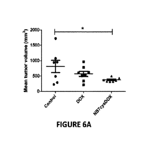
[044] Figs. 6A-6D include graphs and micrographs showing in vivo and in-situ
effects
of NB7cysDOX on PC3-PIP (PSMA) tumors. PC3-PIP xenografts in athymic nude
mice were treated with either saline (control), 2 mg/kg commercial DOX, or 1.4
mg/kg
NB7cysDOX. (6A) Mean tumor volume after 8 d of treatment (before any mouse was
excluded from the experiment due to ethical considerations). *p <0.05 versus
control
(Student's t-test; n = 7 for controls and n = 8 for DOX and NB7cysDOX). (613)
The
slope of calculated logarithmic tumor growth in the treated versus control
(T/C) groups.
*p <0.05 versus control (Student's t-test; n =7 for controls, n = 4 for DOX,
and n = 8
for NB7cysDOX; (6C) A representative tissue section from a tumor, obtained 4 d
after
treatment termination, from one mouse treated with NB7cysDOX and labeled with
PE-
anti-PSMA and RTC-anti-His to identify PSMA and NB7, respectively, and nuclei
were also stained (Hoechst). White arrows point to colocalization of PSMA and
NB7.
(6D) Hematoxylin and Eosin (H&E) staining (top row) and terminal
deoxynucleotidyl
transferase dUTP nick end labeling (TUNEL) and propidium iodide (PI) staining
(bottom row) of tissue sections from tumors obtained 4 d after treatment
termination.
White arrows indicate the colocalization of TUNEL and PI. Scale bars apply to
all three
images in the same row.
[045] Figs. 7A-7C include graphs and a micrograph showing NB selection and
purification. (7A) Enzyme-linked immunosorbent assay (ELISA) results
demonstrating
the binding of PSMA by individual bacterial colonies expressing different NB
sequences. Sequences that were chosen for purification: NB7 (clones 7, 9, 22,
28, 31-
8
CA 03149754 2022-2-28
WO 2021/038571
PCT/1L2020/050940
33, and 46), NB8 (clones 8 and 21), NB13 (clones 13 and 18), and NB37 (clone
37).
(7B) A representative size-exclusion chromatography for NB7 (the chromatograms
for
NB 8, NB13, and NB37 were similar to the one shown here). (7C) SDS-PAGE gel
results
showing the four purified NBs. All proteins were in the expected size of -16
kDa.
[046] Fig. 8 includes a vertical bar graph showing PSMA activity assay. PSMA
was
incubated with a substrate in the presence or absence of NBs to measure
glutamate
carboxypeptidase activity. An inactive (denaturated) PSMA and a commercial
PSMA
inhibitor (PMPA) served as controls. Results were compared and normalized to
the
fluorescence of untreated PSMA. The experiment was performed in triplicate,
and
results are presented as means SEM. ***p <0.005 (Student's t-test, n = 3).
[047] Figs. 9A-9B include small angle X-ray scattering (SAXS) analysis and the
Rg of
the monomeric PSMA. (9A) SAXS analysis of PSMA in PBS (0.5-3 mg/nil). (913)
The
radius of gyration (Rg) values for free PSMA increases slightly with higher
concentrations due to the interaction between species in the solution. The Rg
values,
determined by using Guinier plots, are 43 A at 0.5 mg/m1 and 46 A at 3 mg/ml.
Colors
in both panels reflect PSMA concentrations (darker = higher).
[048] Figs. 10A-10D include graphs showing SAXS curves and the corresponding
Guinier plots of PSMA with increasing concentrations of NBs. PSMA (0.5 mg/ml)
was
premixed with increasing concentrations of NBs (10A: NB7, 1013: NB8, 10C:
NB13,
and1OD: N1337), ranging from 0.1 mg/ml to 0.5 mg/ml, which represent PSMA:NB
molar ratios from 1:0.5 to 1:5. Colors reflect the NB concentrations (darker =
higher).
[049] Fig. 11 includes a graph showing the effect of NBs on the Rg of PSMA.
SAXS
results showing the Rg values of PSMA (0.5 mg/ml) at increasing concentrations
of NBs
(0.05-0.58 mg/m1). The dashed line indicates the Rg of PSMA without NBs.
[050] Figs. 12A-12E include graphs showing the results of the custom-made
script
analysis of the SAXS data. The analysis was done using an automated procedure
based
on a script and the computer program GNOM_ The "Total estimate" score was used
to
choose the best result. PSMA (12A), PSMA+NB7 (128), PSMA+NB8 (12C),
PSMA+NB13 (12D), and PSMA+NB37 (12E).
[051] Fig. 13 includes a graph showing the distance distribution function,
P(r), of
PSMA and NBs at a molar ratio of 1:2. P(r) was determined using the program
GNOM.
9
CA 03149754 2022-2-28
WO 2021/038571
PCT/1L2020/050940
[052] Figs. 14A-14D include micrographs showing ex vivo optical imaging of
prostate
cancer xenografts. The ex-vivo signal is shown at three time points in various
dissected
organs of mice that were injected with either NB7 (14A), NB13 (141I), NB8
(14C), or
NB37 (14D) (the data are shown in Fig. 3). Intensity bars are shown below the
images
of NB13 and apply to all images.
[053] Fig. 15 includes a graph showing the internalization of the NBs into PC3-
PIP
cells. PC3-PIP cells were incubated in 96-wells plates for 1 h with either
NB7, NB8,
NB13, or NB37. Then, the wells were imaged using Operetta and the number of
cells
with NBs on their membranes or inside their cytoplasm was counted every 40
min, so
that the first imaging round was completed 1 h and 40 min after incubation.
The
internalization process is reflected in the number of cells with NB s in their
cytoplasm
relative to the number of cells with NBs in their membrane
("cytoplasm/membrane
ratio"), such that a higher ratio indicated more NBs that were internalized
into the
cytoplasm.
[054] Figs. 16A-16D include an illustration of a process and graphs showing
the
conjugation of NB7cys to DOX. (16A) The conjugation process. (16B) Size-
exclusion
chromatography for NB7cysDOX, analyzed using Superdex 75 10/300. "1" and "2"
designate absorbance in 280 nm and 488 nm, respectively. (16C) Mass-
spectrometry of
NB7cys (1) and NB7cysDOX (2), analyzed using matrix-assisted laser
desorption/ionization time-of-flight (MALDI-TOF). (16D) Flow cytometry binding
of
NB7cys and NB7cysDOX to PC3-PIP cells at the indicated concentrations. The
experiment was performed in triplicate, and the results indicate means SEM.
A
Student's t-test indicated that there were no significant differences between
NB7 and
NB7cysDOX at any of the examined concentrations.
[055] Fig. 17 includes a graph showing 1H-nuclear magnetic resonance (NMR)
spectrum of DOX-BMPH linker.
[056] Figs. 18A-18C include graphs showing the effect of NB7cysDOX on cell
viability. (18A) The number of PC3-PIP cells was counted after a 24 h
treatment with
either NB7cys, DOX, or NB7cysDOX. The experiment was performed in triplicate
and
the results are presented as means SEM. *p <0.05, **p <0.01 (Student's t-
test, as
compared with untreated cells). (18B) FACS analysis of PC3-1113 cells treated
with
either NB7 (1), DOX (2), or NB7cysDOX (3), or left untreated (4, partially
masked by
CA 03149754 2022-2-28
WO 2021/038571
PCT/1L2020/050940
the "1" histogram), and then incubated with PI. (18C) PC3-PIP cells were
treated with
N87, DOX, NB7cysDOX, or FCCP, or were left untreated, and then incubated with
TMRE ("no TMRE") was used as a negative control, and the fluorescent signal of
TMRE was measured by using a plate reader. The fluorescence of the untreated
sample
was set as 1, and all other samples were normalized and compared to it. The
experiment
was performed in triplicate and results are presented as means + SEM. *p
<0.05,
**p <0.01, ***p < 0.005 (Student's Hest).
[057] Figs. 19A-19B include graphs showing in vivo tumor growth inhibition by
NB7cysDOX. PC3-131P xenografts in athymic nude mice were treated with either
saline
(control), DOX (2 mg/kg), or NB7cysDOX (L4 mg/kg). (19A) Tumor volume was
measured twice a week during the treatment duration. Some mice were euthanized
during the trial period due to ethical considerations, and their tumor sizes
were estimated
by using an equation based on logarithmic tumor growth (see Materials and
Methods
section). The solid portions of the curves indicate time points in which all
animals were
included in the analysis, while the dashed portions indicate logarithmically
extrapolated
data. (19B) Percentage of mice that were included in the experiment at each
time point.
"1": control (n = 7 mice), "2": DOX (n = 8 mice), "3": NB7cysDOX (n = 8 mice).
[058] Fig. 20 includes a table showing predicted interactions between NB7 and
PSMA
(A: monomer A of PSMA; B: monomer B of PSMA). CDR1 shows an electrostatic
interaction with monomer B, between aspartic acid 29 of the NB and lysine 223
of
PSMA. CDR2 shows an electrostatic interaction with monomer A, between aspartic
acid
62 of the NB and lysine 718 of PSMA. CDR3 shows two electrostatic interactions
to
monomer B: one between glutamic acid 113 of the NB and arginine 281 of PSMA,
and
the other between lysine 109 of the NB and glutamic acid 285 of PSMA.
[059] Fig. 21 includes a table showing predicted interactions between NB37 and
PSMA. CDRs display electrostatic interactions between glutarnic acid 62 with
two
arginine residues of PSMA (arginine 363 and arginine 411). Another
electrostatic
interaction is between arginine 19 (a non-CDR residue) and aspartic acid 654
of PMSA.
[060] Fig. 22A-22F include micrographs, illustration, and graphs, showing the
incorporation of non-natural amino acids into NB7. (22A) Western blot analysis
showing the incorporation of the unnatural amino acid BOC Lysine in different
positions
on NB7 (A14, A40, G42, K43, and A75). (22B) Western blot analysis showing
11
CA 03149754 2022-2-28
WO 2021/038571
PCT/1L2020/050940
incorporation of the unnatural amino acid BOC Lysine in different positions of
NB7-
Cys (K43, and A75). (22C) an illustration of a non-limiting scheme showing
binding
interaction between PSMA and NB7 cys (+doxorubicin), NB7 K43prop (+Cy5.5), or
NB7 cys K43prop (+doxorubicin and Cy5.5). (22D) is a graph showing size-
exclusion
chromatography. The absorbance of 280 nm for NB7 (1), NB7cys (2), and NB7
K43prop (3) is presented. (22E) Mass-spectrometry of NB7 (1), NB7cys (2), and
NB7
K43prop (3), analyzed using MALDI-TOR (22F) a vertical bar graph showing the
binding of NB7 and NB7 K43PrK to PC3-P1P cells, as was determined using FACS
analysis. The results show no significant change in binding following the
K43PrK
mutation.
DETAILED DESCRIPTION
[061] In some embodiments, the present invention is directed to an antigen-
binding
polypeptide having increased binding affinity to prostate specific membrane
antigen
(PSMA). In some embodiments, the antigen-binding polypeptide is a single-
domain
antibody.
[062] Before explaining at least one embodiment of the invention in detail, it
is to
be understood that the invention is not necessarily limited in its application
to the
details set forth in the following description or exemplified by the Examples.
The
invention is capable of other embodiments or of being practiced or carried out
in
various ways.
[063] The term "prostate specific membrane antigen" or PSMA, as used herein,
refers to glutamate carboxypeptidase II, also known as N-acetyl-L-aspartyl-L-
glutamate peptidase I (NAALADase I or NAAG peptidase). As a non-limiting
example, human PSMA has the UniProt accession no. Q04609.
[064] The terms "antibody" and "antigen-binding polypeptide" (also referred to
as
an "immunoglobulin" or "Ig") refer to a polypeptide or group of polypeptides
that
include at least one binding domain that is specific for one antigen. In
certain
embodiments, the use of a chimeric antibody or a humanized antibody is also
encompassed by the invention.
[065] In some embodiments, the term "antibody fragments" refers to a portion
of an
intact antibody, preferably comprising the antigen binding region thereof.
12
CA 03149754 2022-2-28
WO 2021/038571
PCT/11,2020/050940
[066] The terms "single-domain antibody" refers to an antibody fragment
consisting
of a single variable domain (VHF!). Single-domain antibody is a smaller
functional
fragment of the antibody that also can bind a specific antigen. In some
embodiments,
the single-domain antibody has better tissue penetration than conventional
antibodies
and therefore they are beneficial for clinicaUdiagnostic use.
[067] In some embodiments, the single-domain antibody of the present invention
comprises three complementary-determining regions (CDRs).
[068] In some embodiments, the term "complementary-determining region" refers
to variable heavy chain. In some embodiments, the variable heavy chain
comprises an
amino acid sequence capable of binding a specific PS MA.
[069] Kabat et al. defined a numbering system for variable domain sequences
that is
applicable to any antibody. One of ordinary skill in the art can unambiguously
assign
this system of "Kabat numbering" to any variable domain sequence, without
reliance
on any experimental data beyond the sequence itself. As used herein, "Kabat
numbering" refers to the numbering system set forth by Kabat et al, U.S. Dept.
of
Health and Human Services, "Sequence of Proteins of hrnnunological Interest"
(1983).
[070] In some embodiments, the antigen-binding polypeptide comprises three
CDRs
comprising: GY'TDSNYYMS (CDR-H1; SEQ ID NO: 1),
GVNTGRGSTSYADSVKG (CDR-H2; SEQ ID NO: 2), and
AACHFCDSLPKTQDEY1L (CDR-H3; SEQ 11) NO: 3).
[071] In some embodiments, the antigen-binding polypeptide comprises three
CDRs
comprising: GWPYSTYSMN (CDR-H1; SEQ ID NO: 4), GISSTMSGIFFAES (CDR-
112; SEQ NO: 5), and RRDYSLSSSSDDFDY (CDR-H3; SEQ ID NO: 6).
[072] In some embodiments, the antigen-binding polypeptide comprises three
CDRs
comprising: GYTASFS (CDR-H1; SEQ ID NO: 7), GVAVINVGVGSTYYADSV
(CDR-H2; SEQ ID NO: 8) and SLRWSRPPNPISEDAYNY (CDR-H3; SEQ ID NO:
9).
[073] In some embodiments, the antigen-binding polypeptide comprises three
CDRs
comprising: GWPYSTYSMN (CDR-H1; SEQ ID NO: 4),
GISSTMSGI1FAESKAGQFTISQDNA (CDR-H2; SEQ ID NO: 10), and
RRDYSLSSSSDDFDY (CDR-H3; SEQ ID NO: 6).
13
CA 03149754 2022-2-28
WO 2021/038571
PCT/11,2020/050940
[074] In some embodiments, the antigen-binding polypeptide comprises the amino
acid
sequence:
QVQLQESGGGSVQXI GGSLRLSCTAPGYTDSNYYMSWERQX2PX3X4EREWV
AGVNTGRGSTS YADSVICGRFTISQDNX5ICNTMELQMNSLICPEDTAIYYCAV
AACHFCDSLPKTQDEY1LWGQGTQVTVSSAAAYPYDVPDYGS (SEQ TD NO:
15), wherein: Xi is selected from Alanine or an artificial or non-naturally
occurring
amino acid, X2 is selected from Alanine or an artificial or non-naturally
occurring
amino acid, X3 is selected from Glycine or an artificial or non-naturally
occurring
amino acid, X4 is selected from Lysine or an artificial or non-naturally
occurring
amino acid, and X5 is selected from Alanine or an artificial or non-naturally
occurring
amino acid. In some embodiments, the artificial or non-naturally occurring
amino
acid comprises or consists of the amino acid BOC-Lysine.
[075] In some embodiments, the antigen-binding polypeptide comprises the amino
acid
sequence:
QVQLQESGGGSVQAGGSLRLSCTAPGYTDSNYYMSWFRQAPGKEREWVA
G VNTGRGST S Y ADS V KGRFTIS QDNAICNTMFLQMNS LKPEDTAIYYCA VAA
CHFCDSLPKTQDEY1LWGQGTQVTVSSAAAYPYDVPDYGS (SEQ ID NO: 11).
[076] In some embodiments, the antigen-binding polypeptide comprises the amino
acid
sequence:
QVQLQESGGGSVQXIGGSLRLSCARSGWPYSTYSMNWFRQX2PX3X4EREAV
AGISSTMSGTIFAESICAGQFTISQDNX5ICNTVYLQMNNLICPEDTAIYYCAARR
DYSLSSSSDDFDYWGQGTQVTVSSAAAYPYDVPDYGS (SEQ 1D NO: 16),
wherein: X1 is selected from Alanine or an artificial or non-naturally
occurring amino
acid. X2 is selected from Alanine or an artificial or non-naturally occurring
amino
acid, X3 is selected from Glycine or an artificial or non-naturally occurring
amino
acid, X4 is selected from Lysine or an artificial or non-naturally occurring
amino acid,
and X5 is selected from Alanine or an artificial or non-naturally occurring
amino acid.
In some embodiments, the artificial or non-naturally occurring amino acid
comprises
or consists of the amino acid BOC-Lysine.
[077] In some embodiments, the antigen-binding polypeptide comprises the amino
acid
sequence:
QVQLQESGGGSVQAGGSLRLSCARSGWPYSTYSMNWFRQAPGICEREAVAG
14
CA 03149754 2022-2-28
WO 2021/038571
PCT/11,2020/050940
ISSTMSGHFAES KAGQFTISQDNAKNTVYLQMNNLKPEDTAIYYCAARRDY
SLSSSSDDFDYWGQGTQVTVSSAAAYPYDVPDYGS (SEQ ID NO: 12).
[078] In some embodiments, the antigen-binding polypeptide comprises the amino
acid
sequence:
Q VQLQES GGGS VQ TGGSLRLSCAAS GYT AS FSWIGYFRQX1PX2X3EFtEGV A
VINVGVGSTYY ADS V KGRFTIS RDNTENTIS LEMNS LKPEDT GLY YC AGSL R
WSRPPNPISEDAYNYWGQGTQVTVSSAAAYPYDVPDYGS (SEQ ID NO: 17),
wherein: Xi is selected from Alanine or an artificial or non-naturally
occurring amino
acid, X2 is selected from Glycine or an artificial or non-naturally occurring
amino
acid, and X3 is selected from Lysine or an artificial or non-naturally
occurring amino
acid. In some embodiments, the artificial or non-naturally occurring amino
acid
comprises or consists of the amino acid BOC-Lysine.
[079] In some embodiments, the antigen-binding polypeptide comprises the amino
acid
sequence:
Q VQLQES GGGS VQ TGGSLRLSCAAS GYT AS FSWIGYFRQ APGKEREGV AVI
N VGVGSTY YADS VKGRFTISRDNTENTIS L EMNS L ICPEDTGLYYCA GS LRW
SRPPNPISEDAYNYWGQGTQVTVSSAAAYPYDVPDYGS (SEQ NO: 13).
[080] In some embodiments, the antigen-binding polypeptide comprises the amino
acid
sequence:
QVQLQESGGGSVEXIGGSLRLSCARSGWPYSTYSMNWFRQX2PX3X4EREAV
AGISSTMSGHFAESKAGQVFISQDNX5KNTVYLQMNNLKPEDTAIYYCAARR
DYSLSSSSDDFDYWGQGTQVTVSSAAAYPYDVPDYGS (SEQ ID NO: 18),
wherein: X1 is selected from Alanine or an artificial or non-naturally
occurring amino
acid, X2 is selected from Alanine or an artificial or non-naturally occurring
amino
acid, X3 is selected from Glycine or an artificial or non-naturally occurring
amino
acid, X4 is selected from Lysine or an artificial or non-naturally occurring
amino acid,
and X5 is selected from Alanine or an artificial or non-naturally occurring
amino acid.
In some embodiments, the artificial or non-naturally occurring amino acid
comprises
or consists of the amino acid BOC-Lysine.
[081] In some embodiments, the antigen-binding polypeptide comprises the amino
acid
sequence:
QVQLQESGGGSVEAGGSLRLSCARSGWPYSTYSMNWFRQAPGICEREAVAG
CA 03149754 2022-2-28
WO 2021/038571
PCT/1L2020/050940
ISSTMSGHFAES KAGQFTISQDNAKNTVYLQMNNLKPEDTAIYYCAARRDY
SLSSSSDDFDYWGQGTQVTVSSAAAYPYDVPDYGS (SEQ ID NO: 14).
[082] In some embodiments, the antigen-binding polypeptide has a specific
binding
affinity to PSMA.
[083] As used herein, the term "specific binding" refers to a non-covalent
physical
association of a first and a second moiety of two entities. In some
embodiments, the
association between the first and second moieties is at least 10 times as
strong, at least
50 times as strong, or at least 100 times as strong as the association of
other moieties
present in the environment in which binding occurs.
[084] In some embodiments, the binding of two or more entities may be
considered
specific if the equilibrium "dissociation constant", KD, is less than 10-3 M,
less than
10-4 M, less than 10-5 M, less than 10-6 M, less than 10-7M, less than 10-8 M,
less
than 10-9 M, less than 1(110 M, less than 1(111 M, or less than 1(112 M, or
any value
and range therebetween. Each possibility represents a separate embodiment of
the
invention. In some embodiments, the binding of two or more entities may be
considered specific if the equilibrium "dissociation constant", KD, is 10-1 M
- 10-3
M, 10-12 M - lr M. Each possibility represents a separate embodiment of the
invention. In some embodiments, specific binding can be accomplished by a
plurality
of weaker interactions. Calculation of a peptide's dissociation constant (KD)
is known
to a skilled artisan and is also show in the Examples section herein below.
[085] In some embodiments, the term "binding constant", or "association
constant",
refers to a special case of the equilibrium constant Ka, which is the inverse
of the
dissociation constant.
[086] In some embodiments, the antigen-binding polypeptide is characterized by
binding constant (Ka) of at least 103, at least 10x103, at least 104, at least
2x10⁴, at
least 105, or at least 5x10⁵ Molar sec-' (M-1 s-1) to PSMA, or any value and
range
therebetween. Each possibility represents a separate embodiment of the
invention.
[087] In non-limiting exemplary embodiments, the antigen-binding polypeptide
comprising the amino acid sequence as set forth in SEQ ID NO: 11 is
characterized
by binding constant (IQ of about 7.1x10⁵ M-1 s to PSMA.
16
CA 03149754 2022-2-28
WO 2021/038571
PCT/1L2020/050940
[088] In non-limiting exemplary embodiments, the antigen-binding polypeptide
comprising the amino acid sequence as set forth in SEQ ID NO: 12 is
characterized
by binding constant (I(a) of about 2x10⁴ M-1 s-1 to PSMA.
[089] In non-limiting exemplary embodiments, the antigen-binding polypeptide
comprising the amino acid sequence as set forth in SEQ ID NO: 13 is
characterized
by binding constant (Ka) of about 3.6x10⁴ s-1
to PSMA.
[090] In non-limiting exemplary embodiments, the antigen-binding polypeptide
comprising the amino acid sequence as set forth in SEQ ID NO: 14 is
characterized
by binding constant (Ka) of about 2.2x10⁴ M-1 s to PSMA.
[091] In some embodiments, the antigen-binding polypeptide is characterized by
dissociation constant (KD) of less than 10 pM, less than 50 pM, less than 500
pM, less
than 15 nM, less than 50 nM, or less than 500 nM to PSMA, or any value and
range
therebetween. Each possibility represents a separate embodiment of the
invention.
[092] In non-limiting exemplary embodiments, the antigen-binding polypeptide
comprising the amino acid sequence as set forth in SEQ ID NO: 11 is
characterized
by dissociation constant (KD) of about 55 pM to PSMA.
[093] In non-limiting exemplary embodiments, the antigen-binding polypeptide
comprising the amino acid sequence as set forth in SEQ ID NO: 12 is
characterized
by dissociation constant (KD) of about 6 nM to PSMA (as calculated herein
below in
the Examples section).
[094] In non-limiting exemplary embodiments, the antigen-binding polypeptide
comprising the amino acid sequence as set forth in SEQ ID NO: 13 is
characterized
by dissociation constant (KD) of about 0.6 nM to PSMA.
[095] In non-limiting exemplary embodiments, the antigen-binding polypeptide
comprising the amino acid sequence as set forth in SEQ ID NO: 14 is
characterized
by dissociation constant (KD) of about 3.4 nM to PSMA.
[096] In some embodiments, the polypeptide binds to a non-catalytic site of
PSMA.
In one embodiment, the polypeptide binds to an extracellular domain of PSMA.
[097] As used herein, the terms "peptide", "polypeptide" and "protein" are
used
interchangeably to refer to a polymer of amino acid residues. In another
embodiment,
the terms "peptide", "polypeptide" and "protein" as used herein encompass
native
17
CA 03149754 2022-2-28
WO 2021/038571
PCT/1L2020/050940
peptides, peptidomimetics (typically including non-peptide bonds or other
synthetic
modifications) and the peptide analogues peptoids and semipeptoids or any
combination thereof.
[098] In some embodiments, the polypeptide binding to PSMA is characterized by
allowing further interaction to PSMA. In some embodiments, the polypeptide
binding
to PSMA is characterized by retaining PSMA enzyme activity. Methods of
determining PSMA activity are known in the art and are also exemplified herein
below, as a non-limiting example.
[099] In some embodiments, the antigen-binding polypeptide is characterized by
molecular weight of less than 15 kDa, less than 20 kDa, less than 25 kDa, less
than 35
kDa, or less than 50 kDa, or any value and range therebetween. Each
possibility
represents a separate embodiment of the invention.
[0100] In some embodiments, the antigen-binding polypeptide is characterized
by
thermal stability (T.) of at least 60 C, at least 70 C, at least 90 C, or at
least 95 C,
or any value and range therebetween_ Each possibility represents a separate
embodiment of the invention.
[0101] As used herein, the term "thermal stability", refers to a substance
resistance to
irreversible change in its chemical or physical structure at an elevated
temperature. In
some embodiments, T. indicates the thermal energy that caused the
denaturation/unfolding of a protein or a peptide.
[0102] In some embodiments, the N- or C-terminus of the antigen-binding
polypeptide comprises a tag motif. In some embodiments, the tag motif
comprises at
least six amino acids. In some embodiments, the antigen-binding polypeptide
comprises histidine (His)-tag. In some embodiments, the antigen-binding
polypeptide
comprises human influenza hemagglutinin (HA)-tag.
[0103] According to another embodiment, the polypeptides of the invention
encompass truncated forms and/or fragments of any one of SEQ ID NOs: 1-14 as
long
as they are capable of binding PSMA.
[0104] Conservative substitution of amino acids as known to those skilled in
the art
are within the scope of the present invention. Conservative amino acid
substitutions
include replacement of one amino acid with another having the same type of
functional group or side chain e.g. aliphatic, aromatic, positively charged,
negatively
18
CA 03149754 2022-2-28
WO 2021/038571
PCT/1L2020/050940
charged. One of skill will recognize that individual substitutions, deletions
or
additions to peptide, polypeptide, or protein sequence which alters, adds or
deletes a
single amino acid or a small percentage of amino acids in the encoded sequence
is a
"conservatively modified variant" where the alteration results in the
substitution of an
amino acid with a chemically similar amino acid. Conservative substitution
tables
providing functionally similar amino acids are well known in the art.
[0105] The following six groups each contain amino acids that are conservative
substitutions for one another: 1) Alanine (A), Serine (5), Threonine (T); 2)
Aspartic
acid (0), Glutamic acid (E); 3) Asparagine (N), Glutamine (Q); 4) Arginine
(R),
Lysine (K); 5) Isoleucine (I), Leucine (L), Methionine (M), Valine (V); and 6)
Phenylalanine (F), Tyrosine (Y), Tryptophan (W) (see, e.g., Creighton,
Proteins,
1984).
[0106] The term "conservative substitution" also includes the use of a
chemically
derivatized residue in place of a non-derivatized residue provided that such
peptide
displays the requisite function of modulating the immune system's innate
response as
specified herein.
[0107] In some embodiments, the polypeptide of the invention comprises a non-
naturally occurring amino acid.
[0108] Methods for integrating a non-naturally occurring amino acid into a
polypeptide are common and would be apparent to one of ordinary skill in the
art.
[0109] In some embodiments, any non-naturally occurring amino acid is
envisioned
by the current invention as long as the resulting polypeptide comprising the
non-
naturally occurring amino acid maintains its activity, e.g., high affinity
binding to
PSMA, or any other activity such as disclosed herein.
[0110] In some embodiments, a non-naturally occurring amino acid is selected
from:
3-Iodo-L-tyrosine, NE -Benzyloxycarbonyllysine (ZLys), NE -Acetyllysine
(AcLys),
N6 - Cyclopentyloxycarbon yl-L-lysine (Cyc), N6 ¨(((1R,2R)-2-
azidocyclopentyloxy)c arbony1)-L-lysine (ACPK), o-Nitrobenzyl-Otyrosine, o-
Nitrobenzyloxycarbonyl-N2 -Llysine, N -[(1-(6-Nitrobenzo [d][1,3]dioxo1-5y1)
ethoxy)carbonyll- L-lysine, NC -[(2-(3-Methyl-3Hdiazirin-3-
yflethoxy)carbonyll-
Llysine, (3-(3-Methyl-3Hdiazirine-3-y1)- propaminocarbonylNE -L-lysine
(DiZPK),
BCN (exo isomer), BCN (endo isomer), TCO, N2 -(1- Methylcycloprop-2-
19
CA 03149754 2022-2-28
WO 2021/038571
PCT/1L2020/050940
enecarboxamido)lysin e (CpK), N6 -Acryloyl-L-lysine, pNO2ZLys, TmdZLys, N6 -
Crotonyl-L-lysine (Kcr), 2 -Chloro - L ¨ phenylalanine, 2 -Bromo - L ¨
phenylalanine,
2 dodo - L ¨ phenylalanine, 2 -Methyl - L ¨ phenylalanine, 2 -Methoxy - L ¨
phenylalanine, 2 -Nitro - L ¨ phenylalanine, 2 -Cyan o - L ¨ phenylalanine,
and N -
(tert - Butoxycarbonyl) - L - lysine (BOC-Lysine).
[0111] In some embodiments, a non-naturally occurring amino acid comprises or
consists of BOC-Lysine.
Methods for treatment and diagnosis
[0112] In another embodiment, the present invention provides a method for
targeting
PSMA by contacting a sample comprising PSMA with an antigen-binding
polypeptide
of the invention, thereby targeting PSMA.
[0113] In one embodiment, the present invention provides a method for
treating,
diagnosing, prognosticating or determining the suitability for treatment of a
subject
suffering from a PSMA-associated disorder, the method comprising administering
to
the subject a pharmaceutical composition comprising an effective amount of the
antigen-binding polypeptide of the invention, a cytotoxic agent or a
theranostic agent,
and a pharmaceutical acceptable carrier, thereby treating diagnosing,
prognosticating
or determining the suitability for treatment of a subject suffering from a
PSMA-
associated disorder in said subject.
[0114] In one embodiment, there is provided a method for imaging PSMA in a
subject, such as a subject suffering from or suspected to suffer from a PSMA-
associated disorder, the method comprising administering to the subject a
composition
comprising an effective amount of the antigen-binding polypeptide of the
invention,
and an imaging agent; and detecting the PSMA in the subject, thereby imaging
PSMA
in a subject.
[0115] In some embodiments, the imaging agent is selected from, without being
limited thereto, a fluorescent label (e.g., fluorescein isothiocyanate), a
chromophore,
a radioactive label, a paramagnetic ion (e.g., Gc1+3), and any combination
thereof.
[0116] In some embodiments, the term "chromophore" refers to a material that
absorbs certain wavelength of light from UV to near infrared region and may be
or
may not be emissive.
CA 03149754 2022-2-28
WO 2021/038571
PCT/1L2020/050940
[0117] In one embodiment, the imaging agent is a radioactive label (e.g.,
isotope). In
another embodiment, the therapeutic agent is a radioactive label (e.g.,
isotope). In
some embodiments, the isotope is selected from, but not limited to:
18F, 47Sc, 51Cr, 52Fe, 521"Mn, 56Ni, 57Ni, 62Cu, 61Cu, 67Ga, 68Ga, 72As, 75Br,
76Br, 77Br,
82Br, 89Zr, "n'Tc, 97Ru, 99"Tc, mln, 1231, 1241,
134, 1911:14 197 11 ng,
201rn, 203Pb, "69111201 ,
"C, 18F, and 13N.
[0118] In some embodiments, the imaging techniques are selected from, without
being limited thereto, computed X-ray tomography (CT), ultrasound (US), and
magnetic resonance imaging (MRI), positron emission tomography (PET), single-
photon emission computed tomography (SPECT), fluorescence and radio assays,
cytofluorimetry, and fluorescence activated cell sorting. The principles of
such
techniques can be found in immunochemistry handbooks, for example: A Johnstone
and R. Thorpe, Immunochemistry in practice, 2" Edition (1987), blackwell
Scientific
publications, Oxford London Edinburgh Boston Palo Alto Melbourne.
[0119] Non-limiting exemplary embodiments demonstrate the diagnosis of
prostate
tumors in vivo by near infra-red (NW) imaging after 24 hours from the
administration
of the antigen-binding polypeptide conjugated to a fluorescent label.
[0120] In one embodiment, the method further comprises determining the
relative
percentage of the PSMA subpopulations by the administration of antigen-binding
polypeptide.
[0121] In some embodiments, the antigen-binding polypeptide of the present
invention can be used in conjunction with other therapeutic treatment
modalities,
including surgery, cryosurgery, radiation, thermotherapy, hormone treatment,
chemotherapy, immunotherapy, vaccines, and any combination thereof.
[0122] In some embodiments, the therapeutic agent can include any agent (e.g.,
molecule, drug, pharmaceutical composition, etc.) capable of preventing,
inhibiting,
or arresting the symptoms and/or progression of a disease.
[0123] In some embodiments, the therapeutic agent is selected from, but not
limited
to: a chemotherapeutic agent (e.g., methotrexate, cisplatin and paclitaxel),
an anti-
oncogenic agent, an anti-angiogenic agent, a tumor suppressor agent, an anti-
microbial agent, or an expression construct comprising a nucleic acid encoding
a
therapeutic protein.
21
CA 03149754 2022-2-28
WO 2021/038571
PCT/1L2020/050940
1101241 In some embodiments, the PSMA-associated disorder is prostate cancer.
[0125] In some embodiments, the PSMA-associated disorder is a neurological
disorder. In some embodiments, the neurological disorder is selected from, but
not
limited to: Parkinson disease, Alzheimer disease, Huntington disease,
amyotrophic
lateral sclerosis (ALS), and schizophrenia.
Pharmaceutical compositions
[0126] The present invention also contemplates pharmaceutical compositions for
human medical use, the composition comprising at least one antigen-binding
polypeptide as described herein.
[0127] The present invention also contemplates the use of an antigen-binding
polypeptide as described herein, for the manufacture of a pharmaceutical
composition
for the treatment, diagnosis, theranostic or prophylaxis of cancer or
neurological
disorder.
[0128] In some embodiments, the pharmaceutical composition comprises a
therapeutic or diagnostic effective amount of the antigen-binding polypeptide
described herein, with optionally any one of additional therapeutic
ingredient(s),
imaging agent(s), and combination thereof, and one or more pharmaceutically
acceptable carriers.
[0129] The pharmaceutical compositions of the invention can be formulated in
the
form of a pharmaceutically acceptable salt of the polypeptide of the invention
or their
analogs thereof. Pharmaceutically acceptable salts include those salts formed
with free
amino groups such as salts derived from non-toxic inorganic or organic acids
such as
hydrochloric, phosphoric, acetic, oxalic, tartaric acids, and the like, and
those salts
formed with free carboxyl groups such as salts derived from non-toxic
inorganic or
organic bases such as sodium, potassium, ammonium, calcium, ferric hydroxides,
isopropylamine, triethylamine, 2-ethylamino ethanol, histidine, procaine, and
the like.
In one embodiment, pharmaceutical compositions of the present invention are
manufactured by processes well known in the art, e.g., by means of
conventional
mixing, dissolving, granulating, dragee-making, levigating, emulsifying,
encapsulating, entrapping or lyophilizing processes.
[0130] The term "analog" includes any peptide having an amino acid sequence
substantially identical to one of the sequences specifically shown herein in
which one
22
CA 03149754 2022-2-28
WO 2021/038571
PCT/1L2020/050940
or more residues have been conservatively substituted with a functionally
similar
residue and which displays the abilities as described herein. Examples of
conservative
substitutions include the substitution of one non-polar (hydrophobic) residue
such as
isoleucine, valine, leucine or methionine for another, the substitution of one
polar
(hydrophilic) residue for another such as between arginine and lysine, between
glutamine and asparagine, between glycine and serine, the substitution of one
basic
residue such as lysine, arginine or histidine for another, or the substitution
of one
acidic residue, such as aspartic acid or glutamic acid for another. Each
possibility
represents a separate embodiment of the present invention.
[0131] The term "pharmaceutically acceptable" means suitable for
administration to
a subject, e.g., a human. For example, the term "pharmaceutically acceptable"
can
mean approved by a regulatory agency of the Federal or a state government or
listed
in the U. S. Pharmacopeia or other generally recognized pharmacopeia for use
in
animals, and more particularly in humans. The term "carrier" refers to a
diluent,
adjuvant, excipient, or vehicle with which the therapeutic compound is
administered.
Such pharmaceutical carriers can be sterile liquids, such as water and oils,
including
those of petroleum, animal, vegetable or synthetic origin, such as peanut oil,
soybean
oil, mineral oil, sesame oil and the like, polyethylene glycols, glycerin,
propylene
glycol or other synthetic solvents. Water is a preferred carrier when the
pharmaceutical composition is administered intravenously. Saline solutions and
aqueous dextrose and glycerol solutions can also be employed as liquid
carriers,
particularly for injectable solutions. Suitable pharmaceutical excipients
include starch,
glucose, lactose, sucrose, gelatin, malt, rice, flour, chalk, silica gel,
sodium stearate,
glycerol monostearate, talc, sodium chloride, dried skim milk, glycerol,
propylene
glycol, water, ethanol and the like. The composition, if desired, can also
contain minor
amounts of wetting or emulsifying agents, or pH buffering agents such as
acetates,
citrates or phosphates. Antibacterial agents such as benzyl alcohol or methyl
parabens;
antioxidants such as ascorbic acid or sodium bisulfite; and agents for the
adjustment
of tonicity such as sodium chloride or dextrose are also envisioned. The
carrier may
constitute, in total, from about 0.1% to about 99.99999% by weight of the
pharmaceutical compositions presented herein.
[0132] The compositions can take the form of solutions, suspensions,
emulsions,
tablets, pills, capsules, powders, gels, creams, ointments, foams, pastes,
sustained-
23
CA 03149754 2022-2-28
WO 2021/038571
PCT/1L2020/050940
release formulations and the like. The compositions can be formulated as a
suppository, with traditional binders and carriers such as triglycerides,
microcrystalline cellulose, gum tragacanth or gelatin. Oral formulation can
include
standard carriers such as pharmaceutical grades of mannitol, lactose, starch,
magnesium stearate, sodium saccharine, cellulose, magnesium carbonate, etc.
Examples of suitable pharmaceutical carriers are described in: Remington's
Pharmaceutical Sciences" by E.W. Martin, the contents of which are hereby
incorporated by reference herein. Such compositions will contain a
therapeutically
effective amount of the active agent and the antigen-binding polypeptide of
the
invention, preferably in a substantially purified form, together with a
suitable amount
of carrier so as to provide the form for proper administration to the subject.
[0133] An embodiment of the invention relates to an antigen-binding
polypeptide
presented in unit dosage form and is prepared by any of the methods well known
in
the art of pharmacy. In an embodiment of the invention, the unit dosage form
is in the
form of a tablet, capsule, lozenge, wafer, patch, ampoule, vial or pre-filled
syringe. In
addition, in vitro assays may optionally be employed to help identify optimal
dosage
ranges. The precise dose to be employed in the formulation will also depend on
the
route of administration, and the nature of the disease or disorder, and should
be
decided according to the judgment of the practitioner and each patient's
circumstances.
Effective doses can be extrapolated from dose-response curves derived from in-
vitro
or in-vivo animal model test bioassays or systems.
[0134] Depending on the location of the tissue of interest, the antigen-
binding
polypeptide of the present invention can be supplied in any manner suitable
for the
provision of the antigen-binding polypeptide to cells within the tissue of
interest.
Thus, for example, a composition comprising the antigen-binding polypeptide
can be
introduced, for example, into the systemic circulation, which will distribute
the
antigen-binding polypeptide to the tissue of interest. Alternatively, a
composition can
be applied topically to the tissue of interest (e.g., injected, or pumped as a
continuous
infusion, or as a bolus within a tissue, applied to all or a portion of the
surface of the
skin, etc.).
[0135] In an embodiment of the invention, the antigen-binding polypeptide is
administered via oral, rectal, vaginal, topical, nasal, ophthalmic,
transdermal,
subcutaneous, intramuscular, intraperitoneal or intravenous routes of
administration.
24
CA 03149754 2022-2-28
WO 2021/038571
PCT/1L2020/050940
The route of administration of the pharmaceutical composition will depend on
the
disease or condition to be treated. Suitable routes of administration include,
but are
not limited to, parenteral injections, e.g., intradermal, intravenous,
intramuscular,
intralesional, subcutaneous, intrathecal, and any other mode of injection as
known in
the art. Although the bioavailability of antigen-binding polypeptides
administered by
other routes can be lower than when administered via parenteral injection, by
using
appropriate formulations it is envisaged that it will be possible to
administer the
compositions of the invention via transdermal, oral, rectal, vaginal, topical,
nasal,
inhalation and ocular modes of treatment. In addition, it may be desirable to
introduce
the pharmaceutical compositions of the invention by any suitable route,
including
intraventricular and intrathecal injection; intraventricular injection may be
facilitated
by an intraventricular catheter, for example, attached to a reservoir.
1101361 For topical application, the antigen-binding polypeptide of the
present
invention, or analog thereof, can be combined with a pharmaceutically
acceptable
carrier, an imaging agent, and one or more therapeutic agents, so that an
effective
dosage is delivered, based on the desired activity. The carrier can be in the
form of,
for example, and not by way of limitation, an ointment, cream, gel, paste,
foam,
aerosol, suppository, pad or gelled stick.
[0137] For oral applications, the pharmaceutical composition may be in the
form of
tablets or capsules, which can contain any of the following ingredients, or
compounds
of a similar nature: a binder such as microcrystalline cellulose, gum
tragacanth or
gelatin; an excipient such as starch or lactose; a disintegrating agent such
as alginic
acid, Primogel, or corn starch; a lubricant such as magnesium stearate; or a
glidant
such as colloidal silicon dioxide. When the dosage unit form is a capsule, it
can
contain, in addition to materials of the above type, a liquid carrier such as
fatty oil. In
addition, dosage unit forms can contain various other materials which modify
the
physical form of the dosage unit, for example, coatings of sugar, shellac, or
other
enteric agents. The tablets of the invention can further be film coated.
[0138] For purposes of parenteral administration, solutions in sesame or
peanut oil or
in aqueous propylene glycol can be employed, as well as sterile aqueous
solutions of
the corresponding water-soluble salts. Such aqueous solutions may be suitably
buffered, if necessary, and the liquid diluent first rendered isotonic with
sufficient
CA 03149754 2022-2-28
WO 2021/038571
PCT/1L2020/050940
saline or glucose. These aqueous solutions are especially suitable for
intravenous,
intramuscular, subcutaneous and intraperitoneal injection purposes.
[0139] The compositions of the present invention are generally administered in
the
form of a pharmaceutical composition comprising the antigen-binding
polypeptide of
this invention together with a pharmaceutically acceptable carrier or diluent.
Thus, the
compositions of this invention can be administered either individually or
together in
any conventional oral, parenteral or transderrnal dosage form.
[0140] Pharmaceutical compositions according to embodiments of the invention
may
contain 0.1%-95% of the antigen-binding polypeptide(s) of this invention and
active/imaging agent(s), preferably 1%-70%. In any event, the composition or
formulation to be administered may contain a quantity of antigen-binding
polypeptide
and active and/or imaging agents according to embodiments of the invention in
an
amount effective to treat or diagnose the condition or disease of the subject
being
administered.
[0141] The compositions also comprise preservatives, such as benzallconium
chloride
and thimerosal and the like; chelating agents, such as EDTA sodium and others;
buffers such as phosphate, citrate and acetate; tonicity agents such as sodium
chloride,
potassium chloride, glycerin, mannitol and others; antioxidants such as
ascorbic acid,
acetylcystine, sodium metabisulfote and others; aromatic agents; viscosity
adjustors,
such as polymers, including cellulose and derivatives thereof; and polyvinyl
alcohol
and acid and bases to adjust the pH of these aqueous compositions as needed.
The
compositions may also comprise local anesthetics or other actives.
[0142] In addition, the compositions may further comprise binders (e.g.
acacia,
cornstarch, gelatin, carbomer, ethyl cellulose, guar gum, hydroxypropyl
cellulose,
hydroxypropyl methyl cellulose, povidone), disintegrating agents (e.g.
cornstarch,
potato starch, alginic acid, silicon dioxide, croscarmellose sodium,
crospovidone, guar
gum, sodium starch glycolate), buffers (e.g., Tris-HCI., acetate, phosphate)
of various
pH and ionic strength, additives such as albumin or gelatin to prevent
absorption to
surfaces, detergents (e.g., Tween 20, Tween 80, Pluronic F68, bile acid
salts), protease
inhibitors, surfactants (e.g. sodium lauryl sulfate), permeation enhancers,
solubilizing
agents (e.g., glycerol, polyethylene glycerol), anti-oxidants (e.g., ascorbic
acid,
sodium metabisulfite, butylated hydroxyanisole), stabilizers (e.g.
hydroxypropyl
26
CA 03149754 2022-2-28
WO 2021/038571
PCT/1L2020/050940
cellulose, hydroxypropylmethyl cellulose), viscosity increasing agents (e.g.
carbomer,
colloidal silicon dioxide, ethyl cellulose, guar gum), sweeteners (e.g.
aspartame, citric
acid), preservatives (e.g., Thimerosal, benzyl alcohol, parabens), lubricants
(e.g.
stearic acid, magnesium stearate, polyethylene glycol, sodium lauryl sulfate),
flow-
aids (e.g. colloidal silicon dioxide), plasticizers (e.g. diethyl phthalate,
triethyl citrate),
emulsifiers (e.g. carbomer, hydroxypropyl cellulose, sodium lauryl sulfate),
polymer
coatings (e.g., poloxamers or poloxamines), coating and film forming agents
(e.g.
ethyl cellulose, acrylates, polymethacrylates) and/or adjuvants.
[0143] The antigen-binding polypeptide of the present invention or analog
thereof can
be delivered in a controlled release system. Thus, an infusion pump can be
used to
administer the antigen-binding polypeptide such as the one that is used, for
example,
for delivering insulin or chemotherapy to specific organs or tumors. In one
embodiment, the antigen-binding polypeptide of the invention is administered
in
combination with a biodegradable, biocompatible polymeric implant, which
releases
the antigen-binding polypeptide over a controlled period of time at a selected
site.
Examples of preferred polymeric materials include, but are not limited to,
polyanhydrides, polyorthoesters, polyglycolic acid, polylactic acid,
polyethylene
vinyl acetate, copolymers and blends thereof (See, Medical applications of
controlled
release, Langer and Wise (eds.), 1974, CRC Pres., Boca Raton, Fla., the
contents of
which are hereby incorporated by reference in their entirety). In yet another
embodiment, a controlled release system can be placed in proximity to a
therapeutic
target, thus requiring only a fraction of the systemic dose.
[0144] In one embodiment, compositions of the present invention are presented
in a
pack or dispenser device, such as an FDA approved kit, which contain one or
more
unit dosage forms containing the active ingredient. In one embodiment, the
pack or
dispenser device is accompanied by instructions for administration.
[0145] In one embodiment, it will be appreciated that the antigen-binding
polypeptide
of the present invention can be provided to the individual with active agents
to achieve
an improved therapeutic effect as compared to treatment without a targeting
agent. In
another embodiment, measures (e.g., dosing and selection of the complementary
agent) are taken to adverse side effects which are associated with combination
therapies.
27
CA 03149754 2022-2-28
WO 2021/038571
PCT/1L2020/050940
[0146] A "therapeutically effective amount" of the active agent and the
antigen-
binding polypeptide is the amount sufficient to provide a beneficial effect to
the
subject to which the composition is administered. More specifically, a
therapeutically
effective amount means an amount of the active agent and the antigen-binding
polypeptide effective to prevent, alleviate or ameliorate tissue damage or
symptoms
of a disease of the subject being treated.
[0147] In some embodiments, preparation of effective amount or dose can be
estimated initially from in vitro assays. In one embodiment, a dose can be
formulated
in animal models and such information can be used to more accurately determine
useful doses in humans.
[0148] In one embodiment, toxicity and therapeutic efficacy of the
active/targeting
agents described herein can be determined by standard pharmaceutical
procedures in
vitro, in cell cultures or experimental animals. In one embodiment, the data
obtained
from these in vitro and cell culture assays and animal studies can be used in
formulating a range of dosage for use in human. In one embodiment, the dosages
vary
depending upon the dosage form employed and the route of administration
utilized.
In one embodiment, the exact formulation, route of administration and dosage
can be
chosen by the individual physician in view of the patient's condition. [See
e.g., Fingl,
et al., (1975) "The Pharmacological Basis of Therapeutics", Ch. 1 p.11.
[0149] In one embodiment, depending on the severity and responsiveness of the
condition to be treated, dosing can be of a single or a plurality of
administrations, with
course of treatment lasting from several days to several weeks or until cure
is effected
or diminution of the disease state is achieved. In one embodiment, the amount
of a
composition to be administered will, of course, be dependent on the subject
being
treated, the severity of the affliction, the manner of administration, the
judgment of
the prescribing physician, etc. In one embodiment, compositions including the
preparation of the present invention formulated in a compatible pharmaceutical
carrier
are also prepared, placed in an appropriate container, and labeled for
treatment of an
indicated condition.
General
[0150] As used herein the term "about" refers to 10 %.
28
CA 03149754 2022-2-28
WO 2021/038571
PCT/1L2020/050940
[0151] The terms "comprises", "comprising", "includes", "including", "having"
and
their conjugates mean "including but not limited to". The term "consisting of'
means
"including and limited to" The term "consisting essentially of' means that the
composition, method or structure may include additional ingredients, steps
and/or
parts, but only if the additional ingredients, steps and/or parts do not
materially alter
the basic and novel characteristics of the claimed composition, method or
structure.
[0152] The word "exemplary" is used herein to mean "serving as an example,
instance
or illustration". Any embodiment described as "exemplary" is not necessarily
to be
construed as preferred or advantageous over other embodiments and/or to
exclude the
incorporation of features from other embodiments.
[0153] The word "optionally" is used herein to mean "is provided in some
embodiments and not provided in other embodiments". Any particular embodiment
of the invention may include a plurality of "optional" features unless such
features
conflict.
[0154] As used herein, the singular form "a", "an" and "the" include plural
references
unless the context clearly dictates otherwise. For example, the term "a
compound" or
"at least one compound" may include a plurality of compounds, including
mixtures
thereof
[0155] Throughout this application, various embodiments of this invention may
be
presented in a range format. It should be understood that the description in
range
format is merely for convenience and brevity and should not be construed as an
inflexible limitation on the scope of the invention. Accordingly, the
description of a
range should be considered to have specifically disclosed all the possible
subranges
as well as individual numerical values within that range. For example,
description of
a range such as from 1 to 6 should be considered to have specifically
disclosed
subranges such as from 1 to 3, from 1 to 4, from 1 to 5, from 2 to 4, from 2
to 6, from
3 to 6 etc., as well as individual numbers within that range, for example, 1,
2, 3, 4, 5,
and 6. This applies regardless of the breadth of the range.
[0156] Whenever a numerical range is indicated herein, it is meant to include
any
cited numeral (fractional or integral) within the indicated range. The phrases
"ranging/ranges between" a first indicate number and a second indicate number
and
"ranging/ranges from" a first indicate number "to" a second indicate number
are used
29
CA 03149754 2022-2-28
WO 2021/038571
PCT/1L2020/050940
herein interchangeably and are meant to include the first and second indicated
numbers and all the fractional and integral numerals therebetween.
[0157] As used herein the term "method" refers to manners, means, techniques
and
procedures for accomplishing a given task including, but not limited to, those
manners, means, techniques and procedures either known to, or readily
developed
from known manners, means, techniques and procedures by practitioners of the
chemical, pharmacological, biological, biochemical and medical arts.
[0158] As used herein, the term "treating" includes abrogating, substantially
inhibiting, slowing or reversing the progression of a condition, substantially
ameliorating clinical or aesthetical symptoms of a condition or substantially
preventing the appearance of clinical or aesthetical symptoms of a condition.
[0159] In those instances where a convention analogous to "at least one of A,
B, and
C, etc." is used, in general such a construction is intended in the sense one
having skill
in the art would understand the convention (e.g., "a system having at least
one of A,
B, and C" would include but not be limited to systems that have A alone, B
alone, C
alone, A and B together, A and C together, B and C together, and/or A, B, and
C
together, etc.). It will be further understood by those within the art that
virtually any
disjunctive word and/or phrase presenting two or more alternative terms,
whether in
the description, claims, or drawings, should be understood to contemplate the
possibilities of including one of the terms, either of the terms, or both
terms. For
example, the phrase "A or B" will be understood to include the possibilities
of "A" or
"B" or "A and B."
[0160] It is appreciated that certain features of the invention, which are,
for clarity,
described in the context of separate embodiments, may also be provided in
combination in a single embodiment. Conversely, various features of the
invention,
which are, for brevity, described in the context of a single embodiment, may
also be
provided separately or in any suitable subcombination or as suitable in any
other
described embodiment of the invention. Certain features described in the
context of
various embodiments are not to be considered essential features of those
embodiments, unless the embodiment is inoperative without those elements.
CA 03149754 2022-2-28
WO 2021/038571
PCT/1L2020/050940
[0161] Various embodiments and aspects of the present invention as delineated
hereinabove and as claimed in the claims section below find experimental
support in
the following examples.
EXAMPLES
[0162] Generally, the nomenclature used herein, and the laboratory procedures
utilized in the present invention include molecular, biochemical,
microbiological and
recombinant DNA techniques. Such techniques are thoroughly explained in the
literature. See, for example, "Molecular Cloning: A laboratory Manual"
Sambrook et
at, (1989); "Current Protocols in Molecular Biology" Volumes
Ausubel, R. M.,
ed. (1994); Ausubel et at, "Current Protocols in Molecular Biology", John
Wiley and
Sons, Baltimore, Maryland (1989); Perbal, "A Practical Guide to Molecular
Cloning",
John Wiley & Sons, New York (1988); Watson et at, "Recombinant DNA",
Scientific
American Books, New York; Birren et al. (eds) "Genome Analysis: A Laboratory
Manual Series", Vols. 1-4, Cold Spring Harbor Laboratory Press, New York
(1998);
methodologies as set forth in U.S. Pat. Nos. 4,666,828; 4,683,202; 4,801,531;
5,192,659 and 5,272,057; "Cell Biology: A Laboratory Handbook", Volumes I-Ill
Cellis, J. E., ed. (1994); "Culture of Animal Cells - A Manual of Basic
Technique" by
Freshney, Wiley-Liss, N. Y. (1994), Third Edition; "Current Protocols in
Immunology" Volumes Coligan J. E., ed.
(1994); Stites et al. (eds), "Basic and
Clinical Immunology" (8th Edition), Appleton & Lange, Norwalk, CT (1994);
Mishell
and Shiigi (eds), "Strategies for Protein Purification and Characterization -
A
Laboratory Course Manual" CSHL Press (1996).
[0163] Reference is now made to the following examples which, together with
the
above descriptions, illustrate the invention in a non-limiting fashion.
Materials and Methods
Animal procedures
[0164] All animal experiments were approved by the Ethical Committee for
Animal
Experiments of Israel (authorization numbers 11-220-6 and 48-07-2012 for camel
and
mouse procedures, respectively). Extensive efforts were made to minimize the
number
and suffering of animals used in this study.
31
CA 03149754 2022-2-28
WO 2021/038571
PCT/1L2020/050940
Generation and purification of anti-PSMA NBs
[0165] The protocol for NB generation was adapted from Pardon et at and Vincke
et
at. Briefly, a camel (Came/us dromedarius) was immunized seven times, with two
weeks between successive injections, with 1 mg of the purified extracellular
domain
of PSMA [residues 44-750; purchased from Caltech Protein Expression Center,
CA].
The RNA from camel lymphocytes was then isolated and converted to DNA, and the
DNA encoding for variable heavy homodimer (VHH) was amplified and ligated to a
pMECS vector. This DNA library was transformed to TGI Escherichia coil
competent
cells and the resulting library (107 clones) was subjected to selection using
phage
display through infection with an M13 helper phage. After two rounds of
panning
against PSMA, 47 bacterial colonies were individually evaluated for PSMA
binding
using ELISA, and then sequenced (NIBN sequencing laboratory, Ben-Gurion
University of the Negev, Israel). The DNA encoding for the four selected NBs
(NB7,
NB8, NB 13, and NB37), and for NB7 with an added cysteine in the C-terminus
(NB7cys), was transformed to WK6 E. coll. The bacteria were grown a TB medium
(17 mM K.H2PO4, 94 mM IC2HPO4, 12 g/1 peptone, 24 g/1 yeast extract, 0.4%
glycerol)
at 37 C and 250 rpm, until they reached Dux) = 0.5. Then, 1 mIVI of IPTG was
added
to the medium and the temperature was adjusted to 28 C overnight, followed by
a
periplasmic extraction using 12 ml of TES buffer (500 mM sucrose, 200 inIVI
Tris-
HC1, 0.5 mM EDTA, pH 8) for 3 h, and then using 24 ml of TES buffer (diluted
1:4)
overnight. The NBs were further purified using affinity chromatography on Ni-
NTA
gravitational beads (Invitrogen, CA). The eluted fraction was subjected to
FPLC
purification using a Superdex 75 16/600 column (GE Healthcare, MA). The size
and
purity of the proteins was evaluated by using SDS-PAGE gel electrophoresis and
mass
spectrometry, confirming the expected size of ¨16 kDa and >95% purity.
Surface plastnon resonance binding assay
[0166] The affinity of each NB to PSMA was determined by using surface plasmon
resonance (SPR) spectroscopy on a ProteOn XPR36 chip (Bio-Rad, CA). The chip
was activated by using sulfo-NHS (0.1 M N-hydroxysuccinimide) and EDC [0.4 M
1-ethyl-3-(3-dimethylaminopropy1)-carbodiimide]. Each NB (0.2 pg) was
immobilized in a 10 mM sodium acetate buffer, pH 5.0, at a flow rate of 30
pl/min.
Bovine serum albumin (BSA) (3 lig) was immobilized on the chip as a negative
control. Unbound esters were deactivated with 1 M ethanolamine HC1 at pH 8.5.
The
32
CA 03149754 2022-2-28
WO 2021/038571
PCT/1L2020/050940
soluble PSMA was then applied over the chip at concentrations of 2.94, 5.88,
11.75,
23.50, or 47.00 nM (for NBs 8, 13, and 37) or of 25, 50, 100, 1,600, or 3,200
pM (for
NB7), at a flow rate of 25 pl/min. During this time, the association between
the NBs
and PSMA was measured. The dissociation was measured while flowing 50 pl/min
PBST (namely, a phosphate-buffered saline with 0.005% Tween). For each protein
complex, a binding sensogram was generated by subtracting the values of the
PSMA
response to BSA from those of the PSMA response to the NBs. The dissociation
constant (ICD) was determined from the Langmuir 1:1 kinetic model. The
temperature
throughout the binding measurements was set at 25 'C.
PSMA activity assay
[0167] The enzymatic N-acetylated-alpha-linked-acidic dipeptidase (NAALADase)
activity of PSMA was determined by using the assay protocol suggested by R&D
systems for recombinant PSMA. Briefly, PSMA was diluted to 0.4 jig/m1 and an
Ac-
Asp-Glu substrate (Sigma Aldrich) was diluted to 40 pM in 50 mM HEPES, 0.1 M
NaCl, pH 7.5. A working solution was generated by combining 125 pl of the PSMA
and substrate solutions. For a negative control, the PSMA was deactivated by
thermal
denaturation. As a control for inhibition, 0.5 nM of a commercial PSMA
inhibitor
(PMPA, Tocris, Israel) was added to the solution containing the PSMA and the
substrate. NB7, NB8, NB13, and NB37 (100 nM each) were added to this solution
and incubated for 1 h at 37 C and then for 5 min at 95 'C. Next, 250 I of 15
mM
phthaldialdehyde (Sigma Aldrich) in 0.2 M NaOH and 0.1% beta-mercaptoethanol
were added to each sample. The samples were incubated at room temperature for
min and their fluorescence was measured (excitation: 330 nm, emission: 450
nm).
The fluorescence value of the untreated PSMA sample was set as 1, and all
other
samples were normalized accordingly.
Cell binding assay
[0168] PC3-PIP (PSMA-positive, PSMA) cells and PC3-flu (PSMA-negative,
PSMA-) cells were grown in RPM! 1640 medium supplemented with 10% fetal bovine
serum (FBS), L-glutamine, penicillin, and streptomycin (Biological Industries,
Israel).
Once the cells reached 70% confluence, 105 cells were added to each well of 96-
well
U-shaped bottom plates (Greiner Bio-One, Austria), centrifuged at 150 g for 5
min,
and washed with PBSA (namely, PBS + 1 g/1 BSA). NBs were added to the cells in
33
CA 03149754 2022-2-28
WO 2021/038571
PCT/1L2020/050940
concentrations of 0.1, 0.5, 2, 5, 10, 20, 50, 100, 500, or 1000 nM. The cells
were
incubated with the NBs for 2 h, followed by three PBSA washing steps. An anti-
His
antibody conjugated to fluorescein isothiocyanate (FTTC) (Invitrogen) was then
added
at a dilution of 1:100, incubated with the cells for 1 h, and washed three
times with
PBSA. The cells were kept on ice throughout the experiment. The fluorescence
of
each sample was measured using an Accuri CO flow cytometry analyzer (BD
Biosciences, CA). Each experimental condition was repeated three times. To
generate
a titration curve, the value for each sample was determined using the
equation:
[11 1] Ftv
FizighFloyv
where Fsainpie is the mean fluorescence value, Flow is the fluorescence at the
lowest
concentration for PC3-PIP cells, and Fhigh is the fluorescence at the highest
concentration for PC3-PIP cells. A binding curve was generated using GraphPad
Prism
5Ø
Protein crystallization, data collection, structure determination, and
refinement
[0169] NB7, NB8, and NB37 (5 mg/m1) were mixed at a 1:1 (v/v) ratio with a
reservoir solution and crystallized, at room temperature, by the sitting-drop
vapor
diffusion method over a reservoir containing either 1.7 M ammonium sulfate and
6.57% 2-propanol (for NB7); 0.1 M trisodium citrate, pH 3.5, and 3 M NaCl (for
NB 8); or 0.1 M trisodium citrate, pH 3.5, and 25% polyethylene glycol 3350
(for
NB37). The crystals were then harvested, cryo-protected, and flash-cooled in
liquid
nitrogen. X-ray diffraction (XRD) data were collected at beamline lD30B of the
European Synchrotron Radiation Facility (ESRF, Grenoble, France). Data were
collected at 100 K from one crystal of each NB that diffracted to a maximum
resolution of 1.5 A for NB8 and NB37 and of 2.65 A for NB7. The NB7 crystal
belongs to the space group P21, with unit cell dimensions of a 53.563, b
171.716, and
c 83.479, and it contains eight copies of the protein in the asymmetric unit.
The NB8
crystal belongs to the space group 1222, with unit cell dimensions of a
55.945, b
68.857, and c 75.647, and it contains one copy of the protein in the
asymmetric unit.
The NB37 crystal belongs to the space group 1222, with unit cell dimensions of
a
55.949, b 69.087, and c 75.869, and it contains one copy of the protein in the
asymmetric unit. X-ray data were merged and scaled using XDS and solved by
34
CA 03149754 2022-2-28
WO 2021/038571
PCT/1L2020/050940
molecular replacement using Phaser in CCP4. Protein Data Bank (PDB) ID: 5M7Q
was used as a search model. Refinement included alternating cycles of manual
rebuilding in COOT and automated refinement using Phenix. The coordinates and
structure factors were submitted to the PDB under the accession codes 6XXN
(NB7),
6XXO (NB8), and 6XXP (NB37).
Small-angle X-ray scattering, analysis, and three-dimensional structure
reconstruction
[0170] The small-angle X-ray scattering (SAXS) of monomeric PSMA was measured
in PBS at a final concentration of 0.5-3 mginl. For PSMA¨NB complex samples,
the
concentration of PSMA was 0.5 mg/ml and the concentrations of the NBs were 0.1-
0.5 mg/ml. Measurements were performed in bearnline BM29 at the ESRF. The X-
ray wavelength was 1.5 A and the temperature was 4 C. The detector was
Pilatus 1 M
and the sample-to-detector distance was set at 2.86 m, with a scattering
vector (q)
range of 0.0025-0.5 A-'. At a scattering angle of 20, the magnitude of the
scattering
vector (q) is defined as:
[21 q Insine
[0171] The experimental SAXS data for all samples were linear in the low q,
Guinier
region. The radii of gyration (Re) were derived from data in the qRig. < 1
region by
using the Guinier approximation:
2cia
[3] 1(q) = I(0)expre,q
[0172] The inventors analyzed the small-angle region (0.012 <q <0.08 A-1) of
the
scattering profiles using the Guinier approximation embedded in the GNOM
method.
[0173] The scattering curve reflects structural characteristics in reciprocal
space.
Scattering profiles were translated into real space by Fourier transformation,
resulting
in the pairwise-distance distribution function P( r). This function reflects
the distances
between pairs of scattering points within the macromolecule, allowing the
determination of the maximum dimension of the particle (D.). To obtain a
reliable
quantification of D., the inventors incorporated GNOM with in-house scripts.
The
Rg of monomeric PSMA extracted from SAXS data was compared to the calculated
Rg from the crystal structure of monomeric PSMA (PDB 3D7D) using CRYSOL. The
overall three-dimensional ab initio models of PSMA and PSMA¨NB complexes were
CA 03149754 2022-2-28
WO 2021/038571
PCT/1L2020/050940
restored from the experimental scattering data by using Dammin. Shape
reconstruction was performed to represent the molecular shape as a closely
packed
sphere assembly within a search volume defined by Dõ,,õ, chosen with a x2 <
1.3 for
all models. For all samples, 20 low-resolution models were averaged using the
program DAMAVER to yield an averaged model representing the general structural
features of each reconstruction.
Computational analysis of binding epitopes
[0174] The protein crystal structure of PSMA was selected for the docking
procedure
(PDB 1Z8L). NB37 (PDB 6XXP) and NB7 (PDB 6XXN) were docked to a monomer
form and to a homodimer form of the PSMA crystal structure by using Discovery
Studio 4.5 (Biovia, Dassault Systems, San Diego, CA) with ZDOCK. The ZRANK
method was then used to quickly and accurately re-rank the docked protein
complexes
predicted by ZDOCK. For each docking simulation, the final top 2000 complexes
of
docking solution orientations were clustered into groups. Classification was
based on
the spatial proximities of the solution, using a maximal ligand interface RMSD
cutoff
of 6 A from the cluster center and an interface cutoff of 9 A, which defines
the
interface region between PSMA and the NB, to obtain better defined clusters.
This
process allowed us to select the most promising docking solutions for further
analysis.
The geometry of the selected docking solution was optimized by using an energy
minimization protocol and the Biovia Smart Minimizer algorithm. For the
selected
minimized solution, the binding interface between two protein domains was
identified
and the interactions between the domains were calculated. The interface
residues¨
namely, residues whose solvent-accessible surface area is different when the
proteins
are in a complex versus isolated¨were identified and the types of interaction
(hydrogen bonds, electrostatic and hydrophobic interactions, etc.) were
determined.
Prior to docking all proteins, PSMA and the NBs were subjected to the Prepare
Protein
protocol, which corrects the enumeration of hydrogens by using either standard
or
predicted pKa values for Asp, Glu, Mg, Lys, His, Tyr, Cys, and the N-termini
and C-
termini of each chain, which are titratable. The outcomes of using this
protocol are the
preferred hydrogen representations and protonation states of chain termini and
side-
chains.
In vivo optical imaging
36
CA 03149754 2022-2-28
WO 2021/038571
PCT/1L2020/050940
1101751 Tumor xenografts were generated in 6-week old male athymic nude mice
by
using PC3-PIP and PC3-flu cells. Each mouse was simultaneously injected
subcutaneously with 2x10⁶ cells of each line, diluted 1;1 with Matrigel
(Corning,
USA); PC3-PIP cells were injected above the right upper flank, while PC3-flu
cells
were injected above the left one. Nine days after the inoculation, as the
tumors reached
a size of -200 nun3, these mice were injected intravenously with 1.5 nmole of
either
NB7, NB8, NB13, or NB37 (four mice per group) labeled with NHS-ester
AlexaFluor680 (Invitrogen). In addition to these 16 mice, four tumor-bearing
mice
were not injected with any NB, while four other mice were injected with the
labeled
NB s (a different NB per mouse) but were not implanted with a xenograft. The
mice
were anesthetized with isoflurane at different time points (see below) and the
distribution of the fluorescently labeled protein was measured in near infra-
red (NW)
optical imaging using the IVIS Lumina system (PerkinElmer, USA). Exposure time
was set at 1 s. The fluorescence signal was measured at the time of injection,
and 0.5,
1, 2, 3, 6, 10, 18, 24, 28, 32, 36, 48 and 56 h after injection. Images of the
mice were
acquired 3 and 6 h after injection, and again when a signal was no longer
detected
(24-56 h after injection). At each time point, one mouse from each group of
tumor-
bearing trace that had been injected with a NB was euthanized for an ex vivo
quantification of the fluorescent signal in its organs, using the Living Image
software.
PDX conjugation to NB7cys
[0176] The doxorubicin conjugate (presented in Fig. 16) (1) was synthesized
according to standard procedures (Fig. 16A). N-(fl-maleimidopropionic acid)
hydrazide trifluoroacetic acid salt (2, 39 mg, 0.13 nunol) was added to a
solution of
doxorubicin hydrochloride (DOX, 3, 29 mg, 0.05 nunol) in 10 ml of anhydrous
methanol. Trifluoroacetic acid (3 pl) was added to the reaction mixture, which
was
then stirred at room temperature for 18 h in the dark. The reaction mixture
was
concentrated to a volume of 1 ml and added dropwise to acetonitrile (20 ml)
while
stirring. The resulting solution was allowed to stand at 4 C for at least 24
h. The final
product (1) was isolated by centrifugation, washed with fresh 1:10
methanol/acetonitrile solution, and dried under vacuum to yield 1, 25 mg, 71%
yield.
1H-NMR (DMSO-d6) 8= 10.46 (s, IH), 7.94-7.92 (m, 2H), 7.67 (dd, J = 7.4 and
3.4
Hz, 111), 6.87 (s, 211), 5.78 (t, J= 4.9 Hz, 111), 5.51 (s, 111), 5.40 (d, J =
3.9 Hz, 111),
5.26 (d, J = 2.0 Hz, 111), 4.91 (t, J = 7.8 Hz, 111), 4.40 (t, J = 4.4 Hz,
211), 3.99 (s,
37
CA 03149754 2022-2-28
WO 2021/038571
PCT/1L2020/050940
4H), 2.73 (d, J = 15.6 Hz, 1H), 2.34-2.24 (m, 2H), 2.15-2.10 (m, 2H), 1.88-
1.81 (m,
211), 1.71-1.66 (m, 211), 1.14 (d, J = 6.8 Hz, 311) ppm (Fig. 511). MS (ESI)
calculated
for C34H3iN4013 [M + Hif: 709.23; observed: 709.14. Using maleimide-based
chemistry, NB7cys was then conjugated to 1 at a molar ratio of 1:20 (24 h at 4
C).
NB7cysDOX was separated from the unconjugated NB7cys by FPLC using Super(lex
75 10/300 (GE healthcare, MA). The conjugation of DOX to the protein was
verified
based on absorbance at 488 nm during the FPLC run and by mass spectrometry.
Confocal imaging
[0177] The NBs were labeled at a 1:3 molar ratio with Dylight 488 NHS-ester
(Thermo Scientific, IL). Phycoerythrin (PE)-anti PSMA antibody (BioLegend, CA)
and Hoechst 33342 (Invitrogen) were incubated for 15 min with 3 x 10⁴ PC3-P1P
or
PC3-flu cells, which were grown overnight in an 8-well p-slide (ibidi GmbH,
Germany) in the presence or absence of 100 nM of a labeled NB. NB7cys and
NB7cysDOX were labeled at a 1:3 molar ratio with Dylight 650 NHS-ester.
Hoechst
33342 and 1.5 pg/ml DOX (Teva, Israel) or an equivalent molar amount of
labeled
NB7cys or labeled NB7cysDOX were incubated with PC3-P1P and PC3-flu cells,
grown as described above. The cells were imaged with an Olympus FV1000
confocal
microscope (Olympus, Japan), with a long-working distance x60/1.35 numerical
aperture, oil-immersion objective.
Time-dependent quantification of NB internalization
[0178] NB7, NB8, NB13, and NB37, each labeled with Dylight488, were
individually
incubated for 1 h (at 100 nM) in a 96-well plate. On each well, 1.5 x 10⁴ PC3-
PIP
cells were seeded and grown overnight, and then the wells were imaged every 40
min
for a total of 16 h, using the Operetta CLS high-content analysis system
(Perkin
Elmer). Each well was imaged as 24 fields, which were later combined to create
an
image of the entire well. Using the Operetta analysis software, the cells were
qualitatively classified into two groups according to the distribution of
(i) mostly
on the cell membrane, and (ii) mostly inside the cytoplasm. The number of
cells in
each group was quantified at each time point and the ratio between the numbers
of
cells in each group was calculated.
38
CA 03149754 2022-2-28
WO 2021/038571
PCT/1L2020/050940
Cell quantification assay
[0179] PC3-P1P cells (5 x 10⁴) were seeded in 24-well plates. After the cells
were
attached to the plate, they were either left untreated or were treated with
DOX (1.5
pg/ml) or an equivalent molar amount of NB7cys or NB7cysDOX. After 24 h of
treatment, the number of cells in each well was counted using the Countess II
automated cell counter (Invitrogen).
Cell viability assay
[0180] PC3-PIP cells were grown and treated as described in the cell
quantification
assay section, above. The cells were harvested, incubated with 0.5 1.1.g
propidium
iodide (PI; Biolegend), and their fluorescence intensity was measured in a BD
C6 flow
cytometer.
Mitochondrial potential assay
[0181] PC3-PIP cells (2 x 10⁴) were seeded on 96-well plates. After the cells
adhered
to the plate, they were treated with either 1.5 pg/m1 DOX or an equivalent
molar
amount of NB7cys or NB7cysDOX, or they were left untreated as a control. After
24
h, tetramethylrhodarnine ethyl ester (TMRE; Abeam, UK) was added according to
the
protocol provided by the manufacturer. Fluorescence intensity was measured at
an
excitation wavelength of 549 nm and an emission wavelength of 575 nm. Carbonyl
cyanide 4-(trifluorometboxy) phenylhydrazone (FCCP) served as a negative
control,
used according to the manufacturer's protocol.
In vivo tumor growth inhibition
[0182] PC3-PIP xenografts were grown in athymic nude mice, as described in the
in
vivo optical imaging section, above. When the average tumor size reached 200
nun3,
the mice were divided into three groups (controlled for average tumor size),
each
subjected to a different treatment: 150 pl saline (n = 7), 2 mg/kg DOX (n =
8), or
1.4 mg/kg (-40 pg) NB7cysDOX (n = 8). The treatment was administered to the
tail
vein twice a week for three consecutive weeks. At each sample point, the tumor
volume was calculated (V = 0.5 xLxWx H), as previously described (Tomayko and
Reynolds, 1989). A mouse was euthanized when tumor volume reached 1,500 nun3
or when its physical condition deteriorated, according to the guidelines of
the
Committee for the Ethical Care and Use of Animals in Research at BIGU. The
39
CA 03149754 2022-2-28
WO 2021/038571
PCT/1L2020/050940
estimated tumor volume prior to euthanasia and the rate-based T/C were
determined
as described previously (Aston et al., 2017).
Histology
[0183] Four days following the final dose of each treatment in the in vivo
tumor
growth inhibition assay, the mice were euthanized, and their xenografts were
fixated
in 4% formaldehyde and embedded in paraffin. Tumor sections (5 pm thickness)
were
subjected to hematoxylin and eosin (H&E) staining, TUNEL assay, and
inununofluorescence (IF), as previously described (Pittala et al., 2018,
Fischer et at,
2008). For IF, anti-PSMA conjugated to PE and anti-HIS conjugated to FITC were
used to detect PSMA and NB7cysDOX, respectively. 4',6-diamidino-2-phenylindole
(DAPI) was used for nuclei staining. H&E-stained sections were visualized
using a
panoramic MIDI II scanner (3DHISTECH Kft., Hungary). TUNEL, PI, and IF were
visualized in a confocal microscope.
incorporation of non-natural amino acids
[0184] First, the inventors changed the TAG stop codon in the 3' of NB7 gene
on
pMECS to TAA. Then, the inventors mutated 5 different AA positions into TAG:
A14, A40, G42, K43 and A75, so that each NB7 gene contains one of these
stop codon options. The inventors co-transformed pMECS, containing the mutated
NB7 genes, and pEVOL into WK6 bacteria. All five mutants were grown for small-
scale purification at 37 C until they reached OD0.5, induced with IPTG and
the
temperature was set at 28 C for O.N. The bacteria were provided BOC-lysine in
their
media. Then, 1 nil of bacteria from each culture, as well as a GFP positive
control,
were collected, centrifuged and resuspended with 100 pl PBS. The bacteria were
boiled at 95 C for 5 minutes, pelletized and the supernatant, containing the
NBs was
collected. Twenty five (25) pl of the supernatant were collected for WB, using
mouse
anti-His followed by HRP-anti mouse Ab, and were developed using F.7-ECL kit.
K43 mutant was chosen for large-scale purification as it showed good
expression, and
the inventors hypothesized that changing lysine (K) to PrK is not likely to
compromise
the stability and activity of the protein. SEC and MS chromatograms confirmed
that
the addition to the Mw of NB7 following the addition of PrK correlated with
the
theoretical Mw. Binding of NB7 and NB7 K43PrK to PC3-PIP cells was determined
CA 03149754 2022-2-28
WO 2021/038571
PCT/1L2020/050940
using FACS and revealed no significant change in binding following the K43PrK
mutation.
[0185] A Similar procedure was performed on NB7cys. Two positions showed
detectable levels of NB7cysBOC-lysine in WB, but no protein was detected in
large
scale purifications of NB7cysK43PrK.
Statistical analyses
[0186] Unless indicated otherwise, each experiment was performed in triplicate
and
the results indicate means SEM. Statistical significance was determined
using
Student's t-test.
EXAMPLE 1
Isolation of anti-PSMA NBs
[0187] RNA extracted from the lymphocytes of a PSMA-injected camel served as
the
basis for a NB phage-display library in the size of 107 variants. The phage-
display
panning process against PSMA yielded 47 bacterial colonies that express NB
variants,
wherein 32 unique NB sequences were identified. Of these, four NBs whose
sequences repeated several times, and which showed the strongest binding to
PSMA
in ELISA, were chosen for purification (Figs. 7A-7B). The purified NB s¨termed
NB7, NB8, NB13, and NB37¨were of the expected size of -16 lcDa (Fig. 7C), and
the yield was 4-18 mg/1 culture.
EXAMPLE 2
NBs bind to PSMA with a pieo- to nano-molar affinity
[0188] SPR revealed that the in vitro binding affinity of the four purified
NBs to
PSMA was in the pico- to nanomolar range but varied considerably between the
NBs
(Table 1, and Figs. 1A-1D).
Table 1. Kinetic binding constants for the interaction between PSMA and the
NBs, as
measured by SPR.
NB K.. pticil Ka t Is-
11 KD [BM]
NB7 7.1x10⁵ 4.5x10² 3.9x10⁻⁵
2.8x10⁻⁷ 0.055
41
CA 03149754 2022-2-28
WO 2021/038571
PCT/1L2020/050940
N118 2.0x10⁴ 4.2x101
1.2x10⁻⁴ 5.3x10⁻⁷ 6.0
NB13 3.6x 10⁴ 8.9x101
2.2x10⁻⁵ 9.9x10⁻⁷ 0.60
NB37 2.2x10⁴ 5.3x101
7.5x10⁻⁵ 6.6x10⁻⁷ 3.4
Values represent means SD.
[0189] FACS-based titration curves showed that all four NBs bind to PC3-PIP
(PSMA+) prostate cancer cells in a dose-dependent manner (Fig. 1E), but they
do not
bind to PC3-flu (PSMA¨) cells (Fig. 1F). Notably, the FACS binding curves did
not
reach a plateau, presumably because the NBs were internalized into the cells
(see
below); therefore, this dataset was not used to calculate the KD values. An
enzymatic
activity assay revealed that the NBs do not compromise the enzymatic NAALADase
activity of PSMA (Fig. 8), suggesting that they bind to non-functional
epitopes of the
protein.
EXAMPLE 3
NB structures and their PSMA binding epitopes
[0190] The considerable variability (up to 100-fold) in the KD values of the
four NBs
could stem from their potentially different PSMA-binding epitopes. To test
this
possibility, the crystal structures of NB7, NB8, and NB37 were solved at a
resolution
of 1.5 A (NB8, NB37) or 2.65 A (NB7) (Figs. 2A-2C, and Tables 2-4); NB13
crystals could not be obtained under any of the attempted growth conditions.
While
the structures of NB8 and NB37 were very similar¨in line with the high
homology
of their sequences, which differ in a single amino acid residue¨the structure
of NB7
was markedly different and included more I3-sheets and fewer random regions. A
SAXS analysis (Fig. 9) indicated that the monomeric PSMA is stable in PBS and
shows concentration-dependent intermolecular interactions, suggesting that
PSMA
monomers interact with each other in the solution, which corroborates with the
ability
of PSMA to form dimers. Scattering curves of the monomeric PSMA in the
presence
of increasing NB concentrations show that the Guinier region (S2 < 0.006 A-2)
was
linear, indicating little or no aggregation in any of the samples (Fig. 10).
However,
adding low concentrations of NB13 to PSMA, namely, at PSMA:NB13 ratios between
1:0.5 and 1:3, increased the radius of gyration (Re) of PSMA (Fig. 11). The Rg
of the
PSMA¨NB complexes shifted from the original Rx of PSMA (43 A, Fig. 11) to a
42
CA 03149754 2022-2-28
WO 2021/038571
PCT/1L2020/050940
higher Rg for NB7 and to a lower Rg for NB8, NB13, and NB37. The Rg of PSMA
alone was comparable to the calculated Rg value based on the crystal structure
of the
PSMA monomer. The distribution of pairwise distances within the particle (Fig.
12)
is represented by P(r) (see Methods). The Dõ,ax of the PSMA P(r) was 115A
(Fig. 13),
and the shape of the P(r) distribution indicates an elongated structure. The
binding of
PSMA to NB7 and to NB13 increased D., while its binding to NB8 and to NB37
decreased it (Table 5).
[0191] Next, the inventors calculated 20 reconstituted ab-initio models from
the data
that were avenged using DAMMIN and DAMAVER. The inventors used the crystal
structures of PSMA, NB7, and NB37 and fit them to the reconstituted structures
of a
sample containing either PSMA alone (0.5 mg/nil) or PSMA (0.5 mg/ml) with NB7
or NB37 (0.2 mg/nil in each case) (Figs. 2D-2F). The inventors assumed that
the
binding mechanism of NIBS is similar to that of NB37 due to their high
sequence and
structure homologies, and we did not generate a model of NB13 because we did
not
have its crystal structure.
[0192] The models suggested that PSMA forms a non-biological dimer with
interactions between the N-termini of both monomers (Fig. 2D), similar to
those
observed in the tetrameric crystal structure of PSMA (PDB 1Z8L). The low-
resolution
structure is asymmetrical, such that one monomer appears to be smaller than
the other;
notably, however, this apparent asymmetry could have stemmed from the
presence,
in the solution, of both monomers and dimers, such that the average size could
reflect
the combined size of both species. The models suggested that, in the presence
of NB7,
another PSMA monomer is added to the dimer complex by forming a biological
dimer
with one of the PSMA monomers. According to this model, NB7 binds each monomer
in the biological dimer with a different complementarity-determining region
(CDR)
(Fig. 2E), leading to an increase in the complex size and in Rg. In the case
of the
PSMA¨NB37 complex, NB37 appears to bind to PSMA at the N-terminus (Fig. 2F),
thus disrupting the non-biological dimer, leading to a decrease in Rg.
EXAMPLE 4
Docking analysis
[0193] SAXS results and molecular docking simulation of NB7 (PDB 6XXN) with
PSMA (PDB 1Z8L) revealed that NB7 binds to PSMA close to the dimerization
43
CA 03149754 2022-2-28
WO 2021/038571
PCT/1L2020/050940
interface and simultaneously interacts with both monomers (Fig. 2G, and Fig.
20).
N87 interacts with one PSMA monomer mainly via CDR3 and CDR1, while CDR2
and several non-CDR residues interact with the second monomer in the homodimer
(the main contributing interactions are presented in Fig. 2G and are further
detailed
in Fig. 20). According to the docking simulation, NB37 (PDB 6XXP) binds to an
epitope close to the N-terminus of PSMA (Fig. 211, Fig. 21). The predicted
interactions between NB37 and PSMA occur mainly via CDR2, and some occur via
CDR3; the main contributing interactions are presented in Fig. 2H and are
detailed
further in Fig. 21. In total, NB7 has more interactions than NB37, as the
ligand contact
surface area of the former is 969.34 A2, as compared with 443.72 A2 of the
latter
(Table 8).
EXAMPLE 5
NBs accumulate in PSMA-expressing tumors in vivo
[0194] Next, the inventors aimed to determine whether the NBs bind
specifically to
PSMA-expressing PCa tumors in vivo, and whether differences between their
affinities correlate with their in vivo accumulation in tumors. To this end,
the inventors
acquired whole-body near infra-red (NW) optical images of nude mice inoculated
with PC3-PIP and PC3-flu xenografts. The inventors captured the images 3 h and
6 h
after injecting the labeled NB (early and middle time points, respectively),
and again
when the fluorescent signal could no longer be detected in vivo (late time
point). In
some mice, the signal was still detectable 56 h after the injection; we
euthanized these
mice due to ethical considerations and we denote the late time point in these
cases as
>56 h.
[0195] At the early imaging time point, the NBs were detected both in the
kidneys
and in the PC3-PIP tumors, but not in the PC3-flu tumors. At the middle
imaging time
point, however, they were completely cleared from the kidneys and remained
only in
the PC3-PlP tumors (Fig. 3). The duration until the fluorescent signal was no
longer
detected (late time point) depended on the affinity of the NB to PSMA, such
that NBs
with higher affinities (lower KD) required longer durations for signal
clearance (24 h
for NB37, 32 h for NB8 and NB13, and >56 h for NB7). These low clearance rates
suggest that all four NBs can potentially be used for in vivo applications,
such as
clinical imaging and tumor-specific drug delivery. In fact, even after the
fluorescent
44
CA 03149754 2022-2-28
WO 2021/038571
PCT/1L2020/050940
signal was undetectable by whole-body imaging, it was still observed in tumors
at
vivo (Fig. 14), where it increased in the PC3-PIP tumors, relative to the
kidneys, over
time (Fig. 3D). The signal in other organs and in the PC3-flu tumors was much
weaker
throughout the experiment. For example, for NB8 after 3 h, the signal
intensity was
171,000 counts/cm2/s in the PC3-PIP tumor and 94,700 counts/cm2/s in the
kidneys,
as compared with 21,300 counts/cm2/s in the PC3-flu tumor. This finding
suggests
that the NBs accumulated specifically in the PSMA + tumors and were then
cleared
predominantly by the kidneys, as could be expected given the small size of NBs
[16 kDa, while the renal cutoff is -60 ic.Da].
EXAMPLE 6
NBs are internalized into PSMA-expressing cells
[0196] For the NBs to be able to deliver chemotherapeutic agents into PSMA+
prostate tumor cells¨a prerequisite for the efficacy of many existing
drugs¨they
must be internalized specifically into PSMAt cells. To test the
internalization
capability of the four NBs, the inventors labeled them fluorescently and
incubated
them with either live PC3-PIP (PSMA) or live PC3-flu (PSMA-) cells, together
with
a PE-anti-PSMA antibody and a Hoechst nuclear staining solution. Confocal
imaging
of the PC3-PIP cells revealed that the NBs colocalize with PSMA and appear
both in
the cell membranes and in clusters inside the cells (Figs. 4A-4D). Notably,
the anti-
PSMA antibody was not found inside the cells in the absence of a NB (Fig. 41),
suggesting that the NB may prompt the internalization of PSMA while it is
still bound
to the anti-PSMA antibody. Imaging of the PC3-flu cells showed that neither
the NBs
nor the anti-PSMA antibodies bind to or internalize into the cells (Figs. 4E-
411). A
long-term internalization assay revealed that NBs with higher affinities to
PSMA
(namely, NB7 and NB13) were internalized into the PSMA-expressing cells much
faster than those with lower affinities (Fig. 15). Based on the in vitro and
in vivo
affinities to PSMA and on the purification yields of each NB, the inventors
chose to
generate a NB-drug conjugate using NB7.
CA 03149754 2022-2-28
WO 2021/038571
PCT/1L2020/050940
EXAMPLE 7
Conjugation of NB7cys to DOX
[0197] The clustered pattern of the NBs and PSMA inside the target cells
suggests
that their internalization is mediated by intracellular vesicles, as was
previously shown
for other PSMA binders. As both intracellular vesicles are typically acidic,
the
inventors conjugated NB7 to DOX via the p11-sensitive linker N-(fl-
maleimidopropionic acid) hydrazide (BMPH) (Figs. 16A, and 17), which is
hydrolyzed at pH <6Ø The inventors hypothesized that the acidic conditions
in the
vesicles would hydrolyze the covalent bond between the linker and DOX, thus
releasing DOX from the conjugate and enabling it to diffuse outside the
vesicles and
into the cytosol, where it could penetrate the nucleus and, presumably,
inhibit DNA
transcription.
101981 The conjugated protein, termed NB7cysDOX, was purified using size-
exclusion chromatography (Fig. 16B). The addition of DOX to NB7 slightly
increased
its size (namely, by -700 Da), but the hydrophobic nature of DOX reduced its
elution
rate, which allowed us to separate the conjugated from the non-conjugated
proteins.
The fluorescence of DOX (ex. 495 and ern. 560) led to the absorbance of only
the
conjugated protein at 488 nm, which is sufficiently close to 495, and further
distinguished between the conjugated and non-conjugated protein fractions. The
NB7cysDOX fraction was further evaluated using mass spectrometry in acidic pH
(pH
= 4), in which DOX is cleaved from the linker and, thereby, from the NB. This
analysis
revealed that the mass of the conjugated protein is higher by 185 Da than that
of
NB7cys alone (namely, 16,141 Da, as compared with 16,326 Da, respectively;
Fig. 16C); this difference reflects the combined size of NB7cys and the BMPH
linker,
indicating that all NB molecules are conjugated to DOX and that DOX is
released
under acidic conditions. A FACS analysis of the binding of NB7cys and
NB7cysDOX
to the PSMA-expressing PC3-1113 cells (Fig. 16D) revealed that the conjugation
of
DOX does not compromise the binding of NB7cys to these cells.
46
CA 03149754 2022-2-28
WO 2021/038571
PCT/1L2020/050940
EXAMPLE 8
NB7cysDOX is cytotoxic to PSMA-expressing cells
1101991 To evaluate the ability of the NB7cysDOX conjugate to internalize
specifically
to PSMA-expressing cells and the successive detachment of DOX from the
conjugate,
we incubated PC3-P1P and PC3-flu cells for 15 min with Hoechst 33342 (nuclear
staining) and with either 1.5 pg/m1 DOX (which is fluorescent) or a molar
equivalent
of Dylight650-labeled NB7cys or NB7cysDOX (Fig. 5). When incubated alone with
the PCa cells, DOX¨a small and hydrophobic molecule¨diffused spontaneously
into both PC3-PIP and PC3-flu cells, where it was found homogeneously
scattered
throughout the cytosol. Conversely, as expected from its PSMA-dependent
internalization mechanism (see Fig. 4A), NB7cys accumulated only in PC3-PIP
cells,
where it was found mostly in the cell membrane and had begun internalizing
into the
cytosol. The distribution of NB7cysDOX was very similar to that of NB7cys¨
namely, in defined regions on the membranes and cytosols of PC3-P1Pcells, but
not
of PC3-11u cells¨but DOX was scattered in multiple regions within the cells,
mostly
separate from NB7cys (although small amounts of NB7cys were found within the
DOX clusters). To test whether the DOX released from the internalized
conjugate
retains its cytotoxic activity, the inventors incubated PC3-P1P cells for 24 h
with
1.5 pg/ml DOX or with an equivalent molar amount of NB7cysDOX or NB7cys,
counted the number of cells in each well, and compared it to that of untreated
cells
(Fig. 18A). This assay revealed that the incubation with DOX or, to a greater
extent,
with NB7cysDOX significantly reduced the number of cells in the well. Next, in
a
different set of experiments, we incubated PC3-PIP cells for 24 h with either
DOX,
NB7cys, or NB7cysDOX (as described above), and then labeled the cells with
propidium iodide (PI)¨a fluorescent marker of late apoptosis and necrosis. A
FACS
analysis (Fig. 18B) revealed that, whereas treating the cells with NB7cys did
not
change the PI signal, treating them with either DOX or, to a greater extent,
NB7cysDOX considerably increased the signal. Treating PC3-P1P cells with
TMRE¨a reagent that labels active mitochondria¨revealed that DOX and
NB7cysDOX similarly reduced the mitochondrial membrane potential (Fig. 18C).
Treating the cells with FCCP, which interrupts the mitochondrial membrane
potential
47
CA 03149754 2022-2-28
WO 2021/038571
PCT/1L2020/050940
and served as a positive control, also significantly reduced the mitochondrial
membrane potential of the PC3-PIP cells. In contrast, NB7cys alone did not
change
the TMRE signal, as compared with untreated cells. Taken together, these
results
indicate that NB7cysDOX is at least as cytotoxic to PSMA+ cells as DOX alone.
EXAMPLE 9
NB7cysDOX inhibits tumor growth in mice
[0200] To test the activity of NB7cysDOX in vivo, the inventors created PC3-
P1P
tumor xenografts in athymic nude mice and, once the tumors reached -200 mm3,
the
inventors intravenously treated them¨twice a week for three weeks¨with either
saline (control); 2 mg/kg (2.86 pmol/kg) commercial DOX, which was previously
shown to be effective in mice and is similar to that used in humans; or 1.4
mg/kg
(0.087 prnol/kg) NB7cysDOX, which represents a molar dose of DOX that is 42-
fold
lower than that used for DOX alone. The inventors measured the size of the
tumor
before each injection, but some mice had to be euthanized due to ethical
considerations (namely, large tumor burden or physical deterioration) by 8 d
following treatment initiation; in these mice, we estimated the tumor size in
successive
time points by extrapolation.
[0201] The last time point at which tumors from all live mice were included in
the
analysis was 8 d following treatment initiation; at that time point, the
average tumor
size was significantly smaller in mice treated with NB7cysDOX than in those
treated
with saline (Fig. 6A). An over-time analysis (Fig. 19A) and rate-based growth
slopes
for treatment/control (T/C; Fig. 6B) revealed that the tumor growth rate was,
indeed,
lower in mice treated with NB7cysDOX than in those treated with saline.
Moreover,
although the amount of DOX administered to NB7cysDOX-treated mice was
significantly lower than that administered to DOX-treated mice (who received a
2
mg/kg dose of DOX), the tumor growth rate was similar in both groups,
indicating the
effectiveness of the NB7cysDOX conjugate relative to DOX administered alone.
Notably, three mice were excluded from the DOX-treated group for the rate-
based
analysis. In line with these findings, more mice reached a maximal tumor
size¨i.e.,
the size in which mice were euthanized due to ethical considerations¨in the
saline-
treated group than in either the DOX-treated or the NB7cysDOX-treated groups,
which were not significantly different from each other (Fig. 19B).
48
CA 03149754 2022-2-28
WO 2021/038571
PCT/1L2020/050940
[0202] Next, the inventors extracted tumors from the treated mice 4 d after
the final
dose of NB7cysDOX and labeled them with PE-anti-PSMA and MC-anti-His. A
histological analysis revealed that, while PSMA was localized mostly to the
membranes of the tumor cells, NB7cysDOX appeared either colocalized with PSMA
or in the cytoplasm (Fig. 6C), indicating that NB7cysDOX indeed reaches and
remains within tumors for at least four days. Staining the tumors with H&E
revealed
that, while tumors obtained from mice treated with saline were crowded and
strongly
labeled by H&E, tumors from mice treated with DOX or with NB7cysDOX were
necrotic, fewer, and with large vacancies between cells (Fig. 6D, top). A
TUNEL
assay revealed only a few apoptotic cells in tumors obtained from saline-
treated mice,
as compared with significant apoptosis in tumors obtained from DOX- or
NB7cysDOX-treated mice (Fig. 6D, bottom). These findings demonstrate that,
while
the number of DOX molecules administered to NB7cysDOX-treated mice is less
than
3% of that administered to the DOX-treated mice, the cytotoxic effect of the
drug is
similar in both groups.
[0203] Table 2: Crystallographic statistics for NB7
Structure name
NB71
PDB ID 6XXN
Data Collection
Beamline
ID30B
Wavelength (A)
0.97625
Resolution range (A)
47.2-2.65
Space group
P21
a, b, c (A)
53.563, 171.716, 83.479
90, 91.81, 90
R-merge
0.1332 (1.073)
R-meas
0.1512 (1.212)
R-pim
0.067 (0.55)
CC1/2
0.993 (0.495)
Multiplicity
4.5 (4.7)
Completeness (%)
98.92 (99.42)
Mean 1/sigma (I)
8.49 (1.25)
Refinement
Reflections used in refinement
43211 (4306)
Reflections used for R-free
2120 (234)
R-work / R-free
0.18 / 0.23
Number of non-hydrogen atoms
7731
Macromolecules
7615
Ligands
105
Solvent
11
49
CA 03149754 2022-2-28
WO 2021/038571
PCT/1L2020/050940
Protein residues
996
RMSD bonds (A)
0.011
RMSD angles (0)
1.63
Ramachandran favored (%)
95.82
Ramachandran allowed (%)
2.96
Ramachandran outliers (%)
1.22
Average B-factor
54.34
Macromolecules
53.90
Ligands
87.43
Solvent
43.60
1 Numbers in parentheses indicate the highest resolution shell
[0204] Table 3: Crystallographic statistics for NB8
Structure name
NM!
PDB ID 6XXO
Data Collection
Beamline
ID30A-3
Wavelength (A)
0.9677
Resolution range (A)
27.34-1.5
Space group
1222
a, b, c (A)
55.945, 68.857, 75.647
et, [3,111( )
90, 90, 90
R-merge
0.0278 (0.105)
R-meas
0.032 (0.12)
R-pim
0.0153 (0.0581)
CC1/2
0.999 (0.988)
Multiplicity
3.9 (3.9)
Completeness (%)
97.94 (98.3)
Mean 1/sigma (I)
28.37 (9.85)
Refinement
Reflections used in refinement
23276 (2307)
Reflections used for R-free
1107 (108)
R-work / R-free
0.16 / 0.18
Number of non-hydrogen atoms
1227
Macromolecules
1060
Solvent
167
Protein residues
127
RMSD bonds (A)
0.007
RMSD angles ( )
0.93
Ramachandran favored (%)
97.60
Ramachandran allowed (%)
1.60
Ramachandran outliers (%)
0.80
Average B-factor
17.21
Macromolecules
15.92
Solvent
25.38
CA 03149754 2022-2-28
WO 2021/038571
PCT/1L2020/050940
1 Numbers in parentheses indicate statistics for the highest resolution shell
[0205] Table 4: Crystallographic statistics table for NB37
Structure name
NB371
PDB ID 6XXP
Data Collection
Beamline
ID30A-3
Wavelength (A)
0.9677
Resolution range (A)
27.41-1.5
Space group
1222
a, b, c (A)
55.949, 69.087, 75.869
CE, 0, 1' ( )
90, 90, 90
R-merge
0.0529 (0.497)
R-meas
0.061 (0.574)
R-pim
0.029 (0.282)
CC1/2
0.999 (0.764)
Multiplicity
4.0 (4.0)
Completeness (%)
98.84 (98.89)
Mean 1/sigma (I)
14.42 (2.35)
Refinement
Reflections used in refmement
23645 (2310)
Reflections used for R-free
1149(104)
R-work / R-free
0.173 /0.238
Number of non-hydrogen atoms
1159
Macromolecules
1035
Solvent
124
Protein residues
127
RMSD bonds (A)
0.005
RMSD angles ( )
0.80
Ramachandran favored (%)
96.80
Ramachandran allowed (%)
2.4
Ramachandran outliers (%)
0.80
Average B-factor
21.25
Macromolecules
20.28
Solvent
29.36
1 Numbers in parentheses indicate statistics for the highest resolution shell
[0206] Table 5: Parameters for the SAXS analysis of PSMA with and without NBs.
Sample Method
Rg, A DOMIN, A
Guinierl
43.1
PSMA GNOM2
38.7 115
CRYSOL3
43.4 125
51
CA 03149754 2022-2-28
WO 2021/038571
PCT/1L2020/050940
Guinier
48.2
PSMA+NB7
GNOM
42.4 120
Guinier
40.5
PSMA+NB8
GNOM
39.4 100
Guinier
43.8
PSMA+NB13
GNOM
40.5 120
Guinier
40.4
PSMA+NB37
GNOM
38.6 110
1 Determined by linear fitting to the Guinier region.
2 Determined by using the GNOM software 2. Results of the coarse search were
refined to obtain a smooth P(r).
3 Determined by using the CRYSOL software 3 for the PSMA monomer [PDB ID
3D7D 4].
[0207] Table 6: PSMA-NB7 and PSMA-NB37 interface analysis
NB7
NB37
Total Pi interaction
1 1
Total hydrogen bonds
43 15
Total salt bridges
1 0
Ligand contact surface area
969.34 443.72
Ligand polar contact surface area
548.33 218.82
Ligand nonpolar contact surface area
421.02 224.91
Receptor contact surface area
989.17 447.68
Receptor polar contact surface area
598.62 219.73
Receptor nonpolar contact surface area
390.55 227.96
[0208] Further, the inventors showed that incorporation of a non-naturally
occurring
amino acid to the polypeptide of the invention is feasible (Fig. 22).
Specifically, the
inventors showed that substitution of K43 by K43PrK mutation did not
significantly
affect NB7 binding of PSMA (Fig. 22E).
[0209] Although the invention has been described in conjunction with specific
embodiments thereof, it is evident that many alternatives, modifications and
variations
will be apparent to those skilled in the art. Accordingly, it is intended to
embrace all
such alternatives, modifications and variations that fall within the spirit
and broad
scope of the appended claims.
[0210] All publications, patents and patent applications mentioned in this
specification are herein incorporated in their entirety by reference into the
52
CA 03149754 2022-2-28
WO 2021/038571
PCT/1L2020/050940
specification, to the same extent as if each individual publication, patent or
patent
application was specifically and individually indicated to be incorporated
herein by
reference. In addition, citation or identification of any reference in this
application
shall not be construed as an admission that such reference is available as
prior art to
the present invention. To the extent that section headings are used, they
should not be
construed as necessarily limiting.
53
CA 03149754 2022-2-28